Enzyme-linked immunomagnetic electrochemical detection of Salmonella typhimurium
11
ELSEVIER Journal of Immunological Methods 195 (1996) 15-25 JOURNAL OF lMMUNOLOGlCAL METHODS Enzyme-linked Andrew G. Gehring, immunomagnetic electrochemical detection of Salmonella typhimurium ’ C. Gerald Crawford, Ralph S. Mazenko, Lisa J. Van Houten, Jeffrey D. Brewster * United States Department of Agriculture, Agricultural Research Service. Eastern Regional Research Center, 600 E. Mermaid Lane, Philadelphia, PA 19118, USA Received 6 October 1995: revised 14 March 1996; accepted 19 March 1996 Abstract There is a need for rapid methods to detect pathogenic bacteria in food products as alternatives to the current laborious and time-consuming culture procedures. We report a microbial detection technique that combines the selectivity of antibody-coated superparamagnetic beads with the rapidity and sensitivity of electrochemical detection in a format termed enzyme-linked immunomagnetic electrochemistry. In it, Salmonel/a r~phimrrium were sandwiched between antibody-coated magnetic beads and an enzyme-conjugated antibody. With the aid of a magnet, the beads (with or without bound bacteria) were localized onto the surface of disposable graphite ink electrodes in a multi-well plate format. Enzyme substrate was added and conversion of substrate to an electroactive product was measured using electrochemical detection. The electrochemical response was directly proportional to the number of captured bacteria. Using this technique, a minimum detectable level of 8 X 10’ cells/ml of Sa/mor~ei/a typhimuriunz in buffer was achieved in ca. 80 min. Keywords: Magnetic bead: Electrochemistry; Immunomagnetic; Salmonella; Bacterium: Detection 1. Introduction Conventional culture methods achieve highly sen- sitive and selective bacterial detection, but typically require days to weeks to complete. In recent years, researchers have strived to develop ‘rapid’ methods Abbreviations: AP. alkaline phosphatase; IMB. immunomag- n&c beads; ELIMC, enzyme-linked immunomagnetic colorime- try: ELIME, enzyme-linked immunomagnetic electrochemistry; pAPP. p-aminophenylphosphate; TBS. Tris-buffered saline * Corresponding author. Tel.: 215-233-6447: Fax: 215-233. 6559. ’ Mention of brand or firm names does not constitute an endorsement by the U.S. Department of Agriculture over others of a similar nature not mentioned. that can replace arduous culture techniques (Feng, 1992; Hartman et al., 1992). Rapid methods require assay times on the order of minutes to days and include ATP bioluminescence (Griffiths, 1993). the antibody-direct epifluorescent filter technique (Tortorello and Stewart, 1994), enzyme immunoas- says (Ibrahim, 1986; Park et al., 19941, impedance (Connolly et al., 1993), the polymerase chain reac- tion (Bej et al., 1994), the resonant mirror biosensor (Watts et al.. 19941, and the quartz crystal microbal- ante biosensor (Minunni et al., 199.5). Electrochemical methods typically have the ad- vantage of being highly sensitive, rapid, and inex- pensive. While immunoassays using electrochemical detection were developed about 1.5 years ago (Heine- 0022-1759/96/$15.00 Copyright 0 1996 Elsevier Science B.V. All rights reserved PII SOO22- 1759(96)00076-2
Transcript of Enzyme-linked immunomagnetic electrochemical detection of Salmonella typhimurium
ELSEVIER Journal of Immunological Methods 195 (1996) 15-25
JOURNAL OF lMMUNOLOGlCAL METHODS
Enzyme-linked
Andrew G. Gehring,
immunomagnetic electrochemical detection of Salmonella typhimurium ’
C. Gerald Crawford, Ralph S. Mazenko, Lisa J. Van Houten, Jeffrey D. Brewster *
United States Department of Agriculture, Agricultural Research Service. Eastern Regional Research Center, 600 E. Mermaid Lane,
Philadelphia, PA 19118, USA
Received 6 October 1995: revised 14 March 1996; accepted 19 March 1996
Abstract
There is a need for rapid methods to detect pathogenic bacteria in food products as alternatives to the current laborious and time-consuming culture procedures. We report a microbial detection technique that combines the selectivity of antibody-coated superparamagnetic beads with the rapidity and sensitivity of electrochemical detection in a format termed enzyme-linked immunomagnetic electrochemistry. In it, Salmonel/a r~phimrrium were sandwiched between antibody-coated magnetic beads and an enzyme-conjugated antibody. With the aid of a magnet, the beads (with or without bound bacteria) were localized onto the surface of disposable graphite ink electrodes in a multi-well plate format. Enzyme substrate was added and conversion of substrate to an electroactive product was measured using electrochemical detection. The electrochemical response was directly proportional to the number of captured bacteria. Using this technique, a minimum detectable level of 8 X 10’ cells/ml of Sa/mor~ei/a typhimuriunz in buffer was achieved in ca. 80 min.
Keywords: Magnetic bead: Electrochemistry; Immunomagnetic; Salmonella; Bacterium: Detection
1. Introduction
Conventional culture methods achieve highly sen- sitive and selective bacterial detection, but typically require days to weeks to complete. In recent years, researchers have strived to develop ‘rapid’ methods
Abbreviations: AP. alkaline phosphatase; IMB. immunomag-
n&c beads; ELIMC, enzyme-linked immunomagnetic colorime-
try: ELIME, enzyme-linked immunomagnetic electrochemistry; pAPP. p-aminophenylphosphate; TBS. Tris-buffered saline
* Corresponding author. Tel.: 215-233-6447: Fax: 215-233.
6559. ’ Mention of brand or firm names does not constitute an
endorsement by the U.S. Department of Agriculture over others of a similar nature not mentioned.
that can replace arduous culture techniques (Feng, 1992; Hartman et al., 1992). Rapid methods require assay times on the order of minutes to days and include ATP bioluminescence (Griffiths, 1993). the antibody-direct epifluorescent filter technique (Tortorello and Stewart, 1994), enzyme immunoas- says (Ibrahim, 1986; Park et al., 19941, impedance (Connolly et al., 1993), the polymerase chain reac- tion (Bej et al., 1994), the resonant mirror biosensor (Watts et al.. 19941, and the quartz crystal microbal- ante biosensor (Minunni et al., 199.5).
Electrochemical methods typically have the ad- vantage of being highly sensitive, rapid, and inex- pensive. While immunoassays using electrochemical detection were developed about 1.5 years ago (Heine-
0022-1759/96/$15.00 Copyright 0 1996 Elsevier Science B.V. All rights reserved
PII SOO22- 1759(96)00076-2
16 A.G. Gehrirq et al. / Journal of Imnumlogical Methods 195 f 199f5) 15-25
mann et al., 1979), application of this combination electrode and eliminating contact between the elec- for the rapid detection of whole bacterial cells has trode and the sample and/or conjugate solutions. only recently been reported (Brooks et al., 1992; lmmunomagnetic beads (IMB) have been success- Hadas et al., 19921. In a typical ELISA assay, an fully applied to rapidly and conveniently separate enzyme-conjugated antibody or antigen is immobi- cells from complex matrices by Molday et al. (1977). lized on the walls of tubes or microwells via an Recently, IMB have also been utilized in several immunological interaction. During incubation with rapid methods for the capture of bacteria prior to substrate, (typically 30-120 min) the product is analysis (Fratamico et al., 1992; Olsvik et al., 1994). formed at relatively high concentration at the wall IMB have also been applied in immunoelectrochemi- surface, and then diffuses from the walls into the cal assays for the detection of choriogonadotropin bulk solution. Following incubation. the concentra- (Robinson et al., 1985) and immunoglobulin G tion of product in the (now) homogeneous solution is (Weetall and Hotaling. 1987) in cell-free systems. measured by appropriate means. This homogeneous These latter methods, which for convenience, we detection approach was used by Brooks et al. (1992). shall refer to as ELIME (enzyme-linked immuno- who employed a substrate/enzyme system which magnetic electrochemistry), combined the selectivity produced an electroactive product that could be of antibody, the convenience of separation with mag- quantitated amperometrically using electrodes in- netic particles. and the sensitivity of electrochemical serted sequentially into separate microwells. detection.
Hadas et al. (1992) utilized a heterogeneous de- tection approach, in which capture antibody was immobilized directly on a carbon felt electrode sur- face. An ensuing sandwich immunoassay resulted in localization of an antibody-enzyme conjugate at the surface and formation of a relatively high local concentration of product at the electrode after addi- tion of enzyme substrate. Because electrochemical response is proportional to the surface concentration of analyte, the heterogeneous detection method can be orders of magnitude faster and more sensitive than homogeneous methods. Using the heteroge- neous method, a limit of detection of - 10 cells/ml of Stuph$ococcus aureus and Escherichia coli bac-
teria with - 25 min total assay time has been re- ported (Rishpon et al., 1992).
In this study, we combined a sandwich im- munoassay and immunomagnetic separation that in- cluded anti-Salmonella antibody-coated IMB, the blocking reagent casein, and alkaline phosphatase- conjugated anti-Salmonella antibody with Ostery- oung square wave voltammetry performed at a mag- netized graphite ink strip electrode surface for the analysis of Salmonella typhirnurium. The results demonstrated that this modified form of ELIME shows promise for the rapid detection of whole bacterial cells.
2. Material and methods
2. I. Materials The procedure of Hadas et al. (1992) employed a
rotating disk electrode with high surface area to promote rapid and efficient antibody-mediated cap- ture of bacteria. However, the electrode was exposed to both sample and conjugate solutions, and interfer- ence due to adsorption of sample and/or conjugate constituents cannot be ignored. The high capacitance of the felt electrodes can also result in slow rise times and high charging currents which effectively prevent the use of sensitive pulse electrochemical techniques. To circumvent these problems, an alter- native approach can be used that retains a high surface area solid phase for efficient antibody-media- ted analyte capture, while minimizing the size of the
Materials used in this research included: Salmonella typhimurium ATCC 14028 (Difco Labo- ratories, Detroit, MI), heat-killed Salmonella ty- phimurium cells (reconstituted in 50% glycerol and stored at - 10°C until use) and alkaline phosphatase (AP)-conjugated goat anti-Salmonella antibody (anti-Salmonella conjugate; Kirkegaard & Perry Lab- oratories. Gaithersburg, MD), goat anti-Sulmonella M-280 Dynabeads (immunomagnetic beads or IMB; - 6.5 X lO’/ml, Dynal, Lake Success, NY), 10% palladium on charcoal (Baker & Co. Catalysts, Newark, NJ), tryptic soy broth, Butterfield’s buffer (Fisher Scientific, Pittsburgh, PA), AP-conjugated
A.G. Gehring et al. /Journal of Immunological Methods 195 (1996) 15-25 17
rabbit anti-goat antibody (anti-goat conjugate; prod- uct #A4187), gelatin (300 bloom, porcine skin), and Tween 20 (Sigma, St. Louis, MO), flat bottomed polystyrene microwell plates (Nunc, Denmark), graphite ink silk screen printed on Mylar (1 cm wide graphite ink strips separated by 3 mm on 31 cm X 38 cm Mylar sheets; The Motson Co., Flourtown, PA), Alnico magnets (cylindrical, 4.8 mm diameter X 25.4 mm; Edmund Scientific Co., Barrington, NJ), double coated plastic Tuck Carpet Installation Tape (Tesa Tape, Charlotte, NC), platinum wire (0.25 mm diam- eter, 99.99%; Aldrich, Milwaukee, WI), and dis- odium p-nitrophenyl phosphate (pNPP) along with the following blocking buffers in Tris-buffered saline: Blocker Blotto, 1% Blocker casein, 10% bovine serum albumin (BSA), and 1% SuperBlock blocking buffer (Pierce, Rockford, IL). Other chemicals used were of reagent grade.
2.2. Apparatus
All reactions with shaking were performed on a Vortex-Genie 2 (Scientific Industries, Bohemia, NY) at ‘shake’ setting #l. All reactions with rotating were performed on a Labquake Shaker (Labin- dustries, Berkeley, CA). A magnetic tube holder that incorporated fixed Alnico magnets with their poles
directed towards the sloping lower sides of micro- centrifuge tubes was custom-built in our laboratory. Bacteria samples were counted on a Petroff-Hausser bacteria counting chamber (Thomas Scientific, Swedesboro, NJ). All calorimetric measurements were obtained using a Bio-Tek EL 311s microplate autoreader (Bio-Tek Instruments, Winooski, VT) in the dual wavelength mode (A, = 405 nm, A, = 630 nm). All electrochemical measurements were ob- tained with a BAS lOOB/W electrochemical ana- lyzer (Bioanalytical Systems, West Lafayette, IN) and accompanying BAS 1OOW software (version 1 .O>. An Ag/AgCl reference electrode (0.6 cm X N 7 cm, Vycor tipped, Bioanalytical Systems), wrapped with a platinum wire that served as a counter elec- trode, was inserted into the tested solutions during electrochemical measurements.
2.3. Multi-well electrode/magnet assembly
A locally constructed 12-well electrode/magnet (see Fig. 1) was fashioned from two polymethyl methacrylate blocks, cylindrical Alnico magnets, graphite ink electrode strips on Mylar, and double coated tape. A solution holding block was con- strutted by boring cylindrical holes (6.4 mm diame-
ters on 12.7 mm centers) through a polymethyl
Fig. 1. Multi-well electrode/magnet assembly. The double-headed arrows indicate positioning of the solution and magnet holding blocks thus precisely aligning each magnet underneath separate sample wells. The individual strip electrodes were effectively sandwiched between
a magnet and a sample well with the graphite ink exposed to and forming the bottom of each sample well. Only three of the 12 sample wells
are shown.
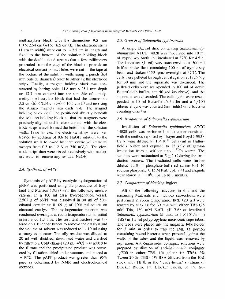
methacrylate block with the dimensions 9.5 mm (h) X 2.54 cm (w) X 16.5 cm (1). The electrode strips (1 cm in width) were cut to - 2.5 cm in length and fixed to the bottom of the solution holding block with the double-sided tape so that a few millimeters protruded from the edge of the block to provide an electrical contact point. Holes were cut in the tape at the bottom of the solution wells using a punch (6.4 mm outside diameter) prior to adhering the electrode strips. Finally, a magnet holding block was con- structed by boring holes (4.8 mm X 25.4 mm depth on 12.7 mm centers) into the top side of a poly- methyl methacrylate block that had the dimensions 3.2 cm (h) X 2.54 cm (w) X 16.5 cm (1) and inserting the Alnico magnets into each hole. The magnet holding block could be positioned directly beneath the solution holding block so that the magnets were precisely aligned and in close contact with the elec- trode strips which formed the bottoms of the solution wells. Prior to use. the electrode strips were pre- treated by addition of 0.6 M NaOH solution to the solution wells followed by three cyclic voltammetry sweeps from 0.3 to 1.2 V at 250 mV/s. The elec- trode strips then were rinsed extensively with nanop- ure water to remove any residual NaOH.
2.4. Synthesis of pAPP
Synthesis of pAPP by catalytic hydrogenation of pNPP was performed using the procedure of Boy- land and Manson (1957) with the following modifi- cations. In a 100 ml glass hydrogenation vessel, 2.503 g of pNPP was dissolved in 30 ml of 50% ethanol containing 0.109 g of 10% palladium on charcoal catalyst. The hydrogenation reaction was conducted overnight at room temperature at an initial pressure of 1.3 atm. The resultant mixture was fil- tered on a Biichner funnel to remove the catalyst and the volume of solvent was reduced to - IO ml using a rotary evaporator. The oily residue was diluted to 20 ml with distilled. de-ionized water and clarified by filtration. Cold ethanol (20 ml. 4°C) was added to the filtrate and the precipitated product was recov- ered by filtration, dried under vacuum. and stored at - 10°C. The pAPP product was greater than 95% pure as determined by NMR and electrochemical methods.
2.5. Growth of Salmonella typhimuriutn
A single Bactrol disk containing Salmotlella ty- phimurium ATCC 14028 was inoculated into 10 ml of tryptic soy broth and incubated at 37°C for 4.5 h. The inoculant (1 ml) was transferred to a 500 ml baffled shake flask containing 100 ml of tryptic soy broth and shaken (150 rpm) overnight at 37°C. The cells were pelleted through centrifugation at 1725 X g for 30 min and the supernate was discarded. The pelleted cells were resuspended in 100 ml of sterile Butterfield’s buffer, centrifuged (as above), and the supernate was discarded. The cells again were resus- pended in 10 ml Butterfield’s buffer and a 1 / 100 diluted aliquot was counted (ten fields) on a bacteria counting chamber.
2.6. Irradiation of Salmonella typhimurium
Irradiation of Salmonella typhitnurium ATCC 14028 cells was performed in a manner consistent with the method reported by Thayer and Boyd (1993). Cells were diluted to 1 X 1O’O cells/ml in Butter- field’s buffer and exposed to 12 kGy of gamma irradiation from a self-contained 13’Cs source. The samples were maintained at 5 k 1°C during the irra- diation process. The irradiated cells were further diluted 1: 10 in phosphate-buffered saline (0.1 M sodium phosphate, 0.15 M NaCl, pH 7.4) and aliquots were stored at - 10°C for up to 3 months.
2.7. Comparison of blocking buffers
All of the following reactions in this and the remaining Materials and methods subsections were performed at room temperature. IMB (20 ~1) were reacted by shaking for 30 min with either TBS (25 mM Tris, 150 mM NaCl, pH 7.6) or irradiated Salmonella ~phimurium (diluted to I X lO’/ml in TBS) in I .5 ml polypropylene microcentrifuge tubes. The tubes were placed into the magnetic tube holder for 3 min in order to trap the IMB (a portion containing bound bacteria when present) against the walls of the tubes and the liquid was removed by aspiration. Anti-Salmonella conjugate solutions were prepared by dilution of anti-Salmonella conjugate l/500 in either TBS. 1% gelatin (in TBS), 2% Tween 20 (in TBS). 1% BSA (diluted from the 10% stock with TBS). or the ‘ready-to-use’ solutions of Blocker Blotto, 1% Blocker casein. or 1% Su-
A.G. Gehring et al. / Journal of Immunological Methods 195 (1996) 15-25 19
perBlock. The IMB were resuspended in 1 ml of anti-Salmonella conjugate solution by gentle vortex- ing. The mixtures again were reacted by shaking for 30 min followed by separation of the IMB with the magnetic tube holder for 3 min. The IMB were washed twice with TBS to facilitate the removal of unreacted anti-Salmonella conjugate. Each wash consisted of resuspension of the IMB in 1 ml of TBS followed by separation with the magnetic tube holder for 3 min and removal of the liquid. The IMB then were resuspended in 0.2 ml TBS, transferred to the solution holding block of the multi-well electrode/magnet (Fig. 11, the magnet holding block was applied for 2 min to trap the IMB against the electrodes, and the liquid was removed by aspiration. After removing the magnets, the IMB were resus- pended by gentle vortexing in 0.22 ml pNPP (2.7 mM in 0.2 M Tris, pH 9.6) and allowed to react for 90 min. During the last 2 min of the pNPP reaction, the magnet holding block was reapplied for 2 min to trap the magnetic beads at the electrode surface. The liquid (0.2 ml) was immediately transferred to polystyrene microwell plates and absorbance was measured at 405 nm. All absorbance values were pNPP blank subtracted to compensate for any spon- taneously formed p-nitrophenol present in the pNPP.
2.8. Preparation of alkaline phosphatase-associated IMB (AP-IMB)
IMB were mixed with anti-goat conjugate in a 1.5 ml polypropylene microcentrifuge tube and diluted with TBS to yield a final concentration of N 1.3 X
10’ IMB/ml and a l/280 anti-goat conjugate dilu- tion. The tube was rotated for 30 min to allow the anti-goat conjugate to react with the IMB. To facili- tate the removal of unreacted anti-goat conjugate, the mixture was centrifuged (2000 X g for 2 min) fol- lowed by removal of the supemate by aspiration. The pellet was washed twice by resuspension through gentle vortexing in TBS containing 0.05% Tween 20, centrifugation (2000 X g for 2 min), and removal of the supemate. The pellet then was washed by resuspension in TBS. centrifugation (2000 X g for 2 min), and removal of the supernate. The resultant AP-associated IMB (AP-IMB) were resuspended in TBS to a concentration of N 1.3 X 10’ AP-IMB/ml. Serially diluted AP-IMB, covering a range from 8.1 X lo* to 2.6 X 10’ AP-IMB/ml, were mixed
with unmodified IMB so that the total bead concen- tration remained at 1.3 X lo7 beads/ml. This gave bead suspensions having varying numbers of AP-IMB yet a constant total amount of beads.
2.9. Calorimetric and electrochemical detection of AP-IMB
Calorimetric analyses were conducted by placing 0.2 ml aliquots of the serially diluted AP-IMB in 0.6 ml polypropylene microcentrifuge tubes and pellet- ing them by centrifugation at 2000 X g for 5 min. Only 180 pl of the supemates were removed by aspiration, 0.2 ml of pNPP (2.7 mM in 0.2 M Tris, pH 9.6) was mixed by gentle vortexing into each tube, and calorimetric development was allowed to proceed for 30 min without agitation. The reaction was stopped by the addition of 25 ~1 of 6 M NaOH and the tubes were again centrifuged at 2000 X g for 5 min. Aliquots (200 pl) of each supemate were transferred to a polystyrene microwell plate and ab- sorbance was measured at 405 nm. All absorbance values were pNPP blank subtracted.
For electrochemical analysis, 200 pl of the seri- ally diluted AP-IMB were added to the solution holding block of the multi-well electrode/magnet (Fig. 1). The beads were magnetically trapped against the electrodes by application of the magnet holding block for 2 min and the liquid was removed by aspiration. With the magnetic field applied, 200 pl of pAPP (2.7 mM in 0.2 M Tris, pH 9.6) was added to the well and allowed to react for 3 min. Produc- tion of electroactive p-aminophenol was measured using Osteryoung square wave voltammetry ( - 300- 300 mV, 25 mV sweep width amplitude, 5 Hz frequency, 4 mV step potential, 10m6 A/V sensitiv- ity) and the peak current was determined by drawing a tangent line across the base of the peak using the BAS 1OOW software.
2.10. Enzyme-linked immunomagnetic calorimetric (ELIMC) and ELIME detection of Salmonella
Using reaction volumes suggested by Dynal, 20 ~1 of IMB were placed in 1.5 ml polypropylene microcentrifuge tubes, 1 ml of heat-killed Salmonella typhimurium (in TBS) was added, and the mixture was incubated by shaking for 30 min. The tubes were placed into the magnetic tube holder for 3 min in order to trap the IMB (a portion containing bound
20 A.G. Gehring et ul./Joumnl of’lnmunological Methods 195 (1996) 15-25
ELIMC ELIME .
Antibody-enzyme
Bacteria 1
Capture ~ \// antibody
pAPP
PAP
Mngnet- \ Graphite ink
electrode
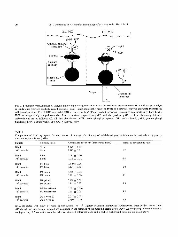
Fig. 2. Schematic representations of enzyme-linked immunomagnetic calorimetric (ELIMC) and electrochemical (ELIME) assays. Analyte
is sandwiched between antibody-coated magnetic beads (immunomagnetic beads or IMBl and antibody-enzyme conjugate followed by
addition of substrate. For ELIMC. suspended IMB are mixed with pNPP and product formation is measured calorimetrically. For ELIME.
IMB are magnetically trapped onto the electrode surface, exposed to pAPP, and the product, PAP, is electrochemically detected.
Abbreviations are as follows: AP, alkaline phosphatase; pNPP. p-nitrophenyl phosphate: pNP. p-nitrophenol; pAPP, p-aminophenyl
phosphate; PAP. p-aminophenol: and pQ1, p-quinone imine.
Table 1 Comparison of blocking agents for the control of non-specific binding of AP-labeled goat anti-Salmonella antibody conjugate to
immunomagnetic beads (IMB)
Sample Blocking agent Absorbance at 405 nm (absorbance units) Signal-to-background ratio
Blank None 2.142 f 0.185 lo6 bacteria None 2.513 f 0.231 1.2
Blank Blotto 0.011 * 0.019 lo6 bacteria Blotto 0.005 f 0.002 0.4
Blank I% BSA 0.140 k 0.067 10’ bacteria 1% BSA 0.277 * 0. I I I 2.0
Blank 1 c/c casein 0.002 * 0.004 lOh bacteria 1 c?c casein 0.185 * 0.094 90
Blank 1% gelatin 0.189 i 0.061 1 O6 bacteria 1 B gelatin 0.345 + 0.202 1.8 _
Blank 1%’ SuperBlock 0.012 + 0.006 1 O6 bacteria 1 B SuperBlock 0.111 *0.051 9.2
Blank 2% Tween 20 0.047 f 0.002 lo6 bacteria 2% Tween 20 0.259 + 0.016 5.5
IMB, incubated with either 0 (blank or background) or IO’ (signal) irradiated Salrnottella ~phimurium, were further reacted with
AP-labeled goat anti-Salmonella antibody conjugate in the presence of the blocking agents listed above. After washing to remove unbound conjugate, any AP associated with the IMB was detected calorimetrically and signal-to-background ratios are indicated above.
A.G. Gehring et al./Journal of Immunological Methods 195 (1996) 15-25 21
08
0 1 2 3 4 5
number of AP-IMB x 1O-4
B
0 s” 0 1 2 3 4 5
number of AP-IMB x 1O-4
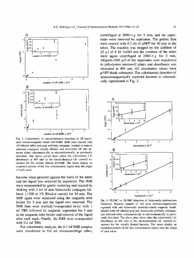
Fig. 3. Calorimetric vs. electrochemical detection of AP-associ-
ated immunomagnetic beads CAP-IMB). IMB were reacted with
AP-labeled rabbit anti-goat antibody conjugate, washed to remove
unbound conjugate, serially diluted, and associated AP was de-
tected either calorimetrically or electrochemically, as previously
described. The above curves show either the calorimetric (A:
absorbance at 405 nm) or the electrochemical (B: current) re-
sponses for the serially diluted AP-IMB. The insets display an
expanded portion of the low concentration region near the origin
of each curve.
bacteria when present) against the walls of the tubes and the liquid was removed by aspiration. The IMB were resuspended by gentle vortexing and reacted by shaking with 1 ml of anti-Salmonella conjugate (di- luted l/500 in 1% Blocker casein) for 30 min. The IMB again were separated using the magnetic tube holder for 3 min and the liquid was removed. The IMB then were washed/resuspended twice with 1 ml TBS followed by magnetic separation for 3 min in the magnetic tube holder and removal of the liquid after each wash. Finally, the IMB were resuspended with 0.2 ml TBS.
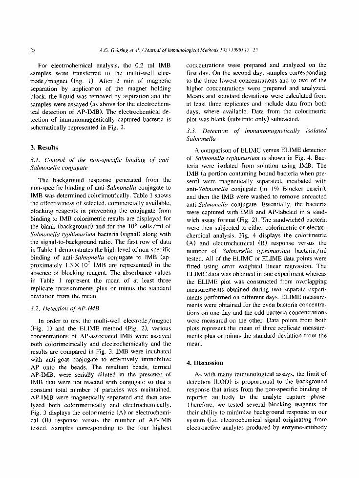
For calorimetric analysis, the 0.2 ml IMB samples were transferred to 0.6 ml microcentrifuge tubes,
centrifuged at 2000 X g for 5 min, and the super- nates were removed by aspiration. The pellets then were reacted with 0.2 ml of pNPP for 30 min in the tubes. The reaction was stopped by the addition of 25 ~1 of 6 M NaOH and the contents of the tubes were again centrifuged at 2000 X g for 5 min. Aliquots (200 ~1) of the supemates were transferred to polystyrene microwell plates and absorbance was measured at 405 nm. All absorbance values were pNPP blank subtracted. The calorimetric detection of immunomagnetically captured bacteria is schemati- cally represented in Fig. 2.
0 i i 3 4 5
bacteria/ml x IO-5
B
bacteriahl x 10-S
Fig. 4. ELIMC vs. ELIME detection of Sulmonella typhimurium
(bacterial. Bacteria samples (1 ml) were immunomagnetically captured with anti-Salmonella antibody-coated magnetic beads,
labeled with AP-labeled goat anti-Suialmonelkr antibody conjugate,
and detected either calorimetrically or electrochemically as previ- ously described. The above plots show either the calorimetric (A: absorbance at 405 nm) or the electrochemical (B: current) re-
sponses for the serially diluted bacteria. The insets display an expanded portion of the low concentration region near the origin
of each curve.
22 A.G. Gehring et al. /Journal of ImmunologicaI Methods 195 (1996) 15-25
For electrochemical analysis, the 0.2 ml IMB samples were transferred to the multi-well elec- trode/magnet (Fig. 1). After 2 min of magnetic separation by application of the magnet holding block, the liquid was removed by aspiration and the samples were assayed (as above for the electrochem- ical detection of AP-IMB). The electrochemical de- tection of immunomagnetically captured bacteria is schematically represented in Fig. 2.
3. Results
3.1. Control of the non-specific binding of anti- Salmonella conjugate
The background response generated from the non-specific binding of anti-Salmonella conjugate to IMB was determined calorimetrically. Table 1 shows the effectiveness of selected, commercially available, blocking reagents in preventing the conjugate from binding to IMB calorimetric results are displayed for the blank (background) and for the lo6 cells/ml of Salmonella typhimurium bacteria (signal) along with the signal-to-background ratio. The first row of data in Table 1 demonstrates the high level of non-specific binding of anti-Salmonella conjugate to IMB (ap- proximately 1.3 X lo7 IMB are represented) in the absence of blocking reagent. The absorbance values in Table 1 represent the mean of at least three replicate measurements plus or minus the standard deviation from the mean.
3.2. Detection of AP-IMB
In order to test the multi-well electrode/magnet (Fig. 1) and the ELIME method (Fig. 2), various concentrations of AP-associated IMB were assayed both calorimetrically and electrochemically and the results are compared in Fig. 3. IMB were incubated with anti-goat conjugate to effectively immobilize AP onto the beads. The resultant beads, termed AP-IMB, were serially diluted in the presence of IMB that were not reacted with conjugate so that a constant total number of particles was maintained. AP-IMB were magnetically separated and then ana- lyzed both calorimetrically and electrochemically. Fig. 3 displays the calorimetric (A) or electrochemi- cal (B) response versus the number of AP-IMB tested. Samples corresponding to the four highest
concentrations were prepared and analyzed on the first day. On the second day, samples corresponding to the three lowest concentrations and to two of the higher concentrations were prepared and analyzed. Means and standard deviations were calculated from at least three replicates and include data from both days, where available. Data from the calorimetric plot was blank (substrate only) subtracted.
3.3. Detection of immunomagnetically isolated Salmonella
A comparison of ELIMC versus ELIME detection of Salmonella typhimurium is shown in Fig. 4. Bac- teria were isolated from solution using IMB. The IMB (a portion containing bound bacteria when pre- sent) were magnetically separated, incubated with anti-Salmonella conjugate (in 1% Blocker casein), and then the IMB were washed to remove unreacted anti-Salmonella conjugate. Essentially, the bacteria were captured with IMB and AP-labeled in a sand- wich assay format (Fig. 2). The sandwiched bacteria were then subjected to either calorimetric or electro- chemical analysis. Fig. 4 displays the calorimetric (A) and electrochemical (B) response versus the number of Salmonella typhimurium bacteria/ml tested. All of the ELIMC or ELIME data points were fitted using error weighted linear regression. The ELIMC data was obtained in one experiment whereas the ELIME plot was constructed from overlapping measurements obtained during two separate experi- ments performed on different days. ELIME measure- ments were obtained for the even bacteria concentra- tions on one day and the odd bacteria concentrations were measured on the other. Data points from both plots represent the mean of three replicate measure- ments plus or minus the standard deviation from the
mean.
4. Discussion
As with many immunological assays, the limit of detection (LOD) is proportional to the background response that arises from the non-specific binding of reporter antibody to the analyte capture phase. Therefore, we tested several blocking reagents for their ability to minimize background response in our system (i.e. electrochemical signal originating from electroactive analytes produced by enzyme-antibody
A.G. Gehring et al. /Journal of Immunological Methods 195 (1996) 15-25 23
conjugate non-specifically bound to IMB). It should be noted that background response due to non- specific binding is distinct from background current that may originate from charging current and/or solvent oxidation during electrochemical processes. Since ELIME employs a small electrode surface area (relative to carbon felt immunoelectrodes), back- ground current is expected to be considerably re- duced with this system. As shown in Table 1, the use of Blocker casein was found to elicit the highest signal-to-background response ratio and hence the most effective at blocking the non-specific binding of anti-Salmonella conjugate to IMB.
The suspended flocculent solids in Blocker Blotto appeared to interfere with the magnetic isolation of the IMB by the custom-built magnetic tube holder. The subsequent poor isolation efficiency (observed visually) of the IMB in the presence of Blotto proba- bly accounted for the low signal displayed (Table 1). Poor isolation of the IMB was also observed when gelatin was used, however the relatively high colori- metric signals reported may reflect an overall poor blocking ability for gelatin. Low isolation efficien- cies for beads in glycerol solutions by the magnetic tube holder (data not shown) suggest that high vis- cosity contributed to the poor IMB isolation in the gelatin solutions. The use of either stronger magnets or longer magnetic isolation times with the magnet tube holder is expected to increase bead isolation efficiency. Research groups that have used magnetic devices, containing rare earth metals, have reported that complex sample matrices, including food, did not adversely affect the separation of bacteria with IMB (Skjerve et al., 1990; Luk and Lindberg, 1991; Skjerve and Olsvik, 1991).
For the detection of AP-IMB (Fig. 3), both the calorimetric and electrochemical techniques exhib- ited similar LOD. However, the electrochemical de- tection of AP-IMB was observed to become nonlin- ear for amounts of AP-IMB that corresponded to current responses of N 2-3 pA. Although the non- linear electrochemical response was a reproducible phenomenon, it was not further investigated since relatively high analyte (enzyme-labeled bacteria) concentrations that produced current responses greater than or equal to this were not of interest. Variance in the electrochemical responses was ob- served to increase with the concentration of analyte,
but this phenomenon also was not further investi- gated for the same reason.
In Fig. 4, the results for both the ELIMC and ELIME detection of Salmonella typhimurium are shown. As for the detection of AP-IMB, both tech- niques produced similar responses and LOD. Many researchers often specify the LOD as two or three times the standard deviation of the mean of a sys- tems blank response added to the blank response. However, this method for defining LOD presumes a constant variance for the responses over the range of analyte concentration measured. ELIME, and to a lesser extent ELIMC, response was observed to dis- play higher variance with increasing concentration of bacteria. We therefore determined the LOD for the two techniques using an alternative protocol. Calibra- tion curves that included data from Fig. 4 were produced by running replicated analyses of standards containing 0, 5 X 10³, 1 X 10⁴, and 5 X lo4 cells/ml and fitting a straight line by weighted linear regres- sion of the data. The LOD was determined using the following formula (adapted from Currie, 1968):
LOD = [ t( nB - 1, +San,“’
+ t( n, - 1, P)SDn,‘/*)]m-
where IZ is the number of replicates, t(k, P) is the abscissa of the t (Student’s) distribution for k de- grees of freedom at the probability level 1 - P, S is the standard deviation of the replicates, and m is the slope of the calibration line. The subscripts B and D refer to the blank and the LOD, respectively. Using na = no = 3, (Y = B = 0.05 (95% confidence limit), and using the standard deviation for the 5 X lo3 cells/ml standard as an estimate of the variance for S, we calculated a LOD of 2 X lo4 cells/ml for ELIMC and 8 X lo3 cells/ml for ELIME. The total assay times, based on a single sample, were w 110 min for ELIMC and - 80 min for ELIME. The rapidity of the ELIME assay relative to ELIMC is attributed to the ten times shorter substrate reaction time and ultimately the highly sensitive nature of electrochemical detection.
Future work will address reducing both the LOD and assay time for ELIME detection of bacteria. The LOD may be lowered by increasing the bead/bacteria capture efficiency. Recovery of Salmonella livingstone in phosphate-buffered saline
24 A.G. Gehring et al./Joumal oj’lmmunological Methods 195 (19961 15-25
has been reported to be 5 1 .O + 7.8% for 10’ cells/ml reacted for 60 min with IO7 antibody-coated Dyn-
abeads (Vermunt et al., 1992). Increased bead/bacteria incubation temperature may also im- prove capture efficiency and/or analysis speed. The use of protein A in immobilizing antibodies to beads in a ‘proper’ orientation has been demonstrated to enhance the recovery of Salmonella (Widjojoatmodjo
et al., 1993). Lectins (Payne et al., 1993) or carbo- hydrates (Nilsson and Mandenius, 1994) added to or replacing the antibodies on the beads may also en- hance recovery.
Acknowledgements
We thank Al Armbruster (The Motson Co., Flour- town, PA) for helpful discussions, Dr. Donald Thayer, Dr. Augustine Kim, and Glenn Boyd (U.S. Department of Agriculture, Agricultural Research Service, Eastern Regional Research Center) for sup- plying the irradiated Salmonella typhimurium cells, and Susan Wilzer (U.S. Department of Agriculture, National Agricultural Library) for conducting litera- ture searches.
References
Bej, A.K., Mahbubani, M.H.. Boyce, M.J. and Atlas, R.M. (19941
Detection of Salmonella spp. in oysters by PCR. Appl. Envi-
ron. Microbial. 60, 368-373.
Boyland, E. and Mattson, D. (1957) The oxidation of aromatic
amines part V oxidation by perphosphoric acids. J. Chem. Sot.
4689-4694.
Brooks, J., Mirhabibollahi, B. and Kroll, R.G. (19921 Experimen- tal enzyme-linked amperometric immunosensors for the detec-
tion of salmonellas in foods. J. Appl. Bacterial. 73. 189-196.
Connolly, P. Bloomfield, S.F. and Denyer, S.P. (19931 A study of
the use of rapid methods for preservative efficacy testing of
pharmaceuticals and cosmetics. J. Appl. Bacterial. 75, 456-
462.
Currie, L.A. (1968) Limits for qualitative detection and quantita- tive determination, Anal. Chem. 40. 586-593.
Feng, P. (1992) Commercial assay systems for detecting food- borne Salmonella: a review. J. Food Prot. 55, 927-934.
Fratamico, P.M., Schultz, F.J. and Buchanan, R.L. (19921 Rapid isolation of Escherichia coli 0157:H7 from enrichment cul-
tures of foods using an immunomagnetic separation method.
Food Microbial. 9, 105-l 13. Griftiths. M.W. (1993) Applications of bioluminescence in the
dairy industry. J. Dairy Sci. 76. 3118-3125.
Hadas, E., Soussan, L., Rosen-Margalit. E., Farkash, A. and
Rishpon, J. f 1992) A rapid and sensitive heterogeneous immu-
noelectrochemical assay using disposable electrodes. J. Im-
munoassay 13. 23 l-252.
Hartman, P.A., Swaminathan, B.. Curiale, M.S., Firstenberg-Eden,
R.. Sharpe, A.N., Cox. N.A.. Fung. D.Y.C. and Goldschimidt,
M.C. (1992) Rapid methods and automation. In: C. Van-
derzant and D.F. Splittstoesser (Eds.1, Compendium of Meth-
ods for the Microbiological Examination of Foods, 3rd edn.
American Public Health Association, Washington, DC, pp.
665-746.
Heinemann. W.R., Anderson, C.W. and Halsall, H.B. (19791
Immunoassay by differential pulse polarography. Science 204.
865-866.
Ibrahim, G.F. (1986) A review of immunoassays and their applica-
tion to salmonellae detection in foods. J. Food Prot. 49,
299-3 10.
Luk J.M.C. and Lindberg, A.A. (19911 Rapid and sensitive detec-
tion of Salmonel~u (0:6.71 by immunomagnetic monoclonal
antibody-based assays. J. Immunol. Methods 137, l-8.
Minunni. M.. Mascini, M., Cluilbault. G.G. and Hock, B. (1995)
The quartz crystal microbalance as biosensor, a status report
on its future. Anal. Lett. 28, 749-764.
Molday. R.S., Yen. S.P.S. and Rembaum, A. (1977) Application
of magnetic microspheres in labelling and separation of cells.
Nature 268, 437-438.
Nilsson. K.G.I. and Mandenius, C.-F. (19941 A carbohydrate
biosensor surface for the detection of uropathogenic bacteria.
Bio/Technology 12, 1376-1378.
Olsvik, 8.. Popovic, T., Skjerve, E., Cudjoe, KS., Homes, E.,
Ugelstad. J. and UhlCn, M. (19941 Magnetic separation tech-
niques in diagnostic microbiology. Clin. Microbial. Rev. 7.
43-54.
Park, C.E., Akhtar, M. and Rayman, M.K. (19941 Evaluation of a
commercial enzyme immunoassay kit (RIDASCREEN) for
detection of staphylococcal enterotoxins A, B. C. D, and E in
foods, Appl. Environ, Microbial. 60, 677-68 1,
Payne, M.J.. Campbell. S. and Kroll, R.G. (1993) Lectin-magnetic
separation can enhance methods for the detection of Staphylo-
coccus aureus. Salmonella enteritidis, and Listeria monocpo-
genes. Food Microbial. 10, 75-83.
Rishpon. J., Gezundhajt, Y., Soussan, L., Rosen-Margalit. I. and
Hadas, E. (19921 Immunoelectrodes for the detection of bacte-
ria. In: Biosensor. Design and Application, Ch. 6. ACS Sym-
posium Series no. 511. pp. 59-72.
Robinson, G.A., Hill, H.A.O., Philo, R.D.. Gear. J.M.. Rattle, S.J.
and Forrest, G.C. (19851 Bioelectrochemical enzyme im-
munoassay of human choriogonadotropin with magnetic eiec-
trades. Clin. Chem. 31. 1449-1452. Skjerve, E. and Olsvik, 0. (1991) Immunomagnetic separation of
Salmonella from foods. Int. J. Food Microbial. 14, 11-18. Skjerve, E., Rorvik, L.M. and Olsvik, 0. (19901 Detection of
Listeria monocgtogenes in foods by immunomagnetic separa- tion Appl. Environ. Microbial. 56, 3478-3481.
Thayer, D.W. and Boyd. G. (1993) Elimination of Escherichin
coli 0157:H7 in meats by gamma irradiation. Appl. Environ.
Microbial. 59. 1030-1034.
Tortorello, M.L. and Stewart. D.S. (19941 Antibody-direct epiflu- orescent filter technique for rapid direct enumeration of Es-

A.G. Gehring et al. /Journal of Immunological Methods 195 (1996) 15-25 25
cherichia coli 0157:H7 in beef. Appl. Environ. Microbial. 60,
3553-3559.
Vermunt, A.E.M., Franken, A.A.J.M. and Beumer, R.R. (1992)
Isolation of salmonellas by immunomagnetic separation. J.
Appl. Bacterial. 72. 112-l 18.
Watts, H.J., Lowe. C.R. and Pollard-Knight, D.V. (1994) Optical
biosensor for monitoring microbial cells. Anal. Chem. 66,
2465-2470.
Weetall, H.H. and Hotaling. T. (1987) A simple, inexpensive,
disposable electrochemical sensor for clinical and immuno-as-
say. Biosensors 3, 57-63.
Widjojoatmodjo, M.N., Fluit, A.C., Torensma, R. and Verhoef, J.
(1993) Comparison of immunomagnetic beads coated with
protein A, protein G, or goat anti-mouse immunoglobulins:
Applications in enzyme immunoassays and immunomagnetic
separations, J. Immunol. Methods 165, 1 l-19.