Enhancement of the Detection Performance of Paper-Based ...
31
Citation: Pang, R.; Zhu, Q.; Wei, J.; Meng, X.; Wang, Z. Enhancement of the Detection Performance of Paper-Based Analytical Devices by Nanomaterials. Molecules 2022, 27, 508. https://doi.org/10.3390/ molecules27020508 Academic Editors: Giuseppe Cirillo, Chongjun Zhao and Sungsu Park Received: 27 November 2021 Accepted: 10 January 2022 Published: 14 January 2022 Publisher’s Note: MDPI stays neutral with regard to jurisdictional claims in published maps and institutional affil- iations. Copyright: © 2022 by the authors. Licensee MDPI, Basel, Switzerland. This article is an open access article distributed under the terms and conditions of the Creative Commons Attribution (CC BY) license (https:// creativecommons.org/licenses/by/ 4.0/). molecules Review Enhancement of the Detection Performance of Paper-Based Analytical Devices by Nanomaterials Renzhu Pang 1 , Qunyan Zhu 2 , Jia Wei 1,2 , Xianying Meng 1, * and Zhenxin Wang 2,3, * 1 Department of Thyroid Surgery, The First Hospital of Jilin University, Changchun 130021, China; [email protected] (R.P.); [email protected] (J.W.) 2 State Key Laboratory of Electroanalytical Chemistry, Changchun Institute of Applied Chemistry, Chinese Academy of Sciences, Changchun 130022, China; [email protected] 3 School of Applied Chemical Engineering, University of Science and Technology of China, Hefei 230026, China * Correspondence: [email protected] (X.M.); [email protected] (Z.W.) Abstract: Paper-based analytical devices (PADs), including lateral flow assays (LFAs), dipstick assays and microfluidic PADs (μPADs), have a great impact on the healthcare realm and environmental monitoring. This is especially evident in developing countries because PADs-based point-of-care testing (POCT) enables to rapidly determine various (bio)chemical analytes in a miniaturized, cost- effective and user-friendly manner. Low sensitivity and poor specificity are the main bottlenecks associated with PADs, which limit the entry of PADs into the real-life applications. The application of nanomaterials in PADs is showing great improvement in their detection performance in terms of sensitivity, selectivity and accuracy since the nanomaterials have unique physicochemical properties. In this review, the research progress on the nanomaterial-based PADs is summarized by highlighting representative recent publications. We mainly focus on the detection principles, the sensing mecha- nisms of how they work and applications in disease diagnosis, environmental monitoring and food safety management. In addition, the limitations and challenges associated with the development of nanomaterial-based PADs are discussed, and further directions in this research field are proposed. Keywords: paper-based analytical devices; nanomaterials; point-of-care testing; signal enhancement 1. Introduction As an easily accessible and cheap material made from cellulose (the most abundant polymer on earth) or nitrocellulose, paper offers many advantages for development of biosensing platforms, in particular point-of-care-testing (POCT) devices [1–7]. For instance, various in situ analyses can be achieved by the paper-based analytical devices (PADs) because many recognition probes, such as ligands, antibodies and aptamers, can be easily immobilized within the (nitro)cellulose matrix [8–20]. Because of the controlled porosity and capillary forces of the nitrocellulose/cellulose network, the fluidics can be efficiently transported via capillary flow. In addition, the PADs are compatible with different detection systems, including naked eye and simple optical or electrical readers, which meets the needs in developing countries [21–25]. In the past decades, PADs, including lateral flow assays (LFAs), dipstick assays and microfluidic PADs (μPADs), have been well developed and shown a great impact on the clinical diagnosis, environmental monitoring and food safety management [8–35]. For example, one type of PAD, the lateral flow immunoassay (LFIA), is commercially available and is extensively used as a powerful diagnostic platform for the rapid detection of antibodies or antigens at a low cost [34–36]. The LFIA has dominated the market of rapid diagnostic testing since the lateral flow immunochromatographic strip was first developed for screening the supernatants of hybridomas in 1982 through antigen–antibody interaction on paper to produce a color change visible to the naked eye [34–37]. Molecules 2022, 27, 508. https://doi.org/10.3390/molecules27020508 https://www.mdpi.com/journal/molecules
-
Upload
khangminh22 -
Category
Documents
-
view
3 -
download
0
Transcript of Enhancement of the Detection Performance of Paper-Based ...
Citation: Pang, R.; Zhu, Q.; Wei, J.;
Meng, X.; Wang, Z. Enhancement of
the Detection Performance of
Paper-Based Analytical Devices by
Nanomaterials. Molecules 2022, 27,
508. https://doi.org/10.3390/
molecules27020508
Academic Editors: Giuseppe Cirillo,
Chongjun Zhao and Sungsu Park
Received: 27 November 2021
Accepted: 10 January 2022
Published: 14 January 2022
Publisher’s Note: MDPI stays neutral
with regard to jurisdictional claims in
published maps and institutional affil-
iations.
Copyright: © 2022 by the authors.
Licensee MDPI, Basel, Switzerland.
This article is an open access article
distributed under the terms and
conditions of the Creative Commons
Attribution (CC BY) license (https://
creativecommons.org/licenses/by/
4.0/).
molecules
Review
Enhancement of the Detection Performance of Paper-BasedAnalytical Devices by NanomaterialsRenzhu Pang 1, Qunyan Zhu 2, Jia Wei 1,2, Xianying Meng 1,* and Zhenxin Wang 2,3,*
1 Department of Thyroid Surgery, The First Hospital of Jilin University, Changchun 130021, China;[email protected] (R.P.); [email protected] (J.W.)
2 State Key Laboratory of Electroanalytical Chemistry, Changchun Institute of Applied Chemistry,Chinese Academy of Sciences, Changchun 130022, China; [email protected]
3 School of Applied Chemical Engineering, University of Science and Technology of China, Hefei 230026, China* Correspondence: [email protected] (X.M.); [email protected] (Z.W.)
Abstract: Paper-based analytical devices (PADs), including lateral flow assays (LFAs), dipstick assaysand microfluidic PADs (µPADs), have a great impact on the healthcare realm and environmentalmonitoring. This is especially evident in developing countries because PADs-based point-of-caretesting (POCT) enables to rapidly determine various (bio)chemical analytes in a miniaturized, cost-effective and user-friendly manner. Low sensitivity and poor specificity are the main bottlenecksassociated with PADs, which limit the entry of PADs into the real-life applications. The applicationof nanomaterials in PADs is showing great improvement in their detection performance in terms ofsensitivity, selectivity and accuracy since the nanomaterials have unique physicochemical properties.In this review, the research progress on the nanomaterial-based PADs is summarized by highlightingrepresentative recent publications. We mainly focus on the detection principles, the sensing mecha-nisms of how they work and applications in disease diagnosis, environmental monitoring and foodsafety management. In addition, the limitations and challenges associated with the development ofnanomaterial-based PADs are discussed, and further directions in this research field are proposed.
Keywords: paper-based analytical devices; nanomaterials; point-of-care testing; signal enhancement
1. Introduction
As an easily accessible and cheap material made from cellulose (the most abundantpolymer on earth) or nitrocellulose, paper offers many advantages for development ofbiosensing platforms, in particular point-of-care-testing (POCT) devices [1–7]. For instance,various in situ analyses can be achieved by the paper-based analytical devices (PADs)because many recognition probes, such as ligands, antibodies and aptamers, can be easilyimmobilized within the (nitro)cellulose matrix [8–20]. Because of the controlled porosityand capillary forces of the nitrocellulose/cellulose network, the fluidics can be efficientlytransported via capillary flow. In addition, the PADs are compatible with different detectionsystems, including naked eye and simple optical or electrical readers, which meets the needsin developing countries [21–25]. In the past decades, PADs, including lateral flow assays(LFAs), dipstick assays and microfluidic PADs (µPADs), have been well developed andshown a great impact on the clinical diagnosis, environmental monitoring and food safetymanagement [8–35]. For example, one type of PAD, the lateral flow immunoassay (LFIA),is commercially available and is extensively used as a powerful diagnostic platform for therapid detection of antibodies or antigens at a low cost [34–36]. The LFIA has dominatedthe market of rapid diagnostic testing since the lateral flow immunochromatographicstrip was first developed for screening the supernatants of hybridomas in 1982 throughantigen–antibody interaction on paper to produce a color change visible to the nakedeye [34–37].
Molecules 2022, 27, 508. https://doi.org/10.3390/molecules27020508 https://www.mdpi.com/journal/molecules
Molecules 2022, 27, 508 2 of 31
The PADs detection methods include optical techniques (colorimetry, fluorescenceand surface enhancement Raman scattering (SERS), etc.) and electrochemical (EC) methods(amperometry, potentiometry, voltammetry, electrochemical impedance spectroscopy (EIS),electrochemiluminescence (ECL) and photoelectrochemistry (PEC)) [21–25]. Among thesedetection methods, colorimetry offers simplicity and convenience and, until 2009, hadbeen one of the main detection methods in PADs. The biggest advantage of paper-basedcolorimetric devices is that the presence of a specific analyte can be distinguished easilywith the naked eye without expensive and complex instruments through the change ofcolor. However, the colorimetric method is limited to qualitative yes/no detections and/orsemi-quantitative analysis because it has several inherent disadvantages, such as narrowdynamic range, poor sensitivity and being easily interfered with by environmental light andbiased by users’ subjectivity. The EC method was first used as a PAD detection technique in2009 by Dungachi et al. [38]. Generally, the analytical performances (especially sensitivity)of electrochemical paper-based analytical devices (also known as ePADs) are better thanthose of paper-based colorimetric analytical devices [18,23–25,28,31]. Unfortunately, theanalytical performance (in particular, selectivity) of ePADs can be decreased significantlyin complex matrices. Currently, the EC detection and optical detection are accounted for asthe two main detection methods of PADs (as shown in Figure 1).
Molecules 2022, 27, x FOR PEER REVIEW 2 of 28
immunochromatographic strip was first developed for screening the supernatants of hy-bridomas in 1982 through antigen–antibody interaction on paper to produce a color change visible to the naked eye [34–37].
The PADs detection methods include optical techniques (colorimetry, fluorescence and surface enhancement Raman scattering (SERS), etc.) and electrochemical (EC) meth-ods (amperometry, potentiometry, voltammetry, electrochemical impedance spectros-copy (EIS), electrochemiluminescence (ECL) and photoelectrochemistry (PEC)) [21–25]. Among these detection methods, colorimetry offers simplicity and convenience and, until 2009, had been one of the main detection methods in PADs. The biggest advantage of paper-based colorimetric devices is that the presence of a specific analyte can be distin-guished easily with the naked eye without expensive and complex instruments through the change of color. However, the colorimetric method is limited to qualitative yes/no de-tections and/or semi-quantitative analysis because it has several inherent disadvantages, such as narrow dynamic range, poor sensitivity and being easily interfered with by envi-ronmental light and biased by users’ subjectivity. The EC method was first used as a PAD detection technique in 2009 by Dungachi et al. [38]. Generally, the analytical performances (especially sensitivity) of electrochemical paper-based analytical devices (also known as ePADs) are better than those of paper-based colorimetric analytical devices [18,23–25,28,31]. Unfortunately, the analytical performance (in particular, selectivity) of ePADs can be decreased significantly in complex matrices. Currently, the EC detection and opti-cal detection are accounted for as the two main detection methods of PADs (as shown in Figure 1).
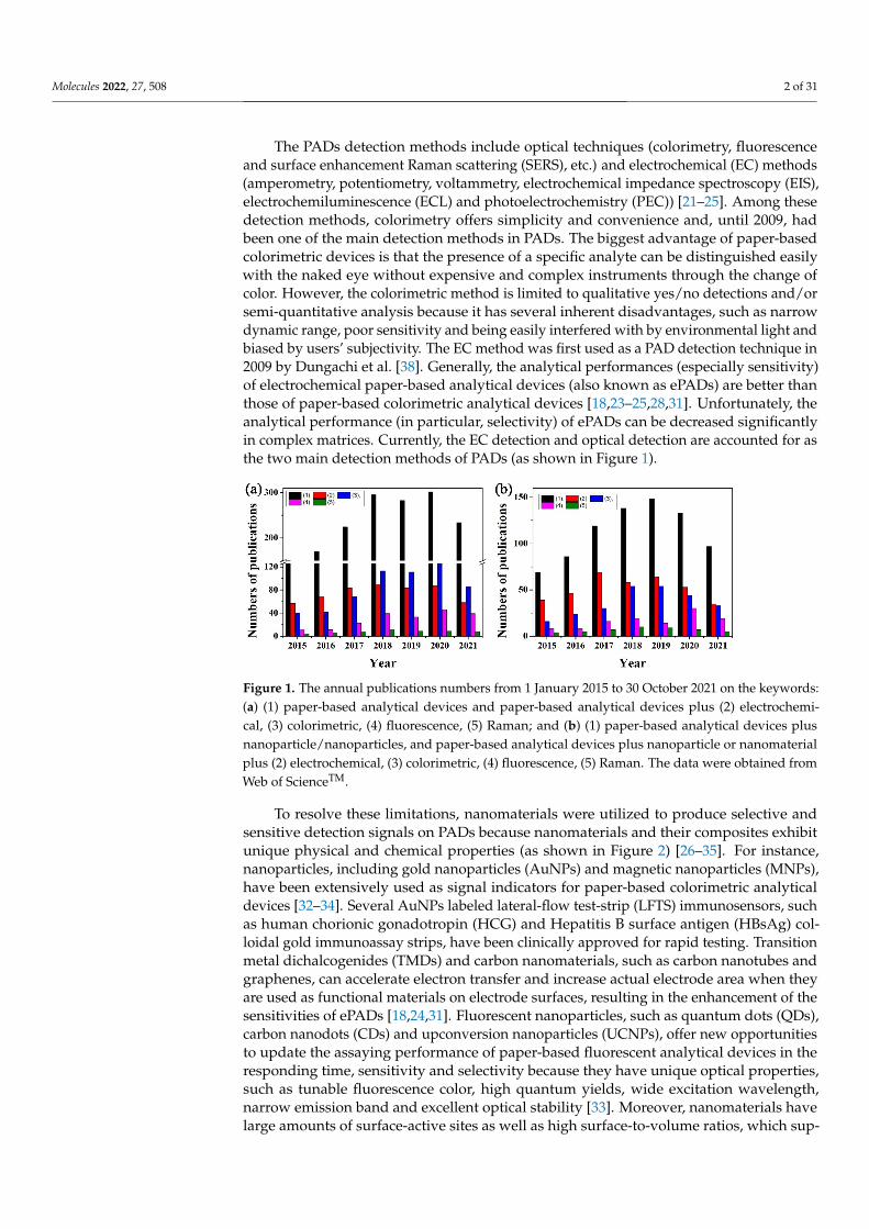
Figure 1. The annual publications numbers from 1 January 2015 to 30 October 2021 on the keywords: (a) (1) paper-based analytical devices and paper-based analytical devices plus (2) electrochemical, (3) colorimetric, (4) fluorescence, (5) Raman; and (b) (1) paper-based analytical devices plus nano-particle/nanoparticles, and paper-based analytical devices plus nanoparticle or nanomaterial plus (2) electrochemical, (3) colorimetric, (4) fluorescence, (5) Raman. The data were obtained from Web of ScienceTM.
To resolve these limitations, nanomaterials were utilized to produce selective and sensitive detection signals on PADs because nanomaterials and their composites exhibit unique physical and chemical properties (as shown in Figure 2) [26–35]. For instance, na-noparticles, including gold nanoparticles (AuNPs) and magnetic nanoparticles (MNPs), have been extensively used as signal indicators for paper-based colorimetric analytical devices [32–34]. Several AuNPs labeled lateral-flow test-strip (LFTS) immunosensors, such as human chorionic gonadotropin (HCG) and Hepatitis B surface antigen (HBsAg) colloidal gold immunoassay strips, have been clinically approved for rapid testing. Tran-sition metal dichalcogenides (TMDs) and carbon nanomaterials, such as carbon nanotubes and graphenes, can accelerate electron transfer and increase actual electrode area when they are used as functional materials on electrode surfaces, resulting in the enhancement of the sensitivities of ePADs [18,24,31]. Fluorescent nanoparticles, such as quantum dots (QDs), carbon nanodots (CDs) and upconversion nanoparticles (UCNPs), offer new op-portunities to update the assaying performance of paper-based fluorescent analytical
Figure 1. The annual publications numbers from 1 January 2015 to 30 October 2021 on the keywords:(a) (1) paper-based analytical devices and paper-based analytical devices plus (2) electrochemi-cal, (3) colorimetric, (4) fluorescence, (5) Raman; and (b) (1) paper-based analytical devices plusnanoparticle/nanoparticles, and paper-based analytical devices plus nanoparticle or nanomaterialplus (2) electrochemical, (3) colorimetric, (4) fluorescence, (5) Raman. The data were obtained fromWeb of ScienceTM.
To resolve these limitations, nanomaterials were utilized to produce selective andsensitive detection signals on PADs because nanomaterials and their composites exhibitunique physical and chemical properties (as shown in Figure 2) [26–35]. For instance,nanoparticles, including gold nanoparticles (AuNPs) and magnetic nanoparticles (MNPs),have been extensively used as signal indicators for paper-based colorimetric analyticaldevices [32–34]. Several AuNPs labeled lateral-flow test-strip (LFTS) immunosensors, suchas human chorionic gonadotropin (HCG) and Hepatitis B surface antigen (HBsAg) col-loidal gold immunoassay strips, have been clinically approved for rapid testing. Transitionmetal dichalcogenides (TMDs) and carbon nanomaterials, such as carbon nanotubes andgraphenes, can accelerate electron transfer and increase actual electrode area when theyare used as functional materials on electrode surfaces, resulting in the enhancement of thesensitivities of ePADs [18,24,31]. Fluorescent nanoparticles, such as quantum dots (QDs),carbon nanodots (CDs) and upconversion nanoparticles (UCNPs), offer new opportunitiesto update the assaying performance of paper-based fluorescent analytical devices in theresponding time, sensitivity and selectivity because they have unique optical properties,such as tunable fluorescence color, high quantum yields, wide excitation wavelength,narrow emission band and excellent optical stability [33]. Moreover, nanomaterials havelarge amounts of surface-active sites as well as high surface-to-volume ratios, which sup-
Molecules 2022, 27, 508 3 of 31
port diverse functionalization with high densities of recognition units. The phenomenonfurther improves the detection performances of PADs. Therefore, the integration of nano-materials with PADs enables strong quantitative capabilities of PADs and expands theirapplicable fields.
Molecules 2022, 27, x FOR PEER REVIEW 3 of 28
devices in the responding time, sensitivity and selectivity because they have unique opti-cal properties, such as tunable fluorescence color, high quantum yields, wide excitation wavelength, narrow emission band and excellent optical stability [33]. Moreover, nano-materials have large amounts of surface-active sites as well as high surface-to-volume ra-tios, which support diverse functionalization with high densities of recognition units. The phenomenon further improves the detection performances of PADs. Therefore, the inte-gration of nanomaterials with PADs enables strong quantitative capabilities of PADs and expands their applicable fields.
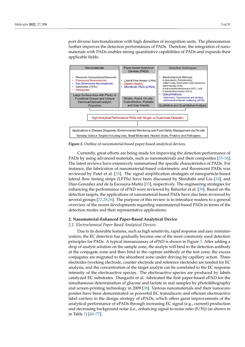
Figure 2. Outline of nanomaterial-based paper-based analytical devices.
Currently, great efforts are being made for improving the detection performance of PADs by using advanced materials, such as nanomaterials and their composites [33–36]. The latest reviews have extensively summarized the specific characteristics of PADs. For instance, the fabrication of nanomaterial-based colorimetric and fluorescent PADs was reviewed by Patel et al. [33]. The signal amplification strategies of nanoparticle-based lat-eral flow testing strips (LFTSs) have been discussed by Shirshahi and Liu [34], and Díaz-González and de la Escosura-Muñiz [35], respectively. The engineering strategies for en-hancing the performance of ePAD were reviewed by Baharfar et al. [39]. Based on the detection targets, the applications of nanomaterial-based PADs have also been reviewed by several groups [27,28,36]. The purpose of this review is to introduce readers to a general overview of the recent developments regarding nanomaterial-based PADs in terms of the detection modes and their representative applications.
2. Nanomaterial-Enhanced Paper-Based Analytical Device 2.1. Electrochemical Paper-Based Analytical Devices
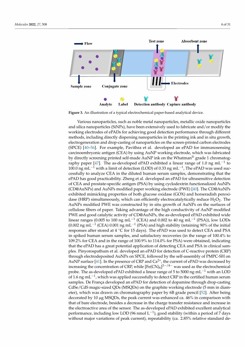
Due to its desirable features, such as high sensitivity, rapid response and easy min-iaturization, the EC detection has gradually become one of the most commonly used de-tection principles for PADs. A typical immunoassay of ePAD is shown in Figure 3. After adding a drop of analyte solution on the sample zone, the analyte will bind to the detec-tion antibody at the conjugate zone and then bind to the capture antibody at the test zone; the excess conjugates are migrated to the absorbent zone under driving by capillary ac-tion. Three electrodes (working electrode, counter electrode and reference electrode) are needed for EC analysis, and the concentration of the target analyte can be correlated to the EC response intensity of the electroactive species. The electroactive species are pro-duced by labels catalyzed EC substrates. Dungachi et al. fabricated the first paper-based ePAD for the simultaneous determination of glucose and lactate in real samples by pho-tolithography and screen-printing technology in 2009 [38]. Various nanomaterials and their nanocomposites have been demonstrated as powerful EC transducers and efficient
Figure 2. Outline of nanomaterial-based paper-based analytical devices.
Currently, great efforts are being made for improving the detection performance ofPADs by using advanced materials, such as nanomaterials and their composites [33–36].The latest reviews have extensively summarized the specific characteristics of PADs. Forinstance, the fabrication of nanomaterial-based colorimetric and fluorescent PADs wasreviewed by Patel et al. [33]. The signal amplification strategies of nanoparticle-basedlateral flow testing strips (LFTSs) have been discussed by Shirshahi and Liu [34], andDíaz-González and de la Escosura-Muñiz [35], respectively. The engineering strategies forenhancing the performance of ePAD were reviewed by Baharfar et al. [39]. Based on thedetection targets, the applications of nanomaterial-based PADs have also been reviewed byseveral groups [27,28,36]. The purpose of this review is to introduce readers to a generaloverview of the recent developments regarding nanomaterial-based PADs in terms of thedetection modes and their representative applications.
2. Nanomaterial-Enhanced Paper-Based Analytical Device2.1. Electrochemical Paper-Based Analytical Devices
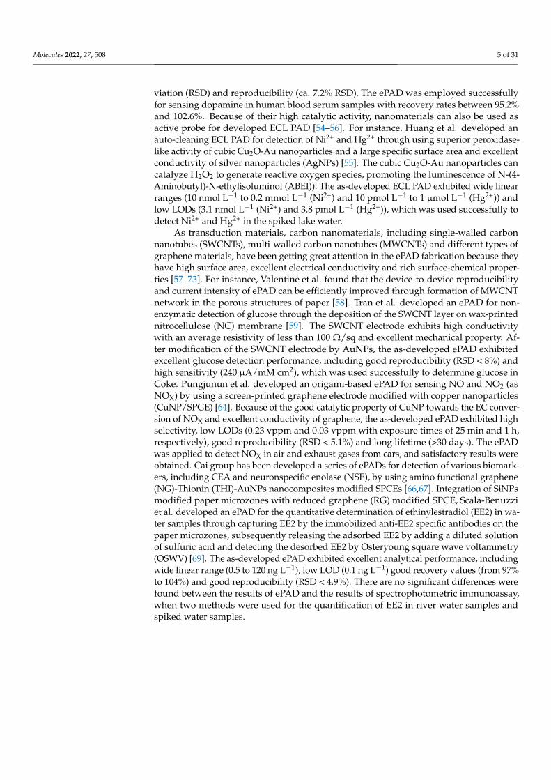
Due to its desirable features, such as high sensitivity, rapid response and easy miniatur-ization, the EC detection has gradually become one of the most commonly used detectionprinciples for PADs. A typical immunoassay of ePAD is shown in Figure 3. After adding adrop of analyte solution on the sample zone, the analyte will bind to the detection antibodyat the conjugate zone and then bind to the capture antibody at the test zone; the excessconjugates are migrated to the absorbent zone under driving by capillary action. Threeelectrodes (working electrode, counter electrode and reference electrode) are needed for ECanalysis, and the concentration of the target analyte can be correlated to the EC responseintensity of the electroactive species. The electroactive species are produced by labelscatalyzed EC substrates. Dungachi et al. fabricated the first paper-based ePAD for thesimultaneous determination of glucose and lactate in real samples by photolithographyand screen-printing technology in 2009 [38]. Various nanomaterials and their nanocom-posites have been demonstrated as powerful EC transducers and efficient electroactivelabel carriers in the design strategy of ePADs, which offers great improvements of theanalytical performance of ePADs through increasing EC signal (e.g., current) productionand decreasing background noise (i.e., enhancing signal-to-noise ratio (S/N)) (as shown inin Table 1) [40–77].
Molecules 2022, 27, 508 4 of 31
Molecules 2022, 27, x FOR PEER REVIEW 4 of 28
electroactive label carriers in the design strategy of ePADs, which offers great improve-ments of the analytical performance of ePADs through increasing EC signal (e.g., current) production and decreasing background noise (i.e., enhancing signal-to-noise ratio (S/N)) (as shown in in Table 1) [40–77].
Figure 3. An illustration of a typical electrochemical paper-based analytical device.
Table 1. The typical nanomaterial-enhanced ePADs for sensing various analytes.
Nanomaterials Modification
Methods Electrochemical
Method Analytes Linear Ranges
Limit of Detection
Real Samples Recovery Ref.
AuNPs In situ growth DPV CEA and
PSA
5 × 10⁻³ to 100 ng mL−1 (CEA) and 2 × 10⁻³ to
40 ng mL−1 (PSA)
2 × 10⁻³ ng mL−1 (CEA) 1 × 10⁻³ ng
mL−1 (PSA)
Human se-rum
- [40]
AuNPs Electrodeposition DPV CRP 5 to 5 × 10³ ng
mL−1 1.6 ng mL−1
Certified hu-man serum
- [41]
AuNPs Electrodeposition DPV EGFR 0.5 to 500 nmol
L−1 0.167 nmol L−1
Saliva sam-ples
- [42]
AuNPs Drop-casting DPV
H1047R (A3140G) missense mutation in exon 20
- 5 nmol L−1 (signal
on) and 6 nmol L−1 (signal off)
- - [43]
AuNPs Electrodeposition Impedimetry miRNA
155 0 to 4 × 10³ ng
mL−1 6.9 × 10² ng mL−1 (93.4 nmol L−1)
Fetal bovine serum
- [44]
AuNPs Drop-casting SWASV Hg2+ 5 to 200 ng
mL−1 2.5 ng mL−1
Drinking wa-ter
95% to 104%
[45]
Poly (N-vinylpy-rolidone) AuNPs
Screen-printing Chronoam-perometry
Glucose 1 × 10⁴ to 1.5 × 10⁶ nmol L−1
2.6 × 10⁴ nmol L−1 - - [46]
Poly (N-vinylpy-rolidone) AuNPs
Screen-printing DPV CEA 1 to 100 ng
mL−1 0.33 ng mL−1
Diluted hu-man serum
99.58% to 102.50%
[47]
Pd decoration of Cu/Co-doped CeO2 (CuCo-
CeO2-Pd) nano-spheres and ur-chin-like AuNPs
In situ growth DPV Amyloid-
β 1 × 10⁻³ to 100
nmol L−1 5 × 10⁻⁵ nmol L−1
Artificial cer-ebrospinal
fluid and hu-man serum
99% to 100.5%
[48]
N-CDs, TiO2 NPs and Pt NPs
Drop-casting PEC CEA 2 × 10⁻³ to 200
ng mL−1 1.0 × 10⁻³ ng mL−1
Living MCF-7 cells
- [49]
Figure 3. An illustration of a typical electrochemical paper-based analytical device.
Various nanoparticles, such as noble metal nanoparticles, metallic oxide nanoparticlesand silica nanoparticles (SiNPs), have been extensively used to fabricate and/or modify theworking electrodes of ePADs for achieving good detection performance through differentmethods, including directly dispersing nanoparticles in the printing ink and in situ growth,electrogeneration and drop-casting of nanoparticles on the screen-printed carbon electrodes(SPCE) [40–56]. For example, Pavithra et al. developed an ePAD for immunosensingcarcinoembryonic antigen (CEA) by using AuNP working electrode, which was fabricatedby directly screening printed self-made AuNP ink on the Whatman® grade 1 chromatog-raphy paper [47]. The as-developed ePAD exhibited a linear range of 1.0 ng mL−1 to100.0 ng mL−1 with a limit of detection (LOD) of 0.33 ng mL−1, The ePAD was used suc-cessfully to analyze CEA in the diluted human serum samples, demonstrating that theePAD has good practicability. Zheng et al. developed an ePAD for ultrasensitive detectionof CEA and prostate-specific antigen (PSA) by using cyclodextrin functionalized AuNPs(CD@AuNPs) and AuNPs modified paper working electrode (PWE) [40]. The CD@AuNPsexhibited mimicking properties of both glucose oxidase (GOX) and horseradish peroxi-dase (HRP) simultaneously, which can efficiently electrocatalytically reduce H2O2. TheAuNPs modified PWE was constructed by in situ growth of AuNPs on the surfaces ofcellulose fibers of paper. Taking advantage of the high conductivity of AuNP modifiedPWE and good catalytic activity of CD@AuNPs, the as-developed ePAD exhibited widelinear ranges (0.005 to 100 ng mL−1 (CEA) and 0.002 to 40 ng mL−1 (PSA)), low LODs(0.002 ng mL−1 (CEA) 0.001 ng mL−1 (PSA) and high stability (retaining 90% of the initialresponses after stored at 4 C for 15 days). The ePAD was used to detect CEA and PSAin spiked human serum samples, and satisfactory recoveries (in the range of 100.4% to109.2% for CEA and in the range of 100.9% to 114.0% for PSA) were obtained, indicatingthat the ePAD has a great potential application of detecting CEA and PSA in clinical sam-ples. Pinyorospathum et al. developed an ePAD for detection of C-reactive protein (CRP)through electrodeposited AuNPs on SPCE, followed by the self-assembly of PMPC-SH onAuNP surface [41]. In the presence of CRP and Ca2+, the current of ePAD was decreased byincreasing the concentration of CRP, while [Fe(CN)6]3−/4− was used as the electrochemicalprobe. The as-developed ePAD exhibited a linear range of 5 to 5000 ng·mL−1 with an LODof 1.6 ng·mL−1, which was applied successfully to detect CRP in the certified human serumsamples. De França developed an ePAD for detection of dopamine through drop-castingCdSe/CdS magic-sized QDs (MSQDs) on the graphite working electrode (5 mm in diam-eter), which was drawn on chromatography paper by 6B grade pencil [52]. After beingdecorated by 10 µg MSQDs, the peak current was enhanced ca. 46% in comparison withthat of bare electrode, besides a decrease in the charge transfer resistance and increase inthe electroactive area of the sensor. The as-developed ePAD exhibited excellent analyticalperformance, including low LOD (96 nmol L−1), good stability (within a period of 7 dayswithout major variations of peak current), repeatability (ca. 2.85% relative standard de-
Molecules 2022, 27, 508 5 of 31
viation (RSD) and reproducibility (ca. 7.2% RSD). The ePAD was employed successfullyfor sensing dopamine in human blood serum samples with recovery rates between 95.2%and 102.6%. Because of their high catalytic activity, nanomaterials can also be used asactive probe for developed ECL PAD [54–56]. For instance, Huang et al. developed anauto-cleaning ECL PAD for detection of Ni2+ and Hg2+ through using superior peroxidase-like activity of cubic Cu2O-Au nanoparticles and a large specific surface area and excellentconductivity of silver nanoparticles (AgNPs) [55]. The cubic Cu2O-Au nanoparticles cancatalyze H2O2 to generate reactive oxygen species, promoting the luminescence of N-(4-Aminobutyl)-N-ethylisoluminol (ABEI)). The as-developed ECL PAD exhibited wide linearranges (10 nmol L−1 to 0.2 mmol L−1 (Ni2+) and 10 pmol L−1 to 1 µmol L−1 (Hg2+)) andlow LODs (3.1 nmol L−1 (Ni2+) and 3.8 pmol L−1 (Hg2+)), which was used successfully todetect Ni2+ and Hg2+ in the spiked lake water.
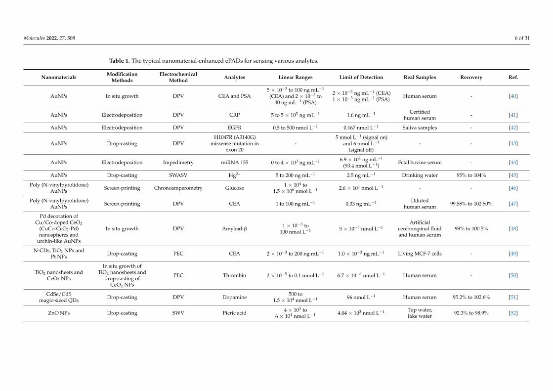
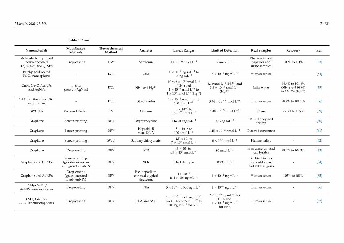
As transduction materials, carbon nanomaterials, including single-walled carbonnanotubes (SWCNTs), multi-walled carbon nanotubes (MWCNTs) and different types ofgraphene materials, have been getting great attention in the ePAD fabrication because theyhave high surface area, excellent electrical conductivity and rich surface-chemical proper-ties [57–73]. For instance, Valentine et al. found that the device-to-device reproducibilityand current intensity of ePAD can be efficiently improved through formation of MWCNTnetwork in the porous structures of paper [58]. Tran et al. developed an ePAD for non-enzymatic detection of glucose through the deposition of the SWCNT layer on wax-printednitrocellulose (NC) membrane [59]. The SWCNT electrode exhibits high conductivitywith an average resistivity of less than 100 Ω/sq and excellent mechanical property. Af-ter modification of the SWCNT electrode by AuNPs, the as-developed ePAD exhibitedexcellent glucose detection performance, including good reproducibility (RSD < 8%) andhigh sensitivity (240 µA/mM cm2), which was used successfully to determine glucose inCoke. Pungjunun et al. developed an origami-based ePAD for sensing NO and NO2 (asNOX) by using a screen-printed graphene electrode modified with copper nanoparticles(CuNP/SPGE) [64]. Because of the good catalytic property of CuNP towards the EC conver-sion of NOX and excellent conductivity of graphene, the as-developed ePAD exhibited highselectivity, low LODs (0.23 vppm and 0.03 vppm with exposure times of 25 min and 1 h,respectively), good reproducibility (RSD < 5.1%) and long lifetime (>30 days). The ePADwas applied to detect NOX in air and exhaust gases from cars, and satisfactory results wereobtained. Cai group has been developed a series of ePADs for detection of various biomark-ers, including CEA and neuronspecific enolase (NSE), by using amino functional graphene(NG)-Thionin (THI)-AuNPs nanocomposites modified SPCEs [66,67]. Integration of SiNPsmodified paper microzones with reduced graphene (RG) modified SPCE, Scala-Benuzziet al. developed an ePAD for the quantitative determination of ethinylestradiol (EE2) in wa-ter samples through capturing EE2 by the immobilized anti-EE2 specific antibodies on thepaper microzones, subsequently releasing the adsorbed EE2 by adding a diluted solutionof sulfuric acid and detecting the desorbed EE2 by Osteryoung square wave voltammetry(OSWV) [69]. The as-developed ePAD exhibited excellent analytical performance, includingwide linear range (0.5 to 120 ng L−1), low LOD (0.1 ng L−1) good recovery values (from 97%to 104%) and good reproducibility (RSD < 4.9%). There are no significant differences werefound between the results of ePAD and the results of spectrophotometric immunoassay,when two methods were used for the quantification of EE2 in river water samples andspiked water samples.
Molecules 2022, 27, 508 6 of 31
Table 1. The typical nanomaterial-enhanced ePADs for sensing various analytes.
Nanomaterials ModificationMethods
ElectrochemicalMethod Analytes Linear Ranges Limit of Detection Real Samples Recovery Ref.
AuNPs In situ growth DPV CEA and PSA5 × 10−3 to 100 ng mL−1
(CEA) and 2 × 10⁻³ to40 ng mL−1 (PSA)
2 × 10⁻³ ng mL−1 (CEA)1 × 10⁻³ ng mL−1 (PSA)
Human serum - [40]
AuNPs Electrodeposition DPV CRP 5 to 5 × 10³ ng mL−1 1.6 ng mL−1 Certifiedhuman serum - [41]
AuNPs Electrodeposition DPV EGFR 0.5 to 500 nmol L−1 0.167 nmol L−1 Saliva samples - [42]
AuNPs Drop-casting DPVH1047R (A3140G)
missense mutation inexon 20
-5 nmol L−1 (signal on)
and 6 nmol L−1
(signal off)- - [43]
AuNPs Electrodeposition Impedimetry miRNA 155 0 to 4 × 10³ ng mL−1 6.9 × 10² ng mL−1
(93.4 nmol L−1)Fetal bovine serum - [44]
AuNPs Drop-casting SWASV Hg2+ 5 to 200 ng mL−1 2.5 ng mL−1 Drinking water 95% to 104% [45]
Poly (N-vinylpyrolidone)AuNPs Screen-printing Chronoamperometry Glucose 1 × 10⁴ to
1.5 × 10⁶ nmol L−1 2.6 × 10⁴ nmol L−1 - - [46]
Poly (N-vinylpyrolidone)AuNPs Screen-printing DPV CEA 1 to 100 ng mL−1 0.33 ng mL−1 Diluted
human serum 99.58% to 102.50% [47]
Pd decoration ofCu/Co-doped CeO2
(CuCo-CeO2-Pd)nanospheres and
urchin-like AuNPs
In situ growth DPV Amyloid-β 1 × 10⁻³ to100 nmol L−1 5 × 10⁻⁵ nmol L−1
Artificialcerebrospinal fluidand human serum
99% to 100.5% [48]
N-CDs, TiO2 NPs andPt NPs Drop-casting PEC CEA 2 × 10⁻³ to 200 ng mL−1 1.0 × 10⁻³ ng mL−1 Living MCF-7 cells - [49]
TiO2 nanosheets andCeO2 NPs
In situ growth ofTiO2 nanosheets and
drop-casting ofCeO2 NPs
PEC Thrombin 2 × 10⁻⁵ to 0.1 nmol L−1 6.7 × 10⁻⁶ nmol L−1 Human serum - [50]
CdSe/CdSmagic-sized QDs Drop-casting DPV Dopamine 500 to
1.5 × 10⁴ nmol L−1 96 nmol L−1 Human serum 95.2% to 102.6% [51]
ZnO NPs Drop-casting SWV Picric acid 4 × 10³ to6 × 104 nmol L−1 4.04 × 10³ nmol L−1 Tap water,
lake water 92.3% to 98.9% [52]
Molecules 2022, 27, 508 7 of 31
Table 1. Cont.
Nanomaterials ModificationMethods
ElectrochemicalMethod Analytes Linear Ranges Limit of Detection Real Samples Recovery Ref.
Molecularly imprintedpolymer coated
Fe3O4@Au@SiO2 NPsDrop-casting LSV Serotonin 10 to 106 nmol L−1 2 nmol L−1
Pharmaceuticalcapsules andurine samples
100% to 111% [53]
Patchy gold coatedFe3O4 nanospheres - ECL CEA 1 × 10⁻⁴ ng mL−1 to
15 ng mL−1 3 × 10⁻⁵ ng mL−1 Human serum - [54]
Cubic Cu2O-Au NPsand AgNPs
In situgrowth (AgNPs) ECL Ni2+ and Hg2+
10 to 2 × 10⁵ nmol L−1
(Ni2+) and1× 10−2 nmol L−1 to
1× 10³ nmol L−1 (Hg2+)
3.1 nmol L−1 (Ni2+) and3.8 × 10−3 nmol L−1
(Hg2+)Lake water
96.4% to 101.6%(Ni2+) and 96.0%to 104.0% (Hg2+)
[55]
DNA-functionalized PtCunanoframes - ECL Streptavidin 1 × 10⁻⁴ nmol L−1 to
100 nmol L−1 3.34 × 10⁻⁵ nmol L−1 Human serum 98.4% to 106.5% [56]
SWCNTs Vaccum filtration CV Glucose 5 × 10⁻⁵ to1 × 107 nmol L−1 1.48 × 10⁵ nmol L−1 Coke 97.3% to 105% [59]
Graphene Screen-printing DPV Oxytetracycline 1 to 200 ng mL−1 0.33 ng mL−1 Milk, honey andshrimp - [60]
Graphene Screen-printing DPV Hepatitis Bvirus DNA
5 × 10⁻² to100 nmol L−1 1.45 × 10⁻³ nmol L−1 Plasmid constructs - [61]
Graphene Screen-printing SWV Salivary thiocyanate 2.5 × 10⁴ to7 × 105 nmol L−1 6 × 10³ nmol L−1 Human saliva - [62]
Graphene Drop-casting DPV ATP 3 × 10² to4.5 × 105 nmol L−1 80 nmol L−1 Human serum and
cell lysates 95.4% to 104.2% [63]
Graphene and CuNPsScreen-printing
(graphene) and insitu growth CuNPs
DPV NOx 0 to 150 vppm 0.23 vppmAmbient indoorand outdoor air,
and exhaust gases- [64]
Graphene and AuNPsDrop-casting
(graphene) andlabel (AuNPs)
DPVPseudopodium-
enriched atypicalkinase one
1 × 10⁻²
to 1 × 10³ ng mL−1 1 × 10⁻² ng mL−1 Human serum 103% to 104% [65]
(NH2-G)/Thi/AuNPs nanocomposites Drop-casting DPV CEA 5 × 10⁻² to 500 ng mL−1 1 × 10⁻² ng mL−1 Human serum - [66]
(NH2-G)/Thi/AuNPs nanocomposites Drop-casting DPV CEA and NSE
1 × 10⁻² to 500 ng mL−1
for CEA and 5 × 10⁻² to500 ng mL−1 for NSE
2 × 10⁻³ ng mL−1 forCEA and
1 × 10⁻² ng mL−1
for NSE
Human serum - [67]
Molecules 2022, 27, 508 8 of 31
Table 1. Cont.
Nanomaterials ModificationMethods
ElectrochemicalMethod Analytes Linear Ranges Limit of Detection Real Samples Recovery Ref.
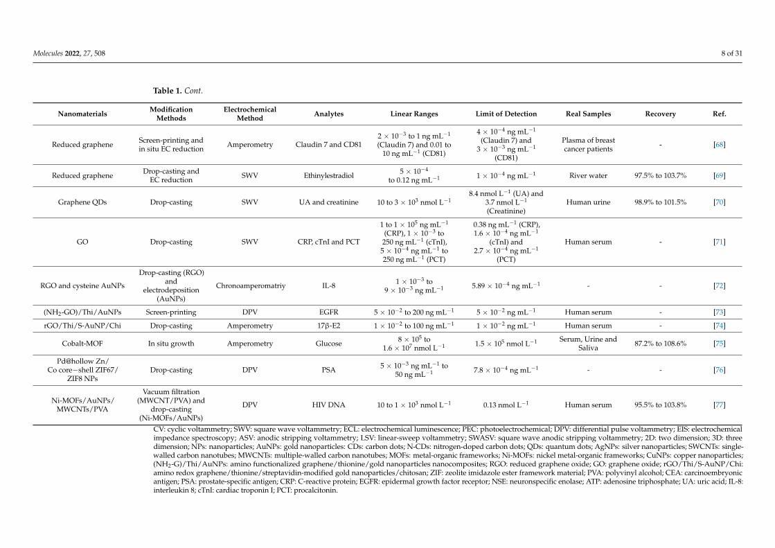
Reduced graphene Screen-printing andin situ EC reduction Amperometry Claudin 7 and CD81
2 × 10⁻³ to 1 ng mL−1
(Claudin 7) and 0.01 to10 ng mL−1 (CD81)
4 × 10⁻⁴ ng mL−1
(Claudin 7) and3 × 10−3 ng mL−1
(CD81)
Plasma of breastcancer patients - [68]
Reduced graphene Drop-casting andEC reduction SWV Ethinylestradiol 5 × 10⁻⁴
to 0.12 ng mL−1 1 × 10⁻⁴ ng mL−1 River water 97.5% to 103.7% [69]
Graphene QDs Drop-casting SWV UA and creatinine 10 to 3 × 10³ nmol L−18.4 nmol L−1 (UA) and
3.7 nmol L−1
(Creatinine)Human urine 98.9% to 101.5% [70]
GO Drop-casting SWV CRP, cTnI and PCT
1 to 1 × 10⁵ ng mL−1
(CRP), 1 × 10⁻³ to250 ng mL−1 (cTnI),
5 × 10⁻⁴ ng mL−1 to250 ng mL−1 (PCT)
0.38 ng mL−1 (CRP),1.6 × 10−4 ng mL−1
(cTnI) and2.7 × 10−4 ng mL−1
(PCT)
Human serum - [71]
RGO and cysteine AuNPs
Drop-casting (RGO)and
electrodeposition(AuNPs)
Chronoamperomatriy IL-8 1 × 10⁻³ to9 × 10−3 ng mL−1 5.89 × 10⁻⁴ ng mL−1 - - [72]
(NH2-GO)/Thi/AuNPs Screen-printing DPV EGFR 5 × 10⁻² to 200 ng mL−1 5 × 10⁻² ng mL−1 Human serum - [73]
rGO/Thi/S-AuNP/Chi Drop-casting Amperometry 17β-E2 1 × 10⁻² to 100 ng mL−1 1 × 10⁻² ng mL−1 Human serum - [74]
Cobalt-MOF In situ growth Amperometry Glucose 8 × 10⁵ to1.6 × 107 nmol L−1 1.5 × 10⁵ nmol L−1 Serum, Urine and
Saliva 87.2% to 108.6% [75]
Pd@hollow Zn/Co core−shell ZIF67/
ZIF8 NPsDrop-casting DPV PSA 5 × 10⁻³ ng mL−1 to
50 ng mL−1 7.8 × 10⁻⁴ ng mL−1 - - [76]
Ni-MOFs/AuNPs/MWCNTs/PVA
Vacuum filtration(MWCNT/PVA) and
drop-casting(Ni-MOFs/AuNPs)
DPV HIV DNA 10 to 1 × 10³ nmol L−1 0.13 nmol L−1 Human serum 95.5% to 103.8% [77]
CV: cyclic voltammetry; SWV: square wave voltammetry; ECL: electrochemical luminescence; PEC: photoelectrochemical; DPV: differential pulse voltammetry; EIS: electrochemicalimpedance spectroscopy; ASV: anodic stripping voltammetry; LSV: linear-sweep voltammetry; SWASV: square wave anodic stripping voltammetry; 2D: two dimension; 3D: threedimension; NPs: nanoparticles; AuNPs: gold nanoparticles: CDs: carbon dots; N-CDs: nitrogen-doped carbon dots; QDs: quantum dots; AgNPs: silver nanoparticles; SWCNTs: single-walled carbon nanotubes; MWCNTs: multiple-walled carbon nanotubes; MOFs: metal-organic frameworks; Ni-MOFs: nickel metal-organic frameworks; CuNPs: copper nanoparticles;(NH2-G)/Thi/AuNPs: amino functionalized graphene/thionine/gold nanoparticles nanocomposites; RGO: reduced graphene oxide; GO: graphene oxide; rGO/Thi/S-AuNP/Chi:amino redox graphene/thionine/streptavidin-modified gold nanoparticles/chitosan; ZIF: zeolite imidazole ester framework material; PVA: polyvinyl alcohol; CEA: carcinoembryonicantigen; PSA: prostate-specific antigen; CRP: C-reactive protein; EGFR: epidermal growth factor receptor; NSE: neuronspecific enolase; ATP: adenosine triphosphate; UA: uric acid; IL-8:interleukin 8; cTnI: cardiac troponin I; PCT: procalcitonin.
Molecules 2022, 27, 508 9 of 31
The unique features of metal-organic frameworks (MOFs), including high porosity,tunable framework structures, large surface areas and multiple functionalities, make themextremely attractive for improving the detection performance of biosensors [78,79]. Re-cently, MOFs and their nanocomposites have been used for developing ePAD with excellentdetection performance [75–77]. Wei et al. fabricated a cobalt-MOF (Co-MOF) modifiedcarbon cloth/paper (CC/Paper) hybrid button-based PAD (Co-MOF/CC PAD) for nonen-zymatic quantitative EC detection of glucose through in situ growth of Co-MOF on CC [75].As a typical nanozyme, the environment tolerance of Co-MOFs is much better than thatof natural enzyme, which can increase significantly the stability of ePAD. Densely grownCo-MOF on CC can maximize its catalytic sites, resulting in high sensitivity of ePAD. TheCo-MOF/CC PAD exhibits linear range from 0.8 mmol L−1 to 16 mmol L−1 with an LODof 0.15 mmol L−1 and maintains at a stable detection performance in 60 days, and thengradually decreased to about 60% after 120 days. The ePAD was used successfully todetermine glucose in multiple body fluids, including serum, urine and saliva. Lu et al.developed an ePAD-based DNA hybridization for detection of human immunodeficiencyvirus (HIV) DNA by using the nickel MOF (Ni-MOF) composite/AuNPs/CNTs/polyvinylalcohol (Ni–Au composite/CNT/PVA) paper electrode as working electrode and methy-lene blue (MB) as a redox indicator [77]. The CNT/PVA were deposited on the cellulosemembrane by vacuum filtration, and Ni–Au composites were loaded on CNT/PVA filmby the drop-casting method. The Ni–Au composite/CNT/PVA film electrode has largespecific surface area and conjugated π-electron system, which makes a higher loading ofthe single-stranded DNA probe than that of CNT/PVA film electrode. The phenomenonimproves the sensitivity for detecting target DNA. The ePAD exhibited excellent sens-ing performance with a wide linear range of 10 nmol L−1 to 1 µmol L−1, a low LOD of0.13 nmol L−1, good selectivity against one-base mismatch DNA sequences and excellentstability after 20 days of storage. The target HIV DNA was detected successfully even incomplex serum samples by the as-developed ePAD.
2.2. Colorimetric Paper-Based Analytical Devices
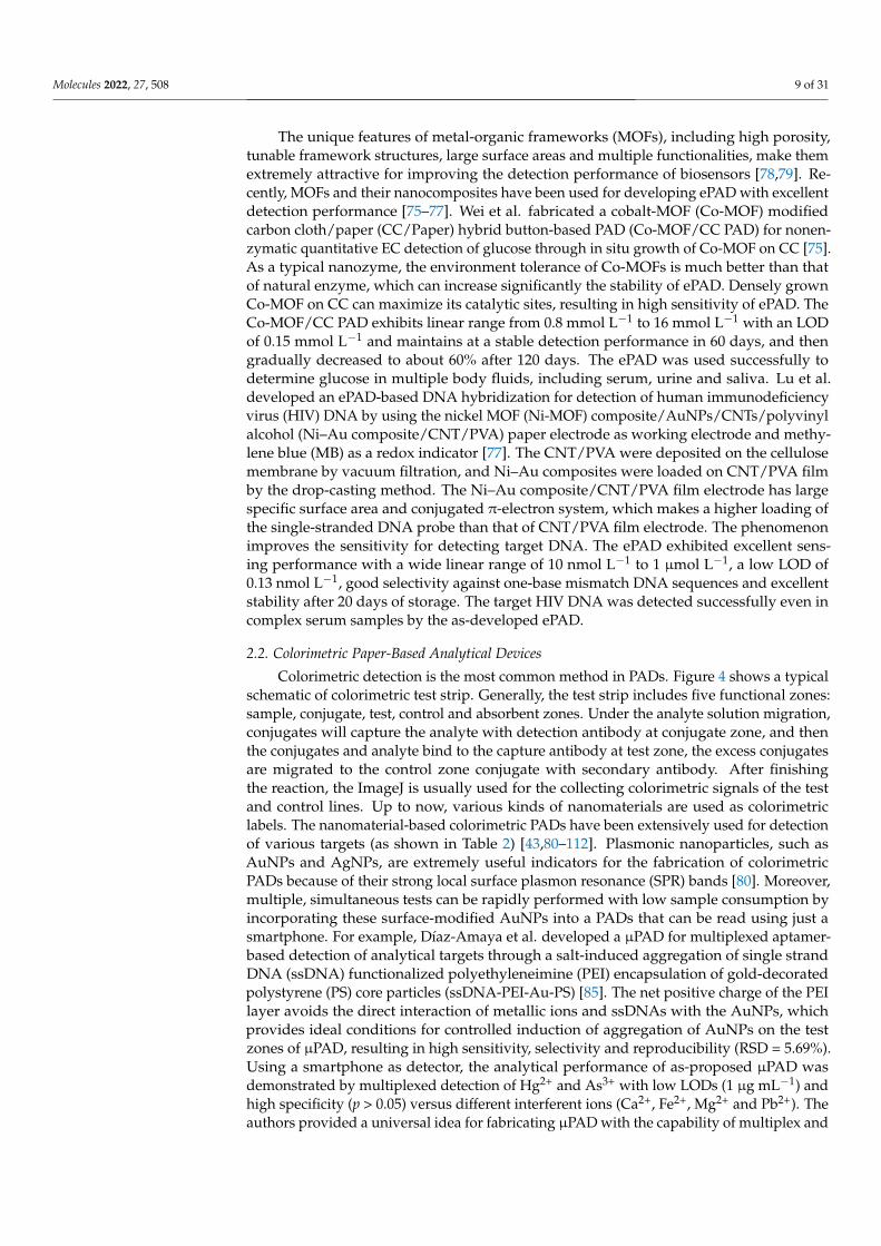
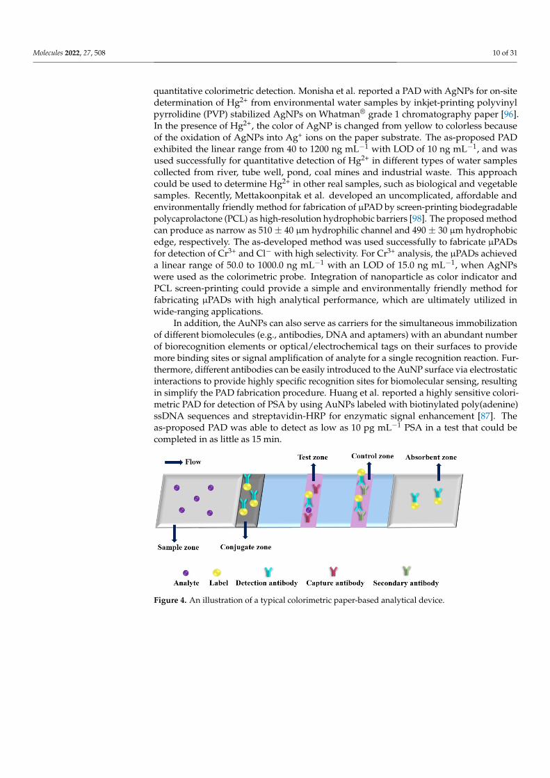
Colorimetric detection is the most common method in PADs. Figure 4 shows a typicalschematic of colorimetric test strip. Generally, the test strip includes five functional zones:sample, conjugate, test, control and absorbent zones. Under the analyte solution migration,conjugates will capture the analyte with detection antibody at conjugate zone, and thenthe conjugates and analyte bind to the capture antibody at test zone, the excess conjugatesare migrated to the control zone conjugate with secondary antibody. After finishingthe reaction, the ImageJ is usually used for the collecting colorimetric signals of the testand control lines. Up to now, various kinds of nanomaterials are used as colorimetriclabels. The nanomaterial-based colorimetric PADs have been extensively used for detectionof various targets (as shown in Table 2) [43,80–112]. Plasmonic nanoparticles, such asAuNPs and AgNPs, are extremely useful indicators for the fabrication of colorimetricPADs because of their strong local surface plasmon resonance (SPR) bands [80]. Moreover,multiple, simultaneous tests can be rapidly performed with low sample consumption byincorporating these surface-modified AuNPs into a PADs that can be read using just asmartphone. For example, Díaz-Amaya et al. developed a µPAD for multiplexed aptamer-based detection of analytical targets through a salt-induced aggregation of single strandDNA (ssDNA) functionalized polyethyleneimine (PEI) encapsulation of gold-decoratedpolystyrene (PS) core particles (ssDNA-PEI-Au-PS) [85]. The net positive charge of the PEIlayer avoids the direct interaction of metallic ions and ssDNAs with the AuNPs, whichprovides ideal conditions for controlled induction of aggregation of AuNPs on the testzones of µPAD, resulting in high sensitivity, selectivity and reproducibility (RSD = 5.69%).Using a smartphone as detector, the analytical performance of as-proposed µPAD wasdemonstrated by multiplexed detection of Hg2+ and As3+ with low LODs (1 µg mL−1) andhigh specificity (p > 0.05) versus different interferent ions (Ca2+, Fe2+, Mg2+ and Pb2+). Theauthors provided a universal idea for fabricating µPAD with the capability of multiplex and
Molecules 2022, 27, 508 10 of 31
quantitative colorimetric detection. Monisha et al. reported a PAD with AgNPs for on-sitedetermination of Hg2+ from environmental water samples by inkjet-printing polyvinylpyrrolidine (PVP) stabilized AgNPs on Whatman® grade 1 chromatography paper [96].In the presence of Hg2+, the color of AgNP is changed from yellow to colorless becauseof the oxidation of AgNPs into Ag+ ions on the paper substrate. The as-proposed PADexhibited the linear range from 40 to 1200 ng mL−1 with LOD of 10 ng mL−1, and wasused successfully for quantitative detection of Hg2+ in different types of water samplescollected from river, tube well, pond, coal mines and industrial waste. This approachcould be used to determine Hg2+ in other real samples, such as biological and vegetablesamples. Recently, Mettakoonpitak et al. developed an uncomplicated, affordable andenvironmentally friendly method for fabrication of µPAD by screen-printing biodegradablepolycaprolactone (PCL) as high-resolution hydrophobic barriers [98]. The proposed methodcan produce as narrow as 510 ± 40 µm hydrophilic channel and 490 ± 30 µm hydrophobicedge, respectively. The as-developed method was used successfully to fabricate µPADsfor detection of Cr3+ and Cl− with high selectivity. For Cr3+ analysis, the µPADs achieveda linear range of 50.0 to 1000.0 ng mL−1 with an LOD of 15.0 ng mL−1, when AgNPswere used as the colorimetric probe. Integration of nanoparticle as color indicator andPCL screen-printing could provide a simple and environmentally friendly method forfabricating µPADs with high analytical performance, which are ultimately utilized inwide-ranging applications.
In addition, the AuNPs can also serve as carriers for the simultaneous immobilizationof different biomolecules (e.g., antibodies, DNA and aptamers) with an abundant numberof biorecognition elements or optical/electrochemical tags on their surfaces to providemore binding sites or signal amplification of analyte for a single recognition reaction. Fur-thermore, different antibodies can be easily introduced to the AuNP surface via electrostaticinteractions to provide highly specific recognition sites for biomolecular sensing, resultingin simplify the PAD fabrication procedure. Huang et al. reported a highly sensitive colori-metric PAD for detection of PSA by using AuNPs labeled with biotinylated poly(adenine)ssDNA sequences and streptavidin-HRP for enzymatic signal enhancement [87]. Theas-proposed PAD was able to detect as low as 10 pg mL−1 PSA in a test that could becompleted in as little as 15 min.
Molecules 2022, 27, x FOR PEER REVIEW 10 of 28
Figure 4. An illustration of a typical colorimetric paper-based analytical device.
Table 2. The typical nanomaterial-enhanced colorimetric PADs for sensing various analytes.
Nanomaterials Analytes Linear Ranges Limit of Detection Real Samples Recovery Ref
AuNPs Gallic acid 1 × 10⁴ to 1 × 10⁶ nmol
L−1 1 × 10³ nmol L−1 Tea 85.2% to 93.1% [80]
Avidin functionalized AuNPs
Ig G - 300 ng mL−1 - - [81]
Citrate stabilized AuNPs
Melamine 100 to 106 ng mL−1 100 ng mL−1 Milk - [82]
Citrate stabilized AuNPs
NADH - 1.25 × 10⁴ nmol L−1 Cell Lysate - [83]
Aspartic acid modified AuNPs
Cysteine 9.99 × 10⁴ to 9.987 × 10⁵
nmol L−1 1 × 10³ nmol L−1 Human plasma 99.2% to 101.1% [84]
ssDNA-PEI-Au-PS Hg2+ and As3+ 0 to 3 × 10⁴ ng mL−1 1 × 10³ ng mL−1 River water 96.2% to 116.7% [85] ssDNA functionalized
AuNPs Tuberculosis DNA
1.95 × 10⁻² to 19.5 ng mL−1
1.95 × 10⁻² ng mL−1 Infected tissue - [86]
ssDNA functionalized AuNPs
PSA - 1 × 10⁻² ng mL−1 Human serum - [87]
Antibody functional-ized AuNPs
Ig G - 284.52 ng
mL−1 Whole blood - [88]
Antibody functional-ized AuNPs
Yersinia Pestis - 2.5 × 10⁻² ng mL−1 - - [89]
Antibody functional-ized AuNPs
Influenza A H1N1 and H3N2 viruses
-
2.7 × 10³ pfu/assay for H1N1 detection and 2.7 ×10⁴ pfu/assay for
H3N2 detection
Cell lysate - [90]
Antibodies functional-ized AuNRs
sIL-2R 1 to 6.25 × 10³ ng mL−1 1.0 ng mL−1 Mouse serum 93% to 109% [91]
Antibodies functional-ized AuNRs
CRP 50 to 1 × 10⁴ ng mL−1 1.3 ng mL−1 Human plasma - [92]
Co(II) catalyst, second-ary antibody, luminol multifunctionalized
AuNPs
H-FABP, cTnI and copeptin
1 × 10⁻⁴ to 1 × 10³ ng mL−1, 5 × 10⁻⁴ to 1 × 10³ ng mL−1 and 1 × 10⁻³ to 1 × 10⁶ ng mL−1 for H-
FABP, cTnI and copep-tin
6 × 10⁻⁵ ng mL−1, 3 × 10⁻⁴ ng mL−1 and 4 × 10⁻⁴ ng mL−1 for H-
FABP, cTnI and copep-tin
Human serum 94% to 108% [93]
Cu/Co-doped CeO2 (CuCo-CeO2-Pd)
Amyloid-β 1 × 10⁻² to 100 nmol L−1 5 × 10⁻⁴ nmol L−1 Artificial cerebro-
spinal fluid 99% to 100.5% [48]
Figure 4. An illustration of a typical colorimetric paper-based analytical device.
Molecules 2022, 27, 508 11 of 31
Table 2. The typical nanomaterial-enhanced colorimetric PADs for sensing various analytes.
Nanomaterials Analytes Linear Ranges Limit of Detection Real Samples Recovery Ref.
AuNPs Gallic acid 1 × 10⁴ to 1 × 10⁶ nmol L−1 1 × 10³ nmol L−1 Tea 85.2% to 93.1% [80]
Avidinfunctionalized AuNPs Ig G - 300 ng mL−1 - - [81]
Citrate stabilized AuNPs Melamine 100 to 106 ng mL−1 100 ng mL−1 Milk - [82]
Citrate stabilized AuNPs NADH - 1.25 × 10⁴ nmol L−1 Cell Lysate - [83]
Aspartic acidmodified AuNPs Cysteine 9.99 × 10⁴ to
9.987 × 10⁵ nmol L−1 1 × 10³ nmol L−1 Human plasma 99.2% to 101.1% [84]
ssDNA-PEI-Au-PS Hg2+ and As3+ 0 to 3 × 10⁴ ng mL−1 1 × 10³ ng mL−1 River water 96.2% to 116.7% [85]
ssDNAfunctionalized AuNPs Tuberculosis DNA 1.95 × 10⁻² to 19.5 ng mL−1 1.95 × 10⁻² ng mL−1 Infected tissue - [86]
ssDNAfunctionalized AuNPs PSA - 1 × 10⁻² ng mL−1 Human serum - [87]
Antibodyfunctionalized AuNPs Ig G - 284.52 ng mL−1 Whole blood - [88]
Antibodyfunctionalized AuNPs Yersinia Pestis - 2.5 × 10⁻² ng mL−1 - - [89]
Antibodyfunctionalized AuNPs
Influenza A H1N1 andH3N2 viruses -
2.7 × 10³ pfu/assay for H1N1detection and 2.7 ×10⁴
pfu/assay forH3N2 detection
Cell lysate - [90]
Antibodiesfunctionalized AuNRs sIL-2R 1 to 6.25 × 10³ ng mL−1 1.0 ng mL−1 Mouse serum 93% to 109% [91]
Antibodiesfunctionalized AuNRs CRP 50 to 1 × 10⁴ ng mL−1 1.3 ng mL−1 Human plasma - [92]
Co(II) catalyst, secondaryantibody, luminol
multifunctionalized AuNPs
H-FABP, cTnIand copeptin
1 × 10⁻⁴ to1 × 103 ng mL−1, 5 × 10⁻⁴
to 1 × 10³ ng mL−1 and1 × 10−3 to
1 × 10⁶ ng mL−1 forH-FABP, cTnI and copeptin
6 × 10⁻⁵ ng mL−1,3 × 10⁻⁴ ng mL−1 and4 × 10−4 ng mL−1 for
H-FABP, cTnI and copeptin
Human serum 94% to 108% [93]
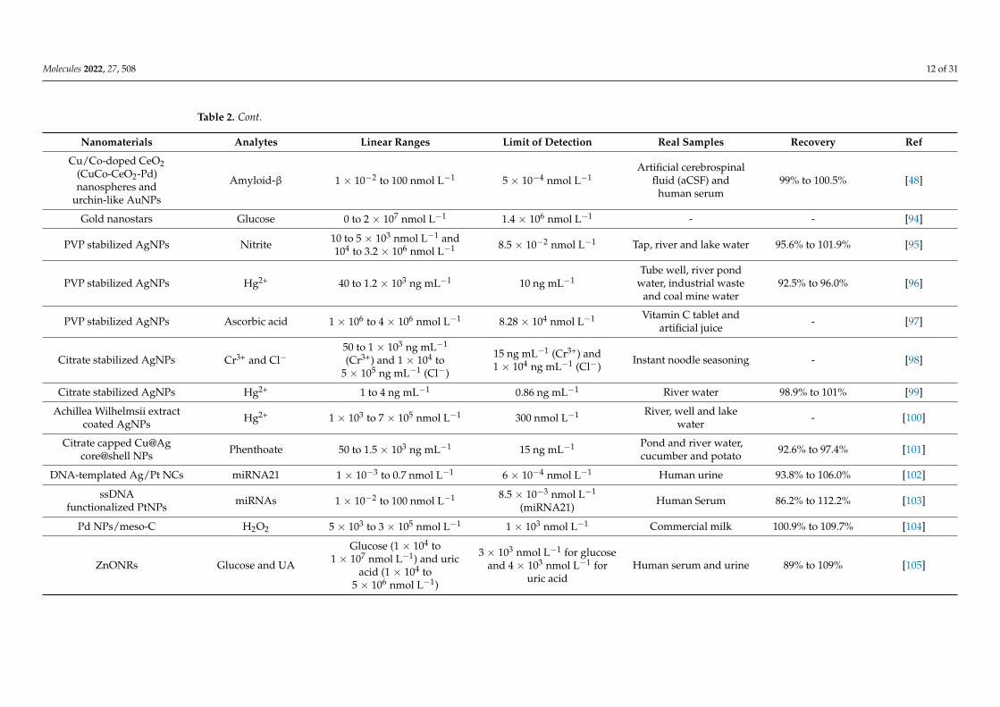
Molecules 2022, 27, 508 12 of 31
Table 2. Cont.
Nanomaterials Analytes Linear Ranges Limit of Detection Real Samples Recovery Ref
Cu/Co-doped CeO2(CuCo-CeO2-Pd)nanospheres and
urchin-like AuNPs
Amyloid-β 1 × 10⁻² to 100 nmol L−1 5 × 10⁻⁴ nmol L−1Artificial cerebrospinal
fluid (aCSF) andhuman serum
99% to 100.5% [48]
Gold nanostars Glucose 0 to 2 × 10⁷ nmol L−1 1.4 × 10⁶ nmol L−1 - - [94]
PVP stabilized AgNPs Nitrite 10 to 5 × 10³ nmol L−1 and104 to 3.2 × 10⁶ nmol L−1 8.5 × 10⁻² nmol L−1 Tap, river and lake water 95.6% to 101.9% [95]
PVP stabilized AgNPs Hg2+ 40 to 1.2 × 10³ ng mL−1 10 ng mL−1Tube well, river pond
water, industrial wasteand coal mine water
92.5% to 96.0% [96]
PVP stabilized AgNPs Ascorbic acid 1 × 10⁶ to 4 × 10⁶ nmol L−1 8.28 × 10⁴ nmol L−1 Vitamin C tablet andartificial juice - [97]
Citrate stabilized AgNPs Cr3+ and Cl−50 to 1 × 10³ ng mL−1
(Cr3+) and 1 × 10⁴ to5 × 105 ng mL−1 (Cl−)
15 ng mL−1 (Cr3+) and1 × 104 ng mL−1 (Cl−)
Instant noodle seasoning - [98]
Citrate stabilized AgNPs Hg2+ 1 to 4 ng mL−1 0.86 ng mL−1 River water 98.9% to 101% [99]
Achillea Wilhelmsii extractcoated AgNPs Hg2+ 1 × 10³ to 7 × 10⁵ nmol L−1 300 nmol L−1 River, well and lake
water - [100]
Citrate capped Cu@Agcore@shell NPs Phenthoate 50 to 1.5 × 10³ ng mL−1 15 ng mL−1 Pond and river water,
cucumber and potato 92.6% to 97.4% [101]
DNA-templated Ag/Pt NCs miRNA21 1 × 10⁻³ to 0.7 nmol L−1 6 × 10⁻⁴ nmol L−1 Human urine 93.8% to 106.0% [102]
ssDNAfunctionalized PtNPs miRNAs 1 × 10⁻² to 100 nmol L−1 8.5 × 10⁻³ nmol L−1
(miRNA21)Human Serum 86.2% to 112.2% [103]
Pd NPs/meso-C H2O2 5 × 10³ to 3 × 10⁵ nmol L−1 1 × 10³ nmol L−1 Commercial milk 100.9% to 109.7% [104]
ZnONRs Glucose and UA
Glucose (1 × 10⁴ to1 × 107 nmol L−1) and uric
acid (1 × 10⁴ to5 × 106 nmol L−1)
3 × 10³ nmol L−1 for glucoseand 4 × 10³ nmol L−1 for
uric acidHuman serum and urine 89% to 109% [105]
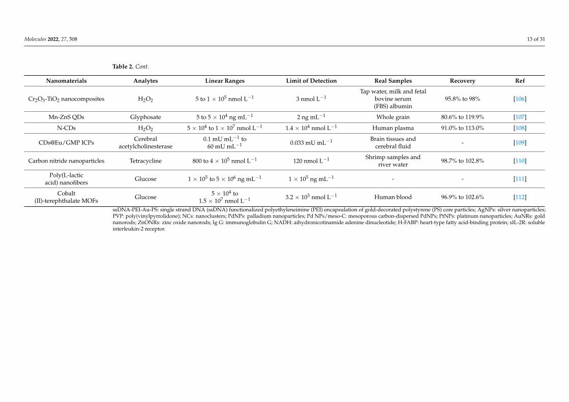
Molecules 2022, 27, 508 13 of 31
Table 2. Cont.
Nanomaterials Analytes Linear Ranges Limit of Detection Real Samples Recovery Ref
Cr2O3-TiO2 nanocomposites H2O2 5 to 1 × 10⁵ nmol L−1 3 nmol L−1Tap water, milk and fetal
bovine serum(FBS) albumin
95.8% to 98% [106]
Mn-ZnS QDs Glyphosate 5 to 5 × 10⁴ ng mL−1 2 ng mL−1 Whole grain 80.6% to 119.9% [107]
N-CDs H2O2 5 × 10⁴ to 1 × 10⁷ nmol L−1 1.4 × 10⁴ nmol L−1 Human plasma 91.0% to 113.0% [108]
CDs@Eu/GMP ICPs Cerebralacetylcholinesterase
0.1 mU mL−1 to60 mU mL−1 0.033 mU mL−1 Brain tissues and
cerebral fluid - [109]
Carbon nitride nanoparticles Tetracycline 800 to 4 × 10⁵ nmol L−1 120 nmol L−1 Shrimp samples andriver water 98.7% to 102.8% [110]
Poly(L-lacticacid) nanofibers Glucose 1 × 10⁵ to 5 × 10⁶ ng mL−1 1 × 10⁵ ng mL−1 - - [111]
Cobalt(II)-terephthalate MOFs Glucose 5 × 10⁴ to
1.5 × 10⁷ nmol L−1 3.2 × 10³ nmol L−1 Human blood 96.9% to 102.6% [112]
ssDNA-PEI-Au-PS: single strand DNA (ssDNA) functionalized polyethyleneimine (PEI) encapsulation of gold-decorated polystyrene (PS) core particles; AgNPs: silver nanoparticles;PVP: poly(vinylpyrrolidone); NCs: nanoclusters; PdNPs: palladium nanoparticles; Pd NPs/meso-C: mesoporous carbon-dispersed PdNPs; PtNPs: platinum nanoparticles; AuNRs: goldnanorods; ZnONRs: zinc oxide nanorods; Ig G: immunoglobulin G; NADH: aihydronicotinamide adenine dinucleotide; H-FABP: heart-type fatty acid-binding protein; sIL-2R: solubleinterleukin-2 receptor.
Molecules 2022, 27, 508 14 of 31
Comparison with natural enzymes, nanomaterials with enzyme-like characteristics(i.e., nanozymes), such as magnetic nanoparticles, noble metal nanoparticles, MOFs, hetero-junctions, etc., exhibit several advantages, including easy production with large-scale,low cost and high stability in harsh environments. These unique properties endowthem with attractive applications in the fabrication of PADs with high analytical per-formance [104,111,112]. Zhang et al. developed a ready-to-use PAD for the determinationof H2O2 by simply immobilization of mesoporous carbon-dispersed palladium nanopar-ticles (Pd NPs/meso-C) and the 3,3′,5,5′-tetramethylbenzidine (TMB) substrate onto acommon chromatography paper [104]. Taking the advantage of large surface area of themeso-C support and the good dispersity of PdNPs, the PdNPs/meso-C show excellentcatalytic performance to trigger the chromogenic reaction of colorless TMB to blue TMBoxmediated by H2O2. The as-developed PAD exhibited a linear range of 5 to 300 mol L−1,and can be used to determine H2O2 in complex matrices, such as milk. Kitchawengkul et al.developed a laminated three-dimensional (3D)-µPAD for colorimetric determination of to-tal cholesterol (TC) in human blood by using the peroxidase-like activity of nitrogen-dopedCDs (N-CDs) [108]. The 3D-µPAD with a 6 mm circular detection zone was fabricated by asimple wax screen-printing technique, which consisted of four layers laminated togethervertically. The 3D-µPAD exhibited a linear range of 0.05 to 10 mmol L−1 with an LODof 0.014 mmol L−1. In particular, TC in human blood could be determined by the nakedeye within 10 min by simple comparison with a color chart. Overall, the as-proposed3D-µPAD serves as a simple, low cost, rapid, sensitive and selective alternative for de-tection of TC in whole blood samples that is friendly to unskilled end users. Cui et al.fabricated an origami PAD (oPAD) assisted by Pd decorated Cu/Co co-doped CeO2 (CuCo-CeO2-Pd) nanospheres, for dual-mode electrochemical/visual detection of amyloid-β(Aβ) peptide with high sensitivity [48]. In this case, the CuCo-CeO2-Pd nanosphereswere introduced as an enhanced “signal transducer layer”, which act as an outstandingcatalyst for catalyzing glucose to produce H2O2 for DPV signal readout and further 3,3′,5,5′-tetramethylbenzidine (TMB) oxidation for colorimetric analysis. The oPAD exhibited linearranges from 1.0 pmol L−1 to 100 nmol L−1 (EC detection) and 10 pmol L−1 to 100 nmol L−1
(visual detection) with LODs of 0.05 pmol L−1 (EC detection) and 0.5 pmol L−1 (visualdetection), respectively. The oPAD was used successfully to analysis Aβ peptide in artificialcerebrospinal fluid (aCSF) and serum samples. Al Lawati et al. developed a PAD forthe colorimetric/fluorometric monitoring of glucose by co-immobilizing two-dimensionalcobalt-terephthalate MOF nanosheets (2D CoMOFs) and GOX on chromatography pa-per [112]. Due to its highly porous and extraordinarily stable structures, the 2D CoMOFincreased significantly the stability and performance of GOX, and also acted as a catalystto accelerate the reaction of H2O2 produced by the enzymatic oxidation of glucose witho-phenylenediamine (OPD) serving as a peroxidase substrate, resulting in a yellow-browncolor change and a high fluorescence emission. The as-developed PAD showed high analyti-cal performance for the quantification of glucose including high accuracy, wide linear range(50 mol L−1 to 15 mmol L−1) and low LODs (16.3 (colorimetric detection) and 3.2 mmol L−1
(fluorometric detection)), and was used successfully to determine glucose in blood samplesfrom healthy and diabetic volunteers.
2.3. Fluorometric Paper-Based Analytical Devices
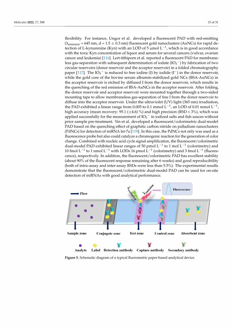
Recently, the nanomaterial-based fluorometric PADs are increasingly being developedfor sensing various targets (as shown in Table 3) [113–161]. The schematic diagram of theworking principle of the fluorometric PAD is shown in Figure 5. The reaction process ofthe analyte on the test strip is common with that of the colorimetric test strip. After thereaction is finished, the fluorometric signals of emission wavelengths at the test and controlzones are recorded by fluorometric spectrophotometer under irradiating with excitationlight. Fluorescent nanomaterials, including metal nanoclusters (NCs), CDs, QDs, UCNPsand MOFs, have unique properties, such as wide excitation wavelength, narrow emis-sion band, tunable fluorescence color, highly optical stability and good surface-modified
Molecules 2022, 27, 508 15 of 31
flexibility. For instance, Ungor et al. developed a fluorescent PAD with red-emitting(λemission = 645 nm, d = 1.5 ± 0.3 nm) fluorescent gold nanoclusters (AuNCs) for rapid de-tection of L-kynurenine (Kyn) with an LOD of 5 µmol L−1, which is in good accordancewith the toxic Kyn concentration of liquor and serum for several cancers (vulvar, ovariancancer and leukemia) [116]. Lert-itthiporn et al. reported a fluorescent PAD for membrane-less gas-separation with subsequent determination of iodate (IO3
−) by fabrication of twocircular reservoirs (donor reservoir and the acceptor reservoir) in a folded chromatographypaper [117]. The IO3
− is reduced to free iodine (I) by iodide (I−) in the donor reservoir,while the gold core of the bovine serum albumin-stabilized gold NCs (BSA-AuNCs) inthe acceptor reservoir is etched by diffused I from the donor reservoir, which results inthe quenching of the red emission of BSA-AuNCs in the acceptor reservoir. After folding,the donor reservoir and acceptor reservoir were mounted together through a two-sidedmounting tape to allow membraneless gas-separation of free I from the donor reservoir todiffuse into the acceptor reservoir. Under the ultraviolet (UV) light (365 nm) irradiation,the PAD exhibited a linear range from 0.005 to 0.1 mmol L−1, an LOD of 0.01 mmol L−1,high accuracy (mean recovery: 95.1 (±4.6) %) and high precision (RSD < 3%), which wasapplied successfully for the measurement of IO3
− in iodized salts and fish sauces withoutprior sample pre-treatment. Yin et al. developed a fluorescent/colorimetric dual-modelPAD based on the quenching effect of graphitic carbon nitride on palladium nanoclusters(PdNCs) for detection of miRNA let-7a [119]. In this case, the PdNCs not only was used as afluorescence probe but also could catalyze a chromogenic reaction for the generation of colorchange. Combined with nucleic acid cycle signal amplification, the fluorescent/colorimetricdual-model PAD exhibited linear ranges of 50 pmol L−1 to 1 mol L−1 (colorimetry) and10 fmol L−1 to 1 nmol L−1 with LODs 16 pmol L−1 (colorimetry) and 3 fmol L−1 (fluores-cence), respectively. In addition, the fluorescent/colorimetric PAD has excellent stability(about 90% of the fluorescent response remaining after 6 weeks) and good reproducibility(both of intra-assay and inter-assay RSDs were less than 5.5%). The experimental resultsdemonstrate that the fluorescent/colorimetric dual-model PAD can be used for on-sitedetection of miRNAs with good analytical performance.
Molecules 2022, 27, x FOR PEER REVIEW 14 of 28
Figure 5. Schematic diagram of a typical fluorometric paper-based analytical device.
Table 3. The typical nanomaterial-enhanced fluorescent PADs for sensing various analytes.
Nanomaterials Analytes Linear Ranges Limit of Detection Real Samples Recovery Ref.
AuNCs/MIL-68(In)-NH2/Cys
Hg2+ 0.02 nmol L−1 to 200
nmol L−1 and 200 to 6 × 10⁴ nmol L−1
6.7 × 10⁻³ nmol L−1 Tap and Lake
water 91.3% to 110.2% [115]
γG-AuNCs L-kynurenine
(Kyn) - 5 × 10³ nmol L−1
Artificial cere-brospinal fluid
- [116]
BSA-AuNCs Iodate 5 × 10³ to 1 × 10⁵ nmol
L−1 5 × 10³ nmol L−1
Iodized salts and fish sauces
90.5% to 102% [117]
PVP-supported CuNCs Iodine 100 to 500 ng mL−1 29 ng mL−1 - 97% to 108% [118]
Graphitic carbon nitride nanosheets and ssDNA functionalized PdNCs
Let-7a
5 × 10⁻² to 1 × 10³ nmol L−1 (Colorimetry) and 1 × 10⁻⁵ to 1 nmol L−1 (Flu-
orescence)
1.6 × 10⁻² nmol L−1 (Colorimetry) and 3
× 10−6 nmol L−1 (Fluo-rescence)
Human serum 91% to 110% [119]
Imprinted polymer grafted CdTe QDs
Cu2+, Cd2+, Pb2+ and Hg2+
-
10 ng mL−1 (Cu2+), 7 ng mL−1 (Cd2+), 9 ng
mL−1 (Pb2+) and 15 ng mL−1 (Hg2+)
Seawater - [120]
CdTe QDs Ag+ and Ag NPs 50 to 1.1 × 10⁴ ng mL−1
(Ag+) 50 ng mL−1 (Ag+)
River water and antibacte-rial Products
94% to 115% [121]
CdTe QDs 2,4-dichlorophe-noxyacetic acid
560 to 8 × 10⁴ nmol L−1 90 nmol L−1 soybean
sprouts and lake water
86.2% to 109.5% [122]
Mercaptosuccinic-acid capped CdTe QDs
Arsenic 50 to 3 × 10⁴ ng mL−1 16 ng mL−1 Water 92% to 112% [123]
Silica-embedded CdTe QDs functionalized with
rhodamine derivative Fe3+ 0 to 3.25 × 10³ nmol L−1 26.5 nmol L−1
Lake water and river water
94.2% to 106.0% [124]
Polythiophene-coated CdTe QDs
Acetylcholinester-ase
- 2.13 U L−1 Human serum 107% to 112% [125]
Antibody functionalized CdTe QD and Au NPs
Immunoglobulin G
10 to 100 ng mL−1 0.4 ng mL−1 Human serum 97% to 104% [126]
CdTe QDs and antibody functionalized AgNPs
Matrix metallopro-teinase-7 (MMP7)
0.01 to 30 ng mL−1 7.3 × 10⁻³ ng mL−1 Human serum 91.7% to 113.3% [127]
CdTe QDs embedded SiNPs
Alpha fetoprotein (AFP)
0.001 to 20 ng mL−1 400 ng mL−1 Human serum - [130]
Figure 5. Schematic diagram of a typical fluorometric paper-based analytical device.
Molecules 2022, 27, 508 16 of 31
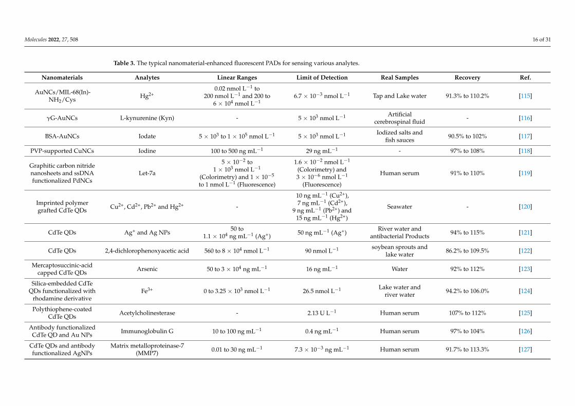
Table 3. The typical nanomaterial-enhanced fluorescent PADs for sensing various analytes.
Nanomaterials Analytes Linear Ranges Limit of Detection Real Samples Recovery Ref.
AuNCs/MIL-68(In)-NH2/Cys Hg2+
0.02 nmol L−1 to200 nmol L−1 and 200 to
6 × 10⁴ nmol L−16.7 × 10⁻³ nmol L−1 Tap and Lake water 91.3% to 110.2% [115]
γG-AuNCs L-kynurenine (Kyn) - 5 × 10³ nmol L−1 Artificialcerebrospinal fluid - [116]
BSA-AuNCs Iodate 5 × 10³ to 1 × 10⁵ nmol L−1 5 × 10³ nmol L−1 Iodized salts andfish sauces 90.5% to 102% [117]
PVP-supported CuNCs Iodine 100 to 500 ng mL−1 29 ng mL−1 - 97% to 108% [118]
Graphitic carbon nitridenanosheets and ssDNAfunctionalized PdNCs
Let-7a
5 × 10⁻² to1 × 103 nmol L−1
(Colorimetry) and 1 × 10⁻⁵
to 1 nmol L−1 (Fluorescence)
1.6 × 10⁻² nmol L−1
(Colorimetry) and3 × 10−6 nmol L−1
(Fluorescence)
Human serum 91% to 110% [119]
Imprinted polymergrafted CdTe QDs Cu2+, Cd2+, Pb2+ and Hg2+ -
10 ng mL−1 (Cu2+),7 ng mL−1 (Cd2+),
9 ng mL−1 (Pb2+) and15 ng mL−1 (Hg2+)
Seawater - [120]
CdTe QDs Ag+ and Ag NPs 50 to1.1 × 104 ng mL−1 (Ag+) 50 ng mL−1 (Ag+)
River water andantibacterial Products 94% to 115% [121]
CdTe QDs 2,4-dichlorophenoxyacetic acid 560 to 8 × 10⁴ nmol L−1 90 nmol L−1 soybean sprouts andlake water 86.2% to 109.5% [122]
Mercaptosuccinic-acidcapped CdTe QDs Arsenic 50 to 3 × 10⁴ ng mL−1 16 ng mL−1 Water 92% to 112% [123]
Silica-embedded CdTeQDs functionalized with
rhodamine derivativeFe3+ 0 to 3.25 × 10³ nmol L−1 26.5 nmol L−1 Lake water and
river water 94.2% to 106.0% [124]
Polythiophene-coatedCdTe QDs Acetylcholinesterase - 2.13 U L−1 Human serum 107% to 112% [125]
Antibody functionalizedCdTe QD and Au NPs Immunoglobulin G 10 to 100 ng mL−1 0.4 ng mL−1 Human serum 97% to 104% [126]
CdTe QDs and antibodyfunctionalized AgNPs
Matrix metalloproteinase-7(MMP7) 0.01 to 30 ng mL−1 7.3 × 10⁻³ ng mL−1 Human serum 91.7% to 113.3% [127]
Molecules 2022, 27, 508 17 of 31
Table 3. Cont.
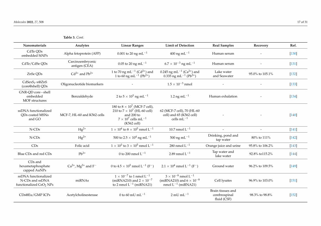
Nanomaterials Analytes Linear Ranges Limit of Detection Real Samples Recovery Ref.
CdTe QDsembedded SiNPs Alpha fetoprotein (AFP) 0.001 to 20 ng mL−1 400 ng mL−1 Human serum - [130]
CdTe/CdSe QDs Carcinoembryonicantigen (CEA) 0.05 to 20 ng mL−1 6.7 × 10⁻³ ng mL−1 Human serum - [131]
ZnSe QDs Cd2+ and Pb2+ 1 to 70 ng mL−1 (Cd2+) and1 to 60 ng mL−1 (Pb2+)
0.245 ng mL−1 (Ca2+) and0.335 ng mL−1 (Pb2+)
Lake waterand Seawater 95.0% to 105.1% [132]
CdSexS1-x@ZnS(core@shell) QDs Oligonucleotide biomarkers - 1.5 × 10⁻³ nmol - - [133]
GNR-QD core−shellembedded
MOF structuresBenzaldehyde 2 to 5 × 10³ ng mL−1 1.2 ng mL−1 Human exhalation - [134]
ssDNA functionalizedQDs coated MSNs
and GOMCF-7, HL-60 and K562 cells
180 to 8 × 10⁷ (MCF-7 cell),210 to 7 × 10⁷ (HL-60 cell)
and 200 to7 × 107 cells mL−1
(K562 cell)
62 (MCF-7 cell), 70 (HL-60cell) and 65 (K562 cell)
cells mL−1- - [140]
N-CDs Hg2+ 1 × 10⁴ to 8 × 10⁵ nmol L−1 10.7 nmol L−1 - - [141]
N-CDs Hg2+ 500 to 2.5 × 10⁴ ng mL−1 500 ng mL−1 Drinking, pond andtap water 80% to 111% [142]
CDs Folic acid 1 × 10³ to 3 × 10⁵ nmol L−1 280 nmol L−1 Orange juice and urine 95.8% to 106.2% [143]
Blue CDs and red CDs Pb2+ 0 to 200 nmol L−1 2.89 nmol L−1 Tap water andlake water 92.8% to115.2% [144]
CDs andhexametaphosphate
capped AuNPsCa2+, Mg2+ and F− 0 to 4.5 × 10⁵ nmol L−1 (F−) 2.1 × 10⁴ nmol L−1 (F−) Ground water 96.2% to 109.5% [149]
ssDNA functionalizedN-CDs and ssDNA
functionalized CeO2 NPsmiRNAs
1 × 10⁻⁷ to 1 nmol L−1
(miRNA210) and 2 × 10⁻⁷
to 2 nmol L−1 (miRNA21)
3 × 10⁻⁸ nmol L−1
(miRNA210) and 6 × 10⁻⁸
nmol L−1 (miRNA21)Cell lysates 96.9% to 103.0% [151]
CDs@Eu/GMP ICPs Acetylcholinesterase 0 to 60 mU mL−1 2 mU mL−1Brain tissues and
cerebrospinalfluid (CSF)
98.3% to 98.8% [152]
Molecules 2022, 27, 508 18 of 31
Table 3. Cont.
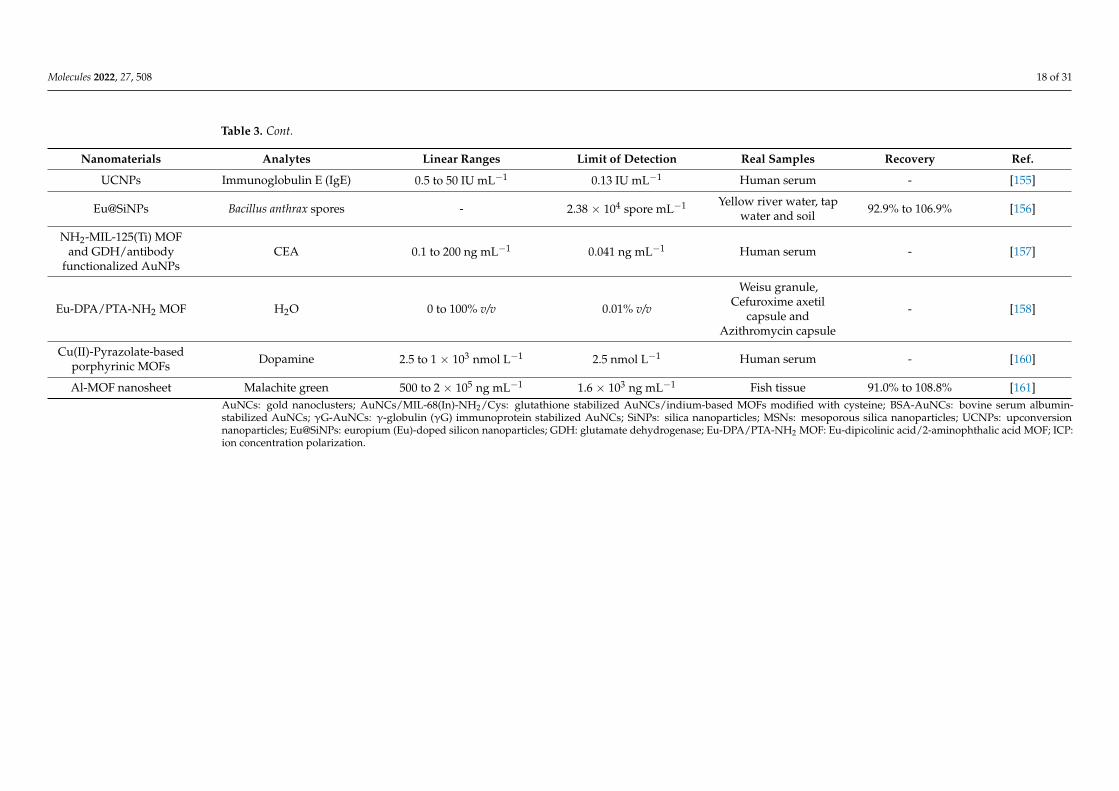
Nanomaterials Analytes Linear Ranges Limit of Detection Real Samples Recovery Ref.
UCNPs Immunoglobulin E (IgE) 0.5 to 50 IU mL−1 0.13 IU mL−1 Human serum - [155]
Eu@SiNPs Bacillus anthrax spores - 2.38 × 10⁴ spore mL−1 Yellow river water, tapwater and soil 92.9% to 106.9% [156]
NH2-MIL-125(Ti) MOFand GDH/antibody
functionalized AuNPsCEA 0.1 to 200 ng mL−1 0.041 ng mL−1 Human serum - [157]
Eu-DPA/PTA-NH2 MOF H2O 0 to 100% v/v 0.01% v/v
Weisu granule,Cefuroxime axetil
capsule andAzithromycin capsule
- [158]
Cu(II)-Pyrazolate-basedporphyrinic MOFs Dopamine 2.5 to 1 × 10³ nmol L−1 2.5 nmol L−1 Human serum - [160]
Al-MOF nanosheet Malachite green 500 to 2 × 10⁵ ng mL−1 1.6 × 10³ ng mL−1 Fish tissue 91.0% to 108.8% [161]AuNCs: gold nanoclusters; AuNCs/MIL-68(In)-NH2/Cys: glutathione stabilized AuNCs/indium-based MOFs modified with cysteine; BSA-AuNCs: bovine serum albumin-stabilized AuNCs; γG-AuNCs: γ-globulin (γG) immunoprotein stabilized AuNCs; SiNPs: silica nanoparticles; MSNs: mesoporous silica nanoparticles; UCNPs: upconversionnanoparticles; Eu@SiNPs: europium (Eu)-doped silicon nanoparticles; GDH: glutamate dehydrogenase; Eu-DPA/PTA-NH2 MOF: Eu-dipicolinic acid/2-aminophthalic acid MOF; ICP:ion concentration polarization.
Molecules 2022, 27, 508 19 of 31
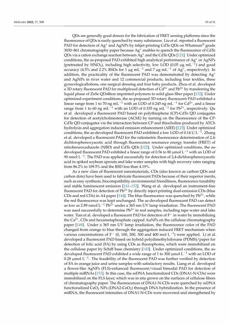
QDs are generally good donors for the fabrication of FRET sensing platforms since thefluorescence of QDs is easily quenched by many substances. Liu et al. reported a fluorescentPAD for detection of Ag+ and AgNPs by inkjet-printing CdTe QDs on Whatman® grade3030–861 chromatography paper because Ag+ enables to quench the fluorescence of CdTeQDs via a cation exchange reaction between Ag+ and the CdTe QDs [121]. Under optimizedconditions, the as-proposed PAD exhibited high analytical performance of Ag+ or AgNPs(pretreated by HNO3), including high selectivity, low LOD (0.05 µg mL−1) and goodaccuracy (4.5% and 2.2% RSDs for 1 µg mL−1 and 7 µg mL−1 of Ag+, respectively). Inaddition, the practicality of the fluorescent PAD was demonstrated by detecting Ag+
and AgNPs in river water and 12 commercial products, including four textiles, threegynecologicallotions, one surgical dressing and four baby products. Zhou et al. developeda 3D rotary fluorescent PAD for multiplexed detection of Cd2+ and Pb2+ by transferring theliquid phase of ZnSe QDs@ion imprinted polymers to solid glass fiber paper [132]. Underoptimized experiment conditions, the as-proposed 3D rotary fluorescent PAD exhibited alinear range from 1 to 70 ng mL−1 with an LOD of 0.245 ng mL−1 for Cd2+, and a linearrange from 1 to 60 ng mL−1 with an LOD of 0.335 ng mL−1 for Pb2+, respectively. Quet al. developed a fluorescent PAD based on polythiophene (CP)-CdTe QD conjugatesfor detection of acetylcholinesterase (AChE) by turning on the fluorescence of the CP-CdTe QD conjugates via the interaction between CP and thiocholine produced by ATChhydrolysis and aggregation induced emission enhancement (AIEE) [125]. Under optimizedconditions, the as-developed fluorescent PAD exhibited a low LOD of 0.14 U L−1. Zhanget al. developed a fluorescent PAD for the ratiometric fluorescence determination of 2,4-dichlorophenoxyacetic acid through fluorescence resonance energy transfer (FRET) ofnitrobenzoxadiazole (NBD) and CdTe QDs [122]. Under optimized conditions, the as-developed fluorescent PAD exhibited a linear range of 0.56 to 80 µmol L−1 with an LOD of90 nmol L−1. The PAD was applied successfully for detection of 2,4-dichlorophenoxyaceticacid in spiked soybean sprouts and lake water samples with high recovery rates rangingfrom 86.2% to 109.5% and the RSD less than 4.19%.
As a new class of fluorescent nanomaterials, CDs (also known as carbon QDs andcarbon dots) have been used to fabricate fluorescent PADs because of their superior merits,such as easy synthesis, biocompatibility, environmental friendliness, fluorescence tunabilityand stable luminescent emission [141–152]. Wang et al. developed an instrument-freefluorescent PAD for detection of Pb2+ by directly inject-printing dual-emission CDs (blueCDs and red CDs) in A4 paper [144]. The blue fluorescence was quenched by Pb2+, whilethe red fluorescence was kept unchanged. The as-developed fluorescent PAD can detectas low as 2.89 nmol L−1 Pb2+ under a 365 nm UV lamp irradiation. The fluorescent PADwas used successfully to determine Pb2+ in real samples, including tape water and lakewater. Tian et al. developed a fluorescent PAD for detection of F− in water by immobilizingthe Ca2+, CDs and hexametaphosphate capped AuNPs on the cellulose chromatographypaper [149]. Under a 365 nm UV lamp irradiation, the fluorescence color of the PADchanged from orange to blue through the aggregation induced FRET mechanism whenvarious concentrations of F− (0, 100, 200, 300 and 400 mol L−1) were applied. Li et al.developed a fluorescent PAD based on hybrid polydimethylsiloxane (PDMS)/paper fordetection of folic acid (FA) by using CDs as fluorophores, which were immobilized onthe cellulose paper by Schiff base chemistry [143]. Under optimized conditions, the as-developed fluorescent PAD exhibited a wide range of 1 to 300 µmol L−1 with an LOD of0.28 µmol L−1. The feasibility of the fluorescent PAD was further verified by detectionof FA in orange juice and urine samples with satisfactory results. Liang et al. developeda flower-like AgNPs (FLS)-enhanced fluorescent/visual bimodal PAD for detection ofmultiple miRNAs [151]. In this case, the ssDNA functionalized CDs (DNA1-N-CDs) wereimmobilized on the FLS layer, which was in situ grown on the surfaces of cellulose fibersof chromatography paper. The fluorescences of DNA1-N-CDs were quenched by ssDNAfunctionalized CeO2 NPs (DNA2-CeO2) through DNA hybridization. In the presence ofmiRNA, the fluorescent intensities of DNA1-N-CDs were recovered and strengthened by
Molecules 2022, 27, 508 20 of 31
FLS. The disengaged DNA2-CeO2 could result in color change after adding H2O2, leadingto the real-time visual detection of miRNA. The as-developed FLS-enhanced fluorescentPAD exhibited linear ranges of 0.1 fmol L−1 to 1 nmol L−1 and 0.2 fmol L−1 to 2 nmol L−1
for miRNA210 and miRNA21 with LODs of 0.03 fmol L−1 for miRNA210 and 0.06 fmol L−1
for miRNA21, respectively. The practicability of the FLS-enhanced fluorescence PAD wasdemonstrated by successful detection of miRNA210 in different cell lysates.
Because of NIR-excitation and the visible light emission nature of UCNPs, the flu-orescent PADs using UCNPs as the label can avoid the interference of autofluorescenceand scattering light from biological samples and paper substrates, resulting in an improve-ment in the detection accuracy of the PAD [153–155]. Recently, He et al. developed aUCNP-based fluorescent PAD for detection of total immunoglobulin E (IgE) in humanserum through resonance energy transfer between UCNPs and organic dye tetramethyl-rhodamine (TAMRA) [155]. The UCNP-based fluorescent PAD exhibited a linear rangeof 0.5 to 50 IU mL−1 with an LOD of 0.13 IU mL−1. The practicability of the UCNP-basedfluorescent PAD was demonstrated by the determination of IgE in 20 human serum samples.The results of the UCNP-based PAD were well consistent with those of the commercialELISA kit. The RSDs (n = 3) of the PAD varied from 2.7% to 19.7%. The results suggestedthat the UCNP-based fluorescent PAD could be used as a POCT device for individualdiagnostic and real-time detection.
Recently, a MOFs-based fluorescent PAD has been developed for the detection of vari-ous targets [157–161]. Lv et al. developed a fluorescent PAD for detection of CEA throughwet NH3-triggered structural change of NH2-MIL-125(Ti) impregnated on paper [157]. TheNH2-MIL-125(Ti)-based PAD exhibited a linear range of 0.1 ng mL−1 to 200 ng mL−1 withan LOD of 0.041 ng mL−1. Yue et al. developed a portable smartphone-assisted ratiomet-ric fluorescent PAD for detection of malachite green (MG) by using fluorescent Al-MOFnanosheet and rhodamine B (RhB) as fluorescent probes [161]. The as-developed fluores-cent PAD exhibited a wide linear range of 0.5 to 200 µg mL−1, a low LOD of 1.6 µg mL−1,satisfactory recoveries (in the range of 81.90% to 108.00% and low RSD (in the range of1.00% to 4.69%). The practicability of the fluorescent PAD was verified by detection of MGin spiked fish tissues. The as-obtained results were in good agreement with those obtainedby high performance liquid chromatography (HPLC).
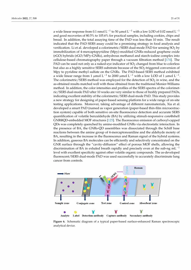
2.4. Paper-Based Surface-Enhanced Raman Spectroscopic Analytical Devices
The basic principle of SERS is that the signal of the analyte is strongly amplifiedthrough LSPR phenomena (i.e., electromagnetic hot spots) generated by light when itinteracts with labels (plasmonic metal nanoparticles), such as gold nanorods (GNRs) andAgNPs, as shown in Figure 6. The PAD-based SERS substrates have gained considerableattention since they enable on-site label-free detection of a wide variety of analytes andprovide “fingerprint” signatures of analytes (as shown in Table 4) [134,162–176]. Sahaand Jana developed a PAD-based SERS assay for the detection of proteins by mixingplasmonic nanomaterials (silver coated AuNPs (Ag@AuNPs)) and analyte in the mobilephase, where the analyte induced Ag@Au nanoparticles form controlled aggregates andgenerate electromagnetic hot spots inside the microfluidic channel, resulting in a strongSERS signal [173]. The as-developed PAD-based SERS assay exhibited high reproducibilityand sensitivity, which can be used to detect 1 fmol L−1 concanavalin A within 3 min.Qi et al. developed an oPAD for miRNA detection through modification of DNA-encodedRaman-active anisotropic AgNPs in the hydrophilic channels [171]. In the presence ofanalyte, the Raman signals on DNA-encoded AgNPs were amplified through a target-dependent, sequence-specific DNA hybridization assembly. The simple and low-costoPAD is generic and applicable to various miRNAs, which holds promising applicationsin point-of-care diagnostics because it can be used to detect as low as 1 pmol L−1 within15 min. Wu et al. developed a PAD for detection of acrylamide (AAm) in food products byusing the strawberry-like SiO2/Ag nanocomposites (SANC) immersed chromatographypaper [172]. Under the optimized conditions, the as-developed PAD SERS assay exhibited
Molecules 2022, 27, 508 21 of 31
a wide linear response from 0.1 nmol L−1 to 50 µmol L−1 with a low LOD of 0.02 nmol L−1
and good recoveries of 80.5% to 105.6% for practical samples, including cookies, chips andbread. In addition, the total assaying time of the PAD was less than 10 min. The resultindicated that the PAD SERS assay could be a promising strategy in food analysis andverification. Li et al. developed a colorimetric/SERS dual-mode PAD for sensing SO2 byimmobilization of 4-mercaptopyridine (Mpy)-modified GNRs-reduced graphene oxide(rGO) hybrids (rGO/MPy-GNRs), anhydrous methanol and starch-iodine complex intocellulose-based chromatography paper through a vacuum filtration method [174]. ThePAD can be used not only as a naked-eye indicator of SO2 changed from blue to colorlessbut also as a highly sensitive SERS substrate because of the SO2-triggered conversion ofMpy to pyridine methyl sulfate on the GNRs. The PAD-based SERS method exhibiteda wide linear range from 1 µmol L−1 to 2000 µmol L−1 with a low LOD of 1 µmol L−1.The colorimetric/SERS method was employed for the detection of SO2 in wine, and theas-obtained results matched well with those obtained from the traditional Monier-Williamsmethod. In addition, the color intensities and profiles of the SERS spectra of the colorimet-ric/SERS dual-mode PAD after 10 weeks are very similar to those of freshly prepared PADs,indicating excellent stability of the colorimetric/SERS dual-mode PAD. This study providesa new strategy for designing of paper-based sensing platform for a wide range of on-sitetesting applications. Moreover, taking advantage of different nanomaterials, Xia et al.developed a smart PAD (named as vapor generation (paper-based thin-film microextrac-tion system) capable of both sensitive on-site fluorescence detection and accurate SERSquantification of volatile benzaldehyde (BA) by utilizing stimuli-responsive core@shellGNR@QD-embedded MOF structures [125]. The fluorescence emission of carboxyl-cappedQDs was completely quenched by amino-modified GNRs via electrostatic interaction. Inthe presence of BA, the GNRs-QD assemblies was dissociated through the Schiff basereactions between the amine group of 4-mercaptonoaniline and the aldehyde moiety ofBA, resulting in the increase in the fluorescence and Raman signal of the hybrid systems.In addition, gaseous BA molecules can be efficiently and selectively concentrated on theGNR surface through the “cavity-diffusion” effect of porous MOF shells, allowing thediscrimination of BA in exhaled breath rapidly and precisely even at the sub-ng mL−1
level with excellent specificity against other volatile organic compounds. The as-developedfluorescent/SERS dual-mode PAD was used successfully to accurately discriminate lungcancer from controls.
Molecules 2022, 27, x FOR PEER REVIEW 18 of 28
a wide linear response from 0.1 nmol L−1 to 50 μmol L−1 with a low LOD of 0.02 nmol L−1 and good recoveries of 80.5% to 105.6% for practical samples, including cookies, chips and bread. In addition, the total assaying time of the PAD was less than 10 min. The result indicated that the PAD SERS assay could be a promising strategy in food analysis and verification. Li et al. developed a colorimetric/SERS dual-mode PAD for sensing SO2 by immobilization of 4-mercaptopyridine (Mpy)-modified GNRs-reduced graphene oxide (rGO) hybrids (rGO/MPy-GNRs), anhydrous methanol and starch-iodine complex into cellulose-based chromatography paper through a vacuum filtration method [174]. The PAD can be used not only as a naked-eye indicator of SO2 changed from blue to colorless but also as a highly sensitive SERS substrate because of the SO2-triggered conversion of Mpy to pyridine methyl sulfate on the GNRs. The PAD-based SERS method exhibited a wide linear range from 1 μmol L−1 to 2000 μmol L−1 with a low LOD of 1 μmol L−1. The colorimetric/SERS method was employed for the detection of SO2 in wine, and the as-obtained results matched well with those obtained from the traditional Monier-Williams method. In addition, the color intensities and profiles of the SERS spectra of the colorimet-ric/SERS dual-mode PAD after 10 weeks are very similar to those of freshly prepared PADs, indicating excellent stability of the colorimetric/SERS dual-mode PAD. This study provides a new strategy for designing of paper-based sensing platform for a wide range of on-site testing applications. Moreover, taking advantage of different nanomaterials, Xia et al. developed a smart PAD (named as vapor generation (paper-based thin-film micro-extraction system) capable of both sensitive on-site fluorescence detection and accurate SERS quantification of volatile benzaldehyde (BA) by utilizing stimuli-responsive core@shell GNR@QD-embedded MOF structures [125]. The fluorescence emission of car-boxyl-capped QDs was completely quenched by amino-modified GNRs via electrostatic interaction. In the presence of BA, the GNRs-QD assemblies was dissociated through the Schiff base reactions between the amine group of 4-mercaptonoaniline and the aldehyde moiety of BA, resulting in the increase in the fluorescence and Raman signal of the hybrid systems. In addition, gaseous BA molecules can be efficiently and selectively concentrated on the GNR surface through the “cavity-diffusion” effect of porous MOF shells, allowing the discrimination of BA in exhaled breath rapidly and precisely even at the sub-ng mL−1 level with excellent specificity against other volatile organic compounds. The as-devel-oped fluorescent/SERS dual-mode PAD was used successfully to accurately discriminate lung cancer from controls.
Figure 6. Schematic diagram of a typical paper-based surface-enhanced Raman spectroscopic ana-lytical device.
Figure 6. Schematic diagram of a typical paper-based surface-enhanced Raman spectroscopicanalytical device.
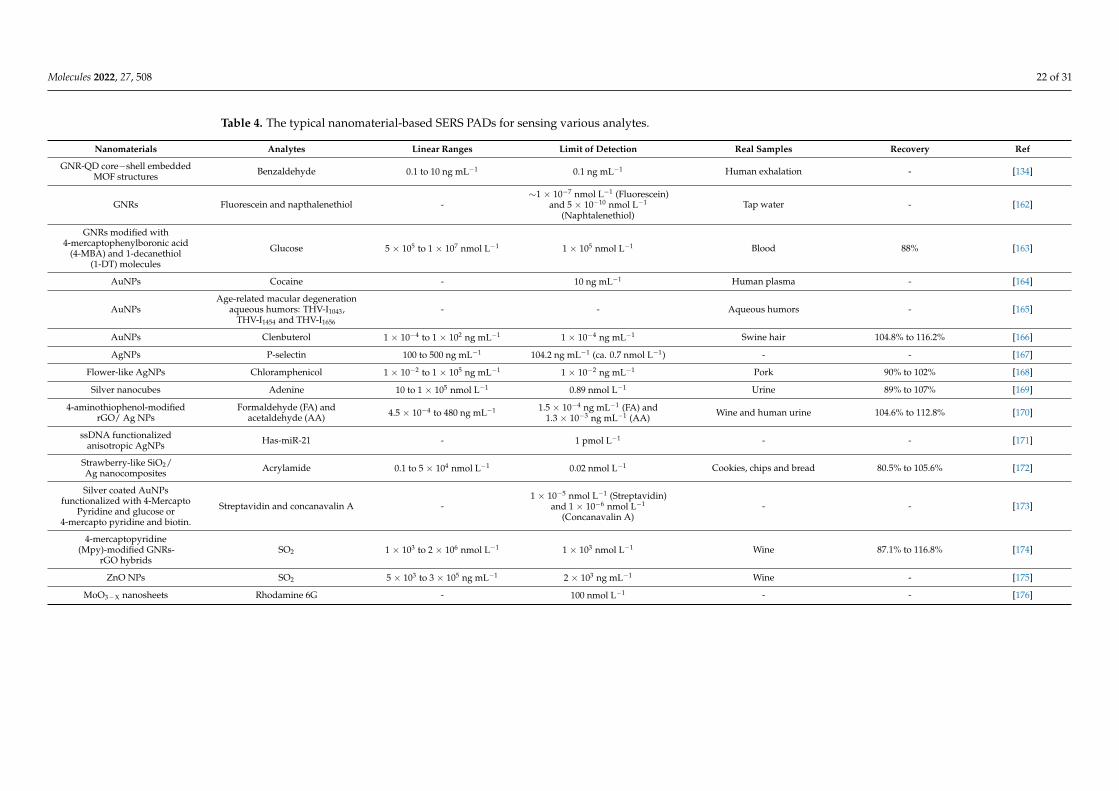
Molecules 2022, 27, 508 22 of 31
Table 4. The typical nanomaterial-based SERS PADs for sensing various analytes.
Nanomaterials Analytes Linear Ranges Limit of Detection Real Samples Recovery Ref
GNR-QD core−shell embeddedMOF structures Benzaldehyde 0.1 to 10 ng mL−1 0.1 ng mL−1 Human exhalation - [134]
GNRs Fluorescein and napthalenethiol -∼1 × 10⁻⁷ nmol L−1 (Fluorescein)
and 5 × 10⁻¹⁰ nmol L−1
(Naphtalenethiol)Tap water - [162]
GNRs modified with4-mercaptophenylboronic acid
(4-MBA) and 1-decanethiol(1-DT) molecules
Glucose 5 × 10⁵ to 1 × 10⁷ nmol L−1 1 × 10⁵ nmol L−1 Blood 88% [163]
AuNPs Cocaine - 10 ng mL−1 Human plasma - [164]
AuNPsAge-related macular degeneration
aqueous humors: THV-I1043,THV-I1454 and THV-I1656
- - Aqueous humors - [165]
AuNPs Clenbuterol 1 × 10⁻⁴ to 1 × 10² ng mL−1 1 × 10⁻⁴ ng mL−1 Swine hair 104.8% to 116.2% [166]
AgNPs P-selectin 100 to 500 ng mL−1 104.2 ng mL−1 (ca. 0.7 nmol L−1) - - [167]
Flower-like AgNPs Chloramphenicol 1 × 10⁻² to 1 × 10⁵ ng mL−1 1 × 10⁻² ng mL−1 Pork 90% to 102% [168]
Silver nanocubes Adenine 10 to 1 × 10⁵ nmol L−1 0.89 nmol L−1 Urine 89% to 107% [169]
4-aminothiophenol-modifiedrGO/ Ag NPs
Formaldehyde (FA) andacetaldehyde (AA) 4.5 × 10⁻⁴ to 480 ng mL−1 1.5 × 10⁻⁴ ng mL−1 (FA) and
1.3 × 10⁻³ ng mL−1 (AA) Wine and human urine 104.6% to 112.8% [170]
ssDNA functionalizedanisotropic AgNPs Has-miR-21 - 1 pmol L−1 - - [171]
Strawberry-like SiO2/Ag nanocomposites Acrylamide 0.1 to 5 × 10⁴ nmol L−1 0.02 nmol L−1 Cookies, chips and bread 80.5% to 105.6% [172]
Silver coated AuNPsfunctionalized with 4-Mercapto
Pyridine and glucose or4-mercapto pyridine and biotin.
Streptavidin and concanavalin A -1 × 10⁻⁵ nmol L−1 (Streptavidin)
and 1 × 10⁻⁶ nmol L−1
(Concanavalin A)- - [173]
4-mercaptopyridine(Mpy)-modified GNRs-
rGO hybridsSO2 1 × 10³ to 2 × 10⁶ nmol L−1 1 × 10³ nmol L−1 Wine 87.1% to 116.8% [174]
ZnO NPs SO2 5 × 10³ to 3 × 10⁵ ng mL−1 2 × 10³ ng mL−1 Wine - [175]
MoO3−X nanosheets Rhodamine 6G - 100 nmol L−1 - - [176]
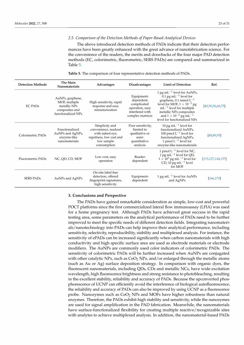
Molecules 2022, 27, 508 23 of 31
2.5. Comparison of the Detection Methods of Paper-Based Analytical Devices
The above introduced detection methods of PADs indicate that their detection perfor-mances have been greatly enhanced with the great advance of nanofabrication science. Forthe convenience of the readers, the merits and drawbacks of the four major PAD detectionmethods (EC, colorimetric, fluorometric, SERS PADs) are compared and summarized inTable 5.
Table 5. The comparison of four representative detection methods of PADs.
Detection Methods The MainNanomaterials Advantages Disadvantages Limit of Detection Ref.
EC PADs
AuNPs, graphene,MOF, multiplemetallic NPs
composites andfunctionalized NPs
High sensitivity, rapidresponse and easy
miniaturization
Equipment-dependent,
complicatedoperation, easyinterfered with
complex matrices
1 pg mL−1 level for AuNPs,0.1 pg mL−1 level for
graphene, 0.1 mmol L−1
level for MOF, 1 × 10⁻³ pgmL−1 level for multiplemetallic NPs compositesand 1 × 10⁻² pg mL−1
level for functionalized NPs
[40,50,56,68,75]
Colorimetric PADs
FunctionalizedAuNPs and AgNPs,
enzyme-likenanomaterials
Simplicity andconvenience, readout
with naked eye,rapidness, low cost and
low sampleconsumption
Poor sensitivity,limited to
qualitative orsemi-
quantitativeanalysis
10 pg mL−1 level forfunctionalized AuNPs,100 pmol L−1 level forfunctionalized AgNPs,
1 pmol L−1 level forenzyme-like nanomaterials
[48,89,95]
Fluorometric PADs NC, QD, CD, MOF Low cost, easyoperation
Reader-dependent
1 pmol L−1 level for NC,1 pg mL−1 level for QD,
1 × 10³ pg mL−1 level forCD, 10 pg mL−1 level
for MOF
[115,127,144,157]
SERS PADs AuNPs and AgNPs
On-site label-freedetection, offered
fingerprint signatures,high sensitivity
Equipment-dependent
1 pg mL−1 level for AuNPsand AgNPs
[166,170]
3. Conclusions and Perspective
The PADs have gained remarkable consideration as simple, low-cost and powerfulPOCT platforms since the first commercialized lateral flow immunoassay (LFIA) was usedfor a home pregnancy test. Although PADs have achieved great success in the rapidtesting area, some parameters on the analytical performance of PADs need to be furtherimproved to meet the specific needs of different detection fields. Integrating nanomateri-als/nanotechnology into PADs can help improve their analytical performance, includingsensitivity, selectivity, reproducibility, stability and multiplexed analysis. For instance, thesensitivity of ePADs can be increased significantly when carbon nanomaterials with highconductivity and high specific surface area are used as electrode materials or electrodemodifiers. The AuNPs are commonly used color indicators of colorimetric PADs. Thesensitivity of colorimetric PADs will be further increased when AuNPs are conjugatedwith other catalytic NPs, such as CeO2 NPs, and/or enlarged through the metallic atoms(such as Au or Ag) surface deposition strategy. In comparison with organic dyes, thefluorescent nanomaterials, including QDs, CDs and metallic NCs, have wide excitationwavelength, high fluorescence brightness and strong resistance to photobleaching, resultingin the excellent stability, reliability and accuracy of PADs. Because the upconverted phos-phorescence of UCNP can efficiently avoid the interference of biological autofluorescence,the reliability and accuracy of PADs can also be improved by using UCNP as a fluorescenceprobe. Nanozymes such as CeO2 NPs and MOFs have higher robustness than naturalenzymes. Therefore, the PADs exhibit high stability and sensitivity, while the nanozymesare used for signal amplification in the PAD fabrication. Meanwhile, the nanomaterialshave surface-functionalized flexibility for creating multiple reactive/recognizable siteswith analytes to achieve multiplexed analysis. In addition, the nanomaterial-based PADs
Molecules 2022, 27, 508 24 of 31
are compatible with dual-mode detection, such as colorimetry–fluorescence and EC-opticaldetection, which enables the reliability and accuracy of the PADs.
The nanomaterial-based PADs have managed great achievements for sensing varioustargets, including ions, small molecules, nucleic acids, proteins and pathogens, in thebench research. However, there are limited examples that have transformed from proof-of-principle analytical devices to commercial products. More efforts should be madecontinuously to develop novel strategies for increasing the longevity, robustness andreliability in the real-time monitoring, which are largely dependent on the synthesis ofadvanced nanomaterials, introduction of a new sensing mechanism and multiple detectionmodes, development of a reliable microfabricating methodology/standard and simplifyingthe detection procedures. The next generation of PADs will be simple, cost-effective andmultiplexed and be able to provide on-site qualitative/quantitative analysis by the nakedeye and/or portable equipment, such as smartphones and wearable electronic devices.
Author Contributions: Writing—original draft preparation, R.P., Q.Z., X.M. and Z.W.; writing reviewand editing, R.P., Q.Z., J.W., X.M. and Z.W. All authors have read and agreed to the published versionof the manuscript.
Funding: This work was supported by the National Natural Science Foundation of China (GrantNo. 21775145), and Science and Technology Developing Foundation of Jilin Province (Grant No.20210204049YY) for financial support.
Institutional Review Board Statement: Not applicable.
Informed Consent Statement: Not applicable.
Data Availability Statement: Not applicable.
Acknowledgments: The authors would like to thank the National Natural Science Foundation ofChina and Science and Technology Developing Foundation of Jilin Province for financial support.
Conflicts of Interest: The authors declare no conflict of interest.
References1. Cate, D.M.; Adkins, J.A.; Mettakoonpitak, J.; Henry, C.S. Recent Developments in Paper-Based Microfluidic Devices. Anal. Chem.
2015, 87, 19–41. [CrossRef]2. Xia, Y.; Si, J.; Li, Z. Fabrication Techniques for Microfluidic Paper-Based Analytical Devices and Their Applications for Biological
Testing: A Review. Biosens. Bioelectron. 2016, 77, 774–789. [CrossRef]3. Ahmed, S.; Bui, M.N.; Abbas, A. Paper-Based Chemical and Biological Sensors: Engineering Aspects. Biosens. Bioelectron. 2016,
77, 249–263. [CrossRef]4. Liu, S.; Su, W.; Ding, X. A Review on Microfluidic Paper-Based Analytical Devices for Glucose Detection. Sensors 2016, 16, 2086.
[CrossRef]5. Mahato, K.; Srivastava, A.; Chandra, P. Paper Based Diagnostics for Personalized Health Care: Emerging Technologies and
Commercial Aspects. Biosens. Bioelectron. 2017, 96, 246–259. [CrossRef]6. Wang, P.; Kricka, L.J. Current and Emerging Trends in Point-of-Care Technology and Strategies for Clinical Validation and
Implementation. Clin. Chem. 2018, 64, 1439–1452. [CrossRef]7. Li, Y.; Feng, L. Progress in Paper-Based Colorimetric Sensor Array. Chin. J. Anal. Chem. 2020, 48, 1448–1457. [CrossRef]8. Lin, Y.; Gritsenko, D.; Feng, S.; Teh, Y.C.; Lu, X.; Xu, J. Detection of Heavy Metal by Paper-Based Microfluidics. Biosens. Bioelectron.
2016, 83, 256–266. [CrossRef]9. Meredith, N.A.; Quinn, C.; Cate, D.M.; Reilly, T.H.; Volckens, J.; Henry, C.S. Paper-Based Analytical Devices for Environmental
Analysis. Analyst 2016, 141, 1874–1887. [CrossRef]10. Kaushik, A.; Tiwari, S.; Jayant, R.D.; Marty, A.; Nair, M. Towards Detection and Diagnosis of Ebola Virus Disease at Point-of-Care.
Biosens. Bioelectron. 2016, 75, 254–272. [CrossRef] [PubMed]11. Sriram, G.; Bhat, M.P.; Patil, P.; Uthappa, U.T.; Jung, H.-Y.; Altalhi, T.; Kumeria, T.; Aminabhavi, T.M.; Pai, R.K.; Kurkuri, M.D.
Paper-Based Microfluidic Analytical Devices for Colorimetric Detection of Toxic Ions: A Review. Trends Anal. Chem. 2017, 93,212–227. [CrossRef]
12. Altundemir, S.; Uguz, A.K.; Ulgen, K. A Review on Wax Printed Microfluidic Paper-Based Devices for International Health.Biomicrofluidics 2017, 11, 041501. [CrossRef]
13. Campbell, J.M.; Balhoff, J.B.; Landwehr, G.M.; Rahman, S.M.; Vaithiyanathan, M.; Melvin, A.T. Microfluidic and Paper-BasedDevices for Disease Detection and Diagnostic Research. Int. J. Mol. Sci. 2018, 19, 2731. [CrossRef] [PubMed]
Molecules 2022, 27, 508 25 of 31
14. Hua, M.Z.; Li, S.; Wang, S.; Lu, X. Detecting Chemical Hazards in Foods Using Microfluidic Paper-Based Analytical Devices(µPADs): The Real-World Application. Micromachines 2018, 9, 32. [CrossRef] [PubMed]
15. Wu, M.; Lai, Q.; Ju, Q.; Li, L.; Yu, H.-D.; Huang, W. Paper-Based Fluorogenic Devices for in Vitro Diagnostics. Biosens. Bioelectron.2018, 102, 256–266. [CrossRef] [PubMed]
16. Restaino, S.M.; White, I.M. A Critical Review of Flexible and Porous SERS Sensors for Analytical Chemistry at the Point-of-Sample.Anal. Chim. Acta 2019, 1060, 17–29. [CrossRef]
17. Ming, T.; Luo, J.P.; Liu, J.T.; Sun, S.; Xing, Y.; Wang, H.; Xiao, G.H.; Deng, Y.; Cheng, Y.; Yang, Z.G.; et al. Paper-Based MicrofluidicAptasensors. Biosens. Bioelectron. 2020, 170, 112649. [CrossRef]
18. Ding, R.Y.; Cheong, Y.H.; Ahamed, A.; Lisak, G. Heavy Metals Detection with Paper-Based Electrochemical Sensors. Anal. Chem.2021, 93, 1880–1888. [CrossRef]
19. Alahmad, W.; Sahragard, A.; Varanusupakul, P. Online and Offline Preconcentration Techniques on Paper-Based AnalyticalDevices for Ultrasensitive Chemical and Biochemical Analysis: A Review. Biosens. Bioelectron. 2021, 194, 113574. [CrossRef]
20. Ozer, T.; Henry, C.S. Paper-Based Analytical Devices for Virus Detection: Recent Strategies for Current and Future Pandemics.Trends Anal. Chem. 2021, 144, 116424. [CrossRef]
21. Fu, L.-M.; Wang, Y.-N. Detection Methods and Applications of Microfluidic Paper-Based Analytical Devices. Trends Anal. Chem.2018, 107, 196–211. [CrossRef]
22. Morbioli, G.G.; Mazzu-Nascimento, T.; Stockton, A.M.; Carrilho, E. Technical Aspects and Challenges of Colorimetric Detectionwith Microfluidic Paper-Based Analytical Devices (mPADs)—A Review. Anal. Chim. Acta 2017, 970, 1–22. [CrossRef]
23. Chinnadayyala, S.R.; Park, J.; Ngoc Le, H.T.; Santhosh, M.; Kadam, A.N.; Cho, S. Recent Advances in Microfluidic Paper-BasedElectrochemiluminescence Analytical Devices for Point-of-Care Testing Applications. Biosens. Bioelectron. 2019, 126, 68–81.[CrossRef]
24. Solhi, E.; Hasanzadeh, M.; Babaie, P. Electrochemical Paper-Based Analytical Devices (ePADs) toward Biosensing: RecentAdvances and Challenges in Bioanalysis. Anal. Methods 2020, 12, 1398–1414. [CrossRef]
25. Chung, T.H.; Dhar, B.R. Paper-Based Platforms for Microbial Electrochemical Cell-Based Biosensors: A Review. Biosens. Bioelectron.2021, 192, 113485. [CrossRef]
26. Quesada-González, D.; Merkoçi, A. Nanoparticle-Based Lateral Flow Biosensors. Biosens. Bioelectron. 2015, 73, 47–63. [CrossRef]27. Devi, R.V.; Doble, M.; Verma, R.S. Nanomaterials for Early Detection of Cancer Biomarker with Special Emphasis on Gold
Nanoparticles in Immunoassays/Sensors. Biosens. Bioelectron. 2015, 68, 688–698. [CrossRef]28. Arduini, F.; Micheli, L.; Moscone, D.; Palleschi, G.; Piermarini, S.; Ricci, F.; Volpe, G. Electrochemical Biosensors based on
Nanomodified Screen-Printed Electrodes: Recent Applications in Clinical Analysis. Trends Anal. Chem. 2016, 79, 114–126.[CrossRef]
29. Xu, Y.; Liu, M.; Kong, N.; Liu, J. Lab-on-Paper Micro- and Nano-Analytical Devices: Fabrication, Modification, Detection andEmerging Applications. Microchim. Acta 2016, 183, 1521–1542. [CrossRef]
30. Ge, S.; Zhang, L.; Zhang, Y.; Lan, F.; Yan, M.; Yu, J. Nanomaterials-Modified Cellulose Paper as a Platform for BiosensingApplications. Nanoscale 2017, 9, 4366–4382. [CrossRef] [PubMed]
31. Wongkaew, N.; Simsek, M.; Griesche, C.; Baeumner, A.J. Functional Nanomaterials and Nanostructures Enhancing ElectrochemicalBiosensors and Lab-on-a-Chip Performances: Recent Progress, Applications, and Future Perspective. Chem. Rev. 2019, 119,120–194. [CrossRef]
32. Selvakumar, B.; Kathiravan, A. Sensory Materials for Microfluidic Paper Based Analytical Devices—A Review. Talanta 2021,235, 122733. [CrossRef]
33. Patel, S.; Jamunkar, R.; Monisha, D.S.; Patle, T.K.; Kant, T.; Dewangan, K.; Shrivas, K. Recent Development in NanomaterialsFabricated Paper-Based Colorimetric and Fluorescent Sensors: A Review. Trends Environ. Anal. Chem. 2021, 31, e00136. [CrossRef]
34. Shirshahi, V.; Liu, G. Enhancing the Analytical Performance of Paper Lateral Flow Assays: From Chemistry to Engineering. TrendsAnal. Chem. 2021, 136, 116200. [CrossRef]
35. Díaz-González, M.; Escosura-Muñiz, A. Strip Modification and Alternative Architectures for Signal Amplification in Nanoparticle-Based Lateral Flow Assays. Anal. Bioanal. Chem. 2021, 413, 4111–4117. [CrossRef] [PubMed]
36. Wang, C.; Liu, M.; Wang, Z.; Li, S.; Deng, Y.; He, N. Point-of-Care Diagnostics for Infectious Diseases: From Methods to Devices.Nano Today 2021, 37, 101092. [CrossRef]
37. Hawkes, R.; Niday, E.; Gordon, J. A Dot-Immunobinding Assay for Monoclonal and Other Antibodies. Anal. Biochem. 1982, 119,142–147. [CrossRef]
38. Dungchai, W.; Chailapakul, O.; Henry, C.S. Electrochemical Detection for Paper-Based Microfluidics. Anal. Chem. 2009, 81,5821–5826. [CrossRef] [PubMed]
39. Baharfar, M.; Rahbar, M.; Tajik, M.; Liu, G.Z. Engineering Strategies for Enhancing the Performance of Electrochemical Paper-Based Analytical Devices. Biosens. Bioelectron. 2020, 167, 112506. [CrossRef]
40. Zheng, X.; Li, L.; Cui, K.; Zhang, Y.; Zhang, L.; Ge, S.; Yu, J. Ultrasensitive Enzyme-Free Biosensor by Coupling CyclodextrinFunctionalized Au Nanoparticles and High-Performance Au-Paper Electrode. ACS Appl. Mater. Interfaces 2018, 10, 3333–3340.[CrossRef]
Molecules 2022, 27, 508 26 of 31
41. Pinyorospathum, C.; Chaiyo, S.; Sae-ung, P.; Hoven, V.; Damsongsang, P.; Siangproh, W.; Chailapakul, O. Disposable Paper-BasedElectrochemical Sensor using Thiol-Terminated Poly(2-Methacryloyloxyethyl Phosphorylcholine) for the Label-Free Detection ofC-Reactive Protein. Microchim. Acta 2019, 186, 472. [CrossRef]
42. Tian, T.; Liu, H.; Li, L.; Yu, J.; Ge, S.; Song, X.; Yan, M. Paper-Based Biosensor for Noninvasive Detection of Epidermal GrowthFactor Receptor Mutations in Non-Small Cell Lung Cancer Patients. Sens. Actuators B Chem. 2017, 251, 440–445. [CrossRef]
43. Cinti, S.; Cinotti, G.; Parolo, C.; Nguyen, E.P.; Caratelli, V.; Moscone, D.; Arduini, F.; Merkoci, A. Experimental Comparisonin Sensing Breast Cancer Mutations by Signal ON and Signal OFF Paper-Based Electroanalytical Strips. Anal. Chem. 2020, 92,1674–1679. [CrossRef]
44. Eksin, E.; Torul, H.; Yarali, E.; Tamer, U.; Papakonstantinou, P.; Erdem, A. Paper-Based Electrode Assemble for ImpedimetricDetection of miRNA. Talanta 2021, 225, 122043. [CrossRef]
45. Ortone, V.; Matino, L.; Santoro, F.; Cinti, S. Merging Office/Filter Paper-Based Tools for Pre-Concentring and Detecting HeavyMetals in Drinking Water. Chem. Commun. 2021, 57, 7100–7103. [CrossRef] [PubMed]
46. Benson, J.; Fung, C.M.; Lloyd, J.S.; Deganello, D.; Smith, N.A.; Teng, K.S. Direct Patterning of Gold Nanoparticles usingFlexographic Printing for Biosensing Applications. Nanoscale Res. Lett. 2015, 10, 127. [CrossRef] [PubMed]
47. Pavithra, M.; Muruganand, S.; Parthiban, C. Development of Novel Paper Based Electrochemical Immunosensor with Self-MadeGold Nanoparticle Ink and Quinone Derivate for Highly Sensitive Carcinoembryonic Antigen. Sens. Actuators B Chem. 2018, 257,496–503. [CrossRef]
48. Cui, K.; Zhou, C.; Zhang, B.; Zhang, L.; Liu, Y.; Hao, S.; Tang, X.; Huang, Y.; Yu, J. Enhanced Catalytic Activity Induced by theNanostructuring Effect in Pd Decoration onto Doped Ceria Enabling an Origami Paper Analytical Device for High Performanceof Amyloid-β Bioassay. ACS Appl. Mater. Interfaces 2021, 13, 33937–33947. [CrossRef]
49. Li, L.; Wang, T.; Zhang, Y.; Xu, C.; Zhang, L.; Cheng, X.; Liu, H.; Chen, X.; Yu, J. Editable TiO2 Nanomaterial-Modified Paper inSitu for Highly Efficient Detection of Carcinoembryonic Antigen by Photoelectrochemical Method. ACS Appl. Mater. Interfaces2018, 10, 14594–14601. [CrossRef]
50. Xue, J.; Zhang, L.; Gao, C.; Zhu, P.; Yu, J. Microfluidic Paper-Based Photoelectrochemical Sensing Platform with Electron-TransferTunneling Distance Regulation Strategy for Thrombin Detection. Biosens. Bioelectron. 2019, 133, 1–7. [CrossRef]
51. De França, C.C.L.; Meneses, D.; Silva, A.C.A.; Dantas, N.O.; De Abreu, F.C.; Petroni, J.M.; Lucca, B.G. Development of NovelPaper-Based Electrochemical Device Modified with CdSe/CdS Magic-Sized Quantum Dots and Application for the Sensing ofDopamine. Electrochim. Acta 2021, 367, 137486. [CrossRef]
52. Mohan, J.M.; Amreen, K.; Kulkarni, M.B.; Javed, A.; Dubey, S.K.; Goel, S. Optimized Ink Jetted Paper Device for ElectroanalyticalDetection of Picric Acid. Colloid. Surface. B. 2021, 208, 112056. [CrossRef]
53. Amatatongchai, M.; Sitanurak, J.; Sroysee, W.; Sodanat, S.; Chairam, S.; Jarujamrus, P.; Nacapricha, D.; Lieberzeit, P.A. HighlySensitive and Selective Electrochemical Paper-Based Device using a Graphite Screen-Printed Electrode Modified with MolecularlyImprinted Polymers Coated Fe3O4@Au@SiO2 for Serotonin Determination. Anal. Chim. Acta 2019, 1077, 255–265. [CrossRef]
54. Zhang, X.; Bao, N.; Luo, X.; Ding, S. Patchy Gold Coated Fe3O4 Nanospheres with Enhanced Catalytic Activity Applied forPaper-Based Bipolar Electrode-Electrochemiluminescence Aptasensors. Biosens. Bioelectron. 2018, 114, 44–51. [CrossRef] [PubMed]
55. Huang, Y.Z.; Li, L.; Zhang, Y.; Zhang, L.N.; Ge, S.G.; Yu, J.H. Auto-Cleaning Paper-Based Electrochemiluminescence BiosensorCoupled with Binary Catalysis of Cubic Cu2O-Au and Polyethyleneimine for Quantification of Ni2+ and Hg2+. Biosens. Bioelectron.2019, 126, 339–345. [CrossRef] [PubMed]
56. Huang, Y.; Zhang, L.; Zhang, S.; Zhao, P.; Ge, S.; Yu, J. Paper-Based Electrochemiluminescence Determination of StreptavidinUsing Reticular DNA-Functionalized PtCu Nanoframes and Analyte-Triggered DNA Walker. Microchim. Acta 2020, 187, 530.[CrossRef] [PubMed]
57. Pokpas, K.; Jahed, N.; McDonald, E.; Bezuidenhout, P.; Smith, S.; Land, K.; Iwuoha, E. Graphene-AuNP Enhanced Inkjet-PrintedSilver Nanoparticle Paper Electrodes for the Detection of Nickel(II)-Dimethylglyoxime [Ni(dmgH2)] Complexes by AdsorptiveCathodic Stripping Voltammetry (AdCSV). Electroanalysis 2020, 32, 3017–3031. [CrossRef]
58. Valentine, C.J.; Takagishi, K.; Umezu, S.; Daly, R.; De Volder, M. Paper-Based Electrochemical Sensors using Paper as a Scaffold toCreate Porous Carbon Nanotube Electrodes. ACS Appl. Mater. Interfaces 2020, 12, 30680–30685. [CrossRef]
59. Tran, V.; Ko, E.; Geng, Y.; Kim, M.; Jin, G.; Son, S.; Hur, W.; Seong, G. Micro-Patterning of Single-Walled Carbon Nanotubes andIts Surface Modification with Gold Nanoparticles for Electrochemical Paper-Based Non-Enzymatic Glucose Sensor. J. Electroanal.Chem. 2018, 826, 29–37. [CrossRef]
60. Jampasa, S.; Pummoree, J.; Siangproh, W.; Khongchareonporn, N.; Ngamrojanavanich, N.; Chailapakul, O.; Chaiyo, S. “Signal-On”Electrochemical Biosensor based on a Competitive Immunoassay Format for the Sensitive Determination of Oxytetracycline. Sens.Actuators B Chem. 2020, 320, 128389. [CrossRef]
61. Srisomwat, C.; Teengam, P.; Chuaypen, N.; Tangkijvanich, P.; Vilaivan, T.; Chailapakul, O. Pop-up Paper Electrochemical Devicefor Label-Free Hepatitis B Virus DNA Detection. Sens. Actuators B Chem. 2020, 316, 128077. [CrossRef]
62. Pungjunun, K.; Yakoh, A.; Chaiyo, S.; Praphairaksit, N.; Siangproh, W.; Kalcher, K.; Chailapakul, O. Laser Engraved MicroapillaryPump Paper-Based Microfluidic Device for Colorimetric and Electrochemical Detection of Salivary Thiocyanate. Microchim. Acta2021, 188, 140. [CrossRef] [PubMed]
63. Wang, P.; Cheng, Z.; Chen, Q.; Qu, L.; Miao, X.; Feng, Q. Construction of a Paper-Based Electrochemical Biosensing Platform forRapid and Accurate Detection of Adenosine Triphosphate (ATP). Sens. Actuators B Chem. 2018, 256, 931–937. [CrossRef]
Molecules 2022, 27, 508 27 of 31
64. Pungjunun, K.; Chaiyo, S.; Praphairaksit, N.; Siangproh, W.; Ortner, A.; Kalcher, K.; Chailapaku, O.; Mehmeti, E. ElectrochemicalDetection of NOx Gas Based on Disposable Paper-Based Analytical Device using a Copper Nanoparticles-Modified Screen-PrintedGraphene Electrode. Biosens. Bioelectron. 2019, 143, 111606. [CrossRef]
65. Prasad, K.; Cao, X.; Gao, N.; Jin, Q.; Sanjay, S.; Henao-Pabon, G.; Li, X. A Low-Cost Nanomaterial-Based ElectrochemicalImmunosensor on Paper for High-Sensitivity Early Detection of Pancreatic Cancer. Sens. Actuators B Chem. 2020, 305, 127516.[CrossRef] [PubMed]
66. Wang, Y.; Xu, H.; Luo, J.; Liu, J.; Wang, L.; Fan, Y.; Yan, S.; Yang, Y.; Cai, X. A Novel Label-Free Microfluidic Paper-BasedImmunosensor for Highly Sensitive Electrochemical Detection of Carcinoembryonic Antigen. Biosen. Bioelectron. 2016, 83, 319–326.[CrossRef]
67. Wang, Y.; Luo, J.; Liu, J.; Sun, S.; Xiong, Y.; Ma, Y.; Yan, S.; Yang, Y.; Yin, H.; Cai, X. Label-Free Microfluidic Paper-BasedElectrochemical Aptasensor for Ultrasensitive and Simultaneous Multiplexed Detection of Cancer Biomarkers. Biosens. Bioelectron.2019, 136, 84–90. [CrossRef]
68. Ortega, F.; Regiart, M.; Rodríguez-Martínez, A.; Miguel-Pérez, D.; Serrano, M.; Lorente, J.; Tortella, G.; Rubilar, O.; Sapag, K.;Bertotti, M.; et al. Sandwich-Type Electrochemical Paper-Based Immunosensor for Claudin 7 and CD81 Dual Determination onExtracellular Vesicles from Breast Cancer Patients. Anal. Chem. 2021, 93, 1143–1153. [CrossRef]
69. Scala-Benuzzi, M.L.; Raba, J.; Soler-Illia, G.J.A.A.; Schneider, R.J.; Messina, G.A. Novel Electrochemical Paper-Based Immuno-capture Assay for the Quantitative Determination of Ethinylestradiol in Water Samples. Anal. Chem. 2018, 90, 4104–4111.[CrossRef]
70. Cincotto, F.H.; Fava, E.L.; Moraes, F.C.; Fatibello-Filho, O.; Faria, R.C. A New Disposable Microfluidic Electrochemical Paper-BasedDevice for the Simultaneous Determination of Clinical Biomarkers. Talanta 2019, 195, 62–68. [CrossRef]
71. Boonkaew, S.; Jang, I.; Noviana, E.; Siangproh, W.; Chailapakul, O.; Henry, C. Electrochemical Paper-Based Analytical Device forMultiplexed, Point-of-Care Detection of Cardiovascular Disease Biomarkers. Sens. Actuators B Chem. 2021, 330, 129336. [CrossRef]
72. Dave, K.; Pachauri, N.; Dinda, A.; Solanki, P. RGO Modified Mediator Free Paper for Electrochemical Biosensing Platform. Appl.Surf. Sci. 2019, 463, 587–595. [CrossRef]
73. Wang, Y.; Sun, S.; Luo, J.; Xiong, Y.; Ming, T.; Liu, J.; Ma, Y.; Yan, S.; Yang, Y.; Yang, Z.; et al. Low Sample Volume Origami-Paper-Based Graphene-Modified Aptasensors for Label-Free Electrochemical Detection of Cancer Biomarker-EGFR. Microsyst. Nanoeng.2020, 6, 32. [CrossRef]
74. Ming, T.; Cheng, Y.; Xing, Y.; Luo, J.; Mao, G.; Liu, J.; Sun, S.; Kong, F.; Jin, H.; Cai, X. Electrochemical Microfluidic Paper-BasedAptasensor Platform Based on a Biotin-Streptavidin System for Label-Free Detection of Biomarkers. ACS Appl. Mater. Interfaces2021, 13, 46317–46324. [CrossRef]
75. Wei, X.; Guo, J.; Lian, H.; Sun, X.; Liu, B. Cobalt Metal-Organic Framework Modified Carbon Cloth/Paper Hybrid ElectrochemicalButton-Sensor for Nonenzymatic Glucose Diagnostics. Sens. Actuators B Chem. 2021, 329, 129205. [CrossRef]
76. Zhou, C.; Cui, K.; Liu, Y.; Hao, S.; Zhang, L.; Ge, S.; Yu, J. Ultrasensitive Microfluidic Paper-Based Electrochemical/VisualAnalytical Device via Signal Amplification of Pd@Hollow Zn/Co Core–Shell ZIF67/ZIF8 Nanoparticles for Prostate-SpecificAntigen Detection. Anal. Chem. 2021, 93, 5459–5467. [CrossRef] [PubMed]
77. Lu, Q.; Su, T.; Shang, Z.; Jin, D.; Shu, Y.; Xu, Q.; Hu, X. Flexible Paper-Based Ni-MOF Composite/AuNPs/CNTs Film Electrodefor HIV DNA Detection. Biosens. Bioelectron. 2021, 184, 113229. [CrossRef]
78. Ma, C.; Cao, Y.; Gou, X.; Zhu, J.-J. Recent Progress in Electrochemiluminescence Sensing and Imaging. Anal. Chem. 2020, 92,431–454. [CrossRef] [PubMed]
79. Xie, L.S.; Skorupskii, G.; Dincă, M. Electrically Conductive Metal–Organic Frameworks. Chem. Rev. 2020, 120, 8536–8580.[CrossRef]
80. Choleva, T.G.; Kappi, F.A.; Giokas, D.L.; Vlessidis, A.G. Paper-Based Assay of Antioxidant Activity Using Analyte-Mediatedon-Paper Nucleation of Gold Nanoparticles as Colorimetric Probes. Anal. Chim. Acta 2015, 860, 61–69. [CrossRef]
81. Alba-Patiño, A.; Russell, S.M.; Rica, R. Origami-Enabled Signal Amplification for Paper-Based Colorimetric Biosensors. Sens.Actuators B Chem. 2018, 273, 951–954. [CrossRef]
82. Xie, L.; Zi, X.; Zeng, H.; Sun, J.; Xu, L.; Chen, S. Low-Cost Fabrication of a Paper-Based Microfluidic using a Folded Pattern Paper.Anal. Chim. Acta 2019, 1053, 131–138. [CrossRef]
83. Liang, P.; Yu, H.; Guntupalli, B.; Xiao, Y. Paper-Based Device for Rapid Visualization of NADH Based on Dissolution of GoldNanoparticles. ACS Appl. Mater. Interfaces 2015, 7, 15023–15030. [CrossRef]
84. Liu, C.; Miao, Y.; Zhang, X.; Zhang, S.; Zhao, X. Colorimetric Determination of Cysteine by a Paper-Based Assay System usingAspartic Acid Modified Gold Nanoparticles. Microchim. Acta 2020, 187, 362. [CrossRef]
85. Díaz-Amaya, S.; Zhao, M.; Allebach, J.P.; Chiu, G.T.-C.; Stanciu, L.A. Ionic Strength Influences on Biofunctional Au-DecoratedMicroparticles for Enhanced Performance in Multiplexed Colorimetric Sensors. ACS Appl. Mater. Interfaces 2020, 12, 32397–32409.[CrossRef] [PubMed]
86. Tsai, T.-T.; Huang, C.-Y.; Chen, C.-A.; Shen, S.-W.; Wang, M.-C.; Cheng, C.-M.; Chen, C.-F. Diagnosis of Tuberculosis UsingColorimetric Gold Nanoparticles on a Paper-Based Analytical Device. ACS Sens. 2017, 2, 1345–1354. [CrossRef] [PubMed]
87. Huang, J.-Y.; Lin, H.-T.; Chen, T.-H.; Chen, C.-A.; Chang, H.-T.; Chen, C.-F. Signal Amplified Gold Nanoparticles for CancerDiagnosis on Paper-Based Analytical Devices. ACS Sens. 2018, 3, 174–182. [CrossRef]
Molecules 2022, 27, 508 28 of 31
88. Sena-Torralba, A.; Alvarez-Diduk, R.; Parolo, C.; Torné-Morató, H.; Müller, A.; Merkoçi, A. Paper-Based Electrophoretic Bioassay:Biosensing in Whole Blood Operating via Smartphone. Anal. Chem. 2021, 93, 3112–3121. [CrossRef] [PubMed]
89. Devadhasan, J.P.; Gu, J.; Chen, P.; Smith, S.; Thomas, B.; Gates-Hollingsworth, M.; Hau, D.; Pandit, S.; AuCoin, D.; Zenhausern, F.Critical Comparison between Large and Mini Vertical Flow Immunoassay Platforms for Yersinia Pestis Detection. Anal. Chem.2021, 93, 9337–9344. [CrossRef]
90. Lei, K.F.; Huang, C.-H.; Kuo, R.-L.; Chang, C.-K.; Chen, K.-F.; Tsao, K.-C.; Tsang, N.-M. Paper-Based Enzyme-Free Immunoassayfor Rapid Detection and Subtyping of Influenza A H1N1 and H3N2 Viruses. Anal. Chim. Acta 2015, 883, 37–44. [CrossRef]
91. Xu, F.Q.; Zhu, Q.Y.; Zhang, H.; Wang, Z.X. Preparation of Colloidal Gold-Labeled Immunochromatographic Strips for Detectionof Soluble Interleukin-2 Receptors in Real Samples. Chinese J. Anal. Chem. 2021, 49, 973–981.
92. Pang, R.Z.; Zhu, Q.Y.; Wei, J.; Wang, Y.Q.; Xu, F.Q.; Meng, X.Y.; Wang, Z.X. Development of a Gold-Nanorod-Based LateralFlow Immunoassay for a Fast and Dual-Modal Detection of C-Reactive Protein in Clinical Plasma Samples. RSC Adv. 2021, 11,28388–28394. [CrossRef]
93. Li, F.; Guo, L.; Hu, Y.; Li, Z.; Liu, J.; He, J.; Cui, H. Multiplexed Chemiluminescence Determination of Three Acute Myocar-dial Infarction Biomarkers Based on Microfluidic Paper-Based Immunodevice Dual Amplified by Multifunctionalized GoldNanoparticles. Talanta 2020, 207, 120346. [CrossRef]
94. Ying, B.; Park, S.; Chen, L.; Dong, X.; Young, E.W.K.; Liu, X. NanoPADs and NanoFACEs: An Optically Transparent Nanopaper-Based Device for Biomedical Applications. Lab Chip 2020, 20, 3322–3333. [CrossRef]
95. Liu, Y.-C.; Hsu, C.-H.; Lu, B.-J.; Lin, P.-Y.; Ho, M.-L. Determination of Nitrite Ions in Environment Analysis with a Paper-BasedMicrofluidic Device. Dalton Trans. 2018, 47, 14799–14807. [CrossRef]
96. Monisha; Shrivas, K.; Kant, T.; Patel, S.; Devi, R.; Dahariya, N.S.; Pervez, S.; Deb, M.K.; Rai, M.K.; Rai, J. Inkjet-Printed Paper-Based Colorimetric Sensor Coupled with Smartphone for Determination of Mercury (Hg2+). J. Hazard. Mater. 2021, 414, 125440.[CrossRef]
97. Ferreira, D.C.M.; Giordano, G.F.; Soares, C.; De Oliveira, J.; Mendes, R.; Piazzetta, M.; Gobbi, A.; Cardoso, M. Optical Paper-BasedSensor for Ascorbic Acid Quantification using Silver Nanoparticles. Talanta 2015, 141, 188–194. [CrossRef]
98. Mettakoonpitak, J.; Khongsoun, K.; Wongwan, N.; Kaewbutdee, S.; Siripinyanond, A.; Kuharuk, A.; Henry, C.S. SimpleBiodegradable Plastic Screen-Printing for Microfluidic Paper-Based Analytical Devices. Sens. Actuators B Chem. 2021, 331, 129463.[CrossRef]
99. Firdaus, M.L.; Aprian, A.; Meileza, N.; Hitsmi, M.; Elvia, R.; Rahmidar, L.; Khaydarov, R. Smartphone Coupled with a Paper-BasedColorimetric Device for Sensitive and Portable Mercury Ion Sensing. Chemosensors 2019, 7, 25. [CrossRef]
100. Mavaei, M.; Chahardoli, A.; Fattahi, A.; Khoshroo, A. A Simple Method for Developing a Hand-Drawn Paper-Based Sensor forMercury; Using Green Synthesized Silver Nanoparticles and Smartphone as a Hand-Held-Device for Colorimetric Assay. Glob.Chall. 2021, 5, 2000099. [CrossRef] [PubMed]
101. Shrivas, K.; Patel, M.; Thakur, S.; Shankar, R. Food Safety Monitoring of the Pesticide Phenthoate using a Smartphone-AssistedPaperbased Sensor with Bimetallic Cu@Ag Core–Shell Nanoparticles. Lab Chip 2020, 20, 3996–4006. [CrossRef] [PubMed]
102. Fakhri, N.; Abarghoei, S.; Dadmehr, M.; Hosseini, M.; Sabahi, H.; Ganjali, M. Paper Based Colorimetric Detection of miRNA-21using Ag/Pt Nanoclusters. Spectrochim. Acta A 2020, 227, 117529. [CrossRef] [PubMed]
103. Shi, L.; Liu, W.; Li, B.; Yang, C.J.; Jin, Y. Multichannel Paper Chip-Based Gas Pressure Bioassay for Simultaneous Detection ofMultiple MicroRNAs. ACS Appl. Mater. Interfaces 2021, 13, 15008–15016. [CrossRef] [PubMed]
104. Zhang, W.; Niua, X.; Li, X.; He, Y.; Song, H.; Peng, Y.; Pan, J.; Qiu, F.; Zhao, H.; Lan, M. A Smartphone-Integrated Ready-to-UsePaper-Based Sensor with Mesoporous Carbon-Dispersed Pd Nanoparticles as a Highly Active Peroxidase Mimic for H2O2Detection. Sens. Actuators B Chem. 2018, 265, 412–420. [CrossRef]
105. Feng, L.-X.; Tang, C.; Han, X.-X.; Zhang, H.-C.; Guo, F.-N.; Yang, T.; Wang, J.-H. Simultaneous and Sensitive Detection of MultipleSmall Biological Molecules by Microfluidic Paper-Based Analytical Device Integrated with Zinc Oxide Nanorods. Talanta 2021,232, 122499. [CrossRef]
106. Jamil, S.; Nasir, M.; Ali, Y.; Nadeem, S.; Rashid, S.; Javed, M.Y.; Hayat, A. Cr2O3−TiO2-Modified Filter Paper-Based PortableNanosensors for Optical and Colorimetric Detection of Hydrogen Peroxide. ACS Omega 2021, 6, 23368–23377. [CrossRef]
107. Sawetwong, P.; Chairam, S.; Jarujamrus, P.; Amatatongchai, M. Enhanced Selectivity and Sensitivity for Colorimetric Determina-tion of Glyphosate using Mn–ZnS Quantum Dot Embedded Molecularly Imprinted Polymers Combined with a 3D-MicrofluidicPaper-Based Analytical Device. Talanta 2021, 225, 122077. [CrossRef]
108. Kitchawengkul, N.; Prakobkij, A.; Anutrasakda, W.; Yodsin, N.; Jungsuttiwong, S.; Chunta, S.; Amatatongchai, M.; Jarujamrus, P.Mimicking Peroxidase-Like Activity of Nitrogen-Doped Carbon Dots (N-CDs) Coupled with a Laminated Three-DimensionalMicrofluidic Paper-Based Analytical Device (Laminated 3D-µPAD) for Smart Sensing of Total Cholesterol from Whole Blood.Anal. Chem. 2021, 93, 6989–6999. [CrossRef]
109. Liu, C.; Luo, Y.; Wen, H.; Qi, Y.; Shi, G.; Deng, J.; Zhou, T. Red-to-Blue Paper-Based Colorimetric Sensor Integrated withSmartphone for Point-of-Use Analysis of Cerebral AChE upon Cd2+ Exposure. Nanoscale 2021, 13, 1283–1290. [CrossRef]
110. Huy, B.; Nghia, N.; Lee, Y.-I. Highly Sensitive Colorimetric Paper-Based Analytical Device for the Determination of Tetracyclineusing Green Fluorescent Carbon Nitride Nanoparticles. Microchem. J. 2020, 158, 105151. [CrossRef]
111. Pimentel, E.S.; Brito-Pereira, R.; Marques-Almeida, T.; Ribeiro, C.; Vaz, F.; Lanceros-Mendez, S.; Cardoso, V.F. Tailoring ElectrospunPoly(L-lactic acid) Nanofibers as Substrates for Microfluidic Applications. ACS Appl. Mater. Interfaces 2020, 12, 60–69. [CrossRef]
Molecules 2022, 27, 508 29 of 31
112. Lawati, H.; Hassanzadeh, J. Dual-Function 2D Cobalt Metal-Organic Framework Embedded on Paper as a Point-of-CareDiagnostic Device: Application for the Quantification of Glucose. Anal. Chim. Acta 2020, 1139, 15–26. [CrossRef]
113. Marín-Barroso, E.; Moreira, C.M.; Messina, G.A.; Bertolino, F.A.; Alderete, M.; Soler-Illia, G.J.A.A.; Raba, J.; Pereira, S.V. PaperBased Analytical Device Modified with Nanoporous Material for the Fluorescent Sensing of Gliadin Content in Different FoodSamples. Microchem. J. 2018, 142, 78–84. [CrossRef]
114. Tang, X.; Zhang, Q.; Zhang, Z.; Ding, X.; Jiang, J.; Zhang, W.; Li, P. Rapid, On-Site and Quantitative Paper-Based ImmunoassayPlatform for Concurrent Determination of Pesticide Residues and Mycotoxins. Anal. Chim. Acta 2019, 1078, 142–150. [CrossRef][PubMed]
115. Wu, X.-J.; Kong, F.; Zhao, C.-Q.; Ding, S.-N. Ratiometric Fluorescent Nanosensors for Ultra-Sensitive Detection of Mercury IonsBased on AuNCs/MOFs. Analyst 2019, 144, 2523–2530. [CrossRef]
116. Ungor, D.; Horváth, K.; Dékány, I.; Csapó, E. Red-Emitting Gold Nanoclusters for Rapid Fluorescence Sensing of TryptophanMetabolites. Sens. Actuators B Chem. 2019, 288, 728–733. [CrossRef]
117. Lert-itthiporn, A.; Srikritsadawong, P.; Choengchan, N. Foldable Paper-Based Analytical Device for Membraneless Gas-Separationand Determination of Iodate Based on Fluorescence Quenching of Gold Nanoclusters. Talanta 2021, 221, 121574. [CrossRef][PubMed]
118. Pena-Pereira, F.; Capón, N.; Calle, I.; Lavilla, I.; Bendicho, C. Fluorescent Poly(vinylpyrrolidone)-Supported Copper Nanoclustersin Miniaturized Analytical Systems for Iodine Sensing. Sens. Actuators B Chem. 2019, 299, 126979. [CrossRef]
119. Yin, X.; Liang, L.; Zhao, P.; Lan, F.; Zhang, L.; Ge, S.; Yu, J. Double Signal Amplification Based on Palladium Nanoclusters andNucleic Acid Cycles on a lPAD for Dual-Model Detection of MicroRNAs. J. Mater. Chem. B 2018, 6, 5795–5801. [CrossRef][PubMed]
120. Wang, M.; Song, Z.; Jiang, Y.; Zhang, X.; Wang, L.; Zhao, H.; Cui, Y.; Gu, F.; Wang, Y.; Zheng, G. A Three-Dimensional Pinwheel-Shaped Paper-Based Microfluidic Analytical Device for Fluorescence Detection of Multiple Heavy Metals in Coastal Waters byRational Device Design. Anal. Bioanal. Chem. 2021, 413, 3299–3313. [CrossRef]
121. Liu, Q.; Lin, Y.; Xiong, J.; Wu, L.; Hou, X.; Xu, K.; Zheng, C. Disposable Paper-Based Analytical Device for Visual SpeciationAnalysis of Ag(I) and Silver Nanoparticles (AgNPs). Anal. Chem. 2019, 91, 3359–3366. [CrossRef] [PubMed]
122. Zhang, Z.; Ma, X.; Li, B.; Zhao, J.; Qi, J.; Hao, G.; Rong, J.; Yang, X. Fluorescence Detection of 2,4-Dichlorophenoxyacetic Acid byRatiometric Fluorescence Imaging on Paper-Based Microfluidic Chips. Analyst 2020, 145, 963–974. [CrossRef]
123. Thepmanee, O.; Prapainop, K.; Noppha, O.; Rattanawimanwong, N.; Siangproh, W.; Chailapakul, O.; Songsrirote, K. A SimplePaper-Based Approach for Arsenic Determination in Water using Hydride Generation Coupled with Mercaptosuccinic-AcidCapped CdTe Quantum Dots. Anal. Methods 2020, 12, 2718–2726. [CrossRef]
124. Kuang, Y.; Wang, X.; Tian, X.; Yang, C.; Li, Y.; Nie, Y. Silica-Embedded CdTe Quantum Dots Functionalized with RhodamineDerivative for Instant Visual Detection of Ferric Ions in Aqueous Media. J. Photoch. Photobiol. A 2019, 372, 140–146. [CrossRef]
125. Ou, Q.; Tawfik, S.M.; Zhang, X.; Lee, Y.-I. Novel “Turn On–Off” Paper Sensor Based on Nonionic Conjugated Polythiophene-Coated CdTe QDs for Efficient Visual Detection of Cholinesterase Activity. Analyst 2020, 145, 4305–4313. [CrossRef] [PubMed]
126. Guo, X.; Chen, Y.; Zhang, L.; Liu, W. An Inkjet Printing Paper-Based Immunodevice for Fluorescence Determination of Im-munoglobulin G. Anal. Methods 2019, 11, 3452–3459. [CrossRef]
127. Huang, L.; Wang, J.; Wang, Q.; Tang, D.; Lin, Y. Distance-Dependent Visual Fluorescence Immunoassay on CdTe QuantumDot–Impregnated Paper through Silver Ion-Exchange Reaction. Microchim. Acta 2020, 187, 563. [CrossRef]
128. Moreira, C.M.; Mar’ın-Barroso, E.; Pereira, S.V.; Raba, J.; Messina, G.A.; Bertolino, F.A. A Nanostructured Paper-Based Device forPhenylalanine Neonatal Screening by LED-Induced Fluorescence. Anal. Methods 2020, 12, 1624–1630. [CrossRef]
129. Lin, Y.-H.; Yeh, Y.-C. Rapid Synthesis of Microwave-Assisted Zinc Oxide Nanorods on a Paper-Based Analytical Device forFluorometric Detection of L-Dopa. Colloids Surf. B 2021, 207, 111995. [CrossRef]
130. Zhao, M.; Li, H.; Liu, W.; Chu, W.; Chen, Y. Paper-Based Laser Induced Fluorescence Immunodevice Combining with CdTeEmbedded Silica Nanoparticles Signal Enhancement Strategy. Sens. Actuators B Chem. 2017, 242, 87–94. [CrossRef]
131. Qiu, Z.; Shu, J.; Tang, D. Bioresponsive Release System for Visual Fluorescence Detection of Carcinoembryonic Antigen fromMesoporous Silica Nanocontainers Mediated Optical Color on Quantum Dot-Enzyme-Impregnated Paper. Anal. Chem. 2017, 89,5152–5160. [CrossRef]
132. Zhou, J.; Li, B.; Qi, A.; Shi, Y.; Qi, J.; Xu, H.; Chen, L. ZnSe Quantum Dot Based Ion Imprinting Technology for FluorescenceDetecting Cadmium and Lead Ions on a Three-Dimensional Rotary Paper-Based Microfluidic Chip. Sens. Actuators B Chem. 2020,305, 127462. [CrossRef]
133. Noor, M.O.; Hrovat, D.; Moazami-Goudarzi, M.; Espie, G.S.; Krull, U.J. Ratiometric Fluorescence Transduction by Hybridizationafter Isothermal Amplification for Determination of Zeptomole Quantities of Oligonucleotide Biomarkers with a Paper-BasedPlatform and Camera-Based Detection. Anal. Chim. Acta 2015, 885, 156–165. [CrossRef]
134. Xia, Z.; Li, D.; Deng, W. Identification and Detection of Volatile Aldehydes as Lung Cancer Biomarkers by Vapor GenerationCombined with Paper-Based Thin Film Microextraction. Anal. Chem. 2021, 93, 4924–4931. [CrossRef] [PubMed]
135. Liang, L.; Lan, F.; Li, L.; Su, M.; Ge, S.; Yu, J.; Liu, H.; Yan, M. Fluorescence “Turn-On” Determination of H2O2 using MultilayerPorous SiO2/NGQDs and PdAu Mimetics Enzymatic/Oxidative Cleavage of Single-Stranded DNA. Biosens. Bioelectron. 2016, 82,204–211. [CrossRef]
Molecules 2022, 27, 508 30 of 31
136. Wang, X.; Hou, J.; Lan, S.; Shen, C.; Huo, D.; Ji, Z.; Ma, Y.; Luo, H.; Zhang, S.; He, Q.; et al. MoS2 QDs-Based Sensor forMeasurement of Fluazinam with Triple Signal Output. Anal. Chim. Acta 2020, 1108, 152–159. [CrossRef] [PubMed]
137. Apilux, W.; Siangproh, N.; Insin, O.C.; Prachayasittikule, V. Paper-Based Thioglycolic Acid (TGA)-Capped CdTe QD Device forRapid Screening of Organophosphorus and Carbamate Insecticides. Anal. Methods 2017, 9, 519–527. [CrossRef]
138. El-Shaheny, R.; Yoshida, S.; Fuchigami, T. Graphene Quantum Dots as a Nanoprobe for Analysis of O- and P-nitrophenols inEnvironmental Water Adopting Conventional Fluorometry and Smartphone Image Processing-Assisted Paper-Based AnalyticalDevice. Microchem. J. 2020, 158, 105241. [CrossRef]
139. Shah, K.G.; Singh, V.; Kauffman, P.C.; Abe, K.; Yager, P. Mobile Phone Ratiometric Imaging Enables Highly Sensitive FluorescenceLateral Flow Immunoassays without External Optical Filters. Anal. Chem. 2018, 90, 6967–6974. [CrossRef]
140. Liang, L.; Su, M.; Li, L.; Lan, F.; Yang, G.; Ge, S.; Yu, J.; Song, X. Aptamer-Based Fluorescent and Visual Biosensor for MultiplexedMonitoring of Cancer Cells in Microfluidic Paper-Based Analytical Devices. Sens. Actuators B Chem. 2016, 229, 347–354. [CrossRef]
141. Ye, J.; Geng, Y.; Cao, F.; Sun, D.; Xu, S.; Chang, J.; Xu, W.; Chen, Q. A Smartphone-Assisted Paper-Based Analytical Device forFluorescence Assay of Hg2+. Chem. Res. Chin. Univ. 2019, 35, 972–977. [CrossRef]
142. Ninwong, B.; Sangkaew, P.; Hapa, P.; Ratnarathorn, N.; Menger, R.F.; Henry, C.S.; Dungchai, W. Sensitive Distance-BasedPaper-Based Quantification of Mercury Ions Using Carbon Nanodots and Heating-Based Preconcentration. RSC Adv. 2020, 10,9884–9893. [CrossRef]
143. Li, W.; Zhang, X.; Miao, C.; Li, R.; Ji, Y. Fluorescent Paper–Based Sensor Based on Carbon Dots for Detection of Folic Acid. Anal.Bioanal. Chem. 2020, 412, 2805–2813. [CrossRef]
144. Wang, H.; Yang, L.; Chu, S.; Liu, B.; Zhang, Q.; Zou, L.; Yu, S.; Jiang, C. Semiquantitative Visual Detection of Lead Ions with aSmartphone via a Colorimetric Paper-Based Analytical Device. Anal. Chem. 2019, 91, 9292–9299. [CrossRef]
145. Hu, J.; Yang, X.; Peng, Q.; Wang, F.; Zhu, Y.; Hu, X.; Zheng, B.; Du, J.; Xiao, D. A Highly Sensitive Visual Sensor for Tetracycline inFood Samples by a Double-Signal Response Fluorescent Nanohybrid. Food Control 2020, 108, 106832. [CrossRef]
146. Yehia, A.M.; Farag, M.A.; Tantawy, M.A. A Novel Trimodal System on a Paper-Based Microfluidic Device for Onsite Detection ofthe Date Rape Drug “Ketamine”. Anal. Chim. Acta 2020, 1104, 95–104. [CrossRef]
147. Ortiz-Gomez, I.; Ortega-Muñoz, M.; Marín-Sánchez, A.; de Orbe-Payá, I.; Hernandez-Mateo, F.; Capitan-Vallvey, L.; Santoyo-Gonzalez, F.; Salinas-Castillo, A. A Vinyl Sulfone Clicked Carbon Dot-Engineered Microfluidic Paper-Based Analytical Device forFluorometric Determination of Biothiols. Microchim. Acta 2020, 187, 421. [CrossRef] [PubMed]
148. Zhang, X.; Chen, S.; Zhuo, S.; Ji, Y.; Li, R. A Carbon Dots Functionalized Paper Coupled with AgNPs Composites Platform:Application as a Sensor for Hydrogen Peroxide Detection Based on Surface Plasmon-Enhanced Energy Transfer. New J. Chem.2021, 45, 6025–6032. [CrossRef]
149. Tian, X.; Wang, J.; Li, Y.; Yang, C.; Lu, L.; Nie, Y. Sensitive Determination of Hardness and Fluoride in Ground Water by a HybridNanosensor Based on Aggregation Induced FRET On and Off Mechanism. Sens. Actuators B Chem. 2018, 262, 522–530. [CrossRef]
150. Ortiz-Gomez, I.; Toral-Lopez, V.; Romero, F.J.; De Orbe-Paya, I.; García, A.; Rodríguez, N.; Capitan-Vallvey, L.; Morales, D.;Salinas-Castillo, A. In Situ Synthesis of Fluorescent Silicon Nanodots for Determination of Total Carbohydrates in a PaperMicrofluidic Device Combined with Laser Prepared Graphene Heater. Sens. Actuators B Chem. 2021, 332, 129506. [CrossRef]
151. Liang, L.; Lan, F.; Yin, X.; Ge, S.; Yu, J.; Yan, M. Metal-Enhanced Fluorescence/Visual Bimodal Platform for MultiplexedUltrasensitive Detection of MicroRNA with Reusable Paper Analytical Devices. Biosens. Bioelectron. 2017, 95, 181–188. [CrossRef]
152. El-Shaheny, R.; Al-Khateeb, L.A.; El-Maghrabey, M. Dual-Excitation In-Lab-Made Device Based on a Handy UV Lamp andGQDs-Modified PADs for Simultaneous Determination of Acetaminophen and Its Endocrine Disrupting Impurity 4-Nitrophenol.Sens. Actuators B Chem. 2021, 348, 130657. [CrossRef]
153. Wang, F.M.; Li, W.; Wang, J.S.; Ren, J.S.; Qu, X.G. Detection of Telomerase on Upconversion Nanoparticle Modified CellulosePaper. Chem. Commun. 2015, 51, 11630–11633. [CrossRef] [PubMed]
154. Huang, Z.; Liu, Y.H.; Chen, Y.; Xiong, Q.R.; Wang, Y.H.; Duan, H.W.; Lai, W.H. Improving the Performance of UpconversionNanoprobe-Based Lateral Flow Immunoassays by Supramolecular Self-Assembly Core/Shell Strategies. Sens. Actuators B Chem.2020, 318, 128233. [CrossRef]
155. He, M.; Shang, N.; Zhu, Q.; Xu, J. Paper-Based Upconversion Fluorescence Aptasensor for the Quantitative Detection ofImmunoglobulin E in Human Serum. Anal. Chim. Acta 2021, 1143, 93–100. [CrossRef]
156. Na, M.; Zhang, S.; Liu, J.; Ma, S.; Han, Y.; Wang, Y.; He, Y.; Chen, H.; Chen, X. Determination of Pathogenic Bacteria—BacillusAnthrax Spores in Environmental Samples by Ratiometric Fluorescence and Test Paper Based on Dual-Emission FluorescentSilicon Nanoparticles. J. Hazard. Mater. 2020, 386, 121956. [CrossRef] [PubMed]
157. Lv, S.; Tang, Y.; Zhang, K.; Tang, D. Wet NH3-Triggered NH2-MIL-125(Ti) Structural Switch for Visible Fluorescence ImmunoassayImpregnated on Paper. Anal. Chem. 2018, 90, 14121–14125. [CrossRef]
158. Yu, L.; Zheng, Q.; Wang, H.; Liu, C.; Huang, X.; Xiao, Y. Double-Color Lanthanide Metal–Organic Framework Based Logic Deviceand Visual Ratiometric Fluorescence Water Microsensor for Solid Pharmaceuticals. Anal. Chem. 2020, 92, 1402–1408. [CrossRef][PubMed]
159. Yu, L.; He, C.; Zheng, Q.; Feng, L.; Xiong, L.; Xiao, Y. Dual Eu-MOFs Based Logic Device and Ratiometric Fluorescence PaperMicrochip for Visual H2O2 Assay. J. Mater. Chem. C 2020, 8, 3562–3570. [CrossRef]
Molecules 2022, 27, 508 31 of 31
160. Zhang, D.; Du, P.; Chen, J.; Guo, H.; Lu, X. Pyrazolate-Based Porphyrinic Metal-Organic Frameworks as Catechol OxidaseMimic Enzyme for Fluorescent and Colorimetric Dual-Mode Detection of Dopamine with High Sensitivity and Specificity. Sens.Actuators B Chem. 2021, 341, 130000. [CrossRef]
161. Yue, X.; Li, Y.; Xu, S.; Li, J.; Li, M.; Jiang, L.; Jie, M.; Bai, Y. A Portable Smartphone-Assisted Ratiometric Fluorescence Sensor forIntelligent and Visual Detection of Malachite Green. Food Chem. 2022, 371, 131164. [CrossRef]
162. Abbas, A.; Brimer, A.; Slocik, J.M.; Tian, L.; Naik, R.R.; Singamaneni, S. Multifunctional Analytical Platform on a Paper Strip:Separation, Preconcentration, and Subattomolar Detection. Anal. Chem. 2013, 85, 3977–3983. [CrossRef] [PubMed]
163. Torul, H.; Çiftçi, H.; Çetin, D.; Suludere, Z.; Boyacı, I.; Tamer, U. Paper Membrane-Based SERS Platform for the Determination ofGlucose in Blood Samples. Anal. Bioanal. Chem. 2015, 407, 8243–8251. [CrossRef] [PubMed]
164. Kong, X.; Chong, X.; Squire, K.; Wang, A.X. Microfluidic Diatomite Analytical Devices for Illicit Drug Sensing with ppb-LevelSensitivity. Sens. Actuators B Chem. 2018, 259, 587–595. [CrossRef]
165. Choi, S.; Moon, S.; Lee, S.; Kim, W.; Kim, S.; Kim, S.; Shin, J.-H.; Park, Y.-G.; Jin, K.-H.; Kim, T. A Recyclable CNC-MilledMicrofluidic Platform for Colorimetric Assays and Label-Free Aged-Related Macular Degeneration Detection. Sens. Actuators BChem. 2019, 290, 484–492. [CrossRef]
166. Zheng, T.; Gao, Z.; Luo, Y.; Liu, X.; Zhao, W.; Lin, B. Manual-Slide-Engaged Paper Chip for Parallel SERS-ImmunoassayMeasurement of Clenbuterol from Swine Hair. Electrophoresis 2016, 37, 418–424. [CrossRef]
167. Huang, Z.; Siddhanta, S.; Zhang, C.; Kickler, T.; Zheng, G.; Barman, I. Painting and Heating: A Nonconventional, Scalable Routeto Sensitive Biomolecular Analysis with Plasmon-Enhanced Spectroscopy. J. Raman Spectrosc. 2017, 48, 1365–1374. [CrossRef]
168. Li, H.; Geng, W.; Hassan, M.; Zuo, M.; Wei, W.; Wu, X.; Ouyang, Q.; Chen, Q. Rapid Detection of Chloramphenicol in Food UsingSERS Flexible Sensor Coupled Artificial Intelligent Tools. Food Control 2021, 128, 108186. [CrossRef]
169. Tegegne, W.; Su, W.; Beyene, A.; Huang, W.; Tsai, M.; Hwang, B. Flexible Hydrophobic Filter Paper-Based SERS Substrate UsingSilver Nanocubes for Sensitive and Rapid Detection of Adenine. Microchem. J. 2021, 168, 106349. [CrossRef]
170. Duan, H.; Deng, W.; Gan, Z.; Li, D.; Li, D. SERS-Based Chip for Discrimination of Formaldehyde and Acetaldehyde in AqueousSolution using Silver Reduction. Microchim. Acta 2019, 186, 175. [CrossRef]
171. Qi, L.; Xiao, M.; Wang, X.; Wang, C.; Wang, L.; Song, S.; Qu, X.; Li, L.; Shi, J.; Pei, H. DNA-Encoded Raman-Active AnisotropicNanoparticles for microRNA Detection. Anal. Chem. 2017, 89, 9850–9856. [CrossRef]
172. Li, D.; Duan, H.; Ma, Y.; Deng, W. Headspace-Sampling Paper-Based Analytical Device for Colorimetric/Surface-EnhancedRaman Scattering Dual Sensing of Sulfur Dioxide in Wine. Anal. Chem. 2018, 90, 5719–5727. [CrossRef] [PubMed]
173. Saha, A.; Jana, N.R. Paper-Based Microfluidic Approach for Surface-Enhanced Raman Spectroscopy and Highly ReproducibleDetection of Proteins beyond Picomolar Concentration. ACS Appl. Mater. Interfaces 2015, 7, 996–1003. [CrossRef] [PubMed]
174. Wu, L.; Zhang, W.; Liu, C.; Foda, M.; Zhu, Y. Strawberry-Like SiO2/Ag Nanocomposites Immersed Filter Paper as SERS Substratefor Acrylamide Detection. Food Chem. 2020, 328, 127106. [CrossRef]
175. Chen, M.; Yang, H.; Rong, L.; Chen, X. A Gas-Diffusion Microfluidic Paper-Based Analytical Device (µPAD) Coupled withPortable Surface-Enhanced Raman Scattering (SERS): Facile Determination of Sulphite in Wines. Analyst 2016, 141, 5511–5519.[CrossRef] [PubMed]
176. Lan, L.; Hou, X.; Gao, Y.; Fan, X.; Qiu, T. Inkjet-Printed Paper-Based Semiconducting Substrates for Surface-Enhanced RamanSpectroscopy. Nanotechnology 2020, 31, 055502. [CrossRef]