Lifestyle modification: Weight control, exercise, and smoking cessation

Enhanced Survival of Spiral Ganglion Cells After Cessationof Treatment with Brain-Derived Neurotrophic Factorin Deafened Guinea Pigs
MARTIJN J. H. AGTERBERG1, HUIB VERSNEL
1, LOTTE M. VAN DIJK1, JOHN C. M. J. DE GROOT
1,AND SJAAK F. L. KLIS
1
1Department of Otorhinolaryngology, Rudolf Magnus Institute of Neuroscience, University Medical Center Utrecht, Room G.02.531(P. O. Box 85500( 3508 GA Utrecht, The Netherlands
Received: 4 November 2008; Accepted: 20 March 2009; Online publication: 14 April 2009
ABSTRACT
Exogenous delivery of neurotrophic factors into thecochlea of deafened animals rescues spiral ganglioncells (SGCs) from degeneration. To be clinicallyrelevant for human cochlear implant candidates, theprotective effect of neurotrophins should persist aftercessation of treatment and the treated SGCs shouldremain functional. In this study, the survival andfunctionality of SGCs were investigated after tempo-rary treatment with brain-derived neurotrophic factor(BDNF). Guinea pigs in the experimental group weredeafened, and 2 weeks later, the right cochleae wereimplanted with an electrode array and drug deliverycannula. BDNF was administered to the implantedcochleae during a 4-week period via a mini-osmoticpump. After completion of the treatment, the osmoticpumps were removed. Two weeks later, the animalswere killed and the survival of SGCs was analyzed. Tomonitor the functionality of the auditory nerve,electrically evoked auditory brainstem responses(eABRs) were recorded in awake animals throughoutthe experiment. BDNF treatment resulted in en-hanced survival of SGCs 2 weeks after cessation ofthe treatment and prevented the decreases in size andcircularity that are seen in the untreated contralateralcochleae. The amplitude of the suprathreshold eABR
response in BDNF-treated animals was significantlylarger than in deafened control animals and compa-rable to that in normal-hearing control animals. Theamplitude in the BDNF-treated group did not de-crease significantly after cessation of treatment. TheeABR latency in BDNF-treated animals was longerthan normal and comparable to that in deafenedcontrol animals. These morphological and functionalfindings demonstrate that neurotrophic interventionhad a lasting effect, which is promising for futureclinical application of neurotrophic factors inimplanted human cochleae.
Keywords: auditory nerve, cochlear implant,deafness, degeneration, electrically evoked auditorybrainstem response, neurotrophic factors
INTRODUCTION
A progressive loss of spiral ganglion cells (SGCs) afterloss of inner hair cells is presented in several animalmodels (Spoendlin 1975;Webster andWebster 1981). Inthese models, electrical stimulation (Lousteau 1987;Hartshorn et al. 1991; Leake et al. 1991) and adminis-tration of neurotrophins (Staecker et al. 1996; Milleret al. 1997; Gillespie and Shepherd 2005; Pettingill et al.2007) via a cochlear implant are effective in preventingSGCs from degeneration after induced deafness. Thepreservation of SGCs might be important since thesuccess of the cochlear implant is thought to be relatedto the number of excitable SGCs. In the majority ofstudies, neurotrophic treatment was started within the
Correspondence to: Sjaak F. L. Klis & Department ofOtorhinolaryngology,Rudolf Magnus Institute of Neuroscience & University Medical CenterUtrecht & Room G.02.531( P. O. Box 85500( 3508 GA Utrecht, TheNetherlands. Telephone: +31-88-7557724; fax: +31-30-2541922; email:[email protected]
JARO 10: 355–367 (2009)DOI: 10.1007/s10162-009-0170-2D 2009 The Author(s). This article is published with open access at Springerlink.com
355
JAROJournal of the Association for Research in Otolaryngology

first 2 weeks after deafening (see review, Gillespie andShepherd 2005). It has been shown that neurotrophictreatment is also effective after degeneration has set in, acondition that mimics the clinical situation (Gillespieet al. 2004; Yamagata et al. 2004; Wise et al. 2005; Milleret al. 2007; Agterberg et al. 2008; Glueckert et al. 2008).In these studies, intracochlear infusion of neurotro-phins by means of an osmotic pump system was started2–6 weeks after deafening. Neurotrophic treatmentprevented SGC degeneration, but SGCs treated withBDNF were larger than SGCs in normal cochleae andtheir myelin layers were reduced (Richardson et al.2005; Shepherd et al. 2005, 2008; Agterberg et al. 2008;Glueckert et al. 2008). These morphological changesmight reflect a suboptimal condition. Nevertheless,SGCs treated with neurotrophins remained electricallyexcitable and animals treated with neurotrophinsshowed reduced thresholds of electrically evoked audi-tory brainstem responses (eABRs) as compared toanimals in deafened control groups infused withartificial perilymph (Shinohara et al. 2002; Yamagataet al. 2004; Shepherd et al. 2005; Maruyama et al. 2008).
An important question with respect to clinicalapplication of neurotrophins is whether continuoustreatment is necessary for lasting protective effects.According to the neurotrophin hypothesis and basedupon findings in other sensory systems (Montero andHefti 1988; Mansour-Robaey et al. 1994), it is predictedthat cessation of neurotrophic treatment would result indegeneration of SGCs. This prediction has beenaddressed in recent studies, which yielded contradictoryresults. Gillespie et al. (2003) and Shepherd et al. (2008)reported an abnormally rapid degeneration of SGCsafter cessation of BDNF treatment. Two weeks aftercessation, the number of SGCs was not significantlydifferent from that in deafened, untreated cochleae. Inaddition, Shepherd et al. (2008) found that chronicelectrical stimulation partially reduced the rate of SGCloss in the basal cochlear turn, i.e., the area adjacent tothe electrode array. In contrast to these findings withBDNF, Maruyama et al. (2008) demonstrated that SGCsurvival and electrical responsiveness were well pre-served after cessation of intracochlear infusion with glialcell line-derived neurotrophic factor (GDNF).
In the present study, we investigated the effects ofcessation of BDNF treatment. Treatment was started2 weeks after deafening, when degeneration had setin. Similar to Gillespie et al. (2003), we used BDNF asneurotrophic factor, and similar to Maruyama et al.(2008), we recorded eABRs to monitor the excitabilityof the SGCs. Light microscopy was used to determinethe SGC packing density and to examine morpholog-ical features, such as cell size and shape (circularity).The functionality of the SGCs was assessed with thesuprathreshold amplitude (Hall 1990) and latency ofthe first negative eABR peak.
METHODS
Animals and experimental design
Twenty albino female guinea pigs (strain: DunkinHartley; weighing 250–350 g) were purchased fromHarlan Laboratories (Horst, The Netherlands) andhoused in the animal care facility of Utrecht University.All animals had free access to both food and water andwere kept under standard laboratory conditions. Lightswere on between 7:00 AM and 7:00 PM. Temperature(21°C) and humidity (60%) were kept constant.
Six guinea pigs were bilaterally deafened and2 weeks thereafter implanted in the right cochlea withan electrode array and cannula. Consecutively, theyreceived BDNF during a period of 4 weeks. Aftercompletion of BDNF treatment, the osmotic pumpswere removed. Two weeks after BDNF treatment (i.e.,8 weeks after deafening), these animals were killed andprocessed for histology. For histological analysis, acomparison was made between BNDF-treated anduntreated ears in the same animals; furthermore, thehistological data of BDNF-treated ears were comparedto data from normal cochleae (n=4 animals) presentedin a previous paper (Agterberg et al. 2008).
After implantation, eABRs in BDNF-treated animalswere recorded weekly to assess auditory function inrelation to electrical stimulation of the treated cochleae.This paradigm is schematically explained in Figure 1 A.The eABR data of the treated animals were comparedto eABRs in both normal-hearing and deafeneduntreated animals. For this, we used three groups ofcontrol animals (Fig. 1 B–D). A group of two animalswere deafened and implanted in the right cochlea witha regular electrode array (without drug deliverycannula) 2 weeks thereafter, and eABRs were recordedup to more than 8 weeks after deafening (Fig. 1 B). Asecond group of eight normal-hearing control animalswere implanted in the right cochlea with a regularelectrode array and eABRs were recorded weekly(Fig. 1 C). In order to examine the effect of deafeningon the eABRs in the same animals, these animals weredeafened after 4 weeks and killed another 6 weeks later(in one of these animals, eABRs could not be recordedanymore in later sessions due to electrode failure). Athird group of four animals were implanted as thesecond group but killed after 4 weeks while in normal-hearing condition (Fig. 1 D). All surgical and experi-mental procedures were approved by the Animal Careand Use Committee of Utrecht University (DEC-UMC# 03.04.036).
Deafening procedure
Animals were anesthetized with Domitor® (medetomi-dine hydrochloride; 10 mg/kg, i.m.) and Ketanest-S®((S)-ketamine; 40 mg/kg, i.m.). Before the deafening
356 AGTERBERG ET AL.: Cessation of Neurotrophic Treatment

procedure, acoustically evoked ABRs (aABRs) wererecorded to check hearing thresholds. Animals wereinjected subcutaneously with kanamycin (400 mg/kg,s.c.) followed (15–60 min later) by slow intravenousinfusion of furosemide (100 mg/kg, i.v.). This proce-dure, originally reported by West et al. (1973), hasbeen shown to eliminate almost all cochlear hair cells(Gillespie et al. 2003; Versnel et al. 2007). For theintravenous infusion of furosemide, the right jugularvein was exposed and a catheter was inserted. Asuccessful insertion was confirmed with withdrawal ofblood into the catheter.
Implantation and cochlear infusion
Animals were anesthetized with Domitor® (10 mg/kg,i.m.) and Ketanest-S® (40 mg/kg, i.m.). In the controlanimals the right cochleae were implanted with aneight-electrode array (Cochlear®, platinum ring electro-des of 0.3 mm width, inter-electrode distance 0.75 mm).In the experimental animals, the right cochleae wereimplanted with a six-electrode array (same electrodeconfiguration as eight-electrode array) with drug deliv-ery cannula (∼80 mm, ID=0.8 mm) with a tip of∼20 mm (ID=0.12 mm; OD=0.16 mm) designed forguinea pigs (Cochlear®), resembling the array-cannula
device described by Shepherd and Xu (2002). Theright bulla was exposed retro-auricularly and a smallhole was drilled to visualize the cochlea. The array wasinserted 3–4 mm through a cochleostomy in the basalturn near the round window. The array cable was fixedonto the bulla with dental cement (Ketac-Cem Aplicap,ESPE dental supplies, Utrecht, The Netherlands) andconnected to the skull with a screw (Brown et al. 1993)and dental cement. An Alzet® mini-osmotic pump(model 2004; flow rate 0.25 μl/h; reservoir 200 μl) wasattached to the cannula and inserted into a subcutane-ous pocket. The incision was closed in two layers withVicryl®. The cannula and the mini-osmotic pump werefilled with BDNF (PeproTech Inc., Rocky Hill, NJ, USA)solution (100 μg/ml). This concentration was chosenbecause concentrations of neurotrophins in this range(50–100 μg/ml) have proved to be effective in severalstudies (Gillespie et al. 2003, 2004; Yamagata et al. 2004;Wise et al. 2005; Miller et al. 2007; Agterberg et al. 2008;Shepherd et al. 2008). Bovine serum albumin (1%) wasadded to the BDNF solution. The pumps were incubat-ed in sterile saline for 48 h at 37°C to guarantee aconstant flow rate at implantation.
Auditory brainstem responses
aABRs and eABRs were measured once or twice aweek in awake and freely moving animals. Allelectrophysiological recordings were performed in asound-attenuated chamber. The aABRs and eABRswere recorded with three stainless steel screws (8.0×1.2 mm) inserted into the skull bone 1 cm posterior tobregma, 2 cm anterior to bregma, and 1 cm lateralfrom bregma, respectively (Mitchell et al. 1997).Stimulus generation and signal acquisition were con-trolled with custom-written software and a personalcomputer. The stimuli were synthesized and attenuatedusing a Tucker-Davis Technologies TDT3 system (mod-ules RP2, PA5 (2x) and SA1). The responses wereamplified differentially using a Princeton AppliedResearch 113 pre-amplifier (amplification 5,000; bandpass filter 0.1–10 kHz) with the posterior and anteriorscrews as active and reference electrodes, respectively,and the lateral screw as ground electrode. Theamplified signal was digitized by the TDT3 system(module RP2) and made available for off-line analysis.
Acoustically evoked auditory brainstem responses
Broadband click stimuli consisting of monophasicrectangular pulses (width 20 μs; interstimulus interval99 ms) were presented in free field, using a Blaupunktspeaker (PCxb352; 4 Ω; 30 W) positioned 10 cm abovethe awake guinea pig. Threshold was defined as thesound level at which the aABR was just visible. The clickstimuli were presented from 75 dB above the average
-4 -2 0 2 4 6 8
Time relative to deafening (weeks)
(n = 6)A
(n = 2)B
(n = 8)C
(n = 4)D
DfCI, pump
Removalpump
Df CI
DfCI
CI
†
†
†
†
10
Df: deafeningCI: cochlear implantation† : sacrificed
no eABRBDNF and weekly eABRweekly eABR
FIG. 1. Treatment schedule of four different animal cohorts (A–D).A Deafened and 2 weeks later implanted and treated with BDNF; Bdeafened and 2 weeks later implanted; C first implanted and 4 weekslater deafened; D only implanted. Deafening was performedsystemically affecting both ears. Cochlear implantation (A–D) andBDNF treatment (A) was applied to the right ear. After implantation,eABRs were regularly recorded in each group (by electricallystimulating the implanted right ear). For electrophysiological analy-sis, eABRs of the BDNF-treated animals (A) were compared to eABRsof normal-hearing animals (C before deafening, D) and to eABRs ofdeafened animals (B, C after deafening). Note that data in normal-hearing and deafened conditions were obtained in the same animals(C). For histological analysis, the main comparison was made withinthe animals treated with BDNF (A): BNDF-treated right ears werecompared to untreated left ears.
AGTERBERG ET AL.: Cessation of Neurotrophic Treatment 357

threshold of normal-hearing animals (∼110 dB peSPL)down to threshold in 10-dB steps. Animals with athreshold shift of 950 dB, measured 14 days afterdeafening, were included as “deafened”.
Electrically evoked auditory brainstem responses
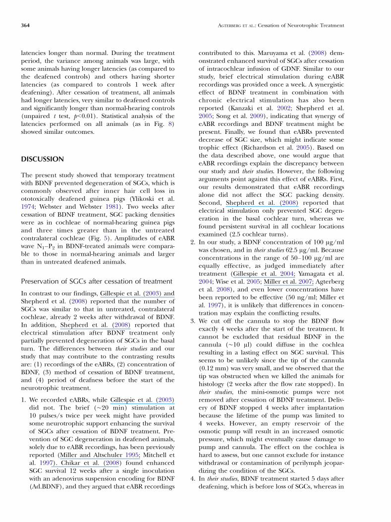
After cochlear implantation, eABRs were recorded.Monophasic rectangular pulses generated and attenu-ated by the TDT3 system were converted to currentpulses by a linear stimulus isolator (type A395, WorldPrecision Instruments). The current pulses (width20 μs; interstimulus interval 99 ms) were presentedwith alternating polarity to the most apical intra-cochlear electrode using the lateral screw in the skullas return electrode (monopolar eABR recordings). Todefine the peaks in the eABRs, they were compared tothe peaks in the aABRs (see Fig. 2). The first positivepeak (P1) in the eABR recordings was often obscured
by the electrical artifact. In approximately half of theanimals, in at least some of the recordings, the thirdpositive peak (P3) was influenced by the digastricmuscle response (Hall 1990). The first negative peak(N1) and second positive peak (P2) were always clearlyvisible and not obscured by the electrical artifact or bythe digastric muscle response. Therefore, wave N1–P2was analyzed. The N1–P2 amplitude, N1–P2 threshold,and N1 latency were determined. The N1–P2 amplitudewas found to correlate well with SGC density (Hall1990), and therefore, it was considered an appropriateparameter to examine excitability of BDNF treatedSGCs. Thresholds were defined as the stimulus levelthat evoked a 2.0-μV (determined with interpolation)reproducible waveform. Stimuli were presented from400 μA down to threshold with 2-dB steps. Highercurrent levels would evoke a whisker response. TheN1–P2 amplitudes for all current levels in the experi-mental and control animals were analyzed usingrepeated-measures analysis of variance (rm ANOVA)and t tests were used to analyze measurements at singlesuprathreshold current levels.
Cessation of BDNF treatment
After 4 weeks of BDNF treatment, the animals wereanesthetized with Domitor® (10 mg/kg, i.m.) andKetanest-S® (40 mg/kg, i.m.). After the connection ofthe cannula with the osmotic pump was checked, thecannula was cut off at about 40 mm from the cochleaand the osmotic pumps were removed. The open endof the remaining cannula was sutured with Vicryl®.The electrode array was left in place to record eABRsafter termination of the BDNF infusion. Two weeksafter cessation of BDNF treatment, the animals wereeuthanized for histology.
Histology
Immediately after the final eABR measurements, theleft and right cochleae were fixed by intralabyrinthineperfusion with a fixative consisting of 3% glutaralde-hyde, 2% formaldehyde, 1% acrolein, and 2.5%DMSO in 0.08 M sodium cacodylate buffer (pH 7.4)followed by immersion in the same fixative for 3 h atroom temperature. Histological processing of thecochleae was carried out according to our standardprotocol (De Groot et al. 1987). After decalcificationwith EDTA, the cochleae were immersed in 1% OsO4
containing 1% K4Ru(CN)6 for 2 h at 4°C and thenrinsed, dehydrated, and embedded in toto in Spurr’slow-viscosity resin. After polymerization, cochleaewere divided into two halves along a standardizedmidmodiolar plane, and these were re-embedded infresh resin. Semithin (1 μm) sections stained with
N1
P3
2 4 6
10 V
time (ms)0
P1
P4
N3aABR
P2
N2
10 V
2 4 6time (ms)
0
eABR
N1
P1
P2
P3
P4
N2
N3
FIG. 2. A typical example of an aABR (top) evoked in a normal-hearing animal, with clicks of 45 dB nSL and an eABR (bottom)evoked with 400-μA pulses. Stimulus onset was at time 0. Note thelarge stimulus artifact in the eABR before peak P1. The peak-to-peakamplitude, N1–P2, of the eABR was measured to assess the functionalstatus of the auditory nerve. When the N1 and/or P2 consisted ofmore than one subpeak, the first subpeak on each site of the fastrising part of the eABR complex (indicated with the arrows) is used inthe analysis.
358 AGTERBERG ET AL.: Cessation of Neurotrophic Treatment

methylene blue and azur II were used for lightmicroscopical evaluation and quantitative analyses.
The efficacy of the deafening procedure wasassessed, in addition to aABR thresholds, by countingthe number of remaining inner hair cells (IHCs) andouter hair cells (OHCs) at seven different locationsalong the basilar membrane at a half-turn spacing(B1, B2, M1, M2, A1, A2, and A3; Fig. 3).
Determination of SGC packing densities wasperformed as described previously (Versnel et al.2007; Agterberg et al. 2008). SGC packing densitieswere determined using digitized light microscopicalimages of the spiral ganglia taken from five differentcochlear locations (B1, B2, M1, M2, and A1; Fig. 3).Using the image analysis program NIH Image (version1.63; US National Institutes of Health, Bethesda, MD),the bony boundaries of Rosenthal’s canal were out-lined and its cross-sectional area (in mm2) wascalculated. The number of SGC perikarya wascounted at each location. The following populationswere included in the counts: (1) all perikaryademonstrating the morphological determinants typi-cal of type-I and type-II SGCs (for details, see Romandand Romand 1984); (2) partial and complete profilesof perikarya; and (3) perikaya with and withoutevident nucleus or nucleoli. SGC packing density wascalculated by dividing the number of SGCs by thecross-sectional area of Rosenthal’s canal andexpressed as the mean number of SGCs per squaremillimeter.
The cellular features perikaryal area and cellcircularity—as a measure of perikaryal cell size andcell shape, respectively—were selected for furtherquantitative analysis. Because there is a considerableregional variation in SGC perikaryal area within thecochlea (Leake et al. 1999), measurements wereperformed in digitized light microscopical images ofthe spiral ganglion at one specific location in the basal
turn (location B2 in Fig. 3). This location was chosen,because (1) the effect of BDNF on SGC packingdensities is most prominent in the basal turn, and (2)it was occasionally not possible to determine SGCpacking densities for location B1, which is near to thehook region, due to tangential sectioning of Rosenthal’scanal. Only type-I SGCs with an evident nucleus weremeasured. Perikaryal area was determined by outliningthe myelin sheath surrounding the type-I perikaryon.Cell circularity is a feature that can bemeasured directlyin NIH Image after delineating the cell’s perimeter, i.e.,the myelin sheath. It is calculated as follows: 4π×A/L2,where A is the area and L is the perimeter (circularity is1 for a perfect circle and less than 1 for an imperfectcircle, e.g., 0.78 for a square).
Statistical analyses were performed using SPSS® forWindows (version 15.0.1). SGC packing densities atthe different locations (B1–A1) were analyzed usingrepeated-measures analysis of variance (rm ANOVA).Statistical comparisons of perikaryal area and cellcircularity at location B2 were made using paired ttests.
RESULTS
Effects of deafening procedure
Two weeks after the deafening procedure, all controland experimental animals demonstrated thresholdshifts of 960 dB for click-evoked aABRs. This severehearing loss was microscopically confirmed. All haircells at cochlear locations B1–A1 were lost, except intwo animals (one control animal and one experimentalanimal). In these animals, a few inner hair cells stillremained.
SGC packing densities
Figure 4 shows light micrographs of Rosenthal’s canal atcochlear location B2, providing typical examples ofSGCs in cochleae of normal-hearing (A) and deafenedanimals (B–D), and in the left untreated and rightBDNF-treated cochlea of an experimental animal (E, F).In the normal cochlea, Rosenthal’s canal contained thefull complement of SGCs and nerve fibers embedded ina matrix consisting of vascularized connective tissue(Fig. 4A). Two weeks after deafening, the cellulardistribution within the spiral ganglia was similar tonormal (Fig. 4B). Figure 4C illustrates the dramatic lossof SGCs in the left cochlea of a control animal that wasexamined 6 weeks after deafening. In the right cochlea,which was electrically stimulated for short periods withthe purpose to record eABRs, the loss of SGCs wasequally dramatic (Fig. 4D). This indicates that briefelectrical stimulation did not prevent degeneration.Eight weeks after deafening, a dramatic loss of SGCs
B1
n. VIII
A3
A2
A1
M2
B2
M1
1.5A3
3.5A2
5.5A1
7.5M2
9.5M1
12.5B2
16.5B1
Location Distance from apex (mm)
FIG. 3. Light micrograph of a midmodiolar section (1 μm) of anormal guinea pig cochlea showing the different locations at whichSGCs were examined. The distance from the apex of the locations B1through A3 are specified in the table. n. VIII cochlear nerve.
AGTERBERG ET AL.: Cessation of Neurotrophic Treatment 359

was evident in the untreated, contralateral cochlea of aBDNF-treated animal (Fig. 4E). The surviving SGCs hadlost their characteristic ovoid shape and, instead, hadacquired a more elongated or dendritic appearance. Inthe BDNF-treated cochlea of the same animal, 2 weeksafter cessation of BDNF treatment, no signs of degener-ation were observed (Fig. 4F). The amount of SGCs wascomparable to that in the normal cochlea, and the SGCskept their characteristic ovoid shape.
Figure 5 compares SGC packing densities, atcochlear locations from B1 through A1, averagedacross animals (n=6), in BDNF-treated cochleae withthose in the untreated contralateral cochleae and withnormal data (dashed lines). The data confirm theresult shown in Figure 4E, F. SGC packing densities inBDNF-treated cochleae were normal at locations B1–M2 and by a factor 3 larger than those in untreatedcochleae (rm ANOVA: F(1, 5)=65, pG0.001).
FIG. 4. Light micrographs of Rosenthal’s canal at location B2(upper basal turn) with the distribution of SGCs (arrowheads) andnerve fibers (arrows). SGCs and nerve fibers in the spiral ganglionfrom A a normal cochlea, B a cochlea 2 weeks after deafening, C aleft cochlea 6 weeks after deafening, D the right implanted cochlea
from the same animal as in (C), E the left cochlea of an animal8 weeks after deafening, and F the right implanted cochlea treatedwith BDNF from the same animal as in (E) (2 weeks after cessation oftreatment).
360 AGTERBERG ET AL.: Cessation of Neurotrophic Treatment

It is possible that electrical stimulation during eABRrecordings contributed to the enhanced survival ofSGCs (cf. Mitchell et al. 1997). Therefore, withinuntreated deafened animals, we compared the SGCpacking densities in the stimulated implanted ears (seeexample in Fig. 4D) to those in the nonstimulatedcontralateral ears (see Fig. 4C). There were no statisti-cally significant differences in SGC packing densitybetween stimulated implanted ears and nonstimulatedcontralateral ears (rm ANOVA: F(1, 5)=3.2, p90.1).
Perikaryal area and circularity
Figure 6 shows the mean values of the perikaryal area(Fig. 6A) and circularity (Fig. 6B) of SGCs at cochlearlocation B2. The perikaryal area of BDNF-treatedSGCs was larger than that of SGCs in the untreatedcontralateral cochleae with a difference of 25%(paired t test, pG0.05). Circularity of BDNF-treatedSGCs was larger than that of SGCs in the contralateralcochleae with a difference of 8% (paired t test, pG0.05). Perikaryal area and circularity of BDNF-treatedSGCs were not statistically different from perikaryalarea and circularity of SGCs in normal-hearing guineapigs. SGCs in the implanted cochleae that wereelectrically stimulated during eABR recordings werelarger than those in the unstimulated contralateralears (data not shown, 12%, paired t test, pG0.05).There was no significant difference in circularity ofSGCs in the implanted ears as compared to theunstimulated contralateral ears (paired t test, p90.2).
Electronically evoked auditory brainstemresponses
Figure 7 shows eABR recordings in two animals (topand middle row) in the normal (A, C) and deafened(B, D) condition. The decrease in N1–P2 amplitudeafter deafening was prominent in the recordings afterdeafening for current levels of 318 and 400 μA. Thethresholds in these examples did not change afterdeafening. The P3 of the eABR recordings in gp-sp02(A, B) at stimulus intensities of 400 and 318 μA wasinfluenced by the digastric muscle response (arrow).Figure 7E, F depicts eABR recordings in gp-mr02 after4 weeks of BDNF treatment (i.e., 6 weeks afterdeafening) and 2 weeks after cessation of BDNFtreatment (i.e., 8 weeks after deafening). The N1–P2amplitude did not decrease after cessation of BDNFtreatment, and the threshold did not change.
eABR amplitude
Figure 8 shows the input–output curves of eABRs innormal-hearing animals, animals 6 weeks after deaf-ening, animals after 4 weeks of BDNF treatment (i.e.,6 weeks after deafening), and animals 2 weeks aftercessation of BDNF treatment. The amplitude of theN1–P2 complex was decreased in deafened animalsfor stimulus intensities of 252 μA (unpaired t test, pG0.05), 318 μA (unpaired t test, pG0.01), and 400 μA(unpaired t test, pG0.001) as compared to theamplitude in normal-hearing animals. At lower stim-ulus currents (100–200 μA), the amplitude differ-ences were smaller and not significant (p90.2). The
left (untreated)
right (BDNF treated)
0
500
1000
1500
2000
B1(16.5)
B2(12.5)
M1(9.5)
M2(7.5)
A1(5.5)
Location (distance from apex in mm)
SG
C p
acki
ng
den
sity
(ce
lls/m
m )
FIG. 5. Mean SGC packing densities at cochlear locations B1, B2,M1, M2, and A1 in the left (untreated) and right (BDNF-treated)cochlea of deafened animals (n=6). Dashed lines represent SGCdensities at locations B1–A1 in normal cochleae. Error bars: SEM.
*
left (untreated) N=75 cells
right (BDNF treated) N=212 cells
0
50
100
150
200
250
Per
ikar
yal a
rea
(µm
2 )
A
0 .5
0 .6
0 .7
0 .8
0 .9
1
Cel
l cir
cula
rity
B
*
FIG. 6. Perikaryal area (A) and cell circularity (B) of SGCs in leftuntreated versus BDNF-treated cochleae. Only SGCs with anobvious nucleus in basal location B2 were included. Data wereobtained by averaging all individual SGC measurements within onespiral ganglion, followed by averaging across cochleae. Dashed linesrepresent measurements in normal-hearing animals. n the number ofSGCs measured. Error bars: SEM. *pG0.05.
AGTERBERG ET AL.: Cessation of Neurotrophic Treatment 361

amplitudes in BDNF-treated animals were near theamplitudes found in normal-hearing animals (un-paired t test, p90.4) and significantly larger than inthe deafened animals for stimulus intensities of318 μA (unpaired t test, pG0.05) and 400 μA (un-paired t test, pG0.01). There was no significant changein amplitude at any current level after cessation oftreatment (paired t test, p90.1).
Figure 9 shows the mean time course of eABRamplitudes elicited with 400 μA pulses for the BDNF-treated animals and the control animals. In bothgroups, amplitudes were increasing during the first 2–3 weeks after implantation. The control animalsshowed a gradual and significant decrease in ampli-tude after deafening. Six weeks after deafening, theamplitude was at 60% of the original value. The
0 2 4 6Time (ms)
P2
0 2 4Time (ms)
gp-sp02 normal hearing
20 VP2
gp-ju02 normal hearing
0 2 4 6Time (ms)
gp-sp02 five weeks after deafening
P2
0 2 4 6Time (ms)
P2
252 A
318 A
400 A
200 A159 A126 A100 A80 A
252 A
318 A
400 A
200 A
159 A
126 A
gp-ju02 six weeks after deafening
20 V
252 A
318 A
400 A
200 A
159 A
126 A
20 V
20 V
0 2 4 6Time (ms)
0 2 4 6Time (ms)
252 A318 A
400 A
200 A
159 A
126 A100 A
E F
gp-mr02 after four weeks of BDNF treatment (six weeks after deafening)
gp-mr02 two weeks aftercessation of BDNF treatment (eight weeks after deafening)
P2
P2
252 A318 A
400 A
200 A159 A126 A
100 A
20 V 20 V
C D
A B
T
T
TT
T
252 A
318 A
400 A
200 A159 A126 A100 A80 A
6
T
N1 N1
N1 N1
N1
N1
FIG. 7. Recordings of representative eABRs evoked with currentpulses of 400 μA down to below threshold in two control animals(gp-sp02 and gp-ju02) and one experimental animal (gp-mr02).Recordings of two control animals are depicted to illustrate the inter-animal variability. A, C Recorded in normal-hearing condition, prior
to deafening; B, D recorded 5 and 6 weeks after deafening,respectively. E Recorded after 4 weeks of BDNF treatment and F2 weeks after cessation of the treatment. T indicates the threshold ofwave N1–P2. The arrow indicates the peak that probably reflects thedigastric muscle response.
362 AGTERBERG ET AL.: Cessation of Neurotrophic Treatment

amplitude did not change significantly after cessationof BDNF treatment (p90.6). The eABR recordings inthese animals also yielded threshold and latency datawhich will be described in the following sections.
eABR threshold
Figure 10 shows amoderate decrease in eABR thresholdin the control group during the first weeks afterimplantation (rm ANOVA: pG0.05). The threshold in
these animals did not change significantly during6 weeks after deafening (rm ANOVA: p90.5). The eABRthreshold significantly decreased over time in the grouptemporarily treated with BDNF (rm ANOVA: pG0.01).After cessation of BDNF treatment, the eABR thresholdsdid not demonstrate any significant changes.
eABR latency
Figure 11 shows the time course of N1 latencies ofeABRs to 400-μA pulses for both the BDNF-treatedand control animals. Deafening caused a prominentlatency change, which occurred mostly in the first2 weeks: first a decrease and then an increase. In thedeafened condition, latencies were longer by about0.15 ms than in the normal condition (paired t test6 weeks after deafening versus normal, pG0.001). Ingeneral, animals treated with BDNF demonstrated
BDNF cessation
BDNF
deafened
normal hearing
Stimulus intensity (µA)
Am
plit
ud
e o
f w
ave
N1-
P2
(µV
)
0
5
10
15
20
25
100 200 400
*
*
FIG. 8. Mean input–output functions of eABRs recorded in controlanimals before deafening (normal hearing, n=12) and 6 weeks afterdeafening (n=9), and in the experimental animals immediately after4 weeks of BDNF treatment (n=5) and 2 weeks after cessation ofBDNF treatment (n=5). Error bars: SEM. *pG0.05 indicates statisticalsignificant difference between the untreated animals 6 weeks afterdeafening and each of the other data points.
control (n=7)
experimental (n=5)
Deafening
BDNF treatment
0
5
10
15
20
25
30
-6 -4 -2 4 6 8Time after deafening (weeks)
Am
plit
ud
e o
f w
ave
N1-
P2 (µ
V)
20
FIG. 9. Mean N1–P2 amplitudes of eABRs recorded to 400-μApulses in control (n=7) and experimental (n=5) animals. The controlanimals were implanted at −4 weeks and deafened at 0 weeks. Theexperimental animals were deafened at 0 weeks and implanted inthe right cochlea at 2 weeks. Following implantation, the experi-mental animals received BDNF for a 4-week period (indicated with ahorizontal bold bar). Error bars: SEM.
Time after deafening (weeks)
Th
resh
old
of
eAB
R (
µµA)
control (n=7)
experimental (n=5)
BDNF treatment
Deafening
0
50
100
150
200
250
300
-6 -4 -2 0 4 862
FIG. 10. Mean eABR thresholds observed for wave N1–P2 incontrol (n=7) and experimental (n=5) animals. The experimentalanimals were deafened at 0 weeks and implanted in the rightcochlea at 2 weeks. Following implantation, the experimentalanimals received BDNF for a 4-week period (indicated with ahorizontal bold bar). Threshold criterion 2 μV; error bars: SEM.
0.4
0.5
0.6
0.7
0.8
0.9
1.0
-4 -2 0 4 8Time after deafening (weeks)
Lat
ency
(m
s)
BDNF treatment
control (n=7)
experimental (n=5)
Deafening
62
FIG. 11. Mean N1 latencies of eABRs evoked with 400-μA pulses,observed in control (n=7) and experimental (n=5) animals. Theexperimental animals were deafened at 0 weeks and implanted inthe right cochlea at 2 weeks. Following implantation, the experi-mental animals received BDNF for a 4-week period (indicated with ahorizontal bold bar). Statistical analyses within each group are notshown, but are described in the text. Error bars: SEM.
AGTERBERG ET AL.: Cessation of Neurotrophic Treatment 363
latencies longer than normal. During the treatmentperiod, the variance among animals was large, withsome animals having longer latencies (as compared tothe deafened controls) and others having shorterlatencies (as compared to controls 1 week afterdeafening). After cessation of treatment, all animalshad longer latencies, very similar to deafened controlsand significantly longer than normal-hearing controls(unpaired t test, pG0.01). Statistical analysis of thelatencies performed on all animals (as in Fig. 8)showed similar outcomes.
DISCUSSION
The present study showed that temporary treatmentwith BDNF prevented degeneration of SGCs, which iscommonly observed after inner hair cell loss inototoxically deafened guinea pigs (Ylikoski et al.1974; Webster and Webster 1981). Two weeks aftercessation of BDNF treatment, SGC packing densitieswere as in cochleae of normal-hearing guinea pigsand three times greater than in the untreatedcontralateral cochleae (Fig. 5). Amplitudes of eABRwave N1–P2 in BDNF-treated animals were compara-ble to those in normal-hearing animals and largerthan in untreated deafened animals.
Preservation of SGCs after cessation of treatment
In contrast to our findings, Gillespie et al. (2003) andShepherd et al. (2008) reported that the number ofSGCs was similar to that in untreated, contralateralcochleae, already 2 weeks after withdrawal of BDNF.In addition, Shepherd et al. (2008) reported thatelectrical stimulation after BDNF treatment onlypartially prevented degeneration of SGCs in the basalturn. The differences between their studies and ourstudy that may contribute to the contrasting resultsare: (1) recordings of the eABRs, (2) concentration ofBDNF, (3) method of cessation of BDNF treatment,and (4) period of deafness before the start of theneurotrophic treatment.
1. We recorded eABRs, while Gillespie et al. (2003)did not. The brief (∼20 min) stimulation at10 pulses/s twice per week might have providedsome neurotrophic support enhancing the survivalof SGCs after cessation of BDNF treatment. Pre-vention of SGC degeneration in deafened animals,solely due to eABR recordings, has been previouslyreported (Miller and Altschuler 1995; Mitchell etal. 1997). Chikar et al. (2008) found enhancedSGC survival 12 weeks after a single inoculationwith an adenovirus suspension encoding for BDNF(Ad.BDNF), and they argued that eABR recordings
contributed to this. Maruyama et al. (2008) dem-onstrated enhanced survival of SGCs after cessationof intracochlear infusion of GDNF. Similar to ourstudy, brief electrical stimulation during eABRrecordings was provided once a week. A synergisticeffect of BDNF treatment in combination withchronic electrical stimulation has also beenreported (Kanzaki et al. 2002; Shepherd et al.2005; Song et al. 2009), indicating that synergy ofeABR recordings and BDNF treatment might bepresent. Finally, we found that eABRs preventeddecrease of SGC size, which might indicate sometrophic effect (Richardson et al. 2005). Based onthe data described above, one would argue thateABR recordings explain the discrepancy betweenour study and their studies. However, the followingarguments point against this effect of eABRs. First,our results demonstrated that eABR recordingsalone did not affect the SGC packing density.Second, Shepherd et al. (2008) reported thatelectrical stimulation only prevented SGC degen-eration in the basal cochlear turn, whereas wefound persistent survival in all cochlear locationsexamined (2.5 cochlear turns).
2. In our study, a BDNF concentration of 100 μg/mlwas chosen, and in their studies 62.5 μg/ml. Becauseconcentrations in the range of 50–100 μg/ml areequally effective, as judged immediately aftertreatment (Gillespie et al. 2004; Yamagata et al.2004; Wise et al. 2005; Miller et al. 2007; Agterberget al. 2008), and even lower concentrations havebeen reported to be effective (50 ng/ml; Miller etal. 1997), it is unlikely that differences in concen-tration may explain the conflicting results.
3. We cut off the cannula to stop the BDNF flowexactly 4 weeks after the start of the treatment. Itcannot be excluded that residual BDNF in thecannula (∼10 μl) could diffuse in the cochlearesulting in a lasting effect on SGC survival. Thisseems to be unlikely since the tip of the cannula(0.12 mm) was very small, and we observed that thetip was obstructed when we killed the animals forhistology (2 weeks after the flow rate stopped). Intheir studies, the mini-osmotic pumps were notremoved after cessation of BDNF treatment. Deliv-ery of BDNF stopped 4 weeks after implantationbecause the lifetime of the pump was limited to4 weeks. However, an empty reservoir of theosmotic pump will result in an increased osmoticpressure, which might eventually cause damage topump and cannula. The effect on the cochlea ishard to assess, but one cannot exclude for instancewithdrawal or contamination of perilymph jeopar-dizing the condition of the SGCs.
4. In their studies, BDNF treatment started 5 days afterdeafening, which is before loss of SGCs, whereas in
364 AGTERBERG ET AL.: Cessation of Neurotrophic Treatment

our study, it started after 2 weeks when SGCdegeneration has started (Versnel et al. 2007).
Considering the four issues addressed above, wepropose eABR recordings as the most likely candidateto explain the discrepancy between our data andthose of Gillespie et al. (2003). However, we cannotexclude any of the other three arguments.
Although the lasting effect of the neurotrophictreatment is evident in the present study (Fig. 5), thiseffect is only evaluated 2 weeks after cessation.Degeneration after deafening is not pronounced after2 weeks (∼80% of normal density, Versnel et al. 2007).Also the decline in eABR amplitude after deafening israther slow (Fig. 9). Therefore, to investigate, bothhistologically and functionally, whether degenerationafter cessation of BDNF treatment is slower thandegeneration after deafening without intervention, itis important to apply longer periods of survival.
Morphology of SGCs after cessation of BDNFtreatment
Immediately after BDNF treatment, SGCs are largerthan SGCs in normal-hearing animals (Shepherd et al.2005; Richardson et al. 2005; Agterberg et al. 2008). Wefound that 2 weeks after cessation SGCs had a normalsize (while larger than in the untreated contralateralcochlea). This implies that the perikaryal area decreasedafter cessation from larger-than-normal, to normalvalues. The question remains if the back-to-normal areais an indication of a normal condition. On the otherhand, the decrease of cell size following cessationmay bean early indication for initiation of a degenerativeprocess (Staecker et al. 1996; Leake et al. 1999;Agterberg et al. 2008). To answer this last question, thesurvival of SGCs needs to be investigated for longerperiods after cessation of the neurotrophic treatment.
eABR amplitudes
We applied suprathreshold amplitudes as a measure ofexcitability of the auditory nerve, since the amplitudereflects the summed neural firings of a large popula-tion of SGCs, whereas the more commonly usedparameter threshold reflects the function of only themost sensitive auditory nerve fibers. Indeed, at least incase of monopolar stimulation, the amplitude of earlyeABR waves has been found to correlate well with thenumber of SGCs (Smith and Simmons 1983; Hall1990) whereas correlations between eABR thresholdand nerve survival were weak (Smith and Simmons1983). Note that for bipolar stimulation a bettercorrelation was found for threshold measures thanfor amplitude measures (Miller et al. 1994).
The normal eABR amplitude in BDNF-treated ani-mals indicates that treated SGCs responded as normal toelectric stimuli. It should be noted that the amplitudeafter deafening was smaller, probably because less SGCswere present, thus implicating that the remaininguntreated cells responded normally. Our data agreewith the results of Maruyama et al. (2008) who reportedthat amplitudes in neurotrophically treated animalswere larger than in untreated animals and were stableafter cessation of the treatment.
A decrease of eABR threshold was found the firstweeks after implantation in the normal-hearing con-dition. This might be due to mechanisms as tissuegrowth around the electrodes and recovery frominsertion-induced trauma (Su et al. 2008). We assumethe threshold decrease found in the animals treatedwith BDNF was rather due to these mechanisms thanto the preservation of SGCs mediated by the BDNF.
Our threshold data do not agree with other inves-tigators who reported lower thresholds (by 4–9 dB) indeafened guinea pigs treated with neurotrophins ascompared to deafened and untreated guinea pigs(Shinohara et al. 2002; Yamagata et al. 2004; Shepherdet al. 2005; Chikar et al. 2008; Maruyama et al. 2008). Inthese studies, the decrease in eABR threshold corre-sponded with enhanced survival of SGCs after neuro-trophic treatment. Contrary to these studies, in whicheABR thresholds were often assessed on the basis ofvisual inspection of peak 3 and recordings wereperformed in anesthetized animals, we assessed theeABR threshold on the basis of interpolation of N1–P2amplitudes and we recorded in awake animals. Possibly,the difference in peaks analyzed explains the differentthreshold data asMitchell et al. (1997) reported that thethreshold of the late eABR peaks in deafened animalsdecreased over time, while early peak thresholds did notsignificantly change. Further, the state of the animalplays a role in that the threshold increases withanesthesia as was found for aABRs in mice by Van Looijet al. (2004). The effects of anesthesia may be nonlinearand differ between various experimental conditions.
eABR latencies
The eABR N1 latencies were longer in deafened andBDNF-treated animals than in normal-hearing animals(Fig. 11). The initial latency decrease after deafeningmay be caused by degeneration of dendrites and a lossof spontaneous activity, and the subsequent increase canbe attributed to a reduction of the number of myelinlayers, which follows dendritic degeneration (Leakeand Hradek 1988). The BDNF-treated nerve cells havea similar degree of myelination as untreated cells(Agterberg et al. 2008; Glueckert et al. 2008), and thismight explain the similar latencies of BDNF-treated anduntreated animals.
AGTERBERG ET AL.: Cessation of Neurotrophic Treatment 365

Clinical implications
Our results demonstrate that SGC survival in deafenedanimals, in both morphological and functional sense, isnot reduced after cessation of intracochlear applicationof BDNF. The enhanced survival of SGCs is a promisingfinding for cochlear implant candidates since there arecurrently no potential methods available to provideneurotrophins for periods that extend the order ofmonths. Lifelong neurotrophic treatment via an osmot-ic pump is not clinically preferable because of repetitiveinvasive manipulations needed to replace the pump andhigh infection risks.
Before neurotrophic treatment can be considereda clinical option, it is important to investigate inanimal studies whether neurotrophic treatment doesactually improve the sound perception performancewith a cochlear implant. A major topic that needs tobe further explored is the assumption that more andbetter functioning SGCs would result in a betterperformance of cochlear implant users. Alternativeto this hypothesis, it might be that a minimumnumber of SGCs is required for electrical hearing,and that an increase above this minimum would havelittle functional impact (Blamey 1997). Clinical datado not support the assumption that more SGCs wouldlead to a better behavioral performance (Nadol et al.2001; Khan et al. 2005; Fayad and Linthicum 2006;Nadol and Eddington 2006). These data indicate thatabnormalities in the central auditory pathways are atleast as important as SGC survival in limiting theperformance of cochlear implant users (Nadol et al.2001). Furthermore, the overall performance ofcochlear implant users improves markedly withinweeks as a result of implant use, confirming the roleof the central auditory system and its plasticity (Kraland Eggermont 2007). Still, we, and others, think thatit is unlikely that SGC loss does not affect theperformance with a cochlear implant and that thehuman data sets are still too limited to conclude thatthe extent of SGC survival does not influence cochlearimplant performance (Khan et al. 2005; Leake et al.2008). Moreover, it should be considered that withimproved cochlear implant technology the number ofSGCs might become more important (Green et al.2008).
ACKNOWLEDGMENTS
The authors would like to thank Leone Nijman and RikMansvelt-Beck for their excellent practical and technicalassistance, Ferry Hendriksen for assisting with histology, andRené van de Vosse for developing the recording and analysissoftware. We thank Victor Wiegant for his suggestions on themanuscript. This study was supported by the Heinsius-Houbolt Foundation.
Open Access
This article is distributed under the terms of the CreativeCommons Attribution Noncommercial License which per-mits any noncommercial use, distribution, and reproductionin any medium, provided the original author(s) and sourceare credited.
REFERENCES
AGTERBERG MJH, VERSNEL H, DE GROOT JCMJ, SMOORENBURG GF,ALBERS FWJ, KLIS SFL. Morphological changes in spiral ganglioncells after intracochlear application of brain-derived neurotrophicfactor in deafened guinea pigs. Hear. Res. 244:25–34, 2008.
BLAMEY P. Are spiral ganglion cell numbers important for speechperception with a cochlear implant? Am. J. Otol. 18(suppl):11–12, 1997.
BROWN JN, MILLER JM, ALTSCHULER RA, NUTTALL AL. Osmotic pumpimplant for chronic infusion of drugs into the inner ear. Hear.Res. 70:167–172, 1993.
CHIKAR JA, COLESA DJ, SWIDERSKI DL, POLO AD, RAPHAEL Y, PFINGST BE.Over-expression of BDNF by adenovirus with concurrentelectrical stimulation improves cochlear implant thresholdsand survival of auditory neurons. Hear. Res. 245:24–34, 2008.
DE GROOT JCMJ, VELDMAN JE, HUIZING EH. An improved fixationmethod for guinea pig cochlear tissues. Acta Otolaryngol104:234–242, 1987.
FAYAD JN, LINTHICUM FH, JR. Multichannel cochlear implants: relationof histopathology to performance. Laryngoscope 116:1310–1320, 2006.
GILLESPIE LN, SHEPHERD RK. Clinical application of neurotrophicfactors: the potential for primary auditory neuron protection.Eur. J. Neurosci. 22:2123–2133, 2005.
GILLESPIE LN, CLARK GM, BARTLETT PF, MARZELLA PL. BDNF-inducedsurvival of auditory neurons in vivo: cessation of treatment leadsto accelerated loss of survival effects. J. Neurosci. Res. 71:785–790, 2003.
GILLESPIE LN, CLARK GM, MARZELLA PL. Delayed neurotrophintreatment supports auditory neuron survival in deaf guinea pigs.Neuroreport 15:1121–1125, 2004.
GLUECKERT R, BITSCHE M, MILLER JM, ZHU Y, PRIESKORN DM,ALTSCHULER RA, SCHROTT-FISCHER A. Deafferentation-associatedchanges in afferent and efferent processes in the guinea pigcochlea and afferent regeneration with chronic intrascalarbrain-derived neurotrophic factor and acidic fibroblast growthfactor. J. Comp. Neurol. 507:1602–1621, 2008.
GREEN SH, ALTSCHULER RA, MILLER JM. Cell death and cochlearprotection. In: Schacht J, Popper AN, Fay RR (eds) AuditoryTrauma, Protection, and Repair. New York, Springer, pp. 275–319, 2008.
HALL RD. Estimation of surviving spiral ganglion cells in the deaf ratusing the electrically evoked auditory brainstem response. Hear.Res. 45:123–136, 1990.
HARTSHORN DO, MILLER JM, ALTSCHULER RA. Protective effect ofelectrical stimulation in the deafened guinea pig cochlea.Otolaryngol. Head Neck Surg. 104:311–319, 1991.
KANZAKI S, STOVER T, KAWAMOTO K, PRIESKORN DM, ALTSCHULER RA,MILLER JM, RAPHAEL Y. Glial cell line-derived neurotrophic factorand chronic electrical stimulation prevent VIII cranial nervedegeneration following denervation. J. Comp. Neurol. 454:350–360, 2002.
KHAN AM, HANDZEL O, BURGESS BJ, DAMIAN D, EDDINGTON DK, NADOL JB.Is word recognition correlated with the number of surviving spiral
366 AGTERBERG ET AL.: Cessation of Neurotrophic Treatment

ganglion cells and electrode insertion depth in human subjectswith cochlear implants? Laryngoscope 115:672–677, 2005.
KRAL A, EGGERMONT JJ. What’s to lose and what’s to learn:development under auditory deprivation, cochlear implantsand limits of cortical plasticity. Brain Res. 56:259–269, 2007.
LEAKE PA, HRADEK GT. Cochlear pathology of long term neomycininduced deafness in cats. Hear. Res. 33:11–33, 1988.
LEAKE PA, HRADEK GT, REBSCHER SJ, SNYDER RL. Chronic intracochlearelectrical stimulation induces selective survival of spiral ganglionneurons in neonatally deafened cats. Hear. Res. 54:251–271, 1991.
LEAKE PA, HRADEK GT, SNYDER RL. Chronic electrical stimulation by acochlear implant promotes survival of spiral ganglion neuronsafter neonatal deafness. J. Comp. Neurol. 412:543–562, 1999.
LEAKE PA, STAKHOVSKAYA O, HRADEK GT, HETHERINGTON AM. Factorsinfluencing neurotrophic effects of electrical stimulation in thedeafened developing auditory system. Hear. Res. 242:86–99,2008.
LOUSTEAU RJ. Increased spiral ganglion cell survival in electricallystimulated, deafened guinea pig cochleae. Laryngoscope97:836–842, 1987.
MANSOUR-ROBAEY S, CLARKE DB, WANG YC, BRAY GM, AGUAYO AJ.Effects of ocular injury and administration of brain-derivedneurotrophic factor on survival and regrowth of axotomizedretinal ganglion cells. Proc. Natl. Acad. Sci. U. S. A. 91:1632–1636, 1994.
MARUYAMA J, MILLER JM, ULFENDAHL M. Glial cell line-derived neuro-trophic factor and antioxidants preserve the electrical respon-siveness of the spiral ganglion neurons after experimentallyinduced deafness. Neurobiol. Dis. 29:14–21, 2008.
MILLER JM, ALTSCHULER RA. Effectiveness of different electricalstimulation conditions in preservation of spiral ganglion cellsfollowing deafness. Ann. Otol. Rhinol. Laryngol. Suppl. 166:57–60, 1995.
MILLER CA, ABBAS PJ, ROBINSON BK. The use of long-duration currentpulses to assess nerve survival. Hear. Res. 78:11–26, 1994.
MILLER JM, CHI DH, O’KEEFFE LJ, KRUSZKA P, RAPHAEL Y, ALTSCHULER RA.Neurotrophins can enhance spiral ganglion cell survival afterinner hair cell loss. Int. J. Dev. Neurosci. 15:631–643, 1997.
MILLER JM, LE PRELL CG, PRIESKORN DM, WYS NL, ALTSCHULER RA.Delayed neurotrophin treatment following deafness rescuesspiral ganglion cells from death and promotes regrowth ofauditory nerve peripheral processes: effects of brain-derivedneurotrophic factor and fibroblast growth factor. J. Neurosci.Res. 85:1959–1969, 2007.
MITCHELL A,MILLER JM, FINGER PA,HELLER JW, RAPHAELY, ALTSCHULER RA.Effects of chronic high-rate electrical stimulation on the cochleaand eighth nerve in the deafened guinea pig. Hear. Res. 105:30–43,1997.
MONTERO CN, HEFTI F. Rescue of lesioned septal cholinergic neuronsby nerve growth factor: specificity and requirement for chronictreatment. J. Neurosci. 8:2986–2999, 1988.
NADOL JB, JR, EDDINGTON DK. Histopathology of the inner earrelevant to cochlear implantation. Adv. Otorhinolaryngol 64:31–49, 2006.
NADOL JB, JR, SHIAO JY, BURGESS BJ, KETTEN DR, EDDINGTON DK, GANTZ BJ,KOS I, MONTANDON P, COKER NJ, ROLAND JTJ, SHALLOP JK. Histopa-thology of cochlear implants in humans. Ann. Otol. Rhinol.Laryngol. 110:883–891, 2001.
PETTINGILL LN, RICHARDSON RT, WISE AK, O’LEARY SJ, SHEPHERD RK.Neurotrophic factors and neural prostheses: potential clinical
applications based upon findings in the auditory system. IEEETrans. Biomed. Eng. 54:1138–1148, 2007.
RICHARDSON RT, O’LEARY S, WISE A, HARDMAN J, CLARK G. A single dose ofneurotrophin-3 to the cochlea surrounds spiral ganglion neuronsand provides trophic support. Hear. Res. 204:37–47, 2005.
ROMAND R, ROMAND MR. The spiral ganglion. In: Friedmann I,Ballantyne J (eds) Ultrastructural Atlas of the Inner Ear.London, Butterworths, pp. 165–183, 1984.
SHEPHERD RK, XU J. A multichannel scala tympani electrode arrayincorporating a drug delivery system for chronic intracochlearinfusion. Hear. Res. 172:92–98, 2002.
SHEPHERD RK, COCO A, EPP SB, CROOK JM. Chronic depolarizationenhances the trophic effects of brain-derived neurotrophicfactor in rescuing auditory neurons following a sensorineuralhearing loss. J. Comp. Neurol. 486:145–158, 2005.
SHEPHERD RK, COCO A, EPP SB. Neurotrophins and electricalstimulation for protection and repair of spiral ganglion neuronsfollowing sensorineural hearing loss. Hear. Res. 242:100–109,2008.
SHINOHARA T, BREDBERG G, ULFENDAHL M, PYYKKO I, OLIVIUS NP,KAKSONEN R, LINDSTROM B, ALTSCHULER R, MILLER JM. Neuro-trophic factor intervention restores auditory function in deaf-ened animals. Proc. Natl. Acad. Sci. U. S. A. 99:1657–1660, 2002.
SMITH L, SIMMONS FB. Estimating eighth nerve survival by electricalstimulation. Ann. Otol. Rhinol. Laryngol. 92:19–23, 1983.
SONG BN, LI YX, HAN DM. Delayed electrical stimulation and BDNFapplication following induced deafness in rats. Acta Otolaryngol129:142–154, 2009.
SPOENDLIN H. Retrograde degeneration of the cochlear nerve. ActaOtolaryngol 79:266–275, 1975.
STAECKER H, KOPKE R, MALGRANGE B, LEFEBVRE P, VAN DE WATER TR. NT-3and/or BDNF therapy prevents loss of auditory neurons followingloss of hair cells. Neuroreport 7:889–894, 1996.
SU GL, COLESA DJ, PFINGST BE. Effects of deafening and cochlearimplantation procedures on postimplantation psychophysicalelectrical detection thresholds. Hear. Res. 241:64–72, 2008.
VAN LOOIJ MAJ, LIEM SS, VAN DER BURG H, VAN DERWEES J, DE ZEEUW CI,VAN ZANTEN BGA. Impact of conventional anesthesia on auditorybrainstem responses in mice. Hear. Res. 193:75–82, 2004.
VERSNEL H, AGTERBERGMJH, DE GROOT JCMJ, SMOORENBURG GF, KLIS SFL.Time course of cochlear electrophysiology and morphology aftercombined administration of kanamycin and furosemide.Hear. Res.231:1–12, 2007.
WEBSTER M, WEBSTER DB. Spiral ganglion neuron loss following organof corti loss: a quantitative study. Brain Res. 212:17–30, 1981.
WEST BA, BRUMMETT RE, HIMES DL. Interaction of kanamycin andethacrynic acid. Severe cochlear damage in guinea pigs. Arch.Otolaryngol 98:32–37, 1973.
WISE AK, RICHARDSON R, HARDMAN J, CLARK G, O’LEARY S. Resproutingand survival of guinea pig cochlear neurons in response to theadministration of the neurotrophins brain-derived neurotrophicfactor and neurotrophin-3. J. Comp. Neurol. 487:147–165, 2005.
YAMAGATA T, MILLER JM, ULFENDAHL M, OLIVIUS NP, ALTSCHULER RA,PYYKKO I, BREDBERG G. Delayed neurotrophic treatment preservesnerve survival and electrophysiological responsiveness in neo-mycin-deafened guinea pigs. J. Neurosci. Res. 78:75–86, 2004.
YLIKOSKI J, WERSALL J, BJORKROTH B. Degeneration of neural elementsin the cochlea of the guinea-pig after damage to the organ ofcorti by ototoxic antibiotics. Acta Otolaryngol. Suppl. 326:23–41,1974.
AGTERBERG ET AL.: Cessation of Neurotrophic Treatment 367
Copyright © 2022 FDOKUMEN