Engineered Thermobifida fusca cutinase with increased activity on polyester substrates
10
Biotechnology Journal DOI 10.1002/biot.201000391 Biotechnol. J. 2011, 6, 1230–1239 1230 © 2011 Wiley-VCH Verlag GmbH & Co. KGaA, Weinheim 1 Introduction Fibers made of poly(ethylene terephthalate) (PET) have numerous favorable properties compared with others; however, they also present substantial disadvantages, such as hydrophobicity, build-up of electrostatic charge, pilling tendency and difficult oil stain release. Different techniques are available to modify the surface of PET fibers, diminishing these disadvantages that are caused by the PET crystallinity and hydrophobicity. Chemicals like acids or alkalis can be used to obtain an acceptable level of polymer hydrolysis and, in that way, in- crease/create hydrophilic groups. However, the lack of a good control over these chemical reactions frequently leads to high fibers damage [1–3]. To- gether with these techniques, others like co-poly- merization or plasma treatments, can be applied to modify PET surfaces; however, the outcome is still unsatisfying [4]. In the last decade new environmentally benign and specific processes have been developed based on modern enzyme technology. Various research groups have assessed the potential of laccases, li- pases, polyesterases (serine esterase) and cutinas- es in the oxidation or hydrolysis of PET, aiming at the modification and eventual functionalization of this material [3, 5–25]. Nowadays, cutinases from Fusarium solani pisi, Fusarium oxysporum and Thermobifida fusca are the most frequently studied ones regarding PET modification, and their structures and properties Research Article Engineered Thermobifida fusca cutinase with increased activity on polyester substrates Carla Silva 1 , Shi Da 1 , Nádia Silva 2 , Teresa Matamá 2 , Rita Araújo 2 , Madalena Martins 1 , Sheng Chen 3,4 , Jian Chen 3,4 , Jing Wu 3,4 , Margarida Casal 2 and Artur Cavaco-Paulo 1 1 University of Minho, Textile Engineering Department, Guimarães, Portugal 2 Centre of Molecular and Environmental Biology (CBMA), Department of Biology, University of Minho, Portugal 3 State Key Laboratory of Food Science and Technology, Jiangnan University,Wuxi, China 4 School of Biotechnology and Key Laboratory of Industrial Biotechnology, Ministry of Education, Jiangnan University, Wuxi, China A bacterial cutinase from Thermobifida fusca, named Tfu_0883, was genetically modified by site-di- rected mutagenesis to enhance its activity on poly(ethylene terephthalate) (PET). The new muta- tions tailored the catalytic site for PET, increasing the affinity of cutinase to this hydrophobic sub- strate and the ability to hydrolyze it. The mutation I218A was designed to create space and the dou- ble mutation Q132A/T101A was designed both to create space and to increase hydrophobicity. The activity of the double mutant on the soluble substrate p-nitrophenyl butyrate increased two- fold compared to wild-type cutinase, while on PET both single and double mutants exhibited con- siderably higher hydrolysis efficiency. The replacement of specific amino acids at the active site was an effective approach for the improvement of the Tfu_0883 cutinase capacity to hydrolyze poly- ester surfaces. Thus, this study provides valuable insight on how the function and stability of en- zymes can be improved by molecular engineering for their application in synthetic fiber biotrans- formation. Keywords: Genetic modification · Hydrolysis · Mutant · Poly(ethylene terephthalate) · Textiles Correspondence: Dr. Artur Cavaco-Paulo, University of Minho, Textile Engineering Department, 4800-058 Guimarães, Portugal E-mail: [email protected] Abbreviations: PET, poly(ethylene terephthalate); pNPB, p-nitrophenyl butyrate; TPA, terephthalic acid Received 11 November 2010 Revised 7 June 2011 Accepted 20 June 2011
-
Upload
independent -
Category
Documents
-
view
1 -
download
0
Transcript of Engineered Thermobifida fusca cutinase with increased activity on polyester substrates

BiotechnologyJournal DOI 10.1002/biot.201000391 Biotechnol. J. 2011, 6, 1230–1239
1230 © 2011 Wiley-VCH Verlag GmbH & Co. KGaA, Weinheim
1 Introduction
Fibers made of poly(ethylene terephthalate) (PET)have numerous favorable properties comparedwith others; however, they also present substantialdisadvantages, such as hydrophobicity, build-up ofelectrostatic charge, pilling tendency and difficultoil stain release. Different techniques are availableto modify the surface of PET fibers, diminishingthese disadvantages that are caused by the PETcrystallinity and hydrophobicity. Chemicals likeacids or alkalis can be used to obtain an acceptable
level of polymer hydrolysis and, in that way, in-crease/create hydrophilic groups. However, thelack of a good control over these chemical reactionsfrequently leads to high fibers damage [1–3]. To-gether with these techniques, others like co-poly-merization or plasma treatments, can be applied tomodify PET surfaces; however, the outcome is stillunsatisfying [4].
In the last decade new environmentally benignand specific processes have been developed basedon modern enzyme technology. Various researchgroups have assessed the potential of laccases, li-pases, polyesterases (serine esterase) and cutinas-es in the oxidation or hydrolysis of PET, aiming atthe modification and eventual functionalization ofthis material [3, 5–25].
Nowadays, cutinases from Fusarium solani pisi,Fusarium oxysporum and Thermobifida fusca arethe most frequently studied ones regarding PETmodification, and their structures and properties
Research Article
Engineered Thermobifida fusca cutinase with increased activityon polyester substrates
Carla Silva1, Shi Da1, Nádia Silva2, Teresa Matamá2, Rita Araújo2, Madalena Martins1, Sheng Chen3,4, Jian Chen3,4, Jing Wu3,4, Margarida Casal2 and Artur Cavaco-Paulo1
1 University of Minho, Textile Engineering Department, Guimarães, Portugal2 Centre of Molecular and Environmental Biology (CBMA), Department of Biology, University of Minho, Portugal3 State Key Laboratory of Food Science and Technology, Jiangnan University,Wuxi, China4 School of Biotechnology and Key Laboratory of Industrial Biotechnology, Ministry of Education, Jiangnan University, Wuxi, China
A bacterial cutinase from Thermobifida fusca, named Tfu_0883, was genetically modified by site-di-rected mutagenesis to enhance its activity on poly(ethylene terephthalate) (PET). The new muta-tions tailored the catalytic site for PET, increasing the affinity of cutinase to this hydrophobic sub-strate and the ability to hydrolyze it. The mutation I218A was designed to create space and the dou-ble mutation Q132A/T101A was designed both to create space and to increase hydrophobicity.The activity of the double mutant on the soluble substrate p-nitrophenyl butyrate increased two-fold compared to wild-type cutinase, while on PET both single and double mutants exhibited con-siderably higher hydrolysis efficiency. The replacement of specific amino acids at the active sitewas an effective approach for the improvement of the Tfu_0883 cutinase capacity to hydrolyze poly-ester surfaces. Thus, this study provides valuable insight on how the function and stability of en-zymes can be improved by molecular engineering for their application in synthetic fiber biotrans-formation.
Keywords: Genetic modification · Hydrolysis · Mutant · Poly(ethylene terephthalate) · Textiles
Correspondence: Dr. Artur Cavaco-Paulo, University of Minho, Textile Engineering Department, 4800-058 Guimarães, PortugalE-mail: [email protected]
Abbreviations: PET, poly(ethylene terephthalate); pNPB, p-nitrophenylbutyrate; TPA, terephthalic acid
Received 11 November 2010Revised 7 June 2011Accepted 20 June 2011

© 2011 Wiley-VCH Verlag GmbH & Co. KGaA, Weinheim 1231
Biotechnol. J. 2011, 6, 1230–1239 www.biotechnology-journal.com
have been well described [17, 26–29]. Recently,Gross and co-workers studied the degradability ofpolyester using three different cutinases, from Hu-milica insolens, Pseudomonas mendocina andFusarium solani [30]. They showed that the abilityof cutinases to hydrolyze PET is negatively affect-ed by the fiber crystallinity.
Several efforts have been made to identify andcharacterize bacterial cutinases, since until nowthe most studied ones have come from fungalsources. Müller’s group [14] isolated and identifieda thermophilic actinomycete, known as Thermobi-fida fusca. This bacterium exhibits a remarkabledegradation capability regarding aliphatic–aro-matic copolyesters. An extracellular hydrolase(TfH) responsible for the degradation of thesecopolyesters was purified from the culture brothand initially classified as a serine hydrolase withthe highly conserved G-H-S-M-G motif. After-wards, Chen and co-workers [31] identified TfH asa cutinase and renamed it as Tfu_0883. This en-zyme displays very similar enzymatic properties toFusarium solani cutinases. Both belong to the α/βhydrolase fold superfamily, and contain the char-acteristic G-X1-S-X2-G motif as well as a Ser-His-Asp catalytic triad. However, this T. fusca cutinasedemonstrates remarkable thermostability un-matched by the F. solani cutinases [31, 32]. More-over, Tfu_0883 presents a more open active sitewhen compared to cutinase from F. Solani pisi (forillustration, see [31]), which theoretically can bet-ter “accommodate” the PET substrate. T. fusca cuti-nase is, therefore, now one of the most promisingcutinases regarding PET modification [33, 34]. Al-together, this led researchers to further exploreTfu_0883 in terms of PET modification. Regardingenzymatic activity the attained results betweenTfu_0883 and other cutinases were similar, never-theless the advantage of T. fusca cutinase relied onits thermostability, which is a very important fea-ture during a synthetic fiber modification process[32, 33]. It also showed higher stability in the pres-ence of surfactants and organic solvents [14, 32, 34,35].
Although wild-type cutinases from both bacte-ria and fungi are able to modify the surface of PETfibers, turnover rates are still very low. The hy-drophobicity of the heterogeneous substrate andthe natural constrictions of the enzyme near the ac-tive site can be the underlying factors. Araújo et al.[20] performed a genetic modification of F. solanicutinase, modifying and enlarging the active site toimprove the fit between enzyme and the PETchains. Indeed, they succeeded in increasing thecutinase activity on PET, obtaining new variants
with an five-fold improved activity relative to thewild-type enzyme.
The work reported here describes in the im-provement of T. fusca cutinase (Tfu_0883) activityon PET substrates by genetic manipulation of spe-cific amino acids in the active site of the enzyme.The mutations were designed to promote the activesite enlargement and to increase its hydrophobiccharacter. The new engineered cutinase is expect-ed to better “accommodate” the synthetic substrate,leading to an increase of hydrolysis efficiency, asobtained for F. solani cutinase [20], allying thisimprovement to the native thermal stability ofTfu_0883. In this work, the different cutinase mu-tants were tested on a PET fabric and the hydroly-sis efficiency was further assessed by quantifica-tion of hydrolysis products and evaluation of fibersurface modification.
2 Materials and methods
2.1 Materials
Wild-type Tfu_0883 [31] and its mutations wereproduced by recombinant DNA technology in theState Key Laboratory of Food Science and Technol-ogy, Wuxi, China. The fabric used in this work was100% polyester (107 g/m2, 18 yarns/cm warp andweft, 29 Tex) obtained from Rhodia, Switzerland.The dye used for coloration was Reactive Black 5(RB5) from Ciba, Switzerland. Lutensol AT25, anon-ionic surfactant, was obtained from BASF,Spain. All other chemicals used were laboratorygrade reagents from Sigma, Spain.
2.2 Mutation design
Since there was no 3-D structure for Tfu_0883, aTfu_0883 3-D model was created. It was predictedby the SWISSMODEL, an automated comparativeprotein modeling server (http://swissmodel.expasy.org/workspace/index.php?func=modelling_simple1).The template was SeL (PDB code 1jfr), whichshares 63% sequence identity with Tfu_0883. Thismodel (Fig. 1) was used to study the hypothetic ac-tive site 3-D structure of Tfu_0883, and to designmutations that could make the enzyme more hy-drophobic and/or wider, to redirect and/or better“accommodate” a larger and more hydrophobicsubstrate like PET, respectively.
The active site of serine hydrolases typicallycontains a highly conserved -G-X1-S-X2-G- se-quence, which has been found to be the -G-H-S-M-G- sequence in TfH (named Tfu_0883 by Chen andco-workers) [31, 35]. Comparing the Tfu_0883 se-

BiotechnologyJournal Biotechnol. J. 2011, 6, 1230–1239
1232 © 2011 Wiley-VCH Verlag GmbH & Co. KGaA, Weinheim
quence and the Tfu_0883 3D model, the catalytictriad seems to be formed by Ser170, His248, andAsp216 [31].
Regarding the mutations design, three residueslocated in the active site opening edges (I218, onthe left and Q132/T101, on the right) were selected(Figs. 2A and C). Since the goals were to increasethe hydrophobicity and/or create space, the strate-gy was based on changing those residues by asmaller and/or a hydrophobic one. Therefore, ala-nine was the chosen residue. Isoleucine is a larger
aliphatic hydrophobic residue than alanine, itssubstitution with alanine is expected to only en-large the active site opening; glutamine and threo-nine are polar hydrophilic large residues, hencetheir substitution is considered to both create spaceas well as to increase the hydrophobicity of the ac-tive site opening. Given this, two Tfu_0883 mutantswere considered: I218A (Fig. 2B) and Q132A/T101A(Fig. 2D).
2.3 Generation of cutinase mutants
Site-directed mutagenesis was used in the wild-type recombinant Tfu_0883 to make the followingamino acid substitutions: I218A and Q132A/T101A.They were generated by the standard QuikChangemutagenesis methodology using complementaryprimers. The mutant cutinase genes were generat-ed using pET20b-Tfu_0883 as template, and thepresence of each specific mutation was confirmedby sequencing [31].
For I218A, the forward primer was GCCGACCTCGACACGGCCGCGCCGTCGCCACG, and thereverse primer was CGTGGCGACCGGCGCGGCCGTGTCGAGGTC. For Q132A, the forward primerwas CATCACCACCCTCGACGCGCCGGACAGCCGGGCAG, and the reverse primer was CTGCCCGGCTGTCCGGCGCGTCGAGGGTGGTGATG. Thedouble mutant Q132A/T101A was generated usingpET20b-Q132A as template. The forward primerwas GATCTCCCCCGGCTACGCCGGCACTGAGGCTTC, and the reverse primer was GAAGCCTCAGTGCCGGCGTAGCCGGGGAGATC.
Figure 1. Homology modeling of Tfu_0883. Cartoon diagram of a pre-dicted Tfu_0883 model, using SWISSMODEL. The –G-H-S-M-G-motif isshown in light gray. The residues from the catalytic triad are representedas spheres (H248 and D216, in gray; S170 from G-H-S-M-G motif, in lightgray). The resulting diagram was obtained with the VMD 1.8.7.
Figure 2. Comparison of the 3-D representa-tions of wild-type, single mutant (I218A) anddouble mutant (Q132A/T101A) enzymes. (A) Wild-type Tfu_0883 (I218 in gray spheres);(B) I218A mutant of Tfu_0883 (A218 in grayspheres); (C) wild-type Tfu_0883 (Q132/T101in light gray spheres); (D) Q132A/T101Amutant of Tfu_0883 (A132/A101 in light gray spheres); the black part refers to the –G-H-S-M-G-motif.

© 2011 Wiley-VCH Verlag GmbH & Co. KGaA, Weinheim 1233
Biotechnol. J. 2011, 6, 1230–1239 www.biotechnology-journal.com
2.4 Protein expression and purification
The recombinant Tfu_0883 mutants were ex-pressed and purified according to the proceduresalready described by Chen and co-workers for thewild-type recombinant Tfu_0883 [31], except theexpression host. The expression vectors carryingthe genetically modified Tfu_0883 cutinases wereused to transform chemically competent T7 ex-pression host strain Escherichia coli BL21(DE3).
2.5 Esterase activity
Esterase activity in solution was determined by acontinuous spectrophotometric assay using p-ni-trophenyl butyrate (pNPB) as substrate. One unit ofenzyme activity is defined as the production of1 µmol p-nitrophenol per minute.The standard as-say was performed at 20ºC in a final volume of 1 mLcontaining pNPB (1 mM), the enzyme, and the as-say buffer (20 mM Tris-HCl, 10 mM NaCl and50 mM sodium taurodeoxycholate, pH 8.0). The re-action was initiated by the addition of pNPB. Thehydrolysis of pNPB was monitored by the forma-tion of the p-nitrophenol at 405 nm [36].
2.6 Lipase activity
Lipase activity was measured as reported by Jaegeret al. [37] with the following modification: trioleinserved as substrate and the reaction solution con-tained 2.5 mL 25 mM potassium phosphate buffer(pH 8) and 2 mL emulsified triolein (3% polyvinylalcohol:triolein, 3:1). The reaction was initiated byadding the enzyme to the reaction solution andquenched by adding 7.5 mL ethanol. The releasedfatty acids were quantified via titration using 0.05 NNaOH. One unit of lipase activity was defined as therelease of 1 µmol fatty acid per min.
2.7 Hydrolysis of PET with wild-type andgenetically modified cutinases
For the enzymatic treatments of PET fabrics, a pre-treatment was performed by washing the fabricwith 1 g/L Lutensol at 60ºC for 60 min and dried in an oven at 40ºC for 24 h. Then, the PET fabricsamples, 0.4 g each, were incubated at 60ºC with50 mgprot/L of each enzyme (wild type, Q132A/T101A and I218A) in a 100 mL 0.2 M Tris-HClbuffer, pH 7.5.These treatments were performed inErlenmeyer flasks (120 cm3 capacity) in a shakingwater bath at 75 rpm. For coloration assays, contactangle measurements and FTIR analysis, the sam-ples were washed after the enzymatic treatment toremove adsorbed protein. Washing was performed
at 40ºC, first, with 2 g/L sodium carbonate for 1 hand then with a nonionic surfactant, LutensolAT25, for 30 min.
2.8 Terephthalic acid determination
For quantifying the amount of terephthalic acid(TPA), one of the soluble products of PET hydroly-sis, flask liquor samples were taken after certainperiods of incubation (0, 0.5, 1, 2, 3, 4, 8, 24, and48 h). A scan was made with a luminescence spec-trophotometer between 300 and 600 nm after thereaction of TPA solution with hydrogen peroxide at90ºC, using an excitation wavelength of 315 nm.Thesolution (1 mL) was added to 2 mL of hydrogen per-oxide (30%) and heated at 90ºC for 30 min. Aftercooling to room temperature, the fluorescence wasmeasured and the intensity of the peak at 425 nmwas used to calculate TPA concentration [13]. Allmeasurements were done in duplicate.
2.9 Kinetic studies
The classical Michaelis-Menten kinetic model, de-rived for homogeneous reactions, is based on en-zyme-limited conditions. However, in the case ofpolymer enzymatic hydrolysis, where the substrateis usually insoluble and the reaction is limited tothe surface of the substrate (substrate-limited), theMichaelis-Menten model do not apply. Alternativeheterogeneous kinetic models based on substrate-limited conditions have therefore been developedfor the hydrolysis of different materials like poly(β-hydroxybuterate), cellulose and PET films. In thiswork the approach of Ronkvist et al. was used [30].They compared the catalytic activities of cutinasesusing PET films as model substrates, and foundthat the enzymatic degradation could be expressedby a two-step kinetic model according to the fol-lowing initial reaction rate (V0) equation:
(1)
Where [S0] is the initial surface concentration, [E0]is the initial enzyme concentration, k2 is the hy-drolysis rate constant and K is the adsorption equi-librium constant defined as the ratio of the rateconstant for adsorption to that of desorption(k1/k–1).
For hydrolysis rate determination (V0), severalconcentrations of cutinase (WT and Q132A/T101A)were used to hydrolyze PET fabric samples at 60ºCfor 48 h. The hydrolysis rates for each concentra-tion were determined from the initial linear slopesof [TPA] versus time data.
V k SK E
K E0 2 00
0= ⎡⎣ ⎤⎦
⎡⎣ ⎤⎦+ ⎡⎣ ⎤⎦

BiotechnologyJournal Biotechnol. J. 2011, 6, 1230–1239
1234 © 2011 Wiley-VCH Verlag GmbH & Co. KGaA, Weinheim
2.10 Protein adsorption
The protein in solution was measured at differenttime points to indirectly calculate the percentage ofadsorption. For this, samples were taken from thesupernatant at 0, 0.5, 1, 2, 3, 4, 8, 24, and 48 h of in-cubation. The percentage of adsorption was calcu-lated according to the method described previous-ly [13].
2.11 Surface evaluation by reactive dyeing
To evaluate the modifications at the fabric surfacecatalyzed by the tested cutinases, a colorationprocess was applied based on the chemical reac-tion between the hydrolysis products and a reactivedye.The extension of hydrolysis is directly propor-tional to the intensity of fabric color after reactivedyeing. The coloration process was performed at60ºC, below the glass transition temperature (Tg) ofPET fibers. Polyester fabric samples after enzy-matic treatment (4 h and 48 h) were stained to-gether in the same sealed, stainless steel dye pot of120 mL capacity in an Ahiba machine (laboratoryscale dyeing machine). The coloration was per-formed with Reactive Black 5 (RB5) (2% on weightof fabric [o.w.f.], bath ratio of 100:2, 100 mL liquorfor 2 g fabric), at pH 11.0 using sodium carbonate(20 g/L) and sodium sulfate (60 g/L), at 60ºC for90 min with agitation (30 rpm). After the colorationprocess, samples were all extensively washed for1 h at 50ºC in water and then dried at room tem-perature.
2.12 Scanning electron microscopy (SEM)
The hydrolyzed samples were scanned using theelectron microscopy at different places of the fab-ric using 10 000× magnification. PET fabric sampleswere coated with a thick layer of gold before scan-ning and all images were carefully taken using thesame sample position on weft and warp directions.
2.13 Contact angle measurements
Water contact angles were measured with deion-ized water droplet on the treated and control fab-rics at room temperature. Contact angles weremeasured after 15 s and the data were obtainedfrom the averages of measurements taken from tendifferent points on the surface of each sample.Thevolume of the water droplet was set as 15 µL usinga Hamilton 500-µL syringe type. The Ellipse fittingwas the model selected for the contact angle meas-urement.
3 Results and discussion
3.1 Mutations design
The mutation design studies were performed witha 3-D model of Tfu_0883 using the SWISSMODELmodeling server. The server retrieved an inferred3-D structure sharing 63% of homology with SeL(PDB code 1jfr) (Fig. 1). The highly conserved –G-X1-S-X2-G- sequence, typically found in serinehydrolases, showed 100% homology between SeLand Tfu_0883 (-G-H-S-M-G- sequence); compar-ing the Tfu_0883 sequence and the 3D structure ofSeL, the Tfu_0883 catalytic triad seems to beformed by Ser170, His248, and Asp216 [31]. Allthese data led us to use the SeL model to devise theTfu_0883 mutations.
The mutations were designed to increase thehydrophobicity and/or to create more space in theactive site, so that the hydrophobic PET polymercould be better fitted.
Structural analysis on the inferred active sitesuggested that the replacement of the bulky sidechain of I218, Q132 and T101 by a smaller residuesuch as alanine (the I218A and Q132A/T101A mu-tants, respectively) would provide a less-restrainedactive site, allowing a better accommodation of alarger substrate (Fig. 2) like PET, ultimately im-proving the enzyme activity. Moreover, replace-ment of polar hydrophilic residues like Q132 andT101 by aliphatic hydrophobic ones, like alanine(Q132A/T101A mutant), gives a more hydrophobicactive site than the wild-type one. Our experimen-tal data have corroborated this hypothesis (see databelow).
3.2 Activity study on different substrates
Since the fungal cutinase from F. solani is able tohydrolyze both insoluble and soluble esters, wefirst characterized the ability of the bacterial cuti-nase from T. fusca to hydrolyze triolein and pNPB.As shown in Fig. 3, both the wild-type and the mu-tant cutinases exhibit activity towards pNPB sub-strate. The Q132A/T101A mutant presented thehighest value of cutinase activity compared withthe wild-type.
Regarding the lipase activity, all the enzymesshowed similar abilities to use triolein as a sub-strate, although both mutants appeared to show aslight improvement over the wild type.The highestefficiency in hydrolyzing triolein was shown by theI218A mutant (Fig. 4). The hydrophobicity and theincrease in space in the active site, due to the aminoacid replacements, may be responsible for thehigher catalytic activity on both pNPB and triolein

© 2011 Wiley-VCH Verlag GmbH & Co. KGaA, Weinheim 1235
Biotechnol. J. 2011, 6, 1230–1239 www.biotechnology-journal.com
seen for the mutant cutinases. All three enzymesshowed a preference to hydrolyze the shorter car-bon chain of pNPB, in accordance with the fact thatcutinase is an esterase and not a true lipase.
The ability of cutinase mutants to hydrolyze thesolid substrate was compared to the wild-type en-zyme.The PET fabric samples were incubated sep-arately for several hours with the wild-type and themutant cutinases. The hydrolysis products werequantified in terms of TPA formation, which is re-leased during the hydrolysis of most superficialpolymer chains of PET.
The results in Table 1 indicate that the TPA for-mation was faster for the mutants, which caused atwo-fold increase in the release of TPA during thefirst 2 h of incubation compared to the wild type.For both mutants the values of TPA in the samplestaken after the longest period of incubation (48 h)
are very similar to those obtained for the shortestperiod (2 h), which indicates a saturation level.However, the mutants presented both a higher cat-alytic efficiency and higher levels of protein ad-sorption when compared to the wild-type enzyme.Thus, both mutants were successful in hydrolyzingPET. The space created around the active site maybe the main factor for the improved substrate at-tachment and further hydrolysis. Replacing thebulky side chain of I218 by a smaller residue, suchas alanine, provides a less-restrained active site, al-lowing a better accommodation of the syntheticsubstrate (PET). This mutation allows the hy-drophobic cleft of the enzyme active site to open,providing a better fit of PET substrate. Besides cre-ating more space, the double amino acid substitu-tions (Q132A/T101A) led to a more hydrophobicbinding site, which may be the reason for the slightimprovement in both hydorlysis product formation(TPA and k/s results) and protein adsorption re-garding the single mutant cutinase (Table 1).
3.3 Kinetic study using PET fabric as substrate
Following the preliminary tests, a complete kineticstudy was undertaken for the double mutant(Q132A/T101A), comparing its performance withthe wild-type cutinase on solid PET substrate. ThePET fabric samples were incubated separately forseveral hours with both enzymes, and the hydroly-sis products were used to determine the depend-ence of the PET fabric hydrolysis rate on cutinaseconcentration. Concentrations of both cutinases,wild-type and mutant, were varied at a fixed PETfabric surface concentration (13 cm2/mL). Incuba-tions were performed for 48 h and the quantifica-tion of hydrolysis products formation was followedover time (Figs. 5A and B).
From the TPA quantification, we were able todetermine that the hydrolysis of PET surface oc-curred within the first 4 h of incubation for both en-zymes assayed. After this point, the substrate wassaturated by the enzyme so that hydrolysis couldnot occur, and hence the TPA level remained sta-tionary. The sites for enzyme attachment on thefabric, where the microenvironment allows the en-zymatic reaction to occur, were totally occupiedleading to the saturation of the solid substrate.
It was also observed that TPA production wasdirectly related to the enzyme concentration,reaching however different levels for both en-zymes. The mutant cutinase showed the highestamount of hydrolysis.The amount of TPA after thefirst 4 h of incubation with Q132A/T101A was ten-fold higher when compared with the values ob-tained for the wild type (Figs. 5A and B).The space
Figure 3. Esterase activity of wild-type and mutant cutinases [one unit ofactivity was defined as the amount of enzyme required to convert 1 μmolpNPB to p-nitrophenol (p-NP) per minute]. The experiments were done intriplicate and the error bars correspond to the SD.
Figure 4. Lipase activity of wild-type and mutant cutinases on triolein sub-strate, expressed in terms of fatty acid released; experiments were done intriplicate and the error bars correspond to the SD.

BiotechnologyJournal Biotechnol. J. 2011, 6, 1230–1239
1236 © 2011 Wiley-VCH Verlag GmbH & Co. KGaA, Weinheim
created around the active site may be the main fac-tor for the more efficient protein attachment andconsequent hydrolysis by the mutant Tfu_0883.Besides creating more space, the higher hy-drophobic character due to the double amino acidsubstitution also contributed to the high level of
hydrolysis catalyzed by the mutant Q132A/T101A(Fig. 5B).
Plots of experimental enzymatic hydrolysisrates versus cutinase concentrations were pre-pared for both double mutant (Q132A/T101A) andwild-type enzymes. Initial hydrolysis rates were
Table 1. TPA release (mM) during incubation of PET with different cutinases Tf_0883 forms; spectral values of PET fabrics after enzymatic treatment (k/s);level of protein adsorption at two different incubation periods (%); the experiments were done in triplicate (± SD)
TPA variation (mM) k/s Protein adsorption (%)a)
0 h 2 h 48 h 2 h 48 h 2 h 48 h
No enzyme 0.11 ± 0.02 0.11 ± 0.02 5.9 ± 0.1 0.070 ± 0.003 0.090 ± 0.002 – –Tfu_0883 5.4 ± 0.1 7.2 ± 0.2 12.3 ± 0.1 0.24 ± 0.06 0.30 ± 0.04 56 ± 3 77 ± 4I218A 1.73 ± 0.05 15.3 ± 0.2 15.4 ± 0.1 0.25 ± 0.03 0.38 ± 0.07 15 ± 3 48 ± 7Q132A/T101A 1.1 ± 0.2 16.9 ± 0.2 19.3 ± 0.1 0.26 ± 0.03 0.36 ± 0.02 59 ± 2 84 ± 2
a) The values were calculated with respect to time zero.
A
B
C
D
Figure 5. TPA (mM) vs. time of incubation for the several concentrations used of (A) wild-type Tfu_0883 and (B) Q132A/T101A Tfu_0883; fitting of initialrate of TPA production as a function of (C) wild-type and (D) double mutant Tfu_0883 concentrations.

© 2011 Wiley-VCH Verlag GmbH & Co. KGaA, Weinheim 1237
Biotechnol. J. 2011, 6, 1230–1239 www.biotechnology-journal.com
determined from initial linear slopes of TPA pro-duction versus time data and V0 was further calcu-lated for each enzyme concentration. Experimentaldata points are shown in Figs. 5C and D with thecorresponding data fitting. The maximum ratesreached by the wild-type enzyme and the Q132A/T101A mutant were 0.4 µmol and 1 µmol, respec-tively. Using the software Origin 8, values for K andk2 were found by fitting Eq. (1) to experimental dataaccording to a least mean-square regression fit(Table 2).The highest hydrolysis rate k2 (0.63 µmol/cm2/h) was obtained for the double mutant, almostten-fold higher than the k2 obtained for the wildtype. This is in agreement with the experimentalfindings from the TPA quantification. The adsorp-tion constant K, shown in Table 2, is a measure ofthe enzyme affinity to PET substrate. The doublemutant (Q132A/T101A) showed a higher K valuewhen compared to the wild-type cutinase. Thisfinding was also confirmed experimentally whenthe percentage of protein adsorbed during thetreatment was quantified (14% for wild type and58% for Q132A/T101A). The active site of Q132A/T101A enzyme displays a higher hydrophobiccharacter than the native enzyme, since Q132/T101polar hydrophilic residues were substituted byA132/A101 aliphatic hydrophobic residues.The ex-perimental results, depicted in Table 2, agree wellwith the expected behavior for the designed muta-tions, and they support the fact that the enzymebinding to PET involves the residues Q132 andT101 according to the structural analysis based onthe predicted 3D model of Tfu_0883.
3.4 Surface quantification of hydrolysis products
The main goal of the work was to increase the hy-drophilic groups on fabric’s surface, through partialhydrolysis of the polymer chain. Thus, to measurethe presence of these groups (carboxylic and hy-droxyl groups) at the surface of PET fabrics, a tex-tile evaluation was performed, based on the dyeingof the hydrolyzed samples with a reactive dye ableto specifically react with the hydroxyl groups. Thecolor strength after reactive staining should be di-rectly proportional to the number of hydrophilicgroups resulting from enzymatic hydrolysis. The
spectral values (k/s) obtained after staining areshown in Fig. 6. Comparing with control values, theenzymes used were efficient in modifying the sur-face of the textile, presenting higher levels of col-oration at all periods of incubation. As expected,the samples treated with the double mutant(Q123A/T101A) revealed the highest color levels atboth incubation periods. These results corroboratethose obtained for TPA quantification, where theincreased space and higher hydrophobicity of theactive site allowed a higher adsorption to PET, in-creasing the level of surface modification by themutant cutinase when compared to the wild type.An increase in the hydrophilic groups at fiber’s sur-face is responsible for a higher hydrophilicity anddye uptake.
3.5 Scanning electron microscopy
Enzymatic hydrolysis of PET fabric surfaces caus-es several changes, either physicochemical or mor-phological, that can be detected by different tech-niques. One of these techniques is the scanningelectron microscopy. The results shown in Fig. 7demonstrate the morphological changes seen inrandom cuts at the surface of the treated fibers. En-zyme action is constricted to the fiber surface, pro-moting the formation of hydrolysis products thatcould improve the intrinsic properties of the textilefibers, like hydrophilicity and dye uptake.The mostevident modification was obtained when Q132A/T101A was used.
3.6 Surface wettability – Water contact angle
The polyester surface changes caused by the enzy-matic action were quantified in terms of water con-tact angle. Similar to other studies performed by
Table 2. Kinetic parameters of wild-type and double mutant (Q132A/T101A) Tfu_0883, derived from Eq. (1), determined for PET hydrolysis at60°C for 48 h; [S]0, the initial surface concentration; K, adsorption equilib-rium constant; k2, hydrolysis rate constant
[S]0 (μM) K (μM–1) k2(μmol/cm2/h)
Wild-type (Tfu_0883) 13 0.48 0.07Q132A/T101A 13 2.54 0.63
Figure 6. k/s values for hydrolyzed fabric samples dyed with ReactiveBlack 5 (previous enzymatic treatments of 4 and 48 h at 60°C using10 μM/mL wild-type and Q132AT101A cutinases); the experiments weredone in triplicate and the error bars correspond to the SD.
BiotechnologyJournal Biotechnol. J. 2011, 6, 1230–1239
1238 © 2011 Wiley-VCH Verlag GmbH & Co. KGaA, Weinheim
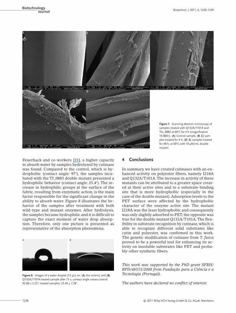
Feuerhack and co-workers [21], a higher capacityto absorb water by samples hydrolyzed by cutinasewas found. Compared to the control, which is hy-drophobic (contact angle: 97º), the samples incu-bated with the Tf_0883 double mutant presented ahydrophilic behavior (contact angle: 25.4º). The in-crease in hydrophilic groups at the surface of thefabric, resulting from enzymatic action, is the mainfactor responsible for the significant change in theability to absorb water. Figure 8 illustrates the be-havior of the samples after treatment with bothwild-type and mutant enzymes. After hydrolysis,the samples became hydrophilic and it is difficult tocapture the exact moment of water drop absorp-tion. Therefore, only one picture is presented asrepresentative of the absorption phenomena.
4 Conclusions
In summary, we have created cutinases with an en-hanced activity on polyester fibers, namely I218Aand Q132A/T101A.The increase in activity of thesemutants can be attributed to a greater space creat-ed at their active sites and to a substrate-bindingsite that is more hydrophobic (especially in thecase of the double mutant).Adsorption levels to thePET surface were affected by the hydrophobiccharacter of the enzyme active site. The mutantI218A was the least hydrophobic and consequentlywas only slightly adsorbed to PET; the opposite wastrue for the double mutant Q132A/T101A.The flex-ibility in substrate recognition by cutinase, which isable to recognize different solid substrates likecutin and polyester, was confirmed in this work.The genetic modification of cutinase from T. fuscaproved to be a powerful tool for enhancing its ac-tivity on insoluble substrates like PET and proba-bly other synthetic fibers.
This work was supported by the PhD grant SFRH/BPD/46515/2008 from Fundação para a Ciência e aTecnologia (Portugal).
The authors have declared no conflict of interest.
Figure 7. Scanning electron microscopy ofsamples treated with Q132A/T101A andTfu_0883 at 60°C for 4 h (magnification10 000×). (A) Control sample; (B, C) sam-ples treated for 4 h; (D, E) samples treatedfor 48 h, at 60°C with 10 μM/mL doublemutant.
Figure 8. Images of a water droplet (15 μL) on: (A) the control, and (B)Q132A/T101A-treated sample after 15 s; contact angle values:control:92.00 ± 3.25°; treated samples: 25.40 ± 7.28°.

© 2011 Wiley-VCH Verlag GmbH & Co. KGaA, Weinheim 1239
Biotechnol. J. 2011, 6, 1230–1239 www.biotechnology-journal.com
5 References
[1] Kim, H., Song, W., Lipase treatment of polyester fabrics.Fibers Polymers 2006, 7, 339–343.
[2] Brueckner, T., Eberl, A., Heumann, S., Rabe, M., Guebitz, G.M., Enzymatic and chemical hydrolysis of poly(ethyleneterephthalate) fabrics. J. Polymer Sci. A Polymer Chem. 2008,46, 6435–6443.
[3] Donelli, I.,Taddei, P., Smet, P. F., Poelman, D. et al., Enzymaticsurface modification and functionalization of PET: A watercontact angle, FTIR, and fluorescence spectroscopy study.Biotechnol. Bioeng. 2009, 103, 845–856.
[4] Pang, K., Kotek, R., Tonelli, A., Review of conventional andnovel polymerization processes for polyesters. Prog. Poly-mer Sci. 2006, 31, 1009–1037.
[5] Wang, P., Wang, Q., Fan, X., Cui, L. et al., Effects of cutinaseon the enzymatic shrink-resist finishing of wool fabrics. En-zyme Microb. Technol. 2009, 44, 302–308.
[6] Silva, C. M., Carneiro, F., O’Neill,A., Fonseca, L. P. et al., Cuti-nase—A new tool for biomodification of synthetic fibers. J.Polymer Sci. A Polymer Chem. 2005, 43, 2448–2450.
[7] Alisch, M., Feuerhack, A., Müller, H., Mensak, B. et al., Bio-catalytic modification of polyethylene terephthalate fibresby esterases from actinomycete isolates. Biocatal. Biotrans-form. 2004, 22, 347–351.
[8] Hsieh,Y., Cram, L., Enzymatic hydrolysis to improve wettingand absorbency of polyester fabrics Textile Res. J. 1996, 68,311–319.
[9] Hsieh, Y., Hartzell, M., Boston, M., Clarkson, K. et al., USPatent 2003, 20030119172.
[10] Yoon, M., Kellis, J., Poulouse, A., Enzymatic modification ofpolyester. AATCC Rev. 2002, 2, 33–36.
[11] Miettinen-Oinonen, A., Puolakka, A., Buchert, J., Modifica-tion of textile fibres by laccase. Cost Action 847 Workshop2004, 29–30.
[12] Alisch-Mark, M., Herrmann, A., Zimmermann, W., Increaseof the hydrophilicity of polyethylene terephthalate fibres byhydrolases from Thermomonospora fusca and Fusariumsolani f. sp. Biotechnol. Lett. 2006, 28, 681–685.
[13] O’Neill, A., Cavaco-Paulo, A., Monitoring biotransforma-tions in polyesters. Biocatal. Biotransform. 2004, 22, 353–356.
[14] Müller, R.-J., Schrader, H., Profe, J., Dresler, K., Deckwer,W.-D., Enzymatic degradation of poly(ethylene terephthalate):Rapid hydrolysis using a hydrolase from T. fusca. Macromol.Rapid Commun. 2005, 26, 1400–1405.
[15] Silva, C., Cavaco-Paulo, A., Biotransformations in syntheticfibres. Biocatal. Biotransform. 2008, 26, 350–356.
[16] Vertommen, M. A. M. E., Nierstrasz,V. A.,Veer, M. v. d., War-moeskerken, M. M. C. G., Enzymatic surface modification ofpoly(ethylene terephthalate). J. Biotechnol. 2005, 120, 376–386.
[17] Nimchua, T., Punnapayak, H., Zimmermann, W., Compari-son of the hydrolysis of polyethylene terephthalate fibersby a hydrolase from Fusarium oxysporum LCH I and Fusar-ium solani f. sp. pisi. Biotechnol. J. 2007, 2, 361–364.
[18] Kontkanen, H., Saloheimo, M., Pere, J., Miettinen-Oinonen,A., Reinikainen, T., Characterization of Melanocarpus al-bomyces steryl esterase produced in Trichoderma reesei andmodification of fibre products with the enzyme. Appl. Mi-crobiol. Biotechnol. 2006, 72, 696–704.
[19] Wang, X., Lu, D., Jonsson, L., Hong, F., Preparation of a PET-hydrolyzing lipase from Aspergillus oryzae by the additionof bis(2-hydroxyethyl) terephthalate to the culture medium
and enzymatic modification of PET fabrics. Eng. Life Sci.2008 8, 268–276.
[20] Araújo, R., Silva, C., O’Neill, A., Micaelo, N. et al., Tailoringcutinase activity towards polyethylene terephthalate andpolyamide 6,6 fibers. J. Biotechnol. 2007, 128, 849–857.
[21] Feuerhack,A.,Alisch-Mark, M., Kisner,A., Pezzin, S. H. et al.,Biocatalytic surface modification of knitted fabrics made ofpoly (ethylene terephthalate) with hydrolytic enzymes fromThermobifida fusca KW3b. Biocatal. Biotransform. 2008, 26,357–364.
[22] Liebminger, S., Eberl, A., Sousa, F., Heumann, S. et al., Hy-drolysis of PET and bis-(benzoyloxyethyl) terephthalatewith a new polyesterase from Penicillium citrinum. Biocatal.Biotransform. 2007, 25, 171–177.
[23] Liu, Y., Wu, G., Gu, L., Enzymatic treatment of PET fabricsfor improved hydrophilicity. AATCC Rev. 2008, 8, 44–48.
[24] Almansa, E., Heumann, S., Eberl, A., Fischer-Colbrie, G. etal., Enzymatic surface hydrolysis of PET enhances bondingin PVC coating. Biocatal. Biotransform. 2008, 26, 365–370.
[25] Dutta, K., Sen, S., Veeranki, V. D., Production, characteriza-tion and applications of microbial cutinases. ProcessBiochem. 2009, 44, 127–134.
[26] Carvalho, C., Aires-Barros, M., Cabral, J. M. S., Cutinasestructure, function and biocatalytic applications. Electron. J.Biotechnol. 1998, 1, 160–173.
[27] Carvalho, C. M. L., Aires-Barros, M. R., Cabral, J. M. S., Cuti-nase: From molecular level to bioprocess development.Biotechnol. Bioeng. 1999, 66, 17–34.
[28] Eberl, A., Heumann, S., Brückner, T., Araujo, R. et al., Enzy-matic surface hydrolysis of poly(ethylene terephthalate)and bis(benzoyloxyethyl) terephthalate by lipase and cuti-nase in the presence of surface active molecules. J. Biotech-nol. 2009, 143, 207–212.
[29] Gouda, M. K., Kleeberg, I., van den Heuvel, J., Müller, R.-J.,Deckwer, W.-D., Production of a polyester degrading extra-cellular hydrolase from Thermomonospora fusca. Biotech-nol. Prog. 2002, 18, 927–934.
[30] Ronkvist, A. s. M., Xie, W., Lu, W., Gross, R. A., Cutinase-cat-alyzed hydrolysis of poly(ethylene terephthalate). Macro-molecules 2009, 42, 5128–5138.
[31] Chen, S., Tong, X., Woodard, R., Guochen, D. et al., Identifi-cation and characterization of bacterial cutinase. J. Biol.Chem. 2008, 283, 25854–25862.
[32] He, G., Huo, G., Liu, L., Zhu,Y. et al., Enhanced cutinase pro-duction of Thermobifida fusca by a two-stage batch and fed-batch cultivation strategy. Biotechnol. Bioprocess Eng. 2009,14, 46–51.
[33] Kleeberg, I.,Welzel, K.,VandenHeuvel, J., Müller, R. J., Deck-wer,W. D., Characterization of a new extracellular hydrolasefrom Thermobifida fusca degrading aliphatic aromaticcopolyesters. Biomacromolecules 2004, 6, 262–270.
[34] Eberl, A., Heumann, S., Kotek, R., Kaufmann, F. et al., Enzy-matic hydrolysis of PTT polymers and oligomers. J. Biotech-nol. 2008, 135, 45–51.
[35] Chen, S., Su, L., Billig, S., Zimmermann, W. et al., Biochemi-cal characterization of the cutinases from Thermobifida fus-ca. J. Mol. Catal. B Enzymatic 2010, 63, 121–127.
[36] Fett, W., H., G., Moreau, R., Osman, S., Jones, L., Cutinaseproduction by Streptomyces spp. Curr. Microbiol. 1992, 25,165–171.
[37] Jaeger, K., Ransac, S., Dijkstra, B., Colson, C. et al., Bacteriallipases FEMS Microbiol. Rev. 1994, 15, 29–63.