The Iturin and Fengycin Families of Lipopeptides Are Key Factors in Antagonism of Bacillus subtilis...
11
430 / Molecular Plant-Microbe Interactions MPMI Vol. 20, No. 4, 2007, pp. 430–440. doi: 10.1094/MPMI-20-4-0430. © 2007 The American Phytopathological Society The Iturin and Fengycin Families of Lipopeptides Are Key Factors in Antagonism of Bacillus subtilis Toward Podosphaera fusca Diego Romero, 1 Antonio de Vicente, 1 Rivo H. Rakotoaly, 2 Samuel E. Dufour, 2 Jan-Willem Veening, 3 Eva Arrebola, 1 Francisco M. Cazorla, 1 Oscar P. Kuipers, 3 Michel Paquot, 2 and Alejandro Pérez-García 1 1 Departamento de Microbiología, Facultad de Ciencias, Universidad de Málaga, Campus Universitario de Teatinos s/n, E-29071 Málaga, Spain; 2 Unité de Chemie Biologie Industrielle, Faculté Universitaire des Sciences Agronomiques de Gembloux, Passaje des Déportés 2, B-5030 Gembloux, Belgium; 3 Molecular Genetics Group, Groningen Biomolecular Sciences and Biotechnology Institute, University of Groningen, Kerklaan 30, 9751 NN Haren, The Netherlands Submitted 19 July 2006. Accepted 24 November 2006. Podosphaera fusca is the main causal agent of cucurbit powdery mildew in Spain. Four Bacillus subtilis strains, UMAF6614, UMAF6619, UMAF6639, and UMAF8561, with proven ability to suppress the disease on melon in detached leaf and seedling assays, were subjected to fur- ther analyses to elucidate the mode of action involved in their biocontrol performance. Cell-free supernatants showed antifungal activities very close to those previously reported for vegetative cells. Identification of three lipo- peptide antibiotics, surfactin, fengycin, and iturin A or bacillomycin, in butanolic extracts from cell-free culture filtrates of these B. subtilis strains pointed out that antibio- sis could be a major factor involved in their biocontrol abil- ity. The strong inhibitory effect of purified lipopeptide fractions corresponding to bacillomycin, fengycin, and iturin A on P. fusca conidia germination, as well as the in situ detection of these lipopeptides in bacterial-treated melon leaves, provided interesting evidence of their puta- tive involvement in the antagonistic activity. Those results were definitively supported by site-directed mutagenesis analysis, targeted to suppress the biosynthesis of the differ- ent lipopeptides. Taken together, our data have allowed us to conclude that the iturin and fengycin families of lipopep- tides have a major role in the antagonism of B. subtilis toward P. fusca. Additional keywords: antifungals, biological control, cucurbits. Powdery mildew is probably the most common, conspicu- ous, widespread, and easily recognizable disease of cucurbits, responsible for serious damage to almost all cucurbit crops un- der both field and greenhouse conditions. As the most charac- teristic visual symptom, the disease induces development of a whitish, talcum-like, powdery fungal growth on both leaf sur- faces, petioles, and stems. Infected leaves usually wither and die, and plants senesce prematurely (Zitter et al. 1996). The disease can be caused by two species Golovinomyces cichoracearum or Podosphaera fusca, obligate biotrophic ecto- parasites that induce identical symptoms but can be distin- guished easily under light microscopy. In Spain, however, P. fusca has been identified as the sole cause of the disease, and is responsible for significant yield reductions and increasing production costs (del Pino et al. 2002; Fernández-Ortuño et al. 2006). Application of fungicides is currently the principal practice in most cucurbit crops for managing powdery mildew. However, the increasing problem of fungicide resistance and the consequent control failures (Fernández-Ortuño et al. 2006; McGrath 2001), along with public concern over the hazardous effect of chemicals on the environment, have led growers to explore and develop suitable environmentally friendly alterna- tives or complements to chemicals, biological control being one way forward (Kiss 2003). The use of bacterial strains as biological control agents has received great attention because of the ability of such strains to suppress different plant diseases involving a blend of diverse modes of action (Baehler et al. 2006; Cazorla et al 2006; Fogliano et al. 2002; Shoda 2000) and the possibilities to be combined with other control methods (Kondoh et al. 2001; Nofal and Haggag 2006; Omar et al. 2005). Among the most promising candidates for bacterial biocontrol agents are sev- eral species of the genus Bacillus, ubiquitously occurring safe microorganisms with proven excellent colonization aptitudes, versatility to protect plants effectively against pathogens (Kloepper et al. 2004; Romero et al. 2004; Shoda 2000), and an outstanding ability to sporulate, which assures their preva- lence in the environment and guarantees future suitable formu- lation strategies (Schallmey et al. 2004). Several Bacillus strains have been considered to be natural factories of biologically active compounds such as lipopep- tides, and the significance of their involvement in plant micro- bial disease control have been demonstrated (Asaka and Shoda 1996; Emmert and Handelsman 1999). Lipopeptides, oligo- peptides synthesized in a nonribosomal manner by large mul- tienzyme complexes, are the most frequent antibiotic com- pounds produced by bacilli, exhibiting a wide antimicrobial spectrum and exceptional surfactant activities (Magnet-Dana and Peypoux 1994; Vanittanakom et al. 1986; Vater et al. 2002; Vollembroich et al. 1997). These amphiphilic compounds share a common cyclic structure consisting of a β-amino or β- hydroxy fatty acid integrated into a peptide moiety. The main differences rely on the amino acid sequence and fatty acid branching, criteria that allow their classification in three families. The iturin family, represented by iturin A, mycosub- tilin, and bacillomycin, are heptapeptides with a β-amino fatty acid which exhibit strong antifungal activity (Duitman et al. 1999; Moyne et al. 2004; Thimon et al. 1995; Tsuge et al. Corresponding author: A. Pérez-García; Telephone: +34-952131890; Fax: +34-952131889; E-mail: [email protected]
-
Upload
independent -
Category
Documents
-
view
0 -
download
0
Transcript of The Iturin and Fengycin Families of Lipopeptides Are Key Factors in Antagonism of Bacillus subtilis...

430 / Molecular Plant-Microbe Interactions
MPMI Vol. 20, No. 4, 2007, pp. 430–440. doi: 10.1094 / MPMI -20-4-0430. © 2007 The American Phytopathological Society
The Iturin and Fengycin Families of Lipopeptides Are Key Factors in Antagonism of Bacillus subtilis Toward Podosphaera fusca
Diego Romero,1 Antonio de Vicente,1 Rivo H. Rakotoaly,2 Samuel E. Dufour,2 Jan-Willem Veening,3 Eva Arrebola,1 Francisco M. Cazorla,1 Oscar P. Kuipers,3 Michel Paquot,2 and Alejandro Pérez-García1 1Departamento de Microbiología, Facultad de Ciencias, Universidad de Málaga, Campus Universitario de Teatinos s/n, E-29071 Málaga, Spain; 2Unité de Chemie Biologie Industrielle, Faculté Universitaire des Sciences Agronomiques de Gembloux, Passaje des Déportés 2, B-5030 Gembloux, Belgium; 3Molecular Genetics Group, Groningen Biomolecular Sciences and Biotechnology Institute, University of Groningen, Kerklaan 30, 9751 NN Haren, The Netherlands
Submitted 19 July 2006. Accepted 24 November 2006.
Podosphaera fusca is the main causal agent of cucurbit powdery mildew in Spain. Four Bacillus subtilis strains, UMAF6614, UMAF6619, UMAF6639, and UMAF8561, with proven ability to suppress the disease on melon in detached leaf and seedling assays, were subjected to fur-ther analyses to elucidate the mode of action involved in their biocontrol performance. Cell-free supernatants showed antifungal activities very close to those previously reported for vegetative cells. Identification of three lipo-peptide antibiotics, surfactin, fengycin, and iturin A or bacillomycin, in butanolic extracts from cell-free culture filtrates of these B. subtilis strains pointed out that antibio-sis could be a major factor involved in their biocontrol abil-ity. The strong inhibitory effect of purified lipopeptide fractions corresponding to bacillomycin, fengycin, and iturin A on P. fusca conidia germination, as well as the in situ detection of these lipopeptides in bacterial-treated melon leaves, provided interesting evidence of their puta-tive involvement in the antagonistic activity. Those results were definitively supported by site-directed mutagenesis analysis, targeted to suppress the biosynthesis of the differ-ent lipopeptides. Taken together, our data have allowed us to conclude that the iturin and fengycin families of lipopep-tides have a major role in the antagonism of B. subtilis toward P. fusca.
Additional keywords: antifungals, biological control, cucurbits.
Powdery mildew is probably the most common, conspicu-ous, widespread, and easily recognizable disease of cucurbits, responsible for serious damage to almost all cucurbit crops un-der both field and greenhouse conditions. As the most charac-teristic visual symptom, the disease induces development of a whitish, talcum-like, powdery fungal growth on both leaf sur-faces, petioles, and stems. Infected leaves usually wither and die, and plants senesce prematurely (Zitter et al. 1996). The disease can be caused by two species Golovinomyces cichoracearum or Podosphaera fusca, obligate biotrophic ecto-parasites that induce identical symptoms but can be distin-guished easily under light microscopy. In Spain, however, P. fusca has been identified as the sole cause of the disease, and
is responsible for significant yield reductions and increasing production costs (del Pino et al. 2002; Fernández-Ortuño et al. 2006). Application of fungicides is currently the principal practice in most cucurbit crops for managing powdery mildew. However, the increasing problem of fungicide resistance and the consequent control failures (Fernández-Ortuño et al. 2006; McGrath 2001), along with public concern over the hazardous effect of chemicals on the environment, have led growers to explore and develop suitable environmentally friendly alterna-tives or complements to chemicals, biological control being one way forward (Kiss 2003).
The use of bacterial strains as biological control agents has received great attention because of the ability of such strains to suppress different plant diseases involving a blend of diverse modes of action (Baehler et al. 2006; Cazorla et al 2006; Fogliano et al. 2002; Shoda 2000) and the possibilities to be combined with other control methods (Kondoh et al. 2001; Nofal and Haggag 2006; Omar et al. 2005). Among the most promising candidates for bacterial biocontrol agents are sev-eral species of the genus Bacillus, ubiquitously occurring safe microorganisms with proven excellent colonization aptitudes, versatility to protect plants effectively against pathogens (Kloepper et al. 2004; Romero et al. 2004; Shoda 2000), and an outstanding ability to sporulate, which assures their preva-lence in the environment and guarantees future suitable formu-lation strategies (Schallmey et al. 2004).
Several Bacillus strains have been considered to be natural factories of biologically active compounds such as lipopep-tides, and the significance of their involvement in plant micro-bial disease control have been demonstrated (Asaka and Shoda 1996; Emmert and Handelsman 1999). Lipopeptides, oligo-peptides synthesized in a nonribosomal manner by large mul-tienzyme complexes, are the most frequent antibiotic com-pounds produced by bacilli, exhibiting a wide antimicrobial spectrum and exceptional surfactant activities (Magnet-Dana and Peypoux 1994; Vanittanakom et al. 1986; Vater et al. 2002; Vollembroich et al. 1997). These amphiphilic compounds share a common cyclic structure consisting of a β-amino or β-hydroxy fatty acid integrated into a peptide moiety. The main differences rely on the amino acid sequence and fatty acid branching, criteria that allow their classification in three families. The iturin family, represented by iturin A, mycosub-tilin, and bacillomycin, are heptapeptides with a β-amino fatty acid which exhibit strong antifungal activity (Duitman et al. 1999; Moyne et al. 2004; Thimon et al. 1995; Tsuge et al.
Corresponding author: A. Pérez-García; Telephone: +34-952131890; Fax:+34-952131889; E-mail: [email protected]
Vol. 20, No. 4, 2007 / 431
2001). Members of the fengycin family, including the related plipastatin, are decapeptides with a β-hydroxy fatty acid that show unusual properties, such as the presence of ornithine in the peptide portion, and also show antifungal activity, although more specific for filamentous fungi (Steller et al. 1999; Vanittanakom et al. 1986). Finally, the most studied family of lipopeptides, the surfactin family, consists of heptapeptides containing a β-hydroxy fatty acid with a number of carbon atoms in the range of 13 to 15, which are possibly the most powerful biosurfactants described, also exhibiting antiviral characteristics; and, although slightly antifungal, they show a strong synergistic action in combination with iturin A (Magnet-Dana et al. 1992). Furthermore, surfactin seems to be also a key factor in the establishment of stable biofilms, and may in-hibit the biofilm formation of other bacteria, thus contributing to the protective action, as shown in Arabidopsis against Pseu-domonas syringae pv. tomato (Bais et al. 2004).
In a previous work, we have shown that four Bacillus sub-tilis strains isolated in our laboratory were able to suppress powdery mildew disease caused by P. fusca on melon in both detached leaf and seedling assays, also showing good abilities to colonize melon phylloplane by establishing themselves as cell aggregates (Romero et al. 2004). In order to gain an insight into the underlying mechanisms responsible for the outstanding biocontrol performance of these B. subtilis strains, the aims of this study were to evaluate the inhibitory effect of B. subtilis cell-free filtrates on P. fusca growth, to identify the putative compounds responsible for the antifungal activity of the super-natants, and, finally, to determine the role of these compounds in the antagonism of B. subtilis toward P. fusca by analysis of B. subtilis transformants constructed by site-directed muta-genesis of genes involved in the biosynthesis of such com-pounds.
RESULTS
Production of antifungal compounds of B. subtilis in liquid cultures.
Relationship between bacterial growth and production of antifungal compounds was analyzed using a growth inhibition assay in 24-well microplates with Botrytis cinerea as the target fungus. Antifungal activity was detected at the transition be-tween exponential and stationary phase of growth, increasing progressively during the later and reaching the highest activity
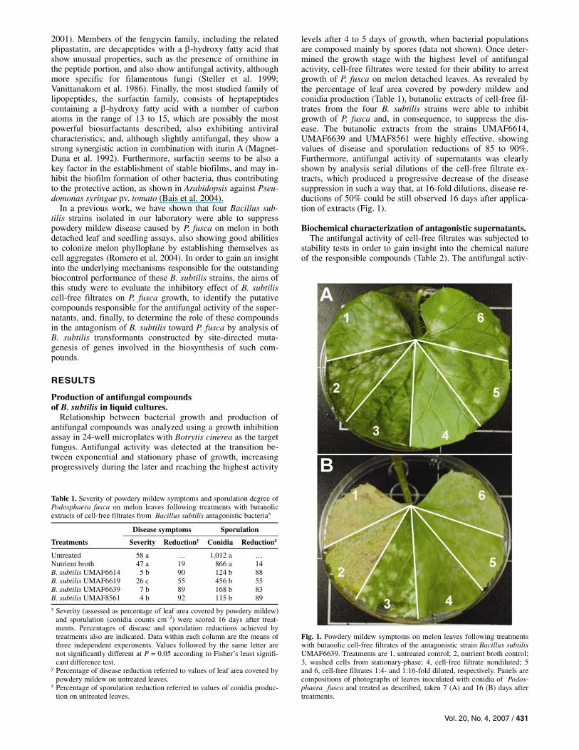
levels after 4 to 5 days of growth, when bacterial populations are composed mainly by spores (data not shown). Once deter-mined the growth stage with the highest level of antifungal activity, cell-free filtrates were tested for their ability to arrest growth of P. fusca on melon detached leaves. As revealed by the percentage of leaf area covered by powdery mildew and conidia production (Table 1), butanolic extracts of cell-free fil-trates from the four B. subtilis strains were able to inhibit growth of P. fusca and, in consequence, to suppress the dis-ease. The butanolic extracts from the strains UMAF6614, UMAF6639 and UMAF8561 were highly effective, showing values of disease and sporulation reductions of 85 to 90%. Furthermore, antifungal activity of supernatants was clearly shown by analysis serial dilutions of the cell-free filtrate ex-tracts, which produced a progressive decrease of the disease suppression in such a way that, at 16-fold dilutions, disease re-ductions of 50% could be still observed 16 days after applica-tion of extracts (Fig. 1).
Biochemical characterization of antagonistic supernatants. The antifungal activity of cell-free filtrates was subjected to
stability tests in order to gain insight into the chemical nature of the responsible compounds (Table 2). The antifungal activ-
Table 1. Severity of powdery mildew symptoms and sporulation degree ofPodosphaera fusca on melon leaves following treatments with butanolic extracts of cell-free filtrates from Bacillus subtilis antagonistic bacteriax
Disease symptoms Sporulation
Treatments Severity Reductiony Conidia Reductionz
Untreated 58 a … 1,012 a … Nutrient broth 47 a 19 866 a 14 B. subtilis UMAF6614 5 b 90 124 b 88 B. subtilis UMAF6619 26 c 55 456 b 55 B. subtilis UMAF6639 7 b 89 168 b 83 B. subtilis UMAF8561 4 b 92 115 b 89 x Severity (assessed as percentage of leaf area covered by powdery mildew)
and sporulation (conidia counts cm–2) were scored 16 days after treat-ments. Percentages of disease and sporulation reductions achieved bytreatments also are indicated. Data within each column are the means ofthree independent experiments. Values followed by the same letter arenot significantly different at P = 0.05 according to Fisher’s least signifi-cant difference test.
y Percentage of disease reduction referred to values of leaf area covered bypowdery mildew on untreated leaves.
z Percentage of sporulation reduction referred to values of conidia produc-tion on untreated leaves.
Fig. 1. Powdery mildew symptoms on melon leaves following treatments with butanolic cell-free filtrates of the antagonistic strain Bacillus subtilisUMAF6639. Treatments are 1, untreated control; 2, nutrient broth control; 3, washed cells from stationary-phase; 4, cell-free filtrate nondiluted; 5 and 6, cell-free filtrates 1:4- and 1:16-fold diluted, respectively. Panels are compositions of photographs of leaves inoculated with conidia of Podos-phaera fusca and treated as described, taken 7 (A) and 16 (B) days after treatments.

432 / Molecular Plant-Microbe Interactions
ity of the supernatants was stable at high temperatures (50 to 100ºC) and resistant to enzymatic degradation (pronase or pro-teinase K), all of which are well known characteristics associ-ated with the lipopeptide antibiotics produced by Bacillus spp. (Katz and Demain 1977; Lin et al. 1998a; Stein 2005). Com-pared with untreated controls, when they were subjected to extremely acidic pH (pH 2), a large precipitation occurred with a consequent lost of antifungal activity in the soluble phase; however, the supernatant activity could be restored by immedi-ately dissolving the precipitates in neutral phosphate-buffered saline. It is interesting to note that acidic precipitation is a typical feature of lipopeptides, which often is used in purifica-tion protocols (Ohno et al. 1992). Furthermore, as previously shown, antifungal activity was efficiently extracted with n-bu-tanol, suggesting the presence of a hydrophobic moiety in the compound which also is typical of Bacillus lipopeptides. In water solution, however, ultrafiltration tests showed that the antifungal activity was always retained in the filter, suggesting that the active molecule was of a molecular size higher than 3 kDa. Both features are typical of biosurfactants and the latter is the result of their aggregational behavior as supramolecular micelles in water (Lin et al. 1998b). Taken together, these re-sults demonstrated that lipopeptides could be the compounds responsible for the antifungal activity exhibited by the B. sub-tilis biocontrol strains.
Identification of lipopeptides of B. subtilis. In order to identify the compounds responsible for antifun-
gal activity, methanolic fractions from the butanolic extracts of cell-free culture filtrates of the four B. subtilis strains were separated initially in silica thin-layer chromatography (TLC) sheets, using purified iturin A, fengycin, and surfactin as stan-dards (data not shown). The four B. subtilis strains produced spots with Rf values similar to fengycin (Rf = 0.09), iturin A (Rf = 0.3), and surfactin (Rf = 0.7). Likewise, when the metha-nolic extracts were analyzed by reverse-phase high-perform-ance liquid chromatography (RP-HPLC), three main groups of peaks were observed at elution times comparable with those observed for standard lipopeptides; therefore, they were tenta-tively named in order of elution as LP-a, LP-b, and LP-c for further analysis (data not shown).
For purification of the antifungal compounds, methanolic extracts were fractioned by Flash chromatography followed by semi-preparative RP-HPLC, using a method previously devel-oped for these families of lipopeptides. To verify the purity of different fractions, analytical RP-HPLC was performed. An example of the results obtained corresponds to strain UMAF6639, which is shown in Figure 2.
For an accurate characterization of the different purified compounds, mass spectra were recorded by matrix-assisted laser desorption-ionization time of flight mass spectrometry (MALDI-TOF-MS). The mass spectra of LP-a showed a series of mass number of m/z = 1,030 to 1,074 for all strains; those of LP-b, m/z = 1,017 to 1,101 for strains UMAF8561 and UMAF6614, m/z = 1,034 to 1,095 for UMAF6639, or m/z = 1,021 to 1,049 for UMAF6619; and those of LP-c, m/z = 1,435 to 1,499 for all strains. An example of these results is shown for the strain UMAF6639 (Fig. 2).
For all fractions, the Fourier transform-infrared spectrum (FT-IR) analysis showed bands in the range of 1,630 to 1,680 cm–1, resulting from the stretching mode of the CO-N bond (amide I band), and at 1,570 to 1,515 cm–1, resulting from the deformation mode of the N-H bond combined with C-N stretching mode (amide II band), both indicating the presence of a peptide component; and also bands at 2,855 to 2,960 cm–1, resulting from typical CH stretching vibration in the alkyl chain. For the fractions LP-a and LP-c, a shoulder also was observed at 1,730 cm–1 due to the lactone carbonyl absorption typical for surfactin and fengycin families of lipopeptides (data not shown).
When the amino acid compositions were determined, it was found that the fraction LP-a contained Asp, Glu, Val, and Leu in a ratio of 1:1:1:4; fraction LP-b comprised Asp, Ser, Glu, Pro, and Tyr in a ratio of 3:1:1:1:1 for strain UMAF6639 and Asp, Ser, Glu, Pro, Tyr, and Thr in a ratio of 2:1:1:1:1:1 for the rest of the strains; and fraction LP-c was composed of Thr, Glu, Pro, and Ala or Val, Ile, Tyr, and Orn in a ratio of 1:3:1:1:1:2:1, valine being observed only in the fraction corre-sponding to strain UMAF6639.
The amino acid sequences were determined by electrospray ionization ion trap mass spectrometry (ESI-MS-MS). As an example, spectra obtained for the fractions LP-a, LP-b, and
Table 2. Biochemical characterization of the antifungal activity of Bacillus subtilis supernatantsz
B. subtilis strains
Culture filtrates UMAF6614 UMAF6619 UMAF6639 UMAF8561
Heat treatments Untreated 22 18 24 23
50ºC, 15 min 22 18 24 23 75ºC, 15 min 22 18 24 22 100ºC, 15 min 22 18 24 22
Enzymatic degradation Untreated 26 19 24 24 Pronase 26 19 24 24 Proteinase K 26 18 24 24
pH tolerance Untreated 25 21 26 26 Acid, pH = 2 23 20 24 20 Basic, pH = 12 14 0 19 16
Organic phase after extraction with organic solvents Untreated 26 18 25 20 n-Butanol 26 19 25 20 Chloroform 0 0 0 0
Ultrafiltration Untreated 24 19 24 24 >3-kDa fraction 26 19 29 30 <3-kDa fraction 0 0 0 0
z Antifungal activity was evaluated after different treatments by an in vitro bioassay against Botrytis cinerea. Antifungal activity measured as diameter (in millimeters) of inhibition zone 15 days after incubation. Each value represents the mean of three independent experiments.

Vol. 20, No. 4, 2007 / 433
LP-c from strain UMAF6639 are given (Fig. 2). Sequences de-duced for these fractions were similar to those corresponding to surfactin, iturin A, and fengycin, respectively. For the other strains, the fractions LP-a, LP-b, and LP-c corresponded to the sequences expected for surfactin, bacillomycin, and fengycin, respectively.
All together, these results showed that the four B. subtilis strains simultaneously produced, in liquid cultures, surfactin, with a number of carbon atoms in the fatty acid moiety of C13-C15, and fengycin C14-C18, together with iturin A C14-C15 (strain UMAF6639), bacillomycin D C14-C16 and bacil-lomycin L C17 (strains UMAF8561 and UMAF6614), or ba-cillomycin L C14-C17 (strain UMAF6619).
Inhibitory effect of purified lipopeptides on P. fusca conidia germination and in planta detection of lipopeptides.
Once lipopeptides present in the culture filtrates of the B. subtilis strains were identified, purified fractions of the differ-ent lipopeptides detected were used to study their inhibitory effect on P. fusca conidia germination. Inhibition assays were carried out on disks of zucchini cotyledons that were exposed
to lipopeptide concentrations of 1 mg/ml and conidia germina-tion was evaluated by bright field microscopy after 48 h of incubation. The results obtained are shown in Figure 3. As can be noted, bacillomycin (Fig. 3C) showed the strongest effect on germination, inducing 67% of inhibition, followed by fen-gycin (Fig. 3E) and iturin A (Fig. 3D), which produced inhibi-tions of 53 and 42%, respectively. By contrast, surfactin (Fig. 3F) had no significant effect on germination according to Fisher’s least significant difference test (P = 0.05), causing 9.5% of inhibition.
To test the hypothesis that production of lipopeptides in planta could be responsible for the suppression of powdery mildew symptoms, lipopeptides were recovered from melon leaves infected with P. fusca conidia and treated with washed cells of B. subtilis UMAF6614, UMAF6639, or UMAF8561, and analyzed by analytical RP-HPLC (Fig. 4). As shown, 5 days after application of bacteria, peaks corresponding to ba-cillomycin, iturin A, and fengycin were detected, whereas sur-factin peaks stayed close to the detection threshold and were undetectable. Twelve days after application, however, it was very difficult to identify single peaks, which became practi-cally undetectable, especially in the case of strain UMAF8561
Fig. 2. Characterization and identification of lipopeptides occurring in butanolic extracts from liquid cultures of the antagonistic Bacillus subtilis strain UMAF6639. Peaks obtained on an analytical reverse-phase high-performance liquid chromatography (RP-HPLC) (detection at 214 nm) for purified fractions LP-a, LP-b, and LP-c, previously fractioned by flash chromatography followed by (semi)-preparative RP-HPLC. Mass spectra scored for the purified frac-tions LP-a, LP-b, and LP-c. Fragmentation spectra observed by electrospray ionization ion trap mass spectrometry (ESI-MS-MS) and amino acid sequences and fatty acid compositions deduced from the parental peaks 1008.7 (LP-a, surfactin), 1065.4 (LP-b, iturin A), and 1464 (LP-c, fengycin).

434 / Molecular Plant-Microbe Interactions
(data not shown). This decrease in lipopeptide levels always was correlated with outbreaks of powdery mildew disease.
Biological control analysis of lipopeptide-deficient B. subtilis transformants.
Site-directed mutagenesis analysis focused to determine the contribution of the different lipopeptides in the antagonism of B. subtilis toward P. fusca was carried out with strains UMAF6614 and UMAF6639, which produced different lipopeptide profiles. Based on the available gene sequences governing the synthesis of bacillomycin, fengycin, and iturin A, a set of suitable primers were designed to obtain the proper DNA fragments to construct the corresponding disruption vec-tors (discussed below). The resulting transformants were grown in medium optimal for lipopeptide production for 4 days at 37ºC. Analysis of butanolic extracts from supernatants of several transformants by TLC (data not shown) and analyti-cal RP-HPLC (Fig. 5) confirmed the expected phenotypes. The derivative transformants disrupted in the fenB gene completely lost the ability to produce fengycin. Likewise, the disruption of bamA and ituD genes resulted in transformants unable to pro-duce bacillomycin and iturin A, respectively. In addition, poly-merase chain reaction (PCR) and Southern hybridization analyses confirmed the correct insertion of the plasmids within the target gene sequences (data not shown). Two single-copy transformants of each strain and phenotype were selected for further analysis (Table 3).
The ability of the different mutants to arrest growth of P. fusca was evaluated by two set of experiments, one using vegetative cells and the other using butanolic extracts of cell-free super-natants. With vegetative cells (Table 4), whereas UMAF6614 and UMAF6639 wild types reduced disease up to 64%, their derivative mutants deficient in bacillomycin and iturin A lost completely their ability to suppress powdery mildew disease (0 to 5% disease reduction), and the mutants deficient in fen-gycin achieved disease reductions of 8 to 12%. In all cases, disease symptoms showed by transformants were significantly different from those observed for wild types and statistically
Fig. 3. Effect of purified lipopeptides on Podosphaera fusca conidia germination. Disks of zucchini cotyledons were treated with the corresponding lipopep-tide at 1 mg/ml and the percentage of conidia germination was evaluated by bright field microscopy after 48 h of incubation. Representative pictures showingthe effect of each lipopeptide treatment are given. A and B, untreated controls; C, D, E, and F, disks treated with bacillomycin, fengycin, iturin A, and sur-factin, respectively. Bars: 40 µm.
Fig. 4. In planta detection of lipopeptides by analytical reverse-phase high-performance liquid chromatography (RP-HPLC). Melon plants were infected with powdery mildew and treated with Bacillus subtilisstrains UMAF6614, UMAF6639, and UMAF8561. Lipopeptides were recovered from melon leaves 5 days after application of B. subtilis. Non-infected and nontreated leaves were used as controls (untreated). Peaks corresponding to bacillomycin (Bac), fengycin (Fen), and iturin A (It) are indicated. Detection at 214 nm.

Vol. 20, No. 4, 2007 / 435
Fig. 5. Reverse-phase high-performance liquid chromatography (RP-HPLC) analysis of lipopeptides produced by Bacillus subtilis antagonistic strains UMAF6614 and UMAF6639 and their derivative mutants in liquid cultures. From strains UMAF6639 and UMAF6614, mutants impaired in production offengycin (fenB mutants) and iturin A (ituD mutants) or bacillomycin (bamA mutants), respectively, were constructed. Peaks corresponding to iturin (It), bacillomycin (Bac), fengycin (Fen), and surfactin (Sf) are indicated. Detection at 214 nm.
Table 3. Microorganisms and plasmids used in this study
Strains or plasmids Relevant characteristicsx Reference
Fungi Botrytis cinerea CECT2850 Isolated from saffron bulb CECTYy Podosphera fusca SF48 Isolated from melon plants Fernández-Ortuño et al. 2006
Bacterial strains and mutants Bacillus subtilis UMAF6614 Producer of bacillomycin, fengycin, and surfactin Romero et al. 2005 B. subtilis UMAF6619 Producer of bacillomycin, fengycin, and surfactin Romero et al. 2005 B. subtilis UMAF6639 Producer of iturin A, fengycin, and surfactin Romero et al. 2005 B. subtilis UMAF8561 Producer of bacillomycin, fengycin and surfactin Romero et al. 2005 6614::bamA-1 UMAF6614 bamA::pCR-Bac; bacillomycin deficient This study 6614::bamA-2 UMAF6614 bamA::pCR-Bac; bacillomycin deficient This study 6614::fenB-1 UMAF6614 fenB::pFen2-2; fengycin deficient This study 6614::fenB-2 UMAF6614 fenB::pFen2-2; fengycin deficient This study 6639::ituD-1 UMAF6639 ituD::pItu2-2; iturin A deficient This study 6639::ituD-2 UMAF6639 ituD::pItu2-2; iturin A deficient This study 6639::fenB-1 UMAF6639 fenB::pFen2-2; fengycin deficient This study 6639::fenB-2 UMAF6639 fenB::pFen2-2; fengycin deficient This study
Plasmidsz pCR-Bac pCR2.1 carrying bamA::Km; Apr, Kmr This study pFen2-2 pUC18 carrying fenB::Cm; Apr, Cmr Romero et al. 2006 pItu2-2 pUC18 carrying ituD::Spc; Apr, Spcr Romero et al. 2006
x Apr = ampicillin resistance, Cmr = cloramphenicol resistance, and Kmr = kanamycin resistance. y CECTY = Spanish type culture collection. z Integrative plasmids containing gene sequences from B. subtilis UMAF6614 or UMAF6639.
436 / Molecular Plant-Microbe Interactions
comparable with untreated controls. Similar results were observed when the level of conidia production was evaluated.
The above results were supported when bioassays of growth inhibition using butanolic extracts of cell-free filtrates were carried out (Table 5). For strain UMAF6614, whereas the wild-type supernatant still showed some antifungal activity (27% of inhibition) 16-fold diluted, extracts from fengycin and bacillo-mycin mutants lost their activity at 4- to 8-fold dilutions. A similar tendency also was observed for strain UMAF6639. Wild-type extracts were very active at 16-fold dilution (82% of inhibition); however, at the same dilution, supernatants from fengycin mutants completely lost their activity, whereas ex-tracts from iturin A mutants showed no inhibition 4- to 8-fold diluted.
DISCUSSION
Several reports have described Bacillus strains worthy to be used as biocontrol agents of plant diseases (Shoda 2000). One of the most convincing properties contributing to that sugges-tion is the amazing battery of antibiotic compounds synthe-sized that exhibit a wide antimicrobial spectrum, the ability to modify attachment of other microorganisms to different sur-faces and to contribute to the survival of the bacillus cells in their habitat (Stein 2005). In our study, the supernatants of four B. subtilis strains have proved to suppress cucurbit powdery mil-dew disease at levels similar to those previously reported for vegetative cells (Romero et al. 2004). The comprehensive chemical analysis of culture filtrates proved the occurrence of three different lipopeptide antibiotics, surfactin, fengycin, and iturin A or bacillomycin, compounds well known for their strong antifungal effect against different necrotrophic phytopa-thogenic fungi (Asaka and Shoda 1996; Vanittanakom et al. 1986). These findings together with the fact that the biocontrol efficiency of each strain was closely correlated with the lipopeptide production yield, strongly supported the relevant role of antibiosis as a major factor involved in the protective effect of these strains, and were in agreement with previous reports that had pointed out that biotrophic fungi, such as P. fusca, could be efficiently targeted by antibiotic-producing microorganisms (Avis et al. 2002).
Additional assays carried out with purified lipopeptide frac-tions showed that iturin A, bacillomycin, and fengycin retained the highest antifungal activities, whereas surfactin showed a limited action. In this sense, although the direct action of anti-microbials in the bacterial antagonism in vitro has been well characterized, evidence of the in situ production of these anti-microbials in their corresponding environments to correlate them with the biocontrol ability has long been a constraint (Raaijmakers et al. 2002). In this study, we have been able to detect iturin A, bacillomycin, and fengycin on melon leaves treated with two strains of B. subtilis. The occurrence of lipopeptides 5 days after bacterial treatments clearly supported the in vitro observations of their inhibitory effect upon P. fusca conidia germination and development because, when lipopep-tides were hardly detected, as occurred 12 days after bacterial application, outbreaks of powdery mildew symptoms occurred, as previously shown in other systems (Asaka and Shoda 1996; Ongena et al. 2005). These results demonstrated a clear contri-bution of iturin A, bacillomycin, and fengycin in the antago-nism of B. subtilis toward P. fusca.
Table 5. Effect of butanolic extracts of cell-free culture filtrates of Bacillus subtilis wild-type strains and their lipopeptide deficient derivative mutants on Podosphaera fusca growth inhibition
Dilution factor of lipopeptide-enriched solutionsz
Treatments 1:0 1:1 1:3 1:7 1:15
UMAF6614 94 ± 4 69 ± 6 50 ± 8 42 ± 8 27 ± 7 Bacillomycin mutants 6614::bamA-1 39 ± 9 4 ± 1 0 ± 0.1 0 ± 0.1 0 ± 0.1 6614::bamA-2 53 ± 15 35 ± 10 11 ± 0.1 4 ± 2 0 ± 0.1
Fengycin mutants 6614::fenB-1 55 ± 15 32 ± 10 15 ± 9 0 ± 0.1 0 ± 0.1 6614::fenB-2 60 ± 20 29 ± 8 7 ± 3 0 ± 0.1 0 ± 0.1
UMAF6639 100 ± 0.1 100 ± 0.1 98 ± 4 94 ± 3 82 ± 9 Iturin A mutants 6639::ituD-1 25 ± 9 11 ± 0.1 0 ± 0.1 0 ± 0.1 0 ± 0.1 6639::ituD-2 23 ± 7 14 ± 2 8 ± 4 0 ± 0.1 0 ± 0.1
Fengycin mutants 6639::fenB-1 91 ± 3 60 ± 7 38 ± 11 15 ± 6 0 ± 0.1 6639::fenB-2 88 ± 7 44 ± 7 20 ± 10 12 ± 6 0 ± 0.1
z Percentage of inhibition of P. fusca growth referred to values of leaf area covered by powdery mildew recorded after 14 days of incubation. Data representthe means ± standard deviation from 10 disks per treatment. The experiment was repeated three times.
Table 4. Severity of powdery mildew symptoms and sporulation degree ofPodosphaera fusca on melon leaves following treatments with vegetative cells from Bacillus subtilis wild-type strains and their lipopeptide deficientderivative mutantsx
Disease symptoms Sporulation
Treatments Severity Reductiony Conidia Reductionz
Untreated 72 a … 1,233 a … UMAF6614 29 b 60 396 b 68 Bacillomycin mutants 6614::bamA-1 68 a 5 1,121 a 9 6614::bamA-2 72 a 0 1,201 a 2
Fengycin mutants 6614::fenB-1 65 a 10 1,051 a 14 6614::fenB-2 63 a 12 1,065 a 14
UMAF6639 26 b 64 308 b 75 Iturin A mutants 6639::ituD-1 70 a 3 1,150 a 7 6639::ituD-2 69 a 4 1,148 a 7
Fengycin mutants 6639::fenB-1 63 a 12 1,034 a 16 6639::fenB-2 66 a 8 1,056 a 14
x Severity (assessed as percentage of leaf area covered by powdery mil-dew) and sporulation (conidia cm–2) were scored 16 days after treat-ments. Percentages of disease and sporulation reductions achieved bytreatments also are indicated. Data within each column are the meansof three independent experiments. Values followed by the same letterare not significantly different at P = 0.05 according to Fisher’s leastsignificant difference test.
y Percentage of disease reduction referred to values of leaf area covered bypowdery mildew on untreated leaves.
z Percentage of sporulation reduction referred to values of conidiaproduction on untreated leaves.
Vol. 20, No. 4, 2007 / 437
To clarify the involvement of antimicrobials in the biocon-trol performance, additional evidence is always offered by mo-lecular analysis either reducing or avoiding biocontrol activity by mutagenesis (Raaijmakers et al., 2002) or enhancing the antimicrobial production, modifying biosynthetic or regulatory genes (Leclere et al. 2005). For mutational analysis, only strains UMAF6614 and UMAF6639, showing good and repro-ducible biocontrol performance and different lipopeptide pro-files, were included. A site-directed mutagenesis strategy to inactivate a single locus was set up to construct a collection of mutants with different combinations of lipopeptide production. Based on the antifungal activity previously demonstrated for each purified lipopeptide, our main attention was focused on the disruption of iturin A, bacillomycin, and fengycin produc-tion. The biocontrol results obtained in bioassays using vegeta-tive cells showed the expected role for iturin A, bacillomycin, and fengycin in the disease suppression ability of these strains, as revealed by the complete loss of biocontrol. The growth inhibition assays using butanolic extracts from supernatants of the different mutants confirmed those data, and allowed us to conclude that iturin A, bacillomycin, and fengycin are indis-pensable for the biocontrol ability of these strains of B. sub-tilis, at least on P. fusca. Furthermore, the fact that extracts from mutants producing only iturin A and surfactin still re-tained a considerable high antifungal activity suggested the possible occurrence of a synergistic antifungal effect between both lipopeptides, as previously reported (Koumoutsi et al. 2004; Magnet-Dana et al. 1992). This point could be clarified by construction of double mutants impaired in the production of both surfactin and iturin A lipopeptides.
Early studies have emphasized the fact that the antimicrobial effect of lipopeptides not only rely on their chemical structure but also on the sterol content of the plasma membrane in the target fungi, due to a buffering effect of sterols on the increase in membrane fluidity caused by the fatty acids (Avis et al. 2002; Latoud et al. 1990). In this sense, the low sterol content of conidia of powdery mildew fungi, compared with other fun-gal plant pathogens and the presence of certain amounts of cholesterol molecules in their membranes (Loeffler et al., 1992) could explain the high susceptibility exhibited by P. fusca conidia to these compounds.
The B. subtilis strains UMAF6614 and UMAF6639 have proven to be excellent candidates to be used in the biological control of cucurbit powdery mildew elicited by P. fusca (Romero et al. 2004). In this work, we have provided experi-mental evidence represented by the antifungal effect of cell-free supernatants as well as the presence of the antifungal compounds bacillomycin, fengycin, iturin A, and surfactin in liquid cultures and in bacterial-treated leaves, which supported the early assumption of antibiosis as the main mode of action displayed by these strains to manage the disease. Additionally, a differential role of the lipopeptides in the biocontrol perfor-mance has been established by mutational analysis, confirming a major contribution of the iturin (bacillomycin and iturin A) and fengycin families of lipopeptides in the antagonism of B. subtilis toward P. fusca, the suppression of conidia germina-tion determining to a large extent the subsequent development of the pathogen. Work to determine the ultrastructural damage induced by these compounds on P. fusca is currently underway.
MATERIALS AND METHODS
Microorganisms and culture conditions. The fungi, bacterial strains, and plasmids used in this study
are listed in Table 3. Routinely, fresh bacterial cultures were obtained from frozen stocks before each experiment. B. subtilis
strains were grown in nutrient agar (NA), nutrient broth (NB), NB supplemented with 1% glucose, or in medium optimal for lipopeptide production (MOLP) (Ahimou et al. 2000) at 37ºC. Escherichia coli was grown in Luria-Bertani broth (LB) at 37ºC. Antibiotics, when required, were added to the culture media at the following concentrations: ampicillin, 100 μg/ml; chloramphenicol, 5 μg/ml; spectinomycin, 100 μg/ml; kana-mycin, 10 μg/ml; and erythromycin, 5 μg/ml.
The plant-pathogenic fungus B. cinerea CECT 2850 was grown in potato dextrose agar (PDA) at 25ºC. Isolate SF48 of P. fusca race 1 was maintained in vitro on zucchini cotyledons as described elsewhere (Pérez-García et al. 2001).
Antifungal activity assays with B. subtilis supernatants. To determine the relationship between growth and produc-
tion of antifungal compounds, B. subtilis strains were grown in Erlenmeyer flasks (250 ml) containing 100 ml of NB or NB supplemented with 1% glucose or with 50 mM diethyl malo-nate, a sporulation inhibitor added at the end of the log phase. Incubation was carried out in an orbital shaker for several days at 30ºC and 80 rpm. Culture samples of 2 ml were taken at various time points and examined for number of vegetative cells and spores and antifungal activity. For the latter, because P. fusca cannot be cultured in artificial media, a growth inhibi-tion assay in 24-well microplates using B. cinerea was set up. Cell-free filtrates from the different liquid cultures were ob-tained by centrifugation at 2,500 × g for 15 min followed by filtration through 0.2-μm cellulose nitrate filters. Microplate wells then were filled with a 1:1 mixture of PDA and the cor-responding dilution of the cell-free filtrates and, after solidifi-cation, a 5-mm-diameter plug of B. cinerea mycelium, ob-tained from PDA plates grown at 25ºC for 4 days, was placed at the center of each well. After 5 days of incubation at 25ºC, antifungal activity was determined by measuring the diameter of the fungal colony and comparing with NB controls.
The cell-free filtrates also were tested for their ability to reduce P. fusca growth in a bioassay using the detached leaf method previously described (Romero et al. 2003). Bacterial cultures were grown on NB supplemented with 1% glucose as described above. After 5 days of incubation, cells were removed by centrifugation at 2,500 × g for 15 min, and supernatants were extracted with n-butanol as described earlier (Yazgan et al. 2001). Once the butanol layer was completely evaporated, the residue was dissolved in sterile distilled water. For biocon-trol assays, an isolate of P. fusca race 1 and 3- to 4-week-old melon plants (Cucumis melo, cv. Rochet) were used (Pérez-García et al. 2001). Inoculation of leaves and incubation con-ditions as well as evaluation of disease progress and sporula-tion degree were carried out as described previously (Romero et al. 2003).
Characterization, purification, and identification of antifungal compounds.
For biochemical characterization of the responsible com-pounds, the antifungal activity of cell-free filtrates was subjected to stability tests, which included resistance to extreme pH and thermal conditions, enzymatic degradation, solubility in organic solvents, and estimation of molecular size (Yazgan et al. 2001). After the different treatments, antifungal activity was evaluated using a bioassay against B. cinerea similar to the technique of dual culture analysis previously described (Romero et al. 2004; Yoshida et al. 2001) but replacing bacterial colonies by culture filtrates (Arrebola et al. 2003), and measuring the diameter of the inhibition zone 15 days after incubation.
For purification and identification of the antimicrobial com-pounds produced by B. subtilis, the strains were grown in Er-lenmeyer flasks containing MOLP. The extraction of the antifun-
438 / Molecular Plant-Microbe Interactions
gal compounds was performed as indicated above, n-butanol layers were collected and evaporated to dryness under vacuum, and residues dissolved in methanol. The methanolic fractions were analyzed first by TLC (Razafindralambo et al. 1993) and afterward by RP-HPLC, using an analytical Zorbax C18 col-umn, 4.6 mm in diameter by 150 mm long (Agilent, Palo Alto, CA, U.S.A.) and solutions of 0.05% trifluoroacetic acid in ace-tonitrile and in milliQ water, with a flow rate of 1 ml/min.
The different groups of peaks from butanolic extracts were fractionated by Flash chromatography as described earlier (Deleu et al. 1999; Razafindralambo et al. 1993), followed by (semi)-preparative RP-HPLC using a Vydak C18 column, 22 mm in diameter by 250 mm long (Separations Group, Hesperia, CA, U.S.A.) and the solutions mentioned above with a flow rate of 23 ml/min.
The identification of the antifungal compounds was con-firmed by scoring the mass spectra contained in the purified fractions using an Ultraflex MALDI-TOF mass spectrometer (Bruker Daltonics, Billerica, MA, U.S.A.) operated in positive ion mode. The samples were prepared as previously described (Williams et al. 2002) with minor modifications. Furthermore, the FT-IR of the purified fractions was obtained as well as the amino acid compositions, as described earlier (Razafindralambo et al. 1993).
The amino acid sequences for the peptide moiety were deter-mined by ESI-MS-MS. Briefly, samples of ring-opened lipopep-tides by cleavage of the lactone bond (Williams et al. 2002) were analyzed on an Esquire 3000 Plus ion trap mass spec-trometer (Bruker Daltonics) and sequences deduced by compar-ing the fragmentation spectra with the available databases.
Microscopic analysis of P. fusca conidia germination. Evaluation of the inhibitory effect of purified lipopeptides
on P. fusca conidia germination was carried out following the zucchini cotyledon disk method (Fernández-Ortuño et al. 2006) with slight modifications. Leaf disks were taken from zucchini cotyledons, disinfected with HgCl2 for 1 min, and rinsed twice in sterile distilled water. The purified lipopeptide solutions were adjusted to 1 mg/ml and 3 ml were poured into sterile six-well plates. The cotyledon disks were placed in these solutions upside down and incubated overnight at 25ºC and a 16-h photoperiod. After incubation, the disks were trans-ferred onto agar plates (Álvarez and Torés 1997) and the upper sides of the disks were inoculated with conidia of P. fusca with the aid of an eyelash. After 48 h of incubation at 22ºC and a 16-h photoperiod, disks were cleared and stained as previously reported (Hückelhoven and Kogel 1998; Lyngkjaer and Carver 1999) and examined under a bright-field microscope. The per-centage of inhibition of conidia germination then was calcu-lated in relation to untreated controls.
Recovery of lipopeptides from melon leaves. The bioassay was set up as previously reported (Romero et
al. 2004). Leaves of melon plants of the susceptible cv. Rochet, grown under greenhouse conditions, were inoculated by spreading of a P. fusca spore suspension adjusted to 105 co-nidia/ml. The bacterial suspensions (108 CFU/ml) were applied 4 days after pathogen challenge until run-off. Leaves from symptomless plants as well as from untreated plants were taken 5 and 12 days after application of bacteria and processed for lipopeptide recovery as described by Asaka and Shoda (1996). Briefly, the leaves were placed in 30 ml of 0.05% trifluoroacetic acid in acetonitrile in a 50-ml Erlenmeyer flask and shaken for 1 h. Once extracted, the samples were evapo-rated and the remaining precipitate was extracted with metha-nol for 2 h. The extract was finally concentrated and prepared for analysis by RP-HPLC as described earlier.
Construction of B. subtilis mutants and analysis of transformants.
Site-directed mutagenesis was used to suppress the produc-tion of the different lipopeptides by inserting disruption vec-tors into selected biosynthetic genes via single-crossover homologous recombination. For construction of the integrative plasmids, DNA fragments of the genes fenB, involved in fen-gycin production (Lin et al. 1998a); ituD, iturin A production (Tsuge et al. 2001); and bamA, bacillomycin production (Moyne et al. 2004) were obtained by PCR using specific primers and Taq polymerase (Amersham Bioscience, Uppsala, Sweden) or PWO superyield DNA polymerase (Roche, Basel, Switzerland). The amplified fragments were cloned into the SmaI-digested pUC18 plasmid and the resulting plasmids sub-sequently were linearized with suitable enzymes and ligated to appropriate antibiotic resistance cassettes (Guérot-Fleury et al. 1995; Romero et al. 2006). The disruption of bacillomycin synthesis was alternatively achieved by construction of the plasmid pCR-Bac, resulting from the cloning of a fragment of bamA gene into the pCR2.1 plasmid (TA Cloning Kit V; Invi-trogen Ltd., Paisley, UK). E. coli transformations were per-formed following the calcium chloride method (Sambrook and Russell 2001) and the plasmid preparations were obtained with the High Pure Plasmid Isolation kit (Roche).
Transformation of B. subtilis was carried out according to the protoplast electroporation protocol previously developed (Romero et al. 2006). The lipopeptide-deficient phenotype of transformants first was evaluated by TLC and subsequently confirmed by RP-HPLC, as described above. In addition, transformants were molecularly analyzed by PCR and South-ern hybridization using the antibiotic resistance cassettes or partial sequences of the target genes as probes, to confirm gene disruption and select single-copy transformants. B. subtilis chromosomal DNA was isolated with the Ultraclean Microbial DNA kit (Mobio Laboratories, Carlsbad, CA, U.S.A.). South-ern hybridizations were carried out by a digoxigenin enzyme-linked immunosorbent assay using a DNA labeling and detec-tion kit (Roche) as recommended in the instruction manual.
Biological control analysis of lipopeptide-deficient B. subtilis mutants.
The disease-suppressive effects of vegetative cells and cell-free supernatants of the different B. subtilis mutants were evaluated following two different bioassays. Biocontrol trials using washed vegetative cells were performed following the melon detached-leaf method previously reported (Romero et al. 2004). Disease progression was assessed by recording dis-ease severity as percent quantification of the leaf area covered with powdery mildew, and conidia production (Romero et al. 2003). Evaluation of antifungal activity of supernatants was carried out following the zucchini cotyledon disks method (Fernández-Ortuño et al. 2006), as mentioned previously. Bu-tanolic extracts of cell-free culture filtrates were obtained as described above. The lipopeptide-enriched solutions were seri-ally diluted in distilled water and 3 ml were poured into sterile six-well plates. The cotyledon disks were placed in these solu-tions and incubated overnight. After incubation, the disks were transferred onto agarized medium and inoculated with P. fusca conidia. After 14 days of incubation at 25ºC and a 16-h photo-period, disease severity and percentage of growth inhibition were calculated as previously described (Fernández-Ortuño et al. 2006).
Statistical analysis. Data were statistically analyzed using analysis of variance,
followed by Fisher’s least significant difference test (P = 0.05), using SPSS-software (SPSS Inc., Chicago).

Vol. 20, No. 4, 2007 / 439
ACKNOWLEDGMENTS
This study was supported by grants from Plan Nacional de Recursos y Tecnologías Agroalimentarias from the Ministerio de Educación y Ciencia, Spain (AGL2001-1837-C02-01 and AGL2004-06056-C02-01). D. Romero was supported by a grant from the former Ministerio de Ciencia y Tecnología. The amino acid sequences of lipopeptides were determined in the Proteomic Laboratory, Centro Nacional de Biotecnología (CSIC), Madrid, Spain.
LITERATURE CITED
Ahimou, F., Jacques, P., and Deleu, M. 2000. Surfactin and iturin A effects on Bacillus subtilis surface hydrophobicity. Enzyme Microb. Technol. 27:749-754.
Álvarez, B., and Torés, J. A. 1997.Cultivo in vitro de Sphaerotheca fuligi-nea (Schlecht. ex. Fr.), efecto de diferentes fuentes de carbono sobre su desarrollo. Bol. San. Veg. Plagas 23:283-288.
Arrebola, E., Cazorla, F. M., Durán, V. E., Rivera, E., Olea, F., Codina, J. C., Pérez-García, A., and de Vicente, A. 2003. Mangotoxin: A novel an-timetabolite toxin produced by Pseudomonas syringae inhibiting or-nithine/arginine biosynthesis. Physiol. Mol. Plant Pathol. 63:117-127.
Asaka, O., and Shoda, M. 1996. Biocontrol of Rhizoctonia solani damp-ing-off of tomato with Bacillus subtilis RB14. Appl. Environ. Micro-biol. 62:4081-4085.
Avis, T. J., and Bélanger, R. R. 2002. Mechanisms and means of detection of biocontrol activity of Pseudozima yeast against plant-pathogenic fungi. FEMS (Fed. Eur. Microbiol. Soc.) Yeast Res. 2:5-8.
Baehler, E., de Werra, P., Wick, L. Y., Péchy-Tarr, M., Mathys, S., Maurhofer, M., and Keel, C. 2006. Two novel Mvat-like global regula-tors control exoproduct formation and biocontrol activity in root-associ-ated Pseudomonas fluorescens CHAO. Mol. Plant-Microbe Interact. 19:313-329.
Bais, H. P., Fall, R., and Vivanco, J. M. 2004. Biocontrol of Bacillus sub-tilis against infection of Arabidopsis roots by Pseudomonas syringae is facilitated by biofilm formation and surfactin production. Plant. Physiol. 134:307-319.
Cazorla, F. M., Dukett, S. B., Berström, E. T., Noreen, S., Odijk, R., Lugtenberg, B. J. J., Thomas-Oates, J. E., and Bloemberg, G. V. 2006. Biocontrol of avocado Dematophora root rot by antagonistic Pseudo-monas fluorescens PCL1606 correlates with the production of 2-hexyl 5 propyl resorcinol. Mol. Plant-Microbe Interact. 10:79-86.
Deleu, M., Razafindralambo, H., Popineau, Y., Jacques P., Thonart, P., and Paquot, M. 1999. Interfacial and emulsifying properties of lipopeptides from Bacillus subtilis. Colloid Surf. A-Physicochem. Eng. Asp. 152:3-10.
del Pino, D., Olalla, L., Pérez-García, A., Rivera, M. E., García, S., Moreno, R., de Vicente, A., and Torés, J. A. 2002. Occurrence of races and pathotypes of cucurbit powdery mildew in southeastern Spain. Phyto-parasitica 30:459-466.
Duitman, E. H., Hamoen, L. W., Rembold, M., Venema, G., Seitz, H., Saenger, W., Bernhard, F., Reinhardt, R., Schmidt, M., Ulrich, C., Stein, T., Leenders, F., and Vater, J. 1999. The mycosubtilin synthetase of Bacillus subtilis ATCC 6633: A multifunctional hybrid between a pep-tide synthetase, an amino transferase, and a fatty acid synthase. Proc. Natl. Acad. Sci. U.S.A. 96:13294-13299.
Emmert, E. A. B., and Handelsman, J. 1999. Biocontrol of plant disease: A (Gram-) positive perspective. FEMS (Fed. Eur. Microbiol. Soc.) Micro-biol. Lett. 171:1-9.
Fernández-Ortuño, D., Pérez-García, A., López-Ruiz, F., Romero, D., de Vicente A., and Torés, J. A. 2006. Occurrence and distribution of resis-tance to QoI fungicides in populations of Podosphaera fusca in south central Spain. Eur. J. Plant Pathol. 115:215-222.
Fogliano, V., Ballio, A., Gallo, M., Woo, S., Scala, F., and Lorito, M. 2002. Pseudomonas lipodepsipeptides and fungal-cell degrading enzymes act synergistically in biological control. Mol. Plant-Microbe Interact. 15:323-333.
Guérot-Fleury, A. M., Shazand, K., Frandsen, N., and Straiger, P. 1995. Antibiotic-resistance cassettes for Bacillus subtilis. Gene 167:335-336.
Hückelhoven, R., and Kogel, K. H. 1998. Tissue-specific superoxide gen-eration at interaction sites in resistant and susceptible near-isogenic bar-ley lines attacked by the powdery mildew fungus (Erysiphe graminis f. sp. hordei). Mol. Plant-Microbe Interact. 11:292-300.
Katz, E., and Demain, A. L. 1977. The peptide antibiotics of Bacillus: Chemistry, biogenesis, and possible functions. Bacteriol. Rev. 41:449-474.
Kiss, L. 2003. A review of fungal antagonists of powdery mildews and their potential as biocontrol agents. Pest Manage. Sci. 59:475-483.
Kloepper, J. W., Ryu, C. M., and Zhang, S. A. 2004. Induced systemic re-
sistance and promotion of plant growth by Bacillus spp. Phytopathol-ogy 94:1259-1266.
Kondoh, M., Hirai, M., and Shoda, M. 2001. Integrated biological and chemical control of damping-off caused by Rhizoctonia solani using Bacillus subtilis RB14-C and flutolanil. J. Biosci. Bioeng. 91:173-177.
Koumoutsi, A., Chen, X. H., Henne, A., Liesegang, H., Hitzeroth, G., Franke, P., Vater, J., and Borris, R. 2004. Structural and functional char-acterization of gene clusters directing nonribosomal synthesis of bioac-tive cyclic lipopeptides in Bacillus amyloliquefaciens strain FZB42. J. Bacteriol. 186:1084-1096.
Latoud, C., Peypoux, F., and Michel, G. 1990. Interaction of iturin A, a lipopeptide antibiotic, with Saccharomyces cerevisiae cells: Influence of the sterol membrane composition. Can. J. Microbiol. 36:384-389.
Leclere, V., Bechet, M., Adam, A., Guez, J. S., Wathelet, B., Ongena, M., Thonart, P., Gancel, F., Chollet-Imbert, M., and Jacques, P. 2005. My-cosubtilin overproduction by Bacillus subtilis BBG100 enhances the organism’s antagonistic and biocontrol activities. Appl. Environ. Microbiol. 71:4577-4584.
Lin, G. H., Chen, C. L., Tschen, J. S., Tsay, S. S., Chang, Y. S., and Liu, S. T. 1998a. Molecular cloning and characterization of fengycin synthe-tase gene fenB from Bacillus subtilis. J. Bacteriol. 180:1338-1341.
Lin, S.-C., Chen, Y.-C., and Lin, Y.-M. 1998b. General approach for the development of high-performance liquid chromatography methods for biosurfactant analysis and purification. J. Chromatogr. A 825:149-159.
Loeffler, R. S. T., Butters, J. A., and Hollomon, D. W. 1992. The sterol composition of powdery mildews. Phytochemistry 31:1561-1563.
Lyngkjaer, M. F., and Carver, T. L. W. 1999. Induced accessibility and inaccessibility to Blumeria graminis f. sp. hordei in barley epidermal cells attacked by a compatible isolate. Physiol. Mol. Plant Pathol. 55:151-162.
Magnet-Dana, R., and Peypoux, F. 1994. Iturins, a special class of pore-forming lipopeptides: Biological and physiological properties. Toxicol-ogy 87:151-174.
Magnet-Dana, R., Thimon, L., Peypoux, F., and Ptak, M. 1992. Surfac-tin/iturin A interactions may explain the synergistic effect of surfactin on the biological properties of iturin A. Biochemie 74:1047-1051.
McGrath, M. T. 2001. Fungicide resistance in cucurbit powdery mildew: Experiences and challenges. Plant Dis. 85:236-245.
Moyne, A. L., Cleveland, T. E., and Tuzun, S. 2004. Molecular characteri-zation and analysis of the operon encoding the antifungal lipopeptide bacillomycin D. FEMS (Fed. Eur. Microbiol. Soc.) Microbiol. Lett. 234:43-49.
Nofal, M. A., and Haggag, W. M. 2006. Integrated management of pow-dery mildew of mango in Egypt. Crop Prot. 25:480-486.
Ohno, A., Ano, T., and Shoda, M. 1992. Production of a lipopeptide antibi-otic surfactin with recombinant Bacillus subtilis. Biotechnol. Lett. 14:1165-1168.
Omar, I., O’Neill, T. M., and Rossall, S. 2005. Biological control of Fusa-rium crown root rot of tomato with antagonistic bacteria and integrated control when combined with the fungicide cabendazim. Plant Pathol. 55:92-99.
Ongena, M., Jacques, P., Tourè, Y., Destain, J., Jabrane, A., and Thonart, P. 2005. Involvement of fengycin-type lipopeptides in the multifaceted biocontrol potential of Bacillus subtilis. Appl. Microbiol. Biotechnol. 69:29-38.
Pérez-García, A., Olalla, L., Rivera, E., del Pino, D., Cánovas I., de Vicente, A., and Torés, J. A. 2001. Development of Sphaerotheca fusca on susceptible, resistant, and temperature-sensitive resistant cultivars. Mycol. Res. 105:1216-1222.
Raaijmakers, J. M., Vlami, M., and de Souza, J. T. 2002. Antibiotic pro-duction by bacterial biocontrol agents. Antonie Leeuwenhoek 81:537-47.
Razafindralambo, H., Paquot, M., Hbid, C., Jacques, P., Destain, J., and Thonart, P. 1993. Purification of antifungal lipopeptides by reversed-phase high performance liquid chromatography. J. Chromatogr. 639:81-85.
Romero, D., Rivera, M. E., Cazorla, F. M., de Vicente, A., and Pérez-García, A. 2003. Effect of mycoparasitic fungi on the development of Sphaerotheca fusca in melon leaves. Mycol. Res. 107:64-71.
Romero, D., Pérez-García, A., Rivera, M. E., Cazorla, F. M., and de Vicente, A. 2004. Isolation and evaluation of antagonistic bacteria to-wards the cucurbit powdery mildew fungus Podosphaera fusca. Appl. Microbiol. Biotechnol. 64:263-269.
Romero, D., Cazorla, F. M., de Vicente A., and Pérez-García, A. 2005. Biological control of Podosphaera fusca by Bacillus subtilis: Role of lipopeptide antibiotics. Page 66 in: Eighth Int. Symp. Microbiol. Aerial Plant Surfaces. Oxford, UK.
Romero, D., Pérez-García, A., Veening, J. W., de Vicente, A., and Kuipers, O. P. 2006. Transformation of undomesticated strains of Bacillus sub-tilis by protoplast electroporation. J. Microbiol. Meth. 66:556-559.
440 / Molecular Plant-Microbe Interactions
Sambrook, J. and Russell, D. N. 2001. Molecular Cloning: A Laboratory Manual, 3rd ed. Cold Spring Harbor Laboratory Press, Cold Spring Harbor, NY, U.S.A.
Schallmey, M., Singh, A., and Ward, O. P. 2004. Developments in the use of Bacillus species for industrial production. Can. J. Microbiol. 50:1-17.
Shoda, M. 2000. Bacterial control of plant diseases. J. Biosci. Bioeng. 89:515-521.
Stein, T. 2005. Bacillus subtilis antibiotics: Structures, syntheses and spe-cific functions. Mol. Microbiol. 56:845-57.
Steller, S., Vollenbroich, D., Leenders, F., Stein, T., Conrad, B., and Hofemeister, J., Jacques, P., Thonart, P., and Vater, J. 1999. Structural and functional organization of the fengycin synthetase multienzyme system from Bacillus subtilis b213 and A1/3. Chem. Biol. 6:31-41.
Thimon, L., Peypoux, F., Wallach, J., and Michel, G. 1995. Effect of the lipopeptide antibiotic, iturin A, on morphology and membrane ultra-struture of yeast cell. FEMS (Fed. Eur. Microbiol. Soc.) Microbiol. Lett. 128:101-106.
Tsuge, K., Akiyama, T., and Shoda, M. 2001. Cloning, sequencing, and characterization of the iturin A operon. J. Bacteriol. 183:6265-6273.
Vanittanakom, N., Loeffer, W., Koch, U., and Jung, G. 1986. Fengycin—A novel antifungal lipopeptide antibiotic produced by Bacillus subtilis F-29-3. J. Antibiot. 39:888-901.
Vater, J., Kablitz, B., Wilde, C., Franke, P., Mehta, N., and Cameotra, S. S. 2002. Matrix-assisted laser desorption ionization-time of flight mass
spectrometry of lipopeptide biosurfactants in whole cells and culture filtrates of Bacillus subtilis C-1 isolated from petroleum sludge. Appl. Environ. Microbiol. 68:6210-6219.
Vollembroich, D., Pauli, G., Özel, M., and Vater, J. 1997. Antimycoplasma properties and application in cell culture of surfactin, a lipopeptide anti-biotic from Bacillus subtilis. Appl. Environ. Microbiol. 63:44-49.
Williams, B. H., Hathout, Y., and Fenselau, C. 2002. Structural characteri-zation of lipopeptide biomarkers isolated from Bacillus globigii. J. Mass Spectrom. 37:259-264.
Yazgan, A., Ozcengiz, G., and Marahiel, A. 2001. Tn10 insertional muta-tions of Bacillus subtilis that block the biosynthesis of bacilysin. Bio-chim. Biophys. Acta-Gene Struct. Expression 1518:87-94.
Yoshida, S., Hiradate, S., Tsukamoto, T., Hatakeda, K, and Shirata, A. 2001. Antimicrobial activity of culture filtrate of Bacillus amyloliquefa-ciens RC-2 isolated from mulberry leaves. Phytopathology 91:181-187.
Zitter, T. A., Hopkins, D. L., and Thomas, C. E. 1996. Compendium of Cucurbit Diseases. American Phytopathological Society Press, St. Paul, MN, U.S.A.
AUTHOR-RECOMMENDED INTERNET RESOURCES
The Bacillus Genetics Stock Center (BGSC) website: www.bgsc.org The SubtiList World-Wide Web Server: genolist.pasteur.fr/SubtiList