Cloning and Heterologous Expression of a -D-Mannosidase (EC 3.2.1.25)Encoding Gene from Thermobifida...
9
APPLIED AND ENVIRONMENTAL MICROBIOLOGY, Apr. 2003, p. 1944–1952 Vol. 69, No. 4 0099-2240/03/$08.000 DOI: 10.1128/AEM.69.4.1944–1952.2003 Copyright © 2003, American Society for Microbiology. All Rights Reserved. Cloning and Heterologous Expression of a -D-Mannosidase (EC 3.2.1.25)-Encoding Gene from Thermobifida fusca TM51 Emese Be ´ki, 1,2 Istva ´n Nagy, 3 Jos Vanderleyden, 3 Szilvia Ja ¨ger, 4 La ´szlo ´ Kiss, 4 La ´szlo ´ Fu ¨lo ¨p, 5 La ´szlo ´ Hornok, 1,2 and Jo ´zsef Kukolya 1 * Department of Agricultural Biotechnology and Microbiology 1 and Department of Chemistry and Biochemistry, 5 Szent Istva ´n University, and Agricultural Biotechnology Center, 2 Go ¨do ¨llo ˜, and Institute of Biochemistry, Faculty of Sciences, University of Debrecen, Debrecen, 4 Hungary, and Centre of Microbial and Plant Genetics, Katholieke Universiteit Leuven, Leuven, Belgium 3 Received 5 August 2002/Accepted 3 January 2003 Thermobifida fusca TM51, a thermophilic actinomycete isolated from composted horse manure, was found to produce a number of lignocellulose-degrading hydrolases, including endoglucanases, exoglucanases, endoxy- lanases, -xylosidases, endomannanases, and -mannosidases, when grown on cellulose or hemicellulose as carbon sources. -Mannosidases (EC 3.2.1.25), although contributing to the hydrolysis of hemicellulose fractions, such as galacto-mannans, constitute a lesser-known group of the lytic enzyme systems due to their low representation in the proteins secreted by hemicellulolytic microorganisms. An expression library of T. fusca, prepared in Streptomyces lividans TK24, was screened for -mannosidase activity to clone genes coding for mannosidases. One positive clone was identified, and a -mannosidase-encoding gene (manB) was isolated. Sequence analysis of the deduced amino acid sequence of the putative ManB protein revealed substantial similarity to known mannosidases in family 2 of the glycosyl hydrolase enzymes. The calculated molecular mass of the predicted protein was 94 kDa, with an estimated pI of 4.87. S. lividans was used as heterologous expression host for the putative -mannosidase gene of T. fusca. The purified gene product obtained from the culture filtrate of S. lividans was then subjected to more-detailed biochemical analysis. Temperature and pH optima of the recombinant enzyme were 53°C and 7.17, respectively. Substrate specificity tests revealed that the enzyme exerts only -D-mannosidase activity. Its kinetic parameters, determined on para-nitrophenyl -D- mannopyranoside (pNP-M) substrate were as follows: K m 180 M and V max 5.96 mol min 1 mg 1 ; the inhibition constant for mannose was K i 5.5 mM. Glucono-lacton had no effect on the enzyme activity. A moderate trans-glycosidase activity was also observed when the enzyme was incubated in the presence of pNP-M and pNP-M; under these conditions mannosyl groups were transferred by the enzyme from pNP-M to pNP-M resulting in the synthesis of small amounts (1 to 2%) of disaccharides. Thermobifida spp. are gram-positive, compost- and soil-in- habiting bacteria with broad degradative activity on plant cell wall constituents. Thermobifida fusca, the most extensively studied species of this genus, is the model organism of ther- mophilic, aerobic cellulolytic bacteria (48). While there are ample data on the cellulolytic system of T. fusca (8, 24, 30, 47, 48), the hemicellulolytic enzyme system of this species is still poorly characterized. Only one endoxylanase- and an endo- mannanase-encoding gene have been cloned to date (20, 22), although the hemicellulolytic enzyme system of T. fusca may be as complex as the well-characterized cellulase system of this organism; the latter consists of three endoglucanases, two exo- glucanases, one processive endoglucanase, and a cellobiase enzyme (23, 43). The complexity of cell wall-degrading enzyme systems is a consequence of the complex nature of plant cell wall. Hemi- celluloses act as linkers between lignin and cellulose. The high percentage of hemicellulose fraction in the cell wall of higher plants makes this material the second most abundant biopoly- mer in nature. Besides xylan, mannan is the other major hemi- cellulose constituent. Galactomannan, found in large quanti- ties in seeds of leguminous plants, is composed of a homogeneous backbone of -1,4-linked mannose residues, whereas acetylated galactoglucomannan, a main constituent of softwoods, has a heterogeneous backbone of -1,4-linked glu- cose and mannose residues (7). The complete conversion of galactomannan into galactose and mannose requires the activ- ity of three types of enzymes, namely, endomannanases (EC 3.2.1.78), -galactosidases (EC 3.2.1.22), and -mannosidases (-D-mannopyranoside hydrolase [EC 3.2.1.25]). Endoman- nanases catalyze the random hydrolysis of the -1,4-manno- sidic backbone of the main mannan chain, -galactosidases cleave the terminal -1,6-linked D-galactosyl residues, and -mannosidases hydrolyze -1,4-linked mannose residues from the nonreducing ends of various oligosaccharides (3). -Mannosidase-encoding genes have already been cloned from mammals (4, 10, 14), fungi (1, 46), and bacteria (9, 17, 45). Almost every -mannosidase belongs to family 2 of glyco- syl hydrolases (GHs) (16), except for the Pyrococcus sp. -man- nosidase, which has been assigned to family 1 (9). Hemicellulases are industrially important enzymes; their use has been shown to promote pulp bleaching in paper industry. More recent investigations are focused on mannan-degrading enzymes (15). These enzymes are widely used in coffee bean * Corresponding author. Mailing address: Department of Agricul- tural Biotechnology and Microbiology, Szent Istva ´n University, Pa ´ter K. u. 1., Go ¨do ¨llo ˜ H-2103, Hungary. Phone: 36 28 526100. Fax: 36 28 526165. E-mail: [email protected]. 1944
-
Upload
independent -
Category
Documents
-
view
1 -
download
0
Transcript of Cloning and Heterologous Expression of a -D-Mannosidase (EC 3.2.1.25)Encoding Gene from Thermobifida...

APPLIED AND ENVIRONMENTAL MICROBIOLOGY, Apr. 2003, p. 1944–1952 Vol. 69, No. 40099-2240/03/$08.00�0 DOI: 10.1128/AEM.69.4.1944–1952.2003Copyright © 2003, American Society for Microbiology. All Rights Reserved.
Cloning and Heterologous Expression of a �-D-Mannosidase(EC 3.2.1.25)-Encoding Gene from Thermobifida fusca TM51
Emese Beki,1,2 Istvan Nagy,3 Jos Vanderleyden,3 Szilvia Jager,4 Laszlo Kiss,4 Laszlo Fulop,5Laszlo Hornok,1,2 and Jozsef Kukolya1*
Department of Agricultural Biotechnology and Microbiology1 and Department of Chemistry and Biochemistry,5 Szent IstvanUniversity, and Agricultural Biotechnology Center,2 Godollo, and Institute of Biochemistry, Faculty
of Sciences, University of Debrecen, Debrecen,4 Hungary, and Centre of Microbial andPlant Genetics, Katholieke Universiteit Leuven, Leuven, Belgium3
Received 5 August 2002/Accepted 3 January 2003
Thermobifida fusca TM51, a thermophilic actinomycete isolated from composted horse manure, was found toproduce a number of lignocellulose-degrading hydrolases, including endoglucanases, exoglucanases, endoxy-lanases, �-xylosidases, endomannanases, and �-mannosidases, when grown on cellulose or hemicellulose ascarbon sources. �-Mannosidases (EC 3.2.1.25), although contributing to the hydrolysis of hemicellulosefractions, such as galacto-mannans, constitute a lesser-known group of the lytic enzyme systems due to theirlow representation in the proteins secreted by hemicellulolytic microorganisms. An expression library of T.fusca, prepared in Streptomyces lividans TK24, was screened for �-mannosidase activity to clone genes codingfor mannosidases. One positive clone was identified, and a �-mannosidase-encoding gene (manB) was isolated.Sequence analysis of the deduced amino acid sequence of the putative ManB protein revealed substantialsimilarity to known mannosidases in family 2 of the glycosyl hydrolase enzymes. The calculated molecular massof the predicted protein was 94 kDa, with an estimated pI of 4.87. S. lividans was used as heterologousexpression host for the putative �-mannosidase gene of T. fusca. The purified gene product obtained from theculture filtrate of S. lividans was then subjected to more-detailed biochemical analysis. Temperature and pHoptima of the recombinant enzyme were 53°C and 7.17, respectively. Substrate specificity tests revealed that theenzyme exerts only �-D-mannosidase activity. Its kinetic parameters, determined on para-nitrophenyl �-D-mannopyranoside (pNP-�M) substrate were as follows: Km � 180 �M and Vmax � 5.96 �mol min�1 mg�1; theinhibition constant for mannose was Ki � 5.5 mM. Glucono-lacton had no effect on the enzyme activity. Amoderate trans-glycosidase activity was also observed when the enzyme was incubated in the presence ofpNP-�M and pNP-�M; under these conditions mannosyl groups were transferred by the enzyme from pNP-�Mto pNP-�M resulting in the synthesis of small amounts (1 to 2%) of disaccharides.
Thermobifida spp. are gram-positive, compost- and soil-in-habiting bacteria with broad degradative activity on plant cellwall constituents. Thermobifida fusca, the most extensivelystudied species of this genus, is the model organism of ther-mophilic, aerobic cellulolytic bacteria (48). While there areample data on the cellulolytic system of T. fusca (8, 24, 30, 47,48), the hemicellulolytic enzyme system of this species is stillpoorly characterized. Only one endoxylanase- and an endo-mannanase-encoding gene have been cloned to date (20, 22),although the hemicellulolytic enzyme system of T. fusca may beas complex as the well-characterized cellulase system of thisorganism; the latter consists of three endoglucanases, two exo-glucanases, one processive endoglucanase, and a cellobiaseenzyme (23, 43).
The complexity of cell wall-degrading enzyme systems is aconsequence of the complex nature of plant cell wall. Hemi-celluloses act as linkers between lignin and cellulose. The highpercentage of hemicellulose fraction in the cell wall of higherplants makes this material the second most abundant biopoly-mer in nature. Besides xylan, mannan is the other major hemi-
cellulose constituent. Galactomannan, found in large quanti-ties in seeds of leguminous plants, is composed of ahomogeneous backbone of �-1,4-linked mannose residues,whereas acetylated galactoglucomannan, a main constituent ofsoftwoods, has a heterogeneous backbone of �-1,4-linked glu-cose and mannose residues (7). The complete conversion ofgalactomannan into galactose and mannose requires the activ-ity of three types of enzymes, namely, endomannanases (EC3.2.1.78), �-galactosidases (EC 3.2.1.22), and �-mannosidases(�-D-mannopyranoside hydrolase [EC 3.2.1.25]). Endoman-nanases catalyze the random hydrolysis of the �-1,4-manno-sidic backbone of the main mannan chain, �-galactosidasescleave the terminal �-1,6-linked D-galactosyl residues, and�-mannosidases hydrolyze �-1,4-linked mannose residues fromthe nonreducing ends of various oligosaccharides (3).
�-Mannosidase-encoding genes have already been clonedfrom mammals (4, 10, 14), fungi (1, 46), and bacteria (9, 17,45). Almost every �-mannosidase belongs to family 2 of glyco-syl hydrolases (GHs) (16), except for the Pyrococcus sp. �-man-nosidase, which has been assigned to family 1 (9).
Hemicellulases are industrially important enzymes; their usehas been shown to promote pulp bleaching in paper industry.More recent investigations are focused on mannan-degradingenzymes (15). These enzymes are widely used in coffee bean
* Corresponding author. Mailing address: Department of Agricul-tural Biotechnology and Microbiology, Szent Istvan University, PaterK. u. 1., Godollo H-2103, Hungary. Phone: 36 28 526100. Fax: 36 28526165. E-mail: [email protected].
1944

fermentation to promote the hydrolysis of �-mannan-basedoligosaccharides (37).
In general, mannosidases constitute only a small percentageof the proteins secreted by hemicellulose degrading organisms;their purification is therefore rather difficult. This problemcould be solved by cloning and heterologous expression ofmannosidase-encoding genes. Earlier we isolated a number ofthermophilic, cellulolytic actinomycetes from a compost pile(27), and one of them, T. fusca TM51, was found to show anoutstanding cellulolytic-hemicellulolytic activity.
The aim of this work was to obtain a better insight into thehemicellulose degrading enzyme system of T. fusca, the modelorganism of thermophilic, aerobic cellulose-degrading bacte-ria, as only a single endoxylanase encoding gene of this systemhas been cloned from this species to date. In the present paperwe describe cloning, sequencing, and heterologous expressionof manB, a �-mannosidase encoding gene from T. fusca TM51,and report the purification and biochemical characterization ofthe enzyme.
Glycosidases, which cleave by retention, such as family 2GHs, hydrolyze their substrates trough a two-step mechanism:first, a covalent glycosyl-enzyme intermediate is produced,which is then hydrolyzed by a general base-catalyzed attack ofwater. However, if an appropriate sugar, capable of binding tothe aglycone site, is present in sufficient concentrations, trans-glycosylation may occur (11). This process provides the basis ofusing glycosidases for enzymatic synthesis of oligosaccharidesof physiological and medicinal relevance (41). The synthesis ofsuch complex carbohydrates is difficult to manage by tradi-tional chemical procedures. Another aim of this work was todemonstrate transglycosylase activity of the �-mannosidase en-zyme produced by T. fusca TM51.
MATERIALS AND METHODS
Chemicals. Restriction endonucleases were from Promega, Gibco-BRL (Bur-lington, Ontario, Canada). T4 DNA ligase, Hybond-N nylon membrane, andthiostrepton were obtained from Boehringer Mannheim (Laval, Quebec, Cana-da), Amersham International (Little Chalfont, England), and Fluka (Buchs,Switzerland), respectively. The different p-nitrophenyl (pNP) glycosides (includ-ing pNP �-D-mannopyranoside [pNP-�M], pNP-�M, pNP �-D-glucopyranoside[pNP-�Glc], pNP-�Glc, pNP �-D-galactopyranoside [pNP-�Gal], pNP-�Gal,pNP �-D-fucopyranoside [pNP-�Fuc], pNP �-D-xylopyranoside [pNP-�Xyl], andpNP �-L-arabinopyranoside [pNP-�Ara]), 4-methylumbelliferyl-�-D-mannoside(MU�Man), isopropyl-�-D-thiogalactopyranoside (IPTG), birch wood xylan, andlocust bean gum (LBG) were purchased from Sigma Chemical Co (St. Louis,Mo.). MN 300 cellulose powder was obtained from Macherey-Nagel (Duren,Germany).
Media and growth conditions. Escherichia coli strains were grown in Luria-Bertani medium at 37°C. Streptomyces lividans strain TK24 was cultivated onbasal medium (NaNO3, 1.0 g; KCl, 0.3 g; MgSO4 � H2O, 0.5 g; K2HPO4, 1.0 g;yeast extract, 0.5 g; peptone, 0.5 g; distilled water, 1 liter [pH of medium, 7.6]),containing 0.2% sucrose, at room temperature for protein production. Mediawere supplemented with 5 �g of thiostrepton ml�1. T. fusca TM51 and T. fuscaATCC 27730T were grown at 50°C in basal salt medium supplemented withdifferent carbon sources. Liquid cultures were shaken at 220 rpm. Solid mediacontained 1.5% (wt/vol) agar-agar (Reanal, Budapest, Hungary).
Induction and localization of mannosidase in T. fusca. Spore suspensions (106
ml�1) of T. fusca TM51 were used to inoculate 100 ml of basal medium con-taining 0.2% glucose, 1% MN 300 cellulose, 0.2% xylan, or 0.2% LBG as carbonsources. Cultures were incubated at 50°C for 7 days, and �-mannosidase activitieswere determined on MU�Man substrate by fluorimetry.
Cell fractionation. Cells from cultures, grown on 40 ml of LBG containingbasal medium at 50°C for 4 days, were harvested by centrifugation at 5,000 � gfor 10 min at 4°C and washed twice with an equal volume of 0.15 M phosphatebuffer. For separating soluble and cell wall-bound intracellular proteins, cells
were disrupted by sonication (Virsonic 300; Virtis, Gardiner, N.Y.) for 6 min at40 W. After centrifugation at 20,000 � g for 15 min at 4°C, the supernatant,containing the soluble intracellular protein fraction, was collected and useddirectly for enzyme assay. The pellet, composed of cell wall and membranedebris, was washed two times with water, centrifuged again, resuspended in thesame volume of 0.15 M phosphate (pH 7.0) buffer, and tested for mannosidaseactivity.
Mannosidase activities of four subsamples—culture supernatant, sonicatedwhole-cell lysate, cell wall plus membrane fraction, and soluble protein frac-tion—were determined on MU�Man substrate by fluorimetry. The reactionmixture contained 1 ml of sample, 1 ml of phosphate buffer (0.1 M, pH 7), and10 �l of MU�Man solution (2 mg ml�1). The reaction mixture was incubated at50°C for 20 min. One enzyme unit was defined as the amount of enzyme thatreleased 10 nmol of 4-methylumbelliferone per min.
Construction of a genomic library of T. fusca TM51. Total DNA isolated fromT. fusca TM51 was partially digested with Sau3AI. Restricted DNA was frac-tionated by agarose gel electrophoresis. The excised �15-kb fragments werepurified with a Qiaex II gel extraction kit according to the manufacturer’s rec-ommendations (Qiagen, Santa Clarita, Calif.). The fragments were ligated intoplasmid pIJ699 (26) according to the method of Blanco et al. (12). S. lividansTK24 protoplasts were transformed with the vector, and transformants wereselected on R2-agar plates by the method of Hunter (21), using thiostrepton (100�g ml�1) as a positive selection marker. The plates were incubated for 3 days at30°C, and transformants were screened for �-mannosidase activity on MU�Man;positive clones fluoresced under UV light.
Cloning and sequencing of manB. The 15-kb insert, carrying the whole manBgene, was subcloned in E. coli DH5�. pBluescript and pUC19 plasmids were usedfor subcloning. Standard methods were used for other DNA manipulations (39).DNA and protein sequences were analyzed by using the Genetics ComputerGroup program. The amino acid sequence and domain structure of ManB weredetermined by using Swiss-Prot, EMBL, NCBI, Pfam, and InterPro data banks(5, 6). A phylogenetic tree was constructed by the neighbor-joining method (38)and with the ARB phylogenetic package.
Southern blot analysis. Total DNA from T. fusca TM51, digested with MluI,SacI, XhoI, and PstI and separated on a 1% agarose gel, was transferred onto aHybond-N nylon membrane and probed with the labeled manB gene fragment.All hybridization procedures were performed as described by Sambrook et al.(39). The DNA probe was labeled using a random-primed labeling kit, followingthe manufacturer’s recommendations.
Production and purification of enzyme. A mannosidase-positive S. lividansclone was grown on sucrose containing basal medium supplemented with 5 �g ofthiostrepton ml�1 for 72 h. Cells harvested by centrifugation (10 min at 10,000 �g), washed in 100 mM phosphate buffer (pH 7.0), and then resuspended in 50mM Tris-HCl (pH 7.4) were disrupted by sonication (10 min at 40 W). Thesuspension was centrifuged for 20 min at 12,000 � g, and the supernatant, usedas cell extract (CFE), was purified with fast-performance liquid chromatography(FPLC) (Pharmacia) by anion-exchange chromatography on a Mono-Q column(buffer A: 50 mM Tris-HCl, pH 7.4; buffer B: buffer A plus 1 M NaCl). Enzymeactivities of the fractions were measured by fluorimetry using the MU�Mansubstrate. The enzyme was further purified from the active fractions by gelchromatography on an FPLC Superose-12 column (50 mM Tris-HCl, pH 7.4).The activity and purity of the enzyme were checked by sodium dodecyl sulfate-polyacrylamide gel electrophoresis (SDS-PAGE) zymography and by silver stain-ing.
PAGE zymography. SDS-polyacrylamide slab gels (0.75 mm thick) were pre-pared according to the method of O’Farrell (35) with 10% acrylamide separatinggel. The silver staining procedure was used for the detection of proteins inpolyacrylamide gels (50). For zymography, gels were renatured by washing twicewith isopropanol containing (25%, vol/vol) phosphate buffer (0.1 M, pH 7.0) atroom temperature. After two subsequent washing steps with phosphate buffer(0.1 M, pH 7.0), gels were stained for activity with 1 mM MU�Man solution.Enzyme activity was detected under UV light.
Enzyme assays. The �-D-mannosidase activity was assayed by using 100 �l of0.005 M pNP-�M in 0.1 M sodium phosphate buffer (pH 7.0) and an appropriatedilution of the enzyme solution in a 1-ml final volume of 0.1 M sodium phosphatebuffer (pH 7.0) at 50°C for 10 min. The reaction was stopped by the addition of2 ml of 0.2 M sodium borate buffer (pH 10.0), and the release of pNP wasmonitored at 400 nm. Blank solutions always contained the same componentsexcept for the enzyme solution. Enzyme activity was calculated as units permilliliter. One unit is defined as the amount of enzyme which liberates 1 �mol ofpNP per min under the assay conditions.
Substrate specificity was determined by using different pNP glycosides (pNP-
VOL. 69, 2003 CLONING OF A �-MANNOSIDASE-ENCODING GENE FROM T. FUSCA 1945

�M, pNP-�M, pNP-�Glc, pNP-�Glc, pNP-�Gal, pNP-�Gal, pNP-�Fuc, pNP-�Xyl, and pNP-�Ara, respectively) according to the protocol described above.
Protein contents were determined according to the method of Lowry as mod-ified by Hartree (19), using bovine serum albumin as the standard.
Characterization of the �-mannosidase. Temperature and pH optima ofManB were determined according to the method of Kurakake and Komaki (29).
For kinetic investigations the reaction rate was determined at six differentconcentrations of pNP-�M (Km, 0.5 and 5). The Km and Vmax values werecalculated by using the GraFit software program (Erithacus Software Ltd.,Staines, United Kingdom). Inhibition experiments were carried out at threedifferent substrate concentrations and six different inhibitor (mannose and glu-cono-lacton) concentrations (Ki, 0.5 to 5). The Ki value was determined fromDixon plots by using the GraFit software.
Transferase activity was examined under different reaction conditions in a0.5-ml final reaction volume. The enzyme was incubated in 0.1 M sodium phos-phate buffer (pH 7.0) with pNP-M at a concentration of 2.5 mM, at roomtemperature in the presence of methyl-, ethyl-, propyl-, and butyl-alcohol addedat 10 and 20% (vol/vol) concentrations. �-Mannosidase was also incubated using25 mM pNP-�M as acceptor and 25 mM pNP-�M as donor compounds in 0.1 Msodium phosphate buffer (pH 7.0), containing 0, 10, and 20% (vol/vol) dimethylsulfoxide at room temperature. Negative controls were run without adding�-mannosidase. Samples taken after 1, 12, 24, and 48 h were analyzed by thin-layer chromatography and high-pressure liquid chromatography (HPLC). AHewlett-Packard 1090 series II liquid chromatograph equipped with a refractiveindex detector, an automatic sampler, and a ChemStation was used to separateproducts of the reaction. The separation was performed on a Hypersil APS(aminopropylsilica) column (4.6 mm by 100 mm), converted into the sulfate formby the method of Kahle et al. (25). Acetonitrile-water mixture (9:1) was used asthe eluant, and 20-�l samples were analyzed. Column temperature was con-trolled by a column oven. Products containing a chromophore group were de-termined by diode array detector at room temperature, at 300 nm. In other cases,a refractive index detector was used at 40°C.
Nucleotide sequence accession number. The GenBank accession number forthe nucleotide sequence of manB is AF489440.
RESULTS AND DISCUSSION
Induction and cellular localization of �-mannosidase in T.fusca. Cultures grown on LBG as the sole carbon sourceshowed high mannosidase activity, whereas cultures grown oncellulose, xylan, or glucose produced no detectable mannosi-dase enzyme. Induction of �-mannosidase production by themannan-containing carbon source is clearly shown in Fig. 1.
The cellular location of �-mannosidase activity was deter-mined by comparing the activity of various cell fractions. Theactivity of cell extracts was about sevenfold higher than thatfound in the culture supernatant (15.2 versus 2.1 U ml�1). Theactivity of the cell wall-plus-membrane fraction was muchlower (1.8 U ml�1) than that of the soluble protein fractionobtained from the disrupted cells (12.1 U ml�1). The low levelsof enzyme activities in the culture supernatant and the cell wallfraction may originate from spontaneous cell lysis during cul-turing and the occurrence of a small number of nondisruptedhyphae, respectively. Apart from these contaminations ManBcan be classified as a typical soluble, cytoplasmic enzyme. Theintracellular location of ManB is explained by its role in man-nan decomposition; i.e., this protein acts as a terminal compo-nent of the mannan-degrading enzyme system. The �-glucosi-dase enzyme of T. fusca is also intracellularly localized, asdemonstrated by Spiridonov and Wilson (43). Thermobifidasand other compost-inhabiting actinomycetes extracellularlyproduce the large variety of cellulases, xylanases, and mannan-ases. On the other hand, their cellobiases, �-xylosidases, and�-mannosidases, which catalyze the final steps of polysaccha-ride decomposition, are often intracellularly located (45). Thisstrategy helps to prevent the consumption of the released sug-
ars by other compost-inhabiting microorganisms that are un-able to utilize cellobiose, xylobiose, or mannobiose as they lackthe appropriate enzyme and transport systems. T. fusca seemsto have the appropriate transport system, as the bglABCoperon, which has recently been described for this organism(43), encodes two types of sugar permeases, sharing substantialhomology with CebG, a cellobiose-cellotriose transport pro-tein known from Streptomyces reticuli (40). Xylobiose and man-nobiose permeases are known from Aureobasidium pullulans(31), and a similar mechanism may support the mannobioseuptake in thermobifidas as well.
Cloning, sequencing, and analysis of mannosidase genefrom T. fusca. A genomic library of T. fusca was screened formannosidase-positive clones by growing individually 2,000 S.lividans transformants on liquid Luria-Bertani medium. Super-natants of each culture were tested with MU�Man, and thisyielded one positive clone, S. lividans J9.
A 15-kb DNA fragment responsible for mannosidase activitywas subcloned into pUC19 in E. coli as host. Pilot DNA se-quencing of the insert showed that this clone contained an 8-kbSalI fragment, harboring the entire mannosidase gene. Thefragment was further subcloned in pBluescript. The ORF iscomposed of 2,523 nucleotides and codes for a protein of 840amino acids (aa), with a calculated molecular mass of 94 kDaand a predicted pI of 4.87. A potential ribosome-binding site,AAGAG, was located 63 bp upstream from the start codon,GTG. An AACCGGTT motif was found at position �115; thesame motif was present in two copies at the promoter region ofthe endomannanase-encoding gene of T. fusca (sequence datafor the promoter region of the endomannanase of T. fusca YXwas obtained from The Institute for Genomic Research web-site [http://www.tigr.org]).
FIG. 1. Induction of ManB on different carbon sources. Zymo-grams stained with MU�Man show that �-mannosidase of T. fuscaTM51 is induced only by a mannan-containing substrate. Carbonsources were glucose (lane 1), MN300 cellulose (lane 2), birch woodxylan (lane3) and LBG (lane 4). The right part of the gel demonstratesthe identity of mannanases produced by T. fusca ATCC 27730T (lanea) and the J9 transformant of S. lividans (lane b).
1946 BEKI ET AL. APPL. ENVIRON. MICROBIOL.

FIG. 2. Alignment of the amino acid sequences of �-mannosidases from T. fusca (CAD33708), C. fimi (AAD42775), X. fastidiosa(NP_298136.1), S. coelicolor (NP_630333), and T. maritima (NP_229424). Identical and similar residues are indicated on black and greybackgrounds, respectively. Dashed lines indicate a sugar binding domain (a), an immunoglobulin-like �-sandwich domain (b), and a TIM barreldomain (c). Asterisks indicate the putative acid base (443) and nucleophile (530) Glu residues of the catalytic center of ManB.
1947
Southern blot analysis clearly showed that manB is presentin one copy in the T. fusca genome (data not presented).
Domain analysis of ManB. The N-terminal part of ManBcontained no signal peptide sequence characteristic of extra-cellular enzymes. Computer-assisted homology analyses usingthe National Library of Medicine Retrieval System (http://www.ncbi.nlm.nih.gov) and the BLAST algorithm to scanGenBank and other databases indicated that ManB is a mod-ular protein. It contains an N-terminal sugar binding domain(aa 79 to 157) with a jelly roll fold, an immunoglobulin-like�-sandwich domain (aa 208 to 312), and a TIM barrel domain(aa 329 to 483). At the C terminus of the protein there is adomain of unknown function (aa 671 to 816), which has alsobeen identified in Man2A of Cellulomonas fimi.
Two glutamate residues have been identified in the catalyticsite of family 2 GHs; the identity of Glu 519 as a nucleophilepartner was confirmed experimentally in the Man2A protein(44). In ManB the predicted nucleophile is Glu 530, whereasthe acid base catalyst is Glu 443. ManB was found to show highsimilarities to �-mannosidases grouped into family 2 of theGHs (16). This family contains enzymes with �-galactosidase,�-mannosidase, and �-glucuronidase activities. Among themembers of the GH family 2, ManB showed the highest overallsimilarity (53%) with Cellulomonas fimi Man2A and the puta-tive �-mannosidases from Streptomyces coelicolor (54%) (Fig.2). The similarity values were 34, 32, 31, 31, and 29% with�-mannosidases from Thermotoga maritima (36), Xylella fastid-iosa (42), bovine (14), mouse (10), and human (4), respectively.A phylogenetic tree based on the full amino acid sequence ofthese enzymes was constructed by the neighbor-joining method(Fig. 3.). Among the GH family 2 enzymes, ManB clusteredtogether in a phylogenetic group with �-mannosidases fromeubacteria and was clearly separated from mannanases ofmammalian origin. As can be seen from the topology of thetree, ManB is most closely related to Man2 of C. fimi. This isnot surprising, as these two species are the closest relatives
within this group of organisms and their plant cell wall-degrad-ing enzyme systems share notable similarities.
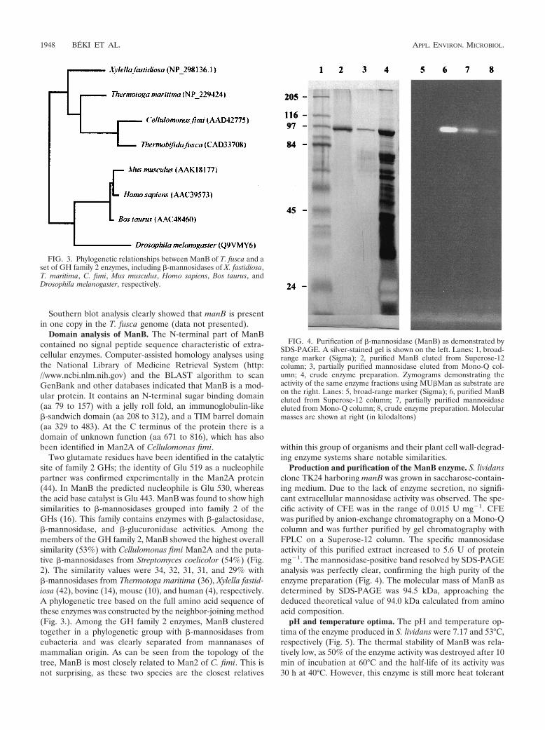
Production and purification of the ManB enzyme. S. lividansclone TK24 harboring manB was grown in saccharose-contain-ing medium. Due to the lack of enzyme secretion, no signifi-cant extracellular mannosidase activity was observed. The spe-cific activity of CFE was in the range of 0.015 U mg�1. CFEwas purified by anion-exchange chromatography on a Mono-Qcolumn and was further purified by gel chromatography withFPLC on a Superose-12 column. The specific mannosidaseactivity of this purified extract increased to 5.6 U of proteinmg�1. The mannosidase-positive band resolved by SDS-PAGEanalysis was perfectly clear, confirming the high purity of theenzyme preparation (Fig. 4). The molecular mass of ManB asdetermined by SDS-PAGE was 94.5 kDa, approaching thededuced theoretical value of 94.0 kDa calculated from aminoacid composition.
pH and temperature optima. The pH and temperature op-tima of the enzyme produced in S. lividans were 7.17 and 53°C,respectively (Fig. 5). The thermal stability of ManB was rela-tively low, as 50% of the enzyme activity was destroyed after 10min of incubation at 60°C and the half-life of its activity was30 h at 40°C. However, this enzyme is still more heat tolerant
FIG. 3. Phylogenetic relationships between ManB of T. fusca and aset of GH family 2 enzymes, including �-mannosidases of X. fastidiosa,T. maritima, C. fimi, Mus musculus, Homo sapiens, Bos taurus, andDrosophila melanogaster, respectively.
FIG. 4. Purification of �-mannosidase (ManB) as demonstrated bySDS-PAGE. A silver-stained gel is shown on the left. Lanes: 1, broad-range marker (Sigma); 2, purified ManB eluted from Superose-12column; 3, partially purified mannosidase eluted from Mono-Q col-umn; 4, crude enzyme preparation. Zymograms demonstrating theactivity of the same enzyme fractions using MU�Man as substrate areon the right. Lanes: 5, broad-range marker (Sigma); 6, purified ManBeluted from Superose-12 column; 7, partially purified mannosidaseeluted from Mono-Q column; 8, crude enzyme preparation. Molecularmasses are shown at right (in kilodaltons)
1948 BEKI ET AL. APPL. ENVIRON. MICROBIOL.
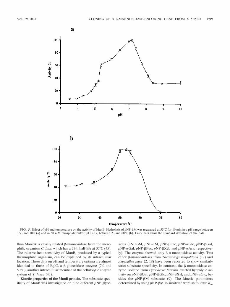
than Man2A, a closely related �-mannosidase from the meso-philic organism C. fimi, which has a 27-h half-life at 37°C (45).The relative heat sensitivity of ManB, produced by a typicalthermophilic organism, can be explained by its intracellularlocation. These data on pH and temperature optima are almostidentical to those of BglC, a �-glucosidase enzyme (7.0 and50°C), another intracellular member of the cellulolytic enzymesystem of T. fusca (43).
Kinetic properties of the ManB protein. The substrate spec-ificity of ManB was investigated on nine different pNP glyco-
sides (pNP-�M, pNP-�M, pNP-�Glc, pNP-�Glc, pNP-�Gal,pNP-�Gal, pNP-�Fuc, pNP-�Xyl, and pNP-�Ara, respective-ly). The enzyme showed only �-D-mannosidase activity. Twoother �-mannosidases from Thermotoga neapolitana (17) andAspergillus niger (2, 18) have been reported to show similarlystrict substrate specificity. In contrast, the �-mannosidase en-zyme isolated from Pyrococcus furiosus exerted hydrolytic ac-tivity on pNP-�Gal, pNP-�Glc, pNP-�Xyl, and pNP-�Glc, be-sides the pNP-�M substrate (9). The kinetic parametersdetermined by using pNP-�M as substrate were as follows: Km
FIG. 5. Effect of pH and temperature on the activity of ManB. Hydrolysis of pNP-�M was measured at 53°C for 10 min in a pH range between3.53 and 10.0 (a) and in 50 mM phosphate buffer, pH 7.17, between 23 and 80°C (b). Error bars show the standard deviation of the data.
VOL. 69, 2003 CLONING OF A �-MANNOSIDASE-ENCODING GENE FROM T. FUSCA 1949
� 180 (12.5) �M and Vmax � 5.96 (0.26) �mol min�1 mg�1,respectively (standard deviations shown in parentheses). TheKm was similar to Km values of other �-D-mannosidases knownfrom different organisms (Table 1.), but it is noteworthy thatManB has the second lowest Km value. The inhibition constantfor mannose is Ki � 5.5 mM. Glucono-lacton had no effect onthe enzyme activity.
Glycosyl transferase activity of ManB. Due to its theoreticaland practical importance we investigated the transglycosylaseactivity of ManB. When different alcohols and pNPMs wereincubated in the presence of ManB the mannosyl moiety of thepNPMs was transferred to these alcohols and alkyl mannosideswere formed. More interestingly, transglycosylase activity wasalso observed, when pNP-�M and pNP-�M were incubated
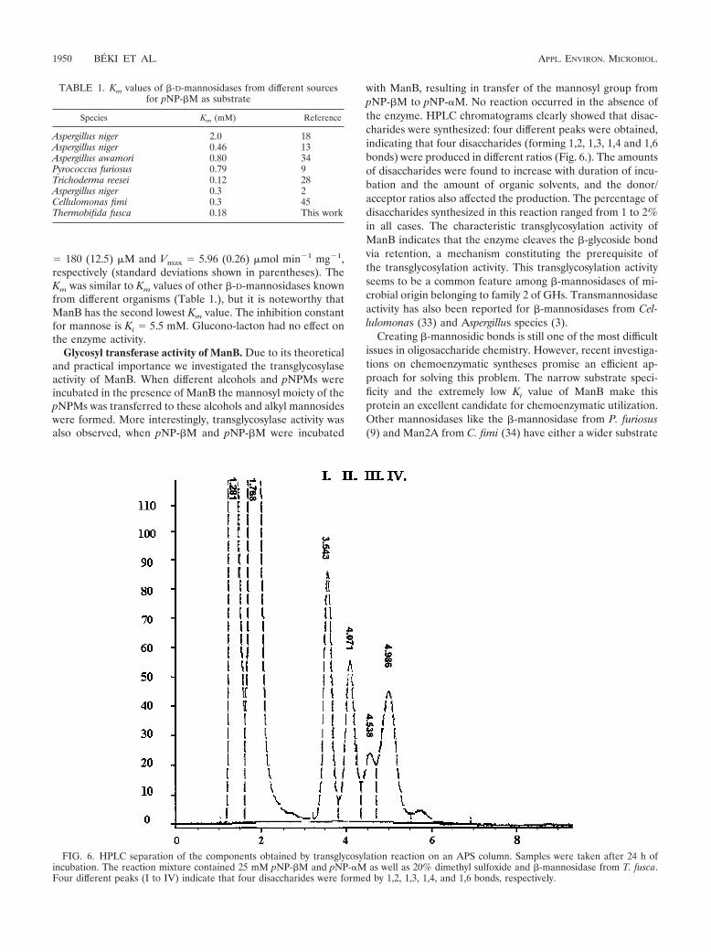
with ManB, resulting in transfer of the mannosyl group frompNP-�M to pNP-�M. No reaction occurred in the absence ofthe enzyme. HPLC chromatograms clearly showed that disac-charides were synthesized: four different peaks were obtained,indicating that four disaccharides (forming 1,2, 1,3, 1,4 and 1,6bonds) were produced in different ratios (Fig. 6.). The amountsof disaccharides were found to increase with duration of incu-bation and the amount of organic solvents, and the donor/acceptor ratios also affected the production. The percentage ofdisaccharides synthesized in this reaction ranged from 1 to 2%in all cases. The characteristic transglycosylation activity ofManB indicates that the enzyme cleaves the �-glycoside bondvia retention, a mechanism constituting the prerequisite ofthe transglycosylation activity. This transglycosylation activityseems to be a common feature among �-mannosidases of mi-crobial origin belonging to family 2 of GHs. Transmannosidaseactivity has also been reported for �-mannosidases from Cel-lulomonas (33) and Aspergillus species (3).
Creating �-mannosidic bonds is still one of the most difficultissues in oligosaccharide chemistry. However, recent investiga-tions on chemoenzymatic syntheses promise an efficient ap-proach for solving this problem. The narrow substrate speci-ficity and the extremely low Ki value of ManB make thisprotein an excellent candidate for chemoenzymatic utilization.Other mannosidases like the �-mannosidase from P. furiosus(9) and Man2A from C. fimi (34) have either a wider substrate
FIG. 6. HPLC separation of the components obtained by transglycosylation reaction on an APS column. Samples were taken after 24 h ofincubation. The reaction mixture contained 25 mM pNP-�M and pNP-�M as well as 20% dimethyl sulfoxide and �-mannosidase from T. fusca.Four different peaks (I to IV) indicate that four disaccharides were formed by 1,2, 1,3, 1,4, and 1,6 bonds, respectively.
TABLE 1. Km values of �-D-mannosidases from different sourcesfor pNP-�M as substrate
Species Km (mM) Reference
Aspergillus niger 2.0 18Aspergillus niger 0.46 13Aspergillus awamori 0.80 34Pyrococcus furiosus 0.79 9Trichoderma reesei 0.12 28Aspergillus niger 0.3 2Cellulomonas fimi 0.3 45Thermobifida fusca 0.18 This work
1950 BEKI ET AL. APPL. ENVIRON. MICROBIOL.
specificity or a higher Ki for mannose. Due to these outstand-ing parameters ManB could be a promising subject of site-directed mutagenesis aiming at modifying its catalytic nucleo-phile to enhance the transmannosidase activity. According torecent reports the glycosynthase product yields have success-fully been improved by this type of mutagenesis (32). Withers(49) produced a mutant Agrobacterium �-glucosidase (Abg)which converted 90% of the monosaccharides into oligosac-charides, demonstrating the efficiency of enzyme engineeringin this field of research.
ManB is the first �-mannosidase enzyme characterized indetail in a representative of the thermophilic aerobic lignocel-lulose-degrading bacteria. The present work revealed one ofthe missing links in the lignocellulose-degrading system ofthese organisms. Proofs of the intracellular location of ManBare especially worthy of mentioning, as this finding points outthat the compost-inhabiting bacteria and fungi follow basicallydifferent strategies for utilizing the final breakdown productsof mannose containing polysaccharides. Bacteria, such as T.fusca or C. fimi, use intracellular enzymes for this purpose toprevent monosaccharide uptake by alien microbes, whereasfungi, such as Trichoderma, may rely on an extracellular man-nosidase, as the strong antagonistic activity exerted by theseorganisms precludes the growth of other microbes in theirmicroenvironment.
ACKNOWLEDGMENTS
This work was supported by grants from OTKA (T 32407), FKFP(0315/99), and IUAP (P5/03).
We thank Gyongyi Gyemant for the HPLC measurements.
REFERENCES
1. Ademark, P., R. P. De Vries, P. Hagglund, H. Stalbrand, and J. Visser. 2001.Cloning and characterization of Aspergillus niger genes encoding an alpha-galactosidase and a beta-mannosidase involved in galactomannan degrada-tion. Eur. J. Biochem. 268:2982–2990.
2. Ademark, P., J. Lundqvist, P. Hagglund, M. Tenkanen, N. Torto, F. Tjer-neld, and H. Stalbrand. 1999. Hydrolytic properties of a �-mannosidasepurified from Aspergillus niger. J. Biotechnol. 75:281–289.
3. Ademark, P., A. Varga, J. Medve, V. Harjunpaa, T. Drakenberg, F. Tjerneld,and H. Stalbrand. 1998. Softwood hemicellulose-degrading enzymes fromAspergillus niger: purification and properties of a beta-mannanase. J. Bio-technol. 63:199–210.
4. Alkhayat, A. H., S. A. Kraemer, J. R. Leipprandt, M. Macek, W. J. Kleijer,and K. H. Friderici. 1998. Human beta-mannosidase cDNA characterizationand first identification of a mutation associated with human beta-mannosido-sis. Hum. Mol. Genet. 7:75–83.
5. Altschul, S. F., T. L. Madden, A. A. Schaffer, J. Zhang, Z. Zhang, W. Miller,and D. J. Lipman. 1997. Gapped BLAST and PSI-BLAST: a new generationof protein database search programs. Nucleic Acids Res. 25:3389–3402.
6. Apweiler, R., T. K. Attwood, A. Bairoch, A. Bateman, E. Birney, M. Biswas,P. Bucher, L. Cerutti, F. Corpet, M. D. R. Croning, R. Durbin, L. Falquet, W.Fleischmann, J. Gouzy, H. Hermjakob, N. Hulo, I. Jonassen, D. Kahn, A.Kanapin, Y. Karavidopoulou, R. Lopez, B. Marx, N. J. Mulder, T. M. Oinn,M. Pagni, F. Servant, C. J. A. Sigrist, and E. M. Zdobnov. 2001. The InterProdatabase, an integrated documentation resource for protein families, do-mains and functional sites. Nucleic Acids Res. 29:37–40.
7. Bacic, A., P. J. Harris, and B. A. Stone. 1988. Structure and function of plantcell walls. p. 279–371. In J. Preiss (ed.), The biochemistry of plants: acomprehensive treatise, vol. 14. Academic Press, New York, N.Y.
8. Barr, B. K., Y. L. Hsieh, B. Ganem, and D. B. Wilson. 1996. Identification oftwo functionally different classes of exocellulases. Biochemistry 35:586–592.
9. Bauer, M. W., E. J. Bylina, R. V. Swanson, and R. M. Kelly. 1996. Compar-ison of a beta-glucosidase and a beta-mannosidase from the hyperthermo-philic archaeon Pyrococcus furiosus. Purification, characterization, gene clon-ing, and sequence analysis. J. Biol. Chem. 271:23749–23755.
10. Beccari, T., L. Bibi, S. Stinchi, J. L. Stirling, and A. Orlacchio. 2001. Mousebeta-mannosidase: cDNA cloning, expression, and chromosomal localiza-tion. Biosci. Rep. 21:315–323.
11. Blanchard, J. E., and S. G. Withers. 2001. Rapid screening of the aglycone
specificity of glycosidases: applications to enzymatic synthesis of oligosac-charides. Chem. Biol. 8:627–633.
12. Blanco, J., J. J. Coque, J. Velasco, and J. F. Martin. 1997. Cloning, expres-sion in Streptomyces lividans and biochemical characterization of a thermo-stable endo-beta-1,4-xylanase of Thermomonospora alba ULJB1 with cellu-lose-binding ability. Appl. Microbiol. Biotechnol. 48:208–217.
13. Bouquelet, S., G. Spik, and J. Montreuil. 1978. Properties of a �-D-manno-sidase from Aspergillus niger. Biochim. Biophys. Acta 522:521–530.
14. Chen, H., J. R. Leipprandt, C. E. Traviss, B. L. Sopher, M. Z. Jones, K. T.Cavanagh, and K. H. Friderici. 1995. Molecular cloning and characterizationof bovine beta-mannosidase. J. Biol. Chem. 270:3841–3848.
15. Clarke, J. H., J. E. Rixon, A. Ciruela, H. J. Gilbert, and G. P. Hazlewood.1997. Family-10 and family-11 xylanases differ in their capacity to enhancethe bleachability of hardwood and softwood paper pulps. Appl. Microbiol.Biotechnol. 48:177–183.
16. Coutinho, P. M., and B. Henrissat. 1999. The modular structure of cellulasesand other carbohydrate-active enzymes: an integrated database approach, p.15–23. In K. Ohmiya, K. Hayashi, K. Sakka, Y. Kobayashi, S. Karita, and T.Kimura (ed.), Genetics, biochemistry and ecology of cellulose degradation.Uni Publishers, Tokyo, Japan.
17. Duffaud, G. D., C. M. McCutchen, P. Leduc, K. N. Parker, and R. M. Kelly.1997. Purification and characterization of extremely thermostable beta-man-nanase, beta-mannosidase, and alpha-galactosidase from the hyperthermo-philic eubacterium Thermotoga neapolitana 5068. Appl. Environ. Microbiol.63:169–177.
18. Elbein, A. D., S. Adya, and Y. C. Lee. 1977. Purification and properties of a�-mannosidase from Aspergillus niger. J. Biol. Chem. 252:2026–2031.
19. Hartree, E. F. 1972. Determination of protein: a modification of the Lowrymethod that gives a linear photometric response. Anal. Biochem. 48:422–427.
20. Hilge, M., S. M. Gloor, W. Rypniewski, O. Sauer, T. D. Heightman, W.Zimmermann, K. Winterhalter, and K. Piontek. 1998. High-resolution na-tive and complex structures of thermostable beta-mannanase from Ther-momonospora fusca—substrate specificity in glycosyl hydrolase family 5.Structure 6:1433–1444.
21. Hunter, I. S. 1985. Gene cloning in Streptomyces, p. 19–44. In D. M. Glover(ed.), DNA cloning: a practical approach. IRL Press, Oxford, United King-dom.
22. Irwin, D., E. D. Jung, and D. B. Wilson. 1994. Characterization and sequenceof a Thermomonospora fusca xylanase. Appl. Environ. Microbiol. 60:763–770.
23. Irwin, D., D. H. Shin, S. Zhang, B. K. Barr, J. Sakon, P. A. Karplus, andD. B. Wilson. 1998. Roles of the catalytic domain and two cellulose bindingdomains of Thermomonospora fusca E4 in cellulose hydrolysis. J. Bacteriol.180:1709–1714.
24. Jung, E. D., G. Lao, D. Irwin, B. K. Barr, A. Benjamin, and D. B. Wilson.1993. DNA sequences and expression in Streptomyces lividans of an exoglu-canase gene and an endoglucanase gene from Thermomonospora fusca.Appl. Environ. Microbiol. 59:3032–3043.
25. Kahle, V., and K. Tesaøik. 1980. Separation of saccharides and their ano-mers by high-performance liquid chromatography. J. Chromatogr. 191:121–128.
26. Kieser, D., and R. D. Melton. 1988. Plasmid pIJ699, a multicopy positiveselection vector for Streptomyces. Gene 65:83–91.
27. Kukolya, J., C. Dobolyi, and L. Hornok. 1997. Isolation and identification ofthermophilic cellulolytic actinomycetes. Acta Phytopathol. Entomol. Hung.32:97–107.
28. Kulminskaya, A. A., E. V. Eneiskaya, L. S. Isaeva-Ivanova, A. N. Savel’ev,I. A. Sidorenko, K. A. Shabalin, A. M. Golubev, and K. N. Neustroev. 1999.Enzymatic activity and �-galactomannan binding property of �-mannosidasefrom Trichoderma reesei. Enzyme Microb. Technol. 25:372–377.
29. Kurakake, M., and T. Komaki. 2001. Production of beta-mannanase andbeta-mannosidase from Aspergillus awamori K4 and their properties. Curr.Microbiol. 42:377–380.
30. Lao, G., G. S. Ghangas, E. D. Jung, and D. B. Wilson. 1991. DNA sequencesof three beta-1,4-endoglucanase genes from Thermomonospora fusca. J. Bac-teriol. 173:3397–3407.
31. Lubomir, K., and B. Peter. 1998. Disaccharides permeases: constituents ofxylanolytic and mannanolytic systems of Aureobasidium pullulans. Biochim.Biophys. Acta 1425:560–566.
32. Moracci, M., A. Trincone, and M. Rossi. 2001. Glycosynthases: new enzymesfor oligosaccharide synthesis. J. Mol. Catal. B Enzyme 11:155–163.
33. Neustroev, K. N., A. S. Krylov, L. M. Firsov, O. L. Abroskina, and A. Y.Khorlin. 1991. Isolation and properties of �-mannosidase from Aspergillusawamori. Biokhimiya 56:1406–1412.
34. Nashiru, O., D. L. Zechel, D. Stoll, T. Mohammadzedeh, A. J. Warren, andS. G. Withers. 2001. �-Mannosynthase: synthesis of �-mannosides with amutant �-mannosidase. Angew. Chem. Int. Ed. 40:417–420.
35. O’Farrell, P. H. 1975. High resolution two-dimensional electrophoresis ofproteins. J. Biol. Chem. 250:4007–4021.
36. Parker, K. N., S. R. Chhabra, D. Lam, W. Callen, G. D. Duffaud, M. A.Snead, J. M. Short, E. J. Mathur, and R. M. Kelly. 2001. Galactomannanases
VOL. 69, 2003 CLONING OF A �-MANNOSIDASE-ENCODING GENE FROM T. FUSCA 1951
Man2 and Man5 from Thermotoga species: growth physiology on galacto-mannans, gene sequence analysis, and biochemical properties of recombi-nant enzymes. Biotechnol. Bioeng. 75:322–333.
37. Sachslehner, A., G. Foidl, N. Foidl, G. Gubitz, and D. Haltrich. 2000. Hy-drolysis of isolated coffee mannan and coffee extract by mannanases ofSclerotium rolfsii. J. Biotechnol. 80:127–134.
38. Saitou, N., and M. Nei. 1987. The neighbor-joining method: a new methodfor reconstructing phylogenetic trees. Mol. Biol. Evol. 4:406–425.
39. Sambrook, J., E. F. Fritsch, and T. Maniatis. 1989. Molecular cloning: alaboratory manual, 2nd ed. Cold Spring Harbor Laboratory, Cold SpringHarbor, N.Y.
40. Schlosser, A., and H. Schrempf. 1996. A lipid-anchored binding protein is acomponent of an ATP-dependent cellobiose/cellotriose-transport systemfrom the cellulose degrader Streptomyces reticuli. Eur. J. Biochem. 242:332–338.
41. Scigelova, M., S. Singh, and D. H. G. Crout. 1999. Glycosidases a greatsynthetic tool. J. Mol. Catal. B Enzyme 6:483–494.
42. Simpson, A. J., F. C. Reinach, P. Arruda, F. A. Abreu, M. Acencio, R.Alvarenga, L. M. Alves, J. E. Araya, G. S. Baia, C. S. Baptista, M. H. Barros,E. D. Bonaccorsi, S. Bordin, J. M. Bove, M. R. Briones, M. R. Bueno, A. A.Camargo, L. E. Camargo, D. M. Carraro, H. Carrer, N. B. Colauto, C.Colombo, F. F. Costa, M. C. Costa, C. M. Costa-Neto, L. L. Coutinho, M.Cristofani, E. Dias-Neto, C. Docena, H. El-Dorry, A. P. Facincani, A. J.Ferreira, V. C. Ferreira, J. A. Ferro, J. S. Fraga, S. C. Franca, M. C. Franco,
M. Frohme, L. R. Furlan, M. Garnier, G. H. Goldman, M. H. Goldman, S. L.Gomes, A. Gruber, P. L. Ho, J. D. Hoheisel, M. L. Junqueira, E. L. Kemper,J. P. Kitajima, C. L. Marino, et al. 2000. The genome sequence of the plantpathogen Xylella fastidiosa. Nature 406:151–157.
43. Spiridonov, N. A., and D. B. Wilson. 2001. Cloning and biochemical char-acterization of BglC, a �-glucosidase from the cellulolytic actinomyceteThermobifida fusca. Curr. Microbiol. 42:295–301.
44. Stoll, D., S. He, S. G. Withers, and R. A. Warren. 2000. Identification ofGlu-519 as the catalytic nucleophile in beta-mannosidase 2A from Cellu-lomonas fimi. Biochem. J. 3:833–838.
45. Stoll, D., H. Stalbrand, and R. A. Warren. 1999. Mannan-degrading enzymesfrom Cellulomonas fimi. Appl. Environ. Microbiol. 65:2598–2605.
46. Takada, G., T. Kawaguchi, T. Kaga, J. Sumitani, and M. Arai. 1999. Cloningand sequencing of beta-mannosidase gene from Aspergillus aculeatus no.F-50. Biosci. Biotechnol. Biochem. 63:206–209.
47. Trigo, C., and A. S. Ball. 1994. Production of extracellular enzymes duringthe solubilization of straw by Thermomonospora fusca BD25. Appl. Micro-biol. Biotechnol. 41:366–372.
48. Warren, R. A. J. 1996. Microbial hydrolysis of polysaccharides. Annu. Rev.Microbiol. 50:183–212.
49. Withers, S. G. 2001. Mechanisms of glycosyl transferases and hydrolases.Carbohydr. Polym. 44:325–337.
50. Wray, W., T. Boulikas, V. P. Wray, and R. Hancock. 1981. Silver staining ofproteins in polyacrylamide gels. Anal. Biochem. 118:197–203.
1952 BEKI ET AL. APPL. ENVIRON. MICROBIOL.