Cirrhotic Patients Are at Risk for Health Care–Associated Bacterial Infections

Endothelin-1 and Transforming Growth Factor-b1Independently Induce Fibroblast Resistance toApoptosis via AKT Activation
Priya Kulasekaran1, Casey A. Scavone1, David S. Rogers1, Douglas A. Arenberg1, Victor J. Thannickal1,and Jeffrey C. Horowitz1
1Department of Medicine, Division of Pulmonary and Critical Care Medicine, University of Michigan Medical Center, Ann Arbor, Michigan
Myofibroblast apoptosis is critical for the normal resolution ofwound repair responses, and impaired myofibroblast apoptosis isassociated with tissue fibrosis. Lung expression of endothelin (ET)-1,a soluble peptide implicated in fibrogenesis, is increased in murinemodels of pulmonary fibrosis and in the lungs of humans withpulmonary fibrosis. Mechanistically, ET-1 has been shown to inducefibroblast proliferation, differentiation, contraction, and collagensynthesis. In this study, we examined the role ET-1 in the regulationof lung fibroblast survival and apoptosis. ET-1 rapidly activatesthe prosurvival phosphatidylinositol 39-OH kinase (PI3K)/AKTsignaling pathway in normal and fibrotic human lung fibroblasts.ET-1–induced activation of PI3K/AKT is dependent on p38 mitogen-activated protein kinase (MAPK), but not extracellular signal-regulated kinase (ERK) 1/2, JNK, or transforming growth factor(TGF)-b1. Activation of the PI3K/AKT pathway by ET-1 inhibitsfibroblast apoptosis, and this inhibition is reversed by blockade ofp38 MAPK or PI3K. TGF-b1 has been shown to attenuate myofibro-blast apoptosis through the p38 MAPK–dependent secretion ofa soluble factor, which activates PI3K/AKT. In this study, we showthat, although TGF-b1 induces fibroblast synthesis and secretion ofET-1, TGF-b1 activation of PI3K/AKT is not dependent on ET-1. Weconclude that ET-1 and TGF-b1 independently promote fibroblastresistance to apoptosis through signaling pathways involving p38MAPK and PI3K/AKT. These findings suggest the potential for noveltherapies targeting the convergence of prosurvival signaling path-ways activated by these two profibrotic mediators.
Keywords: myofibroblast; fibrosis; Fas; p38 mitogen-activated proteinkinase; mesenchymal cells
Endothelin (ET)-1 is a soluble fibrogenic peptide that has beenimplicated in the pathogenesis of pulmonary fibrosis. Secretedas a preproenzyme, active ET-1 is generated through cleavageby ET-converting enzyme. The two Gaq/11-coupled ET recep-tors (ET-A and ET-B) activate phospholipase (PL) C to initiateintracellular signaling events (1). Although ET-1 has beenextensively studied in the pathobiology of pulmonary arterialhypertension, accumulating evidence supports a role for ET-1 inthe pathogenesis of pulmonary fibrosis (2). Animal models showthat increased ET-1 levels precede collagen deposition in thelungs after administration of intratracheal bleomycin, and thattransgenic overexpression of ET-1 is sufficient to induce pro-gressive pulmonary fibrosis (3, 4). Moreover, ET-1 levels areelevated in patients with pulmonary fibrosis from several
underlying etiologies (5). In vitro, ET-1 has profibrotic effectson lung fibroblasts, promoting proliferation (6), differentiation(7), and extracellular matrix (ECM) synthesis (8, 9).
A key feature of pulmonary fibrosis is the accumulation ofmyofibroblasts, contractile mesenchymal cells with a phenotypethat is intermediate between fibroblasts and smooth musclecells, which synthesize, secrete, organize, and remodel the ECM(10). Myofibroblasts are critical effectors of wound-repairresponses, and normal repair requires the ultimate clearanceof myofibroblasts by apoptosis (10, 11). Insufficient myofibro-blast apoptosis is associated with progressive ECM depositionand contraction, leading to tissue fibrosis (12).
We have previously shown that transforming growth factor(TGF)-b1, a cytokine strongly associated with fibrosis in thelung and other organs/tissues, protects myofibroblasts fromapoptosis by p38 mitogen-activated protein kinase (MAPK)–dependent secretion of a soluble factor that activates phospha-tidylinositol 39-OH kinase (PI3K)/AKT signaling (13). ThePI3K/AKT signaling pathway is activated after ligation ofreceptor tyrosine kinases and seven transmembrane G protein–coupled receptors, or through integrin-associated adhesion–mediated signaling cascades (14). Activated AKT may supportcell survival through a number of potential mechanisms, in-cluding regulation of BCL-2 family proteins, NF-kB, caspase-9,and forkhead family transcription factors (14). TGF-b1 hasbeen shown to induce secretion of ET-1 by fibroblasts (15). Thisstudy was undertaken to investigate the role of ET-1 in theregulation of fibroblast fate and to determine if ET-1 mediatesTGF-b1 activation of PI3K/AKT and apoptosis resistance infibroblasts. Portions of this work have been previously pub-lished in abstract form (16).
MATERIALS AND METHODS
Cells and Cell Culture
Normal primary human fetal lung fibroblasts (IMR-90; Institute forMedical Research, Camden, NJ) between passages 7 and 12 were
CLINICAL RELEVANCE
The primary finding of this study is that two fibrogenicgrowth factors, endothelin (ET)-1 and transforming growthfactor (TGF)-b1 independently activate antiapoptotic sig-naling mechanisms in human lung fibroblasts by p38mitogen-activated protein kinase–dependent activation ofphosphatidylinositol 39-OH kinase/AKT. These resultsprovide important insights into the mechanisms regulatingfibroblast survival and apoptosis, with broad implications inthe regulation of lung injury and repair processes. Theconvergence antiapoptotic signaling pathways mediated byET-1 and TGF-b1 may represent novel therapeutic targetsfor the treatment of pulmonary fibrosis.
(Received in original form November 19, 2008 and in final form January 15, 2009)
This work was supported by National Institutes of Health grants K08 HL081059
( J.C.H.) and R01 HL67967 (V.J.T.), the American Lung Association Dalsemer
Award ( J.C.H.), the Entelligence Young Investigator Award ( J.C.H.), and the
ACCP/LUNGevity Foundation (D.A.A.).
Correspondence and requests for reprints should be addressed to Jeffrey C.
Horowitz, M.D., University of Michigan Medical Center, 6301 MSRB III, 1150 W.
Medical Center Dr., Ann Arbor, MI 48109-5642. E-mail: [email protected]
Am J Respir Cell Mol Biol Vol 41. pp 484–493, 2009
Originally Published in Press as DOI: 10.1165/rcmb.2008-0447OC on February 2, 2009
Internet address: www.atsjournals.org
cultured in Dulbecco’s modified Eagle’s medium (DMEM) supple-mented with 100 U/ml penicillin/streptomycin (Sigma, St. Louis, MO),fungizone (Invitrogen, Carlsbad, CA), and 5% FBS (Sigma). IMR-90fibroblasts were incubated in 5% CO2 at 378C to 60–80% confluence,and growth arrested for 24 hours in DMEM with 0.01% FBS beforetreatment.
Primary fibroblast cultures were generated using methods pre-viously described (17). Normal lung fibroblasts were cultured fromhistologically normal portions of lung from patients undergoingsurgical resection of biopsy-proven non–small cell lung cancer. Idio-pathic pulmonary fibrosis (IPF) fibroblasts were obtained via surgicallung biopsy or from lung explants from patients undergoing lungtransplantation. For experiments using normal adult and IPF fibro-blasts, three different normal and three different IPF fibroblast lineswere used. All studies were approved by the Institutional ReviewBoard at the University of Michigan.
Reagents
ET-1 (human, porcine) and cycloheximide were from Sigma. Porcine-derived TGF-b1 and goat polyclonal antibodies to uncleaved andcleaved caspase-3 were from R&D Systems (Minneapolis, MN).SB203580, SP600125, U73122, Wortmannin, LY294002, andSB431542, along with antibodies to serine-473 phospho AKT, totalAKT, phospho-p44/p42 extracellular signal-regulated kinase (ERK)MAPK, phospho p38 MAPK, phospho JNK MAPK, and the corre-sponding total MAPK antibodies were from Cell Signaling Technology(Beverly, MA). Rabbit polyclonal antibodies to glyceraldehyde-3-phosphate dehydrogenase were from Abcam (Cambridge, MA). Sec-ondary horseradish peroxidase–conjugated anti-mouse, anti-goat, andanti-rabbit antibodies were obtained from Pierce (Rockford, IL). Anti-Fas (activating) antibody clone CH11 and mouse monoclonal antibodyto single-stranded DNA (ssDNA) were from Millipore (Billerica, MA).
Western Immunoblotting
Whole-cell lysates were collected in ice-cold RIPA buffer (1% nonidetP-40, 1% sodium deoxycholate, 0.1% SDS, 0.15 M NaCl, 0.01 MNaH2PO4, 2 mM EDTA, 0.5 mM NaF) containing 2 mM sodiumorthovanadate and 1:100 dilution of protease inhibitor mixture III(Calbiochem, San Diego, CA). Protein estimation was done with theBio-Rad DC protein assay (Bio-Rad, Hercules, CA). Whole-celllysates were reduced by mixing with a 1:5 vol/vol ratio of 63
electrophoresis sample buffer (0.2 M EDTA, 40 mM dithiothreitol,6% SDS, 0.06 mg/ml pyronin, pH 6.8) and boiling for 7 minutes. Equalamounts of protein were subjected to SDS-PAGE electrophoresis, aspreviously described (18).
Real-Time PCR
Total RNA was isolated using the TRIzol reagent, according to themanufacturer’s instructions (Invitrogen). A total of 1.0 mg RNA wasreverse transcribed into cDNA using M-MLV reverse transcriptase(Invitrogen). The cDNA was then amplified by real-time quantitativeTaqMan PCR using an ABI Prism 7,700 sequence detection system(Applied Biosystems, Foster City, CA); b-actin was used as an internalcontrol. SYBR Green Master PCR mix (Applied Biosystems) was usedto amplify ET-1, ET-A receptor (ET-A), and ET-B receptor (ET-B).Primers were as follows: (ET-1) forward, 59-GCTCGTCCCTGATGGATAAA-39, reverse, 59-CTGTTGCCTTTGTGGGAAGT-39;(ETAR) forward, 59-GCTTCCTGGTTACCACTCATCAA-39, re-verse, 59-TAGTCTGCTGTGGGCAATAGTTG-39; (ETBR) forward,59-GCCAAGGACCCATCGAGAT-39, reverse, 59-GAAGTGTGGAGTTCCCGATGAT-39; (b-actin) forward, 59-GCCAC GGCTGCTTCCA-39, reverse, 59-GAACCGCTCATTGCCATTG-39. Geneexpression is shown as a fold increase in transcript expression in treatedfibroblasts compared with untreated fibroblasts using the DCt method,per manufacturer’s instructions (Applied Biosystems).
Small Interfering RNA Transfections
RNA interference was accomplished by transfecting cells with ON-TARGET plus SMART pool small interfering RNA (siRNA) for
human ET-1 (EDN1;NM_001955), ET-A (EDNRA;NM_001957), andET-B (EDNRB;NM_003991) from Dharmacon Inc.(Lafayette, CO).Transfections were performed using oligofectamine reagent (Invitro-gen), according to the manufacturer’s instructions. Briefly, for a 35-mmcell culture dish, 5 ml of oligofectamine and 100 nm of siRNA weremixed for 20 minutes at room temperature. A 200-ml aliquot of theoligofectamine/siRNA mixture was then combined with 800 ml ofOpti–Eagle’s minimum essential medium for treatment of the cells.After 24 hours, the media were changed to DMEM with 5% FBS. Cellswere incubated for 48 hours, and growth arrested in serum-freeDMEM for 24 hours before treatment. Nontransfected cells and cellstransfected with a nontargeting pooled siRNA (siControl) (DharmaconInc.) were used as controls.
ET-1 ELISA
Cell culture supernatants collected after treatments were used forquantitative determination of ET-1 levels by EIA kit from AssayDesigns (Ann Arbor, MI), according to the manufacturer’s instruc-tions.
Assessments of Apoptosis
Apoptosis was assessed by Western immunoblotting for cleavedcaspase-3 and ELISA for ssDNA, as previously described (19).
RESULTS
ET-1 Activates PI3K/AKT in Normal Lung Fibroblasts
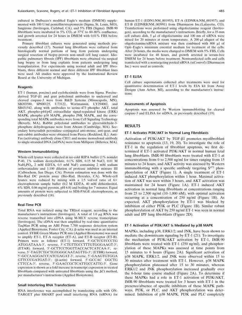
Activation of PI3K/AKT by TGF-b1 promotes myofibroblastresistance to apoptosis (13, 19, 20). To investigate the role ofET-1 in the regulation of fibroblast apoptosis, we first de-termined if ET-1 activated PI3K/AKT in normal human fetallung fibroblasts. IMR-90 fibroblasts were treated with ET-1 atconcentrations from 0 to 2,500 ng/ml for times ranging from 15minutes to 24 hours, and AKT activity was assessed by Westernimmunoblotting with a specific antibody targeting S473 phos-phorylation of AKT (Figure 1). A single treatment of ET-1induced AKT phosphorylation within 1 hour. Maximal activa-tion of AKT was seen within 3 hours, and AKT activation wasmaintained for 24 hours (Figure 1A). ET-1 induced AKTactivation in normal lung fibroblasts at concentrations rangingfrom 25 to 2,500 ng/ml (10–1,000 nM), with optimal activationoccurring at a concentration of 250 ng/ml (Figure 1B). Asexpected, AKT phosphorylation by ET-1 was blocked byinhibition of either PI3K or PLC (Figure 1B). Similar robustphosphorylation of AKT by 250 ng/ml ET-1 was seen in normaladult and IPF lung fibroblasts (Figure 2D).
ET-1 Activation of PI3K/AKT Is Mediated by p38 MAPK
MAPKs, including p38, ERK1/2, and JNK, have been shown tomediate the downstream signaling by ET-1 (21). To investigatethe mechanism of PI3K/AKT activation by ET-1, IMR-90fibroblasts were treated with ET-1 (250 ng/ml), and phosphor-ylation of these MAPKs was assessed at time points from15 minutes to 6 hours (Figure 2A). Significant activation ofp38 MAPK, ERK1/2, and JNK were observed within 15 to30 minutes after treatment with ET-1. However, p38 MAPKphosphorylation plateaued after 15 to 30 minutes, whereasERK1/2 and JNK phosphorylation increased gradually overthe 6-hour time course studied (Figure 2A). To determine ifthese MAPKs had a role in ET-1 activation of PI3K/AKT,IMR-90 fibroblasts were treated for 3 hours with ET-1 in thepresence/absence of specific inhibitors of these MAPK path-ways, PI3K, or PLC, and AKT phosphorylation was deter-mined. Inhibition of p38 MAPK, PI3K and PLC completely
Kulasekaren, Scavone, Rogers, et al.: ET-1 Inhibition of Fibroblast Apoptosis 485

blocked ET-1 activation of AKT (Figure 2B). In contrast,inhibition of ERK1/2 or JNK did not inhibit, and appeared toincrease, ET-1 phosphorylation of AKT. In separate experi-ments, blockade of the type-1 TGF-b receptor (ALK5) had
no significant effect on AKT activation by ET-1 (Figure 2C).Similarly, inhibition of p38 MAPK blocked ET-1 activationof AKT in primary adult normal and IPF lung fibroblasts(Figure 2D).
Figure 1. Endothelin (ET)-1
activation of phosphatidylino-
sitol 39-OH kinase (PI3K)/AKT.Normal human fetal lung fibro-
blasts (IMR-90) were cultured
on tissue culture plates to
80% confluence in Dulbecco’smodified Eagle’s medium
(DMEM) supplemented with
5% FBS before growth arrestfor 24 hours in DMEM with
0.01% FBS. (A) IMR-90 fibro-
blasts were treated with ET-1
(250 ng/ml) for the times in-dicated. (B) IMR-90 fibroblasts
were treated for 3 hours with
the concentration of ET-1
shown, or with 250 ng/ml ET-1 in the presence/absence of inhibitors of PI3K (Wortmannin, 50 nM) or phospholipase (PL) C (U73122, 5 mM). Atthe conclusion of treatment, whole-cell lysates were collected and AKT phosphorylation was assessed by SDS-PAGE immunoelectrophoresis and
Western immunoblotting. Membranes were stripped and probed for total AKT.
Figure 2. AKT phosphorylationby ET-1 requires p38 mitogen-
activated protein kinase (MAPK).
(A) Quiescent IMR-90 fibro-
blasts were treated with ET-1(250 ng/ml) for the time periods
indicated. Whole-cell lysates were
assessed by SDS-PAGE immu-
noelectrophoresis and Westernimmunoblotting for: phospho
p38 MAPK, phospho extracellular
signal-regulated kinase (ERK) 1/2
MAPK, and phospho JNK MAPK.Membranes were stripped and
probed for the corresponding to-
tal MAPK. (B) IMR-90 fibroblastswere treated with ET-1 (250 ng/ml)
for 3 hours in the presence/
absence of inhibitors of p38 MAPK
(SB203580, 6 mM), JNK MAPK(SP600125, 100 nM), ERK1/2
(PD98059, 20 mM), PI3K (Wort-
mannin 50 nM), or PLC (U73122,
5 mM). Whole-cell lysates wereassessed for AKT phosphorylation
by Western immunoblotting, and
the membrane was stripped andprobed for total AKT. (C) IMR-90
fibroblasts were treated with ET-1
(250 ng/ml) in the presence/
absence of SB431542 (1 mM), aninhibitor of the type-1 TGF-b re-
ceptor (ALK5). Cell lysates
obtained at 3 hours were assessed
for AKT phosphorylation by West-ern immunoblotting, and the
membrane was stripped and
probed for total AKT. (D) Primary
normal adult lung fibroblasts (left
panel) and idiopathic pulmonary fibrosis (IPF) fibroblasts (right panel) were treated with ET-1 (250 ng/ml) for 3 hours in the presence/absence ofinhibitors of p38 MAPK (SB203580, 6 mM), PI3K (Wortmannin 50 nM), or PLC (U73122, 5 mM). AKT phosphorylation was assessed by Western
immunoblotting, and membranes were stripped and probed for total AKT. Data represent three normal and three IPF fibroblast cell lines.
486 AMERICAN JOURNAL OF RESPIRATORY CELL AND MOLECULAR BIOLOGY VOL 41 2009
ET-1 Attenuates Fibroblast Apoptosis
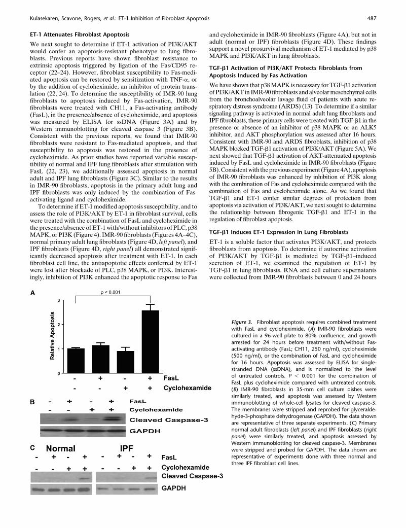
We next sought to determine if ET-1 activation of PI3K/AKTwould confer an apoptosis-resistant phenotype to lung fibro-blasts. Previous reports have shown fibroblast resistance toextrinsic apoptosis triggered by ligation of the Fas/CD95 re-ceptor (22–24). However, fibroblast susceptibility to Fas-medi-ated apoptosis can be restored by sensitization with TNF-a, orby the addition of cycloheximide, an inhibitor of protein trans-lation (22, 24). To determine the susceptibility of IMR-90 lungfibroblasts to apoptosis induced by Fas-activation, IMR-90fibroblasts were treated with CH11, a Fas-activating antibody(FasL), in the presence/absence of cycloheximide, and apoptosiswas measured by ELISA for ssDNA (Figure 3A) and byWestern immunoblotting for cleaved caspase 3 (Figure 3B).Consistent with the previous reports, we found that IMR-90fibroblasts were resistant to Fas-mediated apoptosis, and thatsusceptibility to apoptosis was restored in the presence ofcycloheximide. As prior studies have reported variable suscep-tibility of normal and IPF lung fibroblasts after stimulation withFasL (22, 23), we additionally assessed apoptosis in normaladult and IPF lung fibroblasts (Figure 3C). Similar to the resultsin IMR-90 fibroblasts, apoptosis in the primary adult lung andIPF fibroblasts was only induced by the combination of Fas-activating ligand and cycloheximide.
To determine if ET-1 modified apoptosis susceptibility, and toassess the role of PI3K/AKT by ET-1 in fibroblast survival, cellswere treated with the combination of FasL and cycloheximide inthe presence/absence of ET-1 with/without inhibitors of PLC, p38MAPK, or PI3K (Figure 4). IMR-90 fibroblasts (Figures 4A–4C),normal primary adult lung fibroblasts (Figure 4D, left panel), andIPF fibroblasts (Figure 4D, right panel) all demonstrated signif-icantly decreased apoptosis after treatment with ET-1. In eachfibroblast cell line, the antiapoptotic effects conferred by ET-1were lost after blockade of PLC, p38 MAPK, or PI3K. Interest-ingly, inhibition of PI3K enhanced the apoptotic response to Fas
and cycloheximide in IMR-90 fibroblasts (Figure 4A), but not inadult (normal or IPF) fibroblasts (Figure 4D). These findingssupport a novel prosurvival mechanism of ET-1 mediated by p38MAPK and PI3K/AKT in lung fibroblasts.
TGF-b1 Activation of PI3K/AKT Protects Fibroblasts from
Apoptosis Induced by Fas Activation
We have shown that p38 MAPK is necessary for TGF-b1 activationof PI3K/AKT in IMR-90 fibroblasts and alveolar mesenchymal cellsfrom the bronchoalveolar lavage fluid of patients with acute re-spiratory distress syndrome (ARDS) (13). To determine if a similarsignaling pathway is activated in normal adult lung fibroblasts andIPF fibroblasts, these primary cells were treated with TGF-b1 in thepresence or absence of an inhibitor of p38 MAPK or an ALK5inhibitor, and AKT phosphorylation was assessed after 16 hours.Consistent with IMR-90 and ARDS fibroblasts, inhibition of p38MAPK blocked TGF-b1 activation of PI3K/AKT (Figure 5A). Wenext showed that TGF-b1 activation of AKT-attenuated apoptosisinduced by FasL and cycloheximide in IMR-90 fibroblasts (Figure5B). Consistent with the previous experiment (Figure 4A), apoptosisof IMR-90 fibroblasts was enhanced by inhibition of PI3K alongwith the combination of Fas and cycloheximide compared with thecombination of Fas and cycloheximide alone. As we found thatTGF-b1 and ET-1 confer similar degrees of protection fromapoptosis via activation of PI3K/AKT, we next sought to determinethe relationship between fibrogenic TGF-b1 and ET-1 in theregulation of fibroblast apoptosis.
TGF-b1 Induces ET-1 Expression in Lung Fibroblasts
ET-1 is a soluble factor that activates PI3K/AKT, and protectsfibroblasts from apoptosis. To determine if autocrine activationof PI3K/AKT by TGF-b1 is mediated by TGF-b1–inducedsecretion of ET-1, we examined the regulation of ET-1 byTGF-b1 in lung fibroblasts. RNA and cell culture supernatantswere collected from IMR-90 fibroblasts between 0 and 24 hours
Figure 3. Fibroblast apoptosis requires combined treatmentwith FasL and cycloheximide. (A) IMR-90 fibroblasts were
cultured in a 96-well plate to 80% confluence, and growth
arrested for 24 hours before treatment with/without Fas-activating antibody (FasL; CH11, 250 ng/ml), cycloheximide
(500 ng/ml), or the combination of FasL and cycloheximide
for 16 hours. Apoptosis was assessed by ELISA for single-
stranded DNA (ssDNA), and is normalized to the levelof untreated controls. P , 0.001 for the combination of
FasL plus cycloheximide compared with untreated controls.
(B) IMR-90 fibroblasts in 35-mm cell culture dishes were
similarly treated, and apoptosis was assessed by Westernimmunoblotting of whole-cell lysates for cleaved caspase-3.
The membranes were stripped and reprobed for glyceralde-
hyde-3-phosphate dehydrogenase (GAPDH). The data shown
are representative of three separate experiments. (C) Primarynormal adult fibroblasts (left panel) and IPF fibroblasts (right
panel) were similarly treated, and apoptosis assessed by
Western immunoblotting for cleaved caspase-3. Membraneswere stripped and probed for GAPDH. The data shown are
representative of experiments done with three normal and
three IPF fibroblast cell lines.
Kulasekaren, Scavone, Rogers, et al.: ET-1 Inhibition of Fibroblast Apoptosis 487

after treatment with TGF-b1. Quantitative real-time PCRshowed that TGF-b1 strongly up-regulates ET-1 transcription,with maximal mRNA expression noted between 6 and 16 hours(Figure 6A). Basal levels of active ET-1 in cell culture super-natants were undetectable, and stimulation with TGF-b1 led toa small, but significant, increase in active ET-1 (Figure 6B).
Similar induction of ET-1 by TGF-b1 was found in normal adultand IPF lung fibroblasts (Figure 7C).
TGF-b1 Activation of PI3K/AKT Is Not Mediated by ET-1
TGF-b1 activation of PI3K/AKT requires the p38 MAPK–dependent secretion of a soluble factor (13). Thus, if ET-1
Figure 4. ET-1–mediated protection from apoptosis is dependent on p38 MAPK and PI3K/AKT. (A–C) IMR-90 fibroblasts in a 96-well plate were
treated with the combination of FasL (250 ng/ml) and cycloheximide (500 ng/ml) in the presence/absence of ET-1 (250 ng/ml) with/withoutinhibitors of PLC (U73122, 5 mM), p38 MAPK (SB203580, 6 mM), or PI3K (Wortmannin, 50 nM) for 16 hours. Apoptosis was assessed by (A) ELISA
for ssDNA (four replicates per condition and three separate experiments) and (B) Western immunoblotting for cleaved caspase-3. The membrane in
(B) was stripped and probed for GAPDH. (C) Densitometric analysis of cleaved caspase-3/GAPDH ratios from three independent experiments. (D)Similarly treated primary normal lung fibroblasts (left panel) and IPF fibroblasts (right panel) were assessed for apoptosis by ELISA for ssDNA (n 5 3
normal lung fibroblast lines and 3 IPF fibroblast lines).
488 AMERICAN JOURNAL OF RESPIRATORY CELL AND MOLECULAR BIOLOGY VOL 41 2009

mediates TGF-b1 activation of PI3K/AKT, it should be regu-lated by TGF-b1 in a p38 MAPK–dependent manner. To definethe role of ET-1 in TGF-b1 activation of PI3K/AKT, we treatedfibroblasts with TGF-b1 in the presence or absence of a p38MAPK inhibitor (Figure 7). Surprisingly, inhibition of p38MAPK had no significant effect on TGF-b1 induction of ET-1transcription at 6 hours (Figure 7A), or on the secretion of
active ET-1 at 16 hours (Figure 7B). Moreover, inhibition ofp38 MAPK did not impact TGF-b1 induction of ET-1 in normaladult or IPF-lung fibroblasts (Figure 7C). As inhibition of p38MAPK failed to block TGF-b1–induced synthesis or secretionof ET-1, these data suggest that ET-1 was not the soluble factordirectly mediating TGF-b1 activation of PI3K/AKT.
To confirm that TGF-b1 activation of PI3K/AKT was in-dependent of ET-1, we employed siRNA to knock down ET-1or its receptors, ET-A and ET-B, and examined the impact onTGF-b1 activation of AKT. Knockdown of ET-1 or its recep-tors was confirmed by real-time PCR 72 hours after siRNAtransfection of IMR-90 fibroblasts compared with the expres-sion of cells transfected with a nontargeting (control) siRNA(Figure 8A). Transfected fibroblasts were then treated with/without TGF-b1 for 16 hours, and AKT activation was assessed.As seen in Figure 8B, AKT phosphorylation by TGF-b1 was notaffected by siRNA knockdown of ET-1, ET-A, or ET-B,confirming that neither ET-1 nor its receptors are required forTGF-b1 activation of PI3K/AKT.
DISCUSSION
TGF-b1 and ET-1 have been implicated in the pathogenesis ofpulmonary fibrosis. Both cytokines have been shown to modu-late pulmonary fibrosis in murine models, and both have beenshown to be highly expressed in the lungs of patients withpulmonary fibrosis (4, 25–29). Moreover, both of these media-tors induce fibroblast differentiation, ECM synthesis, andcollagen gel contraction (7, 10). TGF-b1 has been shown topromote myofibroblast resistance to apoptosis (13, 19, 20, 30,31), but few studies have addressed the effects of ET-1 on thisprofibrotic fibroblast phenotype. This study was undertaken toexamine the effects of ET-1 on fibroblast survival signaling andapoptosis.
First, we examined the mechanism of ET-1 activation ofPI3K/AKT in normal and IPF lung fibroblasts. Next, weinvestigated the role of PI3K/AKT activation by ET-1 in the
Figure 5. TGF-b1 activation of PI3K/AKT is de-pendent on p38 MAPK and protects fibroblasts
from apoptosis induced by FasL and cyclohex-
imide. (A) Primary normal adult lung fibroblasts(left panel) and IPF fibroblasts (right panel) were
treated with TGF-b1 (2 ng/ml) in the presence/
absence of inhibitors of p38 MAPK (SB203580,
6 mM) or ALK5 (SB431542, 1 mM) for 16 hours.AKT phosphorylation was assessed by Western
immunoblotting. Membranes were stripped
and probed for total AKT. Data shown represent
three normal and three IPF fibroblast cell lines.(B) IMR-90 fibroblasts were treated with
the combination of FasL (250 ng/ml) and cy-
cloheximide (500 ng/ml) with/without TGF-b1(2 ng/ml) alone or with an inhibitor of PI3K
(LY294002, 10 mM) for 16 hours. Apoptosis was
assessed by ELISA for ssDNA, and is normalized
to the untreated control.
Figure 6. ET-1 expression is increased by TGF-b1 treatment. Quiescent
IMR-90 fibroblasts were treated with TGF-b1 (2 ng/ml) for the times
indicated. (A) RNA was isolated and ET-1 expression was assessed by
quantitative real-time PCR (n 5 3). (B) Active ET-1 in the cell culturesupernatants was assessed by ELISA (n 5 3). *P , 0.001 compared with
untreated controls.
Kulasekaren, Scavone, Rogers, et al.: ET-1 Inhibition of Fibroblast Apoptosis 489

regulation of fibroblast survival/apoptosis. Finally, we sought todetermine if ET-1 functioned as a downstream mediator ofTGF-b1 in the regulation of fibroblast survival, or if ET-1functioned in a parallel, independent manner. Our primaryfindings are that ET-1 robustly activates PI3K/AKT in a p38MAPK–dependent manner, and that ET-1 activation of PI3K/AKT confers apoptosis resistance to fibroblasts. In addition,although TGF-b1 induces ET-1 expression in fibroblasts, ET-1is not required for TGF-b1 activation of PI3K/AKT. Collec-tively, these studies establish that TGF-b1 and ET-1 indepen-dently promote fibroblast survival through p38 MAPK–dependent activation of PI3K/AKT.
Prevailing hypotheses pose that pulmonary fibrosis resultsfrom dysfunctional repair after lung injury from known causes, asin scleroderma and ARDS, or unknown causes, as in IPF (32).Regardless of etiology, pulmonary fibrosis is characterized by
myofibroblast accumulation and excessive deposition of ECM.After injury, recruited fibroblasts differentiate into myofibro-blasts, which synthesize, secrete, organize, and contract the ECM(10, 33). The fate of recruited fibroblasts may be a criticaldeterminant of the outcome of wound repair, as normal resolu-tion requires the apoptotic clearance of myofibroblasts, whereasfibrosis is associated with insufficient myofibroblast apoptosis (11,12, 33, 34). Although the current study was not designed toevaluate differences in the apoptosis susceptibility of normal andfibrotic fibroblasts, several studies have found that fibroblastsisolated from the patients with pulmonary fibrosis of differentetiologies have increased resistance to apoptosis compared withnormal lung fibroblasts (20, 22, 23, 35).
The physiologic stimuli of myofibroblast apoptosis during theresolution phase of wound repair have not been clearly defined.Consistent with several prior studies, we found that lung fibro-blasts demonstrate resistance to extrinsic apoptosis induced byligation of the membrane-bound death receptor, Fas/CD95 (22,24, 35). In contrast, one study demonstrated fibroblast apoptosissusceptibility to Fas activation, but used a significantly higherconcentration of Fas-activating ligand (23). Our study demon-strates fibroblast resistance to apoptosis induced by Fas, which isovercome by cotreatment with cycloheximide, an inhibitor ofprotein translation. The mechanism of fibroblast resistance toFas-mediated apoptosis remains poorly defined, and severalmechanisms have been proposed. Tanaka and colleagues (24)found that resistance to Fas-mediated apoptosis was due toincreased constitutive expression of inhibitors of apoptosis, andthat cycloheximide blocked that constitutive expression, allowingapoptosis to proceed. Frankel and colleagues (22) showed thatTNF-a facilitates recruitment of the Fas-associated death domainto the cytoplasmic domain of Fas, thereby promoting apoptosis.
The regulation of fibroblast survival and apoptosis in lunghomeostasis and disease remains poorly understood, but evi-dence supports a significant role for PI3K/AKT signaling. TGF-b1 activation of PI3K/AKT, and subsequent myofibroblastresistance to apoptosis, is mediated by the p38 MAPK–de-pendent, SMAD-independent secretion of a putative solublefactor (13, 19). Focal adhesion kinase, a nonreceptor tyrosinekinase that is activated by TGF-b1, has also been shown toinduce apoptosis resistance in fibroblasts through activation ofPI3K/AKT (18, 36). Murine models show that phosphorylatedAKT is strongly expressed in areas of pulmonary fibrosis afterintratracheal administration of bleomycin, and that blockade ofPI3K/AKT signaling attenuates pulmonary fibrosis induced byintratracheal bleomycin or TGF-b1 overexpression (37, 38).Finally, increased AKT activation in primary lung mesenchymalcells is associated with resistance to apoptosis and a clinicalcourse of persistent ARDS (20).
In this study, we show that ET-1 inhibits fibroblast apoptosisthrough activation of PI3K/AKT, suggesting a novel fibrogenicmechanism of ET-1. ET-1 has been shown to inhibit cancer cellapoptosis by activation of PI3K/AKT (39). However, no priorstudies have examined the role of ET-1 activation of PI3K/AKTin lung fibroblast survival. One study reported that PI3K/AKTregulates ET-1–induced fibroblast differentiation and contrac-tion (7). Another study recently showed that fibroblasts lackingThy-1 have decreased apoptosis in contractile collagen gels aftertreatment with ET-1 when compared with ET-1–treated fibro-blasts expressing Thy-1 (40). Our studies show that ET-1protects normal fetal, normal adult, and IPF lung fibroblastsfrom apoptosis through activation of PI3K/AKT. Interestingly,the current study suggests that blockade of PI3K enhances Fas-mediated apoptosis in fetal, but not adult, lung fibroblasts(Figures 4A, 4D, and 5B). The mechanism of enhancedapoptosis is unclear, but these data suggest that constitutive
Figure 7. ET-1 induction by TGF-b1 is not dependent on p38 MAPK.(A and B) IMR-90 fibroblasts were treated with TGF-b1 (2 ng/ml) in the
presence/absence of a p38 MAPK inhibitor (SB203580, 6 mM). (A) RNA
was collected at 6 hours, and ET-1 expression was assessed by
quantitative real-time PCR. (B) Cell culture supernatants were collectedat 16 hours and ET-1 levels assessed by ELISA (n 5 3) for (A) and (B).
*P , 0.001 compared with untreated controls. (C) Primary normal
adult fibroblasts (open bars) and IPF fibroblasts (closed bars) were
treated with TGF-b1 (2 ng/ml) with/without a p38 MAPK inhibitor(SB203580, 6 mM) for 16 hours and soluble ET-1 was assessed by ELISA.
n 5 3 normal and 3 IPF fibroblast lines. *P , 0.001 compared with
untreated controls).
490 AMERICAN JOURNAL OF RESPIRATORY CELL AND MOLECULAR BIOLOGY VOL 41 2009

expression of PI3K-mediated prosurvival signals contributes toapoptosis resistance in fetal lung fibroblasts.
In concordance with previous reports, we found that ET-1activates p38, ERK1/2, and JNK MAPKs (21, 41). Early activa-tion of p38 MAPK is required for ET-1 activation of PI3K/AKT,whereas blockade of ERK1/2 or JNK fails to inhibit ET-1activation of PI3K/AKT. Consistently, blockade of p38 MAPKreversed the antiapoptotic effects of ET-1. This study is the firstto demonstrate that p38 MAPK is required for ET-1 activation ofPI3K/AKT and acquisition of apoptosis resistance in mesenchy-mal cells. Collectively, these findings support a novel mechanismby which ET-1 contributes to the pathobiology of pulmonaryfibrosis.
TGF-b1 is associated with fibrosis of most organ systemsstudied, including the lung (42). Among its fibrogenic actions,TGF-b1 induces fibroblast resistance to apoptosis through p38MAPK–dependent secretion of a soluble factor that activatesPI3K/AKT (13). In normal and scleroderma lung fibroblasts,autocrine secretion of ET-1 by TGF-b1 has been found to pro-mote fibroblast differentiation, collagen gel contraction, andECM synthesis (15, 43). In the current study, we show thatET-1 confers protection from apoptosis in fibroblasts throughactivation of PI3K/AKT, and we questioned if autocrine in-duction of ET-1 mediates TGF-b1 activation of PI3K/AKT.Contrary to our original hypothesis, our studies show that TGF-b1 activation of PI3K/AKT is not mediated by ET-1. Althoughwe found that TGF-b1 does induce expression of ET-1 at the geneand protein levels, the absolute increase in soluble, active ET-1 isseveral orders of magnitude lower than the concentrations re-quired for in vitro activation of AKT in fibroblasts. The observedconcentrations of active ET-1 are consistent with those reportedin scleroderma fibroblasts exposed to TGF-b (7, 15). Similarconcentrations of ET-1 were found in the epithelial lining fluid ofpatients with ARDS and bronchoalveolar lavage fluid of patientswith interstitial lung disease (44–46). Although the concentrationof ET-1 secreted by mesenchymal cells stimulated with TGF-b1in vitro approximates these levels reported in vivo, the effectiveconcentration of ET-1 within fibrotic tissue remains unclear, andmay be markedly higher within the pericellular microenvironment.
Our studies indicate that TGF-b1 induction of ET-1 isnot inhibited by blockade of p38 MAPK, and that siRNA-mediated knockdown of ET-1 or its receptors (ET-A or ET-B)has no significant effect on TGF-b1 activation of PI3K/AKT.Thus, although ET-1 and TGF-b1 use the same signaling inter-mediates—p38 MAPK and PI3K/AKT—they independentlypromote fibroblast survival. Additionally, although the biologic
source of ET-1 in patients with IPF has not been defined, ourstudies suggest that ET-1 secreted by epithelial, endothelial, orinflammatory cells may activate fibroblast survival signaling ina paracrine manner (27, 47).
Effective pharmacologic therapies for pulmonary fibrosis arelacking, and current concepts favor therapies targeting fibro-genic mechanisms, including inhibition of TGF-b1 and ET-1(32, 48). Studies with ET receptor antagonists in murine modelsof pulmonary fibrosis have generated conflicting results (49, 50).In one study, a dual ET receptor antagonist reduced the accu-mulation of connective tissue in mouse lungs 28 days afterintratracheal bleomycin (50). A different dual ET receptorantagonist, however, had no significant effect on lung collagencontent in rats at 14 or 21 days after bleomycin administration(49). Recently, a clinical trial of a dual ET receptor antagonistin IPF failed to meet the primary efficacy endpoints; however,patients in the treatment group demonstrated a trend towarddelayed death and disease progression (51). Further clinicaltrials of ET receptor antagonists for IPF are ongoing (clinical-trials.gov: NCT00768300 and NCT00391443). Additionally, on-going clinical trials are investigating anti-TGF-b1 monoclonalantibodies in IPF (clinicaltrials.gov: NCT00125385). The cur-rent study shows that these two fibrogenic mediators—ET-1 andTGF-b1—use the same intracellular signaling intermediates toindependently induce an apoptosis-resistant fibroblast pheno-type. These findings support the hypothesis that the acquisitionof a profibrotic mesenchymal cell phenotype, in the context ofpulmonary fibrosis, may be the result of several mediatorsacting alone or in combination (52). It remains to be seen ifindividuals with pulmonary fibrosis have disease states domi-nated by a single mediator (i.e., TGF-b1 or ET-1), or if severalmediators function in an additive or synergistic manner toinduce fibrosis. Our data suggest that therapy targeting theconvergence of downstream signaling pathways common tofibrogenic mediators, rather than targeting a single mediator,may provide improved clinical efficacy for the treatment ofpulmonary fibrosis.
Conflict of Interest Statement: None of the authors has a financial relationshipwith a commercial entity that has an interest in the subject of this manuscript.
References
1. Miyauchi T, Masaki T. Pathophysiology of endothelin in the cardio-vascular system. Annu Rev Physiol 1999;61:391–415.
2. Denton CP, Black CM, Abraham DJ. Mechanisms and consequences offibrosis in systemic sclerosis. Nat Clin Pract Rheumatol 2006;2:134–144.
Figure 8. TGF-b1 activation of PI3K/AKT is inde-
pendent of ET-1. IMR-90 fibroblasts were trans-
fected with pooled small interfering RNA (siRNA)
targeting ET-1, the ET-A receptor, the ET-B recep-tor, or with a nontargeting (NT) siRNA. (A) At 72
hours after transfection, RNA was isolated from the
transfected cells for quantitative real-time PCR for
ET-1, ET-A, or ET-B to demonstrate that there wassignificant knockdown of the targeted mRNA. (B)
Similarly transfected IMR-90 fibroblasts, or non-
transfected fibroblasts, were treated with/withoutTGF-b1 (2 ng/ml) for 16 hours, and AKT phosphor-
ylation was assessed by Western immunoblotting.
Membranes were stripped and probed for total
AKT. Results are representative of three separateexperiments.
Kulasekaren, Scavone, Rogers, et al.: ET-1 Inhibition of Fibroblast Apoptosis 491
3. Hocher B, Schwarz A, Fagan KA, Thone-Reineke C, El-Hag K,
Kusserow H, Elitok S, Bauer C, Neumayer HH, Rodman DM, et al.Pulmonary fibrosis and chronic lung inflammation in ET-1 transgenicmice. Am J Respir Cell Mol Biol 2000;23:19–26.
4. Mutsaers SE, Foster ML, Chambers RC, Laurent GJ, McAnulty RJ.
Increased endothelin-1 and its localization during the development ofbleomycin-induced pulmonary fibrosis in rats. Am J Respir Cell MolBiol 1998;18:611–619.
5. Antoniu SA. Targeting the endothelin pathway in the idiopathic
pulmonary fibrosis: the role of bosentan. Expert Opin Ther Targets2008;12:1077–1084.
6. Shahar I, Fireman E, Topilsky M, Grief J, Schwarz Y, Kivity S, Ben-
Efraim S, Spirer Z. Effect of endothelin-1 on alpha-smooth muscleactin expression and on alveolar fibroblasts proliferation in interstitiallung diseases. Int J Immunopharmacol 1999;21:759–775.
7. Shi-Wen X, Chen Y, Denton CP, Eastwood M, Renzoni EA, Bou-
Gharios G, Pearson JD, Dashwood M, du Bois RM, Black CM, et al.Endothelin-1 Promotes myofibroblast induction through the ETAreceptor via a Rac/phosphoinositide 3-kinase/Akt-dependent path-way and is essential for the enhanced contractile phenotype of fibroticfibroblasts. Mol Biol Cell 2004;15:2707–2719.
8. Shi-Wen X, Denton CP, Dashwood MR, Holmes AM, Bou-Gharios G,
Pearson JD, Black CM, Abraham DJ. Fibroblast matrix geneexpression and connective tissue remodeling: role of endothelin-1.J Invest Dermatol 2001;116:417–425.
9. Xu SW, Howat SL, Renzoni EA, Holmes A, Pearson JD, Dashwood
MR, Bou-Gharios G, Denton CP, du Bois RM, Black CM, et al.Endothelin-1 induces expression of matrix-associated genes in lungfibroblasts through MEK/ERK. J Biol Chem 2004;279:23098–23103.
10. Hinz B, Phan SH, Thannickal VJ, Galli A, Bochaton-Piallat ML,
Gabbiani G. The myofibroblast: one function, multiple origins. AmJ Pathol 2007;170:1807–1816.
11. Desmouliere A, Redard M, Darby I, Gabbiani G. Apoptosis mediates
the decrease in cellularity during the transition between granulationtissue and scar. Am J Pathol 1995;146:56–66.
12. Thannickal VJ, Horowitz JC. Evolving concepts of apoptosis in idio-
pathic pulmonary fibrosis. Proc Am Thorac Soc 2006;3:350–356.13. Horowitz JC, Lee DY, Waghray M, Keshamouni VG, Thomas PE,
Zhang H, Cui Z, Thannickal VJ. Activation of the pro-survivalphosphatidylinositol 3-kinase/Akt pathway by transforming growthfactor-beta1 in mesenchymal cells is mediated by p38 MAPK–dependent induction of an autocrine growth factor. J Biol Chem2004;279:1359–1367.
14. Datta SR, Brunet A, Greenberg ME. Cellular survival: a play in three
Akts. Genes Dev 1999;13:2905–2927.15. Shi-Wen X, Rodriguez-Pascual F, Lamas S, Holmes A, Howat S,
Pearson JD, Dashwood MR, du Bois RM, Denton CP, Black CM,et al. Constitutive Alk5-independent C-Jun N-terminal kinase activa-tion contributes to endothelin-1 overexpression in pulmonary fibrosis:evidence of an autocrine endothelin loop operating through theendothelin A and B receptors. Mol Cell Biol 2006;26:5518–5527.
16. Horowitz JC, Kulasekaran P, Rogers DS, Thannickal VJ. Endothelin-1
mediates TGF-B1 activation of Akt and resistance to apoptosis inlung fibroblasts [abstract]. Proc Am Thorac Soc 2008;177:A730.
17. Keane MP, Arenberg DA, Lynch JP III, Whyte RI, Iannettoni MD,
Burdick MD, Wilke CA, Morris SB, Glass MC, DiGiovine B, et al.The CXC chemokines, IL-8 and IP-10, regulate angiogenic activity inidiopathic pulmonary fibrosis. J Immunol 1997;159:1437–1443.
18. Thannickal VJ, Lee DY, White ES, Cui Z, Larios JM, Chacon R,
Horowitz JC, Day RM, Thomas PE. Myofibroblast differentiation bytransforming growth factor-beta1 is dependent on cell adhesion andintegrin signaling via focal adhesion kinase. J Biol Chem 2003;278:12384–12389.
19. Horowitz JC, Rogers DS, Sharma V, Vittal R, White ES, Cui Z,
Thannickal VJ. Combinatorial activation of Fak and Akt by trans-forming growth factor-beta1 confers an anoikis-resistant phenotype tomyofibroblasts. Cell Signal 2007;19:761–771.
20. Horowitz JC, Cui Z, Moore TA, Meier TR, Reddy RC, Toews GB,
Standiford TJ, Thannickal VJ. Constitutive activation of prosurvivalsignaling in alveolar mesenchymal cells isolated from patients withnonresolving acute respiratory distress syndrome. Am J Physiol LungCell Mol Physiol 2006;290:L415–L425.
21. Choukroun G, Hajjar R, Kyriakis JM, Bonventre JV, Rosenzweig A,
Force T. Role of the stress-activated protein kinases in endothelin-induced cardiomyocyte hypertrophy. J Clin Invest 1998;102:1311–1320.
22. Frankel SK, Cosgrove GP, Cha SI, Cool CD, Wynes MW, Edelman BL,
Brown KK, Riches DW. TNF-a sensitizes normal and fibrotic humanlung fibroblasts to Fas-induced apoptosis. Am J Respir Cell Mol Biol2006;34:293–304.
23. Moodley YP, Caterina P, Scaffidi AK, Misso NL, Papadimitriou JM,
McAnulty RJ, Laurent GJ, Thompson PJ, Knight DA. Comparison ofthe morphological and biochemical changes in normal human lungfibroblasts and fibroblasts derived from lungs of patients withidiopathic pulmonary fibrosis during Fasl-induced apoptosis. J Pathol2004;202:486–495.
24. Tanaka T, Yoshimi M, Maeyama T, Hagimoto N, Kuwano K, Hara N.
Resistance to Fas-mediated apoptosis in human lung fibroblast. EurRespir J 2002;20:359–368.
25. Wendel M, Petzold A, Koslowski R, Kasper M, Augstein A, Knels L,
Bleyl JU, Koch T. Localization of endothelin receptors in bleomycin-induced pulmonary fibrosis in the rat. Histochem Cell Biol 2004;122:507–517.
26. Sime PJ, Xing Z, Graham FL, Csaky KG, Gauldie J. Adenovector-
mediated gene transfer of active transforming growth factor-beta1induces prolonged severe fibrosis in rat lung. J Clin Invest 1997;100:768–776.
27. Abraham DJ, Vancheeswaran R, Dashwood MR, Rajkumar VS,
Pantelides P, Xu SW, du Bois RM, Black CM. Increased levels ofendothelin-1 and differential endothelin type A and B receptorexpression in scleroderma-associated fibrotic lung disease. Am JPathol 1997;151:831–841.
28. Cambrey AD, Harrison NK, Dawes KE, Southcott AM, Black CM, du
Bois RM, Laurent GJ, McAnulty RJ. Increased levels of endothelin-1in bronchoalveolar lavage fluid from patients with systemic sclerosiscontribute to fibroblast mitogenic activity in vitro. Am J Respir CellMol Biol 1994;11:439–445.
29. Khalil N, O’Connor RN, Flanders KC, Unruh H. TGF-b1, but not TGF-
b2 or TGF-b3, is differentially present in epithelial cells of advancedpulmonary fibrosis: an immunohistochemical study. Am J Respir CellMol Biol 1996;14:131–138.
30. Horowitz JC, Rogers DS, Simon RH, Sisson TH, Thannickal VJ.
Plasminogen activation–induced pericellular fibronectin proteolysispromotes fibroblast apoptosis. Am J Respir Cell Mol Biol 2008;38:78–87.
31. Zhang HY, Phan SH. Inhibition of myofibroblast apoptosis by trans-
forming growth factor b1. Am J Respir Cell Mol Biol 1999;21:658–665.32. Horowitz JC, Thannickal VJ. Idiopathic pulmonary fibrosis: new con-
cepts in pathogenesis and implications for drug therapy. Treat RespirMed 2006;5:325–342.
33. Tomasek JJ, Gabbiani G, Hinz B, Chaponnier C, Brown RA. Myo-
fibroblasts and mechano-regulation of connective tissue remodelling.Nat Rev Mol Cell Biol 2002;3:349–363.
34. Laurent GJ, McAnulty RJ, Hill M, Chambers R. Escape from the
matrix: multiple mechanisms for fibroblast activation in pulmonaryfibrosis. Proc Am Thorac Soc 2008;5:311–315.
35. Buhling F, Wille A, Rocken C, Wiesner O, Baier A, Meinecke I, Welte
T, Pap T. Altered expression of membrane-bound and soluble CD95/Fas contributes to the resistance of fibrotic lung fibroblasts to Faslinduced apoptosis. Respir Res 2005;6:37.
36. Xia H, Nho RS, Kahm J, Kleidon J, Henke CA. Focal adhesion kinase is
upstream of phosphatidylinositol 3-kinase/Akt in regulating fibroblastsurvival in response to contraction of type I collagen matrices viaa beta 1 integrin viability signaling pathway. J Biol Chem 2004;279:33024–33034.
37. Kang HR, Lee CG, Homer RJ, Elias JA. Semaphorin 7A plays a critical
role in TGF-beta1–induced pulmonary fibrosis. J Exp Med 2007;204:1083–1093.
38. Vittal R, Horowitz JC, Moore BB, Zhang H, Martinez FJ, Toews GB,
Standiford TJ, Thannickal VJ. Modulation of prosurvival signaling infibroblasts by a protein kinase inhibitor protects against fibrotic tissueinjury. Am J Pathol 2005;166:367–375.
39. Del Bufalo D, Di Castro V, Biroccio A, Varmi M, Salani D, Rosano L,
Trisciuoglio D, Spinella F, Bagnato A. Endothelin-1 protects ovariancarcinoma cells against paclitaxel-induced apoptosis: requirement forAkt activation. Mol Pharmacol 2002;61:524–532.
40. Sanders YY, Kumbla P, Hagood JS. Enhanced myofibroblastic differ-
entiation and survival in Thy-1(2) lung fibroblasts. Am J Respir CellMol Biol 2007;36:226–235.
41. Aquilla E, Whelchel A, Knot HJ, Nelson M, Posada J. Activation
of multiple mitogen-activated protein kinase signal transduction
492 AMERICAN JOURNAL OF RESPIRATORY CELL AND MOLECULAR BIOLOGY VOL 41 2009
pathways by the endothelin B receptor requires the cytoplasmic tail. JBiol Chem 1996;271:31572–31579.
42. Wynn TA. Common and unique mechanisms regulate fibrosis in variousfibroproliferative diseases. J Clin Invest 2007;117:524–529.
43. Shi-wen X, Kennedy L, Renzoni EA, Bou-Gharios G, du Bois RM,Black CM, Denton CP, Abraham DJ, Leask A. Endothelin isa downstream mediator of profibrotic responses to transforminggrowth factor beta in human lung fibroblasts. Arthritis Rheum 2007;56:4189–4194.
44. Nakano Y, Tasaka S, Saito F, Yamada W, Shiraishi Y, Ogawa Y, Koh H,Hasegawa N, Fujishima S, Hashimoto S, et al. Endothelin-1 level inepithelial lining fluid of patients with acute respiratory distresssyndrome. Respirology 2007;12:740–743.
45. Sofia M, Mormile M, Faraone S, Alifano M, Zofra S, Romano L,Carratu L. Increased endothelin-like immunoreactive material onbronchoalveolar lavage fluid from patients with bronchial asthma andpatients with interstitial lung disease. Respiration 1993;60:89–95.
46. Reichenberger F, Schauer J, Kellner K, Sack U, Stiehl P, Winkler J.Different expression of endothelin in the bronchoalveolar lavage inpatients with pulmonary diseases. Lung 2001;179:163–174.
47. Giaid A, Michel RP, Stewart DJ, Sheppard M, Corrin B, Hamid Q.Expression of endothelin-1 in lungs of patients with cryptogenicfibrosing alveolitis. Lancet 1993;341:1550–1554.
48. Walter N, Collard HR, King TE Jr. Current perspectives on thetreatment of idiopathic pulmonary fibrosis. Proc Am Thorac Soc2006;3:330–338.
49. Mutsaers SE, Marshall RP, Goldsack NR, Laurent GJ, McAnulty RJ.Effect of endothelin receptor antagonists (BQ-485, RO 47-0203) oncollagen deposition during the development of bleomycin-inducedpulmonary fibrosis in rats. Pulm Pharmacol Ther 1998;11:221–225.
50. Park SH, Saleh D, Giaid A, Michel RP. Increased endothelin-1 inbleomycin-induced pulmonary fibrosis and the effect of an endothelinreceptor antagonist. Am J Respir Crit Care Med 1997;156:600–608.
51. King TE Jr, Behr J, Brown KK, du Bois RM, Lancaster L, de AndradeJA, Stahler G, Leconte I, Roux S, Raghu G. BUILD-1: a randomizedplacebo-controlled trial of bosentan in idiopathic pulmonary fibrosis.Am J Respir Crit Care Med 2008;177:75–81.
52. Maher TM, Wells AU, Laurent GJ. Idiopathic pulmonary fibrosis:multiple causes and multiple mechanisms? Eur Respir J 2007;30:835–839.
Kulasekaren, Scavone, Rogers, et al.: ET-1 Inhibition of Fibroblast Apoptosis 493
Copyright © 2022 FDOKUMEN