Measurement of the proton structure function F2 (x, Q2) in the low-x region at HERA
Upload
independentCategory
view
0download
0
Kidney International, Vol. 56 (1999), pp. 471–478
Free-radical–generated F2-isoprostane stimulates cellproliferation and endothelin-1 expression on endothelial cells
TAKAFUMI YURA,1 MEGUMU FUKUNAGA,2 RIZWAN KHAN, GEORGE N. NASSAR, KAMAL F. BADR,and ANGEL MONTERO
Renal Division, Department of Medicine, Emory University and Veterans Affairs Medical Center, Atlanta, Georgia, USA
Free-radical–generated F2-isoprostane stimulates cell prolifer- Recent studies have described the formation of a seriesation and endothelin-1 expression on endothelial cells. of novel prostaglandin F2a (PGF2a)-like compounds pro-
Background. Free-radical–generated F2-isoprostane stimu- duced in vivo in humans by a noncyclooxygenase freelates DNA synthesis and endothelin-1 (ET-1) expression onradical-catalyzed mechanism involving peroxidation ofendothelial cells. 8-Iso-prostaglandin F2a (8-iso-PGF2a) is aarachidonic acid [1]. These biologically active prostanoids,member of the recently discovered family of prostanoids, the
F2-isoprostanes, produced in vivo by cyclooxygenase-indepen- the isoprostanes, are easily detected in human plasmadent, free-radical-catalyzed lipid peroxidation. The goal of our and urine, and their production is increased markedly instudy is to establish the effect of isoprostane on ET-1 produc- animal models of free-radical–induced injury [1]. Recenttion by endothelial cells, as well to determine the receptors
human data documented increased isoprostane plasmaresponsible for these effects.levels in smokers, as compared with nonsmokers, andMethods. The proliferative effect of isoprostanes was mea-
sured as an increase of viable cell number and [3H]-thymidine dramatic reductions in these levels after cessation ofuptake. ET-1 gene expression and protein synthesis were deter- smoking [2]. Elevated plasma endothelin-1 (ET-1) levelsmined by Northern blot and radioimmunoassay, respectively. We are also reported in smokers [3]. In another human dis-also determined inositol 1,4,5-trisphosphate synthesis. Throm-
ease, hepatorenal syndrome, we reported parallel eleva-boxane A2 (TXA2) receptor antagonist SQ29,548 was used totions in plasma and urine levels of isoprostanes [4] andestablish the role of TXA2 receptor in isoprostane effect, as well
as to determine the type of receptors involved in these effects. of ET-1 and ET-3 [5].Results. Our results show that physiological concentrations 8-Iso-prostaglandin F2a (8-iso-PGF2a), one of the more
of 8-iso-PGF2a stimulated cell proliferation, DNA synthesis, abundant F2-isoprostanes produced in vivo, is a highlyand ET-1 mRNA and protein expression in bovine aortic endo-potent renal vasoconstrictor that selectively increasesthelial cells (BAECs). The proliferative effect was partially abol-
ished by treatment with anti-endothelin antibody. 8-Iso-PGF2a preglomerular vascular resistance [6]. This vasoconstric-also increased inositol 1,4,5-trisphosphate formation in these tor effect results in a decrease in glomerular transcapil-cells. These effects were partially inhibited by SQ29,548. In lary hydraulic pressure difference and leads to reductionscompetitive binding assays, two binding sites were recognized
in single-nephron plasma flow and glomerular filtrationon BAECs with dissociation constants (Kd) and binding siterate [6]. However, the mechanisms underlying the co-densities at equilibrium similar to those previously described
in smooth muscle cells and likely represent [3H]-8-iso-PGF2a stimulation of isoprostane and endothelin release in thebinding to its own receptor (high-affinity binding site) and cross- human conditions described above remain uncovered.recognition of the TXA2 receptor (low-affinity binding site). 8-Iso-prostaglandin-F2a stimulates the proliferation of
Conclusion. These studies expand the potential scope of thecultured rat aortic smooth muscle cells through enhance-pathophysiologic significance of F2-isoprostanes, released duringment of phosphoinositide turnover [7]. Vascular smoothoxidant injury, to include alteration of endothelial cell biology.muscle cells, but not glomerular mesangial cells, havedistinct F2-isoprostane binding sites, although these puta-1 Current address: The First Department of Internal Medicine,tive receptors bear homology to the thromboxane A2Osaka-Minami National Hospital, Osaka, Japan.
2 Current address: Department of Integrated Medicine, Kagawa (TXA2) receptor [8]. Thus, at pharmacological concen-Medical University, Kagawa, Japan. trations, TXA2 receptor antagonists, such as SQ29,548,
can reverse the biological actions of F2-isoprostanes inKey words: oxidant injury, endothelial cell, isoprostanes, lipid peroxi-dation, prostanoids, vasoconstrictor. vivo [6] and in vitro [7]. The distribution of F2-isoprostane
receptors on other cell types, however, also remains un-Received for publication September 30, 1998known. In this study, we examined the effects of 8-iso-and in revised form February 11, 1999
Accepted for publication March 8, 1999 PGF2a on cell proliferation and gene expression and pro-duction of ET-1 in cultured bovine aortic endothelial 1999 by the International Society of Nephrology
471

Yura et al: Effect of isoprostanes on endothelial cells472
cells (BAECs), as well its signal transduction mecha- Measurement of DNA synthesisnisms. Because some of these effects are partially inhib- DNA synthesis was estimated as [3H]-thymidine up-ited by TXA2 receptor antagonists, we also examined the take by BAECs, as described elsewhere [7]. In brief,binding characteristics of 8-iso-PGF2a in cultured BAECs. cells were seeded on 24-well cell culture plates (Costar)
as 2.5 3 104 cells per well and made quiescent by culturein FBS- and Nu-Serum I-free DMEM/F-12 medium.METHODSTwenty-four hours later, the medium was changed to
Preparation of bovine aortic endothelial cells DMEM/F-12 plus the agents listed later here. After incu-Bovine aortic endothelial cells were obtained as pre- bation for 18 hours in a CO2 incubator, [3H]-thymidine
viously described [9]. The cells were cultured in Dulbec- (13 Ci/mmol; Amersham) was added as 0.2 mCi/ml, fol-co’s modified Eagle’s medium (DMEM)/F-12 medium lowed by incubation for six more hours. After dissolving(GIBCO BRL Life Technologies, Inc., Gaithersburg, MD, a 5% TCA-insoluble fraction of the cells with 1 N NaOH,USA) supplemented with 7% fetal bovine serum (FBS; [3H]-thymidine uptake was measured. Agents used hereIntergen Co., Purchase, NY, USA), 3% Nu-Serum I were as follows: 10210 to 1027 m 8-iso-PGF2a, 1026 m SQ(Becton Dickinson Labware, Bedford, MA, USA), and 29,548, 1027 m 8-iso-PGF2a plus 1026 m SQ29,548, andantibiotic and antimycotic cocktail (GIBCO BRL Life 1027 m 8-iso-PGF2a plus anti-endothelin antibody.Technologies) consisting of penicillin G (100 U/ml),
Measurement of immunoreactive endothelin-1 releasestreptomycin (100 mg/ml), and amphotericin B (0.25mg/ml). In this study, 5 to 10 passage cells were used. Cells were seeded on 24-well cell culture plates and
made quiescent by culture in FBS- and Nu-Serum I-freeMeasurement of inositol 1,4,5-trisphosphate DMEM/F-12. Twenty-four hours later, the medium was
The production of inositol 1,4,5-trisphosphate (IP3) by changed to reaction medium (DMEM/F-12 plus 25 mmBAECs was measured as previously reported [6]. In HEPES plus 0.1% bovine serum albumin, pH 7.4) plusbrief, cells were seeded on 12-well cell culture plates agents as described in the previous section. After incuba-(Costar, Cambridge, MA, USA) as 105 cells per well. tion for eight hours in a CO2 incubator, immunoreactiveWhen they reached subconfluence, 8-iso-PGF2a (10210 to ET-1 (ir-ET-1) in the reaction medium was measured1027 m), SQ29,548 (1026 m; gift from Dr. Martin Ogletree, with endothelin enzyme immunoassay system (CaymanThe Squibb Institute for Medical Research, Princeton, Chemical Co., Ann Arbor, MI, USA). The conditionsNJ, USA), 8-iso-PGF2a (1027 m) plus SQ29,548 (1026 m), of the eight-hour incubation were confirmed by timeor vehicle were added after preincubation at 378C for course studies (ng/mg protein, N 5 4 for each condition,20 minutes in reaction medium (Hanks’ balanced salt mean 6 se): vehicle, 2 hours 0.46 6 0.04, 4 hours 0.65 6solutions plus 30 mm HEPES, pH 7.4). Ten seconds later, 0.10, 8 hours 1.00 6 0.02, and 16 hours 1.09 6 0.14; andthe medium was rapidly aspirated followed by the addi- with 1027 m 8-iso-PGF2a, 2 hours 0.47 6 0.05, 4 hourstion of 15% ice-cold trichloroacetic acid (TCA) to termi- 0.68 6 0.07, 8 hours 1.32 6 0.23, and 16 hours 1.17 6nate the reaction. IP3 in TCA-soluble fraction was mea- 0.05.sured by IP3-specific assay system (TRK1000; Amersham
RNA isolation and Northern blot hybridizationInternational, Buckinghamshire, UK) after being washedby water-saturated diethyl ether followed by drying in Bovine aortic endothelial cells were incubated with ora vacuum concentrator (Savant Instrument Inc., Farming- without 1027 m 8-iso-PGF2a for 30 minutes, 1 hour or 2dale, NY, USA) and reconstitution in distilled water. The hours, respectively. Total cellular RNA was purified fromcellular protein content was measured by a Bio-Rad pro- cultured BAECs by the Chomczynski and Sacchi’stein assay kit (Bio-Rad Laboratories, Hercules, CA, USA). method [10] using the RNAzol reagent (Biotecx Labora-
tories, Houston, TX, USA). In brief, the cells, seededDetermination of viable cell number on a 10 cm culture dish as 106 cells/dish, were scraped,
Subclonfluent cells (5 3 103/ml in 12-well plates) were homogenized with 1 ml of RNAzol solution buffer, andmade quiescent by culture in FBS- and Nu-Serum I-free subsequently transferred to a polypropylene tube. A 100DMEM/F-12 medium for 24 hours and then treated with ml of chloroform was added to the homogenate, with1027 m 8-iso-PGF2a with or without cotreatment with anti- thorough mixing by inversion. The final suspension wasendothelin antibody (Sigma Chemical Co., St. Louis, shaken vigorously for 15 seconds and cooled on ice forMO, USA). After 24 hours of incubation, the cells were 5 minutes. Samples were centrifuged at 12,000 g for 15resuspended with trypsin-ethylenediaminetetraacetic minutes at 48C. After centrifugation, RNA in the aque-acid (EDTA) (GIBCO BRL Life Technologies) and ous phase was transferred to a fresh tube, mixed withcounted in a hemocytometer, and viable cells were equal volume of isopropanol, and then placed at 48C for
15 minutes. Sedimentation at 12,000 g for 15 minutescounted using the trypan blue exclusion test.
Yura et al: Effect of isoprostanes on endothelial cells 473
was again performed, and the resulting RNA pellet was lington Heights, IL, USA) following an addition of 10 mlof scintillation fluid after neutralization by 25 ml 1 Nresuspended in 75% ethanol and sedimented, dried, andHCl. The number of cells in the cell suspension wasused as total RNA. The expressions of ET-1 mRNA werecounted with a hemocytometer.analyzed by Northern blot hybridization. Total cellular
RNA (20 mg) was electrophoresed on a 1% formalde-Statistical analysishyde-agarose gel, transferred to Hybond-N 1 membrane
Dissociation constants (Kd) and maximal binding ca-(Amersham) and covalently linked by ultraviolet irradia-pacities (Bmax) were calculated using a computer-basedtion using a ultraviolet cross-linker (Stratagene, La Jolla,analysis system of radioligand binding experiments,CA, USA). A radiolabeled probe was prepared usingEBDA and LIGAND, as described by McPherson [11].100 ng of full-length ET-1 cDNAs (gift from Dr. HaruoKd and Bmax, as well as values for IP3, [3H]-thymidineOnda, Takeda Chemical Ind., Tsukuba, Japan) in a stan-uptake, and ir-ET-1, were expressed as mean 6 se ofdard protocol for oligolabeling with [32P]dCTP (Oligo-three to five determinations. Intergroup multiple com-labeling Kit; Pharmacia Biotech Inc., Piscataway, NJ,parisons were performed by Dunnett’s multiple-compar-USA). Approximately 1 to 2 3 106 cpm of labeled probeison procedure. Densitometrically analyzed data at one(specific activity approximately 1 3 109 dpm/mg DNA)hour were expressed as means 6 se from four to sixwere used per ml of hybridization solution. Hybridizationsexperiments of percentile of ET-1/GAPDH ratio againstwere performed at 428C for 16 hours, following which,the value of time 0 as control. Intergroup multiple com-the membrane was washed with a final stringency of 0.2 3parisons were performed by Scheffe’s multiple-compari-standard saline citrate (SSC), 0.1% sodium dodecyl sul-son procedure. Differences were considered as signifi-fate (SDS) at 558C. Autoradiography was performedcant at P , 0.05.using Hyperfilm-MP (Amersham) with an intensifying
screen at 2708C for eight hours. These RNA blots wereafterwards stripped using boiling water with 1% SDS RESULTSand then reprobed for glyceraldehyde 3-phosphate dehy- Measurement of inositol 1,4,5-trisphosphatedrogenase (GAPDH) mRNA to ensure equal loading
As can be seen in Figure 1A, IP3 production was sig-of RNA. Autoradiography films were scanned by Colornificantly stimulated by 8-iso-PGF2a when concentrationsOneScanner (Apple Computer, Inc., Cupertino, CA,of 1029 to 1027 m were used. IP3 production induced byUSA) with a scanning software, Ofoto (Apple), and the1027 m 8-iso-PGF2a was partially inhibited in the presencescanned data were analyzed by densitometric analysisof 1026 m SQ29,548, a TXA2 receptor antagonist (Fig. 1B).
program (Scan Analysis; Biosoft, Cambridge, UK). ThemRNA optical density of ET-1 for each lane was stan- Measurement of viable cell numberdardized to that of the constitutively expressed GAPDH Figure 2A shows the effect of 1027 m 8-iso-PGF2a onmRNA signal. the viable BAEC number. As can be seen, incubation
with isoprostanes significantly increased the number ofBinding studiesviable cells, whereas coincubation with anti-endothelin
Competitive binding assays were performed as pre- antibody reduced, but did not abolish, the isoprostane-viously reported with some modifications [7]. In brief, induced increase.BAECs were seeded on 10 cm culture dishes (Costar) as106 cells/dish and used when they reached subconfluence. Measurement of DNA synthesisAfter trypsinization followed by washing with the reac- In BAECs, 8-iso-PGF2a stimulated DNA synthesis.tion medium [DMEM/F-12 plus 20 mm HEPES (GIBCO) Significant increases in 24-hour [3H]-thymidine uptakeplus 0.1% bovine serum albumin (Sigma Chemical Co.), were found at concentrations higher than 1028 m (Fig.pH 7.4], cells resuspended in the reaction medium were 3A). DNA synthesis stimulated by 1027 m 8-iso-PGF2aincubated at 48C for two hours with 2.5 nm [3H]-8-iso- was also inhibited partially by 1026 m SQ29,548 (Fig. 3B).PGF2a (DuPont, New England Nuclear Research Prod- When cells were coincubated with 1027 m 8-iso-PGF2aucts, Boston, MA, USA) as a radiolabeled ligand, as well plus anti-endothelin antibody, [3H] thymidine uptakeas 10210 to 1025 m of cold 8-iso-PGF2a (Cayman Chemical). was significantly reduced, but the values were still sig-After specific incubation times, the cold and hot ligand nificantly higher than basal ones (Fig. 2B)mixture was transferred onto GF/C glassfiber filters
Endothelin-1 production(Whatman International Ltd., Maidstone, UK) underconstant vacuuming followed by washing with 3 ml Dul- 8-Isoprostane-prostaglandin F2a stimulated both ir-ET-1becco’s phosphate-buffered saline three times. Washed production and endothelin mRNA expression in BAECs.filters were placed in scintillation vials containing 1 ml Significant increases in ir-ET-1 production were foundof 1% SDS 1 0.05 N NaOH for 30 minutes at room at concentrations higher than 1029 m (Fig. 4A). Ir-ET-1temperature, and [3H]-radioactivity was counted by liq- production stimulated by 1027 m 8-iso-PGF2a was inhib-
ited partially in the presence of 1026 m SQ29,548 (Fig. 4B).uid scintillation counter (Beckman Instruments, Inc., Ar-

Yura et al: Effect of isoprostanes on endothelial cells474
Fig. 1. Inositol 1,3,5-trisphosphate (IP3) pro-duction at 10 seconds by 8-iso-prostaglandinF2a (8-iso-PGF2a) in bovine aortic endothelialcells (BAECs). (A) Dose dependency. (B) Ef-fect of 10–6 m SQ29,548 on IP3 production by10–7 m 8-iso-PGF2a. Each value is expressed asa mean 6 se of three or four determinations.*P , 0.05 vs. vehicle; **P , 0.01 vs. vehicle.
Fig. 2. Effect of anti-endothelin antibody onthe proliferative effect of 8-iso-PGF2a, mea-sured as viable cell number (A) and [3H]-thy-midine uptake (B). Each value is expressedas a mean 6 se of 12 determinations. *P ,0.05 vs. vehicle.
Fig. 3. DNA synthesis by 8-iso-PGF2a inBAECs. (A) Dose dependency. (B) Effect of10–6 m SQ29,548 on DNA synthesis by 10–7 m8-iso-PGF2a. Each value is expressed as amean 6 se of three or four determinations.*P , 0.05 vs. vehicle; **P , 0.01 vs. vehicle.
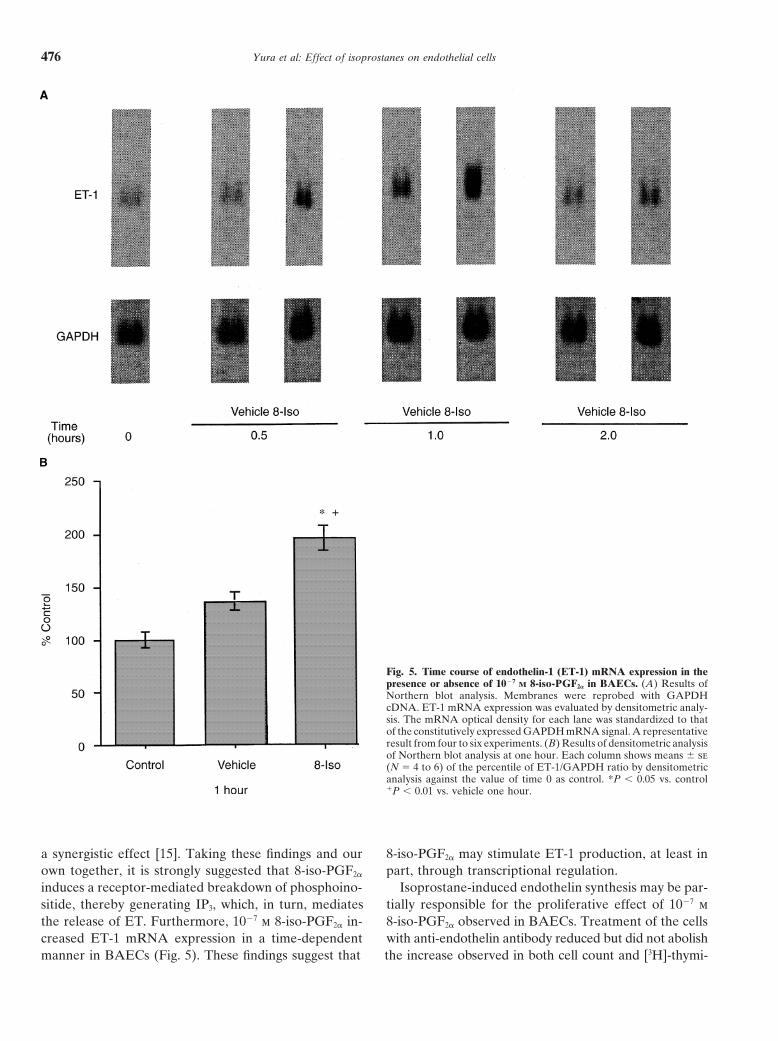
Endothelin-1 mRNA is expressed constitutively by whereas the vehicle had no effect at this time point(Fig. 5B).BAECs. This basal expression was increased by 1027 m
8-iso-PGF2a at 30 minutes, 1 hour, and 2 hours of incuba-Binding studiestion (Fig. 5A). Densitometrically analyzed data of North-
ern blot analysis showed that 1027 m 8-iso-PGF2a signifi- Summarized results of competitive binding studies with[3H]-8-iso-PGF2a are shown in Figure 6. 8-iso-PGF2a dis-cantly increased the ET-1/GAPDH ratio at one hour,

Yura et al: Effect of isoprostanes on endothelial cells 475
Fig. 4. Ir-ET-1 production by 8-iso-PGF2a inBAECs. (A) Dose dependency. (B) Effect of10–6 m SQ29,548 on ir-ET-1 production by 10–7
m 8-iso-PGF2a. Each value is expressed as amean 6 sd of three or four determinations.*P , 0.05 vs. vehicle; **P , 0.01 vs. vehicle.
placed radiolabeled homoligand binding to BAECs in a phate [13], after the addition of 8-iso-PGF2a to BAECs,and assessed the capacity of SQ29,548 in blocking thesemanner suggesting the existence of two distinct binding
sites: a low-affinity binding site (Kd 5 0.83 6 0.15 mm; responses. As shown in Figure 2A, 8-iso-PGF2a increasedIP3 production at 10 seconds in a dose-dependent man-Bmax 5 6.1 6 1.1 nmol/106 cells) and a high-affinity bind-
ing site (Kd 5 26.7 6 3.4 nm; Bmax 5 457 6 152 pmol/ ner. Significant increases were noted at concentrationshigher than 1029 m. Ten times higher concentrations of106 cells).SQ29,548 only partially inhibited IP3 production inducedby 1027 m 8-iso-PGF2a (Fig. 1B). Similar results were ob-
DISCUSSION tained with regard to the stimulation of DNA synthesisOur results show the proliferatory effect of 8-iso-PGF2a by these agents. 8-Iso-PGF2a stimulated [3H]-thymidine
on BAECs. In addition, isoprostanes also increased ET-1 uptake dose dependently, with significant increases atmRNA expression and protein production by these cells. concentrations higher than 1028 m (Fig. 3A).In fact, inhibition of endothelin action with anti-endo- It is reasonable to assume that 8-iso-PGF2a provokesthelin antibody reduces the proliferative effect of 8-iso- an increase in [3H]-thymidine incorporation and a subse-PGF2a. Our data also indicate that the intracellular signal quent enhancement of cellular proliferation via a mecha-transduction mechanism used by isoprostanes in endo- nism involving the release of IP3 and mobilization of intra-thelial cells is the enhancement of phosphoinositide turn- cellular Ca21. The partial inhibition of these increases byover, as previously shown in smooth muscle cells. Be- SQ29,548, the TXA2 receptor antagonist [14], is consistentcause isoprostane effects were only partially inhibited with the notion that isoprostanes and TXA2 recognizeby the TXA2 receptor antagonist SQ29,548, we finally closely related receptors. The increase in DNA synthesissuggest that endothelial cells present a specific receptor by 8-iso-PGF2a raises the possibility that, in addition tofor isoprostanes, different from the TXA2 receptor. This its vasoactive properties, 8-iso-PGF2a may contribute tosuggestion is supported by the results of our competitive the pathological processes that involve expression of en-binding studies. dothelial cell proliferation, such as atherosclerosis.
Proliferation of BAECs, measured as [3H]-thymidine In addition to the immediate activation of signal trans-uptake, was stimulated by concentration of 8-iso-PGF2a duction mechanisms and stimulation of mitogenesis,superior to 1028 m. Because it has been described that 8-iso-PGF2a stimulated ir-ET-1 production in a dose-isoprostane may exert their effect through binding to dependent manner with significant increases at concentra-TXA2 receptor, we also examined the effect of a known tions higher than 1029 m (Fig. 5A). Once more, ir-ET-1TXA2 receptor antagonist, SQ29,548 1026 m, on the iso- production stimulated by 1027 m 8-iso-PGF2a was inhib-
ited partially in the presence of 1026 m SQ29,548 (Fig. 4B).prostane-induced [3H]-thymidine (Fig. 4B). Coincuba-tion of isoprostane-treated BAECs with SQ29,548 only Emori et al demonstrated that both arginine vasopressin
and angiotensin II are potent secretagogues for ET inpartially inhibited the proliferative effect of 8-iso-PGF2a,suggesting a unique mechanism for this stimulation. cultured BAECs [15]; however, these effects appear to
be receptor mediated because stimulated ir-ET secretionIn vascular smooth muscle cells, 8-iso-PGF2a exerts itsbiological actions through enhancement of phosphoino- by these vasoconstrictors was completely abolished by
specific V1- and angiotensin II-receptor antagonists. Pro-sitide turnover as a second-messenger system, with sub-sequent stimulation of cellular mitogenesis, as does tein kinase C-activating phorbol ester and Ca21 iono-
phore ionomycin also had stimulatory effects on ir-ETTXA2 [7, 12]. We therefore examined the changes in IP3,a hydrolysis product of phosphatidylinositol 4,5-bisphos- secretion, and the combination of both compounds had
Yura et al: Effect of isoprostanes on endothelial cells476
Fig. 5. Time course of endothelin-1 (ET-1) mRNA expression in thepresence or absence of 1027 M 8-iso-PGF2a in BAECs. (A) Results ofNorthern blot analysis. Membranes were reprobed with GAPDHcDNA. ET-1 mRNA expression was evaluated by densitometric analy-sis. The mRNA optical density for each lane was standardized to thatof the constitutively expressed GAPDH mRNA signal. A representativeresult from four to six experiments. (B) Results of densitometric analysisof Northern blot analysis at one hour. Each column shows means 6 se(N 5 4 to 6) of the percentile of ET-1/GAPDH ratio by densitometricanalysis against the value of time 0 as control. *P , 0.05 vs. control1P , 0.01 vs. vehicle one hour.
a synergistic effect [15]. Taking these findings and our 8-iso-PGF2a may stimulate ET-1 production, at least inpart, through transcriptional regulation.own together, it is strongly suggested that 8-iso-PGF2a
induces a receptor-mediated breakdown of phosphoino- Isoprostane-induced endothelin synthesis may be par-tially responsible for the proliferative effect of 1027 msitide, thereby generating IP3, which, in turn, mediates
the release of ET. Furthermore, 1027 m 8-iso-PGF2a in- 8-iso-PGF2a observed in BAECs. Treatment of the cellswith anti-endothelin antibody reduced but did not abolishcreased ET-1 mRNA expression in a time-dependent
manner in BAECs (Fig. 5). These findings suggest that the increase observed in both cell count and [3H]-thymi-

Yura et al: Effect of isoprostanes on endothelial cells 477
Fig. 6. Competitive binding assay of 2.5 nM
[3H]-8-iso-PGF2a as a radiolabeled ligand inBAECs. (A) competition binding curve. (B)Scatchard analysis. Each point represents datafrom three to five experiments. Each point isexpressed as a mean of three or four determi-nations. Data are as follows (N 5 6, mean 6se): Kd1 5 26.7 6 3.4 nm; Bmax1 5 457 6 152pmol/106 cells; Kd2 5 0.83 6 0.15 mm; Bmax2 56.1 6 1.1 nmol/106 cells. Analysis of specificbinding data was performed by computer-based analysis system of radioligand bindingexperiments, EBDA and LIGAND [11].
dine uptake. The possible explanation is that isoprostanes using [3H]-8-iso-PGF2a as a radiolabeled ligand clearlysupport the existence of two distinct binding sites for 8-iso-induce cell proliferation by two mechanisms, one being
the synthesis of endothelin, a known mitogen for endo- PGF2a in BAECs: a low-affinity binding site (mean Kd 50.83 mm) and a high-affinity binding site (mean Kd 5thelial cells. The other mechanism would be independent
of endothelin synthesis, and it may be triggered by other 26.7 nm). As we reported previously in aortic smoothmuscle cell membrane fractions, cold 8-iso-PGF2a dis-intracellular signaling, such as cytosolic calcium increases.
Previously, we have shown that circulating concen- placed [3H]-8-iso-PGF2a binding in a pattern that sug-gested the existence of two component binding sites. Thetrations and urinary excretion of F2-isoprostanes were
markedly increased above normal in patients with the Kd value for the low-affinity binding sites was 0.94 60.13 mm and that for the high-affinity binding sites washepatorenal syndrome [4]. This syndrome is character-
ized by intense renal vasoconstriction in the setting of 31.8 6 5.7 nm [8]. These Kd values are indeed similarto those observed in this study. The Ki value of 8-iso-severe liver disease, but the pathogenesis of the vasocon-
striction that characterizes the disorder remains poorly PGF2a in competing for [3H]-SQ29,548 binding in smoothmuscle cells was similar to the Kd value for the putativeunderstood [16]. On the other hand, it was also demon-
strated that ET-1 concentrations in patients with this low-affinity binding sites (Ki 5 0.81 6 0.16 mm) [7].Therefore, it is suggested that the low-affinity bindingsyndrome also increase markedly above those in patients
with liver disease, acute renal failure, or chronic renal sites recognized in BAECs, as well as in smooth musclecells, may represent TXA2 receptors. On the other hand,failure only [5]. The results in this study suggest that an
F2-isoprostane, 8-iso-PGF2a, may contribute to the renal in our previous study, it was suggested that the high-affinity 8-iso-PGF2a binding sites in smooth muscle cellsvasoconstriction in the hepatorenal syndrome, not only
as a potent vasoconstrictor by itself, but also as a stimula- represent specific isoprostane receptors [7].The fact that induction of [3H]-thymidine uptake, in-tor of endothelial cell ET-1 production. In addition, in-
creased levels of both F2-isoprostanes [2] and ET-1 [3] in creased IP3 production, and ET-1 expression occur atthreshold concentrations close to the high-affinity Kdthe circulation of persons who smoke were also reported
recently. Morrow et al showed that the production of value for 8-iso-PGF2a obtained in our ligand bindingstudies further supports the distinct nature of the isopros-F2-isoprostanes is higher in smokers than in nonsmokers,
and levels of F2-isoprostanes in smokers fell significantly tane binding sites.We also compared the effects of U46,619, a TXA2after two weeks of abstinence from smoking [2]. These
results provide compelling evidence that smoking causes agonist, and 8-iso-PGF2a by luminometric assay usingthe Ca21-sensitive photoprotein, aequarin, in Xenopusoxidative modification of biological components in hu-
mans [2]. Our results may clarify an additional patho- oocytes transfected with a human TXA2 receptor cDNA.Maximal responses were obtained with 1026 and 1027 mphysiologic role of 8-iso-PGF2a in the pathogenesis of
diseases caused by smoking. U46,619 with a diminished response with 1028 m, whereas8-iso-PGF2a gave much weaker responses at 1026 andBecause all of the effects described for 8-iso-PGF2a
on endothelial cells were only partially inhibited by the 1027 m with no response at 1028 m [8]. The results showedthat 8-iso-PGF2a did not act through TXA2 receptor bind-addition of a TXA2 receptor antagonist, we carried out
competitive binding experiments in order to confirm the ing at physiological concentrations, whereas at muchhigher concentrations, partial responses to 8-iso-PGF2aexistence of a specific binding site, and therefore a recep-
tor, for isoprostanes in BAECs. The results of these studies are observed, likely through cross-activation of TXA2

Yura et al: Effect of isoprostanes on endothelial cells478
receptors. Taking previous results in rat aortic smooth REFERENCESmuscle cells and the similarity of Kd values between 1. Morrow JD, Hill KE, Burk RF, Nammour TK, Badr KF, Rob-smooth muscle cells and BAECs together, it is likely erts LJ II: A series of prostaglandin F2-like compounds are pro-
duced in vivo in humans by a non-cyclooxygenase, free radical-that the high-affinity binding sites observed in this studycatalyzed mechanism. Proc Natl Acad Sci USA 87:9383–9387, 1992represent true isoprostane receptors, and these putative 2. Morrow JD, Frei B, Longmire AW, Gaziano JM, Linch SM,
receptors are distinct from the TXA2 receptors, although Shyr Y, Strauss WE, Oates JA, Hill KE, Roberts LJ II: Increasein circulating products of lipid peroxidation (F2-isoprostanes) inthey bear homology to the TXA2 receptors.smokers: Smoking as a cause of oxidative damage. N Engl J MedIn summary, our results present evidence to suggest332:1198–1203, 1995
a role for 8-iso-PGF2a as an endothelial cell mitogen. 3. Haak T, Jungmann E, Raab C, Usadel KH: Elevated endothelin-1levels after cigarette smoking. Metabolism 43:267–269, 1994Treatment of BAECs with 8-iso-PGF2a is also associated
4. Morrow JD, Moore KP, Awad JA, Ravenscraft MD, Mariniwith rapid dose-dependent hydrolysis of phosphoinosi-G, Badr KF, Williams R, Roberts LJ II: Marked overproduction
tides with immediate generation of the biologically active of non-cyclooxygenase derived prostanoids (F2-isoprostanes) in thehepatorenal syndrome. J Lipid Med 6:417–420, 1993second messenger, IP3. Our competitive binding studies
5. Moore K, Wendon J, Frazer M, Karani J, Williams R, Badrcharacterized two distinct binding sites in BAECs. It isKF: Plasma endothelin immunoreactivity in liver disease and thesuggested that the low- and high-affinity binding sites hepatorenal syndrome. N Engl J Med 327:1774–1778, 1992
may represent TXA2 receptors and specific isoprostane 6. Takahashi K, Nammour TM, Fukunaga M, Ebert J, MorrowJD, Roberts LJ II, Hoover RL, Badr KF: Glomerular actions ofreceptors, respectively. These results, coupled with thea free radical-generated novel prostaglandin, 8-epi-prostaglandinincreased production of ET-1, may provide insight into F2a, in the rat: Evidence for interaction with thromboxane A2
the cellular basis for 8-iso-PGF2a2endothelial cell inter- receptors. J Clin Invest 90:136–141, 19927. Fukunaga M, Makita N, Roberts LJ II, Morrow JD, Takahashiactions, which may be of potential pathophysiological
K, Badr KF: Evidence for the existence of F2-isoprostane receptorsrelevance under conditions of oxidant injury and certainon rat vascular smooth muscle cells. Am J Physiol 264:C1619–
human conditions and diseases, such as smoking and C1624, 19938. Fukunaga M, Yura T, Grygorczyk R, Badr KF: Evidence forhepatorenal syndrome.
the distinct nature of F2-isoprostane receptors from those of throm-boxane A2. Am J Physiol 272:F477–F483, 1997
ACKNOWLEDGMENTS 9. Myers PR, Guerra R Jr, Harrison DG: Release of NO andEDRF from cultured bovine aortic endothelial cells. Am J PhysiolThis work was supported by a VA MERIT Award Grant to K.F.B.256:H1030–H1037, 1989We acknowledge to Dr. Haruo Onda, Takeda Chemical Ind., Tsukuba,
10. Chomczynski P, Sacchi N: Single-step method of RNA isolation byJapan, who kindly provided the cDNA for ET-1. This study was pre-acid guanidinium thiocyanate-PhOH-chloroform extraction. Analsented in part at the 1994 Meeting of The American Society of Nephrol-Biochem 162:156–159, 1987ogy, Oct. 26–28, 1994, Orlando, Florida, USA.
11. McPherson GA: Analysis of radioligand binding experiments: Acollection of computer programs for the IBM PC. J PharmacolReprint requests to Angel Montero, Ph.D., 1840 Southern Lane,Methods 14:213–228, 1985Decatur, Georgia 30033, USA.
12. Hanasaki K, Nakano T, Kasai H, Arita H: Receptor-mediatedE-mail: [email protected] effect of thromboxane A2 in vascular smooth musclecells. Biochem Pharmacol 40:2535–2542, 1990
13. Berridge MJ, Irvine RF: Inositol trisphosphate, a novel secondAPPENDIXmessenger in cellular signal transduction. Nature 312:315–321, 1984
14. Ogletree ML, Harris DN, Greenberg R, Haslanger MF, Na-Abbreviations used in this article are: BAECs, cultured bovinekane M: Pharmacological actions of SQ29,548, a novel selectiveaortic endothelial cells; Bmax, maximal binding capacity; ET-1, endo-thromboxane antagonist. J Pharmacol Exp Ther 234:435–441, 1985thelin-1; FBS, fetal bovine serum; GAPDH, glyceraldehyde 3-phos-
15. Emori T, Hirata Y, Ohta K, Shichiri M, Marumo F: Secretoryphate dehydrogenase; ir-ET, immunoreactive endothelin; IP3, inositolmechanism of immunoreactive endothelin in cultured bovine endo-1,4,5-trisphosphate; 8-iso-PGF2a, 8-iso-prostaglandin F2a; Kd, dissocia-thelial cells. Biochem Biophys Res Commun 160:93–100, 1989tion constant; PGF2a, prostaglandin F2a; TCA, trichloroacetic acid;
16. Schelling JR, Linas SL: Hepatorenal syndrome. Semin NephrolSDS, sodium dodecyl sulfate; SSC, standard saline citrate; and TXA2,10:565–570, 1990thromboxane A2.
Copyright © 2022 FDOKUMEN