Endobronchial perfluorocarbon reduces inflammatory activity before and after lung transplantation in...
10
Hindawi Publishing Corporation Mediators of Inflammation Volume 2013, Article ID 193484, 9 pages http://dx.doi.org/10.1155/2013/193484 Research Article Endobronchial Perfluorocarbon Reduces Inflammatory Activity before and after Lung Transplantation in an Animal Experimental Model Luiz Alberto Forgiarini Junior, 1,2,3 Arthur Rodrigo Ronconi Holand, 3 Luiz Felipe Forgiarini, 3 Darlan Pase da Rosa, 3 Norma Anair Possa Marroni, 4 Paulo Francisco Guerreiro Cardoso, 5 and Cristiano Feijó Andrade 1,3,6 1 Postgraduate Program in Pulmonology, Federal University of Rio Grande do Sul (UFRGS), 90040-060 Porto Alegre, RS, Brazil 2 Methodist University (IPA), 906301-170 Porto Alegre, RS, Brazil 3 oracic Surgery Department, Laboratory of Airways and Lung, Hospital de Cl´ ınicas de Porto Alegre (HCPA), Ramiro Barcelos 2.350, 90035-903 Porto Alegre, RS, Brazil 4 Lutheran University of Brazil (ULBRA), 92425-900 Canoas, RS, Brazil 5 Department of Cardiopneumology, Division of oracic Surgery, Laboratory of Medical Investigation (LIM 61), Heart Institute (InCor), Hospital das Cl´ ınicas, Faculdade de Medicina da Universidade de S˜ ao Paulo, 05403-000 S˜ ao Paulo, SP, Brazil 6 Santo Antˆ onio Children’s Hospital, 90020-090 Porto Alegre, RS, Brazil Correspondence should be addressed to Cristiano Feij´ o Andrade; [email protected] Received 10 June 2013; Revised 21 September 2013; Accepted 7 November 2013 Academic Editor: Tˆ ania Fr¨ ode Copyright © 2013 Luiz Alberto Forgiarini Junior et al. is is an open access article distributed under the Creative Commons Attribution License, which permits unrestricted use, distribution, and reproduction in any medium, provided the original work is properly cited. Background. e aim of this study was to evaluate the use of liquid perfluorocarbon (PFC) as an adjuvant substance for lung preservation and assess its role in pulmonary protection aſter transplantation. Methods. Seventy-two rat lungs were flushed with low-potassium dextran (LPD) solution and randomized into three main groups: control with LPD alone and experimental with 3 (PFC3) and 7 mL/kg (PFC7) of endobronchial PFC instilled just aſter harvest. Each group was divided into four subgroups according to preservation time (3, 6, 12, and 24 hours). Aſterwards, we performed lung transplantation using rat lungs preserved for 12 hours with LPD alone or with 7 mL/kg of endobronchial PFC. Results. ere was a significant increase in oxidative stress in the control group at 6 h of cold ischemic time compared with the PFC3 and PFC7 groups. e apoptotic activity and NF-B expression were significantly higher in the control group compared with the PFC groups at 3, 12, and 24 h of cold preservation. Aſter transplantation, the NF-B, iNOS, and nitrotyrosine expression as well as caspase 3 activity were significantly lower in the PFC groups. Conclusion. e use of endobronchial PFC as an adjuvant to the current preservation strategy improved graſt viability. 1. Introduction Lung transplantation is an established therapy for patients with end-stage lung disease [1]. In the early postoperative period, recipients may present with severe complications such as ischemia-reperfusion (IR) injury, acute rejection, and infection [2]. IR injury affects up to 20% of patients following lung transplantation and may lead to severe primary graſt dysfunction, increasing morbidity/mortality [3]. Severe primary graſt dysfunction is the end result of a series of events occurring from the time of brain death to the time of lung reperfusion aſter transplantation [4]. Strate- gies to prevent lung dysfunction resulting from IR injury have been focused on the selective assessment of donor lungs [5], effective techniques of lung preservation [6], and careful management of transplanted lungs aſter reperfusion [7]. e early stage of reperfusion injury following lung transplantation is initially mediated by the innate immune response through the activation of alveolar macrophages in the lung donor with the consequent production of proinflam- matory cytokines that exacerbate the inflammatory response, leading to cell death through an apoptotic pathway [8–12].
-
Upload
independent -
Category
Documents
-
view
0 -
download
0
Transcript of Endobronchial perfluorocarbon reduces inflammatory activity before and after lung transplantation in...
Hindawi Publishing CorporationMediators of InflammationVolume 2013, Article ID 193484, 9 pageshttp://dx.doi.org/10.1155/2013/193484
Research ArticleEndobronchial Perfluorocarbon Reduces InflammatoryActivity before and after Lung Transplantation in an AnimalExperimental Model
Luiz Alberto Forgiarini Junior,1,2,3 Arthur Rodrigo Ronconi Holand,3
Luiz Felipe Forgiarini,3 Darlan Pase da Rosa,3 Norma Anair Possa Marroni,4
Paulo Francisco Guerreiro Cardoso,5 and Cristiano Feijó Andrade1,3,6
1 Postgraduate Program in Pulmonology, Federal University of Rio Grande do Sul (UFRGS), 90040-060 Porto Alegre, RS, Brazil2Methodist University (IPA), 906301-170 Porto Alegre, RS, Brazil3Thoracic Surgery Department, Laboratory of Airways and Lung, Hospital de Clınicas de Porto Alegre (HCPA), Ramiro Barcelos 2.350,90035-903 Porto Alegre, RS, Brazil
4Lutheran University of Brazil (ULBRA), 92425-900 Canoas, RS, Brazil5Department of Cardiopneumology, Division ofThoracic Surgery, Laboratory of Medical Investigation (LIM 61),Heart Institute (InCor), Hospital das Clınicas, Faculdade deMedicina da Universidade de Sao Paulo, 05403-000 Sao Paulo, SP, Brazil
6 Santo Antonio Children’s Hospital, 90020-090 Porto Alegre, RS, Brazil
Correspondence should be addressed to Cristiano Feijo Andrade; [email protected]
Received 10 June 2013; Revised 21 September 2013; Accepted 7 November 2013
Academic Editor: Tania Frode
Copyright © 2013 Luiz Alberto Forgiarini Junior et al. This is an open access article distributed under the Creative CommonsAttribution License, which permits unrestricted use, distribution, and reproduction in any medium, provided the original work isproperly cited.
Background. The aim of this study was to evaluate the use of liquid perfluorocarbon (PFC) as an adjuvant substance for lungpreservation and assess its role in pulmonary protection after transplantation. Methods. Seventy-two rat lungs were flushed withlow-potassium dextran (LPD) solution and randomized into three main groups: control with LPD alone and experimental with 3(PFC3) and 7mL/kg (PFC7) of endobronchial PFC instilled just after harvest. Each groupwas divided into four subgroups accordingto preservation time (3, 6, 12, and 24 hours). Afterwards, we performed lung transplantation using rat lungs preserved for 12 hourswith LPD alone or with 7mL/kg of endobronchial PFC. Results. There was a significant increase in oxidative stress in the controlgroup at 6 h of cold ischemic time compared with the PFC3 and PFC7 groups.The apoptotic activity and NF-!B expression weresignificantly higher in the control group compared with the PFC groups at 3, 12, and 24 h of cold preservation. After transplantation,the NF-!B, iNOS, and nitrotyrosine expression as well as caspase 3 activity were significantly lower in the PFC groups. Conclusion.The use of endobronchial PFC as an adjuvant to the current preservation strategy improved graft viability.
1. Introduction
Lung transplantation is an established therapy for patientswith end-stage lung disease [1]. In the early postoperativeperiod, recipients may present with severe complicationssuch as ischemia-reperfusion (IR) injury, acute rejection, andinfection [2]. IR injury affects up to 20% of patients followinglung transplantation and may lead to severe primary graftdysfunction, increasing morbidity/mortality [3].
Severe primary graft dysfunction is the end result of aseries of events occurring from the time of brain death to
the time of lung reperfusion after transplantation [4]. Strate-gies to prevent lung dysfunction resulting from IR injuryhave been focused on the selective assessment of donor lungs[5], effective techniques of lung preservation [6], and carefulmanagement of transplanted lungs after reperfusion [7].
The early stage of reperfusion injury following lungtransplantation is initially mediated by the innate immuneresponse through the activation of alveolar macrophages inthe lung donor with the consequent production of proinflam-matory cytokines that exacerbate the inflammatory response,leading to cell death through an apoptotic pathway [8–12].
2 Mediators of Inflammation
Longer ischemic periods result in an escalating proportion ofcell death, resulting in a significant derangement in lung func-tion, which suggests a positive correlation between preser-vation time and organ performance [13]. This inflammatoryresponse and induction of apoptosis may be mediated bynuclear transcription factor kappa B (NF-!B), which wouldtrigger the posttransplant lung injury and cell death pathways[8, 14].
Numerous studies have been performed to optimize thetechnique of lung preservation [15–19]. Lung retrieval fortransplantation includes the flushing of the lungs with ahypothermic preservation solution to decrease its metabolicrate and energy requirements during storage [20]. Althoughhypothermia remains an essential component of the organpreservation strategy, it is, nevertheless, associated with aseries of events thatmay induce the upregulation ofmoleculeson the cell surface membrane and proinflammatory media-tors [21].
Liquid perfluorocarbons (PFCs) administered endobron-chially have been used in different models of lung injury [22,23], primarily as an alternative method of lung ventilation,known as partial liquid ventilation [19]. PFCs wash outalveoli debris, recruit collapsed alveoli, improve gas exchange,protect pulmonary architecture and have anti-inflammatoryand antioxidant properties [24, 25].There is a limited numberof studies using endobronchial PFCs in the setting of lungtransplantation, with conflicting results [19, 22, 26].
The objective of this study is to evaluate the use of liquidPFC as an adjuvant for lung preservation and its role as alung protective substance after transplantation.The studywasdesigned to test the impact of different PFCdoses for differentpreservation periods and evaluate the effects of PFC on celldeath, the inflammatory response, and oxidative stress in thetransplanted lung grafts.
2. Methods
The protocols for all experiments involving animal surgerywere approved by the Research Ethics Committee of theHospital de Clinicas de Porto Alegre. All animals receivedhumane care in accordance with the National ResearchCouncil (Guide for the Care and Use of Laboratory Animals,NIH revised 19). Male Wistar rats from the hospital’s centralanimal laboratory weighing between 250 and 300 g were usedfor all animal experiments.
2.1. PFC Dose versus Preservation Time Studies. To definethe amount of PFC administered for endobronchial lungpreservation, we evaluated three different dose regimens.Initially, we tested 3, 7, and 15mL/kg body weight of PFCgiven endobronchially in a pilot study. Twenty male Wistarrats were divided into four groups (" = 5) according tothe PFC dose. One control group did not use PFC. Theanimals were anesthetized (ketamine 100mg/kg and xylazine15mg/kg, administered intraperitoneally) and ventilatedwitha tidal volume of 8mL/kg of body weight, a respiratory rate of70–80 breaths/min, a fraction of inspired oxygen of 0.2 (roomair), and a positive end expiratory pressure of 2 cmH2O(Harvard Rodent Ventilator, model 683; Harvard Apparatus
Co., Millis, MA, USA). The animals were ventilated for 15minutes to stabilize the physiological parameters. Accordingto the calculated dose of PFC for each group, the dose wasdivided into three equal aliquots that were administeredat one-minute intervals through tracheotomy (room tem-perature) to achieve a homogeneous distribution. Followingthe administration of PFC, the animals were ventilated for30 minutes, and the heart-lung block was harvested forhistological and morphometric analyses. The morphometrywas based on the technique established by Weibel. [27],consisting of determining the number of times that thestructures of the lung parenchyma intersect a set of straightlines.
Upon completion of the dose study, another 72 maleWistar rats weighing 250–300 g were used for the doseversus preservation time study.The animalswere prepared forpulmonary perfusion as described elsewhere [28]. Preserva-tion was performed using 20mL of low-potassium dextranglucose preservation solution (LPD) at 4∘C via antegradecannulation of the pulmonary artery with drainage of theeffluent via the left atrium. After flushing, the lungs wereremoved and randomized into 3 groups (" = 12): controlin which the lungs were flushed with LPD; PFC 3mL/kg(PFC 3), and PFC 7mL/Kg (PFC 7) administered throughtracheotomy at room temperature and perfusedwith the LPDsolution. Each group was divided into four subgroups (" = 6)according to preservation time (3, 6, 12, and 24 hours).
Analysis of the variations of the thiobarbituric acidreactive substances (TBARS), determination of the caspase3 and NF-!B (subunit p65) activities, and TUNEL (termi-nal deoxynucleotidyl transferase-mediated dUTP nick-endlabeling) staining in the lung grafts were used to assess thequality of lung preservation.
2.2. Orthotopic Left Lung Transplantation Study Groups.Orthotopic left lung transplantation was performed usingthe modified cuff technique we previously described [28].Briefly, after general anesthesia with intraperitoneal ketamine(100mg/kg) and xylazine (15mg/kg), orotracheal intubation,mechanical ventilation, anticoagulation (300Uheparin), andmedian sternolaparotomy, the donor rat lungs were submit-ted to an antegrade flush via the pulmonary artery with20mL of cold (4∘C) LPD solution at a pressure of 20 cmH2O.The heart-lung block was extracted with the lungs inflatedat the end-tidal volume. The left lung graft was isolated,prepared, and stored in LPD at 4∘C for 12 hours.The recipientanimals were anesthetized, intubated (14-gauge catheter), andventilated. They then underwent a left thoracotomy, and thepulmonary vessels and left bronchus were anastomosed usinga standard cuff technique [9]. Transplants were performedto 6 animals per group, totaling 24 animals. Mean arterialpressure and arterial blood gases analysis were measuredafter 15 minutes in supine ventilation and at the end of theobservation period.
In the lung transplant group (LTx group), transplantationwas performed as described above. In the LTx/PFC group, thePFC at a dose of 7mL/Kg was instilled endobronchially priorto the heart-lung harvest. After 12 hours of cold ischemia, thelung transplantationwas performed in the same fashion as for
Mediators of Inflammation 3
0123456
(nm
ol/m
g of p
rote
in)
Control
TBARS
3 h 6 h 12 h 24 h
∗
∗
PFC3 mL/KgPFC7 mL/Kg
(a)
Caspase 31.61.41.21.00.80.60.40.2
0(uni
ts/m
g pro
tein·min−1 )
∗
∗
∗
3 h 6 h 12 h 24 h
(b)
00.20.40.60.8
11.21.41.6
(nm
ol/m
g of p
rote
in)
#∗∗
∗
NF-$B (p65)
Control
Control
PFC3 PFC7
CoCConConCooCConConConConCConCoCoConCConoooonConCoConConooononConConooCoooConCCCooooCooooConCo trotrotroootrotrotrotrottrotrottrottrottrotrotrotrotrotroooooooot oolllllllllllllllllll
p65-3 h%-Actin
p65-6 h%-Actin
p65-12 h%-Actin
p65-24 h%-Actin
65 kDa42 kDa
65 kDa42 kDa
65 kDa42 kDa
65 kDa42 kDa
3 h 6 h 12 h 24 h
(c)
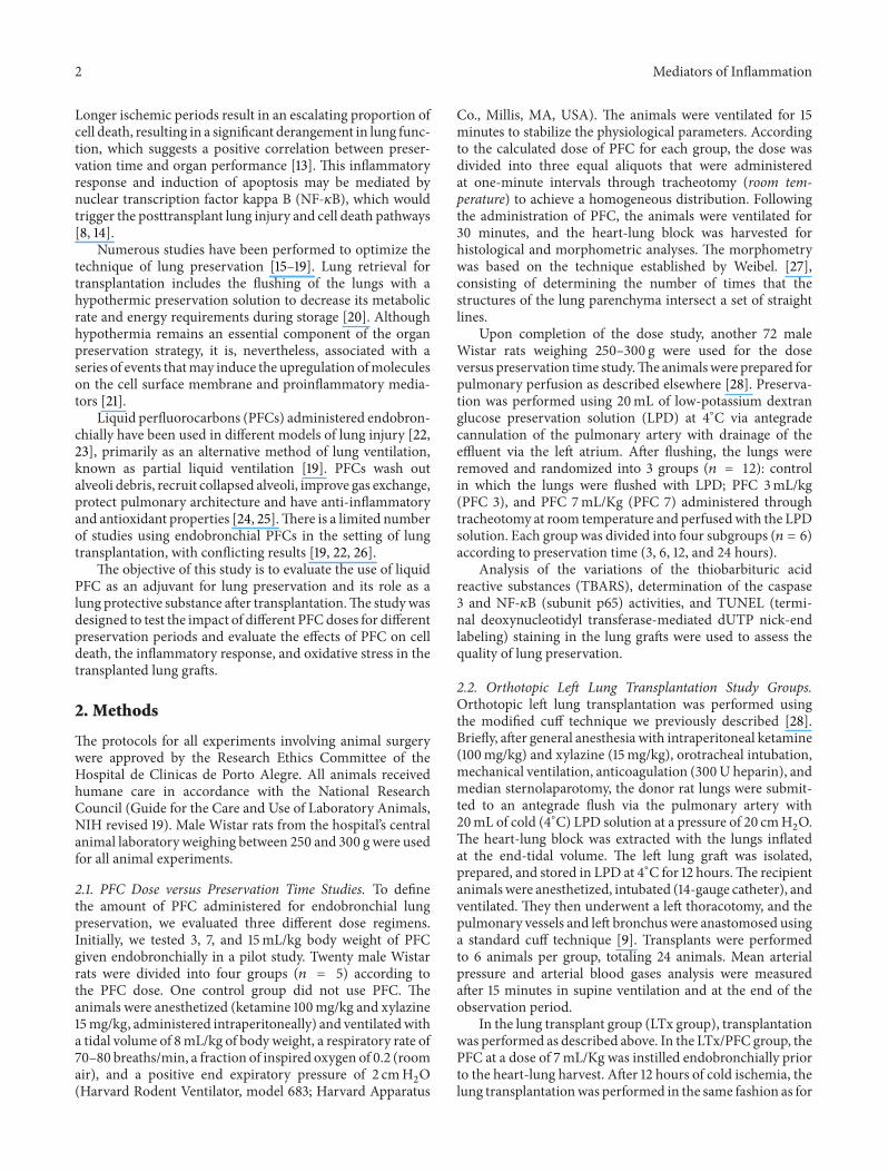
Figure 1: Apoptosis and inflammatory activity during lung preservation. (a) Effects of different periods of lung preservation on pulmonarylipid peroxidation. Using the TBARS assay, there was a significant decrease in lipid peroxidation at 3 hours of preservation in the PFC7 group(∗# > 0.05).The control group showed a significant increase in lipid peroxidation at 6 h (## > 0.05). Data are means ± standard deviation ofthe mean (# > 0.05). (b) Caspase 3 activity at different times of lung preservation.There is a significant increase in caspase 3 activity in thecontrol group at 6, 12, and 24 hours (∗# < 0.001). Data are means ± SD of the mean. (c) NF-!B expression (subunit p65) in the lung tissue atdifferent times of lung preservation. A significant increase in the expression of p65 was observed in the control group at 3, 12, and 24 hourscompared with the animals that used PFC (∗# < 0.05). PFC7 reduced the expression of p65 at 24 hours of preservation compared with PFC3(## < 0.05).the LTx group.The animals were euthanized after 120minutesof reperfusion. Half of the left lung was removed and storedat −80∘C, and the other half was stored in 10% formalin.
2.3. Oxidative Stress and Antioxidant Enzymes. The amountof aldehydes generated by lipid peroxidation was measuredby the TBARS method, which measures the amount ofsubstances reactingwith thiobarbituric acid [29].The analysisof superoxide dismutase (SOD)was based on the inhibition ofthe reaction of the superoxide radical with adrenalin [30].Theanalysis of catalase (CAT) activity was based on measuringthe decrease in hydrogen peroxide formation [31].
2.4. Caspase 3 Activity Assay and TUNEL Staining. Caspase3 activity was analyzed as described by Forgiarini et al. [32]and TUNEL studies as described by Fischer et al. [13].
2.5.Western Blotting. Lunghomogenateswere prepared fromfrozen lungs (−80∘C) and analyzed for the expression ofthe p65 subunit, caspase 3 and nitrotyrosine (Cell Signalling
Biotechnology, Boston, MA, USA), and iNOS (Santa CruzBiotechnology, Santa Cruz, CA, USA) by immunoblot analy-sis [33, 34].
2.6. Immunofluorescence. I!B and caspase 3 immunofluores-cence stainingwas performed on paraffin-embedded sectionsafter deparaffinization in ethanol, xylene, and water. First,primary antibodies for polyclonal anti-I!B and anti-caspase 3(Cell Signaling Biotechnology, Boston, MA, USA) at 1 : 1000were incubated overnight at 4∘C. Cy3-conjugated goat anti-rat secondary antibody (Sigma Aldrich, USA) at 1 : 200 wasincubated for 1 hour. Goat serum and the primary ratantibodywere used as a negative control.Mounted slices werevisualized within 2 hours on an Olympus Zeiss fluorescencemicroscope.
2.7. Lung Histology. Lung tissue specimens were fixed in formalin, dehydrated, cleared, and embedded in paraffin. Speci-menswere cut into 8 %mserial sections and stainedwith hem-atoxylin eosin. One pathologist blinded to the experimentalprotocol and the region of sampling performed quantitative
4 Mediators of Inflammation
Cont
rol
PFC3
PFC7
3 h 6 h 12 h 24 h
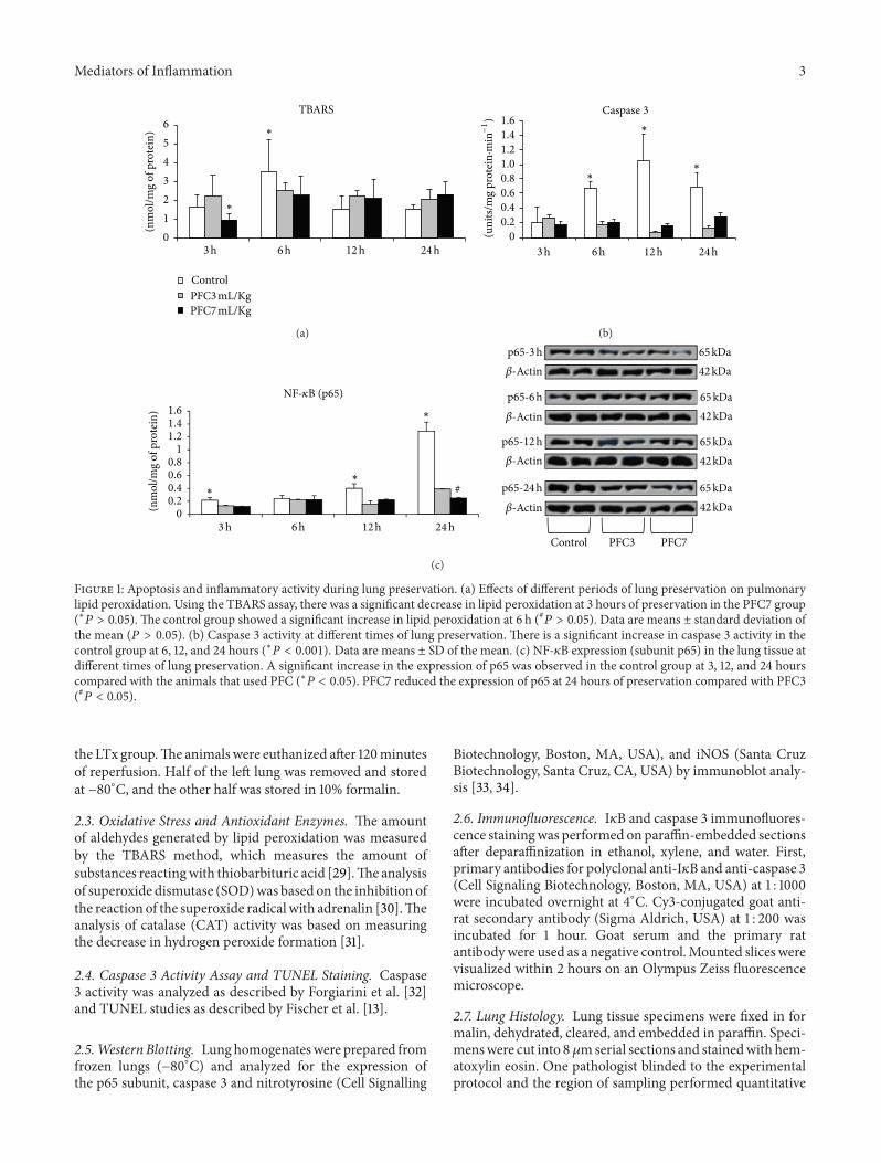
Figure 2: TUNEL staining after the different periods of cold ischemia. DAPI and FITC staining: DAPI-positive cells (nucleated cells) areblue and FITC-positive cells (TUNEL-positive) are green. There is predominance of TUNEL-positive cells in PFC 3 group at 12 h of coldpreservation.
analysis by light microscopy. Each sample was examinedunder low and high power fields. At least four sectionswere obtained from each block, and 20 fields were randomlyselected and analyzed for each section. The severity ofthe histological lesions was assessed using a score (HIS:histological score) [35].
2.8. Statistical Analysis. Datawere expressed asmeans± stan-dard deviation. One-way analysis of variance (ANOVA) wasused to determine the statistical significance. A value of# < 0.05was considered statistically significant.When statis-tical significance was reached, it was followed by a post hocanalysis using the Student-Newman-Keuls test. Nonparamet-ric data were analyzed using two-tailed unpaired &-tests.Thedatawere analyzed using Statistical Package for Social Scienceversion 17.0 statistical software (SPSS Inc., Chicago, IL, USA).
3. Results
A reduction in linear intercepts was found when the PFC7and PFC15 lungs were compared with control lungs (# <0.05). However, the PFC15 lungs showed a more pronouncedreduction in the number of crossed septae compared withthe PFC3 and PFC7 lungs (# < 0.05). Additionally, becauseall PFC15 lungs presented rupture of alveolar septae dueto overdistention, we decided to exclude the 15mL/kg doseexperiments.
When lipid peroxidation was evaluated at the differentlung preservation periods, there were significant differencesbetween the PCF3 and PFC7 groups in the quantity of lipid
peroxidation products at three hours of preservation (# <0.05). At six hours, we observed a significant increase inTBARS in the control group compared with PFC3 and PFC7(# < 0.05). However, at 24 hours of preservation, there wereno differences between the groups with and without PFC(Figure 1). When apoptosis was measured by caspase 3 acti-vity, a significant increase in the lungs of the control groupswas observed compared with the groups using PFC for lungpreservation in the lungs submitted to 6, 12, and 24 hours ofstorage (# < 0.05) (Figure 1). The TUNEL staining showedthe presence of more FITC-positive cells (TUNEL-positive)at 3 and 12 hours of preservation (Figure 2).
The expression of NF-!B was significantly reduced at 3,12, and 24 hours of preservation in the PFC lungs comparedwith the control lungs (# < 0.05). However, after 24 hours oflung preservation, a significant reduction in the expression ofNF-!Bwas found in the PFC7 group comparedwith the lungsin the PFC3 group (# < 0.05) (Figure 1).
After transplantation, there were no differences in lipidperoxidation and antioxidant enzymes between the PFC andcontrol groups in both the TBARS and catalase assays. Theactivity of superoxide dismutase was significantly higher inthe LTx/PFC group compared with the LTx group (# < 0.05).
The partial pressure of arterial oxygen (PaO2), carbondioxide (PaCO2), and mean arterial pressure (MAP) did notshow any significant differences between both groups at thedifferent time points (Table 1). After lung transplantation,there was a significant increase in the nuclear translocationof NF-!B (subunit p65) in the LTx group compared withthe LTx/PFC group (# < 0.05) (Figure 3). There was also
Mediators of Inflammation 5
p65
LTx LTx/PFC
0
0.5
1
1.5
2NF-$B (p65)
%-Actin65 kDa42 kDa
∗
(a.u
.)
LTx LTx/PFC
(a)
0
0.5
1
1.5
2Caspase 3
Cleaved
LTx LTx/PFC
%-Actin
17 kDa42 kDa
∗(a.u
.)
caspase 3
LTx LTx/PFC
(b)
iNOS
iNOS
LTx
0
3
4
2
1
LTx/PFC
%-Actin131 kDa
42 kDa
∗
(a.u
.)
LTx LTx/PFC
(c)
Nitrotyrosine
Nitrotyrosine
0
0.5
1
1.5
2
LTx LTx/PFC
%-Actin
85 kDa
42 kDa
∗(a.u
.)
LTx LTx/PFC
(d)
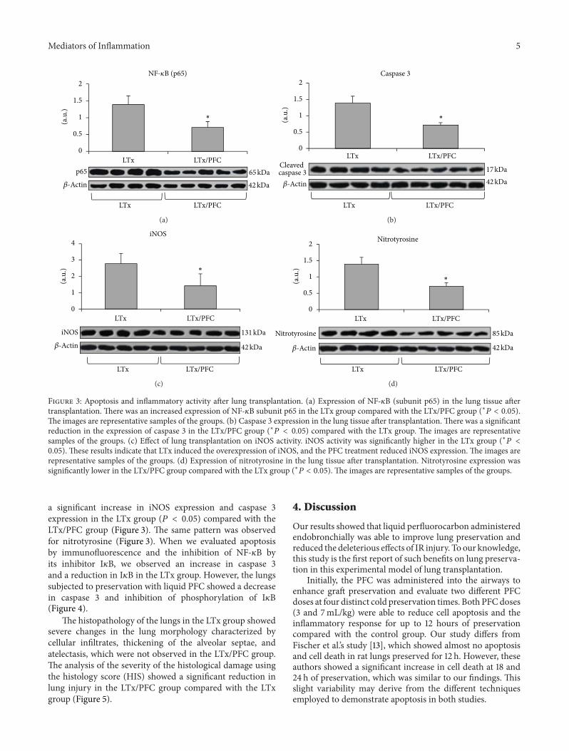
Figure 3: Apoptosis and inflammatory activity after lung transplantation. (a) Expression of NF-!B (subunit p65) in the lung tissue aftertransplantation.There was an increased expression of NF-!B subunit p65 in the LTx group compared with the LTx/PFC group (∗# < 0.05).The images are representative samples of the groups. (b) Caspase 3 expression in the lung tissue after transplantation.There was a significantreduction in the expression of caspase 3 in the LTx/PFC group (∗# < 0.05) compared with the LTx group. The images are representativesamples of the groups. (c) Effect of lung transplantation on iNOS activity. iNOS activity was significantly higher in the LTx group (∗# <0.05).These results indicate that LTx induced the overexpression of iNOS, and the PFC treatment reduced iNOS expression.The images arerepresentative samples of the groups. (d) Expression of nitrotyrosine in the lung tissue after transplantation. Nitrotyrosine expression wassignificantly lower in the LTx/PFC group compared with the LTx group (∗# < 0.05).The images are representative samples of the groups.
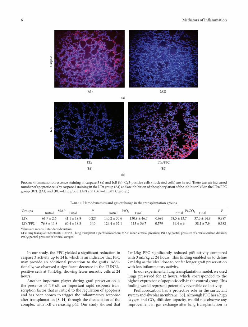
a significant increase in iNOS expression and caspase 3expression in the LTx group (# < 0.05) compared with theLTx/PFC group (Figure 3). The same pattern was observedfor nitrotyrosine (Figure 3). When we evaluated apoptosisby immunofluorescence and the inhibition of NF-!B byits inhibitor I!B, we observed an increase in caspase 3and a reduction in I!B in the LTx group. However, the lungssubjected to preservation with liquid PFC showed a decreasein caspase 3 and inhibition of phosphorylation of I!B(Figure 4).
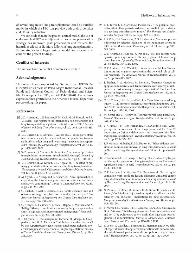
The histopathology of the lungs in the LTx group showedsevere changes in the lung morphology characterized bycellular infiltrates, thickening of the alveolar septae, andatelectasis, which were not observed in the LTx/PFC group.The analysis of the severity of the histological damage usingthe histology score (HIS) showed a significant reduction inlung injury in the LTx/PFC group compared with the LTxgroup (Figure 5).
4. Discussion
Our results showed that liquid perfluorocarbon administeredendobronchially was able to improve lung preservation andreduced the deleterious effects of IR injury. To our knowledge,this study is the first report of such benefits on lung preserva-tion in this experimental model of lung transplantation.
Initially, the PFC was administered into the airways toenhance graft preservation and evaluate two different PFCdoses at four distinct cold preservation times. Both PFCdoses(3 and 7mL/kg) were able to reduce cell apoptosis and theinflammatory response for up to 12 hours of preservationcompared with the control group. Our study differs fromFischer et al.’s study [13], which showed almost no apoptosisand cell death in rat lungs preserved for 12 h. However, theseauthors showed a significant increase in cell death at 18 and24 h of preservation, which was similar to our findings. Thisslight variability may derive from the different techniquesemployed to demonstrate apoptosis in both studies.
6 Mediators of Inflammation
Casp
ase 3
(A1) (A2)
(a)
LTx LTx/PFC
I$B
(B1) (B2)
(b)
Figure 4: Immunofluorescence staining of caspase 3 (a) and I!B (b). Cy3-positive cells (nucleated cells) are in red. There was an increasednumber of apoptotic cells by caspase 3 staining in the LTx group (A1) and an inhibition of phosphorylation of the inhibitor I!B in the LTx/PFCgroup (B2). ((A1) and (B1)—LTx group; (A2) and (B2)—LTx/PFC group.)
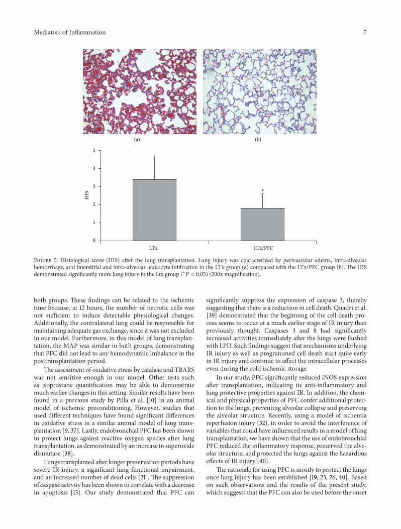
Table 1: Hemodynamics and gas exchange in the transplantation groups.
Groups MAP # PaO2 # PaCO2 #Initial Final Initial Final Initial FinalLTx 61.7 ± 2.6 41.1 ± 19.8 0.227 140.2 ± 30.6 130.9 ± 46.7 0.691 38.5 ± 13.7 37.3 ± 14.8 0.887LTx/PFC 76.8 ± 11.8 60.4 ± 18.8 0.10 124.4 ± 32.1 113 ± 36.7 0.579 34.4 ± 6 38.1 ± 7.9 0.382Values are means ± standard deviation.LTx: lung transplant (control); LTx/PFC: lung transplant + perfluorocarbon; MAP: mean arterial pressure; PaCO2: partial pressure of arterial carbon dioxide;PaO2: partial pressure of arterial oxygen.
In our study, the PFC yielded a significant reduction incaspase 3 activity up to 24 h, which is an indicator that PFCmay provide an additional protection to the grafts. Addi-tionally, we observed a significant decrease in the TUNEL-positive cells at 7mL/kg, showing fewer necrotic cells at 24hours.
Another important player during graft preservation isthe presence of NF-!B, an important rapid-response tran-scription factor that is critical to the regulation of apoptosisand has been shown to trigger the inflammatory responseafter transplantation [8, 14] through the dissociation of thecomplex with I!B-a releasing p65. Our study showed that
7mL/kg PFC significantly reduced p65 activity comparedwith 3mL/kg at 24 hours. This finding enabled us to define7mL/kg as the ideal dose to confer longer graft preservationwith less inflammatory activity.
In our experimental lung transplantation model, we usedlungs preserved for 12 hours, which corresponded to thehighest expression of apoptotic cells in the control group.Thisfinding would represent potentially reversible cell activity.
Perfluorocarbon has a protective role in the surfactantsystemand alveolarmembrane [36]. AlthoughPFChas a highoxygen and CO2 diffusion capacity, we did not observe anyimprovement in gas exchange after lung transplantation in
Mediators of Inflammation 7
0
1
2
3
4
5
LTx LTx/PFC
(a) (b)
HIS
∗
Figure 5: Histological score (HIS) after the lung transplantation. Lung injury was characterized by perivascular edema, intra-alveolarhemorrhage, and interstitial and intra-alveolar leukocyte infiltration in the LTx group (a) compared with the LTx/PFC group (b). The HISdemonstrated significantly more lung injury in the Ltx group (∗# < 0.05) (200x magnification).
both groups. These findings can be related to the ischemictime because, at 12 hours, the number of necrotic cells wasnot sufficient to induce detectable physiological changes.Additionally, the contralateral lung could be responsible formaintaining adequate gas exchange, since it was not excludedin our model. Furthermore, in this model of lung transplan-tation, the MAP was similar in both groups, demonstratingthat PFC did not lead to any hemodynamic imbalance in theposttransplantation period.
The assessment of oxidative stress by catalase and TBARSwas not sensitive enough in our model. Other tests suchas isoprostane quantification may be able to demonstratemuch earlier changes in this setting. Similar results have beenfound in a previous study by Pilla et al. [10] in an animalmodel of ischemic preconditioning. However, studies thatused different techniques have found significant differencesin oxidative stress in a similar animal model of lung trans-plantation [9, 37]. Lastly, endobronchial PFC has been shownto protect lungs against reactive oxygen species after lungtransplantation, as demonstrated by an increase in superoxidedismutase [38].
Lungs transplanted after longer preservation periods havesevere IR injury, a significant lung functional impairment,and an increased number of dead cells [21]. The suppressionof caspase activity has been shown to correlatewith a decreasein apoptosis [13]. Our study demonstrated that PFC can
significantly suppress the expression of caspase 3, therebysuggesting that there is a reduction in cell death. Quadri et al.[39] demonstrated that the beginning of the cell death pro-cess seems to occur at a much earlier stage of IR injury thanpreviously thought. Caspases 3 and 8 had significantlyincreased activities immediately after the lungs were flushedwith LPD. Such findings suggest that mechanisms underlyingIR injury as well as programmed cell death start quite earlyin IR injury and continue to affect the intracellular processeseven during the cold ischemic storage.
In our study, PFC significantly reduced iNOS expressionafter transplantation, indicating its anti-inflammatory andlung protective properties against IR. In addition, the chem-ical and physical properties of PFC confer additional protec-tion to the lungs, preventing alveolar collapse and preservingthe alveolar structure. Recently, using a model of ischemiareperfusion injury [32], in order to avoid the interference ofvariables that could have influenced results in amodel of lungtransplantation, we have shown that the use of endobronchialPFC reduced the inflammatory response, preserved the alve-olar structure, and protected the lungs against the hazardouseffects of IR injury [40].
The rationale for using PFC is mostly to protect the lungsonce lung injury has been established [19, 23, 26, 40]. Basedon such observations and the results of the present study,which suggests that the PFC can also be used before the onset
8 Mediators of Inflammation
of severe lung injury, lung transplantation can be a suitablemodel in which the PFC can provide both graft protectionand IR injury reduction.
We conclude that, in the present animal model, the use ofendobronchial PFC as an adjuvant to the current preservationstrategy has improved graft preservation and reduced thehazardous effects of IR injury following lung transplantation.Future studies in a larger animal model are necessary toconfirm the present findings.
Conflict of Interests
The authors have no conflict of interests to declare.
Acknowledgments
This research was supported by Grants from FIPE/HCPA(Hospital de Clinicas de Porto Alegre Institutional ResearchFund) and National Council of Technological and Scien-tific Development (CNPq, no. 483364/2007-0). The authorsexpressed their gratitude to the American Journal Expert forproofreading this paper.
References
[1] J. D. Hosenpud, L. E. Bennett, B.M. Keck,M.M. Boucek, and R.J. Novick, “The registry of the international society for heart andlung transplantation: eighteenth official report—2001,” Journalof Heart and Lung Transplantation, vol. 20, no. 8, pp. 805–815,2001.
[2] J. D. Christie, L. B. Edwards, P. Aurora et al., “The registry of theinternational society for heart and lung transplantation: twenty-sixth official adult lung and heart-lung transplantation report-2009,” Journal of Heart and Lung Transplantation, vol. 28, no. 10,pp. 1031–1049, 2009.
[3] S.-P. Sommer, S. Sommer, B. Sinha et al., “Ischemia-reperfusioninjuryinduced pulmonary mitochondrial damage,” Journal ofHeart and Lung Transplantation, vol. 30, no. 7, pp. 811–818, 2011.
[4] J. D. Christie, R. M. Kotloff, V. N. Ahya et al., “The effect of pri-mary graft dysfunction on survival after lung transplantation,”The American Journal of Respiratory and Critical Care Medicine,vol. 171, no. 11, pp. 1312–1316, 2005.
[5] M. Cypel, J. C. Yeung, and S. Keshavjee, “Novel approaches toexpanding the lung donor pool: donation after cardiac deathand ex vivo conditioning,” Clinics in Chest Medicine, vol. 32, no.2, pp. 233–244, 2011.
[6] G. Thabut, H. Mal, J. Cerrina et al., “Graft ischemic time andoutcome of lung transplantation: a multicenter analysis,” TheAmerican Journal of Respiratory and Critical Care Medicine, vol.171, no. 7, pp. 786–791, 2005.
[7] C. Kroegel, K. Hekmat, A. Moser, J. Happe, R. Walther, and A.Reißig, “Airway complications following lung transplantationclinic, diagnosis, and interventional management,” Pneumolo-gie, vol. 65, no. 5, pp. 293–307, 2011.
[8] T. Ishiyama, S. Dharmarajan, M. Hayama, H. Moriya, K. Grap-perhaus, and G. A. Patterson, “Inhibition of nuclear factor !Bby I!B superrepressor gene transfer ameliorates ischemia-rep-erfusion injury after experimental lung transplantation,” JournalofThoracic and Cardiovascular Surgery, vol. 130, no. 1, pp. 194–201, 2005.
[9] R. L. Torres, L. K.Martins, M. Picoral et al., “The potential prot-ective effect of low potassiumdextran against lipid peroxidationin a rat lung transplantation model,” The Thoracic and Cardio-vascular Surgeon, vol. 57, no. 5, pp. 309–311, 2009.
[10] E. S. Pilla, G. S. Vendrame, P. G. Sanchez et al., “Ischemic preco-nditioning by selective occlusion of the pulmonary artery inrats,” Jornal Brasileiro de Pneumologia, vol. 34, no. 8, pp. 583–589, 2008.
[11] C. F. Andrade, H. Kaneda, S. Der et al., “Toll-like receptor andcytokine gene expression in the early phase of human lungtransplantation,” Journal ofHeart and Lung Transplantation, vol.25, no. 11, pp. 1317–1323, 2006.
[12] C. F. Andrade, T. K. Waddell, S. Keshavjee, and M. Liu, “Innateimmunity and organ transplantation: the potential role of toll-like receptors,”The American Journal of Transplantation, vol. 5,no. 5, pp. 969–975, 2005.
[13] S. Fischer, A. A. Maclean, M. Liu et al., “Dynamic changes inapoptotic and necrotic cell death correlate with severity of isch-emia-reperfusion injury in lung transplantation,”The AmericanJournal of Respiratory and Critical Care Medicine, vol. 162, no. 5,pp. 1932–1939, 2000.
[14] C.-H. Chiang, H.-I. Pai, and S.-L. Liu, “Ventilator-induced lunginjury (VILI) promotes ischemia/reperfusion lung injury (I/R)andNF-kB antibody attenuates both injuries,”Resuscitation, vol.79, no. 1, pp. 147–154, 2008.
[15] M. Cypel and S. Keshavjee, “Extracorporeal lung perfusion,”Current Opinion in Organ Transplantation, vol. 16, no. 5, pp.469–475, 2011.
[16] E. A. Simes, P.M. Pego-Fernandes, P. F. G. Cardoso et al., “Com-paring the performance of rat lungs preserved for 6 or 12hours after perfusion with low-potassium dextran or histidine-tryptophan-ketoglutarate,” Transplantation Proceedings, vol. 43,no. 5, pp. 1520–1524, 2011.
[17] S. F.Marasco,M. Bailey, D.McGlade et al., “Effect of donor pres-ervation solution and survival in lung transplantation,” Journalof Heart and Lung Transplantation, vol. 30, no. 4, pp. 414–419,2011.
[18] T. Kawamura, C.-S. Huang, N. Tochigi et al., “Inhaled hydrogengas therapy for prevention of lung transplant-induced ischemia/reperfusion injury in rats,” Transplantation, vol. 90, no. 12, pp.1344–1351, 2010.
[19] C. F. Andrade, L. K. Martins, T. A. Tonietto et al., “Partial liquidventilation with perfluorodecalin following unilateral caninelung allotransplantation in non-heart-heating donors,” Journalof Heart and Lung Transplantation, vol. 23, no. 2, pp. 242–251,2004.
[20] N. Pizanis, S. Gillner, M. Kamler, H. de Groot, H. Jakob, and U.Rauen, “Cold-induced injury to lung epithelial cells can be inhi-bited by iron chelators—implications for lung preservation,”European Journal of Cardio-Thoracic Surgery, vol. 40, no. 4, pp.948–955, 2011.
[21] E. Mayer, J. D. Puskas, P. F. G. Cardoso, S. Shi, A. S. Slutsky, andG. A. Patterson, “Reliable eighteen-hour lung preservation at 4∘and 10∘ C by pulmonary artery flush after high-dose prosta-glandin E1 administration,” Journal of Thoracic and Cardiovas-cular Surgery, vol. 103, no. 6, pp. 1136–1142, 1992.
[22] F. Loehe, C. Mueller, I. Bittmann, K. Messmer, and F. W. Schi-ldberg, “Influence of long-termpreservationwith endobronchi-ally administered perfluorodecalin on pulmonary graft func-tion,” Transplantation, vol. 70, no. 10, pp. 1417–1424, 2000.
Mediators of Inflammation 9
[23] C. Andrade, E. Felix, and P. Cardoso, “Ventilacao lıquida—rev-isao da literatura,” Jornal de Pneumologia, vol. 28, no. 6, pp. 351–361, 2002.
[24] J. G. Tuazon, J. H. Modell, C. I. Hood, and E.W. Swenson, “Pul-monary function after ventilation with fluorocarbon liquid(Caroxin-D),” Anesthesiology, vol. 38, no. 2, pp. 134–140, 1973.
[25] F. Gollan, J. McDermott, A. E. Johnson, and R. Namon, “Com-pliance and diffusion during respiration with fluorocarbonfluid,” Federation Proceedings, vol. 29, no. 5, pp. 1725–1730, 1970.
[26] S. A. Hosgood and M. L. Nicholson, “The role of perfluorocar-bon in organ preservation,” Transplantation, vol. 89, no. 10, pp.1169–1175, 2010.
[27] E. R. Weibel, “Principles and methods for the morphometricstudy of the lung and other organs,” Laboratory Investigation,vol. 12, pp. 131–155, 1963.
[28] P. G. Sanchez, L. K. Martins, F. K. Martins, R. Schimer, P. F. G.Cardoso, and C. F. Andrade, “Technical modification of unilat-eral lung transplantation in rats,” Jornal Brasileiro de Pneumolo-gia, vol. 33, no. 4, pp. 448–453, 2007.
[29] J. A. Buege and S. D. Aust, “Microsomal lipid peroxidation,”Methods in Enzymology, vol. 52, no. C, pp. 302–310, 1978.
[30] H. P.Misra and I. Fridovich, “The role of superoxide anion in theautoxidation of epinephrine and a simple assay for superoxidedismutase,” Journal of Biological Chemistry, vol. 247, no. 10, pp.3170–3175, 1972.
[31] H. Aebi, “Catalase in vitro,”Methods in Enzymology, vol. 105, pp.121–126, 1984.
[32] L.A. Forgiarini Jr., G.Grun,N.A.Kretzmann et al., “When is in-jury potentially reversible in a lung ischemia-reperfusion mod-el?” Journal of Surgical Research, vol. 179, pp. 168–174, 2013.
[33] U. K. Laemmli, “Cleavage of structural proteins during the asse-mbly of the head of bacteriophage T4,”Nature, vol. 227, no. 5259,pp. 680–685, 1970.
[34] H. Towbin, T. Staehelin, and J. Gordon, “Electrophoretic trans-fer of proteins frompolyacrylamide gels to nitrocellulose sheets:procedure and some applications,” Proceedings of the NationalAcademy of Sciences of the United States of America, vol. 76, no.9, pp. 4350–4354, 1979.
[35] Y. Fujino, S. Goddon, J.-D. Chiche, J. Hromi, and R. M.Kacmarek, “Partial liquid ventilation ventilates better than gasventilation,” The American Journal of Respiratory and CriticalCare Medicine, vol. 162, no. 2, part 1, pp. 650–657, 2000.
[36] R. B. Hirschl, R. Tooley, A. Parent, K. Johnson, and R. H. Bar-tlett, “Evaluation of gas exchange, pulmonary compliance, andlung injury during total and partial liquid ventilation in the acu-te respiratory distress syndrome,”Critical CareMedicine, vol. 24,no. 6, pp. 1001–1008, 1996.
[37] R. L. Torres, A. Belo-Klein, C. F. Andrade, and P. F. G. Cardoso,“Effect of systemically administered low potassium dextran sol-ution on oxidative stress in a rat model of lung ischemia,” Inter-active Cardiovascular andThoracic Surgery, vol. 8, no. 1, pp. 3–6,2009.
[38] A. T. Rotta, B. Gunnarsson, L. J. Hernan, B. P. Fuhrman, and D.M. Steinhorn, “Partial liquid ventilation with perflubron atten-uates in vivo oxidative damage to proteins and lipids,” CriticalCare Medicine, vol. 28, no. 1, pp. 202–208, 2000.
[39] S. M. Quadri, L. Segall, M. de Perrot et al., “Caspace inhibitionimproves ischemia-reperfusion injury after lung transplanta-tion,”The American Journal of Transplantation, vol. 5, no. 2, pp.292–299, 2005.
[40] L. A. Forgiarini Junior, L. F. Forgiarini, R. Mariano, A. Paludo, J.M. Ulbrich, and C. F. Andrade, “Endobronchial perfluorocar-bon administration decreases lung injury in an experimentalmodel of ischemia and reperfusion,” Journal of Surgical Res-earch, vol. 183, no. 2, pp. 835–840, 2013.

Submit your manuscripts athttp://www.hindawi.com
Stem CellsInternational
Hindawi Publishing Corporationhttp://www.hindawi.com Volume 201
Hindawi Publishing Corporationhttp://www.hindawi.com Volume 201
MEDIATORSINFLAMMATION
of
Hindawi Publishing Corporationhttp://www.hindawi.com Volume 201
Behavioural Neurology
,QWHUQDWLRQDO�-RXUQDO�RI
(QGRFULQRORJ\+LQGDZL�3XEOLVKLQJ�&RUSRUDWLRQKWWS���ZZZ�KLQGDZL�FRP
9ROXPH�����
Hindawi Publishing Corporationhttp://www.hindawi.com Volume 2014
Disease Markers
BioMed Research International
Hindawi Publishing Corporationhttp://www.hindawi.com Volume 201H
OncologyJournal of
Hindawi Publishing Corporationhttp://www.hindawi.com Volume 201
Hindawi Publishing Corporationhttp://www.hindawi.com Volume 2014
Oxidative Medicine and Cellular Longevity
PPARRe sea rch
Hindawi Publishing Corporationhttp://www.hindawi.com Volume ����
The Scientific World JournalHindawi Publishing Corporation http://www.hindawi.com Volume 2014
Immunology ResearchHindawi Publishing Corporationhttp://www.hindawi.com Volume 201
Journal of
ObesityJournal of
Hindawi Publishing Corporationhttp://www.hindawi.com Volume 201
Hindawi Publishing Corporationhttp://www.hindawi.com Volume 201
Computational and Mathematical Methods in Medicine
OphthalmologyJournal of
Hindawi Publishing Corporationhttp://www.hindawi.com Volume 201
Diabetes ResearchJournal of
Hindawi Publishing Corporationhttp://www.hindawi.com Volume 2014
Hindawi Publishing Corporationhttp://www.hindawi.com Volume 201
Research and TreatmentAIDS
Hindawi Publishing Corporationhttp://www.hindawi.com Volume 201
Gastroenterology Research and Practice
Parkinson’s DiseaseHindawi Publishing Corporationhttp://www.hindawi.com Volume 201
Evidence-Based Complementary and Alternative Medicine
Volume 201Hindawi Publishing Corporationhttp://www.hindawi.com