Fascin is required for blood cell migration during Drosophila embryogenesis
Upload
khangminh22Category
view
3download
0
Embryogenesis in Zea mays L. A structural approach to maize caryopsis development in vivo and in vitro
?.urv\"-"K
CENTRALE LANDBOUWCATALOQUS
0000 0213 7699

Promotor: dr. M. T. M. Willemse hoogleraar in de plantkunde
Co-promotor: dr. J. H. N. Schel universitair hoofddocent

A. A. M. van Lammeren
Embryogenesis in Zea mays L. A structural approach to maize caryopsis development in vivo and in vitro
Proefschrift
ter verkrijging van de graad van doctor in de landbouwwetenschappen, op gezag van de rector magnificus, dr. C. C. Oosterlee, in het openbaar te verdedigen op vrijdag 8 mei 1987 des namiddags te vier uur in de aula van de Landbouwuniversiteit te Wageningen
SJi/Utfj D

E ' B L U : ;
a f f i L L I N G a j SNDBUUWL
i
Een vergel i jkende s t r u c t u r e l e analyse van de ontwikkeling van de
maisvrucht in vivo en in v i t ro draagt in hoge mate bi j aan doordacht
veredelingswerk.
(Dit proefschrift)
I I
Het endosperm, ontstaan na dubbele bevruchting, i s een organisme dat
a l s zodanig meer aandacht verdient.
I l l
Voor de immunocytochemische loca l i sa t ie van e iwit ten in plantaardige
weefsels i s het maken van coupes een verantwoorde en aan t e bevelen
methode.
(Van Lammeren, A.A.M., C.J. Kei jzer , M.T.M. Willemse & H. Kief t ,
P lanta 165: 1-11, 1985. Van Lammeren, A.A.M., H.Kieft, E.Provoost &
J.H.N. Schel, Acta Bot. Neerl . 36 (2) i n p ress 1987)
IV
Kaliumpermanganaat heeft in combinatie met g lutaaraldehyde a l s
f ixatief nog n ie t afgedaan bij de elektronenmicroscopie.
V
De gewconte van veel promovendi aan de Landbouwuniversiteit om bi j
hun proefschrift meerdere s tell ingen t e wijden aan daarin beschreven
r e s u l t a t e n , g e t u i g t van een geheel eigen I n t e r p r e t a t i e van he t
promotiereglement.
(Promotiereglement LUW, maart 1987)
VI
Een goed practicumprogramma is niet vervangbaar door "efficientere"
leermethoden zoals hoorcolleges, thuisstudie of audio-visuele media.
(Capaciteitsproblematiek in brief van Faculteitsbestuur 86/1849 - 4
fr A dd 25-8-1986)
VTI
Kennis van de maatschappij en haar verhoudlngen is niet een eerste
vereiste voor een docent maatschappijleer aan het middelbaar
onderwijs.
VIII
Het opeenvolgend gebruik van woorden zoals modieus, eigentijds en
trendy etaleert hoezeer de oommercie van mening is dat niet slechts
uiterlijkheden maar ook de aanprijzing ervan dienen te veranderen.
IX
Gezien de huidige behandeling van onze natuurlijke omgeving kan men
in het gezegde "Boompje groot, plantertje dood" de zelfstandige
naamwoorden beter verwisselen.
X
Echte doe-het-zelvers kunnen het niet laten.
Wageningen, 8 mei 1987 A.A.M. van Lammeren

aan mijn vader aan mijn moeder aan Ineke
Dit proefschrift is tot stand gekomen op de vakgroep Plantencytologie en -
morfologie van de Landbouwuniversiteit Wageningen.
De tekening op de omslag toont het micropylaire deel van een zaadbeginsel
van mais met daarin het embryo in het zygotisch stadium (240x). De afbeel-
ding aan de onderzijde is een rasterelektronenmicroscopische opname van een
maisembryo op 12 dagen na het ontstaan van de zygote (130x).
VII
DANKHOGRD
Gaarne wil ik van de gelegenheid gebruik maken een dankwoord te richten tot
een ieder die direct of indirect bijgedragen heeft aan de totstandkoming van
dit proefschrift.
In de eerste plaats geldt dat mijn ouders. U volgde mijn studie met
veel interesse en wetenschappelijke discussies werden menigmaal door U
uitgelokt. Het doet mij genoegen U dit proefschrift als afronding van mijn
opleiding te kunnen presenteren.
Doctorandus G.M.L. Hillemans, op de middelbare school stimuleerde U
mijn belangstelling voor de biologie in de lessen en in de door U georgani-
seerde vrijdagmiddagexcusies van de biologieclub.
Mijn directe begeleiders Dr. J. Wellen, Dr. T. Benraad, Dr. F. Wanka,
Dr. D. Noordam, Dr.ir. B.J.M. Verduin en Prof.dr. J.P.H. van der Want, U ben
ik zeer erkentelijk voor de inspanningen die U zich getroostte mij in de
diverse wetenschapsgebieden in te leiden.
Vakgroepsgenoten van hieruit wil ik een ieder bedanken voor de raad en
daad waarmee ik veelvuldig terzijde werd gestaan. Dit geldt niet alleen de
goede zorg voor planten en instrumentarium en de hulpvaardigheid die ik de
afgelopen jaren ondervond, maar cok de bijdragen van studenten, stagiaires
en gastmedewerkers die in het onderzoeksproject participeerden.
Henk Kieft, een bijzonder woord van dank aan jou voor de wijze waarop
je met grote kennis van zaken en vaardigheid heel wat experimenten hebt
uitgevoerd die nu in dit proefschrift zijn beschreven.
Dr. Jan H.N. Schel, co-promotor, jou dank ik voor de vele discussies,
de goede suggesties en je kritische begeleiding die een essentiele bijdrage
hebben gevormd aan het ontstaan van de hoofdstukken in dit proefschrift.
Professor dr. M.T.M. Willemse, promotor, U ben ik zeer erkentelijk voor
de wijze waarop U mij de mogelijkheden heeft gegeven het onderzoeksgebied,
wat in het proefschrift beschreven is, te betreden. Uw aandacht voor en ge-
sprekken over het onderwerp betekenden voor mij een stimulans en zijn van
grote waarde geweest bij het interpreteren van de resultaten.
De kwaliteit van opmaak en illustraties van het proefschrift is voor
een belangrijk deel te danken aan de inzet van Siep Massalt, Allex Haasdijk,
Paul van Snippenburg en Joke Cobben-Molenaar. Daarvoor mijn hartelijke dank.
Doctorandus Frans Vegter, jou wil ik bedanken voor al de vrije uren die
je welwillend hebt besteed aan de correctie van de teksten en het geduld
waarmee jij mij wegwijs hebt gemaakt in de grammatica van de Engelse taal.
IX
CONTENTS
GENERAL nnSCMOCTEON XVII
1. Introduction - aims of the study XIX
2. Morphology of the pistillate flower XXI
2.1. Inflorescences of maize XXI
2.2. Differentiation and morphology of the pistillate
spikelet XXII
2.3. The pistil XXIII
2.4. Nucellus and embryo sac XXV
3. References XXVII
1. A COMPARATIVE DLTRASTRUCTORAL STODY OF THE MEGA-
GAMETGPRYTES IN TWO STRAINS OF ZEA MAYS L. BEFORE AND
AFTER FERTILIZATION XXXI
Summary 1
Introduction 1
Materials and methods 2
Results 3
The position of the embryo sac in the ovule 3
The cells of the embryo sac before fertilization 5
- Synergids 5
- Egg cell 7
- Central cell 9
- Antipodals 12
Penetration of the pollen tube into the pistil and
the embryo sac 13
The cells of the embryo sac after fertilization 19
- Synergids 19
- Zygote 19
- Endosperm 22
- Antipodals 23
Discussion 26
Pre-fertillzation and fertilization phase 26
Egg - Zygote 31
Central cell - Endosperm 33
References 35
XI

2 . EMBRYOGENESIS IN ZEA MAYS L. DEVELOPMENT OF STRUCTURE AND
POLARITY DURING PROEMBRYO FORMATION 41
A b s t r a c t 43
1. Introduction 43
2. Materials and Methods 44
3. Results and Discussion 44
4. References 45
3. DEVELOPMENTAL MCRPHOKGY AND CYTOLOGY OF THE YOUNG MAIZE
EMBRYO (ZEA MAYS L.) 49
Summary 51
1. Introduction 51
2. Materials and Methods 52
3. Results 53
3.1. Preparation of maize embryos for SEM 53
3.2. Morphology of the developing embryo proper 55
3.3. Cytology of the developing embryo proper 59
3.3.1. Development of scutellum and coleoptile 59
3.3.2. Development of the embryo axis 63
4. Discussion 64
4.1. Development of bilateral symmetry and of the
single cotyledon 65
4.2. Development of the embryo axis 66
References 69
4. STRUCTURAL DEVELOPMENT OF EXCISED OVARIES AND EMBRYOS OF
MAIZE (ZEA MAYS L.) CULTURED IN VITRO 71
Summary 73
1. Introduction 73
2. Materials and Methods 75
3. Results 76
3.1. In vitro pollination and caryopsis development 76
3.2. In vitro development of excised immature embryos 77
3.2.1. Premature germination 77
3.2.2. Callus formation and plant regeneration from
embryonic tissues 80
3.2.2.1. Selection of culture conditions 80
XII
3.2.2.2. Callus formation 81
3.2.2.3. Plant regeneration 86
3.2.3. Culture in liquid media 86
4. Discussion 91
4.1. Pollination and caryopsis development in vitro 91
4.2. Pollination in vivo followed by embryo develop
ment in vitro 91
4.3. Callus formation and plant regeneration 94
5. Acknowledgements 95
6. References 96
5. INTERACTIONS BETWEEN EMBRYO AND ENDOSPERM DURING EARLY
DEVELOPMENTAL STAGES OF MAIZE CARYOPSES (ZEA MAYS) 101
Abstract 103
Introduction 103
Materials and Methods 103
Results 103
Discussion 110
References 113
6. CELL DIFFERENTIATION IN THE PERICARP AND ENDOSPERM OF
DEVELOPING MAIZE KERNELS (ZEA MAYS L.) WITH SPECIAL
REFERENCE TO THE MICROTUBULAR CYTOSKELETON 115
Abstract 117
1. Introduction 118
2. Materials and Methods 119
3. Results 120
3.1. Development of somatic tissues in the caryopsis 120
3.2. Development of the endosperm 125
3.3. Differentiation of the microtubular cytoskeleton 133
4. Discussion 138
4.1. Cell differentiation in the caryopsis 138
4.2. The microtubular cytoskeleton 141
5. Acknowledgements 143
6. References 143
XIII
7 . SOME CONCLUDING REMARKS ON ENDOGENOUS AND EXOGENOUS
INFLUENCES ON EMBRYO DEVELOPMENT IN MAIZE 149
1. E f fects of endogenous rnorphogenetic factors 151
2 . Ef fects of exogenous rnorphogenetic factors 155
3 . S ignif icance of the study for plant breeding 158
SUMMARY 161
SAMENVATTTNG 167
CURRICULUM VITAE 175
XIV

GBSEIKL INTOQCOCnCN

1. GENERAL INTRODUCTION
1. INTRODUCTION - aims of the study.
Sexual reproduction is an essential part of the life cycle of plants
and has been the subject of many genetical, physiological and mor
phological studies (see Maheshwari, 1950, 1963; Raghavan, 1976;
Johri, 1982, 1984; Willemse and Van Went, 1985). Double fertilization
in higher plants gives rise to a new sporophyte, the embryo, and to a
nutritive tissue, the endosperm. Maize has often been chosen as the
object of research on this process. In the past, the morphological
aspects of maize embryogenesis were well documented by light micro
scopical techniques (Miller, 1919; Avery, 1930; Randolph, 1936; Kies-
selbach, 1949; Cooper, 1951; Sass, 1955; Van Lammeren and Schel,
1983). In addition, more cytological information was obtained by
applying scanning and transmission electron microscopy (Diboll, 1964,
1968a, b; Diboll and Larson, 1966; Russell, 1979; Van Lammeren, 1981;
Van Lammeren and Kieft, 1983; Schel et al., 1984). There is, however,
no report dealing with an ultrastructural investigation of maize
embryogenesis covering the whole area from progamic stage to mature
embryo.
In biological research, experimental data and comparative
studies widely enlarge the knowledge of plant regulation and plant
development. This knowledge is of great value since higher plants
form the main food source in human consumption. Especially the
Gramineae such as rice, wheat, maize, rye and barley contribute to
that for the major part. After wheat and rice maize is the third most
important crop plant in the world and therefore it has often been the
object of experimental biological research. In the study of the
initial stages of plant development the experimental embryology has
greatly expanded. There is a functional approach which aims at under
standing the growth of embryos and at the ways of influencing its
mechanisms. The induction of regeneration and the production of
somatic embryos from plant cells open ways to overcome sexual repro
duction and to multiply individual plants. In many Gramineae, in
cluding maize, the initiation of plant regeneration and somatic
embryogenesis appears to be complicated (Johri, 1982; Sheridan, 1982;
Vasil, 1982; Bright and Jones, 1985). In maize there are serious
IXX

problems with obtaining protoplast cultures capable of regenerating
cell walls and producing totipotent callus (Harms, 1982) although at
least a few genotypes are available for longer-term callus and cell
cultures (Tomes, 1985). Endosperm cultures of maize are hard to
regenerate, too (Shannon, 1982).
In agriculture several varieties of maize are used among which
the starchy, the sugary and the waxy are most important. In the
present report two inbred lines were studied. One is called Black
Mexican Sweet corn (BMS) and the other A188 which is a starchy line.
The BMS is an old standard variety that has been commercially avail
able for over one hundred years. It was one of the two strains which
formed suspension cultures in the experiments of Sheridan (1975).
Strain A188 was chosen because of the regenerative potency of its
immature embryos in experimental conditions in vitro with respect to
callus formation and somatic regeneration (Green and Phillips, 1975).
In this study both morphological and cytolcgical changes during
maize embryogenesis in vivo and in vitro are emphasized in order to
improve the understanding of the developmental processes and to
determine the influence of external conditions. Structural aspects of
the differentiation of both embryo and endosperm are compared to
reveal embryo-endosperm interrelationships. Final goal is to con
tribute to the knowledge of embryonic morphogenesis and seed forma
tion based on (sub) microscopical observations of cytodifferentiation
and tissue interaction in vivo and in vitro.
In Chapter 1 the structure of the ovules of two maize inbred
lines is compared before and after fertilization. The shapes of the
micropyles and the positions of the megagametophytes (embryo sacs)
are investigated in order to compare pollen tube entry in the two
lines. In the cells of the megagametophytes structural changes were
observed after fertilization. These changes are discussed with
respect to the initiation of embryogenesis.
In Chapter 2 the development of the proembryo is presented. The
expression of polarity within the proembryo and the factors in
fluencing shape development are emphasized.
The developmental morphology and cytology of the embryo proper
are investigated in Chapter 3. The symmetry of the embryo changes,
apical meristems are formed and the scutellum develops. The factors
XX
which might induce these phenomena are discussed.
In Chapter 4 the r e s u l t s of some i n v i t r o experiments a r e
p resented. Po l l i n a t i on and f e r t i l i z a t i o n of excised p i s t i l l a t e
s p i k e l e t s and t he subsequent development of the caryopsis a r e
investigated and compared with the in vivo development. The germina
tion of excised immature embryos and the regeneration of cal lus and
embryoids on such embryos i s s tudied under var ious experimental
conditions.
I n t e r a c t i on s between embryo and endosperm a re descr ibed i n
Chapter 5. Emphasis i s put upon cytological features indicating on
the uptake of n u t r i e n t s by t he endosperm from the ovary and t he
release of nutr ients from the endosperm towards the embryo.
The differentiat ion of the pericarp and endosperm i s inves t i
gated i n Chapter 6. Specia l a t t e n t i o n i s paid t o t he development of
ou te r c e l l l aye r s of the endosperm; t o the accumulation of s to rage
products and t o t he d i s t r i b u t i o n of microtubules which appear t o
influence the morphogenesis of the ce l l s .
F i na l l y t he r e s u l t s of the foregoing chapters a r e brought
together in Chapter 7 which presents a general view on maize embryo-
genesis. An attempt i s made to show how and when embryo development
i s influenced by endogenous and exogenous factors.
2. MORPHOLOGY OF THE PISTILLATE FLOWER
2.1. Inf lorescences o f maize
After the maize p l an t had been brought t o Europe in t he s i x t een th
century, Gerarde (1597) described the localization of the flowers:
"At the top of the s t a l k s grow i d l e or barren t u f t s l i k e the common
Reede..." and "Those ears which are f rui tful do grow upon the sides
of the s ta lks among the leaves which are thicke and great..." (cited
by Weatherwax 1955). Maize or corn, a s a member of t he Gramineae
family, bears i t s flowers in spikelets , the character is t ic building
blocks of t he in f lorescence of a l l g r a s ses . I t belongs t o the t r i b e
Maydeae of the subfamily Panicoideae in which the flowers are e i ther
staminate or p i s t i l l a t e . The two kinds of flowers are produced e i ther
i n d i f f e r en t i n f lo rescences , a s i s r egu la r wi th Zea mays, or i n
XXI
different parts of the same inflorescence as with Tripsacum sp. The
male spikelets are the units of the tassel which arises from the
primary shoot meristem. The female spikelets are born on a thick
axis, the rachis of the cob, which is placed in a leaf axil protected
by several husks.
2*2. Differentiation and morphology of the pistillate spikelet
Cob development, including the differentiation of unisexual female
flowers from bisexual initials, has been investigated by light micro
scopy (Miller, 1919; Kiesselbach, 1949 and Weatherwax, 1955) and by
scanning and transmission electron microscopy (Cheng et al.,
1983).The development of the cob begins with an axillary bud meris
tem. First a prophylum and a number of leaf like husks are initiated,
and then rows of spikelet-pair primordia are formed in acropetal
sequence along the inflorescence meristem (Cheng et al., 1983). The
primordia give rise to primary branches on each of which two spike
lets will develop. Within each spikelet usually the top flower comes
to maturity but the stamens stay rudimentary or abort (Fig. 1). For a
detailed report on the initial phase of spikelet morphogenesis see
Cheng et al. (1983), for the later stages of growth of the spikelet
see Weatherwax (1916), Miller (1919), Bonnet (1948) and Kiesselbach
(1949).
The morphology of the pistillate spikelet in maize has been
described by Wigand (1854), True (1893), Guignard (1901), Weatherwax
(1917, 1955), Miller (1919), Stratton (1923), Randolph (1936), Kies
selbach (1949), Bonnet (1940) and Cheng et al. (1983). The functional
upper flower on the short spikelet axis or rachilla consists of a
pistil, three rudimentary stamens, two conspicuous but apparently
functionless lodicules, a palea superior and a palea inferior
(lemma). The two flowers of the pistillate spikelet are partly
enclosed by a pair of empty glumes in the positions as indicated in
Fig. 1. The pistil ends in an elongated 'silk1, which may properly
be referred to either as a stigma or a style, since it is receptive
throughout its entire length. In most varieties of corn the lower
flower aborts but in a few cases it quite regularly has a functional
pistil, too (Kempton, 1913; Steward, 1915; Weatherwax, 1916).
XXII

Fig. 1. Median section of an immature pistillate spikelet of Zea
mays, strain BMS. The adaxial and abaxial sides of the ovary are
indicated with respect to the rachis of the cob (the arrow points
towards the apex of the cob). Ab. s = abaxial side; Ad. s = adaxial
side; B = bract substending a pair of spikelets; G = glume; L =
lemma (palea inferior); P = palea superior; Pi = pistil; R = rachis;
Ra = rachilla; S = stamen; Si = silk.
2.3. The pistil
The morphology of the maize p i s t i l has been descr ibed by, amongst
o t h e r s , Schleiden (1837), Wigand (1854), E ich le r (1875-1878),
XXIII

Weatherwax (1916, 1955), Miller (1919), Randolph (1936), Kiesselbach
(1949) and Cheng et al. (1983). Most of these authors have emphasized
the tricarpellate nature of the pistil. The unilocular ovary consists
of three carpels, two of which are inserted lateral of the ovule, the
third often being a rudiment. Cheng et al. (1983) show that the ovary
wall development begins with a ridge on the abaxial surface of the
apical meristem. The ridge encompasses the meristem as a ring which
is generally considered to represent three undiverged carpels.
Through faster growth at the side from which the ridge is initiated
the silk emerges. The vascular bundles that pass into the silk are
the midrib bundles of the two lateral carpels. So the single style is
a structure presumably formed by the fusion of the two lateral
carpels. In the initial stage of ovary development an overgrowth of
the shoot apex by the ring of tissue leads to the formation of the
stylar canal which can often be detected as a slight protuberance in
the mature ovary as indicated in Fig. 2. The single ovule is attached
at the base of the ovary. It is supplied by the fused marginal
bundles of the two lateral carpels (Randolph, 1936). During its
development it first grows upward in the cavity of the ovary. By the
time of fertilization the ovule has become completely inverted
because the integuments and nucellus grew rapidly on the side which
is oriented toward the palea but only very slowly on the opposite
side (Fig. 2). In form, however, the ovule position is definitely
unlike any standard type and represents an extremely modified con
dition variously referred to as semi-anatropous or modified campylo-
tropous (Randolph, 1936). The sessile ovule has two integuments, a
very broad insertion region in the placental tissue and no well-
defined funiculus. The inner integument completely surrounds the
ovule except at the micropylar orifice. The outer integument does not
completely surround the ovule. It is absent in a limited oval-shaped
area extending from the micropyle in the direction of the silk-
attachment region to the crest of the ovule (Randolph, 1936). The
insertion region of the ovule on the placenta is co-extensive with
the chalaza which is the location where nucellus and integuments of
the ovule are united. Because there is neither a funiculus nor a
raphe the insertion region is called the placento-chalazal region.
XIV
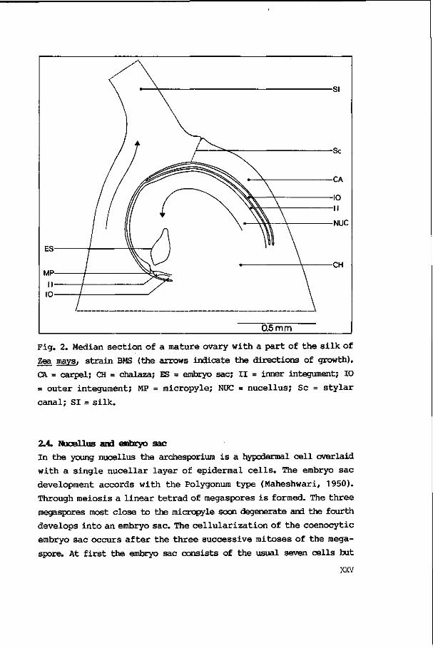
Fig. 2. Median section of a mature ovary with a part of the silk of
Zea mays, strain BMS (the arrows indicate the directions of growth).
CA = carpel; CH = chalaza; ES = embryo sac; II = inner integument; 10
= outer integument; MP = micropyle; NUC = nucellus; Sc = stylar
canal; SI= silk.
2.4. Nucellus and embryo sac
In the young nucellus the archesporium is a hypodermal cell overlaid
with a single nucellar layer of epidermal cells. The embryo sac
development accords with the Polygonum type (Maheshwari, 1950).
Through meiosis a linear tetrad of megaspores is formed. The three
megaspores most close to the micropyle soon degenerate and the fourth
develops into an embryo sac. The cellularization of the coenocytic
embryo sac occurs after the three successive mitoses of the mega-
spore. At first the embryo sac consists of the usual seven cells but
XXV
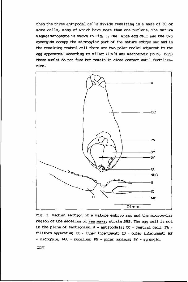
then the t h r ee an t ipodal c e l l s d iv ide r e s u l t i n g i n a mass of 20 or
more c e l l s , many of which have more than one nucleus. The mature
megagametophyte i s shown i n Fig. 3. The l a rge egg c e l l and t he two
synergids occupy the micropylar part of the mature embryo sac and in
the remaining central c e l l there are two polar nuclei adjacent to the
egg apparatus. According t o Miller (1919) and Weatherwax (1919, 1955)
these nuclei do not fuse but remain in close contact un t i l f e r t i l i z a
t ion.
Fig. 3. Median s ec t ion of a mature embryo sac and the micropylar
region of t he nuce l lus of Zea mays, s t r a i n BMS. The egg c e l l i s not
i n the plane of s ec t ion ing . A = an t l poda l s ; CC = c en t r a l c e l l ; FA =
filiform apparatus; I I = inner integument; 10 = outer integument; MP
= micropyle, NUC = nucellus; PN = polar nucleus; SY = synergid.
XXVI
During the interval of meiosis and embryo sac formation the
nucellar epidermis near the micropyle divides periclinally to form a
layer of five or six cells (Kiesselbach, 1949).
3. REFERENCES
Avery, G.S. (1930): Comparative anatomy and morphology of embryos and
seedlings of maize, oats, and wheat. Bot. Gaz. 89(1): 1-39.
Bonnet, O.T. (1940): Development of the staminate and pistillate
inflorescences of sweet corn. J. Agric. Res. 60: 25-37.
(1948): Ear and tassel development in maize. Ann. Mo.Bot. Gard.
35: 269-287.
Bright , S.W.J. & M.G.K. Jones (1985): Cereal t i s s u e and c e l l c u l t u r e .
Martinus Nijhoff/Dr. W. Junk Publishers, Dordrecht, Boston, Lan
caster .
Cheng, P.C., R.I. Greyson & D.B. Walden (1983): Organ i n i t a t i o n and
the development of unisexual flowers in the t asse l and ear of Zea
mays. Am. J . Bot. 70(3): 450-462.
Cooper, D.C (1951): Caryopsis development following matings between
d ip lo id and t e t r a p l o i d s t r a i n s of Zea mays. Am. J . Bot. 38: 702-
708.
D ibol l , A.G. (1964): E lec t ron microscopy of female gametophyte and
early embryo development in Zea mays. Doctoral d isser ta t ion, Univ.
Texas.
(1968a): Fine s t ructural development of the megagametophyte of
Zea mays following f e r t i l i za t ion . Amer. J . Bot. 55(7): 787-806.
(1968b): His tochemistry and f ine s t r u c t u r e of the po l len tube
residue in the megagametophyte of Zea mays. Caryologia 21: 91 -95.
— & D.A. Larson (1966): An e l ec t ron microscopic study of the
mature megagametophyte in Zea mays. Amer. J . Bot. 53(4): 391-402.
Eichler, A.W. (1875-78): Blutendiagramme 2v. in 1, i l l u s . Leipzig.
Gerarde, J . (1597): The herbal o r general h i s t o r i e of p l an t e s . Nor
ton, London, p. 74.
Green, C.E. & R.L. P h i l l i p s (1975): P lan t r egenera t ion from t i s s u e
cultures of maize. Crop Science 15: 417-421.
XXVII

Guignard, L. (1901): La double fecondation dans l e mais . Journ. Bot.
15(2): 37-50.
Harms, CT. (1982): Maize and cereal protoplasts. Facts and perspec
t i v e s . In : Maize for b i o l og i c a l r e sea rch , Sheridan, W.F., Ed.
Univ. Press, Univ. North Dakota, Grand Forks, pp 373-384.
J o h r i , B.M. (1982): Experimental embryology of vascu lar p l a n t s .
Springer-Verlag, Berlin, Heidelberg, New York.
(1984): Embryology of anglosperms. Springer-Verlag, Berlin, Hei
delberg, New York, Tokyo.
Kempton, J.H. (1913): F l o r a l abnormal i t i e s in maize. U.S. Dept. Agr.
Bur. P lan t I ndus t r . Bul l . 278.
Kiesselbach, T.A. (1949): The s t r u c t u r e and reproduct ion of corn.
Nebr. Agr. Exp. S ta . Res. Bui. 161:1-96.
Maheshwari, P. (1950): An i n t roduc t ion t o the embryology of angio
spasms. McGraw-Hill, London, New York.
(1963): Recent advances i n t he embryology of angiosperms.
Catholic Press, Ranchi, India.
Mi l l e r , E.C. (1919): Development of the p i s t i l l a t e s p i k e l e t and
f e r t i l i za t ion in Zea mays L. J . Agricult. Res. 18(5): 255-293.
Raghavan, V. (1976): Experimental embryogenesis in vascular plants.
Acad. Press, London, New York, San Francisco.
Randolph, L.F. (1936): Developmental morphology of the caryopsis in
maize. J . Agr icu l t . Res. 53: 881-916.
Russell, SJJ. (1979): Fine s t ructure of megagametophyte development
i n Zea mays. Can. J . Bot. 57: 1093-1110.
Sass, J.E. (1955): Vegetative morphology. In: Corn and corn improve
ment, Sprague, G.F., Ed. Acad. Press Inc . Publ. , New York, pp 63-
87.
Schel , J.H.N., H. Kieft & A.A.M. van Lammeren (1984): I n t e r a c t i on s
between embryo and endosperm during early developmental stages of
maize caryopses (Zea mays). Can. J . Bot. 62(12): 2842-2853.
Schleiden, M.J. (1837): Einige Blicke auf die Entwicklungsgeschichte
des vegetabilischen Organismus bel den Phanercgamen. Arch. Natur-
geschichte Jahrg . 3 , Bd. 1: (289)-320, 414.
Shannon, J.C. (1982): Maize endosperm cu l t u r e s . In : Maize for b i o
l og i c a l r e search , Sheridan, W.F., Ed. Univ. P ress , Univ. Dakota,
Grand Forks, pp 397-400.
XXVIII
Sheridan, W.F. (1975): Tissue culture of maize. I Callus induction
and growth. Physiol. Plant. 33: 151-156.
(1982): Maize for biological research. Univ. Press, Univ. Dakota,
Grand forks.
Steward, A. (1915): The p i s t i l l a t e s p i ke l e t of Zea mays. Science
n.s.v. 42, no 1089: 694.
S t r a t t on , M.E. (1923): The morphology of the double ke rne l in Zea
mays var . polysperma. N.Y. (Cornell) Agr. Expt. S ta . Mem. 69: 17
pp.
Tomes, D.T. (1985): Cel l c u l t u r e , somatic embryogenesis and p l an t
r egenera t ion in maize, r i c e , sorghum and m i l l e t s . I n : Cereal
t i s s u e and c e l l c u l t u r e , Br ight , S.W.J, and M.G.K. Jones , Eds.
Nijhoff-Junk Publishers, Dordrecht, Boston, Lancaster, pp 175-203.
True, R.H. (1893): On t he development of the ca ryops is . Bot. Gaz. 18:
212-226.
Van Lammeren, AA.M. (1981): Early events during embryogenesis in Zea
mays L. Acta Soc. Bot. Polon. 50(1/2): 289-290.
& H. Kieft (1983): Embryogenesis in Zea mays L. S t r u c t u r a l
a spec t s of primary meristem formation. In : F e r t i l i z a t i o n and
embryogenesis in ovulated p lants , Erdelska, O., Ed., Veda, Bra t is
lava, p. 287.
& J.H.N. Schel (1983): Embryogenesis in Zea mays L. Development
of s tructure and polari ty during proembryo formation. In: F e r t i
l iza t ion and embryogenesis in ovulated plants, Erdelska, O., Ed.,
Veda, Bratislava, pp 283-285.
Vasil, IJC (1982): Somatic embryogenesis and plant regeneration in
cereals and grasses. In: Plant t i ssue culture 1982, Fujiwara, A.,
Ed. Jap. assoc. for plant t i ssue culture, Tokyo, pp 101-104.
Weatherwax, P. (1916): Morphology of the f lowers of Zea mays. Bui.
Torrey Bot. Club 43(3): 127-144.
(1917): The development of the spikelets of Zea mays. Bui. Torrey
Bot. Club 44: 483-496.
(1919): Gametogenesis and fecondation in Zea mays as the basis of
xenia and heredity in the endosperm. Bui. Torrey Bot. Club 46: 73-
90.
XXIX

(1955): S t ruc tu re and development of r eproduct ive organs. I n :
Corn and corn improvement, Sprague, G.F., Ed. Acad. P ress Inc.
Publishers, New York, pp 89-121.
Wigand, A. (1854): Botanische Untersuchungen. Braunschweig. 168 p.
Willemse, M.T.M. & J.L. van Went (1985) (Eds.): Sexual reproduct ion
i n seed p l a n t s , fe rns and mosses. Proc. 8th i n t . symp. on sex.
reprod. in seed p lants , ferns and mosses. Pudoc, Wageningen.
XXX

CHAPTE* 1
A comparative ultrastructural study of the megagametophytes
in two strains of Zea mays L. before
and after fertilization
A.A.M. van Lammeren
(Agric. Univ. Wageningen Papers 86-1 (1986))

AGRICULTURAL UNIVERSITY WAGENINGEN PAPERS 86-1 (1986)
A COMPARATIVE ULTRASTRUCTURAL STUDY OF THE MEGAGAMETOPHYTES
IN TWO STRAINS OF ZEA MA YS L. BEFORE AND AFTER FERTILIZATION
A. A. M. VAN LAMMEREN
Department of Plant Cytology and Morphology Wageningen, Agricultural University, The Netherlands
in Agricultural University Wageningen
The Netherlands 1986

CIP
ISBN 9067540889 ISSN 0169345X
© Agricultural University, Wageningen, the Netherlands, 1986
No part of this publication, apart from bibliographic data and brief quotations embodied in critical reviews, may be reproduced, recorded or published in any form including print, photocopy, microform, electronic or electromagnetic record without written permission from the publisher Agricultural University, P.O. Box 1901,6700 HB Wageningen, the Netherlands.
Printed in the Netherlands by Drukkerij Veenman b.v., Wageningen.
C O N T E N T S
SUMMARY 1
INTRODUCTION 1
MATERIALS AND METHODS 2
RESULTS 3
THE POSITION OF THE EMBRYO SAC IN THE OVULE 3
THE CELLS OF THE EMBRYO SAC BEFORE FERTILIZATION 5
a. synergids 5 b. Egg cell 7 c. Central cell 9 d. Antipodals 12
PENETRATION OF THE POLLEN TUBE INTO THE PISTIL AND THE EMBRYO SAC 13
THE CELLS OF THE EMBRYO SAC AFTER FERTILIZATION 19
a. Synergids 19 b. Zygote 19 c. Endosperm 22 d. Antipodals 23
DISCUSSION 26
PRE - FERTILIZATION AND FERTILIZATION PHASE 26
EGG-ZYGOTE 31
CENTRAL CELL - ENDOSPERM 33
ACKNOWLEDGEMENTS 34
REFERENCES 35
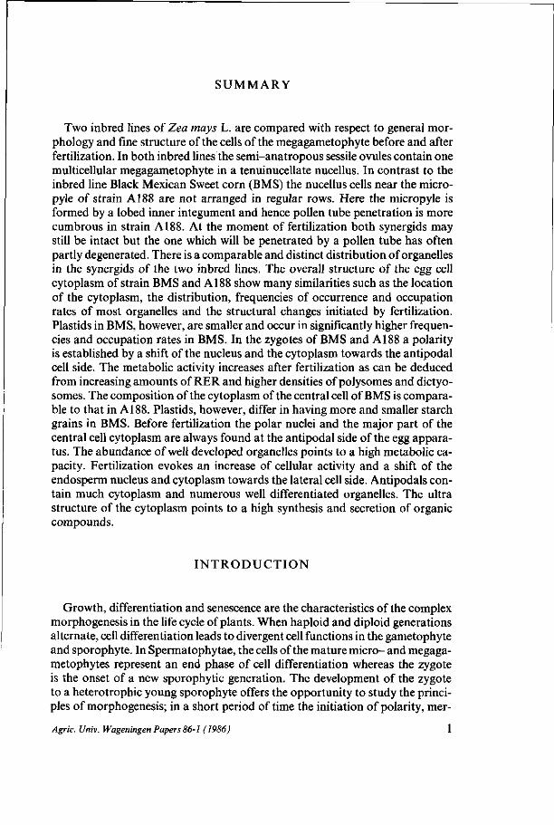
SUMMARY
Two inbred lines of Zea mays L. are compared with respect to general morphology and fine structure of the cells of the megagametophyte before and after fertilization. In both inbred lines the semi-anatropous sessile ovules contain one multicellular megagametophyte in a tenuinucellate nucellus. In contrast to the inbred line Black Mexican Sweet corn (BMS) the nucellus cells near the micro-pyle of strain A188 are not arranged in regular rows. Here the micropyle is formed by a lobed inner integument and hence pollen tube penetration is more cumbrous in strain A188. At the moment of fertilization both synergids may still be intact but the one which will be penetrated by a pollen tube has often partly degenerated. There is a comparable and distinct distribution of organelles in the synergids of the two inbred lines. The overall structure of the egg cell cytoplasm of strain BMS and A188 show many similarities such as the location of the cytoplasm, the distribution, frequencies of occurrence and occupation rates of most organelles and the structural changes initiated by fertilization. Plastids in BMS, however, are smaller and occur in significantly higher frequencies and occupation rates in BMS. In the zygotes of BMS and A188 a polarity is established by a shift of the nucleus and the cytoplasm towards the antipodal cell side. The metabolic activity increases after fertilization as can be deduced from increasing amounts of RER and higher densities of polysomes and dictyo-somes. The composition of the cytoplasm of the central cell of BMS is comparable to that in A188. Plastids, however, differ in having more and smaller starch grains in BMS. Before fertilization the polar nuclei and the major part of the central cell cytoplasm are always found at the antipodal side of the egg apparatus. The abundance of well developed organelles points to a high metabolic capacity. Fertilization evokes an increase of cellular activity and a shift of the endosperm nucleus and cytoplasm towards the lateral cell side. Antipodals contain much cytoplasm and numerous well differentiated organelles. The ultra structure of the cytoplasm points to a high synthesis and secretion of organic compounds.
INTRODUCTION
Growth, differentiation and senescence are the characteristics of the complex morphogenesis in the life cycle of plants. When haploid and diploid generations alternate, cell differentiation leads to divergent cell functions in the gametophyte and sporophyte. In Spermatophytae, the cells of the mature micro- and megaga-metophytes represent an end phase of cell differentiation whereas the zygote is the onset of a new sporophytic generation. The development of the zygote to a heterotrophic young sporophyte offers the opportunity to study the principles of morphogenesis; in a short period of time the initiation of polarity, mer-
Agric. Univ. Wageningen Papers 86-1 (1986) 1
istem formation and organogenesis gives rise to a relatively small organism. The onset of embryogenesis has been studied in detail in plants of various families of both the Dicotyledonae and the Monocotyledonae (MAHESHWARI, 1950, 1963; JOHRI, 1984). The details of fertilization, the developmental pathways and the final organization of the embryos vary widely among families of the Monocotyledonae and even within the family of the Poaceae such as barley (CASS
and JENSEN, 1970;NORSTOG, 1972, 1974;CASS, 1981), wheat (CHANDRA and BHATNAGAR, 1974; Hu, 1964; SMART and O'BRIEN, 1983) and Texas wildrice (EMERY and GUY, 1979).
In maize the shape of the embryo sac, the organelle distribution within the embryo sac and the shape of the developing embryos appear to vary between different strains as has been observed by means of light microscopy (MILLER,
1919; AVERY, 1930; RANDOLPH, 1936; KIESSELBACH, 1949; COOPER, 1951; SASS,
1955) and electron microscopy (DIBOLL, 1964, 1968a, b;CHEBOTARU, 1970; RUS
SELL, 1979; VAN LAMMEREN, 1981; VAN LAMMEREN and KIEFT, 1983; VAN LAM-
MEREN and SCHEL, 1983). Unlike in many other plants (MAHESHWARI, 1950), the first cell divisions of the zygote and the young embryo seem to lack a clearly defined sequence and orientation although, within a strain, the eventual shapes of the embryos are quite similar (RANDOLPH, 1936).
To study the initial phase of maize embryogenesis an inventory was made of the cell shapes and organelle distributions within the embryo sac. Two inbred lines were chosen for the present study which is introductory for the experimental in vitro studies on callus formation and somatic embryogenesis. One inbred line is the sweet corn Black Mexican Sweet (BMS). The second is the starchy corn A188. These strains have been selected because of their favorable properties for experimental manipulation in vitro (SHERIDAN, 1977; GREEN and PHILLIPS,
1975). The present paper describes the changes in the fine structure of the embryo
sac just before fertilization. Then, the interactions of the pollen tubes and the tissues of the pistil including the ovule will be presented. Thirdly the post-fertilization events which occur in the embryo sac will be regarded from a structural and functional point of view. The two inbred lines are compared to detect in-traspecific variations in cytology.
M A T E R I A L S A N D M E T H O D S
The plant material used in this study was obtained from the Zea mays L. inbred lines Black Mexican Sweet (BMS) and A188 which were kindly provided by R.J. Lambert, University of Illinois, Illinois, USA and by C.E. Green, University of Minnesota, St. Paul, USA respectively. Plants were grown under greenhouse conditions; before emergence of the silks, cobs were masked with small bags to prevent uncontrolled pollination. Ovaries were dissected either from unpollinated plants or at defined intervals after hand pollination. Sagittal
Agric. Univ. Wageningen Papers 86-1 (1986)
sections of the ovaries containing the whole embryo sac were fixed with 2,5-6% glutaraldehyde in 0,1 M Na-cacodylate buffer, pH 7.0, for 2 hours at room temperature. Sections were rinsed in the buffer and postfixed in a saturated aqueous K M n 0 4 - solution for 5-15 minutes or in a 1 % 0 s 0 4 - solution in cacodylate buffer, pH 7.2, for 2-4 hours at room temperature. After rinsing, sections were dehydrated in a series of ethanol, ranging from 30% to 100%, followed by a graded series of propylene oxide. The material was then transferred to a propylene oxide - Epon 812 (40:1) mixture and kept overnight at a relative humidity of 30% to permit the propylene oxide to evaporate slowly. Finally, sections were transferred to fresh resin in gelatin capsules. Polymerization occurred for 16 hours at 35 °C, for 8 hours at 45 °C and for 24 hours at 60 °C. Ultrathin sections were cut on an LKB Ultrotome III and, in the case of 0 s 0 4 fixation, poststained in lead citrate for 2-5 minutes and in uranyl acetate for 5-25 minutes. Sections were observed using a Philips EM 301 transmission electron microscope at 60 kV. For the detection of pollen tubes in silks and ovary cavities, sagittal sections (8-50 um) of freshly frozen pistils were cut with a microtome-cryostat (Daman/ IEC division, Mass., USA) at minus 18 °C. Two percent 'Wasserblau' (Merck, Darmstadt, FRG) in 20% aqueous solution of K 3 P0 4 was added to the thawed sections to obtain fluorescence of callosic substances. The observations were recorded on Agfachrom 50L.
RESULTS
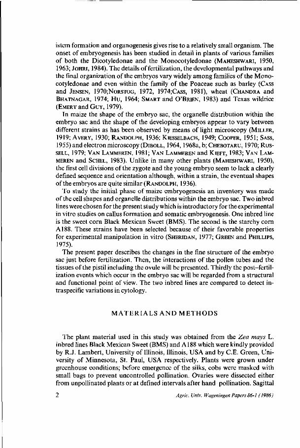
The position of the embryo sac in the ovule The position of the embryo sac in the nucellus with respect to the micropylar
entrance is not similar for the two strains (Fig. 1). In comparison with BMS the embryo sac of strain Al 88 has a more oblique position towards the micropyle and fewer nucellus cells are in between the embryo sac and the inner integument.
In both strains the nucellus cells of the micropylar region divide several times but more regular rows are formed in BMS (c.f. Figs. 2a and b). In BMS, division and enlargement of nucellus cells also occur in a more symmetrical fashion in the micropylar region. In strain A188, cell division and enlargement are less intensive both near the base of the embryo sac and at the side directed towards the single integument. Therefore, the thickness of the nucellus covering the embryo sac is unequal at the two sides of the egg apparatus. Several nucellus cells which border on the mature embryo sac are flattened because of the enlargement of the embryo sac. Here a total collapse of the cells is preceded by a process of cytoplasmic desintegration (Fig. lc).
When a pollen tube penetrates the ovule of A188 there appears not to be one straight way towards the embryo sac like in BMS (Figs. 9a and c). Serial sections of the micropylar region of A188 demonstrate the existence of a lobed micropyle (Fig. 10a). One of the folds in the inner integument lies opposite the egg apparatus forming the passage for the pollen tube (Fig. 10a 5, arrow).
Agric. Univ. Wageningen Papers 86-1 (1986)
Agric. Univ. Wageningen Papers 86-1 (1986)
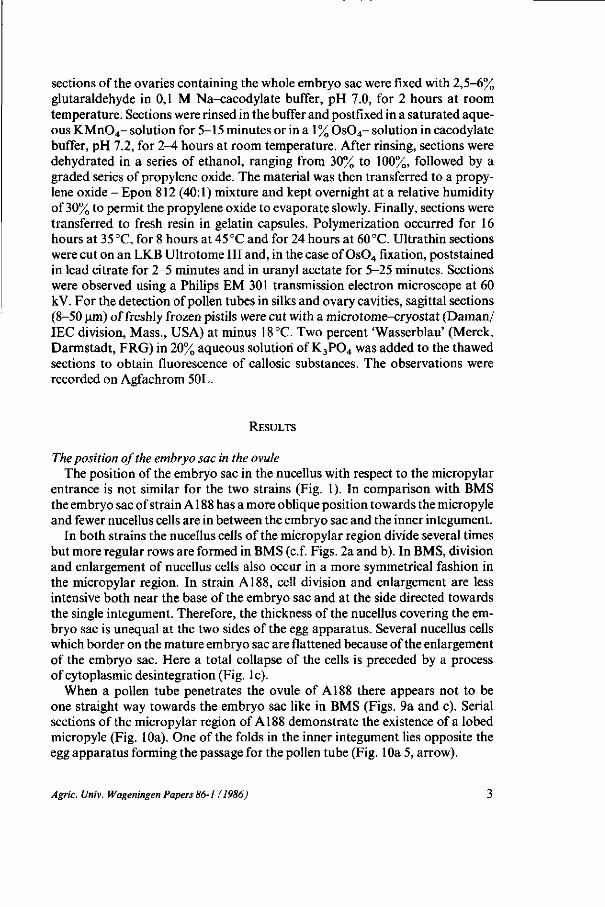
FIG. 2. Median sections through the micropylar parts of the megagametophytes of Zea mays, strain BMS (a) and A188 (b,c). The polar nuclei of the central cell are surrounded by cytoplasm and lie on top of the egg apparatus. In contrast to the synergids the egg cells are highly vacuolated. Their cytoplasm is found in the micropylar half. Note the regular arrangement of the nucellus cells near the micropyle of BMS (a). Several nucellus cells collapse because of the enlargement of the embryo sac.
The cells of the embryo sac before fertilization I n t a c t s yne r g i d s At the time of fertilization both synergids may still be intact. Sometimes, however, one synergid degenerates before fertilization. This synergid will receive the pollen tube. Synergids are about as tall as the egg cell.
FIG. 1. Schematic representation of the megagametophyte positions in the ovaries of Zea mays, strain BMS and A188. A The pistilate spikelet and the ovary with its modified campylotropous ovule. The outer integument does not completely surround the ovule. B, D The embryo sac consists of approximately 20 antipodals, a large central cell and the egg apparatus. C, E With regard to the micropyle there is a difference in position of the egg apparatus in BMS and A188.
Agric. Univ. Wageningen Papers 86-1 (1986) 5

\
»* C / i
/ -^ t .
* > •
N ER
1 AT •*/.• •• .v.!— ••OH , P J I . ^ , . ' ' - - . ' •;.„ -•
! "-.,-• ^ ; * * • . . /
• *r*1*. • • • • ; - ' ^J " ' '
r > » " - W j r*^, .V'-i „*•-*•* • • » . • • • ;
/4gr/c. Univ. Wageningen Papers 86-1 (1986)
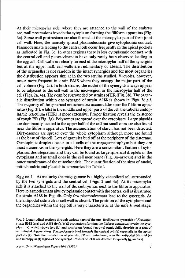
At their micropylar side, where they are attached to the wall of the embryo sac, wall protrusions invade the cytoplasm forming the filiform apparatus (Fig. 3a). Some wall protrusions are also formed at the micropylar part of their joint cell wall. Here, the scarcely spread plasmodesmata give cytoplasmic contact. Plasmodesmata leading to the central cell occur frequently in the apical pockets as indicated in Fig. 3c. In other regions there is less cytoplasmic contact with the central cell and plasmodesmata have only rarely been observed leading to the egg cell. Cell walls are clearly formed at the micropylar half of the synergids but at the upper half, cell walls are rudimentary or absent. The distribution of the organelles is not random in the intact synergids and for most organelles the distribution appears similar in the two strains studied. Vacuoles, however, occur more frequent in strain BMS where they occupy the major part of the cell volume (Fig. 2a). In both strains, the nuclei of the synergids always appear to be adjacent to the cell wall in the mid-region or the micropylar half of the cell (Figs. 2a, 4a). They can be surrounded by strains of ER (Fig. 3b). The organelle distribution within one synergid of strain A188 is shown in Figs. 3d,e,f. The majority of the spherical mitochondria accumulates near the filiform apparatus (Fig. 3f), whilst in the middle and upper parts of the cell the tubular endoplasmic reticulum (TER) is more extensive. Proper fixation reveals the existence of rough ER (Fig. 3g). Polysomes are spread over the cytoplasm. Large plastids are dominantly located in the upper half of the cell but small ones are also found near the filiform apparatus. The accumulation of starch has not been detected. Dictyosomes are spread over the whole cytoplasm although more are found at the base of the cell. Lots of granules bud off at the periphery of the cisternae. Osmiophilic droplets occur in all cells of the megagametophyte but they are most numerous in the synergids. Here they are a concomitant feature of cytoplasmic desintegration and they can be found as large spherical droplets in the cytoplasm and as small ones in the cell membrane (Fig. 3a-arrows) and in the outer membranes of the mitochondria. The quantification of the sizes of nuclei, mitochondria and plastids is summarized in Table I.
Egg cell At maturity the megagamete is a highly vacuolized cell surrounded by the two synergids and the central cell (Figs. 2 and 4a). At its micropylar side it is attached to the wall of the embryo sac next to the filiform apparatus. Here, plasmodesmata give cytoplasmic contact with the central cell as illustrated for strain A188 in Fig. 4b. Only few plasmodesmata lead to the synergids. At the antipodal side a clear cell wall is absent. The position of the cytoplasm and the organelles within the egg cell is very characteristic at the unfertilized stage.
FIG. 3. Longitudinal sections through various parts of the pre- fertilization synergids oiZea mays, strain BMS (a,g) and A188 (b-f). Wall protrusions forming the filiform apparatus invade the cytoplasm (a), which shows free (L) and membrane bound (arrows) osmiophilic droplets as a sign of an initiated degeneration. Plasmodesmata lead towards the central cell (b) especially in the apical pockets (c). Note the distribution of plastids, ER and mitochondria in the antipodal (d), mid (e) and micropylar (1) region of one synergid. Profiles of RER are detected frequently (g, arrows).
Agric. Univ. Wageningen Papers 86-1 (1986)
*o» '*h
V . * • > ,
*• • ••%-"'3k "-J-. '••ft?-/
• • • • • " - . * m
ft
:j&m • b * ? - .:->. 1 H " >
d >X^1| im
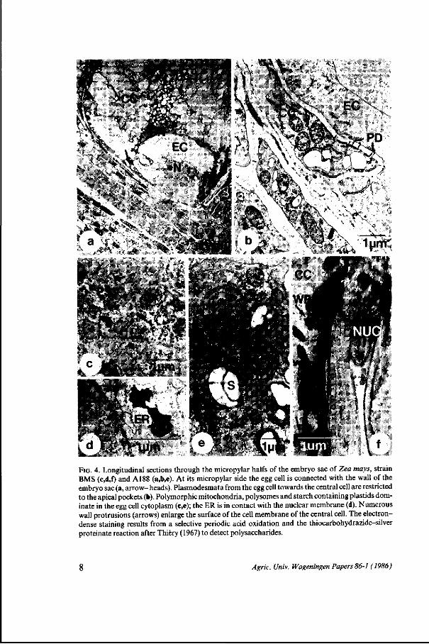
FIG. 4. Longitudinal sections through the micropylar halfs of the embryo sac of Zea mays, strain BMS (c,d,f) and A188 (a,b,e). At its micropylar side the egg cell is connected with the wall of the embryo sac (a, arrow- heads). Plasmodesmata from the egg cell towards the central cell are restricted to the apical pockets (b). Polymorphic mitochondria, polysomes and starch containing plastids dominate in the egg cell cytoplasm (c,e); the ER is in contact with the nuclear membrane (d). Numerous wall protrusions (arrows) enlarge the surface of the cell membrane of the central cell. The electron-dense staining results from a selective periodic acid oxidation and the thiocarbohydrazide-silver proteinate reaction after Thiery (1967) to detect polysaccharides.
Agric. Univ. Wageningen Papers 86-1 (1986)

In both strains the larger part of the cytoplasm and organelles surrounds the spherical nucleus and can often be found near the wall of a synergid in the basal, micropylar half of the cell. Vacuoles of various sizes occupy the upper, antipodal half. A shift of the cytoplasm and the nucleus towards the apex of the cell results in a basal vacuolation and is an early consequence of fertilization as will be shown later on.
Among the organelles mitochondria are most striking in the egg cell cytoplasm. They are often found solitary but data obtained from serial sections show that they can also be arranged around each other, like shells forming a globular structure with a spherical mitochondrion in its centre (Figs. 4c and e). The latter phenomenon is only observed in this cell of the embryo sac and it occurs most frequent in strain Al 88. Many ribosomes and polysomes lie between those mitochondria. Smooth ER and RER are scattered throughout the cytoplasm and the ER makes contact with the outer nuclear membrane (Fig. 4d). The ER may have swollen cisternae in both strains. Polysomes and monosomes occur regularly but dictyosomes contribute for a minor part in the composition of the cytoplasm. There are also few plastids. They are large, contain several starch grains and sometimes they tend to be clustered.
Diagram 1 gives a schematic representation of the organelle shapes in both the egg cells and the zygotes of strain BMS and A188. Diagram 2 and Table II summarize the quantification of sizes, frequences and occupation rates of plastids and mitochondria in the egg cell. It appears that mitochondria and plastids do not differ in average sizes significantly although the graphic presentation of sizes demonstrates the existence of larger mitochondria and plastids in strain A188 (c.f. Diagram 2, BMS 1,5; A188 1,5). The occupation rates of the mitochondria and the plastids, as presented in Table II were calculated from organelle surface measurements within a defined area of cytoplasm in which the surfaces of the nucleus and vacuoles were not included. In both strains, about 35% of the cytoplasm is occupied by mitochondria. The rates measured for plastids were significantly lower: for BMS 20 + 3%,forA188 12 + 4%.
Central cell The cytology of BMS and that of A188 central cells is similar in many aspects. The central cell encompasses the major part of the egg apparatus. The cell wall which borders on the nucellus is well developed, especially in the micropylar region (Fig. 4a, arrows) where it bears small polysaccharide containing protrusions (Fig. 4f, arrows). At the antipodal side of the egg apparatus, however, the cell walls between synergids, egg cell and central cell are often incomplete or undetectable. Here the male gamete is supposed to leave the synergid to fuse with the central cell. In the pre-fertilization phase of the mature megagametophyte the two polar nuclei and the major part of the cytoplasm are always found at the antipodal side of the egg apparatus (Fig. 2a,b; 5a). The nuclei are always close to each other and might already be partly fused before fertilization (Figs. 5b,c). There is continuity between the nuclear envelope and the ER (Fig. 5d). The major part of the central cell is occupied by vacuoles of various sizes. A thin layer of cytoplasm covers the cell membrane adjacent
Agric. Univ. Wageningen Papers 86-1 (1986) 9
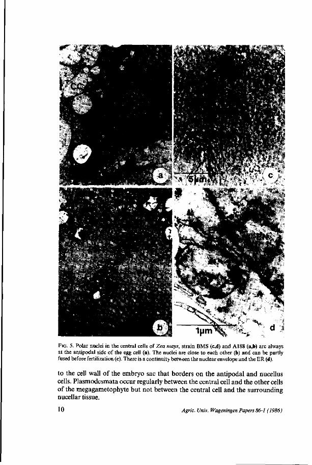
1 y m ^ FIG. 5. Polar nuclei in the central cells of Zea mays, strain BMS (c,d) and A188 (a,b) are always at the antipodal side of the egg cell (a). The nuclei are close to each other (b) and can be partly fused before fertilization (c). There is a continuity between the nuclear envelope and the ER (d).
to the cell wall of the embryo sac that borders on the antipodal and nucellus cells. Plasmodesmata occur regularly between the central cell and the other cells of the megagametophyte but not between the central cell and the surrounding nucellar tissue.
10 Agric. Univ. Wageningen Papers 86-1 (1986)
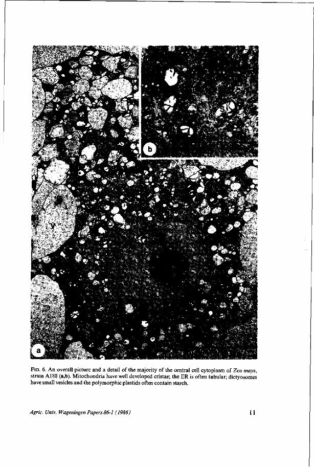
FIG. 6. An overall picture and a detail of the majority of the central cell cytoplasm of Zea mays, strain A188 (a,b). Mitochondria have well developed cristae; the ER is often tubular; dictyosomes have small vesicles and the polymorphic plastids often contain starch.
Agric. Univ. Wageningen Papers 86-1 (1986) 11

For both strains there is a high density of organelles in the cytoplasm around the nuclei as is shown in Fig. 5 and 6. Mitochondria are sometimes polymorphic but mostly ellipsoid and have well developed cristae. The endoplasmic reticulum appears to be mainly tubular in strain A188 whereas in strain BMS long profiles of ER have often been seen in sections. Dictyosomes have small vesicles and are never numerous at this stage. Plastids vary considerably in size and shape and often contain starch grains in strain A188. The organelles of the central cell are schematically represented in Diagram 3. Diagram 2 and Table II summarize the quantification of sizes, frequences and of the occupation rates of mitochondria and plastids in a part of the cytoplasm near the egg apparatus. The mitochondria of the BMS and A188 central cells have about the same size. There are no differences in cytoplasmic occupation rates. For plastids significant differences can neither be detected in mean organelle sizes nor in cytoplasmic occupation rates. When, however, central cells are compared with the egg cells it appears that the occupation rate of mitochondria is significantly higher in the egg cell of strain A188 than in the central cell of that inbred line.
Antipodals Multiplication of the three antipodal cells results in approximately 20 antipodals which form part of the full-grown embryo sac (Figs, lb, Id). Several cells contain more than one nucleus because complete cell separation was not realized after mitosis. Cell walls, adjacent to the nucellar tissue, may
FIG. 7. Antipodals of Zea mays, strain BMS (a,b) contain numerous organelles among which an abundance of ER, dictyosomes and mitochondria. The ER is frequently arranged in long, interconnected sheets parallel to each other (b). Dictyosomes can be in close association with the ER. They have many cristae but secretory granules do not bud off frequently.
12 Agric. Univ. Wageningen Papers 86-1 (1986)
have small protrusions (Fig. 7a) as observed in the central cell, too. Plasmodes-mata allow contact with other antipodals and with the central cell but there is no cytoplasmic contact with the nucellus cells. Some antipodals have large vacuoles, others have only small ones. The cytoplasm contains numerous organelles among which an abundance of ER, dictyosomes and mitochondria (Fig. 7a). In both strains the ER is frequently arranged in long interconnected sheets parallel to each other (Fig. 7b). Intra-cisternal continuity throughout the cytoplasm from the viscinity of the outer nuclear membrane up to the periphery of the cell is obvious. Nuclei may be spherical or invaginated. They are partly enclosed by sheets of ER. Plastids have small thylakoid membranes and may contain starch grains as detected in BMS. In both strains, BMS and A188, a high number of polysomes, many large, well developed dictyosomes, and mitochondria with well developed cristae were observed.
The quantification of the sizes of some organelles is summarized in Table I. The antipodal nuclei are the smallest nuclei in the embryo sac. Antipodal mitochondria and plastids are small, too, like in the synergids. Due to the quantification procedure high standard deviations occur (see legend Diagram 2) and therefore, despite different average sizes, significant distinctions could only be demonstrated incidentally. Only the plastids of BMS antipodals are significantly smaller than the plastids in the egg cell and central cell.
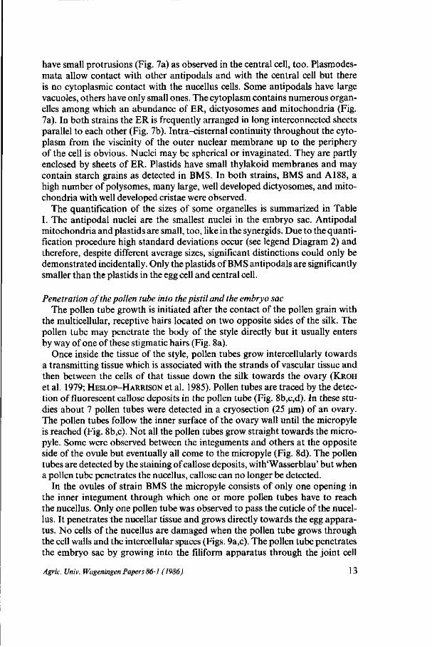
Penetration of the pollen tube into the pistil and the embryo sac The pollen tube growth is initiated after the contact of the pollen grain with
the multicellular, receptive hairs located on two opposite sides of the silk. The pollen tube may penetrate the body of the style directly but it usually enters by way of one of these stigmatic hairs (Fig. 8a).
Once inside the tissue of the style, pollen tubes grow intercellularly towards a transmitting tissue which is associated with the strands of vascular tissue and then between the cells of that tissue down the silk towards the ovary (KROH
et al. 1979; HESLOP-HARRISON et al. 1985). Pollen tubes are traced by the detection of fluorescent callose deposits in the pollen tube (Fig. 8b,c,d). In these studies about 7 pollen tubes were detected in a cryosection (25 urn) of an ovary. The pollen tubes follow the inner surface of the ovary wall until the micropyle is reached (Fig. 8b,c). Not all the pollen tubes grow straight towards the micropyle. Some were observed between the integuments and others at the opposite side of the ovule but eventually all come to the micropyle (Fig. 8d). The pollen tubes are detected by the staining of callose deposits, with'Wasserblau' but when a pollen tube penetrates the nucellus, callose can no longer be detected.
In the ovules of strain BMS the micropyle consists of only one opening in the inner integument through which one or more pollen tubes have to reach the nucellus. Only one pollen tube was observed to pass the cuticle of the nucellus. It penetrates the nucellar tissue and grows directly towards the egg apparatus. No cells of the nucellus are damaged when the pollen tube grows through the cell walls and the intercellular spaces (Figs. 9a,c). The pollen tube penetrates the embryo sac by growing into the filiform apparatus through the joint cell
Agric. Univ. Wageningen Papers 86-1 (1986) 13
Fio. 8. A pollen grain (PG) of Zea mays, strain BMS, adheres to a stigmatic hair. It has germinated and the pollen tube penetrated the body of the silk directly (a, photograph courtesy Dr.Ir. H.J. Wilms). Then pollen tubes grow through the transmitting tissue of the silk into the ovular locule (b,c) towards and between the integuments at the micropyle (c.d).
14 Agric. Univ. Wageningen Papers 86-J (1986)

cc
r~^ .. SY sv
1 ^Ai rji • - * "
i
©
~\
P
i
>G\
FIG. 9. Penetration of the nucellus and embryo sac by the pollen tube in Zea mays, strain BMS; overall view (a), detail (b) and drawing (c). The pollen tube penetrates the cuticle of the nucellus, then it grows through intercellular spaces into the nucellus and eventually it enters the filiform apparatus through the joint cell wall of the two synergids. Pollen tube cytoplasm has been discharged into one synergid.
wall of the two synergids. Then the pollen tube enters a synergid along side the joint cell wall (Fig. 9b). It stops growing and discharges a part of its contents into the synergid which gets filled up completely. When the synergid is penetrated it may have been degenerated already.
When a pollen tube penetrates the ovule of A188 there isn't one straight way towards the embryo sac like in BMS. Serial sections of the micropylar region of A188 demonstrate the existence of a lobed micropyle which has several openings (Fig. 10a,b). The pollen tube winds its way and cannot be observed in one plane of sectioning. One of the folds in the inner integument lies near to the egg apparatus and forms the eventual passage for the pollen tube (Fig.l0a,5, arrow). Once in the nucellus, the pollen tube appears to bend again searching its way to the embryo sac. Reaching the base of the filiform apparatus the pollen
Agric. Univ. Wageningen Papers 86-1 (1986) 15

NUC
NUC
•^r:--i, *H .
16 /Ign'c. £//wv. Wageningen Papers 86-1 (1986)
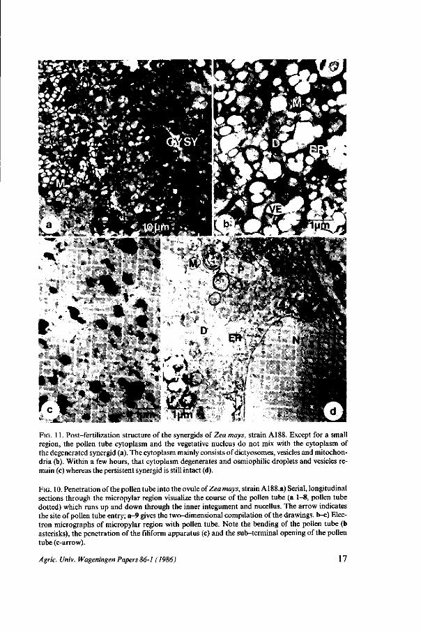
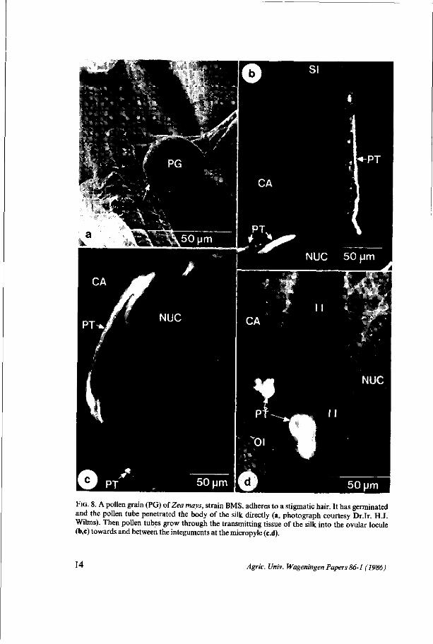
FIG. 11. Post-fertilization structure of the synergids of Zea mays, strain A188. Except for a small region, the pollen tube cytoplasm and the vegetative nucleus do not mix with the cytoplasm of the degenerated synergid (a). The cytoplasm mainly consists of dictyosomes, vesicles and mitochondria (b). Within a few hours, that cytoplasm degenerates and osmiophilic droplets and vesicles remain (c) whereas the persistent synergid is still intact (d).
FIG. 10. Penetration of the pollen tube into the ovule of Zea mays, strain A188.a) Serial, longitudinal sections through the micropylar region visualize the course of the pollen tube (a 1-8, pollen tube dotted) which runs up and down through the inner integument and nucellus. The arrow indicates the site of pollen tube entry; a-9 gives the two-dimensional compilation of the drawings, b-c) Electron micrographs of micropylar region with pollen tube. Note the bending of the pollen tube (b asterisks), the penetration of the filiform apparatus (c) and the sub-terminal opening of the pollen tube (c-arrow).
Agric. Univ. Wageningen Papers 86-1 (1986) 17

i -••/
EN /
kr^-
v- , tW^ L, ...V /&*>&,*••;&
"W
18 /Igr/c. Univ. Wageningen Papers 86-1 (1986)

tube sometimes enlarges and eventually penetrates a synergid like in strain BMS (Fig. 10b,c)
For strain A188 it takes about 21 hours from pollen tube germination up to penetration of the embryo sac and about 26 hours for strain BMS. However, the periods mentioned are largely dependent on the greenhouse conditions.
The cells of the embryo sac after fertilization
Synergids When the pollen tube has penetrated a synergid and discharged its contents into that synergid, the cytoplasm of both cells is not mixed (Fig. 1 la). In some parts of the cell the synergid cytoplasm can still be detected. The cytoplasm coming out of the pollen tube reaches up to the tip of the synergid and can be discerned by its characteristic appearance (Fig. l ib) . Translucent vesicles, originating from the dictyosomes, predominate in the cytoplasm. Mitochondria have long cristae but the accumulation of small osmiophilic droplets which are often attached to the cristae and inner membranes of the envelope indicates the onset of degeneration. Plastids, however, are still intact and some strands of ER are scattered throughout the cytoplasm.
Soon after fertilization the discharged cytoplasm degenerates to such an extent that organelles can hardly be detected (Fig. lie). Large, osmiophilic droplets and numerous small vesicles are the main constituents of the cytoplasm. The persistent synergid may be structurally intact until the division of the zygote (Fig. 1 Id) whereas even before fertilization signs of degeneration were observed (Fig. 3a).
Zygote Just after fertilization the egg cell and zygote are not remarkably different in cell size or in shape and yet they are easily distinguished by observing the location of the cytoplasm. As shown previously for the egg cell, the majority of the cytoplasm surrounds the nucleus in the micropylar half of that cell (Fig. 2). As a striking result of fertilization the cytoplasm and the nuclei of the zygotes of both strains BMS and A188 are translocated towards the antipodal side of the cell (Fig. 12,13,17a). The syngamy proper and the karyogamy, however, were not observed and organelles of the male gamete were not distinguished. Fertilization causes no significant change in size of the zygote but in the cytoplasm an increasing complexity is observed (Diagram 1). This holds for the ER, the dictyosomes and the polysomes rather than for the plastids and mitochondria of which the sizes (Diagram 2), occupation rates and frequences (Table II) do not change significantly. As was observed in the egg cells of BMS and A188, the mitochondria are often stacked, forming globular structures. In the zygotes they have large cristae (Figs. 14a,b). The ER tends to concentrate in some areas (Fig. 14c).
FIG. 12. Phase contrast light micrograph of a longitudinal section through the embryo sac of Zea mays, strain BMS. After fertilization the prophase nucleus and the cytoplasm are located in the antipodal half of the zygote whereas the endosperm cytoplasm shifted lateral.
Agric. Univ. Wageningen Papers 86-1 (1986) 19
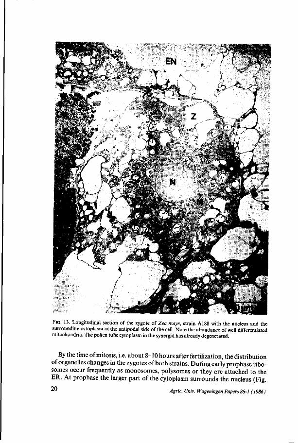
FIG. 13. Longitudinal section of the zygote of Zea mays, strain A188 with the nucleus and the surrounding cytoplasm at the antipodal side of the cell. Note the abundance of well differentiated mitochondria. The pollen tube cytoplasm in the synergid has already degenerated.
By the time of mitosis, i.e. about 8-10 hours after fertilization, the distribution of organelles changes in the zygotes of both strains. During early prophase ribo-somes occur frequently as monosomes, polysomes or they are attached to the ER. At prophase the larger part of the cytoplasm surrounds the nucleus (Fig. 90 i u Agric. Univ. Wageningen Papers 86-1 (1986)
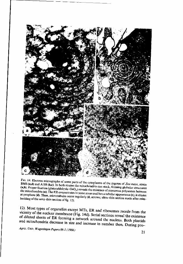
(a,b). Proper fixation ( t f u t a r a K S ^ ^ ^ f S ^ ™ * " * ' forming S^"'3' struct«es the mnochondria (a); L E £ C O » £ £ S Z T * °f n U m e r o u s P 0 ^™* between at prophase W ^ u ^ S ^ ^ ^ ^ ^ ^ ^ ^ 1 ^ ^ ^ ^ ^ ^ ^ beddl„g of the serm-thin section of fig 12) " ( ' ̂ U l t r a ~ t h M S 6 C t i ° n m a d e a f t e r «*»"
21 ^gn'c. t/m'v. Wageningen Papers 86-1 (1986)
: \ . .v - *'j AT
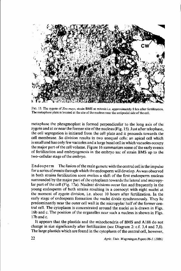
FIG. 15. The zygote of Zea mays, strain BMS at mitosis i.e. approximately 8 hrs after fertilization. The metaphase plate is located at the site of the nucleus near the antipodal side of the cell.
metaphase the phragmoplast is formed perpendicular to the long axis of the zygote and at or near the former site of the nucleus (Fig. 15). Just after telophase, the cell segregation is initiated from the cell plate and it proceeds towards the cell membrane. So division results in two unequal cells: an apical cell which is small and has only few vacuoles and a large basal cell in which vacuoles occupy the major part of the cell volume. Figure 16 summarizes some of the early events of fertilization and embryogenesis in the embryo sac of strain BMS up to the two-cellular stage of the embryo.
Endosperm The fusion of the male gamete with the central cell is the impulse for a series of events through which the endosperm will develop. As was observed in both strains fertilization soon evokes a shift of the first endosperm nucleus surrounded by the major part of the cytoplasm towards the lateral and micropy-lar part of the cell (Fig. 17a). Nuclear divisions occur fast and frequently in the young endosperm of both strains resulting in a coenocyt with eight nuclei at the moment of zygote division, i.e. about 10 hours after fertilization. In the early stage of endosperm formation the nuclei divide synchronously. They lie predominantly near the outer cell wall in the micropylar half of the former central cell. The cytoplasm is concentrated around the nuclei as is drawn in Figs. 16b and c. The position of the organelles near such a nucleus is shown in Figs. 17b and c.
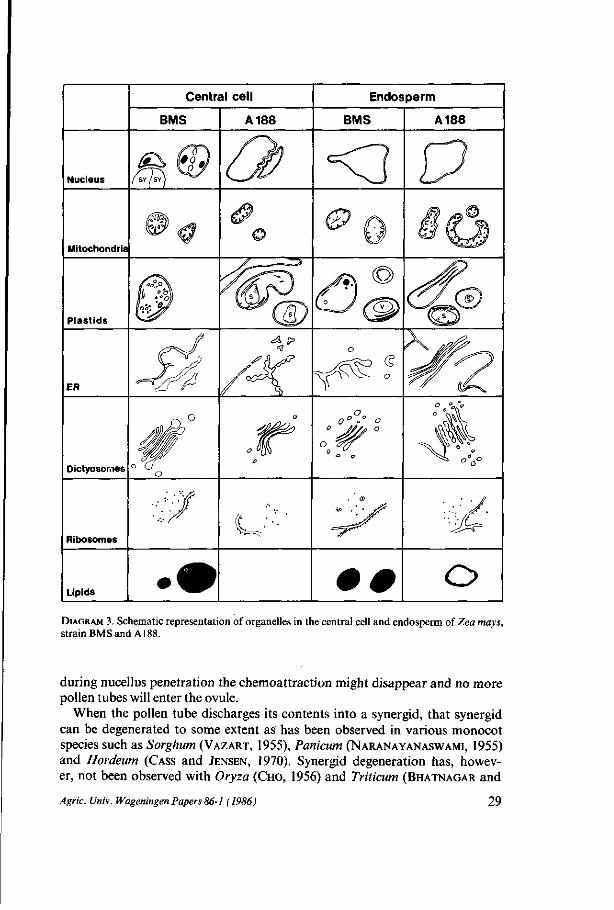
It appears that the plastids and the mitochondria of BMS and A188 do not change in size significantly after fertilization (see Diagram 2: c.f. 3,4 and 7,8). The large plastids which are found in the cytoplasm of the central cell, however,
22 Agric. Univ. Wageningen Papers 86-1 (1986)

FIG. 16. Summary of the early events of fertilization and early embryogenesis in Zea mays, strain BMS. Note the characteristic positions of the cytoplasms of the egg cell and central cell at 0 hours after pollination (A) and the shift of the cytoplasms after fertilization (B, striated arrows). Mitosis results in a small apical cell and a large vacuolated basal cell (C). Than the endosperm is multinucleate.
are not found in the endosperm. The occupation rates of mitochondria and plas-tids of BMS and A188 do not differ significantly in the central cell and the endosperm (Table II). When the mitochondria of the central cell and the endosperm of A188 are compared with the mitochondria of the egg and the zygote respectively, there is no significant difference in size (Diagram 2: c.f. 3,4 and 1,2), but there is a significant difference in occupation rates being twice as high in the egg and zygote (Table II). As a result of fertilization, dictyosomes increase their complexity and a higher concentration of polysomes was detected in the young endosperm of BMS and A188 (Diagram 3). Long profiles of ER were found running parallel to the cell membrane and near the nuclei.
Antipodals Fertilization has no notable influence on the size and shape of the antipodals of BMS and Al 88 (Fig. 18a). Plasmodesmata between antipodals
Agric. Univ. Wageningen Papers 86-1 (1986) 23
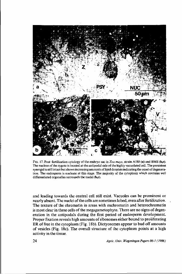
FIG. 17. Post-fertilization cytology of the embryo sac in Zea mays, strain A188 (a) and BMS (b,c). The nucleus of the zygote is located at the antipodal side of the highly vacuolated cell. The persistent synergid is still intact but shows increasing amounts of lipid droplets indicating the onset of degeneration. The endosperm is nucleate at this stage. The majority of the cytoplasm which contains well differentiated organelles surrounds the nuclei (b,c).
and leading towards the central cell still exist. Vacuoles can be prominent or nearly absent. The nuclei of the cells are sometimes lobed, even after fertilization. The texture of the chromatin in areas with euchromatin and heterochromatin is most clear in these cells of the megagametophyte. There are no signs of degeneration in the antipodals during the first period of endosperm development. Proper fixation reveals high amounts of ribosomes either bound to proliferating ER of free in the cytoplasm (Fig. 18b). Dictyosomes appear to bud off amounts of vesicles (Fig. 18c). The overall structure of the cytoplasm points at a high activity in the tissue.
24 Agric. Univ. Wageningen Papers 86-1 (1986)

•. .J- M . ,jfv*«i '."?--_ .-5W • V T . ̂ . -j.' •
* * . ' '-'
-%:-.
N * • * . >
| jm
FIG. 18. Antipodals of Zea mays, strain A188 (a,c) and BMS (b) after fertilization. Well developed dictyosomes (c) and high amounts of ribosomes either bound to intensively proliferating ER (b,c) or free in the cytoplasm point to a high activity in the tissue.
BMS
A188
^ ^ - - ^ce l l s o r g a n e l l e s ^ ^ ^ ^ ^
Nuclei
Mitochondria
Plastids
Nuclei
Mitochondria
Plastids
Synergids
70
0.36 + 0.07
1.23 + 0.6
Egg cell
185
0.78 + 0.66
2.79 + 1.6*
1*1
0.22 + 0.07
1.57 + 1.06
200
1.60 + 1.15
5.38 + 2.93
Central cell
310
0.62 ^0 . 34
*.75 + 4.17
680
0.8* + 0.65
5.00 ^ 3 . 15
Antipodals
30
0.*2 + 0.07
1.21 + 0.39
23
0.2* i 0.07
0.68 + 0.35
TABLE I. Average sizes* of nuclei, mitochondria and plastids in the cells of the unfertilized megaga-metophyte of Zea mays, strain BMS and Al 88. Measurements are performed with an image analyser (Kontron, MOP-30) and expressed in urn2. *See note in legend of diagram 2.
Agric. Univ. Wageningen Papers 86-1 (1986) 25

~~~~~^^^cell types Organelles - ^ ^ ^
Mitochondria
BMS
A188
Egg cell
36 + 12% 56 *_ 21
33 + 14% 19 + 11
Zygote
25 + 7% 32 + 25
24 + 5% 15 + 11
Central cell
27 + 13% 40 + 20
16 + 1% 1 7 + 6
Endosperm
24+ 3% 39 + 24
14 + 4% 17 +_ 4
Plastids BMS
A188
20 + 3% 5.5 ± 2.1
12 + 4% 1.7 + 0.5
12 + 5% 2.8 + 0.8
22 + 7% 3.1 + 1.8
16 + 8% 7.3 +_ 5.0
23 + 7% 4.6 + 1.2
10 + 3% 6.4 + 3.5
23 + 11% 7.8 + 4.6
TABLE II. Occupation rates (italics) and frequencies of occurrence (capitals) of mitochondria and plastids in the egg cells, zygotes, central cells and endosperm of Zea mays, strain BMS and A188. Each value and its standard deviation are based on five independent measurements. Frequencies of occurrence are expressed by the 100 urn2.
DISCUSSION
Prefer tilization and fertilization phase After pollination and the germination of the attached pollen, the pollen tubes
grow through the cortex of the silk towards one of the two parenchyma layers which are associated with the two vascular bundles of the silk (KROH et al., 1979; HESLOP-HARRISON et al., 1985). The parenchyma layers are interpreted as being a transmitting tissue because the pollen tubes grow further down the silk through the intercellular spaces of the fusiform cells of that tissue. Intercellular growth of pollen tubes has also been reported for other members of the Poa-ceae (see RANDOLPH, 1936; CHO, 1956: BONNET, 1961; BATYGINA, 1966; CHAN
DRA and BHATNAGAR, 1974). In maize, not all the pollen tubes which grow down in the transmitting tissue reach the ovule. The late-entering tubes are eliminated either at a stigma abscission zone on the base of the silk or at a constricted zone of the transmitting tracts in the upper ovary wall (HESLOP-HARRISON et al., 1985). Once inside the ovary the pollen tubes penetrate the inner epidermis and the cuticle of the ovary wall and they grow in between an integument and the ovary wall towards the micropyle as has also been observed with Hordeum (CASS and JENSEN, 1970). During the penetration of cell walls and cuticles the pollen tube probably excretes enzymes to digest cellulose, hemicellulose, pectine and cutin. At the same time it is dependent on metabolites provided by the sporo-phyte. Because the pollen tube growth, which will result in porogamy, is clearly directed towards the micropyle, a chemoattraction has often been suggested (see VAN WENT and WILLEMSE, 1984) The starch which was observed to accumulate in the micropylar region of the integuments and in the micropylar region of the nucellus might function in that pollen tube attraction. The carbohydrates are excreted into the intercellular spaces to provide the pollen tube with energy.
26 Agric. Univ. Wageningen Papers 86-1 (1986)

Mitochondria
Egg cell
BMS A188
Zygote
BMS A188
Plastids
ER <SD <? <?
Dictyosomes
Ribosomes
Lipids # o / /
DIAGRAM 1. Schematic representation of organelles in the egg cell and zygote of Zea mays, strain BMSandA188.
SINGH and MALIK (1976) propose that metabolites, such as carbohydrates, are also passed on to the filiform apparatus which serves as an entry for metabolites towards the embryo sac. In both strains of Zea only one pollen tube entered each ovule. Multiple pollen tube entry into the ovule has been observed in Triti-cum (CHANDRA and BHATNAGAR, 1974), in a remote hybridization of Triticum (BATYGINA, 1966) and in Oryza (CHO, 1956). The entry of only one maize pollen tube in the nucellus might be explained by the relative short period of time which is necessary for the growth of that pollen tube through the thin layer of tissue from the nucellar cuticle to the degenerated synergid (distance: 20- 40 um; estimated time 1,5-3 min.). As soon as the synergid is penetrated and probably
Agric. Univ. Wageningen Papers 86-1 (1986) 27

Strain BMS 3 0 '
20-
10-
30-
20-
10-
MITOCHONDRIA %
30' egg cell x =0.78 tO.66 pnV 2 0-
10-1
PLASTIDS egg cell
x = 2.7911.64 | jm 2
1 2 3 4 5 £ 7pm2. zygote x = 1.01 +0.70 pm2 20-
10-
2 _ l
^
30
20
10
2 4 6 8 10 12 14 1 6 p m 2
zygote X -3 .34 + 2.69 pm 2
-4=1-central cell x=0.62 + 0.34 pm2
1
ML. 30 i
20
10-1
x
3 0 i Strain A188 20-
-i n 1 1 1
endosperm x =0.84 tO 88 pm?
30-
20-
10-
central cell x =4.75 +4.75 pm2
7
30'
20
10-
endosperm x = 4.50+1.41 (tm2
egg cell x =5 .38 + 2 98 pm2
10 12 14 16 pm 2
DIAGRAM 2. Size distributions, calculated average sizes and standard deviations of cut areas of mitochondria and plastids in the egg cell-zygote and central cell-endosperm of Zea mays, strain BMS and A188. Each histogram is based on measurements in five different areas of cytoplasm. When organelle surfaces are measured on thin sections one should realize that the organelles can be cut at their maximal diameter and that they can be grazed. Therefore the average sizes do not present real organelle sizes and they coincide with high standard deviations.
28 Ageic. Univ. Wageningen Papers 86-1 (1986)
Central cell
BMS A188
Endosperm
BMS A188
t^rrs^ \ n ml
Nucleus
Mitochondria
Plastids
ER
X
_S>
(g
Dictyosor.ies
o 0° °• o
a
Ribosomes
Lipids o
DIAGRAM 3. Schematic representation of organelles in the central cell and endosperm of Zea mays, strain BMS and A188.
during nucellus penetration the chemoattraction might disappear and no more pollen tubes will enter the ovule.
When the pollen tube discharges its contents into a synergid, that synergid can be degenerated to some extent as has been observed in various monocot species such as Sorghum (VAZART, 1955), Panicum (NARANAYANASWAMI, 1955) and Hordeum (CASS and JENSEN, 1970). Synergid degeneration has, however, not been observed with Oryza (CHO, 1956) and Triticum (BHATNAGAR and
Agric. Univ. Wageningen Papers 86-1 (1986) 29
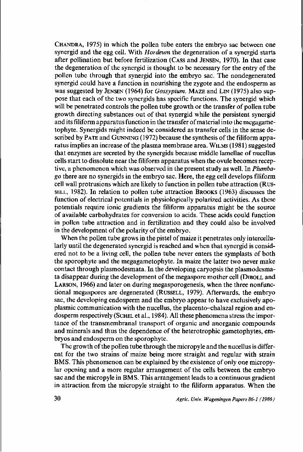
CHANDRA, 1975) in which the pollen tube enters the embryo sac between one synergid and the egg cell. With Hordeum the degeneration of a synergid starts after pollination but before fertilization (CASS and JENSEN, 1970). In that case the degeneration of the synergid is thought to be necessary for the entry of the pollen tube through that synergid into the embryo sac. The nondegenerated synergid could have a function in nourishing the zygote and the endosperm as was suggested by JENSEN (1964) for Gossypium. MAZE and LIN (1975) also suppose that each of the two synergids has specific functions. The synergid which will be penetrated controls the pollen tube growth or the transfer of pollen tube growth directing substances out of that synergid while the persistent synergid and its filiform apparatus function in the transfer of material into the megagame-tophyte. Synergids might indeed be considered as transfer cells in the sense described by PATE and GUNNING (1972) because the synthesis of the filiform apparatus implies an increase of the plasma membrane area. WILMS (1981) suggested that enzymes are secreted by the synergids because middle lamellae of nucellus cells start to dissolute near the filiform apparatus when the ovule becomes receptive, a phenomenon which was observed in the present study as well. In Plumbago there are no synergids in the embryo sac. Here, the egg cell develops filiform cell wall protrusions which are likely to function in pollen tube attraction (RUS
SELL, 1982). In relation to pollen tube attraction BROOKS (1963) discusses the function of electrical potentials in physiologically polarized activities. As these potentials require ionic gradients the filiform apparatus might be the source of available carbohydrates for conversion to acids. These acids could function in pollen tube attraction and in fertilization and they could also be involved in the development of the polarity of the embryo.
When the pollen tube grows in the pistel of maize it penetrates only intercellu-larly until the degenerated synergid is reached and when that synergid is considered not to be a living cell, the pollen tube never enters the symplasts of both the sporophyte and the megagametophyte. In maize the latter two never make contact through plasmodesmata. In the developing caryopsis the plasmodesma-ta disappear during the development of the megaspore mother cell (DIBOLL and LARSON, 1966) and later on during megasporogenesis, when the three nonfunctional megaspores are degenerated (RUSSELL, 1979). Afterwards, the embryo sac, the developing endosperm and the embryo appear to have exclusively apo-plasmic communication with the nucellus, the placento-chalazal region and endosperm respectively (SCHEL et al., 1984). All these phenomena stress the importance of the transmembranal transport of organic and anorganic compounds and minerals and thus the dependence of the heterotrophic gametophytes, embryos and endosperm on the sporophyte.
The growth of the pollen tube through the micropyle and the nucellus is different for the two strains of maize being more straight and regular with strain BMS. This phenomenon can be explained by the existence of only one micropy-lar opening and a more regular arrangement of the cells between the embryo sac and the micropyle in BMS. This arrangement leads to a continuous gradient in attraction from the micropyle straight to the filiform apparatus. When the
30 Agric. Univ. Wageningen Papers 86-1 (1986)
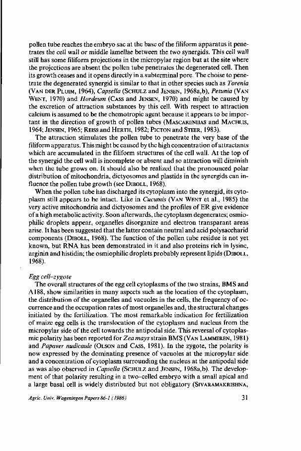
pollen tube reaches the embryo sac at the base of the filiform apparatus it penetrates the cell wall or middle lamellae between the two synergids. This cell wall still has some filiform projections in the micropylar region but at the site where the projections are absent the pollen tube penetrates the degenerated cell. Then its growth ceases and it opens directly in a subterminal pore. The choise to penetrate the degenerated synergid is similar to that in other species such as Torenia (VAN DER PLUIM, 1964), Capsella (SCHULZ and JENSEN, 1968a,b), Petunia (VAN
WENT, 1970) and Hordeum (CASS and JENSEN, 1970) and might be caused by the excretion of attraction substances by this cell. With respect to attraction calcium is assumed to be the chemotropic agent because it appears to be important in the direction of growth of pollen tubes (MASCARENHAS and MACHLIS,
1964; JENSEN, 1965; REISS and HERTH, 1982; PICTON and STEER, 1983).
The attraction stimulates the pollen tube to penetrate the very base of the filiform apparatus. This might be caused by the high concentration of attractants which are accumulated in the filiform structures of the cell wall. At the top of the synergid the cell wall is incomplete or absent and so attraction will diminish when the tube grows on. It should also be realized that the pronounced polar distribution of mitochondria, dictyosomes and plastids in the synergids can influence the pollen tube growth (see DIBOLL, 1968).
When the pollen tube has discharged its cytoplasm into the synergid, its cytoplasm still appears to be intact. Like in Cucumis (VAN WENT et al., 1985) the very active mitochondria and dictyosomes and the profiles of ER give evidence of a high metabolic activity. Soon afterwards, the cytoplasm degenerates; osmio-philic droplets appear, organelles disorganize and electron transparant areas arise. It has been suggested that the latter contain neutral and acid polysaccharid components (DIBOLL, 1968). The function of the pollen tube residue is not yet known, but RNA has been demonstrated in it and also proteins rich in lysine, arginin and histidin; the osmiophilic droplets probably represent lipids (DIBOLL,
1968).
Egg cell-zygote The overall structures of the egg cell cytoplasms of the two strains, BMS and
A188, show similarities in many aspects such as the location of the cytoplasm, the distribution of the organelles and vacuoles in the cells, the frequency of occurrence and the occupation rates of most organelles and, the structural changes initiated by the fertilization. The most remarkable indication for fertilization of maize egg cells is the translocation of the cytoplasm and nucleus from the micropylar side of the cell towards the antipodal side. This reversal of cytoplasmic polarity has been reported for Zea mays strain BMS (VAN LAMMEREN, 1981) and Papaver nudicaule (OLSON and CASS, 1981). In the zygote, the polarity is now expressed by the dominating presence of vacuoles at the micropylar side and a concentration of cytoplasm surrounding the nucleus at the antipodal side as was also observed in Capsella (SCHULZ and JENSEN, 1968a,b). The development of that polarity resulting in a two-celled embryo with a small apical and a large basal cell is widely distributed but not obligatory (SIVARAMAKRISHNA,
Agric. Univ. Wageningen Papers 86-1 (1986) 31

1978; JOHRI, 1984). In contrast to many dicots the position of the cell plate resulting from the first zygotic mitosis is not well predictable in the Gramineae family (see WARDLAW, 1955; JOHRI, 1984). Both the cell size and the orientation of the new cell wall may variate. The change in the compartmentation of the zygote results in the polarity. Whether this polarity is developed by an intensive formation of vacuoles at the micropylar side of the cell or by an active translocation of the cytoplasm and nucleus towards the antipodal side remains to be elucidated. The translocation always coincides with the sideward movement of the cytoplasm and nucleus of the fertilized central cell. The existence of a correlation of these phenomena has, however, not been stated up to now. Whether microtubules are involved in the translocation has not been observed because they have only been detected in the zygote during mitosis.
The overall structures of the cytoplasms of the egg cell and zygote indicate a high potency of energy supply. The egg cell is in a state of relatively low activity first. After fertilization, however, the synthesis capacity of the zygote increases as can be deduced from the increasing amount of RER and the higher density of polysomes and dictyosomes (see Table I). Indeed, this cell needs the equipment for high activities such as cell division and increased production of cytoplasm. SINGH and MALIK (1976) observed very feeble enzyme activity in ovules of Zea mays and strong reactions for several enzymes prior to, during and after fertilization indicating tempory enhancement of metabolic activity. Studies using radioactive nucleic acid and amino acid tracers show that many animal egg cells are also in a state of metabolic inhibition at maturity (MONROY, 1965). The observation that egg cells of maize are in rest and not meristematic corresponds to observations on the barley egg cell (BUTTROSE, 1963) and to several observations on the maize egg cell (VAZART 1958; DIBOLL and LARSON, 1966). In wheat, equal cytological and cytochemical changes like an increased concentration of starch grains around the nucleus, an increased vacuolation, and a decrease of RNA are regarded as manifestations of life activity at a low level (Hu, 1964). With maize, however, mitochondria in the egg cell are well differentiated bearing many cristae. This indicates the potential for the high metabolic rates generally associated with post-fertilization activity (RUNNSTROM et al., 1959). Their clustered location points to high and local demands for ATP in both the egg cell and the zygote. After fertilization the increase in size of the mitochondria and plastids in strain A188 corresponds to the observations of DIBOLL (1968) and this can indeed be a sign of the increased metabolic activity in the zygote. At the time of the first mitosis of the zygote, however, the decrease in size and the increase in number of mitochondria suggest that the division of these organelles precedes mitosis. Protein synthesis is restricted to ribosomes and ER and since protein synthesis is ATP dependent, the occurrence of many ribosomes adjacent to the mitochondria supports the suggestion that there is either a high potential capacity or a high rate of protein synthesis. Starch containing plastids were detected in both the egg cells and zygotes of BMS and A188. The cytoplasm of BMS and A188 egg cells can, however, be distinguished because the plastids of BMS are smaller and occur in significantly higher fre-
32 Agric. Univ. Wageningen Papers 86-1 (1986)
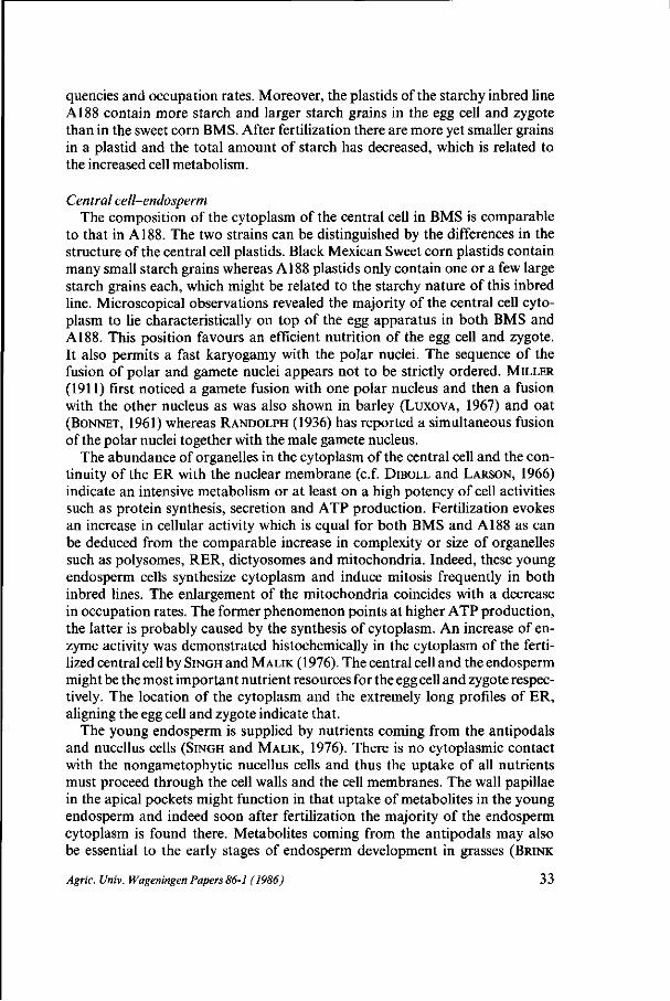
quencies and occupation rates. Moreover, the plastids of the starchy inbred line A188 contain more starch and larger starch grains in the egg cell and zygote than in the sweet corn BMS. After fertilization there are more yet smaller grains in a plastid and the total amount of starch has decreased, which is related to the increased cell metabolism.
Central cell-endosperm The composition of the cytoplasm of the central cell in BMS is comparable
to that in A188. The two strains can be distinguished by the differences in the structure of the central cell plastids. Black Mexican Sweet corn plastids contain many small starch grains whereas A188 plastids only contain one or a few large starch grains each, which might be related to the starchy nature of this inbred line. Microscopical observations revealed the majority of the central cell cytoplasm to lie characteristically on top of the egg apparatus in both BMS and A188. This position favours an efficient nutrition of the egg cell and zygote. It also permits a fast karyogamy with the polar nuclei. The sequence of the fusion of polar and gamete nuclei appears not to be strictly ordered. MILLER
(1911) first noticed a gamete fusion with one polar nucleus and then a fusion with the other nucleus as was also shown in barley (LUXOVA, 1967) and oat (BONNET, 1961) whereas RANDOLPH (1936) has reported a simultaneous fusion of the polar nuclei together with the male gamete nucleus.
The abundance of organelles in the cytoplasm of the central cell and the continuity of the ER with the nuclear membrane (c.f. DIBOLL and LARSON, 1966) indicate an intensive metabolism or at least on a high potency of cell activities such as protein synthesis, secretion and ATP production. Fertilization evokes an increase in cellular activity which is equal for both BMS and A188 as can be deduced from the comparable increase in complexity or size of organelles such as polysomes, RER, dictyosomes and mitochondria. Indeed, these young endosperm cells synthesize cytoplasm and induce mitosis frequently in both inbred lines. The enlargement of the mitochondria coincides with a decrease in occupation rates. The former phenomenon points at higher ATP production, the latter is probably caused by the synthesis of cytoplasm. An increase of enzyme activity was demonstrated histochemically in the cytoplasm of the fertilized central cell by SINGH and MALIK (1976). The central cell and the endosperm might be the most important nutrient resources for the egg cell and zygote respectively. The location of the cytoplasm and the extremely long profiles of ER, aligning the egg cell and zygote indicate that.
The young endosperm is supplied by nutrients coming from the antipodals and nucellus cells (SINGH and MALIK, 1976). There is no cytoplasmic contact with the nongametophytic nucellus cells and thus the uptake of all nutrients must proceed through the cell walls and the cell membranes. The wall papillae in the apical pockets might function in that uptake of metabolites in the young endosperm and indeed soon after fertilization the majority of the endosperm cytoplasm is found there. Metabolites coming from the antipodals may also be essential to the early stages of endosperm development in grasses (BRINK
Agric. Univ. Wageningen Papers 86-1 (1986) 33
and COOPER, 1944). The papillate cell walls of the antipodals are probably functional in the movement of large amounts of nutritive materials from adjacent tissues. SCHNEPF (1964) demonstrated that cells which emerge secretory products have papillate cell walls, too. The cytoplasm of the antipodals exhibits a characteristic increase in organelle number and apparent activity after fertilization as was demonstrated both histochemically (SINGH and MALIK, 1976) and structurally (DIBOLL and LARSON, 1968).
In all, it appears that the cytological development in the cells of themegagame-tophytes show many similarities in the two varieties of Zea mays, both before and after fertilization. Principally some remarkable differences in the structure of the ovule and the position of the embryo sac characterize the two inbred lines. Up to now the functions of all the cell types of the megagametophyte can not be determined unambiguously. The subcellular organization, however, clearly indicates the most probable functions. Synergids and micropylar nucellus cells function in pollen tube attraction. The central cell and endosperm nourish the egg cell and young embryo respectively. The antipodals function in the synthesis and secretion of organic compounds made from precursors coming from the nucellus. Before fertilization the cells of the megagametophyte exhibit signs of moderate metabolic activity. This explains the various stages of degeneration of one synergid at the moment of fertilization. After fertilization the metabolic activity increases.
ACKNOWLEDGEMENTS
Thanks are owed to Mr. S. Massalt for the photographic work. Mr. A.B. Haasdijk and Mr. P.A. van Snippenburg for the artwork and Mrs. J. Cobben-Molenaar for preparing the typescript. The author is also grateful to Prof.dr. M.T.M. Willemse and Dr. J.H.N. Schel for helpful comments and to Drs. F.J. Vegter for reading the English text.
Abbreviations in the figures
A = antipodals; CA= carpel; CC = central cell; CH = chromosome; CU = cuticle; CW = cell wall; CY = cytoplasm; DSY = degenerated synergid; D = dictyosome; EC= egg cell; EN = endosperm; ER = endoplasmic reticulum; ES = embryo sac; FA = filiform apparatus; II = inner integument; IS = intercellular space; L = lipid; M = mitochondrion; MP = micropyle; N = nucleus; NUC = nucellus; OI = outer integument; P = plastid; PD = plasmodesma; PN = polar nucleus; PSY = persistent synergid; PS = polysomes; PT = pollen tube; RER = rough endoplasmic reticulum; S = starch; SH = stigmatic hair; SI = silk; SY = synergid; V = vacuole; VE = vesicles; WP = wall protrusions; Z = zygote.
34 Agric. Univ. Wageningen Papers 86-1 (1986)
REFERENCES
AVERY, G.S., 1930. Comparative anatomy and morphology of embryos and seedlings of maize, oats and wheat. Bot. Gaz. 89(1): 1-39.
BATYGINA, T.B., 1966. The process of fertilization in cases of remote hybridization in the genus Triticum. Bot. Zh. SSSR51: 1461-1470.
BHATNAGAR, S.P. and S. CHANDRA, 1975. Reproductive biology of Triticum. I ll Unfertilized ovule and embryo sac; Fertilization; post-fertilization changes in embryo sac and tranformation of pistil into caryopsis in relation to time. Phytomorphology silver jubilee volume: 471-477.
BONNET, T., 1961. The oat plant: Its histology and development. Bull. 111. Agric. Exp. Stn. 672: 1-112.
BRINK, R.A. and D.C. COOPER, 1944. The antipodals in relation to abnormal endosperm behaviour in Hordeum jubatum x Secale cereale hybrid seeds. Genetics, Princeton 29: 391^106.
BROOKS, M.M., 1963. The electrical property of matter, the trigger mechanism controlling cell growth. Protoplasma 57: 144-157.
BUTTROSE, M.S., 1963. Ultrastructure of the developing wheat endosperm. Austral. J. Biol. Sci. 16:305-317.
CASS, D.D., 1981. Structural relationships among central cell and egg apparatus cells of barley as related to transmission of male gametes. Acta Soc. Bot. Polon. 50: 177-179.
CASS, D.D. and W.A. JENSEN, 1970. Fertilization in barley.Am. J. Bot. 57: 62-70 CHANDRA, S. and S.P. BHATNAGAR, 1974. Reproductive biology of Triticum. II Pollen germination,
pollen tube growth, and its entry into the ovule. Phytomorphology 24: 211-217. CHEBOTARU, A.A., 1970. Maize embryology. Ultrastructure of embryo sac before and at the moment
of fertilization. 7e Congres Int. Micr. Electr., Grenoble 3: 443^144. CHO, J., 1956. Double fertilization in Oryza sativa L. and development of the endosperm with special
reference to the aleuron layer. Bull. natn. Inst, agric. Sci., Tokyo 6D: 61-101. COOPER, D .C , 1951. Caryopsis development following matings between diploid and tetraploid
strains of Zea mays. Am. J. Bot. 38: 702-708. DIBOLL, A.G., 1964. Electron microscopy of female gametophyte and early embryo development
in Zea mays. Doctoral dissertation, Univ. Texas. DIBOLL, A.G., 1968a. Fine structural development of the megagametophyte of Zea mays following
fertilization. Amer. J. Bot. 55(7): 787-806. DIBOLL, A.G., 1968b. Histochemistry and fine structure of the pollen tube residue in the megagame
tophyte of Zea mays. Caryologia 21: 91-95. DIBOLL, A.G. and D.A. LARSON, 1966. An electron microscopic study of the mature megagameto
phyte in Zea mays. Amer. J. Bot. 53(4): 391^402. EMERY, W.H.P. and M.N. GUY, 1979. Reproduction and embryo development in Texas Wildrice
(Zizania texana Hitchc). Bull. Torrey Bot. Club 106: 29-31. GREEN, C.E. and R.L. PHILLIPS, 1975. Plant regeneration from tissue cultures of maize. Crop Science
15:417-421. HESLOP-HARRISON, Y., J. HESLOP-HARRISON and B.J. REGER, 1985. The pollen-stigma interaction
in the grasses. 7. Pollen-tube guidance and regulation of pollen tube number in Zea mays L. Acta Bot. Need. 34(2): 193- 211.
Hu, S.Y., 1964. Morphological and cytological observations on the process of fertilization of the wheat. Scientia Sinica XIII(6): 925-936.
JENSEN, W.A., 1964. Cellular differentiation in embryogenesis. Brookhaven Symposium Biology no. 16.
JENSEN, W.A., 1965. The ultrastructure and histochemistry of the synergids of cotton. Am. J. Bot. 52: 238-256.
JOHRI, B.M., 1984. Embryology of angiosperms. Springer-Verlag, Berlin, Heidelberg, New York, Tokyo.
KIESSELBACH, T.A., 1949. The structure and reproduction of corn. Univ. of Nebraska Coll. of Agr., Agr. Exp. Sta. Res. Bui. 161: 1-96.
Agric. Univ. Wageningen Papers 86-1 (1986) 35

KROH, M., M.H. GORISSEN and P.L. PFAHLER, 1979. Ultrastructural studies on styles and pollen tubes of Zea mays L. General survey on pollen tube growth in vivo. Acta Bot. Neerl. 28: 113-118.
LUXOVA, M., 1967. Fertilization of barley (Hordeum distichum L.). Biol. Plant. 9: 301-307. MAHESHWARI, P., 1950. An introduction to the embryology of angiosperms. McGraw-Hill, London,
New York. MAHESHWARI, P., 1963. Recent advances in the embryology of angiosperms. Catholic Press, Ranchi. MASCARENHAS, J.P. and L. MACHLIS, 1964. Chemotropic response of the pollen of Antirrhinum
majus to calcium. Plant Physiol. (Bethesda) 39: 70-77. MAZE, J. and S.C. LIN, 1975. A study of the mature megagametophyte of Stipa elmeri. Can. J.
Bot. 53: 2958-2977. MILLER, E.C., 1919. Development of the pistillate spikelet and fertilization in Zea mays L. J. Agr.
Res. 18(5): 255-293. MONROY, A., 1965. Chemistry and physiology of fertilization. Holt, Rinehart and Winston, New
York. NARANAYANASWAMI, S., 1955. The structure and development of the caryopsis in some Indian mil
lets. III. Panicum miliare Lamk. and P. mitiaceum Linn. Lloydia 18: 61-73. NORSTOG, K., 1972. Early development of the barley embryo: Fine structure. Am. J. Bot. 59:
123-132. NORSTOG, K.., 1974. Nucellus during early embryogeny in barley: Fine structure. Bot. Gaz. 135(2):
97-103. OLSON, A.R. and D.D. CASS, 1981. Changes in megagametophyte structure in Papaver nudicaule
L. (Papaveraceae) following in vitro placental pollination. Am. J. Bot. 68: 1333-1341. PATE, J.S. and B.E.S. GUNNING, 1972. Transfer cells. Ann. Rev. Plant Physiol. 23: 173-196. PICTON, J.M. and M.W. STEER, 1983. Evidence for the role of Ca2+ ions in tip extensions in pollen
tubes. Protoplasma 115:11-17. RANDOLPH, L.F., 1936. Developmental morphology of the caryopsis in maize. J. Agr. Res. 53:
881-916. REISS, H.D. and W. HERTH, 1982. Disoriented growth of pollen tubes of Lilium longiflorum Thunb.
induced by prolonged treatment with the calcium-chelating antibiotic, chlorotetracycline. Planta 156:218-225.
RUNNSTROM, R., B.E. HAGSTROM and P. PERLMANN, 1959. Fertilization. In: The cell. Vol. I, Brachet, J. and A. Mirsky, Eds. Academic Press Inc., New York, pp 327-379.
RUSSELL, S.D., 1979. Fine structure of megagametophyte development in Zea mays. Can. J. Bot. 57:1093-1110.
RUSSELL, S.D., 1982. Fertilization in Plumbago zeylanica: entry and discharge of the pollen tube in the embryo sac. Can. J. Bot. 60: 2219- 2230.
SASS, J.E., 1955. Vegetative morphology. In: Corn and corn improvement, Sprague, G.F., Ed. Acad. Press Inc. Publ., New York, pp 63-87.
SCHEL, J.H.N., H. KIEFT and A.A.M. VAN LAMMEREN, 1984. Interactions between embryo and endosperm during early developmental stages of maize caryopses (Zea mays). Can. J. Bot. 62(12): 2842-2853.
SCHNEPF, E., 1964. Zur Cytologie und Physiologie pflanzlicher Drusen. IV Teil. Licht- und elektro-nenmikroskopische Untersuchungen an Septalnektarien. Protoplasma 58: 137-171.
SCHULZ, S.R. and W.A. JENSEN, 1968a. Capsella embryogenesis: the egg, zygote and young embryo. Am. J. bot. 55: 807-819.
SCHULZ, S.R. and W.A. JENSEN, 1968b. Capsella embryogenesis: the early embryo. J. Ultrastruct. Res. 22: 376-392.
SHERIDAN, W.F., 1977. Tissue culture of maize. II. Effect of glutamate, aspartate, and aromatic amino acid families on callus growth of several maize strains. Physiol. Plant. 41: 172-174.
SINGH, M.B. and C.P. MALIK, 1976. Histochemical studies on reserve substances and enzymes in female gametophyte of Zea mays. Actabiol. Acad. Scihung. 27(4): 231-244.
SIVARAMAKRISHNA, D., 1978. Size relationships of apical cell and basal cell in two-celled embryos in angiosperms. Can. J. Bot. 56:1434-1438.
SMART, M.G. and T.P. O'BRIEN, 1983. The development of the wheat embryo in relation to the neighbouring tissues. Protoplasma 114: 1-13.
36 Agric. Univ. Wageningen Papers 86-1 (1986)
THIERY, J.P., 1967. Mise en evidence des polysaccharides sur coupes fines en microscopie electron-ique. J. Microsc. 6: 987-1018.
VAN DER PLUIM, J.E., 1964. An electron microscopic investigation of the filiform apparatus in the embryo sac of Torenia fournieri. In: Pollen physiology and fertilization, Linskens, H.F., Ed. North Holland Publ. Co., Amsterdam, pp 8-16.
VAN LAMMEREN, A.A.M., 1981. Early events during embryogenesis in Zea mays L. Acta Soc. Bot. Polon. 50(1/2): 289-290.
VAN LAMMEREN, A.A.M. and H. KIEFT, 1983. Embryogenesis in Zea mays L. Structural aspects of primary meristem formation. In: Fertilization and embryogenesis in ovulated plants. Erdelska, O., Ed. Bratislava, p. 287.
VAN LAMMEREN, A.A.M. and J.H.N. SCHEL, 1983. Embryogenesis in Zea mays L. Development of structure and polarity during proembryo formation. In: Fertilization and embryogenesis in ovulated plants, Erdelska, O., Ed. pp. 283-285.
VAN WENT, J.L., 1970. The ultrastructure of the fertilized embryo sac of Petunia. Acta Bot. Neerl. 19:468-480.
VAN WENT, J.L., C.H. THEUNIS and A.P.M. DEN NIJS, 1985. Embryo sac ultrastructure before and after fertilization in Cucumis sativus. In: Sexual reproduction in seed plants, ferns and mosses, Willemse, M.T.M. and J.L. van Went, Eds. Pudoc, Wageningen. pp 153-154.
VAN WENT, J.L. and M.T.M. WILLEMSE, 1984. Fertilization. In: Embryology of angiosperms, Johri, B.M., Ed. Springer-Verlag, Berlin, Heidelberg, New York, Tokyo, pp 273-317.
VAZART, B., 1955. Contribution a l'etude caryologique des elements reproducteurs et de la feconda-tion chez les vegetaux Angiospermes. Rev. Cytol. Biol. Veget. 16: 209-390.
VAZART, B., 1958. Differentiation des cellules sexuelles et fecondation chez les Phanerogames. Pro-toplasmatologia 7: 1-158.
WARDLAW, C.W., 1955. Embryogenesis in plants. Methuen, London. WILMS, H.J., 1981. Pollen tube penetration and fertilization in spinach. Acta Bot. Neerl. 30:101-122.
Agric. Univ. Wageningen Papers 86-1 (1986) 37

CHAPTER 2
Qnbryogenesis in Zea mays L.
Development of structure and polarity
during proembryo formation
A.A.M. van Lammeren & J.H.N. Schel
(in: Fertilization and embryogenesis in ovulated plants.
Erdelska, 0., Ed., Veda, Bratislava, p. 283-285, 1983)

EMBRYOQENESIS IN ZEA MAYS L .
DEVELOPMENT OF STRUCTURE AND POLARITY DURING PRDEMBRYO FORMATION
ABSTRACT
Early stages (0-7 days after pollination) during the embryogenesis of
Zea mays L. were investigated by light microscopy using semi-thin
sections of Epon-embedded material. Attention was paid to the devel
opment of the proembryo upto the formation of the embryo proper. The
polarity within the zygote, which is apparent from the distribution
of cytoplasm and vacuoles, is maintained during the whole process of
proembryo formation. Further, after an initial enlargement of the
proembryo in the endosperm a shift in orientation takes place which
strongly directs towards the inner integument. This shift probably is
caused by a high activity of endosperm cells at the base of the
embryo.
1. INTRODUCTION
Plant embryogenesis can be considered as a process of both formation
of the proembryo and differentiation of the embryo proper. Within
many plant species, the initial development of the proembryo passes
along rather fixed patterns of cell division and orientation (see
e.g. Maheswari, 1 950). However, in other species such as Zea mays L.
first cell divisions seem to lack a defined sequence and orientation
although the eventual shape of the proembryo is nearly similar
(Randolph, 1936).
In maize, differences in cell size and contents at the base and
apex of the proembryo indicate a polarity in structure which main
tains upto the initiation of the embryo proper. During its develop
ment the proembryo fully depends on nutrients from the endosperm.
Moreover, the specific orientation in the endosperm and the shape of
the proembryo also determine the morphogenesis of the embryo proper.
Therefore, the structure and polarity of the developing proembryo of
43
Zea mays L. and its orientation in the endosperm were registered in
order to get more insight into the initial development of the embryo
proper (see also Van Lammeren and Kieft, 1982).
2. MATERIALS AND METHODS
Ovaries (Fig. 1a) of Zea mays L., s t r a i n s A188 and Black Mexican
Sweet (kindly provided by Dr. C.E. Green, Minnesota and Dr. R.J.
Lambert, University of I l l i no i s , U.S.A., respectively) were dissected
i n the greenhouse and f ixed with 3% g lutaraldehyde in 0.1 M Na-
cacodylate buffer , pH 7.0. They were postf lxed and s t a ined with a
s a tu ra t ed KMnO^ so lu t ion i n water for 5 min, a t room temperature ,
dehydrated and embedded in Epon.
Sampling times varied from the moment of poll ination upto 7 days
a f ter controlled poll ination (DAP). Semi-thin sections were prepared
wi th g l a s s knives on an LKB 8800 Ultrotome. Drawings were made d i
r e c t l y from the s ec t ions using a drawing tubus f ixed onto a Wild
phase-contrast microscope. Also, drawings were prepared from phase-
contrast micrographs.
3. RESULTS AND DISCUSSION
As was reported previously (Van Lammeren, 1981), fertilization in Zea
mays L. is preceded by the penetration of the pollen tube into the
micropylar part of the nucellus and the discharge of its contents
into the degenerating synergid. Prior to fertilization the egg nu
cleus and the majority of the cytoplasm are located near the micro
pylar side of the egg cell. After plasmogamy and karyogamy the situa
tion is reversed, the cytoplasm and the nucleus being located near
the apex of the zygote (Fig. 1b). The degenerated synergid, filled up
with cytoplasm from the pollen tube, has a very thin cell wall near
the top of the zygote. This might suggest that plasmogamy occurs at
the apex of the egg cell.
At about 35 hrs after pollination the first mitosis results in a
large basal and a small apical cell. The former has large vacuoles,
44
the latter is mainly filled with cytoplasm (Fig. 1c). Both cells
divide several times but they do not elongate considerably after each
division. However, the total amount of cytoplasm in the proembryo
increases, most of it being situated in the upper part (Fig. 1d).
At about 3 DAP the proembryo is no longer globular but slightly
elongated, mainly caused by division and elongation of cells at the
base and mid-region of the embryo (Figs. 1d, e). At this time the
endosperm is cellular and surrounds the embryo completely. Where the
embryo penetrates the endosperm, cells of the latter are compressed
and disappear. The penetration is strongly directed towards the
inner integument as is shown in Fig. 1e. This orientation probably is
caused by a high rate of division and elongation of endosperm cells
at the opposite side (Fig. 1e, arrow head). This process continues
for about 24 hrs; then, in the next few days, the proembryo grows
straight on and prallel with the integument upto a length of about
500 ̂ m (Figs. 1f-h).
As a result, the proembryo consists of a suspensor with large,
highly vacuolated cells and an apical region with small cells con
taining few vacuoles. At the apical region the outer cells develop an
epidermis by anticlinal cell divisions (Fig. 1h).
At about 7 DAP the apical region of the proembryo transforms
into the embryo proper which is visualized in Fig. 1i. Some aspects
of the development of this embryo proper are reported elsewhere (Van
Lammeren and Kieft, 1983).
4. REFERENCES
MAHESWARI, P. (1950): An introduction to the embryology of angio-
sperms. McGraw-Hill Book Comp. Inc. New York.
RANDOLPH, L.F. (1936): Developmental morphology of the caryopsis in
maize. J. Agric. Res. 53, 881-917.
VAN LAMMEREN, AJUM; (1981): Early events during embryogenesis in Zea
mays L. Acta Soc. Bot. Pol. 50, 289-290.
& H. KIEFT (1983): Embryogenesis in Zea mays L. Structural
aspects of primary meristem formation. In: O. Erdelska (ed.):
Fertilization and Embryogenesis in ovulated plants, p. 287. Bra
tislava, Czechoslovakia.
45

46

Fig. 1. Early stages of embryogenesis in Zea mays. Abbreviations: A =
an t ipodal c e l l ; Ar = ap i ca l region; C = c a rpe l , E = endosperm; Ep =
epidermis: Es = embryo sac: I i = inner integument; N = nucleus, Nu =
nuce l lus : Oi = ou te r integument: Pt = po l len tube; S = s i l k ; Su =
suspensor, Sy = synergid, Z = zygote.
a. Sagit tal section through an ovary with a mature embryo sac.
b . F e r t i l i z e d embryo sac a t 1 DAP. The cytoplasm in the zygote
concentrates near the apex.
c . Two-cellular stage of the proembryo a t about 1.5 DAP. There i s a
small apical and a large basal c e l l .
d. Late g lobular s tage (about 3 DAP) with smal le r c e l l s near t he
top. The endosperm around the proembryo has become cel lular .
e . Torpedo-like shape a t 4 DAP. The proembryo i s strongly directed
towards t he inner integument (arrow), probably caused by
division and elongation of endosperm ce l l s (arrow head).
f ,g,h Elongated s tages (5-6 DAP). The outer c e l l s of the a p i ca l
region develop in to an epidermal layer,
i . At about 7 DAP the proembryo transforms into a suspensor and the
embryo proper.
47
CHAPTER 3
Developmental morphology and cytology of the young
maize embryo (Zea mays L.)
A.A.M. van Lammeren
(Acta Bot. Neerl. 35(3): 169-188, 1986)
Acta Bot. Need. 35(3), August 1986, p. 169-188
DEVELOPMENTAL MORPHOLOGY AND CYTOLOGY OF THE YOUNG MAIZE EMBRYO (ZEA MAYS L.)
A. A. M. VAN L A M M E R E N
Department of Plant Cytology and Morphology, Agricultural University, Arboretumlaan 4, 6703 BD Wageningen, The Netherlands
SUMMARY
Young embryos of maize were analyzed with light and electron microscopical techniques to determine the sequence of morphological, anatomical and cytological events resulting in the onset and initial development of the embryo axis and the scutellum.
The proembryo stage ends at 5 days after pollination (DAP). The radial symmetry of the proem-bryo changes into a bilateral symmetry, probably caused by the excentric position of the embryo apex within the endosperm. Scutellum formation starts at the apex and posterior side of the embryo proper and is characterized cytologically by different types of cell growth and multiplication in epidermis and mesophyl. From anatomical observations it is concluded that the coleoptile arises as a protuberance of the scutellum.
Shoot meristem formation initiates in the protoderm at the anterior side of the embryo and is characterized cytologically by decreasing cell sizes, by vacuolation and by an increase of cytoplasm. The lateral orientation of the shoot meristem is likely caused by the failure of a second cotyledon to develop.
Root meristem formation is somewhat retarded. Only vacuolized cells at the base of the embryo proper were detected at 5 DAP but at 7 to 8 DAP the root meristem was identified as a group of dividing cells with few vacuoles and much cytoplasm. The meristem is not located at the exterior of the embryo but at the base of the embryo proper, in direct line with the suspensor. The coleorhiza is formed adjacent to the seminal root. Its exogenous character was not established.
The new axis from root to shoot meristem deviates from the axis of the proembryo as a result of the lateral position of the shoot meristem. Lateral outgrowth of the shoot is, however, restricted by mechanical forces from outside and growth of tissues inside the embryo.
In the determination of the location of the meristems next to morphological, also physiological and genetical factors are important. Metabolic gradients and physiological sinks are established already before the zygote divides and expression of polarity is present in all cormophyta.
1. INTRODUCTION
The sexual reproduction of maize has been the object of extensive genetical, physiological and structural research (SPRAGUE 1977, SHERIDAN 1982 and JOHRI
1984). Embryogenesis, which is a part of this process, starts with fertilization and includes both proembryo formation as well as development of the embryo proper. Questions related to developmental morphology made it necessary to visualize differentiation of the embryo with the aid of optical techniques. Many early workers studied fertilization and embryogenesis in maize by light microscopy (LM) (e.g. MILLER 1919; WEATHER WAX 1919, RANDOLPH 1936 and KIESSEL-
BACH 1949). More recently, electron microscopical (EM) techniques have been applied by DIBOLL (1964, 1968), CHEBOTARU (1970), VAN LAMMEREN (1981, 1986) and SCHEL et al. (1984). Recent work on the embryo region of other grass
51
A. A. M. VAN LAMMEREN
caryopses has been that of NORSTOG (1972) on barley and of SMART & O'BRIEN
(1983) on wheat. Morphological data on the embryogenesis of maize were reported by AVERY
(1930), who compared maize with several other monocots, and by RANDOLPH
(1936), who gives a detailed account of the developmental history of the caryop-sis (see also SASS 1977). The first phase of maize embryo development is characterized by the formation of a proembryo in which the epidermis and the apical meristems of the root and the shoot are not yet differentiated.
Cytological studies of proembryo formation in maize have been reported recently (VAN LAMMEREN 1981, VAN LAMMEREN & SCHEL 1983). During the second phase of embryo development a scutellum and two apical meristems differentiate. Examinations on this phase, particularly those concerned with the organization of the system of primary meristems (protoderm, procambium and ground meristem) and of the apical meristem are rather limited in number in monocots and dicots (ARNOTT 1962, BUELL 1952, MILLER & WETMORE 1945, REEVE 1948, SHAH 1982, 1983 and VAN LAMMEREN & KIEFT 1983). In particular, the cytological aspects of primary meristem formation and scutellum development in maize, have only been investigated incidentally. However, the subsequent development of the maize shoot has not only been studied in embryos (AVERY 1930, RAN
DOLPH 1936, ABBE & STEIN 1954, CLOWES 1978b) but in seedlings as well (AVERY,
1930, ABBE et al. 1951. LEDIN 1954, CLOWES 1978b). ABBE et al. (1951) reported on the cytological aspects of cell proliferation in the shoot meristem and CLOWES
(1978b) determined mitotic frequencies in the region of the embryo axis. The root growth in maize seedlings was investigated by, amongst others, CLOWES
& JUNIPER (1964) and CLOWES (1978a) but little attention was paid to the initial development of the seminal root in the developing embryo. Moreover, a report on meristem initiation in which fine structure is combined with LM observations lacks, although it is felt to be of great value to compare the histogenesis in somatic embryos with the in vivo development (VASIL et al. 1985). The cytology of meristem formation in vivo was only scarcely studied in detail with EM techniques (VAN LAMMEREN & KIEFT 1983) and the following questions can still be posed. What determines the location of meristem formation? How do the developmental patterns of cells in a meristem proceed?
Therefore the present report focusses upon the morphological and fine structural changes during the onset of primary meristem formation to analyse the sequence of events taking place during this early phase of embryogenesis. The maize inbred line A188 is used because of its favourable response in experimental conditions for embryogenesis in vitro (GREEN & PHILLIPS 1975). Especially the cytology of the formation of the root and shoot meristems and the resulting embryo axis are analysed using light and electron microscopical techniques.
2. MATERIALS AND METHODS
Maize plants of strain A188 (kindly provided by Dr. C. E. Green, University
52

MORPHOLOGY AND CYTOLOGY OF THE YOUNG MAIZE EMBRYO
of Minnesota, St. Paul, USA) were grown under greenhouse conditions i.e. 16 hrs in the light at 23 °C and 8 hrs in the dark at 18°C in a relative humidity of 70-90%. Sampling times varied from 5 up to 15 days after controlled pollination (DAP). For the cytological studies, ovaries were dissected and from each a thick sagittal section containing the median part of the embryo was fixed for 2 hrs in a solution of 5% glutaraldehyde in 0.1 M Na-cacodylate buffer (pH 7.0) containing 0.1% CaCl2- The sections were rinsed in the buffer and post-fixed with 1% Os04 in buffer for 2 hrs. Alternatively, they were fixed with a saturated, aqueous solution of KMn0 4 for 5 to 10 min., all at room temperature. Preparations were rinsed thoroughly in water and dehydrated in a graded series of ethan-ol. They were transferred to propylene oxid and embedded in Epon. Semi-thin and thin sagittal sections of the median part of the embryo were made using a LKB ultramicrotome equiped with glass and diamond knives respectively. Semithih sections (2 um) were used for LM photography and 'camera lucida' drawings. Cell sizes were quantified with a Kontron MOP-30 by measuring cell areas on photos of semi-thin section. Thin sections were optionally poststained with uranyl acetate and lead citrate and observed with a Philips 301 EM at 60 kV.
For scanning electron microscopy (SEM), intact embryos were dissected from the caryopses and treated with various fixatives. The adapted method which was eventually used for embryo fixation, is given in the results. After dehydration in a graded series of ethanol, the ethanol was substituted by amylacetate and the embryos were critical point dried in a "Balzers Union" equipment, coated with gold in a "Polaron E 5100" sputter coater and examined in a JEOL ISM-35C scanning electron microscope.
3. RESULTS
3.1. P r epa ra t i on o fmaize embryos for SEM In order to study the embryo development by SEM, procedures for specimen preparation were evaluated (see also VAN LAMMEREN & MARTI 1983). Observa-
TYPE OF FIXATION
5% glutaraldehyde
5% Glutaraldehyde
Fig. 1. Survey of 5 fixation procedures for scanning electron microscopical observation of immature embryos ofZea mays L. For additional information see Materials and Methods.
53

A. A. M. VAN LAMMEREN
Fig. 2. Epidermal cells of maize embryos seen after the application of 5 different types of fixation. The micrographes a, b, c, d and e show the results of fixation types 1, 2, 3, 4 and 5 respectively (for fixation types 1-5, see fig. I). Note the good quality of surface preservation with fixation type 3 (glutaraldehyde-KMn04). The bars represent 10 fim.
Abbreviations in the figures: C = coleoptile, CA = calyptra, CG = calyptrogen, CPD = critical point drying, cw = cell wall, cy = cytoplasm, D = dictyosome, DAP = days after pollination, EA = embryo axis, ER = endoplasmic reticulum, LP = leaf primordium (1, 2, 3), M = mitochondrion, N = nucleus, P = plastid, PB = peribleme, PD = plasmodesma, PL = plerome, PRD = protoderm, PS = polysome, RER = rough endoplasmic reticulum, RM = root meristem, S = starch, SC = scutellum, SM = shoot meristem, SN = scutellar node, SU = suspensor, V = vacuole, VE = vesicle.
54
MORPHOLOGY AND CYTOLOGY OF THE YOUNG MAIZE EMBRYO
tions on fresh material with SEM coincided with rapid distortion of the tissue and contamination of the electron microscope. The initial shape, however, appeared to be quite natural. Special attention was then paid to the effect of fixation on the shape of the epidermal cells. The various fixation procedures which were tested are shown infig. 1. Fig. 2 represents epidermal cells of maize embryos seen after various types of fixation. Fixation type 3 with glutaraldehyde and KMn04 gave best results with respect to the preservation of the cell shape (fig. 2c). Artificial shrinkage of the embryos was, however, caused by processing and was quantified (fig. 3). With fixation type 3 the critical point drying and especially the duration of the decompression phase appeared to influence the size of the embryos markedly. When a slow decompression was applied, shrinkage could be minimized, but not prevented. Best results were obtained with glutar-aldhyde-KMn04 fixation and therefore this procedure was selected for the present study.
3.2. Morphology of the developing embryo proper The morphological aspects of the transition of the proembryo to the embryo proper are shown in fig. 4. Proembryo formation took about five days in which a club-shaped embryo with a long cylindrical suspensor and a hemispherical apex reached a length of about 270 /an. The apex of the proembryo had a diameter of 100 /zm. From about 5 DAP onward it gave rise to the embryo proper the side of which directed towards the ovary wall is called the anterior side.
The initial growth at the apex of the proembryo results in a trowel-shaped cotyledon, called the scutellum, on the epidermis of which the cell pattern indicates the direction of cell elongation (figs. 4b arrow and 5al arrow). Already at 6 DAP one can distinguish the region of the shoot meristem which is initiated
type 2 (C.A. -OsO,,fast CPD)
1 st fixation 2 nd fixation dehydrat
type 3
(C.A.-KMnO.,lastCPD)
ype 3
(CA.'KMnOJ,slowCPDlt
type 5 [KMnO.-C.A.fastCPD)
OVERALL SHRINKAGE
0-4%
Fig. 3. Shrinkage of whole maize embryos as a result of various procedures of fixation, dehydration and critical point drying (CPD). For the types of fixation, seefig.l. Fast CPD includes a gradual decompression from 77 to 1 bar in 12 min. Slow CPD includes a gradual pressure decrease from 77 to 65 bar in 30 min. followed by a decompression to 1 bar in 7 min.
55

A. A. M. VAN LAMMEREN
Fig. 4. Morphology of the developing maize embryos from 5 up to 15 days after pollination (DAP). The adaxial side of the scutellum is faced in every picture. The development of the proembryo (a) to a well differentiated embryo (f) is marked by the initiation (b) and rapid enlargement of the scutellum and the concommitant exogenous formation of the shoot meristem. Note the differentiation of the coleoptile from 7 DAP and the formation of the first leaf primordium from 9 DAP (c, d and e, white arrows). Details of b, c and e are given in figs. 5bl, 5cl and 6b respectively. The bar represents 100 /xm for all pictures.
56
MORPHOLOGY AND CYTOLOGY OF THE YOUNG MAIZE EMBRYO
" . * . * ° . . . " * ~* "
. • > . .
'-•"• 100urfi, '" • • „ • • ' • • • -
3 2 .•- r.*t
* * • ;
'; »
; • . ' • • : • : " . > . ' % :
/ t ' •••• --. v-.'-'V
sc Jm
a3
sc ^
„ » l
RM \
b3
1 v<g$
\
C3
su
su
1 S M
|c ISM
LPi
RM
Fig. 5. The developing embryo proper of Zea mays L. from 5 to 10 days after pollination. The SEM pictures show the appearance of the shoot meristem. In the longitudinal sections the anterior side of the scutellum is directed to the right. Note the development of the shoot meristem and coleop-tile in b and the appearance of the root meristem in c. The stippled areas in the drawings accentuate the regions where cells have a meristematic appearance.
57
A. A. M. VAN LAMMEREN
# ^ . *&*&&}>
•-V
& i !*'
lOOprn
100pm. . c
Fig. 6. Longitudinal sections and drawings of a maize embryo at 11 to 12 DAP show the anatomical differentiation of the shoot and the root meristems. The SEM micrograph presents the face view of the shoot of a 13 DAP embryo which is, however, in the same stage of development.
as a small protuberance consisting of relatively small cells at the anterior side near the apex of the embryo {figs. 4b and Sal, asterisk). Enlargment and elongation of the scutellum continues and the promeristem of the shoot is clearly demarcated by a furrow at 7 DAP (figs. 4b and 5bl). Because of that furrow the shoot meristem will get a hemispherical shape as is shown in//g. 9a. From 9 up to 12 DAP the scutellum still enlarges and the coleoptile which is generated
58

MORPHOLOGY AND CYTOLOGY OF THE YOUNG MAIZE EMBRYO
as a ridge of tissue surrounding the shoot meristem envelopes the whole meristem eventually (see figs. 4 c-e and//g. 6b). Starting at 13 DAP and clearly visible at 15 DAP the lateral sides of the scutellum enlarge and partly overgrow and enclose the embryo axis (fig. 4e,f).
At about 9 DAP the shoot meristem itself initiates the development of the first leaf primordium at circa 40 /xm from its tip (fig. 4c, arrow). In the following period of development the first leaf overgrows the apex of the shoot meristem (fig. 4d, e, arrows). The leaf itself is then enclosed by the differentiated coleoptile.
The root meristem is not located at the exterior of the embryo and hence it can not be detected morphologically. The embryo axis, consisting of the two meristems and the tissue in between, elongates and can be discerned from about 13 DAP. The root meristem lies in direct line with the suspensor and it can be noted as a small swelling at the anterior side as indicated in fig. 4f, arrow.
3.3. Cytology of the developing embryo proper The transition of the proembryo to the embryo proper is shown in fig. 5 and 6 and table 1 with respect to overall shape and size of the embryo and to location, size and contents of the composing cells. The development of the scutellum, its coleoptile and the embryo axis will be treated hereafter.
Elongation of the suspensor is the dominating feature of proembryo formation. Sub-apical cells divide once or more and elongate, creating regular rows of cells in which new cell walls are perpendicularly arranged to the long axis of the suspensor. However, at a length of about 500 /xm the proembryo changes shape. The growth of the suspensor decreases considerably and only some cells in the upper part of the suspensor still divide once or more. Then, the spherical apex of the club-shaped proembryo transforms into the embryo proper by the formation of the trowel-shaped scutellum primordium and the promeristem of the shoot of an embryo at 6-7 DAP.
3.3.1. Development of scutellum and coleoptile Scutellum formation is initiated at the abaxial side of the hemispherical apex
Table 1. Cell sizes in scutellum and apical meristems of maize embryos at various stages of development. The cell areas are expressed in /mi . The standard deviations are high because many cells are not sectioned at their maximal width and length. Number of counted cells is 10-25 for each value. (1) anterior side of scutellum, (2) posterior side of scutellum, (3) peribleme, (4) plerome.
Stage (DAP)
5- 6 7- 8 9-10
11-12
Scutellum apex
Epdermis
175 + 77 97 + 36 67 ±40(1)
147 ±55(2) 69 ± 7(1)
155 ±62(2)
Mesophyl
253+ 61 139+ 63 380 ± 146
342 ± 80
Shoot Meristem
Protoderm
81 + 15 57+ 19 41 ± 16
38 ± 11
Inner cells
149 + 25 56 ± 2 5 52 ± 2 5
60 ± 2 3
Root Meristem
156 + 56 82 + 27 62 ± 2 7
111 ±32(3) 94 ± 4 9 (4)
59
A. A. M. VAN LAMMEREN
6 DAP ;-»• ? ; • ̂ •"•v^: 8 DAP
yy J., - * ff~VM" •
7a 5um \5jjm % T f ' '
? xrf
i'-v
VE
Fig. 7. Fine structure of epidermal cells of ihe sculdluni oi immature embryos of maize at 6, 8, 9 and 10 days after pollination. Spherical nuclei occupy a large part of the cell volume. Fixation with KMn04 reveals the existence of various membranous organelles (a, b, d, e, 0 whereas numerous ribosomes are detected after Os04 fixation (c). a, b, c, e are cells at the anterior side, d up to f are cells at the posterior side.
of the proembryo. Already at 6 DAP three zones were distinguished anatomically in the young scutellum; the adaxial protoderm, the mesophyl and the abax-ial protoderm. The protoderm is the primary meristematic tissue that give rise to the epidermis. The figs. 5a2 and 5a3 show the scutellum primordium in a
60

MORPHOLOGY AND CYTOLOGY OF THE YOUNG MAIZE EMBRYO
...:-\\ 8DAP 8DAP
J'
8a 6pm
, -•-' ..10DAP
e * \
Fig. 8. Fine structure of inner cells of the embryo proper of Zea mays L. at 8 and 10 days after pollination after glutaraldehyde-KMn04 (a, b, c) or glutaraldehyde-Os04 (d) fixation. Cells a, c and d are located in between the root and shoot meristem. Cell b takes part of the root meristem. Note the presence of amyloplasts at 10 DAP.
median section at this stage. The stippled area accentuates the region where cells have a meristematic appearance. In the protoderm anticlinal planes of cell division were detected preferentially at this stage. As compared with the region of the shoot meristem, the initial enlargement of the scutellum apex is caused by cell stretching rather than by an increase of cell number (see fig. 5a3 arrows). The subsequent elongation of the scutellum, which can clearly be seen at 8 DAP (fig. 5b), is, however, mainly caused by growth and multiplication of cells in the upper part of the scutellum. The new cell walls are often perpendicular to the long axis of the scutellum and during further development the processes of cell division and cell elongation resulted in regular rows of cells in the epidermis and mesophyl (seefig. 6c arrow).
Epidermal and mesophyl cells appeared to differ in structure from the onset of development. Some epidermal cells of the developing embryo are shown in fig. 7. From 6 DAP onward the greater part of the cell volume of these relatively small cells is occupied by a large nucleus and the cytoplasm. The nucleus has a central position and the other organelles are spread over the cytoplasm. Mito-
61
A. A. M. VAN LAMMEREN
. j tf- J*
9M0\C $tW>™ x Fig. 9. Meristem formation in the embr>o pioper of Zea mays L. at 9 DAP. The root meristem develops endogenous at the base of the embryo proper in direct line with the suspensor. The shoot meristem develops at the outside with an angle of about 40 degrees to the long axis of the embryo proper. Note the high degree of vacuolation in suspensor, scutellum and embryo axis. The meriste-matic cells of the leaf primordium (inset b) have less vacuoles and the cells of the developing root meristem (inset c) contain few vacuoles, too. Note the periclinal division in the protoderm.
chondria and plastids have comparable sizes and can be spherical or elongated. Starch was not found in these cells. Few vacuoles and some strands of RER are scattered throughout the cytoplasm but polysomes occurred frequently as shown in Os04-fixated material (fig. 7c). Only few cytological changes were observed in the subsequent days. A cuticle was already formed at 8 DAP (fig. 7b, arrow) and at 10 DAP, epidermal cells slightly increased the number of dic-tyosome vesicles (fig. 7f). Most mesophyl cells are larger than the epidermal cells (table 1). They tend to vacuolize in the apical and abaxial zones of the scutellum (see fig. 5b2) and they form regular rows in the elongating part of the scutellum as indicated by arrows in fig. 9a.
62
MORPHOLOGY AND CYTOLOGY OF THE YOUNG MAIZE EMBRYO
Near the shoot meristem and in direct line with the suspensor, cells are relatively small and contain only few vacuoles. These cells will take part in the formation of the embryo axis (figs. 5b2 and 9a) and do not belong to the scutellum.
Coleoptile formation started at 7 DAP in the epidermal and subepidermal scutellum cells which border the shoot meristem (fig. 5b). The epidermal cells divided anticlinally and subepidermal cells divided in a restricted zone and formed new cell walls perpendicular to the direction of cell growth. At 9 DAP the division and enlargement in distinct directions gave rise to a ridge of tissue, the coleoptile primordium (figs. 5c and 9a). The coleoptile developed fast and at 12 DAP it was an elongated, leafy structure that partly enveloped the shoot meristem and the developing primary leaves (fig. 6). Its epidermis was still connected with the scutellum epidermis and its mesophyl, which was 3 to 4 cell layers thick, was linked up with the subepidermal scutellum cells.
3.3.2. Development of the embryo axis When the trowel-shaped scutellum primorium is formed at 5 to 6 DAP there is no random distribution of cell divisions in the embryo proper. Most mitoses were found in and near the protoderm at the anterior side at about 100 /im from the apex as is indicated with the stippled area in fig. 5a3. This appears to be the location where the shoot meristem will differentiate. A typical cell of that region is depicted in fig. 7a. Its ultrastructure points to an organization which is common for a meristem. The epidermal cells had large nuclei, small proplastids which never accumulated starch, only few vacuoles and thin cell walls. The subepidermal cells in that region were larger (table 1), contained many more vacuoles and were about isodiametric (fig. 5a2).
At 7 DAP the location of the shoot meristem was marked by the indentation and the coleoptile formation (fig. 5b). The epidermal cells of the meristem region still had the meristematic appearance like in the protoderm of the proembryo. In the proembryo, however, the outer cells of the apical region divided periclinal-ly as well as anticlinally but as soon as the coleoptile primordium was formed, the epidermal cells of the meristem gave rise to a cell layer in which periclinal divisions were not encountered anymore. Subepidermal cells dedifferentiated from 6-8 DAP. The amount of cytoplasm increased, cells divided and formed smaller cells (table 1) which took part in the formation of the shoot meristem.
In direct line with the suspensor a group of cells at the base of the embryo proper dedifferentiated, too (fig. 5b). These cells are the forerunners of the root meristem. Initially they were large (table 1), isodiametric and contained many vacuoles. Thereafter the vacuoles disappear and the cells divide once or more. In the cells close to the suspensor, the orientation of the new cell walls is mainly parallel to the axis of that suspensor. From these relatively large cells the calyptra develops. Cells which are adjacent to them at the side of the embryo proper are smaller and will give rise to the other constituents of the root meristem (fig. 5c). A typical cell of the root meristem at 8 DAP is shown in fig. 8b. The regions of the shoot meristem and the root meristem are separated by a zone of cells which have a higher degree of vacuolation (figs. 5b2, arrows and 8a) as compared
63
A. A. M. VAN LAMMEREN
to the cells of the meristems. This zone will form the scutellar node of the embryo axis (see fig. 6f).
At 9 DAP the shoot meristem was hemispherical and it consisted of many small epidermal and subepidermal cells (table 1) which still had large nuclei, much cytoplasm but hardly any vacuoles. The first leaf primoridum is formed at its anterior side (see figs. 5c and 9). The ultrastructure of some epidermal cells of the primordium is shown in fig. 9b. Except for the tendency of increasing vacuolation there is no striking difference in appearance of these cells and the protoderm cells of the shoot meristem. However, periclinal divisions in the outer cells, as depicted in fig. 9b, were only detected in leaf primordia and not in the protoderm of the meristem. In the zone between the meristems the vacuolized cells clearly enlarged and elongated by which the distance between the meristems increased. The cells of the middle zone of the embryo axis accumulated starch and contained many ribosomes (fig. 8c, d). Moreover, cells which were formed by the root meristem, elongated, too, and thus contributed to the growth of the embryo axis as well (fig. 9a). At 9 DAP one can not yet clearly distinguish the various histogenes within the root meristem, because the differentiation is not that far already (see fig. 9a and c).
The developmental stage of the embryo axis at 12 DAP is shown in fig. 6. The shoot meristem is enveloped by the coleoptile. The first leaf primordium (LP!) had elongated and a second leaf primordium (LP2) had generated at the opposite side of the meristem. The location of the third leaf primordium (LP3) is marked in fig. 6d. The shape of the shoot meristem has changed from hemispherical to pointed but the ultrastructure of its cells is comparable to that in the previous stages. Now the root meristem is composed of well distinguisable tissues as is indicated infig. 6fi One tier of meristematic cells forms the peribleme. A central group of cells forms the plerome (see fig. 6j) and a cluster of cells forms the root cap. Some root formation was observed too. Root formation at 13 DAP is based on an increase of cell size (table 1) and cell number.
During the development from 6 up to 13 DAP the orientation of the shoot meristem changed. With reference to the future root meristem or the axis of the suspensor, the shoot meristem was found at an angle of 90° at 5 DAP. Gradually this angle changed to about 105° at 8 DAP and to about 145° at 10 and 11 DAP. The change of angle is caused by several factors. The wall of the caryop-sis exerts a mechanical force on the embryo. This restricts the lateral development when the embryo axis widens. The development of the primary leaf (LP,) restricts the direction of growth of the shoot meristem as well and when the scutellar node develops, the elongation and multiplication of cells at the anterior side of the embryo axis is accelerated as compared to the opposite side.
4. DISCUSSION
In this report the developmental stages of embryos are related to the number of days after pollination (DAP). The real age of the embryos is about one day
64
MORPHOLOGY AND CYTOLOGY OF THE YOUNG MAIZE EMBRYO
less because that is the time the programic phase takes. Comparing embryos of different ages is only valid when the embryo-bearing plants are grown under identical conditions. Physical conditions such as humidity, temperature, light intensity and duration of irradiation strongly influence the rate of embryogene-sis. The greenhouse conditions applied in these experiments probably caused the faster embryo development as compared to the descriptions of AVERY (1930), RANDOPH (1936), KIESSELBACH (1949) and ABBE & STEIN 1954) who studied em-bryogenesis under field conditions.
4.1. Deve lopment of b i la te ra l symmetry and of the single cotyledon During the differentiation of the embryo proper we questioned what determines the changes of radial symmetry to bilateral symmetry and what determines the locations of the apical meristems and the scutellum. In a study of grass embryo-genesis SOUEGES (1924) found evidence that the origin of organs in Poaceae could be assigned to specific tiers in the young embryo. Like RANDOLPH (1936) we were not able to distinguish such specific tiers in the embryo of maize whatever stage we observed.
In a previous report it was noticed that during the elongation of the proem-bryo there is a curving growth of the proembryo when it protrudes the endosperm at 4 and 5 DAP (VAN LAMMEREN & SCHEL 1983). During this developmental stage the apex of the proembryo nears the endosperm epidermis. Because of its new position in the endosperm the embryo apex is encompassed by endosperm cells that are different among themselves: small epidermal cells are found at the anterior side of the embryo apex and larger inner cells covered the rest of the embryo apex. With reference to the club-shaped proembryo, which itself has a radial symmetry, the endosperm environment has a bilateral symmetry by which that embryo is influenced directly. Exogenous influences like metabolites, minerals, osmotic values or electric potentials (RYCZKOWSKI et al. 1985) might stimulate the embryo to develop a bilateral symmetry which is first expressed by the location of the developing cotyledon that develops into the scutellum.
If a strict bilateral symmetry in the embryo proper exists, the scutellum pri-mordium can only be initiated in the median plane of the embryo which coincides with the median plane of the ovule. Thus a two-ranked leaf sequence can develop. In fact, in many monocots and dicots, e.g. Triticum, Capsella and Ricinus, the attachment of the cotyledons on the embryo axis lies in the median plane of the ovule as well. The epiblast which is found in several monocots is then interpreted as the second but rudimentary cotyledon (see NEGBI & KOLLER
1962). In maize there is no epiblast and the remaining cotyl, the scutellum, is appressed to the inner endosperm from which it will be supplied with nutrients. In dicots the shoot meristem is located at the apex of the embryo and the two cotyledons differentiate lateral. When the cotyledons elongate, the differentiation and growth in the shoot meristem is greatly retarded. In monocots, scutellum formation precedes meristem activation as well. The single cotyledon in
65
A. A. M. VAN LAMMEREN
monocotyledons sometimes appears terminal and the apical meristem lateral. This situation was observed in maize, too.
The relation of the single cotyledon of monocotyledons to the apex of the embryo is discussed by ESAU (1965, 1977) as a matter of controversy (see also BROWN 1960 for references). According to one view the cotyledon is terminal in origin, the shoot apex is lateral, and the whole plant is a sympodium of lateral shoots (SOUEGES 1954 and GUIGNARD 1975). Other authors consider the terminal positions of the cotyledons to be only apparent: the lateral position of the apical meristem results from its displacement by the cotyledon (HACCIUS 1952 and BAUDE 1956). Evidence from dicotyledons which develop only one cotyledon supports the view that the cotyledon is a lateral structure. It has been suggested that several species of the monocotyledon Dioscorea have two cotyledons, one of which has an absorbing function and remains within the endosperm while the other emerges and functions as a leaf (LAWTON & LAWTON 1967). In .the embryo of wheat there is still a rudiment of the second cotyl and one finds the shoot meristem in between that epiblast and the scutellum. It might be argued that the longitudinal symmetry, which is found in proembryos of monocots and dicots and in embryos of dicots, is disturbed in monocots because of the absence or rudimentary development of the second cotyledon. So the lateral position of the apex is caused by the unbalance in development of the two cotyledons. This is already expressed during early embryo development in the lateral location of the shoot meristem due to development and enlargement of only one cotyledon. It is remarkable, however unexplained, that the ontogenesis of the ovary, the ovule and the embryo all lead to bilateral symmetry. With respect to the spikelet axis, the adaxial side of the ovary grows faster resulting in a lateral position of the silk. In the ovule the unequal growth results in the semi-anatropous position. Even the megagametophyte sometimes grows towards the abaxial side as well, and in the embryo the lateral position of the meristem and the scutellum enlargement point to the same phenomena.
There has been much controversy about the status of the coleoptile. Some interpret the coleoptile as the first leaf (GUIGNARD 1961), and BROWN (1960) assumes that the coleoptile is new aqcuisition without homologues in other embryos. We regard the coleoptile as a protuberance of the scutellum, the scutellar sheath, rather than that it would be a product of the apical meristem as was suggested by amongst others PANKOW & VON GUTTENBERG (1957).
4.2. Deve lopment of the embryo axis The formation of the shoot meristem starts soon after the development of the protoderm which is generated by periclinal divisions in the upper part of the proembryo. Protoderm formation is often regarded as the termination of the proembryo stage (see ESAU 1977) and at the anterior side of the proembryo it gradually passes into shoot meristem formation. The first cytological features of the shoot meristem formation in epidermal and subepidermal cells are the enlargement of the nucleus, the increase in amount of cytoplasm and dictyo-somes, and the decrease of vacuolation. Subsequently the cells divide in deter-
66
MORPHOLOGY AND CYTOLOGY OF THE YOUNG MAIZE EMBRYO
mined directions e.g. the anticlinal divisions in the epidermal cells which form the tunica. CLOWES (1978b) studied the development of the shoot apex of maize by stathmokinesis and DNA labeling to find rates of cell proliferation in the regions of the developing shoot apex. It was found that the tunica which contains about half the cells in the apex, contributes a decreasing fraction of the total cell production as the embryo ages. This finding is in agreement with our observations that meristem formation first means a substantial enlargement of the meristem by an increase in cell number in corpus and tunica. Thereafter the meristem enlarged only slowly, it became pointed and cells with a meristematic appearance were predominantly found at its base where leaf primordia were formed. Between the developing meristems of the root and the shoot of 7 DAP embryos we observed cells which neither belong to the meristems nor to the scutellum. They form a procambial plate which is held to be the first node of the young plant (WARDLAW 1955). Because of their specific cytological differentiation we suggest that they already function in the transport of metabolites in an early phase of embryogenesis.
The development of primary roots in lower vascular plants is initiated exogen-ously in the protoderm. The radicula is a strictly lateral organ with respect to the main vertical axis but its place and time of development is variable (FOSTER
& GIFFORD 1959 and VON GUTTENBERG 1964). In dicotyledons, primary root development is exogenously, too. A dermacalyptrogen, which forms the rhizo-dermis and the calyptra, develops directly from the protoderm of the proembryo. Most monocotyledons have a real radicula, as well, because it was inititated in the protoderm of the proembryo. They have, however, a secundary dermato-gen which is descended from the root peribleme. In the Gramineae the seminal root is not a primary root. The extant root meristem develops endogenously as is regular for adventitious roots. The coleorhiza is often considered to be the rudiment of that primary root (VON GUTTENBERG 1964) but we could not establish the exogenous character of the coleorhiza.
In most seed plants the first root is found at a fixed location in the basal part of the embryo proper, in line with the suspensor. With respect to that fixed root position, VON GUTTENBERG (1964) argues that embryos of seed plants are bipolar without exception. The basipolar position of the root might facilitate germination. The identical position of the adventitious root in the Gramineae embryo strengthens this explanation. MCCALL (1934) stresses that the positional relationships of organs in adult plants and in embryos are essentially similar. The axial polarity from shoot apex to root apex often coincides with the polarity of the proembryo. Thus it is important to understand the polarity development in proembryos. Like in the majority of the archegoniate plants, the apex of the young seed plant embryo faces inwardly towards the gametophytic tissue of the embryo sac and away from the neck of the archegonium. This is called endoscopic polarity (FOSTER & GIFFORD 1959). In the proembryo of maize it is already established before the zygote divides (VAN LAMMEREN 1986) and probably it is maintained by metabolic gradients during proembryo development. According to amongst others WARDLAW (1968) and SCHEL et al. (1984) it may reasona-
67
A. A. M. VAN LAMMEREN
bly inferred from visual evidence that nutrients are mainly taken up by the tissues in the basal region of the proembryo and are translocated to the apex. Cells in the apex are specially active, divide repeatedly and so constitute a "physiological sink" to which a general flow of metabolic materials is directed.
The influence of the nutrition flow towards and within the embryo proper can be considered as an important morphogenic factor. As long as the suspensor elongates, it brings the embryo proper in intimate contact with the endosperm. Soon the anterior side of the embryo proper is but covered by a persisting endosperm epidermis whereas the developing scutellum is in contact with the degenerating inner endosperm cells. Hence the uptake of nutrients will be highest at the posterior or scutellar side and nutrients will be transported to the "physiological sinks". During further development the scutellum enlarges fast at the cost of the endosperm at its posterior side. Moreover the uptake by and transport of nutrients in the embryo proper can be deduced from the cytological features observed such as the vacuolation and elongation of the future transport elements in the scutellum and in the embryo axis, and the accumulation of starch and the differentiation of vascular elements in several regions.
The cause of the precise location of root meristem formation on the transition region of the embryo proper and suspensor remains to be elucidated. In most embryos of seed plants the bipolarity of the proembryo remains unchanged in the embryo proper because of the basipolar location of the root meristem. Growth regulators already generated in the shoot apex at the proembryo stage and transported basipetally might evoke the very initiation.
In conclusion it can be stated that there is an ordered sequence of events resulting in the initiation and activation of the meristems and the development of the scutellum. Vacuolation and starch accumulation are early cellular features of differentiation whereas an increase of cytoplasm a decrease of cell size and vacuolation precede meristem formation. The endosperm environment of the embryo can initiate the shift to the bilateral symmetry. The failure of the second cotyledon to develop, causes an apparent change of the orientation of the shoot meristem. The coleoptile and the coleorhiza are not considered to be parts generated by the eventual shoot and root meristems. The former is considered to be a part of the scutellum, the latter probably the rudimentary radicula.
ACKNOWLEDGEMENT
Thanks are owed to Henk Kieft for technical assistence, to Siep Massalt, Allex Haasdijk and Paul van Snippenburg for the photography and the artwork, to Joke Cobben-Molenaar for preparing the typescript and to Jacob Marti for the evaluation of the fixation procedures for SEM. The author is grateful to Michiel Willemse and Jan Schel for their comments and to Frans Vegter for reading the English text.
68
MORPHOLOGY AND CYTOLOGY OF THE YOUNG MAIZE EMBRYO
REFERENCES
ABBE, E. C , B. O. PHINNEY & D. BAER (1951): The growth of the shoot apex in maize: internal features Am. J. Bot. 38: 744-751.
— & O. L. STEIN (1954): The growth of the shoot apex in maize: embryogeny. Am. J. Bot. 41: 285-293.
ARNOTT, H. J. (1962): The seed, germination, and seedling of Yucca. Calif. Univ., Pubis., Bot. 35: 1-164.
AVERY, G. S. (1930): Comparative anatomy and morphology of embryos and seedlings of maize, oats and wheat. Bot. Gaz. 89: 1-39.
BAUDE, E. (1956): Die Embryoentwicklung von Stratiotes aloides L. Planta 46: 649-671. BROWN, W. V. (1960): The morphology of the grass embryo. Phytomorphology 10:215-223. BUELL, K. M. (1952): Developmental morphology in Dianthus. I. Structure of the pistil and seed
development. Am. J. Bot. 39: 194-210. CHEBOTARU, A. A. (1970): Maize embryology. Ultrastructure of embryo sac before and at the mo
ment of fertilization. 7e Congres Int. Micr. Electr., Grenoble 3: 443-444. CLOWES, F. A. L. (1978a): Origin of the quiescent centre in Zea mays. New Phytol. 80:409-419. — (1978b): Development of the shoot apex in Zea mays. New Phytol. 81: 663-669. — & B. E. JUNIPER (1964): The fine structure of the quiescent centre and neighbouring tissue in
root meristems. J. Exp. Bot. 15: 622-630. DIBOLL, A. G. (1964): Electron microscopy of female gametophyte and early embryo development
in Zea mays. Doctoral dissertation, University of Texas. — (1968): Fine structural development of the megagametophyte of Zea mays following fertilization.
Amer. J. Bot. 55: 787-806. ESAU, K. (1965): Plant anatomy. Wiley & Sons. New York, London, Sydney. — (1977): Anatomy of seed plants. Wiley & Sons. New York, Santa Barbara, London, Sydney,
Toronto. FOSTER, A. S. & E. M. GIFFORD (1959): Comparative morphology of vascular plants. Freeman and
company. San Francisco. GREEN, C. E. & R. L. PHILLIPS (1975): Plant regeneration from tissue cultures of maize. Crop Science
15:417-421. GUIGNARD, J. L. (1961): Recherches sur l'embryogenie des Graminees; rapports des Graminees
avec les autres Monocotyledones. Ann. Sci. Nat., Bot. Sir. 12: 491-610. — (1975): Du cotyledon des monocotyledons. Phytomorphology 25: 193-200. HACCIUS, B. (1952): Die Embryoentwicklung bei Ottelia alismoides und das Problem des terminalen
Monokotylen-Keimblattes. Planta 40: 443-460. JOHRI, B. M. (1984): Embryology ofangiosperms. Springer-Verlag, Berlin, Heidelberg, New York,
Tokyo. KIESSELBACH, T. A. (1949): The structure and reproduction of corn. Univ. of Nebraska Coll. ofAgr.,
Agr. Exp. Sta. Res. Bui. 161: 1-96. LAWTON, J. R. S. & J. R. LAWTON (1967): The morphology of the dormant embryo and young
seedling of five species of Dioscorea from Nigeria. Proc. Linn. Soc. Lond. 178: 153-159. LEDIN, R. B. (1954): The vegetative shoot apex of Zea mays. Am. J. Bot. 41: 11-17. MCCALL, M. A. (1934): Developmental anatomy and homology in wheat. J. Agric. Res. 48:283-321. MILLER, E. C. (1919): Development of the pistillate spikelet and fertilization in Zea mays L. J.
Agr. Res. 18: 255-293. MILLER, H. A. & R. H. WETMORE (1945): Studies in the developmental anatomy of Phlox drummon-
dii Hook. I. The embryo. Am. J. Bot. 32: 588-599. NEGBI, M. & D. KOLLER (1962): Homologies in the grass embryo- a re-evaluation. Phytomorphology
12: 289-296. NORSTOG, K. (1972): Early development of the barley embryo: fine structure. Am. J. Bot. 59:
123-132. PANKOW, H. & H. VON GUTTENBERG (1957): Vergleichende Studien iiber die Entwicklung monoko-
tyler Embryonen und Keimpflanzen. Bot. Stud. Heft 7: 1-39.
69

A. A. M. VAN LAMMEREN
RANDOLPH, L. F. (1936): Developmental morphology of the caryopsis in maize. J. Agric. Res. 53: 881-916.
REEVE, R. M. (1948): Late embryogeny and histogenesis in Pisum. Am. J. Bot. 35: 591-602. RYCZKOWSKI, M., A. KOWALSKA and W. RECZYNSKI (1985): Element concentrations in the sap
surrounding the developing embryo. In: M. T. M. WILLEMSE & J. L. VAN WENT (eds.): Sexual reproduction in seed plants, ferns and mosses, Pudoc, Wageningen.
SASS, J. E. (1977): Morphology. In: G. F. SPRAGUE (ed.): Corn and corn improvement. Am. Soc. Agron., Inc. Pub!., Madison, Wisconsin.
SCHEL, J. H. N., H. KIEFT & A. A. M. VAN LAMMEREN (1984): Interactions between embryo and endosperm during early developmental stages of maize caryopses (Zea mays). Can. J. Bot. 62: 2842-2853.
SHAH, C. K. (1982): Morpho-histochemical and SEM studies of some monocotyledonous embryos. Phytomorphology 32: 211-221.
— (1983): Morpho-histochemical studies on the embryos of some monocotyledons. Phytomorphology 33:62-73.
SHERIDAN, W. F. (1982): Maize for biological research. University of North Dakota Press, Grand Forks, ND.
SMART, M. G. & T. P. O'BRIEN (1983): The development of the wheat embryo in relation to the neighbouring tissues. Protoplasma 114: 1-13.
SOUEGES, R. (1924): Embryogenie des Graminees. Developpement de l'embryon chez le Poa annua L. C.R. Acad. Sci. Paris 178: 860-862.
— (1954): L'origine du cone vegetatif de la tige et la question de la "terminalite" du cotyledon des Monocotyledones. Ann. Sci. Nat., Bot. Ser. II15: 1-20.
SPRAGUE, G. F. (1977): Corn and corn improvement. Am. Soc. Agron., Inc. Publ., Madison, Wisconsin.
VAN LAMMEREN, A. A. M. (1981): Early events during embryogenesis in Zea mays L. Acta Soc. Bot. Polon. 50: 289-290.
— & H. KIEFT (1983): Embryogenesis in Zea mays L. Structural aspects of primary meristem formation. In: O. ERDELSKA (ed.): Fertilization and embryogenesis in ovulated plants, p. 287. Bratislava.
— & J. MARTI (1983): Morphogenesis of the embryo of Zea mays L. - Scanning electron microscopy. Acta Bot. Neerl. 32: 355.
— & J. H. N. SCHEL (1983): Embryogenesis in Zea mays L. Development of structure and polarity during proembryo formation. In: O. ERDELSKA (ed.):, Fertilization and embryogenesis in ovulated plants, pp. 283-285. Bratislava.
— (1986): A comparative ultrastructural study of the megagametophytes in two strains of Zea mays L. before and after fertilization. Agric. Univ. Wageningen Papers: in the press.
VASIL, V., C. Lu & I. K. VASIL (1985): Histology of somatic embryogenesis in cultured immature embryos of maize (Zea mays L.). Protoplasma 127: 1-8.
VAN GUTTENBERG, H. (1964): Die Entwicklung der Wurzel. Phytomorphology 14: 265-287. WARDLAW, C. W. (1955): Embryogenesis in plants. Methuen & Co, London, Wiley & Sons, New
York. — (1968): Morphogenesis in plants. Methuen & Co, London. WEATHER WAX, P. (1919): Gametogenesis and fecondation in Zea mays as the basis of xenia and
heredity in the endosperm. Bull. Torrey Bot. Club 46: 73-90.
70
CHAPTER 4
Structural development of excised ovaries and embryos of maize
(Zea mays L.) cultured in vitro
A.A.M. van Lammeren
(submitted for publication)

STRUCTURAL DEVELOPMENT OF EXCISED OVARIES AND EMBRYOS OF MAIZE (ZEA
MAYS L.) CULTURED IN VITRO
SUMMARY
The morphological and cytological aspects of emhryogenesis, prema
ture germination, cal lus formation and plant regeneration in v i t ro
were investigated by l ight and electron microscopy. Embryo develop
ment was induced e i t h e r by in vivo p o l l i n a t i on or by t he in v i t r o
pollination of excised p i s t i l s . I t was found that the in vivo po l l i
na t ion r e s u l t s i n t h e h ighes t f e r t i l i z a t i o n r a t e s . P o l l i n a t i on i n
v i t ro was most effectively when the pollen attachment on s i lks i s
close to the ovary.
The in v i t ro development of excised immature embryos i s strongly
influenced by the composition of the nutrient medium, the presence
of endosperm, the or ientat ion on the medium and the genotype of the
embryo. In f luid media containing 2, AD, the embryos of the inbred
s t ra ins A188 and BMS gave r i s e t o suspended ce l l s which were formed
by root t i p - l ike aggregates. Cell divisions were not observed in the
suspended c e l l s . Cul ture on s o l i d i f i e d medium conta in ing 2,4D r e
sulted in e.g. local c e l l degeneration, d ifferentiat ion of chloren-
chyma,collenchyma and vascu lar t i s s ue and in ce l l multiplication,
especially in the coleorhizal end of the scutellum. Callus formation
and generat ion of adven t i t i ous shoots was observed, provided t he
axis side of the embryo i s in contact with the medium. Regeneration
capaci ty of s t r a i n A188 i s h ighes t . Up t o 100% of the embryos
regenerated up t o 15 adven t i t i ous shoots each. Germination of
immature embryos and maturat ion were achieved from 9 days a f t e r
pollination onwards, provided the scutellum was in contact with the
nutrient medium.
1. INTRODUCTION
Seed formation i s a developmental process in which embryo growth,
organ d i f f e r e n t i a t i o n and t he i n t e r a c t i on between embryo and
endosperm a re of major importance. In v i t r o techniques a re of ten
73
a p p l i e d when d e f i c i e n c i e s i n development l e ad t o low seed
s e t t i n g or embryo abor t ion . In v i t r o p o l l i n a t i on of ten overcomes
i ncompa t ib i l i t y b a r r i e r s (Gengenbach 1977) and the in v i t r o
c u l t u r e of immature sexual embryos prevents embryo abor t ion i n
hybrid seed formation when endosperm development retards or f a i l s
(Sheridan e t a l . 1978). Moreover, in v i t ro embryo culture provides a
tool for the i n i t i a t ion of callus cultures which might give r i s e to
somatic embryos or adven t i t ious r oo t s and shoots (S t ree t 1977,
Sheridan 1982, Evans e t a l . 1983, Vasil 1984).
With maize, the culture of excised immature embryos resulted in
c a l l u s formation and the development of somatic embryos (see,
amongst o t he r s , Green & P h i l l i p s (1975), Vasi l e t a l . (1983,
1984), Duncan e t a l . (1985)). The formation of c a l l u s and somatic
embryos from protoplasts and ce l l suspensions has been realized, too
(Potrykus e t a l . 1977, 1979, Vas i l 1982, Green e t a l .1983, Imbr ie-
Milligan & Hodges 1986) The s t ructural aspects of the development
of maize embryos in v i t ro e.g. the cytological events of premature
germination, the cal lus formation and the embryoid differentiat ion
were s tudied by, amongst o t he r s , Springer e t a l . (1979), Vasi l
(1982) and Vasil e t a l . (1985). Submicroscopical data, however, are
scarce and r e l a t i o n s t o the in vivo development a re drawn only
incidentally. In a ser ies of studies on the sexual reproduction of
maize, cytological and morphological data of the in vivo development
were presented e a r l i e r (Schel e t a l . 1984, Van Lammeren 1986
a ,b ) .
In t h i s r epor t the in v i t r o p o l l i n a t i on and the r e s u l t i n g
caryopsis development, the cal lus formation and plant regeneration
i n v i t r o w i l l be presented and compared with the morphologic
development in v ivo. The na ture and the s ign i f i cance of t he cy to
logical and morphological differences which are caused by embryo
exc i s ion and in v i t r o c u l t u r e w i l l be d iscussed in r e l a t i o n t o
environmental condi t ions . The f i e l d corn inbred A188 was selected
because of i t s potency t o form d i f f e r e n t i a t i n g cu l t u r e s and t o
r egenera te p l an t s in v i t r o (Green & P h i l l i p s 1975). Based on the
data of Sheridan (1975,1977) who obtained c e l l suspensions in
only two v a r i e t i e s of the inbred s t r a i n s s tudied , t he s tandard
v a r i e t y Black Mexican Sweet corn was chosen because of i t s
74

potency to produce callus and to initiate good cell suspensions.
2. MATHOALS AND METHODS
Plant material was obtained from the Zea mays L. inbred lines Black
Mexican Sweet (BMS) and A188 which were kindly provided by R.J.
Lambert, University of Illinois, Illinois, USA and by C.E. Green,
University of Minnesota, St Paul, USA respectively. Plants were
grown under greenhouse conditions, with 16 hrs light at 23°C and 8
hrs darkness at 18°C with a relative humidity of 70-90 %. Cobs were
masked with small bags before the emergence of the silks.
Preparatory to in vitro pollination and ovary culture, the cobs
were surface sterilized with 70% ethanol, the husks were removed in
a laminar flow chamber and the silks were shortened to about 8 cm.
Sectors of the cobs, bearing several spikelets each, were dissected
and placed in Petri dishes on solidified culture medium according to
Green & Phillips (1975) in which the minerals are according to
Murashige and Skoog (1962), supplemented with thiamin (0.5 mg/1), L-
asparagin (0.35 g/1), sucrose (20 g/1) and agar (7 g/1). The medium
is henceforth referred to as GP medium and can be supplemented with
various concentrations of 2,4 dichlorophenoxy acetic acid (2,4D).
The silks were cross-pollinated either on the part inside the Petri
dish or on the part outside the dish.
To study callus formation, embryoid formation and germination of
excised immature embryos, the silks were hand-pollinated in situ.
Ovaries were dissected at defined intervals from 1 day after
pollination (DAP) onwards and they were sterilized according to
Dahmen & Mock (1972) by a dip in 70% ethanol, sometimes followed by
an incubation in 3% Na-hypochlorite in distilled water during 5 min.
They were then thoroughly rinsed in sterile water. Complete embryo
sacs, or blocks of endosperm containing the embryos, were dissected
when the embryos were too small to manipulate, i.e. from 1 up to 6
DAP. Older embryos were either separated from the endosperm or
dissected with some attached endosperm and placed on solidified
culture medium. Various nutrient media were tested; the B5 medium of
75

Gamborg et al. (1968), and the media of Dahmen & Mock (1972), Graebe
& Novelli (1966), Green & Phillips (1975) and Monnier (1976).
Germlings, developed in vitro, were allowed to mature in a mixture
of Sphagnum and sand (1:1) moistened with a 0.1 % solution of chemi
cal manure.
For the morphological, histological and cytological analysis, the
embryo development was registrated during the culture period. Some
embryos were selected and prepared for scanning and transmission
electron microscopy (SEM,TEM) and some for light microscopy (LM)
essentially as reported previously (Van Lammeren 1986b). Embryos
which were prepared for SEM were post-fixed in 1% Os04 in 0.1 M
caccdylate buffer pH 7.0.
Cultures in liquid media were started with freshly dissected
embryos, cultured embryos, and callus. Various media were tested for
the cell culture such as the B5 medium and the media according to
Green & Phillips (1975) and Potrykus et al. (1979). Cell suspensions
were initiated from embryos and embryonic callus. They were cultured
in spinner flasks or in an orbital shaker (140 rev/min. at 27°C and
16 hrs light, 2200 lux).
Viability tests on suspension cells were performed with fluor
escein diacetate (FDA, see Heslop-Harrison and Heslop-Harrison
1970), with lissamine green and by observing cytoplasmic streaming.
Lissamine green is a selective stain for the cytoplasm of degener
ating and degenerated cells because it only permeates disrupted cell
membranes. A 0.1% solution of lissamine green in culture medium is
prepared and stored at -20°C. It was diluted 1:50 with culture
medium just before staining and one drop was added to an equal
volume of cell suspension on a slide and observed after 5 minutes.
3. RESUME
3.1 In vitro pollination and caryopsis development
In order to study the initial phase of embryogenesis under defined
conditions, excised pistillate flowers were pollinated in vitro. The
in vivo pollination of the mature pistillate flowers in the mid
76
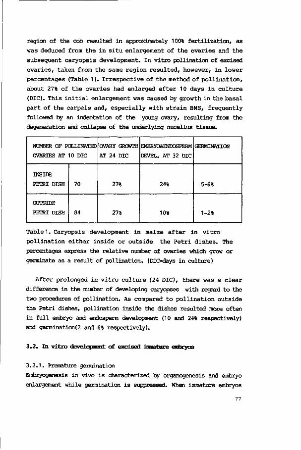
region of the cob resulted in approximately 100% fertilization, as
was deduced from the in situ enlargement of the ovaries and the
subsequent caryopsis development. In vitro pollination of excised
ovaries, taken from the same region resulted, however, in lower
percentages (Table 1). Irrespective of the method of pollination,
about 27% of the ovaries had enlarged after 10 days in culture
(DIC). This initial enlargement was caused by growth in the basal
part of the carpels and, especially with strain BMS, frequently
followed by an indentation of the young ovary, resulting from the
degeneration and collapse of the underlying nucellus tissue.
NUMBER OF POLLINATED
OVARIES AT 10 DIC
INSIDE
PETRI DISH
OUTSIDE
PETRI DISH
70
84
OVARY GROWTH
AT 24 DIC
27%
27%
EMBRYO&ENDOSPERM
DEVEL. AT 32 DIC
24%
10%
GERMINATION
5-6%
1-2%
Table 1. Caryopsis development in maize after in vitro
pollination either inside or outside the Petri dishes. The
percentages express the relative number of ovaries which grow or
germinate as a result of pollination. (DIOdays in culture)
After prolonged in vitro culture (24 DIC), there was a clear
difference in the number of developing caryopses with regard to the
two procedures of pollination. As compared to pollination outside
the Petri dishes, pollination inside the dishes resulted more often
in full embryo and endosperm development (10 and 24% respectively)
and germination(2 and 6% respectively).
3.2. In vitro development of excised immature embryos
3.2.1. Premature germination
Embryogenesis in vivo is characterized by organogenesis and embryo
enlargement while germination is suppressed. When immature embryos
77
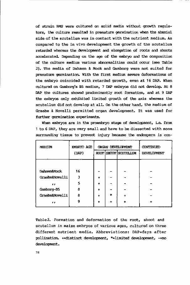
of s t ra in BMS were cultured on solid media without growth regula
tors , the culture resulted in premature germination when the abaxial
s ide of the scutel lum was i n contac t with t he n u t r i e n t medium. As
compared t o the in vivo development the growth of the scutel lum
retarded whereas the development and elongation of roots and shoots
accelerated. Depending on the age of the embryo and the composition
of the culture medium various abnormalities could occur (see Table
2). The media of Dahmen & Mock and Gamborg were not su i t ed for
premature germination. With the f i r s t medium severe deformations of
the embryo coincided with r e t a rded growth, even a t 1 6 DAP. When
cultured on Gamborg's B5 medium, 7 DAP embryos did not develop. At 8
DAP the cultures showed predominantly root formation, and a t 9 DAP
the embryos only exhibited l imited growth of the axis whereas the
scutel lum did not develop a t a l l . On the o ther hand, the medium of
Graebe & Novell i pe rmi t ted organ development. I t was used for
further germination experiments.
When embryos are in the proembryo stage of development, i .e. from
1 t o 6 DAP, they a r e very small and have t o be d i s sec ted with some
surrounding t i ssue to prevent injury because the endosperm i s con-
MEDIUM
Dahmen&Mock
Graebe&Novelli
/ /
Gamborg-B5
Graebe&Novelli
r F
EMBRYO AGE
(DAP)
16
3
5
8
8
9
ORGAN DEVELOPMENT
ROOT
+
+
+
+
+
SHOOT
-
-
-*
+
SCUTELLUM
-
-
-
+
+
CONTINUED
DEVELOPMENT
-
-
-
-
+
Table2. Formation and deformation of the r oo t , shoot and
scutellum in maize embryos of var ious ages , cu l tu red on t h r ee
d i f f e r en t n u t r i e n t media . A b b r e v i a t i o n s : DAP=days a f t e r
pollination. +=distinct development, *=limited development, -=no
development.
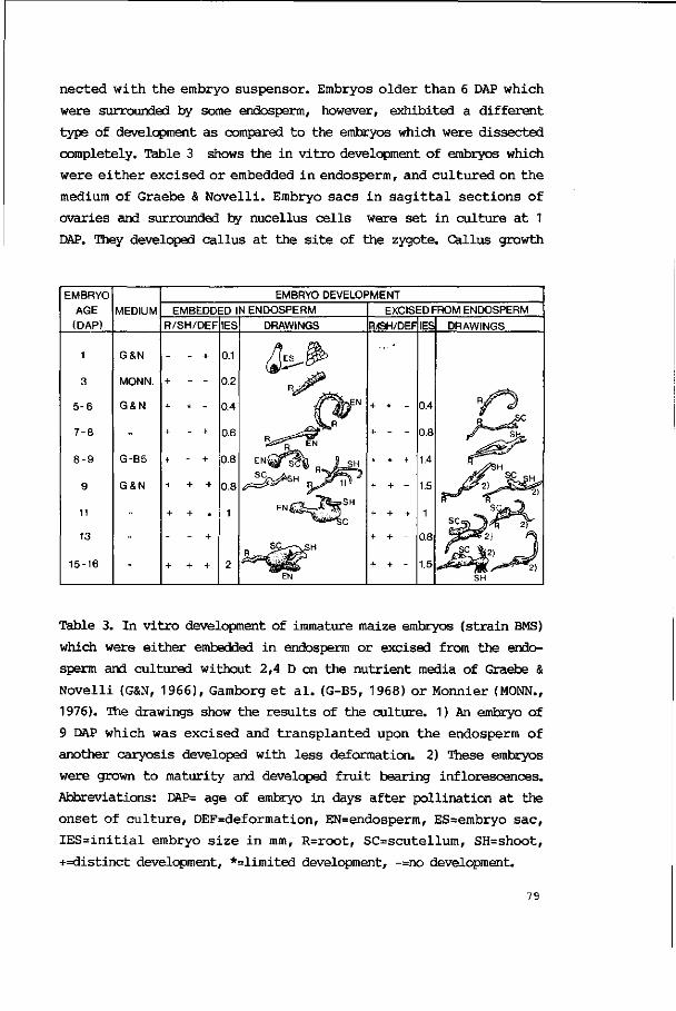
nected with the embryo suspensor. Embryos older than 6 DAP which
were surrounded by some endosperm, however, exhibited a different
type of development as compared to the embryos which were dissected
completely. Table 3 shows the in vitro development of embryos which
were either excised or embedded in endosperm, and cultured on the
medium of Graebe & Novelli. Embryo sacs in sagittal sections of
ovaries and surrounded by nucellus cells were set in culture at 1
DAP. They developed callus at the site of the zygote. Callus growth
EMBRYO AGE
(DAP) MEDIUM
EMBRYO DEVELOPMENT
EMBEDDED IN ENDOSPERM
R/SH/DEF IES DRAWINGS
EXCISED FROM ENDOSPERM
R4SH/DEF IES DRAWINGS
1
3
5 - 6
7 - 8
8 - 9
9
11
13
15-16
G&N
MONN.
G&N
G-B5
G&N
+ - +
+ + +
0.1
0.2
0.4
0.6
0.8
0.8
1
Table 3. In vitro development of immature maize embryos (strain BMS)
which were either embedded in endosperm or excised from the endo
sperm and cultured without 2,4 D on the nutrient media of Graebe &
Novelli (G&N, 1966), Gamborg et al. (G-B5, 1968) or Monnier (MONN.,
1976). The drawings show the results of the culture. 1) An embryo of
9 DAP which was excised and transplanted upon the endosperm of
another caryosis developed with less deformation. 2) These embryos
were grown to maturity and developed fruit bearing inflorescences.
Abbreviations: DAP= age of embryo in days after pollination at the
onset of culture, DEF=deformation, EN=endosperm, ES=embryo sac,
IES=initial embryo size in mm, R=root, SC=scutellum, SH=shoot,
+=distinct development, *=limited development, -=no development.
79

ceased a f t e r 3 days of c u l t u r e and r oo t nor shoot meristems were
formed. When proembryos of 3 DAP were s e t in c u l t u r e wi th some
surrounding endosperm c e l l s , they did not have root or shoot
meristems yet. Root meristems, however, were formed on the medium of
Monnier and roots elongated up to 2 cm. Embryos which were excised
a t 7 DAP did have roo t and shoot meristems a t the beginning of
c u l t u r e . When embedded in some endosperm they developed a mor
phologica l ly normal roo t but only a small d e fec t ive shoot. Older
embryos were a lso embedded in the endosperm during culture in v i t ro ,
they then exhibited defective shoot formation , too.
The culture of embryos which were free from endosperm resulted in
l e s s deformations of t he roo t and the shoot, provided they were
cu l tured on the medium of Graebe & Novell i (see Table 3). The
embryos of 6-7 DAP showed normal root formation but l imited shoot
development. From 9 DAP onwards, however, the immature embryos
formed r oo t s and shoots , germinated completely and could form
p lant le ts which were grown to maturity.
3 .2.2. Cal lus formation and p l an t r egenera t ion from embryonic
t i ssues
3 .2 .2 .1 . Selection of culture conditions
Callus formation and embryoid formation were most effective when the
anterior or axis side of the embryo was in contact with the nutr ient
medium and when growth regulators were added. Root and shoot forma
tion was almost completely inhibited when 10 M zeatin was added.
Premature germinat ion was a l s o prevented by 10~5 M 2,4D which,
however, i n i t i a ted cal lus formation in roots, shoots and scutel lar
t i s s u e . Table 4 p resen t s growth responses of t he embryos of two
maize inbred l ines when cultured on the medium of Green & Phi l l ips ,
supplemented with various concentrations of 2,4D. The highest con
cen t r a t i on of 2,4D (2mg/l) induced morphological abnormalities in
germination such as the curling of coleoptiles, the retarded growth
of roots and the formation of callus.
MAIZE
INBRED
LINE
BMS ii
ii
A188 It
II
2,4 D
mg/ml
-
0.25
2.0
-
0.75
2.0
GERMINATION
ROOT
+
*
*
+
*
*
SHOOT
+
+
*
+
+
*
CALLUS
R
-
+
+
_
*
+
SH
-
-
+
_
-
-
SC
-
-
+
_
+
+
CHLOROPLASTS
IN SCUTELLUM
+
+
+
+
+
+
ADVENTITIOUS
SHOOTS
-
-
+
_
+
+
ROOTS
-
+
+
_
+
+
Table 4. Growth response of excised maize embryos of 16 to 17
DAP on the culture medium of Green & Phillips (1975),
supplemented with various concentrations of 2.4D. +=distinct
development, *=limited development, -=no development. For
abbreviations see Table 3.
3.2.2.2. Callus formation
The morphological development of callus on A188 embryos, cultured
over a period of 45 to 160 hours on GP-2 medium, is shown in Fig. 1.
Various growth responses were discerned. In some cases the whole
scutellum enlarged (Fig. 1a). In other cases a local swelling was
formed at the base of the scutellum (Fig. 1b) and incidentally a
combination was detected (Fig. 1c). Sometimes only the embryo axis
was swollen (Fig. 1d). All these phenomena, however, could appear
simultaneously as well (Fig. 1e). At the surface of the scutellum
the callus was frequently observed at the abaxial side and the
lateral sides. Calli appeared as wart-like structures, as globular
aggregates with epidermal-like smooth surfaces, or as loose, friable
arrangements (Fig.5a, arrow-heads).
The onset of cell proliferation in embryos starts within 24 hours
of culture. Indications of deviating cell growth were seen in the
meristems of the root and shoot and in the scutellum. Initially the
cells of the root meristems were vacuolized and exhibited nuclei
with much heterochromatin (Fig. 2a,b) whereas after 24 hrs of cul-
81
Fig. 1 Morphological development of c a l l u s on embryos of maize
( s t r a i n A188), cu l tu red over a period of 45 t o 160 h r s on GP-2
medium. Culture period for a=45 hrs , b-c=68 hrs , d=90 hrs and e=160
hrs. Note the t o t a l enlargement of the scutellum and the embryo axis
(EA) and the l oca l swe l l ing (*) a t the base of the scutel lum. The
bars represent 100 p i . (SC=scutellum, SH=shcot, SU=suspensor).
82

ture (Fig. 2c) the vacuoles decreased in size and nuclei became
euchromatic. After 45 hrs of culture most vacuoles had disappeared
and cells divided frequently (Fig. 2d).
Within the scutellum,a swelling was caused by the growth of the
mesophyll cells and by the enlargement of the intercellular spaces
m as?" *%jr*
• >
RM-i fr:;>i
m p-5-''-
- A
A- \
5* xi>
• J ) • )'
lOOjum
' •* Vl. ' 4
Fig. 2 Activation of root meristem cells of immature A188 maize
embryos set in culture on GP-2 medium with the axis side in contact
with the medium, a. Light micrograph of a sagittal section of an
embryo grown in vivo for 11 to 12 days, b The root meristem of such
an embryo before culture, c. A root meristem at 24 hrs. and d. the
root meristem after 45 hrs in culture. Note the decrease of vacuolar
size, the changes in the nucleoplasm and the increase of cell
divisions (arrows).
1
» •
V. « ' * ,
i
84

as is visualized in Fig. 3a,b. In addition, meristematic regions
were formed in the inner part of the scutellum and new strands of
vascular tissues were observed as well (Fig. 3d, arrow). Scutellum
cells of the subepidermal regions often developed into chlorenchyma
(Fig. 3b,c) or collenchyma (Fig. 3d, arrow-heads) and in some
regions they showed meristematic activity (Fig. 3e). The latter
regions, in which the epidermis is included, were characterized by
increased mitotic rates and by relatively small cells which had few
vacuoles and many organelles. There was often a marked difference in
staining of the various cells in such a region. A cluster of cells
with a presumed single cell origin exhibited intensive staining
(Fig. 3e, arrows) whereas the organelles of the adjacent cells
showed less contrast. The cluster constitutes an embryoid or a shoot
primordium; the bordering cells the callus tissue. On the other
hand, subepidermal cells at the abaxial scutellum side could also
degenerate within 24 hrs of culture (see Fig. 4a) and at 45 hrs in
culture, the larger part of the scutellum could then have collapsed
(cf. Fig. 1d) because of cell degeneration as visualized in Fig.
4b.
In order to compare the callus-generating ability of the various
scutellum regions, some embryos of 21 DAP were cut in several
transversal parts and cultured on GP-2 medium. Except for the one
containing the apex of the scutellum, all sections had formed callus
after 4 days in culture.
Fig. 3 Cytological differentiation in the maize scutellum during
in vitro culture on GP-2 medium. Intercellular spaces (a,b),
chlorenchyma (b,c), collenchyma (d, arrow-heads), vascular tissues
(d, arrow) and meristematic, callus and shoot or embryoid forming
zones (e) resulted within 13 days in culture (DIC). a=1 DIC, b-d=11
DIC, e=13 DIC (CH=chloroplast, IS=intercellular space).
Callus on the scutellum grew fast and often had a friable appear
ance (Fig. 5a, arrow-heads). Callus formation on roots was observed
as soon as the roots came into contact with the nutrient medium. The
cell proliferation occurred in the epidermis and cortex of the
seminal roots. The aggregates of callus branched frequently, due to
the deficient formation of lateral roots (Fig. 5b,c). Adventitious
roots were generated from the embryo shoot and from the scutellum
(Fig. 5a, arrow). They had a normal appearance as long as they were
not in contact with the nutrient medium.
3.2.2.3. Plant regeneration
After a culture period of three weeks on solidified GP medium with
2mg 2,4D/1, adventitious organs developed on the scutella of A188
embryos. Embryos of strain BMS formed leafy structures after about 6
weeks. As compared to BMS, strain A188 also formed more adventitious
roots and shoots. Up to 100% of the cultured embryos of strain A188
produced up to 15 adventitious organs each in a culture period of
three months whereas only up to 20% of the BMS embryos bore adventi
tious shoots (Fig. 6). It was observed that the wart-like structures
often developed into adventitious roots.
The adventitious shoots (Fig. 6a, arrows) and leafy structures
(Fig. 6b,c) developed at the coleorhizal end on the abaxial side of
the scutellum. Their place of origin was not fixed but often a
greening of the site preceded the development. The epidermis at
such a site was still an undisrupted structure. The leafy structures
bore many epidermal hairs (Fig. 6c) and initially the adventitious
shoots often had a scutellum-shaped appearance with a shoot
meristem-like structure at the surface. Adventitious roots developed
(Fig. 6c, arrows) but were never seen to originate from the adventi
tious shoots under the applied circumstances.
3.2.3. Culture in liquid media
Embryos of BMS and A188 were dissected at 1 6 DAP and cultured in
liquid GP medium supplemented with various concentrations of 2,4D.
When cultured without 2,4D, the embryos formed roots and shoots.
Having added 0.75 mg 2,4D/1 , the scutellum enlarged, the coleoptile
partly developed and root formation was inhibited completely. Some
small clusters of cells were detected in the suspension fluid. When
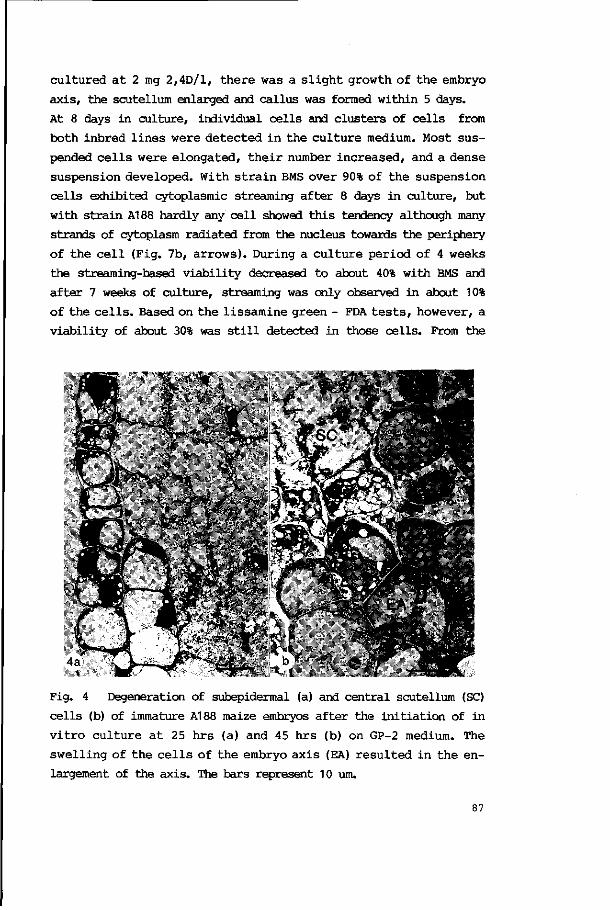
cultured at 2 mg 2,4D/1, there was a slight growth of the embryo
axis, the scutellum enlarged and callus was formed within 5 days.
At 8 days in culture, individual cells and clusters of cells from
both inbred lines were detected in the culture medium. Most sus
pended cells were elongated, their number increased, and a dense
suspension developed. With strain BMS over 90% of the suspension
cells exhibited cytoplasmic streaming after 8 days in culture, but
with strain A188 hardly any cell showed this tendency although many
strands of cytoplasm radiated from the nucleus towards the periphery
of the cell (Fig. 7b, arrows). During a culture period of 4 weeks
the streaming-based viability decreased to about 40% with BMS and
after 7 weeks of culture, streaming was only observed in about 10%
of the cells. Based on the lissamine green - FDA tests, however, a
viability of about 30% was still detected in those cells. From the
t i 2 K 4 '-> t -
*> l * V
^ - ^
Fig. 4 Degeneration of subepidermal (a) and central scutellum (SC)
ce l l s (b) of immature A188 maize embryos af ter the i n i t i a t ion of in
v i t r o c u l t u r e a t 25 h r s (a) and 45 h rs (b) on GP-2 medium. The
swel l ing of the c e l l s of the embryo ax i s (EA) r e su l t ed in the en
largement of the axis. The bars represent 10 urn.
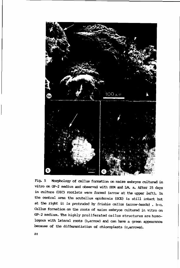
Fig. 5 Morphology of callus formation on maize embryos cultured in
vitro on GP-2 medium and observed with SEM and LM. a. After 25 days
in culture (DIC) rootlets were formed (arrow at the upper left). In
the central area the scutellum epidermis (SCE) is still intact but
at the right it is protruded by friable callus (arrow-heads) . b-c.
Callus formation on the roots of maize embryos cultured in vitro on
GP-2 medium. The highly proliferated callus structures are homo
logous with lateral roots (b,arrow) and can have a green appearance
because of the differentiation of chloroplasts (c,arrows).

beginning of culture, the A188 ce l l s showed cytoplasmic streaming in
l e s s than 5% but l i s samine green - FDA t e s t s , such as shown in
Fig. 7c,d, gave percentages up t o 50% v i a b i l i t y a f t e r 8 weeks.
Subculturing of t h a t suspension in GP-1 medium and a subsequent
incubat ion of 8 days r e su l t ed i n an increased v i a b i l i t y t h a t
exceeded 90% . A remarkable increase in c e l l number was not iced
concomitantly but c e l l divisions were not observed in the suspended
Fig. 6 Regeneration on immature maize embryos (s train A188) on GP-
2 medium. aJormation of adventitious shoots (arrows) a t 25 D1C. b -
c. D i f f e r en t i a t i ng l e a f l e t s of adven t i t i ous shoots a t 38 DIC. The
adven t i t ious r oo t s (c, arrows) do not o r i g i n a t e from the adven t i
tious shoots. The bars represent 1 mm.
89
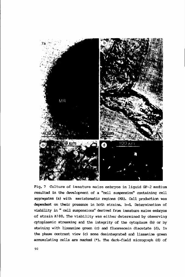
Fig. 7 Culture of immature maize embryos in liquid GP-2 medium
resulted in the development of a "cell suspension" containing cell
aggregates (a) with meristematic regions (MR). Cell production was
dependent on their presence in both strains, b-d. Determination of
viability in " cell suspensions" derived from immature maize embryos
of strain A188. The viability was either determined by observing
cytoplasmic streaming and the integrity of the cytoplasm (b) or by
staining with lissamine green (c) and fluorescein diacetate (d). In
the phase contrast view (c) some desintegrated and lissamine green
accumulating cells are marked (*). The dark-field micrograph (d) of
90

cells. They were observed in cell aggregates of 1.5 to 3.0 mm size
which were not in the inoculum initially. Figure 7a shows such a
cell aggregate which has a root-like appearance. The increase in
cell number was only observed when the cell aggregates were present.
Subculturing of suspended cells often caused the disruption of
cells and the outflow of their cytoplasm. Mannitol was added in 2%
steps up to 12%(w/v) to prevent cell lysis, but in all subcultures
lysed cells were detected. Only a gradual, 20% dilution of the
culture medium was successful to maintain cell stability. Cell
aggregates were detected in subcultures with both high (12%) and low
(0.8%) concentrations of mannitol. Only in these particular sub
cultures the number of suspended cells increased.
4. DISCUSSION
4.1. Pollination and caryopsis development in vitro
The in vitro culture of maize embryos offers the opportunity to
improve the control of nutritional and environmental conditions in
order to study and influence embryo development, callus formation
and somaclonal propagation (see Sheridan 1982, Vasil 1984).In vitro
ernbryogenesis ab ovo was realized by in vitro pollination. The
technique shortens the progamic phase and might hence synchronize
subsequent embryo development (see amongst others Gengenbach 1977,
Dhaliwal & King 1978, Raman et al. 1980). It was found, however,
that the in vitro pollinated pistils did not develop uniformly. This
could either be caused by unsuccessful fertilization, by a failure
of endosperm development or by a lack of interaction between the
ovary and the nutrient medium. Gengenbach (1977) already stressed
the importance of in vitro pollination with maize. He achieved only
5% kernel maturation and suggested that this was caused by deficient
endosperm development because fertilization in his experiments
ranged up to 84% depending upon the genotypes of the parents. In the
the same cells shows exclusively the "living", FDA-positive cells.
The black and white arrows in pictures c and d point towards the
same nucleus. Abbreviations: CP = strand of cytoplasm, MR = meriste-
matic region, N - nucleus, P = plastid.
91
present experiments the highest percentage of kernel development
was achieved when silks were pollinated inside instead of outside
the Petri dish. Because the site of pollen attachment in that case
was closer to the ovaries, more pollen tubes might reach an embryo
sac In the present experiment a limited amount of pollen was used
to prevent contamination. Therefore the percentage of kernel devel
opment might further be increased by administering the optimal
amount of pollen as was demonstrated by Raman et al. (1980).
Although the percentage of kernel development was notably im
proved upon and the method proved to be successful in comparison to
the results of Gengenbach (1977), it was preferred to pollinate
pistils in vivo to ensure fertilization for further in vitro culture
experiments.
4.2. Pollination in vivo followed by embryo development in vitro
When pistils were pollinated in vivo, then excised at 1 DAP and
cultured in vitro, the embryo and endosperm developed (Schel & Kieft
1986). When pistils,however, were pollinated in vivo and sagittal
sections of the ovaries containing the fertilized embryo sacs were
set in culture at 1 DAP, that culture resulted in deficient embryo
development. A callus-like structure formed and germination did not
occur. This result indicates that injury strongly influences embryo
development. In vivo, the cells of the placento-chalazal region, and
the synergids function in the transport of metabolites and inorganic
compounds towards the nucellus and the embryo respectively (Van
Lammeren 1986a). After the nucellus is replaced by the endosperm,
the endosperm cells of the placento-chalazal region and those near
the embryo suspensor do function similarly (see Schel et al. 1984).
When, however, an ovary is sectioned sagittally and the section side
is placed onto the nutrient medium, the nutrients will be taken up
through the section surface rather than through the spikelet axis.
Consequently, the composition of the nutrient flow will be changed
because the selective transport from the somatic tissues of the
ovary towards the endosperm and embryo is disturbed.
Proembryos which were set in culture at 3 DAP, generated roots
with a length up to 2 cm but shoot development failed. Considering
the in vivo stage of development at 3 DAP it was demonstrated before
92

(Van Lammeren 1986b) tha t root meristems were not present yet- Thus
embryo development in v i t ro not only means activation of exist ing
meristems but the formation of such meristems as well. So an excised
proembryo can t ransform i n t o an embryo proper s t age . When o lde r
embryos are set in culture, while they are surrounded by someendo-
sperm, d e f i c i en t shoot and scutel lum formation can a l s o be ob
served. Whith the endosperm surrounding t he embryo i n v ivo, an
intensive interaction between embryo and endosperm can be deduced
from u l t r a s t r u c t u r a l evidence (Schel e t a l . 1984). In v i t r o , the
endosperm inf luences the embryo growth by i t s inf luence on t he
n u t r i t i o n a l environment. In add i t ion t h e r e i s a wound e f f ec t by
which t h e r e i s no s e l e c t i v e t r ans fe r rence of me tabo l i t es i n t he
placentc-chalazal region any more. Therefore, the composition of the
nutrient flow from the endosperm towards the embryo i s a lso changed
and influences embryogenesis.
When embryos were excised and not embedded in endosperm, ger
mination only occurred when the scutellum was in contact with the
nutr ient medium. This suggests the importance of the direct ion of
t he n u t r i e n t flow. As compared with the in vivo development where
most n u t r i e n t s a r e taken up through the abaxia l epidermis of the
scutellum (Van Lammeren 1986b) a s imilar pathway for nutr ients i s
evident here.
The germination of immature embryos indicates that the technique
o f fe rs an oppor tuni ty t o study the morphological d i f f e r e n t i a t i o n
continuously and to simulate germination. Several authors cultured
immature maize embryos t o achieve complete p l an t development.
Embryos, excised from 15 DAP onwards, germinated and developed
normally in the experiments of Mock & Dahmen (1973) who used a solid
agar-sucrose composite high-nutrient medium. Sheridan e t a l . (1978)
repor ted t he successful development of excised embryos of 11 DAP.
From very young excised embryos (3 up t o 7 DAP) only the o l de s t
showed l imited growth in the early experiments of Uttaman (1949). In
the present experiments, using the medium as described by Graebe &
Novell i (1966), t he development of p l a n t l e t s of immature excised
embryos was achieved already from 9 DAP onwards. With respect to the
da ta a va i l ab l e t h i s means a shor tening of the dependence of t he
embryo upon the endosperm. As compared wi th the i n v ivo embryo-
93

genesis (see Van Lammeren 1986b) a decreased scutellum development
is obvious and an activation of meristematic activity and cell
elongation results in the early root and shoot formation. The prema
ture activation of the apical meristems is probably initiated by the
fall-off of the endosperm suppression. Embryos younger than 9 DAP
exhibited growth as well, but deficiencies occurred.
4.3. Callus formation and plant regeneration
Embryo cultures in fluid media were set up to form cell suspensions
which could give rise to somatic embryos. The aim was to analyze
somatic embryos from the single cell stage onwards to compare their
development with the in vivo embryogenesis (see Van Lammeren 1986a).
The single cell stage gives the particular opportunity to study the
development of structural polarity such as induced in the egg cell-
zygote stages in vivo. The culture in media, supplemented with 2.4D
resulted in the production of suspended cells in high densities.
Cell divisions, however, were not observed in these cells. Therefore
the culture is not a real cell suspension but merely the result of
cell production in the meristematic, root-like cell clusters. The
increase of viability after subculturing was also caused by cell
production in the root-like aggregates in the suspension. The tests
based on FDA staining gave the best quantification of viability.
Cytoplasmic streaming proved not to be a reliable parameter and
observing the cytoplasmic integrity is not reliable and laborious.
The regeneration of plantlets from callus cultures of Zea mays L.
on solid nutrient media has been acchieved from various tissues such
as stem tissue from seedlings (Harms et al. 1976) and from excised
immature embryos (see, amongst others, Green & Phillips 1975, Lu et
al. 1983, Novak & Dolezelova 1983, Vasil et al. 1984, Duncan et al.
1985, Kamoet al. 1985).
In the present experiments the culture of immature embryos of 16-
17 DAP on solidified nutrient media resulted in callus formation at
various sites of the embryo. On the scutellum it was mainly re
stricted to the coleorhizal end which may be related to the high
levels of plant growth regulators presumed to be present at this
site (Vasil & Vasil 1982). The induced bipolarity existing within
the scutellum, which is regarded as a leaf (Van Lammeren 1986b), was
94

also assigned by the transversal sectioning of the scutellum; most
sections exhibited callus formation then. Anatomically, cal lus for
mation was found in the epidermal and sub-epidermal zones (see a lso
Springer e t a l . 1979, Vas i l e t a l . 1985). Cal lus formation i s only
one growth response to in v i t ro culture. The swelling of the scute l -
lum was mainly caused by c e l l enlargement, by the formation of many
large in tercel lu lar spaces and, to a lower extent, by the formation
of chlorenchyma, collenchyma and vascular t i ssues . Halperin (1969)
and Springer e t a l . (1979) mentioned a random d i s t r i b u t i o n of
vascular t i ssue in which phloem elements were not detected. Only
the compact scutel lar cal lus which was formed a t the coleorhizal end
showed r egenera t ive capac i ty . Vasi l e t a l . (1983) a l s o repor ted
embryold formation in the nodal region of the scutellum but there
fore the s c u t e l l a r s ide of the embryo was in con tac t with t he
nutrient medium. The areas with ce l l s dividing frequently a lso bore
the bipolar, proembryo-like s tructures. These s tructures might be of
unicellular origin as was a lso shown for Pennisetum embryoids (Vasil
& Vasi l 1982, Bo t t i & Vas i l 1984). In our experiments t h e deve l
opment of leafy s tructures and adventitious shoots was observed (cf
Green & P h i l l i p s 1975 and Springer e t a l . 1979) r a t h e r than t he
formation of somatic embryos as reported by, amongst others, Gordon
e t a l . (1977), Lu e t a l . (1982, 1983), Novak & Dolezelova (1983),
Vasil e t a l . (1985) and Fransz (pers. comm.).The l imited development
may be caused by the hormonal level u t i l ized in t h i s study.
5 . ACKNOWLEDGEMENTS
The author i s indebted t o Prof. d r . M.T.M. Willemse and Dr. J.H.N.
Schel for reading and c r i t i c i z i n g t he manuscript. Thanks a re a l s o
due t o Mr. A.B. Haasdijk and S. Massalt for the ar twork and photo
graphy and to Dr F. Vegter for emending the English text .
95

6 . REFERENCES
Bo t t i , C. & I.K. Vasl l (1984): The ontogeny of somatic embryos of
Pennisetum americanum (L.) K. Schum. I I . In cu l tu red immature
i n f lo rescences . Can. J . Bot. 62: 1629-1635.
Dahmen,W.J. & J . J . Mock (1972): Effects of nu t r ien t -media com
p o s i t i o n on g rowth of s e e d l i n g s from i n t a c t s eeds and
excised embryos of maize (Zea mays L.). Crop Sci. 12: 549-550.
Dhaliwal, S. & P.J. King (1978): Direct p o l l i n a t i on of Zea mays
ovules in v i t r o with Z. mays, Z. mexicana and Sorghum b ico lo r
pol len . Theor. Appl. Genet. 53: 43-46.
Duncan, D.R., M.E. Wil l iams, B.E. Zehr & J.M. Widholm (1985): The
p r oduc t i on of c a l l u s c apab l e of p l a n t r egenera t ion from
immature embryos of numerous Zea mays genotypes. P lanta
165: 322-332.
Evans , D.A., W.R. Sha rp , P.V. Ammirato & Y. Yamada, e d s .
(1983)Handbook of p l an t c e l l c u l t u r e . Co l l i e r Macmillan
Publishers, London.
Gamborg, O.L., R.A. Mi l l e r & K. Ojima (1968): Nut r ien t r e qu i r e
ments of suspension cultures of soybean root cells.Exp. Cell
Res. 50: 151-158
Gengenbach, B.G. (1977): Development of maize caryopses r e s u l t i n g
from in v i t ro pollination. Planta 134: 91-93.
Gordon, P.N., M.M. Robarts & T.B. Rice (1977): P lan t r egenera t ion
from t i s s u e c u l t u r e s of maize. Maize Genet. Coop. News Le t t e r
51: 79-80.
Graebe, J.E. & G.D. Novell i (1966): A p r a c t i c a l method for l a rge
scale plant t i ssue culture. Exp. Cell Res. 41: 509-520.
Green, C.E., C.L. Armstrong & P.C. Anderson (1983): Somatic c e l l
gene t ic systems in corn. In : Downey,K., R.W. Voellmy, F. Ahmad
& J . Schulz, (eds). Advances in gene technology: molecular
gene t ics of p l an t s and animals , pp 147-157, Acad. P re s s , New
York London.
& R.L. P h i l l i p s (1975): P l a n t r e g e n e r a t i o n from t i s s u e
cultures of maize. Crop Science 15: 417-421.
Halperin, W. (1969): Morphogenesis in c e l l c u l t u r e s . Ann. Rev.
Plant Physiol. 20: 395-418.
96

Harms, C.T., H. Lorz & I . Potrykus (1976): Regeneration of p lant le ts
from c a l l u s c u l t u r e s of Zea mays L. Z. Pflanzenzuchtg. 77:347-
351.
Heslop-Harrisson, J . & Y. Heslop-Harrison (1970): Evaluation of
pollen v i ab i l i ty by enzymatically induced fluorescence; i n t ra
c e l l u l a r h y d r o l y s i s of f l u o r e s c e i n d i a c e t a t e . S t a i n
Technol. 45: 115-120.
Imbr ie-Mil l igan , C.W. & T.K. Hodges (1986): Microcal lus formation
from maize protoplasts prepared from embryogenic cal lus . Planta
168: 395-401.
Kamo, K.K., M.R. Becwar & T.K. Hodges (1985): Regeneration of Zea
mays L. from embryogenic cal lus . Bot. Gaz. 146(3): 327-334.
Lu, C , I.K. Vas i l & P. Ozias-Akins (1982): Somatic embryogenesis
in Zea mays L. Theor. Appl. Genet. 62: 109-112.
, V. Vasil & IJC Vasil (1983): Improved efficiency of somatic
embryogenesis and plant regeneration in t i ssue cultures of
maize (Zea mays L.). Theor. Appl. Genet. 66: 285-289.
Mock, J . J . & W.J. Dahmen (1973): S t e r i l e c u l t u r e of immature
maize seeds and embryos. Crop Sci. 13: 764-766.
Monnier, M. (1976): Culture in v i t r o de l'embryon immature de
Capsella Bursa -pas to r i s Moench (L.). Rev. Cyt. B io l . Veget.
39: 1- 120
Murashige, T. & F. Skoog (1962): A r ev i sed medium for r ap id
growth and b ioassays with tobacco t i s s u e c u l t u r e s . Physiol .
P lan t . 15: 473-497.
Novak, F.J. & M. Dolezelova (1983): Somatic embryogenesis and
plant regeneration in Zea mays L. Maydica 28: 381-390.
Potrykus I . , C.T. Harms, H. Lorz & E. Thomas (1977): Cal lus f o r
mation from stem protoplasts of corn (Zea mays L.). Mol. gen. Genet.
156: 347-350.
, , & - — (1979): Cal lus formation from c e l l c u l t u r e
p ro top l a s t s of corn (Zea mays L.). Theor. Appl. Genet. 54:
209-214.
Raman, K., D.B. Walden & R.I. Greyson (1980): F e r t i l i z a t i o n i n
Zea mays by cultured gametophytes. J. Heridity 71, 311 -314.
Schel, J.H.N. & H. Kieft (1986): An u l t r a s t r u c t u r a l s tudy of
embryo and endosperm development during in v i t r o c u l t u r e of
97
maize ova r i e s . Can. J . Bot.: 64,2227-2238.
, & A.A.M. van Lammeren (1984): I n t e r a c t i on s between embryo
and endosperm during early developmental stages of maize caryop-
ses (Zea mays). Can. J . Bot. 62: 2842-2853.
Sheridan, W.F. (1975): Tissue c u l t u r e of maize. I . Cal lus induct ion
and growth. Physiol. Plant. 33: 151-156.
(1977): Tissue c u l t u r e of maize. I I . The e f f ec t of the g l u t a -
mate, a s p a r t a t e , and a romatic amino acid f ami l i e s on c a l l u s
growth of several maize s t ra ins . Physiol. Plant. 41: 172-174.
(1982): Maize for b i o log i ca l r esearch . Univers i ty of North
Dakota Press, Grand Forks, ND.
, M.G. Neuffer & E. Bendbow (1978): Rescue of le thal defective
endosperm mutants by culturing immature embryos. Maize Coop. News
Le t t e r 52 : 88-90.
Spr inger , W.D., C.E. Green & K.A. Kohn (1979): A h i s t o l o g i c a l
examination of t i ssue culture i n i t i a t ion from immature embryos
of maize. Protoplasma 101: 269-281.
S t r e e t , H.E. (1977): P lant t i s s u e and c e l l c u l t u r e (Bot. monographs
Vol. 11) Blackwell Scientif ic Publications, Oxford London Edin
burgh Melborne.
Uttaman, P. (1949): Cul tur ing of pro-embryos of normal d i p lo id
corn (maize) aged 3 to 7 days. Current Science 6: 215-216.
Van Lammeren, A.A.M. (1986a): A comparative u l t r a s t r u c t u r a l s tudy
of the megagametophytes in two s t r a i n s of Zea mays L. before
and a f t e r f e r t i l i z a t i o n . Agr icu l t . Univ. Wageningen Papers
86-1.
(1986b): Developmental morphology and cytology of the young
maize embryo (Zea mays L.) Acta Bot. Neerl. 35:169-188.
Vas i l , I.K. (1982): Somatic embryogenesis and p l an t r egenera t ion
i n c e r ea l s and g r a s ses . In: Fujiwara, A., (ed.) P lant t i s s u e
culture 1982, pp101-104. Tokyo: Maruzen.
(1984): Cel l c u l t u r e and somatic c e l l gene t ics of p l a n t s .
Volume I . Acad. P ress , i nc . New York London .
Vas i l , V., C. Lu & I.K. Vasi l (1983): P r o l i f e r a t i on and p l an t
r egenera t ion from the nodal region of Zea mays L. (maize,
Gramineae) embryos. Am. J . Bot. 70(6):951-954.
98

-, & (1985): Histology of somatic erahryogenesis in cultured
immature embryos of maize (Zea mays L.). Protoplasma 127: 1-8.
- & I.K. Vasi l (1982): The ontogeny of somatic embryos of
Pennisetum americanum (L.) K. Schum. I . In cu l tu red immature
embryos. Bot. Gaz. 143: 454-465.
- , & C. Lu (1984): Somatic embryogenesis in long-term c a l l u s
cultures of Zea mays L. (Gramineae). Am. J . Bot. 71:158-161.
99
CHftPHR 5
Interactions between embryo and endosperm during early developmental
stages of maize caryopses (Zea mays)
J.H.N. Schel, H. Kieft & A.A.M. van Lammeren
(Can. J. Bot. 62(2): 2842-2853, 1984)
Interactions between embryo and endosperm during early developmental stages of maize caryopses (Zea mays)
J. H. N . SCHEL, H. KIEKT, AND A. A. M. VAN LAMMEREN
Department of Plan! Cytology and Morphology, Agricultural University. Arboretumluan A The Netherlands
Received March 23, 1984
6703 BD, Wageningen,
SCHEL, J. H. N., H. KiEFT, and A. A. M. VAN LAMMEREN. 1984. Interactions bewccn embryo and endosperm during early developmental stages of maize caryopses (Zea mays). Can. J. Bot. 62: 2842-2853.
The early developmental stages of maize caryopses were studied at a tine structural level. Emphasis was placed on the interactions between the developing embryo and the surrounding endosperm. It was found that the placcntochalazal region of the endosperm contains cells showing ultrastructural features of transfer cells, including wall ingrowths. This indicates an important function of these cells in the transport of nutrients supplying the developing embryo. Near the basal region of the embryo, densely cytoplasmic endosperm celts occurred, without wall ingrowths but with a mass of highly ordered rough endoplasmic reticulum indicating a synthesis function of this part of the endosperm. The products, probably membranes and proteins, are most likely taken up by the suspensor of the embryo. At about 7 days after pollination, endosperm cells degenerate near the embryo axis and the scutellum. The endosperm remnants might then serve as a food supply for the embryo as well.
SCHEL, J. H. N., H. KIEFT et A. A. M. VAN LAMMEREN. 1984. interactions beween embryo and endosperm during early developmental stages of maize caryopses (Zea mays). Can. J. Bot. 62: 2842-2853.
Nous avons dtudie les premiers stades de developpement des caryopses dc mai's au niveau de l'ultrastructurc ct avons mis l'emphase sur les interactions entre I'embryon qui se devcloppe et l'albumen qui I'cntourc. La region chalazo-placcntaire de l'albumen inclut des cellules dont l'ultrastructurc montre des caracteristiqucs des cellules de transfer!, replis parietaux inclus. Cela indique que ces cellules jouent un role important dans le transport de nutriments a I'embryon en developpement. Pres de la region basale de I'embryon se retrouvent des cellules d'albumen a cytoplasmc dense, sans replis parietaux mais ayant une masse de reticulum endoplasmique granulaire hautement ordonnee indiquant une fonction dc synthese dc cette region dc I'endosperme. Les produits, probablemcnt des membranes ct des proteines. sont probablcment absorbes par le suspenseur de I'embryon. Environ sept jours apres la pollinisation, les cellules dc rendospcrmc degencrent pres de 1'axc embryonnaire ct du scutellum. Les restes dc l'albumen peuvent done servir de source de nourriturc pour I'embryon aussi.
ITraduit par le journal]
Introduction
The process of embryo formation in grasses is well documented, although mainly by descriptions based on light microscopy (for a survey, see Rost and Lersten 1973). As pointed out by Smart and O'Brien (1983), the only recent studies on the embryo region of grass caryopses at a fine structural cellular level are those of Norstog (1972, 1974) on barley. These authors also emphasized the lack of studies dealing with embryo development in relation to the neighbouring tissues. They therefore investigated in detail and at a fine structural level the process of embryo development in wheat, paying particular attention to structure-function relationships between the developing embryo and the surrounding endosperm.
For maize, the process of embryo formation has been described extensively, both by light microscopy (Randolph 1936; Kiesselbach 1949; Wolf et al. 1952; Van Lammeren and Schel 1983) and by electron microscopy (Diboll 1968; Van Lammeren 1981; Van Lammeren and Kieft 1983). Endosperm development in maize has also been studied in great detail (Weatherwax 1930; Lampe 1931; Brink and Cooper 1947; Khoo arid Wolf 1970; Kyle and Styles 1977). However, an integrated study to elucidate the functional relationships between the developing embryo and endosperm from a fine structural point of view has not yet been carried out.
We report here on the fine structure of early developmental stages from maize caryopses. Attention will be given to a functional interpretation of the structural changes of embryo and endosperm. The results will be discussed in relation to data on embryo formation in other plants. It is felt that a detailed knowledge of the in vivo embryogen.esis of a cereal such as maize is required, especially now that in vitro culture methods
using immature embryos from this agronomically important crop are becoming increasingly important (e.g., see Green 1982; Sheridan 1982; Vasil 1982).
Materials and methods
Strain A-188 of Zea mays L. (kindly supplied by Dr. C. E. Green, Minnesota) was used. Seeds were grown to maturity under greenhouse conditions. After emergence of the silks, the plants were cross-pollinated by hand. At defined intervals, ranging from 4 to 11 days after pollination (DAP) developing caryopses were removed from the middle of the ears. Small slices, about 2 mm thick, were prepared using razor blades. They were made in such a way that the whole embryo axis and a large part of its surrounding tissues were excised.
The slices were immediately fixed in 5% glutaraldehyde buffered with 0.1 M sodium cacodylate buffer (pH 7.0) to which 7 mM CaCb was added. Fixation took place for 2 h at room temperature. After rinsing, the samples were postfixed for 2 h with 1% Os04 and dehydrated in a graded ethanol series, ranging from 30 to 100%, followed by propylene oxide. The pieces of tissue were then transferred to propylene oxide — Epon 812 mixtures and finally to fresh resin. Polymerization occurred at about 600 mbar (1 bar = 100 kPa) for 16 h at 20°C, 8 h at 45°C, and 24 h at 60°C. Semithin (2 u,m) and thin sections were prepared on a LKB Ultratome V. The thin sections were poststained with uranyl acetate and lead citrate. They were examined in a Philips EM 301 operating at 60 kV.
Results
The orientation of ovary, ovule, and embryo sac in a median section is represented in Fig. la , which shows the unfertilized condition. The cob on which the kernels are located is situated at the left. Most micrographs presented here show this type of sagittal section and orientation. The developmental stages fol-
103

SCHEL ET AL
12 14 16 days after pollination
-I—i—n—i—m- - IE -developmental stage
ABBREVIATIONS: at, aleuron layer; ap, apex primordium; cp. coleoptile primordium; cu. cuticula; Cw, cell wall; Di, dictyosome; Em, embryo; End, endosperm; End ad, adaxial region of the endosperm; End ab. abaxial region of the endosperm; ER, endoplasmic reticulum; int, integumentary layer; Li, lipid droplet; Mi, mitochondrium; N, nucleus; Nu, nucellus; nu. nucellar layers; p, plastid; pc, pericarp; Pd, plas-modesma; Rer, rough endoplasmic reticulum; sa. shoot apex; Sc. scutellum; Sp. suspensor; sy, synergid; v, vacuole; ve, vesicle.
FIG. I. (a) Schematic representation of a sagittal section through an unfertilized caryopsis of maize. The cob axis is situated at the left (adapted from Randolph 1936). (b) Correlation between grain, embryo, and endosperm length and the number of days after pollination. The developmental stages are numbered 1 to IV. Fie. 2. A nearly sagittal semithin section showing embryo and endosperm development at stage I (3-5 DAP). A degenerated synergid (sy) is visible. The embryo contains large vacuolated basal cells and cytoplasm-rich top cells. The cellular endosperm consists of highly vacuolated cells except in the region bordering the embryo at the adaxial side. In that region, the endosperm cells are small and densely cytoplasmic (arrows).
lowing fertilization which are described in this paper have been numbered I to IV. This classification is based on the measurements of the length of excised embryos and endosperm in relation to the number of days after pollination (Fig. lb). Kernels in which the number of DAP and embryo and endosperm lengths did not correspond with Fig. \b were not used for further examination.
The sagittal semithin section in Fig. 2 shows the development of the embryo and endosperm at stage I ( 3 -5 DAP). The embryo is club shaped with a few large, highly vacuolated basal cells and several small cytoplasm-rich top cells. It is surrounded by the endosperm which at this stage is already cellular and consists of large, highly vacuolated cells. However, both near the base and near the side of the embryo which is oriented rrnst to the cob axis (the "adaxial side"), a region of small, densely cytoplasmic endosperm cells is visible (arrows).
As seen at a higher magnification (Fig. 3), the embryo already has a polar orientation showing a gradient in vacuolation from the top to the basal cells. The endosperm cells surrounding the embryo at the abaxial side (End ab) are large, with
big vacuoles. The adaxial side of the embryo is covered by endosperm cells (End ad) which are thin walled and contain numerous small vacuoles. A detail of the cytoplasm from such endosperm cells is given in Fig. 4a. The cells contain mitochondria which are preferentially located near the crushed nucellus layers. A large amount of rough endoplasmic reticulum (Rer)-is present, often with big intracisternal spaces (arrows). These endosperm cells are interconnected by several plasmodesmata (Pd).
Cross sections made perpendicular to the length axis of the embryo at the level of the basal cells reveal that the region of the densely cytoplasmic endosperm is cup shaped and does not surround the embryo completely (Fig. Ab, arrows). Higher magnifications show dictyosomes which are highly active judging by the many vesicles (ve) emerging at the distal sides (Fig. 4c). At the boundary between endosperm and embryo, invaginations of the plasma membrane of the endosperm cells are often found. They often contain dark osmiophilic material (Fig. 4d, arrows). When cross sections were made near the embryo tip, these membrane pockets were also found, sometimes with dark granules (Fig. 4e, arrow), but also without
104

2844 CAN. J. BOT. VOL. 62. 1984
top " \
• ' > .• i .
^
* " ' * t < *
\ , , . -Endab
basal cells$ ,. , * / \g
3? 4H
**: v<?
25jjm
FIG. 3. Electron micrograph of the embryo-endosperm region at stage I. The degenerated synergid (.vy) is filled with electron-dense material. The polar orientation of the embryo is visible by the change of variolation from top to basal cells. The endosperm cells at the abaxial side (End ab) are large and highly vacuolated; the adaxial endosperm (End ad) shows densely cytoplasmic cells with numerous small vacuoles.
105

FIG. 4. {a) Detail of the adaxial endosperm at stage I. Note the presence of big intracisternal spaces (arrows) occurring in the endoplasmic reticulum, (b) Cross section perpendicular to the length axis of the embryo at the level of the basal cells. The cytoplasm-rich endosperm does not surround the embryo completely but is cup shaped (arrows). (<•) Detail of the cytoplasm of the adaxial endosperm showing a dictyosome pinching off many vesicles (ve). (d) Boundary region between embryo and endosperm at the level of the basal cells. The plasma membrane of the endosperm shows invaginations, often containing dark osmiophilic material (arrows), (e) Embryo—endosperm interface near the embryo tip. Note again the presence of dark granules (arrows). ( /) Invagination of the endosperm plasma membrane with a membranelike inclusion. The plasmalemma infolds possibly are caused by enhanced dictyosome activity.
106

FIG. 5. (a) Light micrograph of a sagittal section through the embryo sac at stage II (5-7 DAP). The embryo has increased considerably in length; the basal cells arc large and highly vacuolated (arrows). In the region near the embryo tip. the endosperm cells are strongly compresssed (arrowheads), (b) Detail of the compressed endosperm cells. No intact cytoplasm can be observed. Osmiophilic droplets are visible (arrows). (<•> Detail of the endosperm near the base of the embryo. Note the presence of the large intracistemal spaces in the rough endoplasmic reticulum (arrows) and the blebbing of the outer nucler membrane (arrowheads). (</) Concentrically arranged ER with many "fenestrae" (arrows) giving the appearance of annulate lamellae.
inclusions (Fig. Ae) or with membranous inclusions (Fig. 4 / , arrow). Enlargement of the plasma membrane can also be seen in some of the endosperm cells in the placental region near the chalaza. Before endosperm formation, cell wall protrusions already are synthesized from the lower part of the central cell. At the end of stage 1 more ingrowths appear in the outer cell walls of the endosperm bordering the placentochalazal region. They are directed towards the cytoplasm and cause the cell membrane to undulate. Most cells have large vacuoles and the cytoplasm is densely stained by the presence of ribosomes. Dictyosomes and endoplasmic reticulum (ER) are not observed in major amounts.
At stage II (5—7 DAP), the embryo has increased considerably in length (Fig. 5a). It shows a regular array of cells with increasing vacuolar size ranking from the top. The basal cells are large and highly vacuolated (arrows). The endosperm is strongly compressed in the region near the embryo tip (arrowheads). Details of these compressed cells are given in Fig. 5b. The cells are crushed and no longer intact, as indicated by the presence of osmiophilic droplets which are most likely lipids or phospholipids (Fig. 5b, arrows). On the contrary, the endosperm lining the base of the developing embryo is highly active. There is a huge amount of ER present, in which large
intracistemal spaces occur (Fig. 5c, arrows). The outer nuclear membrane in these cells was blebbing very frequently (Fig. 5c, arrowheads). In some cases, the ER was arranged concentrically (Fig. 5d) and showed the appearance of annulate lamellae (Maul 1977).
At stage III ( 7 -9 DAP), the embryo has developed a long suspensor (Fig. 6a). The sites of initiation of the embryo axis (Ea) and the scutellum (Sc) ar visible. The endosperm surrounding the base of the suspensor is densely cytoplasmic, especially at the adaxial side (End ad). The other regions of the endosperm surrounding the embryo consist of large vacuolated cells, sometimes broken down, particularly in the tip and scutellar region of the embryo (arrows).
Some details of the densely cytoplasmic endosperm are given in Fig. 6b. There is a large amount of Golgi activity (black arrows), while the ER (mostly Rer) is arranged randomly (arrowheads). Towards the suspensor (Sp) region, however, the arrangement of the ER becomes more ordered, giving rise to parallel arrays of ER cistemae lying close together (white arrows). In Fig. 6c the ordered arrangement of ER, vesicles, and mitochondria within the endosperm cells is very striking. There are stacks of ER, while many mitochondria are located near the plasma membrane of the endosperm lining the
107
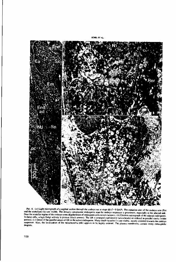
I ' —
m FIG. 6. (o) Light micrograph of a sagittal section through the embryo sac at stage III ( 7 -9 DAP). The initiation sites of the embryo axis (Ea)
and the scutellum (SO are visible. The densely cytoplasmic endosperm near the embryo suspensor is prominent, especially at the adaxial side Near the scutellar region of the embryo some degeneration of endosperm cells occurs (arrows), (b) Electron micrograph of the adaxial endosperm In these eel s, a high Golgi activity is present (black arrows). The ER is arranged capriciously (arrowheads) or ordered in parallel stacks (white arrows), ( r ) Detail of the parallel arrays of ER in the active endosperm. Many small vacuoles (v> are visible, mostly oriented towards the embryo suspensor. Also, the localization of the mitochondria (Mi) appears to be highly ordered. The plasma membranes contain many osmiophilic
108

CAN. J. BOT. VOL. 62. 1984
ate- ^ i a * ^ * * * * " ^ Sfcssap' t;v * 2& ^PfSg»s£r** 5 ^ V yi
Ff^g?
^
??v#^Mfe FIG. 7. Some details of embryo and endosperm cells in stage III ( 7 -9 DAP). («) Detail of an embryo cell from the embryo axis region. There
are several small vacuoles, while the nuclei often contain many domains of heterochromatin (arrows), {b) In the cells of the embryo many osmiophilic droplets occur (arrows), possibly taken up from the surrounding degenerating endosperm. (<•) Detail of the degenerating endosperm near the embryo axis region. In the cytoplasm, many osmiophilic droplets are present. (J) Detail of the cytoplasm remnant showing many lipid droplets (Li). (?) Degeneration is also visible by membrane breakdown, e.g.. nuclear membrane (arrow). ( / ) Accumulation of osmiophilic droplets (arrows) near the walls of the degenerating endosperm cells.
109

FIG. 8. (a) Semithin section of the endosperm bordering the placentochalazal region (Ch) at stage HI. The wall ingrowths are very pronounced (arrows). (6) Higher magnification of the endosperm cells at the end of stage III. The cytoplasm contains extensive profiles of ER and many polysomes and dictyosomes. Both symplastic transport (by plasmodesmata. see arrows) as well as transmembranal transport of osmiophilic material (arrowheads) takes place.
basal suspensor cells. Moreover, all plasma membranes contain many osmiophilic droplets.
The cells of the embryo in the axis region are small and have several small vacuoles (Fig. la). The nuclei often contain prominent domains of heterochromatin (arrows). The cells have many osmiophilic droplets (Fig. lb, arrows). The endosperm in this region has degenerated fully at this stage (Fig. Ic—lf). On the contrary, in the placentochalazal region the wall ingrowths of the endosperm cells have become very pronounced during this stage. They occur initially in the outer layer of the endosperm directly bordering the chalaza (Fig. 8a), causing the cell membrane to undulate intensively. Like in the endosperm cells surrounding the base of the suspensor (see Fig. 6c), the accumulation of osmiophilic droplets is obvious here as well. Compared with the earlier stages the cells are now densely filled with cytoplasm in which mitochondria, dictyosomes, and profiles of Rer are most prominent. These features are still present at the end of stage III (Fig. Sb). Moreover, many plasmodesmata and large ingrowths of peri-clinal and anticlinal cell walls have appeared.
Figure 9a shows a sagittal section through the embryo-endosperm region of a stage IV maize caryopsis (9—11 DAP). The embryo is located very close to the pericarp. The scutellum (Sc) has developed further while the coleptile (cp) and shoot apex (sa) primordia are clearly visible. The endosperm surrounding the basal suspensor cells has the same appearance as shown in stage III. The endosperm near the scutellum, embryo tip, and embryo axis has totally disintegrated, partially by mechanical stress.
In Fig. 9b, the boundary region between endosperm and scutellum is visible. The endosperm has lost its original structure; what remains is a lot of membrane fragments, osmiophilic
droplets, and loosened cell wall material (arrows). These osmiophilic droplets are present also in the walls between the degenerating endosperm and the embryonal cells (arrowheads). At this stage, the outer layer of the endosperm near the seed coat contains small vacuoles, protein bodies and other substances (Fig. 9c). This layer will develop eventually as the aleuron layer.
Osmiophilic droplets also occur in the embryo axis region (Fig. 9d), both near the degenerated endosperm cells and inside the intact embryo cells (arrows), in which the development of starch-containing plastids was also registered around this time (Fig. 9e).
Discussion Plant embryogenesis has for a long time been studied mainly
at a descriptive level. However, during the last decade increased attention has been paid to experimental analysis, placing considerable emphasis on functional relationships (for a survey, see Raghavan 1976). In the descriptions of embryo and endosperm the latter has in most cases been described as the nutritive tissue for the developing embryo (Brink and Cooper 1947; for a recent review, see Bhatnagar and Sawhney 1981). Indeed, the role of endosperm extracts on growth and morphogenesis of the embryo has been firmly established (Ziebur and Brink 1951; Pieczur 1952; Williams and De Lautour 1980; Mapes and Zaerr 1981). Not only the nutrient contents, but also the maintenance of a suitable osmotic environment has been proven to be an important function of the endosperm, as has been pointed out by several authors (Raghavan and Torrey 1963; Ryczkowski 1969; Monnier 1976).
Singh and Mogensen (1976) investigated the embryo—
110
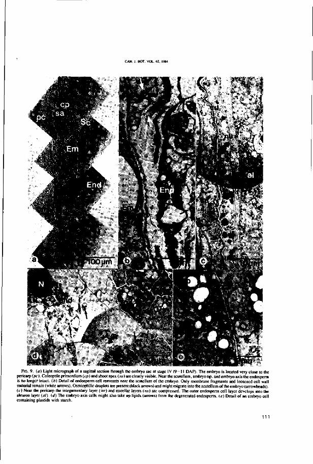
CAN. J. BOT. VOL. 62. 1984
FIG. 9. (a) Light micrograph of a sagittal section through the embryo sac at stage IV (9-11 DAP). The embryo is located very close to the pericarp (pc). Coleoptile primordium (cp) and shoot apex (ra) are clearly visible. Near the scutellum, embryo tip, and embryo axis the endosperm is no longer intact, (b) Detail of endosperm cell remnants near the scutellum of the embryo. Only membrane fragments and loosened cell wall material remain (white arrows). Osmiophilic droplets are present (black arrows) and might migrate into the scutellum of the embryo (arrowheads), (c) Near the pericarp the integumentary layer (iiir) and nucellar layers (nu) are compressed. The outer endosperm cell layer develops into the aleuron layer (al). (d) The embryo axis cells might also take up lipids (arrows) from the degenerated endosperm, (e) Detail of an embryo cell containing plastids with starch.
111

endosperm relationships in Quercus gambelii. Because of the small amount of starch and lipid found in the endosperm, they questioned if the endosperm of this species is indeed functioning as a food source. They argued that the food supply might be present in a soluble form, not easily detected, or that the endosperm functions merely as a medium for the transport of food, a suggestion also made by Marinos (1970) for Pisum sativum. Yeung and Clutter (1978), in their work on the em-bryogeny of Phaseolus coccineus, have also noted that the contribution of the endosperm to the growth of the early embryo might not be very significant because in this plant the endosperm is still developing at the heart stage of the embryo.
The results, presented in this report on maize embryo-genesis, support the emphasis placed on the absorption and transport function of the endosperm during early embryo formation rather than on the nutritive function. We suggest that both the endosperm cells surrounding the basal region of the embryo and those near the placentochalazal region serve this purpose. These cells most likely function as transfer cells in the sense first used by Gunning and Pate (1969), although the very characteristic wall ingrowths were present only in the placentochalazal region, as was observed earlier (Kiesselbach and Walker 1952).
Transport of nutrients from the placentochalazal region into the endosperm was investigated by Shannon (1972), who proved that sucrose is used by developing kernels, provided it is hydrolysed to glucose and fructose by invertase prior to uptake in the symplast of the endosperm. In a later micro-autoradiographic study, Felker and Shannon (1980) revealed that incoming sugars are not confined to the apoplast but rather are present in the cytoplasm and vacuoles of the endosperm cells. Concerning the transport of amino acids in the placentochalazal region, it was recently shown by Lyznik et al. (1982) that translocation takes place against the concentration gradient present in the developing maize kernel. This implies the involvement of energy in the translocation process. Moreover, significant differences in composition of the amino acid pool were found between the placentochalazal region and the endosperm.
Our results further indicate that besides the absorption and transport function of the endosperm, a high degree of synthesis, at least of membranes, also takes place. This was deduced from the huge amount of endoplasmic reticulum occurring both in the placentochalazal region and in the region bordering the base of the embryo. Because most of the highly ordered ER (Fig. 6c) in the endosperm surrounding the base of the embryo proved to be Rer, protein synthesis and subsequent transport in the pinched off vesicles is very likely. This mainly occurs during stages III and IV, in which the embryo has developed a long suspensor. This observation supports the active role of the suspensor in the monocotyledon maize as is the case also in dicotyledons as was shown by Yeung (1980).
The view for maize emerging at a fine structural cytological level closely resembles the situation in wheat as reported by Smart and O'Brien (1983). These authors, however, do not mention the presence of wall ingrowths in the endosperm at all, but only in the nucellar epidermis cells at later developmental stages. Therefore, the term "modified endosperm" has been used by them to describe the highly active endosperm cells lining the embryo, differentiating these cells from transfer cells which are defined by, among other criteria, wall ingrowths. In the modified endosperm of wheat, large quantities of Rer were also found, often with swollen cisternae filled with amorphous
content. For maize, we observed no filling of the ER cisternae. Moreover, we found that, unlike in wheat, dictyosomes were very often clearly resolved (see Fig. 6b).
The dilated appearance of ER cisternae was also observed by Mares et al. (1976) studying differentiation of the aleuron layer in developing wheat kernels. They further noted a rapid and massive proliferation of ER and demonstrated that the outer nuclear membrane is involved in the process of ER formation. The authors suggested that this mechanism might be a specific response of plant cells to a sudden and massive requirement for membrane and synthetic machinery. They further cited Wischnitzer (1974), who ascribed a similar role to the annulate lamellae in oocytes of various organisms (see also Kessel 1968; Maul 1977). Our results, as shown in Figs. 5c and 5d, support these views. At least part of the newly synthesized ER membranes might originate from the nuclear envelope.
Smart and O'Brien (1983) conclude that in wheat the developing embryo is initially supplied by hydrolysis of nucellar parenchyma and endosperm cells. Hydrolysis of endosperm cells also occurs in maize, although in this case it starts somewhat later, during stage II (about 5 DAP). Breakdown is partially caused by mechanical stress, as can be deduced from the Figs. 5a and 5b. Autolysis of the endosperm or digestion by hydrolytic enzymes excreted by the embryo must be an important factor too. The breakdown of the endosperm cells near the scutellum in stage IV (Figs. 9a and 9b) is mainly an autolytic process caused by internal programmed hydrolytic activity. This is evident from the fact that no significant excretion activity is registered in the neighbouring embryo cells as measured by the absence of high Golgi activity. We also did not observe embryo cells with wall crenulations as reported by Olson (1981) for Papaver nudicaule and tentatively suggested by him to be indicative for an embryonic influence on endosperm breakdown. Programmed autolysis of the endosperm has been described before (e.g., see Bhatnagar and Sawhney 1981); in vitro studies using isolated embryo and endosperm tissues support this view (Segers et al. 1981). The products, coming free from the degenerating endosperm near the scutellum, can be absorbed by this part of the embryo. The maize scutellum is known as a fat-storing tissue, as indicated by the presence of glyoxysomes (Longo and Longo 1970). Therefore, the osmiophilic droplets as shown in Figs. 9b and 9r might be markers for such a process. We cannot, however, rule out the possibility that the embryo cells themself have synthesized these droplets.
The ordered arrangement of mitochondria just below the plasma membrane of the endosperm cells next to the integumentary remnants or suspensor cells (see Fig. 6c) might focus on the highly active transport processes taking place in those areas and requiring considerable energy. As proposed by Oparka et al. (1981), in developing rice caryopses these regularly aligned mitochondria, present along the upper periclinal aleuron cell walls, may function as part of a solute transport system requiring localized ATP production. The plasmalemma foldings, which were frequently observed at the embryo-endosperm interface (Figs. 4d\ 4e, and 4 / ) , might also be indicative for the transport processes taking place, e.g., by setting up the standing gradient osmotic flow recently discussed by Harris (1981) for plasmalemma invaginations in cotyledons of germinating mung beans.
In conclusion, we observe a close interrelationship between both embryo and endosperm in the developing maize kernel. During early embryogeny (stage 1 and 11, until about 7 DAP),
112

CAN I BOT VOL 62. 1984
the basal endosperm is especially highly active, supplying the embryo all along the suspensor with nutrients, which are transferred directly from the placentochalazal region by a small area of endospermic transfer cells or are newly synthesized in the modified endosperm region lining the embryo. From this study, no conclusions could be drawn as to the chemical composition of the nutrients. It is probable, however, that the main constituents are dissolved salts, peptides, and amino acids. From stage III, starting about 7 DAP, the nutritive function of the highly vacuolated and degenerating upper endosperm might become increasingly important, carbohydrates and (phospho)-lipids most likely being the main food resources.
Acknowledgements Thanks are owed to Sybout Massalt for the photographic
work, Allex Haasdijk and Paul van Snippenburg for the artwork, and Truus van de Hoef and Joke Cobben for preparing the typescript. The authors are also grateful to Michel Willemse for helpful comments and to Richard Brice for reading the English text.
BHATNACAR, S. P., and V. SAWHNEY. 1981. Endosperm—its morphology, infrastructure and histochemistry. Int. Rev. Cytol. 73: 55-102.
BRINK, R. A., and D. C. COOPER. 1947. The endosperm in seed development. Bot. Rev. 13: 423-541.
DIBOLL, A. G. 1968. Fine structural development of the mega-gametophyte of Zea mavs following fertilization. Am. J. Bot. 55: 797-806.
FELKER, F. C . and J. C. SHANNON. 1980. Movement of l4C-labeled assimilates into kernels of Zea mays L. III. An anatomical examination and microautoradiographic study of assimilate transfer. Plant Physiol. 65: 864-870.
GREEN, C. E. 1982. Somatic embryogenesis and plant regeneration from the friable callus of Zea mays. Plant Tissue Culture 1982. Proceedings of the 5th International Congress on Plant Tissue and Cell Culture, Tokyo. Japan. July 11-16. 1982. Edited by A. Fujiwara. Japanese Association for Plant Tissue Culture. Tokyo, pp. 107-108.
GUNNING, B. E. S.andJ. S. PATE. 1969. Transfer cells'. Plant cells with wall ingrowths specialized in relation to short distance transport of solutes. Their occurrence, structure and development. Proto-plasma, 68: 107-133.
HARRIS. N. 1981. Plasmalemmasomes in cotyledon leaves of germinating Vigna radiata L. (mung beans). Plant Cell Environ. 4: 169-175.
K.ESSEL. R. G., 1968. Annulate lamellae. J. Ultrastruct. Res. Suppl. 10: 1-82.
KHOO. U., and M. J. WOLF. 1970. Origin and development of protein granules in maize endosperm. Am. J. Bot. 57: 1042—1050.
KIESSELBACH, T. A. 1949. The structure and reproduction of corn. Univ. Nebr. Coll. Aerie. Agric. Exp. Stn. Res. Bull. No. 161. pp. 1-96.
KIESSELBACH. T. A., and E. R. WALKER. 1952. Structure of certain specialized tissues in the kernel of com. Am. J. Bot. 39: 561 —569.
KYLE, D. J., and E. D. STYLES. 1977. Development of aleurone and sub-aleurone layers in maize. Planta, 137: 185-193.
LAMPE, L. 1931. A microchemical and morphological study of the developing endosperm in maize. Bot. Gaz. (Chicago). 91: 337-376.
LONGO. C. P.. and G. P. LONGO. 1970. The development of gly-oxysomes in peanut cotyledons and maize scutella. Plant Physiol. 45: 249-254.
LYZNIK, L. A., W. ZDROJEWSKI. M. NEUMANN, J. MACEWICZ, and K. RACZYNSKA-BOJANOWSKA. 1982. A possible role of pedicel-placento-chalazal tissues in the amino acids supply to the devel
oping maize endosperm. Maydica, 27: 191-198. MAPES. M. O., and J. B. ZAERR. 1981. The effect of the female
gametophyte on the growth of cultured Douglas-fir embryos. Ann. Bot. (London), 48: 577-582.
MARES, D . C. JEFFERY. and K. NORSTOG. 1976. The differentiation of the modified aleurone of wheat endosperm: nuclear membrane-mediated endoplasmic reticulum development. Plant Sci. Lett. 7: 305-311. v
MARINOS, N. G. 1970. Embryogenesis of the pea( Pisum sativum). I. The cytological environment of the developing embryo. Proto-plasma, 70: 261-279.
MAUL, G. G. 1977. The nuclear and the cytoplasmic pore complex: structure, dynamics, distribution and evolution. Int. Rev. Cytol. Suppl. No. 6. pp. 75-186.
MONNIER. M. 1976. Culture in vitro de 1'embryon immature de Cap-sella Bursa-pastoris Moench. Rev. Cytol. Biol. Veg. 39: 1-120.
NORSTOG, K. 1972. Early development of the barley embryo: fine structure. Am. J. Bot. 59: 123-132.
1974. Nucellus during early embryogeny in barley: fine structure. Bot. Gaz. (Chicago). 135: 97 - 103.
OLSON, A. R. 1981. Embryo and endosperm development in ovules of Papaver nudicaule after in vitro placental pollination. Can. J. Bot. 59: 1738-1748.
OPARKA, K. J., P. J. GATES, and D. BOULTER. 1981. Regularly aligned mitchondria in aleurone and sub-aleurone layers of developing rice caryopses. Plant Cell Environ. 4: 355-357.
PIECZUR. E. A. 1952. Effect of tissue cultures of maize endosperm on the growth of excised maize embryos. Nature (London), 170: 241-242.
RAGHAVAN, V. 1976. Experimental embryogenesis in vascular plants. Academic Press, London. New York, and San Francisco.
RAGHAVAN. V.andJ.G. TORREY. 1963. Growth and morphogenesis of globular and older embryos of Capseila in culture. Am. J. Bot. 50: 540-551.
RANDOLPH, L. F. 1936. Developmental morphology of the caryopsis in maize. J. Agric. Res. 53: 881-916.
ROST, T. L.. and N. R. LERSTEN. 1973. A synopsis and selected bibliography of grass caryopsis anatomy and fine structure. Iowa State J. Res. 48: 47-87.
RYCZKOWSKI, M. 1969. Changes in the osmotic value of the central vacuole and endosperm sap during the growth of the embryo and ovule. Z. Pflanzenphysiol. 61: 422-429.
SEGERS, J. H. L.. P. C. W. M. VOSSEN. A. A. M. VAN LAMMEREN, and J. H. N. SCHEL. 1981. Ulirastructural changed during in vitro culture of embryo and endosperm tissue from Zea mays L. Abstracts of the 29th Meeting of the European Tissue Culture Society. Noordwijkerhout, The Netherlands. May 12-15. 1981. p. 38.
SHANNON, J. G. 1972. Movement of l4C-labeled assimilates into kernels of Zea mavs L. I. Pattern and rate of sugar movement. Plant Physiol. 49: 1981202.
SHERIDAN. W. F. 1982. Maize for biological research. University of North Dakota Press. Grand Forks. ND.
SINGH, A. P., and H. L. MOGENSEN. 1976. Fine structure of early endosperm in Quercus gambelii. Cytologia. 41: 345—361.
SMART, M. G.. andT. P. O'BRIEN. 1983. The development of wheat embryo in relation to the neighbouring tissues. Protoplasma. 114: 1-13.
VAN LAMMEREN, A. A. M. 1981. Early events during embryogenesis in Zea mays L. Acta Soc. Bot. Pol. 50: 289-290.
VAN LAMMEREN, A. A. M.. and H. KIEFT. 1983. Embryogenesis in Zea mays L. Structural aspects of primary meristem formation. In Fertilization and embryogenesis in ovulated plants. Edited by O. Erdelska. Veda. Bratislava. Czechoslovakia, p. 287.
VAN LAMMEREN. A. A. M.. and J. H. N. SCHEL. 1983. Embryogenesis in Zea mays L. Development of structure and polarity during proembryo formation. In Fertilization and embryogenesis in ovulated plants. Edited by O. Erdelska. Veda. Bratislava. Czechoslovakia, pp. 283-285.
VASIL, I. K. 1982. Somatic embryogenesis and plant regeneration in
113
SCHEL ET AL.
cereals and grasses. Plant Tissue Culture 1982, Proceedings of the 5th International Congress on Plant Tissue and Cell Culture, Tokyo, Japan, July 11-16, 1982. Edited by A. Fujiwara. Japanese Association for Plant Tissue Culture, Tokyo, pp. 101 - 104 .
WEATHERWAX, P. 1930. The endosperm of Zfa and Coii. Am. J. Bot. 17:371-380.
WILLIAMS, E. G., and G. DE LAUTOUR. 1980. The use of embryo culture with transplanted nurse endosperm for the production of interspecific hybrids in pasture legumes. Bot. Gaz. (Chicago), 141: 252-257.
WISCHNITZER, S. 1974. The nuclear envelope: its uttrastructure and
functional significance. Endeavour, 33: 137-142. WOLF, M. J., C. L. BUZAN, M. M. MACMASTERS, and C. E. RIST.
1952. Structure of the mature corn kernel I to IV. Cereal Chem. 29: 321-382.
YEUNG, E. C. 1980. Embryogeny of Phaseolus: the role of the sus-pensor. Z. Pflanzenphysiol. 96: 17-28.
YEUNC, E. C , and M. E. CLUTTER. 1978. Embryogeny of Phaseolus
coccineus: growth and microanatomy. Protoplasma, 94: 19-40. ZIEBUR. N. K., and R. A. BRINK. 1951. The stimulative effect of
Hordeum endosperms on the growth of immature plant embryos in vitro. Am. J. Bot. 38: 253-256.
114

CHAPTER 6
Cell differentiation in the pericarp and endosperm of developing maize
kernels (Zea mays L.) with special reference to the
microtubular cytoskeleton
A.A.M. van Lammeren & H. Kieft

riiT.T. DIFFERENTIATION IN THE PERICARP AND ENDOSPERM OF DEVELOPING
MAIZE KERNELS (ZEA MAYS L.) WITH SPECIAL REFERENCE TO THE MICRO-
TUBULAR CXTOSKELETON
ABSTRACT
Maize kernels were investigated by light and electron microscopy to
determine and quantify cytological changes in fruit wall and seed at
various developmental stages.
Pericarp, integument and nucellus cells differentiated and sub
sequently degenerated within a period of three weeks. Most prominent
feature in the pericarp development was the differentiation of three
zones, each with cells that are shaped and arranged differently. Cell
elongation and wall thickening in exocarp and endocarp cells lead to
protecting layers. The early formation of a spongy mesocarp facili
tated compression of the fruit wall in later stages. Integuments and
nucellus degenerated scon; the endosperm persisted and enlarged.
Endosperm development is characterized by the formation of the
aleurone layer, some sub-aleurone layers and inner endosperm from 5
days after pollination onwards. The cell layers differ from each
other by the shapes of the cells, the diverging development of
vacuoles and plastids, the rate of starch accumulation, the increase
of cytoplasm, and by the formation of various types of spherosomes
and protein bodies. The cellular distribution of the mitochondria in
aleurone and sub-aleurone layers point to an active transport through
cell membranes towards or from the nucellus.
Microtubules were visualized immunocytochemically in semi-thin
sections (2-3 pm) to determine their three-dimensional distribution
in the cytoplasm. Microtubules were observed in three major con
figurations: spindle tubules, cytoplasmic microtubules which run
throughout the cytoplasm, and cortical microtubules which appear to
form various configurations in the periphere cytoplasm. The cortical
microtubules were investigated in particular. They formed various
configurations from parallel to criss-cross arrangements. From the
117
development of ce l l shapes and the observed concomitant configura
tions of microtubules i t i s concluded that these microtubules mediate
in the cytomorphogenesis and l ikely by influencing the mode of ce l l
wall synthesis.
1 . INTRODUCTION
The s t ructural aspects of caryopsis development have been studied for
important c e r ea l s as wheat (Buttrose 1963b, Campbell e t a l . 1981,
Cochrane 1983, Rogers & Quatrano 1983, Smart & O'Brien 1983), barley
(Olsen & Krekling 1980, Felker e t a l . 1984, Kvaale & Olsen 1986), rye
(Pomeranz 1973) and r i ce (Bechtel & Pomeranz 1977, Bechtel & Juliano
1980, Mif l in e t a l . 1981, Oparka & Harr i s 1982). A general survey
related to seed development i s given by Bhatnagar & Sawhney (1981).
Developmental s t ud i e s of maize go back t o t he l a s t century; many
e a r l y workers analyzed s p i k e l e t formation, f e r t i l i z a t i o n , and the
development of f ru i t s and seeds from a s t ructural point of view (see,
amongst o t he r s , Guerin 1899, Mi l l e r 1919, Weatherwax 1919, Lampe
1931, Randolph 1936, Kiesselbach 1949). More r e cen t ly , e l ec t ron
microscopy s t imula ted t he s ub - c e l l u l a r a na ly s i s of f e r t i l i z a t i o n
(Diboll 1964, Van Lammeren 1986a), the development of the proembryo
(Van Lammeren & Schel 1983, Schel e t a l . 1984), the embryo proper
(Van Lammeren 1986b), the endosperm (Khoo & Wolf 1970, Kyle & Styles
1977) and of the interaction between embryo and endosperm (Schel e t
a l . 1984).
The endosperm i s considered as an important nutr i t ional factor for
embryo development and germinat ion. Espec ia l ly the outermost c e l l
layer, the aleurone layer which can be distinguished by the shapes of
i t s c e l l s and the organelles within the ce l l s , accumulates storage
products during seed development and releases hydrolytic enzymes
during germination (Lampe 1931, Bhatnagar & Sawhney 1981). The u l t r a -
s t r u c t u r a l development of the a leurone l ayer and the sub-aleurone
l aye r s have been s tudied before (Khoo & Wolf 1970, Kyle & S t y l e s
1977); however, the development of the ce l l shapes, the interaction
1 1 !
between the various tissues of the caryopsis and the concomitant
manifestation of (ultra)structural phenomena have as yet not been
observed.
The present study focusses on the cellular and sub-cellular
changes in carpels, integument, nucellus and part of the endosperm
from the moment of cellularization of the endosperm up to and in
cluding the senescence of the pericarp cells. In all tissues of the
caryopsis cell differentiation resulted not only in a diverse com
position of the cytoplasm but in varying cell shapes as well. In
order to elucidate the function of the microtubular cytoskeleton in
this process of cytomorphogenesis (Lloyd 1982, Lang Selker S Green
1984, Van Lammeren 1985, Van Lammeren et al. 1985, Traas 1986)
special attention was paid to the structure of the cytoskeleton by
the application of immunofluorescence microscopy.
2. MARSHALS AND METHODS
Maize plants of strain A188 were grown and pollinated as described
previously (Van Lammeren 1986b). From 3 up to 22 days after pollina
tion (DAP) thick (1mm) median sections were cut from the ovaries and
small, 2 mm blocks were excised from a defined region (see Fig. 1).
The blocks contained the pericarp, the aleurone and sub-aleurone
layers and some inner endosperm.
For phase contrast and electron microscopy the tissue blocks were
fixed'with 2.5% glutaraldehyde in 0.02 M phosphate buffer, pH 6.8
with 2 mM EGTA for 2 hrs at 20°C. After rinsing, the samples were
post-fixed in 2% OsO^ in buffer and dehydrated through a graded
series of acetone and embedded in Epon. Semithin (2 pm) sections were
cut with glass knives and ultrathin sections were prepared with a
diamond knife on an LKB Ultrotome V. The latter sections were post-
stained in an LKB Ultrostainer with uranyl acetate for 15-30 min. at
40°C and with lead citrate for 30-60 sec. at 20°C before investiga
tion in a Philips EM 301 operating at 60 kV.
The fixation, embedding and sectioning of plant tissues preceding
the immunocytochemical labelling of microtubules were described
119

previously (Van Lammeren et al. 1985) but small adaptations were
performed. Blocks were fixed in 3% para-formaldehyde and 0.13%
glutaraldehyde in phosphate buffered saline (PBS) pH 7.2 for 1 hr at
20 °C After rinsing and dehydration the samples were transferred into
a mixture of ethanol and two types of polyethylene glycol (PEG
1500/4000, BDH Chemicals Ltd., Poole); ethanol:PEG= 2:1 (w/w) for
1hr, 1:1 for 1 hr, and 100% PEG for 1 hr, all at 55°C. Finally the
samples were embedded in pure PEG 1500/4000 and cooled down.
Sections of 2-3 pm were picked up with hanging drops of 40% (w/w) PEG
6000 in PBS using a wire loop and mounted on poly-lysine coated
slides as described before (Van Lammeren et al. 1985). After that,
the PEG was washed away with PBS; the aldehydes in the embedment-free
sections were quenched with 15 niM NaBH4 and 0.1 M NH^Cl for 5 min.
each and treated with 2% bovine serum albumin (BSA, essentially
globulin free, Sigma, St Louis) in PBS for 30 min. To detect micro
tubules, the sections were then incubated with the IgG fraction of a
polyclonal antitubulin (dilution 1:25 in PBS) raised in rabbit
against rat brain tubulin (see Van Lammeren et al. 1985). After 45
min. incubation at 20°C the sections were rinsed in PBS for 45 min.
and incubated again with a second, FITC-conjugated antibody which was
raised in goat against rabbit (GAR/FITC, Nordic, Tilburg; dilution
1:25 in PBS). After that, the sections were rinsed again and embedded
in a mixture of 20% Mowiol 5-88 (Hoechst, Frankfurt am Main) in
Citifluor (Citifluor Ltd, London).
3. RESULTS
3.1. Development of somatic tissues in the caryopsis
The development, differentiation and degeneration of the carpels, the
integument and the nucellus were traced by means of light and
electron microscopy within a restricted zone at the germinal face of
the caryopsis (Fig. 1) during a period of three weeks after pollina
tion. In the pericarp, three zones were distinguished at three days
after pollination (3 DAP): an endocarp zone near the ovule consisting
120

-ovary -*--ovule
-nucellus ^endosperm
sembryo
pericarp
integument
nucellus
aleurone layer
sub-aleurone layer
: LLinner endosperm
Fig. 1 Schematic representation of the median section of a young
maize caryopsis with the sampling area enlined (A) and the anatomy of
the sampling block (B).
of about three cell layers of small, thin-walled fusiform cells which
were often isodiametric in section and elongated out of the plane of
sectioning (Fig. 2a); a mesocarp zone with larger, loosely arranged
cells which are isodiametric to irregular in shape (Fig. 2a) and an
exocarp zone containing cylindrical cells which bordered each other
leaving only little intercellular space. All pericarp cells had large
central vacuoles and only little cytoplasm in which amyloplasts were
observed (Fig. 3a, arrow). As development proceeded, however, the
growth of the developing seed forced the pericarp to enclose a larger
area. At 8 DAP the exocarp cells had elongated. In the mesocarp
region a spongy mesophyll developed with spherical, slightly elon
gated or curled cells (Fig. 2b). The endocarp cells did not increase
much in diameter but they separated, thus enlarging the intercellular
space (Fig. 2b, arrow). In cross-sections the cells were often found
parallel to each other forming fibrous sheaths covering the devel
oping seed. The length axes of the various sheaths were not fixed. At
1 7 DAP the pericarp cells were still intact yet at an end phase of
development (Fig. 2c). The diameter of the endocarp cells had not
enlarged much. The exocarp cells had reached their maximal length
121

Fig. 2 Low magnif icat ion l i g h t micrographs of 2 ̂ im Epon s ec t ions
from the sampling a rea of the maize caryopsis a t var ious develop
mental stages. Note the enlargement, d ifferentiat ion and subsequent
degenerat ion of the endocarp, mesocarp and exocarp c e l l s . The
122

thereby leaving only little intercellular space. Their cell walls had
thickened giving solidity to the fruit wall. The spongy mesocarp
cells were compressed and so the thickness of the whole pericarp
decreased. The cytoplasm of the remaining cells degenerated at about
20 DAP thus leaving a multi-layered dead fruit wall mainly consisting
of cell walls. At 22 DAP the small endocarp cells were often crushed
whereas the shape of the dead exocarp cells was still preserved (Fig.
2d).
Integuments covered the nucellus and the endosperm during the
early developmental stages. Because of the retarded growth of the
outer integument during ovule development, only an inner integument
covered the nucellus at the sampling area. It consisted of two cell
layers and a thin cuticle. The integumentary cells were cylindrical
at 3 DAP (Fig. 2a, arrow). Sometimes they were only seen at this
stage but occasionally the intact cytoplasm was still observed at 8
DAP. Hereafter cells were crushed between the pericarp and the
expanding endosperm and their thin cell walls and the cuticles dis
appeared completely.
The nucellus is an ephemeral tissue, too. Except for the epidermis
and its cuticle, the nucellus degenerated fast. At 3 DAP a multi-
layered nucellus still existed (Fig. 2a) but at 5 DAP only 1 or 2
layers of nucellus cells remained (Fig. 3a) in the sampling area.
integument and sub-epidermal nucellus cells degenerated scon (a-b).
The endosperm developed into the aleurone, sub aleurone and inner
endosperm, all accumulating proteins and starch. a= 3 DAP; b= 8 DAP;
c= 17 DAP; d= 20 DAP. The bars represent 100 urn.
Abbreviations to the figures: AB= aleurone body; AL=aleurone layer;
DAP=days after pollination; EC=endocarp; EM=embryo; END= endosperm;
EXC=exocarp; EPB=endosperm protein body; I=integument; IE=inner
endosperm; IS=intercellular space; MC=mesocarp; NUC=nucellus; PC=
pericarp; S=starch; SAL=sub-aleurone layer; S1 =spherosome type 1
present in aleurone and sub-aleurone layer; S2=spherosome type 2
present in the aleurone layer from 8 DAP onwards.
123

V PC \
%0* 1
NUC
m\ '̂ 1
9VI
1 ,v- S.. <^T • • w^S^ 4
„^s *"
4 *"*> ;*r „p ]" _ ,
END s fv*?
-.tO pm * *.7 « w.-'ft.;\~-^. P , b «, ?--/<i ,. ,t-' •' •
Fig. 3 Electron micrographs of the developing maize caryopsis in
the sampling area at various developmental stages. Conspicuous
changes in the aleurone and sub-aleurone layers include the differen
tiation of an epidermal-like tissue (a-b) and the accumulation of
starch (S, b-c) and storage products in two types of spherosomes (S1,
S2), aleurone bodies (AB) and endosperm protein bodies (EPB).For
abbreviations see Fig. 2. a=5 DAP; b=12 DAP; c=22 DAP. The bars
represent 10 urn.
I24
Cell degenerat ion occurred near the outermost c e l l l ayer of t he
endosperm. F i r s t the ce l l s exhibited cytoplasmic degeneration. Then
they collapsed and eventually they disappeared. The nucellus epider
mis was observed as a l i v i ng t i s s u e a t 8 DAP. I t cons i s ted of
f lattened ce l l s covered with a thick, conspicuous cut ic le which was
s t i l l found in the l a t e developmental s t ages (Figs. 2 and 3,arrow
heads). There was a large central vacuole and only l i t t l e cytoplasm.
From 12 DAP onwards nucellar epidermis ce l l s collapsed (Figs. 3b, 7-
IV). The remnants were found up t o 22 DAP because the c u t i c l e , t he
ou te r c e l l wa l l s and the p a r t l y thickened a n t i c l i n a l c e l l wa l l s
persisted (Fig. 3c, arrow-heads).
The course of c e l l development of the var ious somatic t i s s u e s
described above i s summarized schematically in Table 1 with respect
to c e l l shape development, c e l l wall formation and v iab i l i ty .
3.2. Development of the endosperm The development of a leurone , sub-aleurone and inner endosperm
involved the multiplication, enlargement and differentiat ion of c e l l s
and the accumulation of s to rage products (see Fig. 1 b for the topo
graphy of t he sampling area and t he F igs . 7 I-V for 'camera luc ida '
drawings). The development of the a leurone l ayer became prominent
from 5 DAP onwards (Fig. 3a-c). I n i t i a l l y the outer endosperm ce l l s ,
which developed in to the aleurone c e l l s , were large (one c e l l of the
nuce l lus epidermis covered 2-3 p re-a leurone c e l l s , F ig. 7-1) and
divided in various directions but a t about 7 DAP they divided simul
taneously and in only one direction. Many nuclei exhibited prophase
conf igura t ions a t 5-6 DAP and a t 8 DAP cy tok ines i s had r e su l t ed i n
the formation of an epidermal-like ce l l layer in which mainly a n t i
c l ina l ce l l walls had formed (Fig. 7-II). The newly formed ce l l s were
much smal le r ; up t o 8 epidermal c e l l s were now covered by one nu
c e l l u s epidermal c e l l which had not increased in s i z e . After t h a t ,
the epidermal c e l l s became rectangular, the amount of cytoplasm and
organelles s t i l l increased, and the accumulation of storage products
s t a r t e d (Fig. 3b, 12 DAP). From 8 t o 1 2 DAP the c e l l s of the ou t e r -
125

PHASE
17 Bp^8* EPIDERMIS
AGE (DAP) 11 13 15 17 19 21
SYMBOLS
Living cells
Cortical MTS criss-cross
Cortical MTS parallel
Cell collapse & degeneration
Table 1 Schematic representation of cell differentiation and con
comitant configurations of microtubular cytoskeletons in various
tissues of the developing caryopsis of maize.
most cell layer of the endosperm differentiated into aleurone cells.
They enlarged in periclinal direction and up to 5 aleurone cells were
covered by one nucellus cell at 12 DAP. Sub-aleurone cells were often
found in line with the aleurone cells indicating that periclinal cell
wall formation took place repeatedly as endosperm development
proceeded. These cells continued to divide, sometimes in a meristem-
like fashion forming rows of cells with new periclinal cell walls.
The newly formed, isodiametric cells were often smaller than the
126
bordering aleurone cells (Pig. 3b). The inner endosperm cells,
however, enlarged as shown in Fig. 3b. During further seed develop
ment the aleurone cells enlarged in periclinal and anticlinal direc
tion (Up to 4 aleurone cells were covered by one cell of the nucellus
epidermis at 17 DAP, Fig. 7-III). In the ensuing development, from 17
to 22 DAP, only slight changes in the sizes and shapes of the cells
were to be seen. The anticlinal and outer cell walls increased in
thickness from 17 DAP onwards (Fig. 3c). Sub-aleurone and inner
endosperm cells, however, still increased in size (cf Figs. 2c and
2d). These cytomorphological data of endosperm formation are sum
marized schematically in Table 1 together with the somatic tissues of
the caryopsis.
The submicroscopical aspects of endosperm differentiation are
summarized for the developing aleurone layer and the sub-aleurone
endosperm in Table 2 and 3 respectively. Nuclei, being the largest
organelles, were about spherical to ellipsoidal and had undulating
membranes. They did not differ in size and shape in aleurone and sub-
aleurone cells, nor did their internal structure. At 3 and 5 DAP the
nuclei of the outermost endosperm layer were often observed near the
outer cell wall. Plastids were found in all endosperm cells. In the
aleurone layer they were smallest and accumulated starch from 12 DAP
onwards. At 22 DAP these amyloplasts occupied up to 12% of the cell
volume. In the sub-aleurone cells the plastids accumulated starch
from 8 DAP onwards and up to 46% of the cell volume was filled with
these second largest organelles at 22 DAP. Thylakoids were found in
all stages but grana were never detected. Mitochondria were never
abundant in the endosperm. At 3 DAP they were not equally spread over
the cytoplasm in the outer cell layers but preferentially found in
the cytoplasm adjacent to the outer periclinal cell walls (Fig. 3a,
thin arrow). This phenomenon could be observed at later stages,too,
and also in the sub-aleurone layer (Fig. 3b, thin arrows).
Vacuoles occupied about 40% of the cell volume of young aleurone
and sub-aleurone endosperm cells (Fig. 2a, 3 DAP). At 22 DAP only
few, small vacuoles were observed in the sub-aleurone cells but in
the aleurone layer about 10% of the cell volume was occupied by
127
m
e> \
Q
&
£f
CO z> Ul _l o
co
< a. a z o x o p
CO UJ _J
o a
co lil O co O m
UJ
CO UJ 5 O CO
o Q
CO UJ
O CO
85 UJ UJ I L o. >-COH
O CO O (N
£<" x 9-CO H
Table 2 Schematic representation of organelle development in dif
ferentiating aleurone cells of Zea mays L. from 3 to 22 days after
pollination (DAP).
12E
^
O o
co LLI
I O
Z
CO Q
CO
<
a: a Z o I o p
CO LU
o o
2 O CO
o m oc DC LU
CO LLI 2 o CO
9 Q
CO LU
2 o °co LU LU x a. a. >-CO H
co £2 2 2 5
O ^ 0 - z CCCOCOQJ L U L u O t I D . D O Q - > Z CC CO I - LU Q.
Table 3 Schematic representation of organelle development in dif
ferentiating sub-aleurone endosperm cells of Zea mays L. from 3 to 22
days after pollination (DAP).
129
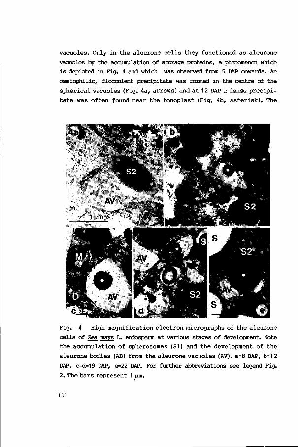
vacuoles. Only in the aleurone cells they functioned as aleurone
vacuoles by the accumulation of storage proteins, a phenomenon which
is depicted in Fig. 4 and which was observed from 5 DAP onwards. An
osmiophilic, flocculent precipitate was formed in the centre of the
spherical vacuoles (Fig. 4a, arrows) and at 12 DAP a dense precipi
tate was often found near the tonoplast (Fig. 4b, asterisk). The
Fig. 4 High magnification electron micrographs of the aleurone
cells of Zea mays L. endosperm at various stages of development. Note
the accumulation of spherosomes (S1) and the development of the
aleurone bodies (AB) from the aleurone vacuoles (AV). a=8 DAP, b=1 2
DAP, c-d=19 DAP, e=22 DAP. For further abbreviations see legend Fig.
2. The bars represent 1 um.
130
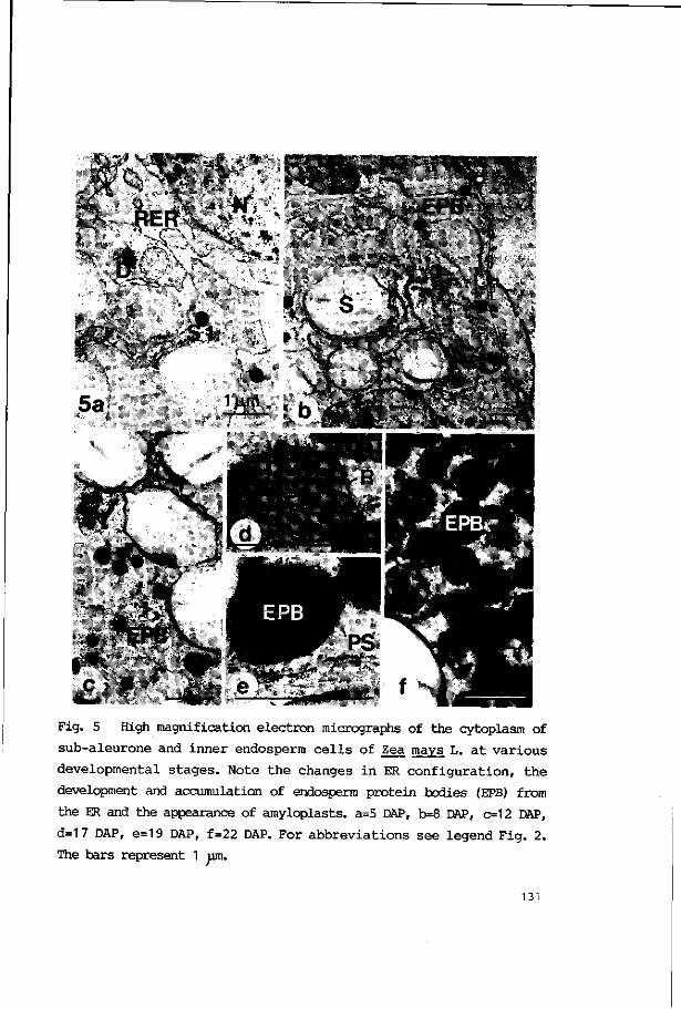
• >. x . . - • • - . * . •
RER : \ r "™: *
• " f t " • " ; • > - *
'• - •
• v ' • \ 1.
i i i ' - ^ j ^ ; ; V v
5a
Fig. 5 High magnification electron micrographs of the cytoplasm of
sub-aleurone and inner endosperm cells of Zea mays L. at various
developmental stages. Note the changes in ER configuration, the
development and accumulation of endosperm protein bodies (EPB) from
the ER and the appearance of amyloplasts. a=5 DAP, b=8 DAP, c=12 DAP,
d=17 DAP, e=19 DAP, f=22 DAP. For abbreviations see legend Fig. 2.
The bars represent 1 um.
131

amount of storage proteins clearly increased and at 22 DAP they
almost filled the vacuoles, which are then called aleurone bodies
(Figs. 4c-e, asterisks).
The endoplasmic reticulum proved to be a conspicuous organelle
system, especially in the sub-aleurone cells which ultrastructural
development is shown in Fig. 5. Initially, at 3 DAP, the ER occupied
but a minor part of the cell volume. At 5 DAP there was, however, a
significant increase of the cisternal space because of the swelling
of the ER by which up to 15% of the cell volume was occupied (Fig.
5a). In the sub-aleurone and inner endosperm the ER, to which many
ribosomes were attached, gave rise to electron-dense globular struc
tures which are called endosperm protein bodies (EPBs, Figs. 5b-e).
These EPBs which were surrounded by a ribosome carrying ER membrane
(Figs. 5d,e) were formed from 8 DAP onwards and had an average dia
meter of 0.1 4 + 0.04 um at 8 DAP and 0.44 + 0.09 jim at 22 DAP. Their
presence was restricted to the sub-aleurone and inner endosperm where
they eventually occupied up to 70% of the cell volume (see Figs. 3c
and 5f) at 22 DAP. The heavily stained ribosomes occurred singly or
as short coils of polysomes. Dictyosomes were not seen frequently and
in the aleurone layer their presence could not be established at 22
DAP because of the densely stained cytoplasm.
Concerning other storage products, two types of spherosomes were
discerned in the developing endosperm. The first type (Type S1) was
mainly seen in the outer cell layers of the young endosperm up to an
age of 8 DAP (Figs. 3a and 5a). Their number and total amount de
creased in the aleurone layer when the first spherosomes of a second
type (Type S2) were discerned. This second type predominantly oc
curred in the aleurone layer (Figs. 3b and 4). Some of these sphero
somes were seen in the adjacent cell layer but none in the inner
endosperm. Tye S2 spherosomes were largest (1.26 + 0.68 p at 17 DAP)
and sometimes filled the aleurone cell cytoplasm for over 90% at 22
DAP (Figs. 3c,4e).
132
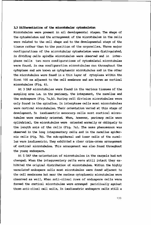
3.3 Differentiation of the microtubular cytoskeletcn
Microtubules were present in all developmental stages. The shape of
the cytoskeleton and the arrangement of the microtubules in the cells
were related to the cell shape and to the developmental stage of the
tissue rather than to the position of the organelles. Three major
configurations of the microtubular cytoskeleton were distinguished.
In dividing cells spindle microtubules were observed and in inter
phase cells two more configurations of cytoskeletal microtubules
were found. In one configuration microtubules ran throughout the
cytoplasm and are known as cytoplasmic microtubules and in the other
the microtubules were found in a thin layer of cytoplasm within the
first 100 nm adjacent to the cell membrane and are known as cortical
microtubules (Fig. 6).
At 3 DAP microtubules were found in the various tissues of the
sampling area i.e. in the pericarp, the integument, the nucellus and
the endosperm (Fig. 7a,b). During cell division microtubules were
only found in the spindles. In interphase cells most microtubules
were cortical microtubules. Their orientation varied at this stage of
development. In isodiametric mesocarp cells most cortical micro
tubules were randomly oriented. When, however, pericarp cells were
cylindrical, the microtubules were oriented normally or obliquely to
the length axis of the cells (Fig. 7a). The same phenomenon was
observed in the long integumentary cells and in the nucellus epider
mis cells (Fig. 7b). The sub-epidermal and inner cells of the nucel
lus were isodiametric. They exhibited a clear criss-cross arrangement
of cortical microtubules. This arrangement was also found throughout
the young endosperm.
At 5 DAP the orientation of microtubules in the carpels had not
changed. When the integumentary cells were still intact they ex
hibited the original distribution of microtubules. Within the highly
vacuolated endosperm cells most microtubules were found adjacent to
the cell membranes but near the nucleus cytoplasmic microtubules were
observed as well. When anti-clinal rows of endosperm cells were
formed the cortical microtubules were arranged pericllnally against
those anti-clinal cell walls. In isodiametric endosperm cells still a
133
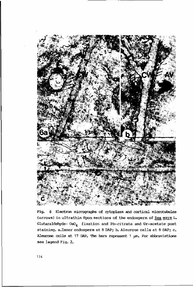
Fig. 6 Electron micrographs of cytoplasm and cortical microtubules
(arrows) in ultrathin Epon sections of the endosperm of Zea mays L.
Glutaraldehyde- 0s04 fixation and Pb-citrate and Ur-acetate post
staining, a.Inner endosperm at 8 DAP; b. Aleurone cells at 8 DAP; c.
Aleurone cells at 17 DAP. The bars represent 1 ym. For abbreviations
see legend Fig. 2.
134

criss-cross pattern of cortical microtubules was observed both in the
outermost layer as well as in the adjacent cell layers.
At 8 DAP microtubules were found in all living cells. Pericarp
cells exhibited predominantly cortical microtubules in the same
orientations as observed before. Additionally the endocarp cells
developed parallel arrays of microtubules perpendicular to' the length
axis of the elongated cells. In the cytoplasmic remains of the com
pressed integuments microtubules were observed in the original dis
tribution. The outermost one or two layers of nucellus cells
exhibited microtubules in an unchanged orientation. The inner endo
sperm cells still had criss-cross patterns of cortical microtubules
(Fig. 7c). After the synchronous division of the outermost cell layer
of the endosperm a higher concentration of cortical microtubules was
observed in the newly formed cells (Fig. 7d, arrow) but near to the
nuclei cytoplasmic microtubules were also found (Fig. 7d, arrow
head).
In the period of 12 to 15 DAP microtubules were still present in
the inner cells of the pericarp and in living cells of the nucellus
epidermis which covered the endosperm. The aleurone layer exhibited a
special arrangement of microtubules from then on. Initially micro
tubules were observed alongside all cell walls (Fig. 7d, arrows).
Hereafter bundles of cortical microtubules were found in parallel
arrays along the anticlinal cell walls (Fig. 7e, arrows). Cytoplasmic
microtubules were frequently observed around the nuclei. Spindle
tubules were seen when anticlinal cell rows were formed.
At 15 to 17 DAP the pericarp still contained living cells with a
well detectable microtubular cytoskeleton. In the fusiform endocarp
cells microtubules exhibited a parallel arrangement and in the spongy
mesocarp cells, if not yet compressed, a random orientation was
observed. Microtubules were found in the nucellus epidermis. Some
times, howevever, the nucellus epidermis had already collapsed and
cytoplasm could no longer be observed. In the aleurone layer the
cortical microtubules had the same orientation as before. The sub-
aleurone and inner endosperm cells exhibited criss-cross patterns of
microtubules when they were isodiametric but they showed an parallel
135
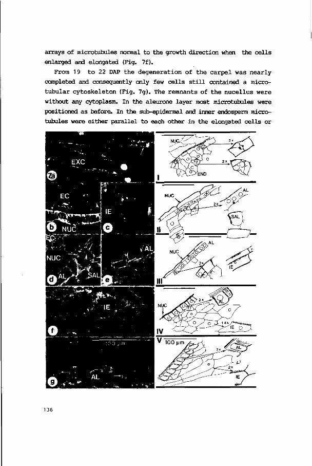
arrays of microtubules normal to the growth direction when the cells
enlarged and elongated (Fig. 7f).
From 19 to 22 DAP the degeneration of the carpel was nearly
completed and consequently only few cells still contained a micro-
tubular cytoskeleton (Fig. 7g). The remnants of the nucellus were
without any cytoplasm. In the aleurone layer most microtubules were
positioned as before. In the sub-epidermal and inner endosperm micro
tubules were either parallel to each other in the elongated cells or
136
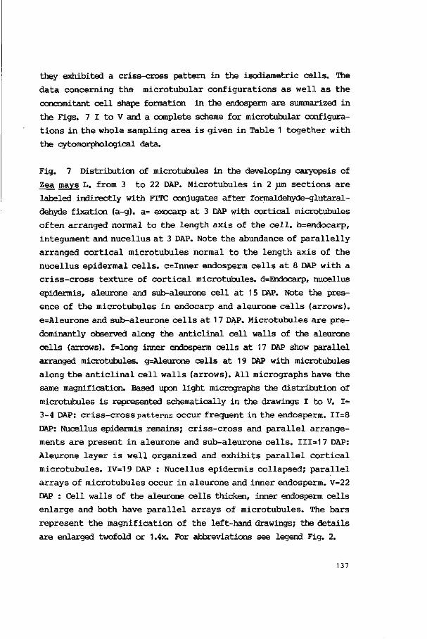
they exhibited a criss-cross pattern in the isodiametric cells. The
data concerning the microtubular configurations as well as the
concomitant cell shape formation in the endosperm are summarized in
the Figs. 7 I to V and a complete scheme for microtubular configura
tions in the whole sampling area is given in Table 1 together with
the cytomorphological data.
Fig. 7 Distribution of microtubules in the developing caryopsis of
Zea mays L. from 3 to 22 DAP. Microtubules in 2 pm sections are
labeled indirectly with FITC conjugates after formaldehyde-glutaral-
dehyde fixation (a-g). a= exocarp at 3 DAP with cortical microtubules
often arranged normal to the length axis of the cell. b=endocarp,
integument and nucellus at 3 DAP. Note the abundance of parallelly
arranged cortical microtubules normal to the length axis of the
nucellus epidermal cells. c=Inner endosperm cells at 8 DAP with a
criss-cross texture of cortical microtubules. d=Endocarp, nucellus
epidermis, aleurone and sub-aleurone cell at 15 DAP. Note the pres
ence of the microtubules in endocarp and aleurone cells (arrows).
e=Aleurone and sub-aleurone cells at 17 DAP. Microtubules are pre
dominantly observed along the anticlinal cell walls of the aleurone
cells (arrows). f=long inner endosperm cells at 17 DAP show parallel
arranged microtubules. g=Aleurone cells at 19 DAP with microtubules
along the anticlinal cell walls (arrows). All micrographs have the
same magnification. Based upon light micrographs the distribution of
microtubules is represented schematically in the drawings I to V. 1=
3-4 DAP: criss-cross patterns occur frequent in the endosperm. 11=8
DAP: Nucellus epidermis remains; criss-cross and parallel arrange
ments are present in aleurone and sub-aleurone cells. 111=1 7 DAP:
Aleurone layer is well organized and exhibits parallel cortical
microtubules. IV=19 DAP : Nucellus epidermis collapsed; parallel
arrays of microtubules occur in aleurone and inner endosperm. V=22
DAP : Cell walls of the aleurone cells thicken, inner endosperm cells
enlarge and both have parallel arrays of microtubules. The bars
represent the magnification of the left-hand drawings; the details
are enlarged twofold or 1.4x. For abbreviations see legend Fig. 2.
137
4. DISCUSSION
A
4.1. dell differentiation in the caryopsis.
The pericarp, known for its determined life time (see , amongst
others, Randolph 1936), developed various cell layers each with cells
of particular shape, wall thickness and corresponding mechanical
properties. From 22 DAP onwards the exocarp consisted of rigid cell
walls while the cytoplasm of the cells was degenerated. Most dead
mesocarp cells were compressed between the exocarp and the seed but
the walls of the small fusiform cells of the endocarp were still well
recognizable. The pericarp, which initially showed a three-partite
structure as is common for the caryopses of many Gramineae, protected
the seed. Throughout their life span pericarp cells had a large
central vacuole. As in other Gramineae the carpels did not contribute
to any food storage. The accumulation of starch was ephemeral. The
intimate relation between pericarp and underlying tissues was ex
pressed by the lack of a real inner epidermis. As was also mentioned
for wheat and barley the fusiform cells of the endocarp vary in
direction between individual caryopses. They appeared as a network of
so called "cellules tubulaires" (Guerin 1899) forming the inner face
of the pericarp. Actually the nucellar cuticle is the demarcation
between fruit wall and seed because the integumentary cells had
already degenerated.
The life time of the integuments has been discussed by various
authors, amongst others, Randolph (1936), who observed initial stages
of the desintegration of the integuments at the germinal face of the
caryopsis in samples examined 3 DAP. He reported that the walls of
the outer cell layer disappeared first while nuclei and cytoplasm
often persisted for some time. In the present study the degeneration
was often accomplished before 5 DAP but incidentally at 8 DAP cyto
plasm with microtubules was still observed. This asynchrony in
development can be attributed to differences in developmental rates
in the various caryopses. The inner integument influences the
pathway of the pollen tube (Van Lammeren 1986a) rather than the
subsequent seed formation.
138

The nucellus is also an ephemeral tissue and it is likely that it
functions in nourishing the developing endosperm when cells are
digested near the endosperm epidermis. The nucellus epidermis and its
cuticle existed longest and covered the endosperm. They might
function as a layer providing mechanical protection because its cells
lived until the cell walls of the aleurone layer started to thicken.
Eventually the cells died and only the remnants of the anticlinal and
outer cell walls and the cuticle of the nucellus persisted. These
remnants were called the nucellar membrane (Randolph 1936, Kiessel-
bach 1949). At this stage that structure is likely to function as a
protective covering of the embryo and endosperm because of its semi
permeable properties (Randolph 1936).
Endosperm formation in maize is of the nuclear type initially but
cellularization started within 3 DAP in the present study. The fine
structure of the formation of the first anticlinal cell walls and the
subsequent elaboration of periclinal cell walls and rows of endosperm
cells has only been described for Triticum aestivum L. (Buttrose
1963a, Morrison et al. 1978, Fineran et al. 1982). For maize the
process was studied by light microscopy (Weatherwax 1930, Lamps 1931,
Randolph 1936 and Kiesselbach 1949). Kyle and Styles (1977) applied
electron microscopy and described the development of the aleurone and
sub-aleurone layer of the maize inbred line W-22. The endosperm
development in wheat was studied by, amongst others, Mares et al.
(1975,1976) and Morrison et al. (1978).
In the caryopses of the maize inbred line A188 the differentiation
of the aleurone layer started at about 7 DAP with the synchronous
cell divisions by which one layer of columnar cells was formed. In
our experiments the phase of differentiation started 3 days earlier
than reported by Kyle & Styles (1977). In contrast to e.g. barley, in
which the aleurone layer consists of as many as four cell layers, one
layer is usual in maize but two cell layers were found incidentally.
Aleurone and sub-aleurone endosperm differed from each other in many
aspects. From 8 DAP onwards periclinal cell divisions were not ob
served any longer in the aleurone layer but the cells enlarged and
became rectangular. Sub-aleurone cells descended from the aleurone
139

ce l l s by asynchronous, an t ic l ina l ce l l divisions.
With respect to the type and location of organelles some s imilar
i t i e s were observed in the aleurone and sub-aleurone layers of t h i s
maize s t ra in . Both layers had the typically located mitochondria. The
alignement along the outer per ic l inal c e l l walls points to a high and
local demand for energy to permit active transmembranal export and
import. This phenemenon was a lso observed in the synergids of maize
before f e r t i l i za t ion (Van Lammeren 1986a) and in the modified endo
sperm bordering t he maize embryo suspensor (see Schel e t a l . 1984).
Both c e l l types showed remarkable p r o l i f e r a t i o n of the ER, though,
not a s i n tense as was r epor ted for wheat where evaginat ions of the
nuc lear membrane took p a r t of t he ER system (Kyle & S t y l e s 1977).
Furthermore, an i nc reas ing amount of cytoplasm was not iced on a l l
c e l l types and starch accumulated in a l l p las t ids which i s in con
t r a s t t o t he observat ions of Khoo & Wolf (1970) who only found p ro-
p l a s t i d s in the a leurone l ayer . The d i f ference might be caused by
varying developmental r a t e s which a re influenced by genotype and
environmental conditions. On the other hand the organization of the
cytoplasm differed between t he epidermal , subepidermal and inner
endosperm c e l l s a s e a r l y as 3 DAP. Differences were found in the
accumulation of spherosomes (Type S1) in the var ious t i s s u e s a t 3
DAP; The synthesis of endosperm protein bodies from 7 DAP onwards was
r es t r ic ted to the sub-aleurone ce l l s and inner endosperm c e l l s , and
the formation of aleurone bodies and spherosomes which s tar ted a t 9
DAP occured only in the aleurone c e l l s .
The development and s tructure of protein bodies and spherosomes
i n seeds i s d iscussed by Lo t t (1980).' Khoo and Wolf (1970) repor ted
about the development of the aleurone vacuoles and protein bodies in
the ou ter c e l l l ayer of t he maize endosperm. They f a i l ed t o d e t ec t
p ro t e in accumulation in t he vacuoles in 7-1 0 day ke rne l s but found
p ro t e in granules by day 1 2. As compared t o e.g. ba r ley and wheat
(Jacobsen e t a l . 1971) maize forms only few aleurone protein bodies
(see a lso Khoo & Wolf 1970, Kyle & Styles 1977) which occupied only a
minor part of the aleurone layer but spherosomes were abundant and
took up much more space. The o r i g i n of the spherosomes was not e s -
140

tablished. The existence of sphere-some membranes in maize was re
ported by Kyle & Styles (1977). Such a membrane was only observed in
the present study when spherosomes were pressed to each other.
Spherosomes show variations in staining which might be related to the
heterogenous composition (Wolf et al. 1969). Aleurone protein bodies
have electron dense and electron opaque inclusions. The electron
dense parts, which are hard and do not penetrate well with embedded
medium, often chatter during sectioning leaving holes in the section.
Protein bodies of the sub-aleurone and inner endosperm were found to
have polysomes associated with the protein body membrane. These
endosperm protein bodies of maize probably accumulate the main
storage protein called zein (Burr & Burr 1976). When the polysomes
were isolated at a time when storage protein synthesis was active,
they were able to synthesize zein.
With respect to the function of the aleurone layer Bhatnagar and
Sawhney (1981) reviewed the literature about hydrolytic enzymes such
as cc-amylase that brings about the breakdown of reserve food
materials in the endosperm tissue which are utilized by the embryo
during its growth and development. Protein bodies have several
functions that were summarized by Matile (1968). First they store
certain reserve substances such as proteins, metals and phytin the
synthesis of which might be mediated by mitochondria (Morrison et al.
1975). Second, during germination the aleurone vacuoles are trans
formed to a 'lysosome' and their food reserves are mobilized. Finally
they may form a lytic compartment in which other constituents of the
cell are broken down.
4.2. The microtubular cytoskeleton.
In the caryopsis of maize microtubules were found in three major
configurations: a) spindle tubules, the functions of which are dis
cussed by amongst others Kreis & Birchmeier (1982), b) cytoplasmic
microtubules running throughout the cytoplasm and probably func
tioning in the positioning and moving of organelles and in sustaining
the integrity of the cytoplasm (Van Lammeren et al. 1985, Lloyd et
141

al. 1986) and c) cortical microtubules interacting with cell shape
development. The latter exhibited a variety of configurations. Criss
cross patterns and parallel arrangements were the extremes. The
patterning is likely to influence the morphogenesis of a growing
plant cell (see e.g. Marchant 1979, Lang Selker & Green 1984, Lloyd
1984) although the data are not equivocal (Emons 1986, Traas 1986).
Cortical microtubules in criss-cross arrangements were observed in
small and large cells which had no clear length axis. This observa
tion might imply that such a configuration of cortical microtubules
restricts or determines cell enlargement and prevents cell elonga
tion. As soon as parallel arrangements were observed, cells were more
or less cylindrical or had a length axis. In these cells it was found
that the direction of the cortical microtubules was normal to the
growth direction which was determined by the increase in length and
in width. When cells just elongated without a remarkable lateral
expansion, as was observed in the integuments and nucellus epidermis,
the microtubular orientation was perpendicular to the length axis.
When, however, the elongated cells expanded in lateral direction the
orientation of the microtubules was oblique with respect to that
direction and the deviation appeared to be related to the intensity
of the lateral expansion. From the above data it is concluded that in
maize caryopses the cortical microtubules are involved in the cyto-
morphogenesis in the sense as described by Lang Selker and Green
(1984). The way how microtubules interact with microfibril deposition
and the direction of the process is not clear up to now although some
possible mechanisms have been described (Emons 1983, Heath & Seagull
1982, Hepler 1985). The majority of the microtubules in the pericarp
cells was observed in the cortical cytoplasm suggesting a relation
with cell wall formation (see also Roberts et al. 1985). In elongated
cells we suppose the microtubules to be the inducers of the dominance
of longitudinal growth. The cells of the aleurone layer exhibited
some deviant phenomena. Parallel arrangements of microtubules were
observed along anticlinal cell walls from 12 DAP onwards. Aleurone
cells elongated only slightly in a direction normal to the plane of
the microtubules. Instead of elongation, cell wall thickening was
142
notable both on an t ic l ina l and outer per ic l inal c e l l walls adjacent
to which only few microtubules were observed.
In conclusion, the data of the present investigation point toward
a s t r i c t sequence of events leading to the maturation of the caryop-
s i s . Pe r icarp d i f f e r e n t i a t i o n r e su l t ed i n a dead p r o t e c t i v e f r u i t
wal l covering the seed. D i f f e r en t i a t i on and c e l l wa l l s yn thes i s in
the pericarp lead to thick-walled exo- and endocarp ce l l s and th in-
walled mesocarp c e l l s . The d i f f e r e n t i a t i o n of t h e endosperm i s
analyzed cy to log i ca l l y , however, t he formation and composition of
various organelles in aleurone and sub-aleurone layers wi l l have to
be s tudied i n more d e t a i l . The conf igura t ions of t h e microtubular
cytoskeleton are related to the development of c e l l shape in growing
c e l l s . Table 1 g ives a scheme which summarizes t he da ta of micro-
tubu la r d i s t r i b u t i o n and c e l l shape development in the var ious
t i ssues of the caryopsis.
5 . ACKN0WU3X3MENTS
We thank Prof. Dr. M.T.M. Willemse and Dr. J.H.N. Schel for reading
the manuscript; Mr A. Haasdijk and Mr. S. Massalt for the artwork and
Dr. F Vegter for emending the English text .
6 . REFERENCES
Bechtel , D.B. & B.O. J u l i ano (1980) Formation of p ro t e in bodies i n
t he s tarchy endosperm of r i c e (Oryza s a t i va L.): a r e - i n v e s t i g a
t ion . Ann. Bot. 45:503-509.
Bechtel D.B. & Y. Pomeranz (1977) Ultrastructure of the mature unger-
minated r i ce (Oryza sativa) caryopsis. The caryopsis coat and the
a leurone c e l l s . Am. J . Bot. 64:966-973.
Bhatnagar, S.P. & V. Sawhney (1981) Endosperm - I t s morphology,
143
ultrastructure and histochemistry. Intern. Rev. Cyt. 73:55-102.
Burr, B. & F.A. Burr (1976) Zein synthesis in maize endosperm by
polyribosomes attached to protein bodies. Proc. Natl. Acad.
Sci. USA 73:515-519.
Buttrose, M.S. (1963a) Ultrastructure of developing aleurone cells of
the wheat grain. Aust. J. Biol. Sci. 16:768-774.
Buttrose, M.S. (1963b) Ultrastructure of the developing wheat
endosperm. Aust. J.Biol. Sci. 16:305-317.
Campbell, W.P., J.W. Lee, T.P. O'Brien & M.G. Smart (1981) Endosperm
morphology and protein body formation in developing wheat grain.
Aust. J. Plant Physiol., 1981, 8:5-19.
Cochrane, M.P. (1983) Morphology of the crease region in relation
to assimilate uptake and water loss during caryopsis development
in barley and wheat. Aust. J. Plant Physiol. 10:473-491.
Diboll, A.G. (1964) Electron microscopy of female gametophyte and
early embryo development in Zea mays. Doctoral dissertation, Univ.
Texas.
Emons, A.M.C (1983) Cortical microtubules and microfibril deposition
in the cell wall of root hairs of Equisetum hyemale. Protoplasma
117:68-81.
Emons, A.M.C. (1986) Cell wall formation in root hairs. Role of
plasma membrane and cytoskeleton in microfibril deposition. Doc
toral dissertation, University of Nijmegen, Nijmegen.
Felker, F.C., D.M. Peterson & O.E. Nelson (1984) Development of
tannin vacuoles in chalaza and seed coat of barley in relation
to early chalazal necrosis in the segl mutant. Planta 161:540-
549.
Fineran, B.A., D.J.C. Wild & M. Ingerfeld (1982) Initial wall forma
tion in the endosperm of wheat, Triticum aestivum: a reevaluation.
Can. J. Bot. 60:1776-1795.
Guerin, P. (1899) Recherches sur le developpement du tegument seminal
et du pericarpe des Graminees. Ann. Sci. Nat., Bot. (8)9:1-59,
illus.
Heath, I.B. & R.W. Seagull (1982) Oriented cellulose fibrils and
the cytoskeleton: a critical comparison of models. In: The cyto-
144

skele ton in p l an t growth and development, (ed. Lloyd, C.W.). pp.
163-182. Academic Press, London.
Hepler, PJC (1985) The plant cytoskeleton. In: Botanical Microscopy
1985 (ed. Robards, A.W.). pp.233-262. Oxford Univers i ty P r e s s ,
Oxford.
Jacobsen, J.V., R.B. Knox & N.A. P y l l o t i s (1971) The s t r u c t u r e and
composition of a leurone g ra ins in the bar ley a leurone l aye r .
Planta 101: 189-209.
Kiesselbach, T.A. (1949) The s t r u c t u r e and reproduct ion of corn.
Nebr. Agr. Exp. S ta . Res. Bui. 161:1-96.
Khoo, U. & M.J. Wolf (1970) Origin and development of p ro t e in
granules in maize endosperm. Am. J . Bot. 57:1042-1050.
Kre is , T.E. & W. Birchmeier (1982) Microinject ion of f l uo rescen t ly
labeled proteins into l iving c e l l s with emphasis on cytoskeletal
p r o t e i n s . I n t . Rev. Cytol . 75:209-227.
Kvaale, A. & O.A. Olsen (1986) Rates of c e l l d i v i s i on in developing
barley endosperms. Ann. Bot. 57:829-833.
Kyle, D.J. & E.D. S ty les (1977) Development of a leurone and sub-
aleurone layers in maize. Planta 137:185-193.
Lang Selker, J.M. & P3 . Green (1984) Organogenesis in Graptopetalum
paraguayense E. Walter: shif ts in orientation of cor t ica l micro
tubule a r rays a r e a s soc ia ted with p e r i c l i n a l d i v i s i on s . P lanta
160:289-297.
Lampe, L. (1931) A microchemical and morphological s tudy of t he
developing endosperm of maize. Bot. Gaz. 91:337-376.
Lloyd, C.W. ed (1982) The cy toskele ton in p l an t growth and develop
ment. Academic Press, London.
Lloyd, C.W. (1984) Toward a dynamic h e l i c a l model for t he inf luence
of microtubules on wal l p a t t e rn s in p l a n t s . I n t e rn . Rev. Cyt.
86:1-51.
Lloyd, C.W., J .S. Hyams & R.M. Warn (eds.) The cy toskele ton: Cel l
function and o rgan iza t ion . J . Ce l l Sc i . Suppl. 5, 1 986.
L o t t , J.N.A. (1980) P r o t e i n b o d i e s . I n : The b i o c h e m i s t r y of
p l an t s , eds. P.K. Stumpf & E.E. Conn. Vol.1 The p l an t c e l l . ed.
N.E. Tolber t . Acad. Press, New York, London.
145
Marchant, H.J. (1979) Microtubules, cell wall deposition and the
determination of plant cell shape. Nature 278:167-168.
Mares, D. J., C. Jeffery and K. Norstog (1976) The differentiation
of the modified aleurone of wheat endosperm: nuclear membrane-
mediated endoplasmic reticulum development. Plant Sci. Lett.
7:305-311.
Mares, D.J., K. Norstog and B.A. Stone (1975) Early stages in the
development of wheat endosperm. I The change from free nuclear
to cellular endosperm. Aust. J. Bot. 23:311-326.
Matile, P. (1968) Aleurone vacuoles as lysosomes. Z. Pflanzenphysiol.
58:365-368.
Miflin, B.J., S.R. Burgess & P.R. Shewry (1981) The development of
protein bodies in the storage tissues of seeds: subcellular sepa
rations of homogenates of barley, maize, and wheat endosperms and
of pea cotyledons. J. Exp. Bot. 32:199-219.
Miller, E.C. (1919) Development of the pistillate spikelet and fer
tilization in Zea mays L. J. Agr. Res. 18(5):255-293.
Morrison, I.N., J. Kuo & T.P. O'Brien (1975) Histochemistry and
fine structure of developing wheat aleurone cells. Planta
123:105-116.
Morrison, I.N., T.P. O'Brien & J. Kuo (1978) Initial cellularization
and differentiation of the aleurone cells in the ventral region of
the developing wheat grain. Planta 140:19-30.
Olsen, O.A. & T. Krekling (1980) Grain development in normal and
high lysine barley. Hereditas 93:147-160.
Oparka, K.J. & N. Harris (1982) Rice protein-body formation: all
types are initiated by dilation of the endoplasmic reticulum.
Planta 154:184-188.
Pomeranz, Y. (1973) Structure and mineral composition of cereal
aleurone cells as shown by scanning electron microscopy. Cereal
Chem. 50:504-511.
Randolph, L.F. (1936) Developmental morphology of the caryopsis in
maize. J. Agr. Res. 53:881-916.
Roberts, K., J. Burgess & P. Linstead (1985) Microtubule rearrange
ment during plant cell growth and development: an immunof luor-
146

escence study. In : Botanical Microscopy 1985 (ed. Robards, A.W.)
pp. 263-283. Oxford University Press, Oxford.
Rogers, S.O. & R.S. Quatrano (1983) Morphological s t ag ing of wheat
caryopsis development. Am. J . Bot. 70:308-311.
Schel , J.H.N., H. Kieft & A.A.M. van Lammeren (1984) I n t e r a c t i on s
between embryo and endosperm during early developmental stages of
maize caryopses (Zea mays). Can. J . Bot. 62:2842-2853.
Smart, M.G. & T.P. O'Brien (1983) The development of the wheat embryo
in re la t ion to the neighbouring t i ssues . Protoplasma 114:1-13.
Traas , J.A. (1986) The cy toskele ton in p l an t c e l l growth, d i f f e r en
t i a t i o n and c e l l wa l l s yn thes i s . Doctoral t h e s i s , Univers i ty of
Nijmegen, Nijmegen.
Van Lammeren, A.A.M., C.J. Kei jzer , M.T.M. Willemse and H. Kief t
(1985) S t ruc tu re and funct ion of t he microtubular cy toskele ton
during pollen development in Gasteria verrucosa (Mill.) H. Duval.
Planta 165:1-11.
Van Lammeren, AJV.M. (1985) Microtubular configurations during endo
sperm and pollen development in Zea and Gasteria as visualized by
immunofluorescence. In: Sexual reproduction in seed plants, ferns
and mosses, pp 59-63, Willemse, M.T.M., Van Went, J.L., eds.
Pudoc, Wageningen.
Van Lammeren, A.A.M. (1986a) A comparative u l t r a s t r u c t u r a l s tudy
of the megagametophytes i n two s t r a i n s of Zea mays L. before
and a f t e r f e r t i l i z a t i o n . Agric. Univ. Wageningen Papers 86-
1,37pp.
Van Lammeren, A.A.M. (1986b) Developmental morphology and cytology
of t h e young maize embryo (Zea mays L.) . Acta Bot . N ee r l .
35:169-188.
Van Lammeren, A.A.M. & J.H.N. Schel (1983) Embryogenesis in Zea
mays L. Development of s t r u c t u r e and p o l a r i t y during proembryo
formation. In: O. Erdelska (ed.), Fer t i l iza t ion and embryogenesis
in ovulated plants. Veda, Bratislava, pp 283-285.
Weatherwax, P. (1919) Gametogenesis and fecondation in Zea mays as
the basis of xenia and heredity in the endosperm. Bui. Torrey Bot.
Club 46:73-90.
147
Weatherwax, P. (1930) The endosperm of Zea and Coix. Am. J. Bot.
17:371-380.
Wolf, M.J., U. Khoo & H.L. Seckinger (1969). Distribution and sub
cellular structure of endosperm protein in varieties of ordinary
and high-lysine maize. Cereal Chem. 46:253-263.
148

CHAPTH* 7
Some concluding remarks on endogenous and exogenous influences
on embryo development in maize
A.A.M. van Lammeren

SOME CONCLUDING REMARKS ON ENDOGENOUS AND EXOGENOUS INFLUENCES ON
EMBRYO DEVELOPMENT IN MAIZE AND THE SIGNIFICANCE OF THE STUDY FOR
PLANT BREEDING
During caryopsis development the shape and structure of both the
embryo and the extra-embryonal tissues of the caryopsis change con
tinuously. This chapter will expand on when and how embryogenesis is
influenced. Factors inducing or influencing the development of the
shape of the embryo will be postulated. They are called endogenous
when their influence manifests from inside the embryo and exogenous
when they come through from outside the embryo.
1. Effects of endogenous morphogenetic factors
Although one has to keep in mind that morphogenesis is largely an
expression of the genetic constitution of the embryo, some charac
teristics of embryo development can be analysed. Embryo development
is determined or influenced by the initiation and maintenance of
polarity. The shape of the embryo is further influenced by the forma
tion and activity of meristems and the directed cell elongation in
the various organs.
Induction of polarity becomes manifest during the transition of
the egg stage to the zygote stage. The new cell polarity is es
tablished when the location of the cytoplasm in the cell changes
(Chapter 1). It can be worked out in later developmental stages and
is then expressed by the positions of the suspensor and the apical
meristems. This polarity determines further proembryo development and
is expressed by the positions of the suspensor and the apex.
Polarity determines further proembryo development and can be con
sidered as a morphogenetic factor. Chapter 2 highlights the fact that
suspensor cells of the proembryo elongate. Elongation occurs after
the apical meristem has developed a certain size and can therefore
be regarded as an effect of polarity. Finally, the transition of the
proembryo stage to the embryo stage should be based upon a regulation
which is expressed in the further development of the apical meristems
which will develop into the shoot meristem and the root meristem
151
(Chapter 3). The nature of this regulation is complex but some char
acteristics will be discussed.
In the above mentioned examples, common structural features are
observed. Among these the symplastic isolation, which precedes the
morphological changes, is obvious. Symplastic isolation is the con
sequence of the absence of plasmodesmata between the embryo and its
environment. Figure 1 gives a schematic representation of five de
velopmental stages of the maize embryo.
-In Fig. 1a the zygote is shown with plasmodesmata at its micropylar
side. Symplastic contact with the endosperm is therefore restricted
to that area and a symplastic pathway for nutrients can only be such
as indicated by the arrows. The cell membrane at the antipodal side
of the zygote forms a so-called symplastic boundary because of the
absence of plasmodesmata. In this area most cytoplasm is concen
trated. The plasmodesmata and the symplastic boundary are formed when
the egg cell develops. It is therefore a property of the zygote which
thus determines the direction of symplastic transport. When the
direction of symplastic transport influences the induction or main
tenance of polarity, this influence is determined by the zygote
because of the location of the plasmodesmata and therefore it can be
said that this morphogenetic factor has an endogenous character. On
one hand the pathway of transport is determined by the structure of
the zygote. On the other, however, the nutrients influence the embryo
from the outside. This implicates that a morphogenetic factor can
have endogenous and exogenous features.
-Figure 1d shows a proembryo such as studied in the Chapters 2 and 5.
Although there is no longer symplastic contact between endosperm and
proembryo, the endosperm functions in feeding the proembryo via the
apoplast, and especially at the base of the suspensor (Chapter 5).
Thus the base of the suspensor is a region of nutrient uptake. The
apex ingests most of these nutrients and is a sink within the embryo.
The flow of nutrients towards the proembryo is indicated by the
striated arrows. It should be noted that the protodermal cells of the
apex can be regarded as symplastic boundary cells containing much
cytoplasm. From these remarks it can be concluded that the embryo
152
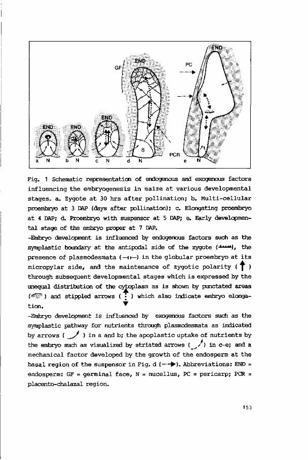
END
l i i i
PCR a N b N c N d N
Fig. 1 Schematic representation of endogenous and exogenous factors
influencing the embryogenesis in maize at various developmental
stages, a. Zygote at 30 hrs after pollination; b. Multi-cellular
proembryo at 3 DAP (days after pollination); c. Elongating proembryo
at 4 DAP; d. Proembryo with suspensor at 5 DAP; e. Early developmen
tal stage of the embryo proper at 7 DAP.
-Embryo development is influenced by endogenous factors such as the
symplastic boundary at the antipodal side of the zygote (•**"*), the
presence of plasmodesmata (—HI—) in the globular proembryo at its
micropylar side, and the maintenance of zygotic polarity ( | )
through subsequent developmental stages which is expressed by the
unequal distribution of the cytoplasm as is shown by punctated areas
{<$Wr ) and stippled arrows ( • ) which also indicate embryo elonga
tion. '
-Embryo development is influenced by exogenous factors such as the
symplastic pathway for nutrients through plasmodesmata as indicated
by arrows ( / ) in a and b; the apoplastic uptake of nutrients by
the embryo such as visualized by striated arrows ( y ) in c-e; and a
mechanical factor developed by the growth of the endosperm at the
basal region of the suspensor in Fig. d (•—• ). Abbreviations: END =
endosperm: GF = germinal face, N = nucellus, PC = pericarp; PCR =
placento-chalazal region.
153

development is not only influenced by the endogenous polarity but by
an exogenous factor, in this case the endosperm which supplies the
nutrients, as well.
-During the development of the embryo proper (Fig. 1e) the shoot
meristem develops from the protoderm cells of the apex of the pro-
embryo. The meristem is therefore formed in symplastic boundary
cells. These cells already formed a sink to which the scutellum
develops laterally. The initial formation and further activity of the
root meristem depends on the developmental stage of the shoot meris
tem and thus appears to be determined endogenously (Chapter 3). At
the onset of the formation of the root meristem, which develops
endogenously, a symplastic boundary is created between the area of
the root meristem and the calyptrogen (Fig. 1e, arrow-head). There
after the meristems are activated. Herewith the necessity of sym
plastic isolation with respect to meristem formation is demonstrated
once again.
In the foregoing examples the sink within the embryo is situated
adjacent to existing or newly created symplastic boundaries. The fact
that sinks are also found far from regions that function in the
uptake of nutrients emphasizes the presence of polarity. In all
examples the apical poles can be distinguished from other areas by
the accumulation of cytoplasm in relatively small cells. The directed
elongation of cells in tissues was frequently observed and is shown
in the Figs. 1b,c and e by arrows ( ). It is remarkable that
cells do not elongate untill the apical pole of the organism contains
relatively large quantaties of cytoplasm. This degree of organization
could well be a prerequisite for the induction of cell elongation.
Cell elongation coincides with certain configurations of the
microtubular cytoskeleton. Microtubules in the central region of the
cytoplasm function in the positioning, motility and movement of
organelles but are not likely to influence cell shapes directly.
Cortical microtubules, however, influence the mode of cell wall
synthesis and determine the development of cell shapes (Chapter 6).
Cell elongation can be connected with a rearrangement of the cortical
microtubules of the cytoskeleton as is illustrated for the endosperm
154
and pericarp cells in Chapter 6. The microtubular cytoskeleton can
therefore be called a morphogenetic factor. Whether the microtubular
cytoskeleton has to be considered as an independent morphogenetic
factor or that the changes in the microtubular configurations are the
expression of an other cell signal, is not elucidated. The latter
possibility is more likely because of the prerequisite of cell elon
gation as mentioned before.
The genetic constitution of embryos appears to be an important
endogenic morphogenetic factor. With in vitro studies in particular
there is a striking difference in the ability to regenerate amongst
various inbred lines, but it is constant within an inbred line
(Fransz, pars, comm.) Such a property is expressed in many structural
aspects which are related to e.g. development of callus and somatic
embryogenesis (Chapter 5).
2. Effects of exogenous morphogenetic factors
Factors exerting an influence on the embryo from outside can be
distinguished in physical, physico-chemical and organic-chemical
factors.
An example of physical influences on shape development is the
mechanical influence which becomes prominent when the suspensor bends
because of the intensive growth of the endosperm at its base by which
a lateral pressure is exerted upon the suspensor (Chapter 2). When,
at a later stage, the elongated embryo axis is cramped by the sus
pensor epidermis and the pericarp at its germinal face (Chapter 3) an
other example is demonstrated. In this context it is remarkable that
in vitro experiments show a sideward outgrowth of the axis when
pericarp and endosperm are removed (Chapter 6). From the foregoing
arguments it appears that the shape of the embryo is, at least partly
influenced by external tissues which develop continuously. Such an
influence can be considered as a force or pressure which is caused by
growth, cell pressure and tension in the surrounding tissues.
Other physical factors such as light, humidity and temperature
strongly influence the whole metabolism of the sporophyte (mother
plant) and thus the rate of success and the speed of embryo and
155
endosperm development. Although both embryo and endosperm a re
heterotrophic organisms in vivo, i llumination in v i t ro i n i t i a t e s the
differentiat ion of chlorenchyma in very young excised embryos. This
experiment demonstrates the s en s i t i v i t y to t h i s exogenous physical
f ac to r but i t a l s o puts forward the a b i l i t y t o autotrophy (Chapter
5). The endosperm lacks t h i s a b i l i t y and o the r types of d i f f e r en
t i a t i o n a r e e i t h e r not observed or l e s s obvious than in t he embryo.
I t i s not known whether in vivo i l lumination in early developmental
stages inhibi ts or stimulates further development.
The physico-chemical and organic chemical in f luences a r e based
upon osmolarity, the composition and the direction of the nutr ient
flow, and the ac t iv i ty of growth regulators amongst others.
I t has been demonstrated that growth and premature germination of
excised embryos on a r t i f i c i a l n u t r i e n t media f requent ly occur.
However, these phenomena are reduced s ignificantly or disappear when
the osmolar i ty of the medium i s increased. In vivo t he premature
germinat ion i s i nh ib i t ed and i t i s l i k e l y t h a t i t i s caused by an
increase of the osmolarity in the endosperm because carbohydrates are
accumulated from 8 DAP onwards (Chapter 6). The ingestion of sugars
i n the endosperm i s expressed by the syn thes i s of s t a r ch in the
p las t ids a t the very moment that the embryo has developed i t s apical
meristems. S tarch i s osmotic i n ac t i ve but i t s accumulation demon
s t ra tes the presence of soluble carbohydrates. I t i s suggested that
the osmolar i ty i n the environment of the embryo i nc reases and i n
h ib i t s germination from the moment that the immature embryo i s ger-
minative though premature germination has to be suppressed.
The d i rec tedness of the n u t r i e n t flow i s an exogenous morpho-
gene t i c f ac to r a t va r ious s t ages of embryo development. - F i r s t , a
d i rect ional symplastic flow of nutr ients towards the zygote can be
deduced from the non-random d i s t r i b u t i o n of plasmodesmata i n t h a t
c e l l . The endogenous aspect of t h i s f ac to r has a l ready been
mentioned.
-Secondly the e longat ion of the embryo br ings the embryo apex i n a
par t of the endosperm which d iffers from the endosperm surrounding
the suspensor. At the apical side highly vacuolized endosperm ce l l s
156
surround the embryo. At its basal side the embryo suspensor is
connected with endosperm cells containing dense cytoplasm. These
cells supply nutrients taken up by the growing embryo (Chapter 5)
through the apoplast.
-Thirdly the scutellum of the embryo proper is formed in the region
where the nutrient supply is abundant. This points to the possibil
ity that the development of a second cotyledon fails to come because
of a local deficiency of nutrients. This in its turn means that the
directedness of nutrient supply influences morphogenesis. Experiments
in which the development of somatic embryos is observed show a
comparable type of development of the cotyledons (Fransz, pers.
comm.) In these experiments there is, however, no endosperm which
supplies nutrients. The embryo protrudes from the callus tissue and
is then surrounded by air at its apex. It must therefore be concluded
that the absence or presence of the nutrient flow is not a pre
requisite any more and that the lack of development of the second
cotyledon must point to an acquired local and structural deficiency.
-A forth type of exogenous morphogenetic influence is observed when
bilateral symmetry is expressd in the embryo. When the new symmetry
becomes prominent there is a bilateral symmetry in the ovary and
ovule. This symmetry is thought to be superimposed onto the existing
radial symmetry of the proembryo by nutritional or other organic-
chemical influences. The directedness of these influences, however,
is determined by the anatomy and thus the developmental history of
the ovule and ovary. On the other hand it should be noted that in
vitro studies demonstrate a change of symmetry within a proembryo in
the absence of endosperm (Chapter 5). In this case, the development
of shapes appears to be regulated by the embryo and might therefore
have become an acquired property.
-Finally, Chapter 6 describes some experiments in which morphogenesis
is clearly influenced by the way the nutrients were ingested. Imma
ture maize embryos of 9 DAP exhibit an independent regulation of
growth which leads to the development of germlings.
157
Conclusion.
The morphogenesis of the embryo can be seen as a continuous process
in which the shape is influenced or determined by a combined action
of exogenous and endogenous factors. The nature of these factors is
partly analyzed but experimental embryology in particular will
provide more details in future.
3.' Significance of the study for plant breeding
This cytomorphological study of the crop plant Zea mays L. provides
information of the level of organization and structural complexity of
the embryo on subsequent moments of differentiation. It was found
that the investigated inbred lines exhibited differences in shape and
position of structures such as the micropyle and embryo sac. This
kind of study might be of use in the research of intra-specific
crossings and the success or failure of fertilization. The structural
investigation of inbred lines revealed inbred line-specific dif
ferences in developmental rates and in organ and organelle differen
tiations. Therefore it provides the possibility to get an accurate
embryological characterization of lines which can be of use to
analyse lines and the intra-specific crosses. The (ultra)structural
study of in vivo embryogenesis provides a tool to determine the
regular sequence of morphological events and furthermore the in
fluence of external factors on that sequence. Long before the matur
ity or the degeneration of the caryopsis the expression of the in
fluence of the factors which accelerate or impede development can be
registrated.
In vivo studies about cell differentiation and the function of the
cytoskeleton such as described in Chapter 6 not only revealed the
relation of the arrangement of microtubules with the way of cell
enlargement, but they also put forward the advantages of the tech
nique of immunological labeling of tubulin with fluorescein isothio-
cyanate (FITC) in sections of large tissue blocks which were original
embedded in polyethylene glycol (PEG). The investigation of the
structural aspects of the initiation of polarity, the development of
158
meristems and the directed c e l l enlargement in proembryo and embryo
(Chapters 1,2 and 3) a t t r i b u t e s t o a b e t t e r knowledge of embryo-
genes is . The knowledge can be t he t oo l t o manipulate s p e c i f i c a l l y
during differentiat ion.
In v i t r o experiments open ways t o i n t e r f e r e and compare with
normal embryogenesis. In v i t r o experiments showed the embryo t o
become increasingly independent from the endosperm which ult imately
can be replaced. The i nc reas ing independence can be r e l a t e d t o t h e
complexity of s t r u c t u r e and o rgan iza t ion wi th in the embryo. This
procedure offers the opportunity t o determine anatomical and cy to -
l og i c a l parameters t o e s t a b l i s h the moment t o i n i t i a t e in v i t r o
culture. The differentiat ion of the apical meristems might be such a
moment. Culture of excised immature embryos in the absence of endo
sperm i s of value in crosses with deficient endosperm development.
Cytological approaches permit the ear ly regis t ra t ion of s t ructural
phenomena induced by phys ica l and organic-chemical f a c t o r s . For
i n s t ance , the e a r l y r ecogni t ion of var ious d i f f e r e n t i a t i o n forms,
such as the development of ca l lus , adventitious shoots and somatic
embryos.
In v i t r o c u l t u r e makes i t poss ib le t o manipulate t he developing
embryos which might lead t o acqui re p rope r t i e s which might be of
advantage in a l a t e r phase of plant l i f e . For example, the inhibit ion
of the early germination of somatic embryos might r esu l t in a further
embryological development which resu l t s in arger embryos with larger
meristems and perhaps less dominance towards adjacent somatic em
bryos.
Conclusion.
This morphological s tudy on maize embryogenesis characterizes the
var ious developmental s t ages of t he embryo, t h e endosperm and t he
pe r i ca rp . I n t e r a c t i on s between the t i s s u e s can be shown and inbred
l ine specific properties can be v i sua l i zed . This approach combined
with the experimental approach to the in v i t ro culture of embryos
opens ways to breeding.
159
SUMMARY
In t h i s thes is the embryological development of the maize plant (Zea
mays L.) i s descr ibed. The i nve s t i ga t i on s aim a t analys ing t he de
velopment of p o l a r i t y , the i n i t i a t i o n of meris tems, t h e d i f f e r en
t i a t ion of t i ssues within the embryo and the interaction between the
embryo and the extra-embryonal t i ssues of the growing caryopsis. The
(ultra) s t ructural aspects of the ontogenesis of both the embryo and
endosperm form the main t o p i c
Caryopses which developed in vivo were used; besides that , t i ssue
c u l t u r e techniques were appl ied t o p o l l i n a t e in v i t r o , t o c u l t u r e
excised immature embryos, t o r egenera te somatic embryos and t o
i n i t i a t e cal lus formation. The s t ructural analysis was mainly based
upon transmission electron microscopy, scanning electron microscopy
and l i g h t microscopy with t he belonging (immuno)cytochemical and
morphometrical t echniques. The gathered information contributes to
t he knowledge of embryogenesis i n vivo and i n v i t r o and t o t h e
pract ical insight in how to influence embryogenesis. The comparison
of inbred l i n e s and the comparison of in vivo and i n v i t r o embryo
development in par t icular resulted in a be t ter understanding of the
embryogenesis of maize.
Comparable investigations mainly date from the f i r s t half of t h i s
century and information on t he l eve l of o rgane l l es l acks for t he
greater par t in those reports. More recently, sub-microscopical in
v e s t i g a t i on s on the in vivo embryogenesis of maize have been pe r
formed i n c i den t a l l y . The i n t e r a c t i on of embryo and endosperm,
however, has not been considered intensively. The l a t t e r aspect has
been stressed in the present investigations. In the various chapters
of t h i s thes is one wi l l find subsequently a t r ea t i s e of the s t ruc
t u r e of the ovule and embryo sac (megagametophyte) in the progamic
phase, of the f e r t i l i za t ion , the early embryogenesis in vivo, and in
v i t ro , the interaction between the young embryo and the endosperm,
and f inal ly the developmental history of the endosperm and the f ru i t
wal l (pericarp).
161

The general introduction reviews the literature, summarizes the
aims of the study and of the various chapters, and describes the
morphology and anatomy of the inflorescences of maize, its pistillate
spikelets and the pistil itself.
Chapter 1 presents the results of an ultrastructural study in
which two inbred lines, A188 and BMS, are compared. The distribution
of organelles in the cells of the megagametophyte has been determined
qualitatively and quantitatively.
Before fertilization the synergids exhibit a conspicuous distri
bution of organelles such as mitochondria which might be correlated
to the transmembranal transport of nutrients in the filiform appa
ratus. With reference to the location of the cytoplasm in the egg
cell, the cytoplasm in the zygote moves towards the antipodal side
of the cell. A change in polarity is thus created and it will be
maintained in the developing proembryo. In the central cell of the
megagametophyte the cytoplasm changes place, too, because of fertili
zation. The structure and the number of organelles point to a high
metabolic capacity which permits the fast synthesis of cytoplasm and
the frequent occurrence of cell divisions. Differences between both
inbred lines are expressed in, amongst others, the sizes of orga
nelles and sometimes in their functions such as the accumulation of
starch in plastids. Concerning the structure of the ovule, the de
velopment of the inner integument and the micropylar part of the
nucellus appears different in the two lines. In strain BMS the pollen
tube penetrates the integument and the nucellus without curving
whereas in A188 no straight growth was detected.
Chapter 2 comprises a light microscopical investigation of the
morphogenesis of the proembryo from 1 up to 7 days after pollination
(DAP). The cellular polarity of the zygote results in an unequal
first cell division at about 36 hours after pollination. The two-
celled proembryo is spherical and this shape is maintained up to 3
DAP. In this period several small apical cells and somewhat larger
basal cells are formed. A pattern in cell division was not observed.
Hereafter, the increase in length is mainly caused by cell division
and directed elongation in the parts below the apex. Because of the
162

stretching of the proembryo, its apex, which will form the embryo
proper, is directed towards the future germinal face of the caryop-
sis. This phenomenon can be explained by the bending of the suspensor
which is caused by an intensive development of the endosperm near the
base of the suspensor. The data mentioned point towards the impor
tance of polarity, to the function of the suspensor which pushes the
embryo apex into the endosperm and to the morphogenetic influence of
the endosperm.
Chapter 3 gives a compilation of data concerning the induction of
bilateral symmetry in the embryo, the development of a single coty
ledon, and the formation of the embryo axis. The morphogenesis and
cytological differentiation of the young embryo are analysed in order
to follow the initiation and development of the apical meristems from
the proembryo stage (5 to 6 DAP) up to the moment the shoot and root
have been formed (about 13 DAP). The morphology of the embryo is
determined by SEM, its histogenesis with LM and its cytological
development with TEM. The original radial symmetry of the proembryo
changes into a bilateral symmetry. This is caused or influenced by
the excentric position of the embryo apex in the endosperm which on
its turn influences the embryo as an exogenous factor. The devel
opment of the shoot meristem is preceded by the formation and en
largement of the scutellum and is localized in the protoderm at the
future germinal face of the embryo. The shoot meristem is oriented
asymmetrically because a second cotyledon is not developed. One or
two days after the appearance of the shoot meristem, the root meris
tem is established at the inside of the embryo proper and in line
with the suspensor. Initially it is discernable as a group of cells
with few vacuoles and much cytoplasm. The new root-shoot axis
deviates from the original length axis of the proembryo but during
further development a sideward outgrowth of shoot and coleoptile is
impeded mechanically by the pericarp and further development of
tissues in the root-shoot axis of the embryo.
Chapter 4 presents the investigations on in vitro pollination and
the development of embryos which are cultured under various experi
mental conditions. The pollination of excised pistillate spikelets
163
resulted in the development of 24% of the ovaries. Next it was deter
mined at which age and developmental stage excised immature embryos
were still able to germinate and to grow to maturity on nutrient
media without growth regulators. It was found that both apical
meristems have to be formed in order to establish an endogenous
regulation for further root and shoot development. Embryos should
have an age of at least 9 DAP and a size of about 1.5 mm.
Regeneration phenomena have been studied in immature embryos. The
presence of 2,4 D appeared a prerequisite for the induction of re
generation. In order to initiate regeneration, the germinal face of
the embryo had to be placed onto the nutrient medium. Various tissues
were formed such as chlorenchyma, collenchyma, vascular and callus
tissues and more complex structures such as adventitious shoots,
roots and somatic embryos. Finally attempts have been made to
initiate embryogenesis in suspension cultures. Cell suspensions were
prepared from callus tissues which were generated by cultured em
bryos. A suspension of living cells was established. The cells,
however, were formed in root meristem-like cell conglomerates and did
not divide at all.
Chapter 5 presents an ultrastructural study of the interaction
between the embryo and the endosperm from 3 to 11 DAP. Pathways of
nutrient flow towards the embryo are characterized structurally by
the positions and numbers of various organelles. Nutrients enter the
ovule through the placento-chalazal region. They are taken up by the
endosperm in a layer of cells with transfer characteristics, and
move to various regions in the inner endosperm such as the region of
the embryo suspensor. The nutrient flow is characterized structurally
by the positions of organelles such as mitochondria, dictyosomes, ER
and osmiophilic droplets. A second pathway of nutrient flow, which
runs from the degenerating nucellus towards the endosperm, is deduced
from the positions of mitochondria in the outer endosperm cells.
They were found especially adjacent to the cell membranes bordering
the nucellus.
From early developmental stages onwards (3 to 5 DAP) the endosperm
cells in the basal region of the suspensor exhibit a large number of
164

ordered ER profiles. Products, synthesized by the ER, are likely to
be ingested by the suspensor of the embryo seeing the accumulation of
osmiophilic material in invaginations of the cell membranes bordering
the suspensor cells. Energy is supplied locally by mitochondria which
are found here in particular. Near to the enlarging embryo proper
endosperm cells degenerate continuously and the degraded products are
also taken up by the embryo.
The in vivo development of the extra-embryonal parts of the
caryopsis is described in Chapter 6. Transmission electron micro
scopy, LM and immunocytochemical techniques are used to visualize
cell differentiation in the pericarp, integument, nucellus and endo
sperm from 3 to 22 DAP. The arrangements of the microtubular cyto-
skeleton in these tissues are determined and the accumulation of
organelles in the various layers of the endosperm is quantified.
Microtubules were observed in three main configurations: a. Spindle
tubules which function in the movement of the chromosomes; b. cyto
plasmic microtubules which run throughout the cytoplasm and which are
likely to have a function in the motility and movement of organelles
and in stabilizing the cytoplasm; and c. cytoplasmic microtubules
which run in the cortical cytoplasm. The function of the cortical
microtubules is related to cytomorphogenesis.
In the pericarp three zones differentiate. The development of
cell shapes appears to be related to the organization of the cyto-
skeleton. The cell differentiation in each zone of the pericarp
coincided with a certain arrangement of the cortical microtubules.
Mesocarp cells exhibited preferentially criss-cross microtubules.
These cells were not cylindrical, had thin cell walls and disappeared
almost completely, including the cell walls. Endocarp and exocarp
cells elongated and got thick cell walls. The differentiation coin
cided with parallel arrangements of microtubules. Most cells had
degenerated at 22 DAP but their cell walls remained. In the cells of
the integument, which is a transient tissue, most microtubules are
arranged normal to the length axis of the cylindrical cells. Nucellus
cells were mostly iscdiametrical but the outer cells were flattened.
Depending on the type of the cell shape either criss-cross or paral-
165
lei arrangements of microtubules were observed. At 17 DAP only cell
walls of the epidermis and its cuticle were observed. The endo
sperm development is characterized by the formation of the aleurone
layer, some sub-aleurone layers and the inner endosperm. The cells of
the various layers have different shapes and a different patterning
of microtubules. Bundles of cortical microtubules were found in
parallel arrays along the anticlinal cell walls of the aleurone
cells. Criss-cross patterns were found in isodlametrical cells of the
sub-aleurone layers and parallel configurations in cells which had a
length axis.
From these results it is concluded that when cortical microtubules
are found in parallel arrangements in growing cells, elongation is
about perpendicular to the microtubules. When cortical microtubules
are arranged in criss-cross textures, Isodiametrical patterns of
growth are found or are to be expected. Although the development of
cell shapes can be predicted it has as yet not been found which
factor determines the microtubular configuration.
The distribution, the sizes and numbers of organelles within the
various cell layers of the endosperm, were determined. Clear dif
ferences were found in the three layers with respect to variations
in the accumulation of storage products such as starch in the
plastids, proteins in the two types of protein bodies and of lipids
in the spherosomes.
Finally in Chapter 7 the data of the foregoing chapters are summa
rized and related to each other. In a model of differentiation
endogenous and exogenous factors which influence the development of
the embryo shape continuously are postulated. Some developmental
stages are mentioned in particular and the influence of transport and
the accumulation of nutrients, of polarity, genetical variability and
experimental parameters are emphasized.
166

Dit proefschrift beschrijf t de embryonale ontwikkeling van de mais-
plant. Het doel van het onderzoek was de ontwikkeling van po l a r i t e i t ,
de meris teeminl t ia t ie en de weefseldifferentiatie in het embryo t e
volgen en de in terac t ie tussen embryo en extxa-embryonale weef se ls in
de groeiende graanvrucht (caryopsis) t e analyseren. Daarbij lag het
accent op de (ul tra)structurele aspecten van de ontogenie van embryo
en endosperm. Er i s gebruik gemaakt van z ich i n vivo ontwikkelende
maiskorrels. Tevens zijn weefselkweektechnieken toegepast om in v i t ro
bestuiving, kweek van uitgeprepareerde onvolgroeide embryos en ver-
meerdering in celsuspensies a l s experimentele condities in t e voeren.
Bij de s t ructurele analyse i s in hoofdzaak uitgegaan van transmissie
elektronenmicroscopie, scanning elektronenmicroscopie en l ichtmicro-
scopie met bi jbehorende (immuno)cytochemische en morfometrische
technieken. De verworven kennis i s een b i jd rage t o t i n z i ch t hoe de
maisplant in vivo en in v i t ro opgekweekt kan worden.
Vergelijkbaar onderzoek dateert voornamelijk u i t de eers te he l f t
van deze eeuw waarbij i n format ie op organelniveau g ro tendeels
on tbreekt ; meer r ecen t i s e ch te r s l e ch t s i n c iden tee l submicro-
scopisch onderzoek v e r r i c h t aan de in vivo embryogenese van mals .
Daarbij i s bovendien de in te rac t ie tussen embryo en endosperm groten
deels buiten beschouwing gebleven, een aspect dat j u i s t in het h ier
beschreven onderzoek s t e r ke r aandacht heef t gehad. In de v e r s c h i l -
lende hoofdstukken van d i t p r oe f s ch r i f t worden dan ook ach te reen-
volgens de progame fase , de bevrucht ing, de vroege embryogenese i n
vivo en in v i t r o , de i n t e r a c t i e van he t jonge embryo met he t endo
sperm en de ontwikkeling van het endosperm en de vruchtwand behan-
deld. Uit deze s t ud i e b l i j k t da t v e rge l i j k ing van i n t e e l t l i j n e n
onderling en van de in vivo en de in v i t ro ontwikkelingen bijdragen
t o t een beter begrip van de embryogenese van mais.
De algemene inleiding geeft, naas t de algemene d o e l s t e l l i n g van
het onderzoek en de specifieke doelstellingen van de verschillende
hoofdstukken een kort l i t e ra tuuroverz ich t en een morfologische be-
167
schrijving van de bloemgestellen, de stamperbloemen en de stamper van
mais.
In hoofdstuk 1 worden de resultaten van een ultrastructureel
onderzoek weergegeven waarbij twee inteeltlijnen, A188 en Black
Mexican Sweet, vergeleken zijn. Van de cellen van de megagametofyt
(embryozak) is de organelverdeling gelokaliseerd en gekwantificeerd.
Vcor de bevruchting is in het cytoplasma van de synergiden bij beide
lijnen een uitgesprcken verdeling van organellen zeals mitochondrien
waargenomen hetgeen in verband lijkt te staan met het stoftransport
door de celmembraan van het fadenapparaat. Ten opzichte van de eicel
vertonen de zygotes van beide lijnen een verschuiving van het cyto
plasma naar de antipodale zijde van de cellen waardcor in de zygote
een nieuwe polariteit ontstaat die zich in het pro-embryo blijkt te
handhaven. Het cytoplasma van de centrale eel verandert eveneens van
plaats als gevolg van de bevruchting. De hoeveelheid organellen in
het cytoplasma en de structuur ervan wljzen op een hoge metabolische
capaciteit die na bevruchting leidt tot een snelle toename van de
hoeveelheid cytoplasma en een frequent optreden van kerndelingen.
Verschillen tussen beide lijnen komen met name tot uiting in de
verdeling van de grootte van organellen en in b.v. de mate van zet-
meelaccumulatie in plastiden. De ontwikkeling van zowel het binnenste
integument als van de nucellus blijkt bij de twee lijnen verschillend
waardcor de penetratieweg van de pollenbuis bij BMS rechtlljnig is en
bij lijn A188 kronkelig verloopt.
Hoofdstuk 2 omvat een lichtmicroscopisch onderzoek naar de mor-
fogenese van het pro-embryo van 1 tot 7 dagen na bestuiving (1-7
DAP=days after pollination). De cellulaire polariteit in de zygote,
geeffectueerd in de excentrische ligging van het cytoplasma, resul-
teert ongeveer 36 uur na bestuiving in een inequale celdeling. Het
twee-cellig embryo is bolvormig. Het pro-embryo behoudt deze vorm
tot ongeveer 3 DAP in welke periode door deling meerdere kleine
apicale en wat grotere basale cellen ontstaan. Een patroon in volg-
orde van celdeling is niet aangetoond. De hieropvolgende lengte-
toename wordt vnl. veroorzaakt door celdeling en celstrekking aan de
basis, in het midden- en sub-apicale deel van het pro-embryo. Tijdens
168
de s t rekking van he t embryo wordt de apex, waarui t z ich he t e igen-
l i jke embryo zal ontwikkelen, s terk gericht naar de kiemzijde van de
caryopsis hetgeen verklaard kan worden doordat de suspensor t i jdens
de groei een kromming gaat vertonen a l s gevolg van een sterke lokale
endospermontwikkeling aan de b a s i s van de suspensor. Deze gegevens
wijzen ene rz i jd s op he t belang van p o l a r i t e i t om he t e i g en l i j k e
embryo, gedragen door de suspensor, goed t e pos i t ione ren i n he t
endosperm, en anderzijds op de bslangrijke vormbepalende invloed van
de in te rac t ie tussen embryo en endosperm.
In hoofdstuk 3 worden onderzoeksgegevens betreffende de ontwik-
ke l ing van de b i l a t e r a l e symmetrie van he t embryo, van he t enkele
cotyl en van de embryo-as in een differentiatiepatroon samengebracht.
De morfologie en de cy to log ie van z ich ontwikkelende jonge mars-
embryos i s geanalyseerd met SEM, TEM en LM technieken om de i n i t i a t i e
van de a p i ca l e meristemen vanaf he t e inde van he t pro-embryo (5-6
DAP) stadium t e volgen t o t he t stadium waarin zowel wo r t e l - a l s
spruitmeristemen aangelegd z i jn (13 DAP). De embryo-morfologie i s met
SEM bepaald, de histogenese met LM en de cytologische ontwikkeling
met TEM. De oorspronkelijke radia le symmetrie van het pro-embryo, dat
een e igen, endogene p o l a r i t e i t b e z i t , verandert in een tweezijdige
symmetrie hetgeen veroorzaakt i s of beinvloed wordt door de excen-
tr ische pos i t ie van de embryo-apex in het endosperm dat op zijn beurt
a l s een exogene factor op het embryo inwerkt. De ontwikkeling van het
spruitmeristeem wordt voorafgegaan door de vanning van het scutellum
en vindt p laats in het protcderm aan de toekomstige kiemzijde van het
embryo. De asymmetrische o r i e n t a t i e van het spruitmeristeem vindt
z i j n oorzaak in he t achterwege b l i j ven van de ontwikkel ing van een
tweede zaadlob. Het wortelmeristeem ontwikkel t z ich een t o t twee
dagen l a t e r binnen he t e i g en l i j k e embryo in he t verlengde van de
suspensor en wordt daar gezien a l s een groepje z ich delende c e l l en
met weinig vacuolen en veel cytoplasma. De nieuwe wortel-spruit-as
w i jk t af van de oorspronkel i jke l eng te -a s van he t pro-embryo maar
t i jdens de verdere ontwikkeling wordt een zijdelingse ui tgroei ver-
hinderd door de mechanische eigenschappen van he t p e r i ca rp en de
verdere weefselontwikkeling in de spruit-wortel-as van het embryo.
169
Hoofstuk 4 beschrijf t de in v i t ro ontwikkeling van embryo's onder
diverse experimentele omstandigheden. Ten eerste zi jn kolfdelen met
vruchtbeginsels op kunstmatige voedingsmedia gehracht. In v i t ro be-
stuiving had t o t r esul taa t dat 24% van de vruchtbeginsels zich ont-
wikkelde. Ten tweede i s geanalyseerd vanaf welk ontwikkelingsstadium
uitgeprepareerde embryo's in s taa t blijken t e zi jn zich t o t volwassen
planten t e ontwikkelen op media zonder groeiregulatoren. Het b l i j k t
dat in a l l e gevallen het pro-embryostadium n ie t t o t d ie ontwikkeling
l e i d t maar da t beide ap i ca l e meristemen aangelegd moeten z i j n om
onder de kunstmatige omstandigheden sprui t - en wortelvorming t e regu-
leren en t e real iseren; dat i s b i j een leef t i jd van minimaal 9 DAP en
een g roo t t e van 1,5 mm. Ten derde i s he t regeneratievermogen van
onvolgroeide embryo's bestudeerd. Anatomisch en cy to logisch b l i j ken
een aantal differentiatieprocessen in het embryo op t e treden met a l s
resul taat de vorming van chlorenchym, collenchym, v aa t - en c a l l u s -
weefsel en de i n i t i a t i e van adventiefspruiten en somatische embryos.
Zowel de aanwezigheid van g roe i regu la to ren a l swel de wijze van
nutrienten-opname bleken b i j deze experimenten essentieel . De opname
van nutrienten wordt beinvloed door de pos i t ie van het embryo op het
vaste voedingsmedium. Tenslotte i s gepoogd de zygotische embryogenese
in suspensiecultuur t e simuleren door u i t t e gaan van een celsuspen-
s i e s d i e afkomstig waren van i n v loe ibaar medium gebracht c a l l u s
weefsel van embryonale oorsprong of van embryos die reeds wortel- en
sprui tmeris temen gevormd hadden. Er o n t s t a a t een suspensie van
levende cellen maar deze cellen blijken afkomstig van celproduceren-
de, wor te lmer is teem-acht ige celklompjes en vertonen in het geheel
geen de l ingsac t iv i t e i t .
In he t v i j fde hoofdstuk i s de i n t e r a c t i e tussen embryo en endo
sperm t i j d en s de vroege ontwikkel ingsfase (3-11 DAP) op u l t r a -
structureel niveau bestudeerd. De nutrientenroute naar het embryo i s
gekenmerkt door de frekwentie van voorkomen en ligging van organellen
en loopt enerzijds van de placento-chalazale regio via een aangepaste
buitenste endospermcellaag met transfer-eigenschappen door het endo
sperm naar o.a. het gebied van de embryo-suspensor. Anderzijds i s e r
een s t e rke i n d i c a t i e da t voedingsstof fen opgenomen worden vanui t
170
degenererend nucellusweefsel hetgeen afgeleid kan worden uit de
speciale ligging van de mitochondrien nabij de celmembranen grenzend
aan de nucellus. Al in een zeer vroeg stadium van ontwikkeling (3-5
DAP) zijn de endosperracellen in de basale regio van de suspensor rijk
aan cytoplasma en bevatten een enorme hoeveelheid sterk geordend ER.
De producten, door dat ER gesynthetlseerd, worden waarschijnlijk
opgenomen door de suspensor van het embryo, getuige de accumulatie
van osmiof iel materiaal in de invaginaties van de celmembranen gren
zend aan de suspensorcellen. Ook op deze plaats wordt aan energie-
behoeften voldaan door een concentratie van mitochondrien. Nabij het
eigenlijke embryo degenereren endospermcellen als het scutellum in
grootte toeneemt en de afbraakprcducten worden dan eveneens door het
embryo opgenomen.
De in vivo ontwikkeling van de extra-embryonale delen van de
caryopsis vanaf 3 tot 22 DAP wordt in hoofdstuk 6 beschreven. Hierbij
is gebruik gemaakt van TEM, LM en immunocytochemische technieken
ter detectie van het microtubulaire cytoskelet. Vanaf het moment van
bestuiven ontwikkelen vruchtwand, integument en nucellus zich op
verschillende wijzen. De vruchtwand vormt drie lagen en wel een
exocarp, een mesocarp en een endocarp . Na een periode van celgroei
en -differentiatie waarin met name de ordening van de microtubuli een
bepalende rol bij de morfogenese speelt, verdwijnt het mesocarp
vrijwel geheel inclusief de celwanden. Tijdens de dif ferentiatie van
exo- en endocarp ontwikkelen de celwanden juist sterk en ze zijn aan
het einde van de onderzoeksperiode de enige structuren die over
blijven. Het integument bestaat kortstondig en van de nucellus res-
teren op 17 DAP slechts de celwanden en de cuticula van de epider-
mis.De endospermontwikkeling wordt gekarakteriseerd door de ontwik
keling van de aleuronlaag, enige sub-aleuronlagen en het centraal
gelegen endosperm. Kwantif icering van organelverdeling, organel-
grootte en organelhoeveelheid geeft een duidelijk verschil te zien
tussen de diverse weefsellagen. Belangrijke cytologische gebeurte-
nissen zijn ondermeer de accumulatie van voedingsstoffen zoals zet-
meel in plastiden, opslageiwitten in twee typen "protein bodies" en
van vetten in sferosomen, ieder met een eigen lokatie in het endo-
171
sperm. De lokatie hangt samen met de differentiatie van verschillende
lagen in net endosperm hetgeen zich ondermeer uit in de ontwikkeling
van verschillende celvormen. Die morfogenese van de cellen in de
maiscaryopsis is gerelateerd aan de organisatie van het cytoskelet.
Drie hoofdvormen van microtubulaire configuraties zijn onderscheiden.
Allereerst zijn er de microtubuli in de spoelfiguren. Zij hebben een
functie bij de verplaatsing van chromosomen tijdens kerndelingen. Ten
tweede zijn er cytoplasmatische microtubuli die door het cytoplasma
lopen en waarschijnlijk de beweeglijkheid en de verplaatsing van
organellen beinvloeden en een functie hebben in het stabiliseren van
de ruimtelijke ordening binnen het cytoplasma . Tenslotte zijn er
cytoplasmatische microtubuli die in het perifere ofwel corticale
cytoplasma van de eel voorkomen. Deze corticale microtubuli blijken
een functie te hebben in de cytomorfogenese. De ordening van de
corticale microtubuli in de verschillende weefsellagen bleek als
volgt. In de langwerpige cellen van exocarp, endocarp en integument
werden microtubuli in parallelle rangschikking gevonden en wel bijna
loodrecht op de lengterichting van de cellen. In de isodiametrische
en armpallisade-vormige mesocarpcellen werd een kris-kras patroon
gezien. In de aleuronlaag liggen microtubuli parallel aan elkaar
tegen de anticlinale celwanden. In de isodiametrische cellen van de
sub-aleuronlagen worden kris-kras patronen gezien en in langwerpige
cellen weer parallelle patronen. Hieruit wordt geconcludeerd dat
wanneer parallelle ordening wordt aangetroffen dit aanleiding geeft
tot het langwerpig worden of verlengen van cellen in een richting
loodrecht op die van de microtubuli. Indien echter corticale micro
tubuli in een kris-kras configuratie worden gevonden is er ofwel
sprake van een isodiametrisch groeipatroon of het is te verwachten.
Alhoewel op deze wijze de vormontwikkeling van cellen verklaard kan
worden, is met dit onderzoek niet geanalyseerd welke factoren de
ordening van de microtubulaire configuraties bepalen.
In het zevende hoofdstuk tenslotte worden de gegevens van de
voorgaande zes hoofdstukken met elkaar in verband gebracht en in een
breder kader geplaatst met als doel de morfogenese als proces in
iedere ontwikkelingsfase herkenbaar te maken en te verbinden met
172
diverse parameters. Daarbij wordt gelet op de relaties met nutrien-tentransport en -accumulatie, po la r i t e i t , genetische var ia t ie en experimentele beinvloeding.
173

CURRICULUM VnSffl
Andreas Antonius Maria van Lammeren werd op 5 december 1950 t e
Eindhoven geboren. In 1969 behaalde h i j he t HBS-b diploma aan he t
Hertog Jan College t e Valkenswaard. In hetzelfde j aa r begon hij z i jn
universi ta i re studie aan de Katholleke Universiteit Nijmegen en be
haalde net doctoraalexamen in het hoofdvak Biologie in 1975.
De doctoraalstudie omvatte het hoofdvak Botanie en de bijvakken
Medische Biologie en Chemische Cytologie. Het hoofdvak met specia l i -
s a t i e Vi rologie bestudeerde h i j b i j de vakgroep Virologie van de
toendertijd nog geheten Iandbouwhogeschool t e Wageningen, de Medische
Biologie met s p e c i a l i s a t i e Endocrinologie aan he t P r e - k l i n i s ch
Ins t i tuut van het Academisch Ziekenhuis in Nijmegen en de Chemische
Cytologie aan de Facul tei t der Wiskunde en Natuurwetenschappen van de
Katholieke Universiteit Nijmegen.
Vanaf augustus 1975 t o t december 1976 doceerde h i j Biologie aan
de Rooms Katholieke Havo School Maris S te l la t e Dongen.
Sedert September 1 976 i s h i j verbonden aan de Landbouwuniver-
s i t e i t a l s univers i ta i r docent b i j de vakgroep Plantencytologie en -
morfologie. In d i e per iode verzorgde h i j onderwijs en v e r r i c h t t e
onderzoek op he t gebied van de g e s l a ch t e l i j k e voor tp lan t ing b i j de
hogere plant wat geleid heeft t o t het verschijnen van diverse publi-
ca t ies en de totstandkoming van d i t proefschrift.
175
Copyright © 2022 FDOKUMEN