Ellagitannins from Rubus Berries for the Control of Gastric Inflammation: In Vitro and In Vivo...
12
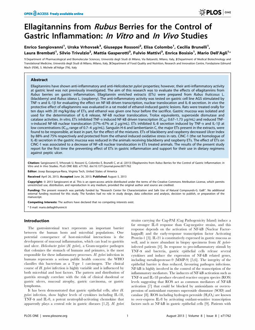
Ellagitannins from Rubus Berries for the Control of Gastric Inflammation: In Vitro and In Vivo Studies Enrico Sangiovanni 1 , Urska Vrhovsek 3 , Giuseppe Rossoni 2 , Elisa Colombo 1 , Cecilia Brunelli 1 , Laura Brembati 1 , Silvio Trivulzio 2 , Mattia Gasperotti 3 , Fulvio Mattivi 3 , Enrica Bosisio 1 , Mario Dell’Agli 1 * 1 Department of Pharmacological and Biomolecular Sciences, Universita ` degli Studi di Milano, Via Balzaretti, Milano, Italy, 2 Department of Medical Biotechnology and Translational Medicine, Universita ` degli Studi di Milano, Milano, Italy, 3 Department of Food Quality and Nutrition, Research and Innovation Centre, Fondazione Edmund Mach (FEM), S. Michele all’Adige (TN), Italy Abstract Ellagitannins have shown anti-inflammatory and anti-Helicobacter pylori properties; however, their anti-inflammatory activity at gastric level was not previously investigated. The aim of this research was to evaluate the effects of ellagitannins from Rubus berries on gastric inflammation. Ellagitannin enriched extracts (ETs) were prepared from Rubus fruticosus L. (blackberry) and Rubus idaeus L. (raspberry). The anti-inflammatory activity was tested on gastric cell line AGS stimulated by TNF-a and IL-1b for evaluating the effect on NF-kB driven transcription, nuclear translocation and IL-8 secretion. In vivo the protective effect of ellagitannins was evaluated in a rat model of ethanol-induced gastric lesions. Rats were treated orally for ten days with 20 mg/kg/day of ETs, and ethanol was given one hour before the sacrifice. Gastric mucosa was isolated and used for the determination of IL-8 release, NF-kB nuclear translocation, Trolox equivalents, superoxide dismutase and catalase activities. In vitro, ETs inhibited TNF-a induced NF-kB driven transcription (IC 50 : 0.67–1.73 mg/mL) and reduced TNF- a-induced NF-kB nuclear translocation (57%–67% at 2 mg/mL). ETs inhibited IL-8 secretion induced by TNF-a and IL-1b at low concentrations (IC 50 range of 0.7–4 mg/mL). Sanguiin H-6 and lambertianin C, the major ETs present in the extracts, were found to be responsible, at least in part, for the effect of the mixtures. ETs of blackberry and raspberry decreased Ulcer Index by 88% and 75% respectively and protected from the ethanol induced oxidative stress in rats. CINC-1 (the rat homologue of IL-8) secretion in the gastric mucosa was reduced in the animals receiving blackberry and raspberry ETs. The effect of ETs on CINC-1 was associated to a decrease of NF-kB nuclear translocation in ETs treated animals. The results of the present study report for the first time the preventing effect of ETs in gastric inflammation and support for their use in dietary regimens against peptic ulcer. Citation: Sangiovanni E, Vrhovsek U, Rossoni G, Colombo E, Brunelli C, et al. (2013) Ellagitannins from Rubus Berries for the Control of Gastric Inflammation: In Vitro and In Vivo Studies. PLoS ONE 8(8): e71762. doi:10.1371/journal.pone.0071762 Editor: Josep Bassaganya-Riera, Virginia Tech, United States of America Received April 26, 2013; Accepted June 26, 2013; Published August 5, 2013 Copyright: ß 2013 Sangiovanni et al. This is an open-access article distributed under the terms of the Creative Commons Attribution License, which permits unrestricted use, distribution, and reproduction in any medium, provided the original author and source are credited. Funding: The present research was partially funded by ‘‘Research Center for Characterization and Safe Use of Natural Compounds-G. Galli’’. No additional external funding received for this study. The funders had no role in study design, data collection and analysis, decision to publish, or preparation of the manuscript. Competing Interests: The authors have declared that no competing interests exist. * E-mail: [email protected] Introduction The gastrointestinal tract represents an important barrier between the human hosts and microbial populations. One potential consequence of host-microbial interactions is the development of mucosal inflammation, which can lead to gastritis and ulcer. Helicobacter pylori (H. pylori), a Gram-negative pathogen that colonizes the stomach of humans and primates, is the most responsible for these inflammatory processes. H. pylori infection in humans represents a serious public health concern: the WHO classifies this bacterium as a Type 1 carcinogen. The clinical course of H. pylori infection is highly variable and is influenced by both microbial and host factors. The pattern and distribution of gastritis strongly correlate with the risk of clinical duodenal or gastric ulcers, mucosal atrophy, gastric carcinoma, or gastric lymphoma. It has been demonstrated that gastric epithelial cells, after H. pylori infection, show higher levels of cytokines including IL-1ß, TNF-a and IL-8, a potent neutrophil-activating chemokine that apparently plays a central role in gastric diseases [1,2]. H. pylori strains carrying the Cag-PAI (Cag Pathogenicity Island) induce a far stronger IL-8 response than Cag-negative strains, and this response depends on the activation of NF-kB (Nuclear Factor- kappaB) and the early-response transcription factor Activating Protein-1 [3]. IL-21 is constitutively expressed in gastric mucosa as well, and is more abundant in biopsy specimens from H. pylori- infected patients [4]. In response to pro-inflammatory stimuli by TNF-a and bacteria, gastric epithelial cells release several cytokines and induce the expression of NF-kB related genes, including metalloprotease-9 (MMP-9) [5,6]. The integrity of the epithelial barrier is thus reduced, favouring pathogen infections. NF-kB is highly involved in the control of the transcription of the inflammatory mediators. The inducers of NF-kB activation such as TNF-a and IL-1ß produce elevated reactive oxygen species (ROS) levels suggesting that ROS act as common mediators of NF-kB activation [7] that could be blocked by antioxidants or overex- pression of antioxidant enzymes superoxide dismutase (SOD) and catalase [8]. ROS including hydrogen peroxide (H 2 O 2 ) are known to over-express IL-8 by activating oxidant-sensitive transcription factors such as NF-kB in gastric epithelial cells [9]. Patients with PLOS ONE | www.plosone.org 1 August 2013 | Volume 8 | Issue 8 | e71762
-
Upload
fondazioneedmundmach -
Category
Documents
-
view
1 -
download
0
Transcript of Ellagitannins from Rubus Berries for the Control of Gastric Inflammation: In Vitro and In Vivo...
Ellagitannins from Rubus Berries for the Control ofGastric Inflammation: In Vitro and In Vivo StudiesEnrico Sangiovanni1, Urska Vrhovsek3, Giuseppe Rossoni2, Elisa Colombo1, Cecilia Brunelli1,
Laura Brembati1, Silvio Trivulzio2, Mattia Gasperotti3, Fulvio Mattivi3, Enrica Bosisio1, Mario Dell’Agli1*
1 Department of Pharmacological and Biomolecular Sciences, Universita degli Studi di Milano, Via Balzaretti, Milano, Italy, 2 Department of Medical Biotechnology and
Translational Medicine, Universita degli Studi di Milano, Milano, Italy, 3 Department of Food Quality and Nutrition, Research and Innovation Centre, Fondazione Edmund
Mach (FEM), S. Michele all’Adige (TN), Italy
Abstract
Ellagitannins have shown anti-inflammatory and anti-Helicobacter pylori properties; however, their anti-inflammatory activityat gastric level was not previously investigated. The aim of this research was to evaluate the effects of ellagitannins fromRubus berries on gastric inflammation. Ellagitannin enriched extracts (ETs) were prepared from Rubus fruticosus L.(blackberry) and Rubus idaeus L. (raspberry). The anti-inflammatory activity was tested on gastric cell line AGS stimulated byTNF-a and IL-1b for evaluating the effect on NF-kB driven transcription, nuclear translocation and IL-8 secretion. In vivo theprotective effect of ellagitannins was evaluated in a rat model of ethanol-induced gastric lesions. Rats were treated orally forten days with 20 mg/kg/day of ETs, and ethanol was given one hour before the sacrifice. Gastric mucosa was isolated andused for the determination of IL-8 release, NF-kB nuclear translocation, Trolox equivalents, superoxide dismutase andcatalase activities. In vitro, ETs inhibited TNF-a induced NF-kB driven transcription (IC50: 0.67–1.73 mg/mL) and reduced TNF-a-induced NF-kB nuclear translocation (57%–67% at 2 mg/mL). ETs inhibited IL-8 secretion induced by TNF-a and IL-1b atlow concentrations (IC50 range of 0.7–4 mg/mL). Sanguiin H-6 and lambertianin C, the major ETs present in the extracts, werefound to be responsible, at least in part, for the effect of the mixtures. ETs of blackberry and raspberry decreased Ulcer Indexby 88% and 75% respectively and protected from the ethanol induced oxidative stress in rats. CINC-1 (the rat homologue ofIL-8) secretion in the gastric mucosa was reduced in the animals receiving blackberry and raspberry ETs. The effect of ETs onCINC-1 was associated to a decrease of NF-kB nuclear translocation in ETs treated animals. The results of the present studyreport for the first time the preventing effect of ETs in gastric inflammation and support for their use in dietary regimensagainst peptic ulcer.
Citation: Sangiovanni E, Vrhovsek U, Rossoni G, Colombo E, Brunelli C, et al. (2013) Ellagitannins from Rubus Berries for the Control of Gastric Inflammation: InVitro and In Vivo Studies. PLoS ONE 8(8): e71762. doi:10.1371/journal.pone.0071762
Editor: Josep Bassaganya-Riera, Virginia Tech, United States of America
Received April 26, 2013; Accepted June 26, 2013; Published August 5, 2013
Copyright: � 2013 Sangiovanni et al. This is an open-access article distributed under the terms of the Creative Commons Attribution License, which permitsunrestricted use, distribution, and reproduction in any medium, provided the original author and source are credited.
Funding: The present research was partially funded by ‘‘Research Center for Characterization and Safe Use of Natural Compounds-G. Galli’’. No additionalexternal funding received for this study. The funders had no role in study design, data collection and analysis, decision to publish, or preparation of themanuscript.
Competing Interests: The authors have declared that no competing interests exist.
* E-mail: [email protected]
Introduction
The gastrointestinal tract represents an important barrier
between the human hosts and microbial populations. One
potential consequence of host-microbial interactions is the
development of mucosal inflammation, which can lead to gastritis
and ulcer. Helicobacter pylori (H. pylori), a Gram-negative pathogen
that colonizes the stomach of humans and primates, is the most
responsible for these inflammatory processes. H. pylori infection in
humans represents a serious public health concern: the WHO
classifies this bacterium as a Type 1 carcinogen. The clinical
course of H. pylori infection is highly variable and is influenced by
both microbial and host factors. The pattern and distribution of
gastritis strongly correlate with the risk of clinical duodenal or
gastric ulcers, mucosal atrophy, gastric carcinoma, or gastric
lymphoma.
It has been demonstrated that gastric epithelial cells, after H.
pylori infection, show higher levels of cytokines including IL-1ß,
TNF-a and IL-8, a potent neutrophil-activating chemokine that
apparently plays a central role in gastric diseases [1,2]. H. pylori
strains carrying the Cag-PAI (Cag Pathogenicity Island) induce a
far stronger IL-8 response than Cag-negative strains, and this
response depends on the activation of NF-kB (Nuclear Factor-
kappaB) and the early-response transcription factor Activating
Protein-1 [3]. IL-21 is constitutively expressed in gastric mucosa as
well, and is more abundant in biopsy specimens from H. pylori-
infected patients [4]. In response to pro-inflammatory stimuli by
TNF-a and bacteria, gastric epithelial cells release several
cytokines and induce the expression of NF-kB related genes,
including metalloprotease-9 (MMP-9) [5,6]. The integrity of the
epithelial barrier is thus reduced, favouring pathogen infections.
NF-kB is highly involved in the control of the transcription of the
inflammatory mediators. The inducers of NF-kB activation such as
TNF-a and IL-1ß produce elevated reactive oxygen species (ROS)
levels suggesting that ROS act as common mediators of NF-kB
activation [7] that could be blocked by antioxidants or overex-
pression of antioxidant enzymes superoxide dismutase (SOD) and
catalase [8]. ROS including hydrogen peroxide (H2O2) are known
to over-express IL-8 by activating oxidant-sensitive transcription
factors such as NF-kB in gastric epithelial cells [9]. Patients with
PLOS ONE | www.plosone.org 1 August 2013 | Volume 8 | Issue 8 | e71762
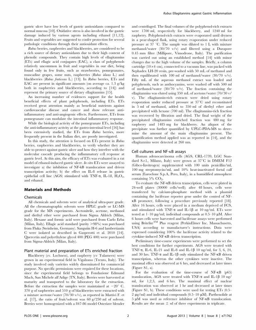
gastric ulcer have low levels of gastric antioxidants compared to
normal mucosa [10]. Oxidative stress is also involved in the gastric
damage induced by various agents including ethanol [11,12].
Fruits and vegetables are suggested to confer protection for several
pathologic conditions through their antioxidant effects.
Rubus berries, raspberries and blackberries, are considered to be
a rich source of dietary antioxidants due to their high content of
phenolic compounds. They contain high levels of ellagitannins
(ETs) and ellagic acid conjugates (EAC), a class of polyphenols
relatively uncommon in fruit and vegetables in our diet, being
found only in few fruits, such as strawberries, pomegranates,
muscadine grapes, some nuts, raspberries (Rubus idaeus L.) and
blackberries (Rubus fruticosus L.) [13]. In Rubus berries, ETs and
EAC are present in significant amounts (on average ca. 1.3 g/kg
both in raspberries and blackberries, according to [14]) and
represent the primary source of dietary ellagitannins [15].
An increasing number of evidences support for the health
beneficial effects of plant polyphenols, including ETs. ETs
received great attention mainly as beneficial nutrients against
cardiovascular disease and cancer and show relevant anti-
inflammatory and anti-angiogenic effects. Furthermore, ETs from
pomegranate can modulate the intestinal inflammatory response.
While the biological activity of the pomegranate ETs, including
the anti-inflammatory activity at the gastro-intestinal level [16] has
been extensively studied, the ETs from Rubus berries, more
frequently present in the Italian diet, are poorly investigated.
In this study, the attention is focused on ETs present in Rubus
berries, raspberries and blackberries, to verify whether they are
able to protect against gastric ulcer and how they interfere with the
molecular cascade producing the inflammatory cell response at
gastric level. At this aim, the efficacy of ETs was evaluated in a rat
model of ethanol-induced gastric ulcer. In vitro ETs were assayed to
investigate a) the inhibition of NF-kB translocation and driven
transcription activity; b) the effect on IL-8 release in gastric
epithelial cell line (AGS) stimulated with TNF-a, IL-1ß, H2O2,
and ethanol.
Materials and Methods
ChemicalsAll chemicals and solvents were of analytical ultra-pure grade.
All the chromatographic solvents were HPLC grade or LC-MS
grade for the MS experiments. Acetonitrile, acetone, methanol
and diethyl ether were purchased from Sigma Aldrich (Milan,
Italy). Hexane and formic acid were purchased from Carlo Erba
(Milan, Italy). Ellagic acid standard (purity .98%) was purchased
from Fluka (Steinheim, Germany). Sanguiin H-6 and lambertianin
C were isolated as described in Gasperotti et al. 2010 [14].
Quercetin and polyethylene glycol 400 (PEG 400) were purchased
from Sigma-Aldrich (Milan, Italy).
Plant material and preparation of ETs enriched fractionBlackberry (cv. Lochness), and raspberry (cv Tulameen) were
grown in an experimental field in Vigalzano (Trento, Italy). The
study involved only varieties widely disseminated for commercial
purpose. No specific permissions were required for these locations,
since the experimental field belongs to Fondazione Edmund
Mach, San Michele all’Adige (TN, Italy). Berries were harvested at
maturity and transported to the laboratory for the extraction.
Before the extraction the samples were maintained at 220u C.
370 g of raspberries and 510 g of blackberries were extracted with
a mixture acetone/water (70/30 v/v), as reported in Mattivi F. et
al. [17], the ratio of fruit/solvent was 60 g/250 ml of solvent.
Berries were homogenized with a 847-86 model Osterizer blender
and centrifuged. The final volumes of the polyphenol-rich extracts
were 1700 ml, respectively for blackberry, and 1240 ml for
raspberry. Polyphenol-rich extracts were evaporated until dryness
in a pear-shaped flask, using rotary evaporation under reduced
pressure at 37 uC. The sample was diluted to 1 L with mixture
methanol/water (30/70 v/v) and filtered using a Durapore
0.45 mm filter (Millipore, Vimodrone, Italy). The purification
was carried out using an established method [14] with minor
changes due to the high volume of the samples. Briefly, a column
cartridge (1064 cm), connected to a vacuum line, was packed with
Sephadex LH-20 resin, pre-washed with 50 mL of methanol and
then equilibrated with 100 ml of methanol/water (30/70 v/v).
Fifty mL of the aqueous methanol extract was loaded and
polyphenols, such as anthocyanins, were washed off with 500 ml
of methanol/water (30/70 v/v). The fraction containing the
ellagitannins was eluted using 350 mL of acetone/water (70/30 v/
v). The ellagitannin-rich extracts were dried using rotary
evaporation under reduced pressure at 37uC and reconstituted
in 5 ml of methanol, added to 350 ml of diethyl ether and
precipitated with hexane (700 ml). The ellagitannins-rich fraction
was recovered by filtration and dried. The final weight of the
precipitated ellagitannins enriched fraction was 480 mg for
raspberry and 1485 mg for blackberry. An aliquot of the
precipitate was further quantified by UPLC-PDA-MS to deter-
mine the amount of the main ellagitannins present. The
quantification method applied was as reported in [14], and the
ellagitannins were detected at 260 nm.
Cell cultures and NF-kB assaysHuman adenocarcinoma cells (AGS, CRL-1739, LGC Stan-
dard S.r.l., Milano, Italy) were grown at 37uC in DMEM F12
(Gibco-Invitrogen) supplemented with 100 units penicillin/ml,
100 mg streptomycin/ml, and 10% heat-inactivated foetal calf
serum (Euroclone S.p.A, Pero, Italy), in a humidified atmosphere
containing 5% CO2.
To evaluate the NF-kB driven transcription, cells were plated in
24-well plates (30000 cells/well); after 48 hours, cells were
transfected by calcium-phosphate method with a plasmid
containing the luciferase reporter gene under the control of NF-
kB promoter, following a procedure previously reported [18].
After 16 hours, cells were placed in a medium deprived of FCS,
and stimulated with TNF-a and IL-1b at 10 ng/ml. ETs were
tested at 1–10 mg/ml; individual compounds at 0.5–10 mM. After
6 hours cells were harvested and luciferase assays were performed
using BriteliteTM Plus reagent (PerkinElmer Inc. Massachusetts,
USA) according to manufacturer’s instructions. Data were
expressed considering 100% the luciferase activity related to the
cytokine-induced NF-kB driven transcription.
Preliminary time-course experiments were performed to set the
best conditions for further experiments. AGS were treated with
TNF-a, IL-6, IL-21 and IL-8 and IL-1b 10 ng/ml, for 3, 6, 24,
and 30 hrs. TNF-a and IL-1b only stimulated the NF-kB driven
transcription, whereas the other cytokines were inactive. The
maximal effect was observed at 6 hrs, and decreased at later times
(Figure S1, a).
For the evaluation of the time-course of NF-kB (p65)
translocation, AGS were treated with TNF-a and IL-1b 10 ng/
ml, for 1,2,3, and 6 hrs. The maximal effect of nuclear
translocation was observed at 1 hr and decreased at later times
(Figure S1, b). These conditions were used for testing ETs (0.5–
2 mg/ml) and individual compounds (0.5–10 mM). Parthenolide at
5 mM was used as reference inhibitor of NF-kB translocation.
Results are the mean 6 sd of three experiments in triplicate.
Rubus Ellagitannins against Gastric Inflammation
PLOS ONE | www.plosone.org 2 August 2013 | Volume 8 | Issue 8 | e71762
For the NF-kB (p65) nuclear translocation assay, AGS cells were
plated at the concentration of 1.56106 cells/ml in 60-mm plates.
After 48 hours, cells were treated for 1 hour with the inflamma-
tory mediators and the extracts/compounds under study. Nuclear
extracts were prepared using Nuclear Extraction Kit from
Cayman Chemical Company (Michigan, USA) and stored at
280uC until assayed. The same quantity of total nuclear proteins,
measured by the method of Bradford [19], was used to assess NF-
kB nuclear translocation using the NF-kB (p65) transcription
factor assay kit (Cayman) followed by spectroscopy (signal read
450 nm, 0.1 s). Data were expressed considering 100% the
absorbance related to the cytokine-induced NF-kB nuclear
translocation.
Cell cultures and IL-8 releasePreliminary evaluation of IL-8 secretion was performed on AGS
cells treated with TNF-a and IL-1b 10 ng/ml, for 1, 2, 3, and
6 hrs (Figure S1, c). Cells were grown in 24-well plates for 48 hrs
(30000 cells/well) before the cytokine treatment. IL-8 was
quantified by using Interleukin-8 High Sensitivity Human ELISA
Set (Immunotools, Germany) using the method described below.
Briefly, Corning 96 well EIA/RIA plates from Sigma-Aldrich
(Milan, Italy), were coated with the antibody provided in the
ELISA Set, overnight at 4uC. After blocking the reaction, 100 ml
of samples in duplicate, were transferred into wells at room
temperature for 1 hr. The amount of IL-8 in the samples was
detected by spectroscopy (signal read 450 nm, 0.1 s) by the use of
biotinylated and streptavidin-HRP conjugate antibodies, evaluat-
ing 3,5,3,59-tetramethylbenzidine (TMB) substrate reaction. The
quantification of IL-8 was done using an optimized standard curve
supplied with the ELISA Set (1.0–240.0 pg/ml). The IL-8
secretion reached the maximum at 6 hrs and this time was
selected for the experiments to test the effect of ETs (1–10 mg/mL)
and individual compounds (0.25–5 mM), (Figure S1, c). Partheno-
lide (5 mM) was used as reference inhibitor of IL-8 secretion.
Results are the mean 6 sd of three experiments in triplicate.
IL-8 secretion in AGS treated with H2O2 and ethanolCells were grown in 24-well plates for 48 hrs (30000 cells/well)
and then incubated for 12 hrs in the presence of 500 mM H2O2, or
for 24 hrs in the presence of 2% ethanol, following the procedure
described in [9], with slight modifications. ETs were tested at 1–
25 mg/mL. IL-8 was quantified as described above. Quercetin
(10 mM) was used as reference inhibitor of IL-8 secretion. Results
are the mean 6 sd of three experiments in triplicate.
Cytotoxicity assayThe integrity of the cell morphology before and after treatment
was assessed by light microscope inspection. Cell viability of AGS
was measured by MTT method [20]. No sign of cytotoxicity was
observed in cells treated with ETs at 0.05–25 mg/mL. These
concentrations are far below the content of ETs at gastric level
after consumption of a portion of 100 g of berries at meals
(100 mg for blackberry and 80 mg for raspberry). Ethanol and
H2O2 were not toxic to AGS cells at the concentration used in the
experiments.
AnimalsThirty male Wistar rats (Charles River Laboratories, Calco,
Lecco, Italy), weighing 175–200 g, were used. 3 Rats per cage
were housed under constant environmental conditions (22 6 1uC,
50 6 5% relative humidity, 12-h light/12-h dark cycle), with free
access to standard laboratory rat chow (014RF21C; Mucedola,
Settimo Milanese, Milan, Italy) and tap water. Animals were
acclimatized for a period of at least 7 days before the use. The
study was approved (protocol number 16/2010) by the Animal
Ethics Committee of University of Milan (Italy), and communi-
cated to the Italian Ministry of Health, having regard to the article
7 of the D.L. 116/92. In addition, the study was carried out in
strict accordance with the recommendations in the Guide for the Care
and Use of Laboratory Animals published by the US National Institutes
of Health (NIH Publication No. 85–23, revised 1996). All efforts
were made to minimize animal suffering.
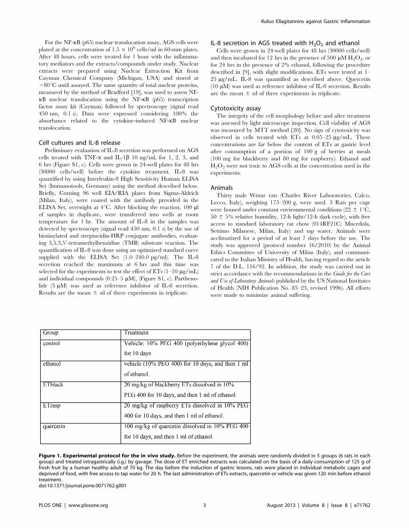
Figure 1. Experimental protocol for the in vivo study. Before the experiment, the animals were randomly divided in 5 groups (6 rats in eachgroup) and treated intragastrically (i.g.) by gavage. The dose of ET enriched extracts was calculated on the basis of a daily consumption of 125 g offresh fruit by a human healthy adult of 70 kg. The day before the induction of gastric lesions, rats were placed in individual metabolic cages anddeprived of food, with free access to tap water for 20 h. The last administration of ETs extracts, quercetin or vehicle was given 120 min before ethanoltreatment.doi:10.1371/journal.pone.0071762.g001
Rubus Ellagitannins against Gastric Inflammation
PLOS ONE | www.plosone.org 3 August 2013 | Volume 8 | Issue 8 | e71762

ProtocolBefore the experiment, the animals were randomly divided in 5
groups (6 rats in each group) and treated intragastrically (i.g.) by
gavage, according to Figure 1.
The dose of ET enriched extracts was calculated on the basis of
a daily consumption of 125 g of fresh fruit by a human healthy
adult of 70 kg (Alvarez-Suarez 2011). The day before the
induction of gastric lesions, rats were placed in individual
metabolic cages and deprived of food, with free access to tap
water for 20 h. The last administration of ETs extracts, quercetin
(as positive control) or vehicle was given 120 min before ethanol
treatment.
Assessment of gastric mucosal damageOne hour after the administration of 1 ml of ethanol, rats were
sacrificed under ether anesthesia by cervical dislocation; the
stomach was removed and opened along the greater curvature.
The stomach was rinsed with water, pinned open for microscopic
examination by a microscope (Opmi 6; Carl Zeiss S.p.A., Arese,
MI, Italy) and for photo-documentation by a digital camera (EOS
1100D, Canon Italia S.p.A., Cernusco Sul Naviglio, MI, Italy).
Gastric hemorrhagic lesions in the glandular part were examined
under a dissecting microscope (X10). Gastric damage was assessed
in a blind manner. The Ulcer Index (UI) was obtained by a 0–3
scoring system based on the number and severity of the lesions
[21,22]. Severity was defined according to the length of the lesions:
Figure 2. Composition of blackberry (ETblack) and raspberry (ETrasp) ellagitannins. Berries ETs were extracted with acetone/water 70:30,isolated by Sephadex LH 20 column chromatography, precipitated with hexane and quantified by UPLC-PDA-MS. Ellagitannins were detected at260 nm.doi:10.1371/journal.pone.0071762.g002
Rubus Ellagitannins against Gastric Inflammation
PLOS ONE | www.plosone.org 4 August 2013 | Volume 8 | Issue 8 | e71762
0, no lesions; 1, lesions 1–2 mm; 2, lesions 2–3 mm; 3, lesions
.3 mm. UI was calculated as the total number of lesions
multiplied by their respective severity score.
Preparation of gastric mucosa homogenatesSamples of 50 mg from normal and ulcerated rat gastric
mucosa were homogenized in buffer A [10 mM TRIS-HCl
(pH 8), 150 mM NaCl, 1 mM EDTA, 1 mM phenylmethylsulfo-
nyl fluoride (PMSF), 2 mg/ml aprotinin, 2 mg/ml leupeptin, and
1% Triton X-100] using Tissue Lyser II (Qiagen) for 2 minutes at
the highest frequency 30/s. The homogenates were centrifuged at
12,000 g for 10 min at 4u C and the supernatants collected, and
stored at 280uC until use. Protein concentration was determined
using Bradford protein assay (Bio-Rad) with bovine serum albumin
as a standard.
Cinc-1 (rat IL-8) release from gastric mucosaThe quantity of 40 mg of total proteins was used to assess Cinc-1
release using GRO/CINC-1 (rat) EIA kit (Enzo Life Sciences
International, Inc., Plymouth Meeting, PA, USA). This kit uses a
polyclonal antibody to rat GRO/CINC-1 labelled with the
enzyme horseradish peroxidase. After a short incubation (10
minutes) the enzyme reaction was followed by spectroscopy (signal
read 450 nm, 0.1 s). The concentration of rat GRO/CINC-1 in
the samples was determined by interpolation with a GRO/CINC-
1 standard curve. The results are expressed as pg of CINC-1 per
mL of sample.
Measurement of oxidative stress in rat gastric mucosaThe antioxidant capacity of the gastric mucosa homogenates
was assessed by Oxygen Radical Absorbance Capacity (ORAC) assay.
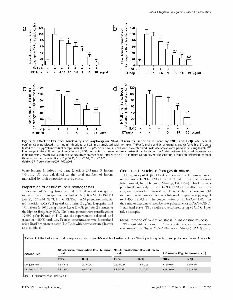
Figure 3. Effect of ETs from blackberry and raspberry on NF-kB driven transcription induced by TNFa and IL-1b. AGS cells atconfluence were placed in a medium deprived of FCS, and stimulated with 10 ng/ml TNF-a (panel a and b) or (panel c and d) for 6 hrs. ETs weretested at 1–10 mg/ml; individual compounds at 0.5–10 mM. After 6 hours cells were harvested and luciferase assays were performed using BriteliteTM
Plus reagent (PerkinElmer Inc. Massachusetts, USA) according to manufacturer’s instructions. Inhibition by 5 mM parthenolide, used as referenceinhibitor, was 72% on TNF-a induced NF-kB driven transcription, and 71% on IL-1b induced NF-kB driven transcription. Results are the mean 6 sd ofthree experiments in triplicate. * p,0.05, ** p,0.01, ***p,0.001.doi:10.1371/journal.pone.0071762.g003
Table 1. Effect of individual compounds sanguiin H-6 and lambertianin C on NF-kB pathway in human gastric epithelial AGS cells.
COMPOUND
NF-kB driven transcription IC50 mM (mean± s.d.)
NF-kB translocation IC50 mM (mean± s.d.) IL-8 release IC50 mM (mean ± s.d.)
TNFa IL-1b TNFa IL-1b TNFa IL-1b
Sanguiin H-6 1.560.35 2.760.30 0.8760.16 1.960.23 0.5860.05 1.060.06
Lambertianin C 2.760.45 4.860.35 1.260.39 1.760.30 0.5760.03 1.260.06
doi:10.1371/journal.pone.0071762.t001
Rubus Ellagitannins against Gastric Inflammation
PLOS ONE | www.plosone.org 5 August 2013 | Volume 8 | Issue 8 | e71762

This method measures the oxidative degradation of fluorescein
(Sigma-Aldrich Spa, Milan, Italy), after the addition of the free
radical generator AAPH (2,29-azobis(2-methylpropanimidamide)-
dihydrochloride) (Sigma-Aldrich S.p.a., Milan, Italy). The oxida-
tion of fluorescein by free radicals, leads to a decrease in
fluorescence, prevented by the presence of antioxidant com-
pounds. All reagents were prepared in 75 mM phosphate buffer,
pH 7.4 and Trolox (4–160 mM) was used as the reference
compound. Samples from gastric mucosa were suitably diluted in
the phosphate buffer. Each well of a 96-well microplate contained
120 mL of fluorescein (0.07 mM) and 20 mL of the samples
(corresponding to 5 mg protein) in a final volume of 200 mL assay
solution. After the addition of AAPH (60 mL, 12 mM), the plate
was shaken automatically for 2 seconds and the fluorescence was
measured at 37uC every 2 min for 60 min with emission and
excitation wavelengths of 528 and 485 nm, respectively, by using a
microplate fluorescence reader (VictorTM 63 Perkin Elmer 2030;
Perkin Elmer Waltham MA, USA). The ORAC values were
calculated as area under the curve and expressed as micromole of
Trolox equivalent (TE) per gram of gastric mucosa sample ( mmol
TE/g of gastric mucosa sample).
Evaluation of CAT activity in rat gastric mucosaCAT activity in gastric mucosa homogenates was determined by
Catalase Assay Kit (Cayman Chemical, Ann Arbor, MI, USA),
which utilizes the peroxidative function of CAT for the
determination of the enzyme activity. The method is based on
the reaction of the enzyme with methanol in the presence of an
optimal concentration of H2O2. The formaldehyde produced is
measured colorimetrically with 4-amino-3-hydrazino-5-mercapto-
1,2,4-triazole as the chromogen using a microplate reader
(Microplate Reader iMarkTM, Bio-Rad Laboratories S.r.l.,
Segrate, Italy) at 540 nm absorbance. Before starting the reaction,
each well of a 96 well microplate contained 100 mL of diluted
assay buffer, 30 mL of methanol and 20 mL of diluted gastric
mucosa homogenates (2.5 mg/well). The amount of formaldehyde
was calculated by means of a calibration curve of formaldehyde
standard. CAT activity is expressed as units (U) of CAT per mg of
proteins. One unit of CAT is defined as the amount of enzyme
that will cause the formation of 1.0 nmol of formaldehyde per
minute at 25uC.
Evaluation of SOD activity in rat gastric mucosaSOD activity was measured by using a SOD activity kit (Enzo
Life Sciences International, Inc., Plymouth Meeting, PA, USA).
This colorimetric assay evaluates the ability of SOD to reduce the
superoxide ion concentration generated from the conversion of
xanthine and oxygen to uric acid and hydrogen peroxide by
xanthine oxidase. SOD activity was determined from percent
inhibition of the rate of WST-1-formazan formation, a coloured
product that absorbs light at 450 nm. Each sample was loaded in a
96 well microplate to the final amount of 6.25 mg/well.
Immediately after the addition of xanthine, the plate was
transferred to a microtiter plate reader (VictorTM 63 Perkin
Elmer 2030; Perkin Elmer Waltham MA, USA) and absorbance
was read at 450 nm every minute for 10 minutes at room
temperature, under 10 second orbital shake before each reading.
The amount of SOD in the samples was calculated by correlating
the inhibition percentage of WST-1-formazan formation with the
logarithm of the SOD units in a standard calibration curve. SOD
activity is expressed as U/mg of proteins.
Statistical analysisAll data are expressed as mean 6 sd, with the exception of the in
vivo experiments expressed as mean 6 se. Differences between
means were calculated using the unpaired t test or one-way
analysis of variance (ANOVA) followed by Tukey’s post-hoc test
for multiple group comparisons. Statistical analysis was done using
GraphPad Prism 5.00 software (GraphPad Software Inc., San
Diego, CA, USA); p,0.05 was considered statistically significant.
IC50 was calculated using GraphPad Prism 5.00.
Results
ETs analysis and compositionBlackberry and raspberry, both members of genus Rubus are
known to own similar profile of ellagitannins having sanguiin H-6
and lambertianin C as the main ellagitannins [14,23]. In enriched
fractions, ETs from blackberry (ETblack) corresponded to
343 mg/100 g of fresh fruits, ETs from raspberry (ETrasp) were
155 mg/100 g of fresh fruits. The composition of ETs was as
follows: in ETblack sanguiin H-6 represented 12%, lambertianin
C 56%, and ellagic acid 1% of the precipitate, while in ETrasp
sanguiin H-6 represented 19%, lambertianin C 35%, and ellagic
acid 1% (Figure 2).
Figure 4. Effect of ETs from blackberry and raspberry on NF-kBnuclear translocation induced by TNFa and IL-1b. AGS cells atconfluence were placed in a medium deprived of FCS, and stimulatedwith 10 ng/ml TNF-a (panel a) or IL-1b (panel b) for 1 hr. AGS cells wereplated at the concentration of 1.5 6 106 cells/ml in 60-mm plates. NF-kB nuclear translocation was assessed using the NF-kB (p65)transcription factor assay kit (Cayman) followed by spectroscopy (signalread 450 nm, 0.1 s). Inhibition by 5 mM parthenolide used as referenceinhibitor was 37% on TNF-a induced NF-kB nuclear translocation, and40% on IL-1b induced nuclear translocation. Results are the mean 6 sdof three experiments in triplicate. * p,0.05, ** p,0.01, ***p,0.001.doi:10.1371/journal.pone.0071762.g004
Rubus Ellagitannins against Gastric Inflammation
PLOS ONE | www.plosone.org 6 August 2013 | Volume 8 | Issue 8 | e71762
Effect of ETs on NF-kB driven transcription in AGS cellsResults are shown in Figure 3. ETblack and ETrasp inhibited
the increase of NF-kB driven transcription induced by TNFa in a
concentration dependent manner. IC50s were 0.6760.17 and
1.760.6 mg/ml respectively for blackberry and raspberry. When
IL-1b stimulated NF-kB driven transcription, inhibition by ETs
was lower and comparable for blackberry and raspberry (IC50
10.361.0 and 9.960.9, respectively for ETblack and ETrasp).
Lambertianin C and sanguiin H-6 inhibited NF-kB driven
transcription stimulated by TNFa with IC50 of 2.760.45 and
1.560.35 mM, respectively. When NF-kB driven transcription was
stimulated by IL-1b, the concentration required for obtaining 50%
inhibition was 4.860.35 mM and 2.760.30 mM respectively for
lambertianin C and sanguiin H-6 (Table 1).
Effect of ETs on NF-kB (p65) translocation in AGS cellsETblack and ETrasp, at 2 mg/ml, inhibited TNFa-induced
translocation by 67% and 57% respectively (Figure 4, panel a).
The inhibitory effect of ETs on IL-1b induced translocation was
much lower (37% and 22% at 2 mg/ml of ETblack and ETrasp
respectively) (Figure 4, panel b).
Lambertianin C at 5 mM and sanguiin H-6 at 2.5 mM reduced
translocation of NF-kB at the basal levels (control without
stimulus). IC50s were 1.260.39 and 0.8760.16 mM for lambertia-
nin C and sanguiin H-6, respectively. When cells were treated with
IL-1b, IC50s were 1.760.30 and 1.960.23 mM for lambertianin C
and sanguiin H-6, respectively (Table 1).
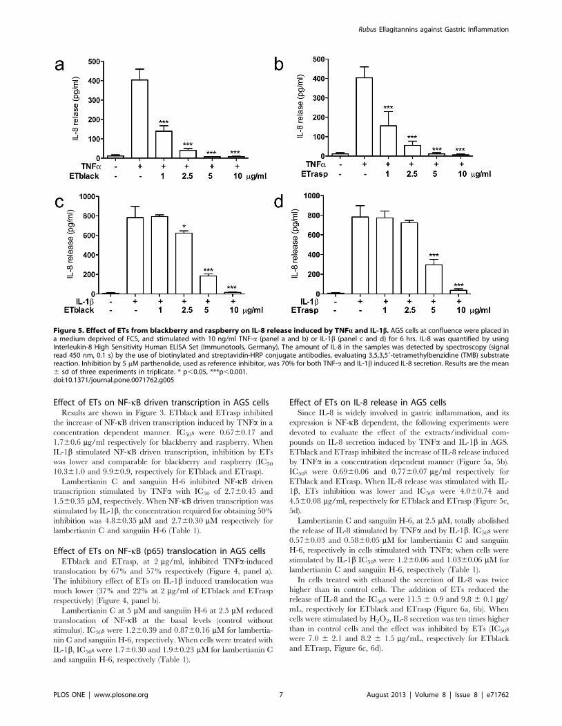
Effect of ETs on IL-8 release in AGS cellsSince IL-8 is widely involved in gastric inflammation, and its
expression is NF-kB dependent, the following experiments were
devoted to evaluate the effect of the extracts/individual com-
pounds on IL-8 secretion induced by TNFa and IL-1b in AGS.
ETblack and ETrasp inhibited the increase of IL-8 release induced
by TNFa in a concentration dependent manner (Figure 5a, 5b).
IC50s were 0.6960.06 and 0.7760.07 mg/ml respectively for
ETblack and ETrasp. When IL-8 release was stimulated with IL-
1b, ETs inhibition was lower and IC50s were 4.060.74 and
4.560.08 mg/ml, respectively for ETblack and ETrasp (Figure 5c,
5d).
Lambertianin C and sanguiin H-6, at 2.5 mM, totally abolished
the release of IL-8 stimulated by TNFa and by IL-1b. IC50s were
0.5760.03 and 0.5860.05 mM for lambertianin C and sanguiin
H-6, respectively in cells stimulated with TNFa; when cells were
stimulated by IL-1b IC50s were 1.260.06 and 1.0360.06 mM for
lambertianin C and sanguiin H-6, respectively (Table 1).
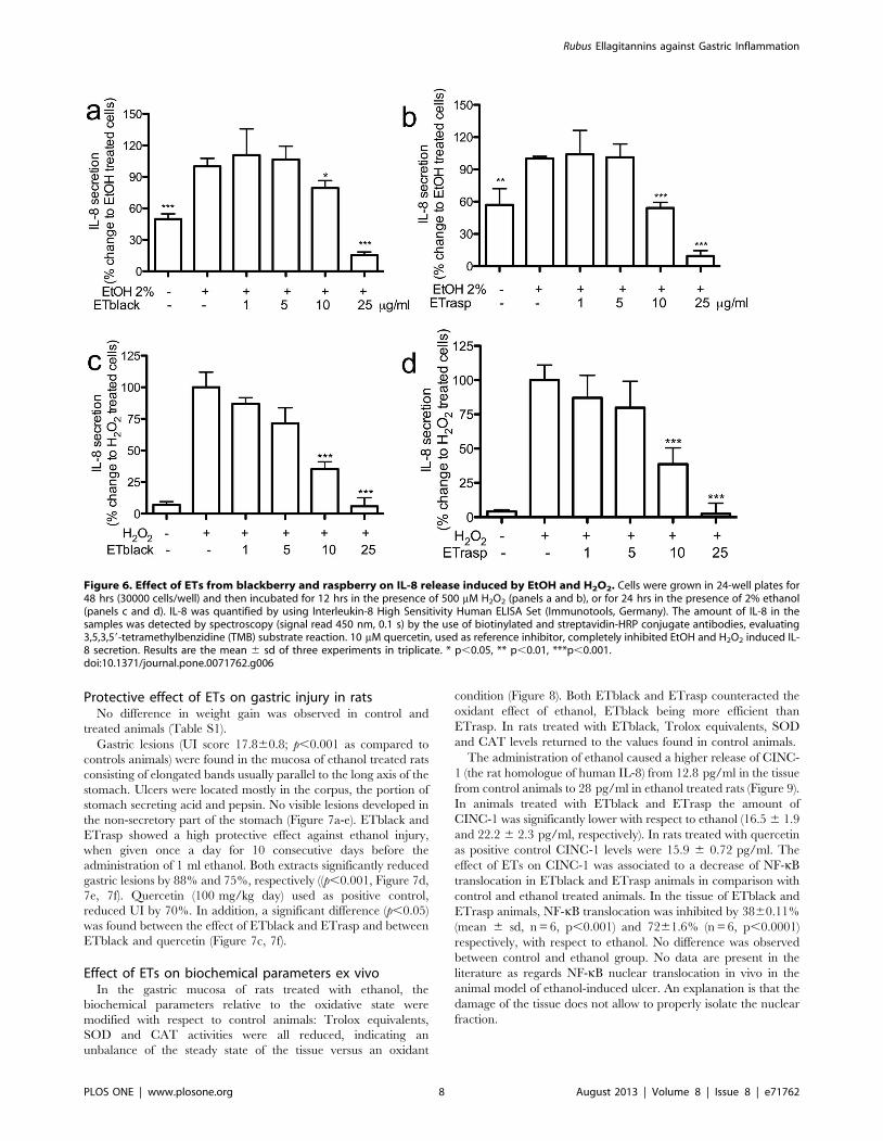
In cells treated with ethanol the secretion of IL-8 was twice
higher than in control cells. The addition of ETs reduced the
release of IL-8 and the IC50s were 11.5 6 0.9 and 9.8 6 0.1 mg/
mL, respectively for ETblack and ETrasp (Figure 6a, 6b). When
cells were stimulated by H2O2, IL-8 secretion was ten times higher
than in control cells and the effect was inhibited by ETs (IC50s
were 7.0 6 2.1 and 8.2 6 1.5 mg/mL, respectively for ETblack
and ETrasp, Figure 6c, 6d).
Figure 5. Effect of ETs from blackberry and raspberry on IL-8 release induced by TNFa and IL-1b. AGS cells at confluence were placed ina medium deprived of FCS, and stimulated with 10 ng/ml TNF-a (panel a and b) or IL-1b (panel c and d) for 6 hrs. IL-8 was quantified by usingInterleukin-8 High Sensitivity Human ELISA Set (Immunotools, Germany). The amount of IL-8 in the samples was detected by spectroscopy (signalread 450 nm, 0.1 s) by the use of biotinylated and streptavidin-HRP conjugate antibodies, evaluating 3,5,3,59-tetramethylbenzidine (TMB) substratereaction. Inhibition by 5 mM parthenolide, used as reference inhibitor, was 70% for both TNF-a and IL-1b induced IL-8 secretion. Results are the mean6 sd of three experiments in triplicate. * p,0.05, ***p,0.001.doi:10.1371/journal.pone.0071762.g005
Rubus Ellagitannins against Gastric Inflammation
PLOS ONE | www.plosone.org 7 August 2013 | Volume 8 | Issue 8 | e71762
Protective effect of ETs on gastric injury in ratsNo difference in weight gain was observed in control and
treated animals (Table S1).
Gastric lesions (UI score 17.860.8; p,0.001 as compared to
controls animals) were found in the mucosa of ethanol treated rats
consisting of elongated bands usually parallel to the long axis of the
stomach. Ulcers were located mostly in the corpus, the portion of
stomach secreting acid and pepsin. No visible lesions developed in
the non-secretory part of the stomach (Figure 7a-e). ETblack and
ETrasp showed a high protective effect against ethanol injury,
when given once a day for 10 consecutive days before the
administration of 1 ml ethanol. Both extracts significantly reduced
gastric lesions by 88% and 75%, respectively ((p,0.001, Figure 7d,
7e, 7f). Quercetin (100 mg/kg day) used as positive control,
reduced UI by 70%. In addition, a significant difference (p,0.05)
was found between the effect of ETblack and ETrasp and between
ETblack and quercetin (Figure 7c, 7f).
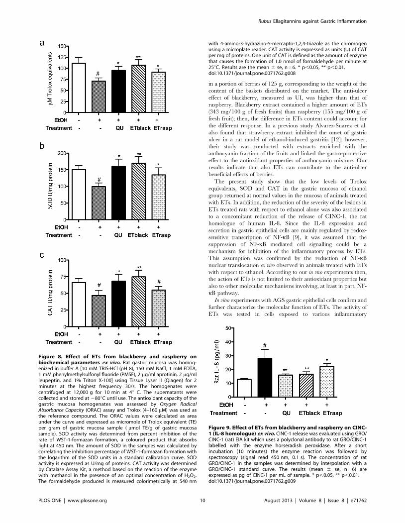
Effect of ETs on biochemical parameters ex vivoIn the gastric mucosa of rats treated with ethanol, the
biochemical parameters relative to the oxidative state were
modified with respect to control animals: Trolox equivalents,
SOD and CAT activities were all reduced, indicating an
unbalance of the steady state of the tissue versus an oxidant
condition (Figure 8). Both ETblack and ETrasp counteracted the
oxidant effect of ethanol, ETblack being more efficient than
ETrasp. In rats treated with ETblack, Trolox equivalents, SOD
and CAT levels returned to the values found in control animals.
The administration of ethanol caused a higher release of CINC-
1 (the rat homologue of human IL-8) from 12.8 pg/ml in the tissue
from control animals to 28 pg/ml in ethanol treated rats (Figure 9).
In animals treated with ETblack and ETrasp the amount of
CINC-1 was significantly lower with respect to ethanol (16.5 6 1.9
and 22.2 6 2.3 pg/ml, respectively). In rats treated with quercetin
as positive control CINC-1 levels were 15.9 6 0.72 pg/ml. The
effect of ETs on CINC-1 was associated to a decrease of NF-kB
translocation in ETblack and ETrasp animals in comparison with
control and ethanol treated animals. In the tissue of ETblack and
ETrasp animals, NF-kB translocation was inhibited by 3860.11%
(mean 6 sd, n = 6, p,0.001) and 7261.6% (n = 6, p,0.0001)
respectively, with respect to ethanol. No difference was observed
between control and ethanol group. No data are present in the
literature as regards NF-kB nuclear translocation in vivo in the
animal model of ethanol-induced ulcer. An explanation is that the
damage of the tissue does not allow to properly isolate the nuclear
fraction.
Figure 6. Effect of ETs from blackberry and raspberry on IL-8 release induced by EtOH and H2O2. Cells were grown in 24-well plates for48 hrs (30000 cells/well) and then incubated for 12 hrs in the presence of 500 mM H2O2 (panels a and b), or for 24 hrs in the presence of 2% ethanol(panels c and d). IL-8 was quantified by using Interleukin-8 High Sensitivity Human ELISA Set (Immunotools, Germany). The amount of IL-8 in thesamples was detected by spectroscopy (signal read 450 nm, 0.1 s) by the use of biotinylated and streptavidin-HRP conjugate antibodies, evaluating3,5,3,59-tetramethylbenzidine (TMB) substrate reaction. 10 mM quercetin, used as reference inhibitor, completely inhibited EtOH and H2O2 induced IL-8 secretion. Results are the mean 6 sd of three experiments in triplicate. * p,0.05, ** p,0.01, ***p,0.001.doi:10.1371/journal.pone.0071762.g006
Rubus Ellagitannins against Gastric Inflammation
PLOS ONE | www.plosone.org 8 August 2013 | Volume 8 | Issue 8 | e71762

Discussion
Polyphenols are a class of heterogeneous compounds that are
recognized to possess several biological activities. Among poly-
phenols, flavonoids, anthocyanins, and to a lesser extent
proanthocyanidins, were the most studied, while the biological
effects of ETs were randomly investigated. ETs are contained in
medicinal plants [24] and in some fruits such as pomegranate,
blackcurrant, nuts, and muscadine grapes [13]. The majority of
studies (many in vitro and few in humans), devoted to investigate
the beneficial effects of dietary ETs, focused on cardiovascular
diseases and cancer [23,25,26]. Almost all studies were conducted
with ETs enriched extracts derived from pomegranate [27], while
ETs from other sources such as Rubus, frequently present in the
European diet and highly appreciated for their flavour, are poorly
investigated [28]. The chemical composition of the ETs fraction is
depending of the fruit source, and sanguiin H-6 and lambertianin
C represent the main compounds in blackberry and raspberry
[14].
Our interest for the ETs and peptic ulcer derives from the
observation that plant rich in tannins have a traditional use for
treating ulcer and hydrolysable tannins show anti-bacterial activity
against Helicobacter pylori [24,29]. At gastric level, the acidic
conditions and the gastric enzymes are unable to hydrolize the
original ellagitannin [30]. Metabolism of ETs takes place in the
intestine where the physiological pH of the small intestine causes
the hydrolysis of ETs and the release of ellagic acid. The latter is
then metabolized by the gut microflora to several metabolites
called urolithins [30]. Both ellagic acid and urolithins are then
absorbed and found in plasma [15]. Therefore the effects observed
at the gastric level are totally associated to the unmodified
ellagitannin structure, and are not related to the metabolic
transformation in contrast to that occurring in the intestine or
other systemic situations.
This study reports for the first time that ETs from blackberries
and raspberries are able to protect the stomach against the gastric
lesions caused by ethanol. It is remarkable that the effect was
obtained at a dose of ETs comparable with the amount consumed
Figure 7. Protective effect of ETs from blackberry and raspberry against ethanol induced gastric injury. Wistar rats were randomlydivided in 5 groups (6 rats in each group). Controls were treated daily with vehicle (10% polyethylene glycol 400; PEG 400) for 10 days. Ethanol groupreceived the vehicle (10% PEG 400) daily for 10 days, and then 1 ml of ethanol. ETblack group received 20 mg/kg of blackberry ETs dissolved in 10%PEG 400 for 10 days, and then 1 ml of ethanol. ETrasp group recived 20 mg/kg of raspberry ETs dissolved in 10% PEG 400 for 10 days, and then 1 mlof ethanol. Quercetin group (positive control) received 100 mg/kg of quercetin dissolved in 10% PEG 400 for 10 days, and then 1 ml of ethanol. Thelast administration of ETs, quercetin or vehicle was given 120 min before ethanol. Treatment was performed intragastrically by gavage. Gastricdamage was assessed in a blind manner by a scoring system based on the number and severity of the lesions: 0, no lesions; 1, lesions 1–2 mm; 2,lesions 2–3 mm; 3, lesions .3 mm. Ulcer Index was calculated as the total number of lesions multiplied by their respective severity score. Results arethe mean 6 se, n = 6. ***p,0.001.doi:10.1371/journal.pone.0071762.g007
Rubus Ellagitannins against Gastric Inflammation
PLOS ONE | www.plosone.org 9 August 2013 | Volume 8 | Issue 8 | e71762
in a portion of berries of 125 g, corresponding to the weight of the
content of the baskets distributed on the market. The anti-ulcer
effect of blackberry, measured as UI, was higher than that of
raspberry. Blackberry extract contained a higher amount of ETs
(343 mg/100 g of fresh fruits) than raspberry (155 mg/100 g of
fresh fruit); then, the difference in ETs content could account for
the different response. In a previous study Alvarez-Suarez et al.
also found that strawberry extract inhibited the onset of gastric
ulcer in a rat model of ethanol-induced gastritis [12]; however,
their study was conducted with extracts enriched with the
anthocyanin fraction of the fruits and linked the gastro-protective
effect to the antioxidant properties of anthocyanin mixture. Our
results indicate that also ETs can contribute to the anti-ulcer
beneficial effects of berries.
The present study show that the low levels of Trolox
equivalents, SOD and CAT in the gastric mucosa of ethanol
group returned at normal values in the mucosa of animals treated
with ETs. In addition, the reduction of the severity of the lesions in
ETs treated rats with respect to ethanol alone was also associated
to a concomitant reduction of the release of CINC-1, the rat
homologue of human IL-8. Since the IL-8 expression and
secretion in gastric epithelial cells are mainly regulated by redox-
sensitive transcription of NF-kB [9], it was assumed that the
suppression of NF-kB mediated cell signalling could be a
mechanism for inhibition of the inflammatory process by ETs.
This assumption was confirmed by the reduction of NF-kB
nuclear translocation ex vivo observed in animals treated with ETs
with respect to ethanol. According to our in vivo experiments then,
the action of ETs is not limited to their antioxidant properties but
also to other molecular mechanisms involving, at least in part, NF-
kB pathway.
In vitro experiments with AGS gastric epithelial cells confirm and
further characterize the molecular function of ETs. The activity of
ETs was tested in cells exposed to various inflammatory
Figure 8. Effect of ETs from blackberry and raspberry onbiochemical parameters ex vivo. Rat gastric mucosa was homog-enized in buffer A [10 mM TRIS-HCl (pH 8), 150 mM NaCl, 1 mM EDTA,1 mM phenylmethylsulfonyl fluoride (PMSF), 2 mg/ml aprotinin, 2 mg/mlleupeptin, and 1% Triton X-100] using Tissue Lyser II (Qiagen) for 2minutes at the highest frequency 30/s. The homogenates werecentrifuged at 12,000 g for 10 min at 4u C. The supernatants werecollected and stored at 280uC until use. The antioxidant capacity of thegastric mucosa homogenates was assessed by Oxygen RadicalAbsorbance Capacity (ORAC) assay and Trolox (4–160 mM) was used asthe reference compound. The ORAC values were calculated as areaunder the curve and expressed as micromole of Trolox equivalent (TE)per gram of gastric mucosa sample ( mmol TE/g of gastric mucosasample). SOD activity was determined from percent inhibition of therate of WST-1-formazan formation, a coloured product that absorbslight at 450 nm. The amount of SOD in the samples was calculated bycorrelating the inhibition percentage of WST-1-formazan formation withthe logarithm of the SOD units in a standard calibration curve. SODactivity is expressed as U/mg of proteins. CAT activity was determinedby Catalase Assay Kit, a method based on the reaction of the enzymewith methanol in the presence of an optimal concentration of H2O2.The formaldehyde produced is measured colorimetrically at 540 nm
with 4-amino-3-hydrazino-5-mercapto-1,2,4-triazole as the chromogenusing a microplate reader. CAT activity is expressed as units (U) of CATper mg of proteins. One unit of CAT is defined as the amount of enzymethat causes the formation of 1.0 nmol of formaldehyde per minute at25uC. Results are the mean 6 se, n = 6. * p,0.05, ** p,0.01.doi:10.1371/journal.pone.0071762.g008
Figure 9. Effect of ETs from blackberry and raspberry on CINC-1 (IL-8 homologue) ex vivo. CINC-1 release was evaluated using GRO/CINC-1 (rat) EIA kit which uses a polyclonal antibody to rat GRO/CINC-1labelled with the enzyme horseradish peroxidase. After a shortincubation (10 minutes) the enzyme reaction was followed byspectroscopy (signal read 450 nm, 0.1 s). The concentration of ratGRO/CINC-1 in the samples was determined by interpolation with aGRO/CINC-1 standard curve. The results (mean 6 se, n = 6) areexpressed as pg of CINC-1 per mL of sample. * p,0.05, ** p,0.01.doi:10.1371/journal.pone.0071762.g009
Rubus Ellagitannins against Gastric Inflammation
PLOS ONE | www.plosone.org 10 August 2013 | Volume 8 | Issue 8 | e71762

challengers known to activate NF-kB cell signalling: cytokines
TNF-a and IL-1b, or the pro-oxidant agents ethanol and H2O2.
The results show that ETs interfere with the metabolic cascade
deriving from the activation and translocation of NF-kB that in
turn activates the transcription of targeted genes including that of
IL-8. ETs inhibited IL-8 secretion also in cells stimulated by pro-
oxidant agents such as ethanol and H2O2 at concentrations slightly
higher (7–8 mg/mL) than those required when cells were
stimulated by TNF-a or IL-1b cytokines. The results of these
experiments mimic those observed in the animal model of ethanol-
induced gastric inflammation.
In AGS stimulated by cytokines (TNF-a and IL-1b) ETs
inhibited the translocation and driven-transcription of NF-kB and
the release of IL-8. The concentration necessary to obtain 50%
inhibition (IC50) ranged between 1 and 2 mg/mL of extracts.
Considering a volume of rat gastric juice of 20 ml, the
consumption of 125 g of fresh fruits by an adult allows to reach
a concentration of ETs in the stomach around 6.4 mg/ml, which
is 103 higher than the concentration required to inhibit
translocation of NF-kB in the nucleus and then activation of gene
transcription. The effect by ETs was less potent in cells stimulated
by IL-1b compared to cells stimulated by TNF-a. The reasons for
this observation is presently unknown, but it was previously shown
that antioxidants showed more potent inhibitory effect on TNF-a-
induced IL-8 secretion compared with IL-1b-induced IL-8
secretion [31]. An explanation may reside in the different role of
ROS in the NF-kB activation by TNF-a and IL-1b [32,33] and
how/when ETs block ROS generation. At this stage of the
research explanations can be only speculative. The cascade of cell
events generated by TNF-a for the activation of NF-kB is different
as compared to IL-1b [34], and ETs could interact at different
sites along the cascade. Either ETs can function as binding
inhibitors at the receptor sites of TNF-a (TNFR1) and IL-1b (IL-
1R1). The compounds may act at different levels in the complex
regulatory process of NF-kB pathway, a question that can be a
matter for future studies.
ETs used in this study are a mixture of compounds that
contribute to the total activity of the extract. ETblack and ETrasp
have qualitatively similar profile both having lambertianin C and
sanguiin H-6 as the main ellagitannin, but different composition:
the compounds represent 47% and 74% of the total ETs in
ETrasp and ETblack, respectively. The experiments with single
ET were performed to study the reciprocal contribution of each.
Both showed an impressive inhibition of NF-kB activation and IL-
8 secretion and were equally potent; then, the contribution to the
overall effect of ETs mixture depends on their amount in the
extract. As found in all the experiments, ETblack containing
higher amounts possess higher activity than ETrasp. Since the in
vitro studies performed with the mixtures are well reproducible in
the in vivo animal model, it is conceivable that lambertianin C and
sanguiin H-6 are the active principles of ETblack and ETrasp.
In conclusion we have shown that ETs from Rubus berries
efficiently protect against the onset of gastric ulcer in a rat animal
model. ETs act through the inhibition of the NF-kB cascade either
directly on the cell response to pro-inflammatory cytokines or as
antioxidant agents inhibiting ROS generated in several inflam-
matory conditions including ethanol damage or Helicobacter pylori
infection. For these reasons, the study of the biological activity of
this class of compounds deserves more attention. Further studies
will consider experimental models of Helicobacter pylori dependent
ulcer and the immunomodulatory effect of these compounds in the
context of peptic ulcer. Polyphenols such as ETs and anthocyanins
are constituents of the fruits of Rubus spp. It would be of interest to
study if and how these two classes of important dietary
components interact at gastric level for the modulation of
inflammatory response.
The outcome of this research will allow to draw the attention of
the clinical/dietology community towards the benefits of the fruits
of Rubus spp. as integration in dietary regimens designed for
inflammatory gastrointestinal diseases.
Supporting Information
Figure S1 Time course experiments in order to set thebest conditions for further experiments with the com-pounds under study. AGS were treated with TNF-a, IL-6, IL-
21 and IL-8 and IL-1b 10 ng/ml, for 3, 6, 24, and 30 hrs. TNF-aand IL-1b only stimulated the NF-kB driven transcription,
whereas the other cytokines were inactive. The maximal effect
was observed at 6 hrs, and decreased at later times (panel a). For
the evaluation of the time-course of NF-kB (p65) translocation,
AGS were treated with TNF-a and IL-1b 10 ng/ml, for 1,2,3, and
6 hrs. The maximal effect of nuclear translocation was observed at
1 hr and decreased at later times (panel b). Preliminary evaluation
of IL-8 secretion was performed on AGS cells treated with TNF-aand IL-1b 10 ng/ml, for 1, 2, 3, and 6 hrs. IL-8 secretion was
higher at 6 hrs and this time was selected for ETs and for
individual compounds evaluation (panel c).
(TIFF)
Table S1 Effect of the treatment with the extracts on ratweight. No difference in weight gain was observed in the 4
groups of rats (group 2–5), as compared with controls animals
(group 1) receiving only the chronic administration of vehicle
(PEG 400).
(DOCX)
Acknowledgments
The fellowship of Elisa Colombo is partially sponsored by FSE, Regione
Lombardia. The authors thank Ms. Elda Desiderio Pinto for excellent
administrative management.
Author Contributions
Conceived and designed the experiments: FM EB MDA. Performed the
experiments: ES UV GR EC CB LB ST MG. Analyzed the data: MDA ES
GR UV. Contributed reagents/materials/analysis tools: MDA EB GR
FM. Wrote the paper: EB MDA FM GR.
References
1. Crabtree JE, Peichl P, Wyatt JI, Stachl U, Lindley IJ (1993) Gastric interleukin-8
and IgA IL-8 autoantibodies in Helicobacter pylori infection. Scand J Immunol
37: 65–70.
2. Crabtree JE, Xiang Z, Lindley IJ, Tompkins DS, Rappuoli R, et al. (1995)
Induction of interleukin-8 secretion from gastric epithelial cells by a cagA
negative isogenic mutant of Helicobacter pylori. J Clin Pathol 48: 967–969.
3. Yasumoto K, Okamoto S, Mukaida N, Murakami S, Mai M, et al. (1992)
Tumor necrosis factor alpha and interferon gamma synergistically induce
interleukin 8 production in a human gastric cancer cell line through acting
concurrently on AP-1 and NF-kB-like binding sites of the interleukin 8 gene.
J Biol Chem 267: 22506–22511.
4. Caruso R, Fina D, Peluso I, Fantini MC, Tosti C, et al. (2007) IL-21 is highly
produced in Helicobacter pylori-infected gastric mucosa and promotes
gelatinases synthesis. J Immunol 178: 5957–5965.
5. Gooz M, Gooz P, Smolka AJ (2001) Epithelial and bacterial metalloproteinases
and their inhibitors in H. pylori infection of human gastric cells. Am J Physiol
Gastrointest Liver Physiol 281: G823–832.
Rubus Ellagitannins against Gastric Inflammation
PLOS ONE | www.plosone.org 11 August 2013 | Volume 8 | Issue 8 | e71762

6. Rautelin HI, Oksanen AM, Veijola LI, Sipponen PI, Tervahartiala TI, et al.
(2009) Enhanced systemic matrix metalloproteinase response in Helicobacter
pylori gastritis. Ann Med 41: 208–215.
7. Handa O, Naito Y, Takagi T, Shimozawa M, Kokura S, et al. (2004) Tumor
necrosis factor-alpha-induced cytokine-induced neutrophil chemoattractant-1
(CINC-1) production by rat gastric epithelial cells: role of reactive oxygen species
and nuclear factor-kappaB. J Pharmacol Exp Ther 309: 670–676.
8. de Haan JB, Cristiano F, Iannello R, Bladier C, Kelner MJ, et al. (1996)
Elevation in the ratio of Cu/Zn-superoxide dismutase to glutathione peroxidase
activity induces features of cellular senescence and this effect is mediated by
hydrogen peroxide. Hum Mol Genet 5: 283–292.
9. Kim Y, Seo JH, Kim H (2011) beta-Carotene and lutein inhibit hydrogen
peroxide-induced activation of NF-kappaB and IL-8 expression in gastric
epithelial AGS cells. J Nutr Sci Vitaminol (Tokyo) 57: 216–223.
10. Nair S, Norkus EP, Hertan H, Pitchumoni CS (2000) Micronutrient
antioxidants in gastric mucosa and serum in patients with gastritis and gastric
ulcer: does Helicobacter pylori infection affect the mucosal levels? J Clin
Gastroenterol 30: 381–385.
11. Lee JS, Oh TY, Kim YK, Baik JH, So S, et al. (2005) Protective effects of green
tea polyphenol extracts against ethanol-induced gastric mucosal damages in rats:
stress-responsive transcription factors and MAP kinases as potential targets.
Mutat Res 579: 214–224.
12. Alvarez-Suarez JM, Dekanski D, Ristic S, Radonjic NV, Petronijevic ND, et al.
(2011) Strawberry polyphenols attenuate ethanol-induced gastric lesions in rats
by activation of antioxidant enzymes and attenuation of MDA increase. PLoS
One 6: e25878.
13. Koponen JM, Happonen AM, Mattila PH, Torronen AR (2007) Contents of
anthocyanins and ellagitannins in selected foods consumed in Finland. J Agric
Food Chem 55: 1612–1619.
14. Gasperotti M, Masuero D, Vrhovsek U, Guella G, Mattivi F (2010) Profiling and
accurate quantification of Rubus ellagitannins and ellagic acid conjugates using
direct UPLC-Q-TOF HDMS and HPLC-DAD analysis. J Agric Food Chem 58:
4602–4616.
15. Quideau S (2009) Chemistry and biology of ellagitannins. Singapore: World
Scientific.
16. Colombo E, Sangiovanni E, Dell9agli M (2013) A review on the anti-
inflammatory activity of pomegranate in the gastrointestinal tract. Evid Based
Complement Alternat Med 2013: 247145.
17. Mattivi F, Zulian C, Nicolini G, Valenti L (2002) Wine, biodiversity, technology,
and antioxidants. Ann N Y Acad Sci 957: 37–56.
18. Sangiovanni E, Colombo E, Fumagalli M, Abbiati F, Caruso D, et al. (2012)
Inhibition of NF- kappaB Activity by Minor Polar Components of Extra-Virgin
Olive Oil at Gastric Level. Phytother Res.
19. Bradford MM (1976) A rapid and sensitive method for the quantitation of
microgram quantities of protein utilizing the principle of protein-dye binding.Anal Biochem 72: 248–254.
20. Denizot F, Lang R (1986) Rapid colorimetric assay for cell growth and survival.
Modifications to the tetrazolium dye procedure giving improved sensitivity andreliability. J Immunol Methods 89: 271–277.
21. Stroff T, Plate S, Ebrahim JS, Ehrlich KH, Respondek M, et al. (1996)Tachykinin-induced increase in gastric mucosal resistance: role of primary
afferent neurons, CGRP, and NO.Am J Physiol 271: G1017–1027.
22. Rossoni G, Manfredi B, Tazzari V, Sparatore A, Trivulzio S, et al. (2010)Activity of a new hydrogen sulfide-releasing aspirin (ACS14) on pathological
cardiovascular alterations induced by glutathione depletion in rats.Eur J Pharmacol 648: 139–145.
23. Mullen W, McGinn J, Lean ME, MacLean MR, Gardner P, et al. (2002)Ellagitannins, flavonoids, and other phenolics in red raspberries and their
contribution to antioxidant capacity and vasorelaxation properties. J Agric Food
Chem 50: 5191–5196.24. de Jesus NZ, de Souza Falcao H, Gomes IF, de Almeida Leite TJ, de Morais
Lima GR, et al. (2012) Tannins, peptic ulcers and related mechanisms. Int J MolSci 13: 3203–3228.
25. McDougall GJ, Ross HA, Ikeji M, Stewart D (2008) Berry extracts exert
different antiproliferative effects against cervical and colon cancer cells grown invitro. J Agric Food Chem 56: 3016–3023.
26. Ross HA, McDougall GJ, Stewart D (2007) Antiproliferative activity ispredominantly associated with ellagitannins in raspberry extracts. Phytochem-
istry 68: 218–228.27. Adams LS, Seeram NP, Aggarwal BB, Takada Y, Sand D, et al. (2006)
Pomegranate juice, total pomegranate ellagitannins, and punicalagin suppress
inflammatory cell signaling in colon cancer cells. J Agric Food Chem 54: 980–985.
28. Beekwilder J, Hall RD, de Vos CH (2005) Identification and dietary relevance ofantioxidants from raspberry. Biofactors 23: 197–205.
29. Funatogawa K, Hayashi S, Shimomura H, Yoshida T, Hatano T, et al. (2004)
Antibacterial activity of hydrolyzable tannins derived from medicinal plantsagainst Helicobacter pylori. Microbiol Immunol 48: 251–261.
30. Larrosa M, Garcia-Conesa MT, Espin JC, Tomas-Barberan FA (2010)Ellagitannins, ellagic acid and vascular health. Mol Aspects Med 31: 513–539.
31. Shimada T, Watanabe N, Hiraishi H, Terano A (1999) Redox regulation ofinterleukin-8 expression in MKN28 cells. Dig Dis Sci 44: 266–273.
32. Gloire G, Legrand-Poels S, Piette J (2006) NF-kappaB activation by reactive
oxygen species: fifteen years later. Biochem Pharmacol 72: 1493–1505.33. Morgan MJ, Liu ZG (2011) Crosstalk of reactive oxygen species and NF-kappaB
signaling. Cell Res 21: 103–115.34. Kuno K, Matsushima K (1994) The IL-1 receptor signaling pathway. J Leukoc
Biol 56: 542–547.
Rubus Ellagitannins against Gastric Inflammation
PLOS ONE | www.plosone.org 12 August 2013 | Volume 8 | Issue 8 | e71762