Electron tunneling through sensitizer wires bound to proteins
13
Electron tunneling through sensitizer wires bound to proteins Matthew R. Hartings a , Igor V. Kurnikov b , Alexander R. Dunn c , Jay R. Winkler a , Harry B. Gray a , and Mark A. Ratner d a Beckman Institute, California Institute of Technology, Pasadena, CA 91125 b Department of Chemistry, Carnegie Mellon University, 4400 Fifth Avenue, Pittsburgh, PA 15213 c Department of Chemical Engineering, Stanford University, Stanford, CA 94305 d Department of Chemistry, Northwestern University, Evanston, IL 60208 Abstract We report a quantitative theoretical analysis of long-range electron transfer through sensitizer wires bound in the active-site channel of cytochrome P450cam. Each sensitizer wire consists of a substrate group with high binding affinity for the enzyme active site connected to a ruthenium-diimine through a bridging aliphatic or aromatic chain. Experiments have revealed a dramatic dependence of electron transfer rates on the chemical composition of both the bridging group and the substrate. Using combined molecular dynamics simulations and electronic coupling calculations, we show that electron tunneling through perfluorinated aromatic bridges is promoted by enhanced superexchange coupling through virtual reduced states. In contrast, electron flow through aliphatic bridges occurs by hole-mediated superexchange. We have found that a small number of wire conformations with strong donor-acceptor couplings can account for the observed electron tunneling rates for sensitizer wires terminated with either ethylbenzene or adamantane. In these instances, the rate is dependent not only on electronic coupling of the donor and acceptor but also on the nuclear motion of the sensitizer wire, necessitating the calculation of average rates over the course of a molecular dynamics simulation. These calculations along with related recent findings have made it possible to analyze the results of many other sensitizer-wire experiments that in turn point to new directions in our attempts to observe reactive intermediates in the catalytic cycles of P450 and other heme enzymes. 1. Introduction We have shown that electrons and holes can be delivered rapidly to the active sites of metalloenzymes through substrates (or ligands) linked to redox-active photosensitizers (“sensitizer wires”) [1-12]. Electron transfer (ET) is initiated by optical excitation of the sensitizer at the end of the wire and monitored by spectroscopic methods. Dramatic variations of ET rates through the wires have been observed upon changes in the chemical compositions of both linkers and substrates [1,3,6,10,12]. We would like to know the origin of these large variations in wire ET rates, as detailed understanding will aid the design of improved wires for the study of biological redox reactions. © 2009 Elsevier B.V. All rights reserved. Publisher's Disclaimer: This is a PDF file of an unedited manuscript that has been accepted for publication. As a service to our customers we are providing this early version of the manuscript. The manuscript will undergo copyediting, typesetting, and review of the resulting proof before it is published in its final citable form. Please note that during the production process errors may be discovered which could affect the content, and all legal disclaimers that apply to the journal pertain. NIH Public Access Author Manuscript Coord Chem Rev. Author manuscript; available in PMC 2011 February 1. Published in final edited form as: Coord Chem Rev. 2010 February 1; 254(3): 248–253. doi:10.1016/j.ccr.2009.08.008. NIH-PA Author Manuscript NIH-PA Author Manuscript NIH-PA Author Manuscript
-
Upload
independent -
Category
Documents
-
view
3 -
download
0
Transcript of Electron tunneling through sensitizer wires bound to proteins

Electron tunneling through sensitizer wires bound to proteins
Matthew R. Hartingsa, Igor V. Kurnikovb, Alexander R. Dunnc, Jay R. Winklera, Harry B.Graya, and Mark A. RatnerdaBeckman Institute, California Institute of Technology, Pasadena, CA 91125bDepartment of Chemistry, Carnegie Mellon University, 4400 Fifth Avenue, Pittsburgh, PA 15213cDepartment of Chemical Engineering, Stanford University, Stanford, CA 94305dDepartment of Chemistry, Northwestern University, Evanston, IL 60208
AbstractWe report a quantitative theoretical analysis of long-range electron transfer through sensitizer wiresbound in the active-site channel of cytochrome P450cam. Each sensitizer wire consists of a substrategroup with high binding affinity for the enzyme active site connected to a ruthenium-diimine througha bridging aliphatic or aromatic chain. Experiments have revealed a dramatic dependence of electrontransfer rates on the chemical composition of both the bridging group and the substrate. Usingcombined molecular dynamics simulations and electronic coupling calculations, we show thatelectron tunneling through perfluorinated aromatic bridges is promoted by enhanced superexchangecoupling through virtual reduced states. In contrast, electron flow through aliphatic bridges occursby hole-mediated superexchange. We have found that a small number of wire conformations withstrong donor-acceptor couplings can account for the observed electron tunneling rates for sensitizerwires terminated with either ethylbenzene or adamantane. In these instances, the rate is dependentnot only on electronic coupling of the donor and acceptor but also on the nuclear motion of thesensitizer wire, necessitating the calculation of average rates over the course of a molecular dynamicssimulation. These calculations along with related recent findings have made it possible to analyzethe results of many other sensitizer-wire experiments that in turn point to new directions in ourattempts to observe reactive intermediates in the catalytic cycles of P450 and other heme enzymes.
1. IntroductionWe have shown that electrons and holes can be delivered rapidly to the active sites ofmetalloenzymes through substrates (or ligands) linked to redox-active photosensitizers(“sensitizer wires”) [1-12]. Electron transfer (ET) is initiated by optical excitation of thesensitizer at the end of the wire and monitored by spectroscopic methods. Dramatic variationsof ET rates through the wires have been observed upon changes in the chemical compositionsof both linkers and substrates [1,3,6,10,12].
We would like to know the origin of these large variations in wire ET rates, as detailedunderstanding will aid the design of improved wires for the study of biological redox reactions.
© 2009 Elsevier B.V. All rights reserved.Publisher's Disclaimer: This is a PDF file of an unedited manuscript that has been accepted for publication. As a service to our customerswe are providing this early version of the manuscript. The manuscript will undergo copyediting, typesetting, and review of the resultingproof before it is published in its final citable form. Please note that during the production process errors may be discovered which couldaffect the content, and all legal disclaimers that apply to the journal pertain.
NIH Public AccessAuthor ManuscriptCoord Chem Rev. Author manuscript; available in PMC 2011 February 1.
Published in final edited form as:Coord Chem Rev. 2010 February 1; 254(3): 248–253. doi:10.1016/j.ccr.2009.08.008.
NIH
-PA Author Manuscript
NIH
-PA Author Manuscript
NIH
-PA Author Manuscript

There have been many investigations of the effects of molecular fluctuations on ET rates inbridge-mediated systems [13-25]. Bridge dynamics can have profound effects on ET kineticsin biological systems, as, for example, in conformationally gated processes [13,15,16,20,21].Gating occurs when the coupling between two cofactors is increased dramatically as a resultof large-scale nuclear motions and the rate of charge transfer is greatly increased. Bridgedynamics also can enhance electronic coupling through equilibrium fluctuations of thetorsional angles along the length of the bridge [14,15,17-19,21].
We have calculated the rates of electron transfer through Ru-diimine sensitizer wires to theheme of cytochrome P450. Using a combination of molecular dynamics and electroniccoupling calculations, we have found that subtle rearrangements modulate electron flow fromthe Ru-diimine sensitizer through a wire to the heme. We have analyzed the effects of theserearrangements on the experimentally observed rates as well as other factors that might leadto more rapid electron transfer in these protein:wire conjugates.
2. StructuresRapid heme reduction is successfully achieved through two similar photoprocesses in the caseof five related Ru(II) sensitizer wires. In the first process, electronically excited Ru(II), or Ru(II)*, directly reduces the heme. The driving force of the reaction (-ΔGo) is dependent on theRu(II) complex employed in the experiment. Here we have examined two complexes: [Ru(2,2′-bipyridine)2L]2+ (bpyRu) and [Ru(4,4′,5,5′-tetramethylbipyridine)2L]2+ (tmbpyRu). The(bpyRu)3+/2+* potential is -0.62 V vs NHE; (tmbpyRu)3+/2+* is -0.75 V vs NHE [6]. In thesecond process, Ru(II)* is reductively quenched by an external redox partner, followed by Ru(I) to Fe(III) ET to produce an Fe(II) heme. The (bpyRu)2+/+ potential is -1.24 V vs NHE)[1].
The sensitizer wires that have been investigated are shown in Figure 1. Two types ofhydrophobic bridges have been examined: aliphatic hydrocarbon chains (C11 and C13) andfluorinated biphenyls (F8bp). Three types of terminal substrate groups have been investigated:imidazole (Im), adamantane (Ad) and ethylbenzene (EB). One difference in the terminal groupsthat is significant for electron transfer is the ability of imidazole to coordinate directly to theFe atom in Fe-porphyrins, while interactions of adamantane or ethylbenzene with the Fe-porphyrin are noncovalent in nature. We have performed calculations on [bpyRu-F8bp-Ad]2+, which showed no heme reduction in flash/quench experiments, because it is similar to[bpyRu-F8bp-Im]2+ and [tmbpyRu-F8bp-Im]2+, whose electronic excited states are capable ofdirectly reducing the Fe(III) heme on submicrosecond timescales.
3. Molecular Dynamics SimulationsCrystal structures are available for P450 conjugates with bpyRu-F8bp-Ad and bpyRu-C9-Ad(Figure 2) wires [2]. Other wires were constructed using molecular mechanics modeling.Substantial flexibility of the wires in conjugates with P450 is expected (as indicated by thepresence of “alternative” structures in the X-ray determinations of P450 with bpyRu-F8bp-Adand bpyRu-C9-Ad); therefore, molecular dynamics simulations were used to generate arepresentative set of configurations for the protein:wire conjugates.
Simulations of P450:wire conjugates were performed with the AMBER 7 suite of programs[26]. Parameters for protein amino acids and the Fe-porphyrin were taken from the Amber-94force field [27]. Parameters for the carbon and hydrogen atoms of Ad and hydrocarbon bridgeswere taken as in aliphatic amino-acid sidechains such as valine and leucine. Atomic chargesfor atoms of fluorinated biphenyls (F8bp) were set using Hartree-Fock ab initio calculationswith a 6-31G* basis set and the Merz-Kollman charge-fitting scheme [28]. The equilibriumgeometry of F8bp was taken from an X-ray structure [2]. Force-field parameters for a torsional
Hartings et al. Page 2
Coord Chem Rev. Author manuscript; available in PMC 2011 February 1.
NIH
-PA Author Manuscript
NIH
-PA Author Manuscript
NIH
-PA Author Manuscript

angle defining the relative orientation of fluorinated phenyl rings were ones that fit the potentialenergy profile obtained by constrained energy minimization of F8bp (the torsional angle valuewas a parameter in quantum chemical calculations) using the MP2 method and a 6-31G* basisset. The initial geometry for bpyRu-F8bp-Ad was taken from X-ray data. Initial geometries forother structures were generated by replacing the wire in the X-ray structure of bpyRu-F8bp-Ad or bpyRu-C9-Ad followed by energy minimization to remove unfavorable atom-atomcontacts. 500 ps molecular dynamics trajectories were generated for conditions of constantpressure (1 atm) and constant temperature (300 K). The first 100 ps of MD trajectories werediscarded as nonequilibrated and snapshots were then collected at regular intervals alongremaining portions of the trajectories.
4. Electronic Coupling CalculationsTo compute electronic donor-acceptor interactions we used an energy splitting method whereeigenstates of a model electronic Hamiltonian of the system that corresponds to the donor andacceptor electronic states are tuned into quasi-degeneracy by a Hamiltonian perturbation (inthis work by applying an electric field in the donor-acceptor direction) [29-31]. The minimalenergy splitting of these two eigenstates of the system is equal to twice the donor-acceptorelectronic coupling ΔEmin=2HDA. In Hartree-Fock calculations of donor-acceptor systems,HDA can be found from the energy splitting of eigenstates (molecular orbitals) of the one-electron Hamiltonian (Fockian) of the system. For our calculations we compute the energysplitting for both electron and hole transfer. To perform these calculations we either add anextra electron (for the case of electron transfer) or remove an electron (for the case of holetransfer) from the system in accordance with Koopman’s theorem and literature precedent[32-36].
In the ET experiments, the heme at the active site of P450 cycles between Fe(III) and Fe(II)redox states [1-12]. In this redox transition the electronic density changes mainly on the ironatom; therefore, it is likely that both the highest occupied molecular orbital of the Fe(II)-porphyrin and the lowest unoccupied molecular orbital of Fe(III)-porphyrin will be Fe-localized. We assume in our calculations that the occupied Fe-localized orbitals (dxy, dxz,dyz) are equally involved in ET; and, the average ET coupling will be the root mean square(RMS) of ET couplings computed for individual Fe-porphyrin d orbitals.
For the case of computing HDA for electron transfer, the Ru-diimine donor was represented bya single dihydropyrazine ring. The HOMO of dihydropyrazine has the same symmetry andnodal structure as the LUMO of pyridine; thus, the dihydropyrazine HOMO is a reasonablemodel for the donor one-electron state.
In the second approach for calculating HDA for hole transfer, electrons were removed (to aformal Fe(VI) state). A large compensatory point charge (-5e) was placed on Fe; the resultingenergies of Fe-localized unoccupied molecular orbitals are roughly -4 eV, in agreement withthe expected electron binding energies of the acceptor with a reduction potential near -0.3 Vvs NHE [37] (0.0 V vs NHE approximately corresponds to a -4.5 eV electron binding energy)[38]. The Ru-diimine donor electronic state was modeled in these calculations by the LUMOof the pyridinium cation. The computed value of the pyridinium cation LUMO energy was -3.5eV in the Hartree-Fock calculations, in agreement with the reduction potential of the donor (~-0.62 V for the excited state and -1.2 V for the anion donor (vs NHE)) [39].
5. Bridge Conformational DynamicsThe molecular bridges we have investigated are quite flexible, and multiple conformations areobserved in MD calculations. The importance of taking into account molecular bridgefluctuations while computing ET rates has been emphasized several times in the literature
Hartings et al. Page 3
Coord Chem Rev. Author manuscript; available in PMC 2011 February 1.
NIH
-PA Author Manuscript
NIH
-PA Author Manuscript
NIH
-PA Author Manuscript

[13-23]. Figure 3 (top) shows snapshots of MD trajectories of the P450 complex with a bpyRu-C13-Im wire. We found that the computed electronic coupling varies substantially for thedifferent conformations of the P450:wire conjugate. Figure 3 (bottom) shows the Fe to Ru ET-coupling (HDA) values computed along the MD trajectory. Typically, the computed ETcouplings vary by about an order of magnitude from configuration to configuration(corresponding to two orders of magnitude difference in the computed nonadiabatic ET rates).There are geometries with even smaller ET coupling values, some smaller than the maximalcoupling by a factor of 30. The observed rates can be accounted for by the contribution of afew configurations that exhibit strong donor-acceptor couplings. It follows that a computedET rate will almost certainly be much smaller than the experimental value if the coupling iscomputed in a randomly chosen configuration (or in a geometry derived from X-ray structuralanalysis). Indeed, appropriate averaging here is complicated, since a few geometries favoringET are dominant. This issue was found to be especially important for wires with Ad and EBterminal groups that interact with the heme via weak van der Waals interactions, as only certainconformations show good overlap between the orbitals of the terminal group and those of theiron atom.
Beratan and coworkers have published an extensive theoretical analysis of electron tunnelingthrough Ru-modified cytochrome b562 [22]. They analyze different Ru-modified proteins, andfind two general categories of electronic coupling behavior. In one, ET occurs from the hemeedge through the protein to the Ru(III) acceptor; here, there are multiple possible pathwaysthrough the protein into Ru(III) orbitals. This is a structure-insensitive regime - while it wasfound that specific conformations are likely to have greater couplings than other conformations,dynamical averaging along multiple pathways largely negated the dependence of ET rates onspecific nuclear coordinates. However, the second case, where electrons tunnel from an axialheme position to Ru(III), does show a structure-dependent limit. Here, conformationalfluctuations influence the ET rate, as there is only one entry pathway. This case is similar tothe P450:wire system under discussion here.
There are other important cases where ET rates are known to be influenced by moleculardynamics, for example, Troisi and coworkers found that the rate of charge transfer in a C clampmolecule depends on the fluctuations of intervening solvent molecules [19]. Dynamics alsohave been found to tune the ET rates in certain molecular transport junctions [40,41].
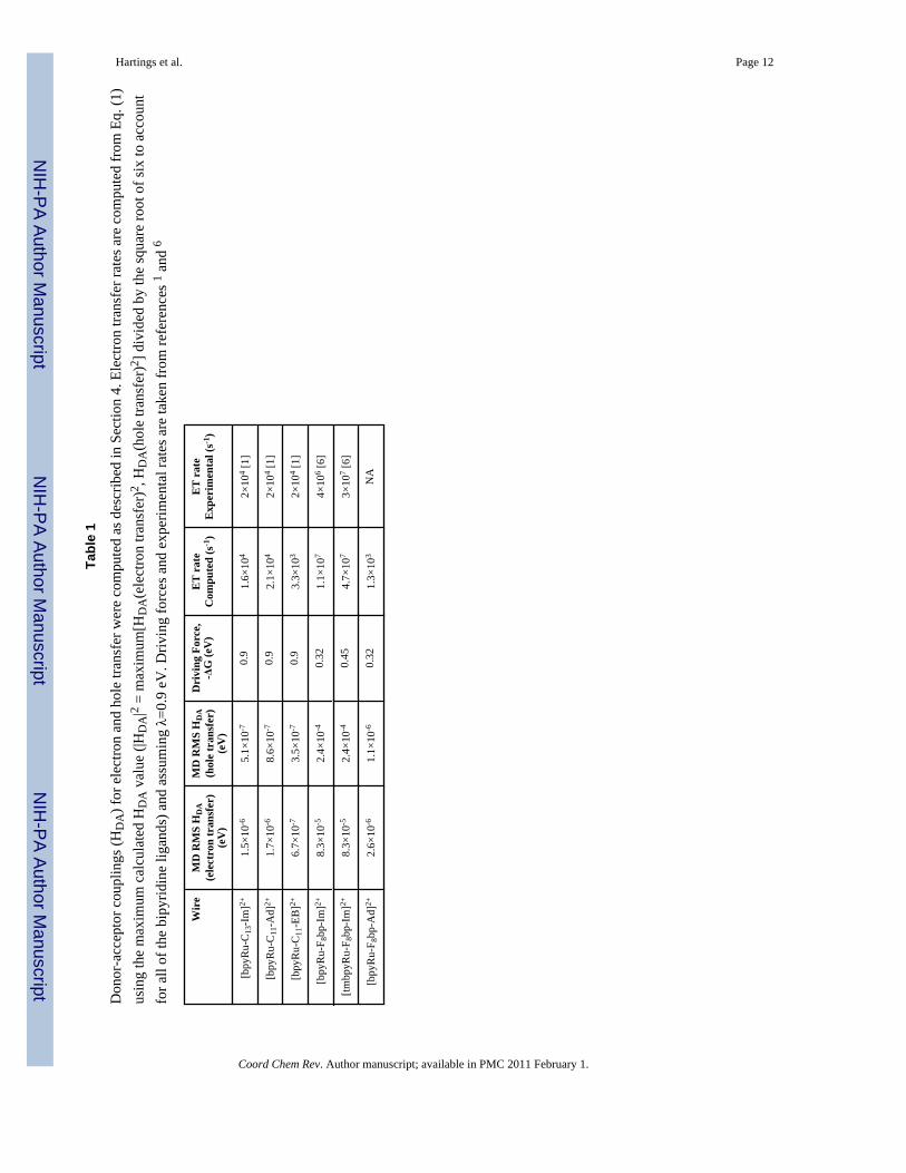
6. Bridge Orbital EnergiesOur Hartree-Fock calculated electronic couplings in Ru-wire-Fe-porphyrin systems used twodifferent charge states of the system and different models for the Ru-diimine donor. One modeldescribes superexchange interactions through occupied electronic states of the bridge (holetransfer), while the second model reproduces superexchange interactions through unoccupiedone-electron states of the bridge (electron-mediated superexchange). Table 1 gives the ETcouplings computed for dxy, dxz, and dyz orbitals of the Fe-porphyrin and an orbital representingthe donor (dihydropyrazine for electron transfer and pyridinium ion for hole transfer).
Electron transfer rates computed using the high-temperature nonadiabatic form of the Marcusexpression (1) [42-45] and donor-acceptor couplings obtained from the quantum chemicalcalculations are set out in Table 1 along with experimental values.
(1)
Hartings et al. Page 4
Coord Chem Rev. Author manuscript; available in PMC 2011 February 1.
NIH
-PA Author Manuscript
NIH
-PA Author Manuscript
NIH
-PA Author Manuscript

Rates averaged over thermally accessible geometries agree reasonably well (within a factor of4) with the observed rates in P450:wire conjugates (Table 1, Figure 4). For [bpyRu-F8bp-Im]2+ and [tmbpyRu-F8bp-Im]2+, superexchange interactions are dominated by couplingthrough unoccupied electronic states of the aromatic bridge (probably because of the inductiveeffect of F-substitution, which lowers bridge energy levels). For the saturated hydrocarbonbridge C13-Im, the main contributor to donor-acceptor coupling is hole superexchange. Thehole and electron contributions to the superexchange coupling differ by about a factor of two.
It is interesting to note that calculations predict slower electron transfer through the [bpyRu-F8bp-Ad]2+ wire. The Ru(II) excited state likely decays back to the ground state before electrontransfer can occur. There are two main differences between this wire and F8bp analogues. Thefirst is that it has an adamantyl terminal group that does not coordinate directly to the heme.The absence of terminal group-heme coordination likely reduces the overall donor-acceptorcoupling. The second difference is that coupling for [bpyRu-F8bp-Ad]2+ is primarily due tohole superexchange, as is the case for all of the sensitizer wires with aliphatic bridge groups.We calculate that [bpyRu-F8bp-Ad]2+ ET is slower than [bpyRu-C11-Ad]2+ ET, -ΔG0 is lessthan λ for [bpyRu-F8bp-Ad]2+, while -ΔG0 is equal to λ for [bpyRu-C11-Ad]2+.
We turn now to comparisons of our work on P450cam with related sensitizer-wire experimentsinvolving inducible nitric oxide synthase (iNOS) [7-12]. Photoinduced Fe(III) heme reductionin the iNOS systems is much faster than in P450cam even though the donor-acceptor distancesare similar (Table 2). For conjugates of iNOS with [Re-Im-F8bp-Im]+ and [Re-Im-C3-F8bp-Im]+, we proposed that the redox process is initiated by rapid reduction of Re(I)* by a nearbytryptophan residue, analogous to the first step in an experimentally validated azurin [Cu(I)-Trp-Re(I)*] hopping model system [8,46]. We also found that an iNOS Fe(III) heme can bereduced rapidly by a surface-bound [bpyRu-F9bp]2+ sensitizer wire (k = 2 × 10⁷ s-1) [12]. whichcould represent another case where multiple pathways couple the sensitizer to the edge of theheme [22].
7. Conclusions and the Path ForwardOur calculations of electron transfer in complexes of P450 with molecular wires account fordramatic differences in the rates through bridges of different chemical compositions, therebydemonstrating the predictive power of ab initio Hartree-Fock theory. We show that electronflow is modulated by a minority of accessible complex geometries with stronger donor-acceptor electronic couplings; therefore, inclusion of multiple complex geometries generatedin molecular dynamics simulations will be required for reliable theoretical estimates of electrontransfer rates in systems with structural flexibility. Comparison of donor-acceptor couplingvalues computed using energy splittings of occupied and unoccupied molecular orbitalsindicates that superexchange interactions through aromatic fluorinated-phenyl-imidazolebridges involve virtual reduced states (electron-type superexchange), while holesuperexchange accounts for the coupling of systems with aliphatic hydrocarbon bridges.
In work that is taking us in a new direction, we are employing polyfluorinated-phenyl side-binding sensitizer wires to phototrigger ET from a Ru(II)-diimine to an Fe(III) heme [12].These side-binding wires are very attractive for studying heme enzyme catalysis because theydo not block entry of substrates to the active center. We are currently following this lead byconstructing modified enzymes with sensitizers attached covalently to specific surfacepositions, as this will allow strategic placement of aromatic residues to control electron flowby facilitating multistep tunneling processes between donor and acceptor sites [8,46]).
Hartings et al. Page 5
Coord Chem Rev. Author manuscript; available in PMC 2011 February 1.
NIH
-PA Author Manuscript
NIH
-PA Author Manuscript
NIH
-PA Author Manuscript

AcknowledgmentsWe thank David Beratan for helpful discussions. This work was supported by NIH (GM078792 to MRH andDK019038 to HBG and GM068461 to JRW) and NSF (CHE-0802907 to HBG and JRW). MR thanks the MURIprogram of the AFOSR and the Chemistry Division of the NSF for support. This paper is dedicated to Ralph Pearson-friend, mentor, colleague, scientist and honest broker.
9. References[1]. Wilker JJ, Dmochowski IJ, Dawson JH, Winkler JR, Gray HB. Angew. Chem. Int. Ed 1999;38:90–
92.[2]. Dmochowski IJ, Crane BR, Wilker JJ, Winkler JR, Gray HB. Proc. Natl. Acad. Sci. USA
1999;96:12987–90. [PubMed: 10557259][3]. Dmochowski, IJ. Probing Cytochrome P450 with Sensitizer-linked Substrates. California Institute
of Technology; 2000.[4]. Dunn AR, Dmochowski IJ, Bilwes AM, Gray HB, Crane BR. Proc. Natl. Acad. Sci. USA
2001;98:12420–25. [PubMed: 11606730][5]. Dunn AR, Hays AM, Goodin DB, Stout CD, Chiu R, Winkler JR, Gray HB. J. Am. Chem. Soc
2002;124:10254–55. [PubMed: 12197708][6]. Dunn AR, Dmochowski IJ, Winkler JR, Gray HB. J. Am. Chem. Soc 2003;125:12450–56. [PubMed:
14531688][7]. Dunn AR, Belliston-Bittner W, Winkler JR, Getzoff ED, Stuehr DJ, Gray HB. J. Am. Chem. Soc
2005;127:5169–73. [PubMed: 15810851][8]. Belliston-Bittner W, Dunn AR, Nguyen YHL, Stuehr DJ, Winkler JR, Gray HB. J. Am. Chem. Soc
2005;127:15907–15. [PubMed: 16277534][9]. Contakes, SM.; Nguyen, YHL.; Gray, HB.; Glazer, EC.; Hays, AM.; Goodin, DB. Photofunctional
Transition Metals Complexes. Springer-Verlag Berlin; Berlin: 2007. p. 177-203.[10]. Nguyen YHL, Winkler JR, Gray HB. J. Phys. Chem. B 2007;111:6628–33. [PubMed: 17536854][11]. Whited CA, Belliston-Bittner W, Dunn AR, Winkler JR, Gray HB. J. Porphyrins Phthalocyanines
2008;12:971–78.[12]. Whited CA, Belliston-Bittner W, Dunn AR, Winkler JR, Gray HB. J. Inorg. Biochem 2009;103:906–
11. [PubMed: 19427703][13]. Graige MS, Feher G, Okamura MY. Proc. Natl. Acad. Sci. USA 1998;95:11679–84. [PubMed:
9751725][14]. Balabin IA, Onuchic JN. Science 2000;290:114–17. [PubMed: 11021791][15]. Berlin YA, Burin AL, Siebbeles LDA, Ratner MA. J. Phys. Chem. A 2001;105:5666–78.[16]. Davis WB, Ratner MA, Wasielewski MR. J. Am. Chem. Soc 2001;123:7877–86. [PubMed:
11493061][17]. Stuchebrukhov AA. Adv. Chem. Phys 2001;118:1–44.[18]. Troisi A, Nitzan A, Ratner MA. J. Chem. Phys 2003;119:5782–88.[19]. Troisi A, Ratner MA, Zimmt MB. J. Am. Chem. Soc 2004;126:2215–24. [PubMed: 14971957][20]. Weiss EA, Tauber MJ, Kelley RF, Ahrens MJ, Ratner MA, Wasielewski MR. J. Am. Chem. Soc
2005;127:11842–50. [PubMed: 16104763][21]. Valis L, Wang Q, Raytchev M, Buchvarov I, Wagenknecht HA, Fiebig T. Proc. Natl. Acad. Sci.
USA 2006;103:10192–95. [PubMed: 16801552][22]. Prytkova TR, Kurnikov IV, Beratan DN. Science 2007;315:622–25. [PubMed: 17272715][23]. Beratan DN, Skourtis SS, Balabin IA, Balaeff A, Keinan S, Venkatramani R, Xiao D. Acc. Chem.
Res. 2009 #. #.[24]. Balabin IA, Beratan DN, Skourtis SS. Phys. Rev. Lett 2008;101:158102. [PubMed: 18999647][25]. Skourtis, SS.; Lin, J.; Beratan, DN. Modern Methods for Theoretical Physical Chemistry of
Biopolymers. Starikov, EB.; Lewis, JP.; Tanaka, S., editors. Elsevier; Boston: 2006.[26]. Case, DA.; Pearlman, DA.; Cladwell, JW.; Cheatham, TE., III; Wang, J.; Ross, WS.; Simerling, C.;
Darden, T.; Merz, KM.; Stanton, RV.; Cheng, A.; J, VJ.; Crowley, M.; Tsui, V.; Gohlke, H.; Radmer,
Hartings et al. Page 6
Coord Chem Rev. Author manuscript; available in PMC 2011 February 1.
NIH
-PA Author Manuscript
NIH
-PA Author Manuscript
NIH
-PA Author Manuscript

R.; Duan, Y.; Pitera, J.; Massova, I.; Seibel, GL.; Singh, UC.; Weiner, P.; Kohlman, PA. AMBER7- Package of molecular simulation programs. Department of Pharmaceutical Chemistry, Universityof California at San Francisco; 2002.
[27]. Cornell WD, Cieplak P, Bayly CI, Gould IR, Merz KM, Ferguson DM, Spellmeyer DC, Fox T,Caldwell JW, Kollman PA. J. Am. Chem. Soc 1995;117:5179–97.
[28]. Besler BH, Merz KM, Kollman PA. J. Comput. Chem 1990;11:431–39.[29]. Larsson S. J. Chem. Soc. Farad. T. 2 1983;79:1375–88.[30]. Newton MD. Coord. Chem. Rev 2003;238:167–85.[31]. Prytkova TR, Kurnikov IV, Beratan DN. J. Phys. Chem. B 2005;109:1618–25. [PubMed: 16851133][32]. Paddon-Row MN, Jordan KD. J. Am. Chem. Soc 1993;115:2952–60.[33]. Berlin YA, Hutchison GR, Rempala P, Ratner MA, Michl J. J. Phys. Chem. A 2003;107:3970–80.[34]. Newton MD. Theor. Chem. Acc 2003;110:307–21.[35]. Newton MD. Chem. Rev 1991;91:767–92.[36]. Liang CX, Newton MD. J. Phys. Chem 1992;96:2855–66.[37]. Gunsalus IC, Meeks JR, Lipscomb JD. Ann. N.Y. Acad. Sci 1973;212:107–21. [PubMed: 4532472][38]. Pearson RG. J. Am. Chem. Soc 1986;108:6109–14.[39]. Sligar SG, Gunsalus IC. Proc. Natl. Acad. Sci. USA 1976;73:1078–82. [PubMed: 1063390][40]. Mayor M, Weber HB. Angew. Chem. Int. Ed 2004;43:2882–84.[41]. Basch H, Cohen R, Ratner MA. Nano Lett 2005;5:1668–75. [PubMed: 16159203][42]. Newton MD, Sutin N. Annu. Rev. Phys. Chem 1984;35:437–80.[43]. Marcus RA. Rev. Mod. Phys 1993;65:599–610.[44]. Barbara PF, Meyer TJ, Ratner MA. J. Phys. Chem 1996;100:13148–68.[45]. Gray HB, Winkler JR. Proc. Natl. Acad. Sci. USA 2005;102:3453–549. [PubMed: 15716358][46]. Shih C, Museth AK, Abrahamsson M, Blanco-Rodriguez AM, Di Bilio AJ, Sudhamsu J, Crane BR,
Ronayne KL, Towrie M, Vlcek A Jr. Richards JH, Winkler JR, Gray HB. Science 2008;320:1760–62. [PubMed: 18583608]
Hartings et al. Page 7
Coord Chem Rev. Author manuscript; available in PMC 2011 February 1.
NIH
-PA Author Manuscript
NIH
-PA Author Manuscript
NIH
-PA Author Manuscript

Figure 1.Sensitizer wires (top to bottom: bpyRu-C13-Im, bpyRu-C11-Ad, bpyRu-C11-Eb, bpyRu-F8bp-Im, tmbpyRu-F8bp-Im, and bpyRu-F8bp-Ad). Synthesis and characterization of these wiresare described in references 1 and 6.
Hartings et al. Page 8
Coord Chem Rev. Author manuscript; available in PMC 2011 February 1.
NIH
-PA Author Manuscript
NIH
-PA Author Manuscript
NIH
-PA Author Manuscript

Figure 2.Structure of bpyRu-C9-Ad bound to P450cam (pdb code: 1QMQ) [2].
Hartings et al. Page 9
Coord Chem Rev. Author manuscript; available in PMC 2011 February 1.
NIH
-PA Author Manuscript
NIH
-PA Author Manuscript
NIH
-PA Author Manuscript

Figure 3.Top: bpyRu-C13-Im conformations taken from snapshots along the MD simulation. This figureillustrates the large variety of structures available to a sensitizer wire. Bottom: Average donor-acceptor (pyrazine-Fe) electronic couplings (HDA) for different P450:bpyRu-C13-Im MDsnapshots. Calculated HDA values cover a range from 0.2 × 10⁻⁷ to around 2 × 10⁻⁷ atomicunits.
Hartings et al. Page 10
Coord Chem Rev. Author manuscript; available in PMC 2011 February 1.
NIH
-PA Author Manuscript
NIH
-PA Author Manuscript
NIH
-PA Author Manuscript

Figure 4.Experimental [1,6] and computed ET rates for P450:wires.
Hartings et al. Page 11
Coord Chem Rev. Author manuscript; available in PMC 2011 February 1.
NIH
-PA Author Manuscript
NIH
-PA Author Manuscript
NIH
-PA Author Manuscript
NIH
-PA Author Manuscript
NIH
-PA Author Manuscript
NIH
-PA Author Manuscript
Hartings et al. Page 12
Tabl
e 1
Don
or-a
ccep
tor c
oupl
ings
(HD
A) f
or e
lect
ron
and
hole
tran
sfer
wer
e co
mpu
ted
as d
escr
ibed
in S
ectio
n 4.
Ele
ctro
n tra
nsfe
r rat
es a
re c
ompu
ted
from
Eq.
(1)
usin
g th
e m
axim
um c
alcu
late
d H
DA
val
ue (|
HD
A|2 =
max
imum
[HD
A(e
lect
ron
trans
fer)
2 , H
DA
(hol
e tra
nsfe
r)2 ]
div
ided
by
the
squa
re ro
ot o
f six
to a
ccou
ntfo
r all
of th
e bi
pyrid
ine
ligan
ds) a
nd a
ssum
ing λ=
0.9
eV. D
rivin
g fo
rces
and
exp
erim
enta
l rat
es a
re ta
ken
from
refe
renc
es 1 a
nd 6
Wir
eM
D R
MS
HD
A(e
lect
ron
tran
sfer
)(e
V)
MD
RM
S H
DA
(hol
e tr
ansf
er)
(eV
)
Dri
ving
For
ce,
-ΔG
(eV
)E
T r
ate
Com
pute
d (s
-1)
ET
rat
eE
xper
imen
tal (
s-1)
[bpy
Ru-
C13
-Im
]2+1.
5×10
-65.
1×10
-70.
91.
6×10
42×
104 [
1]
[bpy
Ru-
C11
-Ad]
2+1.
7×10
-68.
6×10
-70.
92.
1×10
42×
104 [
1]
[bpy
Ru-
C11
-EB
]2+6.
7×10
-73.
5×10
-70.
93.
3×10
32×
104 [
1]
[bpy
Ru-
F 8bp
-Im
]2+8.
3×10
-52.
4×10
-40.
321.
1×10
74×
106 [
6]
[tmbp
yRu-
F 8bp
-Im
]2+8.
3×10
-52.
4×10
-40.
454.
7×10
73×
107 [
6]
[bpy
Ru-
F 8bp
-Ad]
2+2.
6×10
-61.
1×10
-60.
321.
3×10
3N
A
Coord Chem Rev. Author manuscript; available in PMC 2011 February 1.

NIH
-PA Author Manuscript
NIH
-PA Author Manuscript
NIH
-PA Author Manuscript
Hartings et al. Page 13
Table 2
Results of sensitizer-wire/protein experiments with cytochrome P450cam and iNOS. Superscripts denotereferences. Subscripts: (a) distances inferred from [Re-Im-F9bp]+; (b) the heme was reduced using a flash/quenchmethod; (c) the heme was reduced using a flash/quench method but a rate was not determined; (d) the heme wasreduced directly from Ru(II)* or Re(I)*
Wire RRu-Fe (Å) Rate of Heme Reduction (s-1)
cytochrome P450cam
[bpyRu-C11-Im]2+ NA NA
[bpyRu-C13-Im]2+ 21.2[2] 2×10⁴ [1]b
[bpyRu-C9-Ad]2+ 21.4[2] NA
[bpyRu-C11-Ad]2+ 21.0[2] 2×10⁴ [1]b
[bpyRu-C7-EB]2+ 19.5[2] 1×10³ [3]b
[bpyRu-C9-EB]2+ 19.4[2] NA [3]c
[bpyRu-C10-EB]2+ 19.9[2] NA [3]c
[bpyRu-C11-EB]2+ 20.1[2] 2×10⁴ [1]b
[bpyRu-C12-EB]2+ 20.5[2] NA [3]c
[bpyRu-C13-EB]2+ 20.6[2] NA [3]c
[bpyRu-F8bp-Im]2+ 22.1[2] 4.4×10⁶ [6]d
[tmbpyRu-F8bp-Im]2+ 18.1[6] 2.8×10⁷ [6]d
[Ru-F8bp-Ad]2+ 22.1[6] NA
[tmbpyRu-F9bp]2+ 17.0[6] NA
iNOS
[Re-Im-F8bp-Im]+ 18.0[7]a 7×10⁹ [8]d
[Re-Im-C3-F8bp-Im]+ 18.0[7]a 3×10⁹ [8]d
[Re-Im-C8-ArgNO2]+ 25.5[10] > 1×10⁶ [10]d
[Re-Im-F9bp]+ 18.0[7] NA
[tmbpyRu-F9bp]2+ 20.0[7] 2×10⁷ [12]b
Coord Chem Rev. Author manuscript; available in PMC 2011 February 1.