Electrochemical Detection Platform Based on RGO ... - MDPI
14
Citation: Chiticaru, E.A.; Pilan, L.; Ioni¸ t˘ a, M. Electrochemical Detection Platform Based on RGO Functionalized with Diazonium Salt for DNA Hybridization. Biosensors 2022, 12, 39. https://doi.org/ 10.3390/bios12010039 Received: 7 December 2021 Accepted: 10 January 2022 Published: 13 January 2022 Publisher’s Note: MDPI stays neutral with regard to jurisdictional claims in published maps and institutional affil- iations. Copyright: © 2022 by the authors. Licensee MDPI, Basel, Switzerland. This article is an open access article distributed under the terms and conditions of the Creative Commons Attribution (CC BY) license (https:// creativecommons.org/licenses/by/ 4.0/). biosensors Article Electrochemical Detection Platform Based on RGO Functionalized with Diazonium Salt for DNA Hybridization Elena A. Chiticaru 1 , Luisa Pilan 2, * and Mariana Ioni¸ tă 1,3, * 1 Faculty of Medical Engineering, University Politehnica of Bucharest, Gh Polizu 1-7, 011061 Bucharest, Romania; [email protected] 2 Department of Inorganic Chemistry, Physical Chemistry and Electrochemistry, University Politehnica of Bucharest, Gh Polizu 1-7, 011061 Bucharest, Romania 3 Advanced Polymer Materials Group, University Politehnica of Bucharest, Gh Polizu 1-7, 011061 Bucharest, Romania * Correspondence: [email protected] (L.P.); [email protected] (M.I.) Abstract: In this paper, we propose an improved electrochemical platform based on graphene for the detection of DNA hybridization. Commercial screen-printed carbon electrodes (SPCEs) were used for this purpose due to their ease of functionalization and miniaturization opportunities. SPCEs were modified with reduced graphene oxide (RGO), offering a suitable surface for further functionalization. Therefore, aryl-carboxyl groups were integrated onto RGO-modified electrodes by electrochemical reduction of the corresponding diazonium salt to provide enough reaction sites for the covalent immobilization of amino-modified DNA probes. Our final goal was to determine the optimum conditions needed to fabricate a simple, label-free RGO-based electrochemical platform to detect the hybridization between two complementary single-stranded DNA molecules. Each modification step in the fabrication process was monitored by cyclic voltammetry (CV) and electrochemical impedance spectroscopy (EIS) using [Fe(CN) 6 ] 3-/4- as a redox reporter. Although, the diazonium electrografted layer displayed the expected blocking effect of the charge transfer, the next steps in the modification procedure resulted in enhanced electron transfer properties of the electrode interface. We suggest that the improvement in the charge transfer after the DNA hybridization process could be exploited as a prospective sensing feature. The morphological and structural characterization of the modified electrodes performed by scanning electron microscopy (SEM) and Raman spectroscopy, respectively, were used to validate different modification steps in the platform fabrication process. Keywords: graphene; reduced graphene oxide; electrochemistry; electrochemical impedance spectroscopy; DNA biosensor; diazonium chemistry; screen-printed electrodes; DNA hybridization 1. Introduction Deoxyribonucleic acid (DNA) is a biopolymer that can self-assemble from two single- strands (ss) in a unique way that conforms to the Watson-Crick base pairing rules [1]. This process of specific self-assembly between two complementary polynucleotides is called hybridization and it is centered around the hydrogen bonds formed between the nucleobase base pairs, producing a double-helix structure [2]. The complementarity of DNA bases has been highly exploited in nanotechnology [3], in medicine [4] and in sensing applications [5,6]. The high specificity of the self-pairing between single-stranded DNA molecules can be used to detect specific sequences or biomarkers, which are related to bacterial [7] or viral [8] infections, for example. Moreover, the hybridization event can also be employed in the detection of DNA or messenger ribonucleic acid (mRNA) oligonu- cleotides, which can be extremely valuable in point-of-care applications [9], as well as for the diagnosis of various genetic mutations and diseases [10,11]. Electrochemical DNA biosensors are highly advantageous for the detection of par- ticular ssDNA molecules due to their fast response time, inexpensive instrumentation Biosensors 2022, 12, 39. https://doi.org/10.3390/bios12010039 https://www.mdpi.com/journal/biosensors
-
Upload
khangminh22 -
Category
Documents
-
view
1 -
download
0
Transcript of Electrochemical Detection Platform Based on RGO ... - MDPI
Citation: Chiticaru, E.A.; Pilan, L.;
Ionita, M. Electrochemical
Detection Platform Based on RGO
Functionalized with Diazonium Salt
for DNA Hybridization. Biosensors
2022, 12, 39. https://doi.org/
10.3390/bios12010039
Received: 7 December 2021
Accepted: 10 January 2022
Published: 13 January 2022
Publisher’s Note: MDPI stays neutral
with regard to jurisdictional claims in
published maps and institutional affil-
iations.
Copyright: © 2022 by the authors.
Licensee MDPI, Basel, Switzerland.
This article is an open access article
distributed under the terms and
conditions of the Creative Commons
Attribution (CC BY) license (https://
creativecommons.org/licenses/by/
4.0/).
biosensors
Article
Electrochemical Detection Platform Based on RGOFunctionalized with Diazonium Salt for DNA HybridizationElena A. Chiticaru 1 , Luisa Pilan 2,* and Mariana Ionită 1,3,*
1 Faculty of Medical Engineering, University Politehnica of Bucharest, Gh Polizu 1-7,011061 Bucharest, Romania; [email protected]
2 Department of Inorganic Chemistry, Physical Chemistry and Electrochemistry,University Politehnica of Bucharest, Gh Polizu 1-7, 011061 Bucharest, Romania
3 Advanced Polymer Materials Group, University Politehnica of Bucharest, Gh Polizu 1-7,011061 Bucharest, Romania
* Correspondence: [email protected] (L.P.); [email protected] (M.I.)
Abstract: In this paper, we propose an improved electrochemical platform based on graphene for thedetection of DNA hybridization. Commercial screen-printed carbon electrodes (SPCEs) were usedfor this purpose due to their ease of functionalization and miniaturization opportunities. SPCEs weremodified with reduced graphene oxide (RGO), offering a suitable surface for further functionalization.Therefore, aryl-carboxyl groups were integrated onto RGO-modified electrodes by electrochemicalreduction of the corresponding diazonium salt to provide enough reaction sites for the covalentimmobilization of amino-modified DNA probes. Our final goal was to determine the optimumconditions needed to fabricate a simple, label-free RGO-based electrochemical platform to detect thehybridization between two complementary single-stranded DNA molecules. Each modification stepin the fabrication process was monitored by cyclic voltammetry (CV) and electrochemical impedancespectroscopy (EIS) using [Fe(CN)6]3−/4− as a redox reporter. Although, the diazonium electrograftedlayer displayed the expected blocking effect of the charge transfer, the next steps in the modificationprocedure resulted in enhanced electron transfer properties of the electrode interface. We suggestthat the improvement in the charge transfer after the DNA hybridization process could be exploitedas a prospective sensing feature. The morphological and structural characterization of the modifiedelectrodes performed by scanning electron microscopy (SEM) and Raman spectroscopy, respectively,were used to validate different modification steps in the platform fabrication process.
Keywords: graphene; reduced graphene oxide; electrochemistry; electrochemical impedancespectroscopy; DNA biosensor; diazonium chemistry; screen-printed electrodes; DNA hybridization
1. Introduction
Deoxyribonucleic acid (DNA) is a biopolymer that can self-assemble from two single-strands (ss) in a unique way that conforms to the Watson−Crick base pairing rules [1].This process of specific self-assembly between two complementary polynucleotides iscalled hybridization and it is centered around the hydrogen bonds formed between thenucleobase base pairs, producing a double-helix structure [2]. The complementarity ofDNA bases has been highly exploited in nanotechnology [3], in medicine [4] and in sensingapplications [5,6]. The high specificity of the self-pairing between single-stranded DNAmolecules can be used to detect specific sequences or biomarkers, which are related tobacterial [7] or viral [8] infections, for example. Moreover, the hybridization event canalso be employed in the detection of DNA or messenger ribonucleic acid (mRNA) oligonu-cleotides, which can be extremely valuable in point-of-care applications [9], as well as forthe diagnosis of various genetic mutations and diseases [10,11].
Electrochemical DNA biosensors are highly advantageous for the detection of par-ticular ssDNA molecules due to their fast response time, inexpensive instrumentation
Biosensors 2022, 12, 39. https://doi.org/10.3390/bios12010039 https://www.mdpi.com/journal/biosensors
Biosensors 2022, 12, 39 2 of 14
and miniaturization potential [12]. Due to these advantages, DNA biosensors have beenused so far in various applications, such as clinical diagnostics [13], drug interactions [14]and detection [15], and environmental monitoring [16]. The development of simple andeffective electrochemical methods is of high interest in nucleic acid detection [17]. In thiscontext, label-free approaches have attracted widespread interest as they can simplify themanufacturing processes and are free from the possible unfavorable effects of the labels,which proves their effectiveness for target sensing [18,19]. Many of these label-free detec-tion strategies exploit the variation in the electrochemical signal of some charged freelydiffusing redox species as a result of the change in their accessibility to the electrode surfacedriven by the target analyte binding [20]. One of the most employed redox species is thenegatively charged [Fe(CN)6]3−/4−, and the hybridization events are detected by severalmethods, such as cyclic voltammetry (CV), differential pulse voltammetry (DPV), squarewave voltammetry (SWV), and electrochemical impedance spectroscopy (EIS).
The discovery of graphene and its related materials offers great opportunities forthe design of superior electrochemical DNA biosensors due to their unique properties,such as high surface area, which can increase the immobilization capacity, increase theelectron transfer rate, and can contribute to excellent biosensor sensitivity [21]. The func-tional groups present on the surface of graphene oxide (GO) can often be considered adisadvantage in sensing applications because they limit the electrical conductivity of thematerial. However, there are some strategies that exploit these functionalities by eitherremoving them through different approaches (chemical, electrochemical, thermal, etc.) [22]or by increasing their reactivity through carbodiimide chemistry [23] in order to be furthercovalently linked to amino-terminated molecules. As pointed out in the recent literature,reduced graphene oxide (RGO) material offers a multitude of advantages compared toother biosensing transducer materials by providing a balance between the beneficial prop-erties of pristine graphene and GO, and through its feasibility for various functionalizationschemes suitable for the biorecognition elements immobilization [24,25]. It was confirmedby different studies [26–28] that graphene functionalization is substantially enhanced whenthe reaction is performed under electrochemical control. The Fermi level of graphenecan be shifted by adjusting the applied electrochemical potential, which is a simpler wayto increase the reactivity of the material than using aggressive chemicals to disrupt thecovalent sp2 bonds [29]. Among numerous functionalization strategies, the electrochemicalreduction of diazonium salts [30–34] has been proposed as a versatile method that affordsthe grafting of a large variety of molecules at conductive substrates [35,36]. So far it hasbeen applied for different types of carbon-based substrates such as glassy carbon [37],screen-printed carbon [38], carbon paste [39], graphite pencil [40] or carbon nanomaterialselectrodes [41,42], that function as transducers in a large number of sensors for bioanalytes,such as proteins [43], DNA [44], RNA or cells [45]. Although great effort has been dedicatedto this subject, there is still no refined method available to mass-produce highly stable,efficient biosensors with real applicability in biomedicine. Therefore, our aim was to designan improved DNA detection platform in terms of stability, sensitivity and reproducibility,by modulating the electrochemical parameters.
In a previous study [25], we have developed an electrochemical platform for DNAdetection by physical adsorption of DNA probe (DNAp) molecules onto RGO-modified elec-trodes. Moreover, we have introduced a new protocol for the pretreatment of screen-printedcarbon electrodes (SPCEs) that were used in our work, which improved their electrochemi-cal properties. Considering that the immobilization of oligonucleotides through physicaladsorption does not permit a high level of precision and control for DNA attachment onthe substrate, we are proposing a new approach that consists in the covalent immobiliza-tion of amino-modified DNA probes onto a carboxylic functionalized RGO surface. Thisstrategy provides stronger and highly specific binding between the bioreceptor and theworking electrode by eliminating the possibility of DNA desorption [12], as opposed to thenon-covalent method [46,47]. The amino-modified ssDNA probes are covalently attachedat the RGO substrate through stable amide link via carbodiimide coupling reaction. Unlike
Biosensors 2022, 12, 39 3 of 14
other literature studies that report the same coupling chemistry for the biorecognitionelement at the carboxylic functions of the GO [48], this work also exploits the significantadvantages of diazonium chemistry to provide the stable and controlled functionalizationof the RGO electrode with aryl carboxylic groups (Ar-COOH) [46], with the implied ben-efits for ssDNA probe immobilization. A reproducible platform for DNA hybridizationdetection was envisaged by putting special emphasis on the electrochemically controlledfabrication steps for both the preparation of the RGO substrate electrode and the covalentbinding of the biorecognition element. In addition to the portability and cost-effectivenessof SPCEs, the reproducible modification of these substrates with RGO and the controlledcovalent binding of the biorecognition element can enable the fabrication of competitive,stable, reproducible and novel platforms for DNA hybridization detection. The effect of thesuccessive steps in the sensor fabrication process was established by the electrochemicalproperties of the modified electrodes quantified by the charge transfer resistance (Rct)and the electron transfer rate constant (k0) parameters. The electrochemical results werein agreement with morphological and structural characterization studies performed byscanning electron microscopy (SEM) and Raman spectroscopy, respectively. The improve-ment in the charge transfer after the DNA hybridization process could be exploited as aprospective sensing feature.
2. Materials and Methods2.1. Reagents and Materials
Graphene oxide dispersion in water (2 mg/mL), KCl, HCl, H2NaO4P, HNa2O4P, N-(3-Dimethylaminopropyl)-N′-ethylcarbodiimide hydrochloride (EDC), N-Hydroxysulfosuccinimide sodium salt (NHS), NaNO2 and 4-aminobenzoic acid (4-ABA) were supplied bySigma-Aldrich (St. Louis, MO, USA). Potassium ferricyanide (K3[Fe(CN)6]) and potassiumferrocyanide (K4[Fe(CN)6] × 3H2O) were purchased from Merck Co., (Darmstadt, Ger-many). Single-stranded DNA probe modified with amino groups (5′-AmC6-TTT CAA CATCAG TCT GAT AAG CTA TCT CCC-3′), single-stranded DNA target (DNAt, 5′-GGG AGATAG CTT ATC AGA CTG ATG TTG AAA-3′) and 10 mM Tris, 0.1 mM EDTA (IDTE) bufferwere acquired from Integrated DNA Technologies, Inc (Coralville, IA, USA). The SPCEswere thoroughly rinsed with ultrapure water (Adrona Crystal EX water purification sys-tem, 18.2 MΩ × cm resistivity) after each modification. Screen-printed carbon electrodes(SPCE-DRP 110) were purchased from Metrohm DropSens, Spain.
2.2. Procedures2.2.1. Electrochemical Measurements
The SPCEs consisted of a 4 mm diameter working electrode (WE), a silver pseudo-reference electrode (RE), and a carbon counter electrode (CE). Each SPCE modification wasinvestigated by cyclic voltammetry and electrochemical impedance spectroscopy, whichwere performed at room temperature using a potentiostat/galvanostat Autolab PGSTAT204 (Metrohm Autolab, Netherlands) equipped with NOVA 2.1 software. The SPCE wasattached to a connector (DSC) device from Metrohm Dropsens, functioning as an interfacebetween the SPCE and the potentiostat. The electrochemical measurements were recordedby adding 100 µL 0.1 M KCl electrolyte solution containing 1 mM [Fe(CN)6]3−/4− redoxprobe on the SPCE. CV curves were recorded by scanning the potential between −0.2 Vand +0.6 V, at a sweep of 0.05 V/s (unless stated otherwise). The impedance spectra wererecorded in the frequency range of 0.01–105 Hz, with 10 mV AC amplitude at an appliedpotential of 0.16 V, which is the formal potential of the [Fe(CN)6]4−/3− redox probe vs. theAg pseudo reference electrode.
2.2.2. Morphological and Structural Characterization
A FEI high-resolution focused ion beam scanning electron microscopy (FIB-SEM)system model Versa 3D DualBeam (FEI Company, Hillsboro, OR, USA) was used to charac-terize the selected sample series. The plane view (0 tilt) samples’ surface morphology were
Biosensors 2022, 12, 39 4 of 14
investigated by detecting the secondary electrons (SE) signals in High-Vacuum operationmode (6.1 × 10⁻⁴ Pa) at a working distance of 10 mm, using 10 kV as the acceleratingvoltage and a spot size of 4.5. Moreover, the SmartSCAN scanning strategy and DCFIdrift suppression features of Versa 3D DualBeam tool were involved to fully ensure theimaging stability.
Raman spectroscopy was performed in order to investigate the structural changes onthe surface of the electrodes. Raman spectra were obtained with a Renishaw inVia Ramanconfocal spectrometer (Renishaw, Brno-Cernovic, Czech Republic), using a laser excitationwavelength of 473 nm and the 100× objective.
2.2.3. RGO-Modified Electrode Preparation Procedure
The electrodes coated with electrochemically reduced graphene oxide (further denotedas RGO/SCPE) were prepared following the procedure described in [25]. Before anymodification, the SPCEs were pretreated by 5 voltametric cycles performed in 0.1 M HCl,from +0.5 to −1.5 V, at the scan rate of 0.05 V/s, followed by 5 cycles in 0.1 M phosphatebuffer solution (PBS), pH 7, from 0 to +2 V, 0.05 V/s. This activation treatment ensures ahigher electron transfer rate for the redox probe at SPCE, illustrated by a smaller peak topeak separation in the CV signal [25]. Subsequently, prior to the graphene modificationstep, 3 µL PBS was deposited on the WE surface, and then the electrodes were washedwith ultrapure water and dried at 60 C. As mentioned previously [25], this step is highlyimportant because it changes the surface properties of the carbon surface and it guaranteesa reproducible GO modification. Once the SPCEs cooled down, 3 µL of GO dispersion(0.3 mg/mL) was cast on their surface, then dried at 60 C for 2 h and kept at roomtemperature overnight. Finally, the GO/SPCEs were reduced electrochemically by 10 CVcycles applying a potential between 0 and −1.5 V, at a scan rate of 0.1 V/s, in 0.5 M KCl.The measurements were performed in triplicate in order to ensure the reproducibility ofthe procedure.
2.2.4. RGO/SPCE Functionalization by Diazonium Chemistry
The functionalization of RGO/SPCEs with carboxyphenyl layer was conducted within situ generated aryldiazonium cations that involved the electrochemical reduction ofthe corresponding aniline (4-aminobenzoic acid) in acidic media. Specifically, 2 mM 4-aminobenzoic acid was solubilized in 0.5 M aqueous HCl, to which 2 mM of sodium nitritewas added to generate the aryl diazonium salt. After 5 min stirring at room temperature,100 µL of mixture was immediately added onto the electrode surface, so as to cover allconnections. The electrochemical procedure employed for grafting was based on theprotocol described by Eissa et al. [49] and consisted in potential cycling for one cyclebetween +0.3 V and −0.6 V, with a scan rate of 0.1 V/s. At this stage, an irreversiblereduction peak should be observed as a preliminary validation of the functionalizationprocess. After surface derivatization, the electrodes were rinsed with high amounts ofultrapure water. The blocking properties of the modified electrodes evaluated by CV andEIS in the presence of soluble electroactive [Fe(CN)6]3−/4− species also confirmed thisgrafting step.
2.2.5. Fabrication of DNA Biosensor
The DNA biosensor was fabricated by covalently immobilizing the amino ssDNAprobe onto RGO/SPCE using carbodiimide chemistry. For this purpose, 0.2 M EDC and0.05 M NHS in 0.1 M PBS buffer solution of pH 6, were used as activation agents for thecarboxyl groups confined at the electrode surface. After 2 h activation at room temperature,the Ar-COOH/RGO/SPCEs were immersed overnight in the ssDNA probe solutions atroom temperature to obtain the detecting electrodes. Fresh DNA probes were preparedfrom the stock solution of 100 µM to 10, 5, 1, and 0.5 µM using IDTE buffer to dilute.The hybridization testing of the developed platform for the complementary target DNAsequence was achieved by dropping an appropriate concentration of target DNA solution
Biosensors 2022, 12, 39 5 of 14
(in IDTE buffer) onto the SPCE modified with the recognition layer and incubating atroom temperature for 2 h. For longer times (i.e., 4 h and 6 h), no significant change inthe electrochemical studies occurred, indicating that the ssDNA probes immobilized ontoSPCE were completely hybridized by the target DNA after 2 h.
3. Results and Discussions3.1. Morphological Characterization
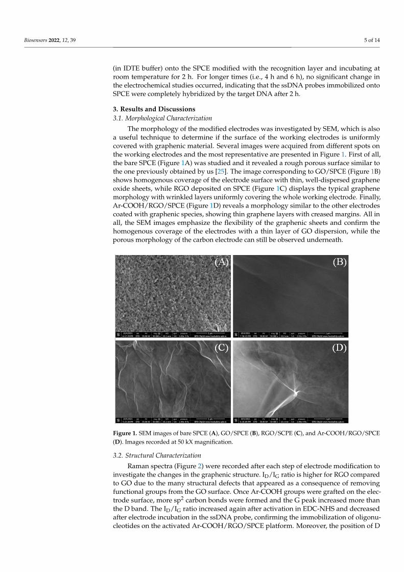
The morphology of the modified electrodes was investigated by SEM, which is alsoa useful technique to determine if the surface of the working electrodes is uniformlycovered with graphenic material. Several images were acquired from different spots onthe working electrodes and the most representative are presented in Figure 1. First of all,the bare SPCE (Figure 1A) was studied and it revealed a rough porous surface similar tothe one previously obtained by us [25]. The image corresponding to GO/SPCE (Figure 1B)shows homogenous coverage of the electrode surface with thin, well-dispersed grapheneoxide sheets, while RGO deposited on SPCE (Figure 1C) displays the typical graphenemorphology with wrinkled layers uniformly covering the whole working electrode. Finally,Ar-COOH/RGO/SPCE (Figure 1D) reveals a morphology similar to the other electrodescoated with graphenic species, showing thin graphene layers with creased margins. All inall, the SEM images emphasize the flexibility of the graphenic sheets and confirm thehomogenous coverage of the electrodes with a thin layer of GO dispersion, while theporous morphology of the carbon electrode can still be observed underneath.
Biosensors 2022, 12, x FOR PEER REVIEW 5 of 14
at room temperature to obtain the detecting electrodes. Fresh DNA probes were prepared from the stock solution of 100 µM to 10, 5, 1, and 0.5 µM using IDTE buffer to dilute. The hybridization testing of the developed platform for the complementary target DNA se-quence was achieved by dropping an appropriate concentration of target DNA solution (in IDTE buffer) onto the SPCE modified with the recognition layer and incubating at room temperature for 2 h. For longer times (i.e., 4 h and 6 h), no significant change in the electrochemical studies occurred, indicating that the ssDNA probes immobilized onto SPCE were completely hybridized by the target DNA after 2 h.
3. Results and Discussions 3.1. Morphological Characterization
The morphology of the modified electrodes was investigated by SEM, which is also a useful technique to determine if the surface of the working electrodes is uniformly cov-ered with graphenic material. Several images were acquired from different spots on the working electrodes and the most representative are presented in Figure 1. First of all, the bare SPCE (Figure 1A) was studied and it revealed a rough porous surface similar to the one previously obtained by us [25]. The image corresponding to GO/SPCE (Figure 1B) shows homogenous coverage of the electrode surface with thin, well-dispersed graphene oxide sheets, while RGO deposited on SPCE (Figure 1C) displays the typical graphene morphology with wrinkled layers uniformly covering the whole working electrode. Fi-nally, Ar-COOH/RGO/SPCE (Figure 1D) reveals a morphology similar to the other elec-trodes coated with graphenic species, showing thin graphene layers with creased margins. All in all, the SEM images emphasize the flexibility of the graphenic sheets and confirm the homogenous coverage of the electrodes with a thin layer of GO dispersion, while the porous morphology of the carbon electrode can still be observed underneath.
Figure 1. SEM images of bare SPCE (A), GO/SPCE (B), RGO/SCPE (C), and Ar-COOH/RGO/SPCE (D). Images recorded at 50 kX magnification.
3.2. Structural Characterization Raman spectra (Figure 2) were recorded after each step of electrode modification to
investigate the changes in the graphenic structure. ID/IG ratio is higher for RGO compared to GO due to the many structural defects that appeared as a consequence of removing
Figure 1. SEM images of bare SPCE (A), GO/SPCE (B), RGO/SCPE (C), and Ar-COOH/RGO/SPCE(D). Images recorded at 50 kX magnification.
3.2. Structural Characterization
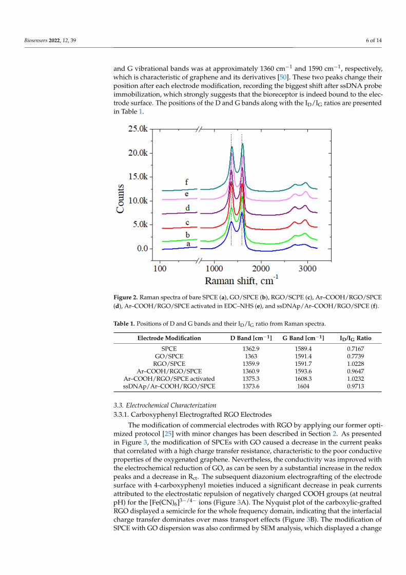
Raman spectra (Figure 2) were recorded after each step of electrode modification toinvestigate the changes in the graphenic structure. ID/IG ratio is higher for RGO comparedto GO due to the many structural defects that appeared as a consequence of removingfunctional groups from the GO surface. Once Ar-COOH groups were grafted on the elec-trode surface, more sp2 carbon bonds were formed and the G peak increased more thanthe D band. The ID/IG ratio increased again after activation in EDC-NHS and decreasedafter electrode incubation in the ssDNA probe, confirming the immobilization of oligonu-cleotides on the activated Ar-COOH/RGO/SPCE platform. Moreover, the position of D
Biosensors 2022, 12, 39 6 of 14
and G vibrational bands was at approximately 1360 cm−1 and 1590 cm−1, respectively,which is characteristic of graphene and its derivatives [50]. These two peaks change theirposition after each electrode modification, recording the biggest shift after ssDNA probeimmobilization, which strongly suggests that the bioreceptor is indeed bound to the elec-trode surface. The positions of the D and G bands along with the ID/IG ratios are presentedin Table 1.
Biosensors 2022, 12, x FOR PEER REVIEW 6 of 14
functional groups from the GO surface. Once Ar-COOH groups were grafted on the elec-trode surface, more sp2 carbon bonds were formed and the G peak increased more than the D band. The ID/IG ratio increased again after activation in EDC-NHS and decreased after electrode incubation in the ssDNA probe, confirming the immobilization of oligonu-cleotides on the activated Ar-COOH/RGO/SPCE platform. Moreover, the position of D and G vibrational bands was at approximately 1360 cm−1 and 1590 cm−1, respectively, which is characteristic of graphene and its derivatives [50]. These two peaks change their position after each electrode modification, recording the biggest shift after ssDNA probe immobilization, which strongly suggests that the bioreceptor is indeed bound to the elec-trode surface. The positions of the D and G bands along with the ID/IG ratios are presented in Table 1.
Figure 2. Raman spectra of bare SPCE (a), GO/SPCE (b), RGO/SCPE (c), Ar–COOH/RGO/SPCE (d), Ar–COOH/RGO/SPCE activated in EDC–NHS (e), and ssDNAp/Ar–COOH/RGO/SPCE (f).
Table 1. Positions of D and G bands and their ID/IG ratio from Raman spectra.
Electrode Modification D Band [cm−1] G Band [cm−1] ID/IG Ratio SPCE 1362.9 1589.4 0.7167
GO/SPCE 1363 1591.4 0.7739 RGO/SPCE 1359.9 1591.7 1.0228
Ar–COOH/RGO/SPCE 1360.9 1593.6 0.9647 Ar–COOH/RGO/SPCE activated 1375.3 1608.3 1.0232 ssDNAp/Ar–COOH/RGO/SPCE 1373.6 1604 0.9713
3.3. Electrochemical Characterization 3.3.1. Carboxyphenyl Electrografted RGO Electrodes
The modification of commercial electrodes with RGO by applying our former opti-mized protocol [25] with minor changes has been described in Section 2. As presented in Figure 3, the modification of SPCEs with GO caused a decrease in the current peaks that correlated with a high charge transfer resistance, characteristic to the poor conductive properties of the oxygenated graphene. Nevertheless, the conductivity was improved with the electrochemical reduction of GO, as can be seen by a substantial increase in the redox peaks and a decrease in Rct. The subsequent diazonium electrografting of the elec-
Figure 2. Raman spectra of bare SPCE (a), GO/SPCE (b), RGO/SCPE (c), Ar–COOH/RGO/SPCE(d), Ar–COOH/RGO/SPCE activated in EDC–NHS (e), and ssDNAp/Ar–COOH/RGO/SPCE (f).
Table 1. Positions of D and G bands and their ID/IG ratio from Raman spectra.
Electrode Modification D Band [cm−1] G Band [cm−1] ID/IG Ratio
SPCE 1362.9 1589.4 0.7167GO/SPCE 1363 1591.4 0.7739
RGO/SPCE 1359.9 1591.7 1.0228Ar–COOH/RGO/SPCE 1360.9 1593.6 0.9647
Ar–COOH/RGO/SPCE activated 1375.3 1608.3 1.0232ssDNAp/Ar–COOH/RGO/SPCE 1373.6 1604 0.9713
3.3. Electrochemical Characterization3.3.1. Carboxyphenyl Electrografted RGO Electrodes
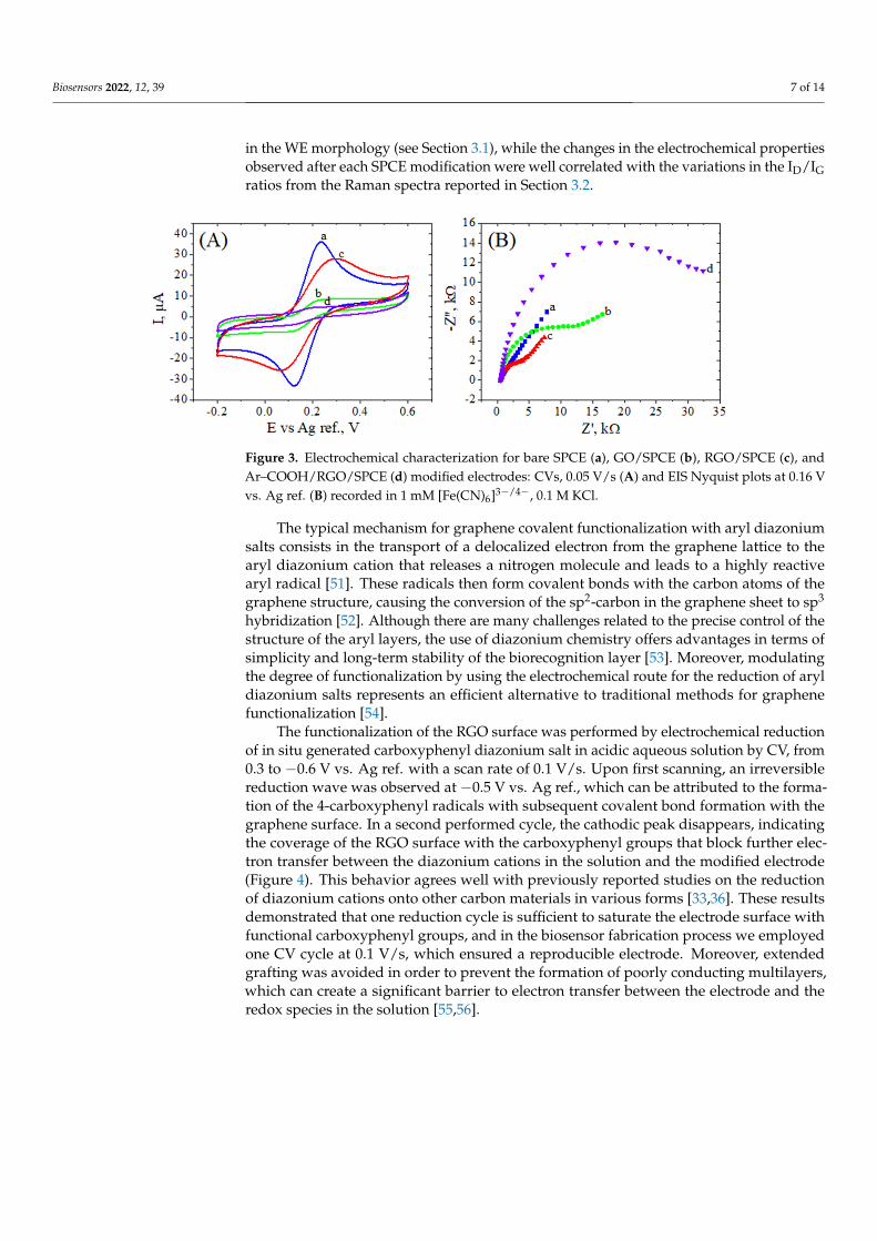
The modification of commercial electrodes with RGO by applying our former opti-mized protocol [25] with minor changes has been described in Section 2. As presentedin Figure 3, the modification of SPCEs with GO caused a decrease in the current peaksthat correlated with a high charge transfer resistance, characteristic to the poor conductiveproperties of the oxygenated graphene. Nevertheless, the conductivity was improved withthe electrochemical reduction of GO, as can be seen by a substantial increase in the redoxpeaks and a decrease in Rct. The subsequent diazonium electrografting of the electrodesurface with 4-carboxyphenyl moieties induced a significant decrease in peak currentsattributed to the electrostatic repulsion of negatively charged COOH groups (at neutralpH) for the [Fe(CN)6]3−/4− ions (Figure 3A). The Nyquist plot of the carboxylic-graftedRGO displayed a semicircle for the whole frequency domain, indicating that the interfacialcharge transfer dominates over mass transport effects (Figure 3B). The modification ofSPCE with GO dispersion was also confirmed by SEM analysis, which displayed a change
Biosensors 2022, 12, 39 7 of 14
in the WE morphology (see Section 3.1), while the changes in the electrochemical propertiesobserved after each SPCE modification were well correlated with the variations in the ID/IGratios from the Raman spectra reported in Section 3.2.
Biosensors 2022, 12, x FOR PEER REVIEW 7 of 14
trode surface with 4-carboxyphenyl moieties induced a significant decrease in peak cur-rents attributed to the electrostatic repulsion of negatively charged COOH groups (at neu-tral pH) for the [Fe(CN)6]3−/4− ions (Figure 3A). The Nyquist plot of the carboxylic-grafted RGO displayed a semicircle for the whole frequency domain, indicating that the interfacial charge transfer dominates over mass transport effects (Figure 3B). The modification of SPCE with GO dispersion was also confirmed by SEM analysis, which displayed a change in the WE morphology (see Section 3.1), while the changes in the electrochemical proper-ties observed after each SPCE modification were well correlated with the variations in the ID/IG ratios from the Raman spectra reported in Section 3.2.
Figure 3. Electrochemical characterization for bare SPCE (a), GO/SPCE (b), RGO/SPCE (c), and Ar–COOH/RGO/SPCE (d) modified electrodes: CVs, 0.05 V/s (A) and EIS Nyquist plots at 0.16 V vs. Ag ref. (B) recorded in 1 mM [Fe(CN)6]3−/4−, 0.1M KCl.
The typical mechanism for graphene covalent functionalization with aryl diazonium salts consists in the transport of a delocalized electron from the graphene lattice to the aryl diazonium cation that releases a nitrogen molecule and leads to a highly reactive aryl rad-ical [51]. These radicals then form covalent bonds with the carbon atoms of the graphene structure, causing the conversion of the sp2-carbon in the graphene sheet to sp3 hybridiza-tion [52]. Although there are many challenges related to the precise control of the structure of the aryl layers, the use of diazonium chemistry offers advantages in terms of simplicity and long-term stability of the biorecognition layer [53]. Moreover, modulating the degree of functionalization by using the electrochemical route for the reduction of aryl diazonium salts represents an efficient alternative to traditional methods for graphene functionaliza-tion [54].
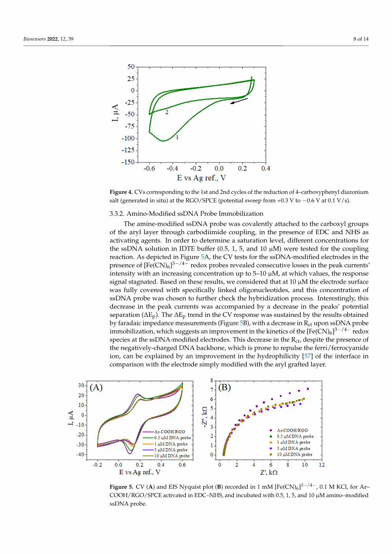
The functionalization of the RGO surface was performed by electrochemical reduc-tion of in situ generated carboxyphenyl diazonium salt in acidic aqueous solution by CV, from 0.3 to −0.6 V vs. Ag ref. with a scan rate of 0.1 V/s. Upon first scanning, an irreversible reduction wave was observed at −0.5 V vs. Ag ref., which can be attributed to the for-mation of the 4-carboxyphenyl radicals with subsequent covalent bond formation with the graphene surface. In a second performed cycle, the cathodic peak disappears, indicat-ing the coverage of the RGO surface with the carboxyphenyl groups that block further electron transfer between the diazonium cations in the solution and the modified elec-trode (Figure 4). This behavior agrees well with previously reported studies on the reduc-tion of diazonium cations onto other carbon materials in various forms [33,36]. These re-sults demonstrated that one reduction cycle is sufficient to saturate the electrode surface with functional carboxyphenyl groups, and in the biosensor fabrication process we em-ployed one CV cycle at 0.1 V/s, which ensured a reproducible electrode. Moreover, ex-tended grafting was avoided in order to prevent the formation of poorly conducting mul-tilayers, which can create a significant barrier to electron transfer between the electrode and the redox species in the solution [55,56].
Figure 3. Electrochemical characterization for bare SPCE (a), GO/SPCE (b), RGO/SPCE (c), andAr–COOH/RGO/SPCE (d) modified electrodes: CVs, 0.05 V/s (A) and EIS Nyquist plots at 0.16 Vvs. Ag ref. (B) recorded in 1 mM [Fe(CN)6]3−/4−, 0.1 M KCl.
The typical mechanism for graphene covalent functionalization with aryl diazoniumsalts consists in the transport of a delocalized electron from the graphene lattice to thearyl diazonium cation that releases a nitrogen molecule and leads to a highly reactivearyl radical [51]. These radicals then form covalent bonds with the carbon atoms of thegraphene structure, causing the conversion of the sp2-carbon in the graphene sheet to sp3
hybridization [52]. Although there are many challenges related to the precise control of thestructure of the aryl layers, the use of diazonium chemistry offers advantages in terms ofsimplicity and long-term stability of the biorecognition layer [53]. Moreover, modulatingthe degree of functionalization by using the electrochemical route for the reduction of aryldiazonium salts represents an efficient alternative to traditional methods for graphenefunctionalization [54].
The functionalization of the RGO surface was performed by electrochemical reductionof in situ generated carboxyphenyl diazonium salt in acidic aqueous solution by CV, from0.3 to −0.6 V vs. Ag ref. with a scan rate of 0.1 V/s. Upon first scanning, an irreversiblereduction wave was observed at −0.5 V vs. Ag ref., which can be attributed to the forma-tion of the 4-carboxyphenyl radicals with subsequent covalent bond formation with thegraphene surface. In a second performed cycle, the cathodic peak disappears, indicatingthe coverage of the RGO surface with the carboxyphenyl groups that block further elec-tron transfer between the diazonium cations in the solution and the modified electrode(Figure 4). This behavior agrees well with previously reported studies on the reductionof diazonium cations onto other carbon materials in various forms [33,36]. These resultsdemonstrated that one reduction cycle is sufficient to saturate the electrode surface withfunctional carboxyphenyl groups, and in the biosensor fabrication process we employedone CV cycle at 0.1 V/s, which ensured a reproducible electrode. Moreover, extendedgrafting was avoided in order to prevent the formation of poorly conducting multilayers,which can create a significant barrier to electron transfer between the electrode and theredox species in the solution [55,56].
Biosensors 2022, 12, 39 8 of 14Biosensors 2022, 12, x FOR PEER REVIEW 8 of 14
Figure 4. CVs corresponding to the 1st and 2nd cycles of the reduction of 4–carboxyphenyl diazo-nium salt (generated in situ) at the RGO/SPCE (potential sweep from +0.3 V to –0.6 V at 0.1 V/s).
3.3.2. Amino-Modified ssDNA Probe Immobilization The amine-modified ssDNA probe was covalently attached to the carboxyl groups of
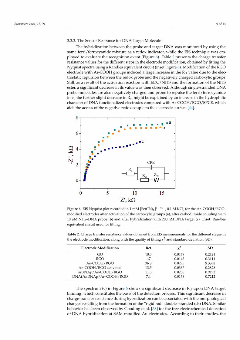
the aryl layer through carbodiimide coupling, in the presence of EDC and NHS as activat-ing agents. In order to determine a saturation level, different concentrations for the ssDNA solution in IDTE buffer (0.5, 1, 5, and 10 µM) were tested for the coupling reaction. As depicted in Figure 5A, the CV tests for the ssDNA-modified electrodes in the presence of [Fe(CN)6]3−/4− redox probes revealed consecutive losses in the peak currents’ intensity with an increasing concentration up to 5–10 µM, at which values, the response signal stagnated. Based on these results, we considered that at 10 µM the electrode surface was fully cov-ered with specifically linked oligonucleotides, and this concentration of ssDNA probe was chosen to further check the hybridization process. Interestingly, this decrease in the peak currents was accompanied by a decrease in the peaks’ potential separation (ΔEp). The ΔEp trend in the CV response was sustained by the results obtained by faradaic impedance measurements (Figure 5B), with a decrease in Rct upon ssDNA probe immobilization, which suggests an improvement in the kinetics of the [Fe(CN)6]3−/4− redox species at the ssDNA-modified electrodes. This decrease in the Rct, despite the presence of the nega-tively-charged DNA backbone, which is prone to repulse the ferri/ferrocyanide ion, can be explained by an improvement in the hydrophilicity [57] of the interface in comparison with the electrode simply modified with the aryl grafted layer.
Figure 5. CV (A) and EIS Nyquist plot (B) recorded in 1 mM [Fe(CN)6]3−/4−, 0.1M KCl, for Ar–COOH/RGO/SPCE activated in EDC–NHS, and incubated with 0.5, 1, 5, and 10 µM amino–modified ssDNA probe.
Figure 4. CVs corresponding to the 1st and 2nd cycles of the reduction of 4–carboxyphenyl diazoniumsalt (generated in situ) at the RGO/SPCE (potential sweep from +0.3 V to −0.6 V at 0.1 V/s).
3.3.2. Amino-Modified ssDNA Probe Immobilization
The amine-modified ssDNA probe was covalently attached to the carboxyl groupsof the aryl layer through carbodiimide coupling, in the presence of EDC and NHS asactivating agents. In order to determine a saturation level, different concentrations forthe ssDNA solution in IDTE buffer (0.5, 1, 5, and 10 µM) were tested for the couplingreaction. As depicted in Figure 5A, the CV tests for the ssDNA-modified electrodes in thepresence of [Fe(CN)6]3−/4− redox probes revealed consecutive losses in the peak currents’intensity with an increasing concentration up to 5–10 µM, at which values, the responsesignal stagnated. Based on these results, we considered that at 10 µM the electrode surfacewas fully covered with specifically linked oligonucleotides, and this concentration ofssDNA probe was chosen to further check the hybridization process. Interestingly, thisdecrease in the peak currents was accompanied by a decrease in the peaks’ potentialseparation (∆Ep). The ∆Ep trend in the CV response was sustained by the results obtainedby faradaic impedance measurements (Figure 5B), with a decrease in Rct upon ssDNA probeimmobilization, which suggests an improvement in the kinetics of the [Fe(CN)6]3−/4− redoxspecies at the ssDNA-modified electrodes. This decrease in the Rct, despite the presence ofthe negatively-charged DNA backbone, which is prone to repulse the ferri/ferrocyanideion, can be explained by an improvement in the hydrophilicity [57] of the interface incomparison with the electrode simply modified with the aryl grafted layer.
Biosensors 2022, 12, x FOR PEER REVIEW 8 of 14
Figure 4. CVs corresponding to the 1st and 2nd cycles of the reduction of 4–carboxyphenyl diazo-nium salt (generated in situ) at the RGO/SPCE (potential sweep from +0.3 V to –0.6 V at 0.1 V/s).
3.3.2. Amino-Modified ssDNA Probe Immobilization The amine-modified ssDNA probe was covalently attached to the carboxyl groups of
the aryl layer through carbodiimide coupling, in the presence of EDC and NHS as activat-ing agents. In order to determine a saturation level, different concentrations for the ssDNA solution in IDTE buffer (0.5, 1, 5, and 10 µM) were tested for the coupling reaction. As depicted in Figure 5A, the CV tests for the ssDNA-modified electrodes in the presence of [Fe(CN)6]3−/4− redox probes revealed consecutive losses in the peak currents’ intensity with an increasing concentration up to 5–10 µM, at which values, the response signal stagnated. Based on these results, we considered that at 10 µM the electrode surface was fully cov-ered with specifically linked oligonucleotides, and this concentration of ssDNA probe was chosen to further check the hybridization process. Interestingly, this decrease in the peak currents was accompanied by a decrease in the peaks’ potential separation (ΔEp). The ΔEp trend in the CV response was sustained by the results obtained by faradaic impedance measurements (Figure 5B), with a decrease in Rct upon ssDNA probe immobilization, which suggests an improvement in the kinetics of the [Fe(CN)6]3−/4− redox species at the ssDNA-modified electrodes. This decrease in the Rct, despite the presence of the nega-tively-charged DNA backbone, which is prone to repulse the ferri/ferrocyanide ion, can be explained by an improvement in the hydrophilicity [57] of the interface in comparison with the electrode simply modified with the aryl grafted layer.
Figure 5. CV (A) and EIS Nyquist plot (B) recorded in 1 mM [Fe(CN)6]3−/4−, 0.1M KCl, for Ar–COOH/RGO/SPCE activated in EDC–NHS, and incubated with 0.5, 1, 5, and 10 µM amino–modified ssDNA probe.
Figure 5. CV (A) and EIS Nyquist plot (B) recorded in 1 mM [Fe(CN)6]3−/4−, 0.1 M KCl, for Ar–COOH/RGO/SPCE activated in EDC–NHS, and incubated with 0.5, 1, 5, and 10 µM amino–modifiedssDNA probe.
Biosensors 2022, 12, 39 9 of 14
3.3.3. The Sensor Response for DNA Target Molecule
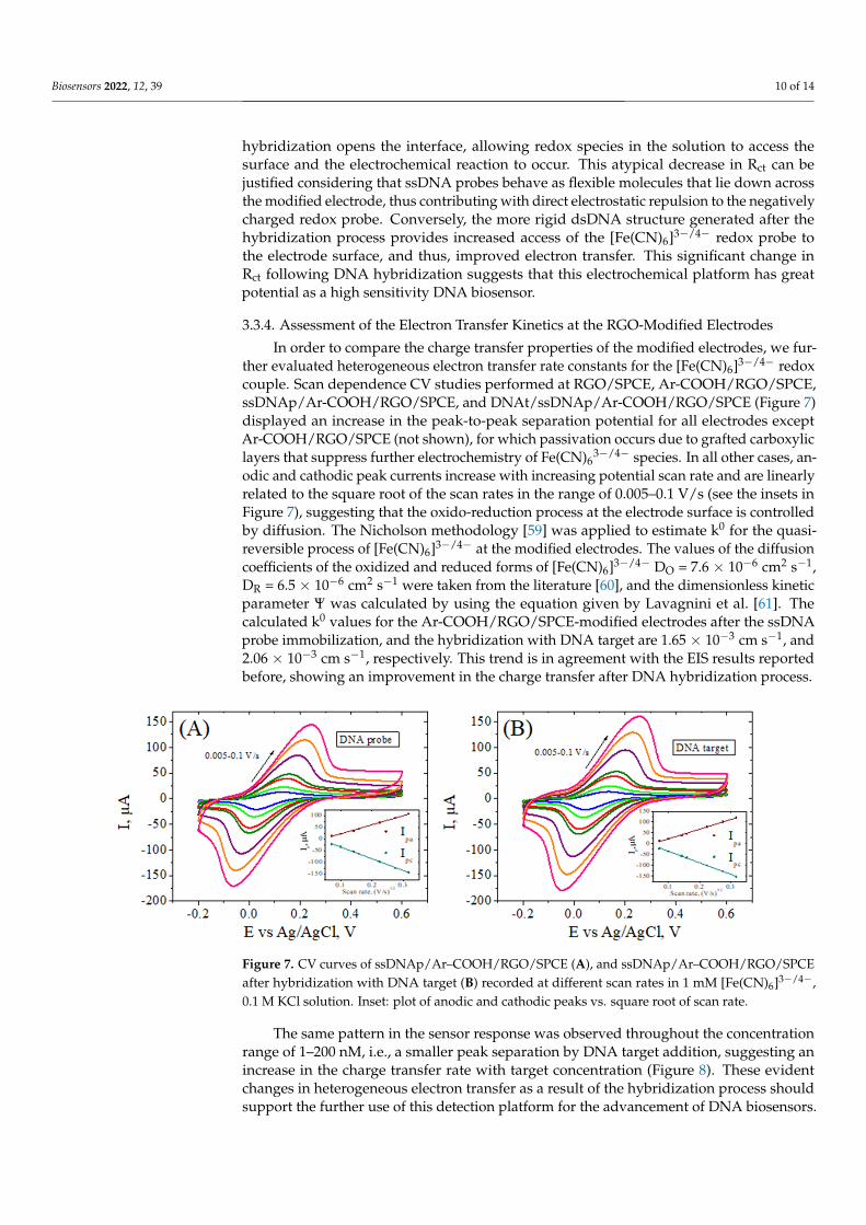
The hybridization between the probe and target DNA was monitored by using thesame ferri/ferrocyanide mixture as a redox indicator, while the EIS technique was em-ployed to evaluate the recognition event (Figure 6). Table 2 presents the charge transferresistance values for the different steps in the electrode modification, obtained by fitting theNyquist spectra using a Randles equivalent circuit (inset Figure 6). Modification of the RGOelectrode with Ar-COOH groups induced a large increase in the Rct value due to the elec-trostatic repulsion between the redox probe and the negatively charged carboxylic groups.Still, as a result of the activation reaction with EDC/NHS and the formation of the NHSester, a significant decrease in its value was then observed. Although single-stranded DNAprobe molecules are also negatively charged and prone to repulse the ferri/ferrocyanideions, the further slight decrease in Rct might be explained by an increase in the hydrophiliccharacter of DNA functionalized electrodes compared with Ar-COOH/RGO/SPCE, whichaids the access of the negative redox couple to the electrode surface [44].
Biosensors 2022, 12, x FOR PEER REVIEW 9 of 14
3.3.3. The Sensor Response for DNA Target Molecule The hybridization between the probe and target DNA was monitored by using the
same ferri/ferrocyanide mixture as a redox indicator, while the EIS technique was em-ployed to evaluate the recognition event (Figure 6). Table 2 presents the charge transfer resistance values for the different steps in the electrode modification, obtained by fitting the Nyquist spectra using a Randles equivalent circuit (inset Figure 6). Modification of the RGO electrode with Ar-COOH groups induced a large increase in the Rct value due to the electrostatic repulsion between the redox probe and the negatively charged carboxylic groups. Still, as a result of the activation reaction with EDC/NHS and the formation of the NHS ester, a significant decrease in its value was then observed. Although single-stranded DNA probe molecules are also negatively charged and prone to repulse the ferri/ferrocy-anide ions, the further slight decrease in Rct might be explained by an increase in the hy-drophilic character of DNA functionalized electrodes compared with Ar-COOH/RGO/SPCE, which aids the access of the negative redox couple to the electrode surface [44].
The spectrum (c) in Figure 6 shows a significant decrease in Rct upon DNA target binding, which constitutes the basis of the detection process. This significant decrease in charge-transfer resistance during hybridization can be associated with the morphological changes resulting from the formation of the “rigid rod” double stranded (ds) DNA. Simi-lar behavior has been observed by Gooding et al. [58] for the free electrochemical detection of DNA hybridization at SAM-modified Au electrodes. According to their studies, the hybridization opens the interface, allowing redox species in the solution to access the sur-face and the electrochemical reaction to occur. This atypical decrease in Rct can be justified considering that ssDNA probes behave as flexible molecules that lie down across the mod-ified electrode, thus contributing with direct electrostatic repulsion to the negatively charged redox probe. Conversely, the more rigid dsDNA structure generated after the hybridization process provides increased access of the [Fe(CN)6]3−/4− redox probe to the electrode surface, and thus, improved electron transfer. This significant change in Rct fol-lowing DNA hybridization suggests that this electrochemical platform has great potential as a high sensitivity DNA biosensor.
Figure 6. EIS Nyquist plot recorded in 1 mM [Fe(CN)6]3−/4−, 0.1M KCl, for the Ar–COOH/RGO–mod-ified electrodes after activation of the carboxylic groups (a), after carbodiimide coupling with 10 µM NH2–DNA probe (b) and after hybridization with 200 nM DNA target (c). Inset: Randles equivalent circuit used for fitting.
Figure 6. EIS Nyquist plot recorded in 1 mM [Fe(CN)6]3−/4−, 0.1 M KCl, for the Ar–COOH/RGO–modified electrodes after activation of the carboxylic groups (a), after carbodiimide coupling with10 µM NH2–DNA probe (b) and after hybridization with 200 nM DNA target (c). Inset: Randlesequivalent circuit used for fitting.
Table 2. Charge transfer resistance values obtained from EIS measurements for the different stages inthe electrode modification, along with the quality of fitting χ2 and standard deviation (SD).
Electrode Modification Rct χ2 SD
GO 10.5 0.0149 0.2121RGO 1.7 0.0143 0.3111
Ar–COOH/RGO 36.3 0.0295 9.3338Ar–COOH/RGO activated 13.5 0.0367 0.2828ssDNAp/Ar–COOH/RGO 11.5 0.0236 0.9192
DNAt/ssDNAp/Ar–COOH/RGO 7.4 0.0178 0.7212
The spectrum (c) in Figure 6 shows a significant decrease in Rct upon DNA targetbinding, which constitutes the basis of the detection process. This significant decrease incharge-transfer resistance during hybridization can be associated with the morphologicalchanges resulting from the formation of the “rigid rod” double stranded (ds) DNA. Similarbehavior has been observed by Gooding et al. [58] for the free electrochemical detectionof DNA hybridization at SAM-modified Au electrodes. According to their studies, the
Biosensors 2022, 12, 39 10 of 14
hybridization opens the interface, allowing redox species in the solution to access thesurface and the electrochemical reaction to occur. This atypical decrease in Rct can bejustified considering that ssDNA probes behave as flexible molecules that lie down acrossthe modified electrode, thus contributing with direct electrostatic repulsion to the negativelycharged redox probe. Conversely, the more rigid dsDNA structure generated after thehybridization process provides increased access of the [Fe(CN)6]3−/4− redox probe tothe electrode surface, and thus, improved electron transfer. This significant change inRct following DNA hybridization suggests that this electrochemical platform has greatpotential as a high sensitivity DNA biosensor.
3.3.4. Assessment of the Electron Transfer Kinetics at the RGO-Modified Electrodes
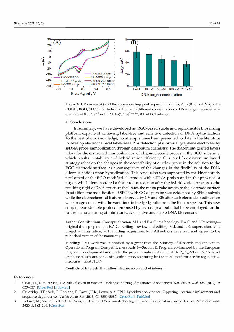
In order to compare the charge transfer properties of the modified electrodes, we fur-ther evaluated heterogeneous electron transfer rate constants for the [Fe(CN)6]3−/4− redoxcouple. Scan dependence CV studies performed at RGO/SPCE, Ar-COOH/RGO/SPCE,ssDNAp/Ar-COOH/RGO/SPCE, and DNAt/ssDNAp/Ar-COOH/RGO/SPCE (Figure 7)displayed an increase in the peak-to-peak separation potential for all electrodes exceptAr-COOH/RGO/SPCE (not shown), for which passivation occurs due to grafted carboxyliclayers that suppress further electrochemistry of Fe(CN)6
3−/4− species. In all other cases, an-odic and cathodic peak currents increase with increasing potential scan rate and are linearlyrelated to the square root of the scan rates in the range of 0.005–0.1 V/s (see the insets inFigure 7), suggesting that the oxido-reduction process at the electrode surface is controlledby diffusion. The Nicholson methodology [59] was applied to estimate k0 for the quasi-reversible process of [Fe(CN)6]3−/4− at the modified electrodes. The values of the diffusioncoefficients of the oxidized and reduced forms of [Fe(CN)6]3−/4− DO = 7.6 × 10⁻⁶ cm2 s−1,DR = 6.5 × 10⁻⁶ cm2 s−1 were taken from the literature [60], and the dimensionless kineticparameter Ψ was calculated by using the equation given by Lavagnini et al. [61]. Thecalculated k0 values for the Ar-COOH/RGO/SPCE-modified electrodes after the ssDNAprobe immobilization, and the hybridization with DNA target are 1.65 × 10⁻³ cm s−1, and2.06 × 10−3 cm s−1, respectively. This trend is in agreement with the EIS results reportedbefore, showing an improvement in the charge transfer after DNA hybridization process.
1
Figure 7. CV curves of ssDNAp/Ar–COOH/RGO/SPCE (A), and ssDNAp/Ar–COOH/RGO/SPCEafter hybridization with DNA target (B) recorded at different scan rates in 1 mM [Fe(CN)6]3−/4−,0.1 M KCl solution. Inset: plot of anodic and cathodic peaks vs. square root of scan rate.
The same pattern in the sensor response was observed throughout the concentrationrange of 1–200 nM, i.e., a smaller peak separation by DNA target addition, suggesting anincrease in the charge transfer rate with target concentration (Figure 8). These evidentchanges in heterogeneous electron transfer as a result of the hybridization process shouldsupport the further use of this detection platform for the advancement of DNA biosensors.
Biosensors 2022, 12, 39 11 of 14Biosensors 2022, 12, x FOR PEER REVIEW 11 of 14
Figure 8. CV curves (A) and the corresponding peak separation values, ΔEp (B) of ssDNAp/Ar–COOH/RGO/SPCE after hybridization with different concentration of DNA target, recorded at a scan rate of 0.05 Vs−1 in 1 mM [Fe(CN)6]3−/4−, 0.1M KCl solution.
4. Conclusions In summary, we have developed an RGO-based stable and reproducible biosensing
platform capable of achieving label-free and sensitive detection of DNA hybridization. To the best of our knowledge, no attempts have been presented to date in the literature to develop electrochemical label-free DNA detection platforms at graphene electrodes by ssDNA probe immobilization through diazonium chemistry. The diazonium-grafted lay-ers allow for the controlled immobilization of oligonucleotide probes at the RGO sub-strate, which results in stability and hybridization efficiency. Our label-free diazonium-based strategy relies on the changes in the accessibility of a redox probe in the solution to the RGO electrode surface, as a consequence of the changes in the flexibility of the DNA oligonucleotides upon hybridization. This conclusion was supported by the kinetic study performed at the RGO-modified electrodes with ssDNA probes and in the presence of target, which demonstrated a faster redox reaction after the hybridization process as the resulting rigid dsDNA structure facilitates the redox probe access to the electrode surface. In addition, the modification of SPCE with GO dispersion was evidenced by SEM analysis, while the electrochemical features observed by CV and EIS after each electrode modifica-tion were in agreement with the variations in the ID/IG ratio from the Raman spectra. This new, simple, reproducible protocol proposed by us has great potential to be employed for the future manufacturing of miniaturized, sensitive and stable DNA biosensors.
Author Contributions: Conceptualization, M.I. and E.A.C.; methodology, E.A.C. and L.P.; writ-ing—original draft preparation, E.A.C.; writing—review and editing, M.I. and L.P.; supervision, M.I.; project administration, M.I.; funding acquisition, M.I. All authors have read and agreed to the published version of the manuscript.
Funding: This work was supported by a grant from the Ministry of Research and Innovation, Op-erational Program Competitiveness Axis 1—Section E, Program co-financed by the European Re-gional Development Fund under the project number 154/25.11.2016, P_37_221/2015, “A novel gra-phene biosensor testing osteogenic potency; capturing best stem cell performance for regenerative medicine” (GRABTOP).
Conflicts of Interest: The authors declare no conflict of interest.
References 1. Cisse: I.I.; Kim, H.; Ha, T. A rule of seven in Watson-Crick base-pairing of mismatched sequences. Nat. Struct. Mol. Biol. 2012,
19, 623–627. 2. Ouldridge, T.E.; Sulc, P.; Romano, F.; Doye, J.P.K.; Louis, A.A. DNA hybridization kinetics: Zippering, internal displacement
and sequence dependence. Nucleic Acids Res. 2013, 41, 8886–8895. 3. DeLuca, M.; Shi, Z.; Castro, C.E.; Arya, G. Dynamic DNA nanotechnology: Toward functional nanoscale devices. Nanoscale
Horiz. 2020, 5, 182–201.
Figure 8. CV curves (A) and the corresponding peak separation values, ∆Ep (B) of ssDNAp/Ar–COOH/RGO/SPCE after hybridization with different concentration of DNA target, recorded at ascan rate of 0.05 Vs−1 in 1 mM [Fe(CN)6]3−/4−, 0.1 M KCl solution.
4. Conclusions
In summary, we have developed an RGO-based stable and reproducible biosensingplatform capable of achieving label-free and sensitive detection of DNA hybridization.To the best of our knowledge, no attempts have been presented to date in the literatureto develop electrochemical label-free DNA detection platforms at graphene electrodes byssDNA probe immobilization through diazonium chemistry. The diazonium-grafted layersallow for the controlled immobilization of oligonucleotide probes at the RGO substrate,which results in stability and hybridization efficiency. Our label-free diazonium-basedstrategy relies on the changes in the accessibility of a redox probe in the solution to theRGO electrode surface, as a consequence of the changes in the flexibility of the DNAoligonucleotides upon hybridization. This conclusion was supported by the kinetic studyperformed at the RGO-modified electrodes with ssDNA probes and in the presence oftarget, which demonstrated a faster redox reaction after the hybridization process as theresulting rigid dsDNA structure facilitates the redox probe access to the electrode surface.In addition, the modification of SPCE with GO dispersion was evidenced by SEM analysis,while the electrochemical features observed by CV and EIS after each electrode modificationwere in agreement with the variations in the ID/IG ratio from the Raman spectra. This new,simple, reproducible protocol proposed by us has great potential to be employed for thefuture manufacturing of miniaturized, sensitive and stable DNA biosensors.
Author Contributions: Conceptualization, M.I. and E.A.C.; methodology, E.A.C. and L.P.; writing—original draft preparation, E.A.C.; writing—review and editing, M.I. and L.P.; supervision, M.I.;project administration, M.I.; funding acquisition, M.I. All authors have read and agreed to thepublished version of the manuscript.
Funding: This work was supported by a grant from the Ministry of Research and Innovation,Operational Program Competitiveness Axis 1—Section E, Program co-financed by the EuropeanRegional Development Fund under the project number 154/25.11.2016, P_37_221/2015, “A novelgraphene biosensor testing osteogenic potency; capturing best stem cell performance for regenerativemedicine” (GRABTOP).
Conflicts of Interest: The authors declare no conflict of interest.
References1. Cisse:, I.I.; Kim, H.; Ha, T. A rule of seven in Watson-Crick base-pairing of mismatched sequences. Nat. Struct. Mol. Biol. 2012, 19,
623–627. [CrossRef] [PubMed]2. Ouldridge, T.E.; Sulc, P.; Romano, F.; Doye, J.P.K.; Louis, A.A. DNA hybridization kinetics: Zippering, internal displacement and
sequence dependence. Nucleic Acids Res. 2013, 41, 8886–8895. [CrossRef] [PubMed]3. DeLuca, M.; Shi, Z.; Castro, C.E.; Arya, G. Dynamic DNA nanotechnology: Toward functional nanoscale devices. Nanoscale Horiz.
2020, 5, 182–201. [CrossRef]
Biosensors 2022, 12, 39 12 of 14
4. Kimna, C.; Lieleg, O. Molecular micromanagement: DNA nanotechnology establishes spatio-temporal control for precisionmedicine. Biophys. Rev. 2020, 1, 011305. [CrossRef]
5. Trotter, M.; Borst, N.; Thewes, R.; von Stetten, F. Review: Electrochemical DNA sensing—Principles, commercial systems, andapplications. Biosens. Bioelectron. 2020, 154, 112069. [CrossRef]
6. Wu, X.; Mu, F.; Wang, Y.; Zhao, H. Graphene and graphene-based nanomaterials for DNA detection: A review. Molecules 2018,23, 2050. [CrossRef]
7. Pei, Q.; Song, X.; Liu, S.; Wang, J.; Leng, X.; Cui, X.; Yu, J.; Wang, Y.; Huang, J. A facile signal-on electrochemical DNA sensingplatform for ultrasensitive detection of pathogenic bacteria based on Exo III-assisted autonomous multiple-cycle amplification.Analyst 2019, 144, 3023–3029. [CrossRef] [PubMed]
8. Rasouli, E.; Shahnavaz, Z.; Basirun, W.J.; Rezayi, M.; Avan, A.; Ghayour-Mobarhan, M.; Khandanlou, R.; Johan, M.R. Advance-ments in electrochemical DNA sensor for detection of human papilloma virus-A review. Anal. Biochem. 2018, 556, 136–144.[CrossRef]
9. Mohanraj, J.; Durgalakshmi, D.; Rakkesh, R.A.; Balakumar, S.; Rajendran, S.; Karimi-Maleh, H. Facile synthesis of paper basedgraphene electrodes for point of care devices: A double stranded DNA (dsDNA) biosensor. J. Colloid Interface Sci. 2020, 566,463–472. [CrossRef] [PubMed]
10. Zeng, N.; Xiang, J. Detection of KRAS G12D point mutation level by anchor-like DNA electrochemical biosensor. Talanta 2019,198, 111–117. [CrossRef] [PubMed]
11. Huang, Y.; Xu, J.; Liu, J.; Wang, X.; Chen, B. Disease-related detection with electrochemical biosensors: A review. Sensors 2017,17, 2375. [CrossRef]
12. Rashid, J.I.A.; Yusof, N.A. The strategies of DNA immobilization and hybridization detection mechanism in the construction ofelectrochemical DNA sensor: A review. Sens. Bio-Sens. Res. 2017, 16, 19–31. [CrossRef]
13. Mahmoodi, P.; Rezayi, M.; Rasouli, E.; Avan, A.; Gholami, M.; Mobarhan, M.G.; Karimi, E.; Alias, Y. Early-stage cervical cancer di-agnosis based on an ultra-sensitive electrochemical DNA nanobiosensor for HPV-18 detection in real samples. J. Nanobiotechnology2020, 18, 1–12. [CrossRef] [PubMed]
14. Bolat, G. Investigation of poly (CTAB-MWCNTs) composite based electrochemical DNA biosensor and interaction study withanticancer drug Irinotecan. Microchem. J. 2020, 159, 105426. [CrossRef]
15. Javar, H.A.; Garkani-Nejad, Z.; Dehghannoudeh, G.; Mahmoudi-Moghaddam, H. Development of a new electrochemical DNAbiosensor based on Eu3+—Doped NiO for determination of amsacrine as an anti-cancer drug: Electrochemical, spectroscopic anddocking studies. Anal. Chim. Acta 2020, 1133, 48–57. [CrossRef] [PubMed]
16. Kumar, V.; Guleria, P. Application of DNA-Nanosensor for Environmental Monitoring: Recent Advances and Perspectives.Curr. Pollut. Rep. 2020, 1–21. [CrossRef]
17. Chen, Z.; Liu, X.; Liu, D.; Li, F.; Wang, L.; Liu, S. Ultrasensitive electrochemical DNA biosensor fabrication by coupling an integralmultifunctional zirconia-reduced graphene oxide-thionine nanocomposite and exonuclease I-assisted cleavage. Front. Chem. 2020,8, 521. [CrossRef]
18. Zhu, D.; Liu, W.; Zhao, D.; Hao, Q.; Li, J.; Huang, J.; Shi, J.; Chao, J.; Su, S.; Wang, L. Label-free electrochemical sensing platformfor microRNA-21 detection using thionine and gold nanoparticles co-functionalized MoS2 nanosheet. ACS Appl. Mater. Interfaces2017, 9, 35597–35603. [CrossRef]
19. Kavita, V. DNA biosensors-a review. J. Bioeng. Biomed. Sci. 2017, 7, 222.20. Ferapontova, E.E. DNA electrochemistry and electrochemical sensors for nucleic acids. Annu. Rev. Anal. Chem. 2018, 11, 197–218.
[CrossRef] [PubMed]21. Cho, I.-H.; Kim, D.H.; Park, S. Electrochemical biosensors: Perspective on functional nanomaterials for on-site analysis. Biomater.
Res. 2020, 24, 6. [CrossRef] [PubMed]22. Alazmi, A.; Rasul, S.; Patole, S.P.; Costa, P.M. Comparative study of synthesis and reduction methods for graphene oxide.
Polyhedron 2016, 116, 153–161. [CrossRef]23. Benvidi, A.; Rajabzadeh, N.; Mazloum-Ardakani, M.; Heidari, M.M.; Mulchandani, A. Simple and label-free electrochemical
impedance Amelogenin gene hybridization biosensing based on reduced graphene oxide. Biosens. Bioelectron. 2014, 58, 145–152.[CrossRef] [PubMed]
24. Gosai, A.; Khondakar, K.R.; Ma, X.; Ali, M. Application of Functionalized Graphene Oxide Based Biosensors for Health Monitoring:Simple Graphene Derivatives to 3D Printed Platforms. Biosensors 2021, 11, 384. [CrossRef]
25. Chiticaru, E.A.; Pilan, L.; Damian, C.-M.; Vasile, E.; Burns, J.S.; Ionită, M. Influence of Graphene Oxide Concentration whenFabricating an Electrochemical Biosensor for DNA Detection. Biosensors 2019, 9, 113. [CrossRef]
26. Sarkar, S.; Bekyarova, E.; Haddon, R.C. Reversible Grafting of α-Naphthylmethyl Radicals to Epitaxial Graphene. Angew. Chem.Int. Ed. 2012, 51, 4901–4904. [CrossRef] [PubMed]
27. Xia, Z.Y.; Giambastiani, G.; Christodoulou, C.; Nardi, M.V.; Koch, N.; Treossi, E.; Bellani, V.; Pezzini, S.; Corticelli, F.; Morandi, V.Synergic exfoliation of graphene with organic molecules and inorganic ions for the electrochemical production of flexibleelectrodes. Chem. Plus. Chem. 2014, 79, 439–446. [CrossRef] [PubMed]
28. Xia, Z.; Leonardi, F.; Gobbi, M.; Liu, Y.; Bellani, V.; Liscio, A.; Kovtun, A.; Li, R.; Feng, X.; Orgiu, E. Electrochemical func-tionalization of graphene at the nanoscale with self-assembling diazonium salts. ACS Nano 2016, 10, 7125–7134. [CrossRef][PubMed]
Biosensors 2022, 12, 39 13 of 14
29. Shih, C.-J.; Wang, Q.H.; Jin, Z.; Paulus, G.L.; Blankschtein, D.; Jarillo-Herrero, P.; Strano, M.S. Disorder imposed limits ofmono-and bilayer graphene electronic modification using covalent chemistry. Nano Lett. 2013, 13, 809–817. [CrossRef]
30. Paulus, G.L.; Wang, Q.H.; Strano, M.S. Covalent electron transfer chemistry of graphene with diazonium salts. Acc. Chem. Res.2013, 46, 160–170. [CrossRef]
31. Kongsfelt, M.S.; Ceccato, M.; Nilsson, L.; Jørgensen, B.; Hornekær, L.; Pedersen, S.U.; Daasbjerg, K. Chemical modifications ofgraphene using diazonium chemistry. In Proceedings of the Annual World Conference on Carbon 2010, Clemson, SC, USA,11–16 July 2010.
32. Raicopol, M.; Vlsceanu, I.; Lupulescu, I.; Brezoiu, A.M.; Pilan, L. Amperometric glucose biosensors based on functionalizedelectrochemically reduced graphene oxide. UPB Sci. Bull. Ser. B 2016, 78, 131–142.
33. Ott, C.; Raicopol, M.D.; Andronescu, C.; Vasile, E.; Hanganu, A.; Pruna, A.; Pilan, L. Functionalized polypyrrole/sulfonatedgraphene nanocomposites: Improved biosensing platforms through aryl diazonium electrochemistry. Synth. Met. 2018, 235,20–28. [CrossRef]
34. Ge, L.; Wang, W.; Li, F. Electro-grafted electrode with graphene-oxide-like DNA affinity for ratiometric homogeneous electro-chemical biosensing of microRNA. Anal. Chem. 2017, 89, 11560–11567. [CrossRef] [PubMed]
35. Allongue, P.; Delamar, M.; Desbat, B.; Fagebaume, O.; Hitmi, R.; Pinson, J.; Saveant, J.-M. Covalent modification of carbonsurfaces by aryl radicals generated from the electrochemical reduction of diazonium salts. J. Am. Chem. Soc. 1997, 119, 201–207.[CrossRef]
36. Gan, L.; Zhang, D.; Guo, X. Electrochemistry: An efficient way to chemically modify individual monolayers of graphene. Small2012, 8, 1326–1330. [CrossRef] [PubMed]
37. Tavakkoli, Z.; Goljani, H.; Sepehrmansourie, H.; Nematollahi, D.; Zolfigol, M.A. New insight into the electrochemical reductionof different aryldiazonium salts in aqueous solutions. RSC Adv. 2021, 11, 25811–25815. [CrossRef]
38. Yáñez-Sedeño, P.; Campuzano, S.; Pingarrón, J.M. Integrated Affinity Biosensing Platforms on Screen-Printed ElectrodesElectrografted with Diazonium Salts. Sensors 2018, 18, 675. [CrossRef]
39. Yuliandari, P.; Wibowo, R.; Nurani, D.A. Para-carboxyphenyl diazonium-modified carbon paste electrode for analysis Cu (II) inwater. In AIP Conference Proceedings; AIP Publishing LLC: Melville, NY, USA, 2021; p. 020002.
40. Gökçe, G.; Ben Aissa, S.; Nemceková, K.; Catanante, G.; Raouafi, N.; Marty, J.-L. Aptamer-modified pencil graphite electrodes forthe impedimetric determination of ochratoxin A. Food Control 2020, 115, 107271. [CrossRef]
41. Raicopol, M.; Necula, L.; Ionita, M.; Pilan, L. Electrochemical reduction of aryl diazonium salts: A versatile way for carbonnanotubes functionalisation. Surf. Interface Anal. 2012, 44, 1081–1085. [CrossRef]
42. Randriamahazaka, H.; Ghilane, J. Electrografting and Controlled Surface Functionalization of Carbon Based Surfaces forElectroanalysis. Electroanalysis 2016, 28, 13–26. [CrossRef]
43. Gillan, L.; Teerinen, T.; Johansson, L.-S.; Smolander, M. Controlled diazonium electrodeposition towards a biosensor for C-reactiveprotein. Sens. Int. 2021, 2, 100060. [CrossRef]
44. Mousavisani, S.Z.; Raoof, J.-B.; Turner, A.P.F.; Ojani, R.; Mak, W.C. Label-free DNA sensor based on diazonium immobilisation fordetection of DNA damage in breast cancer 1 gene. Sens. Actuators B Chem. 2018, 264, 59–66. [CrossRef]
45. Polsky, R.; Harper, J.C.; Wheeler, D.R.; Arango, D.C.; Brozik, S.M. Electrically addressable cell immobilization using phenylboronicacid diazonium salts. Angew. Chem. Int. Ed. 2008, 120, 2671–2674. [CrossRef]
46. Hu, Y.; Li, F.; Han, D.; Wu, T.; Zhang, Q.; Niu, L.; Bao, Y. Simple and label-free electrochemical assay for signal-on DNAhybridization directly at undecorated graphene oxide. Anal. Chim. Acta 2012, 753, 82–89. [CrossRef] [PubMed]
47. Giovanni, M.; Bonanni, A.; Pumera, M. Detection of DNA hybridization on chemically modified graphene platforms. Analyst2012, 137, 580–583. [CrossRef] [PubMed]
48. Gong, Q.; Yang, H.; Dong, Y.; Zhang, W. A sensitive impedimetric DNA biosensor for the determination of the HIV gene based onelectrochemically reduced graphene oxide. Anal. Methods 2015, 7, 2554–2562. [CrossRef]
49. Eissa, S.; Jimenez, G.C.; Mahvash, F.; Guermoune, A.; Tlili, C.; Szkopek, T.; Zourob, M.; Siaj, M. Functionalized CVD monolayergraphene for label-free impedimetric biosensing. Nano Res. 2015, 8, 1698–1709. [CrossRef]
50. Wu, J.-B.; Lin, M.-L.; Cong, X.; Liu, H.-N.; Tan, P.-H. Raman spectroscopy of graphene-based materials and its applications inrelated devices. Chem. Soc. Rev. 2018, 47, 1822–1873. [CrossRef] [PubMed]
51. Sun, Z.; Guo, D.; Wang, S.; Wang, C.; Yu, Y.; Ma, D.; Zheng, R.; Yan, P. Efficient covalent modification of graphene by diazochemistry. RSC Adv. 2016, 6, 65422–65425. [CrossRef]
52. Jiang, D.-e.; Sumpter, B.G.; Dai, S. How do aryl groups attach to a graphene sheet? J. Phys. Chem. B 2006, 110, 23628–23632.[CrossRef]
53. Pilan, L. Tailoring the performance of electrochemical biosensors based on carbon nanomaterials via aryldiazonium electrografting.Bioelectrochemistry 2021, 138, 107697. [CrossRef]
54. Ambrosio, G.; Brown, A.; Daukiya, L.; Drera, G.; Di Santo, G.; Petaccia, L.; De Feyter, S.; Sangaletti, L.; Pagliara, S. Impact ofcovalent functionalization by diazonium chemistry on the electronic properties of graphene on SiC. Nanoscale 2020, 12, 9032–9037.[CrossRef]
55. Lee, L.; Ma, H.; Brooksby, P.A.; Brown, S.A.; Leroux, Y.R.; Hapiot, P.; Downard, A.J. Covalently anchored carboxyphenylmonolayer via aryldiazonium ion grafting: A well-defined reactive tether layer for on-surface chemistry. Langmuir 2014, 30,7104–7111. [CrossRef] [PubMed]
Biosensors 2022, 12, 39 14 of 14
56. Phal, S.; Shimizu, K.; Mwanza, D.; Mashazi, P.; Shchukarev, A.; Tesfalidet, S. Electrografting of 4-carboxybenzenediazoniumon glassy carbon electrode: The effect of concentration on the formation of mono and multilayers. Molecules 2020, 25, 4575.[CrossRef]
57. Wallen, R.; Gokarn, N.; Bercea, P.; Grzincic, E.; Bandyopadhyay, K. Mediated electron transfer at vertically aligned single-walledcarbon nanotube electrodes during detection of DNA hybridization. Nanoscale Res. Lett. 2015, 10, 1–11. [CrossRef] [PubMed]
58. Gooding, J.J.; Chou, A.; Mearns, F.J.; Wong, E.; Jericho, K.L. The ion gating effect: Using a change in flexibility to allow label freeelectrochemical detection of DNA hybridisation. Chem. Commun. 2003, 15, 1938–1939. [CrossRef]
59. Nicholson, R.S. Theory and application of cyclic voltammetry for measurement of electrode reaction kinetics. Anal. Chem. 1965,37, 1351–1355. [CrossRef]
60. Ojeda, I.; Barrejón, M.; Arellano, L.M.; González-Cortés, A.; Yáñez-Sedeño, P.; Langa, F.; Pingarrón, J.M. Grafted-double walledcarbon nanotubes as electrochemical platforms for immobilization of antibodies using a metallic-complex chelating polymer:Application to the determination of adiponectin cytokine in serum. Biosens. Bioelectron. 2015, 74, 24–29. [CrossRef]
61. Lavagnini, I.; Antiochia, R.; Magno, F. An extended method for the practical evaluation of the standard rate constant from cyclicvoltammetric data. Electroynalysis 2004, 16, 505–506. [CrossRef]