An Extended Clq-Binding Assay Using Lactoperoxidase- and Chloramine-T-Iodinated Clq
lable at ScienceDirect
Biomaterials 30 (2009) 5667–5674
Contents lists avai
Biomaterials
journal homepage: www.elsevier .com/locate/biomateria ls
Elaboration of radiopaque iodinated nanoparticles for in situ controlof local drug delivery
Damia Mawad a,d,e, Hanna Mouaziz a,d, Alexandra Penciu a,d, Henri Mehier b, Bernard Fenet c,d,Hatem Fessi a,d, Yves Chevalier a,d,*
a Laboratoire d’Automatique et de Genie des Procedes, LAGEP, universite Lyon 1, CNRS UMR 5007, 43 Bd 11 Novembre, 69622 Villeurbanne, Franceb CERMA, Batiment Actipro, Parc d’affaires international, 74160 Archamps, Francec Centre Commun de RMN, Universite Lyon 1, ESCPE Lyon, 43 Bd 11 Novembre, 69616 Villeurbanne, Franced Universite de Lyon, 69003 Lyon, Francee Intelligent Polymer Research Institute, University of Wollongong, Fairy Meadow, New South Wales 2519, Australia
a r t i c l e i n f o
Article history:Received 24 April 2009Accepted 15 June 2009Available online 4 July 2009
Keywords:NanoparticleRadiopacityCellulose acetateIodinated polymersDOSYDrug delivery
* Corresponding author. Laboratoire d’AutomatiquLAGEP, universite Lyon 1, CNRS UMR 5007, 43 Bd 11 NoFrance. Tel.: þ33 472431877; fax: þ33 472431682.
E-mail address: [email protected] (Y. C
0142-9612/$ – see front matter � 2009 Elsevier Ltd.doi:10.1016/j.biomaterials.2009.06.027
a b s t r a c t
Drug delivery systems can benefit from intrinsic radiopacity as this property will allow following up thediffusion path of the nanoparticles containing the therapeutic drug after their local administration.Herein, we report the synthesis of iodinated derivatives of cellulose acetate (CA) and their formulationinto aqueous radiopaque nanoparticle suspensions. Modification and purification of CA with mono- ortri-iodobenzoyl chloride were characterized by NMR spectroscopy and elemental analysis of iodine. Inparticular, measurements of diffusion coefficients by the DOSY 2D NMR method allowed controlling thecomplete elimination of non-grafted iodinated materials. Pure radiopaque CA was successfully achievedwith an iodine content varying between 14 and 32%. Aqueous suspensions of nanoparticles weresuccessfully formed, characterized by being spherical, <100 nm in size and stable as a suspension over 3months. The degree of substitution, in particular the triiodo moieties, imparted a good level of radio-pacity whether in dry powder form (2627 HU) or as a nanoparticle suspension (298 HU). These values arecomparable to radiopacity of systems reported in literature to be in vivo visible. Loading of paclitaxel wassuccessfully attempted, suggesting that the developed radiopaque nanoparticles can ultimately functionas a drug delivery system.
� 2009 Elsevier Ltd. All rights reserved.
1. Introduction
Local administration allows therapeutic drugs to reach the targetmore efficiently in comparison to systemic administration routessuch as intraveneous or oral. As a result, the doses may be reduced,the administrations may be less frequent, and the side effects ofdrugs are reduced. Local administration aims at delivering the drugin the close vicinity of the target; thus diffusion throughout the bodyis not expected. An important issue is monitoring the diffusion ofdrugs after their local injection by labeling either the drug itself orthe drug carrier. Radiopaque materials are localized inside the bodyby non-invasive techniques of X-ray tomography [1].
The present paper reports the fabrication of iodinated nano-particles designed to be ultimately used as a local drug delivery
e et de Genie des Procedes,vembre, 69622 Villeurbanne,
hevalier).
All rights reserved.
system. The advantage of having the nanoparticles radiopaque istracking the diffusion of the drug-loaded nanoparticles in the bodyafter a local administration. As an example, the diffusion of ananticancer drug loaded inside the iodinated nanoparticles could bemonitored after an intratumoral injection. Among carriers forhydrophobic drugs, liposomes, o/w nanoemulsions or polymerparticles [2,3], polymer nanoparticles are preferred because oftheir much better stability.
The choice of the iodinated polymer material and the adminis-tration mode depends on the specific medical application. Drugdelivery systems are made of either polymer micro- or nano-particles; nanoparticles are preferred for their better ability to passthrough biological barriers (membranes). Most iodinated materialsreported so far were designed for embolization and implants suchas stents. Some of them can be utilized for drug delivery.
Requirements are quite drastic according to the dual goal, drugdelivery and radiopacity. Polymer nanoparticles should disperse inwater as stable colloidal suspension; the preparation processshould allow efficient loading of the drug; the suspension should beconcentrated enough for it to have a high radiopacity detectable

D. Mawad et al. / Biomaterials 30 (2009) 5667–56745668
with a conventional X-ray scanner. The purpose of the present workis designing a drug delivery system made of iodinated polymer thatwould meet such requirements.
Various approaches have been reported to render polymericbiomaterials radiopaque. For example, inorganic contrast agents(bismuth oxide, metrizamide, tantalum) are mixed with liquidembolics [4,5]. Drawbacks are increasing viscosity of the injectedsolution, making it harder to be delivered via a microcatheter,sedimentation of the contrast agent inside the catheter as it isdelivered, release into the surrounding [1], and possible toxicity[6–8].
To overcome those limitations, polymers with intrinsic radio-pacity are prepared by covalently binding brominated or iodinatedmoieties to the polymer backbone [9]. Quite a large variety ofiodinated polymer types have been synthesized either by radicalpolymerization of iodinated methacrylate derivatives [10,11], or bypolycondensation of iodinated monomers [12,13], or by chemicalgrafting of iodinated lateral substituents to the polymer backbone[14–16]. In particular Mottu et al. [14] modified cellulose witharomatic iodinated groups to be used as embolics. The medicalapplications claimed involved bulk radiopaque materials: emboli-zation, filling materials for dentistry, or medical devices such ascatheters, heart valves and stents.
Iodinated nanoparticles have been scarcely reported; mostreports deal with beads or microparticles. Galperin et al. reportedthe preparation of poly(2-methacryloyloxyethyl-(2,3,5-triiodo-benzoate) latex particles by means of either dispersion [17] oremulsion [18,19] polymerization of the corresponding monomer.Nanoparticles containing 58 wt% iodine were prepared and hencethe radiopacity of the suspension was quite high. Loading suchparticles with a drug is not possible however because the mech-anism of classical emulsion polymerization does not allow loadingaccording to the conventional means. Conversely, the ‘‘mini-emulsion polymerization’’ process might allow drug loading [20].Nanocapsules that showed a prolonged circulation time in vivohave been prepared by a process reminiscent of interfacial poly-condensation that should withstand drug loading [21]. An o/wnanoemulsion of Lipiodol, an iodinated derivative of poppy seedoil, is encapsulated by a shell made of PEG/Pluronic F127 materialcross-linked in basic conditions (pH> 9). To our knowledge, this isthe unique report where all the requirements of the X-ray scannermonitored drug delivery application are fulfilled. However, thepreparation of liquid-filled nanocapsules is complex and difficultto control in regards to repeatability and scale-up [22]. On theother hand, the pharmacokinetic properties of polymer nano-particles are different to those of liquid-filled nanocapsules.Hence, it will be beneficial to develop full polymer nanoparticlesas an alternative.
The objective of this study was to prepare radiopaque nano-particles based on iodinated cellulose acetate. The advantage ofusing cellulose acetate besides its biocompatibility [23] andbiodegradability [24,25] is its enhanced solubility over cellulose inorganic solvents. Cellulose acetate was modified by grafting eithermono- or tri-iodobenzoic acid. Aqueous suspensions of radi-opaque nanoparticles were prepared by the ‘‘nanoprecipitation’’process [26–30]. Accordingly, the ‘‘spontaneous emulsification’’[31] takes place when a solution of the polymer in a polar organicsolvent (acetone) is poured into an aqueous phase. Polymerdroplets of sub-micronic size (w100–500 nm) are formed whenthe starting solution is dilute enough. Easy drug loading is ach-ieved by dissolving the drug in the starting organic solution to beemulsified. Size and morphology of the nanoparticles were char-acterized; the long-term stability of the suspensions was assessed.Lastly, the radiopacity of the pure materials and the suspensionswas measured.
2. Experimental
2.1. Materials
Cellulose acetate (Mw¼ 30,000 g/mol, 39.8 wt% acetyl content) was purchasedfrom Aldrich. 4-Iodobenzoyl chloride and 2,3,5-triiodobenzoic acid were purchasedfrom Aldrich and used as received. 2,3,5-triiodobenzoyl chloride was prepared from2,3,5-triiodobenzoic acid by reaction with thionyl chloride as described in theliterature [11]: 2 g of the powdered acid was refluxed for 8 h with 10 mL of thionylchloride (from Fluka) and 0.1 mL of dimethylformamide (anhydrous N,N-dimethyl-formamide from Sigma–Aldrich); excess thionyl chloride was evaporated; the finalproduct was precipitated by pouring a concentrated solution in chloroform intocooled dry n-hexane (purum, �98.0% from Fluka); finally the pure 2,3,5-triiodo-benzoyl chloride was dried in vacuum. Anhydrous N,N-dimethylacetamide andanhydrous pyridine were from Sigma–Aldrich, they were taken from the bottlethrough the Sure-Seal stopper and transferred to the reaction vessel with a syringe.Acetone (ACS reagent, � 99.5% from Fluka) and Tween� 80 (Sigma–Aldrich) wereused as received. Paclitaxel was purchased from Chengdu Kejie Hi-tech Develop-ment Co (Chengdu, China). Deionized water of 18 MU cm�1 resistivity was used forthe preparation of suspensions.
2.2. Methods
2.2.1. Modification of cellulose acetate with radiopaque compoundsCellulose acetate (0.5 g) was dissolved in anhydrous dimethylacetamide
(DMAc) (15 mL) at 80 �C under dry nitrogen atmosphere in a three necked roundbottom flask equipped with two dropping funnels. The solution was stirredmagnetically until the polymer dissolved, and then cooled down to 0 �C byimmersing the round bottom flask into an ice bath. A solution of 4-iodobenzoylchloride (0.035 mol, 9.33 g) or 2,3,5-triiodobenzoyl chloride (0.035 mol, 18.14 g) inanhydrous DMAc (5 mL) was added drop-wise from the dropping funnel. Anhy-drous pyridine (3.32 mL) was then added from the second dropping funnel andstirring was continued for 12 h. The mixture was filtered and the filtrate wasprecipitated in water. The precipitate was collected, washed several times withethanol and dried under vacuum. The purification was repeated several times till1H NMR analyses were satisfactory. The reaction yielded 0.40 g of iodinatedcellulose acetate. The cellulose acetate modified with 4-iodobenzoyl chloride and2,3,5-triiodobenzoyl chloride are referred to as MonoiodoCA and TriiodoCArespectively in the following.
2.2.2. Characterization of the modified polymer2.2.2.1. 1H NMR analysis. The modification procedure of cellulose acetate with 4-iodobenzoyl chloride and 2,3,5-triiodobenzoyl chloride was followed by 1H NMR.The spectra of the iodinated cellulose acetate (30 mg) solutions in 1 mL deuteratedchloroform (CDCl3) were recorded at 28–30 �C with a Bruker DRX 300 spectrometer.Processing was done using Bruker WINNMR-1D software package. The degree ofsubstitution was calculated for each product by comparing the area under the peakscorresponding to the aromatic protons to the area under the peaks corresponding tothe methyl protons of the cellulose acetate backbone at d¼ 1.8–2.4 ppm.
2.2.2.2. Diffusion-ordered NMR spectroscopy (DOSY). Modified polymers werefurther characterized by diffusion-ordered NMR spectroscopy to verify that theradiopaque compounds are indeed grafted on the polymer backbone and not justbeing detected as a mixture with the polymer. The 1H NMR DOSY spectra werecollected in CDCl3 solution at 25 �C on a Bruker DRX500 spectrometer using thestandard ledbpgp2s sequence. Strong gradients of short duration were obtainedusing a trapezoidal shape of gradient pulses (p30¼ 5 ms, falling and raising times of200 ms) and the diffusion delays were set at 100 ms. Data were processed withXwinnmr 3.5. The same diffusion coefficient for the polymer and the graftedcompound demonstrates that the grafting reaction was really done.
2.2.2.3. Elemental analysis for iodine content. Elemental analysis of iodine in thepolymer was performed by Service Central d’Analyse (SCA-CNRS, chemin du canal,BP 22, 69390 Solaize, France). The reported values are an average of measurementsdone on 2 samples of each tested material.
2.2.3. Formulation of nanoparticles based on radiopaque cellulose acetateNanoparticles of modified cellulose acetate were prepared by the nano-
precipitation method [26,27]. Modified polymer (0.1675 g) was dissolved in acetone(12.5 mL) under reflux to form the diffusing phase. The diffusing phase was thenadded by means of a syringe to the dispersing phase constituted of deionized water(25 mL) containing 0.5% surfactant Tween� 80 under magnetic stirring. The acetonewas removed under reduced pressure and the nanoparticles were collected bycentrifugation for 30 min with an Eppendorf Centrifuge 5417C rotating at14,000 rpm and dried under vacuum at room temperature.
2.2.4. Nanoparticle size and morphologyParticle size distribution (PSD) of nanoparticles was determined by dynamic
light scattering with a Malvern NanoZS instrument. The range of particle diameter
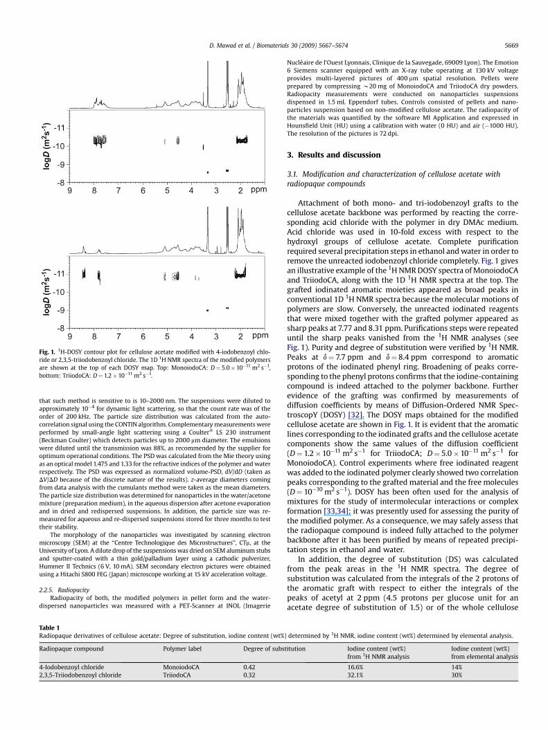
Fig. 1. 1H-DOSY contour plot for cellulose acetate modified with 4-iodobenzoyl chlo-ride or 2,3,5-triiodobenzoyl chloride. The 1D 1H NMR spectra of the modified polymersare shown at the top of each DOSY map. Top: MonoiodoCA: D¼ 5.0� 10�11 m2 s�1,bottom: TriiodoCA: D¼ 1.2�10�11 m2 s�1.
D. Mawad et al. / Biomaterials 30 (2009) 5667–5674 5669
that such method is sensitive to is 10–2000 nm. The suspensions were diluted toapproximately 10�4 for dynamic light scattering, so that the count rate was of theorder of 200 kHz. The particle size distribution was calculated from the auto-correlation signal using the CONTIN algorithm. Complementary measurements wereperformed by small-angle light scattering using a Coulter� LS 230 instrument(Beckman Coulter) which detects particles up to 2000 mm diameter. The emulsionswere diluted until the transmission was 88%, as recommended by the supplier foroptimum operational conditions. The PSD was calculated from the Mie theory usingas an optical model 1.475 and 1.33 for the refractive indices of the polymer and waterrespectively. The PSD was expressed as normalized volume-PSD, dV/dD (taken asDV/DD because of the discrete nature of the results). z-average diameters comingfrom data analysis with the cumulants method were taken as the mean diameters.The particle size distribution was determined for nanoparticles in the water/acetonemixture (preparation medium), in the aqueous dispersion after acetone evaporationand in dried and redispersed suspensions. In addition, the particle size was re-measured for aqueous and re-dispersed suspensions stored for three months to testtheir stability.
The morphology of the nanoparticles was investigated by scanning electronmicroscopy (SEM) at the ‘‘Centre Technologique des Microstructures’’, CTm, at theUniversity of Lyon. A dilute drop of the suspensions was dried on SEM aluminum stubsand sputter-coated with a thin gold/palladium layer using a cathodic pulverizer,Hummer II Technics (6 V, 10 mA). SEM secondary electron pictures were obtainedusing a Hitachi S800 FEG (Japan) microscope working at 15 kV acceleration voltage.
2.2.5. RadiopacityRadiopacity of both, the modified polymers in pellet form and the water-
dispersed nanoparticles was measured with a PET-Scanner at INOL (Imagerie
Table 1Radiopaque derivatives of cellulose acetate: Degree of substitution, iodine content (wt%)
Radiopaque compound Polymer label Degree of subst
4-Iodobenzoyl chloride MonoiodoCA 0.422,3,5-Triiodobenzoyl chloride TriiodoCA 0.32
Nucleaire de l’Ouest Lyonnais, Clinique de la Sauvegade, 69009 Lyon). The Emotion6 Siemens scanner equipped with an X-ray tube operating at 130 kV voltageprovides multi-layered pictures of 400 mm spatial resolution. Pellets wereprepared by compressing w20 mg of MonoiodoCA and TriiodoCA dry powders.Radiopacity measurements were conducted on nanoparticles suspensionsdispensed in 1.5 mL Eppendorf tubes. Controls consisted of pellets and nano-particles suspension based on non-modified cellulose acetate. The radiopacity ofthe materials was quantified by the software MI Application and expressed inHounsfield Unit (HU) using a calibration with water (0 HU) and air (�1000 HU).The resolution of the pictures is 72 dpi.
3. Results and discussion
3.1. Modification and characterization of cellulose acetate withradiopaque compounds
Attachment of both mono- and tri-iodobenzoyl grafts to thecellulose acetate backbone was performed by reacting the corre-sponding acid chloride with the polymer in dry DMAc medium.Acid chloride was used in 10-fold excess with respect to thehydroxyl groups of cellulose acetate. Complete purificationrequired several precipitation steps in ethanol and water in order toremove the unreacted iodobenzoyl chloride completely. Fig. 1 givesan illustrative example of the 1H NMR DOSY spectra of MonoiodoCAand TriiodoCA, along with the 1D 1H NMR spectra at the top. Thegrafted iodinated aromatic moieties appeared as broad peaks inconventional 1D 1H NMR spectra because the molecular motions ofpolymers are slow. Conversely, the unreacted iodinated reagentsthat were mixed together with the grafted polymer appeared assharp peaks at 7.77 and 8.31 ppm. Purifications steps were repeateduntil the sharp peaks vanished from the 1H NMR analyses (seeFig. 1). Purity and degree of substitution were verified by 1H NMR.Peaks at d¼ 7.7 ppm and d¼ 8.4 ppm correspond to aromaticprotons of the iodinated phenyl ring. Broadening of peaks corre-sponding to the phenyl protons confirms that the iodine-containingcompound is indeed attached to the polymer backbone. Furtherevidence of the grafting was confirmed by measurements ofdiffusion coefficients by means of Diffusion-Ordered NMR Spec-troscopY (DOSY) [32]. The DOSY maps obtained for the modifiedcellulose acetate are shown in Fig. 1. It is evident that the aromaticlines corresponding to the iodinated grafts and the cellulose acetatecomponents show the same values of the diffusion coefficient(D¼ 1.2�10�11 m2 s�1 for TriiodoCA; D¼ 5.0�10�11 m2 s�1 forMonoiodoCA). Control experiments where free iodinated reagentwas added to the iodinated polymer clearly showed two correlationpeaks corresponding to the grafted material and the free molecules(D¼ 10�10 m2 s�1). DOSY has been often used for the analysis ofmixtures for the study of intermolecular interactions or complexformation [33,34]; it was presently used for assessing the purity ofthe modified polymer. As a consequence, we may safely assess thatthe radiopaque compound is indeed fully attached to the polymerbackbone after it has been purified by means of repeated precipi-tation steps in ethanol and water.
In addition, the degree of substitution (DS) was calculatedfrom the peak areas in the 1H NMR spectra. The degree ofsubstitution was calculated from the integrals of the 2 protons ofthe aromatic graft with respect to either the integrals of thepeaks of acetyl at 2 ppm (4.5 protons per glucose unit for anacetate degree of substitution of 1.5) or of the whole cellulose
determined by 1H NMR, iodine content (wt%) determined by elemental analysis.
itution Iodine content (wt%)from 1H NMR analysis
Iodine content (wt%)from elemental analysis
16.6% 14%32.1% 30%

Fig. 2. Normalized particle size distribution of TriiodoCA (C) and MonoiodoCA (B)nanoparticles as measured by dynamic light scattering.
D. Mawad et al. / Biomaterials 30 (2009) 5667–56745670
backbone from 3.4 to 5.5 ppm (8.5-DS protons per glucose unit).Both calculations gave the same result and no cleavage of theacetyl esters was observed. The substitution degree of mono-iodobenzoyl chloride and triiodobenzoyl chloride is 0.42 and 0.32respectively; the iodine content was calculated from the degreeof substitution (Table 1). The iodine contents in MonoiodoCA andTriiodoCA were 14% and 30% respectively as measured byelemental analysis, in good agreement with the values calculatedfrom 1H NMR analyses. Possible migration of iodine groupselsewhere in the macromolecule was discarded because thearomatic part of the NMR spectrum met the expectation and theiodine contents assessed from NMR and elemental analysis werequite close.
3.2. Preparation and characterization of nanoparticles based onmodified cellulose acetate
3.2.1. PreparationRadiopaque cellulose acetate nanoparticles were prepared by
means of the nanoprecipitation process. Acetone was selected asa polar organic solvent in which the iodinated cellulose acetate issoluble. Polymer nanoparticles formed immediately as the polymersolution was slowly poured into the aqueous phase. A stable milky
Fig. 3. SEM pictures of TriiodoCA par
suspension in water-acetone (2:1) mixture formed. An aqueoussuspension was obtained by means of gentle evaporation ofacetone under reduced pressure (rotary evaporator). Centrifugationof the nanoparticles was easy because of the high density of theiodinated polymer material; it was achieved by a moderatecentrifugation field. The sediment was collected and dried. Driednanoparticles of the modified cellulose acetate (34 mg) were re-dispersed in water (0.75 mL) with the help of a very short (fewseconds) dispersion in an ultrasound bath cleaner. A homogeneousmilky suspension resulted at the end of the preparation process.
3.2.2. CharacterizationThe particle size distribution measured by dynamic light scat-
tering was identical for the nanoprecipitated dispersion in water-acetone (2:1), the aqueous dispersion after evaporation of acetone,and the dried and re-dispersed suspension. It is worth noticing thatthe particle size distribution is not always kept the same afterdrying and gentle redispersion; frequent cases of failure result fromeither irreversible coagulation or coalescence. Successful redis-persion requires hard particles that do not undergo coalescencewhen they are at close contact in the dry material. Additionally,redispersion requires the presence of an efficient stabilizing layer atthe surface of the particles. This showed that the stabilization bythe Tween� 80 surfactant was quite efficient and made thesuspension robust with respect to destabilization processes.
Fig. 2 shows the particle size distribution (PSD) of redispersednanoparticles in water measured by means of dynamic light scat-tering. The particles are well dispersed with one single populationand an average hydrodynamic radius of 187 and 85 nm for Mono-iodoCA and TriiodoCA respectively.
Small-angle light scattering provides the PSD in a differentrange of diameters from c.a. 100 nm to 2 mm. Such technique doesnot allow an accurate measurement of the present dispersionsbecause the diameter range is not correct. The measured PSDis truncated towards small particles; it is however very sensitive tothe presence of particles larger than 2 mm that dynamic lightscattering would miss. In the present case, the PSD measured bysmall-angle light scattering did not show particles larger than 1 mm.Therefore this method confirmed that the PSD contained sub-micronic particles only; and it validated the use of dynamic lightscattering as an accurate granulometric method.
Scanning electron microscopy (SEM) pictures were taken asa supplementary method for measuring the particle size distri-bution and to further characterize the morphology of the
ticles at different magnifications.

Fig. 4. X-ray absorption pictures of the pure polymers pressed as pellets observed with the X-ray scanner. (A): Non-modified CA; (B): MonoiodoCA; (C): TriiodoCA. The lowresolution of the picture is that of the X-ray scanner itself.
Table 2Radiopacity of modified polymers as pure powder form and dispersion in waterquantified in HU.
Compound Radiopacity (HU)
Pure non-modified CA pellet �130Pure MonoiodoCA pellet 819Pure TriiodoCA pellet 2627Suspension of non-modified CA nanoparticles (26 mg mL�1) �19Suspension of MonoiodoCA nanoparticles (26 mg mL�1) 86Suspension of TriiodoCA nanoparticles (26 mg mL�1) 298
D. Mawad et al. / Biomaterials 30 (2009) 5667–5674 5671
nanoparticles. Fig. 3 represents SEM pictures taken for TriiodoCA.A large collection of particles observed at low magnification(Fig. 3A) showed that only a single population of nanoparticleswas present; no particles of diameter larger than 1 mm could be
Fig. 5. X-Ray absorption pictures of the aqueous suspensions of nanoparticles in 1.5
detected in the pictures. Particles appear slightly aggregatedbecause of drying the suspension before SEM observation. Theparticle size distribution measured on a series of magnifiedpictures (Fig. 3B) was fairly narrow: the number-average diameterof TriodoCA particles was 60 nm with a standard deviation of18 nm for a collection of 250 particles. The intensity-averagediameter D [3,4] calculated from SEM was 79 nm, in good agree-ment with the mean diameter measured by dynamic light scat-tering; the slight difference between the two may be ascribed tothe thickness of the hydrophilic coating of the nanoparticleswhich adds up to the particle radius in defining the hydrodynamicradius measured by dynamic light scattering. The good correlationbetween dynamic light scattering and SEM measurements wasalso observed for MonoiodoCA.
mL Eppendorf tubes. (A): Non-modified CA; (B): MonoiodoCA; (C): TriiodoCA.

Fig. 6. Normalized particle size distribution of blank (C) and loaded with 26% Pacli-taxel (B) TriiodoCA nanoparticles as measured by dynamic light scattering.
D. Mawad et al. / Biomaterials 30 (2009) 5667–56745672
SEM also revealed the morphology of the particles at highermagnifications. Their overall shape was indeed spherical but theirsurface did not appear smooth as for usual polymer nanoparticles(Fig. 3C). The pictures suggested a porous internal structure ofthe nanoparticles which was rather surprising and remainedunexplained.
3.2.3. StabilityThe stability of the aqueous suspensions was checked by means
of 3 months storage at room temperature for both MonoiodoCA andTriiodoCA, either after their preparation by the nanoprecipitationprocess, or after they were centrifuged, dried and re-dispersed.Visual observation of the suspensions did not reveal any sedi-mentation during this period. In addition, the particle sizemeasured by dynamic light scattering did not significantly varyafter 3 months storage, showing that the suspensions are stableover at least this period of time.
Fig. 7. SEM pictures of blank (A) and loaded with
3.3. Radiopacity
Fig. 4 represents the X-ray visibility of the modified polymersin their powder form compressed into pellets. The X-ray imagesof MonoiodoCA and TriiodoCA (Fig. 4B and C respectively)demonstrate the significantly increased radiopacity of the poly-mers compared to the non-modified cellulose acetate (Fig. 4A).Table 2 lists the radiopacity quantified in HU for the modifiedpolymers. The value increased from �130 for the non-modifiedpolymer to 819 and 2627 for MonoiodoCA and TriiodoCArespectively.
Fig. 5 shows the X-ray tomography pictures of aqueous disper-sions of radiopaque nanoparticles. The respective radiopacity ofsuspensions containing 26 mg mL�1 of polymer are listed in Table 2.Suspension of radiopaque nanoparticles in water displayed anenhanced radiopacity in comparison to nanoparticles formulatedfrom non-modified cellulose acetate. Also, the radiopacity fornanoparticles based on TriiodoCA (298 HU) had an increased visi-bility in comparison to nanoparticles based on MonoiodoCA (86HU). This result is to be expected since the iodine content inTriiodoCA suspension (7.7 mg(I).mL�1) is higher than in Mono-iodoCA (3.6 mg(I).mL�1) as the elemental analysis of the purepolymers showed (Table 1). Such radiopacity values are in agree-ment with those measured by Galperin et al. [17,18] for the sameiodine content.
The measured radiopacity is expected to enhance X-ray visibilityafter injection of the radiopaque nanoparticles in vivo. Based ona study by Galperin et al. [18], a suspension of radiopaque poly-methacrylate nanoparticles (3 mg mL�1) that had a radiopacity of115 HU showed that visibility of tissues in the lymph node wasenhanced from 40 HU before to 80 HU after injection. Based on thisresult, we can safely speculate that in vivo injection of the TriiodoCAnanoparticles (298 HU) will enhance the visibility and help trackingthe injectible system.
3.4. Loading with paclitaxel
TriiodoCA nanoparticles were loaded with the anticancer agentpaclitaxel as a demonstration example. The nanoprecipitation
26% Paclitaxel (B) TriiodoCA nanoparticles.
D. Mawad et al. / Biomaterials 30 (2009) 5667–5674 5673
process allows easy loading by various hydrophobic drugs. Thedrug is dissolved in the organic phase that is poured into theaqueous phase for nanoprecipitation of nanoparticles. The mutualsolubility of the drug and polymer in the polar organic solvent isa requirement. In the present case, paclitaxel and the iodinatedpolymers were soluble in acetone. In typical experiment, an organicphase made of 60 mg TriiodoCA and 21 mg paclitaxel dissolved in5 mL acetone was slowly poured under gentle magnetic stirringinto an aqueous phase made of 50 mg Tween� 80 in 10 mL water. Astable milky suspension was obtained after the preparation processwas completed; the suspension of loaded nanoparticles contained2.1 mg of Paclitaxel per mL of suspension. The loading efficiencywas estimated by measuring the concentration of paclitaxelpresent in the aqueous phase by UV absorbance at 227 nm aftercentrifugation. An estimate of such concentration from 3 repeatedexperiments was 50� 30 mM, much lower than the total concen-tration of paclitaxel in the suspension, and much higher than thesolubility of paclitaxel in water (0.35 mM). This suggested that partof the paclitaxel was solubilized in the micelles of Tween� 80.Therefore, the major part of paclitaxel was encapsulated in theiodinated nanoparticles; the small remaining part was mainlysolubilized inside surfactant micelles. The loading efficiency was98� 1%. Loading expressed as wt% paclitaxel with respect to drynanoparticles is 26%.
The particle size distribution of the suspension showed thesuccessful loading inside nanoparticles. Both the particle sizedistribution measured by dynamic light scattering and themorphology of the particles as observed by SEM were not signifi-cantly altered by drug loading. Thus, the particle size distribution ofblank and loaded nanoparticles measured by dynamic light scat-tering was nicely superimposed (Fig. 6). SEM pictures of loadednanoparticles were also quite similar to the blank particles (Fig. 7).Failure of encapsulation of the drug in nanoparticles would result inthe formation of crystalline particles made of pure drug. Theobservations showed that there were no such particles of purepaclitaxel; therefore, loading was successful.
4. Conclusions
The aim of this study was to introduce radiopacity in polymericmaterials while maintaining their ability to form nanoparticles thatcould be loaded with drugs. Requirements are X-ray visibility insuspension, efficient loading with therapeutic drugs and stabilityover extended storage period of time. Cellulose acetate was chosenfor its biocompatibility and its pendant hydroxyl group per eachunit that can be easily modified. Attaching mono and triiodo-benzoic moieties to the backbone was successful at 14 and 30 wt%iodine respectively. Nanoparticles of 80 nm diameter weresuccessfully prepared by the nanoprecipitation technique. Aqueousnanoparticle suspensions displayed good X-ray visibility, and werestable over 3 months storage period. Loading of the anticancer drugpaclitaxel was achieved at 26 wt% with respect to the dry material,suggesting that the iodinated nanoparticles can be potentialcandidates for drug delivery. For future work, the loading efficiencyshould be optimized and the nanoparticles should be assessedin vivo for controlled release of loaded therapeutic agents.
Acknowledgements
We are grateful to Nathalie Gaillardet and Jerome Chedin fortheir help in measuring the radiopacity of the samples with theX-ray scanner at INOL. The present work was part of the projectTargeted Multi-Therapy supported by the ‘‘Agence Nationale de laRecherche’’ program ANR RIB-2005 under grant ANR05PRIB00902.
References
[1] Mottu F, Rufenacht DA, Daniel A, Doelker E. Radiopaque polymeric materialsfor medical applications: current aspects of biomaterial research. Invest Radiol1999;34:323–35.
[2] Benita S, editor. Microencapsulation. Methods and industrial applications.New York: Marcel Dekker; 1996.
[3] Arshady R, editor. Microspheres, microcapsules & liposomes: preparation &chemical applications, vo1. 1. London: Citus Books; 1999.
[4] Kazushi K, Shinya M, Shohei T, Kenji S, Ichiro K, Kouji T, et al. Cellulose acetatepolymer thrombosis for the emergency treatment of aneurysms: angiographicfindings, clinical experience, and histopathological study. Neurosurgery1994;34:694–8.
[5] Tokunaga K, Kinugasa K, Mandai S, Handa A, Hirotsune N, Ohmoto T. Partialthrombosis of canine carotid bifurcation aneurysms with cellulose acetatepolymer. Neurosurgery 1998;42:1135–42.
[6] Hopkins LN, Lopes DK. Partial thrombosis of canine carotid bifurcationaneurysms with cellulose acetate polymer. Neurosurgery 1998;42:1142–3.
[7] Standard CS, Guterman LR, Hopkins LN. Cellulose acetate polymer thrombosisfor the emergency treatment of aneurysms: angiographic findings, clinicalexperience, and histopathological study. Neurosurgery 1994;34:699–701.
[8] Stoltenberg M, Larsen A, Zhao M, Danscher G, Brunk UT. Bismuth inducedlysosomal rupture in J774 cells. APMIS 2002;110:396–402.
[9] Aldenhoff YBJ, Kruft MB, Pijpers AP, van der Veen FH, Bulstra SK, Kuijer R, et al.Stability of radiopaque iodine-containing biomaterials. Biomaterials 2002;23:881–6.
[10] Davy KWM, Anseau MR, Berry C. Iodinated methacrylate copolymers as X-rayopaque denture base acrylics. J Dentistry 1997;25:499–505.
[11] Jayakrishnan A, Chithambara B. Synthesis and polymerization of some iodine-containing monomers for biomedical applications. J Appl Polym Sci1992;44:743–8.
[12] Carbone AL, Song MJ, Uhrich KE. Iodinated salicylate-based poly(anhydride-esters) as radiopaque biomaterials. Biomacromolecules 2008;9:1604–12.
[13] Maurer CA, Renzulli P, Baer HU, Mettler D, Uhlschmid G, Neuenschwander P,et al. Hepatic artery embolisation with a novel radiopaque polymer causesextended liver necrosis in pigs due to occlusion of the concomitant portal vein.J Hepatology 2000;32:261–8.
[14] Mottu F, Rufenacht DA, Laurent A, Doelker E. Iodine-containing cellulosemixed esters as radiopaque polymers for direct embolization of cerebralaneurysms and arteriovenous malformations. Biomaterials 2002;23:121–31.
[15] James NR, Philip J, Jayakrishnan A. Polyurethanes with radiopaque properties.Biomaterials 2006;27:160–6.
[16] James NR, Jayakrishnan A. On imparting radiopacity to a poly(urethane urea).Biomaterials 2007;28:3182–7.
[17] Galperin A, Margel D. Synthesis and characterization of new radiopaquemicrospheres by the dispersion polymerization of an iodinated acrylatemonomer for X-ray imaging applications. J Polym Sci A 2006;44:3859–68.
[18] Galperin A, Margel D, Baniel J, Dank G, Biton H, Margel S. Radiopaque iodin-ated polymeric nanoparticles for X-ray imaging applications. Biomaterials2007;28:4461–8.
[19] Galperin A, Margel S. Synthesis and characterisation of radiopaque magneticcore-shell nanoparticles for X-ray imaging applications. J Biomed Mater ResPart B Appl Biomater 2007;83B:490–8.
[20] Rajot I, Bone S, Graillat C, Th Hamaide. Nonionic nanoparticles by mini-emulsion polymerization of vinyl acetate with oligocaprolactone macro-monomer or Miglyol as hydrophobe. Application to the encapsulation ofIndomethacin. Macromolecules 2003;36:7484–90.
[21] Kong WH, Lee WJ, Cui ZY, Bae KH, Park TG, Kim HJ, et al. Nanoparticles carriercontaining water-insoluble iodinated oil as a multifunctional contrast agentfor computed tomography imaging. Biomaterials 2007;28:5555–61.
[22] Moinard-Checot D, Chevalier Y, Briançon S, Fessi H, Guinebretiere S. Nano-particles for drug delivery: review of the formulation and process difficultiesillustrated by the emulsion-diffusion process. J Nanosci Nanotechnol2006;6:2664–81.
[23] Miyamoto T, Takahashi S, Ito H, Inagaki H, Noishiki Y. Tissue biocompatibilityof cellulose and its derivatives. J Biomed Mater Res 1989;23:125–33.
[24] Puls J, Altaner C, Saake B. Degradation and modification of cellulose acetatesby biological systems. Macromol Symp 2004;208:239–53.
[25] Buchanan CM, Dorschel D, Gardner RM, Komarek RJ, Matosky AJ,White AW, et al. The influence of degree of substitution on blend misci-bility and biodegradation of cellulose acetate blends. J Polym Environ1996;4:179–95.
[26] Fessi H, Puisieux F, Devissaguet J-P, Ammoury N, Benita S. Nanocapsuleformation by interfacial polymer deposition following solvent displacement.Int J Pharm 1989;55:R1–4.
[27] Fessi H, Devissaguet J-P, Puisieux F, Thies C. Procede de preparation de sys-temes colloıdaux dispersibles d’une substance, sous forme de nanocapsules.French Patent 1986; FR 2608988, EP 0275796, US 5118528.
[28] Stainmesse S, Orecchioni A-M, Puisieux F, Nakache E, Fessi H. Formation andstabilization of a biodegradable polymeric colloidal suspension of nano-particles. Colloid Polym Sci 1995;273:505–11.
[29] de Labouret A, Thioune O, Fessi H, Devisaguet JP, Puisieux F. Application of anoriginal process for obtaining colloidal dispersions of some coating polymers.

D. Mawad et al. / Biomaterials 30 (2009) 5667–56745674
Preparation, characterization, industrial scale-up. Drug Develop Ind Pharm1995;21:229–41.
[30] Montasser I, Briançon S, Lieto J, Fessi H. Methodes d’obtention et mecanismesde formation de nanoparticules polymeriques. J Pharm Belg 2000;55:155–67.
[31] Ganachaud F, Katz JL. Nanoparticles and nanocapsules created using the Ouzoeffect: spontaneous emulsification as an alternative to ultrasonic and high-shear devices. Chem Phys Chem 2005;6:209–16.
[32] Johnson Jr CS. Diffusion ordered nuclear magnetic resonance spectroscopy:principles and applications. Progr NMR Spectrosc 1999;34:203–56.
[33] Kapur GS, Cabrata EJ, Berger S. The qualitative probing of hydrogen bondstrength by diffusion-ordered NMR spectroscopy. Tetrahedron Lett2000;41:7181–5.
[34] Viel S, Mannina L, Segre AL. Detection of a p–p complex by diffusion-orderedspectroscopy (DOSY). Tetrahedron Lett 2002;43:2515–9.
Copyright © 2022 FDOKUMEN