eight1 eth annual meeting - United States Animal Health ...
564
-
Upload
khangminh22 -
Category
Documents
-
view
0 -
download
0
Transcript of eight1 eth annual meeting - United States Animal Health ...


PROCEEDINGS
EIGHT1 ETH
ANNUAL MEETING
of the
UNITED STATES
ANIMAL HEALTH
ASSOCIATION
AMERICANA HOTEL
November 7,8,9,10,1 I , 12,1976

Copyright 1974
United States Animal Health Association
by
Library of Congress Catatog Curd Number: 17-12842
Printed by Spencer Printing Company
1903 Semmes Avenue Richmond, Virginia 23225

iii
CONTENTS Page
vii Officers and Committees . . . . . . . ......................... Record of Previous Meetings ............................... xxi Invocation and Memorial Service-F. James Schoenfeld . . . . . . . . . . . . . xxiv Memorial Service ....................................... xxv Report of the Secretary-W. L. Bendix ........................ xxvi Treasurers Report-J. C. Shook ............................. xxviii Welcoming Speech-Doyle Conner ........................... xxxiii Response to Welcome-J. G. Flint ........................... xxxvi A Challenge and a Presentation-F. J. Mulhern . . . . . . . . . . . . . . . . . . . xxxviii President Elect’s Address-A. E. Janawicz President’s Remarks-Harry E. Goldstein ....................... Report of the Committee on Nominations-J. F. Andrews Resolutions ..........................................
...................... . . . . . . . . . . .
Report of the Committee on Livestock Commerce-John Hudelson, et al . Report of the Committee on Wild and Marine Life Diseases-Frank A.
Report of the Committee on Parasitic Diseases and Parsiticides-J. L. Hayes, et al ..................................... Hourrigan, et al ...................................
Report of the Committee on Import-Export-G. B. Rea, et al Report of the Committee on Rabies-R. K. Sikes, etal
. . . . . . . . . . . . . . . . . . . . . .
Report of the Committee on Food Animal Hygiene and Inspection-E. D. Baker,etal ......................................
Report of the Committee on Animal Welfare-A. E. Decoteau, et al . . . . . Report of the Committee on State-Federal Relations-A. E. Janawicz, et a1 Report of the Committee on Infectious Diseases of Horses-C. L. Camp-
bel1,etal ....................................... Report on Equine Infectious Anemia Research Worker’s Conference-C. L.
Report of the Committee on Evaluation and Development of State-Fed- era1 Programs-J. L. O’Harra, et a1 .......................
Report of the Committee on Mastitis-L. F. Williams, et a1 . . . . . . . . . . . Report of the Committee for Zoological Gardens-R. M. S . Temple, et al . .
Campbell and W. 0. Kester ...........................
BIOLOGICS Development of Standard and Diagnostic Reference Reagents and Methods
-S.McConnell ................................... Problems in the Field Evaluation of Live Virus Vaccines for Cattle-R. F.
Kahrs and R. B. Hillman ............................. Report of the Committee on Biologics-Richard F. Hall, et al . . . . . . . . .
CATTLE Anaplasmosis Failure of Anaplasma Marginale Theiler to Survive Natural Winter Con-
ditions on a Dermacentor Andersoni=(Venustus) Infested Range- K. J. Peterson, etal .................................
Report of the Committee on Anaplasmosis-A. P. Schneider, et a1 . . . . . . Immunity to Bovine Anaplasmosis after Elimination of the Carrier Status
with Oxytetracycline Hydrochloride-H. W. Renshaw, et al . . . . . . .
xliv xlvii xlviii xlix
1
3
5 24 26
29 34 35
40
42
46 47 48
50
56 61
66 78
79

iv
Brucellosis Report on Efficacy of Adult Cattle Vaccination-C. L. Campbell . . . . . . . A Preliminary Report on the Efficacy of Adult Cattle Vaccination Using
Strain 19 in Selected Dairy Herds in Florida-Paul Nicoletti . . . . . . Evaluation of Sera of Leptospiral Vaccinated Cattle for Brucella Anti-
bodies-L. E. Hanson, et al . . . . . . . . . . . . . . . . . . . . . . . . . . . . Progress of the Cooperative State-Federal Brucellosis Eradication Pro-
gram-Billy Johnson . . . . . . . . . . . . . . . . . . . . . . . . . . . . . . . . . Report of the Committee on Brucellosis-L. E. Bartelt, et a1 . . . . . . . . . . Infectious Diseases Experimental Mycotoxicoses in Calves with Aflatoxin, Ochratoxin, Rubra-
toxin, and T-2 Toxin-A. C. Pier, et a1 . . . . . . . . . . . . . . . . . . . . . The Bovine Herpesviruses: An Overview-Paul C. Smith . . . . . . . . . . . . . Current Status of IBR-IPV Virus Infection in Bulls-R. D. Schultz, et a1 . . . Report of the Committee on Infectious Diseases of Cattle-G. D. Gurss,
etal . . . . . . . . . . . . . . . . . . . . . . . . . . . . . . . . . . . . . . . . . . . Leptospirosis Evaluation of the Immune Response of Cattle to Single and Multiple Vac-
cination with a Polyvalent Leptospiral Bacterin-D. N. Tripathy, et a1 . . . . . . . . . . . . . . . . . . . . . . . . . . . . . . . . . . . . . . . . . . .
Report of the Committee on Leptospirosis-H. G. Stoenner, et a1 . . . . . . . LIVESTOCK IDENTIFICATION
Current International Status of Electronic Identification-Dale M. Holm . . The Use of Electronic Identification in the DHIA Program-Bliss H.
Crandall . . . . . . . . . . . . . . . . . . . . . . . . . . . . . . . . . . . . . . . . Use of Electronic Identification in Dairy Animal Health Records Manage-
ment-Richard E. Nelson . . . . . . . . . . . . . . . . . . . . . . . . . . . . . Report of the Committee on Livestock Identification-S. H. Flora, et a1 . . Remote Temperature Monotoring in Animal Health Management-G. L.
Seawright . . . . . . . . . . . . . . . . . . . . . . . . . . . . . . . . . . . . . . . Beef Improvement and Remote Identification-Robert C. deBaca . . . . . . .
FOREIGN ANIMAL DISEASES Vesicular Stomatitis in Mexico-John Mason, et a1 . . . . . . . . . . . . . . . . . The Influence of Infectious Bovine Rhinotracheitis Virus on the Foot-and-
Mouth Disease Carrier State-J. W. McVicar, et a1 . . . . . . . . . . . . . Experimental Infection of White Tailed Deer with Mycoplasma Mycoides
Report of the Committee on Foreign Animal Diseases-Brigadier General T. G. Murnane, etal . . . . . . . . . . . . . . . . . . . . . . . . . . . . . . . . . Var. Mycoides-R. J. Yedloutschnig and A. H. Dardiri . . . . . . . . . .
DISEASES OF POULTRY Methods for Biologic Preparation, Isolation, Identification and Character-
ization of Avian Influenza A Viruses-W. K. Butterfield Immunosuppressive Effects of the Infectious Bursal Agent and Relation-
ships to Other Poultry Diseases-J. K. Rosenberger and J. Gelb . . . . Report of the Committee on Transmissible Diseases of Poultry-R. A.
Bankowski,etal . . . . . . . . . . . . . . . . . . . . . . . . . . . . . . . . . . . Field Applications of MA and MAG Test for Detection of Avian Sal-
monellosis-J. E. Williams and A. D. Whittemore . . . . . . . . . . . . . .
. . . . . . . . .
89
91
107
114 125
130 149 159
169
173 182
186
195
204 212
214 23 1
234
254
262
268
274
283
290
291

V
PHARMACEUTICALS Winning Support for Animal Health-Lloyd Faulkner . . . . . . . . . . . . . . . Report of the Subcommittee on Effects or Lack of Effects to the Public
Health from Use of Antibiotics in Animal Feeds-Jacob Mosier . . . . Report of the Committee on Pharmaceuticals and Toxicology-R. A.
Gessert, e t a l . . . . . . . . . . . . . . . . . . . . . . . . . . . . . . . . . . . . .
AD HOC EPIZOOTIC ATTACK PLANS Laboratory Support for Emergency Programs-H. A. McDaniel . . . . . . . . Guidelines for Indemnification Under Disease Eradication Programs with
Special Emphasis on Foot-and-Mouth Disease : A Summary Report- Nasser A. Aulaqi and W. B. Sundquist .....................
Virginia’s Emergency Disease Awareness Program-A. J. Roth . . . . . . . . . Report of the Committee on Epizootic Attack Plans-H. Q. Sibley, e t al . .
SALMONELLOSIS Salmonellosis: Animal-Human Environmental Impact-Stanley L. Diesch . . Report of the Committee on Salmonella-E. V. Morse, et al . . . . . . . . . . . Past Salmonella Control Activities for Rendered Products-W. B. Bixler . . . Salmonella and Arizona Serotypes from Animals and Related Sources-
Reported During Fiscal Year 1975-B. 0. Blackburn . . . . . . . . . . .
SHEEP AND GOATS Progress in the Scrapie Eradication Program-A. L. Klingsporn and J. L.
Hourrigan . . . . . . . . . . . . . . . . . . . . . . . . . . . . . . . . . . . . . . . Report of the Committee of Sheep and Goats-F. James Schoenfeld, e t al .
SWINE Status of the State-Federal Hog Cholera Eradication Program-S. H. Young
and J. W. Walker . . . . . . . . . . . . . . . . . . . . . . . . . . . . . . . . . . . Report of the Committee on Nationwide Eradication of Hog Cholera-
L. W. Hinchman, et al . . . . . . . . . . . . . . . . . . . . . . . . . . . . . . . Aujeszky’s Disease: Clinical Significance and Control-Robert D. Glock
and Howard T. Hill . . . . . . . . . . . . . . . . . . . . . . . . . . . . . . . . . Newer Developments in Understanding Mycoplasmal Pneumonia of Swine
-William P. Switzer and Dan 0. Farrington . . . . . . . . . . . . . . . . . Report of the Committee on Transmissible Diseases of Swine-E. A.
Butler, et al . . . . . . . . . . . . . . . . . . . . . . . . . . . . . . . . . . . . . . Report of the Subcommittee on Transmissible Gastroenteritis-E. 0. Hael-
terman’etal . . . . . . . . . . . . . . . . . . . . . . . . . . . . . . . . . . . . . Report of the Subcommittee on Aujeszky’s Disease (Pseudorabies)-J. P.
Kluge . . . . . . . . . . . . . . . . . . . . . . . . . . . . . . . . . . . . . . . . . .
PUBLIC HEALTH AND ENVIRONMENTAL QUALITY Flu-1976-Richard L. Parker . . . . . . . . . . . . . . . . . . . . . . . . . . . . . . Report of the Committee on Public Health and Environmental Quality-
Richard L. Parker, et al . . . . . . . . . . . . . . . . . . . . . . . . . . . . . . .
304
307
315
3 19
324 331 338
341 3 53 3 56
362
376 393
405
412
414
418
422
423
425
426
432

vi
ENVIRONMENTAL RESIDUE Aflatoxin B1 Increases Infectious Disease Losses in Food Animals-G. T.
a d s and 0. Osuna . . . . . . . . . . . . . . . . . . . . . . . . . . . . . . . . . Aflatoxin Residue in Edible Tissues of Food-Producing Animals Resulting
from Feed Contamination-J. V. Rodricks and Leonard Stoloff . . . . Report of the Committee on Environmental Residue-H. G. Geyer, et al . .
TUBERCULOSIS Epidemiological Report of a Tuberculosis Outbreak in Indiana and Illinois
The Status of the State-Federal Tuberculosis Eradication Program-R. W. Bennett and L. D. Konyha ............................
Report of the Committee on Tuberculosis and Johne’s Disease-P. L. Smith,etaZ . . . . . . . . . . . . . . . . . . . . . . . . . . . . . . . . . . . . . .
-Harold E. McCoy . . . . . . . . . . . . . . . . . . . . . . . . . . . . . . . . .
ANIMAL VIRUS CHARACTERIZATION Report of the Committee on Animal Virus Characterization-S. McConnell,
eta1 . . . . . . . . . . . . . . . . . . . . . . . . . . . . . . . . . . . . . . . . . . . Subnuit Animal Virus Vaccines-Howard L. Bachrach . . . . . . . . . . . . . . Classification of Animal Viruses Based on Molecular Replicative Processes
-Howard L. Bachrach ...............................
434
442 457
459
467
478
482 484
485

vii
OFFICERS AND COMMITTEES FOR 1977
PRESIDENT A. E. Janawicz . . . ... Monkpelier, Vermont
PRESIDENT ELECT L. E. Bart& . . ....... . . . . . . Sacramento, California
FIRST VICE-PRESIDENT T. F. Zweigart . . . . . . . . .... . Raleigh, North Carolina
SECOND VICE-PRESIDENT B. W. Hawkins . . . . . . . . . Ontario, Oregon
SECRETARY W. L. Bendix . . . . . . . . . . Richmond, Virginia
TREASURER J. C. Shook . . Mechanicsburg, Pennsylvania
COMMITTEE Committee on Ad Hoc Epizootic Attack Plans-1977
Dr. H. Q. Sibley, Chairman, Austin, TX Joe T. Finley, Jr., Encinal, TX James R. Glosser, Helena, MT James F. Cavanaugh, Columbus, OH D m Brothers, Paducah, TX T. A. Kincaid, Jr., San Antonio, TX Olive Britt, Ashland, VA R. A. Bankowski, Davis, CA L. C. Grumbles, College Station, TX A. F. Massey, Gainesville, GA Frank Hayes, Athens, GA
L. E. Bartelt, Sacramento, CA Harry E. Goldstein, Reynoldsburg, OH M. A. Mixson, Hyattsville, MD John H. Graves, Greenport, L. I., NY H. A. McDaniel, Hyattsville, MD Jack L. Hyde, Beltsville, MD E. C. Sharman, Hyahville, MD R. E. Omohundro, Hyattisville, MD A. J. Roth, Richmond, VA Percy R. Turner, Water Valley, TX
Ben Hopson, Laredo, TX
Committee on Anaplasmosi+l977
Dr. A. P. Schneider, Chairman, Boise, ID Dr. William E. Brock, Co-Chairman, Stillwater, OK
Fred C. Neal, Gainesville, FL Jack A. Howarth, Davis, CA Thomas 0. &by, Clarksville, MD M. RiBtic, Urbana, IL D. G. Luther, Plaquemine, LA John L. O’Harra, Reno, NV Forrest H. Henderson, Baton Rouge, LA Floyd W. Frank, MOSCOW, ID R. M. Thomas, LiCtle Rock, AR James C. Trace, Fort Dodge, IA Dreyfus Froe, Terre Haute, IN J. Lee Alley, Auburn, AL
A. A. Cuthbertson, Elko, NV R. J. Hidalgo, College Station, TX Harvey F. McCrory, Jackson, MS B. R. McCallon, Adelphi, MD Doyle McAdams, Jr., Huntsville, TX G. I. Roberts, Research Triangle Pk, NC
Robert Gadd, Highmore, SD T. B. Ryan, Cary, NC Joe Pearce, Jr., Okeechobee, FL Bruce Walker, Jacksonville, TX
K. L. Kuttler, Bryan, Tx

viii
Committee on Animal Virus Characterization-1977
Dr. E. A. Carbrey, Chairman, Ames, IA S. McConnell, Co-Chairman, College Station, TX
R. 0. Spertzel, Frederick, MD R. A. Bankowski, Davis, CA E. H. Bohl, Wooster, OH V. J. Cabasso, Berkley, CA R. A. Crandell, Urbana, IL J. H. Gillespie, Ithaca, NY
1,. E. Hanson, Urbana, IL C. J. ma^ Tucson, AZ A. H. Dardiri, GreenpoA, L. I., NY F. A. Murphy, Atlanta, GA J. Casteneda, G., Maracay, EdoAraqua,
Venezuela C. J. York, La Jolla, CA
Committee on Animal Welfare-1977
Dr. A. E. Decuteau, Chairman, Waltham, MA John C. Macfarlane, Co-Chairman, Pembroke, MA
Oscar Clabaugh, Ozawkie, KS Paul Zillman, Hinsdale, IL Page M. Eppele, Weaver, AL H, L. Arnold, Hyattsville, MD Charles Jungmichel, Austin, TX L. N. Butler, Phoenix, A2 Walter F. Lewis, Boston, MA Thomas M. Gustafson, Wakefield, NE Dale Schwindaman, Hyattsville, MD Donald L. Larson, Brookings, SD
'John Smiley, Augusta, ME E. Mickey Stewart, Alliance, NE Mrs. Christine Stevens, Washington, DC R0ber.t M. S. Temple, Aurora, OH
Committee on Riologics-1977
Dr. Richard F. Hall, Chairman, Caldwell, ID Dr. James W. Glosser, Co-Chairman, Helena, MT
Robert B. Bushnell, Davis, CA J. Ralph Bishop, Tipton, IN Earl L. Drake, Reno, NV Donald A. Fuller, Indianola, IA M. T. Goff, Ames, IA Bill Hancock, Fort Dodge, IA L. E. Hanson, Urbana, IL N. Bruce Haynes, Ithaca, NY Majon Huff, Denver, CO G. V. Peacock, Edgewater, MD
Max L. Sutton, St. Joseph, MO Skip Thayer, Ontario, OR I. M. Paton, Overland, OR J. F. Frank, Ottawa, Canada Peter Langer, Ottawa, Canada Lavon Koger, Pullman, WA James W. Jackson, Oakland, CA William H. Beckenhauer, Lincoln, NE Alex Canales, Miami, FL J. Burton Eller, Washington, DC

ix
Committee on Brucellosis-1977
Dr. L. E. Bartelt, Chairman, Sacramento, CA Bert Hawkins, Co-Chairman, Ontario, OR
James A. Acree, Jacksonville, FL John Armstrong, Kingsville, TX Ralph Bishop, Tipton, IN J. D. Branscome, Grenada, US Don G. Brothers, Paducah, TX Jack Dahl, Gackle, ND Burton Eller, Washington, DC A. A. Erdmann, Madison, WI Richard E. Farrar, hsemont, IL D. E. Flagg, Bismarck, ND Robert Gadd, Highmore, SD Bill Gallagher, Stephan, SD Glenn C. Halver, Helena, MT
Walter Stemle
John W. Holcombe, Oklahoma City, OK A. E. Janawicz, Montpelier, VT Billy Johnson, Hyattsville, MD Alfred Keating, Park Ridge, IL Fred Keller, Remond, WA T. A. Kincaid, Jr., San Antonio, TX W. D. Knox, F’t. Atkinson, WI Harvey F. McCrory, Jackson, MS W. C. Ray, Hyattsville, MD A. J. Roth, Richmond, VA Larry Schaffer, O’Neill, NE K. E. “Skip” Thayer, Ontario, OR William C. Tobin, Denver, CO !r, Waterloo, IL
Brucellosis Advisory Committee-1977
J. F. Andrews, Decatur, GA Neal Black, St. Paul, MN Clint Booth, Dallas, TX Leonard Berdan, Albany, NY James F. Cavanaugh, Columbus, OH Paul Doby, Springfield, IL Joe B. Finley, Encinal, Tx William F. Haenel, Albany, NY
Floyd Jones, Austin, TX Forrest Lee, Brownlee, NE Robert Laramore, Gillette, WY Cornelius McIntyre, Sewell, NJ Norman Moser, De Kalb, TX Clayton Paddock, Harrison, AR Fred A. Phillips, Baker, OR John Quinn, Lansing, MI
Paul Zillman, Hinsdale, IL
Brucellosis Technical Committee-1977
R K. Anderson, Minneapolis, MN D. T. Berman, Madison, WI Billy L. Deyoe, Ames, IA
D. E. Pietz, Ames, IA Norman McCullough, East Lansing, MI Margaret Meyer, Davis, CA
E. A. Schilf, Washington, DC
Committee on Environmental Residue1977
Dr. George T. Edds, Chairman, Gainesville, FL E. I. Pilchard, Co-Chairman, Washington, DC
Harry G. Geyer, Fairfax, VA Allen C. Pier, Ames, IA Clifford Hesseltine, Peoria, IL H. D. Anthony, Manhattan, KS J. E. Spalding, Washington, DC
J. R. McDowell, Rockville, MD Leonard Stoloff, Rockville, MD William W. Carlton, Lafayette, IN Farrell R. Robinson, Lafayette, IN Glen H. Nelson, St. Paul, MN

X
Committee on Food Animal Hygiene and Inspection
Dr. James R. Weedon, Chairman, Austin, TX Dr. J. K. Payne, Co-Chairman, Washington, DC
David Bedell, Tifton, GA V. E. Coiner, Meridian, ID W. H. Dubbed, Falls Church, VA E. D. Baker, Madison, MI M. R. Humphrey, Annandale, VA M. A. Simmons, Denton, TX R. J. Lee, College Park, MD J. A. Libby, Bloomington, MN L. J. Rafoth, Alameda, CA James A. Bell, Raleigh, NC D. D. Breeden, Lincoln, NE
G. B. Esl
J. C. Leighty, Washington, DC C. T. Dominquez, Okemos, MI J. W. Holcombe, Oklahoma City, OK E. E. Kerr, San Rafael, CA R. L. Parker, Columbia, SC D. C. Kelley, Manhattan, KS M. R. Levy, Cherry Hill, NJ Mrs. A. J. Dannelley, Elisabethtuwn, PA Ms. Sarah Brite, Washington, DC Henry Woodard, Stone Mountain, GA Gary Smith, College Station, TX
xs, Richmond, VA
Committee on Foreign Animal Diseases-1977
Brig. Gen. Thomas G. Murnane, Chairman, Washington, DC
Dr. C. John Mad, Co-Chairman, Tucson, AZ
Jerry J. Callis, Greenport, L.I., NY Ahmed H. Dardiri, Greenport, L.I., NY D. C. De Tray, Central Point, OR Joe B. Finley, Encinal, TX J. F. Frank, Hull, Quebec, Canada John H. Graves, Greenport, L.I., NY Lynn A. Griner, San Diego, CA John L. Hyde, College Park, MD Edwin W. Jenney, Ames, IA O h H. Timm, Dixon, CA Cilberto S. Trevino, College Station, TX Nels Konnerup, Washington, DC
Fred D. Maurer, College Stdion, TX Stewart McConnell, College Station, TX Peter McKercher, Greenport, L.I., NY John McVicar, Greenport, L.I., NY Norvan L. Meyer, Alexandria, VA R. E. Omohundro, Arlington, VA E. C. Sharman, Hyattsville, MD Keith C. Sherman, Ames, IA Richard 0. Spertzel, Washington, DC Paul Sutmoller, Rio de Janeiro, Brazil Roland J. Yedloutschnig, Greenport, L.I.,
NY
Conaultanta to the Committee-1977
Fritz P. Gluckstein, Bethesda, MD Antonio Gonzales-Origel, Mexico, DF
A. E. Lewis, Ottawa, Canada Larry Schaffer, O’Neill, NE
H. Q. Sibley, Austin, TX

xi
Committee on Infectious Diseases of Cattle-1977
Dr. G. D. Gurss, Chairman, Topeka, KS Dr. Vaughn Seaton, Co-Chairman, Ames, IA
Harry D. Anthony, Manhattan, KS Harvey L. Asmold, Hyattsville, MD R. P. Azelton, St. Joseph, MO D. E. Bartlett, Madison, WI H. Joe Bearden, Mississippi State, M S W. D. BoMon, Burlington, VT W. H. Dreher, Shawano, WI Frank A. Flipse, Colby, KS Clyde Kirkbride, Brookings, SD Norman Kruse, Lincoln, NE George Lambert, Ames, IA Arlan McClurkin, Ames, IA
Rolland McClymont, Holdrege, NE 0. L. Montgomery, Olympia, WA Carl Olson, Philadelphia, PA R. R. Palmer, Pierre, SD M. P. Reeve, Garden City, KS Run Schultz, Ithica, NY R. C. Searl, North Fort Dodge, IA Morton Silberman, Reynolds, GA Ed Slaughter, Des Moines, IA Paul Spencer, Springfield, IL Norman R. Swanson, Cheyenne, WY M. J. Van Der Maaten, Ames, IA
John Wilbur, Austin, TX
Advisory Committee
Dr. George Lambert, Chairman, Ames, IA
Dr. .George Lambert R. C. Searl, Fort Dodge, IA Vaughn Seaton, Ames, IA
David E. Bartlett, Madison, WI Clyde A. Kirkbride, Brookings, SD Harry Anthony, Manhattan, KS
Subcommittee on Artificial Insemination
Dr. Joe Bearden, Chairman, S h t e College, MS
Harvey L. Arnold;Hyattsville, MD David E. Bartlett, Madison, WI
W. H. Dreher, Shawano, WI Harry Anthony, Manhattan, KS
Committee on Nationwide Eradication of Hog Cholera-1977
Dr. Lowell Hinchman, Chairman, Indianapolis, IN
Negron Benedicto, Puerto Rico Don G. Brothers, Paducah, Tx E. A. Butler, Des Moines, IA George B. Estes, Richmond, VA J. M. Garner, Des Moines, IA R. E. Hall, Madison, WI Ralph Hasker, Hyattsville, MD J. S. Hayden, Cedar Rapids, IA
John W. Holcombe, Oklahoma City, OK In-Chang Pan, Southold, NY L. I). Marks, Springfield, VA Ray Pyles, Wichita, KS H. Q. Sibley, Austin, TX Paul Spencer, Springfield, IL M. J. Tillery, Hyattsville, MD Sam Young, Ossian, IN
John D. Villari, Wenonah, NJ

X i i
Committee on Infectious Diseases of Horse+1977
Dr. C. L. Campbell, Chairman, Tallahassee, F L Dr. W. 0. Kester, Co-Chairman, Golden, CO
C. B. Dearborn, Jr., Concord, NH John B. Healy, Sacramento, CA Wayne Kirkham, Lafayette, IN Fred D. Maurer, College Station, TX Sidney R Nusbaum, Ithaca, NY M. B. Teigland, Miami, FL R. C. Knowles, Hyattsville, MD Charles S. Duncan, Albany, NY R. Keith Farrell, Pullman, WA K. Jewell, Trenton, NJ Edward F. Hackett, Columbus, OH Charles D. Vail, Littleton, CO
E. H. Honnen, Denver, CO T. S. Maddox, Frankfort, KY W. L. Anderson, Dallas, TX John A. Smiley, Augusta, ME H. C. King, Hyatksville, MD Wilson Powell, Tallahassee, FL Jack M. Gaskin, Gainesville, FL Floyd Jones, Austin, TX Mrs. Rosalie MacWilliams, Tyron, NC Jesus Castaneda Garcia, Maracay,
Sam Winkelmann, Austin, TX Venezuela
Committee on Import -Export-1977
Dr. Glenn B. Rea, Chairman, Salem, OR
J. S. Walker, Greenport, Long Island,
Ron Caffey, Washington, DC William Wilson, Pensacola, FL Frank Harding, Geneva, IL J. L. Hourrigan, Hyattsville, MD Donald H. Spangler, Lacey, WA John L. Hyde, College Park, MD Bert Hawkins, Ontario, OR
Clint Booth, Dallas, TX J. E. Thomas, Roseville, MN B. C. Swindle, Hyattsville, MD R. L. Evinger, Salem, OR D. E. Herrick, Bowie, MD T. A. Kincaid, San Antonio, TX H. M. Steinmetz, Washington, DC John H. Gray, Austin, TX Ralph Palmer, Pierre, SD
NY
Royce Henderson, Austin, TX
Commit tee on Lep tospirosis-1977
Dr. James W. Glosser, Chairman, Helena, MT Dr. S. L. Diesch, Co-Chairman, St. Paul, MN
H. C. Ellinghausen, Ames, IA Lyle E. Hanson, Urbana, IL W. E. Lyle, Edgerton, WI R. L. Morter, Lafayette, IN Carl J. Norden, Jr., Lincoln, NE H. StewaEt Powell, Nashville, TN L. A. Rosner, Jefferson City, MO
H. L. Rubin, Live Oak, FL Homer R. Smith, Cincinnati, OH R. E. Smith, Amherst, MA Herbert G. Stoenner, Hamilton, MT Guy E. Reynolds, Corvallis, OR Mickey Stewart, Alliance, ND James M. Williams, St. Joseph, MO

xiii Committee on Livestock Identification-1977
Dr. S. H. Flora, Chairman, Brownsville, TX J. Ralph Bishop, Co-Chairman, Tipton, IN
Norman Powers, Lake Luzerne, NY Raymond Schnell, Dickinson, ND Norman R. Swanson, Cheyenne, WY Keith Farrell, Pullman, W A J. H. Baldwin, Greene, NY J. Coleman Hensley, Santa Cruz, CA E. F. Sterner, Denver, CO Richard L. Evinger, Salem, OR E. L. Roukema, Springfield, VA Lee S. Garner, Albuquerque, NM Roger Connor, Columbus, OH
G. R. Snyder, Reston, VA Harold Minderman, Des Moines, IA Bill Jones, Seattle, W A R. H. Henderson, Dripping Springs, TX Charles S. Duncan, Selkirk, NY Harry Christians, Twin Brooks, SD Mark Trask, Elm Springs, SD Richard S. Sechrist, Columbus, OH Richard E. Nelson, Battleboro, VT Thomas V. Haas, Newport, KY Tom Cook, Denver, CO
Leon Locke, Hungford, TX
Committee on Mastitis-1977
Dr. L. F. Williams, Chairman, Storrs, CT R. B. Bushnell, Co-Chairman, Davis, CA
Barbara Coles, Corvallis, OR Charles N. Dobbins, Athens, GA Richard Farrar, Rosemont, IL N. Bruce Haynes, Ithaca, NY Clarence Jordan, Morgan Center, VT C. A. Kirkbride, Brookings, SD R. R. McCallon, Adelphi, MD
John S. McDonald, Ames, IA Kermit J. Peterson, Corvallis, OR Donald S. Postle, Ithaca, NY Arlan R. Smith, Madison, WI Douglas N. Stern, Amherst, MA Gene H. Swenson, Kalamazoo, M I Rufus F. Weidner, Chicago, IL
Kenneth M. Weinland, Lafayette, IN
Committee on Nominations, Resolutions and Internal Affairs-1977
Dr. H. E. Goldstein, Chairman, Columbus, OH
J. F. Andrews, Atlanta, GA 0. H. Timm, Dixon, CA
J. C. Shook, Mechanicsburg, PA W. C. Tobin, Denver, CO
Committee on Parasitic Diseases and Parasiticides-1977
Dr. J. L. Hourrigan, Chairman, Hyattsville, MD Dr. R. L. Pyles, Co-Chairman, Albuquerque, NM
J. E. Christy, Amarillo, TX S. J. Couger, Austin, TX I. C. Elliott, Cmnmerce City, CO F. G. Hamilton, Austin, TX L. W. Hinchman, Indianapolis, IN John Holcombe, Oklahoma City, OK E. E. Kerr, San Francisco, CA Charles Koontz, Amarillo, TX Richard McDonald, Amarillo, TX Howard Meyers, Rockville, MD
G. L. Morrow, Kingsville, TX G. 0. SchubelTt, Hyattsville, MD R. H. Singer, Lexington, KY D. B. Sisk, Lexington, KY H. R. Smith, Cincinnati, OH R. K. Strickland, Beltsville, MD W. C. Tobin, Denver, CO W. W. Utterback, Austin, TX J. B. Young, Austin, TX D. E. Zinter, Beltsville, MD

xiv
Committee on Parmaceutical and Toxicology-1977
Dr. Roland A. Gessert, Chairman, Washington, DC Dr. William A. Rader, Co-Chairman, Chevy Chase, MD
Dan J. Anderson, Fort Worth, TX William B. Buck, Mississippi State, W. C. Burnett, Kansas City, MO E. E. Denlinger, Harrisburg, PA George T. Edds, Gainesville, FL R. Keith Farrell, Pullman, WA Ralph Fogleman, Ringoes, NJ James Fox, Ashland, OH Hsardin E. Gouge, St. Joseph, MO
James C. Hanson, St. Paul, MN MS William Knapp, Rockville, MD
G. Dean Lindsey, Carmel, IN Joseph S. Hayden, St. Louis, MO Fred Oehme, Manhattan, KS Sam F. Scheidy, Bryn Mawr, PA Norman Tufts, Boston, MA Jerry Brunton, Washington, DC Gail B. Smith, Somerville, NJ
Committee on Professional Oversite-1977
Dr. J. L. O’Hma, Chairman, Reno, NV Dr. Douglas R. Shuffer, Co-Chairman, Pickerington, OH
J. F. Andrews, Atlanta, GA L. E. Bartelt, Sacramento, CA J. R. Bishop, Tipton, IN D. E. Flagg, Bismark, ND H. E. Goldstein, Reynoldsburg, OH G. C. Halver, Helena, MT J. B. Healy, Sacramento, CA J. M. Hejl, Washington, DC J. L. Hourrigan, Hyathville, MD
A. E. Janawicz, Montpelier, VT T. A. Ladson, Annapolis, MD Robert Laramore, Gillette, W Y E. A. Schilf, Hyattsville, MD H. Q. Sibley, Austin, TX 0. H. Timm, Dixon, CA W. C. Tobin, Denyer, CO C. D. VanHouweJing, Washington, DC Paul Zillman, Hinsdale, IL
I
I , ,,* ’ / Committee on Public Health-1977 0
Dr. Richard L. Parker, Chairman, Columbia, SC Dr. R. H. Singer, Co-Chairman, Winchester, KY
Richard L. Parker, Columbia, SC R. H. Singer, Winchester, KY J. E. Spaulding, Beltsville, MD E. E. Wedman, Corvallis, OR J. F. Stara, Cincinnati, OH L. P. Thomas, Charleston, WV Charlie Jungmichel, Austin, TX H. G. Geyer, Washington, DC J. H. Steele, Houston, TX M. R. Levy, Philadelphia, PA William Hubbert, Baton Rouge, LA T. B. Snodgrass, Dallas, TX L. W. Schurrenberger, Beltsville, MD J. L. Wilbur, Austin, TX Forrest Ireland, Belvidere, SD H. M. Trabash, Washington, DC
L. W. Hinchman, Indianapolis, IN E. D. Baker, Madison, WI Irvin L. Peterson, Beltsville, MD Jack C. Leighty, Columbia, MD Harry Christian, Twin Brooks, SD James Williams, Athens, GA James E. Pearson, Ames, IA Rowland McClymont, Holdrege, NE C. E. Knolle, Sandia, TX John C. Prucha, Washington, DC
D. D. Juranek, Atlanta, GA W. C. Patterson, Athens, GA J. F. Stara, Cincinnati, OH W. E. Jennings, Santa Rose Beach, FL Leon Russell, College Station, TX A. Bill Childers, College Station, TX
Advisory Committee

xv
Committee on Rabiee1977
Dr. R. Keith Sikes, Chairman, Atlanta, GA Dr. E. A. Carbrey, Co-Chairman, Ames, IA
John Brown, Washington, DC L. N. Butler, Phoenix, AZ Victor Cabasso, Berkeley, CA James Glosser, Helena, VMT M d n Goff, Ames, IA
Bruce Kaplan, Louisville, KY J. F. Frank, Quebec, Canada T. D. Njako, Charles, WV A. L. Strating, Ames, IA E. H. Willers, Honolulu, HI
W. G. Winkler, Atlanta, GA
Committee on Salmonella-1977
Dr. Erskine V. Morse, Chairman, West Lafayette, IN Dr. Harry G. Geyer, Co-Chairman, Washington, DC
Harry E. Goldstein, Sec., Reynoldsburg,
E. T. Mallison, Mechanicsburg, PA G. H. Snoeyenbos, Amherst, MA Conwell W. Johnson, Des Plaines, IL Raymond Schar, Beltsville, MD Frank A. Hayes, Athens, GA Morris Cover, St. Louis, MO Donald C. Blenden, Columbia, MO Billie 0. Blackburn, Washington, DC William Stringer, Columbia, MO Max Crandall, San Juan Station, Puerto
OH
Rico
L. C. Grumbles, College Station, TX Louis N. Locke, Madison, WI Don H. Spangler, L ~ e y , - W A William B. Bixler, R o d d l e , MD Ralph Johnston, Washington, DC Rube Harrington, Jr., Ames, IA Ben S. Pomeroy,-St. Paul, MN Robert Hogue, West Lafayette, IN Morris E. Potter, Atlanta, GA J. E. Williams, Athens, GA Stanley L. Diesch, St. Paul, MN
Committee on Sheep and Goate1977
Dr. F. James Schoenfeld, Chairman, Salt Lake City, UT Dr. M. E. Macheak, Co-Chairman, Ames, IA
Clair E. Terrill, Beltsville, MD Percy R. Turner, Water Valley, TX T. Lynwood Barber, Denver, CO Richard F. Hall, Caldwell, ID R. E. Simmons, Boise, ID Thomas B. Snodgrass, Dallas, TX A. S. Klingsporn, Bowie, MD
H. A. Hancock, Laramie, WY J. S. Hourrigan, Hyattsville, MD Guy E. Reynolds, Corvallis, OR Howard W. Whintford, College Station,
John Neimi, Buffalo, SD Olin H. Timm, Dixon, CA
TX
J. E. Peasson, Ames, IA

xvi
Committee on State-Federal Relations-1977
Dr. L. E. Bartelt, Chairman, Sacramento, CA
A. E. Janawicz, Montpelier, VT T. F. Zweigart, Raleigh, NC H. E. Goldstein, Columbus, OH B. W. Hawkins, Ontario, OR
W. L. Bendix, Richmond, VA J. C. Shook, Frederick, MD D. H. Spangler, Olympia, WA T. A. Ladson, Annapolis, MD
J. L. O’Harra, Reno, NV
Committee on Transmissable Diseases of Poultry-1977 Dr. R. A. Bankowski, Chairman, Davis, CA
Dr. W. Butterfield, Co-Chairman, Plum Island, NY
Everett S. Bryant, Wrrs, CT Francis G. Buzzell, Augusta, ME H. E. Goldstein, Columbus, OH L. C. Grumbles, College Station, TX Porter Halbert, Austin, Tx J. E. Hanley, Dade Cisty, FL R. L. Hogue, Lafayette, IN Lloyd Jones, Hopkinsville, KY Daryl King, Hyattsville, MD Thomas L. Landers, Hot Springs, AR
R. McCapes, Davis, CA W. E. Merritt, Siloam Springs, AR Harold E. Nadler, Albany, NY W. C. Patterson, Jr., Athens, GA Iivin L. Peterson, Beltsville, MD B. S. Pomemy, St. Paul, MN James B. Roberts, Muldrow, OK Terrell B. Ryan, Cary, NC John A. Smiley, Augusta, ME H. Wesley Towers, Dover, DE
Committee on Transmissible Diseases of Swine-1977
Dr. E. A. Butler, Chairman, Des Moines, IA Dr. T. F. Zweigart, Co-Chairman, Raleigh, NC
Neal Black, St. Paul, MN E. H. Bohl, Wooster, OH Carl E. Boyd, Elgin, SC Don Brothers, Paducah, TX James E. Fox, Ashland, OH R. W. Gerding, Hyattsville, MD Robert D. Glock, Ames, IA Donald P. Gustafson, West Lafayette,
E. 0. Haelterman, West Lafayette, IN Robert E. Hall, Madison, WI Howard Hill, Ames, I A
IN
Lowell Hinchman, Indianapolis, IN John P. Kluge, Ames, IA Donald L. Kruger, Olympia, WA N. W. Kruse, Lincoln, NE Don L. Larson, Brookings, SD Tom Powell, Athens, GA Miodrag Ristic, Urbana, IL W. C. Stewart, Ames, IA M. W. Vorhies, Brookings, SD John W. Walker, Hyattsville, MD F. D. Wertman, Des Moines, IA

xvii
Commit tee on Tuberculosis and Johne’s Disease-1977
Dr. P. L. Smith, Chairman, Sacramento, CA Dr. A. R. McLaughlin, Co-Chairman, Madison, WI
R. W. Bennett, Hyattsville, MD Neal Black, St. Paul, MN Carl E. Boyd, Columbia, SC Albert M. Carey, Beltsville, MD John Dick, Harrisburg, PA Charles IS. Duncan, Albany, NY J. G. Flint, St. Paul, MN G. H. Frye, Hyattsville, MD D. W. Johnson, Roseville, MN A. F. Kaufmann, Atlanta, GA Victor LaBranche, Boston, MA A. B. Larsen, Ames, IA
Joseph L. McMillan, Wheatland, CA H. E. Nadler, Albany, NY A. P. Schneider, Boise, ID G. R. Snyder, Washington, DC G. W. Spangler, Des Moines, IA R. J. Stadler, Hartford, CT M. S. Silbennan, Reynolds, GA Charles Thoen, Ames, IA K. M. Weinland, Lafayette, IN Lindsey Horn, Chicago, IL Paul L. Spencer, Springfield, IL J. B. Young, Austin, TX
Committee on Wild and Marine Life Diseases-1977
Dr. Frank A. Hayes, Chairman, Athens, GA
T. Lynwood Barber, Denver, CO Don E. Cooperrider, Kissimmee, FL Ahmed H. Dardiri, Greenport, Long
Joe B. Finley, Jr., Encinal, TX Harry G. Geyer, Washington, DC Lynn A. Griner, San Diego, CA Andrew H. Hulsey, Litkle Rock, AR
Island, NY
Stewart H. Madin, Berkeley, CA Benjamin S. Pomeroy, St. Paul, MN Robert E. Putz, Washington, DC James S. Smith, Hyatbville, MD Gilberto S. Trevino, College Station, TX Lonnie L. Williamson, Washington, DC William G. Winkler, Atlanta, GA Erskin V. Morse, West Lafayette, IN
Committee on Zoological Animals-1977
Dr. R. M. S. Temple, Chairman, Bristolville, OH Dr. Keith C. Sherman, Co-Chairman, Hyattsville, MD
Dale Swindaman, Hyattsville, MD Gordon Hubbell, Miami, FL George Pearson, Washington, DC A1 Decoteau, Wal.tham, MA A. R. McLaughtin, Madison, WI
George Becker, Orlando, FL Don Farst, Brownsville, TX John Banks, Lorena, TX Ron M. Scott, Lansing, MI CharleR P. Chase, Miami, FL

L. E. BARTELT r-
F f I ’ r President-Elect
B. W. HAWKINS
Second Vice-Presiden t
A. E. JANAWICZ
President T. F. ZWEIGART I
First Vice-Presiden t
J. C. SHOOK
Treasurer W. L. BENDIX
Se cre ta ry

REC
OR
D OF
PREV
IOU
S M
EETI
NG
S
Dat
e
1. S
ept.
27-2
8, 1
897t
..
....
2.
Oct
. 11
-12.
1898
...
....
. 3.
Oct
. 11
-12.
189
9tt
....
..
4. O
ct. 2
-3,
1900
...
....
...
5.
OC
: 8-
9, 1
901
....
....
..
6. S
ept.
23-2
4, 1
902
....
...
7. S
ept.
22-2
3. 1
903
....
...
8. A
ug.
23-2
4. 1
904
....
....
IO.
AUg.
15-1
6, 1
906
....
....
11
. Se
pt. 1
6-17
. 190
7 ..
....
. 12
. Se
pt.
14-1
6, 1
908
....
...
13.
Sept
. 13
-15.
190
9+ ..
....
14
. D
ec. 5
-7,
1910
....
....
..
IS.
Dec
54,
1911
...
....
...
16.
Dec
. 3-5
, 19
12 ..
....
....
17
. D
ec.2
4.19
13 ..
....
....
18
. Fe
b. 1
6-18
, 19
14 ..
....
..
19.
Dw
. 2-
3, 1
915
....
....
..
20.
Dec
. 5-7
. 19
16 ..
....
....
21
. D
a.3-
5, 1
917
....
....
..
22.
Dec
. 2-
4. 1
918
....
....
..
23.
Dec
. 1-
3, 1
919
....
....
..
24.
Nov
. 29-
30D
ec.
1. 1
920
. . 25
. NO
V. 2
8-30
.192
1 ..
....
..
26.
Dec
. 6-8
. 19
22 .
....
....
. 27
. D
ec. 5
-7,
1923
...
....
...
28.
Dec
. 3-5
, 19
24 .
....
....
.
9.
Au
~
15-1
6, 1
905
....
....
Plac
e of
Met
ing
Fort
Wor
th. T
ex. ..
..
Om
aha,
Neb
. ..
....
. C
hica
go. 1
11 .
....
...
Loui
will
e. K y
....
..
Buf
falo
, N.Y.
....
..
Wic
hita
, Kan
. ..
....
D
enve
r, C
olo.
..
...
St.
Lou
is, M
o. ..
....
G
uthr
ie, O
kla.
...
...
Sprin
gfie
ld, 1
11.
....
, R
ichm
ond,
Va.
...
..
Was
h., D.C. .
....
..
Chi
cago
, 111
.. ..
....
. C
hica
go, 1
11. .
....
...
Chi
cago
, I II.
...
....
. C
hica
go, 1
11. .
....
...
Chi
cago
, 111
. ...
....
. C
hica
go, 1
11. .
....
...
Chi
cago
, 111
. ...
....
. C
hica
go, 1
11. .
....
...
Chi
cago
, 111
.. ..
....
.
Chi
cago
, Ill.
. ..
....
. C
hica
go, 1
11..
....
...
Chi
cago
, 111
.. ..
....
. C
hica
go, I
ll. ..
....
..
Chi
cago
, Ill.
...
....
.
Chi
cago
. 11
1. ..
....
..
Chi
cago
, 111.
...
....
Pres
iden
t
*Mi.
C. P. J
ohns
on, S
prin
gfie
ld. 1
11.
*Mr.
C. P. J
ohns
on, S
prin
gfie
ld, 1
11.
....
....
. ..
....
...
*Mr.
C. P
. Joh
nson
, Spr
ingf
ield
, I1L
....
....
. *Mi. C.
P. J
ohns
on, S
prin
gfie
ld, 1
11.
....
....
. *D
r. E. P
. Nile
s, V
a.
*Mi.
W. H. D
unn.
Ten
n. .
....
....
....
....
*M
i. W
. E. B
olto
n, W
oodw
ard,
Okl
a.
*Dr.
J. C
. Nor
ton,
Aru
. ..
....
....
....
..
*Mi.
Wm
. P. S
mith
, Mon
ticel
lo, 1
11..
....
....
. *Mi. M
. M. H
anki
ns, Q
uana
h, T
ex.
....
....
. *D
r. D
. F. L
ucke
y, C
olum
bia,
Mo. .
....
....
. *D
r. C
harl
esC
. Lam
b,C
ol. ..
....
....
....
..
*Dr.
W. H. D
alry
mpl
e. B
aton
Rou
ge, L
a. ..
...
*Dr.
C. E. C
otto
n, S
t. P
aul,
Min
n. .
....
....
. *D
r. Jo
hn F
. Dev
ine,
Gos
hen,
N.Y. ..
....
...
*Dr.
Mac
yck
P. R
aven
el, M
adis
on. W
is. .
....
*D
r. Pe
ter
F. B
ahns
en, A
tlant
a. C
a. ..
....
...
'Dr.
S. H. W
ard,
St.
Pau
l, M
inn.
..
....
....
..
*Dr.
J. 1
. Gib
son,
Des
Moi
nes,
Iow
a *D
r. 0. E. D
yson
, Spr
ingf
ield
, 111
. ..
....
....
. *D
r. J.
G.
Will
s, A
lban
y, N.Y.
....
....
....
..
*Dr. M. Ja
cob,
Kno
xvill
e.Te
nn.
....
....
...
*Dr.
C. W
. D
umph
y, L
ansi
ng, M
ich.
...
....
..
*Dr. S. F. M
usse
lman
, Fra
nkfo
rt, K
y. .
....
...
*Dr.
W. F. C
rew
e, B
ism
arck
, N.D
. ..
....
....
. *Dr. T. E. M
unce
, Har
risbu
rg, P
a. ..
....
....
. *D
r. W
. J. B
utle
r, H
elen
a, M
ont.
....
....
....
*D
r. J. G. F
erne
yhou
gh,
Ric
hmon
d, V
a.. ..
...
....
....
....
....
..
....
....
....
....
.
Secr
ctdr
y
*Mr.
D. 0
. Liv
ely.
for
t Wor
th, T
ex.
*Mr.
Tayl
or R
iddl
e. K
an.
*Mr.
Mor
timer
Lev
erin
g. L
afay
ette
. Ind
. *D
r. F.
T. E
ixnm
dn,
Loui
svill
e, Ky.
*D
r. F
. T. E
isen
man
. Lou
isvi
lle. K
y.
*Mt.
Wm
. P. S
mith
, Mon
ticel
lo, 1
11.
*Mi.
Wm
. P. S
mit
h, M
ontic
ello
. 111
. *M
i. W
m P.
Sm
ith, M
ontic
ello
, 111.
*D
r. S. H
. War
d, S
t. Pa
ul, M
inn.
*D
r. S. H. W
ard,
St.
Paul
, M
inn.
*D
r. S. H
. War
d, S
t. Pa
ul, M
inn.
*D
r. C. E. C
otto
n, S
t. P
aul,
Min
n.
*Dr.
C. E. C
otto
n, S
t. Pa
ul, M
mn.
*M
r. J
. J. F
ergu
son.
Chi
cago
, Ill.
*MI. J.
J. F
erys
on, C
hica
go, 1
11.
*Mi.
J. J
. Fer
guso
n. C
hicq
o, 11
1. *M
i. J.
J. F
ergu
son,
Chi
cago
, 111
. *Mr. J.
J.
Ferg
uson
, Chi
cago
, Ill.
*M
r. J.
J. F
ergu
son,
Chi
cago
, 111
. *M
r. J.
J. F
ergu
son,
Chi
cago
, Ill.
*D
r. S. H. W
ard.
St.
Pau
l. M
inn.
*D
r. S. H
. War
d, S
t. Pa
ul. M
inn.
*D
r. D
. M. C
ampb
ell,
Chi
cago
, 111
. *D
r. D
. M. C
ampb
ell.
Chi
cago
, 111
. *D
r. Th
eo. A
. B
urne
tt, C
olum
bus,
Ohi
o *D
r. Th
eo. A
. Bur
nett,
Col
umbu
s, O
hio
*Dr. 0. E. D
yson
, Kan
sas C
ity, Mo.
*Dr.
0. E. D
yson
, Kan
sas C
ity, Mo.
X E.

29.
Dec
. 2-
4, 1
925
....
....
..
30.
Dec
. 1-
3, 1
926
....
....
..
31.
Nov
. 30-
Dec
. 1-
2, 1
927
...
33.
Dec
. 4-6
, 19
29 ..
....
....
34
. D
ec. 3
-5, 1
930
....
....
..
35.
Dec
. 2-4
. 19
31 ..
....
....
36.
Nov
. 30-
Dec
. 1-
2. 1
932
...
31).
Dec
. 57,
1934
....
....
..
39.
Dec
. 44
, 19
35 ..
....
....
40
. D
ec. 2
4,
1936
...
....
...
41.
Dec
. 1-
3, 1
937
....
....
..
42.
Nov
. 30-
Dec
. 1-
2, 1
938
...
43.
Dec
.6-8
, 19
39 ..
....
....
44
. D
ec. 4
6,
1940
....
....
..
45.
Dec
. 3-5
, 194
1 ..
....
....
46
. D
ec. 2
4. 1
942
....
....
..
47.
Dec
. 1-
3, 1
943
....
....
..
48.
Dec
. 6-8
, 194
4 ..
....
....
49
. D
ec. 5
-7, 1
945
....
....
..
50.
Dec
. 4-6
, 19
46 .
....
....
. 51
. D
ec. 3
-5, 1
947
....
....
. 52
. O
ct.
13-1
5. 1
948
....
...
53.
Oct
. 12
-14.
194
9 ..
....
..
54.
NO
V. 1
-3,
1950
.. ..
....
..
55.
N v
14
-16,
195
1 ..
....
..
32.
DC
C. 5
-7.1
928
....
....
..
37.
Dm
.6-8
, I9
33 ..
....
....
56.
Oft
. 29
-31.
195
2 ..
....
. 57
. Se
pt. 2
3-25
. 195
3 ..
...
58.
Nov
. 10
-12,
195
4. .
....
. 59
. NO
V 16
-18,
195
5 ..
....
60
. N
OV
. 28-3
0, 1
956
....
....
61
. NO
V. 1
3-15
. 19
57 ..
....
.. C
hica
go, 1
11. .
....
...
Chi
cago
, Ill.
....
....
C
hica
go, 1
11. .
....
...
Chi
cago
, 111
. ...
....
. C
hica
go, 1
11. .
....
...
Chi
cago
, 111
. ...
....
. C
hica
go, I
II. .
....
...
Chi
cago
, 111.
...
....
. C
hica
go, 1
11. .
....
...
Chi
cago
, Ill.
...
....
. C
hica
go, 1
11. .
....
...
Chi
cago
, 111
. ...
....
. C
hica
go, I
ll. ..
....
..
Chi
cago
, 111.
...
....
. C
hica
go, 1
11. .
....
...
Chi
cago
, 111.
...
....
. C
hica
go, 1
11.. ..
....
. C
hica
go, 1
11. .
....
...
Chi
cago
, 111.
...
....
. C
hica
go ,1
11. .
....
...
Chi
cago
, Ill.
....
....
C
hica
go, I
ll. .
....
...
Chi
cago
, 111
. ...
....
. D
enve
r, Colo.
....
..
Col
umbu
s,O
hio
....
Ph
oeni
x, A
riz. ..
....
K
ansa
sCity
. K
an. ..
. Lo
uisv
ille,
Ky.
. ..
...
Atla
ntic
City
, N.J.
...
O
mah
a, N
eb. ..
....
. N
ew O
rlea
ns, L
a. ..
..
Chi
cago
. 111.
...
....
..
St. L
ouis
, Ma.
...
...
CD
r. J. H.
McN
eil,
Tren
ton,
N.J.
..
... : ..
....
'D
r. Jo
hn R
. Moh
ler,
Was
h., D.C. ..
....
....
. C
Dr.
L. V
an E
s, L
inco
ln, N
eb..
....
....
....
. C
Dr.
C. A
. Car
y, A
ubur
n, A
la..
....
....
....
. C
Dr. C
has.
C. L
amb,
Den
ver,
Col
o. .
....
....
. C
Dr.
A. E
. Wig
ht, W
ash.
, D.C
. ..
....
....
....
'D
r. J.
W. C
onna
way
, Col
umbi
a, M
d. ..
....
..
CD
r. Pet
er M
alco
lm, D
es M
oine
s, Io
wa
C.D
r. E. T. F
auld
er, A
lban
y, N
.Y.
"Dr. T. E. R
obin
son.
Pro
vide
nce,
R.I.
. ..
....
. C
Dr. E
dwar
d R
ecor
ds, R
eno,
Nev
. 'D
r. W
alte
r Wis
nick
y, M
adis
on, W
is. .
....
....
'D
r. R.
W. S
mith
, Con
cord
, N.H
. ..
....
....
. C
Dr. D
.E.
Wes
tmor
elan
d, F
rank
fort
, Ky.
C
Dr. 1
. L. A
xby,
Ind
iana
polis
, Ind
. ..
....
....
'L
Dr. H. D
. Por
t, C
heye
nne,
Wyo
. 'D
r. E.
A. C
ross
man
, Bos
ton,
Mas
s ..
....
...
'Dr.
1. S. M
cAdo
ry, A
ubur
n, A
la. ..
....
....
. D
r. W
. H.
Hen
dric
ks, S
alt L
ake
City
, Uta
h Dr. J
. M. S
utto
n, A
tlant
a,G
a. .
....
....
..
Dr.
C. U. D
uckw
orth
, Sac
ram
ento
, Cal
if.
'Dr.
Will
iam
Moo
re, R
alci
gh, N
.C.
*Mi.
Will
J. M
iller
, Top
eka.
Kan
. 'D
r. Je
an V.
Kna
pp, T
alla
hass
ee, F
la ..
...
'Dr.
T. 0
. Bra
nden
burg
. Bis
mar
ck, N
.D..
....
D
r. C
. P. B
isho
p, H
arris
burg
, Pa.
....
....
...
+Mr.
F. E. M
ollin
. Den
ver,
Col
o ..
....
....
. D
r. R
alph
L. W
est,
St. P
aul,
Min
n.
*Dr.
T.C
hild
s, 0
ttaw
a.C
anad
a..
....
....
...
*Dr.
T. C
. Gre
en, C
harl
esto
n, W
.Va.
....
....
. D
r. H
. F. W
ilkin
s, H
elen
a, M
ont.
....
....
....
D
r. A
. L. B
ruec
kner
, Bal
timor
e, M
d. .
....
....
D
r. G. H
.Goo
d. C
heye
nne,
Wyo
.
....
...
....
....
...
....
....
..
....
.
....
....
...
...
...
....
....
. ..
....
..
....
....
....
....
...
*Dr.
0. E. D
yson
. Wic
hita
, Kan
. *D
r. 0
. E.
Dys
on, W
ichi
ta, K
an.
*Dr.
0. E. D
yson
, Wic
hita
, Kan
. *D
r. 0. E. D
yson
, Wic
hita
, Kan
. *D
r. 0
. E. D
yson
, Wic
hita
, Kan
. *D
r. 0
. E. D
yson
, Wic
hita
, Kan
. *D
r. 0. E. D
yson
, Wic
hita
, Kan
. *D
r. 0
. E. D
yson
, Wic
hita
, Kan
. *D
r. 0
. E. D
yson
, Wic
hita
, Kan
. *D
r. 0
. E.
Dys
on, W
ichi
ta, K
an.
*Dr.
0. E
. Dys
on. W
ichi
ta, K
an.
*Dr. L. E
nos D
ay, C
hica
go, 1
11.
*Dr.
L. E
nos D
ay, C
hica
go, 1
11.
*Dr.
L. E
nos D
ay, C
hica
go, 1
11.
*Dr.
L. E
nos D
ay, C
hica
go, 1
11.
Dr. M
ark
Wel
sh, C
olle
ge P
ark,
Md.
D
r. M
ark
Wel
sh, C
olle
ge Pa
rk, h
id.
Dr. Mark W
elsh
, Col
lege
Par
k, M
d.
Dr.
R. A
. Hen
ders
hott,
Tre
nton
, N.J.
D
r. R
. A. H
ende
rsho
tt, T
rent
on, N
.J.
Dr. R. A
. H
ende
rsho
tt. T
rent
on, N
.J.
Dr.
R. A
. H
ende
rsho
tt, T
rent
on, N
.J.
Dr. R. A
. Hen
ders
hott.
Tre
nton
, N.J
. D
r. R. A
. H
cnde
rsho
tt. T
rent
on, N
.J.
Dr.
K. A
. H
endc
rsho
tt,T
rent
on. N
.J.
Dr.
R. A
. H
endc
rsho
tt,Tr
ento
n, N
.J.
Dr. R. A
. H
ende
rsho
tt, T
rent
on, N
.J.
Dr. R. A
. H
ende
rsho
tt, T
rent
on, N
.J.
Dr. R. A
. H
ende
rsho
tt, T
rent
on, N
.J.
Dr. R. A
. H
ende
rsho
tt,Tr
ento
n. N
.J.
Dr. R. A
. H
ende
rsho
tt, T
rent
on. N
.J.
Dr. R
. A
. H
ende
rsho
tt. T
rent
on, N
.J.
Dr. R. A
. Hen
ders
hott,
Tren
ton.
N.J.
X E.
L.

REC
OR
D OF
PRE
VIO
US
ME
ET
ING
S-C
onti
nued
Dat
e Pl
ace o
f Mee
ting
Pres
iden
t Se
cret
ary
62. N
ov. 4
-6.1
958 ...
......
.. M
iam
i Bea
ch, F
la. ..
63. D
ec. 1
5-18
.195
9 ..
....
...
San Francisco, C
alif
. 64
. Oct
. 17-
21,1
960.
. ...
.....
Cha
rles
ton,
W.V
a. ..
65
. Oct
. 3-N
ov. 1
-3,1
961.
....
. M
inne
apol
is, M
inn.
. 66
. Oct
. 303
1, N
ov. 1
-2.1
962,
...
Was
hing
ton,
D.C
.. ..
67. O
ct. 1
5-18
,196
3.. ...
.....
Alb
uque
rque
, N.M
. .
68. Oct. 1
9-23
.196
4. ..
......
. Mem
phis
, Ten
n.. ...
69
. Oct. 2
5-29
,196
5. ..
......
. La
nsin
g, M
ich.
. ..
..
70. O
ct. 1
0-14
,196
6.. ...
.....
71. Oct. 1
6-20
,196
7. ..
......
. Ph
oeni
x, A
riz.
.....
72
. Oct
. 6-1
1.19
68. ...
......
. N
ew O
rlea
ns, L
a.. ..
73. O
ct. 1
2-19
.196
9, ..
......
. M
ilwau
kee,
Wis
. ...
74
. Oct. 1
8-23
.197
0. ..
......
. Ph
ilade
lphi
a, P
a. ..
. 75
. Oct
. 24-
29.1
971.
. ...
.....
Okl
a. C
ity, O
kla.
. ..
. 76
. Nov
. 5-1
0,19
72 ..
......
.. M
iam
i Bea
ch, F
la. ..
78
.0ct
. 13-18,1974. .
....
... R
oano
ke, V
a. ..
...
79.Nov. 2-7, 1975 ..
....
....
Portl
and,
Ore
.. ..
...
80.Nov. 7-12, 1976
....
....
Miam
i Bea
ch, F
la. ..
.
Buf
falo
, N.Y
. ...
...
77. O
C~
. 14-1
9.19
73. ...
......
St.
Lou
is, M
o.. ...
..
Dr. J
ohn
G. M
illig
an, M
ontg
omer
y, A
la..
....
M
r. F. G
. Buz
zell,
Aug
usta
, Me.
. ..
....
....
. Dr. J
. R. H
ay, C
hica
go, I
ll.. ..
....
....
....
. Dr. A
. P. S
chne
ider
, Boi
se, I
daho
.. ...
......
Dr. W
. L. B
endi
x, R
ichm
ond,
Va.
. ...
......
. Dr. T
.J. G
renn
an, J
r., P
rovi
denc
e, R
.I. ..
...
Dr.L. A
. Ros
ner,
Jef
fers
on C
ity,
Mo.
.....
.. Dr. J
. W. S
affo
rd, H
elen
a, M
ont..
....
......
D
r. C.
L. C
ampb
ell,
Talla
hass
ee, F
la. ...
....
Dr.
Gra
nt S. K
aley
, Alb
any,
N.Y
. ...
......
. Dr. J
ohn
F. Q
uinn
, Lan
sing
, Mic
h.. ...
......
Dr. J
ohn
L. O
'Har
ra, R
eno,
Nev
.. ...
......
. ka
nk B. W
heel
er, B
aton
Rou
ge, L
ouis
iana
. .
M. D. M
itche
ll, P
ierr
e, S
outh
Dak
ota
....
...
J. C
. Sho
ok, M
echa
nics
burg
, Pa.
.....
......
W
.C.T
obin
,Den
ver,
Col
o.
......
......
...
0. H. T
imm
, Dix
on, C
alif.
..
....
....
....
J.
F. A
ndre
ws,
Ga.
..
....
....
....
....
. H. E. G
olds
tein
, Col
umbu
s, O
h. .
....
....
.
Dr. R. A
. Hen
ders
hott
, Tre
nton
, NJ.
Dr. R. A
. Hen
ders
hott
, Tre
nton
, N.J
. Dr. R. A
. Hen
ders
hott
, Tre
nton
, N.J
. Dr. R. A
. Hen
ders
hott
, Tre
nton
, N.J
. Dr. R. A
. Hen
ders
hott
, Tre
nton
, N.J
. Dr. R. A
. Hen
ders
hott
, Tre
nton
, N.J
. D
r.R
. A. H
ende
rsho
tt, T
rent
0n.N
.J.
Dr.
R. A
. Hen
ders
hott
, Tre
nton
, N.J
. D
r.R
. A. H
ende
rsho
tt, T
rent
on, N
.J.
Dr.
R.A
. Hen
ders
hott
, Tre
nton
, N.J
. Dr. W
. L. B
endi
x, R
ichm
ond,
Va.
Dr. W
. L. B
endi
x, R
ichm
ond,
Va.
Dr. W. L.
Ben
dix,
Ric
hmon
d, V
a.
Dr. W. L.
Ben
dix,
Ric
hmon
d, V
a.
Dr. W. L.
Ben
dix,
Ric
hmon
d, V
a.
Dr. W. L.
Ben
dix,
Ric
hmon
d, V
a.
Dr. W. L.
Ben
dix,
Ric
hmon
d, V
a.
Dr. W. L. B
endi
x, R
ichm
ond,
Va.
. Dr. W.
L. B
endi
x, R
ichm
ond,
Va.
*Dec
ease
d t R
epri
nted
in 54th
Ann
ual R
epor
t tt
Rep
rint
ed in
the
66th
Ann
ual R
epor
t +T
his w
as th
e las
t mee
ting
of t
he In
ters
tate
Ass
ocia
tion
of L
ives
tock
San
itar
y B
oard
s

xxiv
INVOCATION AND MEMORIAL SERVICE F. James Schoenfeld, DVM
Salt Lake City, UT
Our Father in Heaven, Hallowed be Thy Name. We come before Thee this night at the convening of the 80th Annual meeting of the United States Animal Health Association and the 19th Annual Con- ference of the American Association of Veterinary Laboratory Diag- nosticians. We thank Thee for this privilege and opportunity to meet to-
gether, to learn together, and CO betjter prepare ourselves for the re- sponsibilities ahead of us.
May we be mindful of the Scriptures thou has given unto us as a guide in our lives and our professions. May we assume the charge of the responsibility given to man over the animal kingdom, that we may be diligent in this charge. We are mindful of the disease prob- lems, and we appreciate the opportunities to work with these prob- lems. We realize also that we will be faced with greater problems in the future. May we be worthy and capable to accept these chal- lenges, that we will have food and rainment for the peoples of this earth. We pray that we will have a greater understanding of the dis- eases that may inflict these animals and mankind as well.
W e pray for the leadership of these organizations. May they be blessed with Leadership ability to carry on the responsibility given to them. May we fulfill our assignments as given by them.
We pray for our families who are not with us, may they be blessed and protected while we are away from them. B l w those families who are with us here at these association meetings. We pray for our friends and colleagues who are not able to at-
tend because of Health and other duties, may they be helped. We pray for the Leadership of this nation, may they seek Thee in
prayer for guidance, and may we support them in their responsibili- ties as they lead this great Nation.
Again Father, we are thankful for thy many blessings, and pray that we may so live our lives to continue to receive thy blessings. We pray in the name of Jesus Christ,
Amen.

xxv
MEMORIAL SERVICE
Mr. President, Members of the Association, Ladies and Gentlemen. A t this time, ~ 1 s is our custom, we pause for a moment of silent
prayer and Reverence to pay tribute to those friends and colleagues who have now completed their mortal existence and have passed on to paradise.
1. Dr. Walt Fechner of Little Rock, Arkansas, who passed away in April 1976. He was Chairman of “Food Animal and Hygiene Inspection Committee.”
2. Dr. Allen Morrow of St. Paul, Minnesota, who passed away May 18, 1976.
3. Dr. Guy W. Eberhardt, summer of 1976.
4. Dr. F. R. Lucas, summer of 1976.

xxvi
REPORT OF THE, SECRETARY W. L. Bendix, DVM Richmond, Virginia
It is a real pleasure to report to the membership that our Associa- tion had a very good year in fiscal 1976. I reported last year at Port- land, that we had increased our dues paying membership by an even 100 members. Again this year, our individual membership has grown by about the same number. During 1976, we had 809 paid individual members. This is healthy growth and represents renewed interest in the work we do. In addition, we have 22 new applications for individ- ual membership to present at this meeting.
Since our Constitution was changed to include what we call, “Allied Organization Members,” 14 such groups have been elected to such membership. Our Constitution provides that these groups must have the same general aims and interests as does our Associa- tion, be non profit, and national in scope and membership. Each such group accepted is given a seat on our Executive Committee and pays the same dues as do our official members.. This year, I will present for consideration to the Executive Committee, the applications of 5 additional organizations ; The Natilonal Dairy Herd Improvement As- sociation, Columbus, Ohio ; The American Stockyards Association, Omaha, Nebraska ; The National Milk Producers Federation, Wash- ington, D. C. ; The American Association of Swine Practitioners, Des Moines, Iowa ; and, The Holstein Friesian Association of America, Brattleboro, Vermont. If each of these is accepted, i t will make a total of 19 such members that represent a true cross section of America’s animal agriculture in all of its aspects. The United States Animal Health Association has the opportunity, if the talent avail- able in its diversified membership is properly used, to stand unique in its service and influence throughout the nation. We must find work for all of this talent to do. Certainly, working together, there is nothing we cannot accomplish in our chosen field.
This year, in addition to our 79th Annual Proceedings, we pub- lished and distributed an updated, “Foreign Animal Disease Hand- book,” 3rd Edition. Our Committee on Foreign Animal Diseases under the very able and dedicated chairmanship of General Thomas G. Murnane, V.C., U. S. Army, is to be congratulated on a job well done. Copies of this new and revised handbook may be purchased from the Association. Send your orders to our office in Richmond or place them here at the registration desk in the lobby. They are now in stock and delivery will be made promptly.
As the work of our Association increases in scope and complexity, and as the list of our Allied and Satellite group grows, finding an adequate place for each of them to meet is becoming increasingly

xxvii
more difficult. This year we have, in addition to our general ses- sions, 11 satellite groups and 32 working committees holding separate meetings, some on consecutive days. We have had separate requests from non-allied but interested groups who will attend our meetings for meeting space of their own. We have reached and maybe passed the bursting point. I sincerely hope that each of you find the space and time provided, adequate to your needs. We have done our best for you. This meeting bids fa r to be the largest we have ever had. We hope you all will contribute something and gain something here this Week.
The report of our very able Treasurer, Dr. John Shook, which will be presented shortly is also heartening. With a growing mem- bership, and increasing demands for service, we face the immediate need for additional office space and additional personnel, a t least part time to get the job done. Inflation is, of course, still our main enemy, but perhaps the corner of solvency has been turned.
Don’t forget to contact T. J. Grennan, if you have suggestions for revision in our Constitution and Bylaws. The first draft will probably be presented at this meeting. We hope you all have a good week here at the Americana and in
the Miami Beach area. If we can be of assistance, please don’t hesi- tate to contact us. The hotel staff tells us that they want your stay to be profitable and most enjoyable, and only wait your requests to be of service. Remember, next year we go to the upper middle west, Minneapolis, Minnesota, and after that to Buffalo, New York. In 1979, we return to the west. The place will be selected at this meet- ing.
Respectfully submitted, W. L. Bendix, DVM Secretary

xxviii
UNITED STATES ANIMAL HEALTH ASSOCIATION 1910 BYRD AVENUE, ROOM 118
RICHMOND, VIRGINIA 23230
STATEMENT O F CASH RECEIPTS AND DISBURSEMENTS FOR PERIOD OCTOBER 1,1975 THROUGH SEPTEMBER 30,1976
CASH BALANCE-OCTOBER 1,1975 : Cash on Hand October 1,1975 Southern Bank and Trust Company Richmond, Virginia
Checking Account Savings Account
$ 137.46
187.73 7,780.33
Trevose Savings and Loan Association
Sandia Savings and Loan Association Morrisville, Pennsylvania
Albuquerque, New Mexico
$8,105.52
1.00
1.00
$ 8,107,52 INCREASED BY CASH RECEIPTS :
Individual Dues Official Dues Proceedings Reprints Foreign Animal Books Registration Fees Tours Interest Income
$16,180.00
3,631,80 1,917,19 544.04
22,360.00 1,139.34 949.36
9,000.00
55,721.73
TOTAL BEGINNING BALANCE and RECEIPTS $63,829.25

xxix
UNITED STATES ANIMAL HEALTH ASSOCIATION 1910 BYRD AVENUE, ROOM 118
RICHMOND, VIRGINIA 23230
STATEMENT O F CASH RECEIPTS and DISBURSEMENTS FOR PERIOD OCTOBER 1,1975 through SEPTEMBER 30,1976
DECREASED BY EXPENDITURES : Annual Meeting Printing Off ice Supplies Salaries Social Security Tax Communication Travel :
Dr. J. C. Shook Dr. W. L. Bendix Dr. T. F. Zweigart Dr. Harry E. Goldstein Ella R. Blanton
Rent-Of f ice Space American Association of Veterinary
Virginia Unemployment Insurance Surety Bond-Treasurer Miscellaneous Expenses Bank Service Charges:
Richmond, Virginia
Morrisville, Pennsylvania Sandia Savings and Loan Association Albuquerque, New Mexico
Livestock Diagnosticians
Southern Bank and Trust Company
Trevose Savings and Loan Association
$ 5,289.94 18,643.06 1,883.13
12,218.38 1,254.78 3,129.72
645.95 1,232.31
444.02 615.36 122.89
1,209.96
5,000.00 52.90 50.00
662.75
42.43
1 .oo 1 .oo
$62,399.68
CASH BALANCE-SEPTEMBER 30,1976 : Cash on Hand-September 30,1976 Southern Bank and Trust Company Richmond, Virginia
Checking Account Savings Account
$ 531.11
34.66 10,863.91
11,429.67

xxx
UNITED STATES ANIMAL HEALTH ASSOCIATION 1910 BYRD AVENUE, ROOM 118
RICHMOND, VIRGINIA 23230
SUMMARY OF OPERATIONS FOR PERIOD OCTOBER 1,1975 through SEPTEMBER 30,1976
REVENUE: Total Cash Receipts $ 55,721.73
LESS-Evpendituras 52,399.58 Excess of R d p h over Expenditures $ 3,322.15
NET WORTH-September 30,1976 : Cash on Hand-September 30,1976 Balance:
Southern Bank and Trust Company Richmond, Virginia
Checking Account Savings Account
Accounts Receivable Petty Cash Fund DeposibC. and P. Telephone Company
Richmond, Virginia Invenbry-Supplies and Proceedings Furniture and Fixtures
$ 531.11
34.65
10,863.95 12,207.97
25.00
100.00 12,000.00 1,861.63
NET WORTH-September 30,1976 $ 37,624.31

xxxi
UNITED STATES ANIMAL HEALTH ASSOCIATION 1910 BYRD AVENUE, ROOM 118 RICHMOND, VIRGINIA 23230
ANALYSIS O F CHANGE IN NET WORTH : Net Worth-September 30,1976 $ 17,108.75 Increased by:
Accounts Receivable 11,097.07 Inventory-Supplies and Proceedings 6,600.00 Furniture and Fixtures 596.30 Savings Account 3,083.62 Cash on Hand 393.66
$ 37,779.39 Decreased by:
Checking Account 153.08 Savings Accounks :
Trevose Savings and Loan Association
Sandia Savings and Loan Association
Momsville, Pennsylvania 1.00
Albuquerque, New Mexico 1.00
$ 166.08
NET WORTH-September 30, 1976 $ 37,624.31
Henry H. Budd, Accountant

xxxii
UNITED STATES ANIMAL HEALTH ASSOCIATION 1910 BYRD AVENUE, ROOM 118
RICHMOND, VIRGINIA 23230
FOR PERIOD OCTOBER 1,1975 through SEPTEMBER 30,1976
ANALYSIS OF MISCELLANEOUS EXPENSE : Auditing Books $ 250.00 Dues-Dr. W. L. Bendix, (A V. M. A.) 75.00 Contract Labor 124.00 State of Delaware-Charter 107.75 RenliSafe Deposit Box
TOTAL
6.00
$ 562.75

xxxiii
WELCOMING SPEECH COMMISSIONER CONNER”
U. S. ANIMAL HEALTH ASSOCIATION MEETING MIAMI BEACH, NOVEMBER 8,1976
I am always happy to welcome this group to Florida. I know you are well aware the Miami Beach and Collins Avenue are not really representative of Florida but it is the scene amociaM with our state by many of our northern visitors so I hope that you will have an opportunity to see “the other Florida” and take a more accurate picture of our state home with you.
I know that ycru have had a busy day and will be busy until about noon Friday and so I will keep my remarks on the brief side tonight.
But I do want to tell you how important I feel your organizations are to our livestock industries, and I hope that you will help us to make further progress in protecting our animal industries. We have major tasks facing us. Pests and disease do not recognize any time outs and our j0La .s scientists and regulatory officials-is to do the very best we can that livestock producers can have some protection from them.
I recall that in the 1972 meeting, I wm very proud to tell you that Florida agriculture was a booming, vibrant induslzy, with cash receipts of some $1.5 billion a year. Well, I take even grater pride in the fact that our state’s agricul turwven with a depressed l ive stock industry-now has exceeded $2.4 billion. And we are going to do do even better this year.
All facets of our multi-faceted agriculture are gaining over last year. Total income from sales of livestock and livestock products in 1975 reached a record of a b u t $626 million, even with depressed prices for cattle and hogs. This was 15 percent over the preceding year as our livestock producers moved more-animals to market to reduce a burdensome inventory and try to improve the price.
Agribusiness is much more than raising cattle and hogs, or vege- tables or field crops. It embraces marketing, transporting, ware- housing, processing and many other aspects of the food business 85 well as providing our clothing and timber needs. This all told in Florida amounts to some $10 billion or mare in our economy per year, and provides one out of every three jobs in Florida.
It is our most important industry. We appreciate, of course, the tourist business for which Florida is famous and adds many dollars to our economy too. You’ve heard the slogan, “Keep Florida Green; Bring Money.” And i t is important that our economy rest upon a *Doyle Conner Commissioner of Agriculture - State of Fla.

xxxiv
wider base than a single industry. But when economic problems occur aa we have seen, and the tourist industry fades, our agriculture is there to maintain our prosperity.
But our agriculture needs and must have protection, and on that subject, Dr. Mulhern, I am very encouraged that our country recently joined with Canada and Mexico-in fact you signed the agreement for us-to fight the harmful plant pests that we now share and to guard against any introduction of new #ones to the North American Continent. The North American plant protection agreement was a great step forward and I hope you are working on an animal pest agreement.
I know also that the Animal and Plant Health Inspection Service is working closely with Mexico under a 1972 agreement establishing a joint Mexico4J.S. Commission for the eradication of screwworn. I appreciate the cooperative attitude of both governments in dealing with this very serious problem. The U.S. infestation exists in Texas- we do not want it again in Florida.
Under the 1972 agreement, a new sterile screwworm fly center is functioning in Southern Mexico and just eight weeks ago, more than three million screwworm flies, raised and sterilized a t the center, were released over infested areas in the Southern part of the Baja Peninsula.
Commission members estimate eradication of the destructive livestock pest will be possible in five to seven years.
Plant and animal pests do not recognize state and national boundaries and I am hopeful that the new agreement with Mexioo and Canada can help the United States and its agricultural producers obtain protection.
Dr. Mulhern, I am sure every livestock producer in Florida and all over the South will agree that Brucellosis is probably our number one problem. I am hopeful that sessions of concerned committees meeting here this week will produce a workable program that will allow us to clean up this dreadful problem expeditiously.
W e can’t afford to live with this disease any longer than we have to, and, thank goodness, we don’t have any hog cholera and hope never to have any again. It is unfortunate that there have been recent outbreaks in other states.
I think it also was unfortunate that the bids received this past summer for construction of the Fleming Key Animal Import Quaran- tine Station down near Key West were above the authorized a p propriation and the specifications had to be revised. This is delaying the operation of a facility for which we have hoped to have for a long, long time. I understand the U. S. Department of Agriculture

xxxv
will be readvertising this month and hope tu award a contract in December.
The procew of importing improved bloodlines of breeding stock from Europe, where some very desirable cattle are raised, is just e00 expensive and burdensome on domestic pmducers, and we hope pos- sibly by 1979 60 haxe the import station on stream to make those bloodlines more readily available without fear of hamning our do- mestic herds. Incidentally, we appreciate the USDA spending six million dollars in Florida and particularly in the Keys. It will help the eocmomy down there.
Regarding the Quarantine Station, I would like to tell this audience that our own Senator, Richard Stone, a city boy originally from Miami, worked very hard for the American bivestock Industry to push the approval through and has kept behind the project ever since he went to Washington. He is doing a great job for agriculture as a member of the Senate Agriculture Cornmiltee.
Now, in view of all the aotivity we have left on our program this evening, and our need to get on with it, let me say to you how glad I am to have this prestigious group meeting here in Florida and to r e p a t my sincere welcome to Florida. At least, it got some of you out of the snow for a few days.

xxxvi
RESPONSE TO WELCOME J. G. Flint
St. Paul, MN
President Goldstein, Commissioner Conner, members 09 the United Statw Animal Health Association, members of the American As- sociation of Veterinary Laboratory Diagnosticians, and Guests : To use a rather timeworn expression, but one that so aptly d e
scribes the situation, i t is a privilege and a pleasure trol respond to Commissioner Conner’s Welcome-a privilege to respond and a pleasure 60 be here at the 80th annual meeting of USAHA and the 19th annual conference of AAVLD. I am sure that those who have not been here before have been looking foreward to this occasion and others of us are most happy to return.
Commissioner Conner gave us an excellent and very thorough run-down on the attributes of the Sunshine State in his welcome address four years ago in 1972. Dr. W. E. Lyle, who gave the response that year, pointed out that the Commissioner had not mentioned one of the state’s more intensive farming enterprises-the cultivation and harveating of tourists-and I might add to that, convention- goers. This is the third time this group has met in Miami Beach- 1968, 1972 and now this year. Obviously Dr. Campbell is doing an outstanding job in more .than just the field of animal health-con- gratulations, Clarence!
This is lhe first time that I have had the honor of responding bo the Welcome Address given by a representative of the host state, whereas art the meeting in Oregon last year, Dr. Campbell gave the response and stated that it was the third time that he had been so honored by the program committee. I assumed that this wm be cause he is such an accomplished and fluent speaker-a fact that I’m sure none of us will conb t . You can imagine how plmed and flat- tered I was when Ella called the last week in August and asked if I would give the reapowe this year. After being a member for 17 years, the Association had finally gotten around to recognizing my hidden Mends! But-I didn’t let well enough alone. I had to ask “Why me?” And Ella told me why-the individual picked is a representative from the state Chat will host the meeting the next year. I almost missed out there, too, because as most of you know, Minnesota won by default when the facilities at Rapid City, South Dakota were deemed inadequate and those under construction would have been too expensive.
yau have already heard here tonight. And, of course, ;this weather is something else. Back in Minnesota we stopped having weather like Chis two montha ago. The
Florida is indeed a wonderful state

. xxxvii
meeting in Minneapolis next year starts on October 16 and it could be cool or even brisk at that time of year. We define cool as above freezing, brisk as from there down to zero, and below that we have to admit that it does seem cold. Year after year I have heard speakers refer 4n this as a working meeting, and I guess we would have to agree. The 1975 Proceedings l isk 32 committees and all of them will be meeting here in the next four days, some of them scheduling two or more sessions. But along with that work there must be some time for relaxation and enjoyment-else why would we come back to this beautiful area for the third time? So, Commissioner, I guess I can assure you that we intend to work, but there is also the old saying that all work and no play makes Jack a dull boy-and this is one Jack that does not intend to get dull from overwork if a t all pos- sible, so we are going to take the time bo enjoy your wonderful state, its recreational and scenic facilities-and its weather-and still make this one of the best meetings yet.
In dosing, I would like to say that we in Minnesota are looking forward to seeing all of you in Minneapolis next October.

xxxviii
A CHALLENGE AND A PRESENTATION F. J. Mulhern*
As I watched the recent debates, I recalled a point being made a t a management seminar that I recently attended when the speaker emphasized that one characteristic of a leader is his ability to con- ceptualize. He explained iks the ability h see the big picture and its component parts, and how those parts interrelate like in a mosaic. This qudity is n m a r y in order for them to lead us t’hru the frustrations of today’s times.
The challenge to most of us in this room is to see the big picture that confronts us in the area of animal health or meat and poultry inspection. The same challenge applies to whatever stage we are in our careers, whether we are leading a few people or many, or even the President of the United States who leads 230,000,000. In order for people to give their support and commitment to the leader they must believe in him or her, know what part they play in the total picture.and what they can do about it.
Let’s look at the so-called mosaic of the USAHA. I’m not going to identify all parts of it but let me show just a few.
What I’m asking you to do is think of all the parts as you know them and consider how they interrelate or better interact. If they interact perfectly we gek a clear picture; if they don’t its distorted. Or put it another way, if they interact effectively we get progress and if they don’t, we get confusion and little or no progress.
Have you ever been in a meeting with officials from foreign countries and explained how the interrelationships between State and Federal government officials occur in our cooperative programs ? You can tell there is either disbelief or bewilderment in their eyes as one relates the story.
It is especially difficult to convince the communist countries, the socialist countries and even those in the western wold like Australia where the six individual states dominate the rule of the country in animal health programs.
Personally, I like nothing better than the system that we have in the United States. This system is considered by outsiders to be both cumbersome and complex. I’m sure those who have been involved in bringing about harmonious and effective working relationships be- tween the different countries of Europe in regard to the EEC plan
*Administrator APHIS

xxxix
wonder if i t will ever work effectively. We have a big advantage though and that is we speak a common language-at least most of the time. Also, even though we have different cultures in our region’s that contrast is not as great as between the countries of Europe.
If we place the United States of America on the western part of France, the eastern border would be in Russia as shown in this slide. If they are having trouble getting just a few of those nations to re- late effectively for the common good, we don’t need to be discouraged by any problems we have between our 50 States.
Another point to put in proper perspective is that sometimes I hear people say, “Well, this country did it, why can’t we”, and my immediate reaction is that they are comparing that other country to a similar situation that has been or could be accomplished by one of our States and in many ‘cases the area and human or livestock popula- tion may be far less than some of our States.
What I’m trying to say is that with all of its faults, we have one beautiful system and you better believe it.
What is that system to which I am referring? Well, first, there isn’t a better example in my opinion of effective State-Federal re- lations than what takes place at this meeting. We really develop action plans and not just a lot of rhetoric. Let’s stop and ask our- selves do we really appreciate what takes place here?
I have been at several congressional hearings in my career and I don’t think that all members of Congress truly appreciate what goes on here between the representatives of the State and Federal govern- ments and the industries that are involved. Otherwise, they wouldn’t be talking about the burdening Federal program or the dominating Federal program. They just don’t appreciate that through this or- ganization State and Federal interests blend into programs that have the support of both sides and that industry views are sought and reacted to in an effort to get maximum commitment.
Thus, when we agree to a certain approach, we call them uniform methods and rules-those become the guidelines tu follow and take on more meaning when they are developed into State and Federal regulations. We, the State and Federal governments, take those uni- form methods and rules seriously but those outside have very little appreciation for their importance. Generally because the image of most people is that Federal programs are shoved down States’ throats.
I’m not naive enough to believe that Federal authority in these programs doesn’t hover over the activities of the States in our co- operative programs but historically i t is used very seldom and I can’t remember using i t when we didn’t have the majority of the States supporting it.

XI
If you review the proceedings of this organization you will readily see that programs take circuitous ways to reach their goals. This is absolutely necessary in order to fit programs into the uniqueness of the various States and particularly to the regions of our country.
Some people tend to belittle compromise, but it is absolutely vital for our country to be progressive as it has been. One document that we are all so proud about is the “Declaration of Independence” and there isn’t a better example in the world that exemplifies the results of compromise when all are seeking an answer and have a common god of reaching a solution for the good of all the people in the 50 States.
Compromise comes hard when people are looking for what is the best solution to a problem. If you don’t believe it, go into some of these committee meetings this week and listen to both sides of the issues. We live in a rapidly changing society and our agribusiness that you and I relate to is changing as fast as any other part that creates different problems than we have ever had to overcome in the past. However, it doesn’t create insurmountable problems.
Brucellosis is a splendid example of what I am talking about. The industry involved and the regions which are having the greatest problems are giving us a message loud and clear. That is-we are for your goal of eradication but we must come up with a practical method of achieving it. Some believe that is a negative statement because they don’t see a practical method and thus react negatively to the challenge-we must find a way that the heavily infected States can reduce their infection and have a minimum adverse effect on their industry which especially at this time, is enduring dramatic financial problems.
In any of these programs we never have the answer to all the technical problems but we spend a great deal of time and effort trying to acquire them. If you want to see this in action visit veteri- nary laboratory diagnostic sections. Or visit the different committees that discuss the research needs of each program or go into the general session and listen to the papers being given.
In these technical areas we must have respect for all sides of the issues until they are resolved by facts through resarch. In carrying out my responsibility as Administrator of APHIS which has both plant and animal programs I have been deeply involved in the areas of residues. In my opinion we have been carried away by the minority views which is leading us down the wrong path. I’m a firm believer that the majority given the same set of facts most of the time will come up with a better decision than that of the minority. We need to listen to the minority views and truly evaluate them but the fact that i t is a minority view means we need to be extra cautious in our evaluation.

Xli
The proceedings of the meeting are looked upon with great esteem by the government veterinary services of the world. Nowhere is there a document that provides more valuable information from a regula- tory veterinary medicine standpoint than the proceedings of this organization.
Finally, let me dwell on State-Federal relations. Some of my own people have been critical of my direction because they claim that I have leaned over backwards to get along with the States to the det- riment of the Federal role. I firmly believe in a strong Federal government, but I also believe in strong State government. I believe the Federal must dominate at times when situations warrant it but this should be seldom in our country. I think that both can be strong without deteriment to the other. They should complement each other and not be opposed in their aims. I have seen the results of these good working relationships in the past twenty five years that I’m a firm believer in them. If we work at making both governments more effective and responsible the whole country will benefit.
In the time that I-have been closely associated with this organiza- tion, I have developed great appreciation for the improved quality of State government, in the animal health and meat inspection areas. Also, since we work with the State Departments of Agriculture on cooperative plant pest and disease programs I likewise respect their capabilities.
If you just want to see dramatic improvements look at the accom- plishments of the State veterinary diagnostic laboratories. I believe that we have also done an outstanding job in improving the compe- tency and facilities at our diagnostic and veterinary biologics labora- tories at Ames, Iowa. It is a splendid example of how State and Federal laboratories complement one another. I am disappointed that we have not been able to get more funds for the reference work being done at the laboratory at Ames.
Many of you were involved in the recent realignment of Veteri- nary Services. The logic behind such a move was for the Federal service to take more responsibility in the area of epidemiology and the States to take over more of the day-to-day testing, getting ani- mals vaccinated, etc. It is recognized now that it was premature and so we are in a readjustment phase. Eventually I believe such a rela- tionship will develop, but apparently not for some time.
If you will go back to past proceedings, you will note a President’s address by John Safford of Montana in which he recommended such a change. I think we will come to a time when we won’t be concerned about who is doing what because each government will have more than i t can do to carry out its responsibilities.

xlii
To me, spending time belittling either form of government just shows that we don’t see the big picture. Sure we will always have differences and both forms have strengths and weaknesses. I have seen us use these to the best advantage of both sides.
I foresee great changes in the work that we will be doing in the next 10 to 20 years in our area of work. As our society changes it affects all of us and so adjustments in how we carry out our job are necessary.
Our involvement in humane care of animals is a good example and I’m sure this movement will continue to have an impact within our society. We will see more emphasis being directed a t how we house our livestock and poultry. I notice that there are already international meetings that were being held by humane society interests to review the conditions produced by housing poultry.
In addition, I have been told fewer new veterinary graduates are going into large animal practice. I have also been told that livestock owners find i t more difficult to get practitioners to provide them veterinary services to vaccinate their animals, etc. Some solution to this problem may become necessary and veterinary services provided by State and Federal agencies may be necessary to fill this void.
If we are going to meet the challenge of the future, we must con- tinue to place high priority on technical competency. Therefore, we must rely heavily upon our research and veterinary diagnostic lab- oratory colleagues. The participation by some of the leading scientists in the committees of this organization should continue to provide the high quality involvement that will be necessary.
This organization has a great history. I t has had some great leaders and some real characters. There were still many of them around 25 years ago-Robbie Smith, Ralph West, Hugh Curry, Ar- thur Boyd, c. E. Kord, Wilkins and Ralph Hendershott, to name a few. State-Federal relationships have not always been harmonious but I believe there has always been general respect for each others responsibility .
It is probably the use of an old cliche but the leaders in this or- ganization in the future will have it more difficult than their prede- cessors as they find i t necessary to adjust to the changes within our society, and adjust the programs accordingly. I feel the challenge will be met. This organization conducts a meeting that allows State and Federal government personnel, the industries involved and the research and laboratory diagnostic personnel to review what we have done since the last meeting, identify what needs to be done to improve our efforts and set our goals for next year. To me, this type of relationship is a good example of “Democrrcy in Action”. We may have many differences and problems, but we are willing to

xliii
recognize them and go on. I hope that all here will recognize their personal responsibility to make this organization a bekter me and at the same time respect what it has done. We must never forget that our common goal is to produce safe and wholesome m a t and poultry for the people of this country and that goal is more important than any part of our individual interests. As both of the presidential can- didates said in their debates, its the people that count and we better believe it.
We do have a major flaw in our outlook in Ohis country and that is we seem to be more prone to criticize what is wrong than we do recognize what is good about it. The record of the past has established the organization’s accomplishments and its worthwhile contribution IO the health of our livestock and poultry as well as protection to our public health. It provided the basis for eradicating Foot and Mouth Disease nine times, eradicated contagious plueral pneumonia, Texas Fever dramatically reduced incidence of buvine tuberculosis, the same can be said for Brucellosis, eradicated sheep scabies, eradi- cated screwworm from the southeast and pushed the incidence to the Texas-U. S. boundaries eradicated Vesicular Exanthema, Venezuelan Equine Encephdomyelitis Exotic Newcastle Disease in poultry, etc. In addition, we have the best veterinary biological services in the world and the same can be said about our Meat and Poultry Inspec- tion Services. The accomplishments of our laboratories and research has been renown. Since 1929 we have kept Food and Mouth Disease out of the United States which is no minor feat. It’s a tribute to all those responsible for it. I could go on, but I want to make a point that State and Federal governments, Universities and Industry work- ing together do get things done. However, the organization can’t rest on its laurels. It must meet the challenge of our times. Whether i t does or not will depend upon how much you and I individually and collectively are willing to become involved and committed to its ob- jectives tu produce the most effective veterinary regulatory programs in the world.
Knowing this organization and most of you personally for many, many years, I am extremely optimistic and confident #that this chal- lenge will continue to be met.

xliv
PRESIDENT ELECT’S ADDRESS A. E. Janawicz, DVM, Montpelier, Vermont
Distinguished Guests, Ladies and Gentlemen It is my sincere pleasure and honor to have this opportunity to
address you as President-Elect of this organization, Having been a member of the Executive Committee for the past 17 years, I have had the opportunity to observe the many problems confronting this or- ganization and the resolution of some of those problems through the excellent leadership of past Presidents. However, one problem that continues Do persist is adequate financial income to the Association. This problem is of long standing as evidenced by the concern ex- pressed by presidents of this organization over the past several years with each having suggested increasing membership. The 1975 Com- mittee on Professional Relations has addressed itself to this problem with the following conclusims and recommendations : quote ;
“The major interest of the Professional Relations Commit- tee is to activate USAHA to inform the segments of the livestock and allied industries and the veterinary profession of the activities and accomplishments of the USAHA.
Increased emphasis needs to be directed toward disseminat- ing this information, in that, the deliberations and resolulions of the USAHA are not completely reaching all segments of the industry.
More effective distribution of USAHA activities would re- sult in greater support for the organization of this objective, the committee recommends that the Board of Directors of USAHA consider methods and means of publicizing USAHA.
The Committee recommends :
(a) The use of a trained Information Specialist for a period prior t o and after the meeting to aid in publi- cizing the oncoming meeting and the organizations activities during and following the meeting.
(b) The continuation of the newsletter on a monthly basis, circulation to include livestock organizations not now members of USAHA.
(c) To finance the above, the committee recommends that $2.00 of every members dues be ear-marked for this purpose.” Unquote.
These recommendations, when implemented, should expand our image and provide this organization with due recognition for its ef- forts in the field of animal health over the past 79 years; however,

xlv
there is some doubt whether increasing membership alone will pro- vide adequate funds in view of todays and tomorrows inflationary rise in costs. The greatest single item cost confronting us is the printing of our “Proceedings” which is in the vicinity of $14,000.00. Since our “Proceedings” are invaluable to much of the scientific community, as evidenced by requests for copies from many nations throughout the world, we proposed to our Board of Directors the idea of ap- proaching philanthropic organizations for their financial support in the printing of our Proceedings. Our Board of Directors responded favorably to this proposal, therefore, we will pursue in this direction. Should we succeed, we believe funds would be available to fully implement and expand the recommendations of the Professional Re- lations Committee.
Another problem of concern to this organization at this time is the national brucellosis eradication effort. We can foresee difficulties in achieving total eradication under the present provisions of the Uniform Methods and Rules. Having been a member of the Brucellosis Committee for many years we are prone to accept the fact that the present program procedures were designed basically for areas of the country where cattle are readily available for frequent testing and calfhood vaccination at ages 2 to 6 months. The program does not specifically provide for procedures conducive to eradication under range and semi-range conditions. APHIS has sponsored a team of experts, known as the Brucellosis Technical Commission, to conduct an in depth review of our present eradication program with recom- mendations on the feasability of eradication and possible alternative approaches to the problem of brucellosis. At the Hearings before the Subcommittee on Department Operations, Investigations and Over- sight House Committee on Agriculture on June 28, 1976, held in Washington, D. C., that Committee was advised that the Brucellosis Technical Commission study will be completed in about two years followed by an evaluation of their findings by the National Academy of Science. We do not believe the eradication program can remain stagnant for two years, therefore, without prejudging the Technical Commission i t is incumbent on this organization through the Brucel- losis Committee to examine the areas of incompatability that may exist in the present eradication program versus eradication under conditions affecting the beef growing industry. During this interim, those of us who have achieved Brucellosis Certified Free Status must be patient and tolerant as long as complete eradication can be achieved within the proposed time frame.
As President-Elect, I would like to take this opportunity to com- mend the Regional Emergency Animal Disease Eradication Organiza- tion Hog Cholera task force for their success in the rapid eradication of the hog cholera outbreak in New Jersey and New England. Since most recent hog cholera outbreaks can be attributed to swine opera-

xlvi
tions feeding garbage, i t would appear that a recommendation to APHIS might be in order for considering restrictions in the inter- state movement of garbage fed feeder swine. Such restrictions would minimize interstate exposure since interstate sale of feeder swine from garbage feeding operations does exist.
Much concern has been expressed by State officials regarding the inefficiency in the use of Federal personnel assigned to the various states due to the line item budgeting procedure adopted by the Office of Management and Budget. With the severe manpower restrictions placed on governmental agencies, i t becomes more critical for cross utilization of Federal and State personnel in the various animal disease activities within any given State regardless of whether such program has or has not been budgeted by Veterinary Services; therefore, it would seem logical to seek changes in this area to overcome some of the problems arising due to Federal manpower shortages.
Salmonellosis has been a topic of concern to governmental agen- cies, the food and feed industry and consumer groups for several years. As a result of this concern and possibly to dispel1 the miscon- ception that USAHA is a one week a year working organization, Dr. Harry Goldstein had requested the USAHA Salmonellosis Committee, last year, to organize a Salmonellosis Symposium to be sponsored by USAHA and to seek oo-sponsorships. To date, response for co-spon- sorship has been gratifying and those having approved to co-sponsor this symposium a t this time include; FDA, USDA, AVMA and the U. S. Airforce. The symposium is tentatively planned for November of 1977 in Washington, D. C.
There are many problems and areas of concern confronting our organization for the ensuing year ; however, with your cooperation and assistance, which we will seek, and with the able leadership of our Committee chairman we have reason to believe that our problems will be resolved and our concerns alleviated.
At this time, I have an extremely pleasant chore to perform which is the presentation of mementoes to President Dr. Harry Goldstein z15 a token of appreciation for his leadership and direction of this Association during the past year.

xlvii
PRESIDENT’S REMARKS Harry E. Goldstein, DVM
Columbus, OH
“1 sincerely thank you for this recognition serving as President of the United States Animal Health Association. We were elected second vice president in Miami Beach so it is most fitting that we finish our term at this meeting.”
“There are many plaudits that need to be made. I want to thank the committee chairmen for serving this year. Committee chairmen are the backbone of this association. I want to thank the executive committee for all the cooperation demonstrated as well as the officers for all their help and assistance. Dr. Bendix and Ella Ruth have once again carried us through another year.”
“I want to thank my dear wife, Ann, who over my 30 years in regulatory activities has always been a large part of my desire to accomplish goals and objectives.”
“When we adjourn this meeting, the affairs of this association will be in most capable hands with Dr. A1 Janawicz, and I for one pledge total support to him and the association.”
“I thank you all most sincerely.”

xlviii
REPORT OF THE COMMITTEE ON NOMINATIONS
Chairman: J. F. Andrews, Atlanta, GA John Quinn, Olin Timm, William Tobin, J. C. Shook The U.S.A.H.A. nominating committee in session November 8,
1976 presents the following names for association offices : PRESIDENT __________________________________________--_-_--___----- _ _ _-_--Dr. A. E. Janawicz
Vermont
PRESIDENT ELECT _________________- -_______ __________________________ _ _ - -Dr. L. E. Bartelt California
1sT VICE PRESIDENT _ _ _ _ _ _ _ _ _ _ _ _ _ _ _ _ _ _ _ _ _ _ _ _ _ _ _ _ _ _ _ _ _ _ _ _ . .--_----.._-Dr. T. F. Zweigart North Carolina
~ N D VICE PRESIDENT _ _ _ _ _ _ _ _ _ _ _ _ _ _ _ _ Bart Hawkins Oregon
TREASURER _ _ _ _ _ _ _ _ _ _ _ _ _ _ _ _ _ _ _ _ _ _ _ _ _ _ _ _ _ _ _ _ _ _ _ _ _ _ _ _ _ _ _ _ _ _ _ .__ . _ _ _ _ _ _ .Dr. J. C. Shook Pennsylvania
REGIONAL INDUSTRY REPRESENTATIVES : Northwest _ _ _ _ _ _ _ _ _ _ _ _ _ _ _ _ _ _ _ _ .__. _ _ _ _ .________ _ _ - _ _ _ _ _ _ _ _ _ _ _ _ -Frailcis Buzzell
E.-S. Bryant North Central _ _ _ _ _ _ _ _ _ _ _ _ _ _ _ _ _ _ _ _ _ _ _ _ _ _ _ _ --_--__-_-J. R. Bishop
Bill Gallagher Southern _ _ _ _ _ _ _ _ _ _ _ _ _ _ _ _ _ _ _ _ _ _ _ _ _ _ _ _ _ _ ~ J. 0. Pearce
Joe Finley Western _____________. _ _ _ _ _ _ _ __________.-____ _ _ _ _ -Bob Laramore
Olin Timm
J. F. Andrews-Chairman John Quinn Olin Timm Wm. Tobin J. C. Shook

xlix
R a s o l u t i o n No. 1 -Meeting . . H i l d a t : M i a m i Beach, F l o r i d a Dates November 7-12. 1 9 7 h
Source : B r u c e l l o s i s
S u b j e c t M a t t e r : B r u c e l l o s i s i n Cat t le
BACKGROUND INFORMATION The i n c i d e n c e o f b r u c e l l o s i s i n catt le h a s been reduced t o
approx ima te ly one h a l f of one p e r o e n t o f t h e p o p u l a t i o n and t h e r e is s e r i o u s d i f f i c u l t y i n e l i m i n a t i n g t h e d i s e a s e from p e r s i s t e n t l y i n f e c t e d problem h e r d s and t h e r e a p p e a r s t o b e numerous unknown r e a s o n s f o r i n a b i l i t y to l o c a t e t h e c o n t i n u i n g s o u r c e of i n f e c t i o n .
RC SOLUT I ON
BE I T RESOLVED t h a t t h e Un i t ed S t a t e s Animal Hea l th A s s o c i a t i o n u rge t h e Congress t o i n c r e a s e s u p p o r t for USDA r e s e a r c h i n deve lop ing better d i a g n o s t i c p rocedures and f i n d i n g t h e p o s s i b l e cause f o r re- c u r r i n g i n f e c t i o n o f t h o s e h e r d s .

1
Resolution No. 2 Eightieth Annual Meeting
Held at: Miami Beach. Florida Dates m e r 7-13. 1 ~ 7 6
Source : Brucellosis
Subject Matter: Brucellosis Calfhood V-
BACKGROUND INFORMATION Inadequate numbers of cattle are resistant to brucellosis and
too few calves are being vaccinated for brucellosis to increase the resistance of the national herd and large numbers of resistant cattle are needed for replacement animals in infected areas.
RESOLUTION
BE IT RESOLVED that the United States Animal Health Association here in session urge the U. S. Department of Agriculture and the Congress to provide a supplemental appropriation to implement an all out calf vaccination effort.

li
E i g h t i e t h Annual Meeting
Dates: November-7-12, 1977
R e s o l u t i o n No. 3
Held a t : M i a m i Beach, Florida-
Source : L ives tock I d e n t i f i c a t i o n
S u b j e c t M a t t e r : E l e c t r o n i c a l l y I d e n t i f y i n g L ives tock
BACKGROUND INFORMATION
The e l e c t r o n i c i d e n t i f i c a t i o n o f l i v e s t o c k enhances a l l p h a s e s o f l i v e s t o c k management and t h e Los A l a m o s S c i e n t i f i c Labora to ry h a s been and is conduc t ing r e s e a r c h t o deve lop a f i f t e e n d i g i t sys t em t o e l e c t r o n i c a l l y i d e n t i f y and t e m p e r a t u r e l i v e s t o c k . T h i s work h a s p r o g r e s s e d t o t h e p o i n t t h a t it can b e s u c c e s s f u l l y comple ted w i t h i n t h e n e x t e i g h t e e n months and commercial sys t ems , t h a t may b e incom- p a t i b l e and d u p l i c a t e i n d i v i d u a l an ima l i d e n t i f i c a t i o n , are commer- c i a l l y a v a i l a b l e . Such d u p l i c a t i o n and i n c o m p a t i b i l i t y would g r e a t l y r educe t h e u s e f u l l n e s s and i n c r e a s e t h e c o s t s o f u s ing e l e c t r o n i c i d e n t i f i c a t i o n o f l i v e s t o c k .
RESOLUTION
BE I T RESOLVED, t h a t t h e S e c r e t a r y o f t h e Uni ted S t a t e s Department of A g r i c u l t u r e be urged to make a v a i l a b l e $100,009 f o r t h e use of t h e Los A l a m o s S c i e n t i f i c Labora to ry i n comple t ing r e s e a r c h and development o f a f i f t e e n d i g i t sys t em o f e l e c t r o n i c a l l y i d e n t i f y i n g l i v e s t o c k .

lii
Eightieth Annual Meeting
Dates November 7 - 12, 1976 Reeolution No. 4
Held at:Mid, Florida
Source: Rabies committee
Subject Matter: Standardized Certificate for Interstate Movement/
Rabies Vaccination
Backuround Information
All states except Hawaii require either a rabies vaccination
certificate or a health certificate for interstate shigment of dogs
and cats.
interstate movement of small animals.
dweloped a Certificate €or Interstate Movement/Rabies Vaccination
which has been reconmended for national use by the Rabies Condttee
and the CQPIIpBrce Canmittee, USAHA, and the Association of State
Public Health Veterinarians.
hesently, there is no standardized health certificate for
The Rabies Committee recently
Resolution
Be it resolved that US- reconmends that all State Veterinarians
adopt the recently developed Certificate for Interstate Movement/
Rabies Vaccination and print these for use by veterinarians in their
state.

R e s o l u t i o n No. 5
Held a t : M i a m i Beach, F l o r i d a
Source: Sheep and Goats
S u b j e c t Matter: Foot-Rot i n Sheep
E i g h t i e t h Annual Meeting
Dates: November 7-12,1977
BACKGROUND INFORMATION
The Committee on Diseases o f Sheep and Goats of t h e USAHA h a s long s t r e s s e d t h e need for Foot-Rot r e s e a r c h . The N a t i o n a l Wool Growers A s s o c i a t i o n , and t h e Na t iona l Lamb F e e s e r s A s s o c i a t i o n i n t h e i r p r i o r i t y l ist h a s p l aced Foot-Rot as t h e d i s e a s e most needing r e s e a r c h . USDA, ARS fund ing has n o t r e f l e c t e d t h e needs of t h e i n d u s t r y , and adequa te r e s e a r c h i s n o t be ing conducted .
RESOLUTION
BE I T RESOLVED t h a t t h e Uni ted S t a t e s Animal Hea l th A s s o c i a t i o n recommends t o t h e USDA t h a t funds f o r Foot-Xot r e s e a r c h b e i n c r e a s e d and g i v e n t o p p r i o r i t y .

liv
Resolution No. 6 E i g h t i e t h Annual Meeting
Held a t : M i a m i Beach, F l o r i d a Dates November 7-12, 1974
Source ; Animal Welfare
Subject Matter: I n c r e a s e Funding and Pe r sonne l f o r V e t e r i n a r y S e r v i c e s
BACKGROUND INFORMATION
Recent n a t i o n a l p u b l i c i t y i n v o l v i n g exposure of puppy fa rms also c l a s s i f i e d as "puppy m i l l s " w i t h stress of inadequa te care and d i s e a s e c o n d i t i o n s h a s invo lved t h e Uni ted S t a t e s Department of A g r i c u l t u r e as a p a r t n e r i n t h e e p i s o d e of shocking puppy fa rms by e x h i b i t i n g a l a c k of funding and s u f f i c i e n t pe r sonne l .
THEREFORE, we s t r o n g l y recommend t h a t immediate c o n s i d e r a t i o n be g iven to INCREASE FUNDING AND PERSONNEL f o r Ve te r ina ry S e r v i c e s , APHIS, to a n e x t e n t great enough to overcome t h e c u r r e n t problems e x i s t i n g th roughou t t h e coun t ry and to a c h i e v e compliance wi th t h e act i n a prompt and o r d e r l y f a s h i o n .
0

lv
Reso lu t ion No. - 7 E i g h t i e t h Annual Meet ing
Held a t : Milkmi Beach. F l o r i d a D a t e s : November 7-12, 1977
Source: P a r a s i t i c Diseases and P a r a s i t i c i d e s
S u b j e c t Matter: I n c r e a s e d Funding for t h e C A t t l e Scab ie s E r a d i c a t i o n Program
BACKGROUND INFORMATION
I n t h e y e a r 1972, 9 1 cases of scabies were r e p o r t e d . I n t h e y e a r 1973, 53 cases : i n 1974, 39 cases; i n 1975, 49 cases; i n 1976, r e p o r t e d cases were up t o 89. w i t h s p r e a d i n g t o states such as Ar izona , Georgia and C a l i f o r n i a . The s i t u a t i o n does n o t remain s t a t i c , w e have e x c e l l e n t p e r m i t t e d d i p s . W e have no r eason t o b e l i e v e t h e p s o r o p t i c m i t e is r e s i s t a n t to t h e s e d i p s . I f t h e e r a d i c a t i o n program is p r o p e r l y funded and i f a s u f f i c i e n t number of pe r sonne l are a s s i g n e d , t r a i n e d and p r o p e r l y mot iva t ed , e r a d i c a t i o n can be achieved .
RESOLUTION
BE IT RESOLVED by t h e USAHA t h a t it is deemed n e c e s s a r y t h a t p r e s e n t and f u t u r e budge t s d t h e USDA and t h e s e v e r a l states p r o v i d e s u b s t a n t i a l i n c r e a s e s f o r t h e n a t i o n a l ca t t le s c a b i e s e r a d i c a t i o n program t o p r o v i d e f o r pe r sonne l and t r a i n i n g f o r a more adequa te , f u l l t i m e i n s p e c t i o n and t r aceback f o r c e i n t h e f i e l d .

R e s o l u t i o n No. a Held a t : M i a m i Beach, F l o r i d a
Source : Food Animal Hygiene I n s p e c t i o n
E i g h t i e t h Annual Meeting
Dates: November 7-12,1977
S u b j e c t Matter: Funding f o r S t a t e Meat I n s p e c t i o n
BACKGROUND INFORMATION
The Wholesome Meat A c t o f 1967, t h e Wholesome P o u l t r y P roduc t s A c t o f 1968, r e q u i r e s t a t e s conduc t ing meat and p o u l t r y i n s p e c t i o n programs t o be e q u a l t o f e d e r a l i n s p e c t i o n r equ i r emen t s . S e v e r a l states have t e r m i n a t e d t h e i r programs because of t h e f i n a n c i a l c o s t s o f meet ing t h i s commitment, and many of t h e remain ing states having meat and p o u l t r y i n s p e c t i o n programs are f a c i n g c o n t i n u i n g d i f f i c u l t i e s because of money problems.
RESOLUTION
BE I T RESOLVED t h a t t h e Uni ted S t a t e s Animal Hea l th A s s o c i a t i o n a c t i v e l y work f o r and s u p p o r t 80% f e d e r a l fund ing f o r s ta te meat and p o u l t r y i n s p e c t i o n programs i n c l u d i n g t h e Talmadge-Aiken Coopera t ive Programs.

lvii
&solution No. 9 &e ting
3tCKGROUND INFORMATION
A number of molds and mycotoxins are associated with feeds such as grain and forages and these mycotoxins have been demonstrated to be injurious to livestock and poultry and are responsible for un- determined annual economic losses in both domestic and international trade. Certain of these mycotoxins are capable of producing residues in meat,nilk and eggs and several of these mycotoxins have been iden- tified as carcinogers and therefomerepresent a potential threat to the public health and welfare. There is a need to intensify research and laboratory proficiency to better evaluate the impact of mycotoxins on animal and public health.
RESOLUTION
BE IT RESOLVED that the U. S. Animal Health Association make known to the Secretary of Agriculture that there is a critical need for a comprehensive assessment of this problem to ascertain appropriate areas of research in order to efficiently diagnose, prevent, or mitigate these problems. Toward this end it is recommended that the USAHA recommend that the Secretary of Agriculture request the National Research Council, National Academy of Sciences establish an expert committee to conduct a comprehensive study and evaluation of those molds and mycotoxins of concern to animal agriculture and the public health in order to determine those research needs and preventive measures relevant to the protection of animal and human health. It is recommended that the Executive Committee communicate this resolution to other responsible and agriculturally allied groups soliciting their endorsement and support.

Resolution No. 10 Eightieth Annual Meeting
Held at : Miami, Florida Date8 November 7-12. 1976
Source: Foreign Animal Diseases
Subject Matter: USDA Cooperative Program with Government of Colombia for
FMD Control
Background Information
The United States National Security Council has recommended that no
further bids for highway contruction be let until the USDA has determined
that effective FMD surveillance and control programs have been established
with Panama and Colombia to assist in preventing the spread of FMD during
construction and upon completion of the highway.
has made good progress and appears to be running smoothly. Howwer,
the USDA cooperative program with animal disease control authorities
of the Government of Colombia has, thus far, failed to accomplish all
of the objectives agreed upon between the two countries. The program
is moving slowly and unless it is speeded up, it will not provide the
FMD protection necessary.
The program in Panama
Resolution
The committee recognizes that the cooperative program with Colombia
to control FMD In the Darien area of Colombia hae failed to achieve
many of the goals established for that program be it resolved that the USDA
continue to work with the Government of Colombia in attempts to establish
changes in the arrangements for the cooperative program to better insure
that the goals of the program can be achieved.

lix
R e s o l u t i o n No. l1 Eightieth Annual Meeting
Held a t : M i a m i Beach, F l o r i d a Dates November 7-12, 1976
Source: Committee on E v a l u a t i o n and Development o f S t a t e - F e d e r a l Programs
S u b j e c t M a t t e r : I n c r e a s e d funding and a c t i v i t y i n t h e C a t t l e Fever Tick E r a d i c a t i o n Program.
Background I n f o r m a t i o n
Improved p a s t u r e s and a c y c l e of more f a v o r a b l e weather
c o n d i t i o n i n South Texas have enabled t h e c a t t l e p o p u l a t i o n t o
double . T h i s i n c r e a s e d l i v e s t o c k p o p u l a t i o n and number and move-
ment of h o s t a n i m a l s h a s r e s u l t e d i n c a t t l e f e v e r t i c k s s p i l l i n g
o u t of t h e a r e a a l o n g t h e Rio Grande R i v e r under F e d e r a l and
S t a t e Q u a r a n t i n e northward and t h r e a t e n i n g n o t o n l y a d d i t i o n a l
a r e a s i n Texas b u t a l s o i n t o o t h e r s t a t e s .
The F e d e r a l and S t a t e funds and manpower a v a i l a b l e h a s n o t
been s u f f i c i e n t t o l o c a t e a l l o f t h e i n f e s t a t i o n s o r t o b r i n g
t h e s i t u a t i o n under c o n t r o l .
R e s o l u t i o n
BE I T RESOLVED THAT USDA, APHIS, V e t e r i n a r y S e r v i c e s , and
t h e S t a t e o f Texas commit s u f f i c i e n t funds and manpower t o t h e
C a t t l e F e v e r T i c k E r a d i c a t i o n Program t o l o c a t e and e l i m i n a t e
t h e i n f e s t a t i o n o u t s i d e and i n s i d e t h e q u a r a n t i n e a r e a and
p r e v e n t spre\d o f i n f e s t a t i o n i n t o a d d i t i o n a l s t a t e s .

Reso lu t ion No. l2 Eightieth Annual Meeting
Held a t : M i a m i Beach, F l o r i d a Dates November 7-12, 1976
Source: Committee on E v a l u a t i o n and Development of S t a t e - F e d e r a l Programs
Sub jec t Ma t t e r : I n c r e a s e d fund ing and a c t i v i t y i n t h e C a t t l e S c a b i e s E r a d i c a t i o n Program
Backuround In fo rma t ion
For t h e decade p r i o r t o FY 1972, t h e number of P s o r o p t i c
C a t t l e S c a b i e s i n f e c t e d h e r d s averaged on ly f i v e p e r y e a r . There
was a d rama t i c i n c r e a s e i n 1972 (91 h e r d s ) , fo l lowed by 53 i n
FY 1973. 39 i n FY 1974, 49 i n FY 1975 and 89 i n FY 1976.
Although we have t h e t o o l s , knowledge and e x p e r i e n c e
necessa ry t o b r i n g t h e d i s e a s e under f u l l c o n t r o l and a c h i e v e
e r a d i c a t i o n t h e program h a s n o t accomplished t h e s e d e s i r e d ends .
Reso lu t ion
BE I T RESOLVED by USAHA t h a t USDA, APHIS, V e t e r i n a r y S e r v i c e s ,
and t h e S t a t e s i nvo lved p r o v i d e t h e necessa ry funds , manpower,
and i n f u s e s u f f i c i e n t ene rgy and enthusiasm i n t o t h e c a t t l e s c a b i e s
e r a d i c a t i o n program t o a c h i e v e f u l l c o n t r o l and e r a d i c a t i o n .

lxi
R e s o l u t i o n Bo. 13 Eightieth Annual Meeting
Meet ine h e l d at:Miami Reach, F l o r i d a Dates November 7-12, 1976
SourceiCornmittee on L e p t o s p i r o s i s
S u b j e c t Ma t t e r : Es tab l i shmen t of a N a t i o n a l L e p t o s p i r a l Reference Labora to ry t o b e suppor t ed by t h e U.S. Department of A g r i c u l t u r e .
Backuround In fo rma t ion
A t l e a s t s i x s e r o v a r s ( s e r o t y p e s ) of l e p t o s p i r e s have
been I s o l a t e d from v a r i o u s s p e c i e s In t h e Un i t ed S t a t e s l i v e -
s t o c k and a d d i t i o n a l s e r o v a r s have been s e r o l o g i c a l l y d e t e c t e d
i n l i v e s t o c k a n d / o r i s o l a t e d from w i l d l i f e . Fu r the rmore , add i -
t i o n a l s e r o v a r s have been i s o l a t e d in a r e a s g e o g r a p h i c a l l y con-
t i n g e n t t o t h e United S t a t e s and, on a g l o b a l b a s i s . more t h a n
20 pa thogen ic s e r o v a r s have been i s o l a t e d from c a t t l e and swine.
Each o f t h e s e s e r o v a r s i s c a p a b l e of c a u s i n g s i g n i f i c a n t d i s e a s e
l o s s e s and each h a s unique a n t i g e n i c components t h a t s t i m u l a t e
t h e p r o d u c t i o n of p r o t e c t i v e immunity i n t h e h o s t . These unique
a n t i g e n i c components must a l s o b e i d e n t i f i e d by h i g h l y t e c h n i c a l
l a b o r a t o r y p rocedures t o t y p e a g iven i s o l a t e . A t t h e p r e s e n t
t i m e , o n l y one l a b o r a t o r y i n North America h a s t h e t e c h n i c a l capa-
b i l i t y t o t y p e l e p t o s p i r e s , and it h a s p r imary r e s p o n s i b i l i t y
f o r t h e p r o t e c t i o n o f human h e a l t h . It i s i m p e r a t i v e t h a t a
t y p i n g l a b o r a t o r y wi th pr imary r e s p o n s i b i l i t y for an ima l h e a l t h
be e s t a b l i s h e d t o suppor t ongoing s u r v e i l l a n c e , i d e n t i f i c a t i o n of
new s e r o t y p e s and c o n t r o l programs. T h i s is a m a t t e r of n a t i o n a l
conce rn and t h e r e f o r e cannot be r e a s o n a b l y p rov ided on a s t a t e o r
r e g i o n a l b a s i s .
Resolutions
THEREFORE BE I T RESOLVED: t h a t t h e S e c r e t a r y ,
Un i t ed S t a t e s Department of A g r i c u l t u r e , t a k e t h e n e c e s s a r y
a c t i o n s t o have t h e Animal and P l a n t H e a l t h I n s p e c t i o n S e r v i c e
immediately e s t a b l i s h a l a b o r a t o r y w i t h t h e c a p a b i l i t i e s of a
WHO L e p t o s p i r a l Re fe rence Labora to ry .

lxii
If t h i s r e s o l u t i o n is approved by t h e U.S.A.H.A., c o p i e s
shou ld be s e n t t o :
Secretary of Agriculture Director of APHIS Director of NADC Director of Magnostic Services American National Cattleman Assoc. Natiaaal Cattle Feeder Assoc. National Pork Producers Assoc. Sheep Producers Assoc. American Association of Veterinary hbora to ry Diagnosticians Livestock Conservation Incorporated American Veterinary Medical Assoc. Bovine Practicioners Assoc. Swine Practicioners Assoc. American Leptospirosis Research Conference

lxiii
Re solution No. 14 Meeting
Held at:Miami Beach, F l o r i d a Dates November 7-12. 1976
Source : S a l m o n e l l a
Subject Matter :
Resolutions .
BE IT RESOLVED That the United States Animal Health Association
urge:
1. Ihe 0. 5. Department of Agriculture and Food and Drug Adminis-
t ra t ion t o continue the i r excellent public information salmonellosis
releases v ia radio and television as w e l l as other new& media. These do
much t o educate the U. S. public as t o the significance of salmonellosis
and its prevention at the consumer level.
2. That adequate and cr i t i ca l ly needed support be provided t o
continue the serotyping and diagnostic support for sa lwnel lae ac t iv i t ies
of the National Animal Disease Center -APEIS., Ames, Iowa and thqCenter
f o r Disease Control (Dept HEW, PRS), Atlanta. Georgia.
the only laborator ies which provide these essent ia l services t o a l l of the
f i f t y states and U. 8 . possessions.
3.
These a r e presently
That support be provided f o r the envieioned national s a b n e l l o s i s
reporting service i n
Anerirau Association
v i t h other concerned
4. That the
animals and poultry which is being developed by the
of Veterinaxy Laboratory Diagnosticians i n conjunction
national groups.
II. S. Animal Health Association reconnuend and seek
legis la t ive support and funding for salmonellosis research i n these
c r i t i c a l areas which re la te direct ly t o hman and animal health and vel1
being. Le.:
(a) Identifying sources of salmonellae i n the environment, L e .
water. effluents. aquatic fauna-especially edible f i s h and wildl j fe .
(b) Assessing the role and public health aspects of salmonellosis
in companion animals, L e . house pets, including caged birds, rep t i les
and aquarium fishes.
has resulted in human infections.
Salmonellosis in horses is a serious problem vhich

lxiv
(c) Ascertaining physiological and toxicological parametes which
effect the course and outcome of s a h n e l l o s i s .
much i n providing more effectual preventive and control meaeuree.
Such knowledge would do
(d) Assessing ways and means of breaking the transmission cycle
of salmonellae in livestock and poultry.
(e) Developing =re eff ic ient , yet accurate, means of detecting
and identifying Salmonella c l in ica l cases and car r ie r animals.
(f) Devising adequate and safe methods for disinfection of feeds
and feed supplements t o eliminate salmonellae i n these products fed t o
livestock, poultry and pet animals.
foods are sometimes consumed by unfortunates i n the lower social
economic groups.
It should be remembered that dog

Resolution NO. 15 Eightieth Annual Meeting
Held at: Miami Beach, Florida Dates: November 3.12,1977
Source: Transmissible Diseases of Swine
Subject Matter:Standarization of Pseudorabies Diagnostic Tests
BACKGROUND INFORMATION
Since 1974 there has been a marked increase in the incidence of Pseudorabies (PR) in the midwest. States reporting an increase in the number of outbreaks have been Illinois, Indiana, Iowa, Kansas, Nebraska, and South Dakota. Deaths have occurred among mature animals as well as piglets indicating increased virulence among the strains present. A high incidence of abortions and stillbirths has become a feature of the disease. High mortality among other farm animals such as cats, dogs, cattle and sheep has occurred with greater frequency. The explosive situation in the midwest has enhanced the chance of PR rapidly becoming a national menace. New states recently reporting foci of PR are: Kentucky, Minnesota, North Carolina, Oklahoma, South Carolina and Wisconsin. The devastating economic impact of PR on the swine industry is demonstrated by a recent study done on 16 farms in Hardin County, Iowa, where PR had occurred. Losses in cattle and swine from deaths and stunting totaled $143,923. In addition there was a potential profit of $288,264 for a total loss of $462,587. Producers are naturally alarmed about the threat PR poses to their herds. As an expression of their outward concern,standing PR committees have been formed by the National Pork Producers Council (NPPC) and Livestock Conservation Institute (LCI) to study the problem and make recommendations. Consid- eration by the United States Animal Health Association (USAHA) is imminent, as a subcommittee on Pseudorabies has been formed to advise the Committee on Transmissible Diseases of Swine. These committees are composed of swine producers, regulatory and industry related officials,veterinary medical laboratory diagnosticians and other members of the scientific community.
RESOLUTION
BE IT RESOLVED THAT THE USAHA MEETING in Miami, November 12, 1976:
1. Supports the efforts of the A.A.V.L. D. Committee for Standardization of Pseudorabies Diagnostic Tests.
2. Opposes the use in this country of pseudorabies vaccine(whether it be live-virus or inactivated virus vaccine) at this time; however, support additional research on a pseudorabies vaccine in the event that such a vaccine would be useful in an eradication program.
and confirmed pseudorabies outbreaks to protect other swine producers.
the USDA, VS, to proceed immediately with a campaign to eradicate the disease.
3. Recommends that states impose appropriate quarantines on active
4. Strongly supports the concepts of pseudorabies eradication and urges

Resolu t ion N o . 16 E i g h t i e t h Annual Meeting
Held a t : M i a m i Beach, F l o r i d a Dates: November 7-12,1977
Source: Wild and Marine L i f e Diseases
S u b j e c t Matter: Hemorrhagic Disease
BACKGROUND INFORMATION
I n 1971 hemorrhagic disease w a s f i r s t confirmed as an impor tan t d i s e a s e o f w i l d c e r v i d s i n t h e s o u t h e a s t e r n United S t a t e s , a t which t i m e b lue tongue v i r u s (BTV) and e p i z o o t i c hemorrhagic d i s e a s e v i r u s (EHDV) were i s o l a t e d from w h i t e - t a i l e d d e e r t h a t succumbed i n seven s o u t h e a s t e r n states; S i n c e t h e n , hemorrhagic d i s e a s e h a s been diagnosed a n n u a l l y i n w i l d d e e r of t h e s o u t h e a s t e r n r e g i o n , which s t r o n g l y s u g g e s t s t h a t BTV and EHDVareenzoot ic th roughout t h e s o u t h A t l a n t i c coastal reg ion . The s t a t u s o f t h e s e d i s e a s e s i n domest ic l i v e s t o c k , p r i m a r i l y catt le, remains v e r y u n c e r t a i n w i t h c u r r e n t f a i l u r e t o recognize t h i s d i s e a s e e n t i t y i n ca t t le a t t r i b u t e d to s e v e r a l fundamental f a c t o r s . It appears t h a t hemorrhagic d i s e a s e (BT and EHD) is a r a p i d l y emerging disease o f c o n s i d e r a b l e s i g n i f i c a n c e f o r t h e ca t t le i n d u s t r y and b i g game animal i n t e r e s t s th roughout t h e e a s t e r n United S t a t e s .
RESOLUTION
BE IT THEREFORE RESOLVED, t h a t t h e Uni ted States Animal Heal th A s s o c i a t i o n , r e q u e s t t h a t USDA i n i t i a t e a program to d e f i n e t h e d i s - t r i b u t i o n of hemorrhagic d i s e a s e (BT and EHD) i n ca t t le throughout t h e Uni ted S t a t e s ; t r a i n a d d i t i o n a l p e r s o n n e l i n state and local l a b o r a t o r i e s i n d i a g n o s t i c procedures f o r d e t e c t i n g t h i s d i s e a s e e n t i t y and use e v e r y a v a i l a b l e means t o have food animal p r a c t i t i o n e r s become more aware of BT and EHD i n f e c t i o n s and t h e v a r i o u s forms s u c h can assume i n cattle.

Resolution No. 17 Meeting
Held a t : M i a m i Be- Dates ~ n v ~ m h ~ ~ 7-12- 1-
SOUCe: Ad HOC E D i Z O O t i C At-
Subject Matter: Swine Fed Raw -a-
BACKGROUND INFORMATION
The feeding of r a w or improperly cooked garbage is i l l e g a l or banned i n every s t a t e and t e r r i t o r y i n t h e United S t a t e s . The feeding of raw or improperly cooked garbage endangers the w i l d l i f e and l i ves tock population of the United S t a t e s , and y e t t h e i l l e g a l feeding of r a w or improperly cooked garbage cont inues i n s p i t e of state and f e d e r a l e f f o r t s t o e f f e c t u a t e t h e state l a w s and f e d e r a l r egu la t ions aimed a t prevent ing the ' spread of d i seases of swine. The owners of r a w garbage fed swine are asking r e l i e f t o a l low those animals t o move i n t e r s t a t e f o r s l augh te r , which could r e s u l t i n widespread outbreaks of d i seases of swine. approval of t h e proposa l t o a l low r a w garbage fed swine to move i n t e r s t a t e for s l augh te r would d e f i n i t e l y encourage more producers to feed r a w garbage.
The
RESOLUTION
BE IT RESOLVED, t h a t t h e United S t a t e s Animal Health Assoc ia t ion (USAHA) urge t h e Sec re t a ry of Agr icu l ture t o continue t h e c u r r e n t f e d e r a l regula t ions p roh ib i t i ng t h e i n t e r s t a t e movement of swine which have eve r benn fed r a w garbage and to seek na t iona l l e g i s l a t i o n to s t r eng then t h a t prohib i t ion .

lxxviii
18 E igh t i e th Annual Meeting Resolution No. Held at:Miami Beach, Flor ida Dates November 7-12, 1976
Source: Epizoot ic Attack Plans
Subject Mattar: Malignant Catarrhal Fever
BACKGROUND INFORMAT I ON
Malignant Catarrhal Fever (MCV) is a se r ious v i r a l disease of c a t t l e and other animals i n Africa. A s imi l a r disease e x i s t s i n the United S ta t e s , but no e t i o l o g i c agent capable of reproducing t h e c l a s s i c a l disease has ever been i so l a t ed from cases i n t h e uni ted States .
RESOLUTION
BE IT RESOLVED, t h a t t he United S t a t e s Animal Health Association encourage S t a t e and Federal agencies t o make extensive e f f o r t s t o i s o l a t e t he causat ive agent from MCF cases i n t h e United S t a t e s so comparative s t u d i e s can be made with the virus i s o l a t e d from MCF Cases i n Africa t o determine if t h e United S t a t e s and African d i seases a r e caused by essent- i a l l y i d e n t i c a l or d i f f e r e n t agents.

Fiesolution N o . 19 l p t h Meeting
Held at:--
Source : Wild and Marine Life Diseases
Subject Matter :
Dates November 7-12, 1976
Darien Gap Highway
BACKGROUND INFORMATION
WHEREAS, through an unprecedented joint effort between the United States and Mexico during the late 1940s and early 195Os, a major outbreak of Foot and Mouth Disease (FMD) was contained and eradicated from the North American Continent at a cost of 136 million dollars to taxpayers of both countries;
WHEREAS, reintroduction of FMD into Panama and Central and North America would exert a disastrous effect on the entire live- stock economy of this continent, with likely involvement and subsequent necessity for slaughter of big game animals and other wildlife species from coast to coast:
WHEREAS, the Federal Highway Administration's plans for constructing a 200 million dollar road through portions of Panama and Colombia to connect the North and South American Continents, threatens to create a dangerous corridor for reintroduction of FMD onto the North American Continent;
WHEREAS, from studies conducted in 1975 by the National Research Council, it is predicted that as a consequence of thls construction FMD will be introduced every 6 years into middle America with average costs to USDA for eradication of the disease conservatively estimated at 20 million dollars per annum:
of Fish and Wildlife Agencies adopted a resolution that opposed completion of the Darien Gap Highway until such time that the Secretary of Agriculture devises a program to insure that Foot and Nouth Disease cannot be introduced into the United States through this land route;
Foreign Animal Diseases recommended that the Secretary of Agriculture insist upon environmental and economic impact state- ments adequately addressed to the effect and cost/benefits to be derived from the proposed Darien Gap Highway; and
WHEREAS, the United States Animal Health Association (USAHA) shares this vital concern for diseases affecting domestic and wild animals of North America, concomitantly with similar concern for indiscriminate spending complemented by disregard for the impact that such will have on the national economy;
WHEREAS, on September 24, 1976, the International Association
WHEREAS, on October 28, 1976, USDA's Advisory Committee on
RESOLUTION
IT THEREFORE IS RESOLVED, that USAHA support USDA's Advisory Committee on Foreign Animal Diseases in urging that the Secretary of Agriculture insist upon preparation of an Environmental Impact Statement adequately addressed to the effect of the proposed Darien Gap Highway on potential spread of foreign animal diseases and parasites to countries north of the Darien Barrier: and
Committee on Foreign Animal Diseases in recommending to the Secretary of Agriculture that responsible agencies prepare an Economic Impact Statement relating to cost/benefits to be derived from completion of the Pan American Highway through the Darien Barrier.
IT IS FURTHER RESOLVED, that USAHA support USDA's Advisory

lXX
Resolution No. 20
Held at:Miami Beach, Florida
Eightieth Annual Meeting
Dates November 7-12. 1976
Source: Zoological Garden Committee
Subject Matter: Post Entry Controlled Animals
Backuround Information
WHEREAS, existing laws do allow progeny of post entry quarantined
animals to be moved without any restrictions.
WHEREAS, contact animals are also free of restrictions on movement.
WHEREAS, improved diagnostic tests and techniques can d e t d n e
with much greater accuracy the presence of latent disease agents.
WHEREAS, a f t e r a period o f residency it would be advantageous t o
the preservation of the species by allowing movement of these imported
animals t o other Zoos for breeding purposes thereby increasing the
gene-pool base of the species among the various zoological parks.
Resolution :
Be it resolved that the Conunittee on zoological gardens, US=,
reaonrmends that USDA, APHIS, Veterinary Services initiate an amendment to
the import-export laws stated in Sec. 306 (a) tariff act of June 17, 1930,
19 USC 1306 to allow a change in the requirements for post entry controlled
without impairing or decreasing the present precautionary measures of pre-
venting introduotlon o f foreign or e x o t i c d iseases into the United S t a t e s .

Resolution No. 21
Held a t Miami Beach, F W Date%-+ 7-17
s 3urce Ad Hoc Epizootic Attack Plans
SLlbJect Matter Cattle Fever Ticks
BAZKGROUND INFORMATION
WHEREAS, during the latter part of the 19th Century, a mysterious malady was imposing staggering financial losses, which at that time were estimated at 40 million dollars annually to cattlemen of the mid-west:
when Texas cattle were driven to the great railheads of the West and coincided with death of countless thousands of northern cattle, the entity became known as Texas Cattle Fever:
WHEREAS, in 1887 it was recognized that previously contami- nated pens, barns. and pastures were dangerous during the warmer months, but from the first heavy autumn frost through the last severe spring frost, this disease was rare;
WHEREAS, in 1893, a protozoan (Babesia bigeminal and a one-host tick (Boophilus annulatus) -concomitantly incrimi- nated as factors causing this costly disease, whereby the arthropod vectors were referred to as Texas Cattle Fever Ticks;
WHEREAS. by the early 1900s fever ticks had become spread throughout most southern states, which posed an ominous threat to the livestock economy of this region as carriers of the deadly cattle disease, piroplasmosis;
conducting a national cattle fever tick eradication program, in 1906 the mammoth project was inaugurated by the Bureau of Animal Industry of the U.F. Department of Agriculture;
WHEREAS, after expenditure of millions of dollars, fever tick eradication was successful until 1931, when "breaks" occurred in Florida where white-tailed deer were incriminated as hosts for E. microplus, the tropical counterpart of E. annulatus;
WHEREAS, as a consequence of these findings over twenty thousand wild deer were annihilated as part of the national fever tick eradication effort resulting in tremendous loss of that resource in the State of Florida;
WHEREAS, since this insidious sickness became conspicuous
WHEREAS, dipping was deemed to be a feasible approach for
WHEREAS, at present all states are free of fever ticks with exception of Texas, where both B. annulatus and E. micro Ius are entrenched in the southern portion of that state an+n to break out of the quarantine zone: and
WHEREAS, cattlemen of Texas are sharing the brunt of burden and responsibility in combating this threat to a significant segment of the livestock and big game animal resources of the United States at a tremendous expense to many individual ranchers.
NOW, THEREFOFtE BE IT RESOLVED That the U.S.A.B.A. strongly urges the U. S. Secretary Of
Agriculture to declare that an emergency exists in regard to cattle
fever ticks which threatens the entire nation.

BE IT FURTHER RESOLVED
That catt le imported from Mexico be handled i n the followrng
manner :
1. Be branded and not permitted to enter any area designated
by State and Federal regulatory officials as a tick infested area.
2. As soon as possible inject Mexico catt le w i t h a prophy-
lactic for the babesia.
steps should be init iate8 to get this approval.
If approval from FDA is required, then
3. As soon as feasible institute testing for piroplasmosis
and rejecting entry to positive reactors.
BE IT FURTHER RESOLVED That the objective of a l l the foregoing shall be to protect
the U. S. livestock industry from entry of piroplasmosis and to
achieve eradication of the Boophilus tick from Texas a t the
earliest possible date.

lxxiii
Eightieth Annual Meeting
Dates November 7-12, 1976
R e s o l u t i o n No: 22
Held a t : M i a m i Beach. F l o r i d a
Source: Committee on Import-Export
S u b j e c t Matter: C a l l i n g for a d d i t i o n a l r e s e a r c h on t h e methods of d i a g n o s i s , c o n t r o l and i f f e a s i b l e , e r a d i c a - t i o n of Blue Tongue.
Backxr ound Materia 1 The p r e s e n c e of Blue Tongue i n t h e U.S. is a d v e r s e l y a f f e c t i n g
t h e U.S. b a l a n c e of t r a d e . Embargoes have been p l a c e d on c a t t l e ,
sheep and semen by s e v e r a l f o r e i g n c o u n t r i e s . Some of t h e s e coun-
t r i e s a re i n d e s p e r a t e need of SQmb
b u t v i l l n o t r i s k t h e i m p o r t a t i o n of Blue Tongue.
products> e s p e c i a l l y senen ,
Al though known t o e x i s t i n c e r t a i n areas o f t h e U.S., i t s
g e n e r a l d i s t r i b u t i o n and t h e degree o f i n f e c t i o n i n t h e s e a r e a s
i s n o t knovn. The e f f i c a c y o f t h e t e s t s a r e s u s p e c t b y some. The
American N a t i o n a l Cat t lemen A s s o c i a t i o n s t r o n g l y u r g e and s u p p o r t
t h e f o l l o w i n g r e s o l u t i o n .
Resolution BE IT THEREFORE RESOLVED t h a t t h e U.S.A.H.A. recommend and
urge t h a t t h e S e c r e t a r y of A g r i c u l t u r e p r o v i d e t h e n e c e s s a r y
r e s o u r c e s b e made a v a i l a b l e t o g r e a t l y augment r e s e a r c h on t h e
d i a g n o s i s and c o n t r o l of Blue Tongue. It is f u r t h e r recommended
t h a t t h e USDA a l l o c a t e t h e n e c e s s a r y r e s o u r c e s t o conduct a
n a t i o n v i d e s u r v e y t o de te rmine t h e d i s t r i b u t i o n of t h e d i s e a s e
i n t h e U.S. and t o deve lop and c a r r y o u t a p i l o t p r o j e c t t o
d e t e r m i n e t h e f e a s i b i l i t y of a c o n t r o l and e r a d i c a t i o n program.

lxxiv
Eightieth Annual Meeting q e s o l u t i o n No. 23
Beld a t : Miami Beach, F l o r i d a Dates November 7-12, 1976
Scource: Committee on ImDort-ExDort
S u b j e c t M a t t e r : Expanded r e s e a r c h on t h e e f f i c a c y and method of a p p l i c a t i o n of p e s t i c i d e s f o r t h e c o n t r o l and e r a d i c a t i o n o f Gulf Coast ticks-Amblyoma maculatum.
Background Material Limited t r i a l s i n d i c a t e t h a t t h e a p p l i c a t i o n of c e r t a i n pes-
t i c i d e s i n impregnated m a t e r i a l s p r o v i d e a lmost comple te c o n t r o l
of t icks and horn f l i e s and a i d i n prevent inn the severe inroads
of Screwworm. Ear t a g s , e a r b a n d s , t a i l bands and l e g bands a l l
have shown promise b u t none of them seem t o be t h e whole answer.
P e r f e c t i o n o f t h e s e methods could g r e a t l y enhance t h o s o l u t i o n t o
some of t h e g r e a t p a r a s i t e problems r e l a t e d t o l i v e s t o c k .
Resolution BE I T RESOLVED THAT f u n d s , f a c t l i t i e s , and r e s e a r c h e f f o r t
be made r a p i d l y a v a i l a b l e on a l a r g e s c a l e f o r t h e s u c c e s s f u l con-
c l u s i o n o f t h e s e p r o m i s i n g t r i a l s .

lxxv
2 4 R e s o l u t i o n No.
Held at:Miarni Beach, F l o r i d a
Scource : Committee on Impor t -Expor t
Eightieth Annual Meeting
Dates November 7-12, 1976
S u b j e c t Matter: C a l l i n g for t h e d e c l a r a t i o n o f a n emergency t o e l i m i n a t e c r i t i c a l e x t e n s i o n s of c a t t l e f e v e r t i c k from Sou th Texas i n t o o t h e r s ta tes .
Background Material C a t t l e f e v e r t i c k s a re c a r r i e d i n t o Sou th Texas b y s t r a y
and Smuggled l i v e s t o c k c r o s s i n g t h e Rio Grande r i v e r f rom Mexico.
Areas i n Texas a d j a c e n t to t h e r i v e r a r e p a t r o l l e d by L i v e s t o c k
I n s p e c t o r s t o d e t e c t t h e r e s u l t i n g i n f e s t a t i o n s and p r e v e n t t h e i r
spread more deeply i n t o the U.S. I n f e s t e d herds a r e dipped every
14 d a y s f o r up t o 9 months t o e l i m i n a t e t h e t i c k from t h e p r e m i s e s .
The area a l o n g t h e r i v e r i s unde r permanent F e d e r a l a n a S t a t e
q u a r a n t i n e t o a s s i s t i n c o n t a i n i n g t h e t i c k t o a r e l a t i v e l y s m a l l
a r e a a l o n g t h e R i o Grande. These p r o c e d u r e s a r e i n a d e q u a t e . P re -
s e n t l y t h e t i c k h a s s p r e a d beyond t h e q u a r a n t i n e d area. A l a r g e
number of h e r d s i n t h e so c a l l e d " f r e e " area h a v e b e e n found t o
b e i n f e s t e d and t h e r e i s r e a s o n t o b e l i e v e t h a t a d d i t i o n a l h e r d s
a re a l s o i n f e s t e d . T h e r e i s no o f f i c i a l and no e f f e c t i v e s u r -
v e i l l a n c e o u t s i d e t h e q u a r a n t i n e d area t h u s p r o v i d i n g t h e p o t e n -
t i a l ?o r a n a l a r m i n g number of unknown f o c i o f i n f e s t a t i o n s from
which s t i l l f u r t h e r s p r e a d i s p o s s i b l e .
R e s o l u t i o n
BE I T THEREFORE RESOLVED t h a t t h e U.S.A.H.A. s t r o n g l y u r g e
t h e U.S. S e c r e t a r y o f A g r i c u l t u r e t o d e c l a r e t h a t a n emergency
e x i s t s i n r e g a r d t o c a t t l e f e v e r t i c k which n o t o n l y t h r e a t e n s
Texas b u t many o t h e r s t a t e ? .

lxxvi
Resolution No. 25 Held at: Miami Beach, Florida Dates November 7-12, 1976
Source: Ad Hoc Epizootic Attack Plans
Subject Matter : Review regulations
BACKGROUND INFORMATION
There is increasing proof of diseased birds being smuggled into the United States and there may be a conflict between regulations and policies of the United States Department of Agriculture and United States Department of Interior relative to confiscation and destruction of avian and mammalian species seized during attempted illegal entry into the United States.
RESOLUTION
BE IT RESOLVED that the United States Animal Health Association urge the U. S. Secretary of Agriculture and the U. S. Secretary of Interior to review their regulations and policies pertaining to importation of avain and mammalian species and authority to destroy avian and mammalian species seized during attempted illegal entry into the United States, so the court will not find conflicting regulations and policies, and can hastily act to prevent introduction of velogenic viscerotropic Newcastle Disease, Fowl Plague and other devastating exotic diseases of poultry and livestock.

lxxvii
Resolution No. 26 . . Nseting
Held at:Miami Beach, Florida Dates
Source: Wild and Marine Life Diseases
Subject Matter : Cattle Fever Ticks
BACKGROUND INFORMATION
WHEREAS, during the latter part of the 19th Century, a mysterious malady was imposing staggering financial losses, which at that time were estimated at 40 million dollars annually to cattlemen of the mid-west;
when Texas cattle were driven to the great railheads of the West and coincided with death of countless thousands of northern cattle, the entity became known as Texas Cattle Fever;
WHEREAS, in 1887 it was recognized that previously contami- nated pens, barns, and pastures were dangerous during the warmer months, but from the first heavy autumn frost through the last severe spring frost, this disease was rare;
WHEREAS, in 1893, a protozoan (Babesia bi emina) and a one-host tick (Boophilus annulatus) m n h l y incrimi- nated as factors causing this costly disease, whereby the arthropod vectors were referred to as Texas Cattle Fever Ticks;
WHEREAS, by the early 1900s fever ticks had become spread throughout most southern states, which posed an ominous threat to the livestock economy of this region as carriers of the deadly cattle disease, piroplasmosis;
WHEREAS, dipping was deemed to be a feasible approach for conducting a national cattle fever tick eradication program, in 1906 the mammoth project was inaugurated by the Bureau of Animal Industry of the U.S . Department of Agriculture;
WHEREAS, after expenditure of millions of dollars, fever tick eradication was successful until 1931, when "breaks" occurred in Floridawhere white-tailed deer were incriminated as hosts for 8. microplus, the tropical counterpart of E. annulatus;
WHEREAS, as a consequence of these findings over twenty thousand wild deer were annihilated as part of the national fever tick eradication effort resulting in tremendous loss of that resource in the State of Florida;
WHEREAS, since this insidious sickness became conspicuous
WHEREAS, at present all states are free of fever ticks with exception of Texas, where both B. annulatus and B. microplus are entrenched in the southern portron of that state-and threaten to break out of the quarantine zone; and
WHEREAS, cattlemen of Texas are sharing the brunt of burden and responsibility in combating this threat to a significant segment of the livestock and big game animal resources of the United States at a tremendous expense to many individual ranchers.
RESOLUTiOY
NOW, THEREFORE, BE IT RESOLVED, that the United States Animal Health Association (USAHA) urge the Secretary of Agriculture to consider fever tick eradication in Texas as a national problem for which there is no assurance that this devastating disease potential will remain within said state.

lxxviii
BE IT FURTHER RESOLVED, that members of this body (USAHA) support the Secretary of Agriculture in procurement of essential funding through Congress for eradicating fever ticks from Texas to prevent spread into other vulnerable states with subsequent jeopardy to this nation's livestock and big game animal resources.

R e s o l u t i o n N o . 27
Held a t : M i a m i Beach, F l o r i d a
E i g h t i e t h Annual Meeting
Dates : November 7-12,1977
Source: I n f e c t i o u s Diseases o f Ca t t l e
S u b j e c t Matter: Bovine Semen
BACKGROUND INFORMATION
Many p r i v a t e d i s t r i b u t o r s o f bovine semen may custom f r e e z e and s e l l semen for b u l l s wi thout r e g a r d to t h e h e a l t h s t a t u s of t h e b u l l s . Semen h a s been c o l l e c t e d by custom p r o c e s s o r s and s o l d from b u l l s r e j e c t e d by commercial A r t i f i c i a l Inseminat ion O r g a n i z a t i o n s , because h e a l t h examinat ions i n d i c a t e d t h e y were p o s i t i v e t o one o r more o f t h e fo l lowing d i s e a s e s : V i r b i o s i s , L e p t o s p i r o s i s , B r u c e l l o s i s , Tr ichomonias i s and T u b e r c u l o s i s . T r a n s m i t t a l o f any of t h e a forement ioned d i s e a s e s could d e s t r o y or i n f l i c t s e r i o u s economic loss upon e n t i r e h e r d s o f d a i r y catt le. A r e s o l u t i o n c a l l i n g f o r t h e American Dai ry S c i e n c e A s s o c i a t i o n t o work w i t h t h e U. S. Department of A g r i c u l t u r e and N a t i o n a l A s s o c i a t i o n of Animal Breeders t o develop h e a l t h s t a n d a r d s f o r b u l l s producing semen f o r d i s t r i b u t i o n , and t h e r e b y p r o t e c t purchasers of bovine semen, w a s passed by t h e American Dairy S c i e n c e A s s o c i a t i o n Extens ion S e c t i o n , and p r e s e n t e d a t t h e U. S. Animal H e a l t h A s s o c i a t i o n meet ing i n 1968. A s s o c i a t i o n subcommittee of t h e committee on I n f e c t i o u s Diseases of Cat t le w a s a p p o i n t e d i n 1968 t o work w i t h t h e USDA's Animal and P l a n t Heal th I n s p e c t i o n S e r v i c e to s t u d y t h i s matter. The U. S . Animal Heal th A s s o c i a t i o n , i n 1969 approved committee a c t i o n to p r o v i d e t h e USDA w i t h a s t a t e m e n t of minimum h e a l t h s t a n d a r d s f o r b u l l s u s e d i n a r t i f i c i a l inseminat ion as a b a s i s f o r development o f a p p r o p r i a t e r e g u l a t i o n s . The i n i t i a l proposed r e g u l a t i o n s were publ i shed i n t h e F e d e r a l R e g i s t e r i n 1971, and t h e American A s s o c i a t i o n of V e t e r i n a r y Labora tory D i a g n o s t i c i a n s prepared a handbook of procedures i n 1972 f o r tests r e q u i r e d by a revased v e r s i o n of t h e s e r e g u l a t i o n s . The sire h e a l t h committee and B o a r d o f - D i r e c t o r s of t h e N a t i o n a l A s s o c i a t i o n of Animal Breeders s t r o n g l y s u p p o r t such r e g u l a t i o n s . The Animal and P l a n t Heal th I n s p e c t i o n S e r v i c e a g r e e d i n 1975 to p u b l i s h proposed r e v i s e d r u l e s i n t h e F e d e r a l Register a t a t i m e when they appear to have widespread i n d u s t r y suppor t .
A I Ini ted S t a t e s Animal H e a l t h
RESOLUTION
BE I T RESOLVED t h a t t h e U. S. Animal Heal th A s s o c i a t i o n ca l l upon t h e Animal and P l a n t Heal th I n s p e c t i o n S e r v i c e t o immediately p u b l i s h i n t h e F e d e r a l R e g i s t e r , r e v i s e d proposed r e g u l a t i o n s govern ing i n t e r s t a t e movement o f bovine semen and, a f t e r due p r o c e s s , implement t h e s e r e g u l a t i o n s .

RBPORT OF THE COMMITTEE ON LIVESTOCK COMMERCE
Chairman: John Hudelson, Denver, CO Co-Chairman: L. N. Butler, Phoenix, AR
Lee Abraham, John C. Augustine, L. G. Berg, W. W. Bird, G. R. Gurss, Forest Lee, M. D. Mitchell, Andrew Sabenstein, Harry Houck, Raymond Schnell, F. J. Schoenfeld, Thomas B. Snodgrass, J. B. Taylor, Mickey Stewart, Ingvard Svarre, Mark Trask, Harry L. Williams, J. R. Taylor, Paul Zillman, Roger Conner.
Mr. Chairman :
The Committee on Livestock Commerce met as scheduled with 12 members and guests in attendance.
Concern was expressed about industry apathy regarding the test- ing of horses for equine infectious anemia, especially in areas where i t is known the incident rate is low. The disease control benefits versus restraint of commerce was Considered. The Committee agreed this matter should be presented to the U. S. Animal Health Associa- tion Committee on Infectious Diseases of Horses for its consideration and evaluation relative to current EIA testing recommendations.
During the discussion of the EIA testing program, a problem concerning laboratory reporting of EIA positive samples back to regulatory officials of the State of origin when samples are proc- essed from another state was considered. The Committee agreed this matter should also be referred to the Infectious Diseases of Horses Committee for its consideration and any necessary action.
A motion was passed to refer to the U.S.A.H.A. Nationwide Eradi- cation of Hog Cholera Committee an industry problem concerning what is felt to be unnecessary paper work and expense for permit requirements now that hog cholera has practically been eradicated from the United States. Again this is a deterrent to commerce if that be the case.
In discussing last year’s recommendation that the Committee be kept informed of any prospective legislative proposals concerning the humane handling of livestock in commerce, i t was unanimously agreed that the Committee Chairman should contact and request that the Chairman of the USAHA Animal Welfare Committee allow the Commerce Committee an opportunity for input prior to final recommendations if an item comes up in that committee’s delibera- tions that would also directly affect livestock commerce.
Areas of the livestock commerce committee’s responsibilities were discussed. Numerous actions are taken by many USAHA committees which often can adversely affect livestock commerce. Defining these
1

2 REPORT O F THE COMMITTEE
problem areas and working out solutions on behalf of the livestock industry is a primary goal. Efforts to do this must be exerted within the USAHA committees, state and federal governments, and within the livestock industry itself. It is requested that the Executive Board of the USAHA clarify the areas of responsibility of the Livestock Commerce Committee to thus assure maximum committee perform- ance.
This constitutes the report of the Livestock Commerce Commit- tee.
I respectfully submit the report for approval by the Executive Committee.
L. N. Butler Co-Chairman

REPORT OF THE COMMITTEE ON WILD AND MARINE LIFE DISEASES
Chairman: Dr. Frank A. Hayes, Athens, GA Donald E. Cooperrider, Joe B. Finley, Jr., Harry G. Geyer, An- drew H. Hulsey, Benjamin S. Pomeroy, Robert E. Putz, James S. Smith
This committee was called to order at 1:30 PM, November 10, 1976, with the members above in attendance.
Numerous distinguished guests also were welcomed for this oc- casion and urged to participate in all discussions that ensued.
The first item on the agenda was a report from Dr. Geyer on Developments between the Extension Service of the U. S. Depart- ment of Agriculture (USDA) and the International Association of Fish and Wildlife Agencies (IAFWA) for establishment of better liaison between agriculture and conservation interests of the United States. Reference was made to a previous presentation by Dr. Jack H. Berryman, Chief, Division of Technical Assistance, U. S. Fish and Wildlife Service of the United States Department of Interior (USDI) , which related to the increasing desirability for better rapport between the Extension Service and those agencies responsible €or this nation’s wildlife interests. It was pointed out that Dr. Berryman’s proposal was well received during the Annual Meeting of IAFWA on Septem- ber 22, 1976, at Dearborn, Michigan. It is hopeful that this will be the beginning of meaningful relations to serve in the best interest of our national welfare.
The next two items on the agenda also were addressed by Dr. Geyer, which consisted of status reports on the Wild Horse Act and Blackbird Control Bill. A need thereby was recognized by the com- mittee for better working relationships between this nation’s wild- life and domestic livestock interests. It was mutually agreed by com- mittee members that such should be pursued.
Next on the agenda was a presentation by Dr. T. Lynwood Barber, Supervisory Veterinary Medical Officer of the Anthropod-Borne Animal Disease Research Laboratory, Agricultural Research Ser- vice, USDA, Denver, Colorado. Dr. Barber’s excellent report on the serotypes of bluetongue and epizootic hemorrhagic disease viruses in wildlife was followed by a lively discussion relating to the need for more information on these emerging disease entities and their im- plications for domestic and wild animals. A resolution thereby evolved from these deliberations, which hopefully will be adopted by USAHA.
In addition, two other disease oriented resolutions originated from this committee. These relate to the increased threat of foot-and-mouth
3

4 REPORT O F THE COMMITTEE
disease (FMD) introduction onto the North American Continent as a consequence of completing construction on the Darien Gap Highway and the current cattle fever tick situation in Texas as such jeopardizes the welfare of this nation’s domestic livestock economy. Hopefully, these resolutions also will be adopted by USAHA.
In conclusion of the meeting of the Committee on Wild and Marine Life Diseases, both members and guests recognized a need for de- velopment of a broad memorandum of understanding between all state and federal agencies that may become involved in the event of foreign animal disease introduction. All parties concerned subsequent- ly are urged by this committee to finalize necessary arrangements to serve mutual interests before the next national disease emergency occurs. Respectfully submitted, November 11, 1976.
Frank A. Hayes, Chairman USAHA Committee on Wild and Marine Life Diseases

REPORT OF THE COMMITTEE ON PARASITIC DISEASES AND PARSITICIDES
Chairman: J. L. Hourrigan, Hyattsville, MD Co-Chairman: R. L. Pyles, Albuquerque, NM
J. H. Wommack, Secretary; S. J. Couger, I. C. Elliott, L. W. Hinchman, E. E. Kerr, H. Meyers, R. McDonald, G. 0. Schubert, R. H. Singer, D. B. Sisk, H. R. Smith, W. W. Utterback, J. B. Young, D. E. Zinter, Charles Kmntz, J. E. Christy, F. G. Hamil- ton, J. Holmmbe, R. K. Strickland, W-. C. Tobin.
Your committee met in open session on Monday, November 8 and discussed the following :
Internal Parasites Dr. G. E. Reynolds (Extension Veterinarian) presented the re-
sults of a survey conducted in Oregon to evaluate the Economic Im- portance of the Common Liver Fluke ~ascioZu hepatica. Co-Authors of the paper were T. P. Kistner (Veterinary Parasitologist) and S. C. Marks (Extension Agricultural Economist) all from Oregon State University, Corvallis, Oregon.
The estimated mean incedence of flukes was 44.9% for cattle and 40% fo r sheep. Estimates (mean) of direct mortality from liver flukes were 0.4% for cattle and 0.8% for sheep. Estimated mortality due to Clostridium novyi and C. hemolyticum, secondary to fluke in- festations were 1.1% for cattle and 0.8% for sheep.
Estimated annual losses in Oregon beef cattle caused by liver flukes, (1975 animal numbers and values) were :
Infection Rate No. Infected Value
Maximum 4570 Minimum 17.5% 595,350 231,525
$115,200,000 $44,800,000
Direct Mortality (0.4 % ) $ 460,800 $ 179,200 Clostridial Losses (1.1 % ) 1,267,200 492,800 Production Losses (7.7 % ) 8,870,400 3,449,600 Liver Condemn. (17.5 % )
(400,OOO/yr. ; $4.00/liver) 280,000 280,000 Vaccination Costs,
$l.O/heaci/yr. (50 % ) 265,500 103,250
Total Annual Costs $11,143,900 $ 4,504,950
Dr. Kendall G. Powers, Bureau of Veterinary Medicine, Division of Veterinary Research, Food and Drug Administration (FDA) dis- cussed anthelmintics developed since 1961 and FDA regulations and guidelines for testing, evaluating, and FDA approval of new animal drugs. The text of his paper will be part of your committee report.
5

6 REPORT O F THE COMMITTEE
FDA REGULATIONS AND PROPOSED GUIDELINES FOR THE EVALUATION O F ANTHELMINTICS IN DOMESTIC ANIMALS
Kendall G. Powers, Ph D,, and Henry C. Hewitt, DVM
During the past 15 years there has been considerable progress by pharmaceutical companies in developing and marketing a series of new, broad spectrum anthelmintics for use by the agricultural in- dustry in this country. In the early 1960’s, thiabendazole, which has a wide range of antiparasitic action, a high degree of efficacy and a good margin of safety, became available for use in domestic animals and introduced what has been referred to as an era of modem anthelmintics. About 10 years later two other benzimidazole deriva- tives (mebendazole and cambendazole) were approved for marketing. Also during the past 15 years we have seen a number of organophos- phates (trichlorfon, dichlorvos and haloxon) , imidazole derivatives (levamisole HC1 and levamisole PO,), and pyrimidine derivatives (pyrantel pamoate and pyrantel tartrate) gain a place in treating a broad spectrum of parasites in various animal species. Although con- siderable progress has been made in many areas of veterinary pars- site chemotherapy there still are several areas of economic need for new, effective anthelmintics. For example, the swine industry urgent- ly needs a drug for kidney worms (Stephanurus dentatus) and the cattle and sheep industries do not have an approved anthelmintic for liver flukes (Fasciola hepatica). Several different compounds are currently being evaluated in these areas and we are optimistic that some of these will have adequate safety and efficacy when used against one or both of these parasites.
There is a continuing need to identify new anthelmintics and a primary mission of the Bureau of Veterinary Medicine is to determine that any new animal drug submitted for approval is safe and effective for use as suggested in the proposed label. Safety considerations re- quire that the product, when used as directed, must: I) be safe for the target animal and 2) not produce hazardous residues of the drug or its metabolites in edible tissues derived from food producing animals to which i t is administered. An additional consideration related to safety is a determination of the effect the usage of this drug may have on the environment.
Efficacy considerations are primarily based upon substantial evi- dence that adequate and well controlled investigations, including field studies, were performed using the proposed dosage form of the drug. Three types of controlled comparisons are used in evaluating the efficacy of a new drug: (1) placebo control-comparison with a placebo, (2) active drug control-compared quantitatively with another drug or modality known to be effective or (3) historical con-
Bureau of Veterinary Medicine, Food and Drug Administration, Rockville, Mary- land 20852 USA

PARASITIC DISEASES AND PARASITICIDES 7
trol-in diseases with high and predictable mortality or with signs and symptoms of predictable duration or severity the results of a new animal drug may be compared quantitatively with prior ex- perience historically derived. Uncontrolled or partially controlled studies are not acceptable as the sole basis for the approval of claims of effectiveness. Isolated case reports, random experience and reports lacking the details which permit scientific evaluation will not be con- sidered. In addition to one of the three controlled investigations, it is essential that the product be carefully evaluated in the field under anticipated conditions of use.
Parasitologists routinely use three methods for evaluating the efficacy of anthelmintics. The controlled test contains comparable groups of animals that are either medicated or retained as un- medicated controls. At necropsy, worm burdens in treated and un- treated animals are compared and efficacy computed. The critical test utilizes the animal as its own control. Total feces are collected each day for a period of time after therapy and worms passed in the feces are compared with worms remaining in the animal a t necropsy. The critical-controlled test combines the critical and the controlled test and efficacy is determined by both methods.
Proposed guidelines are currently being prepared to help in- vestigators design anthelmintic studies that will generate adequate efficacy data for the approval of new animal drugs. Whenever pos- sible recommendations and ideas for these guidelines will be solicited from expert parasitologists from academia, government and indus- trial laboratories. It is desirable that prior to formalizing a guideline for a particular group of animal species, i.e. equine, ruminant, canine or swine, an expert panel will be convened and the proposed guide- line discussed. Such a panel is meeting in open session in Chicago in November 1976 to formulate a guideline for the evaluation of equine anthelmintics. Among the major issues that are expected to be re- solved are the usefulness of the critical test in anthelmintic evalua- tions, the minimum number of animals per trial, the minimum num- ber of worms per animal for each claimed species and the minimum number of controlled investigations and clinical field trials, in dif- ferent geogrzphic areas, nwessary to adequately evaluate an *an- thelmintic. The question of what constitutes an acceptable percent efficacy for a candidate drug, against each claimed species, also will be discussed.
Additional data with particular reference to the problem of flukes in Texm, was presented by Dr. R. R. Bell, Professor and Head, De- partment of Veterinary Parasitology, Texas A&M University.
Dr. Homer R. Smith, Regional Veterinary Medical Officer (Re- gion v) FDA), Cincinnati, Ohio discussed the status of parasiticides under the Federal Food Drug and Cosmetic Act and the areas of responsibility of FDA and the Environmental protection Agency (EPA) .

8 REPORT O F THE COMMITTEE
The Food and Drug Administration is assuming complete au- thority for regulation of new animal drugs containing pesticides. The Agency had shared responsibility for these products with EPA.
In the future new animal drugs, that would have been subject to both registration requirements’ of EPA and FDA’s drug process, will be subject only to FDA approval and regulation as new animal drugs.
The Commissioner of FDA has asked all holders of approved New Animal Drug Applications (NADA) for products that are also the subject of EPA registration, to voluntarily identify their products in writing to the Bureau of Veterinary Medicine, FDA. The deadline for voluntary action was August 30, 1976.
Ectoparasites Dr. Harry W. Kinne, District Veterinarian, VS, APHIS, USDA,
Laredo, Texas, discussed problems in regard to the Cattle Fever Tick Eradication Program and means for overcoming these problems. A plan was developed to eliminate the tick from an area on a large ranch in South Texas where the eradication has been particularly difficult, for evaluating the role of deer as hosts for the tick and for consider- ably broadening the surveillance activities in the quarantined area along the Rio Grande River and also in the area extending north in Texas to Highway 90.
Dr. James E. Christy, Scabies Epidemiologist, VS, APHIS, USDA, Amarillo, Texas, discussed the cattle scabies problem and need to find the sources of outbreaks sooner and more effectively. The com- mittee felt that more effort should be put into both the scabies and the tick activities and that this will take more money and manpower. Resolutions to this effect were forwarded to the executive committee.
CATTLE FEVER TICKS General
During FY 1976 (through June 30,1976) B 0 0 p h i 2 ~ sp. ticks were collected from 51 herds of 306 native livestock located within the area under State and Federal quarantine along the Rio Grande River in southern Texas, and from 84 herds of 1,990 livestock north of this quarantined area. In addition to these infestations, 149 cattle and 54 horses, smuggled or strayed from Mexico, were apprehended in the quarantined a r e a 4 1 of the cattle and none of the horses were in- fested with Boophilus ticks; 13 lots of 27 tick-infested Mexico cattle were found in the quarantined area and 1 lot of 14 infested cattle north of this area.
None of 173 United States-owned livestock straying or being smuggled into Mexico and returned were infested.
A total of 22,399 certificates were issued involving the inspection and intrastate movement of 159,886 livestock. No certificates in- volved the interstate movement of livestock.

PARASITIC DISEASES AND PARASITICIDES 9
During FY 1976, 44 alleged illegal movements and smuggling8 were reported, with 24 defendants fined by year‘s end.
In the quarantined area 476,549 cattle and 30,973 horses or mules and outside the quarantined area in Texas 1,593,330 cattle and 39,760 horses or mules were inspected for ticks.
In the quarantined area, 83,442 cattle were treated (79,713 dipped and 3,729 sprayed) and 19,990 horses or mules were treated (1,488 dipped and 18,502 sprayed). Outside the quarantined area, 197,817 cattle were treated (169,033 dipped and 28,784 sprayed) and 28,794 horses were treated (1,187 dipped and 27,607 sprayed)
Delnav@ was used to spray 46,109 horses or mules but was not used on cattle. C&Ral@ was used to treat 281,259 cattle and 2,675 horses or mules. Neither arsenic nor toxaphene were used. The en- vironmental Protection Agency has canceled the registration for arsenical cattle dip. Arsenic had been a permitted dip since 1911.
Permitted pesticides for official use against ticks are: 1) couma- phos (Co-Ral@) 25 percent wettable powder, 2) dioxathion (Del- nafl) emulsions Del-Tox and Delnav-Extra, and 3) toxaphene emul- sions Cooper-Tox Livestock and Lintox-X Livestock Spray and Dip.
In the quarantined area, of 56 infested herds 2 were found by pasture inspection, 14 by apprehension and inspection of strays, 2 were reported by the owner or manager, 26 during dipping and/or inspection prior to movement, 4 through tracing movements from infested or exposed herds, 1 during inspection of an adjacent herd, and 7 by special inspection. The source for 14 of the outbreaks was believed to have been stray Mexico cattle. The source for the r e maining 42 infestations was not determined.
In the area outside the quarantined area, of 63 infested herds 1 was found by pasture inspection, 3 were reported by the owner or manager, 15 by dipping and inspection prior to movement, 6 through tracing from infested or exposed herds, 25 during special inspections, and 13 during inspection of adjacent herds. The source for the infes- tation was not determined for any of these 63 herds.
The general tick situation in Texas continued to give considerable concern. Additional outbreaks north of the permanent quarantine area, such as in Kleberg and Jim Wells Counties, have increased the workload and further weakened activities in the quarantine area as Federal and State inspectors must be detailed to the outbreaks more distant from the international border.
During the past decade, livestock production in south Texas has changed considerably with the cattle population doubling in many areas and more livestock markets and trading. This has been due to
10 REPORT OF THE COMMITTEE
several factors, including clearing brush and planting improved grasses, more watering tanks, and generally increased rainfall. This has greatly favored survival and spread of ticks.
Additional emphasis was placed on the role of wildlife, particularly whitetailed deer (Odocoileus virginianus) . Boophilus anruulatus were first recorded in the United States from collections taken from deer (Florida, 1821), and there have since been similar collections, in- cluding those made in Texas from time to time. Research clearly showed (Florida, 1933) that either B. annulatus or B. microplus could complete the life cycle on deer. This was further affirmed with B. microplus on St. Croix, U.S. Virgin Islands (1962 and 1966) and with B. an.rzuhtus near Laredo, Texas (1970). Deer populations had to be greatly reduced before B. microplus could be eradicated from Florida, and deer prevented the tick being eradicated from the U.S. Virgin Islands.
Until recently, there was little reason to believe deer played any significant role in tick eradication in Texas or to consider them in the program. However, there has been a tremendous increase in deer as well as cattle populations, and surveys now underway, although not yet completed, are revealing that deer may well require more con- siderati on.
Boophilus microplus ticks were collected from deer in Kleberg County, Texas, in June and August 1976 and B. annulatus ticks were collected from deer in Webb County, Texas, in May, June, August, and September. Boophilus spp. ticks were collected from a deer hide in Zapata County in April 1976. In May, four B. microplus ticks were collected from a fresh deer hide found in Zapata County, Texas.
Training A tick identification and eradication procedures training course
was held at Beltsville, Maryland, October 15-18, 1975. Trainees in- cluded one State and nine Veterinary Services employees from Puerto Rico, New York, New Mexico, and Texas.
Immediately following (October 21-23) a pesticide application training course was held at Beltsville for the same trainees and, in addition, five Veterinary Services employees from Texas.
A “Tick Eradication Uniform Procedures Manual” was developed for use by the Tick Eradication Force in Texas. This will provide uniformity in application of program procedures. It is also being distributed to ranchers. This will provide an understanding of pro- cedures and aid in securing their cooperation with the program.
A manual entitled “Ticks of Veterinary Importance” was de- veloped by Veterinary Services at Beltsville and published as Agri- culture Handbook 485.

PARASITIC DISEASES AND PARASITICIDES 11
Pesticidal Poisoning In July, a Veterinary Services tick inspector was hospitalized with
a diagnosis of pesticidal (Co-Ral@) poisoning, apparently due to overexposure while spraying cattle with a power spray. Efforts to lessen exposure will include greater attention in following safety recommendations, and in monitoring employees' cholinesterase levels and broader use of spray-dip machines, which are to be safety- modified as described in Veterinary Services Memorandum 556.5, in place of power sprayers.
Inspector Killed In August, a Veterinary Services tick inspector was shot and
killed while at one of the tick camps north of Laredo, Texas, along the Rio Grande River. His murderer has not been found.
In January 1975, an inspector was drowned accidentally in the Rio Grande River while roping a stray cow.
P w t o Rico Amblyomma variegatum, the tropical bont tick, first reported from
Puerto Rico during June 1974, has increased from the 26 premises reported in July 1974 to 59 premises as of September 1975 and 70 as of September 30, 1976. Although Veterinary Services of Animal and Plant Health Inspection Service, USDA, and the Commonwealth of Puerto Rico Department of Agriculture developed plans to eradicate this tick, it was not possible to implement the program due to lack of any funds or personnel ceiling for this purpose.
Surveys for Various Species of Ticks Tick survey collections in CY 1975 included 4,542 as follows:
From cattle-3,585 ; horses and mules-440 ; dogs-218 ; native wild- life-I53 ; zoo animals and miscellaneous-94 ; and animals and prod- ucts offered for importation-52.
Federal Quurantines Effective July 22, 1975, Part 72, 9 Code of Federal Regulations
was amended to exclude portions of Jim Wells, Kleberg, and Nueces Counties from Federal quarantine.
TICKS COLLECTED FROM ANIMALS OR MATERIALS BEING IMPORTED INTO THE UNITED STATES
Ectoparasites found included : Rhipicephalus evertsi, R. sanguin- eus, Boophilus annulab, B. microplus, Dermacentor hunteri, D. albipictus, D. occidentalis, D. nitens, D. variabilis, D. nigrolineatus, Amblyomma scutatum, A. ca jennense, A. americanum, Hyalomma aegyptium, Haemaphysalis leachi, Argas sanchezi, and Octo bius

12 REPORT O F THE COMMITTEE
megnini collected from zebra, tortoise, Big Horn sheep, hedgehog, inanimate object, horse, goat, cattle, iguana, human, deer trophy, and orchid.
Haematopinus eurgsternw, Linognathus vituli, and Bovicolo bovis (cattle lice) were collected from cattle being imported from England.
Research The U.S. Livestock Insects Laboratory (USDA-ARS) at Kerrville,
Texas, has established a sublaboratory (Cattle Fever Tick Research Laboratory) a t a 30-acre cite on a peninsula just below Falcon Dam on the Rio Grande River near Falcon, Texas.
Activities in cooperation with the Texas Animal Health Commis- sion (TAHC) , Veterinary Services (USDA-APHIS) , The Institute of Tropical Veterinary Medicine and the Department of Entomology (Texas A&M University) will include research on the concentration of insecticides in dipping vats, disease transmission by Boophilus sp., biology, ecology, and control of Boophilus sp. resistance to acari- cides, survival off the host, and host relationships.
CATTLE SCABIES General
Clearly, the psoroptic cattle scabies situation in the general Pan- handle area, and particularly in Texas and New Mexico, is not en- couraging.
For the decade prior to FY 1972, although the disease had not been completely eliminated, the number of reported infected herds averaged only five per year. There was a dramatic increase in number of infected herds found in 1972 when 91 were reported. This was followed by 53 infected herds in 1973, 39 in 1974, 49 in 1975, and 77 in 1976 through June 30. During July, August, and September 1976 there were 12 additional infected herds reported.
In an effort to bring the disease under control, several special regional meetings were held. These included a meeting in Denver, Colorado, in March 1972; a Scabies Research and Control Work Con- ference at Bushland, Texas, in August 1973; an ad hoc meeting of this committee, also in Denver in April 1975; and the most recent meeting, again in Denver, was held this past September 13.
These scabies meetings confirmed general agreement on what is needed to proceed successfully against the mites, and it seemed that surely history should repeat itself, that the cattle scabies situation would improve and that the overall outbreak would come under con- trol and the number of outbreaks would quickly return to the previ- ous low number. Such was not to be, and the number of infected herds has remained essentially the same. Moreover, the disease is being found distant from the general Texas Panhandle area; for

PARASITIC DISEASES AND PARASITICIDES 13
example, infected herds in southwest Texas, south and west New Mexico, Arizona, California, and into the Southeast in Georgia.
There is little reason to believe the psoroptic mite has developed resistance to the pesticides we use, or that the susceptibility of cattle has changed. We have excellent permitted dips which will, if properly used, eradicate the mite from an infected animal; and if the eradica- tion program is properly funded, a sufficient number of personnel are assigned, trained, motivated, and directed, and if the overall effort is properly organized, eradication can be achieved from an area, county, State, or the entire country.
In a disease eradication program, as in battle, the situation will not long remain static. Either one adversary or the other achieves sufficient strength, momentum, and “will” to overcome the other.
Evidently, we have not brought sufficient strength to bear against the mite or we have not properly organized or utilized the strength and resources available to us.
The 89 outbreaks reported during F Y 1976 were reported from 10 States as follows:
State July 1975- July, Aug.,
Total June 1976 Sept. 1976
Total ____________________________
2 4 1
12 7 7 1 5
34 16
89 -
1 3 1
10 7 7 1 3
30 14
77 -
1 I
2
2 4 2
12 -
Outbreaks reported by month were July 1975-0, A u g u s L l , Sep- tember-5, October-4, November-8, December-17, and January 1976-4, February-16, March-16, April-5, May-0, and June-1 ; a subtotal of 77 for the traditional (July 1-June 30) fiscal year. During the additional 3 months, there were 12 additional outbreaks making a total of 89 for the changed FY 1976: July 1976-3 out- breaks, A u g u s U , and September-6.
The 89 outbreaks were divided nearly equally between feedlots (45 outbreaks) and ranch or range operations (44 outbreaks). Feed- lots included preconditioning lots, lots with “futures” cattle, and finishing lots.

14 REPORT O F THE COMMITTEE
FY 1976 cattle scabies activities compared to recent previous years as follows:
Cattle inspected Cattle dipped Fiscal Infected Public Public year herds States Countit Field stockyards Field stockyards
1970 1 1 1 28,670,897 15,632,899 28,290 2,404 1971 11 3 7 25,321,574 14,019,291 125,602 24,761 1972 91 7 47 36,231,843 12,233,311 1,013,001 147,968 1973 53 8 33 39,181,970 1974 39 9 33 25,749,166 1975 49 7 30 17,610,952 1976* 77 10 40 25,716,711 1976** 12 6 10
None 2,692,377 None 9, 671,005 I? 325,655
705,817
,#
I,
PI 9 )
*Thru June 30 **July, August, and September 1976
Scabies Epidemiologist Assigned A veterinary epidemiologist was assigned to the scabies program
to develop a picture of scabies as it affects the cattle industry in the United States to aid the various States in carrying out uniform eradi- cation procedures. This will provide a study of new marketing and feeding patterns. The development of preconditioning lots, sales of futures, contract feeding, and continuous high-energy rations are all important to the total picture of scabies and scabies eradication. He is stationed at Amarillo, Texas.
During FY 1976, recommended procedures were developed and accepted by the USAHA for treatment and handling of psoroptic cattle scabies. This is the first successful development of such a procedure. It will provide the agencies and the industry a guide for the program.
Training A scabies eradication course at Albuquerque, New Mexico, No-
vember 12-15, 1975, provided training for 14 Veterinary Services and 1 State employee from Oklahoma, Texas, Colorado, and Kansas.
Nine of the trainees also participated in a Pesticide Use Training Course, November 18-19.
Collection of other Mites Activities in regard to chorioptic mites were reduced with only 22
identifications reported from cattle by the Veterinary Services Lab- oratory at Beltsville, Maryland, as follows : Texas-14, Arizona-2, and 1 each from Illinois, Iowa, Louisiana, Missouri, Nebraska, and Pennsylvania.
Chorioptic mites were collected from a horse in Wisconsin.

PARASITIC DISEASES AND PARASITICIDES 15
Sarcoptic mites were collected from seven cattle herds in Vermont. Psoroptic mites were collected from the ears of a thoroughbred
gelding in Maryland.
Federal Regulations Part 73, 9 Code of Federal Regulations was amended effective
February 6, 1976, so that either State or Veterinary Services in- spectors may supervise dipping of scabies-af f ected or -exposed cattle, limited interstate movement following negative inspection to 10 days and provided cattle of similar status may move interstate from either federally quarantined or nonquarantined areas under similar restric- tion. Effective January 23, 1976, owners were required to waive all claims against the Federal Government when their cattle are treated for scabies; and Part 73 was amended many times to conform to the policy that Federal quarantines be placed on minimum geographic areas when cattle scabies was diagnosed and removed promptly when the infestation was eliminated.
Permitted Dips for use against Scabies Mites Effective August 27, 1976, the concentration of Prolate@ (Starbar
GS-118) (first listed as a permitted dip September 11, 1975) was lowered from 0.20-0.25 percent to 0.15-0.25 percent. This was done after review of research efficacy data and Environmental Protec- tion Agency (EPA) label requirements. Dipped animals should be withheld from slaughter for 21 days. Although a field test has been developed to determine bath concentrations of Prolate@, regulatory officials have not yet evaluated the test.
Other permitted dips are toxaphene emulsion (Copper-Tox Live- stock and Lintox-X-Livestock spray and dip) and coumaphos (Co- Ral@) 25 percent wettable powder and lime-sulfur solution.
On one occasion i t was noted that damage to 50 gallon steel drums had resulted in fracture of the required interior baked phenolic resin liner. This exposed the contents (toxaphene emulsion) to the raw steel drum which catalyzes degradation of the emulsifiers. Pesticides from damaged containers should not be used.
One toxaphene emulsion was observed in the field to have a label specifying “Permitted for Use in Official Dipping.” However, the product was not a permitted dip and did not meet emulsion stability requirements.
Vat Management The Texas Agriculture Extension Service sponsored an Extension
Field Day-“Dip Vat Management Systems for Cattle Feedyards.” Although some vats are well managed, laboratory quantitative analy- sis results reveal that some vats need better management.

16 REPORT O F THE COMMITTEE
SCREWWORM (SW) PROGRAM STATUS REPORT USAHA
Laboratory confirmed SW cases were reported as follows :
Arizona Arkansas _ _ _ _ _ _ _ _ __________________________ California __________________________ ______ Colorado ____________________________________ Iowa __________________________________________ Louisiana __________________________________ Nevada _____________________________________ New Mexico ____---_____________________ Oklahoma ________________ Texas ________________________________________ Utah ____________________________ _ _ _ _ _ _ _ _ _ _ _ _
CY 1975 CY 1974 557 197 ____-- 2
1 48
___--- ____-- 266 99 14 21
16,723 6,900
CY 1973 4,714 -____ 235
1 2 1 6
1,103 1
8,913 1
TOTAL 17,561 7,267 14,976
During calendar year 1976 (January 1-October 2, 1976) there were 20,514 confirmed SW cases in the continental United States: Texas-20,258 ; New Mexico-64 ; Arizona-137 ; California-0 ; Okla- homa-53 ; and Arkansas-2. There were 11,394 SW cases reported in Mexico within the barrier zone and 12,564 SW cases south of this barrier. During this period, the Mission, Texas, plant produced nearly 10 billion sterile SW flies with more than 5 billion distributed in the United States and more than 4 billion distributed in northern Mexco.
A new strain of fly was introduced into production in mid-March 1976 and proved to be larger and superior to previous strains pro- duced, according to a battery of tests and performance evaluations. With this strain of sterile fly, the program was successful in keeping infestations in New Mexico, Arizona, and California to a minimum. However, the program was not able to prevent the large number of cases which occurred in Texas, where the native fly population was assisted by continued favorable weather and the usual seasonal huild- up of Gulf Coast ticks, Amblyomma maculutum.
Other factors favoring the wild SW fly includes 100 percent in- crease in cattle populations in south Texas, an even greater increase in deer populations, a change in livestock management practices with fewer cowboys, and calving and dehorning, castrating, etc., occur- ring the year round.
Although the number of cases in Texas up to April 1976 were comparable to those for the same period in 1972, the number of cases reported after April in 1976 rose less dramatically than in 1972, suggesting the new strain of sterile fly had a beneficial effect in holding down the number of screwworms.
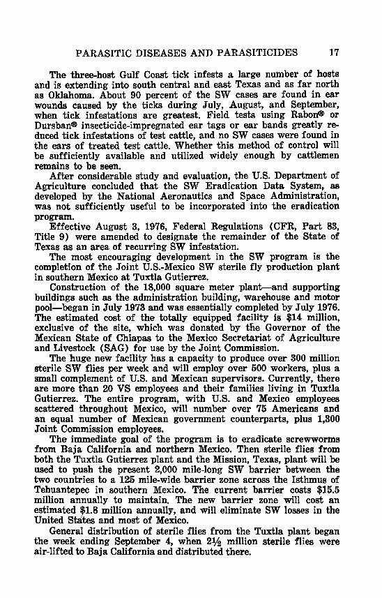
PARASITIC DISEASES AND PARASITICIDES 17
The three-host Gulf Coast tick infests a large number of hosts and is extending into south central and east Texas and as fa r north as Oklahoma. About 90 percent of the SW cases are found in ear wounds caused by the ticks during July, August, and September, when tick infestations are greatest. Field tests using Rabon@ or Dursbane insecticide-impregnated ear tags or ear bands greatly re- duced tick infestations of test cattle, and no SW cases were €ound in the ears of treated test cattle. Whether this method of control will be sufficiently available and utilized widely enough by cattlemen remains to be seen.
After considerable study and evaluation, the U.S. Department of Agriculture concluded that the SW Eradication Data System, as developed by the National Aeronautics and Space Administration, was not sufficiently useful to be incorporated into the eradication program.
Effective August 3, 1976, Federal Regulations (CFR, Part 83, Title 9) were amended to designate the remainder of the State of Texas as an area of recurring SW infestation.
The most encouraging development in the SW program is the completion of the Joint U.S.-Mexico SW sterile fly production plant in southern Mexico a t Tuxtla Gutierrez.
Construction of the 18,000 square meter plant-and supporting buildings such as the administration building, warehouse and motor pool-began in July 1973 and was essentially completed by July 1976. The estimated cost of the totally equipped facility is $14 million, exclusive of the site, which was donated by the Governor of the Mexican State of Chiapas to the Mexico Secretariat of Agriculture and Livestock (SAG) for use by the Joint Commission.
The huge new facility has a capacity to produce over 300 million sterile SW flies per week and will employ over 500 workers, plus a small complement of U.S. and Mexican supervisors. Currently, there are more than 20 VS employees and their families living in Tuxtla Gutierrez. The entire program, with U.S. and Mexico employees scattered throughout Mexico, will number over 75 Americans and an equal number of Mexican government counterparts, plus 1,300 Joint Commission employees.
The immediate goal of the program is to eradicate screwworms from Baja California and northern Mexico. Then sterile flies from both the Tuxtla Gutierrez plant and the Mission, Texas, plant will be used to push the present 2,000 mile-long SW barrier between the two countries to a 125 mile-wide barrier zone across the Isthmus of Tehuantepec in southern Mexico. The current barrier costs $15.5 million annually to maintain. The new barrier zone will cost an estimated $1.8 million annually, and will eliminate SW losses in the United States and most of Mexico.
General distribution of sterile flies from the Tuxtla plant began the week ending September 4, when 2vg million sterile flies were air-lifted to Baja California and distributed there.

18 REPORT O F THE COMMITTEE
TABLE O F ANTHELMINTICS MARKETED IN THE UNITED STATES
Kendall G. Powers, PhD, and Henry C. Hewitt, DVM
Arsenamide sodium-AR Butonate-B U n-Butyl Chloride-NBC Cambendazole-C AM Coumaphos-CO U Crufomate-CRU Dichlorophene & toluene-DT Dichlorvos-DI Diethylcarbamazine-DEC Diethylcarbamazine citrate-
Dec-C Diethylcarbamazine & styryl-
pyridinium chloride-DEC-S Disphenol-DIS Dithiazanine iodide-DTI Haloxon-H AL Hygromycin B-HYG Levamisole HCI-LH
Levamisole HC1 & piperazine
Levamisole phosphate-LPH MebendazolGMB Piperazine adipate-PA Piperazine-carbon disulfide complex with
phenothiazine-PCP Piperazine citrate-PC Pyrantel pamoate-PP Pyrantel t a r t r a t e P T
dihydrochloride-LP
Thenium closylate-TC Thiabendazole-TBZ Thiabendazole & piperazine citrate-TBZ-P Thiabendazole & trichlorfon-TBZ-T TicarbodineTI Trichlorfon-TR
The above does not contain all legally marketed anthelmintics. It does, however, include all those drugs that will be discussed during the presentation and have been codified in the Code of Federal Regulations, Food and Drugs, Part 500 to 599: 21, 1976.
**Listed parasites are not all inclusive and generally represent those species more commonly found in cattle, sheep, swine, equines or dogs.
Bureau of Veterinary Medicine, Food and Drug Administration, Rockville, Maryland 20852 USA

PARASITIC DISEASES AND PARASITICIDES
CATTLE
Name of parasite** Common name Therapy*
19
Haemonchus placei Ostertagia ostertagi Trichostrongylus azei
Trichostrongylus colubrif o m i s
Bunostomum phle b o t omurn
CoopeTiU sp.
papillosus Nematodirus
helvetianus
stTOngy&d e8
Monezia benedeni M. expansa
Trichurk owis 0 eso phagos tornum
radiatum
Dictyocaulus viviparus
Fasciola hepatica Fascioloides magna
ABOMASUM (nematode)
largestomachworm med. stomach worm hairworm
COU, CRU, HAL, LH, LPH, TBZ COU, CRU, HAL, LH, LPH, TBZ COU, CRU, HAL, LH, LPH, TBZ
SMALL INTESTINE (nematodes)
hairworm COU, CRU, HAL, LH, LPH, TBZ
hookworm LH, LPH
Cooperia threadworm
COU, CRU, HAL, LH, LPH, TBZ
thread-necked worm COU, CRU, LH, LPH, TBZ
(cestodes)
common tapeworm common tapeworm
LARGE INTESTINE (nematodes)
whipworm nodular worm CRU, LH, LPH, TBZ
LUNGS (nematode)
lungworm LH, LPH
LIVER (trematodes)
common liver fluke large american fluke

20 REPORT O F THE COMMITTEE
S H E E P
Name of parasite** Common name Therapy*
ABOMASUM (nematodes)
Haemonchus contortuslarge stomach worm CRU, HAL, LH, TBZ Ostertagia med. stomach worm CRU, HAL, LH, TBZ
Trichostrongylus axei hairworm circumcincta
CRU, LH, TBZ
SMALL INTESTINE (nematodes)
Cooperia ewticei Cooperia CRU, HAL, LH, TBZ Strongyloides threadworm CRU, TBZ
Trichostrongylus hairworm CRU, LH, TBZ
Nematodirus spathiger threadnecked worm
papillosua
colu brif ormia LH, TBZ
Bunostomum t r igmcephalum
Monezia expaxsa Thysanosoma
actinioides
Trichurk ovis Oesophagostomum
columbianum Chabertia ovina
Fmciola hepatica Fascioloides magna
Dictyocaulus filaria Muellerius capillaris
hookworm LH; TRZ
(cestodes)
common tapeworm fringed tapeworm
LARGE INTESTINE (nematodes)
whipworms CRU nodular worm LH, TBZ
large mouth LH, TBZ bowel worm
LIVER (trematodes)
common liver fluke large american
liver fluke
LUNGS (nematodes)
thread lungworm L H hair lingworm

PARASITIC DISEASES AND PARASITICIDES 21
SWINE
Name of parasite** Common name
STOMACH (nematodes)
Ascarops 8tTOng2/&~a thick stomach worm DI Hyostrongy~us red stomach worm
mibidus
SMALL INTESTINE (nematodes)
Strongyloides ransomi threadworm LH, TBZ Ascaris suum large roundworm DI, HYG, LH, PT Glo bocephalus hookworm
Trichinella spiralis trichina worm urosubulatus
(acanthocephala)
Macracanthorhynchus thorn-headed worm himdinaceus
LARGE INTESTINE (nematodes)
T&chul-is suiS whipworm DI, HYG Oesophagostomum nodular worm DI, HYG, LH, PT
dentaturn
KIDNEYS (nematode)
Stephanurus dentatus kidney worm
Metastrongylus elongatus
M . pudendotectus
LUNGS ( nemastodes )
lungworm LH
lungworm LH

22 REPORT O F THE COMMITTEE
EQUINES
Name of parasite** Common name Therapy*
Habronema sp. T.rich.ostrongglus axei
Parascark equolum
Stronggloides westmi
Anoplocephala magna A . perfoliata
Strongylus v u l g h
S. equinus
S. edentaturr
P h o b s t m a g k wivipaTa
Oxgulris equi
Cyathostomina
Dictyocaulus amfieMi
Setaria equina
STOMACH (nematodes)
large stomach worm hairwarm
SMALL INTESTINE (nematodes)
large roundworm BU, CAM, DI, LP, MB, PA, PP, PT, PCP, TBZ-P, TBZ-T, TR
threadworm CAM, TBZ, TBZ-P, TBZ-T
(cestodes)
large tapeworm medium tapeworm
large
large
large
small
large
small
LARGE INTESTINE (nematodes)
strongyle CAM, DI, LP, MB, PA, PCP, PP, PT, TBZ, TBZ-P, TBZ-T
strongyle
strongyle
pinworm
CAM, DI, MB, PCP, PP, PT,
CAM, DI, LP, MB, PCP, PP, TBZ, TBZ-P, TBZ-T
PT, TBZ, TBZ-P, TBZ-T
pinworm
strongyles
CAM, DI, LP, MB, PA, PCP,
CAM, DI, LP, MB, PA, PCP, PP, PT, TBZ, TBZ-P, TBZ-T, TR
PP, PT, TBZ, TBZ-P, TBZ-T, TR
LUNGS (nematode)
lungworm
BODY CAVITY (nematode)
abdominal worm

PARASITIC DISEASES AND PARASITICIDES 23
DOGS
Name of parasite** Common name Therapy*
STOMACH (nematode)
Physaloptera sp. stomach worm
SMALL INTESTINE (nematodes)
Strongyloides threadworm
A ncy los tomn caninum hookworm s t ercoralis
4. brndiense hookworm Uncinwa hookwarm
Toxocara canis large roundworm stenocephala
Toxascaris leonina large roundworm
DTI
DEC-S, DI, DIS, DT, DTI, NBC, TC, T I
DIS, NBC DI, DIS, DT, DTI, NBC
TC, T I
DTI, NBC, PA, PC, T I DEC, DEC-C, DEC-S, DI, DT,
IDEC, DEC-C, DI, DT, DTI, NBC, PA, PC
(cestodes)
Diplidizcm canizim double pored DT, T I
Taenia pisiform& no common name DT, T I Ec hinicoccus hydatid tapeworm DT
tapeworm
granulosus
LARGE INTESTINE (nematodes)
Trichzi& vulpis whipworm DI, DTI
HEART (nematode)
Dirofilaria immitis heartworm AR, DEC, DEC-C, DEC-S, DTI Dipetalonema heartworm
reconditum

REPORT OF THE COMMITTEE ON IMPORT-EXPORT
C h a i m n : G. B. Rea, Salem, OR Co-Chuimuw C. K. Jewell, Trenton, NJ
Frank Harding, Donald H. Spangler, John L. Hyde, Bert Hawkins, J. R. Langridge, Clint Booth, J. E. Thomas, B. C. Swindle, R. L. Evinger, Clarence H. Pals, F. E. Henderson, R. W. Gerding, D. E. Herrick, T. A. Kincaid, H. M. Steinmetz, William Wilson, Forrest Ireland, J. L. Hourrigan, Ron Caffey, Mark Trask, J. S. Walker
The Committee on Import-Export met and considered problems
We were updated on the following subjects :
spoken to a year ago as well as four new topics.
1.
2.
3.
4.
6.
6.
7.
The Stewart Air Force Base facility, as a replacement for the Clifton, New Jersey Import Station has been funded at five and eight tenths million dollars and is still expected to be operable by 1979. It will cover 75 acres and provide a greatly expanded import facility.
Fleming Key-The first bids were let and found to be un- acceptable. This has caused a redesign of facilities and new bids will be let on November 19th of this year. It is expected that this facility will be available by late 1978.
Third country cattle imports from foot and mouth infected countries are still authorized, but no requests are pending at this time. This method of importing new genetic strains will be phased out when Fleming Key is activated.
The Committee’s request for EIA and piroplasmosis tests on Equidae imported from other countries was implemented this past year.
Our request for certification of semen presented for export was not acted on due to the lack of similar requirements by the several states. Several private organizations in the semen export business indicated that they could handle the situation effectively by direct negotiation.
The Committee’s request that importers be notified of State animal health requirements which are in addition to those im- posed by the Federal Government was adequately implemented by the Import-Export Staff.
The Committee’s recommendation that a separate off ice be established to handle Mexican border problems was studied by APHIS and found not to be feasible a t this time.
24

IMPORT-EXPORT 25
8. Our concern for the verification of safety of imported milk and milk products has not been satisfied. Under consideration is the UHT (ultra high temperature) procedure presently used by several foreign countries.
9. We received a report on the sealing of galleys and controls of garbage on ships and planes arriving from foreign countries with meat and meat products from Foot and Mouth Disease countries. Plant protection and quarantine, although still not completely satisfied, has increased the number of personnel and is backed up by APHIS personnel. This has improved general surveillance.
New subjects included discussion of the presidential veto of H. R. 10073 dealing with adequate inspection of rabbit meat imported from China and Poland. It is anticipated that a new bill will be introduced into the new Congress with the hope that it will be favorably received.
The continuing problem of strays and smuggled animals being the source of ticks especially, and any of a number of exotic diseases, was discussed at length. It was decided that the present situation posed a serious and potentially explosive situation to the extent that it war- ranted requesting emergency status to be declared by the Secretary of Agriculture. (A resolution to this effect has been presented to the Resolution Committee.)
Concurrently, with this discussion, was the proposal for expanded research on the efficacy and method of application of pesticides to aid in control and eradication of ticks and horn flies. Secondary, but a highly significant benefit is the reduction of the inroads of screw- worm, through the use of such a pesticide applied by means of materials, A La Dog Collar, etc. A resolution on this subject has also been presented to the resolutions committee.
Representatives from the American National Cattlemen’s Associa- tion initiated in depth discussions concerning the limiting of cattle, sheep, and semen exports due to embargoes placed by several foreign countries on these United States products. This embargo is caused by the presence of Bluetongue in our herds and flocks. Research funds for study of the adequacy of our testing methods and procedures and knowledge of the location and severity of the problem in the United States is sorely needed. A Resolution was also proposed and presented to the resolutions committee.

REPORT OF THE COMMITTEE ON RABIES
Chairman: R. Keith Sikes, Atlanta, GA Co-Chairman: E. A. Carbrey, Ames, IA
John Brown, L. N. Butler, Victor Cabasso, James Glosser, Marvin Goff, Bruce Kaplan, J. T. Frank. T. D. Njaka, A. L. Strating, E. H. Willers, W. G. Winkler
The Rabies Committee met on November 8, 1976 with a total of 16 members and guests present. During a three hour meeting, dis- cussions were held and appropriate actions taken as follows :
1. A resolution passed by our Committee in 1975 was inadvertently not presented b h u g h proper channels in Portland, Oregon. Our comm'ib.tee recommended its re-submission and passage as well as printing by the 1976 USAHA Resolutions Committee. It deals with the national acceptance of a standardized rabies vaccination/health certificate for small animals.
2. A special subcommittee was appointed to coordinate efforts to work with other national organizations to implement this resolu- tion. That committee will be composed of:
Dr. Bruce Kaplan, Chairman Dr. Marvin Goff Dr. Jim Glosser Dr. R. K. Sikes
3. The newly revised Compendium of Animal Rabies Vaccines was approved by our USAHA Rabies Committee. Our Committee apol- ogizes for the delay in getting this much needed and up-dated revision of the Compendium of Rabies Vaccines out to every state. The reason for the delay is due to administrative difficulties ex- perienced a t the Center for Disease Control. Due to that difficulty, the national Association of State Public Health Veterinarians has accepted responsibilities for revising the Compendium on an an- nual basis and expects continued cooperation in this activity from USDA and USPHS. Dr. Kenneth Crawford, the Maryland Director of Veterinary Public Health is Chairman of that Committee. This newly revised Compendium will be published in January, 1977. It has the endorsement of two AVMA Councils, as well as the Con- ference of the State and Territorial Epidemiologists and the Veterinary Biologics Licensees Committee of the Animal Health Industries.
4. Discussions were held concerning classification and standardiza- tion for age-specific immunity as well as frequency of booster doses of vaccination in rabies endemic areas. Members of the USAHA Rabies Committee who also serve on the ASPHV Com-
26

RABIES 27
pendium Committee were advised to develop a clear statement on these issues for the 1978 Compendium Committee’s consideration.
6. From a recent international rabies symposium held in Atlanta, the chairman summarized advances in human antirabies treatments. The human origin rabies immune globulin is being used widely in the United States. It has virtually replaced the equine origin, rabies serum product. As expected, no adverse reactions have been reported following use of this product. In addition, the new- ly developed tissue culture inactivated rabies vaccine, using hu- man diploid cells (WI-38 strain) and a production virus strain, has been tested in many hundreds of people in the United States as well as in other selected countries. It has been used as an ex- perimental vaccine following the protocol required of such vaccines under an I.N.D.A. (Investigation of New Drugs Application). All indications thus fa r are that this vaccine is more potent and biologically cleaner than tfie duck embryo rabies vaccine. It is currently being considered for commercial licensure. When licensed, it will expectedly allow a reduction of injections from 23 to about 4. Passive immunization will, of course, continue to be indicated in post-exposure treatments.
6. The USAHA Rabies Committee endorses the recommendations and conclusions of the National Conference on Dog and Cat Con- trol which resulted from a meeting held February 3-5, 1976, in Denver, Colorado. We especially support the goals, and the spirit, in which these were written in an effort to improve animal con- trol in the United States.
7. The Committee was honored to have three distinguished represen- tatives from the Dade County Veterinary Medical Association during a portion of our deliberations. Dr. Bob Knowles, noted veterinary practitioner in Miami, Dr. Tom Campbell, president of the Dade County VMA, and their legal counsel, attorney Everett A. Cleary, stated their problem, more legal than technical. In essence, veterinary practitioners have a dilemma in defining whether Date County is a rabies “high risk area”. If so, the literature which accompanies the so called “3-year vaccines” states that these vaccines would be administered annually.
The Dade County veterinarians will accept vaccines for longer than one year if this dilemma of defining “high risk area” is clarified. Considerable discussion was held on this matter which also affects many states and cities in the U.S. The Committee recognized this particular statement was needed several years ago when there was a higher incidence of dog rabies, but it is no longer needed. Further, we know that the rabies vaccine licensed by the USDA Veterinary Biologics are excellent, perhaps the most immunogenic of all veteri- nary vaccines. Thus, Drs. A1 Strating and Jerry Peacock have been

28 REPORT O F THE COMMITTEE
requested to present this matter to the licensees of rabies vaccines and ask that they consider removing it from their literature. Several months to a year will apparently be required to reach an understand- ing in the matter.
Mr. President, this concludes the report of your 1976 Rabies Com- mittee.
Respectfully submitted R. Keith Sikes, Chairman

REPORT OF THE COMMITTEE ON FOOD ANIMAL HYGIENE AND INSPECTION
Chairman: E . Baker, Madison, WI Co-Chairman: James K. Payne, Washington, DC
David Bedell, W. H. Dubbert, Jim Fowler, M. R. Humphrey, D. C. Kelley, M. R. Levy, Joan Donnelly, Sarah Brite, L. J. Raforth, James A. Bell, D. D. Breeden, C. T. Dominquez, J. W. Holcombe, E. E. Kerr, R. L. Parker, M. A. Simmons, R. J. Lee, J. A. Libby, Gary Smith, Henry Woodard, George Estes The Committee wishes to recognize the loss of Walter Fechner
and express its appreciation for his leadership and contributions 4x1 the Committee over the past years.
Your Committee received reports on the following subjects : 1. A Pilot Program For Enzyme Labeled Antibodies
The federal meat and poultry inspection program’s project to develop a serological system for continuous inspection of swine for the detection of trichinosis has made significant progress in the past year. A false positive problem that had existed has been largely identified and eliminated. During this fiscal year the test will be further refined, adapted to high speed testing and utilized in a field trial.
A study of the incremental costs associated with the production of certified pork has been contracted for by MPI. This will enable packers to compare their current costs for producing certified pork by freezing with the serological system if i t becomes available for that purpose.
Your Committee recommends continued effort in implementing this project. 2. Residue Investigation Memorandum o f understanding (MO U)
A Memorandum of Understanding (MOU) with the states of Dela- ware, Maryland, Pennsylvania and Virginia, the U. S. Department of Agriculture Regional Off ice, APHIS, and FDA has been implemented. It is a coordinated investigation program to follow-up USDA reported tissue residues in meat. Eighty-six original tissue residue investiga- tions have been carried out in 1976 by the four states in cooperation with FDA and USDA. Two planning and evaluation sessions have been held with all of the contracting parties. The first meeting was held in April of 1976 in Dover, Delaware. The second in Annapolis, Maryland on October 20, 1976. To the present we believe this pilot program has been innovative and flexible, bringing to bear a uniform approach to producer compliance and consumer protection. To sum- marize : The avoidance of duplication of investigational efforts and procedures provide better producer compliance and a help in sustain- ing public confidence in our regulatory agencies.
29

30 REPORT O F THE COMMITTEE
3. A Review of the Swine Mgcobacteriosis (Tuberculosis) symposium. Veterinary specialists, meat industry personnel, and other in-
terested persons from throughout the United States met in Madison on September 30 and October 1, 1975 to exchange ideas on the prob- lem of swine tuberculosis as a part of the National Conference on Mycobacterial Infections in Swine.
Economic losses due to this disease were reported to be about $5 million in the United States.
Symposium topics focused on the current knowledge and the present status of this swine disease.
Speakers at the two day event viewed the latest information on the etiology, diagnosis, and epidemiology of tuberculosis in swine. One of the purposes was to discuss the feasibility of developing a national eradication program. Symposium participants recommended research needs to further that goal and also questioned the justif ica- tion of current meat inspection regulations.
USDA and State regulatory agency veterinarians explained epi- demiological findings, diagnosis, and control procedures. An exhibit prepared by USDA, National Animal Disease Center veterinarians, displayed tubercular gross lesions and demonstrated the laboratory aids used in diagnosing the disease. Several speakers discussed the public health concerns relating to swine tuberculosis.
Participants included University of Wisconsin as well as State, National and an International authority on tuberculosis. Among the 16 speakers were Dr. Frank Mulhern, Administrator of the USDA Animal and Plant Health Inspection Service: Dr. H. H. Kleberg, Di- rector of the tuberculosis research unit at Onderstepoort, South Africa ; Dr. Lawrence S. Farer, Assistant Director of the Tuberculosis Control Division of the Center for Disease Control a t Atlanta, Georgia ; Dr. W. L. Mallman, Tuberculosis researcher at Michigm State University; and Dr. Roy Corpe, Public Specialist from the Northwest Georgia Regional Hospital.
Following the formal presentations of the symposium, partici- pants were given an opportunity to state any specific needs or re- quests they thought would help resolve this important disease prob- lem.
Motions or resolutions that were approved included: 1) that a committee be formed to gather known data and act as a clearing house for information on swine mycobacterial infections ; 2) that the major areas of research needed involve the fields of veterinary and human public health, transmission, bacteriology, immunology, man- agement and related factors.
Discussion involved the need for an action group, need for peer review, and the designation and acceptance of Livestock Conservation Institute as the coordinating organization. 3) That the nomenclature be modified so that 171. avian complex hereafter be referred to not as tuberculosis but as mycobacterial infection or mycobacteriosis.

FOOD ANIMAL HYGIENE 31
Discussion involved the difficulty of differentiation between avian and other mycobacterial lesions on postmortem examinations. 4) That we proceed immediately to use the available knowledge and tools to reduce the economic losses to the producer and packer whenever possible. Discussion suggested this be done on a national or unified approach. 5) That industry representatives contact the appropriate officials in the Department of HEW relative to possibly changing the disposition criteria if supplied the supporting data.
Much of the data presented here can be found in more detail in the Symposium Proceedings which can be obtained from Livestock Conservation Institute, 19 W. Chicago Ave., Hinsdale, Illinois.
4. Salmonella Advisory Committee The Executive Secretary of USDA’s Salmonella Advisory Com-
mittee reported on their activities. The Committee charter signed a year ago by the Secretary directed that efforts be made to reduce the incidence of Salmonella in animals, humans and food. In response to the charter, fifteen members representing all facets of the issue met February 25, at which time six subcommittees were identified: 1) Production, 2) Breeder and Hatchery, 3) Feed and Feed Ingredient, 4) Processing and Distribution, 5) Consumer and Education, and 6) Research. At their June 17th meeting the Subcommittees reported on the general direction each planned to take. Recommendations to the Secretary are expected at future meetings. Your Committee recom- mends that we be kept posted on the Salmonella Advisory Committee activities.
5. Contract Study of Meat, Poultry and E g g Inspection Meat, poultry, and egg inspection are mandatory services which
the Secretary of Agriculture is required by law to provide on demand. Not only can individual processors request and receive inspection, but States which cannot or do not wish to maintain their own inspection systems on a par with Federal standards can turn those responsi- bilities over to the USDA.
The cost of Federal inspection activities has increased about 15 percent a year since the passage of the modern program legislation. In order to moderate this trend toward higher costs and increasing Federal regulation, there is need to investigate procedures for in- creasing efficiency in the programs while continuing to assure that the U.S. supply of meat, poultry, and egg products continues to be acceptably branded and unadulterated.
There is some suggestions that the enforcement of complex and stringent Federal inspection procedures may have interfered with growth in the meat, poultry and egg industries and increased costs at both the farm and retail level beyond that necessary for assuring that such products will comply with standards set by the Department.

32 REPORT O F THE COMMITTEE
A. Purpose of Study The purpose of the study is to review the USDA meat and poultry
inspection and egg and egg product inspection programs so as to identify alternative systems that would improve cost-effectiveness, eliminate unnecessary interference in commerce and yet assure that meat, poultry, and eggs for human consumption are unadulterated and not misbranded.
The study will determine the effectiveness of inspection pro- cedures currently used in Federally inspected plants and channels for distribution for detecting different types of adulteration and mis- branding in different parts of the marketing system and to propose alternative procedures which could be used t o detect them more efficiently. The study will specify for each proposed system the changes in risk to human health, changes in the degree of interference in commerce, and estimates of associated changes in Federal costs.
It will be the purpose of the study to present alternatives for more efficient means of pursuing these consumer protection programs. The principal criteria for efficiency will be: (a) reduced Federal cost per unit of work accomplished; (b) reduced obstacles and reduced costs of preparing products for the consumer market, and ( c ) ac- ceptable standards for product quality and safety. B. Conduct of Study
The study proposal should be developed as a study of two separate systems: (1) that involves the marketing of livestock, poultry, and their products ; and (2) that involves similarly the marketing of eggs and egg products. Each of these parts of the proposal should identify separately the study plan, organization and costs associated with that part alone. Contracting of the egg and egg products section will be optional. C. Content of the Study
For each of the two marketing systems discussed in 1I.B. the study will:
(1) List the major types of adulteration and misbranding that may occur and, for each type, describe where in the food marketing chain from farm to ultimate consumer i t may occur; how or why it may occur; the estimated probability of its occurrence; how i t may be detected if i t has occurred; the potential impact on product de- mand if i t occurs; and the potential consequences to the consumer if i t has occurred, including health risk and economic consequences.
(2) In the above eontext review and evaluate on the basis of available evidence and expert judgment current USDA inspection procedures and staffing regarding their capability and efficiency for detecting adulteration and misbranding at identified critical points in the production-marketing system using current USDA standards. The review of staffing will include the study of current USDA posi- tion requirements and organization.

FOOD ANIMAL HYGIENE 33
(3) Review current inspection procedures to determine the ex- tent to which changes in them would effect the probability of adul- teration and misbranding and in turn the economic and health risks to consumers.
(4) Project to 1980 to 1985 the USDA and State resource require- ments for inspection under the current system with current standards assuming a moderate growth in population and real income, sufficient growth in the supply of meat, poultry and eggs to accommodate the demand, and assuring minimal changes from current trends in the structure of meat and poultry industries.
(5) Define and analyze in the context of each of the 1980 and 1985 projections sets of alternative procedures or systems that have potential for implementation and modification of the current system and show the extent to which they minimize :
a. USDA and other public sector outlays. b. Fsrm-retail marketing costs. c. Economic and health risks to consumers and the public in
general. The development and analysis of potential changes to the current
system should also consider : d. The extent to which probability sampling techniques can be
used. e. Technological capabilities and the need for further research and
development. f. Possibilities for revising current product standards to permit
the use of remote monitoring procedures. g. Alternative approaches to inspection for achieving the goal of
consumer protection including such things as consumer education programs or eliminating sources of adulteration at the farm and other points in the production-marketing system.
h. Needs for new legislation and significant changes in Depart- mental regulations.
i. Sources of potential social or political resistance to specific kinds of changes.
6. Present for consideration by the USDA advisory committee a draft summary of an overall strategy for implementing the alterna- tives developed a t (5) with appropriate documentation of significant impact on costs and other performance criteria of the newly proposed systems.
The Management Consulting Firm, BOOZ, Allen & Hamilton, Inc. will contact appropriate state officials for input, contacts will pri- marily be through questionnaire, however, on-site interviews will be held where time permits.
The study is to be completed at the end of nine months.

REPORT OF THE COMMITTEE ON ANIMAL WELFARE Chairman: A. E. Decoteau, Waltham, MA *Co-Chairman: J. C. MacFarlane, Pembroke, MA
Christine Stevens, Oscar Clabaugh, *L. A. Griner, Paul Zillman, Charlie Jungmichel, P. M. Eppele, John Smiley, Walter F. Lewis, Dale Schwindaman, E. Mickey Stewart, *D. H. Spangler, *Clifford Crago The Committee on Animal Welfare submits the following recom-
mendations for presentation to the Secretary of Agriculture. 1. To require photographic identification of each dog and cat sold
to random source dealers for resale and of the license plate of the car of the persons offering the animals for sale. Purpose of the documentation: to discourage theft of animals for resale and to facili- tate successful prosecution in the event of a violation.
This again reiterates the Committee’s concern in the area of dog thefts which cause distress to pet owners.
2. Be it Resolved that USDA is requested to publish final regula- tions on exercise for laboratory dogs.
3. The Animal Welfare Committee recommends that the provisions relating to multiple surgical operations on a single dog published by the Laboratory Animal Resources in “The Guide for the Care and Use of Laboratory Animals” (page 18) 6e incorporated in regulations and/or standards promulgated by the Secretary of Agriculture under the Animal Welfare Act.
4. It was resolvcd that the U. s. Animal Health Associ a, t’ ion re- quest the U. S. Department of Agriculture to revise Veterinary Ser- vices Forms 18-23, Annual Report of Registered Research Facility, to clarify and more accurately reflect the actual use of anesthetics, analgesics, and tranquilizers on animals involved in research, testing or experimentation.
5. It was resolved that the United States Animal Health Associa- tion recommend to the U. S. Department of Agriculture that export requirements for the shipment of horses by ocean vessel be revised and strengthened to assure that ship fittings, space, feed and water provisions and such other requirements necessary be adequate to assure the humane care and treatment of horses transported by water.
A subcommittee was formed to gather information regarding the effects of diesel exhaust fumes on livestock while in transit. If re- search indicates adverse health effects, then further information be sought on improved exhaust channeling to reduce adverse health impact to a minimum. A report will be submitted to the Animal Wel- fare Committee a t the next annual meeting. Subcommittee Chairman is Mickey Stewart of Nebraska. Members are Christine Stevens, Washington, D.C. and Dr. L. N. Butler of Arizona.
If anyone present knows of research regarding diesel fumes, please give me a contact name. *Not present at committee meeting.
34

REPORT OF THE COMMITTEE ON STATE FEDERAL RELATIONS
Chairman: A. E. Janawicz, Montpelier, VT H. E. Goldstein, L. E. Bartelt, W. L. Bendix, J. F. Andrews, J. C. Shook, T. A. Ladson, D. H. Spangler, T. F. Zweigart, N. R. Swan- son
APHIS Veterinary Service Review Brucellosis
The Committee has considered the proposed 10 year Brucellosis eradication program which, in effect, is a 5 year program for eradi- cation in a state, approached nationwide on a staggered basis. The Committee feels i t is a reasonable and feasible approach to the prob- lem, and one that both the organized profession and industry would support, provided there is an open commitment that USDA will implement and forcefully adhere to its provisions. .We are convinced that there should be no compromise with the goal of eradication. Any lesser approach compromises our obligation to the protection of public health and the nation’s economy.
We urge that top administration and policy makers adopt this plan as a top priority item, and make every effort to make the con- tinuing commitments it requires. Hog Cholera
In the current Hog Cholera outbreak, two and one fourth million dollars has been spent for indemnities, and almost $1,000,000 for support. This outbreak can most likely be attributed to the feeding of improperly cooked garbage. The threat still is that, it appears there may be infected pork in trade channels involving a t least 17 states. We were told in the past, that when sufficient states had out- lawed all feeding of garbage to swine within their jurisdictions, that USDA would then move forward toward banning this practice nation- wide. Fifteen states have now done this, and again garbage emerges as the principal culprit for perpetuating the disease. The time has come to ban garbage feeding nationwide. Other Diseases and Activities
The Committee is pleased with the progress in obtaining addi- tional physical plant facilities and the laboratory backup services that are being provided and planned.
The Committee wishes to express support for the ongoing animal disease eradication programs, and feel that at least, acceptable prog- ress is being made with the exception of the following:
(1) Bovine T.B. (2) Scabies and Ticks (3) Anaplasmosis
We urge additional support in these areas.
35

36 REPORT O F THE COMMITTEE
Manpower
In the face of severe manpower restrictions being imposed by Federal authority and some states, we cannot permit the situation to deteriorate to the point where animal health programs lose their ef- fectiveness. We urge USDA to establish a small task force on man- power (both professional and sub-professional,) made up of repre- sentatives from USDA, AVMA, USAHA, and the field of education to seriously study the problem and develop alternative approaches towards a solution. Several alternatives suggest themselves singly or in combination. Those are:
1. Increased Federal employee staffing. 2. Increased State employee staffing. 3. Increased use of temporary employees. 4. Fee basis commitments on part of outside personnel based on
either full time or part time service. 5. Contractual arrangements for specific or area oriented tasks
and strictly governments supervised. There are undoubtedly other alternatives or combinations, but
the time to bring the major concerned elements together, both in and out of government, to seek solution is now.
Meat Inspection The Committee continues to commend APHIS for maintaining a
most outstanding Consumer Protection Program involving meat, poultry, and the products associated with the allied industries. The program presents a most significant insurance as well as assurance to the citizens of this nation.
It was most gratifying to be informed that the finances of this program had improved since our last meeting and we commend the administrators for this improved situation.
The Committee was advised that a total of 17 states now have federal programming in poultry inspection. It was further advised that now the nation has a total of approximately 9000 federal plants representing 90% of the total product in relation to 7400 state in- spection plants 10% of the total product. In answer to direct ques- tions, staff of meat inspection indicated that in some of the recently designated states the number of needed personnel was reduced and that costs were reduced, particularly in states where salary structure were higher than federal.
The Committee then discussed how the industries received desig- nation status in the recently involved states. Concensus, with rare exceptions, indicated that industry has accepted the designated pro- grams.
This Committee expresses sincere concern for the continuation of separate programs that are supposed to be of equal status but in

STATE-FEDERAL RELATIONS 37
reality provide for state programs that are not equal. If state in- spected plants are of equal status, the product from the plants should enjoy the same freedom of movement as federal product. This causes state administrators as well as state policy makers to view state pro- grams as discriminated. The overall economics involving judicial use of governmental funding provides the need for serious consideration of an 80-20 federal funding program.
Perpetuation of state programs that are equal to the federal pro- gram, but having in reality discriminated programs, is a great in- justice to the industry as well as to the taxpayer. The Committee points out that in many of the designated states the program carried out presently is less stringent bhan that program previously required to be carried on by the respective states.
The Committee commends APHIS Meat Inspection for its efforts in assisting the animal health inspection officials with their pro- gramming at plant level. It is further recommended that all effort be put forth to enhance communications with animal health and meat inspection personnel to improve the availability of trace-back animals in programs involving such effort. The Committee is cognizant of the need for a better identification system that would benefit not only animal health programs but also involves public health programs.
The Committee was informed of a Model Uniform Retail Store Ordinance. The Committee recognizes a dire need for this type of uniformity and recommends serious pursuit of this effort.
The Committee recognizes the need for protective residue pro- grams. APHIS Meat Inspection is commended for the pilot program involving Virginia, Maryland and Pennsylvania. All effort should be made to enhance communications to provide expedient traceback to the involved premises.
This Committee encourages the USDA, APHIS and the Food and Drug Bureau of Veterinary Medicine to cooperatively participate in a National Salmonellosis Symposium to be conducted by the USAHA Salmonella Committee sometime in 1977. It should be pointed out that the Council of Public Health and Regulatory Medicine of the AVMA has passed a resolution to cooperate in such a program.
Agricultural Research Service Our Committee reviewed with the ARS staff, the present and
projected research programs related to diseases and parasites of livestock and poultry. We are gratified to learn that suggestions made by this Committee in previous meetings, have been considered, and progress is being made in a number of areas that were of grave concern .
An example of this are the research projects in progress, or

38 REPORT O F THE COMMITTEE
planned for Brucellosis. While the eradication program enjoyed con- siderable success with the tools a t hand, i t became more and more evident that in order to complete the job with minimum delays and expenditures some new diagnostic and/or immunizing agents may be needed. The ARS research for a cell component that would be non- infective, but render immunity is a step in the right direction, and we hope will be given high priority. Review and revaluation of strain 19, which is being cvnducted is also long overdue. Hopefully, more spe- cific and effective diagnostic methods will develop from some of these pmjecb as we learn more intrioate details of the disease. We urge ARS and APHIS tb continue to cooperate in all faceits of these proj- ects to gain the maximum expediency in completing these studies. We are still deeply concerned about the prospect of the introduc-
tion of foreign animal diseases into the United States, particularly f oot-and-mouth disease. Recent discoveries regarding the survival of FMD virus in meat and milk products even after processing, and pasteurization, are placing new threats to our country. More recently ARS has reported the survival in pork casings and certain cheeses that hertofore were thought to be free. The Committee urges con- tinued investigation and research of the survival of FMD virus in meat and milk products, such investigation to determine approval of imported meat and milk derivatives for addition to processed meat and milk products. We support and urge increased emphasis on the FMD vaccine studies being conducted, in case eradication is not possible or feasible. This includes the construction of a vaccine pro- duction plant, warehouse, animal facilities, and increased laboratory space.
Pseudorabies losses in swine have doubled in the past year. While this has been confined to certain few states and areas, we urge a high priority be placed on research for a new diagnostic method to assist in preventing further spread in our swine population as well as other species.
Respiratory diseases of cattle are still responsible for serious losses. Our Committee would urge continuing research in this field to reveal agents involved, diagnostic methods, and control or eradica- tion methods. We support continuing research for immunizing agents for
equine infectious anemia, foot rot in sheep and pink eye in cattle. The discussion regarding the area control of insects and insect
vectors rather than the present individual animal or herd concept, aroused some new thinking in this field. Hopefully, ARS staff will develop recommendations and programs in this area.
An increased interest in poultry diseases and the need to give further attention to the problems in the production area rather than establishing controls and programs relating to the products. A case
I

STATE-FEDERAL RELATIONS 39
in point is the ongoing interest in Salmonella and drug residues. In the latter area we urge close cooperation with FDA, as well as APHIS to define the problems precisely, and attempt to head off the problem before it causes economic chaos to the industry.
While there seemed to be no serious complaints from ARS staff regarding manpower, our discussions with other agencies causes concern that the present personnel ceilings could have adverse effect in development of much needed new programs or even the needed expansion of existing ones. This Committee is willing to lend support at anytime or wherever needed, if we are kept informed of the needs and requirements.
We appreciated the opportunity to meet with staff, and look for- ward to future cooperation between APHIS and the United States Animal Health Association.

REPORT OF THE COMMITTEE ON INFECTIOUS DISEASES OF HORSES
Chairman: C. L. Campbell, Tallahassee, FL Vice Chairman: W. 0. Kester, Golden, CO
C. B. Dearborn, Jr., John B. Healy, Wayne Kirkham, Fred D. Maurer, Sidney R. Nusbaum, M. B. Teigland, R. C. Knowles, Charles S. Duncan, R. Keith Farrell, K. Jewell, Edward F. Hackett, Charles D. Vail, E. H. Honnen, T. S. Maddox, W. L. Anderson, John A. Smiley, H. C. King, Wilson P o ~ e l l , Jack M. Gaskin, Floyd Jones, Rosalie MacWilliams, Jesus Castaneda Garcia The Committee on Infectious Diseases of Horses met in Miami
Beach on November 8, 1976, with approximately 35 to 40 committee members and visitors present. A number of items of importance to the horse industry was discussed.
At the 76th Annual Meeting held at this hotel four years ago, a resolution emanated from the Committee on Infectious Diseases of Horses and was adopted by the Association urging that the Secretary of the Department of State and the Secretary of the Department of Agriculture take steps for the United States to become a fully par- ticipating active member of the Off ice of International Epizootics. It is gratifying to note that within the past year the United States did attain membership in the OIE, which will help in keeping abreast of world animal disease situations. We wish, therefore, to commend the Departments of State and Agriculture for their action to this end.
For several years this Committee has pressed for, and the As- sociation has concurred by resolution, passage of import regulations requiring the AGID test on all horse stock entering this country. We are pleased to report that such regulations have now been imple- mented, and the Committee wishes to express appreciation to the U. S. Department of Agriculture for its action.
The Committee had the opportunity to view the movie recently revised on Equine Infectious Anemia. The inclusion of the AGID test procedure and more recentqy developed scientific data certainly improves the quality of this film, which is intended for horse owner audiences. The film is available not only through extension sources, but from area Veterinarians in Charge and the Veterinary Services headquarters in Hyattsville as well.
You will recall in last year’s report that 34 states had at that time adopted requirements for negative EIA test for entry into those states, nine of which had additional regulations requiring negative tests for animals entering assembly points or for public or private sale. It was reported that currently 40 states have EIA entry require- ments, and ten of the 40 have the additional intrastate requirements for assembly or sale purposes.
40

INFECTIOUS DISEASES O F HORSES 41
The Committee received information that 14 states now have no requirement on the identification of horses reacting to the EIA test. This provides for great difficulty in many instances to control the movement of these animals, and it is only sound disease control and eradication procedure to identify infected animals, and the Com- mittee therefore urges these 14 states in the national effort to im- plement regulations which would provide a method of identifying EIA reactors.
The members were apprised of an Equine Infectious Anemia Research Workers Conference held in New York City on July 27, 1976, sponsored by this Association's Committee on Infectious Dis- eases of Horses. The report which was developed as a result of this conference has received wide circulation, and in the interest of de- termining whether there might be dissension on the part of in- dividual committee members on the various points made therein, it was reviewed in toto and a motion was passed that the Committee concur in and ratify the report as published. A copy of the report is attached and made a part of this report.
In 1974 this Committee refined the guidelines for a state control program for equine infectious anemia, which still contain valid parameters in dealing with EIA infected herds. Therefore, those states contemplating any action toward the implementation of regula- tions directed to controlling the disease should utilize these as current guides until further revision is offered by the Committee.
Comment ensued upon the apparent resentment of horse owners in not having negative EIA test papers checked at horse assembly points or in crossing state lines in too many instances, and relevant discussion disclosed that this was largely a problem of inadequate manpower to police such movements. In some areas horse show management is taking the responsibility for enforcing EIA test re- quirements, which efforts should be commended and encouraged in other areas where the lack of sufficient regulatory manpower is a contributing factor. It was suggested that law enforcement officials be encouraged to assist in further policing of the regulations.
In previous reports the Committee has emphasized the importance of horse identification as essential to disease control, as well as for legal and commercial aspects. The Committee reaffirms that this continues to be a problem and iterates the need for a practical means of identifying horses.
In spite of repeated attempts to stimulate the tabulation of the current horse population within the United States, no progress has been made in obtaining such a census. This Committee still feels that an urgent need exists for such information and recommends that the U. S. Department of Agriculture renew its effort toward the ac- complishment of this goal which is essential to equine disease control.
C. L. Campbell, Chairman Respectfully submitted

REPORT ON EQUINE INFECTIOUS ANEMIA RESEARCH
Clarence L. Campbell*, DVM and W. 0. Kester*, DVM WORKERS’ COmNFERENCE’‘:’
Meeting was called by the U.S. Animal Health Association and chaired by Drs. Clarence L. Campbell and W. 0. Kester. Research workers participating were Dr. Charles Issel-Louisiana State Uni- versity ; Dr. Vincent Saurino-Florida Atlantic University ; Dr. Le- land Grumbles-Texas A&M University; Dr. Leroy Coggins and Dr. Matt Keman-Cornel1 University; Dr. S. Lynn Kittleson and Dr. Robert Tashj ian-New England Institute of Comparative Medicine ; Dr. Fred Neal and Dr. Jack Gaskin-University of Florida; Dr. James Pearson-Veterinary Services Laboratories, U.S.D.A., Ames, Iowa. Participating also were Dr. Ralph Knowles, Chief Staff Veter- inarian, Equine Diseases, U.S.D.A. ; Dr. J. C. O’Connor-President, American Association of Equine Practitioners ; Dr. Charles Dunkin- New York Assistant State Veterinarian ; Dave Goodman, U.S.D.A. ; Dr. R. A. Greene-Editor of “Horse of Course”.
The principal purpose of the meeting was to review developed scientific data and to arrive at a concensus on certain aspects of equine infectious anemia in order to preclude erroneous information being released to the public.
The majority of institutions in the United States involved in E.I.A. research was represented. Progress reports and research plans were presented by each. The value, accuracy and use of the agar gel immunodiffusion test, otKerwise known as the Coggins test, were reviewed and the opinion of the conference is summarized below.
It was agreed that the role of the AGID test positive horse which shows no clinical signs of E.I.A. is the number one problem and should receive top research priority. Statements were noted in a few horse periodicals indicating the belief that such horses are not a potential disease spreader under natural conditions and consequently need not be quarantined. It was the concensus that this might be true in a proportion of the horses but data presented at the meeting by antagonists, as well as protagonists, of this concept demonstrated that inapparent carriers have served as sources of virus for transmis- sion under natural and experimental conditions.
Some of the things the test can and is doing for serious minded horse breeders and owners are:
(1) By requiring a test prior to purchase a buyer is now reason- ably sure he is not running the risk of bringing home an infected horse as a potential spreader of the disease.
(2) By requiring a test before admitting a visiting mare or any
*Co-Chairman Committee on Equine Infectious Diseases **Held in New York City, July 27, 1976
42

EQUINE INFECTIOUS ANEMIA RESEARCH 43
other horse on a breeding farm a breeder is reasonably sure that no spreader of the disease is on his premises.
(3) By requiring a test prior to acceptance the operator of a training stable, center or farm is reasonably sure no spreader of the disease is among his stable of horses.
(41 An auction sales company by requiring a test prior to selling now knows and can reasonably assure their customers that they will buy no positive reactor or potential disease spreader.
(5) Insurance companies by requiring a test prior to insuring a horse can assume with safety that the policy is issued on a healthy animal not infected with E.I.A.
(6) A veterinarian in issuing a health certificate knows that if a Coggins test is allowed and included he has done a more thorough job for his client.
(7) The show horse owner exhibiting at a show where a test is required prior to entry is reasonably sure that his horse will not be exposed while on the show grounds (even though the requirement is not fully enforced)
(8) Race horse owners now know there is essentially no chance of their horse being exposed at a race track because practically all tracks now require a negative test prior to acceptance at the track.
It was noted that race track management learned years ago that if they were to attract valuable horses the best possible assurance must be given that E.I.A. was not present. This was done by requir- ing a negative test on all horses racing a t tracks and by educating horsemen on how the disease was spread from horse to horse on hypodermic needles. It is generally accepted that all recorded out- breaks that have occurred at race tracks in the United States have been caused by the transfer of E.I.A. virus primarily from asympto- matic carriers to susceptible horses by means of hypodermic needles or other instruments in common use. Significantly no new outbreaks have been reported at tracks since use of the Coggins test has been implemented as a protective measure.
(9) Obviously the Coggins test whether voluntary or required has removed a large number of potential disease spreaders from the horse traffic pattern, thus vastly reducing the probability of future out- breaks.
-- -
Test Limitations The term “reasonably sure’’ is used in the foregoing because in
veterinary medicine there is no sure thing. The tight surveillance and quality control system exercised by
U.S.D.A. over the complete laboratory testing program was reviewed and it was agreed that the Coggins test was probably one of the most accurake biological tests yet developed in either human or veterinary medicine. With the exception of some foals with passive antibodies, false positive reactions have not been confirmed. It was

44 REPORT ON WORKERS CONFERENCE
observed that in extremely rare instances a very weak positive re- actor might be most difficult to read and that in spite of their in- tensive training could be recorded as negative by personnel in one laboratory and not by another; thus, further emphasizing the role and need for the U.S.D.A. Veterinary Services Laboratory, Ames, Iowa, for reference on questionable cases.
It was reaffirmed that foals may give a false positive reaction while nursing positive mothers, and that the Coggins test cannot differentiate the infected foal from the foal with passive antibodies until about six months of age. Further, badly diseased horses some- times give false negative reactions. It was noted that the same per- tains to practically all comparable tests in man and animals and, while the individual horse which is the exception might pose a prob- lem, the numbers involved in either category are infinitesimal. Also, the foal with passive antibodies is not a threat and soon corrects itself, and the false negative badly diseased horse being clinically ill will limit its disposition irrespective of test results.
It was reaffirmed also that the Coggins test may miss horses re- cently infected and that the same pertains to all other long useful diagnostic tests. No animal can be found positive for any disease until he has been infected long enough for the body to develop measurable antibodies or other evidence that the disease agent is present. This period may range from a very few to several days and is why followup tests are required in most disease regulatory pro- grams.
It was recognized that none of the foregoing limitations was reason for doubting or not using the Coggins test because these limitations have long been known in veterinary medicine and under- stood, managed and eliminated as practical problems in connection with E.I.A. as well as many other diseases.
Regarding the asymptomatic test positive reactor horse the follow- ing was reaffirmed :
(I) It has been established that some are potential spreaders of E.I.A. (2) It has not been established that all are potential spreaders. (3) It has not been established that any are not potential spreaders. (4) No practical means exists for differentiating between a potential spreader and a possible non-potential spreader. (5) In light of exist- ing knowledge, for regulatory disease control purposes, all must be regarded as potential spreaders of E.I.A.
In discussing other specific problem areas the following was re- affirmed :
(1) Horses known to have been infected virus carriers remain carriers throughout life.
(2) Adult horses known to have been positive reactors to the Coggins test remain positive reactors indefinitely.
(3) Under proper conditions a considerable percent of disease free foals may be produced and raised from E.I.A. infected stallions and/or mares.

EQUINE INFECTIOUS ANEMIA RESEARCH 45
(4) Instances were reported where an infected stallion was safely used on disease free mares. One mare with a vaginal injury was believed to have become E.I.A. infected through virus carried in semen.
(6) Virus has been transmitted by horse flies under controlled conditions from asymptomatic carrier horses to susceptible horses but less readily than from acutely infected horses wherein virus transmission has been demonstrated by a single horse fly.
(6) Species of horse flies are believed to be the principle E.I.A. vectors in nature.
(7) Studies on fly behavior and practical experience indicates that a two hundred yard buffer zone as practiced in some states is an acceptable barrier for quarantine purposes. Effective quarantine requirements may vary with the time of year and geographical loca- tion.
(8) In view of findings in recent research done with washed leukocytes, work is now underway to further extend the efficacy of the animal inoculation test.
(9) Research on chemotherapy should be continued ; however, success in the near future is not foreseen.
(10) Research on vaccine development should continue. Again, success in the near future is not foreseen.
It was agreed that existing control programs had problems and did not appear to be popular in some areas. However, no one could advance any plan or ideas that might improve them. One participant (Dr. Greene) recommended that all control programs be discontinued. It was suggested that test positive reactor horses be released from quarantine if they could clear three negative animal inoculation tests. This will be considered but past experience indicates few if any would be cleared through such procedures. Further, the procedure would be extremely time-consuming and expensive.
It was reaffirmed that research should be continued and ex- pedited in the following areas :
(1) Role of the A.G.I.D. test positive horse that shows no clinical symptoms-is he or is he not always a potential disease spreader under natural conditions and, if not, how does one differentiate be- tween the potential and non-potential spreader horse?
(2) Vaccine development. (3) Chemotherapy treatment. (4) Role of insects as vectors. (5) Chemical disinfectants. (6) Better defining of the mare-foal relationship and the stallion-
mare relationship when either or both are infected with E.I.A. The next E.I.A. researchers’ meeting is scheduled for the Spring
of 1977 at the U.S.D.A. National Animal Disease Center, Ames, Iowa. Informal sessions are anticipated in Miami Beach this November in connection with the annual meeting of the United States Animal Health Association.

REPORT OF THE COMMITTEE ON EVALUATION AND DEVELOPMENT OF STATE-FEDERAL PROGRAMS
Chaivmarx J. L. O’Harra, Reno, NV Co-Chairman: Douglas R. Stauf f er, Pickerington, OH
J. F. Andrews, L. E. Bartelt, W. L. Bendix, J. R. Bishop, D. E. Flagg, Tom Cook, H. E. Goldstein, J. L. Hourrigan, T. A. Ladson, Robert Larramore, E. A. Schilf, H. Q. Sibley, 0. H. T i m , W. C. Tobin, C. D. VanHouweling, Paul Zillman, A. E. Janawicz The U. S. Animal Health Association Evaluation and Develop
ment of State-Federal Programs Committee met at 1 :30 p.m. Thurs- day, November 11, 1976 with eight members and seven visitors present. The 1976 Committee report was reviewed and the Committee requested an updating on the situation in regard to filling Federal positions and other activities to increase efficiency in program man- agement. The Committee was pleased to receive information that the hiring process for Federal employees has been speeded up consider- ably although some damaging delays still exist. The Committee com- mends APHIS on the progress made in overcoming hiring delays that have existed under the present Civil Service procedure. It is recognized that Veterinary Services continues to operate within a ceiling, although some relief appears to be forthcoming.
A discussion was held by the Committee in regard to allocation of resources under the present accelerated Brucellosis Eradication Pro- gram. It was determined that resources are being utilized in those areas capable of most effectively using that additional support.
The Committee recommends that both State and Federal agencies reevaluate manpower assignments on a field level and provide more effective supervision to avoid overlapping assignments and duplica- tion of effort.
The Committee recommends that a program be inaugurated on a StateFederal level in regard to the eradication of Pseudo-rabies in swine. The Committee also strongly recommends new funding for this program and that it not be developed at the expense of present ongoing programs. The Committee expressed concern regarding the production and uncontrolled distribution of Pseudo-rabies serum and urges that each state take the necessary steps to develop adequate controls for such biologics.
The Committee has presented two resolutions to the Resolutions Committee for consideration, one resolution in regard to apathy in the cattle scabies eradication program, the other in regard to the cattle fever tick situation.
The Committee was apprised of the cost relative to operating Veterinary Services Laboratories a t Ames, Iowa, and urges a con- tinuing effort to negotiate reduction of overhead charges.
Mr. Chairman, the Committee respectfully submits this report to the Executive Committee for consideration.
46

REPORT OF THE COMMITTEE ON MASTITIS
Chairman: L. F. Williams, Storrs, CT Co-Chairmart: R. B. Bushnell, Davis, CA
Charles N, Dobbins, Richard Famar, N. Bruce Hayna, D. A. Kirkbride, B. R. McCallon, John S. McDonald, Kermit J. Peterson, Donald S. Postle, Arlan R. Smith, Douglas N. Stern, Gene H. Swenson, Rufus F. Weidner, Kenneth M. Weinland The annual open meeting of the Mastitis Committee of the United
States Animal Health Association was convened at 1:30 PM on November 8, 1976. Six guests and six members were present.
The Mastitis Committee last year considered a resolution regard- ing finite and zero tolerances for drugs in edible animal products. It was referred to this year’s Biologics Committee for their considera- tion. A recommendation was made by this committee that this same resolution be submitted to the Pharmaceutical and Toxicology Com- mittee for their consideration.
This committee recommends that the Executive Committee of the United States Animal Health Association request the National Mas- titis Council to appoint a representative to be included in this Mastitis Committee.
Dr. K. M. Weinland described in detail the results of coordinated herd health programs on mastitis control in Indiana dairy herds.
A discussion was held about established somatic cell limits in milk for interstate shipment. This committee recommends that the limit of one and one-half million somatic cells per milliliter in milk for interstate markets be lowered to one million.
This constitutes the report of the Committee on Mastitis and we respectfully submit this report for approval by the Executive Com- mittee.
47

REPORT OF THE COMMITTEE FOR ZOOLOGICAL GARDENS
Chiman: R. M. S. Temple, Aurora, OH Co-Chiman: Dale Schwindaman, Rockville, MD
Keith Sherman, Secretary ; George Pierson, Gordon Pierson, Gor- don Hubbell, John Banks; Visitors: Mr. Charles Chase, Dr. Mort Silberman, Dr. Ronald Scott
The Committee for Zoological Gardens met at 1 :30PM on Wednes- day November 10, 1976, with the above committee members present.
Dr. Temple called the meeting to order and discussed the purpose and objectives of the committee.
Dr. Dale Schwindaman, Senior Staff Veterinarian, Animal Care Staff, USDA, Veterinary Services, discussed the 1976 amendments to the Animal Welfare Act and their affect on zoological exhibitors. Most of the amendments deal specifically with dogs and cats, but the amendment requiring health certificates to accompany animals when presented to common carriers and intermediate handlers for transport will involve not only dogs and cats, but also include non-human pri- mates. The regulations are being prepared for publication in the Federal Register as a proposed rulemaking, but a t this time, the exact regulations have not been promulgated.
A discussion was carried on with regard to reintroduction of a bill in Congress that would establish a commission which would regulate humane care, treatment and handling of birds, horses, reptiles, farm animals, as well as other animals covered by the Animal Welfare Act. At this time, the legislative status of the bill is not known.
Dr. Sherman, APHIS, Veterinary Services, Animal Care Staff discussed the move by the Department of Agriculture to remove “aquatic animals” from the list of exempt animals which would allow for captive marine mammals to be covered by the Animal Welfare Act of 1970. Minimum standards are now being drafted which will be published in the Federal Register as a proposed rulemaking as soon as the Department’s legal council gives approval. February 1, 1977 is the target date for the publication of the proposed standards.
The members of the committee voted to draft a resolution to be presented to the Executive Board of the USAHA, (resolution at end of report).
The committee wished to make a statement in support of a recom- mendation made by the American Association of Zoo Veterinarians that the national tuberculosis reporting system computerize data relative to post mortem findings and laboratory cultural results and to standardize tuberculin testing methods used among zoo animal collections in regard to tuberculin types, injection sites, dosages and interpretation of responses.
48

ZOOLOGICAL GARDENS 49
The committee wishes to support the position that was taken by the American Association of Zoo Veterinarians in handling this problem as will be published in the American Journal of Zoo Animal Medicine.
Dr. Pierson, APHIS, Veterinary Services, Import-Export Staff discussed a proposed rulemaking requiring that all commercial birds be in the country of origin for a t least 90 days prior to importation into the United States, and that during the last 30 days prior to ex- port the birds be under the supervision of the National Veterinary Services of the country.
The Advisory Committee on Poultry Health to the Department of Agriculture recommended that personally owned pet birds be handled in the same manner as other avian imports. It is planned that ap- propriate amendments will be prepared.
Mr. Chairman, we hereby submit this report for the consideration of the Executive Committee of the U. S. Animal Health Association.

DEVELOPMENT OF STANDARD AND DIAGNOSTIC REFERENCEREAGENTSANDMETHODS
S. McConnell, DVM
The development and use of standardized diagnostic and reference reagents and methods have been goals of scientists for a number of years. This concept has been pursued actively and extensively by a number of national and international organizations and agencies such as the American Type Culture Collection (ATCC) , the National In- stitute of Health (NIHj, Center for Disease Control (CDC) , World Health Organization (WHO), and the U.S. Animal Health Associa- tion (Committee on Animal Virus Characterization, and Committee on Biologics). This paper is presented on behalf of the Biologics Committee of the USAHA.
The main objectives of the presentation are: (a) to review the status of standardization in microbiology, (b) to present sourcea of information and program development which are currently available and (c) to restate the needs for a meaningful involvement in this area of activity by the USAHA.
The ATCC, incorporated in 1947, was an early pioneer in this area. Tine agency has a stated goal,(l) that of collecting, propagating, preserving and distributing authentic cultures of microorganisms and cell substrates. As a part of their activity, they have ensured the authenticity of all materials in the collection and have improved and standardized methods for the use, storage, handling and shipment of the materials. It is the repository for a large number of authentic cultures of microorganisms and certified cell lines. Information on the identity, characteristics, preferred methods of culture, etc., is pro- vided by the ATCC for each of the items available for distribution.
The International Association of Biological Standardization, started in 1955 as a sectioin of the International Association of Microbiological Societies, became the International Association in 1971. The aim of the societyc2) is to inform members of all matters concerned with the standardization of biological substance and to publish papers concerned with the subject. To this end, the associa- tion plans and conducts international symposia, publishes the pro- ceedings of these symposia and publishes the Journal of Biological Standardization.
The Research Resources Prograrnc3) of the National Institute of Allergy and Infectious Diseases (NIAID), NIH, began a Virus Reagent Program in 1961. Its purpose was to produce, test, and distribute reagents designed to serve as reference standards. The application for these identical reference materials was to encourage meaningful comparative virus studies. Subsequently, the program was extended to include mycoplasmas, interferons, and allergen-associated
50

DEVELOPMENT O F STANDARDS AND DIAGNOSTIC 51
reagents. The reference standards produced were extensively studied and tested by a number of laboratories. They were produced under NIAID contracts with industry, universities, and nonprofit organiza- tions and certified by independent laboratories. Availability is lim- ited ; however, the documentation provided for each reagent provides the user with a complete history of the material.
In 1961, the Third International Laboratory for Biologicals Stan- dards was established at the Central Veterinary Laboratory at Wey- bridge, England. This laboratory, established under the sponsorship of the World Health Organization/Food and Agriculture Organiza- tion (WHO/FAO) , undertakes the preparation, storage and distribu- tion of WHO international biologicals bhat are of primary veterinary importancec4).
In 1962, CDC published a “Viral and Rickettsial Reagents Pro- cedural Manual”(5) which provided an outline for the preparation of antigens and specific antiserum. This stepby-step procedure was designed as a guide for standardized laboratory study.
More recently the American Association of Avian Pathologists published a manual(6) of standardized procedures for identifying avian pathogens. This compendium of information furnishes the user with guidelines for the isolation and identification of microbial pathogens which affect avian species, and with the material, tech- niques and references essential to establish a diagnosis.
Lastly, “The World Federation of Culture collection^"(^) was established with the goal of serving to collect, collate and disperse information on the practical aspects of isolation, identification, pres- ervation, manipulation and management of microbial cultures.
The widespread use of microorganisms in research, teaching and industry emphasizes the need for well characterized reagents and standardized techniques to serve as reference reagents and procedural guides. An authentic, properly characterized and reliable reagent, is absolutely essential for comparative purposes with unknowns.
The availability of this type of reference material offers ad- vantages such as: (a) providing standard material, free of con- taminants and with known passage history, which can be used for systematic demonstrations in teaching programs ; to assay for anti- genic drifts; and to monitor for genotypic or phenotypic changes in disease inducing microorganisms, (b) providing a source of unusual or atypical microorganisms that may be difficult to acquire and (c) eliminating the time-consuming and expensive efforts needed to maintain culture stocks essential for a number of purposes.
Considerable efforts in defining standards of reagents for virology have been made. Several years ago the Western Hemisphere Com- mittee on Virus Characterization, (presently the Committee on Virus

52 McCONNELL Characterization of the U. S. Animal Health Association), defined a number of viruses and established suggested criteria for the produc- tion of both the reference virus agents and reference antisera. Sub- sequently, the Western Hemisphere committee on Animal Virus Characterization published a listing of animal reference virus recom- mendation containing more than 200 viral strains then distributed among fifteen distinct viral groups. This compendium@) was updated in 1975 to incorporate the latest changes in nomenclature and to add new viruses to the list. As stated in the publication the committee recommended the adoption of certain specific viral strains as ref- erences, to be used for a number of purposes: (a) the identification of new isolates of virus, (b) the standardization of viral reagents and vaccines, and (c) for use in studies in comparative virology, epidemiology and disease etiology.
The early work of this comrnittee was extended and incorporated into the present World Health Organization’s program on Compara- tive-.Virology. I would like to present the definitions and require- ments for establishing reagents as adopted by this group.(g) Reference reagents are defined as follows: (I) Virus strains (a) A reference strain should as fa r as possible meet all the acknowledged characteris- tics of the virus. Its antigenicity must be such that i t is truly repre- sentative of its serological type. The virus should also be readily workable. (b) It should be a strain which has been sufficiently characterized so that a thorough evaluation of its properties can be made. In some instances the virus chosen will be one that has been characterized and has properties most desirable for a reference virus and not be the original or most frequently described isolate. The selected reference viruses should not be termed prototype viruses although in some instances they may be one and the same. (c) The virus should be as near as possible to the original isolation in respect to the number of transfers in order to minimize the chance of muta- tion and the risk of contamination with other agents. (d) It should be propagated in a host system that minimizes the chance of ad- mission of foreign agents. It should grow in a host that is completely characterized and free of any infectious agent. (e) The virus should be purified by such biological, physical or chemical means as necessary to insure its greatest specificity. ( f ) The purified virus should be checked to confirm its identity using physical-chemical tests, biological tests and serological tests. It is obvious then that the need for a com- pletely defined and characterized diagnostic or vaccine virus must of necessity require intensive study prior to its adoption. (11) Reference Sera; Two grades of standardized sera are recognized : Grade 1 and Grade 2. All other sera not meeting the specifications for these grades are designated as “working sera”. Working sera should, however, be prepared using a virus which has had some preliminary purification.
Requirements for reference sera follows: Grade I . A grade 1
-

DEVELOPMENT O F STANDARDS AND DIAGNOSTIC 53
serum is a reference serum prepared in gnotobiotic animals. It would meet the requirements for adoption as an international reference reagent. The virus used for antigen preparation must satisfy the re- quirements for ccselection of reference viruses” specified by inter- na€ional agreement.
The method used for the production of the antiserum should be such as to ensure that the final preparation contains antibodies only to the single virus in question, within the limits of present day knowledge or technology. Inoculation of gnotobiotic animals with the virus material should be by a natural route wherever possible. Before inoculation, the antigen must be certified as free from any contamina- tion detectable by known methods and if possible free from any other foreign protein. The sera so produced should be potent, specific, stable, and tested by two or three d:fferent laboratories before acceptance of this serum as a grade 1 product.
Grade 2. A grade 2 serum is a reference serum normally prepared in conventional animals. The requirements for specificity and free- dom of the virus from contamination as well as the techniques for checking serum by other investigators should be the same as for grade 1 sera. The final serum preparation will not necessarily be free from antibodies to agents other than those of the virus group in question.
Once we have defined and produced reference reagents whether they be for microbiology, toxicology, immunology, parasitiology, etc., we must then consider the use of these reagents. Reference reagents (especially viral) must also be discussed in conjunction with the cell substrate or host system utilized. Not only must the virus be critically analyzed and characterized but so must the cell type used. Many cell lines used are not stable, not genetically defined and not free from adventitious agents. The fluids used for cell growth and maintenance must also be certified. A recent testing of commercially available fetal bovine serum revealed that over 30% of the lots tested contained bovine viruses.(lO) Similar reports document bacterial virus contami- natioin of fetal bovine sera(ll) and viral vaccines(12).
Standardized reference antisera and working antisera must be used in meaningful comparative test assay systems. The virus-anti- body interaction are an important aspect of disease investigation. The results obtained may be influenced by time, temperature of in- cubation, volume of fluid in which the test is conducted (i.e. microtiter vs macrotiter system), susceptibility of the host system used, and the time interval at which virus effects are read.
When all of these interactions are considered i t can be seen why it is difficult to compare the results obtained by different laboratories unless standardized procedures are used. For example, and this has been seen in published reports, one laboratory using the plaque reduc-

64 McCONNELL
tion test may designate a 50% reduction as significant, whereas other laboratories may consider different percent reductions, e.g. 70 %, 80% or even 90%, to be significant. Are all of these test reports designating the same thing? I doubt it. There are reports available in published literature showing that an 80% plaque reduction is sig- nificant. Is then the use of a 50% phque reduction justified?
In addition, some laboratories use recent isolates as diagnostic reagents without having compared these isolates to well established and characterized serotypes. It is difficult to see how comparisons in producing diagnostic data, in producing vaccines, in testing vac- cines, and in epidemiological investigations can be correlated unless some effort is made to standardize the reagents used and the meth- odology for conducting tests.
Lastly, we must consider the aspects of clinical or diagnostic relevance versus the precise identification and typing of infectious agents and/or their by-products. Precise identification and charac- terization have epidemiological implications relating to the origin of infection and the maintainence of the source in the ecosystem, and are essential if appropriate control measur- are to be applied.
Yet, many diagnostic laboratories do not have the resources re- quired of a reference center and are locked into a pure service func- tion of providing rapid answers the clinical veterinarian can use. Abbreviated methods of laboratory diagnosis do have merit in ra- pidity, economy and speed of initiating treatment and control measures. In addition commercial concerns find it uneconomical to stock strains of microorganisms having a low demand or profit po- tential. As a result, sources of reference type reagents are limited, and those available are frequently not used because of time and budget restrictions.
However, the development and use of reference material or ntandards and the performance of standardized tests are essential if we are to effectively diagnose, control or eradicate diseases. De- veloping standards for laboratory diagnostic products will permit us to : (1) generate reliable laboratory data, (2) establish uniform diagnosis and treatment of disease, (3) provide a basis for inter- laboratory comparisons and (4) provide a common ground for a more meaningful dialogue between diagnostic laboratories, clinicians, researchers and regulatory agencies.
The involvement of the Biologics Committee of the USAHA in this area of activity is essential. The Committee should provide the USAHA with guidelines for the specifications and test systems and coordinate with National and International Organizations involved in similar programs. Without reference reagents and standard proto- cols, Microbiology may become strangled and so will effective disease control programs.

DEVELOPMENT O F STANDARDS AND DIAGNOSTIC 55
REFERENCES
1. The American Type Culture Collection, “Catalogue of Strains” 9th Ed. 1970 pp. vi-viii.
2. International Association of Biological Standardization (1975) Micro- standards, Institut d’Hygiene, Geneva, Switzerland.
3. NIAID Catalog of Research Reagents 1975-77, Department of Health, Edu- cation and Welfare Publicatim No. (NIH) 75-899.
4. Hulse, E. C., Davidson, I., and Shreave, B. F. “The W.H.O.Anternationa1 laboratory for biological standards, Weybridge, England. (1973) J. Biol. Stanaardization 1 : 371-374.
5. Bucca, M.A., “Viral and Rickettsial Reagents Procedural Manual”, (1962), Viral and Rickettsial Reagents Diagnostic Reagents Sectim, Laboratory Branch, CDC, Atlanta, Ga.
6. American Association of Avian Pathologists (1975) ; “Isolation and Identifi- cation of Avian Pathogens”. Eds: S. B. Hitchner, C. H. Domermuth, H. G. Purchase, and J. E. Williams. Arnold Publ. Co. N.Y.
7. World Federation of Culture Collectioms, “ICCC I11 Meets in Bombay”. ASM News July, 1976, 43: No. 7 pp. 405-406.
8. Western Hemisphere Committee on Animal Virus Characterization (1975) “An Updated Listing of Animal Reference Virus Recommendations”. A.J.V.R.
9. “The WHO/FAO Programme on Comparative Virology” : Special Azticle (1973) The Veterinary Record 93: 434-440.
10. Kniazeff, A. J., L. J. Wopschall, H. E. Hopps, and C. S. Morris (1975) “Detection of Bovine Viruses in Fetal Bovine Serum Used in Cell Culture”. In Vitro 1 2 : 400-403.
11. Chu, F. C., Johnson, J. B., Orr, H. C., Probst, H. G., and Petricciani, J. C. (1973) “Bacterial virus contamination of fetal bovine sera.” In Vitro 9 :
12. Petricciani, J. C., Chu, F. C. and Joyce, B. J., (1973) “Bacteriaphage in live virus vaccines.” Proc. Soc. Expl. Biol. & Med. 144: 789-792.
13. World Health Organization : Report on Consultation on Standardization of Diagnostic Methods and Materials in Microbiology. (1975) 27th World Health Assembly, Geneva, Switzerland.
14. WHO Expert Committee on Biological Standardization, 25th Report (1975) Technical Report Series No. 200.
15. Symposia of the Permanent Section of Microbiological Standardization : 2nd Symposium (1960) : “International Symposium of Microbiological Starndardi- zation.”
16. Symposia Series in Immunobiologiaal S,&andardization :
36: 861-872.
31-33.
(a) Vol. 13, 1965), Biotechnical development in bacterial vaccine produc-
(b) Vol. 10, (1967). “Biological assay methods of vaccine and sera.” (c) Vd. 22, (1973), “International Symposium cnn Vaccination Against
tion.
Communicable Diseases.”

PROBLEMS IN THE FIELD EVALUATION OF LIVE VIRUS VACCINES FOR CATTLE
Robert F. Kahrs, DVM and Robert B. Hillman, DVM
INTRODUCTION Efforts to reduce losses from bovine viral infections have largely
relied on modified live virus (MLV) vaccines. Vaccines released for distribution must be potent, pure, safe and efficacious. Purity and potency can usually be evaluated in vitro, but safety and efficacy must be determined in animals.
Vaccination-challenge studies in isolation determine the resistance of vaccinated cattle to standard challenge and also provide data on vaccine safety. Vaccination-challenge studies are essential to vaccine development, but do not satisfy the need to know a product works at the farm, ranch or feedlot. This “need to know” can be fulfilled only by carefully designed field trials. Field trials must be interpreted with caution because of the possible influence of multiple uncontrolled variables and bias.
Vaccine field trials can answer questions of both safety and ef- ficacy. Direct safety data is easily obtainable, but it is difficult to assess possible indirect effects of vaccination.
Development of clinical efficacy data from field trials is frustrat- ing because the investigator must await natural exposure and ap- pearance of clearly definable disease. Natural exposure depends on chance, and its likelihood will k reduced if a portion of a population is immune.
Even if natural exposure occurs, there is little likelihood of diag- nosed clinical disease sufficient to compare vaccinates with controls because bovine viral diseases are difficult to diagnose and viral in- f ections are frequently inapparent with clinical signs requiring special host-agent-environment interactions and appropriate complicating factors. Therefore, seroconversion is frequently used as an indicator of vaccine efficacy.
Technologic advances in virology and immunology are confirming earlier suspicions that (for some viruses) seroconversion per se may not be a reliable indicator of vaccine efficacy. Total reliance upon seroconversion ignores the value of secretory antibody and overlooks the limitations of humoral antibody in preventing infections. Some cattle with specific serum antibody to some viruses can be infected and some cattle without detectable serum antibody have a specific immunologic mechanism for resisting infection. However, the lack of seroconversion in cattle vaccinated with a product which normally induces humoral antibody indicates a lack of vaccine potency or im-
From the New York State College of Veterinary Medicine, Cornel1 University, Ithaca, NY 14853.
56

LIVE VIRUS VACCINES FOR CATTLE 57
proper administration. Tradifionally, cattle found to have serum anti- body when vaccinated are considered unsuitable test animals.
Each of these concerns must be addressed in designing field trials and establishing parameters for measuring safety and efficacy.
DESIGN O F FIELD TRIALS
Stading the Objectives The objectives stated must be more specific than : “to determine if
a vaccine is safe and effective for cattle”. Each criterion evaluated must be defined and the method of measurement stated. It must be decided if vaccinates are to be compared to controls or to some other predetermined standard with respect to each parameter.
Limiting Bias Bias is error in observation or interpretation with consistent
inclination in one direction. Bias can be introduced unconsciously by observers who are eager for success. It is best limited by clearly de- fining parameters and having them measured by an observer who doesn’t know which cattle received the test vaccine. This is accom- plished in double blind trials in which neither the caretaker nor the observer knows which animals received which treatment. Each test vaccine or placebo must be identical in appearance and coded. The observer should not be present at vaccination. The code should be broken only if untoward reactions occur and treatment is contingent knowing the contents.
In double blind studies, assignment of treatment must be a truly random procedure. This means every animal in the study population must have an equal and independent chance of receiving each treat- ment. This is best done after the animal is restrained and identified by flip of a fair coin or a dice toss. Vaccinating the more easily re- strained cattle or the first group caught does not constitute random assignment. The theoretical ideal of random assignment frequently breaks down under practical logistical considerations. In one MLV vaccine trial3 pregnant cattle all received placebo regardless of the outcome of the coin toss. Such breakdown in randomization can in- troduce a whole series of biases.
With instranasal or oral vaccines which are shed from vaccinates and may be spread to unvaccinated contact controls, random assign- ment cannot be u t i l i ~ e d . ~ ~ ~ ~ ~ Having controls in another barn or pasture exposes them to entirely different environments and introduces mul- tiple variables which complicate and obscure interpretation. In a field trial of an intranasal MLV vaccine for infectious bovine rhinotrache- itis (IBR) and bovine myxovirus parainfluenza-3 (BPI-3), we flipped a coin to choose which roomful of veal calves would receive vaccine.

58 KAHRS AND HILLMAN
When mortality from apparently unrelated causes was higher in the room containing vaccinated calves than i t was in the room receiving placebo, we were at a loss to ascribe the problem to anything but chance. Nevertheless, we couldn’t help wondering if MLV vaccine in some way predisposed calves to subsequent demise from salmonellosis and pasteurellosis.
The comparison of vaccinated cattle on one farm with unvaccin- ated on another farm or comparison of animals vaccinated in one year with unvaccinated cattle from a previous year, introduce so much uncontrolled variation that such procedures are unacceptable.
Evaluating Vaccine Safety . The safe vaccine produces minimal undesirable reactions. The
duration after vaccination during which daily safety observations are made should be indicated a t the outset. This interval must be 2-3 times the mean incubation time of the natural disease. However, in bovine viral diseases, incubation periods are not always known, and “crude incubation times” recorded under field conditions do not al- ways agree with experimentally recorded incubation periods.
In the above mentioned field trial in assembled newborn calves, we arbitrarily set the post-vaccination period for direct safety ob- servation period at 7 days. Since sickness and death during that in- terval was minimal and essentially equal in vaccinated and placebo inoculated calves, and because no disease attributable to. vaccine was observed, we concluded that vaccine safety was demonstrated? Sub- sequent mortality due to salmonellosis was substantially higher in vaccinated groups and one hypothesis generated by these data was that MLV vaccination may have indirectly contributed to the eventual demise of some calves. Hypotheses suggested by data, cannot be tested using the data which generated them. So a new experiment should be designed with deliberate effort to control pertinent variables and evaluate a slightly different set of parameters. In this new study the initial hypothesis must include opportunity for the alternate hy- pothesis, (i.e. that vaccination reduces salmonellosis mortality) to be expressed.
When no controls can be used, some safety parameters can be evaluated by comparison with an initially established standard, It is legitimate to state that a vaccine will be rejected as unsafe if greater than .Ol% of vaccinated cattle suffer anaphylaxis or if greater than 3% of pregnant cattle experience abortion within 100 days of vac- cination. Designs involving abortion as a parameter usually in- clude provision for attempting to ascertain the etiology of abortions within the observation period. Unless another etiology is evident, the abortion must be attributed to the vaccine. The difficulties in diag- nosing abortion accentuates the need for placebo inoculated controls in field trials involving pregnant cattle. This need would become

LIVE VIRUS VACCINES FOR CATTLE 59
particularly evident if an abortion storm occurred among pregnant vaccinates and no unvaccinated controls were available because a “shed and spread’’ vaccine made contact controls unfeasible.
Evahting Vaccine Efficacg The improbability of natural infection in trial herds, the difficulty
in attaining a positive etiologic diagnosis and the limiting effect of successful vaccination on the development of a “hot epizootic” in the trial animals have been discussed earlier. If vaccination-challenge studies provide convincing evidence that vaccinated cattle resist challenge, and if an immunologic test of adequate sensitivity and specificity can be shown to be an indicator of resistance to challenge, then further efficacy determinations can be made in field testing without dependence on natural challenge. If seroconversion is judged an appropriate test of field vaccine efficacy then natural challenge actually becomes undesirable because it could cause seroconversion without disease in unvaccinated contact herdmate controls.
Any seroconversion without clinical signs among controls creates a problem. This can be regarded as evidence of vaccine spread or natural inapparent infection. Usually, there is no good basis for deciding which. If a prior assumption of “no spread” is accepted, then it is logical to assume natural inapparent infection is the cause of any seroconversion among controls. However, when controls sero- convert, some seroconversion among vaccinated cattle must also be attributed to natural inapparent infection. A simple formula for determining seroconversion attributable to vaccine is as follows :l
Proportion Seroconversion Attributable to Vaccine = Proportion Seroconversion (Vaccinates) minus Proportion
Seroconversion (Controls)
One minus proportion Seroconversion (Controls) Since serologic test results vary depending on technique and
quality of reagents, serologic criteria for efficacy must be determined in advance with every detail of the test procedure specified. In evalua- tion of seroconversion to IBR vaccines we sometimes get different results using different antigens.
CONCLUSION There are serious limitations on the quality and quantity of in-
formation which can be obtained from field trials of bovine MLV vaccines. Nevertheless, such trials are essential to understanding a vaccine, and if properly designed and executed, they can produce information which cannot be gained in any other way.
In order for field trials to have credibility comparable to other types of research, efforts must be made to increase their sophistica-

60 KAHRS AND HILLMAN
tion. This can be done by anticipating inevitable problems and working to minimize them.
It must be decided if safety determinations will be limited to immediate post-vaccination period or if prolonged observations for delayed sequallae are necessary.
It must also be decided if vaccinated and control animals are to be compared with respect to the incidence of non-specific disease syndromes. If higher incidence of such syndromes among vaccinated cattle are to be attributable to an indirect predisposing effect of the vaccine, then a lower incidence among vaccinated cattle must be evaluated as an indirect beneficial effect.
The unlikelihood of clear-cut natural challenge and the difficulty in diagnosing bovine viral diseases limit the chances that the efficacy of the product will be really tested. Therefore, it must be decided if seroconversion or other immunologic test can be regarded as an adequate indicator of immunity. If seroconversion is judged adequate, it must be defined in terms of the test, the antigen, the magnitude of the serologic response required, and the disposition of cattle with pre-existing humoral antibody. The effect of seroconversion due to natural infection must be evaluated by comparing seroconversion rates among controls with those of vaccinated cattle.
The quality of MLV vaccines for cattle and the credibility of field data could be improved if post-doctoral training programs were available for persons participating in the design, implementation and evaluation of vaccine field trials.
REFERENCES 1. Atkinson, G. F. and Robson, D. F.: Sequential Estimation of Vaccine Ef-
f ectiveness. Biometrics, 18, (June 1962) : 212-223. 2. Blackmer, P. G. : A Practitioners Experience with Experimental Reo-Corona-
virus Calf Diarrhea Vaccine. Proc. 8th Annual Convent., American Assoc. Bovine Prac., 8, (December 1975) : 22-24.
3. Kahrs, R. F.: Studies on Combination Vaccines for Dairy Cattle. Ph.D. Thesis, Cornel1 University, 1965.
4. Kahrs, R. F., Hillman, R. B., and Todd, J. D.: Observations on the Intranasal Vaccination of Pregnant Cattle Against Infectious Bovine Rhinotracheitis and Parainfluenza-3 Infection, JAVMA, 163, (September 1, 1973) : 437-441.
5. Todd, J. D.: Intranasal Vaccination of Cattle Against IBR and PI-3: Field and Laboratory Observations in Dairy, Beef, and Neonatal Calf Populations. Proc. 14th Congress of International Assoc. of Bio. Standardization. Develop. Bio. Standard, 33, (1975) : 391-395.

REPORT OF T H E COMMITTEE ON BIOLO-GICS
Chairman: Richard F. Hall, Caldwell, ID Co-Chairman: James W. Glosser, Helena, MT
Robert B. Bushnell, W. L. Anderson, *J. Ralph Bishop, Tom Cook, Harry R. Cunliffe, Earl L. Drake, D. A. Fuller, *M. T. Goff, *B. B. Hancock, *L. E. Hanson, *N. Bruce Haynes, *Majon Huff, *G. V. Peacock, M. L. Sutton, *Skip Thayer, I. M. Paton, Alex Canales, *J. F. Frank, *Peter Langer, Lavon Koger, James W. Jackson, William H. Beckenhauer Twelve members of the U. S. Animal Health Association Biologics
Committee and 20 others were present at the Committee meeting. The chief concern of the Committee remains the availability of “Low Volume” biologics for the animal industry. During the past year producer organizations, veterinary groups, and agencies within the U. S. Department of Agriculture have been formulating possible actions to alleviate this situation. Reports were heard explaining these efforts.
Mr. Burton Eller, Associate Director, American National Cattle- men’s Association, emphasized the importance and necessity for all segments of the livestock industry to join together for a unified ap- proach in solving the problem. Dr. R. A. Gessert, Chairman, USAHA Committee on Pharmaceuticals and Toxicology, reviewed the Com- mittee’s discussion concerning low volume biologics and minor use drugs. Mr. John C. Morrison, Managing Director, Sheep Industry De- velopment Program, Inc., informed the Biologics Committee through letter of the sheep industry’s need of low volume health care products.
Dr. John E. Spaulding, Chief Staff Officer, Scientific Services, Meat and Poultry Inspection Programs, APHIS, addressed the Com- mittee with a proposal for the registration of minor use drugs and biologics. Dr. Spaulding proposed that a very important phase of the project is to hold meetings where representatives of the drug and biologic industry (AH1 Task Group on Minor Users), the FDA and USDA can meet and discuss the problem inherent in implementing a program where special consideration can be given to minor use biologics and drugs. A proposal for initiating this program has been drafted and will be forwarded in the near future to the Subcommittee on Biologics for comments and consideration.
The Subcommittee on Biological Products with Low Sales Po- tential prepared a situation report. (See attachment #1) . This was distributed to the House Committee on Agriculture, the Senate Com- mittee on Agriculture and Forestry, Deans of Colleges of Veterinary Medicine, Department Heads of Veterinary Science Departments, Directors of Agricultural Experiment Stations, and other interested parties.
*Indicates members presen,t.
61

62 REPORT O F THE COMMITTEE
The subcommittee was reappointed to continue the effort to re- solve these problems. Attention will be directed toward the further identification of specific products required, determination of prior- ities, and the initiation of specific cooperative action to make the products available to the livestock producer. Information relating to these needs should be forwarded to this subcommittee. Members are J. W. Glosser, L. E. Hanson, D. A. Fuller, G. V. Peacock, Skip Thayer, and R. F. Hall.
The Committee continues to express concern over the manufacture and distribution of biologics within states that do not meet federal requirements. Due to the urgency of this problem a resolution has been submitted urging amendment of federal and state laws to correct this situation.
REPORT OF THE SUBCOMMITTEE ON VETERINARY BIOLOGICS ON DEVELOPMENT AND PRODUCTION OF
VETERINARY BIOLOGICAL PRODUCTS WITH LOW SALES POTENTIAL
Part I-Introduction The United States Animal Health Association’s standing commit-
tee for Biologics has been greatly concerned for the past three years over the disappearance of a number of so-called “low volume bio- logics.” These are defined as current licensed biologics which for economic and/or technical reasons are no longer feasible to manu- facture, and potential biologics which have not been made available because of low volume use and/or high costs of development. All of these biologics are or have the potential of being essential t o animal health and in some instances, important t o human health as well.
Due to limited sales potential and the high costs of meeting the USDA license requirements, commercially licensed manufacturers cannot economically justify the development, and in some instances the production, of several badly needed low volume biological prod- ucts. The net effect is that livestock producers will either not use the high cost biologics or will be forced to seek non-licensed private laboratories for production of limited supplies of these products. Most of these unlicensed products needed by livestock producers would be produced without the necessary safety and efficacy require- ments to protect the consumer resulting in products of questionable quality to fulfill demands.
PART II-Species of Animals Affected by The Non-Availability of Biologics
Animal species in the United States affected by this problem in- clude but are not limited to the following: sheep, goats, poultry (specifically the commercial duck industry), pet birds, fish and

BIOLOGICS 63 aquatic animals, and horses. Recent social pattern and life style changes have resulted in an increase in the numbers of certain species such as goats and horses. In contrast, the sheep industry is in dire stress and diminishing, due to increased predation, high labor costs, and other economic factors which have forced many sheep owners to temporarily or permanently cease the sheep produc- tion business.
Another consideration is certain cattle diseases which are of low incidence or limited geographic distribution and would result in low volume products.
PART III-Biological Products Involved
A. CURRENT PRODUCTS 1.
2.
3.
4.
Bhetongue Vaccine Bluetongue vaccine is currently available. However, recent research in the United States has demonstrated the existence of several viral strains which differ from the strains presently used in the current product. Since cross-immunity does not exist between bluetongue strains, polyvalent vaccines are severely needed to fully protect susceptible sheep and cattle. The current vaccine is not recommended for cattle. Further, the U. S. Sheep and cattle industries encounter difficulty in exporting animals to countries which require bluetongue vaccination. Presently animals cleared for export are in- completely protected against all of the recognized strains of bluetongue. Current research is conducted primarily by Agri- cultural Research Service facilities. Private biological pro- ducers do not find it economically feasible to conduct the re- search necessary to develop improved bluetongue vaccines. Contagious Ecthyma Vaccine Contagious Ecthyma vaccine is a very essential biological product for minimizing large economic losses in the sheep industry. The vaccine may no longer be available because of low sales potential. Ram Epididymitis Bacterin Ram Epididymitis bacterin is a highly important vaccine for those sheepmen who raise breeding rams for sale. This prod- uct is no longer availabie commercially. Equine Leptospirosis Bacterin With an increase in the equine population, the incidence of this disease is increasing. Current leptospiral bacterins are not cleared for use in this species, since the necessary re- search has not been conducted to determine efficacy of the currently available bacterins for horses.

64 REPORT O F THE COMMITTEE
B. SOME NEW PRODUCTS FOR WHICH DEVELOPMENT IS NEEDED 1. Enzootic Viral Abortion Sheep and Cattle. 2. Epizootic Bovine Abortion. 3. Pox Virus Vaccine for Canaries. 4. Duck Enteritis Vaccine. 5. Duck Hepatitis Vaccine. 6. Rabies Vaccine for Pet and Zoo Animals (other than dogs
and cats). 7. Newcastle Disease Vaccine for Avian Species (other than
chickens and turkeys). 8. Biological products for laboratory animals and commercially
raised fish.
PART IV-Impact With the absence of biologics for these diseases, certain livestock
industries are further endangered. Sheep numbers are sharply de- clining, and the lack of commonly used protective vaccines will hasten the decline. Without essential biological products this industry may be virtually eliminated. The duck game bird industry and segments of the fish industry may also be endangered without biologics.
The lack of low volume biologics could result in an increase in disease transmission from animals to man. For example, Sore Mouth (Contagious Ecthyma) of sheep results in Orf in humans. Also, Lep- tospirosis may be transmitted from horses to humans.
PART V-Recommendations Research is needed for the development of biologics for the con-
trol of livestock and poultry diseases in areas of low industry demand. Although certain livestock groups have relatively low populations, the scientific community must provide research support to protect these components of the industry. Their over-all importance to the United States and the world for food production often is more sig- nif icant than the current economic value indicates.
Federal and state scientific communities, including the university sector, must assume responsibility in providing the necessary support. It is necessary that the various research needs be identified and priorities be determined for action. In some cases where livestock populations are regionally located, regional research emphasis should be considered.
As the development and/or production of low volume products by

BIOLOGICS 65
the biologics industry is often not economically feasible, special grants may be necessary for product development and production. The establishment of general guidelines for product development would reduce economic pressure on individual biologic development. Cob operative and coordinated efforts to develop such products would re- duce unnecessary repetition. A comprehensive review of foreign literature should be conducted to determine appropriate foreign pro- ducts applicable to U. s. diseases. The products should be evaluated for efficacy and safety, and then made available when appropriate to the U. S. livestock industry.
Financial support might be obtained in the form of special grants from the appropriate livestock groups or from state and federal agencies to provide biologic production in special cases where com- mercial sales would be inadequate to support development and pro- duction.
Another approach is to seek relief in the efficacy and safety re- quirements presently required by the USDA. For example, licensed biologics known to be safe in one or more species could be recom- mended for another species with only limited data for that species. Likewise, efficacy data could be extrapolated to a similar species based on scientific judgment. Field data and experience should be considered as support for both efficacy and safety in lieu of long-term research trials. In addition, the rigid requirements for a serial-by- serial potency test correlated with statistically valid research data in the target species should be modified and indirect tests accepted. The statement “no U. S. standard of potency” should continue to appear on the label, if appropriate.
The committee views this alternative approach as a compromise and a step backward in providing the livestock industries with quality biologics in protecting their animals from disease.
PART VI-Conclusion To safeguard threatened animal groups important to the health
and economy of this country, positive steps must be taken. Although the committee views the last recommendation as a compromise and a step backward in providing the livestock industries with quality biologics in protecting their animals from disease, it does recognize that such a compromise would be better than the emergence of the “bathtub gin” biologics that will flood the market in the future, or the loss of products needed to maintain so-called “minority” animal populations.
Moreover, the committee recognizes the need to alert all respon- sible parties to the serious economic impact that the lack of so-called “low volume” biologics has on certain important livestock popula- tions, and the committee is available to further define these issues.

FAILURE OF ANAPLASMA MARGINALE THEIL,ER TO SURVIVE NATURAL WINTER CONDITIONS ON A
DERMACENTOR ANDERSON1 = (VENUSTUS) INFESTED RANGE
K. J. Peterson, DVM ; R. L. Goulding, PhD ; H. T. Turner, PhD
INTRODUCTION Bovine anaplasmosis is enzootic in the semi-arid sagebrush-bunch-
grass area of eastern and central Oregon where the Rocky Mountain wood tick (Dermacentor aqzdersoni) = (venustus) is the major Ana- plasma marginale Stiles vector (7, 9). This is a hardy northern three host tick which is resistant to severe cold and is found in the Rocky Mountain and Intermountain regions of the United States at eleva- tions of 500 to 9000 feet (0.152 km to 2.74 km) . Small mammals serve as hosts of the larvae and nymphs while adults require large mam- mals, preferably cattle and horses, to complete their development (3, 6). All stages are hematophagous and must ingest a blood meal to survive. The life span of the tick is 2 to 6 years.
It has been suggested that transovarian and transstadial trans- mission of A. marginale occurs in D. udersoni and that under favor- able conditions the organism may be picked up and transferred to all succeeding stages of the tick for a t least 2 generations and still be capable of infecting susceptible cattle (6, 13). Ricketts demonstrated $hat the etiologic agent of Rocky Mountain Spotted Fever (Rickettsia rickettsii) was transmitted transovarially and transstadially by D. andersoni (3). Howell demonstrated transovarian transmission of A. marginale by D. undersoqzi in a single experiment (4). However, numerous similar trials by other researchers failed (1, 11, 12). These trials were not conducted in a D. undersoni indigenous area and al- tered environmental conditions may have influenced the results. Whether transovarian transmission occurs in nature, and if so, to what extent, is presently not known.
Transstadial transmission has been demonstrated in the labratory (1,2, 10) but this type of transmission should be of little significance since under natural conditions the larvae and nymphs parasitize small mammals rather than larger ruminants (3, 6). To date, no species of small mammals has been incriminated as a reservoir host of A. mrgi~zale.
Since under natural conditions transovarian and transstadial
From the School of Veterinary Medicine (Petersca) and the Oregon Agri- cultural Experiment Sta!tion (Goulding and Turner), Oregon State University, Corvallis, OR 97331, Technical Paper No. 4421, Oregon Agricultural Experiment Station, Oregon State Universiky, Corvall-is, Oregon 97331.
This 2 year study was partially funded by the U. S. Department of Agri- culture, Animal amd Plant Health Inspection Service.
66

FAILURE OF ANAPLASMA MARGINALE THEILER 67
transmission appear to be of no significance, adult D. andersoni by repeat feeding must act as the vector of A. marginale. Repeat feeding of adult females is unlikely as females quickly attach to the host, begin feeding and usually remain attached until replete. Following engorgment and fertilization 14 to 17 days after attachment they drop from the host and seek a sheltered place for oviposition which may begin in 3 to 5 days (6). Following oviposition the female dies. Re- peat feeding by males does occur as they move about the host seeking females (6). As suggested by Roseboom et. al. (11) a t this stage the males may leave the host and later parasitize another. Since D. a% dersoni is a biological vector, infected male ticks may transmit A. marginale for a considerable period of time. It has been demonstrated in the laboratory that A. marginale will remain alive in the adult male tick under partial hibernating conditions $or as long as 197 days (1). If i t is capable of remaining alive and viable under natural conditions throughout the entire winter dormant period of the tick, control will be difficult since pastures will remain infected from year to year.
To determine whether under natural conditions A. marginale surviyes the long winter dormant period of D. undersoni a two year study was conducted on the Squaw Butte Experiment Station. This station located in the anaplasmosis enzootic area of Eastern Oregon, consists of two ranches, the Squaw Butte Range of 16,000 acres (6,- 475 ha) situated in the semi-arid sagebrush-bunchgrass area and the winter headquarters ranch of 6'40 acres (259 ha) located 40 miles (64 km) distant on a flood plain meadow. Dermacentor andersoni have not been found on the headquarters ranch although numerous collection attempts by flagging and CO, trapping have been made. Undoubtedly the tick is continually introduced into this meadow habitat on both domestic and wild animals. Apparently the habitat will not support D. undersoni populations. The tick is indigenous to the Squaw Butte Range.
A herd of approximately 600 Hereford cattle is maintained by the station. Most of the animals are grazed from the latter part of April until October on the Squaw Butte Range. All are wintered at the headquaters ranch. Numerous Anaplasmosis Card Agglutination Tests (CT) and Standard Complement-fixation Tests (CF) conducted on this herd during the past 4 years have demonstrated that approxi- mately 70% of adults are latent A. marginale carriers.
MATERIALS AND METHODS
1975 Trial-On April 29, 1975, 35 Holstein-Friesian steers (prin- cipals) 7 to 9 months of age were trucked to the Squaw Butte Range and unIoaded directly on a double fenced sagebrush-bunchgrass pas- ture which had contained no cattle since October, 1974. The principals were purchased from a large dairy located in the Williamette Valley of Oregon where anaplasmosis is seldom observed. On March 18 and

68 PETERSON, GOULDING AND TURNER
April 28 prior to purchase a 5 ml blood sample was collected from each in a vacutainer containing no anticoagulant. After clotting, the samples were refrigerated at 4 C. The CT following the method of Hynson, Wescott and Dunninga was conducted on the serums 72 hours following collection. All tests were negative.
These anaplasmosis free principals grazed the entire season until October 3 on 5 double fenced sagebrush-bunchgrass pastures totalling 1,200 acres (486 ha). These pastures had been grazed the previous summer and for years prior to that by the Squaw Butte herd. To avoid direct contact with the latent A. marginale infected herd, each pasture grazed by the principals was double fenced. An area 15 to 40 feet (4.6 m to 12.2 m) wide separated the fences. A portable corral and chute were used for examining the principals and collecting blood samples. The principals were observed daily for signs of illness and at 2 week intervals 10 ml of blood was collected from each, 5 ml in a vacutainer containing EDTA and 5 ml containing no anticoagulant. A sterile disposable needle was used on each animal and nose-tongs were disinfected between animals. All surgical procedures were per- formed using aseptic techniques. On collection day, packed cell vol- ume percent (PCV) and hemoglobin (Hb) values ( g m / l O O ml as measured by the Spencer Hb. Meter)b were determined for each sample. Blood smears were stained by modified Wright’s method and later examined for A. marginale bodies by light microscopy. Serums were refrigerated at 4°C and later tested by the CT method. Positive control serum was collected for each series of tests from a latent in- fected steer maintained at Oregon State University, Corvallis. Oregon.
On April 23, blood was collected from thirty, 6 to 8 months old Squaw Butte fall-born Hereford calves in the same manner as from the principals. Serums were tested by the CT method and all were negative. On April 28, these calves (controls) with their dams and the main herd were trucked to the Squaw Butte Range. During the grazing season they grazed pastures surrounding those containing the principals. There was no observable difference in these pastures regarding vegetation, animal life or topography. Serum samples were collected from the controls on September 4 and October 8 and the CT conducted on September 7 and October 13 respectively. As with the principals, nose-tongs were disinfected between each control and a sterile disposable needle was used on each. All necessary surgical procedures were performed with aseptic techniques. No insecticides or acaricides were used on either the principals or controls. The con- trols were weaned on September 4 and 25 were moved to a feedlot on the headquarters ranch and 5 to a feedlot a t Corvallis, Oregon. The final tests were conducted on both controls and principals on Oc- tober 13.
aAnaplasmosis Card Test, Hynson, Wescott, and Dunning, Inc., Bdtimore, Md. bSpencer hemoglobin meter, American Optical Corp., Buffalo, New York 14215.

FAILURE O F ANAPLASMA MARGINALE THEILER 69
On June 26, July 23, August 5 and September 3 the principals were examined in a squeeze chute for the presence of D. undersoni by digital palpation and visual observation. Tick numbers were re- corded and ticks classified as either flat or engorged. No ticks were removed or detached.
1976 TriuLEighteen purebred Red Polled females varying in ages from 1 to 10 years were shipped from the Willamette Valley to the Squaw Butte headquarters ranch during February. Blood samples were collected from each on February 26 and the serums were tested for anaplasmosis by both the CT method and CF test on March 4. All tests were negative. On April 23 serum samples were again col- lected. All CTs were negative and on April 26 the cattle were trucked to the Squaw Butte Range where they were unloaded on a double fenced pasture grazed in 1975 by the main herd. Ten of the Red Polled cows were nursing spring born calves.
On April 28, blood samples were collected from 8 aged Hereford non-pregnant cows at the Eastern Oregon Agricultural Research Center located a t Union, Oregon. This center is located in an area where anaplasmosis is not enzootic and a previous 1975 herd test indicated no latent A. marginale carriers. The CT conducted on the serums of the 8 Herefords were negative. The cows were trucked to the Squaw Butte Range on May 4 and were unloaded on the pasture containing the Red Polled cattle. This anaplasmosis free herd of 26 head was maintained as the experimental herd (principals) and was handled and tested in an identical manner to the 1975 principals except that blood collections and tick examinations were made at 3 week intervals and tick examination began on May 20.
Throughout the summer pasture season the principals grazed 2 double fenced pastures totaling 1640 acres (663.7 ha) all of which had been pastured the previous summer by the 1975 controls, their dams and the main herd. The spring born Red Polled calves remained with their dams, throughout the summer. They were tested by the CT method only a t the trial’s termination when they were weaned.
On April 29, 30 fall born Hereford CT negative calves (controls) were moved from the headquarters ranch along with their dams and the main herd to the Squaw Butte Range. Here they were maintained during the summer grazing period on D. undersoni infested pastures similar to those grazed by the principals. Serum from each control was collected on May 10, July 12, August 4 and October 19 and tested by the CT method. These controls were otherwise handled in the same manner as those in the 1975 trial.
Results 1975 trial-No signs of illness were noted in the principals during
the daily observation periods. The CTs remained negative during the

70 PETERSON, GOULDING AND TURNER
enti're trial, no A. marginale bodies were observed in the stained blood smears and PCVs and Hb. values (Table 1) remained within normal ranges.
The control calves were not observed daily since they grazed with the main herd over a large area. However, when observed, none demonstrated clinical signs of anaplasmosis. Thirteen controls de- veloped positive CT reactions by September 4 and 18 of 30 (60%) by October 8 (Table 5).
Derrnacentor undersoni counts conducted upon the principals demonstrated that tick activity decreased after July 1, but a few ticks persisted into August and 1 into September (Table 3).
1976 trial-The principals remained clinically healthy during the entire trial as no signs of anaplasmosis or other illness were noted during the daily observation periods. The CTs again remained nega- tive, no A. marginale bodies were observed in stained blood smears. None of the PCVs and Hb. values were reduced-generally they were slightly higher than normal possibly due to dehydration resulting from heat stress (Table 2). Serum from the ten spring calves were also CT negative when tested at the trials termination.
The control calves were not observed daily. However, none were" observed ill during the grazing season and all appeared healthy when weaned and removed from this range on August 4. Seven of the 30 controls (23 "/o ) developed positive CT reactions by October 21 (Table 6). Four were CT positive on July 12, another developed a positive CT reaction by August 4 and 2 more by October 21. All calves elicitat- ing a positive reaction continued to react in succeeding tests.
Dermacentor undersoni counts (Table 4) as expected were highest during May but ticks were still active on July 22. None were observed on the August 2 count. To avoid possible interference with A. marginale transmission,
ticks were not removed during either the 1975 or 1976 counting pro- cedure. An accurate sex determination of the ticks was therefore not possible. All engorged ticks were females but flat ticks may have been either males or engorging females which had recently infested the host.
Discussion In both 1975 and 1976 trials the anaplasmosis-susceptible princi-
pals remained free of anaplasmosis throughout the summer grazing period which extended from late April or early May into October. During both periods, they grazed D. undersoni infested pastures which had been grazed the previous year by a herd of cattle heavily infected with anaplasmosis. In contrast, 60% of the 1975 and 23% of the 1976 control calves developed positive-CT reactions. They were main-

FAILURE O F ANAPLASMA MARGINALE THEILER 71
tained during the grazing periods on similar pastures but in direct contact with infected cattle. The high transmission rates observed in the controls likely resulted from male D. undersoni ingesting one or more blood meals from infected cows prior to parasitizing the suscep- tible controls.
Results of these trials demonstrated that A. marginale failed under natural range conditions to survive the winter season on D. undersoni infested pastures. It did not remain viable in either nymphs or adult ticks during their long dormant periods. Transovarian and trans- stadia1 transmission, if they occurred, were of no significance since A. marginale infection did not continue into the adult tick stage.
No species of small mammals was involved as an A. marginale reservoir host since identical species must have been present in the adjacent pastures grazed by the principals and controls. In 1976 the principals grazed the identical pastures grazed by the controls in 1975. Mule deer (Odocoileus hemionus hembnus) present on this range a moderate numbers were of no significance as reservoir hosts of A. marginale. It has been demonstrated in Idaho (5) but not in Oregon (8) that mule deer may act as natural reservoir hosts of A. marginale. Mule deer commingling on this range for years with a herd of heavily infected cattle failed to play a role in A. marginale transmission. Apparently A. marginale survives the winter season in this area only in latent infected cattle.
The higher infection rate in the 1975 controls (60%) compared to the 1976 controls (23%) cannot be explained. It is known that the incidence of bovine anaplasmosis differs from year to year in a given area. In previous experiments on this range infection rates in similar controls have varied from 40% to 63%. Whether the popula- tion of D. undersoni, the only proven A. marginale vector on this range (9) was greater in 1975 than in 1976 was not definitely determined. Tick counts were performed on the principals both years, however, in 1975 the first count was conducted on June 26, past the peak of the vector season. (The reason for counting the ticks was to determine when tick activity ceased). However, a cornpaxison of this 1975 count with the July 1, 1976 count shows a greater number of ticks in 1976 (Tables 3-4). Assuming a similar tick population in the controls it would appear that the tick population in 1976 was at least 88 great as in 1975.
It has been stated that D. undersoni are not active after July 1 (3) However, Dermacentor undersoq&i activity in both the 1976 and 1976 trials extended into the latter part of July. In 1975, results suggest that the vector season in this area must be considered to extend from early spring through August. Finally the results of these two trials demonstrated that anaplasmosis could be controlled or eliminated on selected ranches in this area. Recommendations similar to those used

72 PETERSON, GOULDING AND TURNER
in the south and southeastern states for the development of ana- plasmosis free herds would be applicable on ranches in central and eastern Oregon where the cattle are maintained separately and do not intermingle with other cattle of unknown status. These recommenda- tions would be especially applicable to purebred herds where the owners wish to sell animals interstate and internationally. Cleaning up heavily infected herds a t the present will be difficult and it probably is not advisable in herds that mingle with infected cattle. However, with the development of new, more effective, economical and more easily administered drugs, treatment to eliminate latent infected carriers from khese herds may become practical.
SUMMARY Anaplasma margin.de did not survive the natural winter range
conditions of eastern Oregon on D. undersoni infested pastures. Ap- parently the long dormant period of D. understmi was lethal to A. marginale. Transovarian and transstadial transmission, if they oc- curred, were of no significance as infection did not continue into unfed adult ticks. No species of small mammal was involved in the transmission cycle since identical species must have been present in the adjacent pastures grazed by the principals and controls. Mule deer (Odocoileus hemionus hemionus) present on this range in moderate numbers were of no significance as A. marginale reservoir hosts. Apparently A. marginale survives the winter in this area only in in- fected cattle and is transmitted to susceptible cattle by D. andersoni parasitizing infected cattle and later during the same vector season parasitizing noninfected cattle. Results of these trials indicate that anaplasmosis could be eliminated in selected herds.

FAILURE OF ANAPLASMA MARGINALE THEILER 73
l ab le 1. Packed Ce l l Voluiiies and t lmoglobin Values o f the 1975 Pr inc ipa ls - 35 Ho ls te in - r r i cs ian Steers
Date 5/15 5/29 6/12 6/26 7/10 7/23 814 9/20 1014
PCV (%)
Mean 32 32.2 34 38 35.5 35.9 34.7 33.2 33.3
M i n. 26 24 30 32 31 30 28 28 27
Max . 40 38 39 45 46 42 41 40 40
- __-
Hb. (g /d l )
Mean 12.9 12.4 12.4 13.5 12.9 12.5 12.3 11.3 12.3
Min. 10.8 11.0 11.0 11.6 11.2 11.0 10.2 10 10
Max. 14.4 14.4 14.5 15.0 15.0 14.6 14.2 13.8 23
*Steers were 7 t o 9 months o l d a t the beginning o f the t r i a l .
Table a. Packed Ce l l Volume and Hemoglobin Values o f the
Date 5/20 6/9 711 7/22 8/12 9/12 9/23 10/21
1976 Pr inc ipa ls - 26 Red Pol led and Hereford Females.
PCV C X ) Mean 36.0 37.1 39.0 41.2 39.8 40.7 40 38 Min. 31.0 33.0 35.0 36.0 35.0 37.0 36 30 Max. 40.0 45.0 45.0 48.0 46.0 45.0 46 44
Hb. (g/dl) Mean 15.3 15.5 17.0 14.2 15.2 16.0 14.3 14.9 Min. 13.0 13.0 14.5 12.0 12.0 13.0 12 13 Max. 17.5 17.5 19 17.0 17.5 17.5 17 17

74 PETERSON, GOULDING AND TURNER
Table 3. Dermacentor andersoni Observed Parasitizing 1975 Principals
Date 6/ 26 7/23 8/ 5 9/ 3
Flat Ticks 23 20 3 1 Engorged Ticks 02 12 3 0 Total 105 32 6 1
Mean/ s teer 3.1 .94 0.14 0.028
Table 4. Dermacentor andersoni Observed Paras i t i z ing the 1976 Pr inc ipa ls
Date 5/20 6/9 7/1 7/22 8/12 912
F l a t Ticks 244 201 21 8 103 0 0 Engorged Ticks 328 72 158 41 0 0
Tota l 582 273 376 144 0 0
Mean/Animal 23.3 10.5 14.5 5.5 0 0

FAILURE OF ANAPLASMA MARGINALE THEILER 75
Table 5. Anaplasmosis Card Agglutination Test Results of 30, 1975 Control Calves
Calf No.
731 740 743 745 748 752 761 765 766 770 774 781 789 797 798 807 81 2 81 4 820 822 823 826 829 844 847 850 851 853 863 865
Total positfve NT=No t Tested reactors 0 13 18

76 PETERSON, GOULDING AND TURNER
Table 6 . Anaplasiiiosis Card A g g l u t i n a t i o n T e s t Resul ts o f 1976 Control Calves
Calf No. 5/ 10 7/12 8/4 I0/20
G 19 G 20 G 21 G 24 G 27 G 31 G 34 G 46 G 47 G 48 G 51 G 54
60 G 64 G 65 G 68 G 78 G 82 G 93 G 96 G 103 G 106 G 108 G 1 1 1 G 113 G 1 1 7 G 122 G 140 G 147 G 150
+ +
- +
- t
- +
- +
- + +
Total p o s i t i v e reactors 0 4 4 7
NT = Not Tested

1.
2.
3.
4.
5.
6.
7.
8.
9.
10.
11.
12.
13.
FAILURE OF ANAPLASMA MARGINALE THEILER 77 REFERENCES
Anthony, D. W. and Roby, T. 0.: The Experimental Transmission of Bovine Anaplasmosis by 3 Species of North American Ticks. Am. J. Vet. Res., 116, (Jan. 66) : 191-198. Boynton, W. H., Herms, W. B., Howell, D. E., and Woods, G. M.: Anaplas- mosis Transmission by Three Species of Ticks in California. J. Am. Vet. Med.
Herms, W. R.: Medical Entomology, The Macmillan Co., New York, (1939):
Howell, D. E., Stiles, G. O., and Moe, L. H.: The Hereditary Transmission of Anaplasmosis by Dermacentor andersoni Stiles. Am. J. Vet. Res., 2, (April
Northwest. Proc. 3 Nat’l. Anaplasmosis Conf ., (1957) : 79-83. Magonigle, R. A., Harland, M. S., Renshaw, W., Stauber, E., Vaughn, H. W. and Frank, F. W.: Latent Anaplasmosis Infection in Idaho Mule Deer Dem- onstrated by Calf Inoculation. In Proceeding. 79th Annu. Meet. U. S. Anim. Health ASSOC., 1975, (1976) : 64-69. Manual on Livestock Ticks. Animal Disease Eradication Division, Agricul- tural Research Service, U.S.D.A. (June 1965) : 2-64. Muth, 0. H. and Goulding, R. L.: Epizootiology of Anaplasmosis in the Northwest. Proc. 3 Nat’l. Anaplasmosis Conf., (1957) : 79-83. Peterson, K. J., Kistner, T. P., and Davis, H. E.: Epizootiologic Studies of Anaplasmosis in Oregon Mule Deer. J. of Wildlife Dis., 9, (Oct. 1973) : 314- 319. Peterson, K. J., Raleigh, R. J., Stroud, R. K. and Goulding, R. L.: Bovine Anaplasmosis Transmission Studies Conducted Under Controlled Natural Exposure in a Dermacentor andevsoni = (venustus) Indigenous Area of Eastern Oregon. Accepted for Publ. Am. J. Vet. Res. Rees, C. W.: The Experimental Transmission of Anaplasmosis by D. ander- soni. Parasitology, 24, (1933) : 569-573. Rees, C. W., and Avery, J. L.: Experiments on the Hereditary Transmission of Anaplasmosis by Ticks. North Am. Vet., 20, (1939) : 35-36. Rozeboom, L. E., Stiles, G. N. and Moe, L. H.: Anaplasmosis Transmission by D. andersoni Stiles. J. Parasit., 26, (1940) : 95-100. Ticks of Veterinary Importance. Agriculture Handbook No. 485, U. S. Gov- ernment Printing Office, Washington, D.C. 20402, (May 1976) : 3-5.
ASSOC., 88, (1936) : 500-502.
433-436.
1941) : 165-166.

REPORT OF THE COMMITTEE ON ANAPLASMOSIS
Chairman: A. P. Schneider, Boise, ID Co-Chairmaw W. E. Brock, Stillwater, OK
F. C. Neal, J. A. Howarth, T. 0. Roby, M. Ristic, D. G. Luther, J. L. O'Harra, F. E. Henderson, R. M. Thomas, J. C. Trace, Dryfus Froe, Bob Laramore, J. Lee Alley, R. F. Hall, A. A. Cuthbertson, R. J. Hidalgo, B. R. McCallon, Clayton Paddock, Doyle McAdams, Jr., E. E. Kerr, G. I. Roberts, Joe Pearce, K. J. Peterson, Bruce Walker, Jamie White, Charles York, Ken L. Kuttler, Harvey Mc- Crory, E. J. Richey
The Anaplasmosis Committee met at 1 :30 p.m. November 4, 1976 in the Americana Hotel, Miami, Fla. Nineteen persons were in at- tendance. The report of the committee in 1975 was read. The following reports were presented at this meeting.
Dr. K. J. Peterson reported on the continuation of research on the transmission of Anaplasmosis in eastern Oregon. Results from the past year confirm earlier work that there is no insect transmission of anaplasmosis to tick-free cattle in this area of Oregon. Dr. Peterson concludes that in eastern Oregon in the semi-arid sagebrush-bunch- grass area ticks are probably the only vector of anaplasmosis and that the infection does not winter over in the ticks. The species of tick in this area is Dermacenter undersoni. Dr. H. W. Renshaw pre- sented a paper of his work in Idaho on determining the duration of immunity to anaplasm,osis following antibiotic treakment.
Dr. E. J. Richey reported that he was able to cure the anaplas- mosis carrier state with 0.5 mg. of clortetracycline per pound of body weight in the feed for 120 days. Reinfection of the cattle with anaplasmosis six weeks after treatment showed resistance to clinical anaplasmosis similar to vaccinated cattle.
Dr. J. C. Trace reported that the complement-fixation titers re- sulting from vaccination last much longer than were previously reported. Likewise, there is evidence that resistance to clinical anaplasmosis is longer after vaccination than earlier work showed. The incidence of neonatal isoerythrolysis continues to decline.
A. R. Schneider William E. Brock K. J. Peterson A. A. Culbertson E. E. Kerr Harland W. Renshaw
78

IMMUNITY TO BOVINE ANAPLASMOSIS AFTER ELIMINATION OF THE CARRIER STATUS WITH
OXYTETRACYCLNE HYDROCHLORIDE
Harland W. Renshaw, DVM, PhD; Robert A. Magonigle, MS; Willam P. Eckblad, MS; and Floyd W. Frank, DVM, PhD
INTRODUCTION Anaplasmosis is an economically important, infectious, noncon-
tagious, parasitic disease of cattle caused by the microorganism Anaplasma nuzrginule.l It has been estimated that in the United States anaplasmosis has an annual economic impact up to $100 mi1lion.l Losses occur because the disease may cause decreased meat and milk production, reduced reproductive performance, and sometimes death of the infected animal? Clinically recovered animals remain carriers with a low degree of parasitemia and thus act as a reservoir of the di~ease.~ It is the carrier status, which poses the principal problem in control of the disease, because A. marginale can be transmitted from carrier to susceptible cattle by mechanical or biologic vector^.^
A number of treatment and control methods, including preim- muni~at ion,~-~ blood transfusions,8-11 vaccinat i~n, l~-~~ and chemo- therapylSz8 are available for anaplasmosis. At the present time the most widely used treatment regimen is the use of the broad spectrum tetracycline antibiotics. They can be used to treat the acute infection and eliminate the carrier status.21 In conjunction with the serum or plasma rapid card agglutination test, oral or parenterally admin- istered tetracyclines have been used in “test and treatment” programs to determine the feasibility of eliminating the carrier status of A. marginale in cattle raised under various management situation^.^*^^. 2 4 ~ 2 9 In general, successful treatment of cattle that are infected A. marginale carriers has been accomplished only after prolonged ad- ministration of large amounts of tetracyclines (orally, 2.2 mg/kg/day for 41 days, o r parenterally, 11 mg/kg/day for 10 days).23 Recently, we have shown that oxytetracycline hydrochloride, an antibiotic ap- proved for use in food animals by the US Food and Drug Administra- tion (US FDA), when administered intravenously at the rate of 22 mg/kg/day for 5 days will render parasite-free adult cattle that are naturally infected A. marginale carriers.30
From the Agricultural Experiment Station, Department of Veterinary Sci- ence, University of Idaho, Moscow, Idaho 83843. Published with the approval of the Director of the Idaho Agricultural Experiment Station, Moscow, Idaho as Research Paper No. 76816.
Supported by funding from the Idaho Agricultural Experiment Station, Idaho Beef Council, and Cooperative Agreement No. 12-16-100-172 from the US De- partment of Agriculture, Animal and Plant Health Inspection Service.
79

80 RENSHAW, MAGONIGLE AND ECKBLAD
Efforts directed at curtailing the spread of anaplasmosis have met with limited success, in part, because of limited producer sup- port for programs designed to control and eventually eradicate anaplasmosis. Several factors which account for this lack of support of “test and treatment” programs are 1) expense and labor involved in supervision of the program, 2) interference with management practices caused by the program, and 3) an absence of information about the degree of immunity possessed by cattle after elimination of A. marginale by chemotherapy with antibiotics approved for use in food animals by the US FDA.31 Many cattle raisers believe that after A. marginate infected cattle have been rendered parasite-free by chemotherapy they are very susceptible to reexposure. In geographic areas where the risk of reexposure is considerable, this widespread general viewpoint acts as a strong deterrent to treatment of carrier animals in infected herds.32 However, evidence supporting this view- point is limited33 and, in fact, some data suggests that a degree of immunity persists after latent infections are eliminated by chemo- t h e r a ~ y . 3 ~ ~ ~ ~
The purpose of the present study was to determine if some r e sistance (immunity) persists when adult cattle that were naturally infected A. marginale carriers were rendered parasite-free with parenterally administered oxytetracyline hydrochloride.
MATERIALS AND METHODS
Chemotherapeutic Treatment of Carrier Cattle-Eleven 2- to 3-year old Hereford cows that were naturally infected A. marginale carriers were purchased from a herd where anaplasmosis was en- zootic. Reactor (carrier) status of each cow was determined by the standard complement-fixation (CF) test3G serum rapid card agglu- tination (SRCA) te~t t ,~~ and inoculation of blood into susceptible non-splenectomzed 8- to 12-month old Holstein-Friesian calves. Oxytetracycline hydrochloride (Liquamycin [ 50 mg/ml] , Pf izer Inc., Terre Haute, Indiana) diluted 50 ”/o with sterilized physiologic saline was administered intravenously to each cow a t the rate of 22 mg/kg daily for 5 days. After chemotherapy, the serums of all cattle were examined at intervals with the CF and SRCA tests for anaplasmosis reactor status. Carrier status was also determined 4, 12, 18, and 27 months after treatment by subinocualtion of 10 ml of blood from each cow into anaplasmosis susceptible splenectomized 8- to 12-month old Holstein-Friesian calves. Experimental calves were obtained from the anaplasmosis-free dairy herd maintained at the University of Idaho. Before calves were subinoculated with blood from the treated cows, Wright’s-stained blood films from the calves were examined to determine that parasitemia was not present, C F and SRCA tests were conducted to determine there was no serologic evi- dence of anaplasmosis, and microhematocrit values were determined

IMMUNITY TO BOVINE ANAPLASMOSIS 81
to establish that the packed cell volume (PCV) was within the nor- mal range. After calves were subinoculated with blood from the oxytetracycline treated cows, they were evaluated with microscopic exams of Wright's-stained blood films, SRCA tests, and micro- hematocrit determinations twice weekly for 60 days. After the ob- servation period the susceptibility of the calves was determined, after inoculation of 10 ml of blood from a known anaplasmosis carrier, by observing for hematologic, serologic, and clinical evidence of infec- tion.
Immune Status of Chemotherapeutically Treated Cattle-Thirty months after the 11 cows had been rendered parasite-free with oxy- tetracycline hydrochloride an experiment was conducted to deter- mine their clinical, hematologic, and serologic response to challenge exposure with A. marginale. The response of the 11 principals was compared to that of 4 Hereford cows, 4 to 5 years old, that had not been previously exposed to A. marginale. The control and principal cows were inoculated intramuscularly with 250 ml of heparinized (10 units/ml) blood from known anaplasmosis carriers. Each cow received 50 ml of blood from each of 5 different anaplasmosis car- riers. After challenge the cattle were evaluated twice weekly through 35 days and weekly afterwards to 90 days with the SRCA test, microscopic examinations of Wright's-stained blood films, and micro- hematocrit determinations.
Carrier Status of Chemotherapeutically Treated, ChaUenge Ex- posed Cattle-Ninety days after the 11 cows were challenge exposed, 250 ml of heparinized (10 units/ml) blood was transferred from each cow to an anaplasmosis susceptible splenectomized 8- to 12- month old Holstein-Friesian calf. After inoculation the calves were observed for clinical, hematologic, and serologic evidence of infection.
RESULTS Chemotherapeutic Treatment of Carrier Cattle-The experimental
data indicated that intravenous treatment with oxytetracycline hydro- chloride a t the rate of 22 mg/kg daily for 5 days could be used to eliminate carrier anaplasmosis infections in cattle (Table 1). By 4 months after treatment all cows became SRCA test-negative and they remained test-negative through 30 months. Splenectomized calves, which were inoculated with blood from the 11 cows at 4, 12, 18, and 27 months after chemotherapy did not develop anaplasmosis. Ef- f icacy of the oxytetracycline hydrochloride treatment was confirmed when the subinoculated splenectomized calves that did not develop serologic, hematologic, or clinical evidence of infection with A. mar- ginale during the observation period developed anaplasmosis after inoculation with blood from the known carrier. The calves developed clinical signs of acute anaplasmosis and a parasitemia, became SRCA test-positive, and the mean PCV decreased.

82 RENSHAW, MAGONIGLE AND ECKBLAD
Irnwmne Status of Chemother.apeuticaZly Treated Cattle-The ex- perimental evidence indicated that a degree of resistance (immunity) was present in the 11 cows that were challenge exposed to A. mar- ginale 30 months after elimination of the latent infection by chemo- therapy (Table 2). Whereas 3 of 4 control animals died of acute anaplasmosis (day 21, 23, and 29, respectively) only 1 of 11 prin- cipals (No. 3) showed any clinical signs of anaplasmosis. Signs in cow 3 were mild and of short duration, maximal parasitemia of 25% was evident at day 17 after challenge, and the PCV fell from a pre- exposure level of 41% to a low of 17% on day 17. The PCV fell in 9 of the other 10 experimentally challenged animals, but the decline in PCV was usually very transient and not clinically serious. The minimal PCV was 10% or below in each of the 4 control animals. Whereas the maximal anaplasma body count in the blood films of each control animal exceeded 3576, it did not exceed 6% in 10 of 11 principals. By day 14 after challenge 10 of 11 principals became SRCA test-positive and by day 28 the other animal became seropositive.
Carrier Status of Chemotherapeutical19 Treated, ChaUenge Ex- posed Cattle-By 35 days after inoculation each of the splenectomized calves that had been inoculated with 250 ml of blood from the chal- lenge exposed cows developed acute anaplasmosis and 5 of 11 died (Table 2). Each calf developed a parasitemia and anemia; survivors became SRCA test-positive.
DISCUSSION
Experimental evidence obtained from this study indicates that a degree of resistance (immunity) persists in cattle that have had A. marginale infections terminated by chemotherapy with an anti- biotic approved for use in food animals by the US FDA. In a previous investigation it was shown that immunity persisted after the treat- ment of latent infections of A. marginale with imidocarb dipropion- ate, an experimental d r ~ g . ~ 5 Evidence from our study, as well as several others, indicates that active immunity to bovine anaplasmosis is not dependent upon the continual presence of the agent in the ani- mal.33-8s Since the interval between drug therapy and experimental challenge was 30 months i t appears that long lasting immunity per- sists after A. marginale infections have been terminated with tetra- cycline antibiotics.
Blood from the treated cattle was noninfective when subinocu- lated into susceptible splenectomized calves 4, 12, 18, and 27 months after treatment. By 4 months after treatment all principals were SRCA test-negative. After challenge only 1 of 11 principals showed any clinical signs of anaplasmosis. Signs in this animal were mild and transient as compared to the clinical signs of acute anaplasmosis observed in the controls. The pathogenicity of the inoculum used to

IMMUNITY TO BOVINE ANAPLASMOSIS 83
challenge the treated principals was tested in 4 control animals and caused acute anaplasmosis in each and death of 3. After challenge each of the principals became SRCA test-positive and some developed .transient hematologic changes. Furthermore, each principal became an A. marginale carrier, as determined by inoculation of blood into susceptible splenectomized calves. Each inoculated calf developed acute anaplasmosis.
There are a number of tetracycline treatment regimens that will effectively eliminate the A. murginule carrier status (see ref. 30) and can thus be used in “test and treatment” programs. Depending upon the specific management situation, a particular regimen may be advantageous. Although a certain proprietary product was employed in this study, other generic products of equal or greater concentra- tion would probably have given similar results.
The SRCA test appears to be an accurate test for determining the success of treating carrier animals. We allowed 4 months be- tween the time of treatment and the comparisons of the SRCA and calf inoculation tests in order to allow animals freed of infection to convert from seropositive to seronegative. At each of the test p e riods calf inoculation and SRCA test results correlated : seropositive animals were carriers and seronegative animals were not carriers when tested in susceptible splenectomized calves.
The results from this study imply that producers need not fear the use of a “test and treatment” program in herds where anaplas- mosis is endemic and risk vf reexposure to treated animals is con- siderable. A major reason that cattle raisers have been hesitant to utilize “test and treatment” programs is because of a widespread belief that once carriers of A. marginale are freed of infection by chemotherapy that they become very susceptible to A. marginale on reexposure. However, our data indicates that when naturally in- fected A. murginule carriers were freed of infection with oxytetra- cycline hydrochloride and challenge exposed with virulent A. mar- ginule 30 months later, they were highly resistant to infections characterized by clinical signs of acute anaplasmosis. Thus, the dura- tion of immunity following chemotherapy is prolonged and could be life long. In most geographic areas the primary source of infection for susceptible cattle is carrier animals in the same herd,3 with trans- mission occurring from carrier to susceptible by mechanical or bio- logic v e ~ t o r s . ~ * ~ * - ~ ~ Removal of this reservoir of infection within a herd by a “test and treatment” program will reduce the risk of infection to susceptible animals. Our results indicate that even if the treated animal, which was freed of infection was reexposed, the risk of ani- mal loss would be minimal, since i t would not develop acute anaplas- mvsis.

84 RENSHAW, MAGONIGLE AND ECKBLAD
SUMMARY Intravenous administration of oxytetracycline hydrochloride at
the rate of 22 mg/kg/day for 5 days terminated Anaplasma murginaZe infections in 11 naturally infected adult cattle. By 4 months after chemotherapy, results of the serum rapid card agglutination (SRCA) test were negative for all cattle. Thirty months after the 11 cows were rendered parasite-free with an antibiotic, approved for use in food animals by the US Food and Drug Administration, they were challenged with virulent A. marginale to determine their clinical, hematologic, and serologic response. The responses of the 11 princi- pals were compared to that of 4 control cattle that had no previous experience with A. Imurginate. After challenge each of the 11 princi- pals became SRCA test-positive, some developed transient hemat+ logic changes, and 1 exhibited mild clinical signs of anaplasmosis; 3 of 4 control animals which were given the same challenge inoculum as the principals died of acute anaplasmosis. The results indicated that’ a degree of long lasting immunity to infections characterized by clinical signs of acute anaplasmosis persists in cattle that have been rendered free of latent infections of A. marginale by oxytetra- cycline hydrochloride chemotherapy. Each of the chemotherapeu- tically-treated, challenge-exposed cattle became A. marginale car- riers. The relevance of these findings to the utilization of “test and treatment” programs by cattle raisers in endemic areas is discussed.

TABL
E 1 - E
ffec
t o
f O
xyte
trac
yclin
e H
ydro
chlo
ride
, A
dmin
iste
red
Intr
aven
ousl
y (2
2 m
g/kg
/day
) fo
r
5 D
ays,
on
the
Ca
rrie
r S
tatu
s o
f B
ovin
e A
napl
asm
osis
, as
In
dica
ted
by S
erum
Rap
id C
ard
Agg
luti
nati
on
and
Ca
lf I
nocu
lati
on T
ests
.
cow
No.
Seru
m r
ap
id c
ard
aggl
utin
atio
n te
st*
Ca
lf s
ubin
ocul
atio
n te
st
resu
lts*
* B
efor
e M
onth
s a
fte
r tr
eatm
ent
Mon
ths
aft
er
trea
tmen
t tr
eatm
ent
2 4
6 8
12
18
27
4 12
18
27
1 2 3 4 5 6 7 8 9 10
11
+ + + + + + + t + + +
- -
+-
+
-
+-
* SR
CA t
es
t re
acti
ons
are
expr
esse
d as
po
siti
ve (
+)
or
nega
tive
(-)
agg
luti
nati
on.
** C
alf
sub
inoc
ulat
ion
tes
t re
sult
s ar
e ex
pres
sed
as d
evel
opm
ent
(+)
or a
bsen
ce (
-) o
f an
apla
smos
is.
All
cal
ves
tha
t fa
ile
d t
o d
evel
op a
napl
asm
osis
wer
e fo
und
to b
e su
scep
tibl
e w
hen
chal
leng
ed w
ith
vi
rule
nt A.
mar
gina
le.

TABL
E 2 - Re
spon
se of Ca
ttle
Ree
xpos
ed t
o An
apla
sma ma
rgin
ale 30 Months
Afte
r Elimination
of
Anap
lasm
osis
Car
rier
Sta
tus
by O
xyte
trac
ycli
ne Hydrochloride.
Carr
ier
Dav af
ter
infection
stat
us
Micr
ohem
atoc
rit
Mini
mal
valu
e Da
y af
ter
of c
ow
Before
note
d af
ter
infe
ctio
n when
Maximal
parasitemia
when +
serum
Cow
i nfec ti o
n in
fect
ion
mini
mal
value
Day
afte
r rapid
card a
gglu
tina
tion
aft
er
No.
(%I
(%I
note
d %
i nfecti o
n te
st initially n
oted
expo
sure
1 2 3 4 5 6 7 8 9 10
11
39
38
41
36
39
36
37
37
37
37
38
38
32
17
36
33
33
27
34
36
30
28
21
14
17
14
35
14
21
49
21
21 --
21
21
17
14
14
21
14
14
14
14
21
14
10
14
10
10
14 7 10 7 14
28
+ + + + + + + + + + +
Each c
ow was c
hall
enge
exp
osed
wit
h vi
rule
nt h
. ma
rgin
ale and
hematologic
and
sero
logi
c ev
alua
tion
s we
re mad
e tw
ice we
ekly
thr
ough
35 da
ys a
nd t
hen weekly t
o 90 days.
90 day
s af
ter
reex
posu
re by
inoc
ulat
ing 250
ml of
blood i
nto
anaplasmosis s
usce
ptib
le spl
enec
tomi
zed
calv
es.
+ =
positi
ve c
arri
er sta
tus.
Carr
ier
status w
as d
etermined

IMMUNITY TO BOVINE ANAPLASMOSIS 87
REFERENCES 1. Blood, D. C., and Henderson, J. A.: In Veterinary Medicine. 4th ed., Williams
and Wilkins Co., Baltimore, Md., (1974) : 581-584. 2. McCallon, B. R.: Prevalence and Economic Aspects of Anaplasmosis. In
Proceedings. 6th Natl Anaplasmosis Conf, Las Vegas, Nv., (1973) : 1-3. 3. Schilf, E. A,: Bovine Anaplasmosis Can be Controlled. JAVMA, 159, (Dec.
4. Howell, D. E.: Vectors of Anaplasmosis. In Proceedings. 5th Natl Ana- plasmosis Conf, Stillwater, Ok., (1968) : 164-166.
5. Franklin, T. E., and Huff, J. W.: A Proposed Method of Premunizing Cattle with Minimum Inocula of Anaplasma marginale. Res Vet Sci, 8, (1967) :
6. Kuiitler, K. L., and Todorovic, R. A.: Techniques of Premunization for Con- trol of Anaplasmosis. In Proceedings. 6th Natl Anaplasmosis Conf, Las Vegas, Nv., (1973) : 106-112.
7. Norman, B. B.: Current Status of Vaccination for Control of Bovine Ana- plasmosis. In Proceedings, 6th Natl Anaplasmosis Conf, Las Vegas, Nv.,
8. Franklin, T. E., Heck, F. C., and Huff, J. W.: A Review of the Treatment of Amaplasmosis. In Proceedings. 4th Natl Anaplasmosis Conf, Reno, Nv.,
9. Franklin, T. E., and Schmidt, H.: Whole Blood in the Treatment of Ana- plasmosis. Southwestern Vet, 8, (1954) : 42-43.
10. Lotze, J. C.: Blood Transfusions in Bovine Anaplasmosis. Am J Vet Res, 8,
11. Williams, E. I., and Jones, E. W.: Blood Transfusions During Patent Bovine Anaplasmosis. Am J Vet Res, 29, (Mar., 1968) : 703-709.
12. Brock, W. E., Kliewer, I. O., and Pearson, C. C.: A Vaccine for Anaplasmosis.
13. Lora, C. A., and Koechlin, A.: An Attenuated Anaplasma marginale Vaccine in Peru. Am J Vet Res, 30, (Nov., 1969) : 1993-1998.
14. Mercer, H. D.: Antimicrobial Drugs in Food Producing Animals. Vet Clinics
15. Brock, W. E., Parson , C. C., and Kliewer, I. 0.: Anaplasmosis Control by Test and Subsequent Treatment with Chlortetracycline. In Proceedings. 62nd Ann Meeting US Livestock San A, 1958 (1959) : 66-70.
16. Franklin, T. E., Cook, R. W., and Anderson, D. J.: Feeding Chlortetracycline to Range Cattle to Eliminate the Carrier State of Anaplasmosis. In PTO- ceedings. 70th Ann Meeting, US Livestock Sam A, 1966 (1967) : 85-90.
17. Franklin, T. E., Cook, R. W., Anderson, D. J., and Kuttler, K. L.: Medium and Low Level Feeding of Chlortetracycline with Comparison to Ana- plasmosis CT and CA Tests. Southwestern Vet, 20, (1967) : 101-104.
18. Franklin, T. E., Huff, J. W., m d Grumbles, L. C.: Ch1or;tetracycline for Elimination of Anaplasmosis in Carrier Cattle. JAVMA, 147, (Aug. 15,
19. Kuttler, K. L.: Efficacy of Oxytetr,acycline and a Dithiosemicarbazone in the Treatment of Bovine Anaplasmosis. Am J Vet Res, 32, (Sept., 1971) : 1349- 1352.
20. Kuttler, K. L.: Combined Treatment with a Dithiosemicarbazone and Oxy- tetracycline to Eliminate Anaplasma marginale Inf eotions in Splenectomized Calves. Res Vet Sci, 13, (1972) : 536-539.
21. Kuttler, K. L.: Current Status of Control and Treatment with Drugs. In Proceedings. 6th Nakl Anaplasmosis Conf, Las Vegas, Nv., (1973) : 97-99.
1, 1971) : 1531-1533.
415-418.
(1973) : 103-105.
(1962) : 50-53.
(July, 1947) : 284-288.
JAVMA, 147, (Nov. 1, 1965) : 948-951.
N A, 5, (1975) : 3-34.
1965) : 353-356.

88 RENSHAW, MAGONIGLE AND ECKBLAD
22. Kuctler, K. L., land Adam, L. G.: Comparative Efficacy of Oxytetracycline and a Dithiosemicarbazone in Eliminating Ana.pZasma marginale Infection in Splenectomized Calves. Res Vet Sci, 11, (1970) : 339-342.
23. Pearson, C. C., Brock, W. E., and Kliewer, I. 0.: A Study of Tetracycline Dosage in Cattle Which are Anaplasmosis Carriers. JAVMA, 130, (Apr. 1,
24. Roby, T. O., Amerault, T. E., and McCallon, B. R.: Progress in the Control of Bovine Anaplasmosis. In Proceedings. 74th Ann Meeting, US Anim Health
25. Roby, T. 0.: The Inhibitory Effect of Imidocarb on Experimental Ana- plasmosis in Splenectomized Calves. Res Vet Sci, 13, (1972) : 519-522.
26. Roby, T. O., and Mazzola, V.: Elimination of the Carrier State of Bovine Anaplasmosis with Imidocarb. Am J Vet Res, 33, (Oct., 1972) : 1931-1933.
27. Splitter, E. J., and Miller, J. G.: The Apparent Eradication of the Ana- plasmosis Carrier State with Antibiotics. Vet Med, 48, (1953) : 486-488.
28. Twiehaus, M. J.: Control of Anaplasmosis by Feeding an Antibiotic ( A u m - mycin) . In Proceedings. 4th Natl Anaplasmosis Conf, Reno, Nv., (1962) :
29. Wheeler, F. B.: Anaplasmosis Eradication. In Proceedings. 5th Natl Ana- plasmosis Conf., Stillwater, Ok., (1968) : 1-3.
30. Magonigle, R. A., Renshaw, H. W., Vaughn, H. W., Stauber, E. H., and Frank, F. W.: Effect of Five Daily Lntravenous Treatments of Oxytetracy- cline Hydrochloride on the Carrier Status of Bovine Anaplasmosis. JAVMA, 167, (Dec. 15, 1975) : 1080-1083.
31. Renshaw, H. W., Magonigle, R. A., Stauber, E. H., Vaughn, H. W., and Frank, F. W.: Anaplasmosis: Prevalence, Treatment, and Cattle to Elk Transmission Studies. In Proceedings. 79th h n Meeting, US Anim Health
32. Renshaw, H. W., Vaughn, H. W., Magonigle, R. A,, Davis, W. C., Stauber, E. H., and Frank, F. W.: Evaluation of Free-Roaming Mule Deer as Car- riers of Anaplasmosis in an Area of Idaho Where Bovine Anaplasmosis is Enzootic. JAVMA, (In Press).
33. Ristic, M.: Anaplasmosis. In Infectious BZood Diseases of Man and Animals. Vol. 11. Edited by David Weinmann and Miodrag Ristic. Academic Press, New York, NY., (1968) : 474-536.
34. Jones, E. W., and Brock, W. E.: Resistance of Calttle to Anaplasmosis. In Pathology of Parasitic Diseases. Edited by S . M. Gaafar. Purdue University Studies, Lafayette, Im., (1971) : 35-44.
35. Roby, T. O., Amerault, T. E., Mazzola, V., Rose, J. E., land Ilemobade, A,: Immunity in Bovine Anaplasmosis After Elimination of A marginale Infec- tions with Imidocarb. Am J Vet Res, 35, (July, 1974) : 993-995.
36. Agricultural Research Service, United States Department of Agriculture: A Manual for Conducting .the Complement-Fixation Test for Anaplasmosis. ARS, USDA, Washington, DC., 1958.
37. Amerault, T. E., and Roby, T. 0.: A Rapid Card Agglutination Test for Bovine Anaplasmosis. JAVMA, 153, (Dec. 15, 1968) : 1828-1834.
38. Hilts, W. H.: Anaplasmosis Following Dehorning. Cornel1 Vet, 18, (1928) :
39. Rees, C. W.: Experimental Transmission of Bovine Anaplasmosis and Piro- plasmosis by Means of an Infected Lancet. N Amer Vet, 11, (Oct., 1930):
40. Stiles, G. W.: Mechanical Transmission of Anaplasmosis by Unclean In- struments. N Amer Vet, 17, (June, 1936) : 39-41.
1957) ) : 290-292.
A, 1970 (1971) : 122-128.
48-49.
A, 1975 (1976) : 70-84.
330-332.
17-20.

REPORT ON EFFICACY OF ADULT CATTLE VACCINATION C. L. Campbell*, DVM
Mr. Chairman, Members of the Association, and Guests: Although my name appears with that of Dr. Nicoletti in connec-
tion with this next report on the Efficacy of Adult Vaccination in some of our Florida problem dairy herds, I want to make i t clear to you folks that I should not be given credit as being a co-author. Actually, my role in this project has been more or less that of a catalyst in the initiation and continuance of the work. It is in this capacity that I hope to put into proper perspective this morning the significance of Dr. Nicoletti’s findings with relation to the eventual nationwide eradication of brucellosis.
First of all, though, I would like to express my appreciation to the Chairman of our Association’s Brucellosis Committee, Dr. Bartelt, for permitting us to depart from the usual format in the order of presentation of material pertinent to the development of that com- mittee’s final report; not only in this formal scientific session this morning, but also in the deliberations of his committee this after- noon. As you are probably aware, papers relating to a given disease usually immediately precede the report of the committee involved with that particular subject. Further, the second session of the Brucellosis Committee meeting is normally reserved as an executive session so as to afford that group ample time to hammer out the de- tails of those recommendations which are made in its report to this body. However, since the data to be presented now in this, the first scientific paper session, should answer many questions anticipated by committee members, it is felt that a great deal of time can be conserved by following this procedure, with but a minimal delay of the committee’s executive session.
The request to institute these pilot studies was made to this or- ganization a t its meeting in St. Louis three years ago by a group of our Florida dairymen who had been unable to eliminate brucellosis from their herds using the conventional test and slaughter methods prescribed by the Uniform Methods and Rules. Following eventual approval in December of 1974 by the USAHA Brucellosis Committee to initiate limited trials, the project began with the first three prob- lem dairies in the Summer of 1975, being augmented by the addition of two more dairies this year. As the title of this paper indicates, all o€ these herds have been inoculated with Strain 19 vaccine and, of course, Dr. Nicdetti will be discussing the details of the work at length. It is unfortunate, and there are many who share with me this opinion, that a comparative herd trial utilizing Strain 19 and 45/20
*Introductory remarks presented, by Dr. C. L. Campbell, State Veterinarian of Florida, November 9, 1976.
89

90 CAMPBELL
vaccines proposed over a year ago has not been authorized. Other- wise, at this time, we would have been in a position to have given you data on the efficacy of an additional vaccine, which information is sorely needed as we do battle with this disease. I am told that 45/20 is being put to test in the State of Texas currently, and I sin- cerely hope that the protocol by which it is being evaluated is suf- ficiently inclusive so as to be able to arrive a t a determination of its worth without a further waste of time. I am sure that Dr. Nicoletti will be available this afternoon should committee members have fur- ther queries on this subject.
Notwithstanding the fact that insufficient time has elapsed with this project to provide all of the answers which we would like to have on adult vaccination, I am satisfied by the time Paul Nicoletti com- pletes his discussion this morning and with committee members this afternoon, he will have demonstrated that we now have available to us an additional scientific procedure for incorporation in the Uni- form Methods and Rules which will not only provide salvation to a near destitute segment of industry, but will do so without violence to the national effort. On the contrary, I am of the firm belief if we are to succeed in the eradication of brucellosis, we MUST institute all measures available to increase resistance to the disease, particu- larly in high incidence areas, concurrent with other long-range eradi- cation plans now under consideration.
I sometimes feel that regulatory officials are often prone to be- come too dogmatic in their concepts and fail, as scientists, to fully explore sound alternative avenues in the realization of a goal. Of course, this statement is ofttimes as applicable to those who are regulated. Yet, I cannot believe that anyone here this morning is content to live with a disease as devastating as brucellosis can be to animals as well as to man, if that disease can be eradicated through a plan of practical approach which does not, in its application, deci- mate the beneficiary-in this case the cattle or dairy industry.
The inclusion of adult vaccination, coupled with other measures to be presented this afternoon, represents such an approach free from compromise to our down-the-road goal. Without some mid-course steering we will all be faced with a long up-hill battle.

A PRELIMINARY REPORT ON THE
USING STRAIN 19 IN SELECTED DAIRY HERDS IN FLORIDA Paul Nicoletti, DVM, MS
EFFICACY OF ADULT CATTLE VACCINATION
The success of eliminating brucellosis is inversely related t o herd size. In Florida the average dairy herd exceeds 400 and the percent of infected herds increases with total cattle numbers.' Berman et al.,l concluded that large herds of dairy cattle frequently present a serious problem in establishing a successful brucellosis control program. They further stated that the difficulties are accentuated when it is necessary to replace infected animals from outside sources in order to maintain production.
Using computer models, Hugh-Jones et al.,l0 found a threshold of about 250 cows at which a significantly large and prohibitive proportion of reactors had to be removed, and a distinct probability that brucellosis could not be eradicated through test and slaughter methods.
Many reasons can be given for the difficulties of eliminating brucellosis in Florida dairy herds. Most replacements are imported and are likely to be unvaccinated as calves due to the decrease in Strain 19 usage in source areas. This has resulted in a shift in popula- tions from largely vaccinated to largely unvaccinated. It is estimated that less than 25 percent of the dairy cattle in Florida herds have been vaccinated and 50 percent of the dairy cattle are in infected herds.
In May, 1975, studies were initiated to determine if adult cattle vaccination using Strain 19 by various methods could prove useful in eliminating brucellosis in selected herds. The effects of these inocu- lations were evaluated by five serologic tests and bacteriologic studies. Observations were made on establishment and persistence of Strain 19, possible losses of milk production, and abortifacient effects.
The vaccinal methods and doses which were studied included a standard 5 cc (S/C) and reduced dose (R/D) of 0.25 cc administered subcutaneously, 0.2 cc given intradermally (I/D) , and a reduced dose administered by the conjunctival route (I/C) .2*
MATERIALS AND METHODS Blood samples were collected from all cattle in four experimental
herds and from selected cattle in another. Five serologic tests were performed on all serums from the four herds and the card, rivanol, and complement-fixation test on serums from the selected cattle.
Epidemiologist, United States Department of Agriculture, 4316 S.W. 13 Street, Gainesville, FL 32608.
91

92 NICOLETTI
Serologic Studies
The standard tube agglutination test (STT) was performed using dilutions 1:50, 1:000, and 1:200. Cattle were classified according to Uniform Methods and Rules regardless of vaccinal method. The mercaptoethanol (ME) test was performed in the 1:25, 1:50, and 1 : l O O dilutions. Serum amounts of 0.08, 0.04, and 0.02 were added to three tubes and 1.0 ml of O.1M solution of 2-mercaptoethanol and 1.0 ml of double-strength tube test antigen were added to each tube. Complete agglutination in the 1:25 or greater dilutions was con- sidered positive test results. The card test was performed in the usual manner. Trace reactions were considered negative. The rivanol (Riv) test was performed by adding equal quantities (0.2 ml) of rivanol solution and test serum. The mixture was allowed to stand for at least five minutes and tubes centrifuged. The supernatant was tested in the 1:25 dilution by the plate method and, if positive, end point titers up to 1:200 were determined. Complete agglutination in the 1 :25 dilution or greater was considered positive.
The complement fixation (CF) test was performed using modifi- cations of the Kolmer technique? Commercial guinea-pig complement and antisheep hemolysin were used and titrated for optimal activity. Serum was diluted 1 :10 in veronal buffer and inactivated at 56 to 58 C for 30 minutes. Inactivated serums were screened in the 1 :IO dilution in U bottom microtiter plates.a Serums with 50 percent (2+) or greater fixation were serially diluted in l : l O , 1:20, 1:40, and 1 :80 dilutions by the microtiter technique. A 25 percent (I+) fixation in the 1 :40 dilution or greater was considered positive in all serums regardless of source. No suspicious category was used.
Bacteyiologic Studies
Quarter milk samples or secretion from the nonlactating udder were collected from selected cattle in sterile plastic bags. These were centrifuged at approximately 2000 g in 50 ml conical plastic tubes for 20 minutes. The centrifuged cream and sediment from each quarter were inoculated onto two culture plates and incubated for 5-7 days in a CO, atmosphere. One or both supramammary lymph nodes were collected from some cattle considered infected but whose milk cultures were negative. The tissues were homogenized using a VirTis apparatus. Aliquots were inoculated onto several cul- ture plates. The medium was a tryptose base to which 20 ml bovine serum and 25,000 units of bacitracin/liter were added. Suspect Brucella colonies were generally typed by CO, requirement and growth on medium containing erythritol. Isolates were confirmed and further typed by the National Animal Disease Center, Ames, Iowa.
aCooke Laboratory Products, Alexandria, Virginia 22314.

EFFICACY O F ADULT CATTLE VACCINATION 93
Herds for Study
Five large infected dairy herds were included in these studies. Herd 1 consisted of approximately 900 adult cattle. The herd had
been infected for many years and frequent tests and removal of card test reactors had failed to eliminate brucellosis. The cattle were tested in May, 1975, and card test reactors removed. The remaining cattle were vaccinated with a standard 5 cc dose of Strain 19 re- gardless of gestation or lactation status. The herd was retested at three month intervals. No cattle were removed at the first herd test because facilities for bacteriologic studies did not exist. Beginning with the six month post-vaccinal test, cattle considered to have brucellosis were sold for slaughter following bacteriologic studies. Observations were made on the effects of vaccination upon abortions, feed consumption, and milk production. Replacement cattle were vac- cinated upon arrival with the same procedure.
Herd 2 consisted of approximately 400 cattle. The cattle were tested in May, 1975, and card test reactors removed. The balance of the herd was randomly selected so that half the cattle received a standard 5 cc dose of Strain 19 subcutaneously and half the cattle received 0.2 cc intradermally. The follow-up bacteriologic and sero- logic studies were the same as in herd 1. A group of 54 replace- ment cattle entered the herd in September, 1975. Those without evi- dence of calf vaccination (ear tattoo) were given a 5 cc dose of Strain 19 subcutaneously. Thirty-eight cattle with an ear tattoo were not re-vaccinated. No other replacement cattle were purchased until August, 1976. These were vaccinated as in the original procedure.
Herd 3 was approximately 8000 dairy cows. The herd had not been tested for several months and in June, 1975, the nonlactating cattle were tested and card test reactors sold. The high prevalence of card test positive cattle in this group caused the herd owners to refuse to test the balance of the herd (lactating cattle) and immediately sell the card test reatcors. A decision was made to test the cattle at the end of lactation (weekly intervals) and vaccinate card test nega- tive cattle with 5 cc of Strain 19. Reactors to the card test were sold. The vaccinated cattle were tested at the termination of the next lactation and cattle considered infected by results of rivanol and/or complement-fixation tests were sold for slaughter.
Herd 4 consisted of approximately 900 cows and had not been tested for several months. Frequent testing in previous years had failed to eliminate the disease. In November, 1975, the herd was tested and a large number of card test reactors removed. The balance of the herd was vaccinated by randomly selecting the cattle so that half received a 5 cc dose of Strain 19 subcutaneously and half re- ceived 0.25 cc (1/20th standard dose) subcutaneously. The testing frequency was three months after vaccination. Cattle considered to

94 NICOLETTI
be infected by serologic and bacteriologic examinations were sold for slaughter. Replacement cattle were vaccinated by the same procedure.
Herd 5 consisted of approximately 700 cows and had been tested frequently with the card test reactors removed. The infection per- sisted. In February, 1976, the herd was tested and card test reactors removed. The balance of the herd was randomly selected so that 40 percent of the cattle received 5 cc of Strain 19 subcutaneously, 40 percent received a 0.1 cc dose of Strain 19 in the conjunctival sac (adjusted to contain 5 x l o g ) , and 20 percent were left as uninoculated controls. The cattle receiving the conjunctival sac inoculations were reinoculated at four months. Replacement cattle were treated by the same procedures and ratios. The herd retest schedule was two months and cattle considered to be infected with field strains of Brucella were sold for slaughter.
Cattle in herds 4 and 5 from which Strain 19 were isolated were generally left in the herds and repeated cultures made on the follow- ing herd tests.
RESULTS
Each of the five herds included in these studies was infected. Diagnosis of infected cattle following vaccination was complicated by seroagglutinins from Strain 19 inoculation, especially in cattle incubating the disease when vaccinated. Bacteriologic studies were critical in evaluations of seropositive cows.
Previous studies by this author21*12 had determined the efficacy of various standard and supplemental tests in detecting infected cattle in problem herds. The CF test had proven to be superior to others studied in correctly classifying culture positive cattle. There- fore, selection of cattle for cultures in these studies was based largely upon the least sensitive tests and greater probability of infection.
Table 1 summarizes the results of five serologic tests in a com- parison of probable infection and post-vaccinal periods in herd 1. It was not possible to determine the number of infected cattle at three months post-vaccination since facilities for bacteriologic exami- nations were not yet developed. However, 22 of the 27 cattle con- sidered to be infected on the six month post-vaccinal test were posi- tive on all tests conducted three months post-vaccination. The card test was the most sensitive of all serologic procedures followed closely by the ME test.
A comparison of the serologic test results with the subcutaneous (S/C) and intradermal (I/D) vaccinal methods is made in Table 2. No bacteriologic studies could be conducted at the three month post- vaccinal test but 14 cattle of the 17 cattle considered infected on the six month test were positive three months earlier. Only one cow in the S/C group and none of the I/D group was considered infected

EFFICACY O F ADULT CATTLE VACCINATION 95
after the six month post-vaccinal test. This cow yielded Strain 19 organisms and was vaccinated six months previously. She was sero- logically (CF and Riv tests) negative three months post-vaccination. There were considerable differences in the titers on the various tests following S/C and I/D inoculations.
A chronological history of herd 3 is given in Table 3. It is obvious that the prevalence of brucellosis was high in the dry herd and in the cattle tested when they completed lactation. The percentage of card test reactors increased as expected due to the increase in brucellosis in the untested and unvaccinated population. It is consid- ered probable that most of the reactors on the Riv and CF tests are those incubating the disease when vaccinated. It is difficult to assess the efficacy of the vaccine under these conditions.
Table 4 summarizes the comparison of 5 cc (S/C) and 0.25 cc (R.D) doses of Strain 19 with the percentage of infected cattle and persistence of titers. There were little differences in the number of infected cattle in the two groups on the three month post-vaccinal test. It was considered that most of these cattle were incubating the disease when vaccinated. There were large differences in the per- centage of cattle positive on most tests. A total of nine different cattle were culture positive for Strain 19 organisms. Four of these re- ceived the reduced dose. In no case was there a repeat isolation three or more months after the original isolation was made.
The groups of cattle in herd 5 are compared in Table 5. There were large differences between the subcutaneous (S/C) and con- junctival (I/C) inoculated groups in serologic findings.
The four herds (1, 2, 4, 5) in which periodic herd tests were con- ducted are iiicluded in Table 6. The vaccinal methods are compared within herds and with other cattle (calf vaccinates in herd 2; un- vaccinated controls in herd 5). The pre-vaccination reactors per month (24 month average) and number of card test reactors when the cattle were vaccinated are shown. In herd 1 the average number of cattle sold as infected was reduced from 10.8 per month before vaccination to 2 per month six months after vaccination and one per month 15 months after vaccination. This represents an 80 to 90 per- cent reduction in cattle losses due to brucellosis.
There were no apparent differences in protection between the subcutaneous and intradermal inoculations in herd 2. Thirty-eight cattle entered the herd as calf vaccinated replacements and four of these were culture positive nine and 12 months later (Table 6). These were in contact with infected cattle for approximately three months in late 1975.
A total of 32 different cattle in the 5 cc S/C group in herd 4 were considered infected with field strains. This compares with 29

96 NICOLETTI
in the 0.25 cc RID group. This suggests that there were no differ- ences in protection between the two methods.
Twenty-one cattle in herd 5 were considered infected with field strains and which received the 5 cc dose S/C. This compares with 18 in the I/C group. The controls were 20 percent of the herd, or approximately half of each of the other two groups, and had 19 in- fected cattle. These results suggest there were no differences be- tween the S/C and I/C groups in protection and a difference be- tween these inoculated groups and the controls. No statistical analyses were made.
The small variations in total infected cattle in Tables 4 and 5 and those in the last two herds in Table 6 are due to repeated studies in some cattle later considered to be infected with field strains and with Strain 19.
The relationship of classification of cattle by five serologic tests and bacteriologic examinations is given in Table 7. The total cattle inoculated by various methods and uninoculated controls. The num- ber considered probably infected is taken from Tables 1, 2, 4, and 5 and is based upon bacteriologic findings in cattle of similar sero- logic status. There were few tube test negative and no card or ME test negative cattle which were culture positive. The percentage of cattle positive on these tests and which yielded Brucella spp. is rather high but selection for culture attempts was biased by inclusion of mostly cattle whose serum was positive on Riv and/or CF tests. There were eight culture positive cattle whose serum had less than CF titers considered positive in these studies. All except one had fixation of three or four plus in the 1:20 dilution. Four of the eight cattle were Strain 19 culture positive. Cultures from cattle whose udder was infected with Strain 19 often recover very few organisms. CunninghamG suggested the probability of a “udder barrier” to ex- plain differences in indices of udder infection and other sites. Per- haps mild udder infections such as with Strain 19 sometimes fail to provide sufficient antigenic stimulation to produce high levels of complement-f ixing and other antibodies in blood serum.
Attempts were made to observe the abortifacient effects of inocu- lating Strain 19 into cattle in various stages of pregnancy. Over 2000 cattle in four herds received a 5 cc dose. Many of these cattle were in advanced pregnancy (including replacements). Reported abortions following vaccination were almost nil. In herd 3 over 10,000 cattle were given the standard 5 cc dose at approximately 7-8 months of pregnancy and fewer than one percent aborted for any reason.
The number of Strain 19 infections in the udder following inocu- lation by the various routes was investigated by culturing large num- bers of cattle with positive Riv and/or CF test reactions. Over 3000

EFFICACY O F ADULT CATTLE VACCINATION 97
cattle were inoculated by various routes in four herds in which ex- tensive bacteriologic studies were conducted. There was a total of 21 different cows from which Strain 19 was recovered (Table 6). No isolatioiis were made from the cattle which were inoculated in- tradermally. Most Strain 19 infected cattle were negative on sub- sequent culture attempts. The frequency and persistence of Strain 19 infections will continue to receive attention and results of these studies published.
The systemic effects produced by Strain 19 inoculations were re- corded in herd 1 where all cattle were inoculated by the same method on two successive days. There was an average of 17 percent reduction in milk yield and a 15 percent reduction in feed consumption begin- ning two days after vaccination and for the following 10 days. The second and third days post-vaccination were the most severe when there was a 29 percent loss in milk produdion. There was a gradual return to normal production which was 12 days post-vaccination, The loss in milk production in herd 2 was less and averaged 12.5 percent for eight days post-vaccination.
DISCUSSION It can be debated whether the efficacy of vaccines and methods
of their administration can be evaluated under field conditions. The reduction of disease must be of greatest importance and the accom- panying economics in sale of diseased cattle. When these are con- sidered, these studies have shown effective results in large Florida dairy herds when compared with test and slaughter methods.
Many studies have shown effectiveness in vaccinating adult cattle under field c~nditions,~*~s~J~J~*~*s Vaccination practically eliminates clinical disease and reduces exposure of infection to susceptible cattle. In the three herds (Table 6) in which vaccination of all cattle was performed, there was a reduction of diseased cattle of over 80 percent within nine months. In herd 5 there was a reduction of over 60 per- cent after six months in vaccinated cattle. The vaccination of cattle at the end of lactation (Table 3) is a much slower and less effective means of reducing the incidence of brucellosis and should not be rec- ommended.
It is impossible to cite all the published studies on differential diagnostic procedures following Strain 19 administration. Test per- formance methods and classification of results are not standard. Most investigations were prior to the development of most supplemental tests and many studies have shown their superiority in classifying cattle with ~eroagglutinins.~*~~~~2s27
The CF test becomes negative sooner than agglutination tests fol- lowing v a c ~ i n a t i o n . ~ ~ ~ ~ ~ ~ ~ In these studies the CF test was negative prior to the other tests regardless of vaccinal methods (Tables

98 NICOLETTI
1, 2, 4, and 5). The Riv test also became negative rapidly in a high percent of cattle, especially where reduced dosages were used. W ~ r t h i n g t o n ~ ~ found that of 124 cows vaccinated during pregnancy, only four, 14, and seven were still positive to CF, ME, and Riv tests a t six months post-vaccination. The STT, ME, and card tests in these studies were too sensitive and were not very useful in dif- ferentiation of infected cattle from those with post-vaccinal agglu- tinins. The criterion for classification of ME test positive cattle is too strict and consideration of a reduction of STT titers would make the test results more accurate. The card test is a useful screening procedure for serums in which less sensitive and more accurate tests should be performed. The STT has little practical value in the diag- nosis of brucellosis following Strain 19 vaccination in adult cattle. This conclusion was also made by Wor th ingt~n .~~
These investigations seem to confirm the conclusion of Mantheil" that administration of Strain 19 does not alter the course of the dis- ease'in cattle incubating the disease. In three herds in which differ- ent vaccinal methods were compared, the near equal numbers of in- fected cattle on the first herd test in the different groups suggested that they were in the incubative period when inoculations were made.
No attempts were made to correlate calfhood vaccination with possible protection after adult vaccination or subsequent serologic responses. Studies have shown that revaccination is not likely to have benefit.8*8J7*20 Since the percentage of vaccinated cattle is low in most Florida dairy herds and the ear tattoo is not assurance of proper vac- cination, no consideration was given to this subject in these experi- mental herds.
There have been many investigations concerning routes and dos- ages of Strain 19 inoculations. These were summarized by Manthei? Cotton5 found differences produced by S/C and I/D routes of inocu- lation on tube test titers. Manthei et al.,ld confirmed that recedence of agglutinin titers was slower from S/C inoculations. In the Florida studies, there were less titers produced on all tests by the I/D method (Table 2). Generally, no differences in immunity have been ob- served.15
The rapidity of serconversion to negative after Strain 19 inocu- lation is more related to age at time of inoculation and pregnancy and not as much to dose or m e t h ~ d . ~ s ~ * l ~ The agglutinin response is, however, related to dosage and lower titers were found with 0.2 cc than with the stzndard d o ~ e . ~ , ~ ~ These conclusions were confirmed in herd 4 (Table 4) where large differences in serologic results were observed in a comparison of 0.25 cc and 5 cc dosages. MantheP concluded there were no differences in protection between the re- duced and standard amounts. He stated that the minimal number of Strain 19 organisms to produce a good immunity is not known. He cautioned against using reduced doses to avoid the risk of in-

EFFICACY OF ADULT CATTLE VACCINATION 99
sufficient numbers of live Strain 19 due to faulty care of the vac- cine. Our studies suggest that a reduced dosage administered by the S/C route was at least as effective in protection as with the larger dose. These findings are supported by recent trials in England in which no differences between 0.25 cc and 5 cc dosages in protection when administered to cattle which were later ~hal lenged.~~ Earlier work by McDiarmidlQ showed that a small dose (0.2 cc) gave equal protection. It is certain that agglutinins are no measurement of immunity. Parish23 suggests that antibody formation and cell-medi- ated immunity may well be opposing immunogenic processes in adult animals.
The different published studies on the abortifacient effects of Strain 19 are summarized by Manthei.15 There is general agreement that abortions following vaccination with Strain 19 occur but are rare. This conclusion is confirmed by observations in the herds under current investigations. The same conclusion may be made about udder infection with Strain 19. It occurs infrequently. Berman et al.: did not recover vaccine organisms from 479 milk cultures conducted seven days after vaccination. Mantheil5 concluded the establishment of Strain 19 in the udder was rare and transitory. Van Der Schaaf and Jaartsveld26 stated the risk was low and found only three of 502 infected quarters were shedding Strain 19. There appears to be no public health significance.
It is impossible to institute all the management changes necessary for ideal brucellosis control under Florida dairy herd conditions. Segregation of preparturient cattle, herd divisions into small units, and isolation of replacement cattle for repeated retests are imprac- tical. Technical problems such as long incubation periods and sero- negative infected cattle which abort or calve normally would remain. In large populations these become more likely occurrences and threats to brucellosis control efforts. Theref ore, the most effective method for control of brucellosis is through vaccination and the most prac- tical system is immunization of adult cattle. The means exist for vaccination of herds to produce effective, economical, and rapid im- munity. These can be combined with accurate diagnostic tests to greatly reduce the incidmce of brucellosis in large cattle populations.
CONCLUSIONS An evaluation was made of the effectiveness of Strain 19 in re-
ducing brucellosis in five large Florida dairy herds. There was more than an 80 percent reduction in numbers of cattle sold when com- parisons were made in prevaccination methods and nine months or more after vaccination. Vaccinal methods included standard doses administered to all cattle a t one time and at the end of lactation, and reduced doses inoculated intradermally, subcutaneously, and in the conjunctival sac. The serologic procedures included the tube, mer-

100 NICOLETTI
captoethanol, card and rivanol agglutination tests and the comple- ment-f ixation test. Bacteriologic studies were performed on a large number of selected cattle.
Large differences were observed among the serologic procedures performed on serums of cattle vaccinated by the various methods. The complement-fixation test became negative more rapidly after vaccination. The rivanol test was also a useful serologic procedure in differentiation of post-vaccinal titers. The tube, mercaptoethanol, and card tests were of limited value and proved to be too sensitive in Strain 19 vaccinated cattle.
Differences in immunogenicity between vaccinal methods within herds were minimal.
Post-vaccinal abortions and udder infections from Strain 19 inocu- lations were rare. There were definite, but transient, losses of milk production following vaccination and amounts varied according to vaccinal method.
The diagnostic titer problems caused by the administration of Strain 19 can be largely eliminated by using reduced doses of Strain 19 and supplemental test procedures.
Vaccination of adult cattle is an economical, efficient, and prac- tical method of reducing brucellosis in large cattle populations. It will not totally eliminate the disease but infected cattle can be de- tected through practical and accurate diagnostic procedures.

EFFICACY OF ADULT CATTLE VACCINATION 101
Table 1 - Relationship of Time of Vaccination, Infection, and Serologic Results in a Large Dairy Herd Vaccinated with a Standard Dosage (5 cc) of Strain 19.
Months Post-Vaccination
1-3 4-6 7-9 10-12 13-15 Test % % % % % % % % % % Met hod Pos. Inf. Pos . Inf. Pos. Inf. Pos . Inf. Pos. Inf.
Tube 41 N 10 20 6 10 13 6.5 2.5 19
ME 56 t 36 7.5 17.5 3 27 3 6 8 0
Card 70 D 55 5 47.5 1 34 2.5 40 1
RIV 24 t 10.5 26 5 12 3 28.5 .5 100
CF 16 r 5.5 48 1 71 1 100 .5 100
e
e
m i
e No. of Cows 1196 n 993 882 696 623
No. Infected d 27 5 6 (1) 3
( ) = Strain 19 infection
Table 2 - Relationship of Time of Vaccination. Infection and Serologic Results in Adult Cattle Inoculated Subcutaneously ( S I C ) and Intradermally (IID).
Months Poet-Vaccination
1-3 4-6 7-9 10-12 13-15 Method of Inoculation S I C I/D SIC I/D S I C I / D S I C I/D S I C I/D
Test Method Tube
X Positive 43.5 15.5 15 6 4 2 5 2 3 0 X Infected Not Studied 30 73 14 0 0 0 0 0
nE X Positive 46 14 54 16 13 3 20 7 13 3 X Infected Not Studied 8 31 4 0 0 0 0 0
Card X Positive 86 63 66 39 33 18 35 17 30 14 X Infected Not Studied 6.5 13 2 0 0 0 0 0
RIV 0 0 X Postive 13 5.5 10 4.5 3 1 0 0
X Infected Not Studied 44.5 100 20 0 - - - - CF
X Positive 13 2 6.5 4.5 .5 0 0 0 0 0 - - - - X Infected Not Studied 67 100 100 - ~~ ~~~
Number of C w s 188 182 184 180 178 156 173 154 165 1G8
Number of Inected Not Studied 8 9 l(1) 0 0 0 0 0
( ) - Strain 19 Infection

NICOLETTI
Table 3 - Chronological History of a Large Dairy Herd (Approximately 8000) Tested for Brucellosis from June 1975 to September 1976.
l4oNms
JUNE JULY AUC - OCT NOV - JAN PEB - APR HAY - JUN JUL - SEPT Catle Dry Dry Herd Drying Drying Drying N Drying Tested Herd Retest 6 Off Off Off 0 Off
Drying Off (Adult T Vaccinates) a S
Number t Tested 1568 1723 1844 1553 2007 9 1235
Number Reactors (Card Test) 265 148 242 273 447
Percent Positive 16.9 8.6 13.1 17.6 22.3
128*
10.4*
*Based upon Rivanol and Complement Fixation Test Results
Table 4 - Relationship of Time of Vaccination and Infection to Serologic Results in Adult Cattle Inoculated Subcutaneously (5 cc) and with a Reduced Dosage (.25 cc) of Strain 19.
Months Post-Vaccination
Method of Inoculation
Test Met hod
Tube X Positive X Infected
ME X Positive X Infected
Card X Positive X Infected
RIV X Positive X Infected
CF X Positive X Infected
1 - 3 5 cc .25 cc
13 8 51 88
45 19.5 15 35
36 13.5 18 51
14 8 47 86
8.5 7.5 78 88
4 - 6 5 cc .25 cc
6.5 1.5 30 83
16 4 12 29.5
18 5 11 25
5.5 1.5 35 71.5
3 1.5 73 71.5
7 - 9 5 cc
4 18
9 8
19.5 4
3 25
.7 100
.25 cc
1 67
2 40
6 13
1.5 50
1 67
407 409 271 264 Number Cattle Tested 436 4 39
Number Infected 29 (5) 30 (3) 8 5 (1) 2 2
( 1 = Strain 19 Infection

EFFICACY OF ADULT CATTLE VACCINATION 103
Table 5 - Comparison of 3 Groups of Cattle: Relationship of Test Results, Infection, and Inoculation with Strain 19 Administered by the Subcutaneous (SIC) and Conjunctival (I/C) Routes with Unvaccinated Controls .
Months Post-Inoculation 2 4 6
s/c I/c Controls S/C I/c Controls SIC I/C Controls
Test Method
Tube X Positive X Infected
ME X Positive X Infected
Card X Positive X Infected
Rivonal X Positive 2: Infected
CF X Positive X Infected
49 12
92 6
86 7
76 7.5
17 34
6 95
14 41.5
11 51.5
8 71
6.5 89.5
3.5 100
5.5 62.5
4 83
3.5 100
3.5 100
34 11
54 7
41 7
22 16
8 43
5 54
9 30.5
8.5 32
6 47
4.5 58
6 100
7.5 90
7.5 100
7.5 100
6 100
29 9
29 9
34.5 7
11 24
7 35
4 60
7 37.5
7 35
3 75
4 60
2.5 100
5 83
4 100
2.5 100
2.5 100
Number Tested 314 290 143 252 255 132 235 236 114
Number Infected 18(5) 15(2) 5 9(4) 7(3) 9 6(1) 6(2) 5
( ) Strain 19 Infection
TJblc b - Cmp.irison o f Cattle Considered Infected in Different Groups within A L.irge Dairy Herds.
Approximate Ave. No. No. Reactors Group First Second Third Fourth Fifth Herd Reactors at Herd Vac- Identi- Herd Herd Herd Herd Herd Size Per Month cination fication Test Test Test Test Test
5 cc 900 10.8 21 Subcutaneous 22 5 5 6(l) 3
5 CC
$00 10 11 .2 cc Subcutaneous 7 1 (1) 0 0
Intradermal 7 2 0 0 0
Vaccinates Herd 0 0 2 2 Calfhood Entered
5 CC Subcutaneous 27 (5) 8 2
Subcutaneous 27 (3) 4 (1) 2 800 12.7 110 .25 cc
5 cc Subcutaneous 16 ( 5 ) 6 (1) 5
.1 cc 7 50 12.1 16 Conjunctival 14 (3) 5 (1) 4 (1)
Cone rols 5 9 5
( ) = New Strain 19 Infection

NICOLETTI
Table 7 - Relationship of Serologic Classification, Probable Infection and Bacteriologic Result s.
Test Hethod To tal Number Number Culture Positive and Tested ( X ) Probably Culture Field Strain
Classification Infected Attempts Strains (XI 19 (XI
Tube N 6202 (71) 2 ( . 0 3 ) 32 1 (3.0) 1 (3.0)
S 1728 (20) 14 (.E) 69 9 (13.0) 2 (2.9)
R 848 ( 9) 183 (21.5) 273 128 (46.8) 25 (9.1)
H E N 6797 (78) 0 22 0 0
P 1951 (22) 198 (100) 352 138 (39.2) 28 (7.9)
Card N 5934 (68) 0 21 0 0
P 2814 (32) 198 (100) 353 138 (39.2) 28 (7.9)
Riv N 8023 (92) 5 (.06) 63 5 (7.9) 2 (3.2)
P 725 (8) 193 (26.6) 311 133 (42.7) 26 (8.4)
CF N 8413 (96) 5 (.06) 102 4 (3.9) 4 (3.9)
335 (4) 193 (57.6) 272 134 (49.3) 24 (8.8) P
Totals 8748 198 (2.3) 374 138 (36.9) 28 (7.5)*
*5 Isolations from Previous Culture Positive Cows

EFFICACY O F ADULT CATTLE VACCINATION 105
REFERENCES
1. Berman, D. T., Jones, Lois M., land Beach, B. A. Studies on Repeated Vac- cinatiom of Cattle with Brucella abortus Strain 19. 11. Results in a Large “Problem” Herd, Proc. A.V.M.A. 87th Ann. Mtg. (1950) : 171-176.
2. Berman, D. T., Beach, B. A,, and Irwin, M. R. A Comparison of the Effects of Subcutaneous and Intracaudal Vaccination of Sexually Mature Cattle with Brucella abortus Strain 19. Am. J. Vet. Res. 15 (1954) : 406-411.
3. Brinley, Morgan, W. J. Reviews of Progress of Dairy Science. Section E. Diseases of Cattle. Brucellosis. J. Dairy Res. 37 (1970) : 303-360.
4. Campbell, A. D., and Rodwell, A. W. The Relatianship of Dosage and Site of Inoculation to the Agglutinin Response to Brucella abortus Strain 19 Vaccine : A Comparison of the Subcutaneous, Intracutaneous, and Intra- caudal Routes. J. Comp. Path. 55 (1945) : 277-289.
5. Cotton, Cornelia. An Intensive Study of Postvaccinal Responses in Groups of Calves Vaccinated Intracutaneously and Subcutaneously with Strain 19 Brucella abortus Vaccine. Am. J. Vet. Res. 14 (1953) : 337-342.
6. Cunningham, Brendan and O’Connor, Michael. The Use of Killed 45/20 Ad- juvant Vaccine as a Diagnostic Agent in the Final Stages of the Eradica- tion of Brucellosis. Vet. Rec. 89 (1971) : 680-686.
7. Field, Charles W. An Appraisal of Brucellosis in Florida. Florida Vet. J.
8. Gregory, T. S. Bovine Brucellosis with Special Reference to the Duration of Immunity following Strain 19 Vaccination. Aust. Vet. J. 34 (1958):
9. Haring, C. M. and Traum, J. The Effect of Blucella abortus Strain 19 on Cattle of Various Ages and its Bearing on Adult Vaccination. Proc.
10. Hugh-Jones, M. E., Ellis, P. R., .and Feltan, M. R. An Assessment of the Eradication of Bovine Brucellosis in England and Wales. Report of the Dept. of Agriculture and Horticulture, University of Reading, U.K., p. 53.
11. Jones, Lois M., Hendricks, J. B., and Berman, David T. The Standardiza- tion and Use of the Complemmt-Fixation Test for the Diagnosis of Bovine Brucellosis, with a Review of the Literature. Am. J. Vet. Res. 24 (1963) :
12. Lawson, J. R. Strain 19 and the Control of Brucellosis. Vet. Rec. 62 (1950) :
13. Lothe, H. Clinical Observations on Methods of Control of Bovine Brucel- losis. J.A.V.M.A. 103 (1943): 1-5.
14. Manthei, C. A., Mingle, C. K., and Carter, R. W. Comparison of Immunity and Agglutinin Response in Cattle Vaccinated with Brucella abortus Strain 19 by the Intradermal and Subcutaneous Methods Proc. U.S.L.S.A. 56th Ann. Mtg. (1952a) : 100-114.
15. Manthei, C. A. Evaluation of Vaccinal Methods and Doses of Brucella abortus Strain 19. Proc. U.S.L.A. 56th Ann. Mtg. (195213) : 115-125.
16. Manthei, C. A. Summary of Controlled Research with Strain 19. Proc. U.S.L.S.A. 63rd Ann. Mtg. (1959) : 91-97.
17. Manthei, C. A. Brucellosis. Application of Research to Bovine Brucellosis Control and Eradication Programs. J. Dairy Sci. 51 (1968) : 1115-1120.
18. McDiarmid, A. The Vaccination of Pregnant Cattle with Strain 19 BT. abortus Vaccine During an Outbreak of Brucellosis in a Dairy Herd. Vet. Red. 63 (1951) : 265-268.
6 (1976) : 13-14.
327-330.
U.S.L.S.A. 47 (1943) : 42-46.
1143-1151.
823-830.

106 NICOLETTI
19. McDiarmid, A. A Comparison of the Intradermal and Subcutaneous Routes in Producing Immunity to Brucellosis in Cattle. J. Comp. Path. 64 (1954) :
20. McDiarmid, A. The Degree of Duration of Immunity in Cattle Resulting from Vaccination with S19 Br. abwtus Vaccine and its Implication in the Future Control and Eventual Eradication of Brucellosis. Vet. Rec. 69 (1967) : 877-879.
21. Nicoletti, Paul and Muraschi, T. F. Bacteriologic Evaluation of Serologic Test Procedures for the Diagnosis of Brucellosis in Problem Cattle Herds. Am. J. Vet. Res. 27 (1966) : 689-694.
22. Nicoletti, Paul. Further Evaluations of Serologic Test Procedures Used to Diagnose Brucellosis. Am. J. Vet. Res. 30(1969) : 1811-1816.
23. Parish, C. R. The Relationship Between Humoral and Cell-Mediated Im- munity. Transplant. Rev. 13 (1972) : 36-66.
24. Plommet, M., Plomrnet, Anne-Marie, and Fensterbank, R. Vaccination Against Bovine Brucellosis with a Low Dose of Strain 19 Administered by the Conjunctival Route. 111. Serological Response and Immunity in the Pregnant Cow. Ann. Rech. Veter. 7(1976) : 9-23.
25. Report of the Ministry of Agriculture, Fisheries and Food, Diseases of Breeding Department. Central Veterinary Laboratory, Weybridge, Surrey, England, 1975, 16-20.
26. Van Der Schaaf, A. and Jaartsveld, F. H. J. The Relative Immunizing Value of Mucoid L and S19 Strains of BruceZZa abortus with Comparative Observations on the Agglutination and Complement-Fixation Tests. Vet.
27. Worthington, R. W., Mulders, Maria, S. G., McFarlane, I. S., and Becker, Daphne. A Serological Investigation on Adult Cattle Vaccinated with Brucet la abmtus Strain 19. Onderstepoort J. Vet. Res. 40 (1973) : 7-12.
384-391.
R e . 74 (1962) : 1070-1076.

EVALUATION OF SERA OF LEPTOSPIRAL VACCINATED CATTLE FOR BRUCELLA ANTIBODIES L. E. Hanson, DVM, D. N. Tripathy, DVM,
and M. E. Mansfield, DVM
INTRODUCTION The function of diagnostic serologic tests is to provide definitive
identification of antibodies to specif c disease agents or antigens. Many leptospiral and brucella agglutination tests have been con- ducted on sera of cattle following natural infection with no evidence of cross reactions. However, questions have been raised concerning effects of leptospiral multivalent bacterin administration on subse- quent brucella reactions. Scheidy and Live in 1957 reported no cross reactions were produced in 5 L. pomona vaccinated cattle which had previously received Brucella abortus strain 19 vaccine. Since that time, a number of multivalent leptospiral bacterins have been de- veloped and have been used extensively in cattle in the United States. This report provides serologic information on sera of cattle which received multivalent leptospiral bacterins containing 2 to 6 serotypes and have been revaccinated from one to 4 times.
MATERIAL AND METHODS Leptospiral bacterins: The cattle in this study received vari-
ous commercial and experimental leptospiral bacterins. The L. pomona bacterin used in the Dixon Springs Agricultural Center (DSAC) cat- tle in 1965 and 1966 was produced by the Fort Dodge Laboratories, Fort Dodge, Iowa in Stuart's medium which contained rabbit serum. The bivalent pomonu and hardjo bacterin, the trivalent grippotyphosa, hardjo and pomona bacterin, and the experimental pentavalent bac- terin containing canicola, grippotyphosa, hardjo, icterohaemorrhagiae, and pomona bacterins produced by Affiliated Laboratories, White Hall, Illinois were whole cell bacterins prepared in bovine albumin polysorbate 80 medium. A pentavalent cell wall bacterin containing canicola, grippotyphosa, hardjo, icterohaemorrhagiae and pomona antigen was provided by Dr. R. Johnson a t the University of Minne- sota. An experimental trivalent (grippotyphosa, hardjo, pomona) and a monovalent sxwajixak whole cell bacterin produced a t the Uni- versity of Illinois in bovine albumin polysorbate 80 medium was used in some studies (Tripathy et al., 1976a; Tripathy et al., 197610; and Tripathy et al., 1976~).
From the College of Veterinary Medicine and the Illinois Agricultural Experi-
Supported by the Illinois Agricultural Experiment Station Hatch Project 70-302. The authors acknowledge the technical assistance of Mrs. R. Marlowe and Mr.
W. Manuel, and the Illinois Department of Agriculture Serology Laboratory.
ment Station.
107

108 HANSON, TRIPATHY AND MANSFIELD
Microscopic agglutination (MA) tests for leptospiral antibodies were conducted with appropriate serotype antigens according to the procedure described in the U. S. Livestock Sanitary Proceedings of 1960. The test was reported positive if 50% or more of the leptospires were agglutinated as determined by darkfield microscopy in serum dilutions of 1:lOO o r greater. In the experimental leptospiral vac- cine studies, incomplete reactions a t 1 :lo0 were also included to pro- vide information on all leptospiral serum reactions.
Brucella tests used were the standard rapid plate serum aggluti- nation test and the serum card test (Malcolm, 1932 ; Nicoletti, 1967). The tests were conducted at the College of Veterinary Medicine from 1960-1970 and a t the Illinois Department of Agriculture Serology Laboratory in Springfield, Illinois from 1971-1975.
Leptospira hardjo challenge was conducted in one study previous- ly reported by Tripathy et al., 1976a. The sera of the cattle were collected just prior to challenge and 4 weeks later.
Cattle: All the cattle in the OSAC studies were Herefords from the DSAC herd (Hanson et al., 1972). The DSAC cattle sera were collected annually in the fall from the entire herd. The cattle range in age from 6 months to 14 years. The calves in the DSAC herd received Brucella abortzcs strain 19 vaccine until 1968. All the cattle tested in 1966' and 1967 had received L. pomona bacterin from Fort Dodge Laboratories one year previously. From 1968 to 1973 all the DSAC cattle had received a hardjo and pomona bacterin one year previously and in 1974 and 1975 the cattle had previously received a trivalent (grippotyphosa, hardjo, pomom) bacterin. Some bulls were added to the herd in 1970 which had not received leptospiral bacterins.
The experimental multivalent bacterin studies involved Hereford and Angus cattle purchased at approximately 8-12 months of age. None of these cattle received brucella vaccine.
A privately owned herd of Hereford cattle was included in this study as the cattle had been vaccinated with multivalent bacterin since 1968. The cattle had received a pomona and harjo bacterin from 1968 through 1973 and a pomona, hardjo and grippotyphosa bacterin in 1974. The sera were tested in 1975 with leptospiral and brucella antigens.
RESULTS The results of the annual test from 1966 through 1975 on the
sera of the DSAC cattle are compiled in Table 1. During this period, a total of 8500 sera were tested with only 5 suspicious reactions for brucellosis. One of the positive sera in 1966 and one in 1970 were from cattle which had been vaccinated as calves with Brucella abortus strain 19 vaccine. In 1970, one serum came from a bull added to the herd which had not been vaccinated with leptospira bacterin. None of the 5 brucella suspect sera reacted with any of 5 leptospiral anti- gens on the MA test although all the cattle received leptospiral bac-

VACCINATED CATTLE FOR BRUCELLA ANTIBODIES 109
terins. The cattle tested in 1966 had previously received a commercial pornonu bacterin one or more times. The cattle tested from 1968 to 1973 had received a pomona and hurdjo bacterin and the cattle after 1973 received a commercial gr ippotyphosa , hurdjo, pomona bacterin. The MA test reactors to gr ippotyphosa and hurdjo were due to per- sisting antibodies produced in previous infections (Table 1). In 1975, some sera tested were from 222 cattle which had received 4 or more annual vaccinations and 45 cattle tested had been vaccinated an- nually for a t least 10 years none of which had cross reactions with Brucella abor tus antigens.
The tests conducted with various experimental bacterins are shown in Tables 2, 3 and 4. None of the cattle sera tested developed cross reactions to Brucella antigen in serum plate or card test even though various leptospiral .bacterins involving 3 to 6 serotypes were administered to the cattle. Also in the studies reported in Table 2, which involved sera of cattle before and following challenge with a hurdjo culture, the sera did not have cross reactions to brucella test antigens.
The private Hereford herd tested in 1975 had 155 cattle. The cattle received multivalent vaccine from 1968 through 1974. No positive brucella reactors were detected in the sera tested. Six hardjo and one pomona reaction was detected.
SUMMARY The serologic tests conducted on more than 8500 sera of cattle
following various natural leptospiral infections or after vaccination with single or multivalent leptospiral bacterins administered did not demonstrate any evidence of cross reactions to brucella antigens with the serum plate or serum card tests. These studies involved leptospiral bacterins prepared in 2 types of media, Stuart's and bo- vine albumin polysorbate 80 media, which were administered from one to 5 times in one year.

110 HANSON, TRIPATHY AND MANSFIELD
TABLE 1
SEROLOGIC REACTIONS OF DSAC CATTLE SERA TO LEPTOSPIRA AND BRUCELLA ANTIGENS I N DSAC HERD DURING A 10-YEAR PERIOD
Number p o s i t i v e reactions Grippo-
No. Vaccina- Pomona typhosa Bruce1 l a -!p+ No. X Year sera t i o n No. % No. X ~~ ~- ~ - ~~~ ~
1966 810 P 43 5.3 7 0.8 428 52.0 2* 0.2
1967 754 P 4 0.5 3 0.4 260 34.4 0 0
1968 756 P, H 22 2.9 10 1.3 254 33.6 0 0
1969 876 P, H 0 0 2 0.2 204 23.3 0 0
1970 813 P, H 0 0 2 0.3 82 10.1 3** 0.4
1971 769 P, H 0 0 2 0.3 49 6.4 0 0
1972 860 P, H 0 0 14 , 1.6 77 8.9 0 0
1973 905 P, H 0 0 3 0.3 84 9.3 0 0
1974 1054 P, H, G 0 0 3 0.3 45 4.3 0 0
1975 903 P. H, G 0 0 5 .0.5 18 1.9 0 0 ~~ ~~ ~ ~
*Suspects - calfhood vaccinated and lep tosp i ra negative
**Suspects - lep tosp i ra negative
P = pomona; H = hardjo; G = grippotyphosa
1966-1969 = Brucel la p l a t e agg lu t ina t ion t e s t
1970-1975 = Brucel la p l a t e agg lu t ina t ion and card t e s t

VACCINATED CATTLE FOR BRUCELLA ANTIBODIES 111
TABLE 2
REACTIONS OF BOVINE SERA FROI! CATTLE RECEIVING VARIOUS LEPTOSPIRAL SEROTYPE BACTERINS TO BRUCELLA AND LEPTOSPIRAL ANTIGENS
AGGLUTINATION REACTIONS TO CATTLE SERA Preleptospiral chal lenqe 4 weeks a f t e r L. hardjo challenqe
No. EIA leptospira* Brucel l a MA leptospira Brucel la** Bacterin c a t t l e C G I H P C G I H P
Whole c e l l (Hs PI 5 0 0 0 0 2 0 0 0 0 4 3 0
Whole c e l l
Cel l wal l
Controls
(G, H, P) 5 0 0 0 0 0 0 0 0 0 2 0 0
(C. G, I , H, P) 5 4 2 4 0 0 0 4 1 5 4 4 0
(No bacter in) 4 0 1 0 0 0 0 0 0 0 4 0 0
C = canicola. G = grippotyphosa, I = icterohaemorrhagiae; H = hardjo; P = pomona
*MA = microscopic agglut ination tes t ( cap le te o r incomplete a t 1:100)
**Brucella tests - p la te and card
Information compiled p a r t i a l l y from data reported by Tripathy fl., 1976, Vet. Res., z : 5 1
TABLE 3
SEROLOGIC REACTIONS AGAINST LEPTOSPIRA AND BRUCELLA ANTIGENS WITH SERA OF CATTLE RECEIVING SINGLE AND REPEATED LEPTOSPIRA BACTERIN INOCULATES
No. pos i t i ve reactions** T r i va len t bacter in* No. animals P H G Brucel la**
1 dose
5 doses a t 2 month i n t e r v a l s
None (con t ro l )
10
10
10
0 0 0
6 4 7
0 0 0
0
0
0
*Bacter in conta in ing grippotyphosa. hard jo and pomona - 4 f f i l i a t e d Laboratories
**Tests conducted 10 months fo l lowing f i r s t vacc inat ion
***Plate and card agg lu t i na t i on tes ts
Leptospi ra t i t e r s represent incomplete ( I ) 1:100 o r complete 1 : lo0

112 HANSON, TRIPATHY AND MANSFIELD
TABLE 4
SEROLOGIC REACTIONS OF LEPTOSPIRA AND BRUCELLA ANTIGENS WITH SERA OF CATTLE RECEIVING WHOLE CELL
MULTIVALENT LEPTOSPIRAL BACTERINS
No. Pos i t i ve Reactions No. MA - Leptospira Bruce1 l a
Bacter in Animals C G I H P S 0
Whole c e l l
Whole c e l l
Controls
(CGIHP)* 10 0 0 0 0 0 0 0
(CGIHP) + (S)** 10 0 0 0 0 0 0 0
(no bacter in) 9 0 0 0 0 0 0 0
canicola; G = qrippotyphosa; I = icterohaemorrhaqiae; H = hardjo; P = pomona - commercial bac te r in - A f f i 1 i a ted Laboratories
s m a j i t a k - experimental bac te r in
MA - microscopic agg lu t i na t i on tes ts o f 1:lOO o r greater
Brucel la - p l a t e and card t e s t f o r Brucel la abortus

VACCINATED CATTLE FOR BRUCELLA ANTIBODIES 113
REFERENCES
1. Hanson, L. E., Tripathy, D. N. and Killinger, A. H. (1972) Current status of leptospirosis immunization in swine and cattle. JAVMA, 2 62 : 1233-1242.
2. Iblcolm, P. (1932) Reports of Committee on Bangs Disease of the U. S. Livestock Sanitary Association. JAVMA, 33 :323-332.
3. Nicoletti, P. (1967) Utilization of the card test in brucellosis eradication.
4. Tripathy, D. N., Hanson, L. E. and Mansfield, M. E. (197Ga) Evaluation of the immune response of cattle to leptospiral bacterins. Am. J. Vet. Res.,
5. Tripathy, D. N., Hansm, L. E., Nervig, R. N., Cardella, M. A. and Mans- field, M. E. (197613) Evaluation of the immune response of cattle to single
and multiple vaccination with a polyvalent leptospiral bacterin. Proc. U. S. Animal Health Assoc.
6. Tripathy, D. N., Hanson, L. E. and Mansfield, M. E. (19'76~) Evaluation of the immune response of cattle for hcwdjo and sxwujixuk serotypes following vaccination with multivalent leptospiral bacterins. Manuscript in preparation.
JAVMA, 152 :1778-1783.
87 :51-55.

PROGRESS OF THE COOPERATIVE STATE-FEDERAL BRUCELLOSIS E-RADICATION PROGRAM
Billy G. Johnson,* DVM
The year just completed was one during which the StateFederal Brucellosis Eradication Program has been reviewed, challenged, criticized, and at the same time, supported by many others. It has been a year when some States have made tremendous progress, others have recognized problem areas and initiated steps to solve those problems, and certain States have recognized problems and are still searching for solutions. A Brucellosis Technical Commission has been appointed to study the effectiveness and feasibility of the Bru- cellosis Eradication Program and to submit recommendations r e garding changes needed in the program. The National Academy of Sciences is reviewing research being conducted and research needs in the program.
The three previous reports to this group have shown a rate of spread of brucellosis that was exceeding the program efforts to detect and contain the disease. Funding increases in 1975 and 1976 were used to increase program activities and efforts to contain the disease. The effects of these efforts, in reversing the upward trend, can now be measured on a national level by reviewing the program data for the previous year.
BLOOD TESTING CATTLE (Figure 1)
The upward trend in cattle blood tested continued in FY 1976 when 22 million animals were sampled. This significant increase is largely due to a steep rise in the number of animals being sampled under the Market Cattle Identification Program (MCI) . There were 14.6 million animals tested under the MCI program in FY 1976 and 7.4 million cattle tested on farms or ranches (additional 900,000). Although the number of total animals tested was up by 24.3 percent, the number of reactors rose by only 13.2 percent to 283,000. The reactor rate was 1.29 reactors per 100 blood tests compared with 1.46 in 1976, (1.34 in 1974 and 1.16 in 1973). This marks the first re- duction in the reactor rate since it began to increase in 1973.
MARKET CATTLE IDENTIFICATION SYSTEM (Figure 2)
The 14.6 million animals tested under the MCI surveillance pro- gram in FY 1976 waa an increase of 3.4 million samples over FY 1975. This increase includes an additional 2.2 million animals tested at packing plants and a 1.2 million increase at livestock markets. This *Hyattsville, MD
114

BRUCELLOSIS ERADICATION PROGRAM 115
30.4 percent increase in animals sampled was accompanied by a 20 percent increase in reactors with an overall MCI reactor rate of .658. This reactor rate, which possibly may be the best overall indication a€ program progress, is down from 0.71 in 1975 and 0.70 in 1974. This also is the first decrease since 1973.
MCI reactors were traced to 25,170 herds of origin. Infection was disclosed in 8,912 herds (35.4 percent) with an animal infection rate of 16.4 percent, 1.4 percent higher than the previous year.
MILL RING TEST (Figure 3)
There were 2,323 herds reacting to the BRT in F Y 1976 repre senting 0.30 percent of herds sampled. Tests were conducted on 2,174 herds with infection being disclosed in 594 herds (27 percent). In those herds where brucellosis was detected, 3.8 percent of the ani- mals tested were reactors on the initial test, compared to 3.4 percent the previous year.
There were 786,172 ring test samples collected in FY 1976, com- pared to 855,401 in FY 1975. This downward trend has continued since FY 1965 when over 1.8 million tests were run. This reduction has resulted from the decrease in numbers of dairies with a corre- sponding increase in herd size. This points out the critical need to:
1. Collect BRT samples from all dairies no less than three times per year.
2. Adjust sensitivity of BRT according to herd size.
BRUCELLOSIS INFECTED HERDS (Figures 4 & 5)
A total of 16,910 infected herds was disclosed in the 50 States in FY 1976. This represented a 3.1 percent increase over the previous year. This small incrme, compared with the 30.4 percent increase in sampling rate under the MCI program, is another indication that the rapidly increasing infection rate started in 1972 is being slowed. Five-hundred and forty-eight of the total infected herds were found in 28 Certified-Free States, compared with 6'48 in the previous year. These were primarily the Northern and Northeastern States. The States with increases in the number of infected herds were primarily in the western part of the country.
Ninety-point-four percent of the total infected herds were dis- tributed among 11 States. Texas accounted for 34.9 percent of the total. Louisiana, Mississippi, and Tennessee accounted for 27.4 per- cent of the total. Seven States, including Alabama, Arkansas, Florida, Georgia, Kentucky, Missouri, and Oklahoma collectively totaled 28.1

116 JOHNSON
percent with each State experiencing between 300 and 1,000 infected herds. Fourteen States had between 30 and 300 infected herds and 25 States had less than 30 infected herds.
CERTIFICATION STATUS JUNE 30,1976 (Figure 6)
A total of 1,976 counties, representing 62 percent of all counties, were Certifid-Free on June 30, 1976. This is a reduction of 14 coun- ties during the year. The number of Certified-Free States is down one from the year before. One-thousand-one-hundred and seventy- two counties in 22 States and Puerto Rico held Modified-Certified status at the end of FY 1976. Only 3 counties were listed as non- certified a t the end of the fiscal year, although actions were taken to remove the certification status from 130 additional counties during the year. Deficiencies were corrected in these counties and certifica- tion. status was restored.
CALFHOOD VACCINATION (Figure 7)
There were 3.8 million calves vaccinated during FY 1976, com- pared to 3.7 million in FY 1975. Over 49 percent of the calves were again vaccinated in the Certif ied-Free States. This is approximately 31 percent of the eligible calves in those areas. Fifteen and three- tenths of the eligible calves were vaccinated in the Modified-Certi- fied States.
SWINE BRUCELLOSIS
The Swine Brucellosis Program showed little expansion in FY 1976 and most eradication activity was centered in those States which had active ongoing programs when the year began.
The number of swine blood tested declined from 2.4 million in FY 1975 to 1.6 million in FY 1976 (Figure 8). This total includes 336,000 sows and boars tested a t livestock markets and 1.2 million tested at slaughter under the Market Swine Testing Program (MST) .
Most of the decrease in total swine testing can be attributed to 32 percent fewer sows and boars being slaughtered in FY 1976 than in the preceding year, and this resulted in a similar decrease in the number of MST samples collected.
The MST reactor rate was 0.027 percent compared to 0.04 per- cent in FY 1976. This low rate was influenced by the predominance of samples (82 percent) that originated in the Validated Brucellosis- Free States or in Iowa where the program has been underway for several years, and the prevalence of the disease reduced.

BRUCELLOSIS ERADICATION PROGRAM 117
In contrast, heavy infection found on area testing in two New England States (Massachusetts and New Hampshire) caused the national infection rate on farm tests to increase from 0.24 to 0.42 percent and the rate on all tests to increase from 0.07 to 0.11 per- cent over the preceding fiscal year (Figure 9).
One State, South Dakota, attained validated status during the year - joining Arkansas, Colorado, Maine, Minnesota, Montana, Nevada, Oregon, Utah, Vermont, Washington, Wisconsin, and Wyom- ing on the list of Validated Brucellosis-Free States. Arizona failed to meet testing standards for revalidation and its status as a vali- dated State was terminated.
The number of Validated Brucellosis-Free Counties increased from 694 to 736 during FY 1976 (Figure 10). In addition to all coun- ties in the validated States, there were free counties in California- 57 ; Hawaii-3 ; Massachusetts43 ; Michigan-13 ; and Puerto Rico -61.
There was a decrease in Validated Brucellosis-Free herds from 3,905 at the beginning of FY 1976 to 3,798 a t its conclulsion. This program statistic has been subject to considerable up and down variation since the qualifying standards for herd validation was re- duced from two clean tests to one. (Figure 11).
This makes it possible for the validated herd totals to increase rapidly when herds are automatically qualified for status during routine testing for county or State validation, then drop just as rapidly when owners permit this status lapse at the end of a l-year validation period.
Dr. Gerald Fichtner, in presenting the status report a year ago, identified a number of weaknesses in the program. He also stated, “Within the past year, the livestock industry has worked with regu- latory agencies to build momentum toward facing up to our program deficiencies and initiating corrective action. Such momentum must be sustained in the future so that the guilt of complacency, which this program experienced several years ago, will not be repeated. Only when this momentum is built to the point where the rate of disease detection and elimination greatly exceeds the rate of spread will we see the eradication horizon.”
We have seen from the data presented that the MCI reactor rate has declined from 31 in 1975, to .658 in 1976, the reactor rate of all animals tested was reduced from 1.46 per hundred to 1.29, and the number of infected dairy herds found through the BRT sur- veillance program dropped from 646 to 594. We also saw a reduction in the number of infected herds in half of our Certified-Free States. These indicators do not show that the rate of detection and elimina- tion greatly exceeds the rate of spread but there are indications that

118 JOHNSON
improvements have been made so that our efforts to eliminate the disease are approaching the rate of spread in many are= of the country and may be exceeding the rate of spread in some areas.
At the same time, there are deficiencies that must receive our attention if the momentum is built to the point where eradication is possible.
1. The infection remains high in the South Central and South- eastern States. Two of these States have started intensified programs that will lead to rapid eradication. Other States are ready to imple- ment similar procedures. Resources are needed to implement these or other increased efforts needed in these States.
2. The Certified-Free States in the West are still experiencing increased incidence of disease. The problems involved must be identi- fied and proper program standards implemented to correct this situ- ation.
3. The low incidence States in the North Central and Northeast areas have made progress in reducing the number of infected herds. The challenge here must be to prevent reintroduction of the disease and to reach zero infection.
A large number of diseases have been eradicated from the U.S. livestock population. Few were done without problems occurring but these problems were overcome because the industry and veterinary professions were dedicated to the point of searching out the solutions needed. This same dedication is now needed as we get the Brucellosis Eradication Program headed back on the road toward total eradica- tion.

BRUCELLOSIS ERADICATION PROGRAM 119
1977
Brucellosis Eradication
- 14.6 Mil!
I I I I I I I I I I I I
BLOOD TESTING: CATTLE 30
20
10
0
MILLIONS CATTLE TESTED
FARM OR R A N C H M CT
1968 '70 '72 '73 '74 '75 '76 '77
THOUS. REACTORS FOUND 300
t ril I L
100 -
n I
200
" 1968 '70 '72 '73 '74 '75 '76 '77 _ _ FISCAL YEAR
ANIMAL AND PLANT HEALTH INSPECTION SERVICE Fig. 1 U S DEPARTMENT OF AGRICULTURE VETERINARY SERVICES
Brucellosis Eradication
FISCA YEAR
MARKET CATTLE TESTING PROGRAM
1968
1970
1972
1973
1974
1975
1976 a
Fig. 2

120 JOHNSON
Brucellorh Eradication
Fig. 3 MILK RING TEST RESULTS fBRT1
uspicious BRT Tests -up Herd Blood Tests ed Herds Found
1967 1969 1971 1973 1974 -1975 1976 1977 FISCAL YEAR
J S. DEPARTMENT OF AGRICULTURE VETERINARY SERVICES ANIMAL AND PLANT HEALTH INSPECTION SERVICE
Fig- 4
NUMBER INFECTED HERDS
BRUCELLOSIS INFECTED HERDS FOUND In Noncertified, Modified Certified, and Certified-Free States
3 5,ooa
30,000
25,000
20,000
15,000
10,000
5,000
0
Certified-Free Modified Certified Noncertified
STATES WHERE INFECTED HERDS FOUND
219
1968 '70 '72 '74 '76 FISCAL YEAR
I S DEPARTMENT OF AGRICULTURE VETERINARY SERVICES ANIMAL AND PLANT HEALTH INSPECTION SERVICE

BRUCELLOSIS ERADICATION PROGRAM 121
DISTRIBUTION OF BRUCELLOSIS REACTOR HERDS Percent of Total Reactor Herds Found
r 2 S STATES, < 3 0 HERDS 4 STATES, 30<300 HERD
1 STATE, >3,000 I
7 STATES, Y 300 <1,000
15
HERDS
FISCAL YEAR 1976 U S DEPARTMENT OF AGRICULTURE VETERINARY SERVICES ANIMAL AND PLANT HEALTH INSPECTION SERVICE
Fig. 5
Cooperetive State-Federal
BRUCELLOSIS ERADICATION PROGRAM -
U I DEPARTMENT OF AGRICULTURE VETERINARY SERVICES ANIMAL AYO PLANT HEALTH INSPECTION SERVlC
Fig. 6

122 JOHNSON
Brucellosis Eradication
CALVES VACCINATED MILLION CALVES VACCINATED
1953 '55 '57 '59 '61 '63 '65 '67 '69- -'71 '73 '75 '77 FISCAL YEAR
U S DEPARTMENT OF AGRICULTURE VETERINARY SERVICES ANIMAL AND PLANT HEALTH INSPECTION SERVICE
i'ig. 7
SWINE BRUCELLOSIS THOUS. ANIMALS Animals 6l00d Tested 2300
2,000
1,500
1,000
500
n T O T A L TESTS ON FARM
. mMST
'I
1972
1
1973 1974 '75 FISCAL YEAR
1976 1977
U S DEPARTMENT OF AGRICULTURE VETERINARY SERVICES ANIMAL AND PLANT HEALTH INSPECTION SERVICE
Fig. 8

BRUCELLOIS ERADICATION PROGRAM 123
PERCENT 0.6
0.5
0.4
0.3
0.2
0.1
0
SWINE BRUCELLOSIS Infection Rate
=TOTAL TESTS ON F A R M MST
1972 1973 1974 1975 1976 1977 FISCAL YEAR
U S DEPARTMENT OF AGRICULTURE VETERINARY SERVICES ANIMAL AND PLANT HEALTH INSPECTION SERVICE
Fig. 9
U 8. DEPARTMENT OF AGRICULTURE VETERINARY SERVICES ANIMAL AND PLANT HEALTH INSPECTION SERVICE
Fig. 10

124 JOHNSON
Fig. 11

REPORT OF THE COMMITTEE ON BRUCELLOSIS
Chairman: L. E. Bartelt, Sacramento, CA Co-Chairman: B. W. Hawkins, Ontario, OR
James A. Acree, J. F. Andrews, John Armstrong, J. Ralph Bishop, Neal Black, Clint Booth, J. D. Branscome, Don G. Brothers, James T. Cavanaugh, Jack Dahl, Billy L. Deyoe, Paul Doby, Burton Eller, A. A. Erdmann, Richard Farrar, G. J. Fichtner, Joe B. Finley, D. E. Flagg, Robert Gadd, Bill Gallagher, Wiliam F. Haenel, Glen C. Halver, J. W. Holcombe, A. E. Janawicz, Floyd Jones, Alfred Keating, Fred Keller, T. A. Kincaid, Jr., W. D. Knox, Forrest Lee, Robert Laramore, A. E. Lewis, H. F. McCrory, C. M. McIntyre, Norman Moser, Clayton Paddock, J. 0. Pearce, Jr., Fred Phillips, John Quinn, W. C. Ray, A. J. Roth, Larry Schaffer, E. A. Schilf, Walter Stemler, W. E. Thayer, W. C. Tobin, Clyde Wilson, Paul Zillman. All Committee members were in attendance except five. Open ses-
sions were held from 1-5 PM on Monday and 1 :30 to 3 :15-PM Tues- day and then the committee went into executive session from 3 :15 to 11 :30 P M Tuesday. Additional executive committee deliberations were continued Wednesday from 9 to 11:30 AM conclusion.
Proposals, recommendations and statements were presented in open session by the following:
Florida Association of Livestock Markets Florida Cattlemen’s Association National Milk Producers’ Association American National Cattlemen’s Association Colorado Department of Agriculture Montana Department of Livestock Livestock Laws Report Commission Dairy Farmers Inc. of Florida Dr. Frank Mulhern, Administrator, APHIS Dr. David Berman, National Brucellosis Technical
Dr. Paul Becton, Director, USDA Brucellosis Programs Texas Animal Health Commission Dr. A. E. Lewis, Director, Contagious Disease Branch, Health
Mr. Danf orth Browne, representing Florida Dairy Interests Dr. L. Vanderwagen, California Dept. of Food & Agric. Dr. Paul Nicoletti, Epidemiologist, USDA Dr. John Hooper, Texas Animal Industry Florida Farm Bureau Dr. Phil Ross, National Academy of Science Southern Animal Health Association Florida Department of Agriculture & Consumer Serv.
Commission
of Animals, Canada
125

126 REPORT O F THE COMMITTEE
Bronson Lane Dairy Farmers, Inc. Dr. Herb Loyd, Florida, VMA, American Assn. of Bovine Prac-
Mid-America Dairyman Association American Farm Bureau
titioners
The National Brucellosis Technical Commission was in attendance at all sessions of the Brucellosis Committee at the invitation of the Committee for the purpoees of observation and obtaining informa- tion for use in their deliberations.
In the executive session business was conducted as follows: 1. American National Cattlemen’s Association proposal on “special
regulated feedlots” was presented to the Committee and failed to pass.
2. Presentation was made by Mr. Neal Black and unanimously ap- proved by the Committee as follows : “In recognition of the fact that swine are no longer the major source of human brucellosis infection, the Committee commends progress toward eradication of brucellosis from the swine popu- lation and urges expansion of the market swine testing program and elimination of the disease from any infected herds dis- closed.” “Also, the Committee reiterates its appeal for inaugura- tion of a National Identification Program for sows, boars, and stags going to slaughter.”
3. The Colorado proposal was presented by Dr. Bill Tobin and a request was made to APHIS to solicit legal council in an effort to afford greater latitude in the administration of the Coopera- tive State and Federal Brucellosis Program that would allow for professional judgment. Recommendation was passed.
4. Request was made by Jim Cavanaugh that each member of the Brucellosis Committee be provided monthly a list of States show- ing the number of quarantined herds per thousand herds in the order of lowest to highest.
5. A Motion was made to reinstate the word “recommended” before Uniform Methods and Rules in the title of said document that had been deleted last year by this association. Motion passed.
6. It was moved by Mr. John Armstrong that the President of this association be asked t o appoint a brucellosis scientific committee to evaluate scientific and technical information for the guidance of this committee. This committee may or may not be made up by members of the USAHA Brucellosis Committee, however, they should be members of the USAHA or the AAVLD. Motion passed.
7. It was suggested that a meeting of this committee could be called by the Chairman, after conciliation with the President, that

BRUCELLOSIS 127
would deal with major issues if they arise. This meeting would be in the central USA a b u t midyear.
8. It was further recommended that the Chairman consider this committee meeting on Sunday afternoon or evening prior to the first working day of the annual convention. Motion passed.
9. It was moved by John Armstrong that an alternate position for the S brand be established high on the tailhead and be visible from the ground level. Motion passed.
10. The following protocol for eradication of brucellosis from in- fected herds through use of adult vaccination with Brucella abortus strain 19 vaccine was approved:
Herds to be adult vaccinated shall be selected jointly by the State Veterinarian and the Federal Veterinarian responsible for the brucellosis program in the state where the herd is located. Factors to be considered in choosing a herd for adult vaccination shall include the following :
1. Incidence of infection in the herd. 2. Problems of eliminating infection from the herd using
test and slaughter procedures prescribed by the UMR. 3. Herd abortion rate.
Vaccination, testing and supervision of herds selected shall be the responsibility of a veterinarian delegated by the State Veterinarian and Federal Veterinarian in Charge of the state’s brucellosis program.
Procedures : 1. Test all eligible cattle in the herd using UMR approved
test (s) . Remove reactors for slaughter. 2. Vaccinate negative females with Strain 19 within 10
days of test. Identify all adult vaccinates by placing an “AV” (each letter at least 2 x 2 inches) hot brand on the right jaw and an officially coded ear tag in the right ear.
3. Add only cattle from non-quarantined herds which have passed a negative brucellosis test (UMR ap- proved) within the preceding 10 days. Vaccinate fe- males with Strain 19 vaccine before being placed with the herd. Add only officially vaccinated calves to the herd when the supply becomes adequate, such adequacy to be determined jointly by the State Veterinarian and VIC. Quarantined, adult-vaccinated herds may be com-

128 REPORT O F THE COMMITTEE
b i n d if jointly approved by the Strate Veterinarian and VIC.
4. Vaccimte at UMR approved age all heifer calvm raised on the hrm.
5. Maintain herd in quarantine under UMR approved restrictions. “AV” branded cattle shall move only on permit to slaughter or quarantined feedlot, just as “S” branded cattle, except as provided under 3 above.
6. Test all eligible caMe in herd at intervals not to exceed ix (6) months (time may be extended or shortened by joint approval of State Veterinarian and VIC) using UMR approved test(s) and remove reactors. Testing should be continued until herd is eligible for release under the UMR.
7. While the herd is under quarantine, calves under six (6) months of age may move in accordance with the UMR. Dams found reacting to the UMR approved test shall be moved to slaughter with indemnity.
8. Subject to future consideration by the USAHA Bru- cellosis Committee, and availability of official calf vaccinates as replacements, an adult-vaccinated herd shall at the end of 24 months following the first vac- cination of ladults be subject to all UMR requirements which apply to a non-adult vaccinated herd, and no further adult vaccination shall be permitted. However, previously adult-vaccinated animals will remain under restricted movement and special test requirements until sold bo slaughter.
11. The Committee, after reconsideration in a special meeting, decided to recommend only one change in the Uniform Mebhods and Rules in Part 11, Standard Procedures under K Movement of Quarantined and Exposed Cattle.
The change was to move the calf age from six months to eight months in the two places where age is indicated.
12. The Committee moved and passed that exposed cattle may move from a livestock market in a sealed truck bo slaughter under the same conditions that are now allowed from a herd of origin direct to slaughter.
13. The Committee approved the following for USDA and State reg- ulatory official guidance :

BRUCELLOSIS 129
14.
15.
16.
17.
18.
In recognition of the difficulties created in eradication of brucel- losis a;s a result of widesprad unofficial screening tests of pomi- bly exposed animals, it was resolved that stricter controls be enacted to ban the sale of Brucella test antigens except to regu- latory agencies for distribution to accredited veterinarians and regulatory personnel only, and that stricter controls for account- abiliQ be enacted and maintained to prevent the availability of test materials to all other parties at the point of use. It is further recommended that more rigid mebhods of penaliza- tion of accredited veterinarians who willfully misuse their posi- tions of trust in performance of their raspomibilities as an ac- credited veterinarian be developed which would more effectively deter violations than is presently provided for. The Committee recognizes Ibt calf vaccinati'on is very necessary at this time to reach bhe godl of eradication and urges an all out and continuing effort to achieve a very high level calf vaccination. The Committee urges the USAHA to request that khe Congress provide a supplemental appropriation to support that effort. Additional indemnity funds should also be requested for herd depopulation where epidemiorlogical studies anid economics of eradication indicates a need. The Committee recommends that ARS and APHIS make a de- termined effort to secure additional funds for mearch on brucellosis. The Committee is concerned with the lack of progress m d e in securing the necessary legislation to require livestock dealers to maintain adequate records on bhe origin and health of cattle within their control and urges prompt action to implement such legislation on a nationwide basis. The Committee sent two resolutions to the Resolutions Committee for consideration. All other proposals and many variations of pmpolsals were con- sidered but no action was recommended at this time.
The Chairman wishes to commend the diligent effort and long hours the Committee members expended and for the interest ex- pressed in the Committee's work. 4

EXPERIMENTAL MYCOTOXICOSES IN CALVES WITH
A. C. Pier, DVM, PhD; S. J. Cysewski, DVM, PhD; J. L. Richard, PhD; A. L. Baetz, PhD; L. Mitchell, DVM
A number of mycotoxins have been isolated from animal feeds and their biological effects studied in laboratory a n i m a l ~ . ~ * ~ * ~ ~ J ~ Sev- eral studies have determined the effects of purified toxins at high levels which caused acute responses in livestock and p ~ u l t r y . * * ~ ~ ~ ~ s ~ ~ However, with the exception of aflatoxin relatively few studies have been made of the effects of purified mycotoxins given to domestic animals a t levels and durations that produce subacute or chronic re- sponses. 6~11~13v l5.l6 It is the latter information that must be compiled before many field conditions can be diagnosed and the economic losses caused by them ascertained. Acute intoxications are more often diag- nosed because of the dramatic nature of their signs; however, in most cases the level of mycotoxins in animal feeds is below that needed to cause an acute clinical response. Economically, significant effects follow continued low level intake of known mycotoxins; these effects include reduced growth rate, lowered resistance to infection, suppres- sion of immune responses and carcinogeni~ity.~~7~1~-~3
Colonies of animals in commerce such as feeder cattle, dairies, market swine and poultry are particularly prone to exposure to mycotoxins because of their dependence upon stored concentrate ra- tions. These rations may be assembled from several stored ingredients each with its own mycotoxin, thus creating situations where additive or synergistic effects must be considered.
It was the purpose of the study reported here to ascertain certain clinical, clinicopathologic, and pathological effects in calves resulting from prolonged administration of measured quantities of purified aflatoxin, ochratoxin, rubratoxin, T-2 toxin, and combinations of aflatoxin with rubratoxin.
AFLATOXIN, OCHRATOXIN, RUBRATOXIN, AND T-2 TOXIN
MATERIALS AND METHODS Twenty Jersey calves were purchased shortly after birth and de-
livered to the National Animal Disease Center (NADC) where they were raised to approximately 30 days of age during which time they were gradually brought from a commercial antibiotic-free milk re- placer diet to a solid ration consisting of 50% calf pellets (NADC ration 548) and 50% complete cattle pellets (NADC ration 550). The calves were treated with mycotoxins as shown in Table 1.
From the National Animal Disease Center, North Centr,al Region, Agricul- tural Research Service, US Department of Agriculture, P. 0. Box 70, Ames, IA 50010.
The authors acknowledge the assistance of R. E. Fichtner and C. D. An- derson.
No endorsements are implied herein.
130

EXPERIMENTAL MYCOTOXICOSES IN CALVES 131
Partially purified preparations of aflatoxin, ochratoxin, and rub- ratoxin were prepared, dispensed into gelatin capsules and admin- istered as previously de~cribed.~~~~2#25 Production and purification methods for T-2 toxin were similar to those reported by Burmeister,3 except that white cornmeal was substituted for corn grits as a sub- strate and the chloroform-acetone mixture was not processed in a Waring blender during the extraction procedures. The resulting T-2 toxin was dissolved in acetone and dispensed into capsules as above. Doses of all mycotoxins were administered daily for 30 days ac- cording to the weight of the calf at the start of the treatment period; they were not adjusted for subsequent weight changes. The aflatoxin dose was calculated as aflatoxin B, activity;" ochratoxin dose was calculated according to ochratoxin A content and rubratoxin ttc- cording to rubratoxin B content. T-2 toxin was calculated as total toxin extract (96% pure T-2 toxin by analysis).
The calves were observed clinically and temperatured daily be- ginning 2 weeks before treatment. Determinations of body weight, hematocrit (PCV) , leukocyte count (WBC) , bromsulphalein clear- ance half-time (BSP T%), prothrombin time (Quick), total and differential serum protein analysis, and urinalysis (Sp. Gr, pH, glucose, ketones, protein) were conducted weekly by previously de- scribed meth0ds.l~~ Selected serum enzyme activities and other serum chemical levels were determined by established automated method# including glutamic oxalacetic transaminase (GOT), creatinine phos- phokfnase (CPK) , cholesterol, bilirubin and blood urea nitrogen (BUN). At the termination of the treatment period, surviving calves were killed by intravenous injection of succinyl choline and a post- mortem examination was conducted immediately. Specimens of liver, kidney, lung, heart, adrenal, pancreas, spleen, and grossly abnormal tissues were obtained and processed for histopathologic examination.
RESULTS Minimal doses effecting discernible clinical signs were determined
as follows: aflatoxin a t 0.2 mg/kg/day for 14 days; ochratoxin at 1.0 mg/kgjday for 14 days; rubratoxin at 12.0 mg/kg/day for 5 days and T-2 toxin at 0.32 mg/kg/day for 9 days (Tables 2-5). While notations were made of some clinical abnormalities at lower doses, these were considered the first signs of toxin-associated dys- function liable to be noticed. Most of the clinical changes were sub- jective in nature; however, changes in rate of gain in body weight provided objective data in some instances (Fig 1). (Only data dif- fering appreciably from that obtained from the control calves is de- picted in Figures 1-4; values falling within the range of those ob- tained from control calves were considered to be normal and were not separately plotted.)
Meaningful clinicopathologic changes were seen most frequently with BSP clearance (Fig 2), prothrombin times (Fig 3) and serum

132 PIER, CYSEWSKI AND RICHARD
GOT levels (Fig 4). Blood urea nitrogen (BUN) levels ranged be- tween 7.2 to 25.5 mg/100 ml and no trends toward abnormal eleva- tions were observed following any of the mycotoxins. Creatinine phos- phokinase (CPK) values in control calves ranged between 0.07 and 0.28 IU; elevated levels were seen only in the terminal stages of acute aflatoxicosis (13th day a t 0.5 mg/kg/day aflatoxin) when the CPK activity level reached 52.2 IU. Similarly both total and direct bilirubin values were elevated only in the terminal stages of acute aflatoxicosis reaching 78.0 mg/100 ml (controls 0.02-0.5) and 47.3 mg/100 ml (controls 0.0-0.52) respectively ; elevations in total bi- birubin were observed 4 days before death and preceded detectable changes in direct bilirubin. Serum cholesterol levels of control calves ranged between 46 to 79 mg/100 ml and were elevated (109 to 187 mg/100 ml) during aflatoxin (0.2 and 0.5 mg/kg/day) and afla- toxin/rubratoxin consumption (0.2/16.0 and 0.2/12.0 mg/kg/day) .
Detailed changes observed with individual mycotoxin treatments were as follows:
Afhtoxh-The response of calves to 3 dose levels of aflatoxin are summarized in Table 2. At 0.1 mg/kg/day no clinical abnormali- ties were observed; weight gains were equivalent to those of con- trol calves and only minimal changes in BSP clearance were ob- served at this level. No gross pathological changes were observed and no microscopic changes were observed in the liver or other tis- sues examined. At 0.2 mg/kg/day clinical signs (depression and in- apetance) were mild but weight gain ceased after day 14. Liver function was impaired as denoted by BSP clearance and GOT levels marginally exceeded control levels. A t postmortem the liver was mottled yellow and red and had a firm consistency; histologically there was bile duct proliferation, portal fibrosis and scattered foci of degenerating and necrotic hepatocytes. The response to 0.5 mg/ kg/day of aflatoxin B, was considerably more marked. The calf was obviously depressed by day 8, adopted a hunched stance, was inap- pettent, icteric, and developed hematuria and bilirubinuria on day 12. The calf gained little in the first week and lost weight there- after ; BSP clearance was significantly delayed after day 3 ; proth- rombin time and serum transaminase levels were markedly elevated. The calf died on day 14, At necropsy there was generalized icterus; the intestine and kidneys were congested and the kidney pelvis and urinary bladder contained red-brown urine. Hemorrhages were pres- ent in the mesentery and under the capsul of the spleen; the liver was uniformly yellow, firm and had slightly swollen edges. The gall bladder was enlarged 3-f old and thickened. Microscopic changes were primarily confined to the liver where bile duct proliferation without portal fibrosis was observed; severe fatty changes were present with widespread cytoplasmic vacuolization (oil-red-0 positive) in the hepatocytes (Fig 5). Hepatocellular necrosis while present was not widespread. The gall bladder was denuded of epithelium; the wall

EXPERIMENTAL MYCOTOXICOSES IN CALVES 133
was edematous and the submucosa and muscularis were infiltrated with polymorphonuclear leucocytes and macrophages. The spleen had areas of hemorrhage and there was moderate dilitation of tubules in the kidney cortex with cloudy swelling occluding some tubules.
Ochratoxin-The response of calves to 4 dose levels of ochratoxin are summarized in Table 3. At 0.1 and 0.5 mg/kg/day dose levels, detectable abnormalities were limited to polyuria ; weight gains were equivalent to those of control calves. Clinical depression, reduced weight gains, low urinary specific gravity and dehydration were ob- served at the higher two dose levels. Increased serum GOT levels were moderate a t the 1.0 and marked at the 2.0 mg/kg/day dose levels between the 9th and 18th days (Fig 4). These changes and the affects on weight gain both tended to revert toward normal values by the end of the experimental period. At necropsy the kid- neys were pale and there was a mild enteritis. Kidney sections con- tained areas of mild tubular degeneration ; there were considerable quantities of eosinophilic, hyaline material resembling protein de- posited in the tubule and Bowman's capsule (Fig 6a). There was evi- dence of degeneration and scattered cellular necrosis of the proximal convoluted tubular epithelium and interstitial fibrosis in the latter 2 calves (Fig 6b).
Rubrutoxin-The response of calves to 3 dose levels of rubratoxin are summarized in Table 4. Clinical signs were minimal a t the 8.0 mg/kg/day level ; detectable change was limited to substantial delay in BSP clearance observed from day 3 on. At the 12.0 mg/kg level depression was clinically apparent a t day 5; some inappetence was noted, but weight gains were only slightly below those of the con- trol calf. There was a marked delay in BSP clearance. A t necropsy the liver was found to be mottled red and tan; the light colored areas extended into the parenchyma with no apparent lobular dis- tribution. Histopathologic examination revealed degenerative hepatic changes with generalized cloudy swelling and occasional necrosis of single hepatocytes.
A severe clinical course was evident in the calf given 16.0 mg/kg rubratoxin. The calf became inappetant on day 1, had bloody feces on day 2 and died on day 7 before specimens for laboratory exami- nation other than BSP clearance were obtained; this latter test re- vealed a substantially prolonged clearance time. At necropsy acute hepatic changes were evident ; the liver was mottled, friable and the cut surface had a yellowish cast. Histopathologic examination re- vealed disorganization of the hepatic cords, degeneration and scat- tered necrosis of hepatocytes, but neither focal or zonal necrosis was observed (Fig. 7).
Combinations of aflatoxin and rubratoxin effected changes com- mon to the two toxins given individually. The low level of combined dose produced a marked reduction in body weight while neither toxin

134 PIER, CYSEWSKI AND RICHARD
individually at these levels affected weight gain; this was inter- preted as an interaction or synergistic response. A similar weight response was observed at the intermediate dose (0.2 mg aflatoxin and 12.0 mg rubratoxin) ; a tendency to recover toward normal rate of gain was observed a t both of these levels. At the highest dose level weight loss was marked until the calf died at day 10. Delayed clearance of BSP followed the pattern of that for rubratoxin alone; a terminal increase in prothrombin times was observed with the high combination dose resembling qualitatively the change observed with aflatoxin alone at 0.5 mg/kg. Only modest elevations of serum GOT activity were observed a t the medium and high combined doses. At necropsy of the calf dead of the high combination dose, changes included subepicardial and endocardial petechia, icterus, and a pale liver with enlarged gall bladder. Histologically fibroplasia of the portal triads was observed at the low and intermediate combined doses which caused some lobular dissection. Hepatocellular degenera- tive changes were widespread at the intermediate dose level while a t the highest combined dose level disorganization of the hepatic cords, scattered necrotic hepatocites and focal areas of centrolobular n e crosis and congestion were observed (Fig 8a and b).
T-2 Toxin-The response of calves to 4 dose levels of T-2 toxin are summarized in Table 5. At all levels (0.08 through 0.64 mg/kg) some evidence of mild enteritis with loose feces was noticeable al- though clinically apparent signs were confined to doses of 0.16 mg/kg and above. At the two higher dose levels (0.32 and 0.64 mg/kg) an acute enteric response with bloody feces was apparent ; the calves became inappetent, dehydrated and a slight to severe weight loss was observed respective to the dose level. The high dose calf de- veloped a hunched stance and died on day 20. Clinicopathologic changes were restricted to increased prothrombin times which was marked terminally in the calf given the highest dose; increased levels of serum GOT activity were present in calves given the 2 higher doses. At necropsy abomasal ulcers were present in the calf given the 0.16 mg/kg dose and there were ruminal ulcers and ab- omasitis with edema in calves given the 2 higher doses. A mild enteritis was present in calves receiving the lower and intermediate doses. Histopathologically a f oca1 suppurative rumenitis and ab- omasitis was observed in calves given the 2 highest doses.
DISCUSSION
Three of the mycotoxins used in these experiments, aflatoxin, ochratoxin and T-2 toxin, have been found in feeds and are known to cause naturally occurring cases of mycotoxicosis. The fourth toxin, rubratoxin, has not been reported in association with naturally occurring disease but has been studied extensively in the laboratory. Current methods for demonstrating rubratoxin in feeds are relatively

EXPERIMENTAL MYCOTOXICOSES IN CALVES 135
insensitive and are not amendable to the needs of the diagnostic laboratory. In 1971 we encountered material which we presumptive- ly identified as rubratoxin in ground high moisture corn. The feed was associated with fatty livers and prolonged clotting times in feedlot cattle; i t had visible growth of a highly toxigenic strain of Penicillium rubum. Material indistinguishable from rubratoxin stand- ards was demonstrated in thin layer chromatography by the method of Hayes, et als a t levels approximating 6.5 ppm (A. C. Pier, un- published data). Unfortunately, other confirmation of the identity of this material was not obtained. At the present time rubratoxin remains in the category of a laboratory entity with definitive evi- dence of its natural occurrence awaiting confirmation and better testing procedures.
The route of toxin administration (i.e., orally by capsule) was essentially normal. It was chosen over spurious routes such as in- tramuscular, intraperitoneal or intravascular injection as it intro- duced the toxins in exact amounts directly into the enteric canal as would a contaminated feed. However, T-2 toxin is known to exert necrotizing effects on the skin and oral mucosa when cpntaminated feed contacts these parts. Obviously the use of capsules by-passed this initial exposure, obviated interactions with saliva and prohibited early absorbtion in the upper digestive tract.
Minimal doses of these mycotoxins which produced definitive effects on weight gain or clinical signs within the 30 day exposure period were: 0.2 mg/kg/day aflatoxin B,, 1.0 mg/kg/day ochratoxin A, 12.0 mg/kg/day rubratoxin and .032 mg/kg/day T-2 toxin. Com- bination of aflatoxin and rubratoxin a t 0.1 and 8.0 mg/kg/day also effected a definitive weight response which neither toxin did alone at these levels. The effects of aflatoxin on weight gain agreed closely with those observed by Lynch, et al.15
Major effects of aflatoxin and rubratoxin were largely confined to the liver; each toxin caused death apparently by acute hepatic failure yet the action on the liver appeared to be different. Aflatoxin caused icterus, gall bladder enlargement and extensive hepatic cell necrosis a t higher dose levels; a dose related response was observed in BSP clearance, prothrombin time, serum GOT levels, and body weight. Rubratoxin alone did not produce icterus or enlargement of the gall bladder. It produced relatively widespread degenerative changes of hepatic cells but little hepatic cell necrosis. Substantial rises in BSP clearance time were seen at all levels but Prothrombin times and serum GOT levels were not affected.
Ochratoxin did not cause acute clinical disease at the doses tested but i t did clearly cause diminished weight gains, nephropathy and enteritis. While high levels of ochratoxin effected changes in both prothrombin time and serum GOT, no evidence of liver pathology was observed and the changes noted in these clinicopathologic param- eters were attributed to changes in other organs such as the kidney
I

136 PIER, CYSEWSKI AND RICHARD
and small intestine. Several of the effects of ochratoxin (e.g., weight gain, serum GOT activity, and prothrombin time) were biphasic ; that is they tended to recover later in the experiment. Recovery tendency was also noted with rubratoxin (e.g., body weight) but this tendency with rubratoxin has been reported previously in laboratory animals where it was thought to be a manifestation of tolerance. The recovery tendency with ochratoxin also might indicate the de- velopment of tolerance, however, a recent reportlo has indicated in, vitro evidence of degredation of ochratoxin A by rumen content of an adult cow. Our calves were between 2 and 3 months of age and had been on a roughage containing diet for I month by the end of the experimental period. Therefore, the recovery from the effects of ochratoxin in our calves might be associated with the developing competence of their rumens.
As with ochratoxin, changes in prothrombin time and serum GOT in calves given T-2 toxin were not associated with demonstrable changes in the liver but were attributed to changes in other tissues, namely the gut. T-2 toxin is reported to cause necrosis of epidermal and mucosal surf aces, depress hematopoiesis and cause disease epi- sodes featuring massive hemorrhagic lesions. In calves we observed epithelial necrosis but depressions of leucocyte count and massive hemorrhagic lesions were not observed. Perhaps this too is a differ- ence peculiar to the ruminant.
A key question in such an experiment is: “Are the dose levels realistic?” Calculations of the quantity of feed a 50 kg calf would need to consume to reach these dose levels if the feed contained mycotoxin at one of the high levels already found in nature9 are as follows: A 50 kg calf would have to eat 0.05 kg of corn containing 101 ppm aflatoxin to reach the 0.1 mg/kg dose and 0.25 kg to reach the 0.5 mg/kg dose; with ochratoxin at 27 ppm in barley, a 50 kg calf would have to eat 0.18 kg to reach the 0.1 mg/kg dose and 3.6 kg to reach the 2.0 mg/kg dose; with T-2 toxin a t 2 ppm in corn the 50 kg calf would have to eat 2 kg to reach the 0.08 mg/kg dose and 16 kg to reach the 0.64 mg/kg dose; and with rubratoxin at 6.5 ppm (as presumptively identified), the 50 kg calf would have to eat 61 kg and 122 kg to reach the 8.0 and 16.0 mg/kg doses respectively. Ob- viously achieving the rubratoxin level from such a feed are impos- sible and reaching the highest dose of T-2 toxin would be improbable under field conditions at the presently recognized high levels. How- ever, the doses of aflatoxin, ochratoxin and lower doses of T-2 toxin could easily be achieved.

EXPERIMENTAL MYCOTOXICOSES IN CALVES 137
TABLE l--TREATMENT OF CALVES WITH SELECTED MYCOTOXINS
Calf Body w t . Daily dose Duration no. (kg) Mycotoxin (mg/kg body wt) (days)
12
40
84
35
11
9 1
92
14
87
37
9
34
89
90
13
88
38
10
33
57
39
38
38
77
22
20
40
20
31
78
4 3
35
37
5 1
28
32
41
56
Aflatoxin
Aflatoxin
Aflatoxin
Ochratoxin
Ochratoxin
Ochratoxin
Ochratoxin
Rub r a toxin
Rubratoxin
Rub r a toxin
T-2 Toxin
T-2 Toxin
T-2 Toxin
T-2 Toxin
Af la/Rubra
A f 1 a / Rub r a
A f la / Rubr a
None
None
0 . 1
0 . 2
0 . 5
0 . 1
0 . 5
1.0
2 . 0
8 . 0
12.0
16.0
0 . 0 8
0.16
0 . 3
0.6
0 . 1 / 8 . 0
0 . 2 / 1 2 .o 0.2/16.0
85 33 None *Died before end of scheduled t reatment .
30
31
14*
31
30
30
30
30
30
7*
30
31
30
20*
30
30
10*
30
31
30
Table 1.

PIER, CYSEWSKI AND RICHARD
RESPONSE OF CALVES TO AFLATOXIN
DOSE DURATION I T I BSP PROTHROM. (MdKG/DAY) (DAYS) CLINICAL SIGNS (DAY) GAIN PATHOLOGY (T 1/21 (SEC) GOT
0.1 30 N N (t) N N
0.2 32 MILD DEPRESSION (1) HEPATIC DEGEN,, t N (t) INAPPETENCE FAT, NECROSIS,
FIBROSIS
0.5 14. DEPRESSION (8) I ICTERUS t t t HEMATURIA (12) HEMORRHAGES HUNCHED STANCE HEPAT IC NECROSIS
AND FIBROSIS BILEDUCT PROLIF,
*DIED N = NOW b = DECREASED t = INCREASED
( bARGINAL RESPONSE
Table 2.
RESPONSE OF CALVES TO OCHRATOXIN ~ ~~~~ ~~
DOSE DURATION UT I BS PROTHROMU ( M / K ~ D A Y ) (DAYS) CLINICAL SIGNS (DAY) GAIN PATHOLOGY (T U2) (SEC) GOT
0.1 32 POLYURIA (19)
Ou5 30 POLYURIA (22)
1.0 31 DEPRESSED (14) DEHYDRATED b U URINARY SP.GR.
2.0 31 DEPRESSED (2) DEHYDRATED HUNCHED STANCE b U URINARY SPSGRI
N PALE KlONEY MI LD TUBUIAR
HI LD ENTERITIS DEGEN.
N #
N N N
N N N
N N t
N t t
N = NORMAL b = DECREASE 4 = INCREASE
( ) h R G I N A L RESPONSE
Table 3.

EXPERIMENTAL MYCOTOXICOSES IN CALVES 139
RESPONSE OF CALVES TO RUBAATOXIlJ
DOSE DURATION WTO BSP PROTHROM. (MG/KG/DAY) (DAYS) CLINICAL SIGNS (DAY) GAIN PATHOLOGY (T 1/21 (SEC) GOT
8.0 30 RODERATE DEPRESSION N MINIMAL t d ii (23)
12.0 31 MODERATE DEPRESSION 0) HEPATIC DEGEN. N N 8 INAPPETENCE (5)
16.0 7' INAPPETENT (1) - HEPATIC DEGEN, t BLOODY FECES (2) 8 NECROSIS PROSTRATE (6)
*DIED
N = NORMAL I = DECREASE
- = INSUFFICIENT DATA
t = INCREASE ( = h R G I N A L RESPONSE
Table 4.
ESPONSE OF CALVES TO T-2 TOXIN
DOSE DURATION WT I BSP PROTHROM, (HG/KG/DAY) (DAYS) CLINICAL SIGNS (DAY) GAIN PATHOLOGY (T l/2) (SEC) GOT
0 0 8 30
.16 32
0.32 31
0.64 20'
LOOSE FECES (24)
h S E FECES (12) ROUGH
h S E FECES (2) BLOODY FECES (9) INAPPETENT DEHYDR. 8 ROUGH
LOOSE FECES (1) BLOODY FECES (7) INAPPETENT ROUGH. DEHYDR.
HUNCHED
N ABOMASITIS N N N
N ABOMASAL ULCERS N N N
(4) ABOHASITIS N (1) t RUMINAL ULCERS ENTERITIS
4 ABOM4SITIS N t t RUMINAL ULCERS ENTERITIS
'DIU) N ., NORMAL 4 - DECREASE t - INCREASE
( i: MARGINAL RESPONSE
Table 5.

0 2
50
45
40
35
30
I- *5
4
20
& IO
85
Yo
9
-5
15
z
0 -10
’ -15 -20
-25
-30
-35
CONTRO
L - e-*
AFLA 0 I / RUBRA 0.0
AFLA
0.2 /RUBRA 12.0 A
FLA 0
2 / RUBRA 16.0
I I
I
4 t
+ 14
21 20
DAYS
AFLA
0.2
D
m-0
AFLA
0.5 e=* O
CHRA 1.0 O
CH
RA
2.0 h
=d
T-2
0.64
?
# *
I I
I -1
7
14 21
28
DAYS
w
0
rn % B +I 0
tn -r c) a
z w I r(

EXPERIMENTAL MYCOTOXICOSES IN CALVES 141
40 - 36 - 32 -
28 -
5 24-
n z Y
3 2 0 - I- CL 16- v)
12-
8 -
4- 2-
m
CONTROLS M AFLA 01
AFLA 0 2 W RUBRA 120 AFLA 0 2IRUBRA 12 0 0.1.0 AFLA 0 5 L1rnmm.a R U S A 160 H AFLA 0 EIRUBRA 16 0
-a RUBRA 8 0 ** AFLA 0 I IRUBRA 8 0
i l l l l I I r l I 1 , , , 1 1 , 1 1 1 1 1 , t ,
-6 ' 3 9 15 21 27 -6 ' 3 9 15 21 27 -6 ' 3 9 15 21 27
DAYS
Figure 2-Effects of Selected Mycotoxins on Liver Function (BSP Clearance Time) in Calves.

142 PIER, CYSEWSKI AND RICHARD
n m s CONTROLS M A R A 0.2 0-110 AFLA0.5
300
40
2 3 0 z
25
5 20
15
i!i 8 0 K n I O
1
-6 3 9 15 21 27 -6 ' 3 9 15 21 27 I I I I I I I l l i t l I I I I I I l l I l
DAYS
OCHRA 2.0
1-2 0 6
H AFLA OP/RUBRA I6 0
Figure 3-Effects of Selected Mycotoxins on Prothrombin Time (Quick) in Calves.

EXPERIMENTAL MYCOTOXICOSES IN CALVES 143
?
400 -
350 - 3 300 -
6 250 - c3 v)
200 - 150 -
100 -
50 -
A
- Y
6==16 T - 2 0 3
829 1
I l l 1 1 1 1 1 1 1 1 1 ~
- 6 ' 3 9 15 21 27
OCHRA 2 0 *===a OCHRA I O H AFLA 0 I/RUBRA 8 0 m AFLA OL/RUBRA 120
DAYS
r I 1 1 1 1 1 1 1 1 1 4
-6 ' 3 9 15 21 27
Figure "Effects of Selected Mycotoxins in Serum GOT Levels in Calves.

144 PIER, CYSEWSKI AND RICHARDS
Figure 5-A section of liver from the calf given 0.5 mg of aflatoxin/ kg of body weight/day. There was widespread vacuolation of hepa- tocytes and numerous degenerating and necrotic hepatocytes were present. The material in the vacuolated cells stained with oil-red-0. H & E X75.
Figure 6a-Section of kidney from a calf given 0.5 mg of ochatoxin/ kg of body weight/day. An accumulation of homogeneous eosinophilic material is present in Bowman’s space. H & E Stain X200.

EXPERIMENTAL MYCOTOXICOSES IN CALVES 145
Figure 6b-Section of kidney from a calf given 2.0 mg of ochra- toxin/kg of body weight/day. Necrotic cells are present in one tubule and there is interstitial fibrosis. H & E Stain X200.
Figure 7-A section of liver from the calf given 16 mg of rubra- toxin/kg of body weight/day. Scattered degenerating and necrotic hepatocytes are present. H & E Stain X 200.

146 PIER, CYSEWSKI AND RICHARD
Figure 8a-A section of liver from the calf given 0.1 mg of aflatoxin and 8.0 mg of rubratoxin/kg of body weight/day. There is fibro- plasia in the portal triads with dissection of the lobules. H & E Stain XIOO.
"-* F " '"L'
Figure 8b-A section of liver from the calf given 0.2 rng of aflatoxin and 16 mg of rubratoxin/kg of body weight/day. Around a congested central vein there are foci of necrosis and leucocytic infiltration. H & E Stain X200.

EXPERIMENTAL MYCOTOXICOSES IN CALVES 147
REFERENCES
1. Baetz, A. L., Mengeling, W. L., and Booth, G. D.: Blood Constituent Changes Associated with Hog Cholera Virus Infection of Swine. Am J Vet Res, 32,
2. Brook, P. J., and White, E. P.: Fungus Toxins Affecting Mammals. Ann Rev Phytopath, 4, (1966) : 171-194.
3. Burmeister, H. R.: T-2 Toxin Production by Fusarium trichctulni on Solid Substrate. Appl Microbiol, 21, (1971) : 739-742.
4. Cysewski, S. J., Pier, A. C., Engstron, G. W., Richard, J. L., Dougherty, R. W., and Thurston, J. R.: Clinical Pathologic Features of Acute Aflatoxi- cosis in Swine. Am J Vet Res, 29, (1968) : 1577-1590.
5. Doerr, J. A., Huff, W. E., Tung, H. T., Wyatt, R. D., and Hamilton, P. B.: A Survey of T-2 Toxin, Ochratoxin, and Aflatoxin for Their Effects on Coagulation of Blood in Young Broiler Chickens. Poult Sci, 53, (1974) :
6. Garrett, W. N., Heitman, H., and Booth, A. N.: Aflatoxin Toxicity in Beef Cattle. Proc SOC Expt'l Biol Med, 127, (1968) : 188-190.
7. Hamilton, P. B., and Harris, J. R. : Interaction of Aflatoxicosis with Candida albicans Infections and Other Stresses in Chickens. Poult Sci, 50, (1971) :
8. Hayes, A. W., and McCain, H. W.: A Procedure for the Extraction and Estimation of Rubratoxin B from Corn. Food Cosmet Toxicol, 13, (1975) :
9. Hesseltine, C. W.: Natural Occurrence of Mycotoxins in Cereals. Mycopath e t Mycol Appl, 53, (1974) : 141-153.
10. Hult, K., Teiling, A., and Gatenbeck, S.: Degradation of Ochratoxin A by a Ruminant (sic). Appl and Environ Microbiol, 32, (1976) : 443-444.
11. Keyl, A. C., and Booth, A. N.: Aflatoxin Effects in Livestock. J Am Oil Chem SOC, 48, (1971) : 599-604.
12. Kosuri, N. R., Smalley, E. B., and Nichols, R. E.: Toxicologic Studies of Fum~um tricincturn (corda) Snyder et Hansen from Moldy Corn. Am J Vet Res, 32, (1971) : 1843-1850.
13. Krogh, P., et al: Experimental Porcine Nephropathy: Changes of Renal Function and Structure Included by Ochratoxin A Contaminated Feed. Acta Path e t Micro Scand, 82, (1974) : 1-21.
14. Lynch, G. P., et al: Response of Calves to a Single Dose of Aflatoxin. J An Sci, 35, (1972) : 65-68.
15. Lynch, G. P., et al: Responses of Dairy Calves to Oral Doses of Aflatoxin. J Dairy Sci, 54, (1971) : 1688-1698.
16. Newbern, P. M.: Chronic Aflatoxicosis. J Am Vet Med ASSOC, 163, (1973) :
17. Pier, A. C.: An Overview of the Mycotoxicoses in Domestic Animals. J Am Med ASSOC, 163, (1973) : 1259-1261.
18. Pier, A. C.: Effects of Aflatoxin on Immunity. J Am Vet Med ASSOC, 163,
(1971) 1479-1489.
e
1728-1734.
906-912.
221-229.
1262-1267.
(1973) : 1268-1269.

148 PIER, CYSEWSKI AND RICHARD
19. Pier, A. C., Cysewski, S. J., and Richard, J. L.: Mycotoxicoses of Domestic Animals and Their Diagnosis. Proc Am Assoc Vet Lab Diag, 18, (1975) :
20. Pier, A. C., Fichtner, R. E., and Cysewski, S. J.: Effects of Aflatoxin m the Cellular Immune System. Ann de la Nutr e t de Aliment, (1977) : in press.
21. Richard, J. L., Thurston, J. R., Deyoe, B. L., and Boothe, G. D.: Effect of Ochratoxin and Aflatoxin on Serum Proteins, Complement Activity, and Antibody Productim to Brucella abortzls in Guinea Pigs. Appl Microbiol,
22. Richard, J. L., Thurston, J. R., and Graham, C. K.: Changes in Comple- ment Activity, Serum Proteins, and Prothrombin Times in Guinea Pigs Fed Rubratoxin Alone or in Combination with Aflatoxin. Am J Vet Res,
23. Smith, K. J.: Aflatoxin Intake and Animal Performance. Tes Nutr Conf,
24. Szczech, G. M., Carlton, W. W., Tuite, J., and Caldwell, R.: Ochratoxin A Toxicosis in Swine. Vet Path, 10, (1973) : 347-364.
25. Thurston, J. R., et al: Effect of Aflatoxin on Complement Activity in Guinea Pigs (36130). Proc SOC Expt'l Biol Med, 138, (1972) : 500-303.
419-431.
29, (1975) : 27-29.
35, (1974) : 957-959.
27, (1972) : 172-187.

THE BOVINE HERPESVIRUSES : AN OVERVIEW
Paul C. Smith, DVM
INTRODUCTION The International Committee on the Taxonomy of Viruses has
approved the family name of Herpetoviridael for the genera of viruses with the common characteristics recognized many years as belonging to he herpesvirus group. A herpesvirus study group of this Committee has recommended that each herpesvirus be named after the taxonomic unit-the family-to which its primary natural host belongs? Therefore, some of the well-known herpesviruses have been renamed to comply with theve suggestions. A listing of some of the viruses of interest to veterinarians is compiled below?
New Name Previm Name Human herpesvirus 1 Herpes simplex virus type 1 Human herpesvirus 2 Herpes simplex virus type 2 Human herpesvirus 3 Varicella-Zoster or Chickenpox
Human herpesvirus 4 Epstein-Barr or E. B. virus Human herpesvirus 5 Cytomegalovirus Cercopithecid herpesvirus 1 Monkey B Virus
virus
Equid herpesvirus 1
Equid herpesvirus 2 Equid herpesvirus 3 Pig herpesvirus 1 Pig herpesvirus 2 Canine herpesvirus 1 Feline herpesvirus 1 Bovid herpesvirus 1
Bovid herpesvirus 2 Bovid herpesvirus 3 Bovid herpesvirus 4
Equine abortion or rhinopneu-
Cytomegalo-type virus Coital-exanthema virus Pseudorabies virus Inclusion body rhinitis virus Canine herpesvirus Feline rhinotracheitis virus Infectious bovine rhinotracheitis
Bovine mammillitis virus Malignant catarrhal fever virus Progressive pneumonia of sheep
monitis virus
(IBR)
The biochemical, biological and morphological characteristics of the herpesvirus group lare fairly well defined and are expressed in the cryptogram of human herpesvirus 1 [D/2 : 80-100/7 : S/S / V/O]. The biological properties of a nucleic acid replicative cycle in the nucleus, membrane acquisition principally at the nuclear membrane level, and the ability to establish latent or chronic infections of cells are similar for the entire group.4 The virion is a large (100-150 nm) , enveloped partide with an icmhedral capsid consisting of 162 capso-
NADL P. 0. Box 70, Ames, IA 60010.
149

150 SMITH
meres arranged around a single molecule of double stranded deoxy- ribonucleic acid (DNA) with a molecular weight of 80-100 Daltons.
Electron microscopy of ultrathin sections and negatively stained preparations reveal an inner core (25-30 nm) composed of DNA and protein, an inner protein capsid (8-10 nm), a middle protein capsid (15 nm) , an outer protein capsid (12.5 nm) consisting of 162 capsomeres, an inner envelope (10 nm) and an outer lipoprotein en- velope 20 nm thick.3 Veterinary virologists have recently shown a renewed interest in the bovine herpesvirus group. The role of cell- mediated immunity in resistance to IBR infection,5 the chemical re- crudescence of chronic IBR infections with injectable corticoster- oids:~~ the isolation of bovine mammillitis virus from clinical out- breaks of disease in cattle in the United States,gD9 and the many isola- tions of other bovine herpesviruses from diseased and ~1~ 13.14
health^^^^^^ animal tissues has created a flurry of activity in the scien- tific community. Therefore, it seems appropriate to review our knowledge of the bovine herpesviruses.
Bovid Herpesvirus 1 .- Infectious Bovine Rhinotracheitis (IBR) / Infectious Vulvovaginitis (IPV) , (IBR/IPV) , Red Nose Virus.
Respiratory Disease.-Disease of the respiratory tract of cattle due to herpesvirus (IBR) infections was first noticed in Cali- f ~ r n i a ~ ~ ~ l ~ and Coloradole in the early 1950’s. Since that time it has been shown to be worldwide in its distribution.20 The respiratory form is usually described as an acute necrotic rhinotracheitis. Though the frequency and severity of feed lot outbreaks of the disease seems to have been reduced somewhat by the widespread use of modified live-virus vaccines, the syndrome still occurrs in the respiratory dis- ease complex of vaccinated and unvaccinated cattle. The route of entry of the virus into the animal is generally agreed to be by the respiratory tract. The incubation period from entry to overt disease varies from 2-7 days depending upon the dose of infectious virus. Clinical symptoms are characterized by a sudden onset of hyper- thermia, anorexia and depression. The severe inflammation of the epithelial surfaces of the respiratory membranes often progresses to a severe necrotic rhinotracheitis with necrotic plaques ranging in size from 1 cm to extensive sloughing of the mucosal surfaces from the anterior nares to the distal 9’3 of the trachea. Hyperemia of the muzzle, which has led to the term “red nose” often leads to a fairly characteristic crustiness of the muzzle. As with most herpesviruses, the infection of cattle with IBR virus leads to a wide range of dif- f erent disease entities.
Conjunctivitis.-Keratoconjunctivitis, usually without ulceration, is manifested by a copious serous discharge, extensive hyperemia and edema of the conjunctiva. Frequently in natural outbreaks of the respiratory form of the disease, conjunctivitis will also be promi- nent; however, either syndrome may predominate and the other be virtually absent. In respiratory or generalized infections the virus

BOVINE HERPESVIRUSES 151
may occasionally be isolated from almost any internal organ or so- matic tissue but it appears to have an affinity for tissues of the upper respiratory tract and related lymphatic tissue.
Infectious Pustular Vulvovaginitis. - Infectious pustular vulvo- vaginitis (IPV) and balanoposthitis characterized by hyperemia of the vulvo-vaginal and preputial mucous membranes leading to pustule formation and ulceration is caused by a bovine herpesvirus sero- logically indistinct from IBR virus. The spread of the disease in naturally and artificially bred cattle poses a serious problem espe cially with the continued widespread use of frozen semen which preserves the virus for long periods of time. Recently the incrimina- tion of the virus as an etiologic agent of oophoritis and salpingitis with resultant infertility and sterility adds to the seriousness of the inf ection.21B22
Abortions.-The role of bovid herpesvirus 1 (IBR) in bovine abortions has been adequately reviewed recently by D~rham.~3 The list of references is extensive and well chosen and should be con- sulted by those desiring further information. Though the virus was not recognized as an abortifacient until 1964, subsequent reports show that it is widely recognized as a cause of abortion, stillbirths and infertility. Most naturally occurring abortions occur between the 4th and 7th months of gestation but abortions have been reported as early as 3 months and as late as 9 months following conception. Respiratory disease and conjunctivitis may or may not be observed prior to abortion. In experimental infections usually a febrile re- sponse is recorded and is often followed by abortions between 18 and 30 days post infection, but longer intervals up to 60 days have been recorded.
Mening0encephaiitfs.-Meningoencephalitis as a sequale to bovid herpesvirus 1 infections has been reported by several investiga- tors.24p25”G Neurotropic characteristics appear to be associated with certain isolates of bovid herpesvirus 1. Most cases of meningoencepha- litis occur in calves under 6 months of age. The rate of encepha- litis may vary from an OccasionalZG animal to a large portion of the herd? Experimental aerosol infections with non-neurotropic strains in our laboratory have never resulted in overt cases of meningoen- cephalitis. However, experimental infections by aerosol, intravaginal and intracranial injection have resulted in meningoencephalitis when a strain of virus isolated from meningoencephalitis cases was used.27
To my knowledge no important, unequivocal serologic, morpho- logic, or biochemical differences in these strains have been demon- strated.
Diagnosis.-Gross changes observed at necropsy of IBR infected animals vary from minute focal necrotic lesions of the nares, tur-

152 SMITH
binates, epiglottis and upper v 3 of the trachea to extensive slough- ing of m u W surfaces from these areas. Pekhilal to ecchomotic hemorrhages are commonly observed. Focal hemorrhage and swollen edematous lymph nodes associated with the upper respiratory tract are also common findings.
Histopathologic examination usually reveals a mononuclear in- filtrate often mixed with neutrophils. A ballooning degeneration of the stratified squamous cells at the edge of the lesions is also char- acteristic. Intranuclear inclusions may be found occasionally in this area but should not be expected to be a constant feature.
Viral isolation and identification is the criterion for a definitive diagnosis. Seroconversions conf inned by neutralization tests are also helpful.
Bovid Herpesvirus-2: (BHV 2), Bovine Mammillitis Virus, Allen- ton Virus.
An excellent review of this disease has been published recently by Cilli and Castrucci.2s This article cites 80 references to the dis- ease and should be consulted for details of the general discussion given below. The disease is apparently worldwide in its distribution and has been recognized in the United States for several yearsz9 even though virus isolations have been successful only recently in two out- breaks? The disease occurs in two distinct forms: (1) localized necrotic ulcerations on the skin of the teats, udder, escutcheon and perineum, and (2) generalized nodular lesions in large areas of the skin over various parts of the body. The mammary or localized form is characterized by a mild hyperthemia of little consequence but may cause serious trouble to cows in the milking line. The nursing calf may become infected and develop vesicular and ulcerative lesions on the muzzle, buccal mucosa and tongue. The lesions are characterized histopathologically by multifocal necrotic areas of the stratified squamous epithelium, ballooning-degeneration of the epithelial cells and numerous intranuclear inclusion bodies a t the periphery of the lesion. A presumptive diagnosis can be made from the observance of typical lesions and the presence of intranuclear inclusions and bal- looning degeneration of stratified squamous epithelial cells a t the edge of the lesions when surgical biopsy specimens are examined histologically. A definitive diagnosis can be made only by isolation and identification of the virus. Acute phase and convalescent anti- body evaluations are also helpful.
Bovid Herpesvirus 3: Malignant Catarrhal Fever Virus (MCF) . Malignant Catarrhal Fever (MCF) is an acute generalized dis-
ease of cattle and other animals. Plowright’s 1968 review30 of the disease syndrome, etiology and pathogenesis is perhaps the most con- cise thesis to date and little significant scientific information has

BOVINE HERPESVIRUSES 153
been published since that time. The following synopsis has been taken from that article and the reader is referred there for specific refer- ences. The disease is characterized by a low morbidity but a high per- centage of mortality of infected animals. Naturally infected animals have a history of contact with sheep or other philogenetically related animals such as wildebeest. Apparently, contact transmission does not occur among cattle. Clinical symptoms of infected animals are characterized by hyperthermia, depression and a copious mucopuru- lent exudate from the eyes and nose. Keratoconjunctivitis of a fairly peculiar type is evident. The corneal opacity begins a t the periphery and extends centripetally. Progressive enlargement of the peripheral lymph nodes with lymphoreticular cell proliferation of perivascular and lymphatic tissues seems to be a constant and important criterion for histopathologic diagnosis. Severe inflammatory and degenerative lesions of the upper respiratory tract and entire alimentary tract are common. Transmission studies by Plowright and othersa1 have been consistently successful only by intravenous injection of whole blood or high titered viral suspensions grown in calf kidney cells. Plowright has been able to infect animals by other parenteral routes as well as by intranasal instillation. The etiologic agent, though rather poorly characterized, is a rather closely cell-alssociated herpesvirus. The appearance of isolated, patchy, cytopathic changes in cell cul- tures that are nonprogressive is similar to some of the changes noted with recent bovine herpesvirus isolates from cattle in the United States. High cell culture passage viral suspensions failed to immunize cattle o r to significantly modify its pathogenicity. Presently there seems to be little chance of an effective vaccine being developed.
Unclussif i e d Bovid Herpesv iruses: Isolation of unclassified bovine herpesviruses from normal and diseased cattle have increased within the past few years at a phenomenal rate. Efforts to produce a high titered antibody in calves or rabbits are often unsuccessful, and tech- niques to evaluate the antibody present in sera of exposed animals have been less than desirable. Because of these circumstances the confusion surrounding the unclassified group of bovine herpesviruses continues unabated. Only a few of the isolation and characteriza- tion attempts are published. Some of these are discussed below.
Rovar 33/63.-Bartha and colleaguesfo in Hungary isolated a herpesvirus from tissues of a calf with respiratory disease. Experi- mental infections of young calves by the respiratory route failed to produce clinical disease similar to that shown by the animal from which the virus was isolated. The investigators were able to demon- strate the presence of virus neutralizing antibody in only 1 of 30 or 40 rabbits injected repeatedly with large amounts of cell culture propagated virus.32 The agent was not serologically related to IBR, BVM, or MCF virus.

154 SMITH
DN-599.-A bovine herpesvirus designated DN-599 was isolated from nasal swabs taken from an 18 month old steer with clinical symptoms of respiratory disease by Mohanty and coworkersll at the University of Maryland. They reported that experimental infec- tions via the respiratory route caused overt respiratory disease in young calves. In these and subsequent experiment^^^ calves were readily infected, secreted virus in nasal secretions from 1-17 days but developed no detectable serum neutralizing or complement fixing antibodies. This last report indicates that 3 of 7 animals died of the disease. The report states that the virus was not neutralized by Movar 33/66 antiserum.
FTC-2 Virus.-Investigators a t the National Animal Disease Center isolated a bovine herpesvirus from tissues of 4 of 10 IBR vaccinated steers from an outbreak of respiratory disease char- acterized by a hemorrhagic necrotizing tracheitis? The agent was of typical herpesvirus morphology and similar in many respects to the Movar 33/66 and DN-599 isolates. Subsequent experimental ex- posure of colostrum-deprived calves to both FTC-2 and DN-599 agents caused little or no clinical illness. Repeated injections of virus suspensions into calves, goats and rabbits elicited very little or no measurable virus neutralizing or complement-fixing antibody.
DDV-71.-A bovine herpesvirus was isolated13 from the uterine exudate of a dairy cow with metritis. The infected animal was one of a herd of 1800 dairy cows that had experienced an abnormal in- cidence of postpartum metritis. The author states that the agent was not neutralized by hyperimmune serum made against IBR virus and bovine mammillitis virus.
BH-1247 and Clark M/ZO.-Crandell and coworkers in Illinois have recently isolated a bovine herpesvirus from the fetal lungs of an aborted hereford fetus.34 The agent did not cause overt disease when inoculated into young calves. They refer to the agent as bovine herpesvirus-1247 (BH-1247) . A bovine herpesvirus isolated from the pooled tissues of an aborted hereford fetus was recently supplied to us indirectly by Dr. R. E. Smith, University of Massachusetts, for identification? We have found that the agent is similar to the BH-1247 isolate in replicative cycle in cells, the cytopathogenic effect in tissue culture cells and to possess a close serologic relatedness to this isolate.
V-11, CK-54, and BPX/ 11.-Several isolates of bovine herpes- viruses have been made from tissues taken from clinically normal cattle.ll~l”lG There have been no indications that these agents cause overt disease when inoculated into cattle.
Recent research in our laboratory a t the National Animal Dis- ease Center has revealed several interesting findings of serologic re-

BOVINE HERPESVIRUSES 155
latedness and .distinct serologic differences in eight of the bovine herpesviruses mentioned in this paper, and other isolates sent into our laboratory by other investigators for identification. We have modified three rather standard serologic techniques in our laboratory to enable us to group these agents. The details of the modifications of these techniques will be the subject of a subsequent report.
In our laboratory we have been able to produce high titered anti- body in goats by a combination of intranasal and intratracheal ex- posure of goat turbinate cell culture propagated virus suspensions followed by repeated injections of the same viral suspension com- bined with adjuvant. After repeated injections of goats with cell culture propagated viral suspensions and adjuvant we utilized im- munodiffusion in gel, virus plaque reduction-serum neutralization, and the indirect fluorescent antibody test to compare these bovine herpesvirus isolates.
Our studies on the serologic relatedness of nine of these un- classified bovine herpesviruses and the studies by Potgieter and M a d s 5 and the limited work done by others during the original char- acterization attempts leads to the classification suggested in Table 1. Though the agents listed in Group 1 are serologically related there appears to be several subgroup antigens that may provide a further subdivision following more intensive investigations. We have found that two of the viruses (BH-1247 and Clark M/20) are serologically distinct from the other group and are indistinct from each other. Preliminary tests also indicate that both have a serologic related- ness to Equid herpesvirus 1.
Perhaps such a suggestion is premature, but with our present knowledge of the bovine herpesviruses examined and the guidelines provided by the International Committee of Viral Nomenclature, I suggest that we now refer to the viruses listed in Group I, Table 1, as bovid herpesvirus 5 and those listed in Group I1 as bovid herpes- virus 6. The adoption of such a suggestion would simplify the identi- fication and prevent the development of confusion that has already begun to appear in the scientific l i t e ra t~re ,~? such as the designation of a specific acronym developed by another investigator to a new isolate.

156 SMITH
Table 1. Serologic Relatedness of Previously Ungrouped
Bovine Herpesviruses
Group I: Bovid Herpesvirus 5 References
Movar 33/63
DN-599
FTC-2 4
DDV- 7 1
v-11
CK-54
BPX/ 11
Bartha, 10
Mohanty, 11
Smith, 12
Parks, 13
Van Der Maaten, 14
Luther, 16
Belak, 15
Group 11: Bovid Herpesvirus 6 Ref-er ences ~ ~ ~ ~~~ ~~
BH-1247 Crandal'l, 33
Clark M/20 Smith*
*Fersonal Communication

BOVINE HERPESVIRUSES 157
REFERENCES
1. Fenner, F. : The Classification and Nomenclature of Viruses. Intervirology,
2. Herpesvirus Study Group, Roizman, B. (Chairman) : Provisional Labels for Herpesviruses. J. Gen. Virol., 20 : 417-419 (1973).
3. Roizman, B.: The Herpesviruses-A Biochemical Definition of the Group. Curr. Topics. Microbiol. Immunol., 49 : 1-79 (1969).
4. Schaffer, P. A. : Temperature-Sensitive Mutants of Herpesviruses. Curr. Topics Microbiol. Immunol., 70 : 51-100 (1975).
5. Davies, D. H., and Carmichael, L. E.: Role of Cell-Mediated Immunity in the Recovery of Cattle from Primary and Recurrent Infections with Infec- tious Bovine Rhinotracheitis Virus. Infect. ,and Immun., 8 : 510-518 (1973).
6. Sheffy, B. E., and Davies, D. H. Reactivation of a Bovine Herpesvirus After Corticosteroid Treatment. Proc. SOC. Exp. Bid. Med., 140 : 974-976 (1972).
7. Smith, P. C. The Isolation, Characterization and Pathogenicity Studies of Bovine Herpesviruses of Respiratory Origin. Veterinarnomeditsivki Nauki, Sofia, 12: 17-18 (1975).
8. Weaver, L. D., Dellers, R. W., and Dardari, A. H. Bovine Herpes Mam- millitis in New York. J. Am. Vet. Med. Assn., 160 : 1643-1644 (1972).
9. Yedloutschnig, R. J., Breese, S. S., Hess, W. R., Dardari, A. H., Taylor, W. D., Barnes, D. M., Page, R. W., and Ruebke, H. J. Bovine Herpes Mam- millitis-like Disease Diagnosed in the United States. Proc. 74th Ann. Mtg.,
10. Bartha, A. M., Juhasz, H., Lieberman, H., Hautschel, H., and Schulze, P.: Isolierung and Eigenschaften Eines Bovinen Herpesvirus vom Kolb Mit Respiratorischer Krankheit und Kerato-kunjunktivitis. Arch. Exp. Vet- Med., 21: 615-623 (1967).
11. Mohanty, S. B., Hammond, R. C., and Lillie, M. G.: A New Bovine Herpes- virus and its Effect on Experimentally Infected Calves. Archiv, fu r die Gesamete Virusforschung, 34 : 394-396 (1971).
12. Smith, P. C., Cutlip, R. C., Ritchie, A. E., and Young, J. K.: A Bovine Herpesvirus Associated with a Disease of the Upper Respiratory Tract of Cattle. J.A.V.M.A., 161 : 1134-1141 (1972).
13. Parks, J. B., and Kendrick, J. W.: The Isolation and Partial Characteriza- tion of a Herpesvirus from a Case of Bovine Metritis. Arch fu r die gesamte Virusforschung, 41 : 211-215 (1973).
14. Van Der Maaten, M.: National Animal Disease Center, Ames, Iowa (Per- sonal Communication).
15. Belak, S., and Palfi, V.: Characterization of a Herpesvirus Isolated from Spontaneously Degenerated Bovine Kidney Cell Culture. Acta Vet. Sci. Hung., 24: 249-253 (1974).
16. Luther, P. D., Bradley, P. G., and Haig, D. A.: The Isolation and Characteri- zation of a Herpesvirus from Calf Kidney Cell Cultures. Res. Vet. Sci., 12:
17. McIntyre, R. W.: Experimental Studies of Acute Respiratory Infection in Cattle. J.A.V.M.A., 125: 473-474 (1954).
18. Schroeder, R. J., and Mays, M. D.: An Upper Respiratory Infection in Dairy Cattle. J.A.V.M.A., 125 : 471-472 (1954).
6: 1-12 (1975/76).
USAHA, pp. 208-212 (1970).
496-498 (1971).

158 SMITH
19. Miller, N. J. : Infectious Necrotic Rhinotracheitis of Cattle. J.A.V.M.A.,
20. York, C. J. Infectious Bovine Rhinotracheitis. J.A.V.M.A., 152 : 758-762 (1968).
21. MarB, C. J., and van Rensburg, S. J. The Isolatim of Viruses Associated with Infertility in Cattle: A Preliminary Report. J. South African Vet. Med. Assn., 32: 201-210 (1961).
22. Parsonson, I. M., and Snowdon, W. A.: The Effect of Natural and Artificial Breeding Using Bulls Infected with, or Semen Contaminated with Infec- tious Bovine Rhinotracheitis Virus. Aust. Vet. J., 51 : 365-369 (1975).
23. Durham, P. J. K. Infectious Bovine Rhinotracheitis Virus and Its Role in Bovine Abortions. New Zealand Vet. J., 22: 175-180 (1974).
24. Franch, E. L.: A Specific Encephalitis in Calves: Isolation and Characteri- zation of the Causal Agent. Aust. Vet. J., 38 : 216-221 (1962).
25. Johnston, L. A. V., Simmons, G. C., and McGavin, M. D.: A Viral Men- ingoencephalitis in Calves. Aust. Vet. J., 38 : 207-215, (1962).
26. Barenfus, M., Delli Quadri, C. A.. McIntyre, R. W., and Schroeder, R. J. Isolatim of Infectious Bovine Rhinotracheitis Virus from Calves with Meningoencephalitis. J.A.V.M.A., 143 : 725-728 (1963).
27. Hall, W. T. K., Simmons, G. C., Franch, E. L., and Snowdon, W. A. The Pathogenesis of Encephalitis Caused by the Infectious Bovine Rhino- tracheitis Virus. Aust. Vet. J., 42: 229-237 (1966).
28. Cilli, V., and Castrucci, G.: Infection of Cattle with Bovid Herpesvirus 2. Folia Vet. lat., 6: 1-44 (1976).
29. Runnels, R. A., Molnlux, W. S., and Monlux, A. W. Lumpy Skin Disease in Iowa Cattle. Principles of Veterinary Pathology, 7th ed., p. 899.
30. Plowright, W. : Malignant Catarrhal Fever. J.A.V.M.A., 152 : 795-804 (1968). 31. Pierson, R. W., Stortz, J., McChesney, A. E., and Thake, n.: Experimental
Transmission of Malignant Catarrhal Fever. Am. J. Vet. Res., 35: 523-525 (1974).
32. Bartha, A. M. (Personal Communication) 33. Mohamty, S. B., Lillie, M. G., Ingling, A. L., and Hammond, R. C. Effects
of an Experimentally Induced Herpesvirus Infection in Calves. J.A.V.M.A.,
34. Crandell, R. A., Sells, D. M., and Gallina, A. M.: The Isolation and Char- acterization of a New Bovine Herpesvirus Associated with Abortion in Cattle. Theriogenology, 6 : 1-19 (1976).
35. Potgeiger, L. N. D., and Mar6, C. J. Assay and Antigenic Relationships of the Recently Isolated Bovine Herpesviruses, DN 599, FTC and V-11. Arch. f u r die Gesamte Virusfochung, 46 : 238-247 (1974).
36. Smith, R. E., University of Massachusetts (Personal Communication) 37. Reid, D. E. Veterinary Medical Research Institute, Ames, Iowa (Personal
Communication)
125 : 463-467 (1955).
161 : 1008-1011 (1972).

CURRENT STATUS OF IBR-IPV VIRUS INFECTION IN BULLS R. D. Schultz,l PhD; C. E. Hall,3 DVM; B. E. Sheffy,’ PhD;
R. F. Kahrs,2 DVM, PhD and B. H. Bean4
ACKNOWLEDGMENTS
Lynn Perko for their excellent technical assistance. The authors wish to thank Lincoln Adams, James Hardy, and
INTRODUCTION The bovine herpes virlxses (BHV) including infectious bovine
rhinotracheitis virus (IBR) and infectious pustular vulvovaginitis virus (IPV) are known to cause respiratory and genital tract infec- tions in susceptible cattle of all ages. The classic respiratory infec- tion is characterized by discrete pustules or plaques found on the mucosal surf aces, hyperemia of nasal turbinates, nasal discharge, febrile response, rapid respiration, dyspnea and occasional conj unc- tivitis (1,5,7,8,12,13).
Infection of the genital tract results in balanoposthitis in the bull and infectious pustular vulvovaginitis (coital exanthema) in the female. These infections, like the respiratory infections, are char- acterized by pustules or plaques on the penis and prepuce or the vulva of the infected animal. The lesions can persist for several days to more than a week. Frequently the penis will be red and painful and the vagina may have an exudate and the animal will demonstrate pain and frequent urination (9,10,13,14,21). Respiratory infections are rarely associated with genital forms of the disease and genital forms of the disease only occasionally associated with respiratory disease. This observation has led some to believe that slight but sig- nificant differences exist between the viruses that infect the respira- tory tract and those that infect the genital tract. Also it has been demonstrated with neutralization kinetic tests and low affinity antibody that serologic differences may exist for the strains of viruses we collectively refer to as IBR (15).
BHV infections of cattle present a unique problem in that after primary respiratory or genital infection animals remain latently infected, presumably for life. Latent virus can sporadically be re- activated by natural mechanisms or can be consistently reactivated by treatment with ACTH or corticosteroids (2,5,18,19,21,27). In the
From the James A. Baker Institute for Animal Health,l the Department of Veterinary Microbiology,z the Section of Reproductive Studies,3 New York State College of Veterinary Medicine, Cornell University, Ithaca, New York 14853, and Eastern Artificial Insemination Cooperative, Inc.,4 Ithaca, New York 14850.
This study was supported in part by Grants from the Eastern Artificial Insemination Cooperative, Inc., Ithaca, New York 14850.
159

160 SCHULTZ, HALL AND SHEFFY
case of the bull used for semen production, the potential for viral reactivation presents a special problem since semen can be contami- nated with large quantities of IBR-IPV virus (2,18,21,27). Shedding can occur in animals which are clinically healthy in that reactivation of virus in latently infected animals results in mild or no clinically apparent disease, presumably because of the presence of host immune factors like antibody. The recognition of clinical signs of disease will, therefore, be of little or no value in preventing virus-contami- nated semen from being included in the semen pool for distribution and sale. Virus-contaminated semen presents a potential threat to the cattle population in that IBR-IPV virus can cause infectious pustular vulvovaginitis, endometritis, salpingitis and shortened estrous cycles in susceptible cattle (9,11,14). Spread of the virus to susceptible pregnant cattle in the herd can lead to abortions and in- fection of susceptible neonates could potentially lead to a fatal sep- ticemic disease (3,6,16,28). Because of the threat to the cattle popu- lation some have advocated maintaining and collecting only bulls that are serologically negative for IBR virus.
Because of the potential hazards from IBR-IPV virus contami- nated semen we have been involved in: (1) determining conditions which result in reactivation of virus after primary infection; (2) de- veloping and testing numerous types of IBR vaccines with an aim at preventing genital infection or genital shedding of virus; (3) ex- amining the effect of virus contaminated semen on reproductive per- formance and disease of the female; (4) developing sensitive and accurate techniques to detect virus in semen; (5) developing tests to distinguish between vaccine strains and wild virus; and (6) deter- mining the role serologically positive and serologically negative bulls play in the transmission of IBR virus.
This report summarizes results of studies in each of these areas and offers recommendations to assure that semen is free of signifi- cant quantities of IBR-IPV virus.
Reactivation of IBR-IPV Viwis
Several reports have appeared on natural reactivation and shed- ding of IBR virus in semen for periods up to one year after initial primary infection (2,21) . Experiments have demonstrated consistent and repeated shedding of virus after ACTH and corticosteroid treat- ment of cattle in which primary infection occurred months or years prior to treatment (2,5,18,19). IBR virus appears to be more readily reactivated by corticosteroid than the herpes viruses of other species. Furthermore, we have recognized on rare occasion, reactivation of IBR virus a t parturition, presumably as a result of elevation of en- dogenous steroid and reactivation of the virus with febrile responses associated with viral or bacterial infections or neoplastic disease.

VIRUS INFECTION IN BULLS 161
Natural reactivation associated with the above conditions has been assumed because virus was isolated and/or marked increases in viral neutralizing antibody titer occurred. The usual therapeutic doses of corticosteroid used topically or systemically should not lead to re- activation of the virus because the dose is too low and duration of treatment too short. However, treatment with an equivalent of 10 mg or higher of dexamethasone for three days or longer would in general be expected to cause viral reactivation. The mechanism of viral reactivation is not known, however, the immunosuppressive ef- fect of steroid, particularly on the T cells may contribute to replica- tion of the virus (4). It would appear that low levels of neutralizing antibody allow the exteriorization and shedding of the virus, there- fore, maintenance of high antibody titers is recommended (22).
Caution should be exercised in attempting to recognize viral shed- ding by appearance of clinical signs of disease, since they may be mild or nonexistent. Therefore, the potential for viral reactivation is always present if serologically positive bulls are maintained. How- ever, a well managed low stress situation greatly reduces the likeli- hood of reactivation of the virus.
RECOMMENDATIONS Do not collect semen from a bull with clinical signs of respiratory
or genital IBR-IPV infection. Do not treat serologically positive bulls with corticosteroid preparations that could activate the latent virus. Exercise extreme caution in collecting bulls that are febrile for more than three days. Maintain a low-stress management situation at all times.
Vaccination Studies
Numerous questions need to be considered when testing vaccines for their efficacy and safety. Special considerations need to be given to vaccination of bulls because of the potential for: viral shedding in the semen, the effect vaccination may have on reproductive per- formance, semen quality and the potential for reactivation of the vac- cine virus. The obvious safety features of a killed viral vaccine led to early studies with inactivated virus vaccine (17,22). Inactivated viral vaccine administered in two or more doses resulted in viral neutralization titers similar to titers after live viral vaccine or after inapparent natural infection. However, bulls challenged with virulent virus did not develop signs of clinical disease but they did become latently infected and when later treated with corticosteroid, virus was reactivated and isolated from nasal secretions and the genital tract (20). Therefore, it was suggested that killed IBR virus vaccine was not effective in preventing infection and development of the latent carrier state (Sheffy et al., manuscript in preparation). Vac-

162 SCHULTZ, HALL AND SHEFFY
cination with intramuscular vaccines led to latent infection and after steroid treatment virus was isolated from the respiratory and genital tracts (18,20). The intramuscular vaccine viruses have been demonstrated to cause death and abortion of inoculated fetuses (see section on “Cornel1 Fetal Test”) similar to wild virus. Therefore we consider them unsafe and cannot recommend vaccinating bulls with intramuscular vaccine.
An intranasal vaccine for IBR and bovine parainfluenza-3 (BPI- 3) has been described (24,25,26) . Vaccination with an intranasal IBR and BPI-3 vaccine (Nasalgen IP) provided results suggesting this vaccine virus may be both safe and efficacious for bulls. Sero- logically negative bulls were vaccinated intranasally, or intranasally and intraprepucially, with Nasalgen IP. Virus was isolated from the respiratory tract for periods up to 12 days after intranasal vaccina- tion, however, virus was not isolated from the prepuce or the semen at any time after intranasal or intraprepucial vaccination. Steroid treatment of these bulls did not cause virus to be shed from the pre- puce or respiratory tract suggesting that the virus did not become latent or virus is not readily reactivated with the corticosteroid regimen used to reactivate intramuscular vaccine virus or wild virus.
Since intraprepucial vaccination did not result in genital tract infection as determined by the inability to reisolate virus shortly after vaccination, and it did not produce an increase in immunoglobulin or specific antibody in the prepucial washings, intraprepucial vacci- nation was discontinued. Bulls vaccinated intranasally were divided into three groups. The first group consisted of 9, the second 10, and the third 9 bulls. Bulls in group I were challenged intranasally with virulent virus one month after vaccination, bulls in group 11 were challenged intranasally and intravenously 3 months after vaccina- tion, and bulls in group I11 were challenged intranasally and intra- venously 6 months after vaccination with 1 x 10⁶m5 TCID,, of virulent virus. Virus was not isolated from the prepuce during a two week period following viral challenge in any of the three groups of bulls. One month after viral challenge 6 bulls from group I, and all the bulls in groups I1 and I11 were treated with corticosteroid. Virus was not isolated from the prepuce of bulls in group I or group 11, but was isolated from the prepuce of two bulls in group 1x1, sug- gesting that protection against reinfection persisted for a t least three months but for less than six months.
A limited number of intranasally vaccinated bulls were chal- lenged intraprepucially with 1 x lo7 IPV virus. Animals were re- sistant to challenge in that no virus replication could be determined, as demonstrated by an inability to isolate virus 7 days post chal- lenge, and no clinical signs developed.
Our experience for the past two years with intranasal vaccination

VIRUS INFECTION IN BULLS 163
in a stud of approximately 300 bulls, indicates that there are no complications associated with the frequent vaccination, antibody titers are being maintained, and virus is not being shed in the semen. (See section on “Cornel1 Semen Tests.”)
New bulls are vaccinated when they enter the stud. Older bulls that were serologically positive as a result of infection with virulent virus are also vaccinated, however, a small number of these bulls have been demonstrated to shed virus in the semen for short periods after experimental steroid treatment, but have not been demon- strated to shed virus under normal conditions of low-stress manage- ment in the stud.
RECOMMENDATIONS
If vaccination is deemed necessary, administer an intranasal vac- cine (Nasalgen IP) every 4 to 6 months.
E f f eet of Virus Contaminated Semen on Reprodmtion
The effects that virus contaminated semen will have on the female are influenced by at least three factors: 1) the amount of virus in the semen; 2) the strain of virus; 3) the immunologic status of the female.
A number of studies have demonstrated that semen can contain viral doses adequate to cause severe vaginitis, endometritis, salpingitis and shortened estrous cycles in serologically susceptible females (11,14,27). Similar effects may also be seen in serologically positive females (I. M. Parsonson, personal communication).
Studies on the other hand, with small quantities of virus e200 TCID,, in the semen, indicate that neither serologically negative nor serologically positive heifers were adversely affected (Sheffy, B. E., Hall, C. E., Schultz, R. D., in preparation). Of 25 serologically negative heifers, 19 remained serologically negative after breeding with contaminated semen. A majority conceived after the first breeding and the others after the second or third breeding. Six heifers developed minimal lesions on the vaginal mucosa and virus was shed from the vaginal tract in all six animals and from the respiratory tract in one of the six animals. Six heifers developed antibody to IBR by 14 days post insemination. One of the six conceived after the first breeding, four of the six after the second breeding and the sixth animal required five services before it settled. One animal in this group aborted, was rebred and had a normal fetus that she carried until slaughtered at 150 days. Of the nine serologically posi- tive heifers included in the study, none developed clinical signs of IPV, six heifers conceived after the first breeding and all conceived a€ter

164 SCHULTZ, HALL AND SHEFFY
three attempts at breeding. Length of period between estrous was normal for the 34 heifers.
Strain differences for IBR-IPV may lead to certain strains caus- ing more severe genital disease than others. In a limited number of serologically susceptible bulls and heifers, inoculation of the prepuce or vagina with JSAV-IBR (Nasalgen IP) did not cause clinical signs of balanoposthitis nor infectious pustular vulvovaginitis. Theref ore, semen contaminated with this vaccine virus would presumably have little or no potential for producing disease.
Virus contaminated semen would be expected to have a less serious effect on immune animals than susceptible animals, however, results of I. M. Parsonson (personal communication) would suggest that immune animals inseminated with viral contaminated semen have sequelae similar to susceptible animals.
RECOMMENDATION
can be detected by the Cornel1 Semen Tests (see next section). Do not inseminate cattle with semen containing IBR virus that
Cornell Semen Tests We have developed an in vit5.o and in vivo test to detect viral
contaminated semen (20).
The in vitro test is best used with single ejaculates or pooled ejaculates from the same bull and can be used with raw or extended semen. Because seminal plasma and sperm are toxic for cell cultures, the seminal plasma is treated with trypsin inhibitor which reduces, but does not always eliminate, the toxicity. The sample is then tested by a standard virus isolation procedure (20).
The in vivo (animal) semen test utilizes pooled semen samples and requires IBR serologically negative calves. Semen samples (0.1 ml raw semen from each ejaculate) are pooled daily and stored in liquid nitrogen. Daily samples are pooled at the end of the week. One-half the total volume of semen is inoculated intranasally and the other intravenously into an IBR susceptible calf. Serum samples collected at the time of inoculation and 3 and 5 weeks later are tested for neutralizing antibody. Animals are arbitrarily used twice for this test. The advantage of the animal test is the ability to pool samples without reducing the sensitivity. Pooling samples also results in a marked reduction in the cost of the test. A significant disad- vantage is that unless there is a low frequency of positive samples, extensive rechecking of individual samples will be required to de- termine which bull is shedding virus.
In an attempt to modify the test to use an animal other than

VIRUS INFECTION IN BULLS 165
cattle; goats, ferrets, rabbits, guinea pigs, rats, hamsters, and mice were inoculated intranasally with 200 TCID,, of IBR or intravenous- ly with 200 TCID,, of IPV, a dose of virus found to cause infection in susceptible cattle. Only cattle were susceptible to infection with this amount of virus as determined by the development of serum neutralizing antibody. Theref ore, the animal test must employ cattle if sensitivity is to be maintained.
Employing both of the “Cornell Semen Tests” we have examined more than 6,000 semen samples from bulls with natural IBR infec- tion and/or bulls vaccinated with Nasalgen.
Comparison of the Cornell Semen Tests In Vitro In Vivo
Cost/e jaculatea Time for results Virus sensitivity
Sample volume required
Total sample that can be tested
Use extended or raw -4nimal required Special provisions
& equipment Ability to detect
other viruses
(TCID,O)
$10.00-$15.00 3 to 4 weeks Requires 6 x 10³ to
0.5 ml semen 5 x lO3/ml raw semen
0.1 ml/tube
Both None Tissue culture equipment
& techniques All CPE viruses
$1.50-$3.00 4 t o 5 weeks 1 x 10³ to 5 x 1 0 3 m
raw semen 0.1 ml semen
30-40 ml/animal
Both Susceptible calves Isolatim or barn facility
tissue culture facility Within limits of sensi-
tivity-all viruses if susceptible animals are employed.
aCost for the in vitro test is based on individual ejaculates of a single bull to maintain sensitivity, and cost for the in vivo test is based on a pool of 50 ejaculates since sensitivity is not affected by pooling.
RECOMMENDATION If small numbers of individual ejaculates are to be tested (Le.,
for export) use the in vitro test. If large numbers of pooled ejaculates need to be tested, use the in vivo (animal) test.
Cornell Fetal Test Currently few or no methods are available to differentiate be-
tween virulent and attenuated IBR viruses. This results from the fact that in. vitro characteristics of virulent and vaccine viruses are similar or identical. One notable exception is the temperature sensi- tive mutant of IBR virus recently used in an intranasal vaccine. In vivo characteristics of certain vaccine strains and virulent virus are similar in that intranasal challenge with virulent virus and in- tramuscular vaccine can lead to either inapparent infection or mild disease, and rarely does severe infection develop. Because of the lack

166 ' SCHULTZ, HALL AND SHEFFY
-of markers we decided to use fetal inoculation as a method to dis- tinguish less attenuated vaccine or wild virus from more attenuated vaccine viruses.
Fetal inoculation with several intramuscular vaccines and with wild virus resulted in fetal death and abortion 9 to 19 days post in- oculation regardless of the age of the fetus. If abortions occur dur- ing the first three days after surgery they are considered to be due to the surgical manipulation rather than the virus.
Unlike intramuscular vaccine and wild virus, fetuses 90 days or older inoculated with Nasalgen IP or JSAV-IBR alone, survived infection and some developed humoral and cellular immunity to IBR virus.
The Fetal Test has been used to determine the virulence of nu- merous intranasal vaccines, field isolates and viruses present in the semen or prepuce of experimentally infected or vaccinated bulls which have been steroid stressed in certain of the studies described previously in this paper.
RECOMMENDATION
tenuated strains of IBR virus. This test should be used to distinguish between virulent and at-
Role of Bulls in the Transmission of Infectious Bovine Rhinotracheitis Virus
Atttmpts to maintain a serologically negative stud will eventually fail because the large number of serologically positive bulls dictates eventual acquisition of an actively or latently infected animal. Also the virus could be introduced by man. Excluding IBR positive bulls could also result in a loss of outstanding genetic potential. Further- more, virus contaminated semen could be collected from serologically negative bulls if the following conditions are present :
1. Susceptible animals actively infected and shedding virus before antibody development.
2. Animals with low titers could be recorded as negative in an insensitive serologic test used to detect antibody.
3. Some infected animals may develop a cellular immune response without measurable humoral (neutralizing anti- body) response.
The likelihood of each of these is low but cases of each have been recognized during this study.
In an attempt to determine the role serologically IBR-IPV posi- tive bulls play in the transmission of IBR-IPV virus we have ex-

VIRUS INFECTION IN BULLS 167
amined more than 6,000 semen samples over the past 2?42 years, including samples collected during the time of inapparent infection. In a cooperative study more than 4,000 additional ejaculates were tested independently in a second laboratory and the results of that study are similar to those presented here (Kahrs, R. F., Schultz, R. D. and Bean, B. H. 1976. Epidemiologic Considerations Regarding the Detection of IBR Virus in Bovine Semen. Proceedings of Veteri- nary Laboratory Diagnosticians Meeting, Miami, Florida).
CONCLUSION
IBR virus reactivation and shedding is a potential threat when serologically positive bulls are used for production of semen. How- ever, after testing more than 10,000 ejaculates for more than 295 years, from approximately 250 bulls that developed a mild or inap- parent respiratory infection with IBR and that have been vaccinated every four months with Nasalgen IP, we conclude that there is NO reason t o suspect these bulls transmitted IBR-IPV virus.
REFERENCES
1. Baker, J. A,, K. McEntee, J. H. Gillespie. 1960. Effects of IBR-IVP Virus on Newborn Calves. Cornell Vet. 50: 156-170.
2. Bitsch, V. 1973. IBR Virus Infection in Bulls, with Special Reference to Preputial Infection. Appl. Micro. 26' : 337-343.
3 Crane, C. S., G. N. Lukas and W. W. Watkins. 1964. IBR Abortion in Cali- fornia Beef Cattle. J. Am. Vet. Med. Assoc. 144: 13-18.
4. Davies, D. H. and L. E. Carmichael. 1973. Role of Cell-Mediated Immunity in the Recovery of Cattle from Primary and Recurrent Infections with IBR Virus. Infect. Immunity 8 : 510-518.
5. Davies, D. H. and J. R. Duncan. 1974. The Pathogenesis of Recurrent Infec- tions with IBR Virus Induced in Calves by Treatment with Corticosteroids. Cornell Vet. G.4: 340-366.
6. French, E. L. 1962. Relatimships Between IBR Virus and a Virus Isolated from Calves with Encephalitis. Austr. Vet. J. 38: 555-556.
7. Gillespie, J. H., K. McEntee, J. W. Kendrick and W. C. Wagner. 1959. Com- parison of IPV Virus with IBR Virus. Cornell Vet. 49: 288-297.
8. House, J. A. 1972. Bovine Herpesvirus IBR-IPV Strain Differences. Cornell Vet. 62: 432-453.
9. Huck, R. A., P. G. Millar and D. G. Woods. 1973. Experimental Infection of Maiden Heifers by the Vagina with IBR-IPV Virus: A n Epidemiological Study. J. Comp. Path. 83: 271-279.
10. Kendrick, J. W., J. H. Gillespie and K. McEntee. 1958. IPV of Cattle 1958. Cornell Vet. 48: 458-495.
11. Kendrick, J. W. and K McEntee. 1967. The Effect of Artificial Insemina- tion with Semen Contaminated with IBR-IPV Virus. Cornell Vet. 57: 3-11.
12. Madin, S. H., C. J. York and D. G. McKercher. 1956. Isolatim of the. IBR Virus. Science 1x4: 721. 1
-

SCHULTZ, HALL AND SHEFFY
13. McKercher, D. G., 0. C. Straub, E. M. Wada and J. K. Saito. 1959. Com- parative Studies of the Etiologic Agents of IBR and IPV. Canad. 3. Comp. Med. 19: 320-328.
14. Parsonson, I. M. and W. A. Snowdon. 1975. The Effect of Natural and Artificial Breeding Using Bulls Infected With or Semen Contaminated With IBR Virus. Austr. Vet. J. 51: 365-369.
15. Potgieter, L. N. D. and C. J. Mare. 1974. Differentiation of Strains of IBR Neutnalization Kinetics With Late 19s Rabbit Antibodies. Infect. Immun.
16. Reed, D. E., E. J. Bicknell and R. J. Bury. 1973. Systemic Form of IBR in Young Calves. J. Am. Vet. Med. Assoc. 169: 753-755.
17. Schipper, I. A. and C. L. Kelling. 1975. Evaluation of Inactivated IBR Vac- cines. Can. J. Comp. Med. 89: 402-405.
18. Sheffy, B. E. and D. H. Davies. 1972. Reactivation of a Bovine Herpes- virus After Corticosteroid Treatment. Proc. SOC. Exptl. Biol. Med. 140:
19. Sheffy, B. E. and S. Rodman. 1973. Activation of Latent IBR Infection.
20. Sheffy, B. E. and M. Krinsky. 1973. IBR in Extended Bovine Semen. Proc. 77th Ann. U.S.A.H.A. Meeting.
21. Snowden, W. A. 1965. The IBR-IPV Virus: Reaction to Infection and Inter- mittent Recovery of Virus from Experimentally Infected Cattle. Aust. Vet.
22. Straub, 0. C. 1975. Vaccination Programs for Cattle. Vet. Sci. 12: 67-69. 23. Studdert, M. J., A. V. Barker and M. Saran. 1964. IPV Virus Infection of
Bulls. Am. J. Vet. Res. 25: 303-314. 24. Todd, J. D., F. J. Volenec, and I. M. Patm. 1971. Intranasal Vaccination
Against IBR: Studies on the Early Onset of Protection and Use of the Vac- cine in Pregnant Cows. J.A.V.M.A. 259 : 1370-1374.
25. Todd, J. D., F. J. Volenec and I. M. Paton. 1972. Interferon in Nasal Secre- tions and Skin of Calves After Intranasal Administration of Avirulent IBR Virus. Infect. Immunity 5: 699-706.
26. Todd, J. D. 1975. Intranasal Vaccination of Cattle Against IBR and P13: Field and Laboratory Observations in Dairy, Beef and Neonatal Calf Popu- lation. Presented at Conference on Vaccination by the Non-Parenteral Route. Int. Assoc. of Biol. Stand. 14th Congress, Douglas, Isle of Man, U.K., Sep- tember 21-26, 1975.
27. White, M. B. and W. A. Snowdon. 1973. The Breeding Record of COWS Inseminated with a Batch of Semen Contaminated with IBR Virus. Aust. Vet. J. 49: 501-506.
28. Wilson, T. E. 1974. Observations and Comments on Two Outbreaks of Abor- tion Associated with IBR Virus. Can. Vet. J. 15: 227-229.
10: 520-527.
974-976.
J.A.V.M.A. 16.9 : 850-851.
J. 41: 135-142.

REPORT OF THE COMMITTEE ON INFECTIOUS DISEASES OF CATTLE
Chairman: G. D. Gurss, Topeka, KS Co-Chairman: Vaughn A. Seaton, Ames, IA
R. P. Azelton, E. A. Butler, D. E. Bartlett, E. L. Henkel, F. W. Hansen, Jr., N o m n Kruse, George Lambert, Rolland McClymont, Ray S. Pyles, P. L. Spencer, R. C. Searl, N. R. Swanson, J. L. Wilbur, Taylor H. Woods, Harry D. Anthony, Joe Bearden, Clyde A. Kirkbride, Arlan McClurkin, M. W. Vorhies, Morton Silber- man Dr. Joe Bearden presented a Subcommittee Report and a Resolu-
tion to be included in the minutes of the Infectious Disease of Cattle Committee. The report is as follows:
On February 3, 1976, the Southern Division of the American Dairy Science Association, assembled for its Annual Business Meeting in Mobile, Alabama, passed the following resolution without opposition :
That the Southern Division of ADSA go on record as support- ing the proposed regulations and to ask USDA to publish them in the Federal Register-
On June 22, 1976, the American Dairy Science Association, as- sembled for its Annual Business Meeting at North Carolina State University, passed a resolution, without opposition, calling upon the Animal and Plant Health Inspection Service to immediately publish in the Federal Register revised proposed regulations governing interstate movement of bovine semen, and after due process, implement these regulations. During the year two points have been raised to members of the Subcommittee that deserve additional attention. 1. The last draft of the proposed regulation requires that records
be kept so that the location of all semen can be maintained until it is used for insemination or otherwise is disposed of. This is not practical from several points of view:
a) The expense would be prohibitive. b) It would tend to stifle free enterprise, particularly sales
between breeders. Sales would almost be limited to semen procedures.
2. Somewhere between the first and last drafts, the section deal- ing with on the farm or ranch collection of semen has been reduced to the point that some fear that this provision is in- tentionally being phased out.
169

170 REPORT O F THE COaMMITTEE
The committee recommends :
1. That the section on records be revised to include first sales only.
2. The section dealing with on the farm or ranch be expanded to remove doubt that such a provision is intended. Even though this provision will be difficult to completely enforce, without it the whole regulation would probably fail.
3. That the committee on infectious diseases of cattle ask U.S. A.H.A. to go on record supporting the immediate publication in the Federal Register the revised proposed regulations gov- erning the interstate movement of bovine semen.
Disculssion ensued as to the propriety of publishing this regulatim without more discussion and as to whether various affected associa- tions had indicated their recognition of the need for such regulations.
Dr. Bearden moved that the Subcommittee report be accepted and forwarded to APHIS for publication in the Federal Register. The motion was seconded and passed.
Dr. Ron Schultz-Cornel1 University Current Status of IBR-IPV Infections in Bulls
IBR virus can remain in the bovine host for the life of the animal. Various stresses can cause periodic shedding. It can be transmitted by both respiratory and the genital tract. It can be spread in the semen of bulls. In the study, Dr. Schultz reported at this time, they were unable to demonstrate infectively through the semen. This paper is in the proceedings of the scientific program of U.S.A.H.A.
Dr. Harry Anthony-Kansas State University- Veterinary Diagnostic Laboratory
Sudden Death Syndrome of Feedlot Cattle
First noticed 8 years ago in cattle 1000 to 1200 pounds in weight. Sudden Death Syndrome is not caused by bloat, nor acidosis nor clostridial infection. Post mortem lesions are not significant. It is believed that the cause is a physiological problem in the rumen as a result of research a t Kansas State University. As the concentrate in the ratio increases, the rumen liquor becomes more acid allowing gram negative organisms to grow more abundantly and believed to produce an endotoxin.
A research paper will soon be published. Dr. Barnett presented a paper prepared by Dr. Janice Miller and
Dr. M. J. Van Der Maaten.

INFECTIOUS DISEASES O F CATTLE 171
Bovine Lymphosarcoma 1976 World-wide opinion regarding the etiologic significance of bovine
leukemia virus and the value of serological tests is perhaps best in- dicated by three statements unanimously agreed to by participants a t a recent European Economic Community symposium on bovine leukosis. The three statements were :
1. Bovine leukemia virus should be officially recognized as es- sential in the etiology of enzootic bovine leukosis.
2. Serologic tests should be incorporated in the governmental regulations concerning enzootic bovine leukosis.
3. A positive result of a recognized serological test should be considered as an indication of the presence of specific antibody to bovine leukemia virus and as specific for disease.
The agar-gel immunodiffusion test, using a glycoprotein antigen, remains the most widely used serological test for bovine leukemia virus infection. The recent commercial availability of antigen and test kits (Pitman-Moore) virtually assures the widespread adoption of this test. This technique has been found to be equal or superior in sensitivity to a number of other tests and compares favorably with results obtained with very sensitive radioimmunoassays.
Our best estimates, based on limited testing with the glycoprotein gel diffusion test and on extrapolations from earlier surveys with less sensitive tests, would be that 21% of midwestern dairy cattle are infected with bovine leukemia virus and that some infected cattle are prescnt in a t least 66% of our dairy herds. Figures for beef herds are much lower with perhaps only 2.5% of the animals infected and infected animals present in only 7% of the herds.
On the basis of these serological surveys, and in the absence of convincing evidence of potential human health hazards, it is our opinion that conventional test and slaughter methods for the control and eradication of bovine leukemia virus are not economically justi- fiable. It would not seem unlikely however, that import regulations may eventually force breeding services export semen, and exporters of breeding stock, to establish bovine leukemia virus free herds. This could probably be accomplished either by serological testing and removal of reactors or, more gradually, by segregation of herds into negative and positive groups of animals. In view of these facts, it may be of value to the cattle industry if some official criteria could be established to certify herds to be bovine leukemia virus free with some provision for retesting and recertification on a periodic basis.
The authors would like a sub-committee formed to suggest regula- tions to proper authorities.

172 REPORT O F THE COMMITTEE
Dr. Howard Hill-Iowa State University Veterinary Diagnostic Laboratory
Pseudorabies in cattle is an acute disease that directly affects only the nervous system and skin. It causes a non-suppurative en- cephalitis or myelitis often without the development of either macro- scopic or microscopic lesions. The morbidity equals the mortality. The morbidity may range from extremely low to 100 $I. Transmission
1. May be direct inoculation through the bite of swine. Clinical signs in these cases will usually include Perineal pruriti
2. Inhalation Diagnosis is based on:
1. Clinical signs. 2. Virus isolation or florescent-antibody technique on the central
nervous system. Differential diagnosis must include causes of rabies, lead poison-
ing or chlorinated hydro-carbon toxicities. If control of Perineal pruritis in swine can be effective, the
problem in cattle will cease to exist because cattle are a dead-end host.

EVALUATION OF THE IMMUNE RESPONSE OF CATTLE TO SINGLE AND MULTIPLE VACCINATION WITH A
POLYVALENT LEPTOSPIRAL BACTERIN D. N. Tr ipam, BVSc & AH, PhD; L. E. H a m n , DVM, PhD;
R. M. Nervig, DVM ; M. A. Cardella, MS and M. E. Mansfield, DVM
INTRODUCTION Leptospirosis of cattle in the United States is caused by one or
more of the 6 serotypes : L. icterohaemorrhagiae, grippotyp hosa, pomona, canicola, hardjo and szwajizak. Serotypes hardjo and szwajizak belong to the same w?rogroup, hebdomad&, but differ significantly in antigenicity. The other 4 serotypes belong to different serogroups. Leptospiral infections of cattle cause significant eco- nomic losses to the cattle industry and may also serve as a source of human infection. Since wildlife is a major source of leptospiral in- fection (with the exception of hardjo and szwajizak having been isolated only from cattle), immunization of susceptible cattle with prevalent serotypes is the only practical means of control of the dis- ease. Both experimental and field immunizations have indicated the usefulness of vaccination (Hanson et aZ., 1972; Diesch et al., 1976; Stoenner, 1976).
During natural leptospiral infection, infected animals develop a significant positive serological response in 7 to 10 days to the in- fecting leptospiral serotypes as measured by the microscopic agglu- tination (MA) tests. The animals vaccinated with current licensed leptospiral bacterins, however, develop little or no MA antibody al- though presence of protective antibody (IgG) x in their sera has been demonstrated by the growth inhibition test, hamster protection test, and direct challenge of vaccinated animals (Huhn et al., 1971 ; Negi et al., 1971; Tripathy et al., 1974; Tripathy et al., 1975; Tripathy et al., 1976).
The objectives of the present study were to (1) compare the re- sponse of cattle to single and multiple vaccination with a polyvalent bacterin with subsequent challenge to serotype hardjo, ( 2 ) to measure if there was any cross protection against sxwajizak, (3) to determine if any hypersensitivity was associated with repeated vac- cination and challenge.
College of Veterinary Medicine, University of Illinois, Urbana, Illinois and Veterinary Services Laboratory Animal Plant Health and Inspection Service, USDA, Ames, Iowa.
Supported by funds from the University of Illinois -4gricultural Experiment Station Hatch Project 70-302 and Affiliated Laboratories, Inc.
The authors acknowledge technical assistance of Mrs. R. Marlowe, Mr. W. Manuel, Mrs. J. L. Kellogg and Miss S. L. Campbell.
173

174 TRIPATHY, HANSON AND NERVIG
MATERIAL AND METHODS I. Cattle. Animals used in this study were Hereford grade steers
of 6 to 8 months of age which were randomly divided in 3 groups of 10 each.
2. Bacteh. The vaccine was a licensed bacterin consisting of serotypes g~ppotyphosa, pornom and hardjo supplied by the Af- filiated Laboratories, White Hall, Illinois.
3. Immunization. Each of the 10 animals of group 1 was inocu- lated with a single dose of 5 cc bacterin subcutaneously. Each of the 10 animals of group 2 were vaccinated 5 times at approximately 2 month intervals. Each animal was vaccinated with 5 cc of bacterin subcutaneously each time. Ten animals of group 3 were left as un- vaccinated controls.
4. Measurement of Immune Response. MA Test. Sera from all animals obtained before vaccination and approximately at 2 month intervals were tested for the MA antibody response. Sera from 15 animals from the 3 groups were tested at weekly intervals following challenge for MA antibody. Sera showing more than 50% agglutina- tion of leptospiral cells at a particular dilution were considered positive while those showing less than 50% agglutination a t a par- ticular dilution were considered as incomplete (I) reaction indicative of some antibody response.
5 . Challenge. Fifteen animals (5 animals from each of the 3 groups) were randomly selected and challenged with serotype hardjo, strain 750, approximately 12 months after primary vaccination. The challenge culture of L. h a d j o had been maintained regularly by hamster passages. The semisolid growth from the last hamster passage was transferred to 100 cc of liquid bovine albumin poly- sorbate 80 medium (BAP 80) to which 10 ml of semisolid medium had been added and the culture was allowed to grow for 7 days. Each animal was inoculated with 5 cc of culture intraperitoneally and 0.lcc intraocularly containing approximately 5.7 x lo9 lepto- spires per ml.
6. Cultural Isolations. All 15 animals (5 from each group) were killed 4 weeks after challenge and kidneys from each animal were examined for gross lesions. A portion of the kidney from each animal was fixed in 10% formalin, stained with hematoxylin and eosin and was examined for histopathological changes. Another portion of the kidney was stained by Warthin Starry method for the presence of leptospires. A suspension of the kidney from each animal was cul- tured in liquid and semisolid BAP 80 medium (Ellinghausen and McCullough, 1965) and was also inoculated in a pair of young ham- sters. The hamsters were killed 3 weeks after inoculation, their kidneys were cultured, and sera were tested for MA antibody.

IMMUNE RESPONSE OF CATTLE 175
7. Passive Hamster Protection Test with L. sxwajizak and Ham- ster Protection from Infection Test (HPIT) with hardjo. (a) Sera from 5 animals of each of the 2 groups obtained 12 months after primary vaccination and 5 controls were tested for hamster protec- tion test against szwajizak. Each of the 5 hamsters were inoculated intraperitoneally with 0.5 cc of undiluted serum from each animal. Twenty-f our hours after serum inoculation, each hamster was chal- lenged with 50 sxwajizak (11904)' lethal dosesJO or 440 infective dosesJo as determined by titration. Fourteen days post challenge, one kidney from each surviving hamster was cultured for the presence of leptospires.
(b) Sera from 5 animals of each of the 2 groups obtained 12 months after primary vaccination and 5 controls were tested for HPIT with hardjo. Each of the 5 hamsters was inoculated with 0.5 cc of undiluted serum from each animal. Twenty-four hours after serum inoculation, each hamster was challenged with 37 hardjo (11601) infective dosesJo as determined by titration. Fourteen days after challenge, one kidney from each animal was cultured for leptospires in semisolid medium.
RESULTS The results of MA antibody response following single and mul-
tiple vaccination are presented in Table l and the post challenge MA antibody responses of the cattle of 3 groups are presented in Table 2. Only 4 animals reacted incompletely a t 1 :lo0 with g&p- potyphosa in the single vaccination group 2 months after vaccination and similar activity was detected in sera of one animal for pomona 10 months after vaccination. MA antibody response of the multiple vaccination group was better than the single vaccination group as several animals had complete or incomplete 1 :lo0 titers at various times (Table 1). Following challenge, however, sera of all animals of the 3 groups had a rise in MA antibody titer, although the re- sponse of the control group was somewhat greater than the 2 vac- cinated groups. Further, the MA antibody in serum of challenged controls declined at a slower rate than in the sera of the vaccinated groups, as 4 out of 5 of the controls had titers of 1 :IO0 while only 1 out of 5 of the single vaccination group and 3 out of 5 of the multiple vaccination group had a complete 1 :lo0 titer 4 weeks after challenge (Table 2). None of the cattle of the single or multiple vaccination group developed any hypersensitivity reactions.
Kidneys from 2 animals of the control group showed multiple white foci on gross examination ranging from 2 to 4 mm in diameter. One animal of the single vaccination group had one small white
*Supplied to APHIS by Dr. Shenberg, Israel Institute for Biological Research, Nes-Ziona, Israel.

176 TRIPATHY, HANSON AND NERVIG
focus on the kidney while another animal had 2 pinpoint foci. Only one animal of the 5 of the multiple vaccinated group had a few small white foci. 0
Histopathologic examination of the kidneys of the vaccinated animals revealed some mild f oca1 interstitial nephritis, while relaP tively more marked changes were observed in the kidneys of the control animals. Two of the control animals had focal areas of marked interstitial nephritis, periglomerular mononuclear cell infiltration hyaline casts and thickening of the basement membrane of the glomeruli (Fig. 1). Leptospires were isolated from the kidney of one control animal. Silver stained section of the kidney from the same animal by Warthin Starry method also revealed the presence of leptospires. Leptospires were not isolated from any of the 10 vacci- nated animals that were challenged, although mild interstitial neph- ritis indicated that the challenge was active but self limiting in the vaccinated animals. Leptospires were not isolated from the hamsters- that were inoculated with the kidneys of the challenged animals. None of the hamster sera had any MA antibody activity.
Protective activity against sxwajizak was detected in some sera of cattle of the multiple vaccination group in hamster protection test although a majoriw of the hamsters (72%) hBd kidney infection. No ham'ster protection adivity against sxwujixuk was demonstrable in single vaccination group (Table 3).
In evalaution of k ~ d j o resistance as measured in the hamster protection studies, kidney infections were present in only 12% of the hamsters which received sera of cattle following multiple vaccina- tion, compared to 84% infectivity for the hamsters receiving serum of cattle with a single vaccination and 96% of the control hamsters (Table 4).
DISCUSSION
Regular yearly vaccination of cattle against existing serotypes in endemic herds has considerably reduced the losses from leptospirosis and has improved the performance of cattle herds (Hanson et al., 1972). Evaluation of the effect of bacterin on the basis of immune response measured by the microscopic agglutination test alone is not adequate. Most vaccinated animals develop little or no significant MA antibody following vaccination in contrast to significant MA antibody response in natural infection or experimental exposures involving live organisms. This difference in the immune response measured by the MA test has a distinct advantage in distinguishing an immune response following vaccination from active natural in- fection. However, presence of protective antibody has been detectable for as long as one year after vaccination.

IMMUNE RESPONSE OF CATTLE 177
In the present study both single and multiple vaccination provided protection against a satisfactory hadjo challenge as demonstrated by no leptospiral isolations made from the vaccinated animals and one isolation made from one of the control animals. Further, char- acteristic kidney changes in 2 of the control animals were marked while very mild or absent in the vaccinated animals which suggested limited infections in the vaccinated groups. Similar observations were made in our previous studies (Tripathy et ut?., 1976).
Morter e t at?. (1963) reported anaphylactic reaction in immunized calves following inoculation intravenously with washed serum-free L. pomonu disrupted cells. None of the cattle in the present study showed any signs of sensitization following multiple vaccination or after challenge by the intraperitoneal route.
In hamster protection test, sera from multiple vaccination group provided some protection against sxwujizuk but not from the single vaccination group. This would be expected because hurdjo and sxwujixuk share common minor antigens as both belong to the same serogroup. Similarly, the immune response of the multiple vaccinated cattle was superior against hwdjo in the hamster protection studies as measured by protection from infection. Huhn et ai!. (1971) de- tected protective antibody against pomonu in the sera of vaccinated cattle up to one year post vaccination by the hamster protection test measured by hamster survival. However, the percentage of renal infection in hamsters was higher than the mortality with the same serum. The serum from cattle showed the greatest protection 2 months following vaccination and a gradual decline thereafter.
SUMMARY Microscopic agglutinating antibody titers of the cattle of the 2
groups did not indicate adequate response for evaluation. Both vac- cinated and control animals showed significant MA antibody re- sponse following challenge, indicating adequate exposure, although the titers of the control animals were higher and remaining for longer periods. Leptospires were isolated from one of the control animals and from none of the vaccinated cattle. Histopathologic changes were more marked in 2 of the control animals indicative of active infection. Some protection against sxwujixuk was detected in sera of multiple vaccination group when tested by hamster protection test but none in the single vaccination group. In hamster protection from infection test (HPIT) for hardjo, considerably more protection was detected in the multiple vaccination group than the single vacci- nation group. None of the vaccinated or control animals revealed any hypersensitive reaction to leptospiral antigen.

TABL
E 1
MIC
ROSC
OPIC
AG
SLUT
INAT
ING
ANT
IBOD
Y RE
SPON
SE O
F CA
TTLE
VAC
CINA
TED
WITH
PO
LYVA
LENT
LEP
TOSP
IRAL
BAC
TERI
N
An
ti bo
dy
Bef
ore
Vac
cina
- re
spon
se
vacc
i -
At
inte
rva
ls a
fte
r pr
imar
y va
ccin
atio
n G
roup
ti
on
agai
nst
na
tio
n
2 m
onth
s 4
mon
ths
6 m
onth
s 8
mon
ths
10 m
onth
s
1 S
ingl
e g
ri pp
o-
0 4 I 1
:lOO
0
0 0
0 (n
=10)
ty
phos
a
hard
jo
0 0
pom
ona
0 0
0 0
0 0
0 0
0 1
I 1:
lOO
2 M
u1 ti
ple
gr
ippo
- 0
4 I 1
:lOO
2 I 1
:lOO
7 I 1
:lOO
3 I 1
:lOO
7
I 1:
lOO
1
1:lO
O
(n=l
O)
typh
osa
hard
jo
0 2
I 1:
lOO
1
I 1:
lOO
1
1:lO
O
7 I
1:lO
O
2 I
1:lO
O
1 1:
lOO
2
1:lO
O
2 1:
lOO
pom
ona
0 0
0 0
4 I
1:lO
O
5 I
1:lO
O
1 1:
lOO
3 N
one
grip
po-
0 0
Con
trol
s ha
rdjo
0
0
pom
ona
0 0
(n=1
0)
ty p h
o sa
0 0
0 0
0 0
0 0
0 0
0 0
~ ~
-
n =
No.
of
ca
ttle
in
eac
h gr
oup
I =
In
com
plet
e re
act
ion
at
1:lO
O d
ilu
tio
n w
here
le
ss t
han
50%
of
the
le
pto
spir
es
are
aggl
utin
ated
. Th
e nu
mbe
r be
fore
'I"
rep
rese
nts
the
anim
als
show
ing
inco
mpl
ete
MA r
ea
ctio
n a
t 1 :
lo0
dil
uti
on
. nu
mbe
r o
f an
imal
s w
ith
a c
ompl
ete
1:lO
O M
A ti
ter
whe
re
50%
or
mor
e le
pto
spir
es
are
aggl
utin
ated
. Th
e nu
mbe
r w
itho
ut "I
" rep
rese
nts
the

IMMUNE RESPONSE OF CATTLE TABLE 2
MICROSCOPIC AGGLUTINATING ANTIBODY RESPONSE OF CATTLE AGAINST HARDJO FOLLOWING CHALLENGE WITH C. HARDJO
179
Vacci na- Before Weekly post challenqe MA antibody response Group t i o n challenge 1 week 2 weeks 3 weeks 4 weeks
~ ~ ~
1 Single 5 (N) 1 I 1:lOO 3 1 : l O O 2 1 : lOO 1 1:lOO 4 (N) 2 I 1:loo 3 I 1:lOO 3 I1: lOO (n=5)
1 1:lO ~~ ~ ~ ~ _ _ _ _
2 Mu l t ip le 3 I 1:lOO 2 1:lOO 1 1:lOOO 3 1:lOO 3 1:lOO 2 (N) 2 I 1 : l O O 2 1:lOO 2 1:lO 1 I 1 : l O O
(n=5)
1 (N) 2 I 1:lOO 1 1:lO
3 None 5 (N) 3 I 1:lOO 5 1:lOOO 3 1:lOOO 4 1:lOO Controls 2 (N) 1 1:lOO 1 I 1 : l O O (n=5) 1 I 1:lOO
~~~~~
I = Incomplete reaction represented by less than 50% agglut inat ion a t 1:lOO d i l u t i o n N = Negative n = No. o f c a t t l e i n each group
TABLE 3
HAMSTER PROTECTION TEST WITH SERA FROM CATTLE VACCINATED WITH
NONVACCINATED CONTROLS TESTED AGAINST L. SZWAJIZAK POLYVALENT (GRIPPOTYPHOSA, HARDJO AND POMONA) BACTERIN AND
Average Vacci na- MA % %
Group t i o n t i t e r s u r v i v a l i n f e c t e d
1 S ing le (n=5)
2.4
2 Mu1 t i p l e 9.6 (n=5)
3 None 0.0 ( cont ro 1 s )
0
32
8
100
72
96
n = No. o f c a t t l e i n each group

180 TRIPATHY, HANSON AND NERVIG
TABLE 4
HAMSTER PROTECTION FROM INFECTION (HPIT) TEST WITH SERA FROM CATTLE VACCINATED WITH POLYVALENT
(GRIPPOTYPHOSA, HARDJO AND POMONA) BACTERIN AND CONTROLS AGAINST L. HARDJO -
~-
Group Vacc ina t i on % i n f e c t e d
S i n g l e (n=5)
Mu1 t i p l e (n=5)
None ( c o n t r o l s ) (n=5)
84
12
96
n = No. o f c a t t l e i n each group
II
Fig. 1. Kidney of a nonvaccinated control steer (#OOl) 4 weeks after inoculation with L. hardjo showing thickening of Bowman’s capsule, glomerular atrophy, mononuclear cell infiltration in the interstitial stroma and tubular hyaline cast. H & E 400 X

IMMUNE RESPONSE OF CATTLE 181
REFERENCES
1. Diesch, S. L., Glosser, J. W., Hanson, L. E., Morter, R. L., Smith, R. E., and Stoenner, H. G. (1976) Leptospirosis of domestic animals. Agricultural Iin- formation Bulletin No. 394., ARS, CSDA, pp. 1-9.
2. Ellinghausen, H. C., Jr. and McCullough, W. G. (1965) Nutrition of Leptospira pomona and growth of 13 other serotypes: Fractionation of oleic albumin complex and a medium of bovine albumin and polysorbate 80. Am. J. Vet. Res., 26:45-51.
3. Hamon, L. E., Tripathy, D. N., Killinger, A. H. (1972) Current status of leptospirosis immunization in swine and cattle. JAVMA, 161 : 1235-1243.
4. Huhn, R. G., Claus, K. D., and Macheak, M. E. (1971) Vaccination of cattle with Leptospira pomona bacterins. 11. Potency assay using the hamster protection test. In: Proceedings 74th Ann. Meeting, U. S. Animal Health
5. Morter, R. L., Valentine, B. L., and Tapacio, T. (1963) Anaphylaxis in cattle receiving serum-free leptospires. Proc. 66th Ann. Meeting, Livestock Sanitary Assoc. 1962, pp. 140-145.
6. Ned, S. K., Myers, W. L., and Segre, D. (1971) Antibody response of cattle to Leptospira pomona : Response as measured by hemagglutination, micro- scopic agglutination and hamster protection tests. Am. J. Vet. Res., 32 :1915- 1920.
7. Stoenner, H. G. (1976) Treatment and control of leptospirosis. In: The Biology of Parasitic Spirochetes, ed. by R. C. Johnson, Academic Press, NY,
8. Tripathy, D. N., Hanson, L. E., and Mansfield, M. E. (1974) Growth in- hibition test for measuremat of immune response of animals vaccinated with leptospiral bacterin. In: Proceedings 77th Ann. Meeting, U. S. Animal Hea1,th Assoc. 1973, pp. 113-118.
9. Tripathy, D. N., Smith, A. R., and Hanson, L. E. (1975) Immunoglobulins in cattle vaccinated with leptospiral bacterins. Am. J. Vet. Res., 36 :1735-1736.
10. Tripathy, D. N., Hanson, L. E., and Masfield, M. E. (1976) Evaluation of the immune response of cattle to leptospiral bacterins. Am. J. Vet. Res.,
ASSOC. 1970, pp. 178-196.
, pp. 375-388.
37 :51-65.

REPORT ON THE COMMITTEE ON LEPTOSPIROSIS
- Ch,&-m,an: H. G. Stoenner, Hamilton, MT . Co-Chuimun: J. W. Glosser, Helena, MT ;
'
S. L. Diesch, H. C. Ellinghausen, L. E. Hanson, Wm E. Lyle, R. Le Morter, H. Stewart Powell, C. S. Roberts, L= A. Rosner, L- Schaffer, H. R. Smith, R. E. Smith, and G. E, Reynolds The committee solicited information about leptospirosis from
diagnostic laboratories throughout the United States. As based on information from twenty-six respondents, the prevalence of lepto- spirosis due to five maj or serovars (serotypes) + ~ o ~ , canicola, ieterohemorrhagiae, grippotyp hosa, and hardjo-was similar to that found in 1975, with the following exceptions; 1) the prevalence of hurdjo in cattle increased in Oregon, New York, Mississippi, and Tennessee, 2) the prevalence of grippotyphosa in swine and cattle in Missouri increased dramatically and the first outbreak due to this serovar was recorded in Massachussetts, and 3) in Minnesota, the seropositive rate for icterohemorrhagiae was 5 to 10-fold higher than in 1975 among swine, horses, and dogs. In about one-third of the states, the hurdjo serovar was considered to be a common cause of infertility in cattle. It was estimated by respondents in eleven states that about 20% of abortion in cattle and swine was due to leptospirosis. Few laboratories have made attempts to isolate lepto- spires from livestock t o determine whether serovars other than the five listed before are involved.
The committee received reports about the problems the livestock industry has experienced in the export of bull semen. Criteria for certifying semen as free of leptospires vary tremendously among countries; some require bulls tu be seronegative (below 1:400) against the pomona serovar whereas others require a seronegative status against numerous serovars. Considering the variations in re- quirements, the committee did not believe it feasible to recommend standard criteria that could be applicable to the general export of bull semen. However, in response to requests from the cattle industry, the committee has drafted guidelines for the prevention and control of leptospirosis in bull studs, which would apply to the sale and dis- tribution of bovine semen in the United States. These guidelines are appended to this report.
The committee is seriously concerned about the lack of a reference laboratory that is readily available to governmental and industrial scientists that deal with leptospirosis as an economic problem of live- stock. Since the Department of Defense disbanded its reference lab- oratory several years ago, the sole remaining reference laboratory in this country is supported by the Center for Disease Control in At- lanta, GA. The immediate and urgent need for resolving this problem
182

LEPTOSPIROSIS 183
has prompted this committee to submit a resolution for action to the Resolutions Committee.
The committee reviewed the problem relating to the use of bac- terins for controlling leptospirosis caused by serovars hardjo and mini-sxwujixak. An accompanying report indicates that the com- mercially available hardjo bacterin may not offer protection against infections due to mini-sxwajixak. Moreover, the committee re-em- phasized the need to attempt isolation of these serovars to establish their prevalence throughout the United States. This concern is based on the fact that both hardjo and mini-sxwujixak have been isolated from clinically affected herds in the U.S., and have been definitely serotyped by internationally recognized reference laboratories.
Further investigations on leptospirosis in horses are needed to identify the leptospires responsible for the high incidence of anti- bodies to canicola, icterohemorrhagiae, and autmmnalis in some parts of the country. With an increase in the horse population, the inci- dence of seropositive horses is increasing. Current leptospiral bac- terins are not licensed by the U.S.D.A. Veterinary Biologics Division for use in that species since the necessary research to determine efficacy and safety has not been conducted. The committee joins the U.S.A.H.A. Committee on Biologics in requesting that U.S.D.A. Veterinary Biologics Division give consideration to the licensure of the presently available bacterins for use in horses.
The Agriculture Information Bulletin No. 394 “Leptospirosis of Domestic Animals,” which was prepared by the committee on Lepto- spirosis, is now on sale a t the Superintendent of Documents, U.S. Government Printing Office, Washington, D.C., 20402.
Guidelines for the Prevention and Control of Leptospirosis in Bull Studs
PREFACE These guidelines are based on current knowledge about the im-
munology, serology, epidemiology, and chemotherapy of leptospirosis. The microscopic agglutination (MA) test and plate test (PT)
currently used for the serodiagnosis of leptospirosis are generally considered to be reasonably reliable. However, results obtained with the MA test in different laboratories vary considerably because 1) uniform methods are not followed, 2) concentration of leptospires, age of culture, and &train of swovar affect sensitivity of antigens, and 3) variable criteria are used for assessing of the extent of agglu- tination. The antigens used in the PT are standardized, but variations in methodology and reading the test are responsible for some diver- gent results obtained in individual laboratories.
Strongly positive serums and completely negative serums are readily identified in most laboratories. However, the classification of

184 REPORT OF THE COMMITTEE
animals with low levels of antibodies as potentially infected or free of disease varies considerably among different laboratories. Results obtained with these serums depend on the relative sensitivity and specificity of the particular method employed ; one laboratory may detect antibodies in the 1 :lo0 dilution of serum (lowest significant titer considered positive) and another may not detect any antibodies at this dilution. The picture is further complicated by the evidence that exposure to nonpathogenic leptospires, such as Illinois 3055, may be responsible for cross-reacting antibodies to pathogenic sero- vars. Most infected animals develop a significant level of antibodies, but it is known that some do not. Consequently, individual bulls can- not be certified to be free of leptospirosis without knowledge of the serologic status of the herd of origin.
The management and salvage of valuable bulls that are sero- positive deserves special consideration. Conceivably, these animals could be rendered free of leptospires by chemotherapy or by isolating animals until shedding ceases naturally. As based on attempts to isolate leptospires from urine of cattle, i t has been found that most cattle cease shedding by 3 months postinfection, and it is not thought that any shed more than 6 months. However, no definitive studies have been conducted on this question. A single treatment of steers with 25mg dihydrostreptomycinjkilo body weight was found to elimi- nate leptospires from their urine, but sufficient experimental trials tol insure 100% efficacy of treatment have not been conducted. Furthermore, i t would appear necessary to evaluate treatment by repeated attempts t o isolate leptospires over extended periods before freedom of leptospires could be established.
I. Facilities and general husbandry. Paddocks, barns, and pastures should be protected against sur-
face drainage from surrounding area by proper differential in ele- vation. Where such differential does not exist, a perimeter drainage ditch should be dug to receive surface waters and drain them away from the premises. Water for drinking and/or cleaning purposes should originate from wells appropriately constructed t o preclude contamination with surface waters. Besides paddocks for individual bulls, the facility should have at least one isolation facility so sep- arated from the main holding pens that cross contamination with surface water or urine of bulls is precluded. No dogs or other do- mestic mammals, except cats, should be permitted on the premises. Rodents and wildlife should be controlled; both water and feed supplies should be protected.
11. Stud additions. A, Serologically negative herds. Bulls added to the stud should originate only from herds de-
termined to be free of leptospirosis, as based on test of at least 25

LEPTOSPIROSIS 185
animals or 10% of the herd, whichever is greater. None of the serums from animals tested shall react at 1:lOO serum dilution on the MA test against pomonu, hardjo, grippotyphosa, canicola, or icterohemor- rhagiue serovars. When serums are tested by plate test, none of the serums shall react at 1 :40 dilution against pomona, grippotyphosa, cunicoZu, and icterohanzorrhagiue serovars or at 1 : 10 dilution against the hardjo serovar. The serum from the bull shall be similarly free of agglutinins to these serovars before entry on the premises and again after 30 days of isolation.
B. Serologically undefined herds. Bulls that originate from herds whose serologic status cannot
be defined or herds with animals that have antibodies detectable at not greater than 1:lOO dilution on the MA test or 1:40 dilution on the plate test shall be considered to be infected. These bulls should be placed in isolation and treated 3 times with 25 mg/kilo of dihydro- streptomycin at each treatment, scheduled at 3 to 6 day intervals. Efficacy of treatment shall be confirmed by 3 attempts to isolate leptospires in weanling hamsters from urine taken at weekly inter- vals, beginning 90 days after the last day of treactment. For each test, 4 weanling hamsters shall be treated, as follows: each of 2 shall be inoculated intraperitoneally with 1 ml of freshly voided urine and 2 with a 1:lOO dilution of urine in bovine albumen poly- sorbate medium. Twenty-one days after inoculation, hamsters shall be seronegative and their kidneys free of leptospires, as determined by culture in semisolid bovine albumin polysorbate medium. Bulls that retain leptospires shall be removed from the stud.
C. All teaser animals must meet the same requirements as out- lined for bulls. 111. Serologic surveillance,
All bovines, (bulls and teaser animals) in a stud shall be tested for leptospiral antibodies at 6-month intervals. Any bull found to react at minimum positive titers defined in section I1 shall be con- sidered infected, moved to isolation facilities, and treated with di- hydrostreptomycin and evaluated for freedom of leptospires as out- lined in section 11. If found to be free of leptospires, the bull may be returned to service. Bulls found to react at titers higher than that defined as “minimum positive” shall be removed from the stud. Teaser animals that become sero-positive at any level shall be removed promptly.
IV. Vaccination. Stud replacements and teaser animals should be immunized with
vaccines against the 5 serovars listed in section I1 at the close of the quarantine period. All other animals should be vaccinated semiannual- ly on the same day blood samples are taken for serologic surveillance.

CURRENT INTERNATIONAL STATUS OF ELECTRONIC IDENTIFICATION"
Dale M. Holm, PhD
ABSTRACT Electronic identification of animals is being developed in Europe
and the United States along two lines. Individual animal identifica- tion for herd management is being developed primarily for dairy herds of less than one thousand animals. The motivation for this developmental work is to provide more efficient production and rec- ords' management within the dairy operation. The other line of d e velopment is for tracing the movement of animals through com- merce, so that effective disease-control measures can be initiated quickly. Both types of systems have reached the stage where they must be seriously considered as possible systems for widespread use. It is time for uniform standards to be developed for both types of systems, and preparations for incorporating electronic identifica- tion into existing regulatory practices at both state and federal levels.
INTRODUCTION For many years, identification of animals was required primarily
as a means of establishing ownership. However, animal production has become much more complicated with state and federal disease pro- grams, frequent movement of animals across state lines, and a sig- nificant economic pressure for higher productivity. The need for individual animal identification has become more acute. A wide variety of identification systems have been developed, but essentially all of these systems required reading of the number by the herdsman. This was intrisically error-prone and not suitable for automation. It has been a serious weak link in dairyherd management, but it has also been a problem for beef herds. Electronic identification offers the potential for automated identification of individual animals, which can be coupled with automated feeding, weighing, and meas- urement of milk production. This will make it possible to optimize production of individual animals rather than treating them as mem- bers of groups.
In Europe, dairy herds strongly dominate the cattle industry, and movement in and out of herds is fairly infrequent. Since labor costs are high, automation has been applied increasingly to the dairy industry, with the result that the cow-to-man ratio has changed from 8 to 40 cows per man in the last 20 years.l It was natural,
*This paper is a review of work being done in various parts of the world. The work at the Los Alamos Scientific Laboratory was supported by the United States Department of Agriculture and the Energy Research and Development Administration under USDA/ERDA interagency agreement.
186

STATUS O F ELECTRONIC IDENTIFICATION 187
therefore, for electronic identification to be developed primarily for increasing the efficiency of herd management in dairies. Individual identification of up to one thousand animals within a herd was a practical goal, and the developmental work w s directed primarily toward this objective.
In the United States, there is considerably more movement of cattle, and beef herds comprise a large fraction of the cattle. The United States Department of Agriculture (USDA) has been pri- marily interested in supporting electronic identification because of its possible use in disease control. Because of the large interstate movement of cattle in the United States, it was necessary to provide for a much larger number than 1,000 specific identification num- bers. For example, a national herd identification system requires nearly one billion separate identification numbers. The exact num- ber of separate identities depends upon the coding scheme used.
The funding for developing electronic identification is being pro- vided from two different motivations : (1) herd management, and (2) disease control. However, there are many other livestock inter- ests that should be considered if the system is to gain wide accept- ance and use. From these considerations, it became apparent that if the number of unique identification possibilities included not only the herd number, but an individual cow number within the herd, it would be possible to satisfy the needs of regulatory agencies, indi- vidual herd owners, feedlot operators, and market operators. For this reason, numerous groups within the United States have sup- ported a 15-digit identification scheme, which would allow for the unique identification of every large livestock animal in the world.
A meeting in Wageningen, The Netherlands, was held in April, 1976, in which all those known to be active in the electronic identifi- cation field were invited. A total of about 80 people attended the meeting, which was entitled, “Symposium on Cow Identification Sys- tems and their Applications.” Three groups from the United States were represented ; Mr. Hoyle Puckett, from the Agriculture Research Service (ARS), Urbana, IL; Dr. John Hanton, of Montana State University ; and I. Because of competitive business reasons, most companies working on the development of electronic identification systems do not publicize their work until i t is marketable. There- fore, there may be many systems of which I am not aware. I will report here on what I learned in Europe, and some subsequent infor- mation that I have obtained.
EXTERNAL ELECTRONIC IDENTIFIERS
The simplest system described at Wageningen2 was developed in Germany. Each animal had a halter with magnetsJocated in any of

188 HOLM
three possible positions. Presence or absence of magnets on the halter would allow the cow to have access to only one of four dif- ferent feed compositions, without dividing the animals into produc- tion groups.
The other external systems described could identify a few hundred individual animals. An English system3 enabled the encoding of up to 190 animals by use of a passive transmitter on the neck of the cow. The animal must pass through a coil to energize the trans- ponder. The particular system is not easily expanded to more animals.
Two systems similar to each other were developed in Holland4-.” and described at the meeting. They were demonstrated a t experi- mental farms for feeding concentrations in milking parlors and in loosehouse barns. The systems allow for the identification of 254 individual animals with similar transponders, but they have differ- ent hardware and computer programs for each. The designers of the transponders reported that they could accommodate a much larger number of separate identification numbers without major changes.
Figure 1 shows one cow in a loose-house, automatic feeding sta- tion and another cow waiting to get in. The protein concentrate feeding unit is operated by the transponder on a neck collar, which is energized as the cow reaches the feeding position. If the computer indicates that the cow is entitled to additional feed, a unit of 400 grams is dispensed. Generally, this is consumed in less than a minute. If the cow is entitled to more feed, and i t remains in the feeding position for more than a minute, a second portion of 400 grams is administered. This continues until the cow leaves or has used up its allotment of concentrates. The central unit is reset twice a day, and the results are read out to the herdsman along with production in- formation after each milking. Every one or two weeks, the perform- ance of each cow is evaluated and adjustments are made, if required, in the amount of ration for each cow. This system is presently be- ing marketed.6
In the milking parlor application of this system, electronic identi- fication is used as one part of a production-monitoring computer system. With this, cows are automatically fed concentrates in the parlor; production data is recorded and results printed out for the herdsman. Figure 2 shows the control booth for this system.
An “electronic key” system has been developed in Scotland for use with feed barriers. This system allows for group or individual animal feeding in a loose-house environment. Control is maintained by different sized barriers and up to 40 different identification num- bers. The “key” activates the barrier release, which gives the ani-

STATUS O F ELECTRONIC IDENTIFICATION 189
mal access to its own feeding station or to a particular feed com- position. This system is being marketed.’
A system developed by Puckettspg is being produced at Alfa-Lava1 for possible commercial sales. This unit has a programmable trans- ponder on a neck chain, which is energized when the cow puts its head through a loop on a feeding manger. Thus, a specific cow can obtain a specific programmed amount of ration from a common dis- penser. It does not have individual cow identification as such. Figure 3 shows an early production model.
The UNICAR dairy cow management system has been developed at the Dairy Institute in Braunshweig, Germany. In this system, the cows spend essentially all of their production life in a small cart, like the one shown in Fig. 4. All of the intake and outputs are meas- ured and recorded on a computer. The charts have five locations for magnets with which 32 numbers can be encoded. By using coils in- stead of magnets, this number can be increased t o a much larger number. It is generally agreed that this system is only useful for research dairies, but is not cost effective for normal dairies.
INTERNAL ELECTRONIC IDENTIFICATION SYSTEMS Neck-chain transponders are not well suited to beef cattle be-
cause of the much greater likelihood of loss and possible injury from being caught on fences. Beef herds also require much larger selec- tion of specific identification numbers than dairy herds, because of the frequent movement in and out of various beef herds during the animal’s lifetime. These factors prompted the development in the United States of somewhat different electronic identification systems than those developed in Europe.
Rumen Implant The work at Montana State University (MSU) by HantonlO has
been directed primarily toward taking advantage of the security of a battery-powered transmitter located inside the animal’s digestive track. Implantation is accomplished with a balling gun to make the animal swallow the transmitter. The specific gravity was such that it permanently resides in the reticulum. The particular development was made possible by the availability of long shelf-life batteries, and a way of turning the transmitter “on” in response to a micro- wave signal from outside the animal. The system described had an amplitude modulated encoding scheme. This method of encoding is currently being modified to a pulse-code modulated scheme, which is less error prone. A field test on a herd of 25 to 50 animals is ex- pected to start in the early spring of 1977. This model will encode approximately 100 separate identification numbers, but the 15 digit code (one million, billion numbers) can be programmed with this system.

190 HOLM
Sfubdermal Implant The last system described at the meeting was a subdermal im-
plant, which was designed for measuring the animal’s subdermal temperature and transmitting up to 15-decimal digits of identif ica- tion. However, at the time the presentation was made, a tempera- ture-only indicating transponder was the only type that had been tested in animals.ll Since that time, a 3-digit identification and tem- perature monitoring transponder system has been demonstrated and will be described at this meeting to the Electronic Identification Committee. Figure 5 shows a portable model of this system.
SPECIFICATIONS O F ELECTRONIC IDENTIFICATION SYSTEMS
The International Brand Conference has suggested a 15-digit system for regulatory functions. Work done at our Laboratory indi- cates that such a system is feasible, but further developments and field testing will be required before i t has been demonstrated as practical. There has been some indication that the basic concepts of the Los Alamos Scientific Laboratory (LASL) system will be incorporated into commercially manufactured units?* However, the dairy industry could be served quite well with an electronic identi- fication system having less than 15 digits of identification. Since the dairy industry is the one most likely to incorporate electronic identification into its practices first, i t is important that impetus be given to the potential manufacturers and livestock industry to in- corporate an identification system, which will be amenable to the beef industry and disease control. The various groups represented here can and should offer suggestions for inducing the manufacturers to produce, and the various user groups t o incorporate a system suitable for disease control. Perhaps, regulatory agencies can see f i t to partially subsidize the use of acceptable identification systems.
CONCLUSIONS Electronic identification of animals is a reality now with a num-
ber of experimental systems and a few commercial ones. The time is right to coordinate the requirements of various livestock industry segments and promote compatible developments. There are other in- dustries with interest in electronic identification. It has been re- ported13 that the tanning industry has a strong interest in reducing and perhaps eliminating the use of hot-iron brands as a means of identification, since it causes considerable damage to the hide and subsequent wastage. A focal point is needed to bring these various factions together and arbitrate their needs. An active ad hoc group needs to be organized to perform this function at least on a tempo- rary basis. This meeting can serve as the initiating force.

STATUS O F ELECTRONIC IDENTIFICATION 191
Figure 1. Automatic feeding in a loose-house environment at an ex- perimental farm in Holland. One cow is eating its protein concentrate allotment while another is waiting for its turn. A neck-strap transponder can be seen on one cow.
c
i
1
Figure 2. The milking parlor instrument booth a t an experimental farm in Holland. The individual animal identification numbers are displayed to the milker who is in a pit be- low the booth. The 32-unit control panel for setting the precise protein concentrate ration for each animal can be seen behind the man.

192 HOLM
Figure 3. An early Alfa-Lava1 production model of the Puckett indi- vidual animal feeding system. The energizing coil to power the transponder is permanently mounted around the entrance hole.

k
STATUS O F ELECTRONIC IDENTIFICATION 193
f
Figure 4. A cow in one of the UNICAR carts. The metered food is supplied and wastes removed and weighed for each cow at each milking. This system is probably only useful for research.
Figure 5. The LASL 1976 model electronic identification and tem- perature monitoring system. The portable design was built to demonstrate its possible use on the range.

194 HOLM
REFERENCES
1. Ir. A. Duinker, “Electronic Aids in Modern Dairying,” in Svmposium on Cow Identification Systems and their Applications, Wageningen, The Nether- lands (1976).
2. H. Pirkelmann and H. Stanzel, “Identification Systems for Group Feeding,” in Symposium on Cow Identification Systems and their Applications, Wageningen, The Ndherlzqds (1976).
3. M. J. B. Turner, A. D. Burgess, M. J. Street, “Operational Problems with Automatic Identification,” in SympositJ.m o n Cow Identification Systems and their Applications, Wageningen, The Netherlands (1976).
4. W. Rossing, “Cow Identification for Individual Feeding in or Outside the Milking Parlour,” in Symposium on Cow Identification Systems and their Appl icat im, Wageningen, The Netherlands (1976).
5. P. G. F. Ploegaert, “Technical Aspects of Cow Identification in Combination with Milk Yield Recording and Concentrate Feeding In and Outside the Milking Parlour,” in Symposium o n Cow Identification Systems and their Applications, Wageningen, The Netherlands (1976).
6. DACA Electronic Engineering & Contracting, P. 0. Box 4 Lelystad, Holland.
7. CALAN, Calan Electronics Ltd., Crossroads, by Ormiston East Lothian, Scotland, Great Britian.
8. H. B. Puckett, G. M. Hyde, E. F. Olver, and K. E. Harshbarger, “An Automated Individual Feeding System,” in Natl. Dairy Housing Conf. Papers, ASAE Special Pbu. SP-01-73, p. 272 (1973).
9. H. B. Puckett, G. M. Hyde, E. F. Olver, and K. E. Harshbarger, “Automatic Individual and Group Feeding Systems for Dairy Cows,” in Proceedings for the 8th International Conference of Section IV CIGR Meeting in Buda- pest, Hungary, October 6-11, 1976.
10. John P. Hanton, “Rumen-Implantable Method of Electronic Identif icaticnn of Livestock,” in Symposium on Cow Identification Systems and their Applicn- 150928, Wageningen, The Netherlands (1976).
11. D. M. Holm, R. E. Bobbett, A. R. Koelle, J. A. Landt, W. M. Sanders, S. W. Depp, and G. L. Seawright, “Passive Electronic Identification with Tempera- ture Monitoring,” in Synzposiunt on Cow Identification Systents and their Applications, Wageningen, The Netherlands ( 1976).
12. Identronix Corp., 300 Harvey West Blvd., Santa Cruz, CA 95060.
13. Eugene L. Kilik, “Current Report on Branding,” in Proceedings of 60th Annual Meeting of the Livestock Conservation Institute (1976).

THE USE OF ELECTRONIC IDENTIFICATION IN THE DHIA PROGRAM Bliss H. Crandall*
INTRODUCTION The National Dairy Herd Improvement Program is a cooperative
dairy record keeping system which provides a member dairyman with an electronic management information system for detailed man- agement and evaluation of cows in his herd. This effective program has been developed over a period of seventy years.
Dairy cows must be managed as individuals. Each cow must be identified, and significant events pertaining to that cow must be recorded by the dairyman as they occur on a daily basis. At about 30-day intervals, a supervisor hired by a local Dairy Herd Improve- ment Association weighs, samples, and evaluates the milk produced by each cow. These data, together with a record of events supplied by the dairyman, are reported to a computing center on a prelisted barn sheet.
The computing centers provide dairymen and all cooperating agencies with authorized information from the dynamic data base on a timely basis to meet the objectives of the national program.
The identification of individual cows and collection of basic data is a costly, tedious, time consuming process subject to human error. The data collection process could be aided greatly by modern elec- tronic technology, to identify individual cows, detect physiological changes associated with heat periods and pregnancy and to capture all events and data including milk weights in machine-readable form for direct entry to computers. Daim Cows Managed As Individuals
Each cow in a dairy herd must be managed as an individual to provide for efficient production. Herds producing fluid milk for a Grade A market should maintain a fairly constant flow of milk from day to day. This requires an operation with replacement heifers in all stag- of growth, cows constantly being culled, and cows in all sbges of lactation with a uniform number calving throughout the year. The result is a complex population with each cow requiring attention almost on a daily basis. The action required depends upon each cow’s stage of lactation, level of production, stage of reproduc- tion, past history, and probable future capability. This makes the data on individual cows ideal for tabulation and analysis by com- puters to aid in the daily decision-making process.
Lactation Curve When a cow calves she begins producing milk a t about ten per-
cent below her peak production, which is usually reached 60 to 90 days later. Her production then drops at about ten percent per month *Owner amd General Manager, DHI Computing Service, Provo, Utah 84601.
195

196 CRANDALL
until she is turned dry, generally about 60 days before her next calving.
Most dairymen try to manage their cows to have a calf every twelve months. This goal is not often reached. The average is gen- erally about 400 days or 131/3 months. A typical lactation cycle and curve for milk production is shown in Figure 1.
LBS. MILK
Check by Record veterinar- breeding pregnancy by ian dates and vetmhwian or Record heat sire w e d his technician dates
OPEN 120 DAYS PREGNANT 280 DAYS t *e >
bo w DAYS 340
} {%%NG 1 {DATE CONCEPTION) {E% 1 {I
Figure 1. Typical Dairy Cow Lactation Cycle.
The lactation curve may be divided into two major parts de- pending upon whether the cow is open or pregnant. A cow is open during the first part of the lactation from calving to the date of conception. She is pregnant from conception until next calving. The pregnancy may be terminated by abortion.
The open period may be divided into two parts-a waiting period and a breeding period, while the pregnancy period covers “milking while pregnant” and the ‘rest or dry period.” (See Figure 1.)
These four periods are discussed below.
Waiting Period. Soon after calving each cow is usually examined by a veterinarian
and treated if necessary to bring the reproductive organs back to a normal, healthy condition. The diagnosis of abnormalities and treat- ment given should become a part of the cow’s permanent record.
One of the most critical problems in dairying is the failure to detect CO~NS in heat. Various methods are used. None is foolproof. It would be great if someone would develop an electronic method of surveillance to monitor the physiology or activity of each cow and signal those that are in heat.
Each date when the cow is observed in heat should be recorded. If the cow is normal, heat dates, once started after calving will occur at about 21-day intervals. Most dairymen will wait for the first heat date after 45 to 60 days in milk before beginning to breed the cow. Heat dates normally stop when conception takes place.

ELECTRONIC IDENTIFICATION DHIA PROGRAM 197
Breeding Period. Most cows in large, well-managed herds are bred artificially
using frozen semen at the first heat period after about 60 days in milk. The frozen semen may be stored indefinitely a t the farm in a liquid nitrogen tank, where i t is available for daily use. This tech- nique makes planned matings possible to maximize genetic improve- ments. In some herds these planned matings are actually made by computer.
Cows do not always conceive when they are bred. On the aver- age, it takes about two breedings for each conception. Services per conception is an important measure of reproductive efficiency and should be known to each dairyman at all times.
It is important that every time a cow is bred the date and bull used be recorded as a part of the cow’s permanent record. Cows are generally examined by a veterinarian from 30 to 90 days after last breeding to determine whether the cow is open or pregnant. An electronic check for pregnancy would be of great value. Cows that remain open become candidates for culling when their production reaches the break-even point.
Making While Pregnant Period. Cows require about 280 days from conception to calving, and
since most dairymen believe that a cow should have a rest period of about 60 days, each cow will milk about 220 days after concep- tion before turned dry.
Attention is required during this period of the lactation to watch for abortions, to breed cows that come back in heat, to cull low producers, and to dry each cow at the proper time to provide the desired rest period before next calving.
Rest or Dry Period. Cows that are not being milked should be prepared for calving
a short time before they are due. The dairyman should be alerted to the due date for each cow and those that do not calve as scheduled should be observed to make sure they are still pregnant.
The birth date, sex, breed, and identification of each calf born when the cow freshens should be made a part of the cow’s permanent record. The cow is transferred to the milking heZ:&-and the next lacta- tion cycle begins. The calving interval is the elapsed time from one calving to the next and is important for each dairyman to know.
Identification for Within Herd Management. In order to meet the problems existing in the above-mentionFdbb
areas, successful dairying requires that the identity of individual cows within a herd be known at all times. When a significant event occurs i t must be recorded and associated with the individual cow.

198 CRANDALL
~ DAILY DAIRY MANAGEMENT FUNCTIONS
I 2. ENTER COWS FOUND IN HEAT ’ 3. ENTER COWS BRED 1 4. LIST COWS TO BE CHECKED BY VETERINARIAN l
1. LIST COWS TO CHECK FOR HEAT
5. ENTER RESULTS OF VETERINARIAN CHECK 6. LIST COWS TO BE TURNED DRY 7. ENTER COWS TURNED DRY 8. LIST COWS DUE TO CALVE 9. ENTER COWS CALVED
10. LIST POTENTIAL CULLS 11. ENTER COWS LEAVING HERD
ENTER DESIRED FUNCTION CODE
Even with today’s large herds a four digit number is sufficient to insure that no two cows will have the same number. When a cow leaves the herd, her number may be reassigned. It would seem that even with electronic identification a visible system of neck chains or plastic ear tags would be necessary for daily management of individual cows.
A four digit electronic I.D. number would be very advantageous in the DHIA system if both the event and cow I.D. could be auto- matically recorded in machine readable form. This would seem pos- sible for milk weights, although weighing and sampling devices would have to be approved for use in the official program.
Failure to detect cows in heat is a very costly mistake often made in managing dairy herds today. An electronic monitoring system to detect cows in heat by measuring variation in physical activity and/or temperature along with each cow’s four digit I.D. Dumber would have great value. These data could be captured in an on-site mini-computer or on a cassette to be transmitted daily to a remote computer.
Another important management activity is determining whether o r not a cow is pregnant. This would be more difficult to determine electronically. There must be changes in the physiology of a cow after conception takes place, and if these changes could be detected and recorded along with the cows four digit I.D. number in ma- chine readble form i t would be most helpful.
On-Line Daily Management System. The DHI Computing Service at Provo, Utah is now offering a
realtime daily dairy management information system. Terminals are made available at the participating dairy to read and update the dynamic data base across phone line to the Central Computer in Provo, Utah.
There are currently eleven functions available, as shown below. Any one of these eleven functions becomes operational when the code number is requested a t the terminal keyboard. Data may be displayed on a CRT and/or printed as a hard copy.

ELECTRONIC IDENTIFICATION DHIA PROGRAM 199
Funckions 1, 4, 6. 8, and 10 list cows requiring action or review by the herdsman. 6nly those cows meeting conditions specified by the dairyman are listed.
FunlotiolniF, 2, 3, 5, 7, 9, and 11 are input formats which are used to enter events or action taken by the herdsman. The record of these events immediately updates the data base for the herd. Sample frames of functions one through eleven are shown on the following four pages.
~~ ~
1. COWS TO CHECK FOR HEAT 05-27-76
COW *SPRING* L * PRODUCTION * LAST BRED OR HEAT * NO. * P T *NO* DIM MILK RV * DATE NO SIRE TECH*
3449 3 4 57 93.5 98 5-07 HEAT 3452 3 4 31 94.8 119 3493 2 4 84 88.5 103 3495 1 4 36 80.0 83 5-06 HEAT 3664 1 3 36 88.5 107 3669 2 3 57 66.3 75 3578 1 2 37 91.3 97 3580 3 3 44 69.0 71 3588 1 3 114 85.5 111 5-06 1 9H107 H
CONTINUED
2. COWS FOUND IN HEAT 05-27-76
COW * DATE IF * NO. *NOT TODAY*
1. 3449 When cows in heat ‘today’ are added to the 2. 3495 master file, the characteristics of these 3. 3588 cows are displayed on the terminal as 4. shown in Frame Sa below. This assists the 5. herdsman in deciding which cows to breed 6. and the service sire to use for each. 7. 8. 9.
2a. COWS FOUND IN HEAT 05-27-76
COW *String* L Pmduction * Planned S. Sire Last Bred Or Heat * Sire Of * h4in NO. * P T *No* Dim Milk RV 1st 2nd * Date No. Sire Tech* Cow * BR-DI 3449 3 4 57 93.5 98 15H120 9H107 5-27 HEAT SIR 6-10 3495 I 4 36 80.0 83 3145 9H107 5-27 HEAT llH151 7-01 3588 1 3 114 85.5 111 9H107 15H120 5-27 1 NEAT TRANS 4-14 END OF LIST

200 CRANDALL
COWS BRED 05-27-76
I 30
cow * SERVICE CULL OR * DAIE IF * NO. * SIRE USED TECH* ONE NOI' TODAY *
1. 3588 9H107 H 2. 3. 4. 5. 6. 7. 8. 9.
4. COWS TO CHECK BY VETERINARIAN 05-27-76
Cow *String * L * Production * Last Brecdinc: Last P CT* No. * P T No Dim Milk RV * Date No. Dys Sire Tech * Dair P-O*
2657 3 7 88 90.8 103 5-09 18 HEAT 2688 1 8 43 116.5 122 2704 1 '7 372 23.5 95 3-05 3 83 llH161 2'799 3 7 194 79.8 115 4-09 2 48 11H103 5-24 0 2874 2 6 147 77.5 87 4-14 1 43 28H198 5-25 0 2895 2 6 179 67.0 83 5-26 1 1 2919 1 6 262 49.8 101 4-18 4 39 REX 5-24 P
2966 3 6 120 114.5 113 4-14 1 43 7H477 2932 2 7 61 82.6 84
CONTINUED
5. COWS CHECKED BY VETERINARIAN 05-27-76
COW * OK TO * PREG OR * DATE IF * NO. * BREED * OPEN * NO1 TODAY *
1. 2688 B 2. 2704 P 3. 2932 B 4. 5. 6. 7. 8. 9.

ELECTRONIC IDENTIFICATION DHIA PROGRAM 201
6. COWS TO BE TURNED DRY 05-27-76
COW" String * L Date *Daily*Cmt*Eset 305 Day* Re1 * Today = 305th Dim * Desired Dry ' No. P T *No.* Due * lMilk* * Milk Fat * Val * Dim Edd * Date Edd *Date Dim Edd'
2653 3 6 7-14 35.8 1 18030 601 88 373 48 3-21 115 5-15 360 60 3263 2 4 7-21 10.5 T 23630 771 114 339 55 4-24 88 5-22 333 60 3372 2 3 7-31 15.0 N 18450 584 89 355 65 4-08 114 6-01 359 60 3391 3 3 7-15 23.3 1 17260 632 92 327 49 5-06 70 5-16 315 60 3451 2 3 8-03 20.0 N 15000 501 77 315 68 5-18 77 6-04 322 60 3573 1 3 7-21 41.0 N 22560 734 116 310 55 5-23 59 5-22 304 60 3636 1 2 7-30 34.0 N 19660 810 112 438 64 1-16 196 5-31 441 60 3728 3 2 7-23 27.8 T 17250 654 98 360 57 4-03 111 5-24 356 60 3734 1 2 7-17 27.3 1 21330 777 121 325 51 5-08 70 5-18 315 60
CONTINUED
7. COWS TURNED DRY 05-27-76
cow * DATE IF * NO. *NOT TODAY*
1. 3391 2. 3573 3. 3728 4. 6. 6. 7. 8. 9.
8. COWS DUE TO CALVE 05-27-76
COW .t STRING * L LAST * LAST DATE NO. + P T * N C * C M T MILK * SERV SIRE * DUE 2018 1 9 3 53.0 STRLITE 2-17 2582 3 6 1 31.5 RUFUS 5-29 3272 1 4 N 29.8 JET 6-10 3414 3 3 1 18.3 A ROCKE 6-04 3639 2 2 N 38.5 PONCHO 5-11 3595 3 2 I 17.5 PONCHO 5-24 3687 1 2 N 45.5 AROCKE 6-10 3707 1 2 N 42.0 MARCUS 5-31 3828 3 1 N 53.3 RUFUS 6-11
ZONTINUED
- E s r *
246 91 211 69 105 86 59 50 61
* DRY +
LEAD * FEED * 2-02 5-14 5-26 5-20 4-26 5-09 5-26 5-16 5-27

202 CRANDALL
1. 3462 8 4 2. 3. 4.
I 6. 7. 8. 9.
I 5.
9. COWS CALVED 05-27-76
cow * DESCRIPTION OF CALF * DATE IF NO. IDENT SEX SIRE TECH DIFF SIZE COND LIV NOT TODAY *
I
1. 3539 2085 M PONCHO H 2 90 1 1 2. 3696 4674 F PONCHO H 2 120 1 1 3. 4. 6. 6. 7.
10. POTENTIAL CULLS 05-27-76
c o w 'String' L Production * Days * Last No. * P T * No * Dim Milk Crnt RV + Open * Date
3075 2 5 DRY 18.0 N 81 223 10-24 3221 2 4 DRY 19.3 3 80 238 11-16 3272 1 4 DRY 29.8 N 71 180 9-01 3419 3 3 DRY 15.8 3 93 155 10-09 3447 3 3 DRY 10.6 3 97 147 10-08 3451 2 3 315 20.0 N 77 99 10-25 3454 2 3 DRY 22.0 N 78 202 10-31 3462 2 3 330 14.0 3 78 328 5-26 3485 1 3 DRY 23.5 3 100 216 10-07
lONTINUED
Breeding * Date * Lin e NO P-0 = DUC * Dull *
4 P 8-02 85 5 P 8-25 83 3 P 6-10 86 3 P 7-18 82 2 P 7-17 77 1 P 8-08 82 3 P 8-09 84 7 0 3-05 CULL 5 P 7-16 84
COWS LEAVING THE HERD 1 11. NO. * 7, 8, 9 3, 4, 5, or 7 *NOT TOD.4Y* COW * CODE * REASON DAlE IF *

ELECTRONIC IDENTIFICATION DHIA PROGRAM 203
If events and cow numbers were recorded electronically on a cassette the data base could be updated by transmitting data from the cassette to the computer on a daily basis to enhance the func- tions of the on-line system.
Individual cow pages are now being modified to include the com- plete health record of each cow. These data will be added to the data base and will therefore be available for display and summary in the on-line system.
Individual Numbers for The National Data Base. In order for the dynamic data base system accumulated on-line
at the computing center to interface with the National Data Base, the four digit control number used for quick and frequent identifi- cation of cows within herds, is cross referenced to a nine position permanent number. This number becomes a part of every record which is added to national files. The breed of each animal always accompanies the number.
The number performs several important functions in the DHI program. The first two high order positions identify the registry, recording agency, or state where the number is assigned. Alpha- betic characters in any or all positions 3, 4, and 5 indicate a grade animal whereas the absence of letters in these positions indicates a registered animal. These functions of the present I.D. number are widely used in all National Cooperative Dairy Herd Improvement Programs and computer systems. Any national livestock electronic numbering system should carefully consider these functions of the present system.
The National Dairy Record program is a model of cooperation among all phases of the industry. It has provided great benefits to both producers and consumers of dairy products. Certainly, when we can keep track of every heart beat of our astronauts on the moon . . . we can use the same technology to do a better job of man- aging individual cows in the dairy herds of the world. Electronic identification has some problems but by working together in this area, mankind is ready to take another giant step forward.

USE OF ELECTRQNIC IDENTIFICATION IN DAIRY ANIMAL HEALTH RECORDS MANAGEMENT
Richard E. Nelson*
INTRODUCTION While being affiliated with a breed registry organization for 27
years, identification of animals has meaning to me that may vary from that of many others but I hope we can agree that identification should be more than identification simply for the sake of putting a number on an animal. My concerns have not only involved the identifi- cation of the individual animal as one among the masses in the national dairy herd, or one of those for which a certificate of registra- tion has been issued for more precise identification with correct sire, and dam and birth date, but down to the extreme of demanding verifi- cation of identification or correction in identification involving in- spection of records, examination of ear tags, and the use of blood typing. The effort to maintain the accuracy and integrity of a record keeping process that supporlx that improvement of the genetic level of the national dairy herd is substantial. While a small degree of error in identification in certain phases or parts of genetic evaluation pro- grams may be permissible, there are those phases in the record keep- ing process whereby error is not permissible.
While we are approaching I00 years of traditio11 supporting the value and integrity of a registration name and a registration number, with these bearing substantial weight of prestige and value to the owner of the animal with which associated, i t is the registration num- ber most adaptable to mechanization, computerization or automation. Be that as it may, this number is not often used in the day-to-day management of the dairy herd.
Each animal in the herd has a barn name, or number. She has an index number if on a production testing program, and thus through computerized processes, there is cross referencing between the regis- tration number, an index number, a barn number or name and, in the case of unregistered animals, the ear tag number takes the place of a registration number, as the unique number for identification.
National Identification System We have a national identification system. Veterinary Services
Memorandum 578.6, dated October 26, 1973, describes a National Identification System and Supplement No. 1 describes the manner in which the code for ear tags are distributed, the manner in which numbers are assigned, and who is responsible for the distribution and
Special Assistant Holstein Friesian Association of America to Electronic Iden- tification Conference.
204

ELECTRONIC IDENTIFICATION DAIRY ANIMAL 205
the manner in which records of assignment are maintained. Supple- menting the uniform ear tagging system is a system of back tagging, a system of tattooing of swine, with prescribed procedures for record keeping.
The National Identification System as brought about through the unique coding of ear tags has been adopted as a universal identifica- tion system by the livestock industry. In the dairy industry in par- ticular. With the exception of the 1 million cows identified with registration numbers, the remainder of the 3y2 million dairy animals enrolled in production testing programs are identified with the Na- tional Ideiitification System now in existence and which has deep roots in computer processing procedures and involving the economic value of each animal with which each number is associated, nearly half of which are integrated into genetic improvement programs so ingrained in the industry that any change is of traumatic significance. There can be cited certain limiting factors affecting the National Identification System now in use in that there is permitted the as- signing of ear tag numbers without the tag inserted in the ear of the
-animal, animals lose the ear tags thus lose their identification unless there is adequate record keeping to maintain continuity with the re- placement tag and, in spite of all of this, nearly 60% of the records of production of the animals supposedly identified are not of value for use in the dairy industry’s genetic evaluation programs because of the lack of records permitting the identification of the sire.
New Emphasis on Identification, Because of the increasing pressure to decrease this loss of informa-
mation that is of substantial value, or could be of substantial value to the genetic improvement of the national dairy herd, there are new programs proliferating throughout the industry to capture the identi- fication of an increasing number of these animals. Hopefully these new programs will capture the identification of the entire population in such a way that identification is useful for genetic improvement. It then follows that the record systems accompanying this identif ica- tion can serve other useful purposes to increase the well-being of the national herd,
Records for Herd Health
Though the assignment for this presentation calls for evaluating electronic identification in connection with dairy herd health records management, and while we could discuss the possibility of detailed applications, there is no experience from which to draw and I want to emphasize that I do not profess to be a specialist in the field of herd health. I try to monitor the national situation for the benefit of our breeders, though even in this, my exposure is limited.

206 NELSON
As we go through dairy publications, as we review the results of research and as we review the total research effort, we must be im- pressed with the attention given the matter of herd health and, in particular, problems relating to reproduction. The public press keeps the problems related thereto before us constantly with the constant admonition for improving health maintenance procedures ; for im- proving the reproductive capacity of our national dairy herd, and in- dividual herds, with the overall admonition that such improvement comes with the improvement in our record keeping systems. Herd health-Reproductive and otherwise, is undoubtedly the weakest part of management procedure.
In support of this, we see example after example of good, complete herd record keeping systems that permit the manager to anticipate the breeding date for a cow, to anticipate the need to start treatment for pneumonia, to anticipate the need to treat for mastitis, or any number of other herd health problems, including the detection of serious contagious diseases. If there is a tendency to misinterpret my use of the word anticipate, in a sense it is intended to mean either anticipate or recognize the symptoms when treatment can be effective. The Sun Belt Dairyman’s Article on McArthur Farms entitled “Ani- mal Health by the Numbers” is an example. In this herd, cow informa- tion must be so readily available, the time factor does not permit participation in regular DHI production testing-turnaround is too slow for the information to be effective.
Cumbersome Identification Procedures We dwell at length on matters of record keeping but who can read
an eartag, a neck chain number or a tatto in a herringbone parlor? Or, who can read those quickly and effectively, or any other type
of milking situation without serious inconvenience. Therefore, lacking personal acquaintance with each cow by the milker and without reference to an attached number, there can be no communication by the milker to identify a cow coming in heat or a cow with a health problem or sysmptoms thereof whether it relates to mastitis, or any- thing else. Much valuable information that could originate from the person milking the animal who has the closest personal contact is lost for lack of identification and communication.
New Procedures are being Dcveloped True, we are figuring out and/or working on devices or gimmicks
and procedures involving human labor to detect cows in heat and to recognize the symptoms of disease in its early stages. While there are now sophisticated devices appearing on the market that are tend- ing toward the demand for management aids of this era-are we not for all practical purposes entering a new era of dairying demanding new herd management techniques, and record keeping systems. Can

ELECTRONIC IDENTIFICATION DAIRY ANIMAL 207
we admit that the identification techniques and record keeping sys- tems of the past refuse to meet the needs of the present and the en- croaching future resulting in a herd management gap that must be closed if we are going to make significant gains on our major manage- ment problem without impossible inconvenience and undue expense.
Trends of the Idwtry
It would undoubtedly be redundant to repeat the statistical history of the dairy industry citing the decrease in the number of farms with dairy cattle, the decrease in the number of dairy cows in the United States, going from 21 million to 11 million in the past 25 years, with a projection estimating the number at 91/2 million by 1984 on 150,000 farms with the number on each farm nearly double that of today.
We canimt argue with history and the degree to which the prog- nistication is accurate may be influenced by factors over which we have no control, some of which are political in nature. Be that as i t may, there is substantial agreement among the prognosticators that this is an accurate sense of direction and, this being the case, lets reflect on that which happens as a herd increases or expands.
Problems with Expansion Studies at Michigan State University in 1971 revealed that the
main problems confronting expanded dairy farms were ranked with animal health in first, labor second, catching cows in heat third, and manure handling fourth. In this study, the culling rate during the early years of expansion increased 2.4 % , veterinarian usage per cow increased 13.1% and reproductive problems per cow increased 119%.
Wrapped up on the number 1 problem of animal health cited in this study is the matter of calf mortality cited from a different study that said calf mortality increased to nearly 35% as herds increased to 200 'or more.
Maintaining animal health, labor, and catching cows in heat, are all related in varying degrees depending upon the broadness or scope of interpretation of each so discussion on one may well relate to the other, all in varying degrees.
Another Michigan study by Burt Mellenberger and David Harri- son involving 881 cows in 12 herds led them to the conclusion that dairymen can save $75 o r more per cow if they can develop a dairy breeding program based on 12 month calving intervals and Dr. John Kendrick reduced this to a figure of an expense of $1.32 for each day a cow is open when comparing a 12 month calving interval with a 14 month calving interval. His study gave a $238 advantage to the cow with a 12 month calving interval.
I recognize i t is difficult to take these short quotes out of context

208 NELSON
and reflect the full intent accurately, and if there is any inaccurate reflection of the fact, I sincerely apologize. But, add to this the results of the Minnesota study that said that 60% of the cows that leave dairy herds go involuntarily with reproductive problems at the top of the list.
Trend will Continue
We’ve established the fact that we will continue the trend toward larger herds, and the studies reveal that management capacity and procedures for management cannot adjust to the new environment without experiencing substantial loss in efficiency, at least during the first years, though recognizing that the successful survivors will increase the efficiency of their operations. However, the loss period can be reduced or corrected by the development of record keeping systems to which the dairyman can adapt with a minimum of in- convenience which will permit adequate herd health management. This record keeping expense that is becoming increasingly costly is demanded by the consequences for the lack of records.
Do Dairymen Know ?
Given all that has been written in the public press describing the content of an adequate record keeping system, given all of the educa- tional effort through the United States Agricultural Extension Ser- vice on that which is adequate for record keeping systems, through direct contact and through publications, given all of the devices and technology that has been made available to the dairyman to this time and which he can afford, and given the economics of the industry, there is an obvious management gap between that which should be, and that which has actually been translated into active practice in the majority of our herds. This observation must not overlook the well managed herds that stay ahead of their problems with the use of good records. At the same time, these herds are searching for more efficiency and less expense.
Is Labor a Problem as Such If labor ranks second as a major problem in the course of expand-
ing the size of an operation, perhaps this can be directly related to the inability of labor to communicate about an animal because of the lack of a way to identify a cow that appears to show symptoms of heat, mastitis, or anything else.
It is not hard to recognize the increased efficiency and value of a milker who sees the identification of a cow as she enters the milking stall through an electronic unit and can then communicate through any of various number of forms the health status of the animal, suspicioned infection and, yes, even the pounds of milk produced.

ELECTRONIC IDENTIFICATION DAIRY ANIMAL 209
Temperature Monitoring If the temperature monitoring capability is added to electronic
identification providing both positive identification and body tem- perature, the increased effectiveness of communication between the milker and the record keeper, or the calf feeder and the record keeper or herdsman and record keeper can create one of the most dramatic improvements in herd health management in the history of the in- dustry.
Corn put erization Add to this the possibility of additional computerization within
the herd that could lead to automatic electronic communication to the record keeper which may be a machine. The impact of electronic identification and temperature monitoring would be magnified and add a totally new dimension to the management process.
In the June 10, 1976 issue of Hoard’s Dairyman, Dr. L. C. Allen- stein of Whitewater, Wisconsin, elaborated at length on the subject of “What an Animal’s Temperature can tell you”. He reiterated his ob- servation in an interview. Few here will disagree with the observa- tion that the body temperature can signal the beginning of pneumonia in one or more of its various forms from 36 to 24 hours in advance of any visible indication of the symptoms and such lead time on treat- ment would obviously reduce losses nearly to the point of elimination, with incalculable savings. These types of observations go on at great length covering a broad spectrum of herd health. This is not meant to indicate that the temperature monitoring capability may not re- quire more sophisticated interrogating equipment than that for pure identification.
There are many indications that the age of electronics in the management of dairy herds is here. Given Dr. Austin Oxender’s 1971 survey of 477 Michigan dairy herds showing an average calf mor- tality rate of 34.9% in herds of 200 cows or more, with mortality greatest in herds using antibiotics compared with those not using them, one tends to conclude that treatment was started too late to be effective. Early treatment that would save these calves would support a substantial investment in a system that would effectively reduce this loss. But, the McArthur Farms 1.2% mortality rate would be hard to beat with any system. However, more dairymen should come close with less sophisticated management if they had more sophisticated management aids.
If the temperature monitoring capability increased the ability to detect heat and increased breeding efficiency by 50 p , the studies cited would indicate an increase average income per cow from $35 to $60. In the case of cows of exceptional merit, as would be the case

210 NELSON
in a registered breeding herd, such improvement would result in in- creased income through additional calves.
SUMMARY In summary, i t seems appropriate to again recognize that over
30% of the nation’s 11 million dairy cows, two years old and over, are attached to one or more computers, all of which cows are identi- fied by the now existing National Numbering System (uniform ear tagging system) with the exception of about 1 million cows that are identified with registration numbers, also considered official for animal health identification. The 3y2 million cows in production test- ing programs come from 53,000 of the 300,000 herds with dairy cows, only 230,000 of which have ten or more cows. To this number of identified animals can be added another 2 million registered animals not on DHIA and about another one million young animals identified in one form or another, leading to an observation that nearly 50% of the national dairy herd is identified with unique numbers.
Though these animals are tied to the computer with registration numbers and ear tag numbers, cross indexed with index numbers which may or may not be herd numbers, the herd owner, the manager, and the cow milker, identify these animals with a herd number sep- arate and apart from the ear tag or the registration number.
However, the registration and the ear tag number assigned to each animal are unique to that animal and permit computerized identification that serves a comprehensive purpose.
Let us recognize the decreasing trend in herds and numbers, with increasing numbers per herd magnifying the management problems, requiring more consulting service. Then, let’s recognize the trend toward the increasing number of dairy animals being registered with their record of ownership maintained by organizations in the private sector that can trace the complete ownership record for the benefit of regulatory or research people, with the trend of an increasing num- ber of animals affiliated with the production testing program and thereby identified through a unique numbering system, all involved in one or more computerized operations permitting recall, identifica- tion, and location. Then we must recognize the dairy industry’s in- creased emphasis and gathering momentum in positively identifying an ever increasing proportion of its total population in order that i t will all contribute to genetic improvement. History supports the ob- servation that the dairy industry identification procedures have con- tributed to the well-being of animal health regulatory procedures with various types of identification having official recognition. In light of these observations, it is reasonable to conclude that a system of electronic identification will find its place in the dairy industry. Given reasonable latitude, it is in demand at the moment.

ELECTRONIC IDENTIFICATION DAIRY ANIMAL 211
A new system will be readily accepted if it can come with a mini- mum of inconvenience in adjusting from one system to another and with sufficient flexibility that perhaps more than one numbering system can be considered unique and official without missing the goal of a total national identification program to be brought to reality on a voluntary basis.
REFERENCES
Hoard's Dairyman-June 10, 1976 Dairy Herd Management-September 1976-August 1976 Sun Belt Dairyman-September 1976 Michigan State University Research Study Dairy Herd Improvement Letter-June 1976 USDA-APHIS-VS Memorandum 578.6, Sup. I 12-3-74 USDA-People on the Farm : Dairying-May 1976 19'76 Dairy Producer Highlights-National Milk Producers Federation

REPORT OF THE COMMITTEE ON LIVESTOCK IDENTIFICATION
Chirmun: S. H. Flora, Brownsville, TX Co-Chairman: J. Ralph Bishop, Tipton, IN
L. R. Barnes, Bernard Ebbing, Norman Powers, Elmer Haas, Tom Cook, Jerry Houck, Mark Trask, Raymolld Schnell, Norman R. Swanson, Keith Farrell, J. H. Baldwin, Floyd Jones, R. H. Henderson, J. B. Young, J. B. Herrick, G. R. Snyder, Lee S. Garner, Richard L. Evinger, Bill Gallagher, Don Brothers, E. C. Roukema, Cliff Crago, Charles S. Duncan, R. Raylor, Harold Minderman, J. Coleman Hensley, Bill Jones, Roger Connor, E. F. Sterner As a result of participation by committee members a t livestock
identification programs and meetings during 1976 it is obvious that electronic identification of livestock is commercially available now. To preclude a proliferation of incompatible systems and duplication of individual animal numbers the committee has proposed and for- warded to the appropriate committee a resolution to urge the Secre- tary of the United States Department of Agriculture to appoint a National Advisory Committee, representative of all concerned, to advise and guide the United States Department of Agriculture in taking those steps necessary to preclude proliferation of incompatible systems and prevent duplication of individual animal numbers. This action to be initiated within six months. This approach to the prob- lems is considered, by the committee on livestock identification, to be essential if the full potential of electronic identification of livestock is to be realized. Failure to develop a single, compatible national sys- tem will greatly increase the amount of money spent to use this tool and limit its use to individual herd management. Suggested criteria for an electronic identification system was made a part of the resolu- tion. The suggested criteria would assure a single, compatible system, create a fifteen digit numbering system uniquely identifying indi- vidual animals, make voluntary individual livestock owner participa- tion, regulate data collection, storage and interrogation to provide confidentiality and privacy of information, and make i t a violation of federal law, if enacted, to remove or tamper with an official elec- tronic identification implant.
A second resolution was proposed and forwarded to the appropriate committee to urge the Secretary of the United States Department of Agriculture to make available to the Los Alamos Scientific Laboratory $100,000 to complete research work, now in progress, on electronic identification of livestock. The Los Alamos Scientific Laboratory has made a firm projection of research completion in late 1977 or early 1978 if the previously mentioned funds are made available for project completion.
212

LIVESTOCK IDENTIFICATION 213
The United States Animal Health Association Committee com- mends the Energy Research and Development Agency for cooperative support of the electronic identification of Livestock project at the Los Alamos Scientific Laboratory.
Following a report by Dr. E. C. Roukema of Veterinary Services of the United States Department of Agriculture the Committee recog- nizes and commends Veterinary Services and Meat and Poultry In- spection Service of the United States Department of Agriculture on the increased efficiency and effectiveness of their current market cattle identification and market cattle testing programs.
Dr. Keith Farrell of Agricultural Research Service of the United States Department of Agriculture reported on his work on the frau- dulent alteration of horses and projects that use of certain marks of signalment may offer a method of detection of such frauds.

REMOTE TEMPERATURE MONITORING IN ANIMAL HEALTH MANAGEMENT
G. L. Seawright, DVM, PhD
ABSTRACT The recent development of an implantable passive transponder
with electronic temperature and animal identification capability provides for new applications of body temperature information in the management of animal health. Subdermally-implanted trans- ponders have provided reliable, maintenance-free temperature data for at least 10 months. Limited evidence indicated that silastic-en- capsulated transponders were accepted by the animal as an inert material and did not migrate from the initial implant site. As a tool for detecting fever in diseased animals, passive-remote temperature telemetry could be useful in the control of epidemics from the live- stock producer through channels of commerce to the packing plant. Temperature telemetry also has potential as a diagnostic aid for recognizing characteristic shapes of febrile response curves, thermal phenomona relating to specific hypersensitivity reactions, and for determining the optimum time for collecting diagnostic specimens. Temperature telemetry may also aid in the detection of stress and heat stress and thereby alert the stockman to the need for corrective action to be taken. Detection of ovulation by temperature telemetry may be useful for determining the optimum time for breeding or artificial insemination. Recognition of thermal markers for disease and stress resistance in breeding stock could be instrumental in the development of livestock that are tolerant to these effects. The im- portance of the implant site is discussed and arguments for and against several sites are presented.
INTRODUCTION Changes in body temperature signal a variety of physiological
events in health and disease. Several conditions which are char- acterized by variations in body temperature are reviewed in this article and suggestions are offered on how the detection of thermal phenomona by temperature telemetry would be of benefit in the management of animal health.
Homeostasis in warm-blooded animals is controlled by thermo- sensitive neurons in the preoptic region of the anterior hypothalamus. The hypothalamic thermoregulatory center behaves like a “thermostat” and is capable of responding to temperature increments as little as 0.01”C (9). Body temperature is finely balanced around a “set point” by the action of thermosensitive neurons which fire and excite either heat conservation or heat dissipation mechanisms (5 ) .
Veterinary Services Laboratories, Ames, Iowa
214

REMOTE TEMPERATURE MONITORING 215
Normal variations in body temperature occur which are related to the time of day. These are designated, 'diurnal variations' and may reflect a link between the thermoregulatory mechanisms and the mechanisms controlling sleep and awakening (3) . In cattle, diurnal variations are about 0.5" C (29) with temperature minima and maxima occurring in the morning and afternoon, respectively (Fig. 1). Additionally, short-term variations occur which are of higher frequency but usually of lower amplitude (Fig. 2). Normal body temperatures of homeotherms can be influenced by a variety of factors, including age, sex, season, time of day, environmental temperature, exercise, eating, digestion, drinking (3) , emotional arousal (22), and sight of feed (10). Of greater interest, from the standpoint oi animal health management, are the temperature varia- tions which occur in disease, stress, and physiological events relating to reproduction.
TEMPERATURE TELEMETRY It is clear from the foregoing that an abundance of useful infor-
mation is available from body temperature. Unfortunately, the only practical method currently available to the stockman for measuring the temperature of animals is the clinical thermometer. Although the thermometer is a universal tool, its use requires animal restraint and only few daily measurements are practical.
An important advance in temperature monitoring in man and animals has been the advent of remote monitoring by radio-telemetry. Numerous battery-powered devices have been designed and used to convert temperature into an electrical signd that can be tele- metered to a remote receiver, amplified, and recorded. However, experience with conventional radio-telemetric systems show that as much time, or more, can be spent solving technical problems relating to equipment on the animal as on the collection of useful information (17). Consequently, remote sensing of temperature has not found wide-scale use outside of the experimental setting.
However, the recent development of an implantable, electronic identification and temperature monitoring transponder at the Los Alamos Scientific Laboratory provides f o r new applications of tem- perature information in animal health management. The new sys- tem, to be practical for wide-scale use in the food animal industry, needed to meet the following requirements (36) : 1) passive (no bat- teries) ; 2) remote sensing (animal restraint not required) ; 3) cap- able of identifying individual animals ; 4) error-free; 5) suitable for direct input into a computer; 6) long life; and 7) low cost. Available experimental data indicate that the passive transponder meets many, and perhaps all, of these requirements.
Experiments with temperature-only transponders, implanted in

216 SEAWRIGHT
cattle at the Veterinary Services Laboratories in Ames, Iowa, showed that after 10 months, reliable temperature data were still being ob- tained. Limited data indicate that within a few weeks after silastic- coated transponders are implanted, they become enclosed in a thin fibrotic capsule. Histologic examination of a capsule removed from a steer seven months after the implant was installed, indicated that i t consisted primarily of semi-mature, fibrous connective tissue and mononuclear cells. No tissue reaction characteristic of immuno- pathologic organ or tissue rejection, or inflammatory responses typical of those evoked by chemical, physical, or microbial irritants, were observed. These findings indicate that the transponder was ac- cepted as an inert material (D. R. Cassidy and W. D. Taylor, un- published). In four animals studied, there has been no measureable migration after 10 months from the time the implants were installed. These observations indicate that implanted transponders can provide maintenance-free temperature information over reasonably long periods of time.
BENEFITS O F REMOTE-PASSIVE TEMPERATURE MONITORING
The benefits that might be derived from electronic temperature monitoring in food animals depend largely on the species and the type of industry being served. Since the distance over which present generation transponders can be remotely activated to transmit tem- perature data is about 30 feet, the frequency of temperature measure- ments will depend on whether the animals in question are at range, in a feedlot, or in confinement. Some ideas of how electronic tem- perature monitoring might be used under these varied circumstances are offered in the following discussion.
Fever Fever is perhaps the best-known characteristic of disease. It re-
sults from the effects of endogenous pyrogen on temperature sensitive neurons in the thermoregulatory center. Endogenous pyrogen, which is released from phagocytes exposed to activators such as bacterial endotoxin, viruses, bacteria, antigen-antibody complexes, steroids or hypersensitivity reaction products, acts to elevate the set point to a higher level around which heat loss and gain are again finely balanced (5)
If an electronic method for identifying febrile animals is widely accepted, i t could be a valuable tool for controlling epidemics, from the producers, through channels of commerce to the packing plant. Temperature telemetry would allow livestock producers to identify sick animals early in the course of infection 2nd facilitate the rapid isolation, diagnosis, and treatment of these animals. As a result of early isolation of diseased animals, potentially explosive outbreaks

REMOTE TEMPERATURE MONITORING 217
of infectious diseases might be minimized or even stopped. Early fever detection in implanted sows in farrowing houses could be in- strumental in the control of devastating outbreaks of transmissible gastroenteritis or pseudorabies. Similarly, early detection of primary sources of mastitis in milking parlors or infectious bovine rhino- tracheitis in feedlots could lead to the prevention of sizable economic losses. Post-parturient metritis, a major cause of reproductive failure in dairy cattle, is characterized by an ephemeral fever early in the course of the disease (57). Early detection and treatment of af- flicted cows would result in improved breeding performance and, possibly, salvage of the animal's reproductive future. Should exotic diseases such as foot and mouth disease, rinderpest, or contagious bovine pluero-pneumonia be introduced into the market place, rapid identification of infected animals and computer traceback to the herd of origin could be a key factor in the control of industry-crip- pling epidemics. An example of fever detection in cattle by telemetric methods is given in Figure 3.
Temperature telemetry could also be used as a diagnostic supple- ment. Diagnosis of tuberculosis in cattle can be made on the basis of an acute hyperthermic response which follows the injection of tuberculin (19). This response was comomnly used by regulatory veterinarians to diagnose tuberculosis but was abandoned, largely because the required temperature measurements were cumbersome to obtain. However, electronic temperature monitoring could make the hyperthermic response a practical diagnostic tool. Figure 4 shows a continuous record of telemetered tympanic membrane temperatures of a tuberculous cow intravenously injected with tuberculin (R. D. Angus and R. VanDeusen, unpublished). The hyperthermic reaction began within minutes after the injection, peaked in 3.5 hours and was over in 8 hours. Temperature maxima were about 3.5"C above normal. As a diagnostic technique for tuberculosis or, perhaps Johne's disease (46), temperature telemetry is appealing because i t is reason- ably rapid and requires only one handling of test animals.
Temperature telemetry would also be of diagnostic value for determining the optimum time for isolating causitive agents from infected animals. The use of temperature-telemetry by McVicar, et al. (49) revealed a temporal relationship between viremia and tempera- ture in deer experimentally infected with foot and mouth disease virus. A temporal relationship between viremia and temperature also occurs in horses infected with Venezuelan equine encephalomye- litis (VEE) virus (34, 43). Since blood is the most reliable source of virus in VEE-infected animals (42), temperature telemetry would be of use for rapidly identifying the best donors for diagnostic speci- mens.
Where the collection of continuous or semicontinuous temperature records is possible, the shapes of febrile response curves may be of

218 SEAWRIGHT
diagnostic value. For example, infection of cattle with bovine virus diarrhea virus is commonly accompanied by a characteristic diphasic febrile response curve (19). Baldwin, et al. ( 6 ) have suggested several other diseases of livestock for which temperature profiles would be of diagnostic value.
Temperature telemetry may also provide an early alert for ab- normal temperatures associated with non-inf ectious diseases. Meta- bolic disorders, such as lactation tetany in cattle and paralytic myoglobinemia in mares, are accompaned by fever while parturient paresis of cattle is characterized by hypothermia (19). In many in- stances, the outcome of these diseases depends on early recognition and treatment of the affected animal.
Stress
Although the stress or “fight of flight” syndrome has important survival value, its effects in food animals can be devastating. Meat quality may be seriously affected in swine that have experienced transport stress shortly before slaughter (31, 35). Stress may also be accompanied by depression of the immune system (25, 64) and increased susceptibility to infectious diseases. Shipping fever in cattle, now the most costly disease of food animals in the United States (4), is commonly regarded as a stress-related syndrome. Recrudescence of latent infections, such as infectious bovine rhino- tracheitis, may also accompany stress (61).
If stress could be detected in livestock by temperature telemetry, it would assist the stockman in identifying and, thus, removing or alleviating the stressor. The stress reaction (60) is accompanied by an arousal of the sympathetic nervous system which effects the redistribution of blood from the skin and splanchnic regions into the main muscle masses (24). This results in changes in body tempera- tures. Figure 5 shows a semi-continuous temperature record of a steer with a subdermally implanted transponder. The steer was held in a room maintained a t 18.5 f 2.5OC. The data show an abrupt drop in skin temperature which coincided with caretaker activity in the animal’s room. Thus, even limited arousal of the animal resulted in significant changes in skin temperature. From the data, it can also be seen that the drop in skin temperature was followed by an in- crease in deep body temperature. Early detection of such alterations in body temperature of livestock could signal the need for deleterious conditions to be abrogated.
Thermal Stress Environmental heat stress (47, 56) causes great economic losses
in food animals throughout the tropical and subtropical. regions of the world. Meat production in cattle (23, 63), swine (39), and

REMOTE TEMPERATURE MONITORING 219
poultry (51) may be severely reduced in stress-prone animals. Milk yield from dairy cattle in the Gulf (58) and southwestern (70) states is seriously depressed during the hot summer months. In ad- dition to reduction in yield, heat stress in dairy cows may also result in lowered nutritional quality and deleterious changes in storage and processing properties of milk (14, 33).
Heat stress also adversely affects fertility in food animals. There are numerous reports that low conception rate is a principal factor limiting production in tropical countries (65) . In cows, high environ- mental temperatures may reduce the duration of estrus (50) or even stop the estrous cycle (65). In bulls, as little as 12 hours of heat stress at 40 O C reduces spermatogenesis and increases the number of abnormal spermatoza, sometimes with damage to the seminiferous tubules (62) ; semen quality may be permanently damaged in some animals (27).
Major economic losses resulting from heat stress may also be ex- perienced in sheep because of infertility (2), embryonic death, dwarf- ing of lambs, and diminished quality of wool (55). Heat stress in swine may result in fewer pigs at birth, a higher rate of still births, and a reduction in litter size and weights (53).
Heat stress is marked by sustained body temperatures that are abnormally high (14). However, by reducing body temperatures in livestock long enough for the thermoregulatory mechanism to gain control, it is possible to renew normal temperature balance for a few hours, even if ambient temperatures reach stress-inducing levels (63). Although thermal stress can be prevented or diminished by air- conditioned structures, i t is argued that the cost-benefit analysis is not generally favorable under present conditions (63). However, Roussel and Beatty (58) have shown that potentially cost-efficient cooling of just the head, neck, and inspired air of individual dairy cows during stressful summer conditions, resulted in an average in- crease in daily milk yield of 19%. Similarly, when feedlot steers exposed to environmental temperatures of up to 473°C were arti- ficially cooled by sprinkling or by evaporative cooling, average daily gains improved by 1.6% and 15%, respectively (54).
Environmental temperature alone is not an absolute measure of the stress threshold since heat stress is also a product of humidity, air movement, and individual susceptibility. It is possible that in milking parlors, farrowing houses, or other confinement facilities electronic feedback signals from transponders could be made to trigger devices for cooling individual animals in danger of thermal stress. Detection of approaching stress in livestock in feedlots or other open facilities could alert the stockman to the need for mass- cooling on a herd basis. If the cooling devices could be powered by solar energy, the cooling capacity would be proportional to the stress-

220 SEAWRIGHT
inducing conditions. Under these circumstances, the cost-benef it ratio could become very attractive.
Thermoregulation and Reproductive Physiology There have been many reports that ovulation and parturition are
accompanied by changes in body temperature. It is estimated that approximately eight million cattle annually are artificially bred but that this number might increase to thirty million if ovulation could be determined more reliably (6). Wrenn, et al. (71) showed that vaginal temperatures of cattle began to drop a few days before estrus, reached a low point two days before estrus and then rose sharply on the day of estrus. Of interest was the frequent observa- tion that body temperatures were depressed in cows during missed or silent heats and it was often possible to note a low point when estrus would have been expected. In agreement with these results were the recent findings of Maatje and Rossing (48) who reported that in 16 of 19 cases studied, there was an increase in milk tempera- tures of at-least 0.3" C during estrus. Tsutsumi, et al. (66) also found that rectal temperatures in swine were lower during estrus than during either the pre- or post-estrus period. These studies sug- gest that temperature telemetry may be a promising tool for deter- mining the optimum time for breeding or artificial insemination.
There are conflicting reports on the usefulness of body tempera- ture for predicting impending parturition. Weisz (67) reported that a rapid fall in body temperature reflected impending parturition in the cow, goat, bitch, and mare. Others argue that temperature changes are of limited value for predicting parturition in the cow (66) or goat (40). There is general agreement that the body tem- perature of swine sharply increases at the time of parturition but that temperature phenomona are not reliable indicators of impend- ing parturition (6, 41, 67). It should be cautioned, however, that all of these studies were based on once- or twice-daily temperature measurements with conventional clinical thermometers. Recently, Bligh, et al. (16) have shown that continuous temperature records, obtained by radio-telemetry, revealed significant increases in body temperatures of alpacas just before birth. Ewbank (26) made a similar observation in the cow, based on conventional temperature measurements, but his limited data were not statistically significant. This suggests that passive-remote temperature telemetry may reveal promising new information concerning the prediction of impend- ing parturition.
BREED IMPROVEMENT Ideally, livestock diseases are best prevented by developing,
through selective breeding, animals that possess heritable resistance

REMOTE TEMPERATURE MONITORING 221
to disease. Most North American cattle are descendants of Euro- pean (Bos tuurus) breeds (63, 69). Further breed development in this country and modern methods of intensive animal production have not allowed for the selection of individuals that possess traits that favor survival as well as high productivity (33). On the other hand, tropical (zebu) breeds of cattle (Bos i n d h ) have followed an evolutionary path that has favored disease and stress (1) toler- ance but not, necessarily, high productivity (28). It has been argued that a vigorous, widespread campaign of mixing and selective breed- ing is needed to combine the best qualities of both Bos taurus and Bos indicus breeds (28). The pace at which such a campaign will progress may hinge on the availability of objective methods for evaluating progress. Although productivity can be readily quanti- fied, objective methods for evaluating resistance to environmental effects are more difficult to come by. Body temperature could prove to be a reliable marker for desirable disease and stress-resistance traits. A capability for detecting these markers by electronic means may, theref ore, serve to “expedite” evolution.
Disease Resistance Fewer patterns in animals experiencing infectious diseases may
provide clues to an individual’s innate resistance to infection. Callow and Pepper (20) have shown that fever in cattle experimentally infected with Babesia urgentinu varied directly with severity of dis- ease. They concluded that fever was a sensitive index to the number of parasites within the animal and that it was superior to the stand- ard criteria, parisitemia and anemia, for quantifying the host-parasite relationship. They concluded that fever was easier to measure than the other criteria and wm subject to fewer risks of extraneous ef- fects, error and subjective judgment.
As indicated, stress may be accompanied by deviations in normal body temperature and i t often precedes the onset of infectious dis- ease processes. A simple method for monitoring animals subjected to stressful situations, such as transport o r parturition, would provide useful information for the selection of stress-resistant breeding stock. Shipping fever in cattle is, economically, the most important livestock disease in the United States; yet, i t affects a minority of the ani- mals a t risk. Similarly, outbreaks of pseudorabies (7) or trans- missible gastroenteritis (30) in farrowing houses may result from reactivation of quiescent infections in some sows experiencing the stress of parturition. The question arises, could the detection of a thermal marker for stress be of help in breeding away from the problem ?

222 SEAWRIGHT
Heat Tolerance One of the most pressing needs in protein-deficient regions of the
world is the development of highly productive livestock that can tolerate hot environments. Francis (28) pointed out that the develop- ment of such cattle should be possible by proper mixing and selec- tive breeding of Bos tuurlus and Bos indicus breeds but that objec- tive methods are needed for evaluating the products of the program. Zebus are heat tolerant because of their ability to 1) dissipate heat by sweating and panting; 2) control heat production; and 3) resist heat uptake because of length and lie of the haircoat (52). In the search for heat-tolerant breeding stock, these qualities are difficult to quantify outside of the laboratory. A simple alternative would be electronic detection of body temperature markers. Morning rectal temperatures are lower in heat acclimatized than non-acclimatized calves (12, 13) and morning rectal temperatures in Zebu cattle are lower in the summer than in the winter (38). It has been suggested that the lower morning temperatures may reflect a thermo-regula- tory advantage in the stress-tolerant animal (13). If this char- acteristic proves to be a reliable marker for thermotolerant geno- types, temperature telemetry would be of value in identifying indi- viduals bearing the marker.
The search for a genetic solution to the thermal stress problem of the livestock industries in the heat-prone regions of the United States may lead to the concomitant development of a valuable export market. It has been pointed out that the developing countries contain 70% of the world’s population and 60% of its livestock, but produce only 20740 of the world’s protein (28). If the growing need for animal protein is to be met, it is necessary that livestock be developed that are both productive and suited for survival in hostile environments. Any tools that can speed this process are badly needed. IMPLANT SITE
Body temperature varies from one part of the body to another (3, 65). Therefore, a balance between meaningful body temperature indices an$. accesibility of the transponder to the interrogiation beam requires careful selection of the implant site. The most com- mon site for measuring deep body temperature has been the rectum (65). Although some (3) feel that rectal temperatures may be re- garded as a good index of a true steady state in body temperature, others (9) question the physiological significance of measuring body temperature at a site where there are no important thermoregulatory structures. It has been pointed out (10) that in ruminants, rectal temperatures are affected by rumen metabolism and ambient tem- peratures and, therefore, are not reliable indices of the regulated tempertaure. We have installed a transponder in the pelvic cavity of a steer, between the rectum and sacrum. This site can be a p

REMOTE TEMPERATURE MONITORING 223
proached through the skin, lateral to the site, or from beneath the tail, caudal to the site. Installation via the latter approach is quite simple but extreme caution must be exercised to avoid penetration of the dorsal wall of the rectum. This problem, in addition to the vulnerability of the site to infection and to signal attenuation through the sacral bones, suggests that the pelvic cavity is not an ideal site for routine use.
In cattle, the rumen has been considered as an implant site be- cause of the ease with which implants can be installed. However, heat generated by intraruminal microorganisms may produce rumen temperatures 2" C, or more, above body temperature. Temperatures may vary by the same amount in different parts of the rumen. Fur- ther, rumen temperatures vary with the amount and temperature of ingested water (68) . Finally, signal attenuation for rumen-im- planted transponders is probably too severe to be mitigated by prac- tical means.
Most of the transponders we have installed in cattle have been subdermally implanted, caudoventral to the withers. This site is easy to approach and, in our experience, has caused no particular problems with signal attenuation. Although skin temperature is not always representative of deep body temperature and it is significant- ly influenced by ambient environmental temperature (15), a dearth of information on skin temperature physiology makes it difficult to fully comprehend its potential value.
The ear canal is an attractive site for monitoring animal tern- peratures because of its proximity to the hypothalamic thermo- regulatory center (8). In cattle, tympanic membrane temperatures are considered superior to rectal temperatures (10, 11, 21, 32) be- cause they more accurately reflect rapid changes in deep body tem- perature and are more resistant to influences of the lower gut (32). As a site for implanting transponders, however, the ear canal suffers from the requirement that the device be externally mounted, with the attendant risks of damage or displacement.
Bligh, et al. (18) reported that temperatures from thermistors embedded 7-8 cm into the muscles of the dorsocaudal area of the neck were in agreement with rectal and carotid artery temperatures. They concluded that temperatures from the neck site were satisfactory meamres of deep body temperatures. If future transponders can be configured to permit the thermistor to be implanted to a depth of 7-8 cm, lateral to the dorsal processes of the second or third thoracic vertebrae, and the antenna to be placed subdermally, a t the withers, this may provide the needed mix of meaningful temperature meas- urements and accessibility to the signal.

224 SEAWRIGHT
CONCLUSION From the foregoing, it is obvious that remote temperature moni-
toring holds great potential as a new tool in the management of animal health. However, much more work needs to be done before this potential is fully realized.
ACKNOWLEDGMENTS The author is grateful to Professor R. M. Nakamura and Dr.
D. M. Holm for advice and to Drs. W. M. Sanders, R. Van Deusen, and Mr. S. K. Hanson for technical assistance. This work was sup- ported by the U.S. Department of Agriculture and the Energy Re- search and Development Administration under ERDA/USDA inter- agency agreement. The work was performed at the Veterinary Ser- vices Laboratories, Ames, Iowa, and the Los Alamos Scientific Laboratory, LOR Alamos, New Mexico.
Fig. 1. Continuous remote ear-canal temperature record of a normal steer. Points are hourly means of measurements taken at 60 s-intervals (from Seawright et al. (59) ) .

REMOTE TEMPERATURE MONITORING 225
39.5 I n 0 L w a 39.0 3 c 4 K E 30.5 L
E 38 .O I- 0 2 4 6 8 IO 12 14 16 18 20 22 24
ELAPSED TIME (HOURS)
t
Fig. 2. Continuous ear-canal temperature record of a normal steer. Points are 5 m means of measurements taken at 60 s-intervals (from Seawright et al. (59) ) .
41.5 T
w 5 40.0.- c a E 39.5- Q E
39.0-
38.0-
44 ?
+ +
t *+ f
4
+ I
Fig. 3. Febrile response of a steer experimentally infected with in- f ectious bovine rhinotracheitis virus. Note that the ampli- tudes of some diurnal variations are amplified as a result of the fever. Points are hourly means of 60 s-interval measure- ments (adapted from Seawright et d. (59) ).

226
40
c39 3 W
SEAWRIGHT
- I I I I I I I
4: DEEP BODY - TEMPERATURE>
- - -.I
w 5 40.4 c a E 39.6 * -
a f W 38.8 .- I-
38.0 1 37.2 1 I
-24 -20 -16 -12 -8 -4 0 4 8 12 16 2 0 24

REMOTE TEMPERATURE MONITORING 227
REFERENCES 1. Abilay, T. A., Johnson, H. D., and Seif, S. 1973. Heat effect on plasma
steroids in zebu and highlands. J. Anim. Sci. 1: 298. 2. Alexander, G. and Williams, D. 1971. Heat stress and the development of
the conceptus in domestic sheep. J. Agri. Sci. 76: 53. 3. Andersson, B. E. 1970. Temperature regulation and environmental physi-
ology. In Dukes Physiology of Domestic Asiimnk. Ed. by Swenson, M. J. Cornel1 University Press. Ithica, NY p 1119.
4. Anon. 1976. Stop feeder cattle losses. Livestock Conservation Institute pamphlet. Chicago, IL.
5. Atkins, E. and Bodel, P. 1972. Fever. New Eng. J. Med. 286: 27. 6. Baldwin, H. A., Depp, S. W., Koelle, A. R. and Freymann, R. W. 1974.
Electronic identification and temperature monitoring. U.S. Anim. Health Assoc. Proc. 77: 141, 1973.
7. Baskerville, A., McFerran, J. B. and now, C. 1973. Aujeszky’s disease in pigs. Vet. Bull. 43: 445.
8. Benzinger, T. H. 1962. Cranial measurements of internal temperature in man. In Temperature : I t s Measurement and Control in Science and Industry. Reinhold Pub. Corp., NY p 111.
9. Benzinger, T. H. 1969. Clinical temperature. J. Amer. Med. Assoc. 209: 1200. 10. Berman, A. 1971. Thermoregulation in intensively lactating cows in near-
natural conditions. J. Physiol. 477-489. 11. Bhattacharya, A. N. and Warner, R. G. 1968. Influences of varying rumen
temperature on central cooling or warming and on regulation of voluntary feed intake in cattle. J. Dairy Sci. 51: 1481.
12. Bianca, W. 1959. Acclimatization of calves to a hot, humid environment. J. Agric. Sci. 52: 296.
13. Bianca, W. 1959. Acclimatization of calves to a hot, humid environment. J. Agri. Sci. 52: 305.
14. Bianca, W. 1965. Cattle in a hot environment. J. Dairy Res. 32: 291.
15. Bligh, J. 1973. Tem.perntiire regitlatiow in 1)7(I1tI1)tcIk atid otlzer vertebrntes. North Holland Pub. Co., Amsterdam. p 77.
16. Bligh, J., Baumann, I., Sumar, J. and Pocco, F. 1975. Studies of body tem- perature patterns in South American camelidae. 50 (4A) : 701.
17. Bligh, J. and Heal, J. W. 1974. The use of radiotelemetry in the study of animal physiology. Nutrit. SOC. Proc. 33: 173.
18. Bligh, J. and Robinson, S. G. 1965. Radiotelemetry in a veterinary research project. Med. Biol. Illust. 15: 94.
19. Blood, D. C. and Henderson, J. A. 1974. V e t e & z u q Medicine. Williams and Wilkins Co., Baltimore, MD.
20. Callow, L. L. and Pepper, R. M. 1974. Measurement of and correlations between fever, changes in packed cell volume and parasitaemia in the evalu- ation of the susceptibility of cattle to infection with Bwbesia nrgentina. Aust. Vet. J. 50: 1.
21. Calvert, D. T., Findlay, V. D. and McLean, J. A. 1972. Quantitative thenno- regulatory responses of the ox to anterior hypothalamic warming. Bio- meterol. 5: 40.

SEAWRIGHT
22. Delgado. J. M. R. and Hanai, T. 1966. Intracerebral temperatures in free moving cats. Amer. J. Physiol. 211: 755.
23. Dowling, D. F. 1969. The effect of atmospheric temperature on growth in beef cattle. Biometerol. 13 (Suppl.) : 18.
24. Duncan, I. J. H., Filshie, J. H. and McGee, I. J. 1975. Radiotelemetry of avian shank temperature using a thin-film hybrid microcircuit. Med. Biol. Eng. 13: 544.
25. Elliot, E. V. and Sinclair, St C. N. R. 1968. Effect of cortisone acetate on 19s and 7s haemolysin antibody. A time course study. Immunol. 15: 643.
26. Ewbank, R. 1963. Predicting the time of parturition in the normal cow: 8 study of the precalving drop in body temperature in relation to the external signs of imminent calving. Vet. Rec. 75 : 367.
27. Faulkner, C. C., Hopwood, M. L., Masken, J. F., Kingman, H. E. and Stod- dard, H. L. 1967. Severe Frostbite in bulls. J. Amer. Vet. Med. Assoc. 151: 602.
28. Francis, J. 1970. Breeding cattle for the tropics. Nature. 227: 557. 29. Gaa1aa8, J. 1945. Effect of atmospheric temperature on body temperature
and respiration rate of Jersey cattle. J. Dairy Sci. 28: 555. 30. Goodwin, R. F. W. and Jennings, A. R. 1959. Infectious gastroenteritis of
pigs. J. Comp. Path. 69: 313-326. 31. Goosens, P. 1973. Changes in skin temperature of pigs after the stress of
running and heat (37" C) in relation to rectal temperature. Inaugural Dis- sertation, Tieraztliche Hochschule, Hannover. 58 pp.
32. Guidry, A. V. and McDowell, R. E. 1966. Tympanic membrane temperature for indicating rapid changes in body temperature. J. Dairy Sci. 49: 74.
33. Hafez, E. S. E. 1969. Physiological analysis of behavioral adaptation. Bio- meterol. 13 (Suppl.) : 21.
34. Henderson, B. E., Chappell, W. A., Johnston, J. G. Jr. and Sudia, W. D. 1971. Experimental infection of horses with three strains of Venezuelan equine encephalomyetitis virus. I. Clinical and virological studies. Amer. J. Epidemiol. 93: 194.
35. Hertrampf, B., Mickwitz, G. von, Muller-Prasuhn, W. and Heeschen, U. 1972. The body temperature, respiratory rate and EGG values before and after stress as possible indications of the quality of pig meat (judged by the Gofo meat colour value). Deutsche Tierarztliche Wocheuschrift 79 : 249.
36. Holm, D. M., Bobbett, R. E., Koelle, A. R., Landt, J. A. Sanders, W. M., Depp, S. W. and Seawright, G. L. 1976. Passive electronic identification with temperature monitoring. Symposium on cow identification and their appli- cations. Wageningen, The Netherlands. April 8 and 9, 1976.
37. Houseknecht, C. R. 1970. Biotelemetry as a technique in disease ecology studies. J. Wildlife Dis. 6: 414.
38. Hutchinson, H. G. and Mabon, R. M. 1954. Studies on the environmental physiology of cattle in Tanganyika. J. Agricr, Sci. 44: 121.
39. Ingram, D. L., Heal, J. W. and Legge, K. F. 1975. Heat loss from young unrestrained pigs in an outdoor environment. Comp. Biochem. Physiol. 50 (1A) : 71.
40. Jones, D. E. and Knifton, A. 1971. Assessment of time of onset of parturi- tion in goats by means of daily recording of rectal temperatures. Vet. Rec. 89: 300.

REMOTE TEMPERATURE MONITORING 229
41. King, G. J., Willoughby, R. A. and Hacker, R. R. 1972. Fluctuations in rectal temperature of swine at parturition. Can. Vet. J. 13: 72.
42. Kissling, R. E. 1972. Epidemic behavior of Venezuelan encephalitis infec- tion. Diseased hosts : Equines. In Venemelan Encephalitis. p 170. P.A.H.O. Sci. Pub. No. 243. Washington, D. C.
43. Kissling, R. E., Chamberlain, R. W. and Nelson, D. B. 1956. Venezuelan equine encephalomyelitis in horses. Amer. J. Hyg. 63: 274.
44. Koelle, A. R., Bobbett, R. W. Landt, J. A,, Sanders, W. M., Holm, D. M. and Depp, S. W. 1975. Passive electronic identification and temperature monitoring system. Los Alamos Scientific Laboratory report, LA-6088-PR.
45. Koelle, A. R., Depp, S. W. and Freyman, R. W. 1975. Short-range radio- telemetry for electronic identification using modulated R F backscatter. Proc. IEEE 63: 1260.
46. Larsen, A. B. and Kopecky, K. E. 1965. Studies on the intravenous admin- istration of Johnin to diagnose Johne's disease. Amer. J. Vet. Res. 26: 673.
47. Lee, D. H. K. 1950. Studies of heat regulation in the sheep with special reference to the merino. Aust. J. Agric. Res. 1:200.
48. Maatje, K. and Rossing, W. 1976. Detecting oestrus by measuring milk temperatures of dairy cows during milking. Livestock Prod. Sci. 3: 1.
49. McVicar, J. W., Sutmoller, P., Ferris, D. H. and Campbell, C. H. 1974. Foot and Mouth disease in white-tailed deer: clinical signs and transmission in the laboratory. U.S. Anim. Health Assoc. Proc. 78: 169. 1973.
60. Madan, M. L. and Johnson, H. D. 1973. Environmental heat effects of bovine luteninizing hormone. J. Dairy Sci. 56 : 1420.
51. Magruder, N. D. and Coune, F. L. 1970. Effect of heat stress on laying performance. Poult. Sci. 49: 1409.
62. Moran, J. B. 1973. Heat tolerance of Brahman cross, buffalo, banteng and shorthorn steers during exposure to sun and as a result of exercise. Aust. J. Agric. Res. 24: 775.
53. Nelson, R. E., Omtvedt, I. T., Turman, E. J. and Stevens, D. F. 1970. Heat stress in mid and late pregnancy in gilts. J. Anim. Sei. 30: 325.
54. Ray, D. E., Roubieck, C. B. Wiersma, F. and Marchello, J. A. 1970. Methods of alleviating heat stress in feedlot steers. J. Anim. Sci. 31: 176.
56. Rich, T. D. and Alliston, C. W. 1970. Influence of programmed circadian temperature changes on the reproductive performance of ewes. J. Anim. Sci. 30: 966.
56. Riek, R. F. and Lee, D. H. K. 1948. Reactions of Jersey calves to hot atmos- pheres. J. Dairy Res. 15: 227.
57. Roberts, S. J. 1971. Veterinary Obstetrics ayzd Genital Diseases. 2nd ed. Ithaca, NY.
58. Roussel, J. D. and Betty, J. F. 1970. Influence of zone cooling on perform- ance of cows lactating during stressful summer conditions. J. Dairy Sci. 53: 1085.
59. Seawright, G. L., Sanders, W. M. and VanDeusen, R. 1976. The use of re- mote ear canal temperature measurements for evaluating viral vaccines in cattle. Biotelemetry IZZ. Proc. Third Int. Symp. Biotelem. Asilomar Con- ference Grounds, Pacific Grove, CA. May 17-30, 1976.
60. Selye, H. 1956. The Stress of Life. McGraw-Hill, NY, 1956.

SEAWRIGHT
61. Sheffy, B. E. and Davies, D. H. 1972. Reactivation of a bovine herpesvirus after corticosteroid treatment. Proc. SOC. Exp. Biol. Med. 140: 974.
62. Skinner, J. D. and Louw, G. N. 1966. Heat stress and spermatogenesis in Bos indicus and Bos t a . w u s cattle. J. Appl. Physiol. 21: 1784.
63 Stewart, R. E. 1971. Electronic control of heat loss in bovine animals. Amer. SOC. Ag. Eng. Trans. 14: 981.
64. Thaxton, P. and Siegel, H. S. 1970. Immunodepression in young chickens by high environmental temperature. Poult. Sci. 49: 202.
65. Thompson, G. E. 1973. Review of the progress of dairy science. Climatic physiology of cattle. J. Dairy Res. 40: 441.
66. Tsutsumi, Y., Chilba, I., Shenohara, T. and Hoga, M. 1970. Fluctuations in the rectal temperature in relation to the reproductive process in the female pig. Jap. J. Zootech. Sci. 41: 414.
67. Weisz, L. 1943. The temperature phenomenon before parturition and its clinical importance. J. Amer. Vet. Med. Assoc. 102: 123.
68. -. Whittow, G. C. 1971. Ungulates. In Co.rnpcwutive Physiology of therworegu- lation. VoZ. 11. Edited by G. C. Whittow. Academic Press, New York.
69. Wiersma, F. and Stott, G. H. 1974. Dairy cow body temperatures in a hot environment. Amer. SOC. Agri. Eng. Trans. 17: 745.
70. Wolff, L. K. and Monty, D. E. Jr. 1974. Physiologic response to intense summer heat and its effect on the estrous cycle in nonlactating and lactating Holstein-Friesian cows in Arizona. Amer. J. Vet. Res. 35: 187.
71. Wrenn, R. T., Bitman, J. and Sykes, J. F. 1958. Body temperature varia- tions in dairy cattle during the estrous cycle and pregnancy. J. Dairy Sci. 41: 1071.

BEEF IMPROVEMENT AND REMOTE IDENTIFICATION Robert C. de Baca, PhD
I’m highly flattered to be asked to speak about the Inpic at hand. I feel that electronic (remote) identification of livestock is one of the most exciting and challenging innovations of the century. I am intrigued with the new technology that is coming available for as I told Dr. Hensley, I’ve been searching ten years for someone to dis- cover the secret to electronic identification WITHOUT BATTERY LIFE PROBLEMS. The transponder concept gives the entire idea a new surge of possibility.
I’m intrigued with the potentials of electronic identification be- cause of what it will do in health management which is the primary interest of this audience. But aa a geneticist and a management consultant, my interest is vastly broader. I foresee remote identifica- tion being used in genetic evaluation, in assessing growth patterns and indeed in helping determine the nutritional management of cattle.
I must be pessimistic at the onset for all my enthusiasm is being expressed at a time when the prices of cattle are at all-time catas- trophic levels. The financial health of the beef industry is BAD. Innovation is not part of todays tempo for indeed the industry is clawing to survive. Couple the drought and low prices and i t spells disaster-now going on three years of it.
It will be difficult to sell the cow calf men on electronic identifica- tion right now because of the horrible cash flow positions most are in. It is not a matter of disinterest but of despair. Nevertheless, there is a tomorrow and the prototypes will come into availability about the time the price cycle changes.
The practical uses of remote identification are many. If the price of the reader units can be kept reasonable, the systems will revolu- tionize cattle breeding and management methodology. They will be big labor savers and will add a great deal of precision to evaluation and management.
I will pass over the value of remote identification in remote temperature measurement because it has been thoroughly treated in this conference. Its value is obvious and I am impatiently awaiting the devices to become readily available and used.
Look now however at the genetic aspects of remote identifica- tion. During the past twenty five years there has been an incessant effort to orient the cattle industry toward more systematic genetic improvement. The systematization has included identification of sires, dams and offspring. The methods have included tattoos, hot brands, freeze brands, metal ear tags, rubber ear tags, neck chains and brisket tags to mention several. Most have been utter failure.
231 ,

232 de BACA
The most successful has been to freeze brand, tatto and double ear tag with rubber tags. Still there is often a problem of legibility caused by erasure, distance or hair growth.
The next step after identification has been to record matings, to record birth dates and weights and then to wait for the calves to become of weaning age-at which time there was (is) a great round up for weighing. Each calf has to be fought and stressed into a weighing crate-ONCE-to evaluate his own growth, the producing ability of his dam and the transmitting ability of the sire. Again when the animal reaches one year and often again at 18 months it is weighed-ONCE-to determine what its growth rate has been. And in spite of the labor and slowness of weighing and the inaccuracy of single weights great strides have been made in changing cattle. We are selecting growthier cattle-we have added much precision to our estimates of genetic merit. We have developed elite herds with these tools-but they are too imperfect, too crude.
When I think of remote identification of beef cattle, I think of electronic weighing in the same thought and of electronic recording of the resulting data (identified). I visualize the day when cattle will walk from pasture to lot through a passage that is set on load cells. The roof of the passage would contain a scanner that would read ID as the animal passed through and a tape or telephone re- corder would record ID and weight when the load cells or scanner activated the reader. With no effort except in building the unit and changing tapes one can keep a record of weight changes on cows and bulls and of growth curves of calves. How much more accurate could our breeding programs be if we would record such data daily, weekly, biweekly or monthly as the case may be. The frequency of said readings would be in accordance with whatever the owner deemed useful or practical. Won’t i t be great some day to say “I think I’ll take weaning weights today, flip the switch to activate the scanner, load cells and recorder in all pastures?” Won’t it be great to check the weights of bulls 011 test or steers on feed on a given intermediate date to record the adequacy of their growth or their rations? Won’t i t be great to weigh the cow and calf periodically to check relative producing efficiency of the cow?
Various attempts are being made today to measure feed con- sumption in very very few cattle to determine efficiency of feed usage. There is one electronic unit that appears to be useful, how- ever it is very limited as to number of animals that can use each unit. Individual identification of each animal (electronically) will broaden the possibilities of measurement of feed efficiency. There are some real problems today in the measurement of feed efficiency. Labor costs to date in measuring efficiency have made the practice impractical. The high relationship between growth rate and efficiency

BEEF IMPROVEMENT 233
gives 80% of knowledge of efficiency by knowing growth as through measurement of efficiency itself. With electronic identification we can go after the other 20% without horrendous cost. One of the greatest dangers in measuring feed efficiency lies in data comparison. There is a tendency to read absolute data and to compare animals on tnem. Differences in metabolic weights of animals influence efficiencj: both in maintenance and in production requirements. Care must be, computers or through subsequent physical data handling. taken to adjust these out of the data either by pre-programing mini
Whereas the genetic possibilities are intriguing, a greater poten- tial exists commercially which utilizes exactly the same methodology. This is in the area of ration adjustment for range (or even feedlot) cattle. In the industry there are persons (and I am one) who can walk or drive through pastures, fields or pens of cattle and visualize the need for ration changes. This requires vast experience-and many cattlemen almost never learn it. For all of the ability that these people have to see livestock condition-the ability is not good enough. Before an animal shows the impact of need, it has under gone some stress or some physiological depr’vstion Vlectronic identi- fication and on-the-spot weighing of cattle on pasture can aviod the inaccuracy of “visual” ration changing. One could set up load cells at water fountains and scan weights weekly or so to determine needed ration adjustments. This aspect of electronic management offers the greatest market opportunity and probably could do more to sophisticate cattle production nationwide or world wide than could any other innovation.
In summary and without a great deal of detail, the beef industry stands to gain greatly in genetic and nutritional management from proposed electronic identification. In the genetic aspects the physical identification itself as well as the related measurement of growth curves and growth rates are of intrigue. In general management, the adjustment of rations through a precise knowledge of weight change trends will be a great boon. The industry will benefit from remote identification.

VESICULAR STOMATITIS IN MEXICO John Mason, DVM; Alfonso Herrera Saldaiia, DVM and
Willie Joe Turner,* DVM
INTRODUCTION Vesicular stomatitis (VS) is a viral disease which causes mouth,
foot and teat lesions in cattle, horses and swine,l and less frequently, an influenza-like illness in ~nan.~J-4 Vesicular stomatitis is grouped with the “vesicular diseases” of livestock, which include f oot-and- mouth disease (FMD), vesicular exanthema of swine and swine vesicular disease (SVD). In addition to being a cause of economic loss in beef and dairy herds, because of lms of weight and a drop in milk production in affected animals, the disease is of paramount importance to animal health authorities because of the clinical simi- larity of the lesions with those produced by FMD.
Vesicular stomatitis in livestock is caused by two antigenically distinct rhabdoviruses, New Jersey and Indiana, isolated in 1925 and 1926? In addition to the original Indiana isolate known as In- diana 1, two additional subtypes have been isolated in recent years, Cocal or Indiana 2, and Alogoas or Indiana 3?
Although VS may exist in other parts of the world, the disease appears to be indigenous to the Western Hemisphere, and has been reported in Canada, the United States, Mexico, Central America and most of South America. Hanson reviewed the early history of VS and the epizootics reported in North and South America up to about 1950’ and has reviewed the current theories on the natural history and epizootiology of the idsease.8s9 Large VS outbreaks have been described in the United States in Colorado in 1945,lO in Wisconsin in 1949,” in Oklahoma in 1957,12 in Texas in 1959,12 in Georgia and Ala- bama in 1963,13 in Texas, Oklahoma, Colorado, Arkansas and Mis- souri in 1964,14J5 and in Colorado and New Mexico in 1965.4 More recent outbreaks have been summarized by Jenney? Vesicular Stomatitis in Mexico
Vesicular stomatitis outbreaks had been reported in Mexico in the 1930’s and 1940’s but the disease was not under close surveillance until the discovery of FMD there in 1946. In fact, i t appears that the first cases of FMD in Mexico may have been mistaken for VS.
Foot-and-mouth disease was detected in the State of Puebla, Mexico in December, 1946, and a joint Mexican-American Commis-
-
*From the Mexican-United States Commission For the Prevention of Foot-and- Mouth Disease-Dr. Mason, Veterinary Medical Officer, U.S. Section ; Dr. Herrera, Technical Sub-Director, Mexican Section ; Dr. Turner, Assistant Co- Director, U.S. Section.
234

VESICULAR STOMATITIS 235
sion* was formed soon afterwards to deal with the outbreak. The disease was finally eradicated in 1954, but the Joint Commission has continued its activities since that time.
Since VS was enzootic in parts of Mexico and is clinically indis- tinguishable from FMD, the Commission was concerned from the very start of the campaign with investigating all reported cases of vesicular disease in livestock. Foot-and-mouth disease has not ap- peared again since 1954, but a large number of VS outbreaks have been investigated and recorded. The complement fixation test (CF) was first used routinely by the Commission in Mexico in July 1949,l’ to differentiate FMD disease from VS, and laboratory and field data relating to the Commission’s activities have been maintained since that time.
The following report is an epidemiological analysis of the data accumulated by the Commission on VS in Mexico for a 27-year pe- riod from 1949 through 1975.
MATERIALS AND METHODS The measures used by the Commission for surveillance of vesicular
disease have varied considerably during the 27-year period. From 1949 to 1954, when FMD was still present in Mexico, periodic rou- tine visits were made, usually on a monthly basis, to all livestock premises in areas which were under quarantine as a result of the FMD outbreak. From 1954 to 1958 practically no field inspections were carried out. From 1958 through 1975 all reported cases of vesicular disease anywhere in Mexico were investigated without delay by a small staff of Mexican and American veterinarians based in Mexico City.
The basic laboratory procedures employed during the 27-year period also have gone through various changes. The C F test, using vesicular epithelium from suspect animals as the test antigen, has served as the basic laboratory test throughout the entire period. Up to 1960, bovines were used for inoculation when field samples were insufficient for a CF test, or when the test was suspicious or equivo- cal for FMD. Mice have been used as test animals since 1960, and tissue culture techniques were introduced in 1969. In cases where the initial CF test with vesicular epithelium was negative, or if the sample was insufficient for the CF test, suckling mice were inocu- lated intraperitoneally (IP) , and 15-18 day old mice intracerebrally (IC). Primary swine kidney and VERO (green monkey kidney) tissue cultures also were inoculated with the field antigen.
*From 1947 to 1954 “The Mexican-United States Commission For the Eradica- tion of Foot-and-Mouth Disease;” from 1954 to date, “The Mexican-United States Commission For the Prevention of Foot-and-Mouth Disease.’’

236 MASON, HERRERA AND TURNER
In the CF test, the field antigen, or brain tissue of mice inocu- lated IC or muscle tissue of suckling mice that died after inocula- tions, or material from tissue cultures that showed cytopathogenic effect, was run against 10 different types of antisera, seven for foot-and-mouth disease (A, 0, C, SAT 1, SAT 2, SAT 3 and Asia I) and three for VS (New Jersey, Indiana 1 and a pool of Indiana 2 and Indiana 3).39 Antiserum for swine vesicular disease also has been used since 1974 if the field sample was from swine.
From 1962 to 1969, blood samples were collected from suspect animals, in addition to vesicular tissue, and C F tests were carried out for the presence of serum antibodies to the NJ and Indiana 1 veaicular stomatitis virus (VSV) . RESULTS Geographical Distribution
The low-lying coastal plain made up of the State of Veracruz, and adjoining parts of Oaxaca, Tabasco and Chiapas can be con- sidered an area where VS is enzootic in Mexico. Since 1949 69% of the VSV positives were found in this area, and 62% of the investi- gations were carried out there, even though only about 25% of the cattle population of the country is found there.
Although VS is seen in the Veracruz area practically every year, the disease has been found sporadically, in epizootic appearance, in other parts of the country, most notably in the States of Jalisco, Michoacan, Hidalgo, Guerrero and Colima. In some areas the dis- ease was reported only once or twice during the 27-year period under review (1949-1975). (Maps I, 11, and 111). Even in the enzootic areas, VS is not reported consistently every year, either on the same ranches or in the same municipios.
Seasonal Distdmtiorc When the number of outbreaks positive for the NJ type of VSV
are tabulated by month of collection of the samples, a rough corre- lation can be seen with rainfall: the number of outbreaks increases with rainfall, reaches a peak a t the height of the rainy season in September and then drops sharply at the end of the rainy season (Graph I). A correlation with rainfall is not seen with the Ind 1 type of VS.
Climatic Conditions in Enzootic Areas The area in Mexico where VS appears to be enzootic is char-
acterized by a tropical rainy climate with no cool season, and with either no dry season or with a short dry season with high total rain- fall. The area has a mean annual temperature of 25" C, is humid

VESICULAR STOMATITIS 237
or even perhumid, and from June to November, the months of highest VS incidence, usually has a water surplus. Vesicular stomatitis is seen only sporadically in the arid and semiarid regions of Mexico or in the mountainous areas in the interior where the mean annual temperature is usually between 15°-200C.18
Distribution of Vesicular Stomatitis by Altitude Although the great majority of the cases of VS in Mexico from
1949 to 1975 were found at low altitudes in coastal areas, particularly in Veracruz, a limited number of cases and outbreaks have been seen in the interior of the country, a t altitudes ranging up to 9000 feet. From the data available, the enzootic areas are found in the coastal regions, while the scattered sporadic outbreaks are more likely to occur at higher altitudes.
Differences between New Jersey and Indiana types of Vesicular Stomatitis
Although the clinical effect produced in animals by the NJ type of VSV is indistinguishable from that seen in Ind 1 type VSV infec- tions, the two virus types appear to be distinct epidemiological en- tities. The NJ type is more prevalent and more enzootic in Mexico. The Ind 1 type is seen less frequently and is more sporadic and epizootic. The two types are rarely found together in the same herd at the same time, or even in the same area. Except for the 1954 to 1958 period when field activities were limited, the N J type has been reported in Mexico every year since 1949, except for 1961. However, the Ind 1 type was not reported during the years 1958, 1959, 1960, 1962, 1964, 1966 and 1970, even though the NJ type was diagnosed during these years. During the one year that the NJ type was not found (1961), the Ind 1 type was reported.
Since 1949, during 315 different months when cases of NJ VS were found in Mexico, Ind 1 VS was also found in the same month and in the same municipio (county) only on 19 different occasions (6%). However, in a recent VS outbreak in the Matias Romero area of Oaxaca (September 1975), both viruses were found on two differ- ent ranches in the same herds at the same time, although in different animals. These premises were about 20 kms. distant from each other. In another outbreak in swine in Tabasco in October, 1975, both the NJ and Ind 1 viruses again were found on one ranch and, in fact, in the same epithelium sample, in two different samples, from two different animals.
The enzootic area for the NJ type of VS is limited to the States of Veracruz, Chiapas, Oaxaca and Tabasco with more sporadic, epizootic occurrence in the States of Jalisco, Michoacan, Hidalgo, Colima, and Guerrero. A smaller number of cases were found in the

238 MASON, HERRERA AND TURNER
States of Aguascalientes, Coahuila, Colima, Dufango, Guanaj uato, Mexico, Morelos, Nayarit, Nuevo Leon, San Luis Potosi, Tabasco, Tamaulipas and Zacatecas. The NJ type has never been reported during the 27-year period in the States of Baja California, Cam- peche, Chihuahua, Queretaro, Quintana Roo, Sinaloa, Sonora, Tlax- cala or Yucatan.
For the Ind 1 type the main enzootic area would also seem to be the State of Veracruz, and the low-lying bordering areas of Oaxaca. A considerable number of positives was found in Guerrero in 1951 and 1952 and the rest of the positives were scattered, isolated find- ings from Colima, Chiapas, Chihuahua, Jalisco, Michoacan, Morelos, Nayarit, Puebla, San Luis Potosi and Tabasco. Cases of the Indiana type were not reported in the rest of the states during the 1949-1975 period. Indiana 2 and Ind 3 types of VSV have never been detected in Mexico.
Regional Vesicular Stomatitis Out breaks Although VS is enzootic in the coastal area of Veracruz and
parts of Tabasco, Chiapas and Oaxaca, and cases are found there practically every year, the appearance of the disease in other parts of the country is characterized by its sporadic occurrence, in a scat- tered and unpredictable manner, and generally on a regional basis. A large regional outbreak of NJ VS was seen in the Isthmus of Tehuantepec area in the latter part of 1975 and some 30 investiga- tions of vesicular disease were carried out during a 3-month period in contiguous areas of Oaxaca, Veracruz, Tabasco and Chiapas, with 80% of the premises visited positive for NJ VSV.
Although a regional outbreak might be seen over a fairly large area, the herds affected in this area usually were scattered within the area, and it was rare to find contiguous ranches affected. The outbreaks were of very short duration, all the cases on an affected premise usually appearing within a one or two-week period. Even on a regional basis, newly affected cases or herds were rarely found after a 1-2 month period.
Attack Rates for Vesicular Stomatitis The number of cattle affected with clinical signs in herds with
VS between 1970 and 1975 was tabulated. Of the total 175 herds, 127 had NJ VS and 48 the Ind 1 type VS. In the herds affected with the NJ type, of a total cattle population of 17229, 1492 (or 9%) were reported affected clinically. In the herds with Ind VS, of the total population of 5366, 289 (or 5%) were affected. This gives an overall attack rate of 8% for both types, although the rates in indi- vidual herds ranged from less than 1% to 100%.
If the attack rates are tabulated for bovines by site of the lesions,

VESICULAR STOMATITIS 239
in herds with NJ VS, with mouth lesions only, the attack rate was 7% ; where the teats only were affected, the rate was also 7% ; for feet only it was 8%; for mouth and feet i t was 11%; for mouth and teats i t was 9% and for all three i t was 12%.
For herds with Ind 1 VS, the attack rate for herds with mouth lesions only was 3 70 ; for teats only i t was also 3% ; and for both, i t was 13%. No foot lesions were reported for Ind 1 VSV.
Horses on premises with VS showed an attack rate of 8% for NJ VS and 4% for Ind 1 VS. Pigs showed a 2% attack rate for NJ VS, and 5% for Ind 1 VS. Neither goats nor sheep have been found affected with VS in Mexico during the period under study.
Of 121 cattle herds affected with NJ VS, animals with mouth lesions were found in 93% of the herds, teat lesions in 24% of the herds and foot lesions also in 24741 of the herds. For some 45 herds affected with Ind 1 VS, 78% of the herds had animals with mouth lesions, and 45% with teat lesions. No foot lesions were seen.
Age Distribution of Cases of Vesicular Stomatitis In 148 cases of NJ VS detected in bovines between 1970 and
1975, the cases were fairly evenly distributed from ages 1-6 years, with only 6% of the cases in animals less than one year of age. In 28 cases of Ind 1 VS, a somewhat similar distribution was seen.
Intervals Between Onset of First Case and Visit to the Affected Premise
Since the primary object of the field investigations was to make a differential diagnosis between FMD and other vesicular diseases, reports were attended as quickly as possible, and usually the af- fected premises were visited within one or two days after the receipt of the report. It was found that the shorter the interval between the onset of the first case and the subsequent visit and collection of specimens, the better the chances of obtaining positive results for VS. The mean interval for herds positive for VS was 7.5 days, and 12.9 days for herds found negative for VS. The best chance to col- lect positive samples was to visit the herd within 8 days after onset. A smaller proportion were still positive up to 15 days, but very few after this time.
In herds where the affected animals were found to have some non-vesicular condition, the mean interval between the onset of the first case and the investigation was 22.0 days. As could be expected statistically, the greater the number of samples collected during an investigation, the greater the chance of finding VS. In 203 investi- gations of herds with vesicular disease, where only one epithelium sample was collected, 39% of 92 herds were positive for NJ or Ind

240 MASON, HERRERA AND TURNER
1 VSV. Where three or more samples were collected 84% of 57 herds were positive.
Relative Susceptibility of Livestock Species to Vesicular Stomatitis
On premises where VS was found, cattle were seen to be most susceptible, then horses, followed by swine. Sheep and goats were not found to be affected. On 91 premises with both cattle and horses, where the cattle were affected with NJ VS, horses were reported also affected on 23 of the premises (25%). On 35 premises with both cattle and horses, where Ind 1 VSD was present in the cattle, horses were affected on six of the premises (17% ) . Cases in horses may be underreported more than in the other species affected, since the Commission may not have been notified of some outbreaks where onlg horses were affected.
Swine were rarely affected with VS when cattle were affected on the same premises. In 45 premises where the cattle had NJ VS, pigs were reported affected on only one. On two premises where pigs were affected with NJ VS, cattle were not affected. On 18 premises where cattle were found with Ind 1 VS, pigs were reported affected on only one location.
Analysis of Field Investigations Since 1962
The primary activity of the FMD Commission in Mexico has been the investigation of reports of cases or outbreaks of vesicular disease in cloven-hoofed animals. From 1962 through 1975, 836 in- vestigations were carried out, with an average of about 65 investiga- tions per yezr. From 1963, the number has been fairly steady, with a low of 36 in 1963 and a high of 110 in 1966. The percent of investi- gations positive for VS has also remained quite constant, averaging about 32% per year. In investigations where epithelial tissue samples could be collected, 56% of the investigations were positive for VS. Of the herds positive for VS, 82% were for NJ VSV and 22% for Ind 1 VSV.
The great majority of samples collected during investigations (93 % ) have been from cattle. A very small number of samples were from horses, pigs, goats or sheep (4%, 2 %, 1 % and 0.4% respective- ly)=
Positive CF tests for VSV have been obtained with epithelial samples from horses (6 for NJ VSV and 2 for Ind 1 VSV) , and from pigs (10 for NJ VSV, 1 for Ind 1 VSV and 2 for both NJ and Ind 1 VSV) but not from sheep or goats.
Of the epithelial samples from bovines, about 90% were from the mouth, 6% from the feet, and 4% from the teats and udder.

VESICULAR STOMATITIS 241
DISCUSSION The findings for VS in Mexico generally confirm what has been
reported about the disease in other a r e a ~ . ~ J ~ J ~ J ~ The disease is seen in enzootic form in low-lying coastal areas, and in epizootic, sporadic form inland at higher altitudes. The incidence of cases is higher dur- ing the rainy season than during the dry season. The NJ type of VSV is more common than the Ind 1 type. Large regional outbreaks are seen but the affected premises are scattered and generally not in close proximity with each other. The great majority of cases are in bovines, with fewer cases in horses and swine. Goats and sheep are not affected. Cattle and horses may be affected on the same premises, but VSD does not seem to occur very often in cattle and hogs at the same time on the same ranch.
The attack rates are generally low, with about 8% of the animals showing clinical signs. The mouth is most usually affected, with lesions less frequently seen on the udder and teats and on the feet. Foot lesions were not seen with Ind 1 VS. Older animals are more likely to be affected.
The mode of transmission of VS is still unknown and data col- lected in Mexico do not suggest any solution to the mystery. On the basis of clinical cases, the sporadic, scattered appearance of cases and affected herds and the low proportion of animals affected in any single herd would suggest that the disease is not very contagious. Indiana VS virus has been isolated from a variety of anthropods 22 eye gnats,lG and sandfliesZ3) but the difficulty (mosquitoes,’O mites,*ls in demonstrating a suitable period of viremia in affected animals would seem to rule out a simple cattle-vector-cattle cycle, for ex- ample. Also, the short course in any affected herd and the simul- taneous appearance of the disease over large areas is not typical for a vector-borne disease, where a gradual build-up of infection and secondary waves of cases during the usual vector breeding season are more commonly seen.
Heavy rainfall or the existence of a water surplus in an area just before or during an outbreak of VS may be a crucial contributing factor in producing the outbreak. Sporadic outbreaks in arid or semi- arid areas or outside the regular rainy season in humid areas may in fact be related to a period of abnormal out-of-season precipitation. Jonkers26 in his review of the ecological factors noted during VS outbreaks, mentioned heavy rainfall or “wet pastures’’ as being com- monly seen. Whether the humid conditions influence a higher vector density, or in some way cause a change in the environment which activates the virus to produce lesions in exposed animals, or animals already harboring the virus, is not yet understood.

242 MASON, HERRERA AND TURNER
JonkersZ6 has presented arguments against VS as a vector-borne disease and has suggested that the VS virus is present in the pastures used by affected herds and is triggered to infect animals by favorable and as yet undefined conditions. Johnson2‘ goes further to suggest that VS is a plant virus that may be transmitted in some way to vertebrates, possibly by sandflies, or some type of nonbiting anthro- pods such as aphids, which are also known to transmit certain plant viruses morphologically similar to the VS viruses.
Serological surveys for VS carried out in other areas have indi- cated that widespread infection with VS virus is common in enzootic areas, in livestock, wildlife and man.28-38 It is very likely that the great majority of the VS infections in livestock, as well as in wildlife species and in man are asymptomatic and i t may be that the clinical cases are rare exceptions which are not true indices of the incidence, distribution or basic epidemiology of the disease.
It may be that in enzootic areas VS virus is a common inhabitant of a wide range of livestock and wildlife species, possibly in some latent or masked form, and that the occasional “outbreaks” of clini- cal cases are the result of some environmental influences which are not related to the basic transmission mechanism nor are necessary for the survival of the virus in nature. It may also be a mistake to assume that because the NJ and Ind 1 types of VSV are morpho- logically similar, and cause similar clinical lesions, that they neces- sarily have the same transmission cycle or reservoir in nature.
It would appear that any significant advance in understanding the basic epidemiology of VS will require rather extended, wide-scale ecological investigations. One possible avenue of investigation of VS is to carry out a longitudinal study of a fairly large herd of cattle pastured in a typical enzootic area of Mexico, over at least a 3-5 year period. Utilizing various serological procedures, i t should be possible to determine the extent and patterns of infection and possibly permit some correlation with special climatic conditions, vector activity or exposure to certain plants or grasses in the area. If, as Johnson et a P suggest, the transmission of the disease depends on some complex interrelationship between the virus, susceptible mammals, various anthropod vectors and certain plants, then an extensive concomitant ecological study would have to be carried out in the area where the sentinel herd was being followed clinically and serologically.
Although a single case of VS in a bovine is indistinguishable clinically from a case of FMD, on a herd or outbreak basis there are certain points of difference between the two diseases, based on the experience with VS in Mexico, and assuming continued complete susceptibility of the cattle population in Mexico to FMD. First, VS would be more likely to affect clinically a much smaller proportion of the exposed animals than FMD would. Secondly, horses would be af-

VESICULAR STOMATITIS 243
fected at times by VS but not by FMD. Swine would be affected much more readily by FMD and with higher attack rates, than with VS, and cattle on the same premises would be more likely to be af- fected with FMD also, than with VS. Foot-and-mouth disease would appear to be much more contagious and spread more readily in the same herd and to neighboring ranches, and secondary cases and out- breaks would occur over a considerable period of time, rather than almost simultaneously over wide areas, as with VS. Foot-and-mouth disease would affect young stock more readily than VS and the over- all mortality would very likely be higher. Any vesicular condition in sheep and goats, particularly with lameness, would be suspect for FMD, since these animals do not seem to be affected clinically with VS in Mexico.
Although any vesicular condition in livestock in Mexico is con- sidered suspect for FMD until laboratory tests can be carried out, the finding of the above herd characteristics for FMD in the course of an investigation would certainly increase the suspicion that the outbreak might be due to this disease rather than VS.

NUM
BER
OF
EPIT
HEL
IAL
SAM
PLES
PO
SITI
VE
FOR
NLW
JEPS
EY V
ESIC
ULAR
ST
OM
ATIT
IS
VIR
US,
MEX
ICO
- 19
49-1
975
BY S
TATE
Y
EA
R
it
NO F
IELD
AC
TIV
ITIE
S 19
54-1
957
M8

NUM
BER
OF
EPIT
HEL
IAL
SAMPLE
S PO
SITI
VE
FOR
INDI
ANA
VESI
CULA
R ST
OM
ATIT
IS V
IRUS*
Hex
ICO
- 19
49-1
975
- BY
ST
ATE
. 49
1
50
1
51
1
52
I 53
I
58
1 5
9
I 6
0
I 6
1
I 62
I
63
164
I65
I
66
I 67
I
68
I
69
I
70
I
71
I 72
I
794b
OT
A13
I
I I
1 I
I I
I I
I I
II
I
I I
I I
I I
I I
17
5
* 1
it-
Fie
ld a
ctiv
itie
s 19
54-1
957.

TABLE
3
INVESTIGATIOW
OF V
ESICULAR DISEASE
neXx
co
1962-1975
(INVESTIGATIONS)
N
rp
Q,
INV
ESTI
GA
-

. ‘I
c M
tn

/
EACH
DOT R
EPRE
SENT
S A
MON
TH W
RIN
G W
HIC
H
LABO
RATO
RY C
ONP
IRM
ED N
J TY
PE V
S W
AS D
ETECTED
ONE
OR M
ORE
TIM
ES I
N A
PA
RTIC
ULAR
WN
ICIP
IO.
DIS
TRIB
UTI
ON
OF
VESI
CULA
R ST
OM
ATIT
IS (
NeW
JERSEY
TYPE
) IN
MEX
ICO,
BY
STA
TE,
PROM
19
49-1
975


250 MASON, HERRERA AND TURNER
GKAi’ll I
xl:sIco
:L\IX’ALL AS RELAYED TO F I E L D IWVESTIGATIOXS POSITIVE FOR VESICULAR ST(
1962 - 1975
RAINFALL
I /
/ /
0
I I I I I I I I I I I I I I I I I I I I I I I I I I I
I I
I I I
I I I I I I I I I I I
‘I I I I I I I I I I 0 L
\
\ I \
\ I \ \
I
- 0 *- -
_ - -
J F M A M J J A S O N D
MONTHS
LTITIS
400
350
300
250
200
150
100
50

VESICULAR STOMATITIIS 251
100
90
80
70
60
50
40
30
20
1 0
0 i 2 3 4 5 6 i 8 i i o i i 1 2 1 3
4-WEEK PERIODS
GRAPH 11
XEXICO
I W E S T I G A T I O N S FOR VESICULAR D I S E A S E
1967 - 1975
I-WEEK PERIODS
1 - DEC. 31 - J A S . 27 2 - JAN. 27 - FEB. 2 4 3 - FXB. 25 - Y i X . 2 4 4 - NAX 25 - A P R I L 2 1 5 - A P R I L 22 - YAY 19 6 - MAY 2 0 - JUNE 16 7 - JVNC 17 - JULY 1 4 8 - JULY 15 - AUG. 1 1 9 - AUG. 12 - SCPT. 8
1 0 - SEPT. 9 - OCT. 6 1 1 - OCT. 7 - NOV. 3 12 - SOV. 4 - DEC. 1 13 - DEC. 2 - DEC. 30
TOTAL INVESTIGATIOS,C FOR VESICULAR D I S E A >
. INVESTIGATIOXS SEGATr # FOR VS
INV ESTIGAT IOXS P O S I T FOR V S

252 MASON, HERRERA AND TURNER
REFERENCES
1. DISEASES OF CATTLE, Edited by W. J. Gibbons, American Veterinary Publications, 1963. pgs. 609-616.
2. Hanson, R. P., Rasmussen, A. F., Brandley, C. A., and Brown, J. W.: HU- MAN INFECTION WITH THE VIRUS OF VESICULAR STOMATITIS. J. Lab. & Clin. Med., 36 (1950) : 754-758.
3. Patterson, W. C., Mott, L. 0. and Jenney, E. W. 1958. A STUDY OF VESIC- ULAR STOMATITIS IN MAN. Jour. Amer. Vet. Med. Assoc. 133:57-66.
4. Fields, B. N. and Hawkins, IC. 1967. HUMAN INFECTION WITH THE VIRUS OF VESICULAR STOMATITIS DURING AN EPIZOOTIC. N.E.J. Med. 277 : 989-994.
5. Cotton, W. E. VESICULAR STOMATITIS. Vet. Med. 1927.22:169-175. 6. Federer, I(. E., Burrows, R., and Brooksby, J. B. 1967. VESICULAR
STRAINS OF THE INDIANA SEROTYPE. Res. Vet. Sci. 1967, 8:103-117. 7. Hanson, R. P. THE NATURAL HISTORY O F VESICULAR STOMATITIS.
Bact. Rev. 1962. 16 (3) :179-204. 8. Hanson, R. P., and Brandley, C. A.: EPIZOOTIOLOGY OF VESICULAR
STOMATITIS. Am. J. Public Health 47 :205-209, 1957. 9. Hanson, R. P., DISCUSSION OF THE NATURAL HISTORY OF VESIC-
ULAR STOMATITIS. Am. J. Epidem. 1968. 87:264-266. 10. Heiny, E. VESICULAR STOMATITIS IN CATTLE AND HORSES IN
COLORADO. No. Amer. Vet. 26 (1945) : 726-730. 11. Brandley, C. A., Hanson, R. P. and Chow, T. L. 1961. VESICULAR STOM-
ATITIS WITH PARTICULAR REFERENCE TO THE 1949 WISCONSIN EPIZOOTIC. Proc. Amer. Vet. Med. Assoc. 88th. Ann. Meeting. Aug. 20-23, 1951.
12. Meyer, N. L., Moulton, W. M., Jenney, E. W., and Rogers, R. J. OUT- BREAKS OF VESICULAR STOMATITIS IN OKLAHOMA AND TEXAS. USLSA Proc. 1960 :64 : 324-332.
13. Jenney, E. W. 1967. VESICULAR STOMATITIS IN THE UNITED STATES DURING THE LAST 5 YEARS (1963-1967) Proc. U.S. Livestock San. Assoc. 71st. Ann. Mtg. 371-386.
14. Acree, J. A., Hodgson, D. R. and Page, R. W. EPIZOOTIC INDIANA VESICULAR STOMATITIS I N SOUTHWESTERN U.S. USLSA Proc.
16. Acme, J. A. COLORADO EPIZOOTIC OF VESICULAR STOMATITIS: OBSERVATIONS ON ITS EFFECTS, TRANSMISSION AND RESPONSE TO THERAPY. Proc. Am. A. Equine Pract. pp. 289-299, 1964.
16. Jenney, E. W. and Brown, C. L. 1972. SURVEILLANCE FOR VESICULAR STOMATITIS IN THE UNITED STATES-January, 1968 through July, 1972. Proc. 76th. Ann. Meeting. U. S. Animal Health Assoc. 1972.
17. Camargo, F., Eichhorn, E. A., Levine, J. M., Tellez Giron, A. A COMPLE- MENT FIXATION TECHNIQUE FOR FOOT AND MOUTH DISEASE AND VESICULAR STOMATITIS. USLSA Proc. 1950 (1951) :207-211.
18. Atlas of Mexico, University of Texas, Austin, 1975. 19. Hanson, R. P. and Karstad, L. ENZOOTIC VESICULAR STOMATITIS,
Proc. 60th. Ann. Meeting U.S. Livestock San. Assoc. 1956 288-292. 20. Sudia, W. D., Fields, B. N. and Calisher, C. H. 1967. THE ISOLATION OF
VESICULAR STOMATITIS VIRUS (INDIANA STRAIN) AND OTHER VIRUSES FROM MOSQUITOES IN NEW MEXICO, 1965. Amer. J. Epid.
21. Jonkers, A. H., Shope, R. E., Aitken, T. H. G., and Spence, L. 1964. COCAL VIRUS, A NEW AGENT IN TRINIDAD RELATED TO VESICULAR STOMATITIS VIRUS, TYPE INDIANA, Am. J. Vet. Res. 25(104) :236-242.
STOMATITIS VIRUS - THE RELATIONSHIP BETWEEN SOME
pp. 61-67.
1964. 68 : 375-379.
86 (3) : 698-602.

VESICULAR STOMATITIS 253
22. Jonkers, A. H., Spence, L. and Aitkin, T. H. G. 1965. COCAL VIRUS EPIZOOTIOLOGY IN THE BUSH FOREST AND THE NARIVA SWAMP, Trinidad, W. I. Further Studies-Amer. J. Vet. Res. 26 (112) : 758-763.
23. Shelokov, A. and Peralta, P. H. 1967. VESICULAR STOMATITIS VIRUS,
FLIES AND HUMANS? Am. J. Epid. 86 (1) :149-167. 24. Jonkers, A. H. LABORATORY STUDIES WITH RODENT VIRUSES IN TRINIDAD I
COCAL VIRUS. Amer. J. Trop. Med. Hyg. 1964, 13(4) : 613. 25. Kowalczyk, T., Hanson, R. P. and Brandley, C. A. INFECTIVITY AND PATHO-
GENICITY OF VESICULAR STOMATITIS VIRUS IN FERRETS. Amer. J. Vet. Res. 1955. 16:180.
26. Jonkers, A. H. 1967. THE EPIZOOTIOLOGY OF THE VESICULAR STOMATITIS VIRUSES : A REAPPRAISAL. Am. J. Epid. 86 (2) : 288-291.
27. Johnson, K. M., Tesh, R. B. and Peralta, P. H. 1969. EPIDEMIOLOGY OF'
POTHESIS FOR TRANSMISSION OF THE INDIANA SEROTYPE.
28. Karstad, L. H., Adam, E. V., Hanson, R. P. and Ferris, D. H. 1966. EVI-
INDIANA TYPE: AN ARBOVIRUS INFECTION OF TROPICAL SAND-
VESICULAR STOMATITIS VIRUS: SOME NEW DATA AND A HY-
JAVMA 156 (12) 2133-2140.
DENCE FOR THE ROLE OF WILDLIFE IN EPIZOOTIC OF VESICU- LAR STOMATITIS. JAVMA 129 (3) :96-96.
29. Karstad, L. H. and Hanson, R. P. 1967. VESICULAR STOMATITIS IN DEER-her. J. Vet. Res. 68(66) : 162-166.
30. Tesh, R. B., Peralta, P. H., and Johnson, I(. M. 1969. ECOLOGICAL STUDIES OF VESICULAR STOMATITIS VIRUS. I. PREVALENCE OF INFECTION AMONG ANIMALS AND HUMANS LIVING IN AN AREA
31. Tramer, D. 0. and Hanson, R. P. 1969. SEROLOGIC EVIDENCE OF ARBOVIRUS INFECTION IN WILD ANIMALS. Am. J. Epid. 90(4) :
32. Jenney, E. W., Hayes, F. A. aad Brown, C. L. 1970. SURVEY FOR VESIC- ULAR STOMATITIS VIRUS NEUTRALIZING ANTIBODIES IN SERUM OF WHITE-TAILED DEER (ODOCOILEUS VIRGINIANUS) OF THE
33. Shelokov, A. I., Peralta, P. H., and Galindo, P. PREVALENCE O F HUMAN INFECTION WITH VESICULAR STOMATITIS VIRUS. J. Clin. Invest. 40 : 1081, 1961.
34. Galindo, P., Srihongse, S., Rodaniche, E., and Grayson, M. A.: AN ECO- LOGICAL SURVEY FOR ARBOVIRUSES IN ALMIRANTE, PANAMA, 1969-1962. Amer. J. Trop. Med. 15 (1962) :385-400.
35. Gkzener, W. C., Cook, R. S., and Tramer, D. 0. 1967. A SEROLOGICAL STUDY OF DISEASES IN THE RIO GRANDE TURKEY. J. Wildl. Manag. 31 ( 1) : 34-39.
36. Hanson, R. P. and Karstad, L. H. 1958. FERAL SWINE AS A RESERVOIR OF VESICULAR STOMATITIS VIRUS IN SOUTHWESTERN UNITED STATES. Proc. U.S. Livestock Sanit. Assn. 62nd. Ann. Meeting 309-315.
37. Srihongse, S. 1969. VESICULAR STOMATITIS VIRUS INFECTIONS IN PANAMANEAN PRIMATES AND OTHER VERTEBRATES. Amer. J. Epid. 90 (1) :69-76.
38. Brody, J. A,, Fischer, G. F., and Peralta, P. H. VESICULAR STOMATITIS VIRUS IN PANAMA. HUMAN SEROLOGIC PATTERNS IN A CATTLE RAISING AREA. Amer. J. Epid. 86 (1967) :158-161.
39. Yedloutschnig, R. J., COMPLEMENT FIXATION TEST FOR DIAGNOSIS OF FOOT AND MOUTH DISEASE AND VESICULAR STOMATITIS USING POLYVALENT GUINEA PIG ANTISERUMS. 1972 U.S. Animal Health Assoc. Proc. 76th Ann. Meet. 172-182.
OF ENDEMIC VSV ACTIVITY. AM. J. EPID 90(3): 255-261.
354-358.
SOUTHEASTERN UNITED STATES. J. WILDL. DIS. 6: 488-493.

THE INFLUENCE OF INFECTIOUS BOVINE RHINOTRACHEITIS VIRUS
J. W. McVicar, DMV; P. D. McKercher, DVM, and J. H. Graves, DVM
ON THE EOOT-AND-MOUTH DISEASE CARRIER STATE
INTERPRETIVE SUMMARY Foot-and-mouth disease (FMD) carrier cattle were mixed with
susceptible cattle and exposed to Infectious Bovine Rhinotracheitis (IBR) virus by inoculation or by contact with inoculated animals. Additionally, FMD carrier cattle previously infected with IBR virus were mixed with susceptible cattle and stressed by a series of cortico- steroid injections. Transmission of FMD virus from the carrier to the susceptible cattle was not demonstrated in either experiment.
INTRODUCTION Epizootiologic evidence exists for the transmission of f oot-and-
mouth disease (FMD) from carrier cattle to susceptible cattle (1) but to date attempts to demonstrate such transmission experimentally have failed (2,3,4). The purpose of the experiments to be described here was to attempt to induce transmission of FMD by exposing carrier cattle to Infectious Bovine Rhinotracheitis (IBR) virus while they were in contact with susceptible cattle. Materials and Methods.
Cattle. Grade Hereford steers approximately 18 months old were used. Methods of management in isolation rooms have been described (5)
V i m . FMD virus subtypes A?,, 0,, C, and the Colorado strain of IBR virus were used. Each virus was passed up to 10 times in primary bovine kidney (BK) cell cultures before use.
Samples taken. Oesophageal-pharyngeal (OP) i'luid was taken with a cup probang and immediately mixed with equal parts of Hanks balanced salt solution with 0.5 % lactalbumin hydrolysate (HLH) and containing 1000 units of penicillin, 1 mg of dihydrostreptomycin, and 125 units of nystatin1 per ml. Selected samples were treated with tricholorotrifluoroethane (TTE ) before assay as previously described (6)
From the Plum Island Animal Disease Center, Agricultural Research Ser- vice, U. S. Department of Agriculture, Greenport, New York 11944
Mention of a trademark or proprietary product does not constitute a guaran- tee or warranty of the product by the U.S. Department of Agriculture and does not imply its approval to the exclusion of other products that may also be suit- able. lMycostatin, E. R. Squibb and Sons, New York, N.Y.
254

INFLUENCE O F IBR VIRUS 255
Nasal mucus was taken with a cotton tipped applicator which was immediately immersed in 5 ml of the diluent described above. Blood was collected from the jugular vein into vials containing heparin solution.
Cell cultwes. Virus was isolated in BK cell cultures or occasionally primary bovine thyroid cell cultures under fluid overlay. Viral in- fectivity was quantitated by plaque assay in BK cell cultures under 1 % methyl cellulose.
Plaque reduction tests. Plaque reduction tests were done in secon- dary BK cell cultures as previously described (7).
Fluorescent antibody test. Secondary BK cell cultures, grown on coverslips in Leighton tubes were inoculated either with the original sample or with fluid harvested from cultures showing cytopathic effect (CPE). After 72 hours incubation at 37 C, the cells were fixed with methyl alcohol and stained with a fluorescein conjugate of IBR specific antiserum?
Experimental degign. Experiment 1. FMD virus was demonstrated in the OP fluid of 6 vaccinated cattle which had been exposed to FMD infected cattle six weeks previously. Two of these carrier cattle, one carrying type A virus and the other type 0, were placed in each of three isolation rooms together with two noncarrier cattle. In one room, the carrier cattle were inoculated intranasally with approxi- mately lo7 pfu of IBR virus. In another room, the noncarrier cattle were inoculated, and in the third room, all four of the cattle were inoculated. Samples of O P fluid, nasal mucus, and heparinized blood were taken from all cattle before inoculation and then daily for 10 days and at 13, 17, 24, 31, and 38 days from the noncarrier cattle in the first two rooms and from all cattle in the third room. Serum samples were taken weekly from all cattle.
Experiment 2. Six weeks after inoculation of the IBR virus, all 12 cattle from Experiment 1 were inoculated intranasally with 10' pfu of FMD virus type C. Seven weeks later, 7 of the cattle were shown to be carriers of type C virus and 6 of these were selected for additional studies. Three of these carrier cattle were put in each of two isolation rooms and an equal number of noncarrier cattle were added. The carrier cattle were then given 10 daily intramuscular injections of 20 mg dexamethasone? Samples of OP fluid, nasal mucus, and heparinized blood were taken from all cattle before the steroid treatment and then daily from the noncarrier cattle for 11 days. Sampling continued from 14-18 days and then a t 21 and 28 days. Nasal mucus only was taken from the carrier cattle on the above schedule with OP fluid being taken weekly. Serum samples were taken weekly from all 12 cattle.
?Kindly supplied by nr. E. Carbrey, Veterinary Serviccs I,abor;itory, .Amcs, Iowa. SAzium, Schering Corp. Knilworth, NJ 07033

256 McVICAR, McKERCHER AND GRAVES
RESULTS Experiment 1. The reaction of the cattle to the intranasal inocula-
tion of IBR virus is shown in Fig. 1. A three to four day febrile re- action was observed in all of the cattle. In the non-inoculated contact cattle, fever was observed 2 to 3 days later than in the inoculated cattle. A cytopathogenic agent, identified by fluorescent antibody test as IBR virus, was recovered from the nasal mucus and/or OP fluid of all cattle generally starting 1 day before and lasting until 2 or 3 days after the febrile response. Infectivity titers of IBR virus in OP fluid samples are listed in Table 1. Treatment with TTE regularly resulted in lower titers for the first 4 to 6 days. Then, with one ex- ception (steer 9), the trend was reversed for a day or so before virus recoveries abruptly ceased. Virus was not detected in any blood sam- ple.
All cattle developed IBR lesions which typically started with hyperemia of the nasal septum followed by the appearance of pale raised plaques approximately 0.5 centimeter in diameter, which even- tually sloughed leaving raw bleeding areas. This reaction was accom- panied by a mucopurulent nasal discharge which occasionally con- tained bits of necrotic tissue or blood. The clinical course appeared to be less severe in the contact cattle which did, nevertheless, develop a transient conjunctivitis after 9 or 10 days; a condition not observed in the inoculated animals.
FMD virus was not detected in samples from any of the contact cattle, and plaque reduction tests on their post exposure serums failed to indicate any development of antibody to this agent. As a matter of fact, FMD virus disappeared from the O P fluid of the two carrier cattle which were sampled starting 1 day after inoculation with IBR virus, and did not reappear during the 4 week sampling period.
Ezperiment 2. We then considered that perhaps the rather severe clinical reaction was in fact detrimental to the transmission of the FMD virus. Therefore, the entire group was infected with a third FMD virus type and a second group of carriers selected. These were to be subjected to steroid stress which has been shown to cause a recrudescence of IBR virus in latently infected cattle and its trans- mission to susceptible contact cattle (8).
Results of virus isolations from the cattle are presented in Table 2. IBR virus was recovered from all of the treated and contact cattle. Nasal lesions were observed in all treated cattle but not in the con- tact cattle. The febrile response was minimal in all cattle with tem- peratures elevated above 103.0 F for only an occasional day in a few animals. Two of the treated cattle developed diarrhea toward the end of the treatment period. Blood was observed in the stools for a day or so and one animal showed tenesmus for about 24 hours. Both ani- mals recovered spontaneously three or four days after the last of the steroid injections. Once again, FMD virus was not detected in

INFLUENCE OF IBR VIRUS 257
samples from the contact cattle and specific serum antibodies were not demonstrated. The OP fluids of the 6 carrier cattle were examined for FMD virus 7 days after the start of the steroid injections and none was found. The last samples were taken at 28 days and were also negative for FMD virus.
DISCUSSION So far, attempts to cause transmission of FMD virus from carrier
cattle to susceptible cattle have failed. Indeed, an attempt at manipu- lation of carrier animals by driving them for some distance resulted in the dissppearance of the FMD virus from the OP fluid (R. S. Hedger, personal communication). Whether the lack of virus in the OP fluid indicates a complete loss of the virus or merely its dis- appearance from the area sampled by the drainage of OP fluid and whether or not the virus may subsequently become detectable, are questions which need to be answered.
IBR virus infected cattle by inoculation and contact. The severity of the clinical response observed in Experiment 1, was not expected because all cattle had been vaccinated on the farm of origin with a commercially available modified live virus IBR vaccine.
Probang samples of OP fluid were found to be a very good source of the IBR virus. During the height of the clinical course of the in- fection, samples of OP fluid and nasal mucus were both positive in nearly all instances but before and after this time, when infectivity titers were low, virus was frequently found in the O P fluid when the nasal mucus sample was negative. The loss of infectivity in OP fluid samples treated with TTE was not unexpected as IBR virus is sensi- tive to ether treatment (9). The subsequent reversal of this trend is of interest as i t suggests that the virus may have been dissociated from inhibitors present in the OP fluid as has been observed with FMD virus (10). This advantage is shortlived, however, as a day or two later, the IBR virus was no longer recoverable even after TTE treatment of the OP fluid samples. Apparently, although a chronic form of infection has been described (11) for this agent, it is not the same as the carrier state reported for foot-and-mouth disease virus (2)
The steroid treatment was successful in causing the recrudescence of the IBR virus but few other agents were isolated in spite of de- termined efforts to do so. In only three instances were cytopathogenic agents other than IBR virus demonstrated in the presence of samples tested. These were not identified but their CPE characteristics were those seen previously with enteroviruses.
ACKNOWLEDGEMENTS The authors would like to acknowledge the help of Dr. Ralph
Grusmark, Mr. James V. Desiderio and the very capable technical assistance of Messers. N. Shuot and W. Parrish.

Table 1.
of oesophageal-pharyngeal (OP) fluid from cattle inoculated intranasally or exposed
by contact
Infectivity titer of Infectious Bovine Rhinotracheitis virus in samples
Steer No.
1 I*
2 I
3 I
4 I
5 I
6 I
9 C*
10
C
Fr**
m***
Fr
TTE
FK
TTE
Fr
TTE
Fr
TTE
Fr
TTE
Fr
TTE
Fr
TTE
01
- 3.19
- 2.9
- 3.2
- 2.1
- 3.2
- 2.6
- 2.8
- 1.2
- 2.8
- 2.7
- 0.7
--
-
-
--
-
-
--
2 5.0
3.1
6.0
4.6
5.3
3.0
3.0 - 3.8
1.7
2.4
1.7 - - - -
3 3.2
1.9
6.1
5.2
5.9
3.2
5.1
2.0
3.8
3.1
3.4
0
1.0 - 2.8
1.0
Days after inoculation
4 3.5
0.7
5.5
4.8
4.7
3.0
3.5
2.6
4.1
2.3
3.2
2.2
2.3 -
>3.5
3.0
5 2.3
2.0
1.4 - 3.3
3.2
4.6
2.6
4.4
4.2
3.6
2.7 - -
>3.5
2.1
6 - - 2.9
4.1 - - 3.9
4.0
2.2
3.2
2.5
3.3
3.0
2.1
B3.5
3.2
7 - - 1.4
2.0 - - 1.3
2.6
1.4
2.3 - - 3.2
2.2
2.1
2.1
*I-inoculated
intranasally; C-contact
infection
**fr
esh
, untreated
sample
+**treated with trichlorotrifluoroethane
§plaque forming units,(log,J
per ml

Table 2.
Recovery o
f Infectious Bovine Rhinotracheitis virus
from cattle after steroid
treatment or contact with
cattle treated with
steroid
Steer
Days after start of
steroid
treatment*
Number
01
23
45
67
89
1011
14
28
3 TR**
+ +
++
+ +
+
+
8 TR
e
§+
++
+ +
+ +
-+
12
TR
e
++
+ +
+
16
C**
++
++
++
+
+ +
+ 17
C
18
C
+ +
++
@
+ +
++
++
+
1 TR
e +
+ +
++
+ +
+
+ 4
TR
++
+ +
++
+ H
+
+ -
5 TR
+
+ +
++
+ +
+
+ 13
C
+ Q
+
+ +
14
C +
++
+ +
+
B
++
15
C +
++
++
* 20 mg
dexamethasone
(Azi
um)
intramuscularly daily
for 10 days.
**TR=steroid t
reatment; C-untreated
contact cattle.
0 +=cytopathogenic agent r
ecovered f
rom nasal mucus or OP
fluid;
=cytopathogenic agent
identified as
IBR virus by fluorescent antibody test.
1: m
M

260 McVICAR, McKERCHER AND GRAVES
6
- 5 !!
CI
LL 0
c
f 4 a E g 3 - 0 c 0 2 a
I
- 1 0 1 2 3 4 5 6 7 8 9 1 0 Days a f te r inoculation or exposure

INFLUENCE O F IBR VIRUS 261
REFERENCES
1. Sutmoller, P., Cottral, G. E., and McVicar, J. W. A review of the carrier state in foot-and-mouth disease. U.S. Livest. Sanit. Assn. Proc. 71, 386-395, (1967).
2. Van Bekkum, J. G., Frenkel, H. S., Frederiks, H. H. J., and Frenkel, S. Observations on the carrier state of uattle exposed to foot-and-mouth disease virus. Tijdschr Diergeneeskd, 84, 1159-1164, (1959).
3. Burrows, R. Studies on the carrier state of cattle exposed to foot-and-mouth disease virus. J. Hyg. (Camb) , 64 (1) , 81-90, (1966).
4. Sutmoller, P., and McVicar, J. W. The epizootiological importance of foot- and-mouth disease carriers 111. Exposure of pigs to bovine carriers. Arch. Gesamte Virusforsch. 37, 78-84, (1972).
6. Callis, J. J., and Cottral, G. E. Methods for containment of animal pathogens at the Plum Island Animal Disease Laboratory. In K. Maramorosch and H. Koprowski (ed.) Methods in Virology IV Aoademic Press, New York, 465- 480, (1968).
6. Sutmoller, P., and Cottral, G. E. Improved techniques for the detection of FMDV in carrier cattle. Arch. Gesamte Virusforsch., 21 (2), 170-177, (1967).
7. McVicar, J. W., Sutmoller, P., and Andersen, A. A. Foot-and-mouth disease virus : Plaque reduction neutralization test. Arch. Gesamte Virusforsch. 44,
8. Sheffy, B. E., and Davies, D. B. Reaction of a Bovine Herpesvirus after corticosteroid treatment. PSEBM, 140, 974-976, (1972).
9. McKercher, P. D. A comparison of the viruses of infectious bovine rhino- tracheitis (IBR) , infectious pustular vulvovaginitis (IPV) , and rinderpest. Part I Studies of antigenic relationships. Can. J. Comp. Med., 28 (4), 77-88, (1964).
10. Blown, F., and Cartwright, B. Purification of the virus of fooband-mouth disease by fluorocarbon treatment and its dissociation from neutralizing antibody. J. Immunol. 85, 309-313, (1960).
11. McKercher, D. G., Wada, E. M. and Straub, 0. C. Distribution and per- sistence of infectious bovine rhinotracheitis virus in experimentally infected cattle. Am. J. Vet. Res., 24, 510-514, (1963).
168-172, (1974).

EXPERIMENTAL INFECTION OF WHITE-TAILED DEER WITH MYCOPLASMA MYCOlDES VAR MYCOIDES R. J. Yedl.outschnig, DVM and A. H. Dardiri, DVM, PhD
INTRODUCTION Contagious bovine pleuropneumonia (CBPP) is a contagious dis-
ease of cattle caused by the organism *IMycopZasma mycoides var. mycoides ( M . mycoides), and characterized by fibrinous interstitial pneumonia and pleurisy. This disease is presently a serious problem of cattle throughout equatorial Africa with a few sporadic cases reported in South Africa, China, India and Kuwait.' The only wild ruminants reported susceptible are water buffalo (Bubalus bubalus), African or
- cape buffalo (Syncems cuffer), yak (Bos grunniens) and bison (Bison) ?om14
Sera from 8 East African wildlife species tested by the comple- ment fixation (CF) test demonstrated antibodies in the white- bearded wildebeest (con?2ochaetes tazcrinzcs) and hippopatamus (Hip- popatamus umphibizcs) ; antibodies were not demonstrated from the Kenya impala (Aepyceros melampus), East African eland (Twrotra- gus oryx) , topi (Damaliscus corrigum), waterbuck (Kobus ellipsi- prymnus), African buffalo (Syncerus caffer) and zebra (Eqzizu bur- cheUi) .** This same report indicated that wildebeest are not suscepti- ble to M. mycoides infection.
Evidence of M . mycoides infection has not been reported in the white-tailed deer (Odocoilecusvirginianus) . This investigation tested the susceptibility of white-tailed deer to experimental infection with M . mycoides.
MATERIALS AND METHODS Anlimals
White-tailed deer (0. virgilzianus) were supplied by Dr. Frank Hayes, Director, Southeastern Cooperative Wildlife Study, Athens, Georgia. Three deer were housed together in an isolation room.' One deer (#42) was utilized as an uninoculated contact control while 2 others (#21 and #25) were each inoculated with M . mgcoides. Inocu- lation routes used were 5 ml intranasally (IN) and 20 ml directly into the trachea with 1" 20 gauge needle attached to a 20 ml syringe.;J:j
United States Department of Agriculture, Plum Island Animal Disease Center, Greenport, Long Island, New York 11944
Mention of a trademark or proprietary product does not constitute a guaran- tee or warranty of the product by the U.S. Department of Agriculture and does not imply its approval to the exclusion of other products that may also be suitable.
262

INFECTION OF WHITE-TAILED DEER 263
An 18 month old grade Hereford steer in a separate isolation room was inoculated simultaneously by endobronchial intubation with 80 ml of the same M. myc&es broth culture utilized for deer inocula- tion. Four steers in another isolation room were later inoculated endobronchially with a culture of M . mycoides isolated from the lung of an infected deer at necropsy.
Mycoplasma f OT Animal Inoculation The virulent gladysdale strain of M . mycoides was used in this
study for inoculation of deer and a steer. It was maintained as a frozen viable seed culture (-70°C) and routinely produces lung lesions in cattle by endobronchial intubation method.* Culture Methods
Brain lieart infusion broth (Difco) containing 20% horse serum (BHIH) with thallium acetate and penicillin as bacterial inhibitors was used for growing M. mycoides organisms.s Cultures were incu- bated 3 to 4 days until the medium became turbid. Viable cells were counted by simple decimal dilution methods2
Mycoplasma Isolation Attempts One ml of whole blood taken from each deer and the inoculated
steer at 5 and 10 days post inoculation (DPI) were added to 9 ml of BHIH medium, and tenfold dilutions were made in BHIH to deter- mine the titer of mycoplasma in the blood (mycoplasmaemia) . Fol- lowing 4 days incubation a t 37°C each sample was streaked on BHIH agar pl.ateS (2% agar) , incubated and observed 7 days for identifi- cation of typical mycoplasma morphology.
A 10% suspension of lung, bronchial lymph node, spleen and heart blood from the necropsied deer were prepared in BHIH and titrated similarly to the whole blood samples above.
Serological Tests Two serological antigens were prepared: 1) for M . mycoides and
2 ) for the mycoplasma isolated from the lung of the necropsied deer. Both were grown in 1 liter batches of BHIH broth, incubated 7 days, then centrifuged a t 48000 X G for 120 minutes in the Sorval RC-2B centrifuge. The sedimented cells (pellet) were then re- suspended in saline containing 1 : 10000 thiomersol and recentrifuged at 48000 X G for 60 minutes; a process which was repeated twice. The final pellets were resuspended in saline to 1/100 of the original volume and used as complement fixing antigen.3 The CF test method was similar to that of the Laboratory Branch Task Force9 utilizing the micro technique with an 18 hour fixation time. This CF test was used to detect *antibodies to M . mycoides from sera of cattle and deer at 0 and 10 DPI. The 2 serological antigens

264 YEDLOUTSCHNIG AND DARDIRI
were identified by means of the CF test using hyperimmune anti- serum to M . mycoides prepared in rabbit?
RESULTS
The M . mycoides culture used for inoculation of the 2 deer and a steer contained lo7 viable organisms per ml. One of the inoculated deer ( #25) and the inoculated steer showed a thermal response 3 to 4 DPI which peaked at about 106°F and continued above normal until the deer died a t 24 DPI or the steer was euthanized a t 21 DPI. Neither the other inoculated deer (#21) nor the uninoculated deer (#42) developed a thermal response.
The only clinical signs observed were slight depression and lethargy of one inoculated deer which was found dead at 24 DPI in a pool of blood exuding from both the oral and nasal cavities. Necropsy revealed gross lesions confined to the respiratory system. The trachea was completely blocked with clotted blood throughout its entire length and extending into the bronchus of the left lung where ruptured blood vessels caused massive hemorrhage. This side, however, contained the only functional lung tissue remaining since the entire right lung was consolidated and filled with tiny, whitish, (1 X 1 mm pustules). No adhesions, excessive fluid or fibrin were seen. All thoracic lymph nodes were enlarged.
Following euthanasia the necropsied steer demonstrated typical lesions consistent with those seen in CBPP?
Mycoplasma Isolation
Mycoplasmaemia was detected in the steer a t 5 DPI only and a t 5 and 10 DPI from deer $25. Mycoplasma were cultured from the lung lesion, bronchial lymph nodes, spleen, and heart blood clot from an inoculated deer and from lung lesions material from the steer and had an average titer of 10.5 organisms per gram of tissue. The myco- plasma isolated from the deer lung was passaged in BHIH, contained 106 viable organisms per ml, and produced typical signs and lesions of CBPP in the inoculated steer.
Complement Fixation Test
The antigen prepared from the mycoplasma isolated from the lung of the inoculated deer was positive against M. mycoides rabbit anti- serum at a C F titer of 600. The 10 DPI serum samples from both inoculated deer (#25 and #42) and the steer showed a rise of CF antibody compared to the pre-inoculation serum ; CF antibodies were not detected in serum from the uninoculated deer (table 1) ; the titers were not subsequently checked.

1
! INFECTION O F WHITE-TAILED DEER 265
DISCUSSION Results in this study showed for the first time that the white-
tailed deer are susceptible to M. mycoides by clinical, serological and pathological evidence. Lesions of marbling, fibrinous adhesions, ex- cessive pleural fluid or sequestra normally observed in cattle affected with CBPP’ were not seen at necropsy of the deer. The massive hem-
way was related to the extensive pneumonia involving the right lung. The combination of hyperexcitability of the deer and long course of infection possibly overstressed the respiratory system producing a breakdown of the pulmonary blood vascular system in the remaining functional lung.
Another deer similarly inoculated with identical cultures of M . mvcoides failed to show clinical signs of illness or mycoplasmaemia. This result is not unusual in pathogenicity studies of M. mycoides in cattle where a great variation in susceptibility of individual ani- mals, even of those never exposed to infection occurs.G A rise in the serum CF antibody does indicate an active infection. Although this deer was not necropsied, not all M . mycoides infected cattle that are serologically CF positive, show signs of illness or visible lung lesions at necropsy.15
Although a limited number of deer were used in this experiment, the success of establishing experimental infection of CBPP in deer raised the question of its role as a transmitter of the disease either to other deer or bovine. Shifrine comments that the role of wildlife in dissemination of CBPP is unknown, but a reservoir of a disease within a wild bovid population could present a big obstacle to its control and eradication in domestic b o ~ i d s . ~ ~ * ~ ~
Two deer inoculated intratracheally with M . mycoides responded with low titer complement fixing antibodies and 1 deer died. M. mycoides in pure culture was isolated from lung, bronchial lymph node, spleen and heart blood clot from the necropsied deer. A viable culture of M . mycoides from the deer lung inoculated by endobron- chial intubation into 4 steers produced typical clinical signs and pathological lesions of CBPP. A third uninoculated contact control deer failed to develop mycoplasmaemia, clinical signs or serological response through 14 days post contact.
b
I ~
orrhage of pulmonary vessels producing blockage of the air passage-
ACKNOWLEDGMENT
ass is t ance. The authors wish to thank Mr. Peter Mikiciuk for his technical

266
0
10
Table 1
#2Ia #25a $42b
2 3 2 2
16 32 2 24
YEDLOUTSCHNIG AND DARDIRI
a i! COMPLEMENT FIXATION T I T E R S 5 F DEER AND BOVINE SERA
FOLLOWING EXPOSURE TO MYCOPLASIFIA MYCOIDES VAR. MYCOIDES
- Days Post Exposure? ,Deer Identification 1 Bovinec
bUni nocul a ted contact control
CEndobronchial - i n t u b a t i o n
i
J
*Reciprocal o f serum dilution producing 50% fixation

INFECTION O F WHITE-TAILED DEER
REFERENCES
267
1. Aniamal Health Yearbook. FAO-WHO-OIE 1974. 207 pages. 2. Brown, R. D. Endobronchial inoculation of Cattle with Various Strains of
Mycoplmmcc mycoides and the Effects of Stress. Res. Vet. Sci. 5 (4) (1964)
3. Campbell, A. D. and A. W. Turner. Studies on Contagious Pleuro-pneumonia of Cattle. IV. An Improved Complement Fixation Test. Aust. Vet. G., 29
4. Callis, J. J. and G. E. Cottral. Methods for Containment of Animal P-atho- gens at the Plum Island Animal Disease Laboratory. In K. Maramorosh and H. Koprowski (ed.) Methods in Virology IV. Academic Press, New York
6. Daubney, R. 1935. Contagious Bovine Pleuropneumonia Note on Experi- mental Reproduction and Infection by Contact. J. Comp. Path. and Therap.
6. Hudson, J. R. Contagious Bovine Pleuropneumonia. Experiments on the Susceptibility and Protection by- Vaccination of Different Types of Cattle. Aust. Vet. J. 44 (1968) 83-89.
7. Hudson, J. R. Contagious Bovine Pleuropneumonia. Food and Agriculture Organization of the United Nations, Rome. (1971) appendix 4, p. 91.
8. Jonas, A. M. and T. L. Barber. Mycoplasma mycoides var capri Isolated from a Goat in Connecticut. J. Infect. Dis. 119 (1969) 126-131.
9. Laboratory Branch Task Force (U.S. Public Health Service) Standardized Diagnostic Complement Fixation Method and Adaptation to Microtiter. Part 1. LBCF Method. Public Health Monograph No. 74. PHS Publication Number 1228. U.S. Government Printing Office, Washington, D.C. 1965.
10. Leach, T. M. The Occurence of Contagious Bovine Pleuropneumonia in Species other than Domesticated Cattle. Bull. Epizoot. Dis. Afr. 5 (1957)
11. Masiga, W. N. and S. S. Stone. Fluorescent Antibody and Agar Gel Diffusion Techniques to Detect Mycoplasma mycoides in Fresh and Formalin Fixed Lung Lesions of Cattle. Bull. Epizoot. Dis. Afr. 16 (4) (1968) : 399-404.
12. Shifrine, M. and C. H. Domermuth. Contagious Bovine Pleuropneumonia in Wildlife. Bull. Epizoot. Dis. Afr. 15 (1967) 319-322.
13. Shifrine, M. and J. E. Moulton. Infection of Cattle with Mycoplasma mycoides by Nasal Instillation. J. Comp. Path. 78 (1968) 383-386.
14. Shifrine, M. S. S. Stone and C. Staak. Contagious Bovine Pleuropneumonia in African Buffalo (S~ncerus caffer) Bull. Epizoot. Dis. Afr. 18 (1970)
15. Stone, S. S. and A. C. Bygrave. Contagious Bovine Pleuropneumonia: Com- parisun of Serological Tests and Post Mortem Observations in Cattle with Resolving Lung Lesions. Bull. Epizoot. Dis. Afr. 16 (4) (1968) : 399-404.
393-404.
(1953) 154-163.
(1968) 465-480.
48 (2) (1935) 83-96.
325-328.
201-205.

REPORT OF THE COMMI'TTEE ON FUREIGN ANIMAL DISEASE
Chairman: Brigadier General Thomas G. Murnane, Washington, DC Co-Chairmaw C. J. Mark, Tucson, AZ
A. H. Dardiri, L. A. Griner, E. C. Sharman, F. D. Maurer, S. McConnell, R. E. Omohundro, J. H. Graves, R. J. Yedloutschnig, J. L. Hyde, Keith Sherman, G. F. Rea, Larry Schaffer, John Mc- Vicar, J. F. Frank, Paul Sutmoller, Joe Finley, Jr., Fritz Gluck- stein, Tom Cook, P. D. McKercher, R. 0. Spertzel, Nels Konnerup, 0. H. Timm, J. J. Callis, A. Gonzales Origel, N. L. Meyer, A. E. Lewis, L. E. Bartelt, R. M. McCully, R. Crandell, Gilberto Trevino The committee on Foreign Animal Diseases met on November 10
and 11, 1976 during the annual meeting of the USAHA held in Bal Harbor, Florida. Fifteen committee members attended the meetings.
The Committee was pleased to report the publication of the hand- book, Foreign Animal Diseases, Their Diagnosis, Prevention and Control. This is the third edition of the publication and continues one of the fundamental objectives and charges of the Committee. This edition of Foreign Animal Diseases presents current concepts and the Federal/State organization for emergency animal disease eradication. The etiology, clinical signs, pathologic changes, diagnosis, epizootiol- ogy, control and eradication and public health aspects of thirty-one exotic diseases of food and work animals are discussed. The Commit- tee Chairman will solicit the aid of the many authors who contributed to the present edition in reporting annually on the incidence of the diseases and new developments of interest. The Committee Chairman wishes to express his appreciation to the many contributors and officials of the USAHA who made the publication possible.
INTERNATIONAL PROGRAMS
Pan American Highway Foot-and-Mouth Disease (FMD) does not exist on the North
American continent, and efforts have thus far been successful in preventing the movement of this disease from Colombia into Panama, Central America, Mexico, and the United States. As reported pre- viously to this committee, the completion of the Pan American High- way between Panama and Colombia would provide a convenient means of ground communication through an area that has previously been an effective barrier against the movement of animals and, con- sequently, the spread of FMD. As a result, USDA cooperative pro- grams have been entered into with Panama and Colombia aimed at providing protection against the northward spread of FMD.
The United States National Security Council has recommended
268

FOREIGN ANIMAL DISEASE 269
that no further bids for highway construction be let until the USDA has determined that effective FMD surveillance and control programs hwe been established with Panama and Colombia. An additional delay to highway construction was experienced when a temporary injunction was placed into effect enjoining the Federal Highway Administration (FHWA) of the U. S. Department of Transportation (DOT) from entering into any contract, obligating or expending any funds, or taking any other action whatsoever in furtherance of the Darien Gap Highway until the FHWA and DOT prepared an en- vironmental impact statement which would comply fully with the requirements of the National Environmental Policy Act of 1969. One of the major elements included in the statement was the impact that FMD would have upon the United States should the FMD surveillance and control programs in Panama and Colombia be unsuccessful in preventing FMD from moving northward.
The program in Panama has made good progress and appears to be running smoothly. However, as was reported to this association last year, the USDA cooperative program with animal disease control authorities in Colombia has, thus far, failed to accomplish all of the objectives agreed upon by the two countries. The administrative difficulties in Colombia, reported to you previously, continue to slow purchases of equipment, building of control posts, movement of cattle herds located near the area borders, and similar activities. The pro- gram is still moving very slowly, and again, we must state that unless it is speeded up, it will not provide the FMD protection necessary.
SCREWWORM (SW) PROGRAM STATUS During calendar year 1976 (January l-October 2, 1976) there
were 20,514 confirmed SW cases in the continental United States: Texas-20,258 ; New Mexico-64 ; Arizona-137 ; California-9 ; Okla- homa-53 ; and Arkansas-2. There were 11,394 SW cases reported in Mexico within the barrier zone and 12,564 SW cases south of this barrier. During this period, the Mission, Texas, plant produced nearly 10 billion sterile SW flies with more than 5 billion distributed in the United W t e s and more than 4 billion distributed in northern Mexico .
A new strain of fly was introduced into production in mid-March 1976. According t o a battery of tests and performance evaluations, this strain of fly proved to be larger and superior to previous strains produced. With this strain of sterile fly, the program was successful in keeping infestations in New Mexico, Arizona, and California to a minimum. However, due to weather conditions favorable to the native fly population a large number of cases occurred in Texas.
Other factors that contribute to the large numbers of the wild SW flies were the 100 percent increase in cattle populations in south

270 REPORT OF THE COMMITTEE
Texas, an even greater increase in the deer population and a change in livestock management practices. There is evidence, however, sug- gesting that the new strain of sterile fly had a beneficial effect in holding down the number of screwworm cases.
The three-host Gulf Coast Tick infests a large number of hosts and is extending into south, central and east Texas and as far north as Oklahoma. About 90 percent of the SW cases are found in ear wounds caused by the ticks during July, August and September when tick infestations are greatest. Field test using Rabona or Dursbana insecticide-impregnated ear tags or ear bands greatly reduced tick infestations of test cattle, and no SW were found in the ears of treated test cattle. Whether this method of control will be sufficiently available and utilized widely enough by cattlemen remains to be seen.
After considerable study and evaluation, the U.S. Department of Agriculture concluded that the SW Eradication Data System, as de- veloped by the National Aeronautics and Space Administration, was not sufficiently useful to be incorporated into the eradication program.
Effective August 3, 1976, Federal Regulations (CFR, Part 83, Title 9) were amended to designate the remainder of the State of Texas as an area of recurring SW infestation.
The most encouraging development in the SW program is the completion of the Joint U. S.-Mexico SW sterile production plant in southern Mexico at Tuxla Gutierrez.
The immediate goal of the SW program is to eradicate screwworms from Baja California and northern Mexico. Sterile flies from the Tuxtla Gutierrez and the Mission, Texas, plants will be used to free northern Mexico of SW infestation and therefore reduce the present 2,000 mile SW barrier between the two countries to a 125 mile barrier zone across the Isthmus of Tehuantepec in southern Mexico. The new barrier zone will cost an estimated $1.8 million annually, and will eliminate SW losses in the United States and most of Mexico.
General distribution of sterile flies from the Tuxtla plant began the week ending September 4, when 2y2 million sterile flies were air- lifted to Baja California and distributed there.
WORLD ANIMAL DISEASE SITUATION Rinderpest
While the widespread use of rinderpest vaccine in Africa and Asia resulted in a marked decrease in the incidence of the disease, rinderpest continues to be a major threat to the cattle populations of those continents.
In Asia, the disease remains sporadic in the Middle East where

FOREIGN ANIMAL DISEASE 271
vaccine is widely used. In India the disease incidence has shown a marked decline from over 1,000 cases in 1973 to 136 cases in 1975.
The JP-15 rinderpest vaccination campaign has continued to be successful, and the incidence of the disease is now very low. Recent political upheavals in parts of Africa, however, have resulted in a breakdown of control procedures, with a resultant increase in sporadic outbreaks of the disease. The continued application of vaccination and other control measures should result in the eventual elimination of rinderpest as a major threat to the cattle industry.
Contagious Bovine Pleuropneumonia (CBPP) CBPP, for many years an important endemic disease in Australia,
was officially declared eradicated from that country in 1973, the last case of the disease having occurred in 1967.
In Africa the disease persists as a serious problem, and progress toward control of the disease is slow. Outbreaks of CBPP occurred in South Africa in 1975, 51 years after the disease was eradicated from that country. The disease has occasionally spread from the African continent into the Middle East where outbreaks have oc- curred in several countries Where vaccination is not practiced. The disease is still endemic in India. Egypt was declared free of the dis- ease in 1976.
The most recently infected countries in Europe, namely France and Spain, have been free of the disease since 1967. Vaccination against CBPP is currently practiced in at least 20 African countries.
African Horse Sickness (AHS) AHS was formerly considered to be limited to East and South
Africa, but during 1959, the Near East experienced a panzootic that resulted in the loss of many hundreds of thousands of horses. Since this epizootic, however, no other cases have been observed in the Near East. There also have been no further cases in Spain since 1966. The only reports of the disease in the last 6 months have been from Africa. Outbreaks of African horse sickness occurred in Kenya in September 1975, and a limited outbreak occurred in Chad, the first since 1961. This disease occurs sporadically in Malawi and still occurs in the Republic of South Africa. Cases have also been reported in the Sudan and Swaziland in 1976.
African Swine F e v e ~ (ASF) ASF continues to occur in Spain (221 cases in 1974 and 516 in
1975) and Portugal (126 outbreaks in 1974 and 468 in 1975). Due to eradication procedures, but mostly due to the management practice of confinement and double fencing, ASF is no longer a threat in

272 REPORT OF THE COMMITTEE
Africa. In 1976, limited outbreaks occurred in Senegal, Malawi, Zambia, and the Republic of South Africa.
Foot-and.Mouth Disease (FMD) FMD is the major barrier to international commerce in animals
and meat and affects a great part of the world. With the exception of the North American Continent, only Australia and New Zealand of the major livestock-producing countries are considered to be free of this disease.
In general, the FMD situation in Europe remained favorable throughout 1975 and 1976. In France, only one limited FMD out- break wcured due to the accidental introduction of virus in Janu- ary 1975. Small outbreaks also occurred in Holland, West Germany, Austria, Czechoslovakia, Spain, and Tunisia. East Germany, pre- viously free since July 1971, experienced an outbreak of type C in pigs in April of 1976. Seven premises were involved and the virus appeared to be quite virulent. It extended into Italy and affected sheep, swine, and cattle even though vaccinated. Malta was infected with type 0 virus in June of 1975 for the first time in 29 years.
An impact of foot-and-mouth disease frequently overlooked is the loss of agricultural power in countries using cloven hoofed animals as the primary source of agricultural motive power. The presence of foot-and-mouth disease in these animals results in production losses which f a r exceed the direct loss of meat and milk.
FOREIGN ANIMAL DISEASE SURVEILLANCE AND TRAINING During fiscal year 1976 (1 July '75-30 June '76) foreign animal
disease surveillance activities resulted in 76 investigations. All were negative for foot-and-mouth disease and other exotic diseases. Dur- ing the transition quarter, (1 July '76-30 September 1977) 23 in- vestigations were conducted. One horse at Prescott, Arizona was serologically positive for N. J. vesicular stomatitis on both acute and convalescent samples indicating a previous infection.
The production of 10 foreign animal disease films by the Emer- gency Programs, APHIS and the Plum Island Animal Disease Center was continued. One film was completed and distributed, and footage was made for nine additional films. These films are directed primarily toward professional audiences but will be useful in increasing public awareness of foreign animal diseases. In addition, four Foreign Ani- mal Diseases Awareness Seminars were held a t four colieges of veterinary medicine. Three Foreign Animal Diseases Training Courses were conducted with 43 veterinarians receiving training.
In late 1971, the newly organized Emergency Programs of Vet- erinary Services, APHIS, USDA, was charged with the responsibility

FOREIGN ANIMAL DISEASE 273
of developing plans for dealing with some 40 animal diseases which pose an economic threat to the United States livestock and poultry industries. A literature retrieval system is a necessary component for an effective disease contra1 program and after careful study a combination system employing microfilm with a special coding sys- tem and a computer bank of coded information was adopted.
Currently, there are over 13,000 articles covering 10 diseases in the system. These include articles on foot-and-mouth disease, hog cholera, trypanosomiasis, Venezuelan equine encephalitis, vesicular exanthema of swine, African swine fever, swine vesicular disease, and Newcastle disease. All articles in the system are in the English language. Articles in other languages are translated before they are added to the system.
NEW DEVELOPMENTS IN VESICULAR DISEASES A significant increase in the use of oil adj uvanted foot-and-mouth
disease vaccines has occurred over the last three years. This type of vaccine, developed by U. S. technology, can be used to successfully immunize pigs which was not possible with previous formulations. These new vaccines are being used in Spain at the level of 9 million doses per year. An extensive trial of a similar vaccine is being done in 100 thousand cattle in Brazil. In addition to protecting swine these vaccines should reduce the frequency of vaccinations required to sustain immunity in cattle.

METHODS FOR BIOLOGIC PREPARATION, ISOLATION, IDENTIFICATION AND CHARACTERIZATION
OF AVIAN INFLUENZA A VIRUSES W. K. Butterfield, PhD
INTRODUCTION Fowl plague has been considered the only exotic disease of any
consequence caused by avian influenza A viruses. However, in ad- dition to fowl plague, avian influenza A viruses have produced other highly virulent disease. The turkey industry in the United States has suffered heavy losses over the past decade as a result of avian influenza A virus inf e~ t ions .~ Disease outbreaks in three laying chicken flocks in Alabama in 1975 resulted in over 50% mortality.? This was the first time since 1929 that an influenza infection was observed in chickens in the U.S.
This report will describe new and accepted methods for the produc- tion of biologics ; review techniques used to differentiate subtypes of avian influenza A viruses ; and point out the importance of identifica- tion and characterization of virus isolates.
MATERIALS AND METHODS A. virus production :
Influenza A viruses with nine avian hemagglutinin subtypes ( Havl+J, six avian neuraminidase subtypes ( Navl-,J, two equine sub- types ( Neal+), and a human neuraminidase subtype (N,) were ob- tained from the Plum Island Animal Disease Center (PIADC) virus repository or through the courtesy of Dr. R. G. Webster, St. Jude Children's Hospital, Memphis, Tennessee, and Dr. G. C. Schild, WHO World Influenza Center, Mill Hill, London, England. The viruses listed in Table 1 represent available subtypes that have the reported hemagglutinin and neuraminidase surf ace antigens.
The viruses were passaged by inoculation into the chorioallantoic sac (CAS) of 10 to 11-day-old embryonating chicken eggs, and the combined amniotic and allantoic fluids (AAF) were harvested 48-72 hours postinoculation. The AAF was clarified by centrifugation at 1500 rpm for 20 minutes. Stock viruses were stored at -7OOC. Virus was concentrated by pelleting and purified by double density gradient centrifugation through sucrose.1 Purified virus was inactivated for 24 hours at 37°C with formalin at a final concentration of 1/2000. Innocuity tests were done by inoculation of 10 embryonating chicken
United States Department of Agriculture, Agricultural Research Service, Northeastern Region, Plum Island Animal Disease Center, Greenport, New York 11944
274

AVIAN INFLUENZA A VIRUS 275
eggs via the CAS with 0.1 ml of inactivated virus per egg and by testing harvested AAF for hemagglutination of chicken red blood cells before inoculation of guinea pigs for antiserum production.
B. Antiserum production and testing : Antiserums were produced with influenza A viruses as p r e
viously described’ and then tested against homologous virus and rep- resentative viruses possessing each of the heterologous hemagglutinin and neuraminidase surface antigens. Three tests were performed : neutralization tests in embryonating chicken eggs, in which constant serum and variable virus dilutions were used ; hemagglutination- inhibition (HAI) tests in microtiter plates ;’ and neuraminidase- inhibition (NI) tests as described by Webster and Campbell.’
C. Isolation and characterization of avian influenza A viruses : Influenza A viruses were isolated by inoculation of 9 to 11-
day-old embryonating chicken eggs via the CAS with suspensions of tracheal or cloacal swab material taken from live birds or tissue homogenates of lung, tracheal mucosa, and blood from dead birds. Egg incubation temperatures of 33-36°C are optimal for virus isola- tion. AAF is harvested 48-72 hours postinoculation and clarified as described under “Virus production”. Two or three blind passages in embryonating chicken eggs may be required, but generally, hemag- glutinin and neutraminidase may be detected after one passage of avian influenza A viruses. AAF was spot tested to detect hemagglu- tinin by mixing one drop of chilled AAF with one drop of 5% chicken red blood cells (diluted in PBS, pH 7.2) and allowing cells to settle. Uninoculated AAF is treated in the same manner to serve as a control. AAF is then clarified by centrifugation at 1500 rpm for 20 minutes. Clarified AAF is used for the determination of influenza virus type with an agar gel immunodiffusion test against anti-in- fluenza ribonucleoprotein serum.I This test may be read within 24 hours.
The virus isolates are subtyped with antiserum against the known hemagglutinin and neuraminidase subtypes in HA1 and NI tests.
Chickens, turkeys, ducks, and other available avian species should be inoculated intratracheally and intranasally with original isola- tion material, with and without antibiotics, for determination of virulence. Birds should be observed for clinical signs, and antibody development should be determined by testing 21-day serum against the homologous virus and all prototype viruses in HA1 and NI tests.
RESULTS
All viruses used for serum production were inactivated by form- alin as determined by innocuity tests in embryonating chicken eggs.

276 BUTTERFIELD
Results of virus neutralization are shown in Table 2. Cross re- actions were observed between antiserums and viruses with the same hemagglutinin subtypes and in one instance between viruses and antiserum with the same neuraminidase subtype.
Hemagglutination-inhibition tests were highly specific as shown in Table 3. Only viruses with the same hemagglutinin subtype and the respective antiserums showed HI reactions at a dilution of 1 :20 or higher.
Strong heterologous reactions were observed only in NI tests although whole virus antigens were used in antiserum production. Heterologous cross reactions were observed in NI tests because the hemagglutinin probably caused steric inhibition of the neuraminidasg reactions as shown in Table 4. In all tests, the reaction was stronger with the homologous system. Heterologous cross NI reactions were observed with two virulent viruses : A/’turkey/England/63 (Hav,- Nav,) and A/tern/South Africa/61 ( Hav5Nav2). All other cross re- actions were observed with viruses and antiserums to homologous hemagglutinin (Hav,) or neuraminidase (Neq,) . DISCUSSION
The battery of avian influenza A viruses and antiserums were produced t o update diagnostic kits produced by the Food and Agri- culture Organization of the United Nations that did not contain all reported virus prototypes and antiserums. The antiserums were pro- duced in guinea pigs to eliminate problems encountered with avian serums.’
When the described biologic production, virus isolation and char- acterization procedures are followed, the diagnostician can identify the subtype of influenza A virus isolated or determine whether he has in fact, isolated a new subtype. A new subtype may be confirmed by back testing the 21-day avian serum against the known antigens and homologous virus in HA1 and NI tests. Problems encountered with cross reactions in the NI test may be alleviated by the produc- tion of mono-specif ic antiserums prepared against specific neu- raminidase after separation from hemagglutinin3 or by the use of recombinant viruses.8
Important criteria of influenza virus characterization are patho- genicity challenges of avian species. Field personnel should take specimens of several grams or several swabs so that the diagnostician may have large enough samples to challenge birds with original isola- tion material with and without the use of antibiotics. AAF sub- mitted to PIADC for virus identification caused chickens to develop specific antibody to the homologous virus. However, clinical disease aigns were not produced in the chickens although mortality rates in

AVIAN INFUENZA A VIRUS 277
the field were over 50%. This lack of clinical disease signs may have been due to the fact that pathogen-free birds were challenged with AAF from eggs inoculated with samples treated with anti- biot ics before receipt at PI AD C.
Characterization of influenza A viruses shows that fowl plague viruses (either Hav,Neq, or Hav,N,) are not the only influenza A viruses that should be classified as exotic disease agents. Many other subtypes produce disease signs with high morbidity and mortality in a variety of avian species. Influenza A/tern/South Africa/61 (Hav,Nav,) , A/chicken/Scotland/59 (Hav,N,) , and A/turkey/Eng- Iand/63 (Hav,Nav,) are just a few of the viruses other than classical fowl plague that are highly pathogenic for poultry. The term fowl plague-like disease is misleading and its use should be discontinued. Many avian influenza A viruses other than the classical fowl plague viruses produce disease manifestations with high morbidity and mortality. The World Health Organization Expert Committee on Influenza has recommended that influenza viruses be placed into type A, B, or C and that viruses be designated by a uniform code system such as influenza A/duck/Ukraine/l/63 where “A” refers to type, “duck” to species of origin, “Ukraine” to the place of origin, “1” to the strain serial number, and “63” t o the year of isolationlo
A second significant reason for characterizing influenza isolates is that once the virus is subtyped, vaccines may be produced with avirulent viruses or with recombinant viruses possessing the same hemagglutinin or neuraminidase surface antigens.
As feral birds and imported avian species are screened for New- castle disease virus, additional influenza virus subtypes will probably be isolated. Avian species may be imported into the U.S. if viruses isolated from them fail to produce disease signs when tested for pathogenicity in the diagnostic laboratory. In light of the facts that fowl plague was introduced into Australian poultry in January 1976, that the pathogenicity of concurrent infections of influenza A viruses with other microorganisms is unknown, and that many influenza viruses are limited as to avian species affected, perhaps stronger legislation should be enacted to allow regulatory officials to deny entry to the U.S. of avian shipments from which any hemagglutinat- ing virus is isolated be i t viscerotropic velogenic Newcastle disease virus, influenza, or other paramyxoviruses.
,
ACKNOWLEDGMENTS I would like to thank Mr. E. V. Kramer, Jr., for technical assist-
ance, Messrs. s. J. Moisa and N. Stilley for animal care, and Mrs. Clara Begley for manuscript preparation.

278 BUTTERFIELD
Table 1. Hemagglutinin and neuraminidase subtypes o f avian in f luenza
viruses used for antiserum production
Virus Hemaggl u t i n i n subtype A Neurami n i dase subtype A
a v1
av2
av3
a v4
av5
av7
a v9
avl
"1
"3
A/ turkey/Engl and/63
A / q u a i l / I t a l y / l l l 7 / 6 5
A/duck/Engl and/56
A/duc k/ Czec h/56
A/tern/South Afr ica/61
A/shearwater/E. Aus t r a l i a/37/72
A/duck/Ukraine/l/63
A/turkey/Ontario/6118/68
A/turkey/Wi s / 6 6 ~ - N ~
A/ FPV/Du tch/27
A/ FPV/Ros tock/ 34
A/ duc k/Memp h i s / 546/ 74
av6
a '8
av3
eq2
a v1
av2
a v5
eq2
av4
N1
eql
N1
1 av
6 av
~ ~~~~
A av = avian; eq = equine; and N = human
recombinant v i rus provided by D r . R.G. Webster, S t . Jude Children's
Research Hospital, Yemphis, Tennessee. 9

Tabl
e 2.
C
ross
ne
utr
ali
zati
on
te
st
resu
lts
wit
h a
vian
inf
luen
za A
vir
uses
and
ant
iser
ums
Vi r
us s
ub ty
pe A,B
A
nti s
erum
1-
3 2-
Neq
2 3-
1 4-
1 5-
2 6-
5 7-
Neq
2 8-
4 9-
N1
l-Neq
l l-
N1
3-6
1-3
6.0
2-N
eq2
3-1
4- 1
5-2
6-5
7-N
eq2
8-4
9-N
l
lNeq
l 5.
5
1 -N1
>6
.0
3-6
>6.6
4.5
4.3
1.5
5.6
95.5
>5.5
>4.5
7.0
3.5
>5.2
6.5
>6.0
>5
.0
5.5
>5.0
>8.0
A =
Avi
an s
ubty
pes
unle
ss i
nd
ica
ted
Neq
(eq
uine
) o
r N
(hum
an);
firs
t nu
mbe
r in
dic
ate
s he
mag
glut
inin
subt
ype
and
seco
nd n
umbe
r in
dica
tes
neur
amin
idas
e su
btyp
e.
= N
umbe
rs i
n f
ield
of
tab
le i
nd
ica
te t
ite
r in
loglo
. B
lank
s in
dic
ate
rea
ctio
ns o
f lo
glol
.O
or
less
.

Tabl
e 3.
C
ross
be
mag
glut
inat
ion
inh
ibit
ion
te
st r
esu
lts
wit
h i
nflu
enza
A v
irus
es a
nd a
ntis
erum
s N
00 0
A 8
V
irus
sub
type
s 4n
tiser
um
1-3
2-N
eq2
3-1
4-1
5-2
6-5
7-N
eq2
8-4
9-N
1 l-
Neq
l l-
N1
3-6
1-3
640
2-N
eq2
3- 1
4- 1
5-2
6-5
7-N
eq2
8-4
9-N
1
l-Neq
l SO
1 'N1
1280
3-6
20
160
320
80
160
160
320
2560
320
5120
1280
40
' = A
vian
sub
type
s un
less
ind
icat
ed N
eq (
equi
ne)
or
N (h
uman
); fi
rst
num
ber
indi
cate
s he
mag
glut
inin
subt
ype
and
seco
nd n
umbe
r in
dica
tes
neur
amin
idas
e su
btyp
e.
= N
umbe
rs i
n f
ield
of
tabl
e in
dica
te a
ntis
erum
dil
uti
on
in
hib
itin
g f
our
hem
aggl
utin
atin
g u
nit
s o
f
viru
s.
Bla
nks
indi
cate
rea
ctio
ns o
f 10
or
less
.
2560
51
20
160
1280
1280

Tabl
e 4.
C
ross
neu
ram
inid
ase
inh
ibit
ion
te
st r
esul
ts w
ith
inf
luen
za A
vir
uses
and
ant
iser
ums
Vir
us s
ubty
peA
sB
Ant
i ser
ums
1-3
2-N
eq2
3-1
4-1
5-2
6-5
7-Ne
q2
8-4
9-N
i 1-
Neq,
1-
N1
3-6
1-3
2-Ne
q2
3-1
4- 1
5-2
6-5
7-Ne
q2
8-4
9-N
i
1 -Ne
ql
1 -N1
3-6
2.66
3.52
1.54
2.23
1.73
2.50
3.23
3.88
3.
36
2.93
3.
40
3.42
4.15
3.24
3.19
1.26
2.
14
2.25
2.70
>4.0
0 2.
11
1.15
1.
20
3.16
3.34
A =
Avi
an s
ubty
pes
unle
ss i
ndic
ated
Heq
(eq
uine
) o
r N
(hum
an);
firs
t nu
mbe
r in
dica
tes
hem
aggl
utin
in
subt
ype
and
seco
nd n
umbe
r in
dica
tes
neur
amin
idas
e su
btyp
e.
= Nu
mbe
rs i
n f
ield
of
tabl
e in
dica
te d
ilu
tio
n i
n lo
glo
in
hib
itin
g 5
0% n
eura
min
idas
e a
ctiv
ity.
B
lank
s
indi
cate
rea
ctio
ns o
f 10
g100
.5
or
less
.

REFERENCES
1. Butterfield, W. K., and J. H. Graves. 1975. Detection and differentiation of strains of Newcastle disease virus by complement fixation. Avian Dis. 19 :
2. Johnson, D. C., and B. G. Maxfield. 1976. An occurrence of avian influenza virus infection in laying chickens. Avian Dis. 20 :422-424.
3. Laver, W. G. 1973. The polypeptides of influenza viruses. Adv. Virus Res.
4. Palmer, D. F., M. T. Coleman, W. R. Dowdle and G. C. Schild. 1975. Ad- vanced Laboratory Techniques for Influenza Diagnosis. U.S. Dept. H.E.W., Public Health Service, Center for Disease Control, Atlanta, Georgia, p.
5. Pomeroy, B. S., and A. K. Bahl. 1974. Observations on avian influenza in Minnesota. Proc. 23rd Western Poultry Dis. Conf. and 8th Poultry Health Symposium, Davis, California, p. 87.
6. RUSS, G., E. Vareckova, and B. Styk. 1974. Steric effects in the reaction of influenza virus neuraminidases with antibodies. Acta Virol. 18 :299-306.
7. Webster, R. G., and C. H. Campbell. 1972. An inhibition test for identifying the neuraminidase antigen on influenza viruses. Avian Dis. 16 : 1057-1066.
8. Webster, R. G., C. H. Campbell, and A. Granoff. 1971. The “in vivo” produc- tion of “new” influenza A viruses. I. Genetic recombination between avian and mammalian influenza viruses. Virology 44 :317-328.
9. Webster, R. G., B. Tumova, V. S. Hinshaw, and G. Lang. 1976. Characteriza- tion of the Hav,, group of avian influenza viruses and separation of A/ turkey/Wis/66 into a new subtype (Hav,). Submitted to WHO Bull. for publication.
10. WHO Expert Committee on Influenza. 1971. A revised system of nomen- clature for influenza viruses. WHO Bull. 45 :119-124.
267-276.
18 ~57-103.
25-103.

IMMUNOSUPPRESSIVE EFFECTS OF THE INFECTIOUS BURSAL AGENT AND RELATIONSHIPS TO OTHER
POULTRY DISEASES J. K. Rmenberger, PhDand J. Gelb, MS
SUMMARY After inoculation with the infectious bursal agent (IBA) at one
day of age, specific pathogen-free chickens were inoculated with either Newcastle disease virus (NDV) , infectious bronchitis virus (IBV) or infectious laryngotracheitis virus (ILTV) . Their immunity was then challenged two to three weeks later with homologous virus and antibody titers were determined.
In addition, several studies were conducted to determine the ef- fects of the infectious bursal agent on the development of persistent or chronic infections with the aforementioned respiratory viruses. Infectious bursal agent susceptible chickens were exposed to the IBA a t one day and subsequently inoculated at two weeks of age with either NDV, IBV or ILTV. At two, three, four and five weeks post- inoculation with the respiratory viruses, tracheal swabbings were collected for virus isolations. The birds were challenged with homol- ogous virus at five weeks postinoculation.
Results from these experiments have shown that early (1 day) infections with the IBA have an affect on the response of young birds to several avian respiratory viruses.
This was demonstrated by decreased resistance to challenge and lowered humoral antibody levels for birds inoculated with ILTV and NDV. Birds infected with IBV and the IBA withstood IBV challenge but were much more prone to persistent infections than were the non-IBA exposed birds. High mortality rates and decreased weights in all IBA inoculated groups demonstrated the damaging effects of early IBA exposure.
INTRODUCTION The bursa of Fabricius in the chicken is a lymphoepithelial struc-
ture located dorsal to the cloaca. In 1962, Cosgrove (1) described a virus induced disease of chickens which affected the bursa of Fabricius, Bursae from infected chickens are characterized by a severe depletion of the lymphoid elements of the medulla, pronounced proliferation of the connective tissue stroma and hypertrophy of the bursal epithelium.
The virus, the infectious bursal agent, has also been found to
Department of Animal Science and -4gricultural Biochemistry, University of Delaware, Newark, Delaware 19711
283

284 ROSENBERGER AND GELB
cause a general lymphocidal effect in the spleen, cecal tonsils and thymus. However, the primary target organ, based on virus replica- tion studies appears to be the bursa of Fabricius.
Hitchner (3) demonstrated that parental antibody to the IBA could prevent degeneration of the bursa of Fabricius in chickens exposed to the IBA as late as three weeks of age. These results were supported by findings (5) that chickens from IBA immune parents were resistant to infection with the IBA when inoculated a t one day of age. These factors are important in understanding the chicken’s resistance or susceptibility to various infectious diseases since sev- eral workers (2,4,6) have demonstrated that atrophy of the bursa of Fabricius induced by early exposure (one day of age) to the IBA depresses the birds ability to produce humoral antibody and to resist infectious disease.
The purpose of this study was to investigate the effects of ex- posure to the IBA at one day of age on the response to several res- piratory disease vaccines in chickens derived from IBA immune and nonimmune parents.
MATERIALS AND METHODS After exposure to the IBA at 1 day of age, specific pathogen-
free single comb White Leghorn chickens were vaccinated at 2 weeks of age with either Newcastle disease virus (NDV), infec- tious bronchitis virus (IBV) or infectious laryngotracheitis virus (ILTV). Two to three weeks later they were challenged with homol- ogous virus and their antibody titers determined.
In addition, several studies were conducted to determine the ef- fects of the IBA on the development of persistent or chronic infec- tions with the previously mentioned respiratory viruses. Infectious bursal agent susceptible chickens were exposed to the IBA a t 1 day and subsequently inoculated a t 2 weeks of age with either NDV, IBV or ILTV. At 2, 3, 4, and 5 weeks postinoculation with the respi- ratory viruses, tracheal swabbings were collected for virus isolations. The birds were challenged with homologous virus a t 5 weeks post- inoculation.
RESULTS Results as shown in Table 1 indicates the IBA does have an ef-
fect on the immune response to NDV in IBA nonimmune chicks. This was reflected by a decrease in NDV antibody titers as well as responses to challenge with velogenic NDV. Birds with IBA pa- rental antibody were unaffected by the IBA. A similar response was observed with the ILTV vaccinated birds (Table 2). Infectious laryngotracheitis virus vaccinated, IBA inoculated chicks from IBA

INFECTIOUS BURSAL AGENT 285
nonimmune parents were more susceptible to challenge than were immune chicks. No difference, however, could be demonstrated as a result of exposure to the IBA when birds were vaccinated with IBV (Table 3). This was based on results obtained from challenge with homologous virus and subsequent isolation attempts.
Birds infected with IBA and IBV were more prone to persistent IBV infections (Table 4) than were non-IBA exposed birds. Infec- tious bronchitis virus could be isolated from tracheal swabbings in . IBA inoculated birds up to 5 weeks postinoculation while being is* lated from non-IBA infected birds only through the second week postinoculation. Increased mortality and decreased weight gains were observed in all of the IBA infected groups. Early infections with the IBA appeared to have little affect on the development of ahmnic infections with ILTV or NDV (Table 4).
DISCUSSION The results of this study and others (2,4,6) have established the
IBA as an effective immunosuppressive agent in young chickens. Because of the potential for decreasing the efficacy of standard vac- cination procedures as well as increasing the susceptibility of chickens to infectious disease, consideration should be given to effective means of controlling the IBA, particularly in young birds (1 day to 2 weeks).
There are at present two methods of preventing IBA induced damage to the immune system of young chickens which can be used commercially. Progeny can be protected by vaccinating the parent stock with the IBA (providing passive protection) or by immuniza- tion of the young birds themselves with a nonpathogenic isolant. Eradication of the IBA by most poultry producers is not a realistic alternative at this time.
There are several factors that Stre associated with current and proposed IBA vaccination procedures that require a continued r e search effort in order to minimize the undesirable effects of the IBA. The optimum time, route, dose and type of isolant for vaccination of the parent stock have not been determined. Other circumstances (age, nutrition, stage of production, breed, etc.) which may affect levels and duration of maternal antibody being passed to the progeny require investigation. If progeny are to be vaccinated, relatively apathogenic strains of the IBA must be utilized. The ability of these strains to produce an effective immune response should be ascer- tained.
Continuing projects to better define these questionable areas are presently in progress a t the University of Delaware.

ROSENBERGER AND GELB
Table 1
'RE EFFECTS OF EARLY EXPOSURE (1 DAY) TO THE INFECTIOUS BURSAL AGENT (IBA) ON THE IlMUNE RESPONSE OF CHICKENS TO NEWCASTLE DISEASE VIRUS (NDV)
~ ~~~~ ~~ ~~ ~
IBA Serological Status a t 35 Days of Age Mean NDV Response t o C h a l l .
Treatment GroupsA (No. Positive/Total Antibody Ti tersB (No. Affected/Total)
Noninoculated Controls
1 ) IBA- 2) IBA+
IBA Controls
3) IBA- 4) IBA+
NDV Vaccine Control
5) IBA- 6) IBA+
NDV Challenge Control
7) IBA- 8) IBA+
IBA and NDV
9) IBA- 10) IBA+
0115 0114
11/11 0113
O/ 14 0113
0113 0/15
l o l l 0 0113
0 0
0 0
47.14 52.30
0 0
-3 .0 60.0
Not Challenged Not Challenged
11/11 13/13
0114 0113
13/13 15/15
5/10 0113
%A- - IBA susceptible; b i rds do not have parental IBA antibody.
IRA+ - IBA immune; b i rds & have parental IBA antibody.
B T i t e r s expressed a s the reciprocal of the highest d i lu t ion which reduced control plaque counts by more than 50%.

Tab
le 2
TlIE
EF
rECT
OF
EARL
Y ES
POSU
KC
(1
DAY
) TO
THE
INF
CCTl
OUS
BURS
AL A
GENT
(I
RA)
OK
1HE
lPPl
ljNt RE
SPON
SE OF
CHIC
KENS
TO
INFE
CTIO
US LAR
YNGO
TKAC
HEIT
IS V
IRUS
(I
LT
V)
---
IBA
Ser
olog
y a
t 35
R
esp
onse
to
In
fra
orb
ita
l IL
TV
Days
of
Age
(N
o.
Sin
us
Ch
alle
nge
N
eutr
ali
zati
on
P
o s i
t iv
e /To t
a 1 )
(N
o.
Af f
ecte
d/T
ota
l)
'1 r e
'i L i
iien
t G
roup
s x
hn
ino
cu
l;~
te
d C
ontr
ol
Ind
ices
2 r 1 )
1I
;A-
1)
1 lLl
+ 01
15
011
5 N
ot Ch
o11
enge
d N
ot
Clia
llen
ged
22/i
2 01
25
01 1
4 01
15
22/2
2 25
/25
0114
0
/15
M
n
0
Nut
Do
ne
W d
Not
D
one
No
t D
oric
t3
N~
L
Don
e 5
\. k5
.0
-5.0
. 1.3
-5
.0

288 ROSENBERGER AND GELB
Table 3
THE EFFECTS OF EARLY EXPOSURE (1 DAY) TO THE INFECTIOUS BURSAL AGENT (IBA) ON THE IMMUNE RESPONSE OF CHICKENS TO INFECTIOUS BRONCHITIS VIRUS (IBV)
IBA Serologica l S t a t u s a t 28 Days of Age Days P o s t Challenge Percent
IBV Recovery Five
Treatment GroupsA (No. P o s I t i v e f T o t a l ) (No. I s o l . f Tota l ) Pro tec t ion
Noninoculated Cont ro ls
1) IBA- 2) IBA+
IBA Cont ro ls
3) IBA- 4) IBA+
IBV Vaccine Cont ro ls 5) IBA- 6) IBA+
I B V Challenge Cont ro ls
7 ) IBA- 8 ) IBA+
IBA and I B V
10) IBA+ 9 ) IBA-
lo / 10 Of 1 3
10111 Of 14
Of 12 Of 14
Of 10 Of 12
9f 9 0114
Of 1 0 Not Challenged Of 1 3 Not Challenged
11f 11 14/14
1f 12 1f 14
l o l l 0 12f 12
1 f 9 1f 14
0 0
92 93
0 0
89 93
AIBA- = B i r d s do not have p a r e n t a l I B A an t ibody; IBA s u s c e p t i b l e . IBA+ = B i r d s & have p a r e n t a l IBA antibody; IBA immune.
T a b l e 4
THE EFFECTS OF EARLY EXPOSURE TO THE INFECTIOUS BURSAL AGENT (IBA) ON A SUBSEQUENT INFECTION WITH INFECTIOUS BRONCHITIS VIRUS (IBV), INFECTIOUS LARYNGOTRACHEITIS VIRUS (ILTV) OR NEWCASTLE DISEASE VIRUS (NDV)
T r e a t m e n t V i r u s I s o l a t i o n s (No. Pos. / T o t a l ) Ave. W t . M o r t a l i t y Chal lenge Group 2 W k 3 W k 4 W k 5 W k (Grams) ( P e r c e n t ) (No.Pos.fTota1)
IBA + IBV 9/10 4 / 1 0 2 /10 218 563.1 66 O f 8
IBA + NDV 3 /10 0110 0110 Of10 635.8 4 1 1 /13
NDV Alone 0/10 0/10 0/10 0 / 1 0 7 6 1 1 4 01 24
IBA + ILTV 1 / 1 0 0110 0110 - 638.6 1 6 3/24
ILTV Alone 1 / 1 0 0110 0/10 - 743.3 3 2f 26

INFECTIOUS BURSAL AGENT 289
REFERENCES
1. Cosgrove, A. A. An Apparently New Disease of Chickens-Avian Nephrosis. Avian Dis. 6: 385-389.
2. Faragher, J. T., W. H. Allen and P. J. Wyeth. Immunosuppressive Effect of Infectious Bursal Agent on Vaccination Against Newcastle Disease. Vet.
3. Hitchner, S. B. Persistence of Parental Infectious Bursal Disease Antibody and its Effects on Susceptibility of Young Chickens. Avian Dis. 15: 894- 900, 1971.
4. Rosenberger, J. K., S. Klopp, R. J. Eckroade and W. C. Krauss. The Role of the Infectious Bursal Agent and Several Avian Adenoviruses in the Hemorrhagic-Aplastic-Anemia Syndrome and Gangrenous Dermatitis. Avian Dis. 19: 718-729, 1975.
5. Winterfield, R. W., A. M. Fadly and A. Bickford. Infectivity and Distribu- tion of Infectious Bursal Disease Virus in the Chicken. Persistence of the Virus and Lesions. Avian Dis. 16: 622-631, 1972.
6. Wyeth, R. J. Effect of Infectious Bursal Disease on the Response of Chickens to S. typhimullium and E. coli Infections. Vet. Rec. 96: 238-243, 1975.
RM. 95: 385-388, 1974.

REPORT OF THE COMMITTEE ON'TRANSMISSIBLE DISEASES OF POULTRY
Chairman: R. A. Bankowski, Davis, CA Co-Chairman: W. Butterf ield, Plum Island, NY
E. S. Bryant, Lloyd Jones, A. H. Dardiri, F. Golan, J. E. Hanley, A. E. Janawicz, W. E. Merritt, W. C. Patterson, W. C. Scho- field, C. J. Pfow, James B. Roberts, T. B. Ryan, John A. Smiley, H. W. Towers, F. G. Buzzell, A. E. Docteau, J. B. Thomas, L. C. Grumbles, R. L. Hogue, T. L. Landers, H. E. Nadler, I. L. Peter- son, B. s. Pomeroy, A. s. Rosenwald, Porter Halbert, R. McCapes
Newcastle Disease T&ir&-g&e no reported cases of velogenic viscerotopic Newcastle
disease (:VJ?wD) in fiscal year 1976 in the continental United States. The last case occurred in a small mixed poultry flock of 28 chickens in Pharr, Texas, on June 6, 1975. Increased effectiveness of sur- veillance activities, especially those associated with border inspection andtiinport facilities may have accounted for this period of success. Several isolates have been made from birds which were confiscated along the Mexican border and a t import stations in the United States.
'VVND still remains a world threat as demonstrated by the fact that at least 10 lots of commercial birds presented for entry in privately-owned quarantine stations were found infected during f is- cal year 1976. The national surveillance program for exotic New- castle disease has enjoyed good lines of communication with poultry and bird industries and practicing veterinarians.
Mycoplasmosis Veterinary Services filled all official requests for reference anti-
gens for mycoplasma received during fiscal year 1976. There were 181 shipments made to 51 laboratories in 39 states. Veterinary Ser- vices is prepared to distribute these reference materials a t approxi- mately the same level in fiscal year 1977.
In response to numerous requests for epidemiologic studies of problem flocks encountered through normal mycoplama testing pro- cedures and clinical outbreaks in the field two Veterinary Services regional poultry epidemiologists were engaged on projects with the poultry industry.
Flock No. I . A problem primary breeder flock of Leghorn chickem, with erratic serological responses, but with negative routine cultures was selected for an in-depth cultural study. Reactions were found in
290

TRANSMISSIBLE DISEASES O F POULTRY 291. . * .
approximately 0.5% of the birds tested with M. gallisepticum anti- gens.
Five months of cultureing efforts resulted in isolation of a myco- plasma characterized as M. lallisepticum. No clinical disease was noted in the breeder parents of their progeny. The isolate, inocu- lated into susceptible chicks, resulted in sero-conversion but produced no lesions. The organism was reisolated from the inoculated chickens.
Flock No. 2. A turkey flock was selected in the North Central Region, that had lesions of mute airsacculitis at 5 weeks of age and erratic and inconclusive serologic test results was selected for cultural and serologic studies.
A mycoplasma was isolated from the flock and inoculated into Susceptible turkeys. The birds were observed regularly with no dra- matic signs of mycoplasmosis, however, some swelling and tender- ness of the hock joints were observed. On necropsy slight lesions were observed in the abdominal airsacs and a mild pericarditis was present in some of the birds. Cultures of the affected tissues yielded a mycoplasma similar in many characteristics to M. gallisepticum, which was characterized by D. S. H. Kleven of Athens, GA to be compatible to avian mycoplasma serotype I.
Comprehensive virological studies on the chicken and turkey flocks were negative.
Since Mycoplasma meleagridis-f ree turkey breeder stock will soon be available, the USDA is urged to seek out commercial sources for production or to. produce and supply antigen for testing turkey breeder flocks and to continue production of reagents necessary to carry out M. gallisepticum and M. synoviae programs. A need is realized for a national M. meleagridis program sanctioned by the USDA with certification of free status by the states.
A mycoplasma eradication program is jeopardized by introduc- tion of “controlled” vaccination. Goals must be realigned because i t is not possible to have clean stock in one section of the poultry industry if another section is to perpetuate infection. A subcommit- tee was appointed for the study of the mycoplasma problems in close cooperation with a similar committee of the American Aswc. of Avian Pathologists (AAAP) . Ornithosis r I
Veterinary Services assisted in studies in an attempt to determine the pathogenesis and reservoirs for Chlamydia in endemic areas where the agent apears to be more virulent. They initiated the pro- duction and distribution of diagnostic reagents to. interested turkey growing states for detecting the diseases in turkey. flocks.
Between May 6 and June 25, 1974 there were 102 confirmed and

292 REPORT OF THE COMMITTEE
suspected human cases of illness and one death from ornithosis in plant employees and poultry inspectors in 3 states. These infections were attributed to turkey flocks originating in Texas. A cooperative program was initiated in Texas to detect flocks infected before they were presented for slaughter. No additional human cases were re- ported after the program was initiated.
In April 1975 five cases of ornithosis in turkeys and diagnosis of the illness in a Texas veterinary diagnostician stimulated the re- establishment of a Texas turkey flock program similar to that em- ployed in 1974. The program was believed successful since there were no further human infections or adverse publicity for the industry.
After several months of study in 1975 it became apparent that the indiscriminate use of low levels of chlorotetracycline clouded the clinical disease and made i t necessary to perform serological tests and isolation attempts before certifying a flock for slaughter. No human ornithosis was reported in turkey slaughter plant employees since the program began on May 26, 1975.
During 1976 an ornithosis surveillance program in turkey breeder flocks in Texas was voluntary since the likelihood of an ornithosis infected flock was diminished. However a breeder hen flock frwm a producer in Hico, Texas was processed in a plant in Gibbon, Ne- braska on June 15 and 16, 1976. Plant employees became ill begin- ning June 21 and 22 with symptoms suggestive of ornithosis and disease was confirmed in the breeder flock as well as the plant work- ers. The ornithosis certification program for Texas turkeys w a again emphasized and all breeder turkeys were tested and observed before slaughter.
Prior to the experiences with ornithosis in turkeys in Texaa in 1974, ornibhosis was considered to be a disease with a cyclical 'buildup O C C U T ~ ~ I I ~ , for the most part, in 7 to 10 year cycles. It appears that ornithosis in cammerciail turkeys is on the increase and will necessarily require more attention from regulatory and health agencies than it has in the past.
Dr. J. Orsborn reported on a conference on psittacosis held in Los Angeles attended by various facets of the pet bird industry, regula- tory and enforcement agencies, private practitioners, state and fed- eral public health officials, and state and university diagnosticians. A report of the in-depth conference is obtainable by writing Dr. J. Orsborn, California State Livestock Diagnostic Laboratory, San GabriFj, Box 255, California, 91778. Price of the proceedings is $6.00.
Avhn Inf Zuenza
In late June and early July, a flock of 12,000 turkeys in West- moreland County, Pennsylvania suffered a respiratory disease with

TRANSMISSIBLE DISEASES O F POULTRY 293
a mortality which increased from 0 to 25% as the disease progressed through 3 pens. Virus was not isolated from the tissues. However, in- fluenza type A antibodies were detected in serum samples tested by the Veterinary Service laboratory. The antibodies were characterized as being compatible to A/Turkey/Oregon/71 with some cross reac- tion with a strain of equine influenza.
In early October, a similar disease condition in a flock of turkey meat birds approximately 15 miles south of the flock described above was reported. Attempts to isolate virus and t o determine anti- body to influenza A virus are in progress at this time.
Dr. W. Butterf ield presented the influenza subcommittee report. Influenza A virus caused over 50% mortality in three chicken laying flocks in Alabama in October-November 1975. This is the first in- fluenza outbreak in chickens in the United States since 1929 when fowl plague was eradicated from the U.S.
Outbreaks of influenza in turkeys were reported in Pennsylvania, Minnesota and Ohio. The virus isolated from turkeys in Ohio was isolated from swine on the same farm and the hemagglutinin was characterized as swine influenza A virus.
In January 1976 a virus reported to be fowl plague virus wa8 isolated from two poultry flocks in Victoria, Australia. Approxi- mately 40,000 chickens as well as 20,000 ducks were destroyed be- cause of serologic evidence of exposure to the virus. The Australian Bureau of Animal Health declared the disease eradicated and the country free of fowl plague in late February, 1976.
In view of the increased occurrence of avian influenza, New- castle Disease and hemagglutinating viruses that are being recovered from imported pet and other birds and the occurrence of outbreaks in chickens and turkeys caused by these hemagglutinating viruses, this committee strongly urges the USDA to. provide increased fund- ing, both intra-and extra-murally, for investigators and research to identify, classify fowl plague and other avian hemagglutinating viruses and the diseases they manifest.
Further to direct the USDA to define diseases caused by avian hemagglutinating viruses such as fowl plague and to develop con- tingency plans and legislation to control disease outbreaks caused by these agents.
Avian Importations into the UNITED STATES The United States Department of Agriculture has approved 35
privately operated quarantine facilities for commercial shipments of birds. Fifteen of these have been approved since June 30, 1975. The facilities exist a t 10 of the 15 designated ports of entry: New York (1) ; Detroit, Michigan (2) ; Chicago, Illinois (3) ; Brownsville, Texas (1) ; Los Angeles, California (15) ; Miami, Florida (9) ; Honolulu,

294 REPORT O F THE COMMITTEE
Hawaii (1) ; New Orleans, Louisiana (1) ; San Francisco, California (1) ; and San Ysidro, California (1).
Since the program began in January, 1974 approximately 400, 000 birds have been imported. For fiscal year 1976, 13 of the 145 lots offered for entry were refused (9.0%). The latest laboratory confirmation for Newcastle disease virus was on November 5, 1976. Prior to this isolation, a two month period had elapsed without any birds being refused entry. All the lots refused entry were due to isola- tions of velogenic viscerotropic Newcastle disease virus except two which were refused because of a mixed viral infection which killed test chickens. Over 50% of these isolations were from Psittacines.
Personally owned pet bird importations increased slightly in fiscal year 1976 to 1,849 lots of which 165 (9%) could not be located after entry. The Poultry Health Committee which is an advisory group to the USDA recommended in October 1976 to rescind the present im- port procedures for pet birds.
During the fiscal year 1976, the U.S. Department of Agriculture operated quarantine facilities handled the following avian species for quarantine purposes : 2,576 pigeons ; 1,131 chickens ; 99 doves ; 66 ducks; 24 pheasants ; others-121 ; for a total of 4,117. In addition to this, 2,938,000 day-old chicks and poults were allowed entry as well as 4,239,000 hatching eggs.
The Committee on Transmissible Diseases of Poultry in 1975 recommended an increase of import station staff. Twenty-two new staff members have been added.
Salmonella Veterinary Services is presently providing and will continue to
provide on request salmonella serotyping service to State diagnostic laboratories. There are more requests for salmonella serotyping than there are funds and personnel to perform the service. The AAAP Disease Report Summary-1975 indicated that Salmonellosis is the fifth most frequently reported identified disease of poultry. In Fis- cal Year 1976 and including the transition quarter, 5,696 cultures of salmonella were serotyped. This compares to 4,591 serotyped cul- tures in 1975. During 1967-74, 36,558 cultures of Salmonella were serotyped ; 60.8 percent of those serotyped were isolated from chickens and turkeys, 14.1 percent were from swine and 15.2 percent from cattle. This'does not infer that chickens and turkeys are more com- monly infected with salmonellosis. It does point up the extreme in- terest that the poultry industry has in identifying their problem.
In 1978, salmonellosis activity in the United States will continue at about the same level as in Fiscal Year 1977, i.e., there will be no field activity. It is possible that in FY 1979, there could be field ac-

TRANMISSIBLE DISEASES O F POULTRY 295
tivity in Salmonella depending upon the recommendations of the Sec- retary’s Advisory Committee on Salmonella.
A paper entitled “Field Applications of the Microagglutination and Microantiglobulin Tests for the Detection of Avian Salmonel- losis” was presented by Dr. J. E. Williams and is published in these proceedings.
National Poultry Health Programs A progress report was made by the subcommittee on current
national poultry health programs and relationship with APHIS. The subcommittee submitted the following report :
The General Conference Committee (GCC) of the National Poul- try Improvement Plan (NPIP) approved and introduced a proposal t~ the state delegates of the 1976 Biennial NPIP Conference, Boston (July) that ARS and APHIS share responsibility for the implemen- tation of NPIP programs in the future. This proposal was defeat&- by a vote of 72 No and 66 Yes.
This vote demonstrated the majority feeling of NPIP delegates that they do not desire formal involvement with APHIS in the NPIP. It also demonstrated that a sizable minority of the delegates do favor some formal ARS-APHIS relationship in the implementation of NPIP programs.
At our last meeting, we indicated our subcommittee’s support of the General Conference Committee’s proposal to investigate a formal ARS-APHIS relationship to NPIP. We noted that such a relation- ship could serve to bring about the necessary, but presently lacking, industry input into APHIS poultry programs.
Since GCC’s proposal was defeated thils July, this means that our subcommittee must now look to some other means to bring about effective and representative state poultry industry involvement in decision making processes affecting APHIS poultry health pro- grams. The recent NPIP vote was decisive and further pursuit of NPIP as the potential industry input vehicle into APHIS programs does not appear productive.
It is recommended that this coming year be devoted to exploring, developing, and recommending alternative means to bring about the needed industry-APHIS relationship in national poultry health pro- grams. Following a lively discussion it was learned that APHIS will continue to recognize states according to standards and procedures as recommended for uniform methods and rules by the U.S. Animal Health Association and the American Association of Avian Patholo- gists and participation in the National Poultry Improvement Plan (turkeys) as described in title 9 CFR, Chapter IV, subchapter A, Part 445, subparts A and D.

296 REPORT O F THE COMMITTEE
The standards as published in Veterinary Services Memorandum 565.1 are the basis for cooperative State-Federal pullorum disease and fowl typhoid disease eradication in turkeys and recognition by Vet- erinary Services of slates participating.
Advisory Committee to the Secretary of Agriculture An advisory group to the Secretary of Agriculture for poultry
health has been formulated. Its first meeting was scheduled for Oc- tober 5, 1976. This group, representing all facets of industry, should furnish improved guidelines and direction for Veterinary Services programs in the future.
Miscellarteow Dr. E. Bryant, reported on the meeting of the New England
Poultry Health Roundtable who wish to re-emphasize and endorse our last years recommendation on the importance of washing of crates used for movement of birds especially when crossing state lines.
The Rountable also requested the need for information pertain- ing to characteristics of virus strains used in poultry vaccines with particular reference to evaluating the pathogenicity of these strains. The group also expressed a need for a diagnostic laboratory for the Northeast area which raises 50-600/0 of the U.S. broiler breeder stock.
A subcommittee was formed to study the location of laboratories that could assist the state diagnostic units with more elaborate and special techniques, required for in-depth investigations of unusual cases.
An accession fee or charge basis for diagnostic services was dis- cussed for state laboratories but some states constitutions forbid such charges. .- ’ The following subcommittees were formed : Avian Influenza: R. A. Bankowski, E. Carbrey, B. Easterday, B. S.
Pomeroy and W. K. Butterf ield, chairman. U.S. Poultry Health Advisory Committee to APHIS: E. Bryant, H.
Goldstein, R. Hogue, I. Peterson, B, Pomeroy, T. Ryan and R. McCapes, chairman. . \
Area Diagnostic Laboratories; E. Bryant, L. Gruhbles, T. L. Land- ers , J. K. Rosenberger, A. s. Rosenwald asd J. E. Williams, chairman.
. \
Mgcoplasmoslis: To be named.

FIELD APPLICATIONS OF MA AND MAG TESTS EOR DETECTION OF AVIAN SALMONELLOSIS
J. E. Williams, DVM, PhD and A. D. Whitternore, BS
SUMMARY Field studies were conducted to evaluate the efficacy and specifio
ity of the microagglutination (MA) and microantiglobulin (MAG) tests along with bacteriologic culture of birds and litter for the de- tection of salmonella infections in chickens and turkeys. The MA test is being effectively applied in the serologic testing programs of 3 states for the detection of pullorum-typhoid and SaZmmelZu typhimur- i2cm infections.
The MAG test used with bird and litter culture proved to be the most reliable means of detecting naturally occurring salmonella infec- tions In multiplier chicken flocks. Through use of the MAG test, some - specific pathogen free flocks were shown to be free of exposure to Salmonella infections. The method also proved beneficial when ap- plied to the detection of salmonella infection introduced into field turkey flocks through the feed.
Confirmation of serologic test results by isolation of specific salmonella serogroups from positive and suspect serologic reactors also confirmed the efficacy of the methods used in these studies. The MAG test procedure did not prove to be so sensitive as to result in false positives.
INTRODUCTION Because of the known low sensitivity of conventional serologic
testing methods for paratyphoid infections of p o ~ l t r y , ~ * ~ ~ a search was made for improved methods that might lend themselves to more widespread application. Results showed that microtest methods could be advantageously applied for the serologic detection of most types of avian salmonellosis.
The microagglutination (MA) test wits first applied for the detection of pullorum-typhoid (group D) inf ectionfo and later ex- tended to the detection of other salmonella infections of poultry in- cluding Salmonella typhimurium (group B) and salmonella group C infections.12 In 1972, the MA test was recognized as an official testing method under the National Poultry Improvement Plan for detection of pullorum-typhoid and S. typhimurium infection. Smyser6 reported the MA test to show complete agreement with the tube ag- glutination test in detecting chickens infected with pullorum disease.
Southeast Poultry Research Laboratory, Agricultural Research Scrvice, U. S. Department of Agriculture, P. 0. Box 1072, Athens, Georgia 30603.
297

298 WILLIAMS AND WHITTEMORE
The microantiglobulin (MAG) test, based on the technique of Coombs et al.,I was reported first for the detection of S. tgphimuhum paratyphoid infection.ll This test was more sensitive than con- ventional procedures and very valuable in the detection of poultry potentially carrying paratyphoid infections.
Kumar e t d4 reported that the MAG test was superior to all other tests in detecting turkeys infected with S. typhimzwiurn. Snoeyenbos' has expressed his concern about the appropriate diagnostic dilution to accept as positive for the MAG test. Zechals has successfully used the MAG test in detecting paratyphoid infections in turkeys during the Dillon Beach studies in California.
Williamsg reported field applications of the MAG test which was found to be effective and specific in detecting salmonella group in- fections and in accrediting flocks as free of exposure to these infec- tions. Williams and Whittemore13 reported the MAG test to be very effective in detecting S. typhimurium laboratory infections in chickens. The MAG titers remained positive long after many of the birds had ceased to carry the organisms. In further laboratory studies, Williams and WhittemoreI4 reported the MAG test to be the most sensitive and reliable procedure for detecting carriers of S. typlzi- murium infection in chicken flocks.
Subsequent to our laboratory studies, we field tested the MAG test for the detection of salmonella group B, C, and D infections in commercial and institutional poultry flocks. These field studies were begun in 1972 and have continued to the present time, and the find- ings of these studies are discussed in this report.
MATERIALS AND METHODS Diagnostic serology. All MA tests for pullorum-typhoid and for
S. typlzimzcriuru were applied according to standard procedureslZ in state serology laboratories (Maine, Virginia, Arkansas) on routinely collected serum samples.
FieZd floclc selections. Three types of field chicken and turkey flocks of varying ages were selected for laboratory study: 1) multi- plier type chicken flocks maintained under commercial production conditions ; 2) chicken flocks kept under tight security and isolation or maintained in positive pressure housing with filtered air (FAPP) or both." (These flocks were studied because we recommended a pro- gram involving MA-MAG testing along with litter, cloacal swab, and feed culturing to monitor the status of salmonella infections in spe- cific-pathogen-free (SPF) flocks?) ; and 3) commerical turkey flocks.
In some experiments, floor litter samples were collected and sub- mitted for culture a t the time blood samples were taken for,serology.

DETECTION O F AVIAN SALMONELLOSIS 299
For the multiplier type chicken flocks, white wing badga were used to aid in the rapid identification of all birds selected for sero- logic and cultural examination. This permitted birds of interest to be readily collected for cultural examination after the results of sero- logic tests were known.
Blood sample collection. For routine serology, in some state lab- oratories, serum samples were transferred to microtest plates be- fore MA tests were set up; in others, samples from conventional tubes were used.
In the study of multiplier-type chicken flocks, serum samples were collected at the time Mycoplasma gallisepticum serum samples were taken. For all field flock studies, fresh blood samples were deposited into tubes in the usual way; either direct wing vein punc- ture or syringe and needle were used. When the serum was collected, about 0.2 ml. of each sample was transferred with a Pasteur pipette into a well of a regular 96-well microtest plate? In an effort to facili- tate shipping and convenience of reporting in the multiplier-type flock studies, 48 samples from each pen were collected and trans- ferred into one-half of a microplate.
Field samples were immediately frozen and then transported to the Southeast Poultry Research Laboratory packed in dry ice and accompanied by a form with appropriate band number information. Receiving samples that were already transferred to microtest plates considerably facilitated our laboratory tests.
Serologic test procedures. Only microtests conducted and inter- preted according to standard procedures1* were used in performing the serologic tests used in these studies. In the MAG test, a reaction at a dilution of 1:40 was interpreted as positive and a reaction a t dilution of 1:20 was interpreted as a suspicious reaction.
Cultural procedures. Standard laboratory procedures already de- scribedS were used for litter and bird organ cultures during this study.
RESULTS Diagnostic Serology
Maine was the first state to apply the MA test in June, 1973 i ~ s a routine procedure in its salmonella serology programs.
The MA procedure was used for all tests. A total of about 106,000 pullorum-typhoid MA tests have been conducted each ye?r. No con- firmed reactors were detected. About 40 suspicious r e s to r s were cultured last year, with 2 positive paratyphoid isolations (S. enter- itidis and S. senftenberg). Microtests were not run for S. typhi- murium infection.

300 WILLIAMS AND WHITTEMORE
Virginia began its salmonella microtest program in 1975. Last year about 25,000 serum samples from chickens and 158,000 serum samples from turkeys were tested for pullorum-typhoid and S. typhi- murium; the MA test procedure was used. No pullorum-typhoid re- actors were detected. A few suspicious reactors with both pullorum and S. typhimuriurn antigens have been detected. Isolates from sus- picious reactors have included arimnae, s. heidelberg, and other sero- logically related salmonellae.
Arkansas began its microtest program for avian salmonella in- fections in 1976; and in a 3 month period, about 60,000 chickens and 9,000 turkeys were tested for pullorum-typhoid and S. typhimurium. No culture-confirmed reactors were detected during this period.
All 3 of these laboratories have been well pleased with the MA test for avian salmonella serology because of the savings of time, space, and cost. No special difficulties or problems in applying the MA test on a routine basis have been encountered.
Field Flock Selections Multiplier type chicken flocks. Early in 1972, a commercial breeder
approached us for help to set up a monitoring program for salmonella infections in replacement flocks. The organization had been experi- encing some problems with a salmonella group B organism (S. heidel- berg). Because the state and national avian salmonella testing pro- grams do not officially rmognize any salmonella other than putlorum- typhoid organisms and S. t yphimurium, conventional serologic tesb ing programs were unable to effectively detect the salmonella infec- tions during this specialized monitoring program. The state officials did agree to culture selected positive and suspicious reactors and also the litter.
From July, 1972 through January, 1975 (30-month period), we conducted the MAG test on 9,072 samples, 4,368 of which had been banded for potential culture after the MAG test results were avail- able. The 9,072 samples represented a monitoring sample of 1.43%, which was well below the recommended number of samples to be taken in a monitoring program?
Among the samples from banded birds, we detected 168 (3.84%) positive reactors and 315 (7.21 % ) suspicious reactors ; whereas, among the samples from the unbanded birds, we detected 181 (3.84%) and 299 (6.36 % ) suspicious reactors. Sixty-one positive reactors were cultured and 19 (31.14%) of these were infected with salmonella group B organisms (S. heidelberg); 74 suspicious reactors were cul- tured, and 18 (24.32%) of these were infected with the same group B organism. Only this one serotype was consistently isolated from these birds.
A total of 147 litter samples were submitted for culture. Twenty-

DETECTION OF AVIAN SALMONELLOSIS 301
six (17.69%) of these were positive for the same salmonella group B organism that had been found in the birds that had been raised on the litter.
SPF flocks. The poultry industry is fast approaching the time when all baby chicks will be pathogen-free when they hatch.17 In 1964, a large commercial firm began the development of a group of SPF chicken flocks with complete isolation in closed filtered-air houses. The fin& generation in the SPF environ.ment tested free of mod common poultry pathogem by the fourth generation. In 1973, paratyphoid infections in salmonella groups B, C, and D were in- cluded among the disease agents for which the flocks were monitored. When the birds were 8 weeks old, our laboratory screened approxi- mately 250 serum samples from these SPF flocks; the MAG test was u s d for dmonella groups B, C, and D with negative results. Find- ings were similar when experimental chicken flocks maintained in FAPP houses on isolated lab,oratory premises were tested.
Some SPF flocks that we have tested have revealed positive re- actions to the MAG test for salmonella group B, C, and D with titers of 1 :I60 or higher. These results indicate that these SPF birds have been exposed to salmonella organisms, probably through their feed or their environment. In such flocks, cultural examinations of the litter and feed as well as the birds themselves are recommended.
Turkeys. During field studies in the Dillon Beach project in Cali- fornia, about 60,000 MAG tests were conducted on turkey serum samples from birds in experimental groups. The test was successful in detecting and confirming a group C ( S . tennessee) paratyphoid infection that was apparently introduced into the flocks with con- taminated feed? At 18 weeks of age, 4.8% of the birds were positive to group C by the MAG test. Twenty-four of these reactors were autopsied and cultured. S. tennessee was isolated from the crop and intestines of 3 reactors. The MAG test was found particularly effec- tive and easy to apply in screening the Dillon Beach flocks.
DISCUSSION Microtest serology in which the MA test is used has been success-
fully applied under field conditions in routine state testing programs for pullorum-typhoid and S. tgphimuriunz. infection in 3 states. We exp-wt other states to adopt the MA test for their programs in the future. Interest has also been expressed in an MA test for M. galli- septicurn, to be used along with the salmonella microtests, and we hope that such a test procedure can be developed. Our laboratory has entered into cooperative field programs with 6 states; we pro- vide technical training for about 12 laboratory workers and supply microtest antigens for use in state testing programs. These antigens will soon be available commercially.

302 WILLIAMS AND WHITTEMORE
Interestingly, in the multiplier-type chicken flock studies, the MAG test was successful in detecting birds infected with salmonella organisms when only about 1.5% of the birds were serologically screened. These results clearly show the ineffectiveness of the MA test alone to detect the paratyphoid infections involved in these flocks. The use of the MAG test was essential for this detection. In establishing monitoring programs for avian salmonella infections, litter culture as well as serologic examinations is essential for niasi- mum detection of contamination.
The specificity of the MAG test was shown by the work done on these multiplier-type chicken flocks ; approximately 31 % of the posi- tive reactors and 24% of the suspicious reactors cultured yielded a salmonella group B organism. The litter was also positive for the same salmonella group B organism as the birds that had been grown on it.
The finding of certain SPF flocks that are serologically negative to the MAG test for salmonella group B, C, and D infections is not in itself unique, but it does show that with proper effort, flocks free of exposure to the major salmonella serogroups can be established. The findings in these SPF flocks is certainly different from experi- ence under field conditions in serologically screening routine pro- duction flocks. In all the latter tests, the number of reactors to the salmonella MAG tests has been high. We feel that in any applica- tion of the MAG test to the general poultry population, many ex- posed birds will be detected.
The MAG test appeared to work as well for turkeys as for chickens. In conducting the test with turkey serum, we found that the rabbit chicken antiglobulin serum enhanced the reactions to the same degree as i t enhanced the reactions of positive chicken serum.
The main application of microtest serology in the future, espe- cially the MAG test, may be for detecting centers of group B, C, and D salmonella infections in specific chicken and turkey flocks and in accrediting those that are free of exposure to the major salmonella infections. Microtest serology should be a part of the salmonella monitoring pFogram for all poultry flocks. Further research is pres- ently in progress on methods for preparing satisfactory group E microtest antigens.

DETECTION OF AVIAN SALMONELLOSIS 303
REFERENCES
1. Coombs, R. R. A,, A. E. Mourant, and R. R. Race. A New Test for the Detection of Weak or “Incomplete” RH Agglutinins. Brit. J. Exp. Pathol.
2. Drury, L. N., W. C. Patterson, and C. W. Beard. Ventilating Poultry Houses with Filtered Air Under Positive Pressure to Prevent Airborne Diseases. Poultry Sci. 48: (1969) : 1640-1646.
3. Ellis, E. M., J. E. Williams, E. T. Mallinson, G. H. Snoeyenbos, and W. J. Martin. Culture Methods for the Detection of Animal Salmonellosis and Arizonosis. Iowa State University Press, Ames, Iowa : (1976).
4. Kumar, M. C., M. D. York, J. E. Williams, A. D. Whittemore, B. S. Pomeroy, and L. T. Ausherman. Development of a Serum-Plate Antigen for Detection of Salmonella Infections in Turkeys. Proc. 17th Ann. Conf. Am. Assoc. Vet. Lab. Diagnosticians: (1974) : 19-32.
5. Olesiuk, 0. M., V. L. Carlson, G. H. Snoeyenbos, and C. F. Smyser. Experi- mental Salmonella typhimwium Infection in Two Chicken Flocks. Avian Dis. 13: (1969) : 500-508.
6. Smyser, C. F. A Preliminary Evaluation of the Microagglutination Test in a Routine Pullorum Testing Program. Proc. 45th N. E. Conf. on Avian Diseases : (1973).
7. Snoeyenbos, G. H. Personal Communication, October 13,1976. 8. Whitternore, A. D., and J. E. Williams. Microsystem for Collecting and Ship-
ping Diagnostic Sera. Appl. Microbiol. 24 : (1972) : 671-672. 9. Williams, J. E. Avian Salmonellosis: Detection and Characterization of
Serologic Response to Major Serogroup Infections. Am. J. Vet. Res. 36:
10. Williams, J. E., and A. D. Whittemore, Serological Diagnosis of Pullorum Disease with the Microagglutination System. Appl. Microbiol. 21 : (1971) : 394-399.
11. Williams, J. E., and A. D. Whitternore. Microantiglobulin Test for Detect- ing Salmonella typhirnurium Agglutinins. Appl. Microbiol. 23 : (1972) :
12. Williams, J. E., and A. D. Whittemore. Microtesting for Avian Salmonello- sis. Proc. 77th Ann. Meet. U. S. An. Hlth. Assoc.: (1973) : 607-613.
13. Williams, J. E., and A. D. Whittemore. Influence of Age on the Serological Response of Chickens to Salmonella tgphimul-ium Infection. Avian Dis. 19:
14. Williams, J. E., and A. D. Whittemore. Comparison of Six Methods of Detecting Salmonella tgphimurium Infection of Chickens. Avian Dis. 20 :
16. Williams, J. E., A. D. Whittemore, and L. H. Dillard. Monitoring SPF Flocks for Salmonella Infections. Proc. XV World’s Poultry Cong. : (1974) :
16. Yamamoto, R., J. G. KiIian, W. E. Babcock, and E. M. Dickinson. Some Observations on Serological Testing for Sa.lmonella tgphimul-ium in Breeder Turkeys. Avian Dis. 6: (1962) : 444-454.
17. Zander, D. V. Toward Pathogen-Free Poultry. Proc. XV World’s Poultry Cong. : (1974) : 349-351.
18. Zecha, B. C. Personal Communication, September 30,1976.
26: (1945) : 255-266.
(1975) : 591-592.
931-937.
(1975) : 746-760.
(1976) : 728-734.
351-353.

WINNING SUPPORT FOR ANIMAL HEALTH Lloyd Faulkner, DVM, PhD*
Members of the United States Animal Health Association: I am pleased and grateful to have this opportunity to communicate with you.
Normally, as Roland Gessert has probably explained, I am a pro- fessor and chairman of the Department of Physiology and Biophysics in the College of Veterinary Medicine and Biomedical Sciences at Colorado State University.
But today, I write this from the Dirksen Senate Office Building in Washington, D. C. I am here as a congressional science fellow, in a program organized and implemented by the American Associa- tion for the Advancement of Science, The Federation of American Societies for Experimental Biology sponsors my fellowship. How did I get here? It started about two years ago, when, as a
member of the AVMA’s council on research, I resolved to do some- thing to enhance the image of veterinary medicine, in general, and federal support for the programs of the profession, specifically.
As a veterinary educator, researcher and member of the research council, I had experienced the fustrations of the conviction that the importance of veterinary medicine was not matched by appropriate governmental appreciation and support.
Associated with that concern, I questioned whether the AVMA was sufficiently progressive and responsive to segments of the pro- fession to meet a diversity of critical needs of an expansive profession in 1976 and beyond. Certainly, the AVMA should represent the practitioner well. Practitioners are the backbone of the profession and by far the most numerous constituents of the AVMA.
But practitioners are only a part of the infrastructure of the AVMA, a part of a greater whole. Moreover, the national image of veterinary medicine is predominantly shaped by segments of the pro- fession that are more nationally visible than a diffuse collection of perceptions of local practitioners. The national image touches every practitioner to a greater or lesser degree.
If one wants to influence the system, he must have ready access to the power structure. I entered the race for the AVMA Executive Board from District IX and won. I’m pleased to report that many of my fears for the administration of the AVMA have been allayed. That does not mean that I have not found things that disturb m- sometimes greatly-but I have found an executive board where a
+Fort Collins, CO.
304

SUPPORT FOR ANIMAL HEALTH 305
member can speak up freely, receive serious consideration and find allies for progressive thought.
I found, in fact, a healthy mix of a variety of viewpoints among members of the executive board. The AVMA staff is meticulously dedicated to implementing the will of the house of delegates and the executive board. No one, contrary to former perceptions, rules the house of delegates. The delegates are uninhibited in the best democratic fashion, articulate and free-thinking.
With respect to our i m a g e 1 believe the AVMA, practitioners and veterinary scientists, in particular, have been insensitive to the needs of the publics we serve-the people and the institutions that use and buy what we have to offer.
We are beginning to pay our dues, I think, for that lack of sen- sitivity. Support for education and research is declining-especially in terms of real dollars. Support for military and other governmental veterinarians wanes. Federal and state trade commissions and de- partments of justice are examining professional standards-standards that we apparently set in concrete in decades past and have failed to examine since. Most notably, we have never bothered to ask for or listen to public perceptions of our standards-the guild has been closed to outsiders and their opinions. Most of us probably have never questioned our standards. Neither did we bother to explain our positions-assuming that professionalism was its own justifica- tion.
We have held dear to concepts laid on us as inviolate by a dusty but kindly old prof in veterinary jurisprudence. The standards were probably reinforced by one or more crusty old practitioners on the State Board of Examiners. Most of us have probably never thought seriously about our code, except in the context of self interest. I don’t at all mean that our standards of professionalism are wrong or even contrary to the public good. I do think we must evaluate them to be sure they are not just self-servicing rhetoric hiding behind the cloak of respectability. I do think veterinary medicine has its justifi- cation in service, and standards of conduct must be consistent with the most good in the inkerest of the public, and the burden of proof is upon us.
Science and medicine ennoble our principles. We live with those principles daily-science and medicine are our daily lives. We negIect to consider that the public does not view science and medicine with our eyes-our beloved profession is only a utility in their view. Veterinary researchers and practitioners must be devoted to new knowledge and understanding to escape obsolescence-thus, we are committed and devoted t o communicating with other veterinary re- searchers and practitioners. In that, we tend to isolate ourselves- and insulate ourselves-f rom our consumers.

306 FAULKNER
We address our publics as though they were we. Or, just as in- appropriately, we talk to them or a t them and not with them. 1s it uncommon in your experience for an authoritative veterinary lec- turer to flash a slide on the screen that cannot be read much beyond the speaker’s lectern? Too many of us are technically incompetent communicators. Worse, too many of us demonstrate a disregard for those who would love to hear what we have to say-those who would dearly like to be allies of veterinary medicine.
Faced with the consequences of provincial, isolationist behavior, we are beginning to appreciate the need to communicate our values and needs to the public-and to the government in which the power of the people rests. We find, unfortunately, that we are ill-prepared to communicate effectively outside the professional and scientific communities. In fact, we have poorly learned to communicate freely among segrnents of our own profession and among the disciplines of science.
If we are to influence politicians, they must fully understand the importance of veterinary medicine to the people who vote to put them in office. If a politician is to serve the people, his first mission
, is to gain and maintain the support of the voting public. Just as - surely, the public votes for veterinary science by its active support, by its opposition, or by its silence on the issues that affect veterinary medicine. We must not expect, nor would i t be healthy for us, to escape all criticism. On the other hand, we could escape a good deal of legitimate criticism if, instead of constantly reacting to issues, we would listen, anticipate, exercise self-discipline and communicate.
So, I greet you from Washington. I have taken the leap into the public domain. I hope to learn the system and meet the people that make the decisions. I hope to get into their heads and to acquaint them with a veterinary scientist. I don’t know if I can do much to prevent the dismantling of the Bureau of Veterinary Medicine in the Food and Drug Administration, but I’ll try. Most of all, I hope I am effective, in some small way, in contributing some good to the profession which has been so good and means so much to me.

RElPORT OF THE SUBCOMMITTEE ON EFFECTS OR LACK OF EFFECTS TO THE PUBLIC HEALTH FROM USE OF
ANTIB1,OTICS IN ANIMAL FEEDS** Jacob Mosier, DVM, MS*
I’m pleased to have been given the opportunity to speak tu this group today. It is particularly important that individuals concerned with animal disease and public health be thoroughly informed on issues involved in the use of antibiotics in animal feeds. Recently the Bureau of Veterinary Medicine of the Food and Drug Administration has re-examined this issue, and a multi-discipline advisory committee, of which I am a member, is in the process of drafting their recom- mendations concerning the use of antibacterial drugs in animal feed.
When looking a t the use of drugs in food-producing animals, there are two safety issues which should be understood and separated as much as possible. One area of concern for both FDA and the U.S. Department of Agriculture is the experience of drug residues in meat, milk, and eggs. Drug metabolism studies, adequate drug withdrawal times before slaughter, and proper labeling all help assure control of the presence of drugs or their metabolites in man’s food.
The second issue is the one which we’ll discuw today. This issue concerns the effect of antibacterial drugs on bacteria in the gastroin- testinal tract of animals, the development of drug resistance, and resistance transfer between the intestinal bacteria. It is in these enteric organisms that an extrachromosomal piece of DNA-termed a plasmids transferred widely among microorganisms by a procem resembling mating. Certain plasmids, sometimes called R-f actors, carrying genes for drug resistance may be selectively favored by the use of antibiotics.
For some 25 years antibacterial drugs have been widely used in the feed of food-producing animals in this country. These drugs are used for increasing rate of weight gain, improving feed efficiency, and/or for prevention and control of animal disease. Some of the drugs, which are used at subtherapeutic levels in animal feeds, are also used for therapy of disease in both man and animals.
In the middle 1960’s, scientists and regulatory agencies throughout the world began to look more closely at the practice of using anti- bacterial drugs in feed, and more specifically, a t the potential prob- lems which might be created due to emergence of transferable multi- ple-drug resistance in bacteria. While several groups had reviewed these problems earlier, the most significant reports and recommendsG tions were those published by the Swann Committee in Great Britain in 1969 and by the Food and Drug Administration Task Force in the U. S. in 1972.
*Manhattan, Kansas **Report to the National Advisory Food and Drug Committee
307

308 MOSIER
The Swann Committee, a joint committee on the use of antibiotics in Animal Husbandry and Veterinary Medicine, was appointed by the British Ministry in 1968. This committee was charged with obtaining information about present and prospective uses of antibiotics in animal husbandry and veterinary medicine, with particular reference to the phenomenon of infective drug resistance, to consider the impli- cations for animal husbandry and for human and animal health, and to make recommendations.
The event which triggered the appointment of the Swann Com- mittee waa an epidemic of SaZmon,eZZa typhimurium type 29 infection in calves which began in 1964, peaked in 1965 and subsided in 1966. This infection spread to humans. Of 2,500 cultures of human S. tgphim/urium submitted to the enteric reference laboratory in London in 1965,23% were phage type 29 and about the same proportion were antibiotic resistant. In the same year, of about 2,000 cultures of bovine S. typhimurium, 73% were phage type 29 and almost all were antibiotic resistant. Thus, the type 29 epidemic played a much larger role in the disease in calves than in humans. However, many of the human cases were people in close contact with infected calves. It ap- peared likely that the people derived their infection from the calves. This epidemic was confined largely to calves in “intensive” or con- fined rearing. In this instance, calves were gathered from various farms shortly after birth, shipped to a collecting point, then on t o n a farm where they would be fed for several months. Some of the dealers were apparently careless in their management and hygiene. Calves were crowded into dirty vehicles and conditions were ideal for the spread of disease. When the epidemic was a t its height, large amounts of antibiotics were used in the attempt to prevent or treat the disease. These attempts were largely futile because the organism had become resistant to most of the commonly used drugs.
The Swann report was based upon evidence drawn from expert witnesses and from selected publications and was published in No- vember 1969.
As a basis for review, the Swann Committee grouped antibiotics into “feed” antibiotics and “therapeutic” antibiotics. Recognizing the usefulness of therapeutic antibiotics for treating diseases in ani- mals and man, the Committee recommended against their use at low levels in feed. The purpose of this recommendation was to reserve the use of therapeutic antibiotics for treatment of disease and to reduce the development of resistance to those drugs valuable for combating disease. The committee further recommended that therapeutic uses of antibiotics (and synthetic antibacterial agents) be limited to use on prescription by veterinarians. A second group of drugs, the feed antibiotics, consisted of those which were not used to b a t disease, which did not usually produce multiple drug resistance, and which were not known to cause the transfer of resistance. These “Feed” use antibacterials were not restricted to veterinary prescription.

ANTIBIOTICS IN ANIMAL FEEDS 309
Meanwhile, in the United States, the subject of antibiotic feed use was also under examination. In April 1970, the Commissioner of the Food and Drug Administration established a task force to undertake comprehensive review of the use of antibiotics in animal feeds. The scientists included ten specialists on infectious diseases and animal science from FDA, the National Institutes of Health, the U. S. De- partment of Agriculture, the Center for Disease Control, and five consultants from universities and industry.
In January, 1972, the Task Force listed their major concerns and recommendations. Among these were : (1) The Salmonella reservior: The Task Force indicated that the
subtherapeutic use of antibacterials should not produce an in- crease in quantity, prevalence, or duration of shedding of Salmonella, nor an increase in the proportion of drug-resistant Salmmella.
2) The transfer of Drug Resistance: The Task Force noted that exposure to certain antibiotics could promote drug resistance in bacteria and that, in many cases, the resistance was transferable to other bacteria. They further noted a potential hazard in that these bacteria might be transmitted to man.
(3) The treatment of clinical disease in animals: The Task Force concern was whether use of subtherapeutic levels of an anti- bacterial drug in feed would compromise subsequent treatment of clinical disease in the animal, should disease occur. They asked- Are the same drugs effective for treatment? Or must other drugs be used?
Based on these concerns and recommendations specific criteria were developed for areas of human health, animal health, and drug effectiveness. These criteria served as guides for drug sponsors in producing studies designed to help answer the questions raised by the Task Force.
On April 20, 1973, FDA published in the Federal Register the final order implementing the recommendations of the Antibiotics Task Force and notifying drug sponsors of the necessary steps to be taken if marketing of subtherapeutic uses of antibacterial drugs was to continue. In addition to drug efficacy on combination products, drug sponsors were asked to submit data concerning the shedding of salmonella and the development of bacterial drug resistance in ani- mals fed antibiotics. The studies were to be performed in pigs, chickens, and calves.
Results of studies assessing the effect of certain priority drugs (tetracyclines, streptomycin, dihydrostreptomycin, sulfonamides, and penicillin) on the salmonella reservoir were required by April 20, 1974. A second group of experiments were scheduled involving “all other antibacterial drugs” (i.e., bacitracin, flavomycin, tylosin, and nitrofurans) ; data from these studies were due no later than April 20, 1976.

r 310 . MOSIER
Research was also scheduled and carried out in FDA laboratories, and through contracts sponsored by both FDA and industry, in the effort to address the Salmonella issue and other problems posed by the Task Force. Taken together with data published in the literature, i t ” W a s hoped that information derived from the new studies would conclusively resolve the issues.
The Food and Drug Administration made efforts to involve the public, the scientific community and the animal industry in the
.Antibiotics in Feed Review. Outside experts were utilized as con- mltants. Consumers and industry were informed about the program
-:through fact sheets, press releases, and speeches; reports were made to the National Advisory Veterinary Medical Committee, and subse- quently to the National Advisory Food and Drug Committee. As the Food and Drug Administration entered the decision-making phase of the program, the NAFDC asked to be actively involved in the attempt to resolve some of ,the difficult policy issues involved. The Committee elected to form a three member subcommittee charged to study the
That Subedmmittee consists of myself; Dr. Nelson Fernandez, a physician and nutritionist at the University of Puerto Rico; and Ms. Camille Haney, a consumer affairs expert from Wisconsin. The ex- pertise of these individuals was supplemented by that of outside con- sultants : Dr. William Flatt, from the Agricultural Experiment Sta- tion, University of Georgia; Dr. Edward Hook, an expert on SaZ- rnonelkc from the University of Virginia Medical School; Dr. Stanley Falkow, an outstanding authority on infectious drug resistance from the University of Washington Medical School; and Dr. George Pop- pensiek, a professor of microbiology from the New York State College of Veterinary Medicine a t Cornel1 University.
The charge to the subcommittee was to consider the risks and bene- fits involved with the use of a number of antibiotics and sulfonamides and to reach judgements as to whether or not the use of these drugs is worthwhile. The subcommittee -was told that there were a number of factors that should enter into their assessment :
, problems and report back to the full Committee. I _
f
. 1.
2.
3.
Is there a risk? What is the extent and nature of that risk, and should i t be accepted by consumers? What are the alternatives to the use of these drugs, either in the use of other drugs or in the use of non-drug methods? If we should accept the use of these drugs and the risks involved, are there restrictions that should be imposed and what are those restrictions?
The subcommittee conducted a series of four (4) meetings. These were held in January, April, July, and August of this year. During these meetings the Subcommittee heard data presented by FDA Staff and consultants on the tetraFyclines, penicillin and sulfaquinoxaline. At the open public hearing portions of the meetings, the subcommit-

ANTIBIOTICS IN ANIMAL FEEDS ;, 311
tee were presented statements from representatives, .oic, :-i-ndustry, scientists, animal producers, and veterinarians.
The Subcommittee recognized very early that the issues involved in the feeding of subtherapeutic levels of antibacterial drugs were extremely complex and controversial. There are apparent voids be- a
tween established fact and the theoretical projections of current in- formation. The extensive reviews presented to the subcommittee re- vealed disparity of opinion by competent scientists as to the signifi- cance of published research and previous use history.
The Subcommittee approached the task with the understanding that the stated policy of the FDA is to reduce and/or eliminate risk to the extent possible, and charged with weighing the risks or po- tential risks against the benefits derived from the use of antibacterial drugs in animal feeds.
rate of weight gain, improved feed efficiency, and the prevention and control of animal disease were present and could generally be quanti- fied. However, the amount of risk to animal or human health due to use of antibacterial agents in animal feed could not be defined.
From the information presented at the four meetings, the Sub- committee summarized the available data on adverse effects as follows : 1. The Subtherapeutic use of antibacterial agents in animal feeds
results in a selection for antibiotic-resistant microorganisms in animals.
2. There is an increase in the ecological pool of R-plasmids. 3. Under pressure of antibiotics, the transfer of R-plasmids from
E. coli to SalmoneWa typhimuriwm can be demonstrated. The experimental studies also show that, when animals fed low
levels of antibacterial drugs are exposed to infecting numbers of drug-resistant salmonella organisms, the animals generally shed in- creased numbers of salmonella organisms. However, animals under the same antibiotic feeding conditions which are exposed only to drug-sensitive salmonella Organisms are likely to shed similar or decreased numbers of organisms, when compared to non-medicated controls.
Experimentally, drug-resistant E. coli have been shown to pass from animals to man, with subsequent transfer or R-plasmids oc- curring in the humans. The techniques utilized in establishing the identity of these organisms, included sero and phage typing, DNA- DNA hybridization, compatibility grouping of plasmids, and use of genetic markers. Various investigators reported higher levels of bacteria resistant to antibiotics in farm families than in urban dwellers not exposed to antibacterial drugs in animal feed.
Passage of drug-resistant Salmonella from animals to man has been demonstrated, and human salmonella outbreaks have been traced to animals. Theoretically drug resistant bacteria should be
. i;;
. _ -
It became apparent to the Subcommittee that benefits of increased .

312 MOSIER
harder to treat. However, studies had not shown increased rates of death or disease in individuals exposed to farm animals given sub- therapeutic levels of antibacterial agents. Furthermore experimental studies have not demonstrated reduced efficacy of therapeutic drugs, when animals previously given subtherapeutic dosages are treated subsequently for diseases.
Perhaps the major theoretical risk to animal and human health would be the transfer of single or multiple resistance plasmids from bacteria in the ecological pool into a clone of highly dangerous patho- gens, with subsequent spread of drug-resistant disease. This has re- cently occurred under pressure of antibiotic therapy in man with the emergence of Haemophilus i i z f Zuenzae, Neisseria gonorrhea and sal- monella @phi resistant to both ampicillin and chloramphenicol.
While the risks are of unknown magnitude, due in part to lack of documentation, it appears prudent, when possible, to curtail feed use of those antibacterial drug products such as penicillin and tetra- cyclirl'es, which are also used for therapy in man and which induce cross-resistance to drugs used for therapy in man. Simultaneously, the subcommittee recognized that there would be a considerable loss of benefibs from a total ban of the use of antibiotics in feeds. Such ban would result in: (A) Increased cost and/or diminished supply of food of animal
origin. (B) Reduced health status of animals with subsequent effect on the
food products of animal origin entering the nation's food supply. In most cases, alternate drugs exist for promotion of feed ef-
ficiency and growth rate in food animals. These alternatives are con- sidered to be effective, economically acceptable, and unlikely to en- courage transfer of multiple resistance to enteric organisms. How- ever, satisfactory alternates do not exist in all cases for prevention of disease. The challenge, then, is to identify those drugs and uses which can be reduced or eliminated with the goal t o reduce the pool of drug-resistant organisms while retaining demonstrated benefits, such as the prevention of disease to protect the wholesomeness of the food supply.
In consideration of the above, and after thorough review of avail- able data presented by FDA, and by university and industry sci- entists, these specific recommendations are being submitted to the NAFDC. They will be considered by the NAFDC in January of 1977.
The recommendations are for :
Penicillin 1. Discontinue use in all species for purposes of growth promotion
and/or feed efficiency. 2. Discontinue use for those instances of disease prevention where
effective substitutes are available. .

ANTIBIOTICS IN ANIMAL FEEDS 313
Sulf aguinoxaline Continue use for disease control as approved for chickens, tur-
keys and rabbits. This use is to be limited to the extent polssible, to those periods of time for which the presence of the drug in the feed is necessary due to the threat of animal disease.
Tetracycline 1. Discontinue use for growth promotion and/or feed efficiency in
all species for which effective substitutes are available.
2. Continue use for disease prevention where effective alternates are available and as approved for the various species. This use is to be limited, to the extent possible, to those periods of time for which the presence of the drug in the feed of a particular animal species is necessary due to the threat of animal disease.
In addition, the Subcommittee is making a number of -general recommendations for activities to be carried out by the Bureau of Veterinary Medicine. These include the following :
1.
2.
3.
4.
5.
The establishment of regulatory measures that will assure that the antibacterial drugs are used in a judicious manner, by limiting sale of products containing tetracyclines and penicillin to feed mills and/or producers who are the holders of an approved Medi- cated Feed Application. However, the Subcommittee recognizes that such action removes the product from use by a practicing veterinarian if he is not the owner of a registered feed mill. With this in mind, the subcommittee further recommends that an order from a licensed veterinarian be recognized as a second means of distribution of these products and that each of these procedures be carefully monitored to assure compliance.
Monitor the incidence and transfer of antimicrobial resistance in human and animal pathogens and the move to establish a baseline of current antibiotic resistance for use in future evaluations.
Evaluation of the ef€ects of implementation of national recom- mendations in other countries.
Re-evaluate within 5 years, the effect of the subtherapeutic uses of antimicrobial products in animal feeds in this country.
Establish the magnitude and define the future significance of the human health risk.

314 MOSIER
6. Affirm that the long range goal of FDA is the elimination from low level animal feed use, those drugs also used for therapy of disease in man. This might be accomplished by encouraging the development of satisfactory alternative measures for disease pre- vention in animals ; e.g., substitute drugs, vaccines, new husbandry practices and genetic improvements.
7. Promote research on new, non-drug methods for improving feed efficiency and weight gain.

REPORT OF THE COMMITTEE ON PHARMACEUTICALS AND TOXICOLOGY
Chaimzan: Roland A. Gessert, Vineland, NJ Co-Chairman: William A. Rader, Washington, DC
Dan J. Anderson, Clifford C. Beck, William B. Buck, W. C. Bur- nett, E. E. Denlinger, George T. Edds, R. Keith Farrell, Ralph W. Fogelman, James Fox, Arthur Freeman, Hardin E. George, James C. Hanson, William Knapp, G. Dean Lindsey, G. A. Mitchell, Fred OeKme, Sam F. Scheidy, Norman Tufts Dr. Donald Gable of the Food and Drug Administration discussed
aspects of the FDA Bio-Research Monitoring Program. Proposed regulations will appear in the Federal Register ; these regulations are intended to correct deficiencies in certain toxicological testing laboratories detected earlier this year by FDA.
Regulations affecting the conduct of clinical efficacy studies of human and animal drugs also are being proposed. The committee is concerned that certain provisions of such regulations as outlined by Dr. Gable may severely restrict the ability to conduct animal drug clinical efficacy evaluations. The committee believes that existing regulations are adequate to assure development of data to confirm animal drug effectiveness. We are concerned that unjustif i d new animal drug regulations may be promulgated as an over-reaction to correcting possible deficiencies in human clinical evaluations.
The committee is unaware that the safety and efficacy of pharma- ceuticals for animal health have suffered due to lack of proper lab- oratory practices and animal laboratory facilities. Theref ore, the com- mittee urges the FDA to carefully consider the unique and varied problems involved in conducting clinical evaluations on animal drugs before proposing regulations which may inhibit the research and development of drugs for veterinary medical use.
During the past several years the committee has recognized the problem of a lack of adequate drugs for minor uses in animals. With the ever increasing requirements in establishing safety, efficacy, and residue tolerances, research costs frequently are not justified by po- tential market for such drugs.
During this past year several organizations have become active in attempting to solve the problem. These organizations include the Food and Drug Administration, American National Cattlemen’s As- sociation, Sheep and Wool Growers, American Association of Indus- trial Veterinarians, Animal Health Institute, Beekeepers Associa- tions, the Biologics Committee of the U.S.A.H.A. and others. During this past year the U.S.D.A. Residue Evaluation and Planning Staff, under the leadership of Dr. John Spaulding, together with the afore-
315

316 REPObRT O F THE COMMITTEE
mentioned organizations, have begun to explore the possibility of a cooperative program to assemble data required for clearance of low volume use drugs. Such programs possibly would be similar to the existing IR-4 Program for agricultural pesticides. The Committee commends Dr. Spaulding and the U.S.D.A. for their efforts in at- tempting to solve the minor animal drug use problem, and continues to solicit the support of all interested organizations.
For the past several years the Committee has considered the effect or lack of effect to the public health from the use of subtherapeutic levels of antibiotics in animal feeds and this year invited Dr. Jacob Mosier, Chairman of the Subcommittee to the National Food and Drug Advisory Committee, t o report to our Association. Any clear- cut adverse effects have been difficult to detect. While Dr. Mosier’s Committee Report has not yet been finally adopted the following recommendations have been proposed :
Penicillin : 1. Discontinue use in all species for purposes of growth promotion
and/or feed efficiency. 2. Discontinue use for those instances of disease prevention where
effective substitutes are available.
Sd f aquinoxaline Continue use for disease as approved for chickens, turkeys, and
rabbits. This use is to be limited, to the extent possible, to those periods of time for which the presence of the drug in the feed is necessary due to the threat of animal diseases.
Tetracycline 1. Discontinue use for growth promotion and/or feed efficiency
in all species for which effective substitutes are available. 2. Continue use for disease prevention where effective alternates
are not available and as approved for the various species. This use is to be limited, to the extent possible, to those periods of time for which the presence of the drug in the feed of a particular animal species is necessary due to the threat of animal disease.
In addition, the subcommittee is making a number of general recommendations for activities to be carried out by the Bureau of Veterinary Medicine. These include the following :
1. The establishment of regulatory measures that will assure that the antibacterial drugs are used in a judicious manner, by limiting sale of products containing tetracyclines and penicillin to feed mills and/or producers who are the holders of an approved Medicated

PHARMACEUTICALS AND TOXICOLOGY 317
Feed Application. However, the subcommittee recognizes that such action removes the product from use by a practicing veterinarian if he is not the owner of a registered feed mill. With this in mind, the subcommittee further recommends that an order from a licensed veterinarian be recognized as a second means of distribution of these products and that each of these procedures be carefully monitored to assure compliance.
2. Monitor the incidence and transfer of antimicrobial resistance in human and animal pathogens and the establishment of a baseline of current antibiotic resistance for use in future evaluations.
3. Evaluation of the effects of implementation of national recom- mendations in other countries.
4. Reevaluate within 5 years, the effect of the subtherapeutic uses of antimicrobial products in animal feeds in this country.
5. Establish the magnitude and determine future significance of the human health risk.
6. Affirm that the long range goal of FDA is the elimination from low level animal feed use, those drugs also used for therapy of disease in-man. This might be accomplished by encouraging the de- velopment of satisfactory alternative measures for disease prevention in animals ; e.g., substitute drugs, vaccines, new husbandry practices and genetic improvements.
7. Promote research on new non-drug methods for improving feed efficiency and weight gain.
The Committee on Pharmaceuticals and Toxicology endorses the recommendations of Dr. Mosier’s Subcommittee.
Dr. Lloyd Faulkner, Chairman and Professor of Physiology and Biophysics at Colorado State University, and also a member of the AVMA Executive Board, is currently serving as a Congressional Fellow, attached to the staff of Senator Mathias of Maryland. Dr. Faulkner urges veterinarians to communicate more freely with society in general in order to better influence society for the cause of veterinary medicine, veterinary research, veterinary education, and animal health in general. The Committee recommends such a con- fitant public relations effort.
The Committee, after adoption by the Executive Board, recom- mends the dissemination of this report, including its recommendations to the appropriate agency officials and members of Congress.
During the past year the following new animal drug entities were approved by FDA:
Niclosamide premix-for control of mouse tapeworm infections.

318 REPORT OF THE COMMITTEE
Diatrizoate meglumine injection-for use in dogs and cats as an aid to radiographic delineation of internal structures, excretory urography, angiocardiography, and cystography.
Sodium oleate solution-to stimulate infiltration of cellular blood components- that subsequently differentiate into fibrous and/or fibrocartilagenous tissue in horses.
Prostalene solution-an injectable to control estrus in mares. Cambendazole--f or oral administration to horses to control large and
small strongyles, pinworms, threadworms, and ascarids. Dinoprost tromethamine sterile solution-a form of prostaglandin
F,alpha for use in mares for its luteolytic effect in controlling the timing of estrus in cycling mares and in clinically anestrous mares that have a corpus luteum.
Naproxen-for injection or oral administration to horses for the re- relief of inflammation and associated pain and lameness exhibited with myositis and other soft tissue diseases of the musculoskeletal system.
Monensin-f or use in cattle for increasing feed efficiency. Ladocid-a coccidiocide for poultry.
During the past year legislation was introduced into Congress which would divide the FDA, placing the Bureau of Veterinary Medicine and Bureau of Foods into separate agencies. Both Bureaus are involved in the review and approval of applications for new ani- mal drugs used in food producing animals. Close liaison between these organizations is necessary for prompt and efficient review of these New Animal Drug Applications. Lack of administrative com- monality may cause inordinate delays in NADA approvals, par- ticularly if and when issues involving safety and efficacy are dis- puted.
The committee believes these delays would increase costs of development of new animal drugs and further stifle animal health research and development, and thereby threaten the tools of animal disease control and animal production. The agency, as now con- stituted, does an excellent job of assuring the safety and effective- ness of animal drugs as well as determining that harmful residues do not occur in animal tissues used for food following use of the drug as recommended.
The committee recommends that the FDA remain intact. Members of the Association should remain alert for developments in this area during the coming year and notify their Congressmen of their senti- ments if related bills are reintroduced.

LABORATORY SUPPORT FOR EMERGENCY PROGRAMS H. A. McDaniel, DVM, PhD
INTRODUCTION Emergency Programs is a part of Veterinary Services, APHIS,
USDA, with the responsibility to deal with those animal diseases requiring an emergency effort to control or eradicate to protect the livestock and poultry industries of the United States and assure the consumer of an adequate supply of animal protein a t a reasonable cost. Since 1970, three national emergencies have been declared by the U.S. Secretary of Agriculture to deal with Venezuelan equine encephalomyelitis, Velogenic Viscerotropic Newcastle disease, and hog cholera.
Animal diseases which pose the greatest threat to U.S. livestock and poultry are those exotic diseases which are endemic in other parts of the world, but not found in the United States. Emergency Programs has the responsibility to deal with these diseases should they gain entry into the United States.
Laboratory support is an essential part of any animal disease eradication or control program. Many excellent diagnostic labora- tories are affiliated with State Departments of Agriculture, uni- versities, commercial organizations, and Federal agencies. Contin- gency plans for dealing with emergency animal diseases include utili- zation of laboratories in the vicinity of the outbreak and strengthen- ing working relations between the various laboratories so personnel, equipment, supplies, and reagents can quickly be moved from other laboratories to the primary support laboratories. Laboratory Administration Dming Emergencies
Veterinary Services, APHIS, USDA, in cooperation with other Federal and State agencies and universities has established five Regional Emergency Animal Disease Eradication Organizations (READEO) for the entire United States to quickly respond to emergency animal diseases. Two laboratory coordinators have been named for each READEO. Laboratory coordinators will work closely with laboratories in the vicinity of the outbreak and other sections of the READEO such as diagnosis, epidemiology, wildlife, and vacci- nation evaluation to quickly develop additional support required.
Specimens will be forwarded from the primary support laboratory to designated reference laboratories when additional work such as animal inoculation and comprehensive serotyping is required.
.
Laboratory Functions During Emergency Operations Laboratory support will usually be required for diagnostic ex-
aminations, epidemiological surveillance and vaccination evaluation
319

320 McDANIEL
if vaccines are utilized. Procedures and techniques will vary accord- ing to the disease. Generally three or four producers will be con- ducted in the primary support laboratories. However, the volume of specimens will likely be large. Any laboratory providing primary support to an emergency operation should be prepared to process 300 to 500 specimens daily, 7 days per week.
Diagnostic Specimens Techniques directly applicable to host tissues such as tissue sec-
tion fluorescent antibody tests, complement fixation, demonstrations of etiologic agents or diagnostically significant lesions are most use- ful for confirmation of the suspected disease. In some diseases clini- cal signs and necropsy lesions will be adequate for the diagnosis of most cases, but laboratory support will be required when signs and lesions are not conclusive.
Speed is of utmost importance in diagnostic examinations. De- population of herds or flocks infected with the disease being eradi- cated quickly follows the diagnosis. Laboratory procedures requiring isolation and identification of the pathogen or antibody detection should not be selected as first choice techniques for diagnostic speci- mens. However, isolation and identification of the pathogen and/or antibody detection should be attempted on a,s many diagnostic speci- mens as possible for quality controls on the more rapid diagnostic procedure selected.
Epidemiology Specimens Most of the laboratory workload will be generated from specimens
collected for epidemiological surveillance. The specimens will be derived from such sources as healthy domestic and wild animals, in- sects, free flying birds, rodents, feed, water, and other fomites. Iso- lation and identification of the pathogen is often the only technique applicable to these specimens. Eradication or control can seldom be achieved without a comprehensive knowledge of the biological vectors, mechanical carrier, and reservoirs.
Qualitative and quantitative assessment of antibodies is another essential role of the laboratory. In programs where vaccines are used, antibody assessment is an essential part of vaccination evaluation. When suitable vaccines are not available, antibodies are often the most detectable tracks when the disease is not lethal.
Training Personnel training is essential for any emergency effort. Field
personnel need to be familiar with the laboratory procedures which have to be conducted. Frequently experimental animals can be in-

, EMERGENCY PROGRAMS 321
fected in the laboratory to demonstrate clinical signs, lesions, and specimen collecting, preserving, and shipping procedures.
Most of the laboratory personnel will need initial orientation concerning the total emergency effort and specific training in tech- niques which they will be conducting. Additional training in other techniques should be provided as soon as possible so personnel can be shifted from one area of responsibility to another as the work- load increases and decreases. A sufficient number of people should be trained to conduct each procedure required so the laboratory operation will not be seriously hampered by the loss of any individual.
Qualitg Control and Biological Security Some of the most competent personnel in the laboratory should
be assigned to a unit with the overall responsibility for quality con- trol and biological security in the laboratory. The supervisor of this unit should report directly to the laboratory director. Personnel in this unit should work closely with other personnel to detect and solve problems as fast as possible, but they should report immedi- ately to the laboratory director when any laboratory results should be questioned. This group should also be assigned the overall respon- sibility to make sure the total laboratory operation is conducted in a manner that will prevent any cross contamination between speci- mens during processing in the laboratory and prevent escape of any viable pathogen from the laboratory. LABORATORY RESOURCES Funds for Emergency Operations
Since emergency animal disease outbreaks are impossible to pre- dict, sufficient funds are seldom, if ever, available from regular State or Federal appropriations to deal with these catastrophies. Funds for emergency operations may be derived from several sources such as State or Federal contingency funds. However, during past emer- gencies, most of the funds were borrowed by USDA from the Com- modity Credit Corporation and later repaid (with interest) from regular appropriation.
Personnel Contingency planning in Emergency Programs includes training
personnel to conduct laboratory procedures required for the exotic animal diseases. These people may be affiliated with Federal agen- cies, State organizations, or universities. The only restriction placed on personnel selected for training is an agreement with the individ- uals and their supervisors that will go to the laboratory serving the area of the outbreak, and remain until a replacement can be hired locally and trained or until they are no longer needed. In no case

322 McDANIEL
would this commitment be for longer than 60 days; however, the individual could decide to remain for a longer period of time.
This first team of trained laboratory personnel will establish the emergency laboratory operation. Supporting personnel will be acquired locally.
Laboratory FaciZities Maximum utilization of existing space will receive first consid-
eration. However, most veterinary diagnostic laboratories do not have adequate space for their normal operations. Additional space will usually be required for emergency operations.
Trailers can be rented in most cities throughout the United States. These units can be readily equipped for most laboratory operations except large animal necropsy.
Biological security may be easier to achieve in trailer laboratories than permanent structures, if only one operation is allowed in each trailer. Facilities for entry-exit showers and change of clothes can readily be installed in trailers. The space between trailers is a bar- rier to the spread of infectious agents.
Space for parking trailers that are to be used as laboratories may present a problem since most trailer parks are restricted by zoning regulations to only residential use. Land owned by local, State, and Federal governments and especially military bases should be in- vestigated. Access to telephone, electricity, sewer, and water service are primary factors in site selection.
Equipment Most veterinary diagnostic laboratories are well equipped for
normal operations. However, the increased workload during emer- gency operations will likely necessitate acquisition of additional equip- ment. Most laboratory equipment required during emergency opera- tions can be readily purchased. Items requiring more than two weeks for delivery or which require special construction or modification will be purchased or constructed and held for emergency use.
Reagents Diagnostic reagents required during outbreaks of exotic animal
diseases present special problems. Many of these reagents consist of viable pathogens. Antiserum for serum neutralization testing, preparation of conjugates, use in agar gel diffusion and other pro- cedures, requires weeks or months to produce. Therefore, part of the contingency planning will be devoted to selection of laboratory

EMERGENCY PROGRAMS 323
procedures to be used for each disease and production, storage, and periodic testing of the reagents required.
DISCUSSION As the emergency operation progresses, the laboratory role
changes. Initially, diagnostic examinations constitute the greatest share of the workload. As the disease is brought under control the number of diagnostic examinations will be reduced, but the number of epidemiological surveillance specimens will increase. The field forces will be concentrating on detecting reservoirs and vectors of the pathogen such as wildlife, free flying birds, insects, rodents, and fomites. The competency of the field diagnosticians should increase so more diagnoses can be made without laboratory assistance.
Antibody surveillance activities will increase as the program pro- gresses. Qualitative and quantitative antibody determinations are needed for epidemiological surveillance and vaccination evaluation.
SUMMARY Laboratory support for emergency animal disease control and
eradication programs will utilize existing laboratory facilities and personnel to the maximum extent possible. The diagnostic labora- tory which normally serves the area where the disease occurs will be expanded to meet the needs of the emergency effort.

GUIDELINES FOR INDEMNIFICATLON UNDER DISEASE ERADICATION PROGRAMS WITH SPECIAL
A SUMMARY REPOR'P Nasser A. Aulaqi, PhD and W. B. Sundquist, PhD2
EMPHASIS ON FOOT-AND-MOUTH DISEASE:
1. Introduction A primary objective of the federal indemnification program for
livestock and poultry (Section 2d. of PL87-S-18, 87th Congress, ef- fective July 2, 1962) is to promote successful control and eradica- tion of exotic animal diseases if and when such diseases are intro- duced to the United States by establishing indemnity payments to producers that are adequate and fair. Evidence has shown that pay- ment of fair and adequate indemnities encourages the cooperation of producers in the control and eradication of animal diseases.
During the Newcastle disease outbreak in California in 1972-73, considerable time, effort and money were devoted to determining acceptable and fair indemnities for condemned poultry flocks which were destroyed under the Newcastle eradication program. With this in mind, the Animal and Plant Health Inspection Service (APHIS) of the USDA contracted with the University of Minnesota to conduct a general study of the economic impact of foreign animal diseases with special emphasis on f oot-and-mouth disease. The development of guidelines to provide equitable indemnities for depopulated live- stock was among the several objectives of the research contract. This paper is a summary of the research report on indemnity guidelines. The whole report can be obtained from the authors. 2. Financing of Indemnity Payments
Because of the externalities3 involved in the benefits and costs
1Paper presented at the Annual Meeting of the United States Animal Health Association, Miami, Florida, November 11, 1976.
2Research Associate and Professor, respectively, Department of Agricultural and Applied Economics, University of Minnesota. The report on which this paper is based is part of a collaborative study on the economic impact of foot-and- mouth disease in the United States by the Department of Agricultural and Ap- plied Economics and the College of Veterinary Medicine, University of Minnesota, under a contract from APHIS. Dr. Hunt McCauley of the College of Veterinary Medicine is co-principal investigator. Dr. E. C. Sharman of APHIS is the project coordinator.
8To illustrate the externality concept let us assume that a farmer decides to control FMD by himself in the absence of government intervention to control the disease. Such action by the farmer will confer an external benefit to his neigh- bors in the form of reduced hazard of getting the disease. The farmer will not, however, receive a reward for protecting his neighbors. On the other hand, a farmer's failure to control FMD on his farm will impose additional costs on his neighbors. Thus, in the absence of government intervention there js a divergence between social and private costs.

GUIDELINES FOR INDEMNIFICATION 325
of FMD control programs, governments in many countries, particu- larly those which use a slaughter policy, have seen fi t to intervene on behalf of producers and consumers alike to control such diseases. Moreover, countries which use the policy of eradication have typical- ly paid for all control and eradication measures including indemnities to producers whose stock or products were destroyed. 2.1 Bases for Public Financing
The United States has in the past, used the most stringent meas- ures to combat FMD epidemics. These measures included direct slaughter of affected and exposed livestock. The use of such measures in the future will surely entail compensation for producers. And, such compensation is well estabilshed in precedent and in legislation.
Another objective rationale for justifying indemnity payments is that of protecting the entire livestock industry, related industries and consumers from severe economic consequences which would re- sult if financial incentives were not available for the livestock sector to restock and rebuild following the destruction of basic breeding herds and inventories of livestock and livestock products. And, be- cause both the supply and demand for livestock products are rela- tively inelastic, the major portion of benefits from FMD control will normally be passed on to consumers in the form of lower retail prices. A preliminary study by Aulaqi has estimated on a gross basis, that a nationwide epidemic of FMD in the U.S. could increase meat expenditures by U.S. consumers by an amount in excess of $3.4 billion in the first year.4 2.2 Extent of Indemnity Coverage
We have reviewed indemnity legislation in many countries of the world and we came t o the conclusion that the United States has one of the most comprehensive and generous indemnity payment pro- grams. For example, the cost of cleaning and disinfection of infected and exposed premises is borne by the U.S. government but in many countries the cost is at least partially borne by the farmers. The reader is referred to Appendix A for further details on indemnifica- tion procedures in other countries.
The issue of extending coverage to secondary or consequential losses has been discussed. Some people argue that indemnity pay- ments should be extended to cover consequential losses. We have found that it is very difficult, if not impossible, to clearly define and quantify all of these losses and the classes of people who may
4AuIaqi, Nasser A., The Impact of Foot-and-Mouth Disease Epidemic on Consumel. Meat and Milk Prices, A Preliminary Report by the FMD Research Group, University of Minnesota, to the Animal and Plant Health Inspection Service, U.S. Department of Agriculture, Washington, D.C., 1975.

326 AULAQI AND SUNDQUIST
claim them. Moreover, the overall impact of consequential losses is not unlike those resulting from fluctuations in the volume of live- stock and livestock products due to weather, high feed prices, etc. And, consequential losses of the latter type are not indemnified.
While we believe that direct indemnity payments should be lim- ited to those whose livestock and/or other products are destroyed as part of the eradication program, we recognize that other producers and firms may suffer real economic losses for which they may be entitled to some form of compensation. It is our suggestion that in cases where consequential losses represent severe economic hard- ships, those hardships should be alleviated via utilization of such policies as low cost loans, liberal tax writeoffs, special unemploy- ment compensation authorization, etc. Such policies, while recogniz- ing the existence of economic hardship and the need for assistance, do not require quantification of nebulous consequential impacts.
3. Appraisal Methods
cation purposes. These are: Three methods may be used in determining values for indemnifi-
(1) Current market value (2) Cost of production and (3) Flat rate
The market value method is the most efficient and fair of the three methods. It is suggested that this method should remain the basis for paying indemnities for all classes and species of livestock. There are, however, certain situations where the open market price method as such cannot be used. For example, if a major disease epi- demic is involved, the use of the current price as defined in the code of Federal Regulations (part 53, Title 9) may not be feasible because of the closure of livestock markets and, therefore, the elimination of any readily ascertainable market in which prices can be measured. A proposed method for evaluating livestock under epidemics which necessitate the closure of markets for long periods of time is de- scribed in Appendix B. 4.
In order to develop an equitable and efficient appraisal system of livestock conforming to that required by law, it is necessary to fol- low some systematic and uniform method of evaluation that con- siders quality, yield and other differences of livestock species. A major section of our report describes in detail procedures for de- termining equitable and uniform indemnity payments for all major species and classes of domestic livestock susceptible to FMD. We suggest that APHIS should give serious consideration to those pro-
Procedures and Guidelines for Appraising Livestock

GUIDELINES FOR INDEMNIFICATION 327
cedures for possible use in future eradication programs, particularly, the section on appraising dairy and breeding animals which con- tains valuable reference material and broad guidelines and pro- cedures for evaluating commercial and purebred animals.
Relevant and accurate data is a necessary requirement for a suc- cessful and fair indemnification program. Reasonably accurate and adequate data is provided by the Federal-State Market News Service on feeder and slaughter livestock. This type of data can be obtained directly by appraisers. Prices for varying grades, qualities and weights can be established a t almost any time for almost any area in the United States.
In contrast to feeder and slaughter livestock, price data on breed- ing and dairy animals is very scanty. There are no regularly estab- lished markets such as terminal markets with regular price quota- tions for breeding and dairy stock. -
Because of the lack of adequate and current price data for breed- ing and dairy animals we propose that APHIS establish, in coopera- tion wtih SRS, FSMNS and other USDA agencies a data reporting system on livestock prices at the local, state, regional and national levels. The proposed system should be used only to collect data not currently available. For example, market reporters employed by FSMNS may expand their activities to include price reporting on breeding animals and dairy cattle. We suggest that data expansion should be planned in advance but that i t need only to be imple- mented during disease ~utbreaks.~ In conjunction with this expan- sion in data acquisition i t is recommended that appropriate USDA agencies be assigned the task of establishing a set of formulae which provide estimates of historical relationships between subsets of ani- mals within different classes of breeding stock (e.g. Table I1 in the report) and of other useful prediction equations (e.g. dairy cow prices, beef prices and milk prices, Table 4 in the report) with ap- propriate adjustment factors for seasonal and locational variations. 5. Concluding Recommendations
We conclude that the use of the open market value for livestock and products at the time of slaughter is the only practical approach to indemnification. If the situation develops in which all major markets are closed and there ceases to be any reliable yardstick on which to base indemnity values we propose that historic prices be used and that they be adjusted to reflect changing seasonal, cyclical and other price movements.
It is suggested that professional appraisers be used for valuation
5We are not passing judgment on the importance (or lack of importance) of these data for purposes other than indemnification for disease control purposes.

328 AULAQI AND SUNDQUIST
of livestock, particularly in the case of registered animals. APHIS should establish a list of appraisers chosen with consultation with the respective breed associations. During emergency disease eradi- cation programs appraisers may be chosen from these lists to ap- praise registered animals.
In order to maintain a degree of uniformity in the valuation of livestock it is suggested that the appraisal officers attached to the Regional Emergency Animal Disease Eradication Organizations (READEO) be given the responsibility of monitoring indemnity payments in their assigned regions.
Full compensation through indemnity payments should be limited to payments for animals, animal products or materials directly de- stroyed in the operation of a disease eradication program. While it is recognized that consequential losses may prove to be substantial it is suggested that direct payment of indemnities should not be made for-'such losses. Rather it is recommended tha t in those cases where consequential losses represent severe economic hardships, those hard- ships should be alleviated via utilization of such policies as low cost loans, liberal tax writeoffs, unemployment compensation, etc.
APPENDIX A. A Summary of Compensation Provisions in Selected Countries"
1. Federal Republic of Germany: Compensation is authorized under an epizootics act which provides for compensation on the basis of the full market value. Compensation payments are financed by an epizootic fund which receives an annual contribution from all the owners (per head of animals) and also receives a state subsidy. Coverage: Compensation covers only slaughtered animals. Loss of income is not compensated. Furthermore, compensation does not cover cattle imported into federal territory within a fixed period of time before the incidence of the disease, unless it has been proven that the animals contracted the disease after im- port. The right to compensation may be lost if the owner fails to notify the authorities about the appearance of the disease in his premises within 24 hours or knowingly bought an animal or animals affected by the epizootic.
2. France : Livestock owners whose animals are destroyed may obtain compensation payments. The amount is fixed by min- isterial order or by decree according to the disease. Animals affected by FMD are compensated at 100 percent of market value if vaccinated and 75 percent of market value if not vac-
*"he information in this Appendix is based on Prevention of Cattle Diseases, International Association of Legal Sciences, Brussels, 1964.

GUIDELINES FOR INDEMNIFICATION 329
3.
4.
5.
6.
7.
cinated. No compensation is made for consequential losses. A producer who finds the estimated compensation payment to be low may contest it before the Perfect. In reality, complaints are very rare because compensation is made by experts pro- posed by producers themselves. Greece: The minister of agriculture appoints a commission to assess the market value of the animals which is fully compen- sated by the government. The decision of assessment of the value of the slaughtered animals is irrevocable. Netherlands: Compensation is paid in full for animals suspected of having the disease and only 75 percent of market value is paid for infected animals. Loss of profits is not compensated. P o l a d : Compensation for slaughtered animals is 100 percent of the estimated market value, and 75 percent for dead animals. The value is based on the value of the healthy animals, accord- ing to current prices in the local market. There is no scale which fixes it in advance. The estimated value takes account of the particular characteristics of the animal, such as milk produc- tivity, pedigree, etc. Sweden: 1 Compensation is made according to the market value the animal would represent if the disease had not occurred. Claims owing to loss of earnings may be compensated within certain limits. The maximum payment is equivalent to the amount paid under the Swedish provisions for health insurance to in- dividuals as compensation for lost earnings in cases of sickness. Britain: Compensation is for full market value. No compensa- tion is paid for any consequential loss caused by eradication procedures such as the loss of profits to producers whose ani- mals are slaughtered. There is accordingly no compensation for any loss ensuing from restrictions on movement of animals. Slaughtered animals are valued individually and not as a herd.
APPENDIX B. Evaluation of Livestock When Livesotck Markets are Closed
Under ordinary situations where only minor epidemics of FMD or other exotic diseases occur the method of appraisal discussed in this paper is adequate for determining fair indemnity values. How- ever, when a major epidemic occurs which extends t o major live- stock production areas of the country and remains for a long period of time (more than two months), the use of the current market price as a basis for indemnification may not be feasible simply because of closure of livestock markets which eliminates any readily ascer- tainable yardstick against which prices could be measured.
It is possible that the situation described above may never occur

330 AULAQI AND SUNDQUIST
in the United States but a t the same time we need to be able to deal with such a situation if it ever arises by having built-in flexibility in the indemnification guidelines.
The following procedure is suggested for use in determining in- demnities when widespread and prolonged outbreaks of disease occur :
1.
2.
3.
It
A base price should be established for each claas of livestock covered by indemnity legislation. The base price may be the market price prevailing prior to the closure of livestock mar- kets. Producers of breeding and dairy animals whose stock is de- stroyed should be paid indemnities on the basis of the estab- lished base price with the understanding that adjustments will be made later to reflect the changing price conditions during the outbreak period. When normal trading in livestock is resumed, APHIS should appoint a panel of livestock marketing specialists whose major responsibility is to oversee the supplementary payment pro- gram.a The panel may determine supplementary payments on the basis of the changes in prices for different classes of livestock. In general the amount of the supplementary pay- ment should be the difference between the base price and the replacement cost for similar (like) animals. It is suggested that supplementary payments be limited to owners of breeding and dairy animals and specifically to only those who restock their farms. The justification for supple- mentary payment to owners of breeding and dairy stock is that such payments will help b r h g the basic inventory of animals to normal levels and, theref ore, minimize the future economic impact on producers and consumers alike. is obvious that there is a need for further evaluation of the
procedure suggested above for appraising livestock under maj or dis- ease epidemics. It is intended only as an illustration and further detail will be needed to make it applicable. However, we recommend that APHIS give serious consideration to such a proposal.
aThe panel may include purebred and commercial producers, dealers, live- stock marketing economist, livestock appraisers, etc.

VIRGINIA’S EMERGENCY DISEASE AWARENESS PROGRAM A. J. Roth, DVM
It has been my experience that every ‘5 years we consider what we would do if a devastating animal or poultry disease outbreak oc- curred in this country. We get concerned for a few days and then forget all about the problem for another 5 years. We cannot con- tinue to be indifferent to this serious threat to our livestock and poultry industry. Thus the challenge must be accepted by the livestock and poultry industry and related industries as well as the veterinary profession. Let’s look at the background, history, and general infor- mation about disease eradication.
It was the belief of the people in the ancient periods of our civili- zation that epidemics and even endemic diseases were sent by their gods as punishment for the sins of man. Since man is willful, wantan, and sinful by nature there was never any difficulty in finding a particular set of sins to justify a specific epidemic. By 430 B.C. the Greeks had concluded that certain plagues were contagious. Man was slow to accept and put into application learned facts about diseases. Almost 150 years passed after the discovery of bacteria and protozoa before any defnite application of this new knowledge was made in the study of the cause of diseases. Almost 100 years passed between the period of Jenner’s cowpox investigations and the establishment of an effective foundation of the science of immunology by Pasteur. In 1843, animal diseases were a minor problem in the United States. Even after the colonization of America, native livestock was free of many of the diseases that trouble us today. Livestock later imported from Europe brought diseases with them. Some of these diseases still plague our livestock and poultry industry.
The slow boats which brought the animals here from Europe served as a floating quarantine. Because of this factor at that time, certain diseases from Europe were prevented from being introduced into this country. Further, the lack of transportation in the United States at that time also limited the spread of diseases to our livestock. As our country and the livestock population grew and our mode of transportation improved, our livestock disease problem grew in proportion.
To elaborate this point, the following examples illustrate the im- portance of animal diseases to man’s well-being and to what extent diseases were spread when animals were shipped in channels of trade.
Hog Cholera first appeared in the United States about 1833 in the Ohio Valley. It did not become widespread for 20-30 years because swine did not go far from home.
The first outbreak of foot-and-mouth disease occurred along our northern border in the winter of 1870. Because animals and people
331

332 ROTH
did not move freely in cold weather, the disease spread very little and died out before spring.
Rinderpest outbreaks occurred in Russia, Poland, and Greece following the invasions of these countries by the various armies during the first World War. This disease caused enormous losses of livestock and played a part in the post-war Russian famine that cost thousands of lives.
Today African Swine Fever is a very serious threat to our swine industry. This disease has killed an estimated 4 million swine in Europe since emerging from Africa in 1957. Further, it has moved to within ninety miles of the United States where it resulted in the death of over 400,000 animals in Cuba in 1971. There is no vaccine available for use against this highly fatal disease.
Some other diseases that are a serious threat to this country are: African Horse Sickness Foot-and-Mouth Disease East Coast Fever Fowl Plague Lumpy Skin Disease Rift Valley Fever Rinderpest
World food production is presently inadequate-two-thirds of the world’s children suffer from malnutrition. World food production now falls nearly 2% behind requirements every year.
The United States is one of only 10 countries with a significant surplus of food. Those 10 countries contain only 15% of the world’s population.
While disease and parasites limit livestock production in the United States by an estimated ll%, countries faxed with exotic dis- eases and inadequate veterinary service frequently lose 30-50 % of potential livestock production.
Control of animal diseases is essential to the improvement of the world’s food supply, the economy of the developing countries and world peace.
Should a devastating contagious or infectious disease be intro- duced into this country by livestock or poultry that have been in channels of trade, a prompt diagnosis is essential in order that a disease eradication program may be initiated immediately.
The livestock or flock owner in this situation is the key. The prompt reporting of any illness in newly acquired livestock by the owner to his attending veterinarian is essential. The attending veterinarian in turn would report the disease situation to a State or Federal regulatory veterinarian.
In Virginia, if a veterinarian, extension agent or a herd owner reports a possible disease outbreak in a county or area of the state, the Code of Virginia states the following:
“It shall be the duty of the State Veterinarian at any time, upon

VIRGINIA’S EMERGENCY DISEASE PROGRAM 333
receipt of reliable information of the existence among domestic ani- mals or poultry of the State of any infectious o r contagious disease, to go at once, or order an assistant veterinarian to go, to the place where such disease is alleged to exist for the purpose of making a careful examination of the animals or poultry believed to be affected with such disease, and ascertain, if possible, what, if any, disease exists, and whether the same is contagious or infectious. If a disease is found to be contagious or infectious, the State Veterinarian, or an assistant, may adopt and enforce such quarantine lines and regula- tions and shall enforce such cleaning and disinfection of premises, cars or vehicles, as may be deemed necessary to prevent the spread of such disease.” From this mandate, it is quite evident that our legislators realized the importance of prompt disease reporting.
For several years it has been the policy of our regulatory vet- erinarians to visit and inspect animals or poultry shipped to our state from foreign countries. Upon our visit we stress the importance to the owner of notifying his veterinarian or a State-Federal vet- erinarian immediately if such animals or poultry became ill. W e feel that once a devastating disease is known and reported, eradication procedures can be initiated immediately. Such action would be initiated by the isolating and quarantining of infected herds or flocks and placing embargoes on the movement of animals, poultry, exotic wild animals or birds.
Some other contributing factors that must be taken in account in disease eradication are migratory animals and birds, wind and sea currents. Inapparent carriers and usual hosts can also play a part in the spread of animal and poultry diseases.
1.
2.
3.
4.
5. 6.
7.
8.
Some of the important tools needed for disease eradication are : Record keeping by dealers. We need to know as rapidly as possible the origin and distribution of exposed animals or poultry. Spot checking intra-interstate movements of livestock and poultry to determine if health regulations are being complied with and to maintain a knowledge of the trade channels and routes through which livestock and poultry are shipped within a state or states. State animal welfare regulations or laws. Again, the origin and distribution of sick exposed animals or birds are a vital key in a disease eradication program. Weekly inspection by State-Federal personnel of each livestock market within the state. Morbidity reporting by the practicing veterinarian. The availability of well trained diagnosticians, livestock inspec- tors, laboratory personnel and epidemiologists. State laws for quarantining, issuing hold orders, and the right of entry. Close working relationship with wildlife people, military and ex- tension personnel, industry leaders and practicing veterinarians.

334 ROTH
The following factors would have to be determined before an emergency could be declared. 1.
2.
3. 4.
5.
6.
Confirmation of the diagnosis to be certain of the disease that must be combated; evaluation of its potential effect on the indus- try affected and the approximate cost to eradicate such a disease. The source of the disease and when i t was introduced into the state. The exact geographic distribution of the cases of the disease. The method of spread, such as herd or flock additions, contami- nated feeds, insect or parasite transmission, etc. Thorough understanding of the characteristics of the disease organism, together with a knowledge of the species, age and sex of animals that can be affected. Recognition and consideration of the relationship of insects, wild animals, or birds, rodents, etc., to the transmission of the animal or poultry disease. The previous discussion has been about the history of disease
eradication. Now let’s look at the type of organization Virginia has developed to work with APHIS-READEO (Regional Emergency Animal Disease Eradication Organization). In Virginia we are most fortunate to have six (6) regional laboratories with StahFederal personnel assigned to each laboratory. Thus we can establish head- quarters in most of the major areas of the state if needed and divide the workload that must be accomplished. We are establishing in our Division a group consisting of veterinarians, laboratory, dairy and meat inspection personnel, and livestock inspectors, who will be as- signed to an emergency animal and poultry disease awareness pro- gram. Also assigned to this unit will be representatives from the Ex- tension Service, the Division of Game and Inland Fisheries, practicing veterinarians, and the military.
Our emergency disease awareness organization will be guided by an Assistant Director of Emergency Diseases, who will work under the direction of the State Veterinarian and APHIS-READEO Director.
Under the Assistant Director there will be three sections. First, an Advisory Staff, consisting of representatives from the Extension Service, military, wildlife, pet dealers, and practicing veterinarians. The second group, the Service Staff will consist of representatives of the Division’s laboratories, Dairy Inspection Service and Meat In- spection Service. The third group, the Technical Support Staff, will be composed of representatives of APHIS and our Division vet- erinarians.
The Organization Chart is on the next page and is followed by a description of the duties of each group.

Vir
gin
ia D
epar
tmen
t of
Ag
ricu
ltu
re a
nd C
omm
erce
D
ivis
ion
of A
nim
al
Hea
lth
and
Dai
ries
S
uit
e 60
0 82
3 E
ast
Mai
n S
tree
t R
ichm
ond,
Vir
gin
ia
2321
9 Ph
one
Num
ber:
786-
2483
Vir
gin
ia A
ssis
tan
t D
irec
tor
A.
J.
Rot
h,
D. V.
M.
Em
erge
ncy
Dis
ease
Aw
aren
ess
Prog
ram
I
I
1
Adv
isor
y S
taff
Cha
irm
an -
S.
L.
Kal
ison
, D.
V.
M.
A.
Ext
ensi
on - C
liff
Dou
glas
, D.
V.
M.
B.
Mil
itar
y - C
ol.
Rob
ert
Shan
non,
D.
V.
M.
C.
Wil
dli
fe -
Jack
V.
Gw
ynn
D.
Pet
Bir
d D
eale
rs -
Har
old
Yan
ick
E.
Pra
ctic
ing
Vet
erin
aria
n -
John
Wis
e,
D. V.
M.,
Oli
ve B
ritt
, D.
V.
M
., W
alke
r T
hom
pson
, D.
V. M.
I
Tec
hnic
al S
uppo
rt S
taff
Cha
irm
an -
Bob
Gai
nes.
D.
V.
M.
Sta
ff V
eter
inar
ians
: C
lin
t C
unni
ngha
m,
D. V.
M.
Don
Die
hl,
V. M.
D.
Osc
ar B
oyd,
D.
V.
M. F
red
Per
kins
, D.
V.
M.
Pr
em D
ua,
D. V.
M.
I
I S
ervi
ce S
taff
I
I C
hair
man
- H.
F
red
Tro
utt
, D.
V
. M.
A.
Lab
orat
ory
Ser
vice
- Jo
hn Y
erby
B.
Dairy
Insp
ecti
on
Ser
vice
- A
rchi
e H
olli
day
C.
Mea
t In
spec
tion
Ser
vice
- R.
D.
Gus
s,
D. V. M.
Eac
h ch
airm
an i
s re
spon
sibl
e fo
r th
e o
ver
all
func
tion
s an
d as
sign
men
ts
in h
is s
ecti
on
as
desi
gnat
ed b
y th
e
Ass
ista
nt
Dir
ecto
r.

336 ROTH
Assistant Director(s) (State): 1. Acts for the READEO Director in his absence. 2. Is liaison officer between READEO and State offkids in the state involved. 3. Moves to the headquarters of the READEO immediately when the outbreak occurs. 4. Maintains in- formation regarding personnel, equipment, and finances available from State sources for use in eradicating emergency animal diseases. 5. Is knowledgeable concerning State authorities, regulations and accepted procedures pertaining to READEO activities.
Advisory Staff: 1. Maintain list of the name, address and loca- tion of livestock and poultry owners in each county. 2. Maintain list of exotic bird breeders in the state. 3. Devise a plan for preventing the introduction of infection on farms or in communities. 4. De- termine availability of military manpower and equipment. 5. Develop procedures to establish and carry out wildlife policies and objectives as they would relate to Emergency Disease Awareness Program. 6. For public information, develop ideas for mass media presentation and for small target groups. 7. Establish procedures for reporting and dissemination of information through the practicing veteri- nari ans .
Service Staff : 1. Determine laboratory capabilities and location of commercial laboratories in the state. 2. Maintain list of Grade A and manufacturing milk producers in the state. 3. Establish pro- cedures a t slaughter plants if infected or exposed animals are slaughtered.
Technical Support Stuff : 1. Ascertain availability of heavy equip ment, office supplies, etc., in each county. 2. Determine movement patterns of livestock after sale at a livestock market and buyers at each livestock market. 3. Develop procedures and teaching aids for training of new employees. 4. Devise a permit system for movement of animals or birds from non-quarantined areas. 5. Forecast area of expected movement of disease. 6. Develop alternate methods for gross handling of carcasses and wastes.
I think i t is quite evident that this type of awareness program, to be successful, requires the cooperation of many agencies and in- dividuals. Also, we need to have at our finger tips current informs tion about the livestock and poultry industry and its various related segments.
Our goal in our Virginia READEO organization is to meet once or twice a year with all members of the task force. In such meetings our agenda will include the following topics: 1. Updating of foreign animal and poultry diseases with respect to
the incidence, the latest tests, countries that the disease exists in, current eradication procedures, etc.

2.
3.
4.
5.
6.
VIRGINIA’S EMERGENCY DISEASE PBOGRAM 337
Establishing and updating organization and group responsibilities and functions. Updating and reviewing of pertinent state data and information, such as list of pet shops and dealers, earth-moving equipment, wildlife stations, private laboratories, etc. Reviewing of task force functions with APHIS-USDA Emergency Disease Staff. Briefing from representatives from wildlife, pet dealers, and the military in regards to the activities in their programs that could affect Virginia’s Emergency Disease Awareness Program. Conducting simulated test exercises to assist in developing a realistic and efficient organization. We feel that these efforts will assist us in maintaining an active
Emergency Disease Awareness Program at all times. In conclusion, I think the challenge in emergency disease aware-
ness programming is keeping everyone continuously concerned and motivated during the periods when there is a lack of a challenge.

REPORT OF THE COMMITTEE ON EPIZOOTIC ATTACK PLANS
Chairman: H. Q. Sibley, Austin, TX James W. Glosser, James F. Cavanaugh, Don Brothers, T. A. Kincaid, Jr., Olive Britt, R. A. Bankowski, L. C. Grumbles, A. F. Massey, Frank Hayes, L. E. Bartelt, Harry E. Goldstein, M. A. Mixson, John H. Graves, H. A. McDaniel, R. E. Omohundro, A. J. Roth, Jack L. Hyde, Joe Finley, Jr. The Epizootic Attack Plans Committee met on November 11,1976.
Forty-two members and guesb were present. Dr. Harry W. Kinne, District Veterinarian, USDA, Laredo, Texas
presented a most informative and alarming report on Tick surveil- lance on the Rio Grande River. Not only fever ticks, but also the causitive agent of bovine piroplasmosis has been involved in a t least two outbreaks in recent years.
The permanent quarantine zone along the Rio Grande River ef- fectively prevented fever ticks in Mexico from invading the United States since eradication was declared in 1945 until 1968, when the first major outbreak occurred. Subsequently, several outbreaks have occurred. In 1972 bovine piroplasmosis killed 8 cattle and was also found during the fever tick outbreak in 1973.
The southern Texas environment has become increasingly favor- able for reproduction, survival, and spread of fever ticks. Buffel grass was introduced and thrived, and rainfall increased. Improved pastures resulted in tripling the livestock population. Instead of having to struggle for survival in a hot dry and barren environment, fever ticks now thrive in warm moist surroundings heavily popu- lated with livestock. There are likely many areas infested with fever ticks that have not yet been found. A good surveillance program is desperately needed. Fever ticks pose a severe threat to Texas and the entire United States.
Dr. Stan Flora, Veterinary Services, reviewed a bird smuggling case in Texas which vividly demonstrated conflicts in the United States Code between Public Law 87-518 and the Lacey Act. Conflict also exists between USDA responsibility to protect the health of the U.S. domestic animal population and the responsibility of the U. S. Fish and Wildlife Service to preserve members of a wild species. The conflicts arose when 326 parrots with an estimated value of $50,000 were seized by the .U. S. Treasury Special Agents after being smug- gled into Texas from Mexico. The parrots were initially retained at a USDA Facility. Veterinary Services requested custody of the parrots so that they could be destroyed. The birds were being con- fined in small cages, were suffering from stress, dehydration, starva-
338

EPIZOOTIC ATTACK PLANS 339
tion and several were dead. The U. S. Attorney requested the birds be kept alive as evidence. U. S. Customs Investigation Service and the U. S. Fish and Wildlife Service made arrangements for the birds to be placed in a bird quarantine facility. Twenty-seven days later the birds were exhibiting disease signs including nasal and ocular discharge, pasted vents, leg and wing paralysis, torticollis, and nu- merous deaths. Thirty-three days following seizure and after Mexico refused to allow the birds to enter Mexico, custody of the birds was given to Veterinary Services and they were immediately destroyed. Subsequently laboratory studies confirmed that the birds were in- fected with viscerotropic Velogenic Newcastle Disease which had recently been eradicated from southern California a t the cost of $56 million.
Dr. John Osborne, Jr., Laboratory Director, San Gabriel, Cali- fornia described and illustrated with vivid photograph outbreaks of Malignant Catarrhal Fever (MCF) in two dairy herds near Chino, California and in the San Diego area. In one dairy herd over 120 cattle were lost. Clinical signs, antimortem and postmorten lesions, and histopathological changes were typical of MCF. The most recent caae occurred in mid October, 1976. The disease was transmitted to a yearling by intraperitoneal inoculations of several hundred milli- liters of whole blood quickly after collections from an affected cow. Specimens were submitted to Plum Island Animal Disease Center from several cows, but no agent was isolated in spite of extensive efforts including animal inoculations.
The causative agent of the African form of MCF is extremely fragile, usually destroyed by freezing, and survive under refrigera- tion only a few hours. No agent capable of causing the typical disease has been isolated from MCF cues in the United States.
Dr. Saul T. Wilson, Assistant Regional Director, USDA, reported on actions taken to eradicate recent outbreaks of hog cholera in New Jersey and New England. Epidemiological investigations indicate the use of viable hog cholera vaccine was the cause of the outbreaks. The last case was in July 1976. High level surveillance will continue for a t least one year after the last case.
Dr. E. Hunt McCauley reviewed data presented last year from the USDA/University of Minnesota Study on the Economic Impact of Foot and Mouth Disease (FMD) in the United States, and new data developed during the current year. The final report with all appendices should be ready for publication by the summer of 1977.
The objectives of the study are :
1. Estimate the economic impact should FMD become endemic in the United States.

340 REPORT OF THE COMMITTEE
2.
3.
1.
2.
3.
Determine the economic consequences of alternative control strate- gies. Provide guidelines for indemnification policies. The following points were discussed. Studies of European experience with FMD indicate that endemic FMD with voluntary control measures results in an average of 4.26% of herds infected annually. If compulsory vaccination and control is practiced the average infection rate is below 0.05 %. The cost of vaccinating animals in the United States is estimated to be $5.88 per head. Transportation, refrigeration, cltorage and other related costs were included in the estimate. Should FMD be allowed to spread throughout the United States without controls, the direct cost the first year are conservatively estimated to be 3.6 billion dollars. Indirect cost could multiply this figure 2 to 3 times. Several sub studies to develop additional data are being conducted
including 1) Production Losses, 2) Large feedlot and dairy produc- tion areas, 3) Guidelines for Indemnification 4) Movement of Milk, 5 ) Operational cost of an FMD Eradication program, 6) Social cost and benefits of FMD control, 7) Animal Movement, and 8) Epidemic Planning using computer systems.
The Committee forwarded 5 resolutions to the resolutions com- mittee on: (1) interstate movement of swine fed raw garbage, (2) processing food waste, (3) declarations of a national emergency to deal with Boophilus ticks in Texas, (4) Illegal importation of birds and animals, and ( 5 ) Malignant Catarrhal Fever.
The Committee recommends 3 manuscripts for publication in the proceedings: (1) State plans for Emergency Diseases by Dr. A. J. Roth, (2) Laboratory support for Emergency Programs by Dr. H. A. McDaniel and (3) Guidelines for Indemnification under Disease eradication programs with special emphasis on Foot and Mouth Disease by Nasser A. Aulagi and W. B. Sundquist.

SALMONELLOSIS : ANIMAEHUMAN ENVIRONMENTAL IMPACT
Stanley L. Diesch,* DMV, MPH
Historical On March 11-13, 1964, a National Conference on Salmonellosis
was held at CDC in Atlanta, Georgia. The purpose of this Conference was for the interchange of ideas in order to open channels of com- munication necessary for the development of a successful salmonella control program.*O In summarizing the conference, Dr. A. Langmuir, offered a few general principles : 1. 2.
3.
4.
We must have better reporting. Urge state health authorities to re-examine their regulations and modernize their procedure for detection and management of carriers. Serious efforts must be made to eliminate Salmonella from foods and feeds. More cooperation in international surveillance is desirable. He
concluded that sooner rather than later, a major reduction in the amount of salmonellosis in the United States wou,ld be achieved. In June, 1967, at the request of the FDA and USDA, the National
Academy of Science conducted an in depth study of the salmonellae problem. As a result of that thorough study, the Academy concluded that i t was unreasonable to expect the eradication of salmonellosis in the forseeable future, but much could be done to reduce the presence of pathogens in the food supply and minimize the likelihood of infection.l9
For six years (1966-1972), the USDA and FDA had functioned and taken positive action under a memorandum of understanding in a Cooperative State-Federal Salmonella Program for animal and marine protein industries. Due to limitations in funds and personnel ceilings, on June 30, 1972, Veterinary Services of APHIS discon- tinued the program.
In 1972, Drs. Walker, Pfow and Allred presented “The Status of the Cooperative State-Federal Salmonellae Program”, at the USAHA meeting here in Florida.24 This was the final report. The USDA dis- continued its support of this with the exception of salmonellae seru- typing reference assistance. This Program had been designed to pre-
,
*Professor, Department of Large Animal Clinical Sciences, College of Veterinary Medicine (St. Paul Campus) ; and Professor of Epidemiology, @hod of Public Health, Minneapolis Campus, University of Minnesota.
341

342 DIESCH
vent or at least reduce Salmonella contamination in feed supplements of animal and marine origin while working to eliminate the pathogen from these two ingredients of livestock and poultry feeds. To measure progress, the program was conducted on an individual plant basis in the following three phases : Phase I “Evaluation”, Phase I1 “Clean- up” and Phase I11 “Approved”. The final report indicates that prog- ress was made. In 1972 inspections and tests, there were 26 plants in Phase I “Evaluation”, 519 plants in Phase I1 “Cleanup”, and 196 plants in Phase I11 “Approved”. The authors concluded that these data, though not dramatic, are encouraging and do reflect progress for industry and cooperating agencies, even though the Program had received a low priority.
In absence of federal support, states and rendering industries were left to continue or drop the program. Some states and individual industries have attempted to continue, however, without a coordinated National effort, progress in the program was extremely difficult.
A Minnesota renderer who had been a participant in the Co- operative State-Federal Program indicated this summer that the salmonellosis control program was the best thing that ever happened to the industry. This effort served to improve the quality of their rendered products. In cooperation with a State Laboratory this par- ticular renderer continues to test the finished product for salmonellae but he may be the exception.
In 1973, the FDA, Bureau of Veterinary Medicine pursued a Vol- untary Cooperative Federal-State-Industry Salmonella Program for animal and marine by-products, and conducted a field survey of the blending plants of rendered animal and marine by-products.
To meet and develop a Voluntary Cooperative Salmonella Program, a Bureau of Veterinary Medicine Working Committee was formed with State-Federal Agency Representatives of USDA, FDA and De- partment of Commerce, and comparable State Agencies. Subsequently, the FDA Commissioner decided that an Operation Research Study was necessary to reassess implementation of the Cooperative Sal- monella Control Program in order to determine whether resources necessary, are commensurate with benefits gained.
On February 14, 1976, the FDA Commissioner accepted the three recommendations of the Task Force, found in the position paper, resulting from the Salmonella Operations Research Study. These recommendations are: 1) the FDA not initiate any new federal regulabry program aimed at the animal and marine by-products industry, with the exception of those individual cases exhibiting a clear and present danger to public health, 2) discontinue the detention of imported rendered products in order to be consistent with the recommendations found in the position paper concerning domestic products, 3) FDA is proceeding to vacate all injunctions against

SALMONELLOSIS 343
domestic rendering plants and their products, since the current pos- ture of the agency does not support the continuation of these in- iunctions.2 This action extensively reduced the efforts of the Federal government in a coordinated control program for salmonellosis.
Present The fact remains that the disease in the U.S. is a significant and
extensive public health problem. It continues to exist as the major zoonotic diseme. Each year more than 20,000 human cases are re- ported with estimates that close to 2 million actually o c ~ u r . ~ J ~
In the period of 1968-1974, the serotypes most frequently isol$ed-s from human and non-human sources varied little from those isokbd ' in 1963-1967. S. typhimurium accounted for 26.5% from human sources and 20.7 % from non-human sources. S. enteriditis, S. newport'+\. S. heidelberg, S . infuntk and S. suint puzd were also commonly iso- lated. No particular vehicle of transmission appeared to be responsi- ble for the large number of infections caused by the above serotypes. The similarity of isolates from human and non-human sources em- phasize the role that non-human reservoirs and sources play in human disease.22
Research The extensive health problems that this disease causes in animals
and humans through their common environment are documented in the literature.
Dr. Erskine Morse and Associates of Purdue University and other researchers have extensively documented the continued economic and public health problem associated with salmonellosis in horses, cattle, swine, dogs, fish, humans and the environment.ll-ls Dr. Morse states that salmonellosis ranks as the most common and economically de- structive zoonosis.
Salmonellosis represents a definitive public health, and an eco- nomic problem which is costly to the total food chain from the pro- ducer to the consumer. The economic cost of the effects on human health appears to be extensive. One can not put a cost figure on human mortality which occurs with greatest frequency in the young and old. In 1974, 59 human deaths in the U.S. were reported from salmonellosis. Salmonella is a contributing factor in other terminal illnesses and may not be reported. The number of human cases re- ported in 1975 declined. CDC officials contribute the decline in re- ported salmonella isolates to diminished activities of reporting centers. In particular, budgetary restraints experienced by reference laboratories have led to a curtailment of serotyping efforts.
The economics has been documented in the Middleton Outbreak

344 DIESCH
which occurred in Minnesota, in 1973, when 125 cases of foodborne salmonellosis resulted from cross contaminated food items served a t a picnic and a, smorgasbord. Food specific attack rates implicated potato salad and chicken dressing as vehicles of transmission. Both were likely contaminated when prepared in pans that shortly before contained uncooked chicken pieces suspected to have harboured salmonellae. Chickens were traced to 3 farms where feed samples were found to contain SaZmoneUa.1o The economic impact of this foodborne salmonellosis outbreak was described by Levy and Mc- Entire,’ the cost of this outbreak which affected 125 individuals was estimated at $28,733. The largest cost was in lost salaries and pro- ductivity of ill wage earners, totaling $18,413. Costs for medical care and health department’s investigation of the epidemic, and the financial burden of the owner of the implicated restaurant accounted for the remainder of the economic consequences. The projected cost per individual case is $230. Because of inflation this is under-esti- mated‘for 1976. Further projection of the possible economics of the 23,445 cases reported in 1975, is nearly $5.4 million. If two million human cases occur, this cost is estimated a t $460,000,000.
Economic figures have been estimated for the cost in animals. Morse indicates that this may range from $30 million to $120 million per year.l*
Further evidence of the magnitude of the problem is the water- borne epidemic which occurred in Riverside, California, in 1966, where a total of > 16,000 cases occurred, a t least 70 were hospitalized and 3 died. S. tgphimurium contaminated municipal water supply was associated with the epidemic.’
Dubhrt,s at the 1975 USAHA meeting stated that salmonella contamination and infection of U.S. meat and poultry products begins with breeding flocks and herds, then continues in rearing areas where endemic infections spread from one animal to another. He stresses the importance of contaminated feed in the infection chain. Efforts have been made or are being considered in the meat and poultry industry to reduce the salmonellae and other microbiological loads.
At the same 1975 meeting, Buyens3 discussed the role and make- up of the Secretary of Agricultures “Advisory Committee on Sal- monella”, and the general problems existing in SaZrnoneZZa control efforts.
There is definite evidence that progress can be made in control. Drs. Pomeroy and Kumar, of the University of Minnesota, have stated that salmonellosis in poultry has been recognized for many years. They further indicated that in the past 10 years, the public health significance of the disease has been well documented with

SALMONELLOSIS 345
meat and poultry products being considered major sources of human infection.
Over a 3 year period they conducted an extensive study in 18 fryer-roaster turkey flocks, representing 200,000 turkeys from the hatchery to the processing plants. The sources of salmonellae identi- fied were: 1. Infected breeder flocks resulting in egg transmission and i n t r e
duction of the agent into the hatchery and infecting newly hatched poults.
2. Contaminated environment of the turkey buildings resulting in the infection of turkeys.
3. Contaminated feed resulting in the introduction of salmonellae into turkey flocks and environmental contamination. They concluded that if infections were to be reduced and ulti-
mately eliminated in poultry-three major areas need to be ap- proached : 1. Elimination of salmonellae from feed ingredients and completed
feeds. 2. Development of sanitation programs to eliminate the pathogens
from contaminated poultry buildings. 3. Development of Salmonella-free breeding flocks. -
Efforts have continued in Minnesota to reduce salmonellosis in turkeys. During 1975-76, the laboratories of the Minnesota Livestock Sanitary Board tested 197 flocks involving 596,319 birds for sal- monellae. Of the flocks tested, 50 (25.25%) flocks were detected to be positive. The testing showed a decline (14.27%) from the 1975 figure of 39.52%.
A dramatic reduction in the prevalence in breeding flocks is shown by the following data from a Minnesota hatchery that was voluntarily using a Salmonella control plan. In the preceding year of the initiation of the program, S. typhimurium, S. newport, S. st. Paul, S. bredeng and S. heidelberg had been isolated from nine breeder flocks in the operation, A monitoring program of the breeder flocks and environment of the turkey buildings was initiated along with a cleaning and disinfecting program. By 1975-76, S. bredeny and S. reading were isolated from three breeder flocks with elimina- tion of S. typhimncrium, S. st. Paul, S. heidelberg and S. newport from the system.
*Personal Communication, Dr. B. S. Pomeroy and Dr. M. C. Kumar, University of Minnesota, 1976.

346 DIESCH
To determine if Salmonella free poults could be placed in a sal- monellae free environment and sent to market salmonella free, a program was established with a commercial operation on six farms, 14 buildings, involving 139,000 birds raised to 17 to 19 weeks.
As a result of these studies, Pomeroy and Kumar concluded that: a) i t is possible to raise salmonellae-free turkeys if precautions are
taken to prevent introduction of the agent from the feed, farm environment and hatchery.
b) the number of infected flocks can be reduced t o a low level by taking all precautions.
IndzcStm * Industry has recognized the need for increased emphasis to re-
duce salmonellae contamination of foods since 1967. This has had a tremendous impact on the food industry. T ~ m p k i n , ~ ~ a food micro- biologist, indicates that while there has been improvement made in the incidence of food contamination, it is debatable whether this has led to the reduction in the incidence of human foodborne salmonel- losis. Industrial microbiologists recognize and support the need for controlling the pathogen in food. There is a reason when salmonellae is found in a food plant environment and the bacteria usually can be brought under control.
Tompkin; questioned the attitude that “Salmonellosis-is the ubiquitous bug” and indicates -that in food plant investigations that this is a false impression. He states that once sites of salmonella contamination are detected and corrections are made, i t is possible to eliminate the organism or bring it under control to the point where it is not detectable. He calls for a positive attitude towards salmonella control and that the programs be developed which will be sustained a t corporate, university and governmental levels which will lead con- tinually and progressively toward improved salmonellae control and hopefully reduction in the incidence of foodborne salmonellosis.
International Efforts Salmonellosis continues to exist as an international problem. In
the United Kingdom, recognition that animals including poultry are the main reservoirs of infection, led to the 1975 Zoonoses Order under the Diseases of Animals Act which aims to improve the control of diseases transmissible from animals to man. It provides legal authorization to investigate and control Salmonella and Brucella infections in animals and infections in birds which represent a threat to human health.s There is consideration for legislation called the “Protein Processing Order” or the basic purpose will ensure that, a) any material of animal, bird or fish origin which may be in-

SALMONELLOSIS 347
corporated in feeding meal intended for livestock and poultry, is processed at premises licensed by the Agriculture Departments, b) that the method of processing is efficient to destroy those organisms which may give rise to diseases of livestock and poultry particularly salmonellosis ; and c) that no recontamination of the processed prod- uct takes place in licensed premises.
An extensive project is being conducted on Walcheren Island, in the Netherlands, in an attempt to control salmonellosis. It is sug- gested that the following cycle of infection takes place: slaughter animal-meat-human-consumer-ef f luent and surf ace water-insects, birds and rodents-slaughter anima1.G In 1973, I had the opportunity to visit the Walcheren Island Project. It is essentially a closed com- munity with about 300 farms. In this program the use of pelleted feed had been implemented-with a usage of 90-95oJo occurring-and an estimated 50% of pelleted feeds usage in the total country.
In Sweden, there is concern for prevention of salmonellosis on farms. In an effort to control the disease, consideration is being made to discontinue liquid manure disposal waste systems. *
At the recent 2nd International Congress on Animal Hygiene, held in Yugoslavia (Oct, 1976), 270 veterinarians from 20 countries participated. Concern was expressed on the control of salmonellosis in food producing animals. Veterinarians and livestock producers can acquire extensive knowledge from the experience of our European colleagues.
, National Efforts At the 1975 USAHA Annual Meeting, the Salmonellosis Commit-
tee indicated by resolution the need for USDA funding to support development of safe, effective and economical procedures for chemo- sterilization or other methods to inactivate salmonella in feeds, in feed ingredients, to prevent recontamination of feeds, and to de- contaminate feeds that may be recontaminated. Great concern was expressed by resolution to ask USDA to request funding, to re- establish a Salmonella Reference Center and to provide the needed technical support.
The USAHA Salmonellosis Committee re-emphasized the need to attack salmonellosis as a total environmental problem instead of at- tacking $only segments, and re-emphasized that effective control of the disease in domestic animals and man can only be achieved by a thorough understanding of its epidemiology.21
At the October, 1976 meeting of the AVMA Council on Public
+Personal Communication, Dr. I. Ekesbo, Skara, Sweden, 1976.
I

348 DIESCH
Health and Regulatory Veterinary Medicine, support was given to the National Symposium on Salmonellosis planned for January in Washington, D.C., 1978 so that tihis major public health and em- nomic livestock disease will again be brought to the attention of federal agencies. The Council is also concerned that a workable pro- gram to prevent spread of numerous salmonella serotypes can be developed and that a cooperative federal-state program will again be reinstituted.
Future It is essential that a more positive National attitude and that long
term efforts toward salmonellae control be re-established. Priority needs that have been previously identified by the USDA
established Advisory Committee on Salmonella' should be re-em- phasized. Objectives of the committee were to reduce the incidence of salmonella in humans, animals, and poultry. Duties of the Advisory Committee are as follows:
-Studying measures to reduce the incidence of Salmonella or- ganisms in live poultry and animals, and to limit the spread of contamination during slaughtering, eviscerating, and further processing operations ;
-Recommending and soliciting the cooperation of affected in- dustries in implementing measures which are developed ;
-Recommending regulatory requirements needed to apply critical control group procedures ; and
-Considering means of disseminating information on preventive practices to all segments of industry and to consumers.
The discussions and efforts of the USAHA Salmonellosis Com- mittee at this meeting and the forthcoming National Symposium on Salmonellosis planned for 1978 must give re-direction and greater effort to this costly public health disease problem.

SALMONELLOSIS 349
Table 1 The 10 most frequently reported serotypes Diesch of Salmonella isolated from human and nonhuman
sources in the United States, 1968-1974. 22
Source, serotype
1963-1967 No. of Percentage ranking
isolations of total (% of total)
Human typhimurium* enter idi tis newport heidelber g infantis saint -Paul thompson typhi derby. javiana
Total All other
Grand total
Nonhuman typhimurium* heidelberg anatum saint-Paul infantis montevideo seftenberg thoinpson derby eimsbuettel
Total Al l other
Grand total
44,395 13,026 12,139 9,844 8,961 7,307 5,112 4,065 3,497 3,431
111,777 55,680
167,457
2,330 1,207
859 809 807 748 625 561 521 480
8,947 2,294
11,241
26.5 7.8 7.2 5.9 5.4 4.4 3.0 2.4 2 .1 2 .0
66.7 33.3
100 .o
20.7 10.7 7.6 7.2 7.2 6 . 7 5.6 5.0 4.6 4.6
79.6 20.4
100 .o
1 (29.8) 6 (5.2) 3 (5.9) 2 (8.1) 4 (5.9) 7 (3.6)
10 (2.4) 8 (3.4) 5 (5.3)
NLt
1 (17.3) 2 (9.2) 4 (5.1) 5 (4.1) 3 (5.5) 6 (4.0) NL NL
NL 7 (3.4)
* Includes var . Copenhagen. i NL = not listed.

350 DIESCH
Year -
1973-74
1975-76
- - --
Table 2 3 e s u l t s : Salmonella I s o l a t i o n s , Pomprog !?c Kumar (1973-76) Minnesota.
Poultry Bldgs. withSalmonella I % Examined Isolations Positive
38 I 14 I 36.84
7.04 71 5 I
- -_.
Diesch
No. of hatcher isolations fluff sample
Year examined No. with isolation
1971-72
(30.7%)
I I I I I
I 1972-73 1973-74 19 74- 75 1975-76
(26.0%) I (20.0%) (23.3%) (20.0%)
1 34 (6.8%) I 500 ! -1973-74 I 1,392
528 2 (0.38%) 1 453 f 1 (0.22%) I - - - -_. - - ------ ----- i-
1975-76

SALMONELLOSIS 351
REFERENCES
1. A Collaborative Report. A Waterborne Epidemic of Salmonellosis in River- side, California, 1967. Epidemiologic Aspects. Amer J Epidem, 9.9, No. 1, (1971) : 33-48.
2. Bixler, W. B.: Past Salmonella Control Activities for Rendered Products, “Where We Are Now”. Speech presented at the Salmonella National Ad- visory Committee Meeting, on Feb. 25, 1976, by committee member, Dr. William B. Bixler, Acting Director, Division of Veterinary Medical Review, BVM, FDA.
3. Buyens, H. J.: Industry Concerns. Proceedings 79th Annual Meeting U.S. Anim Health ASSOC, (1975) : 240-244.
4. Center for Disease Control, Atlanta, Georgia, Salmonella Surveillance Re- port No. 126, Annual Summary, 1975, Issued Sept, 1976.
6. Dubbert, W. H.: Industry Task Force on Salmonella: Government Concern. Proceedings 79th Annual Meeting U. S. Anim Health ASSOC, (1975) : 235-239.
6. Edel, W., Schothorst, M. Van, Kampelmacher, E. H. : Epidemiological Studies on Salmonella in a particular ‘area (“Walcheren Project”). [Presence of Salmonelloa in Man, Swine, Insects and Seagulls as well as in Foods and Effluents]. Tijdschrift voor Diergeneeskund 100, (1975) (24) : 1304-1311.
7. Extension Service/U.S. Department of Agriculture, Washington, D.C. 20250. Veterinary Extension Information Letter, Vol. 1, No. 8, Jan; (1976).
8. Hobbs, B. C., and Robert D.: Salmonella in Poultry. Environmental Health Congress and Exhibition. Oct. 27-31, (1975) : 77-80.
9. Levy, B. S., and McIntire, W.: The Economic Impact of a Foodborne Salmonellosis Outbreak. JAMA, 880, No. 9, Dec, (1974) : 1281-1282.
10. Levy, B. S., McIntyre, W., Damsky, L., Lashbrook, R., Hawk, Jacobson, G. S., and Newton, B.: The Middleton Outbreak: 125 Cases of Foodborne Salmonellosis Resulting from Cross-Contaminated Food Items Served at a Picnic and A Smorgasbord. Amer J Epidem, 101, No. 6, (1975) : 502-511.
11. Morse, E. V. and Duncan, M. A.: Salmonellosis-An Environmental Problem Affecting Animals and Man. Proceedings 78th Annual Meeting U.S. Animal
12. Morse, E. V. and Duncan, M. A.: Salmonellosis-An Environmental Health Problem. JAVMA, 165, No. 11, Dec. 1, (1974) : 1015-1019.
13. Morse, E. V. and Duncan, M. A.: Canine Salmonellosis: Prevalence, Epi- zootiology, Signs and Public Health Significance, JAVMA, 167, No. 9, Nov.
14. Morse, E. V. and Duncan, M. A.: A Survey of Aquarium Fishes, Their Foods, and Environmental Water for Salmonellae : Laboratory Animal Sci- ence, 86, No. 3, June, (1974) : 494-496.
15. Morse, E. V. and Duncan, M. A.: Porcine Salmonellosis-Incidence and Epizootiology in the United States. Abstract form Published : Proceed. International Pig Veterinary SOC. Congress, June 22-24, 1976, Ames Iowa : M I.
16. Morse, E. V., Duncan, M. A., Baker, J. S., Amstutz, H. E., Myhrom, E. P. and Gossett, K. A.: Prevalence, Clinical Aspects, Treatment and Control of Bovine Salmonellosis. Proceedings of 8th Annual Convention, Amer Assoc of Bovine Practitioners.
17. Morse, E. V., Duncan, M. A., Estep, D. A., Riggs, W. A., and Blackburn, B. 0.: Canine SaImonellosis: A Review and Report of Dog to Child Trans- mission of Salmonella Enteritidis: AJPH, 66, No. 1, Jan. (1976) : 82-84.
Health ASSOC, (1974) : 288-299.
1, (1975) : 817-820.

362 DIESCH
18. Morse, E. V., Duncan, M. A,, Page, E. A., and Fessler, J. F.: Salmonellosis in Equidae: A Story of 23 Cases. The Cornel1 Veterinarian, 66, No. 2, April
19. National Academy of Science: An Evaluation of the Salmonella Problem. Nat Acad of Science, Washington, D.C. (1969) : 127.
20. Proceedings, National Conference on Salmonellosis, March 11-13, 1964. U.S. Department of Health, Education and Welfare, Public Health Ser. Pub. No. 1262, March, (1965) : 217.
21. Proceedings, 79th Annual Meeting of the U.S. Animal Health ASSOC, Nov. 2-7, 1975, Report of the Committee on Salmonellosis, (1975) : 245-246.
22. Rydor, R. W., Merson, M. H., Pollard, R. A., Jr., and Gangarosa, E. J.: Salmonellosis in the United States, 1968-1974. J of Inf Diseases, 133, No. 4,
23. Tompkin, R. B.: Detection of Salmonellae in Foods-Past, Present, and Future Activities and Attitudes of the Food Industry. J Milk Food Technol, 39, No. 5, May, (1967) : 369-361.
24. Walker, J. S., How, C. J., and Allred, J. N.: The Status of the Cooperative State-Federal Salmonella Program. Presented at the Seventy-Sixth Annual Meeting U. S. Animal Health Assoc, Bar Harbour, Florida, Nov. 10, (1972) : 24.
(1976) : 198-213.
April, (1976) : 483-486.

REPORT OF THE COMMITTEE ON SALMONELLA
Chairman: Erskine V. Morse, Lafayette, IN Co-Chuirrnun: H. G. Geyer, Washington, DC
E. R. Ames, W. B. Bixler, Ralph Johnston, Marshall Fox, Rube Harrington, Jr., Cornwell Johnson, Raymond Schar, John W. Walker, Max Crandall, L. C. Grumbles, Louis N. Locke, Howard Norton, 111
The Committee was pleased to have with us a number of’dis- tinguished guests representing most facets of the livestock industry as well as state and federal regulatory agencies. The guests con- tributed much to the discussions and provided valuable counsel to the Committee.
Dr. William B. Bixler, FDA Bureau of Veterinary Medicine dis- cussed the position and status of F.D.A. activities related to sal- monellosis in animals and the impact of these programs as they pertain to public health. Dr. Bixler’s position paper is included as part of the Committee’s Report for publication.
Dr. Billie 0. Blackburn, U.S.D.A.-A.P.H.I.S. presented a detailed report of the serotyping activities at National Animal Disease Center, Ames, Iowa. This important service to animal disease diagnostic laboratories as well as researchers in animal diseases and epidemi- ology is a keystone in assessing the incidence, distribution and losses due to Salmonella. The report is included as part of the Committee’s R e t .
Dr. Cornwell W. Johnson, National Renderers Association, u p dated the committee on recent developments on the disinfection of rendered products to destory salmonella.
The Committe chairman was instructed by President Goldstein and the 1975 Committee chairman, Dr. Harry G. Geyer, to develop plans for a National Salmonellosis Symposium to be held at the U.S.D.A. main auditorium in Washington, D. C. in January 1978. Co-sponsorship of other organizations and agencies was to be sought by President Goldstein and the 1976 Chairman. As a result of this charge to the Committee, the following organizations have formally joined U.S.A.H.A. in sponsoring the Symposium:
American Veterinary Medical Association (represented by Dr. Donald Spangler, Treasurer, A.V.M.A.) U. S. Department of Agriculture :
C.E.S.-represented by Dr. Harry Geyer A.P.H.1.S.-represented by Drs. Harry Mussman and
William Dubbert Center for Disease Control (P.H.S.) , Atlanta, Ga.
Represented by Dr. Morris E. Potter
363

354 REPORT O F THE COMMITTEE
F.D.A.-Bureau of Veterinary Medicine (represented by
Office of the Surgeon General, U. S. Air Force (represented Dr. William B. Bixler)
by Col. Edward Menning, Assistant S.G.O. for Veterinary Services)
Support and co-sponsorship is being considered by several other groups concerned with salmonellosis in animals and man.
The Committee discussed possible dates for the Symposium. It was concluded that the meeting be held on either Tuesday and Wednesday o r Thursday and Friday sometime during the first two weeks in January 1978,Dr. Geyer, Co-chairman of the committee has been requested to make arrangements, hopefully for the second week in the month, to reserve the U.S.D.A. Auditorium.
The audience we hope to reach consists of State Cooperative Ex- tension Service specialists in Home Economics, Veterinary Medicine, Animal/Poultry Science, Livestock/Poultry producers, processors as well as retailers. We are most desirous of reaching the consumers and those involved in mass distribution and serving of food. The subject matter will not be at the dialogue level of scientists con- versing with scientists. The aim of the Symposium is to provide a knowledge update to the responsible citizenry at the grass-roots revel. Speakers will be sought who are authorities in the field of salmonellosis. Presentations will stress the applied aspects of epi- demiology, detection, prevention and control of salmonellae infections in both animals and man.
The Committee discussed several resolutions which we respect- fully request our parent organizacion, the U.S.A.H.A., to make for- mally to the appropriate federal agencies and legislative or regulatory bodies concerned with these activities.
These resolutions are as follows : 1. That the U.S.D.A. and F.D.A. continue their excellent public in-
formation salmonellosis releases via radio and television as well as other news media. These do much to educate the U. S. public as to the significance of salmonellosis and its prevention a t the consumer level.
2. That adequate and critically needed support be provided to con- tinue the serotyping and diagnostic support for salmonellae ac- tivities of the National Animal Disease Center-A.P.H.I.S., Ames, Iowa and the Center for Disease Control (Dept HEW, PHS), Atlanta, Ga. These are presently the only laboratories which pro-. vide these essential services to all of the fifty states and U. S. possessions.

SALMONELLA 355
3. That support be provided for the envisioned national salmonellosis reporting service in animals and poultry which is being developed by the American Association of Veterinary Laboratory Diag- nosticians in conjunction with other concerned national groups.
4. That the U.S.A.H.A. recommend and seek legislative support and funding for salmonellosis research in these critical areas which relate directly to human and animal health and well-being, Le.: a) Identifying sources of salmonellae in the environment, i.e.
water, effluents, aquatic f auna-especially edible fish, and wildlife.
b) Assessing the role and public health aspects of salmonellosis in companion animals, i.e. housepets, including caged birds, reptiles and aquarium fishes. Salmonellosis in horses is a serious problem which has resulted in human infections.
c ) Ascertaining physiological and bxicdogical parametes which effect the course and outcome of salmonellosis. Such knowl- edge would do much in providing more effectual preventive and control measures.
d) Assessing ways and means of breaking the transmission cycle of salmonellae in livestock and poultry.
e) Developing more efficient, yet accurate, means of detecting and identifying Salmonella clinical cases and carrier animals.
f ) Devising adequate and safe methods for disinfection of feeds and feed supplements to eliminate salmonellae in these prod- ucts fed to livestock, poultry and pet animals. It should be remembered that dog foods are sometimes consumed by un- fortunates in the lower sociwxmnomic groups.
Mr. Chairman: The report of the Committee on Salmonella with addenda and resolution is respectfully submitted to the parent body, the U. S. Animal Health Association. We request concurrence and action on the resolutions submitted
by your Committee on Salmonella.

PAST SALMONELLA CONTROL ACTIVITIES FOR RENDERED PRODUCTS‘:::
W. B. Bixler, VMD* “WHERE WE ARE NOW”
INTRODUCTORY On May 16, 1975, the Commissioner of the Food and Drug Ad-
ministration (FDA) forwarded to Dr. Mulhern, Administrator, Animal and Plant Health Inspection Service (APHIS), a letter out- lining the past events in FDA concerning Salmonella committees and programs in which our agencies were involved, and summarized present endeavors to control the Salmonella level in food :
For six years (1966-1972), the FDA and the U.S. Department of Agriculture (USDA) functioned under a Memorandum of Under- standing in a Cooperative StateFederal Salmonella Program for the animal and marine protein industries. However, on June 7,1972, the Bureau of Veterinary Medicine (BVM) received a memorandum from Dr. E. E. Saulman, Acting Deputy Administrator, stating that due to expected limitations in funds and personnel ceilings in FY ’73, the Veterinary Services of APHIS was discontinuing the program as of June 30, 1972.
On May 7, 1973, the Report of the FDA Salmonella Task Force was submitted to the Commissioner’s office. The report was ac- cepted and certain assignments were issued from the Commissioner’s office to the FDA bureaus, which conducted program activities in this area. BVM was charged with: (1) pursuing a Voluntary Co- operative Federal-State-Industry Salmonella Control Program for animal and marine by-products; and, (2) conducting a field survey of the blending plants of rendered animal and marine by-products.
A BVM Salmonella Working Committee was formed with repre- sentatives from appropriate State and industry national organizst tions and representatives from Federal Agencies (USDA, FDA, and the Department of Commerce), to meet and develop a Voluntary Cooperative Salmonella Program.
Adoption of an equitable Cooperative Salmonella Control Pro- gram would require resources beyond those available at the present time. Thus, we find FDA in a position similar to the one APHIS removed itself from several years ago. (we refer to Dr. Mulhern’s letter of justification, reference number B-164031(2), dated February 27, 1973, to Mr. Richard J. Woods, Assistant Director, Resources
.
*Acting Director, Division of Animal Feeds, BVM, FDA **Presented before the US AHA Salmonella Committee.
366

PAST SALMONELLA CONTROL 357
and Economic Development Division, U.S. General Accounting Off ice. )
The Commissioner of FDA then decided that an Operations Re- search Study was necessary to reassess implementation of the Co- operative Salmonella Control Program in order to determine whether the’resources necessary are commensurate with the benefits to be gained. It was the Commissioner’s decision that FDA would base its further participation in the Cooperative Salmonella Program for rendered by-products on the conclusions reached in this study.
On February 14, 1976, the Commissioner accepted the recom- mendations of the Task Force found in the Position Paper resulting from the Salmonella Operations Research Study, that FDA not initiate any new Federal regulatory programs directed at the ani- mal and marine by-products industry, with the exception of those individual cases exhibiting a clear and present danger to public health.
Also, he has decided to discontinue the detention of imported rendered products in order to be consistent with the recommenda- tion found in the Position Paper concerning domestic products.
Moreover, FDA is proceeding to vacate all injunctions against domestic rendering plants and their products, since the current posture of the Agency does not support the continuation of these in j unctions.
‘
SUMMARY OF THE POSITION PAPER A. Purpose
This report reassesses the advisability of implementing an equit- able cooperative Salmonella program to insure the production of Salmonelh negative animal and marine rendered by-products. The “Proposed Program for the Control of Salmonella in Ren- dered Animal and Marine Products Intended for Use in Animal Feeds,” involved the participation of the FDA, the states, and the industry. It was developed for the renderers of animal and marine products only, not the blenders of the rendered prod- ucts. The report, therefore, deals only with regulation of the rendering industry. The price of a regulatory program is con- sidered in terms of cost to the FDA, to the industry, and to the consumer. The benefits to be derived from the program were examined and the probability of success, in terms of reduction of human salmonellosis were considered. The Task Force designated to conduct this study was asked to answer two specific questions in the course of its evaluation: 1. What is the contribution of Sulmorcella contaminated rendered

368 BIXLER
animal and marine by-products to animal and human salmonel- losis?
2. Does the contribution of Salmonella in animal feed ingredients to animal and human salmonellosis warrant the resources required to eliminate such contamination?
B. Conclusions The conclusions of the Task Force, aided greatly by the literature search and analysis conducted by Dr. E. M. Foster and Dr. R. H. Deibel, College of Agriculture and Life Sciences, University of Wisconsin, and included as part of the Report, are as follows: 1. The presence of Salmonella in rendered animal and marine
by-products used in animal feed does contribute to salmonel- ,losis in animals and man. a. Food sources of the disease for man.
The principal means of infection by Salmonella in man is via the oral route. Contaminated food, most frequently fresh poultry and meat and eggs, transmit the organism.
b. Other sources of the disease for man. Other sources of the disease for man are human carriers and contact with domestic o r wild animal carriers.
c. Source of the disease for animals. Animals may contact salmonellosis from each other, from environmental sources such as soil, water, and air, and from rodents.
2. There are many potential sources of Salmonella infection in animals. No single source can be clearly implicated as the major contributor to the spread of the disease in animals. a. A survey of members of the Salmonella Committee of the
American Association of Avian Pathologists and other SuZrno~~eZla experts (27) failed to reach a consensus as to which means of transmission was clearly the major source of Salmonella infections.
Although feed was considered an important source of poultry infection by the majority of the respondents, over half considered environmental sources, animal-to- animal, and generation-to-generation transmission to also be important sources of infection. In the case of swine and cattle, animal-to-animal trans- mission and environmental contamination were consid- ered to be important by more respondents than were feed sources.

PAST SALMONELLA CONTROL 359
b. In approximately ten years of research and epidemiology, this basic issue has never been resolved.
Salmonella has been demonstrated to be perpetuated in poultry flocks by transovarian infection of eggs, by contaminated pens and by wild and domestic animals. Intensive rearing practices favor the rapid spread of disease within flocks. In swine and cattle, transmission by animal-to-animal contact, particularly in stress situ- tions which occur in transit and holding pens, has f r e quently been cited as a leading cause of the spread of infection.
3. Any program to eliminate Salmonella from rendered animal and marine by-products would result in higher prices for the feed industry and utlimately for the consumer. The promul- gation of any program would involve an unacceptably high expenditure of resources. a. Cost to FDA. Previous studies conducted by FDA indicate
a direct annual cost to FDA as follows : Inspection of every rendering plant
Inspection of every rendering plant
Inspection of every rendering plant
once every five years -$ 575,000
annually --$1,500,000
and each feed mill annually (This does not include any follow-up inspection and sample analysis)
b. Cost to Consumer. The consumer will bear the financial burden 04 regulatory programs through taxes and any additional costs incurred by industry will be reflected through increased product prices.
c. Cost to Industry. The cost to industry will be both direct and indirect. The direct costs will involve increased capital- ization for plant modification and equipment needs. In- dustry sources estimate an average of $150,000 capitalizac tion/plant would be required to bring plants to reasonable status of performance (600 plants @ $150,000=90 million dollars). Indirect costs would include taxes, interest, utili- ties, etc. Under these circumstances margind plant op- erations might be forced to discontinue services because of the additional capital requirements.
4. Education of the public in the proper handling and prepara- tion of fresh meat and poultry should be continued.
-$3,700,000

360 BIXLER
More than half of the salmonellosis outbreaks reported to the Center for Disease Control between 1966 and 1971 were traced to improper food handling procedures in the home and in commercial food establishments. The public must be informed that bacteria, including Salmo- nella, are present on the surface of meat and poultry. How- ever, precautions taken to avoid contamination of food which is consumed raw and proper storage and cooking of raw meat and poultry can effectively exclude these prod- ucts as a major cause of human salmonellosis.
C . Task Force Response to Charge Questions 1. Salmonella contaminated rendered animal and marine by-
products do contribute, in part, to animal and human salmo- nellosis, however, the contribution has not been measured be- cause other Salmonella sources are acting simultaneously in the population at risk.
2. Based upon the probable cost-benefit ratio, the contribution of rendered animal and marine by-products to animal and human salmonellosis does not warrant the expenditure of re- sources necessary to eliminate SuZmone2Za from animal feed ingredients.
D. Recommendations Considering the above conclusions and costs, the Task Force recommended that FDA not initiate any new Federal regula- tory programs directed solely a t the animal and marine by-prod- ucts industry, with the exception of those individual cases ex- hibiting a clear and present danger to public health.
E. Addendum Report Not being a part of this Task Force’s charge, the following con- clusions are recognized as ancillary but promising in nature. The potential of these programs, although not within FDA’s respon- sibility, appears favorable. 1. A determination should be made as to how to alter those prac-
tices in animal husbandry and meat and poultry processing which lead to an increase in SuZmoneZZa organisms in animals presented for slaughter.
Consideration should be given to see if it is economically I feasible t o permit livestock consigned for slaughter to
’ . return to a normal physiological state before slaughter. 2. A research program should be considered to gain an under-
standing of those factors which lead to the development of clinical salmonellosis in animals and man.

PAST SALMONELLA CONTROL 361
a.
b.
While i t is considered that “stress” is a precipitating factor which leads to the development of clinical symptoms of salmonellosis in previously asymtomatic carriers, the physi- ological mechanisms through which “stress” acts, are largely unknown. Salmonella transmission a t the processing plant should be reduced. The most logical place in the production chain which can be singled out for remedial measures is the meat processing plant and related animal handling facili- ties.
EDUCATIONAL PROGRAMS As has been agreed upon, FDA and USDA each will continue edu-
cational programs to inform consumers about precautions to be taken when preparing and cooking foods, in order to minimize risk of Salmonella infection.
BLENDER SURVEY The blender survey conducted by FDA has been completed and is
presently under review. The 1973 Salmonella Task Force estimated there were 40 firms in the United States that were blending animal and marine products which constituted an estimated 60 percent of the total rendered products used in animal feeds. This Task Force suggested by covering the blenders under a surveillance program, FDA would be using their limited resources and thus be controlling more than half of the rendered protein by-products.
Preliminary reports of this survey indicate that based on esti- mated values (dollar sales) approximately 24 percent of the protein by-products from the rendering industry are further processed by the 27 blending establishments reported. Therefore, it is dubious that a compliance program directed to the blenders only will provide coverage of any major portion of the rendering industry’s out-put 85 was contemplated in the Salmonella Task Force Report of 1973.

SALMONELLA AND ARIZONA SEROTYPES FROM ANIMALS AND RELATED SOURCES
REPORTED DURING FISCAL YEAR 1975 Billie 0. Blackburn, DVM
SUMMARY Serotyping of salmonella and Arizona cultures from animal dis-
ease cases and epidemiologically related sources is reported for July 1, 1974, through June 30, 1975. A total of 3,593 cultures was sero- typed. The most frequently identified salmonella serotypes were Sal- monella typhimurium, s. typhimurium var. Copenhagen, s. chol- erasuis var. Kunzendorf, S. heidelberg, and S. anatum. The most frequently identified Arizona serotype was ‘ia, 7b:1, 7, 8. Turkeys were the most frequent source of cultures and were followed, in order, by cattle, swine, and chickens. INTRODUCTION
The Animal and Plant Health Inspection Service, USDA, main- tains the salmonella reference center a t Veterinary Services Labora- tories, Ames, Iowa. A primary function of the reference center is serotyping of salmonella and arizona cultures from other laboratories in support of animal disease diagnoses. Other animal health labora- tories have serotyping capability and cooperate in reporting their serotyping activities. In addition to the reference center activities, this report includes information from the Animal Health Laboratories of the Wisconsin Department of Agriculture.
Caution must be exercised when interpreting these data. A major consideration must be the fact that the report is based on numbers of cultures serotyped rather than on cases of disease. The relation- ship between cultures serotyped and cases cannot usually be deter- mined. Local interest and assessment of the economic importance of salmonellosis coupled with availability of laboratory service also seem to be quite variable.
No attempt is made to analyze the data extensively. Rather, the raw data is presented (Tables 1-4) so that it will be available for epidemiologists. More commonly needed summaries are provided in Figure 1 and Tables 5-10.
DISCUSSION The 3,593 cultures reported for FY 1975 are a drastic reduction
from the report for the previous year’. This is mostly explainable
From the Diagnostic Baceteriology Section, Veterinary Services Labora- tories, Animal and Plant Health Inspection Service (APHIS), US Department of Agriculture, Ames, Iowa 50010.
362

SALMONELA AND ARIZONA SEROTYPES 363
as due to a concerted effort to reduce unnecessary serotyping and re- fine the data. It is not appropriate to interpret this as being due to a reduction in the salmonella problem.
The appearance of S. enteritidis among the 10 most common serotypes (Table 10) is largely due to problems in white pekin ducks in New York. Serotyping records did not show the number of flocks involved nor the morbidity and mortality data.
Another serotype which should get some attention is S. agona. In the short time from 1968 to 1975, identifications of this serotype have increased steadily from 0 to 89 per year. It now appears among the 10 most common from turkeys, chickens, and swine and ranks 12th overall. These statistics compare well with those from humans. In 1974 S. agona ranked 6th most common2 and the first identific& tion in the United States was recorded in 1967. This serotype does not appear to have any host or geographical adaptations.

BLACKBURN

998
0
N
0 u
N 0
a
0 U
0
e 0
u "
S3dLTiO83.S VNOZI8V a N V VTI3NOWIVS

T4
0L
E
2.
RIS
TP
IBU
Tl3
N C
F
AR
1Zr)
NA
S
EP
OT
YP
ES
B
Y
ST
b-F
- F
Y7
5
S€ 0 0
TV
DE
'IA
.TP
:~
.7
.e
26:
30
26:
29
-30
I3
:lr
2.5
25:
21
-2 P
13:
1 r2
r C
26:
83
-30
15:
22
-2 I
19
: 2
2-2
1
16:
22-2
I
29:
33-3
1
30:
23
-3 I
5
:l e3
91 I
20
: 24-2
E
20:
23
-2 I
9A
r 9R
:2 9
-31
26:
26
-2 E
16:1
3m
14
1.3
3:2
4:2
5
S:2
4-2
5
23
: 29-2
5
28:
32-2
0
I r4
: 29
- 3
1
I6 : 2 3
-3 4
19
: 2
5-2
3
16:
21 -2
2
lr3
3:
23-2
1
1 r
33
:26
-21
2
1:2
6-2
5
IOA*
IOC:
31
9A
9C
: 3 3
-3 I
21:
1.2
95
1 -4
27-
21
9A. 9
C : 29
-31
24:
24
-2 e
9~
.
9e : 2 4
- 3
1
in3
1:3
3-2
8
30:
27
-2 e
AR
C
A
CO
ID
IL
1
N
IA
H
C
UA
C
IN
HC
N
E
YC
N
C
CH
o
b
SC
S
O
ZK
UA
U
1
10
26
1
0 2
2
11
6
l
a0
18
2
3
I3
3
2
3 5
02
1
02
01
20
00
01
c0
00
0c
c0
c2
c
02
00
10
00
00
c0
00
0c
02
00
0
31
00
00
00
00
00
00
0c
0c
c0
0
o1
oo
oo
oo
oc
oo
oc
oc
oo
1o
o
02
00
00
00
00
00
0C
OG
OC
C0
O
01
00
00
00
00
00
00
00
00
00
0
ol
oo
oo
oo
ol
oo
@c
oc
oc
oo
o
o5
oo
oo
oo
c@
co
oo
co
o3
1o
o
00
00
00
00
01
00
00
00
00
00
0
oo
oo
oo
oo
cI
oo
@c
oo
oo
co
o
ol
oo
oo
oo
oo
oo
oo
oc
oo
oo
o
ol
oo
oc
oo
oo
oo
oc
oo
oo
0o
o
OO
OO
OO
CO
Ol
OO
OO
OC
OO
CO
O
6o
oo
oo
oo
oo
oo
o@
lc
oc
oo
o
ol
oo
oo
oo
oo
oo
c@
oc
o@
ob
o
oo
oo
oo
oo
o1
oo
oc
oo
oc
oo
o
01
00
00
00
00
0c
00
0c
c0
00
0
OI
OO
OO
OO
CC
OO
OC
OO
OC
OO
O
oo
oo
oo
oo
oo
oo
oo
lc
oo
oo
c
oo
oo
oo
oo
o2
oc
co
oo
oc
oo
c
O6
OO
OO
OO
OO
OO
GO
OO
OO
OO
O
ol
oo
oo
oo
oc
co
co
oo
oo
oo
o
o2
oo
oc
oo
cc
oo
co
oc
oo
oc
o
ol
oo
oo
oo
oc
oo
cc
oc
oo
oo
o
oo
oo
oo
o@
ol
oo
o@
oc
oo
co
o
oo
oo
oo
oo
ol
@o
oo
oc
oo
oo
o
0l
oo
oo
lo
oo
oo
oo
oc
oo
lo
o
ol
oo
oo
oo
~o
co
oo
oc
oo
oo
o
o2
oo
oo
co
oc
oo
oo
oc
oo
oo
o
02
00
10
00
00
00
00
00
00
00
0
Ol
OO
OO
OO
OO
OO
OO
OC
OO
OO
O
01
00
00
00
00
00
00
00
0C
00
0
oo
oo
oo
oo
oo
oo
oc
lc
oo
oo
o
02
00
00
0C
OO
C0
00
00
C0
00
O
ol
oo
oo
@o
oo
co
oo
cc
oo
oo
o
OO
OO
OO
OO
bO
CO
OO
lO
OO
OO
O
dl
oo
oo
oo
co
oo
oo
oo
o@
co
o
TO
TA
LS
1
08
8
11
6
21
2
6
IS
OL
E
2
3
14
3
2
5
8
22
1

SALMONELLA AND ARIZONA SEROTYPES 367
I A * W z O ~ O O O O ~ ~ O O ~ ~ O N O O o 8 ~ 0 N - o ~ - o - O O ~ O O O O N N ~ ~ O O O O N O O O O O O O O ~ O O
i 3 I
1 % -I o o o o o o o o o o o o o o o o o o n o o o o o o o o o o o ~ o o ~ o - o ~ o o o o ~ - o o o o o o o w o o W X zz U O
w * - 0 0 0 0 0 0 0 0 0 0 0 0 0 - 0 0 0 0 - 0 0 0 0 0 0 0 0 0 0 - 0 0 0 - 0 0 0 0 0 0 0 0 0 - 0 0 0 0 0 0 - 0 0
I
t l
f u 0 0 0 0 0 0 0 0 0 0 0 0 0 0 0 0 0 0 0 0 o o o o a o o ~ o o o o o o o o o o o o o o o o o o o o o o o o o o
W
O n O O O Q O ~ O O ~ O O O O O O ~ O O O ~ W O ~ O 8 N ~ O O ~ ~ ~ Q O O 8 O a ~ ~ O ~ ~ w O O O O O O N O O o - o
n r - u
a z I w * 1- O ~ O o o o O o O O o O ~ O O o O O O O O O O O O o o o o o N o o O O N o ~ O O O O - O o o O o o O O O O O w * w - u o*
6 O N O O O O ~ O O O O O O O O O O O O O O n O O O Q O O O o O O O O O O O O O O o O O O O O O O O O O O O O O O O O " a ii 'a 5 0 0 0 0 0 0 0 0 0 0 0 0 0 0 0 0 0 0 0 0 0 0 0 0 0 0 0 0 0 0 0 0 0 0 0 0 0 0 0 0 0 0 0 0 0 0 ~ 0 0 0 0 ~ 0 0
t Z m a
E o ~ o n o o ~ o o o ~ ~ ~ o o o o o ~ o o o o n ~ o ~ o o ~ ~ o o n o o ~ e - ~ o - o o o - o o - - o ~ o o 0 N
z c s ;r a n L

368 BLACKBURN
I A * p! ~ o o o o ~ o o o o - o o - o o o ~ ~ ~ ~ ~ o o ~ u n w ~ - o o o - u ~ ~ o - o o o - - o n
L s - w 1 % O O O O O n O O O O O O O O O O O O o O o o o o - o o - N o o o - - o ~ - o o o o o o ~ o w >.
0
v w O O O O O - O O O O O O Q - O O O O O O O - O O O - - - O O O O - ~ U O ~ ~ O O O O O O O O - (D 8 A
gp o o o o o - ~ o ~ n ~ o o ~ ~ - ~ ~ - = - o o o - o - ~ o o o o - o o o m - ~ n o - o o - - O W w U
0 0 0 0 0 0 * 0 0 0 - 0 0 0 0 0 0 - 0 0 0 0 0 0 0 0 0 0 0 0 0 0 0 0 0 - 0 0 0 0 0 0 0 0 0 2
i 0 0 0 0 - 0 0 0 0 0 ~ 0 0 0 0 0 - 0 r 0 0 0 - 0 0 0 0 0 0 0 0 0 0 0 0 0 - 0 0 0 0 - 0 0 0 0
q W
m
a E a m
o C o o o o n o o o ~ o o o o o o o o o o o - u o o - o o o - o o o m o a o o o o o o o o rn I 5 l rN
1 Y, 8 - ~ - o o o ~ - o - o ~ - o o o o o ~ o o o o o ~ - - - ~ ~ o o o o ~ u ~ e ~ o o o ~ o o ~ L - N X m 9 2
a u 8 t o n o o o c n - o u m o - o o o o o o o o o o n CI
o n
- 0 0 0 0 0 0 0 0 0 0 ~ ~ 0 0 0 0 0 0 0 0 2 2 e >
L
g~ 0 0 0 0 0 N - o o o ~ o o - o o o o o o o o - m o o o - - o o o o o o ; n o o o o o o o o N ' L: 2 . i
8 O O O O O n O O O O - O O O N O O O O O o o o - o o o o o o o o r o o s * o o o o o o - o 0 ' s
O O O O O O O O O O O O O O O O o O O O o o o o o o o o o o o o o o - u ~ o o o o o o o o s : !
9
a a 4
P

SALMONELLA AND ARIZONA SEROTYPES * 369
I
d f m a I m
- 0 0 0 0 0 0 0 0 0 0 0 0 0 0 0 0 0 0 0 0 0 0 0 0 0 0 0 0 0 0 0 0 0 0 0 0 0 c
u)
W 4 X I a A
L E O O O O O O O O I O O O O O O O O O O O o o o o o o o o o o o - o o o o o o N
D1
4
f 0 . a
x O Q n O O O O O O o O O O O O O O O O O o o o o o o o o o o o o o o o o o o n
1 m
I 0 0 - 0 0 0 0 0 0 0 0 0 0 0 0 0 0 0 0 0 0 0 0 0 0 0 0 0 0 0 0 0 0 0 0 0 0 0
Lc
a 0
UJ
c
U
* al m
0 0 - a O 0 0 0 - 0 0 0 0 0 0 0 0 0 0 0 0 0 0 0 0 0 0 0 0 0 0 0 0 0 0 0 0 0 n
a z w a I - C > O <
n
z Y V
X U ON
01
n 0 0 0 0 0 0 0 0 0 0 0 0 0 0 0 0 0 0 0 0 0 0 0 0 0 0 0 0 0 0 0 0 0 0 0 0 0 n
).
Y P
c
- 0 0 0 0 0 0 0 0 0 0 0 0 0 0 0 0 0 0 0 0 0 0 0 0 0 ~ 0 0 0 0 0 0 0 0 0 0 0 s IL n CI
? $ 0
r r n ** I m o o
y!; LNCU
0
N I N n I n a we
Nn *. n 2:
C I
N N I I
N N
CI n
r;b
t; (VU *- 0
- 0
In N I - .. I n
0 0 (VN I I O F N N
0 - n N I 1 n N N N
o n N - .. *.
- N N
** s
m r r d n - I , n n N -
0 *- nul e. -
n N
f 0
n N
Q .. - N
N
N
e.
!!
n n
t;
N
0 N
m I e ;
UI
4 c t S t
CUCU
.. 0

370 BLACKBURN
Table 5. Turkey--Most Frequently Identified Serotypes in FY 75
Serotype 7a,7b:lJ7,8 Saint Paul Heidelberg San Diego Anatum
Times Percent Identified of Total
151 18.41 112 13.66 100 12.20 98 11.95 71 8.66
Typhimur ium 44 5.37 Reading 39 4.76 Bredeney 30 3.66 Agona 26 3.17 Senftenberg 21 2.56
Total of 10 serotypes 692 84.39
Table 6. Chicken--Most Frequently Identified Serotypes in FY 75
Serotype Infant is Heidelberg Wor thing t on Typhimurium var. Copenhagen Thompson Pullorum Typhimurium Agona En t er i t id i s Anatum
Total of 10 serotypes
Times Identified
71 67 47 45 42 41 37 23 21 20
414
Percent of Total
12.01 11.34 7.95 7.61 7.11 6.94 6.26 3.89 3.55 3.38 70.05
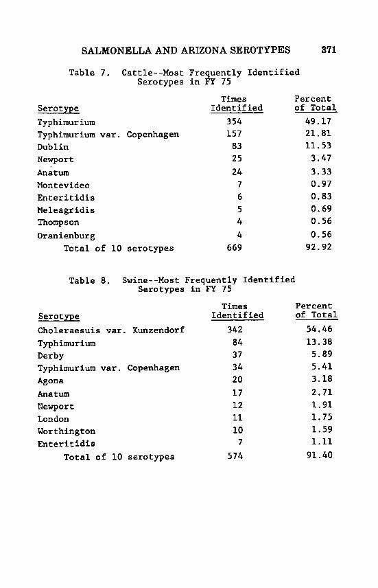
SALMONELLA AND ARIZONA SEROTYPES 371
Table 7. Cattle--Most Frequently Identified Serotypes in FY 75
Times Percent Serotype Identified of Total Typhimur ium 354 49.17 Typhimurium var. Copenhagen 157 21.81 Dublin 83 11.53 Newport 25 3.47 Anitum Montevideo Enteritidis Meleagr idis Thompson
24 7 6 5 4
3.33 0.97 0.83 0.69 0.56
Or an ienbur g 4 0.56 Total of 10 serotypes 669 92.92
Table 8. Swine--Most Frequently Identified Serotypes in FY 75
Times Percent Serotype Identified of Total
Choleraesuis var. Kunzendorf 342 54.46 Typhimurium 84 13.38 Derby 37 5.89 Typhimurium var. Copenhagen Agona Anatum Mewpor t London Wort hington Enteritidis
Total of 10 serotypes
34 5.41 20 3.18 17 2.71 12 1.91 11 1.75 10 1.59 7 1.11
574 91.40

372 BLACKBURN
Table 9. Horse--Most Frequently Identified Serotypes in FY 75
Serotype
Typhimurium Typhimurium var . Copenhagen Anatum Newport Meleagridis
Total of 5 serotypes
Times Percent Identified of Total
79 45.93 26 15.12 18 10.47 1 2
7 142
6.98 4.07
82.56

Tabl
e 10.
Salm
onel
la Se
roty
pes
Iden
tifi
ed Mos
t Fr
eque
ntly
du
ring
FY
197
5 wi
th C
ompa
riso
n Da
ta for
5 Ye
ars
(All
Sources)
Sero
type
1975
1974
Typh
imur
ium
72P (
1)i
kit
1837
(1)
Typh
imur
ium var.
Chol
erae
suis
var.
Cope
nhag
en
349
(2)
570
(2)
Kun z
en d
o r f
346
(3)
404
(3)
Heid
elbe
rg
182
(4)
337
(4)
Anat
um
163
(5)
323
(5)
Sain
t Pa
ul
132
(6)
275
(6)
Newp
ort
123
(7)
188
(8)
Ente
r it id i
s 105
(8)
72 (17)
* Sa
n Di
ego
104
(9)
133
(10)
93 (10)
194
(7)
.-.s
h
n3
In
fant
is
1973
19
72
1158
(1)
676
(2)
413
(2)
327
(5)
290
(3)
133
(12)
277
(5)
417
(3)
198
(8)
187
(8)
288
(4)
392 (4)
257
(6)
222
(7)
63 (
16)
34 (
20)
217
(7)
751
(1)
134 (9)
243
(6)
1971
805
(1)
239
(7)
245
(5)
536
(2)
302 (4)
347
(3)
148
(14)
75 (20)
166
(13)
233
(8)
1970
762
(2)
230
(12)
573
(4)
860
(1)
611
(3)
510
(3)
84 (23)
101
(20)
149
(16)
233
(11)
* Nu
mber
of
time
s th
e se
roty
pe was
ide
ntif
ied.
co
mmon
. **
Ran
k be
ginn
ing
with
the
mos
t 3
c+a

374 BLACKBURN
a . All Serotypes b. Typhimurium
Dublin d . Choleraesuis vor Kuzendorf C .
a. Arizona 7a I 7b : I ,7,8 f . Pullorud
0 NO cultures a t - 5 m 6 - 2 0 21-100 > I O 0
Figure 1 . Geographical distributions of selected serotypes.

SALMONELLA AND ARIZONA SEROTYPES 375
REFERENCES
1. Anon. Report of the Committee on Salmonellosis. Proceedings 78th Annual Meeting USAHA, 1974 :307-323.
2. Center for Disease Control : Salmonella Surveillance Annual Summary 1974, issued August 1975.

PROGRESS IN THE SCRAPIE ERADICATION PROGRAM A. L. Klingsporn*, DVM and J. L. Hourrigan, DVM
Scrapie is a naturally occurring disease of sheep and goats char- acterized by progressive degeneration of the central nervous system which caused the affected animal to rub and scratch, become debili- tated and incoordinated. Most affected animals die at 30 to 50 months of age after showing clinical signs of disease for one to six months.
The causative agent of scrapie has not been identified or com- pletely characterized, however, the agent is transmissible, filterable, particulate, and is a self-replicating agent considered by most re- search workers to be a virus with unusual characteristics. The virus can be experimentally introduced into the host’s body by many routes ; epidurally, intradermally, intranasally, intraocularly, intraperitoneal- ly, intravenously, orally, and by rubbing on the scarificed skin. Once the virus enters the host’s body, it replicates and spreads to many of the body tissues where it establishes rather constant titer levels. Figure 1. However, i t is not until the virus reaches detectable levels in the brain rather late in the infection that lesions can be detected and the clinical signs of scrapie observed.
Scrapie has been recognized in Europe for over 200 years. It has been reported in many countries of Western and Eastern Europe, Colombia in South America, Kenya, and the Republic of South Africa in Africa, Australia, Canada, India, New Zealand, and the United States. Figure 2. The disease has not been officially reported in Rus- sia, however, indications are that scrapie occurs there.
Scrapie spreads through the sale and movement of sheep, par- ticularly of purebred sheep that are incubating the virus. Scrapie has spread to many countries of the world through the sale of pure- bred sheep from Europe, particularly Britain. The disease was first reported in Canada in 1938, and in the United States in Michigan in 1947. In both instances scrapie was first diagnosed in sheep im- ported from Great Britain or in descendants of British sheep. As of October 1976, the disease has been diagnosed in the United States in 214 flocks in 31 States. Figure 3. The 320 confirmed cases were: Cheviot, 16; Hampshire, 2; Montadale, 2; Suffolk, 298; and cross- breds, 2. Although scrapie has occurred in the majority of our States, the disease has not been reported for several years in many, for ex- ample, Missouri has had 9 or aince 1967.
outbreaks, but none reported for 9 years
*Chief Staff Veterinarian and and Ectoparasites Stafi‘, VS,
Senior Staff Veterinarian, Sheep, Goat, Equine, APHIS, USDA.
376

SCRAPIE ERADICATION PROGRAM 377
The Cooperative State-Federal Scrapie Eradication began in Cali- fornia in 1952 as an emergency measure. That program provided for histological confirmation of the disease in animals with clinical signs of scrapie, quarantine and slaughter of all sheep and goats in the infected flocks, and tracing and slaughter of all exposed animals moved from these flocks, and their immediate progeny. These pro- cedures were effective and although California has suffered additional outbreaks, none have been extensions of the 1952 outbreaks.
Scrapie was next reported in five Ohio flocks in 1953. The eradi- cation program that was followed in Ohio included slaughter of the infected flocks with tracing and inspection, but not slaughter, of exposed sheep sold from these flocks and their progeny. During in- spections of these recipient flocks, certain of the exposed animals and their progeny were later found to have scrapie in Ohio and Ten- nessee. During the same year, scrapie was diagnosed in three Illinois flocks. The Illinois program provided for slaughter of infected flocks and exposed sheep moved from them.
It soon became obvious that the disease could not be effectively controlled or eradicated unless source flocks and movement from them were handled in the same manner as infected flocks. Source flocks were those determined to be disseminating scrapie, but in which scrapie had not been confirmed. The program was broadened in April 1957 to provide for slaughter of source flocks, exposed sheep sold from these flocks, and their immediate progeny.
The scrapie program was again modified in 1965 following an International Scrapie Seminar which was held to review scrapie and methods of control or eradication of this disease. These modifica- tions expanded the program to include slaughter of bloodline as well as exposed animals, Bloodline animals are defined as the affected animals, all its descendants, sire and dam, and all full or half brothers or sisters. When the disease was limited to well-defined bloodlines and adequate quarantine measures could be maintained, slaughter could be limited to bloodline animals. Nonbloodline animals in in- fected and source flocks could be held under 24 months quarantine with an additional 18 months surveillance. This procedure was suc- cessful in small flocks where the affected animal had recently been introduced ; however, in large purebred flocks where the affected animal(s) was born in the flock, additional cases occurred in all flocks so handled within a year in animals considered nonbloodline.
The latest modifications to the scrapie eradication program were implemented in September 1975 when Part 54, Title 9, Code of Federal Regulations was amended to : (1) Provide for the destruction of animals which have been directly exposed to scrapie; (2) elimi- nate salvage and use for human or animal consumption of bloodline and exposed animals ; and (3) increase maximum Federal indemnity

378 KLINGSPORN AND HOURRIGAN
which may be paid for animals destroyed to $40 per head for grade animals and to $90 per head for purebred animals.
Thus, the present program provides for the observation of ani- mals showing “scrapie-suspicious” signs, laboratory diagnosis of the disease in affected animals, quarantine of infected flocks, tracing the source of animals introduced into the infected flocks and exposed sales from the infected and source flocks, slaughter of the entire in- fected and source flocks, slaughter of selected bloodline animals and all exposed animals sold from the infected and source flocks and se- lected progeny, disposition of all animals slaughtered by burning or burial, payment of Federal indemnity for animals slaughtered, and conduct of a Scrapie Field Trial Study, Mission, Texas designed to study natural scrapie. Figure 4.
The present program places increased emphasis on the spread of scrapie by contact exposure and by the female vertically either pre- natally or soon after birth to her offsprings, or laterally to other animals’ offsprings pastured or housed with affected females. This program places less emphasis on the spread of scrapie via the ap- parently unaffected, but exposed sire to his offsprings or to other young animals because in many instances the male is never in phys- ical contact with such animals. The reduced emphasis on the spread scrapie via the exposed ram does not, we believe, provide an unac- ceptable risk as the ram is considered much less likely to spread scrapie vertically than ewes. As a result of these changes, fewer sheep will require slaughter due to the ratio of ram progeny to ewe progeny. The prohibition of salvage of slaughtered exposed animals and the requirement that their carcasses must be disposed of by burning or burial has greatly reduced the possible human and animal health hazard from this disease.
The scrapie eradication efforts in the United States has been quite encouraging. The number of outbreaks in the Suffolk breed has declined and the program has held the spread of the disease in the Cheviot, Hampshire, and Montadale breeds to a negligible level. From a high of 22 outbreaks in 1956, outbreaks have gradually been reduced to 3 in 1971, 4 in 1972, 2 each in 1973, 1974, and 1975, and 5 in 1976. Figure 5. While it is our firm conviction that scrapie can be eradicated by following a rigorous quarantine and slaughter policy, the occurrence of five outbreaks during the past fiscal year clearly demonstrates that we must not let-down our guard and that all parts of the program must be actively and promptly carried out if the disease is to be eradicated. To do less invites the probability that scrapie will increase in the affected breeds, spread to the as yet unaffected breeds, and become so widespread and entrenched as to defy eradication.

SCRAPIE ERADICATION PROGRAM 379
STUDY OF NATURAL SCRAPIE, MISSION, TEXAS The data obtained from study of field outbreaks, review of the
research literature, and reports from other countries have long indi- cated that scrapie can be a significant disease, can spread by con- tact, and can cause substantial losses. The tendency in many countries to suppress reports of scrapie, limited research on the natural dia- ease, and the eradication program carried out in the United States and Canada limited the accumulation of data regarding the true nature of scrapie, its spread, and losses caused by it.
To provide information in these areas, a field trial study was established at Moore Air Force Base, Mission, Texas in November 1964. The Mission field trial station is located on 450 acres of pas- tureland surrounding the run-ways of the former Moore Air Force Base. It was designed t o hold and breed previously exposed and un- exposed sheep and goats under close observation for extended pe- riods to determine which animals would develop scrapie, and to study the natural spread of this disease. Five-hundred and forty-seven previously exposed sheep of the Cheviot, Hampshire, Montadale, and Suffolk breeds were purchased from flocks in California, Indiana, Illinois, Maryland, Missouri, New York, Texas, and Virginia, and taken to Mission, Texas for use in the field trial study. These sheep have been maintained in two areas: (1) A series of p a t u r e and pens occupied by male animals only, and (2) a series of pastures and pens occupied by females and young progeny of both sexes. Lambing and kidding occurred in the female area, with lambs remaining with the dam until weaning, when the males are removed to the male area.
When evaluating the data from the Mission study, it soon b e came apparent that criteria had to be established to1 determine whether or not a given animal should be considered as being at risk for scrapie. After considerable study the following criteria was de- veloped for animals considered at risk :
1. Scrapied-an animal confirmed scrapie by histological exami- nation or by mouse inoculation.
2. Free-an animal dying at greater than 24 months of age which was negative clinically and negative on histological study, or an ani- mal brought to Mission that is alive and healthy at 100 months or greater age, or died after living at least to 100 months of age, and was not clinically suspicious or histologically incinclusive. Animals born at Mission had to meet similar criteria except the minimum age was 60 rather than 100 months. One hundred months was estab- lished as a criteria for animals brought to Mission, and 60 months for animals born at Mission because it was found that animals born in the Mission environment developed scrapie consistently at the younger age compared to those brought there.

380 KLINGSPORN AND HOURRIGAN
Using these criteria for the 483 Suffolks taken to Mission, i t was found that during the period from November 1964 to February 1976, some 315 met the criteria for being a t risk, 92 or 29 percent of these animals developed scrapie at ages ranging from 23 to 142 months, while 223 remained free. Table 1. We believe that these levels of losses from a single disease is not compatible with economic production. Additional study of Table 1 reveals that 395 progeny were born at Mission from the previously exposed Suffolks brought there that meet the criteria for being considered at risk. One hundred and forty-six or 37 percent of these progeny have been confirmed scrapie at ages ranging from 22 to 65 months. These data, we believe, dem- onstrates that Suffolk animals from scrapie infected and source flocks if held instead of being slaughtered, will develop scrapie in significant numbers, and that scrapie will continue to occur in their offsprings born and reared on the infected premises at an even greater inci- dence and considerably younger average age.
Study of individual lots from infected and source flocks taken to Mission reveals that scrapie losses ranged from 0 to 57 percent of the flock. Table 2. These losses were computed directly by dividing the numbered scrapied in the lot by the total animals taken to Mission from the flock.
The next question is, “Will scrapie spread by contact to sheep of other breeds and goats?” To answer this question 140 previously unexposed sheep and goats were purchased a t three to ten months of age and placed on the infected premises with a succession of scrapied sheep. The previously unexposed animals consisted of Angora and dairy goats, Hampshire, Rambouillet, Suffolk, and Targhee sheep breeds. Table 3. A study of this chart reveals that none of the pre- viously unexposed goats brought to Mission have developed scrapied. However, 29 percent of the Angora goats born at Mission and at risk have developed scrapie while 59 percent of the at risk progeny of dairy goats born there have developed the disease. In the sheep breeds, one each of the previously unexposed Hampshire, Rambouillet, New Zealand Suffolk, and Targhee have developed scrapie a t rather advance ages. Indicating that animals receiving their initial exposure some months after birth either do not go on to develop clinical scrapie or develop the disease a t more advanced ages than those exposed from birth on. The percentage of scrapie occurring in the offsprings born a t Mission from these previously unexposed sheep ranged from 11 percent for a small group of American Suffolk up to 43 percent for the offsprings of the New Zealand Suffolks, with the Hampshires, Rambouillets, and Targhee offs pings being rather closely grouped between. The Rambouillets affected a t Mission are the only known affected Rambouillets in the United States and the Targhees the only known affected Targhees anywhere in the world.

SCRAPIE ERADICATION PROGRAM 381
Table 3 demonstrated conclusively that scrapie can be trans- mitted by contact between healthy and affected animals and more importantly that i t continues to occur in the offsprings of such ani- mals born on infected premises at consistently greater incidence.
The next question is, “At what age can animals be removed from exposure and not go on to develop clinical scrapie?” To determine the answer to this question dairy goats were removed from expwure at 6 and 20 months of age, Rambouillet, Suffolk, and Targhee sheep were removed at 4, 6, 9, and 20 months of age. Table 4 demonstrated that some sheep of all breeds removed a t 20 months of age developed scrapie a t the expected ages. None of the dairy goats developed scrapie, however, only one lived to the expected age of scrapie. Table 5 demonstrated that Suffolks and Rambouillets removed from ex- posure at 4 and 9 months of age, and dairy goats removed at 6 months developed scrapie. However, none of the Targhees removed at 4 or 9 months have developed scrapie, even though 14 of these animals are now 54 months of age. None of the sheep and goats removed from exposure at birth have developed scrapie, although 20 of these ani- mals have reached 42 months of age and 11 have reached 54 months of age. These studies are continuing. These studies demonstrate that exposed animals sold from infected and source flocks must be traced and destroyed or they will continue to develop scrapie, provide foci for continued exposure, and spread of the disease.
We believe that the study of scrapie in field outbreaks and at Mission, Texas support the present requirements of the eradica- tion program and clearly indicate that if scrapie is to be controlled and eventually eradicated, the present policy of destroying infected and source flocks, exposed animals sold from these flocks, and blood- line animals descending from the ewe must be vigorously carried out to prevent the continued spread of scrapie within the affected breeds, and the extension of the disease into breeds not now affected in the United States.
Another concern of the United States Department of Agriculture regarding scrapie in sheep and goats is its striking similarity of clin- ical signs, primary histological lesions of the brain, and the biological and physical characteristics of the agent of scrapie of sheep and goats, transmissible mink encephalopathy (TME ) of mink, kuru, and Creutzfeldt-Jakob disease of man. Scrapie has not been directly linked to human disease ; however, the evidence continues to draw closer and closer the possibility of human health hazard. Scrapie virus inoculated into Rhesus, Cynomolgus, spider, or squirrel mon- key produces a disease similar in clinical signs and brain damage to that produced by the viruses of TME and the human diseases of kuru and Creutzf eldt-Jakob disease when inoculated into monkeys.

382 KLINGSPORN AND HOURRIGAN
The significance of these “slow viral diseases” was particularly emphasized in October 1976 when Dr. D. Carleton Gajdusek of the National Institute of Health, Bethesda, Maryland shared the honor of being awarded the Nobel Prize for medicine and physiology. Dr. Gajdusek and his colleagues have established that scrapie, and TME, and two human diseases have much in common. This work is char- acterized as a major breakthrough and discovery that has revolu- tionized thinking about a group of neurological diseases of previously unknown etiology.
It is worthy of note that some of Dr. Gajdusek’s experiments are being conducted a t Mission, Texas, in cooperation with Scrapie Field Trial personnel.

SCRA
P/€
TISSU
ES F
ROM
WHIC
H VI
RUS
DEMO
NSTR
ATED
F
RESP
IRAT
ORY
SYST
EM
LUNG
NE
RVOU
S SY
STEM
BR
AIN - C
EREB
RUM
CERE
BELL
UM
FRON
TAL
CORT
EX
MIDB
RAIN
ME
DULL
A CE
REBR
O SP
INAL
FLU
ID
SPIN
AL C
ORD-
CER
VICA
L SP
INAL
LU
MBAR
SC
IATI
C NE
RVE
MUSC
LE
MUSC
ULAR
SYS
TEM
EXCR
ETOR
Y SY
STEM
B C
SF
L
B S
CK
PU
I
II
I
\I
ll
I! LL
s I
BM
SY
KIDN
EY
UTER
US
FETA
L ME
MBRA
NE
REPR
ODUC
TIVE
SYST
EM
DIGE
STIV
E SY
STEM
SA
LIVAR
Y GL
AND
LIVE
R IN
TEST
INAL
WAL
L
ADRE
NAL
GLAN
D PA
NCRE
ATIC
GLA
ND
THYM
US G
LAND
PI
TUIT
ARY
GLAN
D
ENDO
CRIN
E SY
STEM
RETIC
ULO-
ENDO
THEL
IAL
SYST
EM
CIRC
ULAT
ORY
SYST
EM
BONE
MAR
ROW
SP
LEEN
LY
MPHA
TICS
BLOO
D SE
RUM

c I SC
RAPIE
Cou
ntria
wk
a se
raph
has
oce
urrr
d wi
thoul
rrod
iotio
n nw
wrm
. Co
untri
a w
hrr s
crop
ir ha
s oc
curn
d an
d ha
s b
an r
rod
d.
(ounm
a wh
om s
uapi
a ha
occ
umd
and
orod
icotio
n ott
rmpt
rd.
0 Co
untri
a w
hr
scm
pir ha
not
brm
o(ii
cial
ty r
rpa)
rd
7/7
5
US
DE
PA
RTM
EN
T O
F A
GR
ICU
LTU
RE
V
ETE
RIN
AR
Y S
ER
VIC
ES
A
NIM
AL
AN
D P
LAN
T H
EA
LTH
IN
SPEC
TIO
W S
ER
VIC
E

YE
AR
OF
MO
ST
R
ECEN
T O
UTB
RE
AK
JU
NE 3
0, 1
976
AN
IMA
L A
ND
PLA
NT
HE
ALT
H IN
SP
EC
TIO
N S
ER
VIC
E
U. S
DE
PA
RTM
EN
T O
F A
GR
ICU
LTU
RE
V
ETE
RIN
AR
Y S
ER
VIC
ES

I.
2. 3.
4.
5.
6.
SCRA
PIE
ERAD
ICAT
ION
PROG
RAM
w
00
Q, P?
c1
rn
td 0
LAB
OR
ATO
RY
DIA
GN
OS
IS O
F D
ISE
AS
E
QU
AR
AN
TIN
E O
F IN
FEC
TED
FLO
CK
S
TR
AC
ING
D
ISP
OS
ITIO
N O
F:
E % IN
FEC
TED
FLO
CK
S
U
SO
UR
CE
FLO
CK
S
BLO
OD
LIN
E A
NIM
AL
S
EX
PO
SE
D A
NIM
AL
S
* z rn * Z
IND
EM
NIT
Y
FIE
LD T
RIA
L S
TU
DY

N
0
S C R A P I E 0
U T B R E A K S
SCRA
PIE
OUTB
REAK
S FY
195
3 = 19
76"
SCR
API
ED F
LOCK
S U
.S.A
. C
HEV
IOT
10
HA
MPS
HIR
E 2
MO
NTA
DA
LE
2 SU
FFO
LKS
199
CRO
SSBR
EED
1 TO
TAL 214
'194
7 O
utb
reak
no
t Li
ried
FISC
AL
YEAR
S U
S. D
EP
AR
TME
NT
OF
AG
RIC
ULT
UR
E
VE
TER
INA
RY
SE
RV
ICE
S
AN
IMA
L A
ND
PLA
NT
HE
ALT
H IN
SP
EC
TIO
N S
ER
VIC
E

03
00
00
NO. AT RISK
TABLE 1
SCRAPIE
- FATE
OF PREVIOUSLY
-SLED
SUFFOLK SHEEP
EKUG
HC TO
MISSION AT VARIOUS AGES, A
MD THElR P
ROGE
3JY BORN AT M
ISSI
ON
N0.m
SC
RAPm
NO.
f 4&
suFF
oIxR
AM
sAN
DEw
Es
IwouGHT
To M
ISSI
ON
NO.
ATR
ISK
35
c
l?RCG
lWY
AT M
ISSI
ON
I I
I I
r-
395
289
AGE
XRA
PIED
(MONTHS)
MIN
. I
AV.
I MA
X.

Scr
apie
Era
diea
rtio
n INCIDE
NCE O
F SCR
APIE
IN SH
EEP F
ROM
INFEC
TED A
ND
SOUR
CE FL
OCKS
TAKE
N TO M
ISSION
, TEXA
S
ROCK
OF O
RIGIN
INFEC
TED
INFEC
TED
SOUR
CE
INFEC
TED
INFEC
TED
INFEC
TED
INFEC
TED
INFEC
TED
INFEC
TED
SOUR
CE
INFEC
TED
BREE
D
CHEV
IOT
SUFFO
LK
SUFFO
LK
SUFF
OLK
SUFFO
LK
SUFF
OLK
SUFFO
LK
SUFFO
U SuRou(
SUFFO
LK
SUFF
OLK
~~
NUMB
ER O
F SH
EEP I
N FLO
CK
39 7 10
1 44 31 4 7 33 35 19 9
US
. DE
PA
RTM
EN
T O
F A
GR
ICU
LTU
RE
V
ETE
RIN
AR
Y S
ER
VIC
ES
MONT
HS OF
STUD
Y
74 69
92 loo
107 78 105
117
102 68
68
PERC
ENTA
GE OF
SC
RAPIE
MOR
TALll
Y
21 57 20 25 39 25 0 28 14 5 11
AN
IMA
L A
ND
PLA
NT
HE
ALT
H IN
SP
EC
TIO
N S
ER
VIC
E

co
m
0
TABL
E 3
SCRAPIE IN F
BOGE
NY BORN
AT M
ISSI
ON O
F PR
EVIO
USLY
UNE
KPom
P-S
ANIMALS
BROU
GHT To M
ISSI
ON
PROGENY BORN A
T M
ISS1
NO.
AT
RISK
NO.
MPO
SED
ED AG
E
SCRAP
NO.
NO.
FREE
ll 6
ANGO
FU G
OAT
DAIR
Y GO
AT
21
63
15
26
6 37
15
18 4 1
6
31
3
33
88
89
89
- 73
95
83
22 9 16
80
65
18 8 10
8 20
216
TOTA
L 140
4 30
9 87
c
28

SCRAPIE ERADICATION PROGRAM
c
rl
391
Table 4

TAB
LE 5
SC
ElAP
IE IN SHEEP
OR G
OATS
REM
OVED
FRC
IM E
XPOS
URE
9, 6,
OR 4
MONTHS
OF AG
E
1' =
DEAD
NOT SCRAPIE
18 7 8

REPORT OF THE COMMITTEE OF SHEEP AND GOATS
Chairmn: F. James Schoenfeld, Salt Lake City, UT Co-Chairman: C. C. Beck, Manchester, MI
Thomas B. Snodgrass, A. L. Klingsporn, H. E. Metcalf, J. L. Hourrigan, T. L. Barber, Guy E. Reynolds, Howard W. Whit- ford, John Neimi, Olin R. Timm, J. E. Pearson, Richard F. Hall, M. E. Macheak The above Committee Members were in attendance. We had four
guests present, totaling 17. The Committee met as requested by the President of USAHA to
consider the business of the Committee and submit the following report.
Dr. Howard W. Whitford presented a paper pertaining to the problems of Angora Goats in Texas which includes Low Kid Crop, area of infectious diseases, parasitic problems, nutrition and the role of genetics. The complete text of the paper by the personnel of the Texas Agricultural Experiment Station and Texas Agriculture Extension Service is enclosed (Enclosure 1).
Dr. T. Lynwood Barber reported on the updating of Bluetongue typing. There are 4 sero-types in the United States. Type 17 is a new one in the U. S. In 1975 Dr. Barber correlated with other sera types in the world. All Bluetongue sero-typing is done a t the USDA Laboratory in Denver.
A discussion of Bluetongue was directed among the Committee: Dr. Klingsporn said that Bluetongue is still a great concern. Dr. Hall indicated that more work needs to be done to include
Mr. Olin H. Timm spoke concerning Bluetongue. Dr. Hugh Metcalf gave a brief report on the death of 500 antelope
in Wyoming due to Bluetongue. The virus is now being typed at the Denver Laboratory. He also reported losses of White Tailed deer and sheep in Montana.
Dr. A. L. Klingsporn gave an update on the Bluetongue testing done during fiscal year 1976. (See enclosure 2). He also gave a report of Scrapie activities during the past year (See enclosure 3).
the various sero-types in the vaccine.
In connection with Bluetongue and Scrapie the Committee dis- cussed movements of sheep to Canada.
Dr. Richard F. Hall, a member of this Committee and Chairman
393

394 REPORT O F THE COMMITTEE
of the Biologics Committee spoke concerning the “Low Volume Drugs” which are essential and vital to the industry. It is imperative that we seek the ~uppor t~of the sheep and goat industry, as well as the cattle and dairy industries to work with Agricultural and Con- gressional Committees to help and to fund this vital Low Volume vaccine and pharmaceuticals. Mr. Major Huff was asked to speak to this subject as an industry
member. Dr. Hall also reported that Korlan 2 Pour On has been approved
for sheep, and a federal license has been issued. The Committee discussed Anaplasmosis as it pertains to sheep. It
recommends that more extensive work be done on the C F Test to make it a more accurate test for sheep.
Dr. Guy Reynolds reported on Foot-Rot Development a t the Ore- gon Station. He said that six strains of the organism have been iso- lated.
Mr. Olin Timm reported on the development of the vaccine in California.
The Committee again wishes to bring to the attention of the Executive Committee, VS, and ARS, the resolution concerning funding for Foot Rot research at the Oregon Statim (See enclosure 4).
Dr. Reynolds proposed to the Committee and it was approved by the Committee that we support the 172 page proposal of the Na- tional Feed Manufacturers to the FDA to clear the use of Selenium for the use in ruminant animals. This could be a great help in the control of white muscle disease.
Dr. Hall brought to our attention the work being done by Dr. Norman Gates at the DuBois Station in Idaho, with the “Thin Ewe Syndrome.” He also said work is being done in Ram Epiolidymitis as ID the organism, actinobacrillus, being the problem rather than Brucellosis. They are developing an antogenious vaccine to use in young rams.
The Committee agreed that we need to work more closely with the industry by contributing to their publications.
The business of the Committee being completed was adjourned and we submit this report for the Executive Committee approval.
ENCLOSURE #1
TO : Health and Product Development Committee of the Texas Sheep and Goat Raisers Association

SHEEP AND GOATS 395
FROM : . Personnel of the Texas Agricultural Experiment Station and Texas Agricultural Extension Service ; Chairman- Dr. Maurice Shelton, Dr. Ed Huston, Dr. C. H. Living- ston, Jr., and Dr. Howard W. Whitford of the Texas Veterinary Medical Laboratory
SUBJECT : Report and Recommendations Concerning the Problem of Low Reproductive Efficiency in Angora Goats
Low reproductive efficiency of Angora goats has long been con- sidered the norm for Angora goats. Statistics are not available, but frequently expressed figures for long term averages for kid crops are in the range of 40-50 percent. However, individual flocks do at times approach loo%, indicating that poor kid crops are not man- datory with this animal. Thus it is both logical and timely to ask why this disparity between the potential and realized kid crops and what can be done to correct it. Reference has been made to unusually poor kid crops (as low as 20%) in recent years. We have no direct information to confirm or to explain this deterioration in reproduc- tive efficiency. However, we have no reason to or intent to question producer reports in this regard.
(a) A failure to show estrus or to ovulate within the breeding sea- son This is almost entirely explained by a lack of size and develop- ment
Among healthy, well developed does, this would be largely ex- plained by a failure of the males to r u t or to provide adequate service
(c) Abortion or resorption of the embryos The losses from abortion is highly variable and may range from zero to a high proportion in Angora flocks. The explanation for abortion is too complex to attempt a t this point.
(d) Death loss of kids produced Death losses are usually explained by starvation (which in itself may have many explanations), cold stress or predators.
Individual flocks may experience losses from none or all of these causes. Those producing good kid crops usually evade serious losses from any, whereas, problem flocks may experience losses from all these causes.
Reproductive failure may result from :
(b) Failure of conception
Comments on Possible Causes o f Low Reproductive Eff ic iency Infectious and Parasite Diseases : Infection has not been identified as a major cau5e of loss in

396 REPORT O F THE COMMITTEE
reproduction in goats. With other species of livestock, brucellosis, vibriosis and leptospirosis are diseases associated with reproductive failure. In the past, efforts have been made to identify these dis- eases entities as a cause of loss in goats without success. Also the nature of the losses does not suggest infection as the primary cause. However, this may not be true and Dr. Livingston, in cooperation with the Veterinary Diagnostic Laboratory, will continue to investigate cases of loss which are brought to their attention from experimental or producer owned flocks. Aborted fetus, placental membranes and material obtained from aborting nannies will be examined for agents not routinely cultured in the diagnostic laboratories i.e. ureaplasma, mycoplasa and acholeplasma, L-Form of bacteria. Through sero- logical and cultural studies they will survey the Angora goat popula- tion for evidence of disease which may be associated with poor re- production. If disease is identified as a major contributing factor efforts will be directed to means of alleviating the problem. Angora goats are probably more susceptible to internal parasites than any species of farm or ranch livestock. The primary problem is gastro- intestinal nematodes and coccidiosis. Neither of these have a specific effect on reproduction, but they are major factors affecting general health and development of the goat. Thus they can indirectly have an important effect on reproduction.
Recommendations :
1. In the absence of information indicating disease as a major source of loss, no recommendations can be made relating to prevent- ing loss from disease. Producers are encouraged to be on the alert for suspected cases of loss from disease and to call this to the attention of their veterinarian, County Extension Agent or Dr. C. W. Living- ston at the San Angelo Research & Extension Center or Texas Vet- erinary Medical Diagnostic Laboratory at College Station.
2. Producers are also encouraged to become familiar with the prob- lem of internal parasites, especially coccidiosis, and develop manage- ment systems for dealing with this problem.
3. Consideration should be given t o petitioning the Food and Drug Administration to allow any drug which has been approved for sheep to be used with goats. If specific research is required to convince the FDA of the safety of this approach the requirements should be spelled out; efficacy, residue, warning on labels, safety.
Genetics:
In a broad sense, the reproduction problems of the Angora are inherited to the extent that i t is a problem with Angora and not with Spanish goats and the difference between these types is genetic. However, there is not a good reason to think that the fertility prob-

SHEEP AND GOATS 397
lem is due to any single gene or simple gene combination. First, nature itself tends to eliminate genes contributing directly to infer- tility from the polpulation as infertile animals do not leave replace- ments or contributions to future generations. Also by analogy with other species of breeds of livestock, the heritability of fertility is very complex in nature. Also, producers have alluded to a deteriora- tion in fertility in recent years, and there is no way that a significant genetic change can be effected in a large population in the equivalent of one animal generation. Thus we feel strongly that in a short term view and in a restricted sense, inheritance is not the explanation for a deterioration in kid crops. However, in a long term view, in- heritance is the primary explanation in that the phenomenal success in breeding for mohair production has resulted in an animal which is poorly adapted to the environment in which they are produced. A highly selected Angora goat produces fiber at the rate of two to four times that of our better producing sheep. This high level of fiber pro- duction has resulted in an unusually high nutrient requirement for a small animal with a limited foraging capacity or capability. Also the weight of the mohair can be a burden on the animal as well as inter- fere with foraging. Only mutton goats, which do not have the burden of reproduction, are able to maintain themselves in a vigorous state without good range conditions or considerable assistance from man. Even Angora muttons do not become fat as a steer and a dry ewe would do.
Just as short term genetic change cannot be used to explain a deterioration in fertility, selection cannot be relied upon to provide a quick solution to the problem. However, over a long period of time selection provides the only reliable means of alleviating the problem.
Researchers in South Africa have sugegsted that selection for fine hair has resulted in an abnormal adrenal status which contributes directly to abortion and indirectly to other problems. We cannot con- firm or deny that this is the case, but i t seems unlikely that there has been any precipitous change in hair quality or in the genotype of Texas goats in respect to fiber diameter. Reproductive problems have been around for a long period of time with the Angora and abortion, although prevalent, has not been identified as the major source of the problem in Texas. Abnormal adrenal status or response to stress appears to be common with Angora, but this appears to be merely the mechanism of interrupted reproduction and not the basic cause. Although selection for fine hair does not appear to offer an explanation, selection for heavy mohair production, complete cover- ing and high oil content are no doubt important problems. Poor breeders are almost invariably those that shear a heavy clip of mo- hair in respect to their size, but may not be heavier shearing on a per head basis.

398 REPORT OF THE COMMITTEE
Recommendation : 1. At a time of high mohair prices and scarcity of numbers, the
culling to slaughter, of goats because of poor reproduction would appear to be poor judgment. Does which might be culled on repro- ductive efficiency could well be kept for hair production alone. Also any net kids raised out of these does would contribute to numbers in the industry. What would appear to be more important would be to insure that males are not kept from any does with a poor reproduc- tive history. Where numbers permit culling previously dry or aborter does, very small, wooly faced does and does with bad udders are good candidates for culling.
2. The design oaf appropriate selection programs should have a high priority for the future. The high nutritive requirements of the Angora goat places the breeder, especially the stud breeder, in a quandary. If the goats are not provided with good nutrition they will not develop adequately to be selected and successfully used as breeders. However, this practice contributes to the problem of breeding an animal which is poorly adapted to his environment.
Culling of does may make an immediate improvement in kid crop for those remaining, but it will make little genetic change in the population. Also extensive culling of the females is possible only when a surplus exists. Genetic change can only be accomplished through male selection.
Selection for size, with both sexes, is the easiest to implement and will give positive results. However, more long term improve- ment in fertility will result from using some type of records on re- productive performance as an adjunct to size.
Nutrition: Nutrition is thought to be the major direct cause of poor repro-
duction. However, genetics is the predisposing factor in having de- veloped an animal with a higher requirement than can be satisfied under most typical range conditions. Thus the producer is faced with one or a combination of the following choices.
1. Improve the animals environment by utilization of cultivated harvested feeds
2. Breed an “easy care” animal which is better adapted to the conditions under which i t is to be raised
3. Be prepared to accept low kid crops and associated problems/or 4. A combination of the above Which of the above courses of action to be chosen would depend

SHEEP AND GOATS 399
in a large part on what the producer believes the income picture to be for this industry. At current, prices, the immediate choice should be clear in that the animals should be cared for in a manner to maximize their production. However, if history can be taken as a prologue to the future, prices will be erratic or variable and the prices received will not always justify the cash costs of improving the animals environment to the extent that good performance can be expected. This would indicate that simultaneous and immediate goals should be to breed an “easy care” animal.
Recommendation:
Since supplemental feed is supplied to “supplement” pasture or range vegetation i t is necessary to consider what vegetation is avail- able, what it supplies and when before supplemental feed needs can be determined. Therefore i t is crucial that the producer become familiar with the plant species on his range and what the goat is con- suming. For example, if a range has a scattering of a wide variety of plant species including winter growing plants such as scattered liveoak and juniper, Texas wintergrass and Canada wildrye, good conditions and warm winter days bring on desirable annual forbs such as filaree and tallow weed and goats are fairly well fed. Re- search shows that periods of deferment in systematic grazing pro- grams will encourage such diversity. Most spring plants supply ade- quate nutrients for Angora goats. The better goat pastures will have scattered prickly ash, elbow bush, shin oak, hackberry and other deciduous browse plants to supplement summer and fall grasses. Only the growing kid and yearling appear to need extra nutrition from April to November if the rangeland has a good variety. A mixture similar to the following should be fed to kids free choice and to developing yearlings at a rate of about 1/E pound per day during the period of July thru November.
Developing Ration For Kids & Yearlings On Rangeland
Ingredient % Cottonseed meal Sorghum grain Dicalcium phosphate Vitamin A-5000 I.U. per lb.
50 45
5
100
The developing kids and yearlings as well as older breeding does might be fed from about December 1 until spring a t a rate of about 1/2 to %, pounds per day of the following mixture.

400 REPORT O F THE COMMITTEE
Winter Supplement For Developing & Breeding Angora Goats
Ingredient % Cottonseed meal Sorghum grain Dicalcium phosphate Vitamin A-5000 I.U. per lb.
75 23 2
100
The condition of the animal is the key to the level of nutrition and may be taken as a guide to the adequacy and suitability of the ration suggestions.
Research Needs:
The Texas Agricultural Experiment Station should be encour- aged to establish breeding flocks of Angora does a t either or both Hill Ranches (between Sonora and Rocksprings) and the Winters Ranch a t Brady, to monitor reproductive problems with goats and to investigate ration formulation, methods and levels of supplemental feeding on reproduction goats.
Management During Kidding:
It would appear that management during kidding was a likely contributing factor to low net kid crops for the past season. The possibility presents itself that producers overestimated the range condition last fall and failed to prepare their animals by feeding in advance of kidding. Realizing the high value of the kid crop, i t seems possible that they attempted to feed, possibly even hand feed, during kidding, with considerable disruption of kidding or interfering with kid survival.
No totally satisfactory method of feeding during kidding has been developed. As of the present, and in the absence of a better suggestion, the use of salt as a feed limiter seems to be indicated. A compromise between hand feeding and the use of salt as a limiter is to feed a low salt feed (such as 15%) in the amounts desired. This can be done a t irregular intervals, usually without disrupting kidding. This approach can cause the does to spend a lot of time trailing back and forth to the feed trough. If in large pastures, this can create problems. For this reason, i t is preferable that kidding be done in small pastures or traps where feasible.

SHEEP AND GOATS 401
Research should be directed toward development of a more satis- factory feed limiter or methods of feeding distribution for use with goats and sheep, especially during kidding or lambing.
General Recommendutiom:
1. Producers should take advantage of the present favorable price situation to provide adequate care (largely through feeding) to maximize current kid crop and to develop future breeding stock for maximum future production.
2. When feasible, consider kidding under confined conditions to salvage the maximum number of kids.
3. Initiate changes in the breeding program which are designed to develop animals which will be better adapted to production con- ditions and which are not subject to reproductive failure.
4. Develop improved methods of controlling internal. parasites, especially coccidiosis, to eliminate the adverse effects on ‘growth . . and survival of young kids.
6. Initiate immediately research programs to provide better rec- ommendations on ration formulation and methods of supplemental feeding.
6. Initiate broad scale survey studies to pinpoint more specifically the nature of the losses on reproduction and determine the under- lying causes. (Specific work plans will be prepared in which pro- ducer assistance in this matter may be requested)
7. Continue monitoring the population for evidence of infectious diseases that might contribute to the problem.
ENCLOSURE #2 BLUETONGUE
During fiscal year 1976 (July 1, 1975, through June 30, 1976) bluetongue was confirmed by virus isolation in two cattle herds, one each in Colorado and Louisiana; and in six sheep flocks, two in California, two in Texas, and one each in Idaho and Oklahoma. During the same period, epizootic hemorrhagic disease (EHD) virus was isolated from three deer, two in Indiana and one in New Jersey.
In addition, 41,946 modified direct complement-fixation (MDCF) tests were run at Veterinary Services Laboratories, Ames, Iowa, and at 14 approved laboratories with the following results :

402 REPORT OF THE COMMITTEE
Total Tested Negative Suspicious Positive Cattle 37,734 32,833 3,886 1,015 G€mtS 445 422 19 4 Sheep 854 627 119 108 Wildlife 216 166 30 20 Unknown Species 2,697 2,690 Totals 41,946 36,738 4,061 1,147
Positive MDCF samples were collected from the States of Ari- zona, Arkansas, California, Florida, Georgia, Idaho, Iowa, Louisiana, Missouri, Montana, Nebraska, Nevada, New Jersey, New York, Ohio, Oklahoma, Oregon, Texas, Utah, Virginia, Washington, Wisconsin, and Wyoming.
- - 7 -
ENCLOSURE #3 SCRAPIE
Scrapie was reported in five flocks in Illinois, New York, Okla- homa, Pennsylvania and West Virginia during fiscal year 1976 (as of June 30, 1976-) The Illinois outbreak occurred in a Hamp- shire flock in LaSalle County, the New York outbreak in a Suffolk flock in Pottawatomie County, the Pennsylvania outbreak in a Suf- folk flock in York County, and the West Virginia outbreak in a Suffolk flock in Pendleton County. There were: three outbreaks re- ported in fiscal year 1971, four in 1972, two each in 1973, 1974, and 1975, and five in 1976. The number of flocks under surveillance have only decreased from 116 to 112; however, this number will in- crease when tracing from 1976 outbreaks has been completed. Eradi- cation efforts for scrapie have been encouraging over the past years ; however, the disclosure of five outbreaks during 1976 demonstrates that the disease is not eradicated and that there must be no letdown in carrying out all phases of the program if we are to eradicate this disease.
The Illinois outbreak was reported by the owner; the New York and West Virginia outbreaks were discovered during routine sur- veillance inspections ; the Pennsylvania outbreak was disclosed by back-tracing from the West Virginia infected flock; and the Okla- homa outbreak was disclosed when the owner took the infected ewe to Oklahoma State University for diagnosis. All infected flocks have been slaughtered and regulatory officials have endeavored to locate and slaughter all exposed and certain bloodline animals sold from these flocks. The source of the Illinois and Pennsylvania outbreaks has not been determined; the New York vutbreak had its source from a Massachusetts flock, which was also the source of two New Jersey outbreaks; the source of the West Virginia outbreak was the Pennsyl- vania infected flock; and the source of the Oklahoma outbreak was

SHEEP AND GOATS 403
an Illinois flock. The source flocks for New York and West Virginia infected flocks has been slaughtered and regulatory officials are endeavoring to locate and slaughter exposed and certain bloodline animals sold from these flocks. The Illinois source flock (for the Oklahoma outbreak) has not been slaughtered and is being held under State and Federal quarantine. A list of sales from this flock has not been obtained.
The August 27, 1975, 9 CFR, Part 54, was amended to require: The slaughter of exposed sheep and goats as well as bloodline ani- mals; the disposal of their carcasses by burning or burial and pro- hibit the meat from slaughtered animals being salvaged for human or animal food; and compensate owners for loss of salvage by in- creasing maximum Federal Indemnity to $40 for grade animals and $90 for purebred animals. These amendments have reduced the possible human and/or animal health hazard from scrapie.
Effective November 13, 1974, a Federal quarantine had been placed on De Kalb County, Illinois, infected flocks and lifted April 23, 1975, after slaughter of infected and exposed sheep had been completed.
Effective August 31, 1976, a pontion of La Salk County, Illinois, was placed under Federal quarantine due to a source flock being located there. A Suffolk ewe was showing clinical signs of scrapie 2 months after being moved from the Illinois flock into an Oklahoma flock.
Members of the Illinois Sheep Breeders Association have re- quested a national meeting to review scrapie and the Cooperative Scrapie Eradication Program. Veterinary Services will contact in- dustry associations and other interested individuals to determine their interest in such a meeting and to solicit their participation.
SCRAPIE FIELD TRIAL* The scrapie field trial has been underway since November 1964
(142 months). During this period, natural scrapie has been con- firmed by hisfopathological study or by mouse inoculation in 349 sheep or goats.
The findings demonstrate that animals from infected and source flocks will develop scrapie at high incidence if not slaughtered. The findings also demonstrate that scrapie can be transmitted to pre- viously unexposed sheep and goats held and bred on infected prem- ises in contact with scrapie-affected sheep and goats. Therefore, scrapie has the potential t o cause considerable loss within a flock and breed. These statements are supported by the following data:
+Misson, Texas

404 REPORT OF THE COMMITTEE
1. Of 315 Suffolk sheep at risk taken to Mission from infected and source flocks, 92 (29 percent) have developed scrapie. Of their 395 progeny born at Mission, 146 (37 percent) developed scrapie.
2. Only 4 of 123 previously unexposed sheep brought to Mission developed scrapie; however, of their 309 porgeny at risk, 87 (28 percent) were scrapied.
3. Progeny of Suffolk sheep born at Mission developed scrapie at rates varying from 24 percent when both parents were scrapie free to 80 percent when both parents were scrapied.
4. None of the previously unexposed Angora or dairy goats taken to Mission developed scrapie; however, of their progeny born at Mission and a t risk, 6 (29 percent) of the Angora goah and 37 (59 percent) of the dairy goats have developed scrapie.
5. Progeny of dairy goats born at Mission developed scrapie at rates varying from 43 percent when both parents were scrapie free to 100 percent when both parents were scrapied.
6, Goats, Suffolks, and Rambouillets (but not Targhees to date) developed scrapie if born on infected premises and removed from exposure at the following ages: Goats at 6 months, Suffolk at 4 and 9 months, and Rambouillets at 4 and 9 months.
Scrapie has not been directly linked to human disease; however, the evidence continues In draw closer and closer the possibility of the human health hazard. Scrapie virus inoculated into Rhesus, Cynomolgus, spider, or squirrel monkey produces a disease with similar symptoms and brain damage aa that produced by the viruses of transmissible mink encephalopathy (TME) and of the human diseases of kuru and CreutzfeldtiJakob disease (when inoculated) into monkeys.
The significance of these “slow viral diseases” was particularly emphasized in October 1976 when Dr. D. Carleton Gajdusek of the National Institute of Health, Bethesda, Maryland, shared the honor of being awarded the Nobel Prize for medicine and physiology. Dr. Gajduslek and his colleagues have established that scrapie and TUE of mink and two human diseases have much in common. This work is characterized as a major breakthrough and discovery that has revolutionized thinking about a group of neurological diseases of previously unknown etiology.
It is worthy of note that some of Dr. Gajdusek’s experiments are being conducted at Mission, Texas, in cooperation with Scrapie Field Trial permanel.

STATUS OF THE STATE-FEDERAL HOG CHOLERA ERADICATION PROGRAM
S. H. Young, DVM and J. W. Walker, DVM, MPA
Fiscal year 1976 has been somewhat of a disappointment for the State-Federal Hog Cholera Eradication Program. Almost everyone had anticipated the United States would be declared hog cholera free in 1976. Following the rather extensive outbreak resulting in 163 cases in several States in F Y 1973, the incidence of infection was reduced to 2 cases in FY 1974. One of these was confirmed in Mississippi and the other in Puerto Rico.
When hog cholera was not confirmed in FY 1975 and Great Britain permitted the importation of pork from the United States, many concluded the goal of eradication had been achieved. Plans for an appropriate celebration had become a major consideration.
TEXAS BREAK-FISCAL YEAR 1976 The first disappointment came within the first 4 days of FY
1976, when hog cholera was confirmed in Texas on July 4,1975. Two related w e s were confirmed in Texas. An additional 22 herds in Texas and 6 herds in Oklahoma were depopulated as exposed.
The export market to Great Britain was promptly lost following the Texas break. It has been estimated this represented a $15 million loss to the swine industry in FY 1976.
A conclusive source for the Texas break was not established. Many concluded the outbreak had been introduced from Mexico. Most chase to disregard the fact that the break in the Panhandle of Texas was several hundred miles from the Mexican border and that the infection had been there for a minimum of 2 months before it was disclosed, Aside from the immediate task force operation, little increase in surveillance resulted from the Texas break.
PROGRAM CHANGES At the time of the Texas break, tha Federal regulation was
changed to permit total payment of indemnity for swine destroyed because of hog cholera from Federal funds.
Because of the extended period without a case prior to the Texas break, the recommended surveillance period, as part of the require ment for free status, was reduced from 18 months to 12 months.
The phase status that permitted each State to individually move
Prepared by Swine and Poultry Diseases Staff, Veterinary Services, APHIS, USDA, November 1976.
405

406 YOUNG AND WALKER
ahead in the program no longer served a useful purpose, and refer- ences to phases were discontinued. An infected area is presently that area placed under State-Federal quarantines, and the area returns to free status when the quarantine is removed.
NEW JERSEY AND NEW ENGLAND OUTBREAK-FISCAL YEAR 1976
The second outbreak of hog cholera in FY 1976, was first con- firmed in New Jersey on February 24, 1976. This was promptly followed by confirmation of infection in Rhode Island, Massachusetts, and New Hampshire. This outbreak has resulted in the depopula- tion of 10 confirmed and 19 exposed herds in New Jersey, 6 con- firmed and 22 exposed herds in Massachusetts, 1 confirmed and 2 exposed herds in New Hampshire. A total of 24,038 swine were de- populated, involving $2,933,558 in indemnity. Emergency operation costs, excluding the continued increased surveillance in the area and the increased surveillance in other parts of the country, resulted in an additional expenditure of approximately $2.2 million. The last positive case was confirmed on August 1,1976.
EPIDEMIOLOGY O F NEW JERSEY AND NEW ENGLAND OUTBREAK
This outbreak has essentially involved waste feed operations, but there is no supportive evidence that the source of the outbreak was through garbage. Serological evidence, along with the actual dis- closure of outdated hog cholera vaccines, is supportive evidence that the infection resulted from the use of vaccines. These were products stored prior to the restrictions on the interstate movement of vac- cines in 1969. There was no evidence suggesting the use of vaccines in New Jersey. Epidemiological studies support the theory that in- fection had existed in the New England area for an extended period. New England is believed to be the source of the infection in New Jersey, but the exact method of transmitting the virus was not established. Most swine movements from the New England area are toward the New Jersey area, and the same markets are also used by New Jersey producers.
SURVEILLANCE Continued surveillance is essential to assure completion of the
hog cholera eradication program. The charts showing surveillance activities indicate a rather substantial increase over FY 1975. Un- fortunately, all of the increase has resulted from task force activities in the known infected areas. The chronic nature of the remaining hog cholera virus is such that i t can apparently remain active in a herd for as long as a year or more without producing substantial

HOG CHOLERA ERADICATION PROGRAM 407
death losses. In addition, experimentally infected animals have been demonstrated to1 continue to shed virus for periods exceeding 3 months. The extensive surveillance activities by the task force opers tions were not sufficient to assure disclosure of all the infection on two occasions. It is essential that all areas of the country continue hog cholera surveillance. If this type of virus does not exist in other areas, undoubtedly a considerable amount was distributed through meat products sold prior to the time quarantines were imposed. Hog chsolera free status is near, but it may still require some continued effort for realization. (Figure 1, Figure 2, Table 1, and Table 2)

HOG
CHOL
ERA
INFE
CTED
COU
NTIE
S FY
197
6
U.S
. D
EP
AR
TME
NT
OF
AG
RIC
ULT
UR
E
VE
TE
RIN
AR
Y S
ER
VIC
ES
A
NIM
AL
AN
D P
LA
NT
HE
AL
TH
INS
PE
CTI
ON
SE
RV
ICE

HOG
CHOL
ERA
CASE
S RE
PORT
ED
' By
Fis
cal
Yea
r N
UM
BER
CAS
ES
7,20
0
6,00
0
4,80
0
3,60
0
2,40
0
1,20
0
-n 6.
01 5
5.85
8 55
97 I-
-
1,34: m E
1967
19
69
$9
Neg
ativ
e P
ositi
ve
3.44
8 i""l
1971
19
73
1975
*
1977
FI
SCAL
YEA
R
0
* Doe
s no
t in
clud
e Tr
ansi
tion
Qu
arte
r.
U.S
. DE
PA
RTM
EN
T O
F A
GR
ICU
LTU
RE
V
ETE
RIN
AR
Y S
ER
VIC
ES
A
NIM
AL
AN
D P
LAN
T H
EA
LTH
INS
PE
CTI
ON
SE
RV
ICE

HOG
CHOL
ERA
CASE
S FI
SCAL
YEA
R 19
76*
POSI
TIVE
A
NIM
ALS
EX
POSE
D
AN
IMA
LS
IND
EM
NIT
Y
STA
TE
CASE
S D
EPO
PULA
TED
CA
SES
DEP
OPU
LATE
D
Texa
s 2
21 8
22
2,l 3
2 20
6,01
7
Oklah
oma
6 1 ,I 3
4 13
0,67
3
New
Jers
ey
10
12,5
00
19
6,48
0 2,
373,
757
Mas
sach
uset
ts
6 1,
722
22
878
287,
708
Rhod
e Isl
and
1 1,
571
2 11
0 17
4,59
8
New
Ham
pshi
re 1
72
9 2
65
97,4
95
-
-
Tota
l 20
16
,740
73
10
,799
3,
270,
248
*Inc
lude
s Tr
ansi
tion
Qu
orb
r.
U.S
. D
EP
AR
TME
NT
OF
AG
RIC
ULT
UR
E
VE
TE
RIN
AR
Y S
ER
VIC
ES
A
NIM
AL
AN
D P
LAN
T H
EA
LT
H IN
SP
EC
TIO
N S
ER
VIC
E

HOG
CHOL
ERA
SURV
EILL
ANCE
Fi
scal
Yea
r 79
76*
On F
arm
Insp
ectio
ns
Mar
ket I
nspe
ctio
ns
Labo
rato
ry S
cree
ning
Fl
ores
cent
Ant
ibod
y (F
A) T
ests
Se
rum
Neu
traliz
atio
n (S
N) T
ests
*Do
er n
ot in
clud
e Tr
ansi
tion
Qu
arte
r.
U.S.
DE
PA
RTM
EN
T O
F A
GR
ICU
LTU
RE
V
ETE
RIN
AR
Y S
ER
VIC
ES
HERD
S SW
INE
143,
957
7,86
9,13
2
522,
037
11,7
25,7
94
6,77
6 3,
523
80,3
17
99,1
53
AN
IMA
L A
ND
PLA
NT
HE
ALT
H IN
SP
EC
TIO
N S
ER
VIC
E

REPORT OF THE COMMITTEE ON NATIONWIDE ERADICATION OF HOG CHOLERA
Chairmarc: Lowell W. Hinchman, Indianapolis, IN Co-Chairnun: J. B. Taylor, Montgomery, AL
Benedicto Negron, Don G. Brothers, E. A. Butler, George B. Estes, J. M. Garner, R. E. Hall, J. S. Hayden, John W. Holcombe, In-Chang Pan, C. H. Mannasmith, L. D. Marks, Ray Pyles, H. Q. Sibley, Paul Spencer, M. J. Tillery, John Villari The United States again approaches the threshold of being de-
clared hog cholera-free. It does so with the confidence of a veteran warrior who has felt the elation of a successful battle, felt the pangs and disappointment of defeat, but still retains the confidence and knowledge that the war can be won.
The plan of battle consists of the regrouping and pursuit of tenets and principles that have been instrumental in the successful approach to the eradication of hog cholera, plus some new approaches to those problems which may have contributed to stalemate and un- successful conclusion of the campaign this last year.
The Committee recommendations for 1976 by subject are: 1. Surveillance : Continued surveillance procedures, including the
serological screen testing for hog cholera, must be given a priority in the state and federal committees, and the strict compliance to the guidelines established in the minimum standards of surveillance as defined in Memorandum 561.1 must be adhered to by all state and federal regulatory officials.
2. Tissue screening: All swine tissues submitted to state and fed- eral diagnostic laboratories must continue to be screened by the florescent antibody (FA) technique and irradiated, non-infective hog cholera control tissues should immediately replace all live virus tissues used in this diagnostic screening process.
3. Identification: The urgent need for a swine identification pro- gram still remains to be one of the tools necessary to successfully combat the disease of hog cholera.
4. Communication : The necessity of maintaining and increasing direct contact with veterinary practitioners, the swine industry vo- cational educators, news media and all interested parties becomes more important than in previous years in an effort to maintain the necessary level of interest and neutralize the apparent state of apathy and complacency that exists today.
5. Vaccines: State regulatory officials should proceed to enact legal capabilities to prevent the use of any vaccine in swine which
412

NATIONWIDE ERADICATION OF HOG CHOLERA 413
will deter or confuse the diagnosis of hog cholera; such enactments should prohibit the sale of ownership of hog cholera vaccine. The U.S.D.A. is strongly urged to secure the necessary legislation to make it a criminal offense to use or possess hog cholera vaccine.
6. Waste Feeding: The adequate enforcement of state laws re- lating to waste feeding must be maintained and the committee urges the U.S.D.A. to make funds available to further investigate, develop and improve alternative methods of feeding food waste. Since the feeding of food waste may potentially present disease problems other than hog cholera, the committee recommends that a permanent com- mit& on Food Waste be established to study and develop guide lines for the proper disposition and use by the participants in this industry.
The committee recommends that the U.S.D.A. maintain the hog cholera eradication program in a high priority position in an effort to properly pursue the eradication and surveillance procedure neces- sary for the programs success.
This report is respectfully submitted to the Executive Committee of the U.S.A.H.A. for approval.

AUJESZKY’S DISEASE : CLINICAL SIGNIFICANCE AND CONTROL
Robert D. Glock”, DVM, PhD and Howard T. Hill**, DVM, PhD
Aujeszky’s Disease (pseudorabies) has been recognized through- out the world including the United States for many years. The virus is pathogenic in most species of animals with the probable exception of man but its primary host is swine. Sporadic .outbreaks of clinical significance have been noted in swine in the United States but, until recently, the most dramatic losses were in cattle exposed to carrier swine. The past several years have brought a change in the expres- sion of Aujeszky’s disease in the United States with more severe clinical syndromes similar to those in intensified swine raising areas of Europe.
Clinical Significance An example of the increasing significance of Aujeszky’s disease
in the midwestern United States is reflected in the number of in- fected herds confirmed in Iowa over the past several years. An aver- age of about 5 cases per year were detected in Iowa from 1969 to 1974. Suddenly in 1974, the confirmed number of infected herds in- creased to 28. The number further increased to 60 in 1975 and ap- proximately 186 in the first 9 months of 1976. These figures repre- sent confirmed laboratory diagnoses and the prevalence is probably higher because the disease is becoming so common in some areas that producers and veterinarians are failing to confirm the diagnosis. Similar increases in prevalence have been reported in several other states.
The rapid increase in incidence has been accompanied by a con- current increase in virulence. Losses have been much higher in recent outbreaks. Macroscopic lesions including necrotic foci in livers, spleens, lungs, and tonsils of affected suckling pigs have been fre- quently observed. The reason for changes in the disease pattern are unknown but importation of foreign strain of virus or mutation of domestic viruses have been suggested as possibilities. The suggestion that there is a relationship to the terminatiaon of hog cholera vac- cination has never been supported by fact or logical argument.
Detailed descriptions of the clinical effects of Auj eszky’a dis- ease have been published. Losses in recent outbreaks have been re- flected in high death loss in suckling pigs which usually have signs of central nervous system damage prior to death. Other losses are
*Department of Veterinary Pathology, Iowa State University, Ames, Iowa
**Veterinary Diagnostic Laboratory, Iowa State University, Arnes, Iowa 59011.
50011.
414

AUJESZKY’S DISEASE 415
observed in older pigs and breeding stock where the disease may be respiratory in nature. Fetal resorption, fetal mummification, abor- tion, and other forms of reproductive failure are often observed in pregnant BOWS or gilts exposed tu this virus. A specific herd may experience major losses due to any or, more likely, a combination of these reproductive problems. Most herds return to normal per- formance after a few weeks but losses during the outbreak may be severe. A survey of 16 herds in central Iowa resulted in an esti- mated loss of approximately $28,900.00 per herd. One of the most difficult losses to evaluate is the restrictions being placed on move- ment of swine i n b the various states and into foreign countries. If the present trend continues, export losses could become very sig- ni f icant . Control-Prev ention
Prevention is the best means of controlling Aujeszky’s disease since no satisfactory treatment exists. Spread from one geographic area to another occurs with movement of swine which are infected with the virus. Spread within a local area may be by other means. Some circumstantial evidence suggests that wild and domestic ani- mals such as raccoons, foxes, dogs, or cats may be involved. Possible vectors other than swine are not well defined.
The prudent producer may take several precautions, including purchase of animals from known sources and testing for antibody titers against pseudorabies virus. The serological y positive animal is a potential virus shedder and breeding stock should not be pur- chased from herds containing seropositive animals. Not all seroposi- tive animals shed the virus and viral shedding is sporadic, perhaps enhanced by stressful conditions. Until some improved method of specific carrier identification is developed, all seropositive animals should be rejected.
The second precaution to be taken by the producer is to practice a closed-herd concept taking care to exclude wild animals, stray animals, and human traffic from his premises. Until better infor- mation is available, a number of potential vectors must remain suspect.
Control-Inf ected Herds The first and most urgent obligation is t o quarantine the herd
suspected of having Aujeszky’s disease. Some states have quarantine laws which restrict movement from infected herds for up to 30 days after clinical signs vanish. This is a partial aid to control since virus shedders are very numerous in the herd with clinical disease. Movement of shedders is probably greatly reduced by these quaran- tine procedures but the producer should be made aware that carrier

416 GLOCK AND HILL
animals may persist well beyond 30 days and that there is a definite moral obligation to prevent movement of any seropositive animals.
Little can be done t o reduce losses in the affected herd. Porcine origin antiserum with titers of at least 1:256 have proven effective in reducing death losses if administered to neonatal pigs. However, success has been variable in some herds with an experimental product and no commercial source of antiserum exists at present.
Antibiotic therapy in animals over 3 to 4 weeks old has proven useful in reducing secondary bacterial infections. Perhaps the great- est challenge is management of the breeding herd. If adequate isola- tion is available it may be wise to try to prevent transmission to various parts of the affected herd. However, some field experience suggests that it may be wise to intentionally infect non-pregnant breeding stock in commercial herds. This should be attempted only by contact with affected animals and not by the practice of feeding dead pigs which may be fatal.
Extreme care should be taken to provide maximum isolation from other livestock and pets. Aerosal exposure can occur, at least over short distances, so separation should provide some open air be- tween groups of animals. One basis for optimism is that many com- mercial herds of swine have returned to normal productivity follow- ing an outbreak. Unfortunately, this is not always the case as a variety of reproductive problems and recurrences have been observed in some herds. Movement should be only to slaughter in any case until the convalescent herd can be shown to be seronegative.
ControZ-Vaccination Modified live virus vaccines used in some parts of Europe have
been successful in reducing losses. However, there is no evidence that they prevent infection or the establishment of carrier animals. Killed virus vaccines have proven less satisfactory. The obvious disad- vantage to vaccination is the development of seropositive animals which would confound any epidemiological or control programs which relied on the detection of antibodies to identify pseudorabies virus exposed animals.
Contro &Eradication There is an urgent need to examine the feasibility of eradicating
Aujeszkys’ disease in the United States while the number of infected herds is relatively small and endemic areas are identifiable. All available evidence suggests that, although other species may be in- fected, swine are the reservoir of the virus. A strong program to determine incidence and identify infected herds could be based on serologic evidence.

AUJESZKY’S DISEASE 417
Strict enforcement of uniform rules against movement of sero- positive animals could be followed by testing programs directed at the quarantine and slaughter of positive herds. Such a program has a good likelihood of stopping the spread of infection and ultimately eliminating foci of infection. If current incidence rates are a pre- diction of the future, it is imperative that procedures be initiated in the near future before the disease becomes so disseminated that it would be unmanageable.
A part of any control program based on serologic evidence is the need for restriction of vaccine usage which would produce numerous seropositive animals whose potential carrier status could not be determined. Other needs include the adoption of uniform serologic procedures and adequate support to testing laboratories so that high volume testing may be expeditiously accomplished.
REFERENCES
1. Akkennans, J.P.W.M.: Serum Prophylaxis in Aujeszky’s Disease in Pigs. Neth. J. Vet. Sci., 3:12-17 (1970).
2. Baskerville, A.; McFerran, J. B.; Dow, C.: Aujeszky’s Disease in Pigs. Vet. Bull., 43 :465-480 (1973).
3. Beran, G. W.; Zierke, A. V.; Hill, H. T.; MarC, C. J.: An Epidemiological Study of Pseudorabies in a Rural Iowa Community. Proc. of Inter. Pig Vet. SOC., G 4 (1976).
4. Gustafson, D. P.: Pseudorabies. In Diseases of Swine, 4th ed. Editkd by Dunne, H. W., and Leman, A. D., ISU Press, Ames, Iowa (1975).
5. Gustafson, D. P.; Kanitz, C. L.; Mitchell, M. V.: Immunologic Approaches to the Control of Pseudorabies. J.A.V.M.A., 160 :623-628 (1972).
6. Hill, H. T.; Glock, R. D.: The Efficacy of Pseudorabies Antiserum Prophy- laxis in Suckling Pigs. Proc. of Inter. Pig Vet. SOC., G. 7 (1976).
7. Mare, C. J.; Kluge, J. P.; Beran, G. W.: Reproductive Failure in Swine Infected with Pseudorabies (Aujeszky’s) Virus. Proc. of Inter. Pig Vet. SOC., G. 1 (1976).
8. Sabo, A.; Grunert, Z: Persistence of Virulent Pseudorabies Virus in Herds of Vaccinated and Nonvaccinated Pigs. Acta. Virol., 15 :87-94 (1971).
9. Skoda, R.: The Control of Aujeszky’s Disease in Large Swine Units by Use of Attenuated Live Vaccines. Proc. of Inter. Pig Vet. SOC., G. 6 (1976).

NEWER DEVELOPMENTS IN UNDERSTANDING MYCOPLASMAL PNEUMONIA OF SWINE
William P. Switzer, DVM, PhD and Dan 0. Farrington, DVM, PhD
With the identification of the causative agent as Mycoplaswm hyopneumoniae over a decade ago i t would seem that considerable progrcss should have been made toward an understanding of practi- cal control of myooplasmal pneumonia. Such has not been the case. One of the major reasons for the slow progress is that the organism is relatively difficult In isolate and grow. It is very difficult to isolate in pure culture when i t is present in a mixed population with the more robust Mycoplasma hyorhinis, as i t frequently is, and once grown in pure culture i t is difficult to separate the Mycoplasma hyopneumoniae from medium components.
In spite of some of these problems there has been progrellys made and additional information is being developed by several different research groups. It is basic to the study of this disease that adequate techniques be developed to recognize and monitor the presence of the infection in swine herds. Since culturad detection of infected animals is not practical at this time considerable effort has been invested in development and evaluation of serological tests, The tests that appear to hold the most promise a t the present time are the rapid slide agglutination of antigen coated latex particles, the indirect hemagglutination test and the microtiter complement fixation test ukilizing guinea pig complement modified by the addi- tion of n o m 1 swine serum. This latter test is proving bo be a very useful research tool and is being evaluated for possible use under selected conditions in field herds.
The use of serological procedures has assisted in developing several facts concerning reliance on gross lung lesions for diagnosis of this disease. It is apparent that uncomplicated gross lesions may heal in eight tol twelve weeks and leave very little gross alteration of lungs. In addition, some strains of this organism do not appear to be of adequate virulence to produce gross lesions in most of the infected pigs, but will produce typical .microscopic lesions and elicit sempwitive conversion. ‘Such infections may give rise to distrust in the validity of the serologic test unless a thorough examination for microscopic lung lesions is conducted.
Application of the CF test to field herds has revealed that pigs eight to ten weeks of age infected with virulent Mycoplasma hyopneurnoniae to produce definite clinical signs of pneumonia will usually have positive titers. Pigs of this age affected with less virulent strains will usually be test negative. In such herds affected with mild strains the animals will usually become seropositive by about sixteen weeks of age. Application of the C F test to infected herds with mild infections, that is to “normal’’ conventional herds
Iowa State University, Ames, Iowa 50011
418

MYCOPLASMAL PNEUMONIA O F SWINE 419
usually indicates a peak incidence of seropositive animals in the six month to one year age range. In this group the incidence of seropositive animals will often reach sixty to sixty-five percent.
As older breeding stock is tested there is usually a steady decline in the incidence of seropositive animals until an expected twenty to twenty-five per cent rate is reached in breeding animals over thirty months of age. This is apparently due to resolution of the lesions with subsequent seronegative conversion. Such seronegative animals do not always remain negative and may undergo episodes of sero- positive conversions. Such positive conversions are frequently of fairly short duration and may not persist for more than three or four weeks. These seropositive episodes are sometimes, but not al- ways, associated with periods of stress such as farrowing.
The appearance of the first seropositive titers in the “normal” infected conventional herds are also frequently associated with periods of stress such as moving of animals to new pens or into new groupings. The onset of estrus may also trigger the development of the initial seropositive conversions. It is currently believed that groups of pigs that reach six months of age with the entire group seronegative for Mycoplasma hyopneumoniae probably represent groups that are free of the infection.
At the present time there are no known cross reactions between Mycoplasma hyopneumoniae and other swine mycoplasma or plant substances. One possible exception to this may be the recently de- scribed swine mycoplasma recovered from the respiratory tract of pigs in Denmark and named Mycoplasma floccular. It was initially reported that there may be some cross reactions. This point needs to be clarified and a survey made to determine if Mycoplasma floc- cular occurs in swine in this country.
The identification of Mycoplasma hyopneumonia once it is isolated in pure culture has posed many problems in the past. The recent development of techniques to identify colonies with specific monotypic antiserum tagged with either flourescein, peroxidase or latex par- ticles has changed this and resulted in rapid specific identification.
The incidence of lesion free, seronegative, mature animals in in- fected herds that remain asymptomatic carriers of the organism is not well established. The results of farrowing forty such sows in individual isolated units with subsequent evaluation of their litters at eight weeks of age, indicates that a fairly high percentage of such sows do not infect their litters.
Research is underway to attempt t o develop and evaluate practi- cal production facilities that will allow the selection of such sero- negative mature sows, farrowing them in isolated units and rearing their litters as isolated units from birth to market weight. Such litters that are free of clinical signs -of the disease and have all in- dividuals test negative a t market weight are probably free of the disease. Such litters will be evaluated as Mycoplasma hyopneumoniae

420 SWITZER AND FARRINGTON
free breeding stuck for founding herds free of this disease. It is still too early CO evaluate the reliability of this program as a practical measure.
The availability of an effective chemotherapeutic compound to clear this organism from the respiratory tract of swine would greatly improve the chances of success with such a control program. There are several candidate compounds that are undergoing or have under- gone evaluation by different pharmaceutical companies. It is hoped that effective compounds may become available in the not too distant future. When such is the case, then a combination of specialized management procedures, serologic testing and chemotherapy may give a practical program for eradication of this disease from seed- stock and production herds.
The application of the microtiter C.F. serologic tes;t to clinically affected breeding herds with subsequent removal of reactors from the breeding herd has usually failed to completely eliminate the in- fection from the herd. However, almost all such herds have had a dramatic reduction in the clinical signs of the disease in the subse- quent farrowing of pigs. Most of the producers who have applied this procedure have been impressed with the improvement in the health of their herds.
Recent immunization trials in pigs using Mgcoplasma hgopnew moniae antigens combined with good adjuvants have indicated that mme protection is afforded against subsequent challenge. This is evidenced both by the number lof pigs that remain free of gross lesions and by the reduced extent of the lesions in the immunized pigs that do develop gross lesions. Never-the-less, this organism is a weak anrtigen and much research remains before an immunization product with an acceptable efficiency can be produced. Even after such a product is available it may be very difficult to produce it at an acceptable cost.
The only mode of transmission of this disease that is currently known is aerosol transmission between infected and susceptible swine over fairly short distances. The causative organism is be- lieved to be quite short-lived in the environment. However, ex- perience with the appearance of this disease in surgically derived herds and conventional herds believed to be free of the infection indimtes that either the infectious areosols can travel relatively long distances, that is up to a quarter of a mile, under some conditiona or that other factors are involved in its transmimion.
Recent attempts to demonstrate transmission of the infection by lungworms have been negative. 0

MYCOPLASMAL PNEUMONIA O F SWINE 421
REFERENCES
Bruggman, S. T. et at. A New Method for the Demonstration of Antibodies
Farrington, D. 0. Immunization of Swine Against Mycoplasmal Pneumonia.
Friis, N. F. Mycoplasmas in Pigs. 1974. Ph.D. Thesis. Royal Veterinary and
Holmgren, N. Immunologic Studies of Swine Enzootic Pneumonia. 1973.
Jericho, K. W. F. et al. Pneumonia and Efficiency of Pig Production. Canad.
Little, T. W. A. et al. Respiratory Disease in Pigs: A study. Vet. Rec. 96
Livingston, C. W. et at. Pathogenesis of Mycoplasmal Pneumonia in Swine. Am. J. Vet. Res. 33(1972) : 2249-2258.
McKean, J. D. et al. Evaluation of Current Diagnostic Criteria for the Detection of Mgcoplnsma hgopneumoniae pneumonia. Proc. I.P.V.S. Congress. (1976) : PP.9.
Preston, K. S. and W. P. Switzer. Failure of Lungworm larvae-infected Earthworms to Transmit Mycoplasmal Pneumonia to Swine. Vet. Micro. 1
Ross, R. F. Pathogenicity of Swine Mycoplasmas. An. N.Y. Acad. Sci. 225
Slavik, M. F. and Switzer, W. P. Development of a Micro-titration Comple- ment-fixation Test for Diagnosis of Mycoplasmal Swine Pneumonia. Iowa State J. Res. 47(1972) : 117-128.
Switzer, W. P. Advances in Understanding Certain Swine Respiratory Dis- eases. T-aiwan J. Vet. Med. 25(1974) : 21-32.
Switzer, W. P. and R. F. Ross. Mycoplasmal Diseases. In Diseases of Swine, 4th ed. edited by H. W. l h n n e and A. D. Leman. Iowa State Univ. Press. Ames,
Whittlestone, P. Effect of Certain Conditions on Enzootic Pneumonia of
Against Mycoplasma suipneumoniae. Vet Rec. 19 (1976) : 101.
Proc. I.P.V.S. Congress. (1976) : PP. 4.
Agricultural University. Copenhagen, Denmark.
Ph.D. Thesis. Royal Veterinary College, Stockholm, Sweden.
Vet. J. 16(1975) : 44-49.
(1976) : 540-544.
(1976) : 15-18.
(1973) : 347-368.
Iowa. (1975) : 741-764.
Pigs. Int. J. Biometeror. 20 (1976) : 42-48.

REPORT OF THE COMMITTEE ON TRANSMISSIBLE DISEASES OF SWINE
Chairman: E. A. Butler, Des Moines, IA Co-Chairman: T. F. Zweigart, Raleigh, NC
E. H. Bohl, John R. Ragan, W. C. Stewart, Norman Kruse, D. P. Gusbfson, R. E. Hall, R. E. Thompson, J. E. Fox, M. Ristic, Don Brothers, E. 0. Haelterman, Taylor Woods, Don L. Larson, Robert Block, John P. Kluge, Lowell Hinchman, Tom Powell, Fred Wert- man, Don L. Kruger, Howard Hill Reports were presented by the subcommittees on TGE and
pseudo-rabies. Dr. Robert Glock reported on the current status of research on
the etiology, diagnosis, control and treatment of swine dysentery as well as the increased incidence and increased losses from the disease. Those losses were estimated to be 34 million dollars in 1973 and 64 million in 1974. Losses seem to be increasing each year.
Dr. J. E. Fox reported on the new test requirements being imposed by the Bureau of Foods on diamatrydazole. He indicated that these new tests could delay legal use of that class of drugs for a t least 2 yaws.
The Committee again urges the Bureau of Foods to, make all possible haste in the clearance of these drugs for treatment of swine dysenkery.
Dr. Wm. Switzer reported on the status of a bacterin being de- veloped for protection against Rhinitis caused by a Bordatella or- ganism. This bacterin looks very promising when used in baby pigs. Two injections are required the first being at 1 week of age and the second at 4 weeks. He expects the bacterin t o be useful for use in sows prior to farrowing to protect the baby pigs until immunity would develop from the pig injections.
The Committee reviewed the short term control program pro- posed by the Iowa Pork Producers and suggests it as a guideline for control programs in other states until such a time that a national eradication progmm can be implemented.
I. Regulatory The plan is as follows:
A. Quarantine to the premise or to slaughter all swine with pseudorabies antibodies.
B. Quarantine to be released by negative herd test. C. Require a negative S. N. test on all imported breeding swine
and accept no swine from a herd in which there has been clinical evidence of the disease within 12 months.
D. All feeder pigs sold should be quarantined to slaughter with provision for movement of animals in case of hardship cir- cumstances.
422

REPORT OF THE SUBCOMMITTEE ON TRANSMISSIBLE GASTROENTERITIS
Chairman: E. 0. Haelternan, Lafayette, IN E. A. Bohl, L. J. Kemeny Among the more persistent and expensive problems of the swine
industry are infectious diarrheal diseases of young pigs. Two viruses are now associated with such disease. Transmissible gastroenteritis virus is well recognized as causing highly fatal disease of newborn pigs. The second is a rotavirus. Several terms have been used, but it appears most likely that the name rotavirus will be accepted for this agent. It belongs to a group which has been called “reovirus-like agents” “duoviruses” or “orbiviruses” and have been etiologically incriminated in diarrheal disease of calves, foals, lambs and human infants. It is important that the existence of rotaviruses be recog- nized by diagnosticians and others working with swine enteric diseases.
Limited studies suggest that swine rotovirus is widely distributed in the USA. In herds where i t has been identified, diarrheal disease has affected mainly pigs aged one to three weeks. Sows and growing swine are rarely if ever involved. Vomition may occur but is in- frequent compared to TGE, and diarrhea persists from 1 to a few days. Death losses are much lower than in typical outbreaks of TGE in which newborn pigs are involved. It may, however, resemble en- zootic TGE that occurs in some herds in which continuous farrowing is practiced.
Rotaviruses of pigs (and other species except calves) have proven difficult to adapt to cell cultures. Diagnosis is based mainly on elec- tron microscopy and immune electron microscopy of negatively stained preparations of fecal and intestinal fluid samples. Direct and indirect immunofluorescence techniques have also been developed.
Limited studies on the pathogenesis of rotaviral diarrhea indi- cate that the virus replicates in enterocytes of the small intestine resulting in their sloughing and the consequent shortening of in- testinal villi. The process appears to be similar, but generally less severe, than that occurring in TGE. It is possible that much of disease in rotavirus-infected herds is mitigated by maternal antibody, be- cause of persistence of the virus in herds.
Although TGE has been studied for many years i t continues to be a major cause of death losses. The problems center mainly about development of immunizing procedures and controlling spread of the virus.
423

424 REPORT O F THE SUBCOMMITTEE
Attempts to protect neonatal pigs by active immunization or interfering viral infections have failed and emphasis has been on stimulating immunity in sows to be transmitted to their pigs via colostrum and milk. Currently one attenuated parenterally ad- ministered vaccine is licensed. Parenteral injection of live or inac- tivated virus is followed by high titers sf antibody in colostrum. It appears, however, that the intestine must be stimulated in order to cause production of the levels of IGA in milk required to provide optimal lactogenic immunity through the lactation period. The prob- lem thus becomes one of how to stimulate adequately the small in- testine without rworting to the use of virulent virus.
The persistence and excretion of TGE’virus in swine and infeo t i m of species other than swine are important points in developing sanitary control and practical management of infected herds. During €he acute stages, virus may be shed from the respiratory tract and milk of infected sows a,s well as through the feces. Although there are reports of TGE virus persisting in the lungs or intestines for up to 3 months, most experimental work and observations of natural outbreaks would indicate that the virus is rarely If ever shed for more than 2-3 weeks after infection. These questions concerning the carrier state in swine are, however, f a r from being resolved at this time.
Recent work has corroborated earlier reports that Canidae may be infected with-- TGE virus and shed it for up to two weeks, al- though they d o h o t sicken. Dogs could serve as vectors between herds. TGE virus has produced diarrhea in puppies after a number of serial passages but this appears not to occur naturally. A ques- tion remains z1s to whether TGE virus spreads naturally between dogs.,Dogs that have not had contact with swine have had antibody Do TGE but this could have been due t o cross reaction with a recently described coronavirus of dogs.
Studies with starlings have shown that TGE virus may survive in their bodies for some hours after experimental inoculation, but they do not become infected. Many swine raisers believe that these birds do spread TGE and their role in the epidemiology of TGE should be defined.

REPORT OF THE SUBCOMMITTEE ON AUJESZKY’S DISEASE (PSEUDORABIES)
Chairman: John P. Kluge, Ames, IA The subcommittee held an open meeting on November 9, 1976.
Position papers prepared by Dr. A. Leman et UP., APHIS2, and the Executive Board of the Iowa Pork Producers were circulated3. Mr. S. Beckley presented recommendations of the Board vf Directors, National Pork Producers Council‘.
Dr. H. Hill reported that the A.A.V.L.D. had established a com- mittee fior standardization of pseudorabies diagnostic techniques. The cmmmittee will also evaluate new techniques such as the intra- dermal skin test and an enzyme-labled assay. Discussion included availability of laboratory facilities for serum neutralization tests (SN), mt/test, and sources of financial support for testing. Mr. Beckley reported the results of a recent survey of laboratories cur- rently involved in pseudorabies SN testing5. Cost estimates ranged from $2.50-$10.00 a test depending on the number of samples. It was estimated that at the present time 2000 sera could be tested per week. Presently there appears to be adequate laboratory capability if financial support were available.
A motion by Dr. R. Glock in support of the A.A.V.L.D. Commit- tee for Standardization of Diagnostic Tests was passed.
Dr. C. J. Mar6 stated he believed the disease wi?s still relatively rare in the U.S.A., that there was still time to eradicate the disease, and that he felt the use of vaccines would complicate the problem. Dr. Mar6 made the following motion for presentation to the Trans- missible Diseases of Swine Committee (U.S.A.H.A.) . . .
The Committee: 1. Opposes the use in this country of psuedorabies vaccine (whether
it be live-virus or inactivated virus vaccine). 2. Recommends that states impose appropriate quarantines on active
and confirmed pseudorabies outbreaks to protect other swine producers.
3. Strongly supports the concept of pseudorabies eradication and urges the U.S.D.A., Veterinary Services to proceed immediately with a campaign to eradicate the disease. Discussion followed on each point of the motion. Dr. Bass re-
ported on a modif ied-live-virus vaccine, prepared from the Bucharest strain, Norden Laboratories has asked APHIS for permission to con- duct field trials with this vaccine. The advantages and disadvantages of limited use of vaccines, their use in endemic areas, and the intra- and interstate control of vaccines were discussed. Dr. Mar65 motion was passed with one negative vote.
425

FLU - 1976 Richard L. Parker, DVM, MPH
To clarify some lof the misunderstandings that have been gener- ated by the National Influenza Immunization Program, I will ex- plain the basis for the decision that was made and the ground rules for the Program. The paper on influenza presented yesterday gave some technical insight into the problems of classification of the organism that causes influenza, not only in humans but also in certain animal species. My remarks are restricted primarily to the hunian aspects &of the disease, and the rationale for the program that has Been promulgated at the Federal level and which is currently being implemented by all of the States in the country.
Approximately every ten years there appears to be a major change in the genetic composition of the Type-A influenza viruses that in- fect man. These major shifts are of sufficient magnitude that the “new” virus is changed enough from the previously prevalent strains of Type-A influenza virus that it is then, in essence, reaching im- munologically virgin populations of human beings. Within the ten- year time periods when there are major shifts, there are lesser changes in the antigenic composition of the viruses which have been termed “drifts.” There were major outbreaks of influenza in 1946-47, again in 1956-57, 1968 and on the basis of the ten-year cycle, 1976-77 is a reasonably appropriate time period to expect another antigenic shift. Within the ten-year periods, the minor drifts produce a series of smaller outbreaks of influenza. The last major outbreak, of course, was the Hong Kong virus in 1968 and the antigenic composition of the viruses that have appeared since that time are fairly close to the Hong Kong virus, however there are persons in the population with low or no antibody titers against them. Perhaps an example of this would be the outbreak of A/Victoria which occurred in the United States last winter and which involved a fair number of people. There have been estimates that there were excess deaths associated with that outbreak ranging from 7 to 12,000 in this country alone. This virus, of course, has continued to appear throughout the world causing a minor pandemic of influenza.
In February of 1976 an outbreak of A/Victoria occurred a t Fort Dix, New Jersey, and a n alert medical officer recognized a slightly different clinical syndrome in certain of the troops. He did not actually anticipate that the change he was seeing was due to an in- fluenza virus; he felt i t was more likely to be due to an adenovirus. Nevertheless, specimens were collected and submitted to the Xew
Chief, Bureau of Epidemiology, South Carolina Department of Health and Environmental Control, Columbia, South Carolina.
426

FLU - 1976 427
Jersey State Health Department laboratory where the virus identified was found to be considerably different than any influenza virus isolated there in recent years. The virus was referred to a WHO Reference Laboratory, in this case at CDC in Atlanta. The virus iso- lated from the troops at Fort Dix very closely resembled the influenza virus which has been frequently isolated from swine in this country. Serologic studies indicated the virus had spread to several hundred soldiers with only a small number having clinical disease.
At that time, unfortunately, the press opted to dub the virus “A/Swine” or simply “Swine Influenza Virus” which is technically a misnomer. Influenza viruses are identified first by their type, then by the locale from which they are isolated (in this case New Jersey), and lastly by a numerical designation relating to the year of isola- tion. The virus that was isolated is properly referred to as A/New Jersey/’76.
There was a careful review of the potentials for an epidemic: the set of circumstances !of the major shift, the approximately ten years since the last shift, and the definite evidence that this particular virus was spreading from person to person. On that basis, a decision was made to approach the President of the United States and the Con- gress to develop a human vaccination program in anticipation of an outbreak in the coming winter season. In March the Congress passed, and the President signed, legislation which provided $135 million for the National Influenza Immunization Program. There were several potential types of programs considered. A program was first con- sidered to provide only vaccine from a totally federally funded pro- gram. As the program was finally conceptualized, federal provision of the vaccine was made, some money was allotted to the States for administration and some money was set aside for development of the program and for evaluating the effectiveness of the campaign. Included in the decision-making process was an assessment of cost- benefit ratios without trying to estimate the cost for the potential human illness and loss of life. The estimated cost was nearly one quarter billion dollars to develop a program for providing and ad- ministering the vaccine to the major portion of the human population of this country. Retrospective assessments were made of the relative cost, in terms of 1968 dollars, of the outbreaks that had occurred in that year and the 1918 pandemic, which was perhaps the most serious that has ever been recorded. On the basis of these projections, i t was felt that a moderate outbreak with a new type of virus would cost billions of dollars. A moderate estimate of 2.5 billion dollars was made as a projection against which one could measure the anticipated cost of a quarter billion. On that basis, a potential IO-to-1 cost- benefit ratio, the decision was made to try to produce enough vaccine.
Field trials were launched in more than 5,000 individuals in the United States to assess the efficiency of the vaccines in producing

428 PARKER
antibody response, to test the safety in terms of side reactions, and to establish the appropriate dose that would elicit the desired anti- body response while maintaining an acceptably low level of side reac- tions. Four manufacturers of the vaccine supplied vaccine for those field trials. The vaccine was manufactured by technically different means by each of two groups of the manufacturers. The field trials were reported in June, and a t that time i t was felt that there was adequate evidence to establish the correct dose for the adult popula- tion and to make rmommendations as to which types of vaccines should be used in which groups of the population. It was further recognized that there were unresolved problems in recommending dosage and type of vaccine for younger people. Following the field trials and the development sf definite recommendations, negotiations were started with the various companies for the production of ade- quate supplies of the vaccine to achieve the goals that had been set forth in March.
The newspapers and other media have carried endless accounts of the problems that evolved following the March decision to “go ahead.” One problem occurred when a technician. inadvertently re- moved the wrong seed stock virus from the freezer which resulted in the production of an incorrect batch of the vaccine. The vaccine producing companies became ooncerned because of an adverse court ruling on a totally different type of vaccine, which involved millions of dollars, regarding the financial and legal aspects of undertaking a program of the magnitude of the flu program-the largest public health measure that has ever been attempted in any country at any time in the history of man. The resulting debates between insurers, producers and the federal government again made unfortunate head- lines, because of the manner in which the press chose to treat the issue without discussing the underlying causes for the concern. The final legislation that was passed in August was apparently drafted hastily, and unfortunately has some serious shortcomings, not the least of which is an absolute prohibition against a profit for the producers of the major portions of the vaccine. The legislation re- quires that a person receiving the vaccine through public facilities must sign a consent form indicating that he w a advised of the potential benefits and risks of receiving such a vaccination. In return flor having signed such a document, the government legislation would provide protection not only to the producers of the vaccine, but to all those associated with its administration. Suits launched against either the manufacturer of the vaccine or those administering it would first be fielded by federal attorneys, and only if there was a judgment of actual negligence on the part of the producer or the person administering the vaccine would there be any attempt to re- cover from those individuals. This again caused some concern when it became known that as a part of this provision any physician ad-

FLU - 1976 429
ministering vaccine in his ,office would not be accorded this protection if he made any charge whatmever for administering the vaccine, for overhead, for nurses’ services or even for the needle and syringe with which the vaccine might be administered. In most states normal mal- practice insurance would cover physicians, as i t has in past years during the administration of influenza vaccine, but I think i t is quite realistic to recognize that the publicity associated with the vaccine this year has raised a concern on the part of physicians that the public may be more suit-conscious than in previous years.
There are three types of influenza vaccine available in the United States at this time. At the time the decision was made to produce an A/New Jersey vaccine and to provide it to as many people as might wish to avail themselves of it, vaccine manufacturers had already begun production of the A/Victoria strain, which was prevalent last year ; vaccine production *of the A/Victoria strain was stopped when the decision was made to produce A/New Jersey. Concurrently with the production of the A/Victoria vaccine, there had been production of a Type-B vaccine designated B/Hong Kong/”72 which was also terminated when the emphasis was placed on A/New Jersey. An odd situation developed. The Federal government pre-empted all of the Type-A vaccine to be produced in the United States this year, both A/New Jersey and AjVichria. The A/New Jersey is being combined with the A/Victoria to produce a bivalent vaccine which theoretically is to be administered to people at high medical risk-risk of develop- ing serious complications should they be infected with influenza. (Risk in this sense is generally defined as thwe over 65 and those of any age with serious heart, lung, metabolic or related disease prob- lems. People who have had an experience with a heart attack, people with chronic bronchitis, people with diabetes, even those with sickle cell anemia are considered to be at unusual risk of the complications of influenza infection.) This left the B/Hong Kong strain of virus available for vaccine production, but i t was not being purchased by the government ; therefore, i t has been marketed by the individual vaccine producers. All this has resulted in a good bit of confusion as to what vaccines are available when, where and for what uses. In summary, TypeB vaccines, which ordinarily protect against influenza outbreaks of limited m p e , are available to some degree commercial- ly, and unfortunately have been referred to by some as “regular flu vaccinations.” The Type-A/New Jersey and Type-A/Victoria bivalent vaccine has been made available by the Federal government for the high risk group. The Type-A/New Jersey monovalent vaccine is generally available throughout the United States for anybody who wishes to receive i t on a voluntary basis. The Department of Defense has opted to administer a d,ouble dose of vaccine to active duty military personnel and to a large degree, Reserve and National Guard troops throughout the United States.

430 PARKER At this time, very definite recommendations have been made for
the civilian population, not only as to the type of vacche, but also the type of production of vaccine for various age groups. The follow- ing table summarizes the current recommendations :
~ ~ -~~ ~ ~~
CATEGORY OF RECIPIENT VACCINE TYPE VACCINE MANUFACTURER
1. High Risk Age 3 through 17 Bivalent Split Wyeth 2 doses !required Parke-Davis
2. High Risk Bivalent Whole Merck Age 18 through 24* Merrill National
3. High Risk 25 and Over Bivalent Whole or All Four Manufacturers Split
4. General-Age 17 No Recommendations and Under Yet Received
5. General-Age 18 Monovalent, Whole Merck through 24* Merrill National
6. General-Age 25 Monovalent, Whole All Four Manufacturers and Over or Split
*Initial dose to be given-Additional trials now under way may indicate need for a second dose of vaccine for proper immune response.
Ooupled with the vaccination program has been an intensive sur- veillance program to monitor the appearance of influenza viruses throughout the world. There have been repeated outbreaks of A/ Victoria influenza in varilous parts of the world, and recently there have even been outbreaks in United States associated territories such as the Island of Guam and at Clark Air Force Base in the Philip- pines. The failure to recognize A/New Jersey virus any place in the United States suggests that we have not yet seen the surfacing of what may become the dominant strain of influenza virus in the United States this year. This further adds to the confusion as to whether or not we really are on the verge of an outbreak of any sort of Type-A influenza.
We are on the verge of the influenza season in the United Stailm; indeed, many large outbreaks have previously established themselves by mid-November. At this time, although we have seen sporadic cases of Type-B influenza here and there, including South Carolina in the la& part of September, we have not yet seen the establish- ment of a dominant A-type influenza virus in the United States or in North America this Fall. That leaves us right back where we started in February, 1976. We have evidence that we had an emergent virus that ww significantly different from the previously recognized

FLU - 1976 431
viruses circulating in the population. We have evidence that i t spread from permn to person, and we know that this type of inapparent spread that occurred in several hundred troops at Fort Dix is exactly the sort of seeding that has triggered pandemics in past years. We have active surveillance which has indicated that the A/Victoria has continued to plague southern hemisphere countries of the world during the summer months and is appearing in countries, or in parts of the Oceana that have frequent contact with the United States. Failing to have the established dominance of any Type-A influenza virus any place in the United States or in North America, a t this point in time we have to assume that either might reassert itself as the dominant virus.
The program in South Carolina, as in most other States, is going forward. We cannot say when or where, or even if an outbreak will occur, but we oan say that if it dsoes occur in an unvaccinated and immundogically virgin population, the likelihood of excess mortality and the likelihood of great losses in wages, productivity and indeed, the mental and physical loss to the individuals who suffer from such an infection, make continuation of an immunization program well worthwhile. I would editorially add that I think nearly every State in the country is disappointed in the response of the general popula- tion in availing themselves of vaccine, and I rather suspect this is in some way related to factors related to publicity-both negative pub- licity associated with the program and the lack of a serious attempt to put in perspective the relatively minor problems of the program considering i&s size and the potential risks if the program fails to provide protection should an outbreak of A/New Jesey/76 occur.

REPORT OF THE COMMITTEE ON PUBLIC HEALTH AND ENVIRONMENTAL QUALITY
Clucirmun: Richard L. Parker, Columbia, SC Co-Chairman: R. H. Singer, Winchester, KY
Lewis Locke, J. E. Spaulding, C. W. Schwabe, E. E. Wedman, J. F. Stara, M. J. Tweihaus, L. P. Thorn=, Mark Trask, J. D. Kornder, Charlie Jungmichel, D. D. Juranek, W. C. Patterson, V. C. McKaughan, H. G. Geyer, W. B. Buck, Raymond Fagan, W. E. Jennings, J. H. Steele, D. E. Flack, M. R. Levy, R. A. Lilly, William Hubbert, T. B. Snodgress, L. W. Schurrenberger, J. L. Wilbur, Forrest Ireland, Sharon Corey, H. M. Trabash, L. W. Hinchman The committee met at 1 :30 P.M., November IO, 1976. Dr. Erskine
Morse announced a planned educational meeting on salmonella con- trol in Washington during the early part of November 1977. Nineteen members were present and discussed at length the practical aspects of utilizing animals as sentinels of disease of public health signif- icance. Several specific instances of maintaining sentinel animals for monitoring the effectiveness of infectious agent containment were presented.
Outbreaks of disease in animals that oould have had public health importance were discussed and included infectious diseases such aa anthrax and non-infectious diseases caused by agricultural chemicals, pollution associated with mining operations and intoxications result- ing from accidental feeding of heavy meals and lother chemicals bo f d producing animals.
Approaches to dealing with the problem discussed included quaran- tine, indemnification, various surveillance systems and eross re- porting of incidents. The discussion clearly indicated a breadth and complexity of the problem that precluded its resolution during the committee meeting.
A resolution wa made, seconded and passed to atablish a sub- committee to advance a t next year’s U.S.A.H.A. meeting a resolu- tion advocating passage of quarantine measures for animals having any conditionof public health significance with such powers residing in the authority of the state now having quarantine authority over animal diseases.
This resolution should also advocate requirement that diagnostic laboratories be required to report any animal condition having public health signficance to state quarantine authorities and to state public health authorities for appropriate action to prevent human illness from developing.
432

PUBLIC HEALTH AND ENVIRONMENTAL QUALITY 433
The Sub-committee will be composed of: Dr. Robert S i n g e r 4 h u i w . w ~ Dr. W. C. Burnett Dr. James Pearson Dr. M. J. Twiehaus Dr. F. James Schoenfeld Dr. H. M. Trabosh Mr. Charles Jungmichel Dr. James Steele indicated a desire to present an overview of
Yersinia infections as a public health problem at next years meeting. Apparent travel complications prevented the planned presenta-
tions of two individuals of material of committee interest and re- sulted in adjournment at an early hour.
This report is respectfully submitted to the Executive Committee for its consideration and acceptance.

AFLATOXIN B, INCREASES INFECTIOUS DISEASE LOSSES IN EOOD ANIMALS
G, T. Edds, DVM, PhD and 0. Osuna, DVM, MS
Improvements in animal productivity is based on the control and eradication of disease, improved nutrition and better breeds of live- stock. Thus, two important factors in this triangle are related to quality lof the ration being consumed by food animals as this may in- fluence production of quality meat, milk and eggs. Losses from disease in livestock resulting from wasted labor and nutritionally inadequate rations are immense. For instance, the mycotoxins have been de- tected at levels in feed in the Southeastern U.S. that result in liver damage and stunting in both swine and poultry. Both in the developed and developing countries these two species afford the quickest way to provide proteins needed for human requirements.
The term aflatoxin was assigned to the toxic materials associated wi%h certain &rains of the mold A. flavus before its complex nature was determined. Mycotoxicosis was defined as poisoning of a host after entrance into the body of toxins of fungal origin. The afla- toxins B, and B, produced blue fluorescence on thin-layer chroma- tography plates and GI and G, produced greenish fluorescence.
During the last decade, scientific discoveries and continuing com- mercial agricultural interests have emphasized the hazards of afla- toxins in animal and poultry production as well aa to mankind. Peanuts, pecans, cassava, bread, cheese, cream, and butter, and rice, corn, sorghum, and other grains are susceptible to invasion by boxigenic strains of A. flavus.
Table I Aflatoxin B, Guidelines f o r Animal Feeds
Canadad ‘ Acceptable’’ Francc+700 ppb. basic ingredient; 50 ppb. finished feed Germany, Westi2OD ppb. Britain-50 ppb. Common Market Nations-150 ppb. Sweden400 ppb.
P o l a n d 4 level for calves to 3 months, piglets, laying hens, breeding South Afri&00-1200 ppb.
hens, broilers, and meat turkeys -4 to 99 ppb. cattle and sheep feeds
Factors Influencing A f latoxicosis 1. Species of Animal 2. Ag-Young animals very susceptible University of Florida, Gainesville, Florida 32611
434

3. 4. 5. 6. 7. 8.
435 DISEASE LOSSES IN FOOD ANIMALS
Sex-Males more susceptible than females Parasitic diseases with liver damage ; flukes, ascarids. Large, single dose, 0.5-1.0 mg/kg vs chronic, 500 ppb. in feed Adequate nutrition ; 18 7; protein levels better than 14 %-wine Vitamin K addition to protect against decreased prothrombin Bile deficiency, obstructive jaundice with poor Vitamin K ab- sorption, Vitamin A absorption, storage and utilization Previous reports have provided data on the acute and chronic
effects of aflatoxins in animah, including poultry (Table 11). An extensive episode of toxicologic disease in dogs was reported
in 1952, the problem was later associated with a commercial dog food in which contaminated peanut meal was the principal protein source. This condition was similar bo moldy corn poisoning in swine. Research associated with extensive l w e s in turkeys in Eng- land in 1960 from consumption of feed in which Brazilian ground- n u b (peanuts) were a source of prdtein led to the isolation and characterization of the aflatoxins. These agents cause severe hepa- tutoxicosis in many species of animals and carcinogenesis in others. In fislh, birds, and mammals, the high carcinogenicity associated with feeding amounts as low as 15 p.p.b., in comparison with other hepatic carcinogens, would indicate the aflatoxins, as exemplified by aflatoxin B,, are the mmt potent carcinogens SIO far recognized.
Domestic animal species, such as cattle, swine, turkeys, chickens, and ducks, consuming sublethal amounts .of aflatoxins for several days develop a toxic syndrome in which liver damage is the most significant change. Cumulative toxic amounts vary from 0.3 to 15 p.p.m.
Moldy corn toxicoses in swine and cattle were characterized by hemorrhaging in many tissues including liver, esophagus, stomach, intestines, skeletal muscle, and subcutis. The intestines and some- times the abdominal cavity of pigs were filled with blood. Similar characteristics included frequent icterus ; variable amounts of fatty changes in the liver, visible grossly and microscopically, with cir- rhosis developing in the more chronic cases; necrosis of hepatic cells, followed by atrophy of affected lobes ; considerable bile duct prolifera- tion; and hemorrhages in several organs, tissues, and cavities im- mediately preceding death.
Species of A. flavus grow at temperatures above 70 F. (21.1 C.) and a t a moisture content of 10 to 40%. Drying techniques that re- duce the moisture content of shelled peanuts or grains to less than 14% reduce the storage problem.
Samples of corn from commercial markets, previously shown to contain aflatoxin, were examined to determine the distribution of toxin within the bulk of the corn. A greenish-gold fluorescence under ultraviolet light (365 nm.) was associated with the presence of

436 EDDS AND OSUNA
aflatoxin. Damaged kernels with the characterisitic fluorescence omtained as high as 88,500 to 101,000 p.p.b. aflatoxin B,, indicating that the toxin contamination could be concentrated within a few kernels in a corn sample. In two of thirteen contaminated samples ex- amined, aflabxin B, was found in high concentrations in the broken mrn-foreign material, accounting for most of the toxin in the total ample.
Investigation of toxin occurrence in ears of field cum grown at geographicsilly diverse loa,tions demonsltrated that corn from areas in the southern United Stabs had significantly higher levels of toxin than did similar samples from the Midwest. Other studies sup ported the premise that growing conditions in the South were more conducive to A. fhvus infection of mrn and subsequent synthesis of toxin. A definitive examination of the field occurrence of A. fhvus and aflatoxin was made in 1973 with corn from a region of South Carolina. Of the test samples, 60 percent contained kernels internally colonized by the fungus and 32 percent were contaminated with the toxin at levels exceeding 20 parts per billion, the action guideline of the Fold and Drug Administration.
In addition to species and breed variations in susceptibility to aflabxins, nutritional and disease factors influence the degree of response to these toxins. Striking differences in susceptibility to aflatoxin were obsemed in 17 different breeds and strains of polultry and game birds, 2 to 6 weeks of age, fed a ration containing 800 p.p.b. aflatoxin B,. A diet low in lipotropic factors predisposed to greater hepabtoxicity in rats. Cockerels fed a semipurified diet at 4 protein concentrations, and containing aflatoxin, had more Serious lesions at the lowest pratein concentration. Both 4 and 10- weekeld pigs, when fed rations containing high amounts of protein (20.6 to 17.0%) vs low concentrations (14.1 to 11.4%), developed more serious signs and laions at the lower concentrations. These signs and lesions included stunting, icterus, with hepatic cell necrosis, hemorrhage, and bile duct hyperplasia, along with hydropic and fatty degeneration of hepatocytes.
Aflatoxin (B, activity of 0.25 t o 0.5 p.p.m.) has been shown to interfere with the development or the manifesktion of acquired resistance in 20 to 67% of the turkey poults and chicks that have eaten i t during or after the period of immunization against Pusturella multocida. When aflatoxin was fed for 3 weeks, i t caused reduced rate of body weight gain and microscopic changes in the liver. Feed- ing an aflatuxin-free ration for the next 3 weeks resulted in a return to normal gmwth rate and liver functions. However, the immunologic defect remained in the principals that were vaccinaM during the period of aflatoxin consumption.~5
Experimnets were performed here to observe the sequence of changes produced by aflatoxin B, in New Hampshire chicks to de-

DISEASE LOSSES IN FOOD ANIMALS 437
termine its possible interference with the protection afforded by a coccidiostat against cecal coccidiosis or the resistance developing therefrom.
The coccidiostat afforded a high dewee of protection against cecal coccidiosis in the treated groups of chicks (groups 5 through 8) ; only 1 chick died 5 days after exposure in the B1-exp& vac- cinated group (group 8). In contrast, in the 4 nonkreated groups of chicks (groups 9 through 12), 20 chicks died within 6 days in the B-expmed nonvaccinated and vaccinated groups (groups 10 and 12) , and 10 died in the nonvaccinated and vaccinated groups (groups 9 and 11) given normal feed (Fig. 1).
Studies have been done on the comparative response of New Hampshire or commercial broiler chicks fed aflatoxin Bl-contaminated feed for 28 days, allowed a 21-day recovery period (fed standard starter feed), and then infected with cecal coccidiosis. Results in preliminary experiments had shown that aflatoxin B, concentration of 2 ppm in starter feed was required in broiler chicks to produce toxicosis and lesions comparable with those seen in New Hampshire chicks explosed to feed containing 0.2 ppm.
In these experiments, although there was apparent recovery from aflatoxicosis in both New Hampshire and broiler chicks (on the h i s of physical appearance and weight gains), aflatoxin-fed and non-coccidiostat-treated chicks were more susceptible and had more severe hepatic and cecal lesions when challenge exposed to E. t e n e k oocysts, even at 49 days of age. Lack of deaths in the older chicks may have resulted from the relatively lower infective dosage of coccidial mcysts per kilogram of body weight. These results sup- port the earlier conclusion that factor (s) associated with the broiler’s resistance had been lowered by exposure to aflatoxin B,. This in- creased susceptibility to cecal coccidiosis persisted, even though the chicks had regained normal appearance and weight gains. The inter- ference with normal hepatic function probably reduced synthesis of serum-immune globulins, which adversely influenced pathogenesis and morbidity rate.
Siller and Ostler isolated Salmonella from the internal organs of turkeys with aflatoxicosis. Brown and Abrams consistently iso- lated Salmonella from both chickens and ducklings receiving dietary aflatoxin. Subsequent studies showed that dietary aflatoxin in- creased the susceptibility of chickens to fowl typhoid, paratyphoid infections, and Candida albicuns infection. The interaction of a mycotoxin with other diseases is not limited solely to aflatoxin since T-2 toxin can also interact with SuZmoneZZu infections (I).
An experiment, “Toxic Effects of Aflatoxin B, In Male Holstein Calves with Prior Infection by Flukes” was designed to compare three single dose levels of aflatuxin B, (0.0, 0.5, and 1.0 mg/kg of body weight) and two levels #of flukes (0 and 220) to determine

438 EDDS AND OSUNA
whether an additive effect from aflatoxin B, occurs when faxialiasis is present in dairy calves. Twenty-four male, Holstein calves, four weeks old, and averaging 101 pounds, were assigned at random to six treatment groups, four calves per group : Group l-negative con- trol; Group 2-0.5 mg/kg B,; Group 3-1.0 mg/kg 33,; Group A 220 flukes; Group 5-220 flukes plus 0.5 mg/kg B,; and Group 6- 220 flukes plus 1.0 mg/kg B,.
Each calf in groups 4-6 received a single oral dose of 220 fluke metacercariae a t the beginning of the ten week experiment. Five weeks later, each calf in groups 2, 3, 5, and 6 received a single oral dose of aflatoxin B,.
Results in the principals included a significant decrease of dry matter intake (P < 0.006), body weight (P < 0.024), and serum albumin (P < 0.04), and, in groups infected with 220 flukes, sig- nificantly increased valuw of prothrombin time (P < 0.007). Signif- icant differences in the number of flukes recovered from the livers were seen in the groups receiving 0.5 and 1.0 mg/kg (P< 0.007). Significant differences in the number of flukes recovered from the livers were seen in the groups receiving 0.5 and 1.0 mg/kg (P < 0.046) of aflatoxin.
A single oral dose of 220 fluke metacercariae resulted in signifi- cantly increased levels of serum total protein (P < 0.003) and globulins (P < 0.01). Observations on the development of the flukes from metacercariae to the mature state with descriptions of sizes, numbers, f d i n g habib, and pathologic lesions were included.
The differences in the numbers of flukes recovered from the livers between the fluke-infected groups 4-6 and presence of pneumonia in calves of group 6 suggested that aflatoxin B, produced persisting lowered resistance. In all animals necropsied, the liver was the organ m a t affected with aflatoxin B, as well as with flukes. Periportal fibrosis, monocytic infiltration, fatty filtration, and bile duct pro- liferation were the characteristic lesions induced by aflatoxin B,. In conclusion, additive toxic effects were observed in the groups dosed with flukes and aflatoxin B, with significant variations of serum and plasma values as well as severity of histopathologic lesions. See Table 111.
A flutoxicosis Results in Increased Susceptibility
-Pasteurella infections -Salmonella infections
-Coccidiosis-”. teneZZa -Marek’s disease
-Flukee-F. hepatica -Clostridial infections-C. novyi; C. perfringens
1. Turkeys-Candidiasis
2. New Hampshire Chicks
3. Mtale Holstein Calves
4. Aflatoxicosis represents one of the most serious diseases of poultry, livestock, other animals and mankind.

DISEASE LOSSES IN FOOD ANIMALS
TABLE IT Aflatoxicosis in Animals - Veterinary Science
1967 - 1975
Species Stunting
Quai 1
Turkey
Duck1 ings
New Hampshire Chicks
Broi lers
Leghorns
Dogs
Pigs
Cal ves , Holstein
- + + + + +
- ++ ++
BDP VAC - - ++ + ++ +t
++ ++ ++ ++ + + + +
++ ++ ++ ++ ++ ++
Icterus
+ +
++ + + - + t
+
Depression
+ + + + + - + + +
(Rough hair-coats, arched backs, severe straining, edema, and hemorrhage)
BDP - Bile Duct Proliferation VAC - Parenchymal Cell Vacuolation
439

440 EDDS AND OSUNA
Table flL Gross Liver Lesions Observed at Necropsy
calf Fibrosis Nodular Ecchymtic Fluke No. Liver Surface Hemorrhage NOS 0
13
14
15
16
17
18
19
20
21
22
23
24
* I
** * * *
** *
** **
**e
*I**
- * - - L
- - -
** **
*** ****
*** c
- - -
** - -
** -
*** ****
19
5
11
17
58
46
39
15
(Avg .13)
(Avg, 40) 48
21 95
208 (Avg ,931
- = normal
* - ***I = mild to severe

I O .
5 .
u)
0 f 8
DISEASE LOSSES I N FOOD ANIMALS
GROUP NO. TREATMENT
5 6 7 8 9
10 11 12
441
Nonvaccinated, exposed t o E. t e n e l l a , given cocc id ios t a t Nonvaccinated, fed B1, exposed t o E . t e n e l l a , given cocc id ios t a t Vaccinated, exposed t o E. t e n e l l a , given cocc id ios t a t Vaccinated, fed Bl , exposed t o E. t e n e l l a , given cocc id ios t a t Nonvaccinated, exposed t o E. t e n e l l a Nonvaccinated, fed B1, exposed t o E. t e n e l l a Vaccinated, exposed t o e. t e n e l l a Vaccinated, fed B1, exposed t o e. t e n e l l a
Figure 1

AFLATOXIN RESIDUE IN EDIBLE TISSUES
RESULTNG FROM FEED CONTAMINATION Joseph V. Rodricks, DVM and Leonard Stoloff, DVM
OF FOOD-PRODUCING ANIMALS
Even before the chemistry of the aflatoxins had been fully known, Allcroft and Carraghan (1963) postulated that the residues of these compounds might be present in the meat, milk, and eggs of animals receiving aflatuxin-contaminated feeds. That this postulate is correct was demomtrated, in part, by these same investigators and in full by the more than 30 studies and surveys which have followed their pioneering work. The subject was reviewed exhaustively in 1972 (Purchase, 1972) but a good deal more information has developed since that time and is reviewed here together with the earlier work.
Only cursory reference will be made to the chemistry and toxi- cology of the various aflatoxins which have been detected in foods of animal origin. Rather, this review will dwell for the most part on two elements of the problem:
1) The experimentally-derived relationships between aflatoxin levels in animal feeds and aflatoxin residue levels in edible tissues.
2) The natural occurrence of aflatoxins in meat, milk, and eggs.
EXPERIMENTAL STUDIES Parameters Affecting Tissue Residue Patterns
From experience with drugs used in food-producing animals, it is known that the quantitative and, in some cases the qualitative, nature of tissue residues can be affected by a number of parameters, most important of which are:
1) Species and breed of the animals receiving the drug. 2) Level, mode, and duration of exposure to the drug. 3) State of health and diet of the animal, and 4) Time after cessation of drug exposure that tissues are ana-
lyzed. At least in terms of the ratio of aflatoxin levels in feed and levels
of aflatoxin in edible tissues of animals receiving contaminated feed (feed/tissue ratio), it appears that aflatoxin is subject to the same set of influences. As will be seen in the data summary given below, the most striking differences appear among species, but significant intraspecies differences appear to occur as well. The latter may be due to breed differences but may also result from differences in mode
442

AFLATOXIN RESIDUE IN EDIBLE TISSUES 443
of administration of aflatoxin, duration of exposure, or the age, health, and diet lof the animals. No study has been conducted to determine which of these parameters is most important or, indeed, to determine if the apparent differences are statistically significant di f f erencm.
The tabulation of feed/tissue ratio data which we have under- taken t o review, is intended to serve the purpose of regulatory and public health officials seeking to control human exposure 40 aflatoxin in food of animal origin by placing restrictions on the aflatoxin levels in feeds. Although certain studies may simulate conditions of animal husbandry in some countries more than others and while there may be several other reasons for believing that the results of one or more specific studies are most appropriate for regulatory purposes, we have collected all available data, representing a range of possible field Conditions which might obtain anywhere, and have nort elimi- nated any study which approximated a possible field situation.
Feed/Tissue Ratios All studies of the metabolism and disposition of aflatoxin in
food-producing animals have been limited by the lack of assay methods oapable of detecting metabolites other than aflatoxin M,. Thus, experimental studies have been restricted to measurements of aflatoxins B,, B, G,, G , and MI. In most cases the analytical methods used were capable of measuring these compounds at levels of .0'5-0.1 ppb (or ug/liter) , although in some of the earlier studies the detec- tion limits were only ca 1-2 ppb.
In Tables I-IV are collected the data which have been generated on aflatoxin feed/tissue ratios. The Tables are organized by animal species and, in order to present the data in a uniform format, all feed concentration data have been expressed as aflatoxin B, levels in ug/kg dry feed weight. Although such a treatment of the experi- mental data may tend to oversimplify studies in which other a f la toxins were present in the feed, in almost all cases the tissue de- terminations were for B, and/or M, only, both of which derive from dietary B, only. Further, the Tables omit tissue data for aflatoxins other than B, and MI since, in almost all cases, the other detected aflatoxins were a t much lower levels.
Those who wish to make an in-depth study of the subject of aflac toxin residues are urged to consult the original studies from which the data in Tables I-IV were collected since there was considerable variation in experimental design and execution among these studies. Our purpose is to show the range of the most significant feed/tissue ratios expected under a wide variety of possible field situations.
In Table V are summarized the average feed/tissue ratios derived

444 RODRICKS AND STOLOFF
in the case of dairy cattle by considering each experiment as a single data point and ignoring the number of d a k points per experiment and are derived for poultry, beefcattle, and swine by using only the data which could be quantitated and those studies that simulate a condition of animal husbandry expected in the United States. Re gardless of whether policy-makers choose to consider the average, the minimum, or some other feed/tissue ratio as the most appropriate starting point for devising feed control regulations, the current state of our knowledge of this subject is summarized fvor their use in Tables I-V. Questions such as the effects of various aflatoxin levels on the health of man and animals and the effects of any pro- posed tolerances on the food supply obviously have to be considered, but all are outside the scope of this paper.
Experimental studies have shown that aflatoxin M, can be ex- creted in the milk of lactating animals other than dairy cattle in- cluding goats, ewes, and man (Purchase, 1972) and aflatoxin B, has been measured in the eggs of quails fed rations containing B,. In the latter me, .025% to 0.570 of the B, content of the ration could be detected in the eggs (Mitzlaff et al, 1974).
Natural Occurreme of Aflatoxins in Meat, Milk, and E g g s The laboratory observations discussed above can be reflected in
field situations. Surveillance of meat, milk, and eggs in the market- place has been limited, but aflatoxin M, residues have been detected in commercial milk and milk products. If the experimental feed/ tissue ratio data approximate reality, then detectable residues of aflatoxin are likely to be found in meat or eggs only when feed is contaminated a t levels considerably higher than those which can give rise to milk contamination.
The available data on the occurrence of aflatoxin MI in milk are summarized in Table VI. In light of the information on the levels and incidence of aflatoxin contamination of several important feed ingredients (oilseed meals and grains) the data on the M, residues in dairy products is not surprising; indeed, a relatively high inci- dence of milk contaminatilon probably exists everywhere the afla- toxin problem exists and is no doubt a good indication of the overall extent of contamination of oilseeds and grains.
To our knowledge, .only a single survey of commercial eggs for aflatoxin contamination has been carried out. In a joint study by the U.S. Department of Agriculture and the F.D.A., eggs were selected from those areas of the United States where aflatoxin con- tamination of feed ingredients occurs most frequently and a t highest levels. No aflatoxin was detected in any of 75 sampler of liquid and dried eggs. The limit of detection of the method of analysis was 0.2 ug BJkg whQle egg (Stoloff, 1975).

AFLATOXIN RESIDUE IN EDIBLE TISSUES 445
No infvrmation has been develolped on the natural occurrem of aflatoxin in meat. Assuming the experimental data reflect reality, i t is expected that pork is more susceptible to contamination than poultry meat or beef, since the observed feed/tissue ratios appear bo be lower for .swine than for broilers or beef catkle.
OTHER CONSIDERATIONS Other Aflatoxin Metabolites
The major shortcoming in the available data on the accumulation of aflatoxins in edible tissum of food producing animals, is the ab- sence of information on metabolites other than M,. Those metab- olites which have been identified in experimental animals are (afl+ bxieol, and aflatoxins P, Q, Bza) have not been investigated in f d - p d u c i n g animals but it is not unlikely that one or more of these compounds accumulate in edible tissues. Since none appears to be more toxic than aflatoxin B,, their detection in meat, mialk or eggs would be significant only if the level at which they occur substantially exceed-s the level of B, or M,. Water-soluble conjugalxs of these or other aflatoxin metabolites are also likely contaminants of edible animal tiis;sues, but such compounds are, perhaps, less likely to be of human health concern than the nonconjugated aflatoxins.
Since some anima.1 feed ingredients are known to be susceptible t o aflatoxin contamination, a thorough investigation of the fate of at l a s t aflatoxin B, in food-producing animals is an important public health objective. A good supply of radiolabelled B, is obviously going to be necessary to accomplish this task efficiently.
Withdrawal Periods The studies collected and summarized in Tables I-IV varied to
some extent in the lengths of time (withdrawal periods) after afla- toxin exposure that animal tissues were examined. However most of the studies rather closely simulated normal conditions of animal husbandry (“normal” can of course vary among clountrias or regions). It has been demonstrated that the level of aflatoxin residues in meat, milk, and eggs d w not increase but indeed falls off with time after exposure, and some of the studies contain extensive data on posbexposure residue depletion. In some cases (i.e, the use of v&r- inary drugs) withdrawal periods are an integral consideration in establilshing the mnditions of safe drug use and in devising regula- tion, but i t is not reasonable to expect that aflatoxin-contaminated feed commodities will be used routinely in the same, well-controlled w a n that animal drugs are used. It is reasonable IO work from the premise that contaminated feed can be present in the animal diet up to the last feeding before slaughter. The posbexpmure depletion data might be used to guide feeding in situations where controls exist.

446 RODRICKS AND STOLOFF
Detoxification of Animal Feeds Several procedures have been found useful for detoxification of
cotton seed meal, peanut meal, and corn and are being reviewed by Goldblatt.
In the United States, the FDA has required that tissue from ani- mals receiving feed that has undergone ammoniation for purposes of detoxification be examined for toxic residues, even though the re- duction of aflatoxin levels during ammoniation has been shown to be substantial. This precaution is necessary because of the possible conversion of aflatoxin to other toxic products during ammoniation, or because of the possible reconversion to aflatoxin of the reaction products after ingestion by the animals.
In one such study, ammoniated, aflatoxin-contaminated cotton- seed meal was fed to cows. Although no aflatoxins were detected in the milk from these cows, the milk produced significant liver damage and an incidence of liver cancer when fed to rainbow trout known to be highly sensitive to the effects of aflatoxin. In the same study market milk used as a control produced a low incidence of liver tumors ; this observation merits considerable additional study (Sinn- huber et al, 1976).
It appears that considerable further investigation on the edible tissues from food-producing animals receiving aflatoxin detoxified feed is necessary before i t can be concluded that such products are truly detoxified.
CONCLUSIONS This review of aflatoxin residues in foods of animal origin is by
no means a thorough examination of the subject but rather was in- tended to emphasize data most useful for regulatory decisions. As more countries enter into some form of aflatoxin regulation, diversion of contaminated commodities from food use to feed use may become more common. Such a practice will be beneficial in that i t will in- creaaingly protect man from direct exposure to at least the highest levels of aflatoxins.
But i t must be emphasized that this practice should not be under- taken without adequate controls. Diversion of heavily contaminated corn from direct human use, to dairy cattle use could result in wider human exposure to aflatoxins through milk although, the level of exposure will certainly be much lower than the level which those individuals consuming the corn would have received. Additionally, the diversion to dairy cattle would result in increased risk of infant exposure, and the decision to divert contaminated feed from direct human use to animals (especially dairy cattle) should not be made without regard to such consequences.

AFLATOXIN RESIDUE IN EDIBLE TISSUES 447
On the basis of the data presented in Tables I-IV, some animal feed uses (beef cattle, broilers, layers) would allow considerably higher levels of aflatoxin before detectable residues appear in edible products. In simplest terms, contaminated feed for use in dairy cattle ought to be more carefully controlled than other feed uses. Of course, further investigations into the metabolic fate of aflatoxin in food-producing animals may reveal that our present conclusions need modification, but significant surprises are not expected.

448
!),690 I5 6 4 6 1
K I P TI’ 0.35 0.33 0. G5 1.16 2.8 7.70 9.90
114.2
Not Civc-ii 2 3 40
66 128 133 3lI7
1,lOL 1,468 1,739
r) 4
2 - - - - - - 183 200 197 166 131 143 148 127
Lot Given
1101:. t c b i n
3
4
500 3.75 133
4 0 80
557
0.7 1.5 4.7
57 53 119
3 16 86
466
ND 0.01 0.245 0.84
5 - - - 1,600
351 5 54
l l o l s t e in 356 4 70 633 893
1,089 926
3.1 13.7 17.6 16.9 20.2 15.6
39 34 36 53 54
6
59 Average 46 -
1101 s t c in 357 599
1,493 3,390
10.0 36 8.4 7 1 12.4 120 16.9 201
i b i d .
Not Gi ve n G 02 1.55 4 95 7
Brindle Brindle x
540 liol s t c i n 540
0.92 587 1.33 4 96
8 8
a r:D = not di.tc-ct:lblr~ T = tracc amount
(cont . on ncxr page)

Thsue
Liver
Kidney
Muscle
Liver Kidney Muscle
AFLATOXIN RESIDUE IN EDIBLE TISSUES 449
TABLE II
Summaries of avai lable s tudies i n c a t t l e of the relationship between feed levels of af la toxins and
B 1
1,250 1,000
1,250 1,000
1,250 1,000
69 3 693 693
and M 1 levels i n edible t issues .
Level in Tissue (pg/kg wet ut . ) -
A f l a t o a n B L M 1
0.09 0.16 <5 4
0.23 0.72 1 - 2 ND
ND ND 1 - 2 ND ND ND
0.05 0.1 ND 0.2 ND ND
Ratio - Feed/Tissue -
13,900 7,800 >200 *200
5,434 1,736 ca. 700 >1,000
>12,500 >12,500 cn. 700 >1,000
>1,000 ~ 1 . 0 0 0
13,860 6,930 ~ 1 4 , 0 0 0 3,465 >14,000 >14,000
Owithdrawal time 26 hours *withdrawal time 18-24 h r s . **withdrawal times 72 hours ***withdrawal time 6-7 hours
Study 1
Polan e t a l . , 1974; Telcon John Hayes
Study 2
Keyl et a l . , 1970; Keyl and Booth, 1971; Garrett e t al.. 1968; Masri, 1974; Telcon Masri
Study 3
Study
lo 2
1 2
1 2* 2**
3*** 3 3
McKinney e t a l . , 1973; Telcon A. S. Hovcrsland

450 RODRICKS AND STOLOFF *
TABLE 111
Summaries of available studies i n swine of the
B1 and PI1 levels i n edible t i s s u e s re la t ionship between fecd leve ls of a f la toxins and
Level i n Tissue Feed Level i n Tissue Ratio - FeedlTissuc Study
bJg / kg) Aflatoxin A€ I n t ox i n Aflatoxin
b g / k g wet w t . 1
- B L - B L - M L Liver 400 av. 1 . 3 1.4 1
max. 2.7 2.0 148 200 200 av. 0.5- 0.6 400 333
max. 0.8 1 .5 250 133 100 av. 0.2 0.1 500 1000
0.3 0.2 333 500
810 <1 ND >810 >IO00
41 7 av. 12 T max. 51 3
250 av. 10 T max. 92 3
35 >400 8 139
25 >250 3 83
1,760 av. 0.1 0.05 17,600 35,200 max. 0.4 0.2 4,440 8,800
3,986 av. 3 1.5 1,329 2,657 max. 6 3 664 1,328
Kidney 4 00 av. 1 . 2 0.1 333 4,000
200 av. 0.3 0.1 667 2,000 max. 0.5 0.2 400 1,000
100 av. 0.2 0.06 500 1,667
m x . 3.3 0.2 121 2,000
max. 0.3 0.1 333 1,000
810 ca. 4 M) ca. 200 ~ 8 0 0
417 av. 6 1 max. 50 6
250 av. 4 T max. 10 3
70 417 8 70
63 >250 25 83
1,760 av. 0.2 1 . 3 8,800 1,353 max. 0.7 2.5 2,514 704
3,986 av. 0.1 0.5 39,860 7,972 mx. 0.2 1.0 19,930 3,986
2
3
4
1
2
3
4

AFLATOXIN RESIDUE IN EDIBLE TISSUES 451
~ a b l c 111 (continued)
Muscle 4 00
200
100
81 0
417
250
3,986
Level i n T i s s E R a t i o - Feed/Tlssui (IJg/kg W C t W t . )
Afl.atoxin -- Aflatoxin B L M L -B1 '1-
av. 1.0 0.2 400 2,000 max. 2.2 0.4 182 1,000
av. 0.5 0.07 400 2,857 1Tlilx. 0 .7 0.09 286 2,222 av. 0.2 0.03 500 3,333
mnx. 0.2 0.04 500 2,500
ca. 0.5 ND ca 1,620 >1,600
av. T ND >400 >800 max. T ND >400 >800
av. T ND >250 ,500 max. T hV >250 >SO0
0.05 0.02 79,720 199,300 0.1 0.05 39,860 79,720
Study 1
Jacobson et a l . , 1975; Telcon W. C. Jacobson
Study 2
Keyl e t al., 1970; Keyl and Booth, 1971; Rintz e t a l . , 1967; Masri l t r . , 1974; Telcon S. Masri.
Study 3
Krogh et a l . , 1973
Study
1
J
2
3
4
Study 4
Jemmali and Murthy, 1974

452 RODRICKS AND STOLOFF
TABLE IV
Summaries of available studies in poultry of the relationship between feed levels of aflatoxin and aflatoxin B1 and MI
levels in edible tissues
Level in 5 P e Tissue Bird Ratio-FeedITissue Study Level in Tissue -
(vg/ks wet wt 1 Af Idtoxin
-%- 11 <1 4
A f 1 at o x in - B L
1,600 800 400
Aflatoxin -B1-
Liver Ba >1,600 ,800 ,400
3
B?
B
B
1 , 700 e2 a850 4
6
7
- M t 7 9 6
7 9 6
2
8
1,360 <2 >680
4 , 300 ‘2 >2,150
-MI.- av. 20 180
max. 23 207 B
Lb
1,170 59 51
1 , 170 av. ?O 180 max. 22 198
59 53
L
L
1,860 >1,860
88 , 000 (Tota1-112,600)
687 estimated 537
B 100 av . 500 av .
1,000 av . 2 , 000 av . 5 , 000 av . 10 , 000 av . 15,000 av .
max . max . max . max . ma%.
max . max .
0.12 0.35 0.38 1.10 0.25 3.70 7.90 22.10 5.95 18.35 8.72 21.37 15.45 21.10
833 285
1 , 316 455
4,000 270 253 90 840 272
1,147 468 971 711
10
aB = broiler
b~ 5 layer (Continued on next page)

AFLATOXIN RESIDUE IN EDIBLE TISSUES 453
Tohlc TY (continued)
Tisue
Muscle
(Rr eas t ) (Leg)
Type Level in Bird Fced Level in Tissue
( d k g wet we. I -- (v 6 / k d Af lntoxin Aflatoxin - B L - B L
--.-
R 1,100 550 280
B? 700 e10
B 1,360 e10
B 4,300 =2
L 1,860 <1
L 88,000 83 estimated (Tota1-112,600) 66 estimated
B 100 av. 0.02 max. 0.23
500 av. 0.11 max. 0.9
1,000 av. 0.01 max. 0.19
2,000 av. 1.19 max. 5.90
5,000 av. 1.57 max. 6.47
max. 10.85 15,000 av. 8.10
max. 24.20
10,000 8V. 1-59
L 4 00 av. 1.4 200 m x . 3.3 200 av. 0.8
max. 2.2 100 av. 0.2
max. 0.4
Ratio - Feed/Tissuc
>1,100 > s o >280
>170
,136
>2,150
B1.860
1,375 1,702
5,000 435
4,545 556
100,000 5,263 1,681 339
3,184 773
6,289 922
1,852 620
286 121 250 91 500 2 50
L 2,700 <1 >2,700

454 RODRICKS AND STOLOFF
Table IV (continued)
Study 3
Kratzer et al., 1969; Key1 et al., 1.970; Garrett et al., 1968
Study 4
Smith et al., 1965a; Smith et al., 1965b
Study 5
Abrams, 1965
Study 6
Platanow, 1965; Smith et al., 1965b
Study 7
VanZytveld, 1968
Study 8
Sawhney et al., 1973; Mabee, 1972
Study 9
Mabee, 1972; Mabee Telcon
Study 10
Mintzlaff et al., 1974
Study 11
Pons et al., 1974

AFLATOXIN RESIDUE IN EDIBLE TISSUES 455
TABLE V
Average Ratios of Feed Levels of Aflatoxin B1 to Edible Tissue Levels of Aflatoxin 81 and X i
Average derived from considering each of the studies sununarited in Tables I-IV
as a single data point
Feed to Tis Ratio - Anlp1.1 Edible Tiesue Aflatoxin in Tissue -
Beef Cattle l iver 14 s 000
Dairy Cattle milk M l
Lepers ems El 2,200
Broilers l iver 1 200
300
Swine l iver B1 800

TABLE VI
Occ
urre
nce
of
Afl
atox
in I11
in D
airv
Pro
duct
s
Typ
es of
Sam
ples
No.
of S
mp
les
Per
cent
C
ount
ry
(Yea
r)
Exa
mtn
ed
Exa
min
ed
Posi
t ic
n
Sout
h M
arke
t m
ilk
Afr
ica
(196
8)
Uni
ted
Sta
tes
(197
3)
Cot
tage
che
ese,
d
ry c
urd,
no
nfat
d
ry K
ilk,
ev
apor
ated
mil
k
Wes
t G
erm
any
(197
2)
Dri
ed m
ilk
Wes
t G
erm
any
(197
4)**
D
ried
mil
k
Uni
ted
Sta
tes(
1974
-5)
Che
eses
im
port
ed
from
Eur
ope
21
320
166
120
156
24X
52
62%
8%
Ran
ge
of
Lev
els
(ng :nl
) e
o.
02-0
.2
Fg*u
nd
0.0
5-0
.5
0.07
-0.2
0.02
-0.4
0.1-1.0
ng/g
ch
eese
--
Ref
eren
ces
Pur
chas
e e
t a1
(1
968)
- -
FDA
(19
i3)
Neu
man
n-K
lein
paul
an
d T
erpl
im
(197
2)
Jung
and
3sn
sse;
r r
(197
4)
Unp
ubli
shed
FDA
re
po
rt.
* All
leve
ls r
epor
ted
on f
luid
mil
k b
asis
**Sa
mpl
es
pick
ed u
p a
t m
onth
ly
inte
rval
s ov
er 1
1/2
yea
r pe
riod
.

REPORT OF THE COMMITTEE ON ENVIRONMENTAL RESIDUE
Cb6rnuzn: H. G. Geyer, Washington, DC Co-C’huimn: George T. Edds, Gainesville, FL
Allen C. Pier, Clifford Hesseltine, Harry D. Anthony, John E. Spaulding, Edwin I. Pilchard, John R. McDowell, Leonard Stoloff, William W. Carlbn, Farrel R. Robinson A total of seven (7) Committee members and five (5) guests ab
tended this initial meeting. The term “environmental residues” is intended to exclude drugs
m these are considered by the Pharmaceutical and Toxicology Com- mittee. Environmental residues include pesticides, metallic poisons, agricultural fertilizers, biphenyls and other industrial chemicals, certain hormonally active exogenous substances in animals, radio- active substames, harmful molds and mycotoxins. However, only the problem of mycotoxins as they affect livestock, poultry and foods of animal origin will be considered initially.
There is substantial evidence that mycotoxins can and do exert adverse effects on the production of foods of animal origin that are important both economically and in their implications for food safety.
A questionnaire was submitted to the states during 1976 to ascer- tain diagnostic laboratory services that are available for the quantita- tion of mycotoxins. A total of 24 of the 50 States responded. Of the respondents, 17 had mycologists, 20 had chemists and 17 determine mycotoxins by officially recommended laboratory methods.
It is apparent that the laboratory testing and educational needs of the veterinary profession and allied professions serving livestock and poultry producers are inadequately served in the area of myco- toxins, even though there is a significant body of knowledge on specific molds and mycotoxins.
The Committee recommends the following actions : 1. Adequate public support of research to determine the bio-
logical effects of specific mycotoxins and their metabolites. 2. Identification of the information needed by laboratory
diagnosticians, practicing veterinarians, extension special- ists, livestock producers and others on mycotoxicoses and develop educational and training programs that will fulfill this need.
3. Evaluation of the adequacy of laboratory facilities, capa- bilities and methods in actud use for diagnosing mycotoxi- coses as a basis for identifying appropriate research and
457

458 REPORT OF THE COMMITTEE
educational priorities. While “black light” ultraviolet ex- citation of flourescense is recognized as a useful screen- ing test of suspected aflatoxins, its proper use requires specialized training and further definitive testing for veri- fication and quantitation.
4. Development of recommendations for methods to utilize feeds contaminated with mycotoxins as an alternative to their destruction.
The Animal and Plant Health Inspection Service (APHIS) of the U.S. Department of Agriculture should take responsibility to assist the States by providing training for laboratory diagnosticians in mycotoxin assay and to provide appropriate laboratory reference standards. Public funding should be requested and obtained for these purposes.
In furthering the objectives of this Committee the following reso- lution is submitted for consideration and appropriate action by the Executive Committee.

EPIDEMIOLOGICAL REPORT OF A TUBERCULOSIS OUTBREAK IN INDIANA AND ILLINOIS
Harold E. McCoy, DVM*
Special recognition must be given to Mr. Wayne R. Cooper, Registrar, American Angus Association, St. Joseph Missouri.
Complete cooperation was received from the American Angus Association as to the detailed registration transfers of individual animals and complete printouts of all transfers from many herds. The epidemiology of this outbreak would have been impossible with- out this cooperation due to the lack of records of the original source herd.
I will describe with the assistance of seven overhead trans- parencies the outbreak.
Transparencies 1 and 2 will describe the epidemiology the 6-35 traceback to the
3 4
5
6
7 Special credit for an outstanding task force operation must be
given to: Dr. Jack Winslade, Co-ordinator Dr. Norman Decker, Indiana Mr. L. Held, Illinois Mrs. Alice Chittum, Office Manager Along with many State and Federal veterinarians and Livestock Inspectors
herd. will delineate the testing procedure for the task force epidemiology of the Indiana trace epidemiology of the Illinois trace final statistics for Illinois and Indiana final total for the task force
+Tuberculosis epidemiologist, Northern Region USDA, APHIS, VS
459

460 McCOY

3 % 1
TUBERCULOSIS OUTBREAK 461

PROCEDURES FOR T
MC
ING
EXPOSED ANIMALS
1.
Qua
rant
ine
enti
re h
erd.
2.
D
epop
ulat
ed
expo
sed
w/i
ndem
nity
(or) t
est
exp
osed
an
imal
(s)
only
- 0
.2
cc C
ervi
cal.
R
ead
test
In
72 h
ours
. A
ny
resp
ome
that
can
be
seen
, fe
lt, or m
easu
red,
cl
assi
fy a
s R
eact
or.
- Try t
o d
epop
ulat
e ex
pose
d iv
e w
ith
inde
mni
ty
Test r
emai
nder
of
herd
- 0
.1 c
c C
auda
l Fold
If n
egat
ive,
no
fu
rth
er t
esti
ng
nec
essa
ry.
Qua
rant
ine
is r
elea
sed.
No
CLD
.
Pos
t "'Itor
eact
or
Les
rion
a NO cr
oss'
usi
on
s I
Subm
it to
VSL
If
neg
ativ
e, CC
D w
ithi
n 15
day
s.
Ret
est
enti
re h
erd
In 6
0 da
ys.
If n
egat
ive,
re
leas
e qu
aran
tine
.
Rev
erts
to
NGL s
tatu
s
Not
e:
Com
para
tive
-Cer
vica
l ca
n be
use
d on
susp
ects
of
Cau
dal
Fol
d te
sts.
TaR
I B
rand
any
rea
cto
rs
CID
wit
hin
15 d
ays
aft
er
rem
oval
Ret
est
60 d
ays
0.2
cc
Cer
vica
l or
dep
opul
ate
of
reac
tors
Do n
ot u
se 0
.2
cc C
ervi
cal
on e
xpos
ed a
nim
al(s
) an
d 0
.1 c
c C
auda
l F
old
on r
est
of h
erd
at
the
sam
e time.
Test w
ith
Cau
dal
Fold
on
ly after r
eadi
ng 0
.2
cc C
ervi
cal
test
of
expo
sed
anim
al (8
)


464 McCOY

TUBERCULOSIS OUTBREAK 465
Summary-Illinois
1. 2.
3. 4. 5. 6. 7. 8. 9.
10. 11. 12.
Known herds yet to be tested Unknown herds yet to be tested
(6-4 incomplete) Complete herd tests Incomplete herd tests Total cattle tested Total herds with reactors Herds with lesioned reactors Herds with lab confirmed lesions Herds released from quarantine Herds depopulated Animals depopulated Total reactors
Summary-Indiana
Week Ending 10/2/76 Cumulative
1 0
48 0 0 0 1 0 0 0
Week Ending 10/2/76
118 26
7,438 27 10 2
69 6
581 96
Cumulative
1. Known herds yet to be tested 2. Unknown herds yet to be tested
3. Complete herd tests 4. Incomplete herd tests 5. Total cattle tested 6. Total herds with reactors 7. Herds with lesioned reactors 8. Herds with lab confirmed lesions 9. Herds released from quarantine
(6-4 incomplete)
10. Herds depopulated 11. Animals depopulated 12. Total reactors
1 1
242 0 0 0 0 0 0 0
335 123
17,788 68 18 9
267 4
114 473'
*Total reactors increased to include 39 disclosed prior to establishment of the Tuberculosis Task Force.

466 McCOY
FINAL REPORT
TERRE HAUTE TUBERCULOSIS WEEKLY ACTIVITIES REPORT
Tuberculosis Task Force 35 Southland Terre Haute, IN
Reported by:
October 02, 1976
Week Ending
Week Ending Summary-Illinois and Indiana 10/02/76 Cumulative
1.
2.
3.
4.
5.
6.
7.
8.
9.
10.
11.
12.
Known herds yet to be tested Unknown herds yet to be tested
Complete herd te&s
Incomplete herd tests Total cattle tested Total herds with reactors Herds with lesioned reactors Herds with lab confirmed lesions Herds released from quarantine Herds depopulated Ani mal s depopulated Total reactors
(6-4 incomplete) 2
1
290
0
0
0
1 0
0
0
453
149
25,226
95
28
11
336
10
695
569*
*see increase in Indiana total

THE STATUS OF THE STATE-FEDERAL TUBERCULOSIS ERADICATION PROGRAM
R. W. Bennett, DVM* and L. D. Konyha, DVM, MS**
How does one measure progress, or lack thereof, in a program to eradicate a chronic, insidious disease such as bovine tuberculosis? Do we use the yardstick of comparing the numbers of M. bovis herds found in a given fiscal year to previous fiscal years? If we use this measuring tool, we are going backwards in the program because in fiscal year 1974 we found 30 M. bowis herds; in fiscal year 1975-47 M. bovis herds; and, in fiscal year 1976-52 M. bovis herds. Fortunately, the increase in the number of tuberculosis af- fected herds over the past 3 years is probably due to better and more intensive epidemiology aimed at locating herds earlier than in the past. The task force concept first employed in the Georgia outbreak in fiscal year 1974-75 was continued and expanded in fiscal year 1976. Task forces were established in McHenry County, Illinois, Terra Haute, Indiana, and Beatrice, Nebraska, to cope with out- breaks of bovine tuberculosis in Illinois, Indiana, Kansas, and Ne- braska. These task forces included a director, one or more epi- demiologists, and sufficient State and Federal veterinarians and animal health technicians to trace and tuberculin test all tuberculosis exposed cattle. The advantage of the task force approach in bovine tuberculosis is that the personnel involved can give their undivided attention to tuberculosis without the long delays in tracing and test- ing animals that often occur when routine procedures are applied. The sooner we can locate tuberculous herds, the fewer the numbers of exposed animals which may be sold from them to spread and perpetuate the disease. The results of the work of these three task forces is summarized as follows:
*Chief Staff Veterinarian, Tuberculosis Eradication, Hyattsville, MD **Chief Staff Veterinarian, Tuberculosis Epidemiology, Hyattsville, MD
467

468 BENNETT AND KONYHA
COMBINED CUMULATIVE TOTAL O F ILLINOIS, INDIANA & NEBRASKA TASK FORCES
ILL. - Complete Herd Tests 191 Incomplete Herd Tests 0 Total Cattle Tested 10,488 Total Herds with Reactors 23 Herds with Lesioned Reactors 5 Herds with Lab Confirmed
Lesions 5 Herds Released from Quarantine 30 Herds Depopulated 5 Animals Depopulated 513 Total Reactors 116
IND.
453 149
25,226 95 28
11 336 10 695 569
NEB.
210 30
13,247 19 4
4 18 0 0
341
TOTAL
854 179
48,961 139 37
20 384 15
1,208 1,026
There has been a dramatic change in the tuberculosis status of the individual States in fiscal year 1976. Five additional States- Colorado, Minnesota, North Dakota, Utah, and Wyoming have reached Accredited Free status this fiscal year bringing the total number of States in this category to 10. The other five Accredited Free States are Maine, Connecticut, New Hampshire, New Mexico, and Rhde Island. There are presently 40 States with Modified Ac- credited area status, 12 of which have not reported Mycobacterium bovis (M. bovis) in the past 5 years (Figure 1).
As previously mentioned, 52 tuberculous herds were found nation- wide this fiscal year (Figure 2). Of these 52 herds, 7 were known t o have been previously infected with M. bovis and 45 were newly discovered this year.
Indiana led the country with nine M. bovis herds this year, fol- lowed closely by Illinois with eight; Kentucky with seven; Puerto Rico with six; Tennessee with four; Massachusetts and Nebraska with three each; California, Kansas, Missouri, and Florida with two each ; and Texas, Louisiana, Oklahoma, and Wisconsin with one each. Three States-Indiana, Illinois, Kentucky and the Commonwealth of Puerto Rico accounted for 30 of the 52 M. bovis herds found. The task force operations in Illinois and Indiana accounted for 15 of the 17 tuberculous herds found in those 2 States.
What methods were used to locate the 45 tuberculosis affected herds initially found in fiscal year 1976? Figure 3 reveals that 17 of these herds were found by tracing tuberculous regular kill slaughter mttle to their herds of origin. This compares to 14 herds located in this way last fiscal year. Tracing exposed cattle sold from af- fected herds accounted for the location of 19 additional herds, which is one less than the 20 affected herds found in this manner last fiscal year. Last fiscal year, only one affected herd was found by area testing. This year, however, five herds were found by this means-

TUBERCULOSIS ERADICATION 469
one by routine testing and four by selected area testing around an M. bovis herd. Kentucky found four of their seven herds by this selected area testing. The tracing of purchased animals resulted in the location of two tuberculosis affected herds in fiscal year 1976. Milk ordinance and herd reaccreditation testing located one herd each.
Epidemiology is the major tool we have in eradicating bovine tuberculosis. We are committed for economic and manpower reasons to an eradication program based upon epidemiology. It would require an army of veterinarians and a mint of money to revert to the routine, down-the-road tuberculin testing program of the 1930’s and 1940’s. Such an approach is neither feasible nor practical a t this point in time. We must, theref ore, continue to perfect our epidemiologic methods and employ them to find tuberculosis. Figure 4 shows the total number of tuberculous herds found and the number of these herds found by epidemiologic tracing over the past 11 years. It is apparent from this chart that the percentage of affected herds found by epidemiologic methods has steadily increased over this period of time. It is anticipated that this trend will continue in future years.
Figures 5 and 6 show the number of tuberculous herds depopu- Iated in fiscal year 1976. Seven States-Illinois, Kentucky, Louisiana, Missouri, Pennsylvania, Texas, and Wisconsin depopulated every known M. bowis herd in the State in fisca.1 year 1976. For the rec- ord, Illinois has depopulated every known M. bowis herd in the State for the past 10 years. Illinois, Long a leader in bovine tuber- culosis eradication, has in the past year, taken another step for- ward in eradicating the disease. Illinois now has a regulation requiring that any herd in which M. bowis has been found must remain under State quarantine as long as the herd remains intact. Cattle from such herds can only move direct to slaughter on permit. This action effectively blocks the sale, for other than slaughter purposes, of tuberculosis exposed cattle from herds where the re- quired regimen of tuberculin testing under Uniform Methods & Rules -Bovine Tuberculosis Eradication-may have failed to eliminate all infection. Of the 37 herds depopulated this fiscal year, 4 resulted from the fact that all of the cattle in the herd were classed a s reactors to the turberculin test. The remaining 33 herds were depopulated as tuberculosis exposed with indemnity.
As mentioned earlier, the success of the program in eradicating bovine tuberculosis hinges, to a large extent, on our epidemiologic prowess. The primary factor in our ability to trace cattle is the de- gree to which such cattle are individually identified. This fact is very evident in Figure 7, where the traceback success rate of tuber- culous regular kill animals is 85 percent on cattle with individual identification, but only 55 percent on cattle with no individual identification. A similar picture is seen in Figure 8, which depicts

470 BENNETT AND KONYHA the traceback success rate in feeders versus that in adult cattle. The rate of success in traceback of adult cattle is almost exactly twice that in feeder cattle. This is due primarily to the fact that more adult cattle are individually identified than feeder cattle. We define a successful traceback as one where we are reasonably sure we have found the source herd whether additional infection is found in that herd or not.
The total number of investigations completed from regular kill slaughter cattle suspected of being tuberculous declined slightly to 1,628 in fiscal year 1976 from the high of 1,682 in fiscal year 1975. The number of cases that were labnoratmy confirmed as tuberculosis also declined from 195 in fiscal year 1975 to 167 in fiscal year 1976. The percentage of total granuloma submissions found to be tubercu- lous has dropped from 23.4 percent in fiscal year 1970 to 10.3 per- cent in fiscal year 1976.
Even though the total number of cases closed has remained reasonably constant at 1,600 plus for the past two fiscal years. Figures 10 and 11 indicate that many more submissions should be coming in _each year. Figure 10 shows that of 174 federally inspected slaughter establishments, each slaughtering over 20,000 cows an- nually, 60 establishments submitted no samples for at least 1 year. An additional 81 establishments submitted from 1 to 7 cases during fiscal year 1976, and only 33 establishments submitted from 8 to 38 cases. It is, of course, true that there is a difference in the quality of animals slaughtered in these various plants. It is also true, how- ever, that several ,of the establishments which have not submitted any suspected cases of tuberculosis slaughter a good percentage of cull cattle and should be finding lesions suspicious of being tuber- culous. Figure 11 presents a similar picture of 134 establishments slaughtering over 100,000 total cattle annually. One-third of these 134 establishments did not submit any suspected cases of tuberculosis in fiscal year 1976. An additional 64 establishments submitted from 1 t o 7 cases, and only 26 establishments submitted 8 to 38 cases. Only regular visits to each slaughter establishment by Veterinary Services and State field personnel can stimulate additional submission of specimens.
The comparative-cervical (c-c) tuberculin test continues to be used a t an increased rate as an aid in classifying suspects and deviators to the caudal fold test. According t o the c-c test charts submitted to Hyattsville, 1,176 (41 percent) of the 17,493 caudal fold suspects during fiscal year 1976 were retested by the c-c test, with 75 percent of them being retested within 10 days of the caudal fold injection. Twelve of the 52 M. bowis herds were so classified by the c-c test. There are now 572 State and federally employed veter- inarians trained and approved to conduct the test (Figure 12). Every

TUBERCULOSIS ERADICATION 471
State except Alaska has at least one trained and approved veter- inarian.
By tabulation of those c-c tests that were forwarded to staff for review as requested, the test was used a total of 2,317 different times. The leading State was New York, followed closely by Pennsyl- vania and then California.
In conclusion, we seem to be doing a reasonably good job of con- trolling bovine tuberculosis, but the progress toward eradication of the disease is painfully slow. If we are to attain the goal of eradica- tion by the target date of 2001, we must redouble our efforts to find every tuberculous slaughter animal, trace that animal to its source herd, and eliminate such herds as possible sources of spread of the disease.

472 BENNETT AND KONYHA
L
luboreubds Erodkdon
52 TUBERCULOUS HERDS BY LOCATION
luberculods €rodicofion
BOVINE TUBERCULOSIS AREA STATUS
NO *M. BOVlS REACTORS FOR OVER 5 YEARS us. DEPARTMENT OF AGRICULTURE VETERINARY SERVICES ANIMAL AND PLANT HEALTH INSPECTION SERVICE

TUBERCULOSIS ERADICATION 473
Tuberculosis Eradication I METHODS OF LOCATING TUBERCULOUS HERDS I INITIALLY DETECTED IN FISCAL YEAR 1976
T OF
TRACING EXPOSED CATTLE FROM
AFFECTED HERDS
-_ - _- - MILK ORDINANCE (if HERD REACCREDITATION ( 1 )
U S DEPARTMENT OF AGRICULTURE VETERINARY SERVICES ANIMAL AND PLANT HEALTH INSPECTION SERVICI
tuberculosis Eradication
DETECTING HERDS WITH TB INFECTION
FISCAL YEAR VETERINARY SERVICES ANIMAL AND PLANT HEALTH INSPECTION SERVICE
U S DEPARTMENT OF AGRICULTURE

474 BENNETT AND KONYHA
~ub.mrbs& Emdkdion
PROPORTION OF TUBERCULOUS HERDS FY 1965 THROUGH 1976 AND THOSE DEPOPULATED
1 5 0 [141

TUBERCULOSIS ERADICATION 475
tuberculosis Erodlccrtlon
167 TUBERCULOUS CASES (Regular Kill) ANIMALS IDENTIFIED AND UNIDENTIFIED
(Fiscd Yeor 1976)
113 WITH IDENTIFICATION
UNSUCCESSFUL = SUCCESSFUL
54 NO IDENTIFICATION
U A DEPARTMENT OF AGRICULTURE VrnRlNAnY ORVlCEe ANINAL AND P U N T HEALTH INPLCIION YRWCE
I lubemdosis Emdicdon
167 TUBERCULOUS CASES (Regular Kill) BY SLAUGHTER CLASS
(Fhcol Year 1976)
UNSUCCESSFUL
SUCCESSFUL
FEEDERS 35
ADULTS 132
VETERINARY SERVICE8 ANIlYAL AND PUNT HEALTH IlypLClloN SERVMX U S DEPARTMENT OF AGRICULTURE

SlN3WHSIlWlS3 E& SlN3 W HSllUVlS3 18 SlN3W HSllUVlS3 09
9L6L
BZ91
I
6f 8
Oi61
',
S8f
_..1__
SZOL
VHANOX (INV LL3NN3'8 9LP

TUBERCULOSIS ERADICATION 477
I h m M Emdkdon
GRANULOMAS SUBMITTED FROM 131 FEDERAL ESTABLISHMENTS SLAUGHTERING OVER 100- CATTLE'
(FY 7976)
TS 64 ESTABUSHMENTS
8-30
44 ESTABUSI
Tuberculosis Eradidon
VETERINARIANS APPROVED' NDUCT COMPARATIVE-CERVICAL TEST
*TOTAL APPROVED - 5 US DEPARTMENT OF AGRICULTURE VETERINARY SERVICE8 ANIMAL AND PUNT WULTW IWCIIOII SERVICI

REPORT OF THE COMMITTEE ON TUBERCULOSIS AND JOHNE’S DISEASE
Chuimaun: P. L. Smith, Sacramento, CA Co-Chuirmctn: A. R. McLaughlin, Madison, WI
R. W. Bennett, A. M. Carey, G. H. Frye, A. B. Larsen, H. E. Nadler, G. W. Spangler, Neil Black, John Dick, D. W. Johnson, R. J. Stadler, Lindsey Horn, J. B. Young, K. M. Weinland, R. M. S. Temple, J. L. Blair, C. S. Duncan, A. F. Kaufmann, W. L. Mall- mann, A. P. Schneider, Charles Thoen, H. &. Sibley, Carl E. Boyd, J. G. Flint, Victor LaBranch, J. L. McMillan, G. R. Snyder, Paul L. Spencer, W. L. Weldy The Committee on Tuberculosis and Johne’s Disease met after-
noons on Tuesday and Wednesday. The following reports were made :
1. Symposium on Mycobacterial Infections of Zoo Animals, Front
2. Report on the meeting of the American Association of Zoo Veter- inarians, Committee on Infectious Diseases and Tuberculosis.- Drs. M. S. Silberman and R. M. S. Temple.
3. Mycobacterial Isolations from Zoo Animals-Dr. C. 0. Thoen 4. Indiana - Illinois Tuberculosis Task Force-Dr. H. E. McCoy 5. Status of the Comparative Cervical Test-Dr. L. D. Konyha 6. Review on Johne’s Disease and Swine Tuberculosis Research-Dr.
A. B. Larsen 7. Livestock Conservation Institute, Swine Tuberculosis Survey-
Mr. Paul Zillman Committee discussion covered these reports and other topics on
-‘Royal, Virginia, October, 1976-Dr. R. M. S. Temple
bovine tuberculosis eradication and Johne’s disease.
SWINE The committee recognizes the continuing economic burden swine
mycobacteriosis causes the swine industry and related increased consumer costs. The committee is pleased the USDA recently re- quested the Public Health Service (PHS) to review the disposition of swine carcasses with tuberculosis-like lesions. A report from the Center for Disease Control working group which reviewed the re- quest was discussed. The basic conclusion of the report is that there is no new information at this time which would justify a change in the conclusions and recommendations made by the PHS in 1970.
478

TUBERCULOSIS AND JOHNE’S DISEASE 479
Based on this report the committee felt no additional recommenda- tions could be madsexcept to urge all involved agencies and industry groups to continue studies to provide sound scientific evidence which may lead to reconsideration of the PHS position.
EXOTIC ANIMALS An increased awareness of the problem of tuberculosis in emtic
animals has evolved during the past few years. The committee com- mends the National Zoological Park, Washington, D. C., for sponsor- ing a symposium on mycobacterial infections in zoo animals at Front Royal, Va.
At its annual meeting, the American Association of Zoo Veter- inarians (AAZV) endorsed the proposal of their Committee on In- fectious Diseases and Tuberculosis to set up the mechanisms for a central reporting system of infectious diseases in zoological animals. The primary initial concern will be tuberculosis. The Tuberculosis and Johne’s Disease Committee recommends the USDA offer assist- ance to the AAZV in accumulating, evaluating, and distributing this data.
The committee commends the AAZV for adopting proposals for standardizing tuberculin testing procedures for primates and hoofed stock before movement. However, the committee felt that a great deal of additional information is needed before uniform procedures could be recommended by the U. S. Animal Health Association for interstate shipment requirements.
The committee feels i t is in the best interest of both the zoo and livestock industries that authority to control the interstate move- ment of zoo animals be with animal health agencies. It is advised that animal health officials should give consideration to enabling laws.
The committee recognizes the need for old tuberculin (O.T.) by zoological collections, primate centers, and research organizations. There is concern that continued production of 0. T. will be seriously curtailed after December, 1977, when the USDA contract will be converted to the production of M. bovis PPD. The committee recom- mends all animal health associations support the AAZV and other or- ganizations in their request of biological firms to continue an ade- quate production level of 0. T.
CATTLE Because of the nationwide problem of disposal of tuberculin
reactors, the Committee recommended at last year’s meeting that the USDA conduct or sponsor studies to determine thermal death points of the mycobacterial organisms found in livestock a t the time of slaughter. We are pleased t o be informed funds have been allocated to meat and poultry inspection for a time-temperature study.

480 REPORT O F THE COMMITTEE
The use of paramedical personnel in surveillance tuberculin test- ing was reviewed. It was decided that many unanswered questions remained concerning the field application of this concept. It was further decided that trial projects would be evaluated during the next year to determine the effectiveness, types of training, and supervision necessary for the utilization of paramedicals. Dr. A. R. McLaughlin, Chairman, and Dr. J. M. Dick were appointed to a subcommittee to evaluate these trial projects. It is anticipated the regional epidemiologists of Veterinary Services will assist them in their study. The subcommittee was instructed to report their findings and recommendations to the committee members prior to the 1977 meeting.
It was again suggested that the deviator classification of tubercu- lin test interpretation be dropped. It is still the committee’s opinion the devistor classification helps assure adequate reporting of tuber- culin test responses.
The subcommittee to study making the tuberculosis comparative cervical (C-C) test a mandatory retest requirement for all suspects reported on their findings. They reported a majority of the states favored retesting of suspects by the C-C test and 42 of 47 responding states felt they had an adequate number of approved veterinarians to conduct C-C tests on all suspects. They recommended the require- ment of a C-C test on the retest of suspects if all states are assured adequate capability of conducting the test. They recognized the present change in the USDA Training Program whereby a Veter- inary Services staff member no longer need be part of the C-C test training team and the appointment of the Regional Tuberculosis Epidemiologist to assume this responsibility may permit adequate training. The full committee was assured by Veterinary Services that adequate training would be provided a t the state level. The sub- committee report and recommendation were accepted. To make the C-C test the only retest procedure, we recommend the following change in the Uniform Methods and Rules-Bovine Tuberculosis Eradication (U.M.R.) :
Delete phrase “or caudal tuberculin test” from UMR Part 111; Section B; Subsection 1-b. Section B determines the disposition of suspects. Currently reads “b. Retested by the comparative-cervical tuberculin test or caudal fold tuberculin test after 60 days, or” Recommend to read “b. Retested by the comparative-cervical tuber- culin test after 60 days, or”.
JOHNE’S DISEASE The committee, along with all animal health agencies and as-
sociations, recognizes that Johne’s disease contributes a significant

TUBERCULOSIS AND JOHNE’S DISEASE 481
financial loss to the cattle industry. Present diagnostic tests, manage- ment procedures, and control measures are inadequate. The com- mittee recommends that all animal health agencies provide support to continuing and increasing the funding of research projects. State animal health officials may assist in this effort by urging qualified institutions in their states to participate in research efforts.
The disease is a recognized major problem, however, adequate prevalence and economic studies have not been made. State animal health agencies with the capabilities to make contributions in these areas are urged to develop dependable information which could sup- port requests for research funding.
The committee recommends that the U. S. Animal Health As- sociation demonstrate its concern by presenting a half -day symposium on Johne’s disease at the next annual meeting. If the Executive Committee and program planners agree to this recommendation, the Tuberculosis and Johne’s Disease Committee would offer their serv- ices to plan the format and schedule speakers.

REPORT OF THE COMMITTEE ON ANIMAL VIRUS CHARACTERIZATION
Chairman: S. McConnell, College Station, TX Co-Chairman: C. J. York, La Jolla, CA
R. A. Bankowski, E. H. Bohl, B. R. Burmeskr, V. J. Cabasso, E. A. Cabrey, R. A. Crandell, J. H. Gillespie, A. S. Greig, L. E. Hanwn, A. J. Kniazeff, C. J. Mare, D. M. Robinson, R. 0. Spert- zel, T. E. Walton, A. H. Dardiri, F. A. Murphy, J. Casteneda, L. V. Melendez I n keeping with the Animal Virus Characterization Committee's
function to keep the US Animal Health Association informed of de- velopments in virus classification and characterization, Dr. Howard L. Backrack presented in committee session, a briefing on subunit vaccines and comparative molecular biology of the animal groups.
Development and utilization of viral subunit vaccines promises to be one of the most fruitful areas of future vaccine exploitation. Viral subunit vaccines of Influenza A, New Jersey (Swine flu) have been produced and extensively tested as part of the current U.S. influenza program and is particularly recommended for use in children. Hexon subunit Adenovirus vaccines have been successfully used in West Germany.
Experimentally, subunits of turkey herpesvirus has been shown to confer complete immunity to virulent Marek herpesvirus infection. An envelope glycoproteon has been shown to immunize mice against Friend leukemia virus. A similar vaccine for bovine leukemia virus should be theoretically possible. Researchers are actively pursuing possible subunit vaccines against Rhabdoviruses (rabies virus), Para- myxviruses (measles virus), Togaviruses (equine arteritis, hog cho- lera, etc.) , and Picornaviruses (foot-and-mounth disease virus).
The VP, (virus protein, type 3) surface protein of FMDV has been shown to produce precipitin and serum neutralizing antibody, and to confer protection against homologous challenge in limited trials. This should facilitate the development of an effective poly- valent FMD vaccine.
The influences of molecular biology on virus characterization and its implications and applications to future viral research for preven- tion and therapy of viral diseases was discussed. Abstracts of these presentations and recommended re-structuring of the Committee membership are appended. This report is respectfully submitted for Executive Committee consideration.
482

ANIMAL VIRUS CHARACTERIZATION 483
Recommended changes to USAHA Committee on Animal Virus
Chairman : E. A. Carbrey, Ames, IA Co-Chairman : S. McConnell, College Station, TX Secretary : R. 0. Spertzel, Frederick, MD
Characterization
Deletions : B. R. Burmester, East Lansing, MI L. V. Melendez, Washinbrton, DC D. A. Robinson, Washington, DC A. $. Greig, Hull, Quebec, Canada

SUBUNIT ANIMAL VIRUS VACCINES Howard L. Bachrach
Subunit vaccines have been reported for a number of viruses: the penton and penton fibers of adenovirus, the hemagglutinin and neuraminidase of influenza virus, hemolysin from measles virus, a glycoprotein from rabiw virus, surf ace proteins from the hepatitis B 22 nm particle, proteins from Marek’s disease virus, and a glyco- protein (gp 71) from the envelope of Friend leukemia virus. These subunit vaccines and one for foot-and-mouth disease virus will be discussed.
Plum Island Animal Disease Center, Agricultural Research Service, U. S. Department of Agriculture, Greenport, New York 11944
484

CLASSIFICATION OF ANIMAL VIRUSES BASED ON MOLECULAR REPLICATIVE PROCESSES
Howard L. Bachrach
Virus replication both mimics and interacts with cellular macro- molecular synthetic processes in many ways. Fine distinctions in the classification of viruses are elucidated when the similarities and differences in the synthetic pathways are considered. Controlling factors 'in the synthetic pathways are the compositions of the environs and, more importantly, of the informational content and synthetic activity of the nucleic acid-containing cores which initiate virus- specific macromolecular syntheses. These principles will be used for illustrating the familial and inter-familial relationships of viruses.
Plum Island Animal Disease Center, Agricultural Research Service, U. S. Department of Agriculture, Greenport, New York 11944
485

81st ANNUAL MEETING
October 16-21, 1977
RADISSON HOTEL, DOWNTOWN
Minneapolis, Minnesota
82nd ANNUAL MEETING
October 27-November 3, 1978
THE STATLER HILTON HOTEL
Buffalo, New York
83rd ANNUAL MEETING
October 28-November 2, 1979
THE TOWN AND COUNTRY HOTEl
San Diego, California