Effects of trunk-to-head rotation on the labyrinthine responses of rat reticular neurons
15
EFFECTS OF TRUNK-TO-HEAD ROTATION ON THE LABYRINTHINE RESPONSES OF RAT RETICULAR NEURONS M. BARRESI, a C. GRASSO, a L. BRUSCHINI, b S. BERRETTINI b AND D. MANZONI a * a Department of Physiological Sciences, Pisa University, I-56127 Pisa, Italy b II° ENT Department, Pisa Hospital, I-56127 Pisa, Italy Abstract—Vestibulospinal reflexes elicited by head dis- placement become appropriate for body stabilization owing to the integration of neck input by the cerebellar anterior ver- mis. Due to this integration, the preferred direction of spinal motoneurons’ responses to animal tilt rotates by the same angle and by the same direction as the head over the body, which makes it dependent on the direction of body displace- ment rather than on head displacement. It is known that the cerebellar control of spinal motoneurons involves the retic- ular formation. Since the preferred directions of corticocere- bellar units’ responses to animal tilt are tuned by neck rotation, as occuring in spinal motoneurons, we investi- gated whether a similar tuning can be observed also in the intermediate station of reticular formation. In anaesthetized rats, the activity of neurons in the medullary reticular forma- tion was recorded during wobble of the whole animal at 0.156 Hz, a stimulus that tilted the animal’s head by a con- stant amplitude (5°), in a direction rotating clockwise or counter clockwise over the horizontal plane. The response gain and the direction of tilt eliciting the maximal activity were evaluated with the head and body axes aligned and during a maintained body-to-head displacement of 5–20° over the horizontal plane, in either direction. We found that the neck displacement modified the response gain and/or the average activity of most of the responsive neurons. Rotation of the response direction was observed only in a minor percentage of the recorded neurons. The modifica- tions of reticular neurons’ responses were different from those observed in the P-cells of the cerebellar anterior ver- mis, which rarely showed gain and activity changes and often exhibited a rotation of their response directions. In conclusion, reticular neurons take part in the neck tuning of vestibulospinal reflexes by transforming a head-driven sensory input into a body-centred postural response. The present findings prompt re-evaluation of the role played by the reticular neurons and the cerebellum in vestibulospinal reflexes. Ó 2012 IBRO. Published by Elsevier Ltd. All rights reserved. Key words: vestibulospinal reflexes, neck input, cerebellum, reticular formation. INTRODUCTION Vestibular information elicited by head displacement in space exerts a prominent role in the control of posture (Magnus, 1928; Roberts, 1978), particularly after reduction of visual and proprioceptive information or when the base of the support is unstable (Igarashi et al., 1970; Lackner et al., 1999; Welgampola and Colebatch, 2001; Cenciarini and Peterka, 2006). Labyrinthine signals elicit vestibulospinal (VS) reflexes that are spatially organized, each muscle being maximally activated for a particular (preferred) direction of head displacement (Wilson et al., 1986). For instance, in the decerebrate cat, the maximal activation of the forelimb extensor triceps brachii takes place during a roll tilt in the frontal plane directed towards the recording side. However, vestibular signals must be integrated with proprioceptive neck signals related to the body-to-head position (von Holst and Mittelstaedt, 1950; Roberts, 1978) in order to stabilize the body position. This process has two different aspects: one is that the vestibular and neck reflexes elicited by coplanar head and neck rotation interact in order to modify the postural tone only when the position of the trunk in space changes, as pointed out by von Holst and Mittelstaedt (1950) and Roberts (1978). It is well established that this interaction of VS and cervicospinal reflexes is nearly linear (Lindsay et al., 1976; Ezure and Wilson, 1983; Manzoni et al., 1983). This mechanism, which has been documented in decerebrate preparations, also underlies the perception of body motion in healthy humans (Mergner et al., 1991, 1997). The other aspect, which is addressed in the present study, is that a maintained head-to-body orientation, which elicits a tonic neck input, allows one to infer body motion from labyrinthine signals, leading to substantial changes in the pattern of VS reflexes. As shown in Fig. 1, the same labyrinthine signals are elicited by body sway (black arrow) in the sagittal plane, when subjects have their head directed forwards (Fig. 1A) and in the frontal plane, when the head is rotated by 90° towards a shoulder, in the same direction as the body sway (see Fig. 1B). However, the two illustrated conditions require 0306-4522/12 $36.00 Ó 2012 IBRO. Published by Elsevier Ltd. All rights reserved. http://dx.doi.org/10.1016/j.neuroscience.2012.08.011 * Corresponding author. Address: Dipartimento di Scienze Fisiologi- che, Universita` di Pisa, Via S. Zeno 31, 56127 Pisa, Italy. Tel: +39-50-2213466; fax: +39-50-2213527. E-mail address: [email protected] (D. Manzoni). Abbreviations: BF, base frequency; CCW, counter clockwise; CW, clockwise; D, direction; ND, nose-down; NU, nose-up; RF, reticular formation; SD, side-down; S max , maximal sensitivity vector; S min , minimal sensitivity vector; SN, signal-to-noise; SPDH, sequential pulse density histogram; STC, spatiotemporal convergence; SU, side-up; VS, vestibulospinal. Neuroscience 224 (2012) 48–62 48
Transcript of Effects of trunk-to-head rotation on the labyrinthine responses of rat reticular neurons

Neuroscience 224 (2012) 48–62
EFFECTS OF TRUNK-TO-HEAD ROTATION ON THE LABYRINTHINERESPONSES OF RAT RETICULAR NEURONS
M. BARRESI, a C. GRASSO, a L. BRUSCHINI, b
S. BERRETTINI b AND D. MANZONI a*aDepartment of Physiological Sciences, Pisa University,
I-56127 Pisa, Italy
b II� ENT Department, Pisa Hospital, I-56127 Pisa, Italy
Abstract—Vestibulospinal reflexes elicited by head dis-
placement become appropriate for body stabilization owing
to the integration of neck input by the cerebellar anterior ver-
mis. Due to this integration, the preferred direction of spinal
motoneurons’ responses to animal tilt rotates by the same
angle and by the same direction as the head over the body,
which makes it dependent on the direction of body displace-
ment rather than on head displacement. It is known that the
cerebellar control of spinal motoneurons involves the retic-
ular formation. Since the preferred directions of corticocere-
bellar units’ responses to animal tilt are tuned by neck
rotation, as occuring in spinal motoneurons, we investi-
gated whether a similar tuning can be observed also in the
intermediate station of reticular formation. In anaesthetized
rats, the activity of neurons in the medullary reticular forma-
tion was recorded during wobble of the whole animal at
0.156 Hz, a stimulus that tilted the animal’s head by a con-
stant amplitude (5�), in a direction rotating clockwise or
counter clockwise over the horizontal plane. The response
gain and the direction of tilt eliciting the maximal activity
were evaluated with the head and body axes aligned and
during a maintained body-to-head displacement of 5–20�over the horizontal plane, in either direction. We found that
the neck displacement modified the response gain and/or
the average activity of most of the responsive neurons.
Rotation of the response direction was observed only in a
minor percentage of the recorded neurons. The modifica-
tions of reticular neurons’ responses were different from
those observed in the P-cells of the cerebellar anterior ver-
mis, which rarely showed gain and activity changes and
often exhibited a rotation of their response directions. In
conclusion, reticular neurons take part in the neck tuning
of vestibulospinal reflexes by transforming a head-driven
sensory input into a body-centred postural response. The
present findings prompt re-evaluation of the role played by
0306-4522/12 $36.00 � 2012 IBRO. Published by Elsevier Ltd. All rights reservehttp://dx.doi.org/10.1016/j.neuroscience.2012.08.011
*Corresponding author. Address: Dipartimento di Scienze Fisiologi-che, Universita di Pisa, Via S. Zeno 31, 56127 Pisa, Italy.Tel: +39-50-2213466; fax: +39-50-2213527.
E-mail address: [email protected] (D. Manzoni).Abbreviations: BF, base frequency; CCW, counter clockwise; CW,clockwise; D, direction; ND, nose-down; NU, nose-up; RF, reticularformation; SD, side-down; Smax, maximal sensitivity vector; Smin,minimal sensitivity vector; SN, signal-to-noise; SPDH, sequential pulsedensity histogram; STC, spatiotemporal convergence; SU, side-up; VS,vestibulospinal.
48
the reticular neurons and the cerebellum in vestibulospinal
reflexes. � 2012 IBRO. Published by Elsevier Ltd. All rights
reserved.
Key words: vestibulospinal reflexes, neck input, cerebellum,
reticular formation.
INTRODUCTION
Vestibular information elicited by head displacement in
space exerts a prominent role in the control of posture
(Magnus, 1928; Roberts, 1978), particularly after
reduction of visual and proprioceptive information or
when the base of the support is unstable (Igarashi et al.,
1970; Lackner et al., 1999; Welgampola and Colebatch,
2001; Cenciarini and Peterka, 2006). Labyrinthine
signals elicit vestibulospinal (VS) reflexes that are
spatially organized, each muscle being maximally
activated for a particular (preferred) direction of head
displacement (Wilson et al., 1986). For instance, in the
decerebrate cat, the maximal activation of the forelimb
extensor triceps brachii takes place during a roll tilt in
the frontal plane directed towards the recording side.
However, vestibular signals must be integrated with
proprioceptive neck signals related to the body-to-head
position (von Holst and Mittelstaedt, 1950; Roberts,
1978) in order to stabilize the body position. This
process has two different aspects: one is that the
vestibular and neck reflexes elicited by coplanar head
and neck rotation interact in order to modify the postural
tone only when the position of the trunk in space
changes, as pointed out by von Holst and Mittelstaedt
(1950) and Roberts (1978). It is well established that
this interaction of VS and cervicospinal reflexes is nearly
linear (Lindsay et al., 1976; Ezure and Wilson, 1983;
Manzoni et al., 1983). This mechanism, which has been
documented in decerebrate preparations, also underlies
the perception of body motion in healthy humans
(Mergner et al., 1991, 1997).
The other aspect, which is addressed in the present
study, is that a maintained head-to-body orientation,
which elicits a tonic neck input, allows one to infer body
motion from labyrinthine signals, leading to substantial
changes in the pattern of VS reflexes. As shown in
Fig. 1, the same labyrinthine signals are elicited by body
sway (black arrow) in the sagittal plane, when subjects
have their head directed forwards (Fig. 1A) and in the
frontal plane, when the head is rotated by 90� towards a
shoulder, in the same direction as the body sway (see
Fig. 1B). However, the two illustrated conditions require
d.

A B
DC
Fig. 1. Neck tuning of VS reflexes. (A, B) The relative position of the head with respect to the body changes the coupling between the vestibular
input elicited by body sway (black arrows) and the postural response (white arrows). Note that in both (A) and (B) the direction of head displacement
(and of the elicited vestibular input) is identical but the postural responses must be different in order to maintain balance. (C, D) Effect of a change in
body-to-head position on the preferred direction of the triceps brachii response to tilts in vertical planes. In (C), when the head-and body longitudinal
axes are aligned, the maximal EMG response is obtained for a roll tilt in the frontal plane (indicated by the thick, black arrow), around the longitudinal
head–body axis (y). Tilt around an obliqual axis (y0) elicit a smaller EMG response. In (D) the body was rotated with respect to the head, so that its
axis was parallel to y0. In this position, the maximal response was obtained for a tilt around the y0 axis, while the response to tilt around the y axis getssmaller, so that the preferred response direction (thick, black arrow) remained perpendicular to the longitudinal body axis. The insets represent the
modulations of EMG activity observed during tilt around y and y0.
M. Barresi et al. / Neuroscience 224 (2012) 48–62 49
different postural reflexes (white arrows) in order to
maintain balance. This non-linear interaction of
vestibular and neck signals, which is expected when the
head and neck rotation are not coplanar (Mergner et al.,
1997), can be appreciated when galvanic vestibular
stimulation is delivered with the head oriented in
different positions with respect to the body (Lund and
Broberg, 1983; Britton et al., 1993; Fitzpatrick et al.,
1994). In this instance the central nervous system
interprets the stimulus as a sway in the direction of the
stimulated labyrinth and generates a postural response
in the opposite direction. Both the perceived and the
elicited body sway rotate by the same angle as the
head over the body. The neurophysiological mechanism
of this transformation has been addressed in the
decerebrate cat, where it has been shown that body-
to-head rotation modifies by the same angle the
preferred response direction of the forelimb extensor
muscles response to animal tilt (Manzoni et al., 1998).
In this way the pattern of VS reflexes changes by
changing the position of the head with respect to the
body, so that a given muscle results maximally activated
for a particular direction of the body rather than head
displacement (see Fig. 1C, D). This obviously makes
the reflex appropriate for maintaining body stability. It
has been documented in decerebrate cat (Manzoni
et al., 1998), as well as humans (Kammermeier et al.,
2009) that the tuning exerted on VS reflexes by neck
rotation is abolished by inactivation (Manzoni et al.,
1998) and pathology (Kammermeier et al., 2009) of the
cerebellar vermis, respectively: this means that without
the cerebellum there is a loss of the neck–vestibular
integration allowing to transform information about head
motion into a postural response appropriate to
counteract body sway.
It is of interest that, in decerebrate cat body-to-head
displacement modifies the labyrinthine responses of the
P-cells of cerebellar anterior vermis, similar to what was
observed in the spinal motoneuron: on the average the
preferred response direction of P-cells rotates in the
same direction and by the same amplitude as the body
with respect to the head (Manzoni et al., 1999). Thus,
given the lack of neck tuning on motoneuronal
responses to labyrinthine input which occurs following
cerebellar inactivation, it was proposed that directional
modifications of P-cells may induce similar changes in
the vestibular responses of their target neurons located
within the fastigial and the vestibular nuclei and, as a
consequence, in spinal motoneurons (Manzoni et al.,
1999). Indeed, following changes in the body-to-head

50 M. Barresi et al. / Neuroscience 224 (2012) 48–62
position, directional modifications of responses to
vestibular stimulation have been observed at the level of
the fastigial nucleus (Kleine et al., 2004; Shaikh et al.,
2004), but not within the rostral part of the medial and
lateral vestibular nuclei. Thus, it could be hypothesized
that cerebellar influences on spinal motoneurons related
to neck rotation are exerted through the pontomedullary
reticular formation (RF) (Pompeiano, 1967). This
prompts investigation of the possible role of RF in the
neural processes responsible for changing the effects of
the labyrinthine input from a head-centred into a body-
centred reference frame.
With this aim, we recorded the activity of RF neurons in
urethane anaesthetized rats during labyrinthine stimulation
elicited by animal wobble. These stimuli were run either with
the body and head aligned along the longitudinal axis, as
well as with the body kept at a fixed angle of rotation with
respect to the head in the horizontal plane, towards and
away from the recording side. Since both corticocerebellar
neurons (Manzoni et al., 1999; Kleine et al., 2004; Shaikh et
al., 2004) and spinal motoneurons (Manzoni et al., 1998)
showed directional changes in response to vestibular input
when the head was maintained at a fixed angle with respect
to the body, we expected a similar result at the level of the
RF, which is an integration structure intermediate between
the cerebellum and spinal cord. Since this did not occur our
findings suggest a reconsideration of the role of the
cerebellum in this sensorimotor transformation. A
preliminary report of the present study has been presented
(Barresi et al., 2010).
EXPERIMENTAL PROCEDURES
Animal preparation and unit recording
Experiments were performed in adult, urethane anaesthetized,
Wistar rats (initial dose: 0.75–1.3 g/kg, i.p.). In most of the
experiments a dose of chloralose (60 mg/kg, i.p.) was added
and the dose of urethane was lowered to 0.75 g/kg. All
procedures complied with the National Institute of Health
Guidelines for the Care and Use of Laboratory Animals, as well
as with the European Communities’ guidelines for studies in
animal models (Council Directive of 24/11/1986). In order to
avoid pain and discomfort, the skin and the subcutaneous
tissues incised during the surgery were infiltrated with
novocaine; in addition, the levels of leg withdrawal and corneal
reflex and the electrocardiogram (recorded by needle
electrodes) were monitored. The instantaneous heart rate was
evaluated by analysing the electrocardiogram by a window
discriminator and a rate metre. When corneal and leg
withdrawal reflexes showed any tendency to recover (with
increasing heart rate), additional doses of the anaesthetic were
administered.
Once leg withdrawal and corneal reflexes were depressed by
anaesthesia, the spinous process of the T12–L1 spinal segments
was exposed. The animal’s head (pitched 20� nose-down) was
fixed to a stereotaxic apparatus (David Kopf). The animal’s
body was secured to a spinal cord frame (by a clamp placed
on the T12–L1 spinous process), which was manually rotated
on a horizontal plane around an axis passing approximately
through the first cervical joint and blocked at the new position.
A rubber-heating pad prevented body displacement. The
animals were heated through the body rubber pad, servo-
controlled by a feedback system, in order to maintain the rectal
temperature between 37 and 38 �C. Finally, holes were drilled
in the occipital bone and theta barrel glass microelectrodes
were lowered through the cerebellum. In most caudal
recordings, the microelectrodes were directly inserted into the
medulla’s surface. The recording barrel was filled with a 4 M
solution of NaCl, while the marking barrel contained Pontamine
Sky Blue (5%). The micromanipulator was inclined by 10–25�with respect to the vertical, in order to avoid possible damage
to the transverse sinus. Trackings were performed at distances
from the midline ranging between 0.1 and 2.4 mm. Most of the
penetrations were performed on the left side. On the average,
we recorded about 8 units from each experiment. Marking of
the recording side was performed by iontophoretic application
of the Pontamine Dye (cathodal current, 30 lA, 15–30 min).
A gel of Agar–Agar (2%) covered the exposed brain surface
preventing drying as well as displacement of the cerebellum
and medulla during tilt.
Wobble stimuli and data analysis
The tilting table could rotate around three axes (transverse-pitch,
longitudinal-roll and vertical-yaw) passing through the centre of
the animal’s head (Pompeiano et al., 1997). The roll and pitch
axes of the table were driven with sinusoids (0.156 Hz, 5�) outof phase by 90�. In this way, the animal was submitted to a
‘‘wobble’’ stimulus, i.e. to a tilt of constant amplitude (5�),whose direction rotated at constant velocity (56.2�/s) over the
horizontal plane, either clockwise (CW) or counter clockwise
(CCW) (Schor et al., 1984). The reason for using wobble
stimuli instead of tilt oriented in a specific direction is that the
former approach allows one to define the unit’s response
characteristics with only two stimulation sequences running CW
and CCW. The tilt procedure requires a larger number of
stimulation sequences, whose gain data have to be fitted by a
sinusoidal model. As a consequence, any spontaneous
variability in the gain of tilt response will deeply modify the
estimate of the response direction while this is not the case
when wobble stimuli are utilized. The frequency of stimulation
used allowed for the activation of both otolith and ampullar
afferents, as occurs during natural head movement. During CW
stimuli, units recorded on the right side of the brain were
successively analysed while the animal was tilted side-down
(SD), nose-up (NU), side-up (SU) and nose-down (ND)
(Fig. 2A, upper trace). During CCW stimuli the successive
positions were SD, ND, SD and NU (Fig. 2A). These
sequences reversed when the units were recorded on the left
side. For this reason, in the text, independently of the actual
direction of rotation, the terms CW and CCW rotations will refer
to wobble stimuli eliciting the sequences SD–NU–SU–ND and
SD–ND–SU–NU, respectively. The cell’s firing frequency was
fed to a rate metre and converted to standard pulses by a
window discriminator. The output of the window discriminator
was analysed by a signal processor that counted the number of
spikes generated during 512 sequential intervals (bins) of
100 ms that covered a stimulation sequence of four complete
cycles of wobble in a given (CW or CCW) direction (128 bins
for each cycle). The period of the individual wobble cycle was
6.4 s. The instrument averaged the responses to four
stimulation sequences and generated four-cycle sequential
pulse density histograms (SPDH). For illustration purposes, the
first and the second cycles of the SPDHs were averaged with
the third and fourth, respectively, to obtain a two-cycle display
(see Fig. 2B). The SPDHs were submitted to a Fast Fourier
analysis providing the base frequency (BF, in impulses/s), i.e.
the mean discharge rate of the unit during wobble, the gain (in
imp/s/�, indicated as GCW and GCCW, for CW and CCW
rotation, respectively) and the phase angle (in arc degrees) of
the first harmonic component (0.156 Hz) of the averaged
responses. The phase angle corresponded to the direction of
head tilt giving rise to the peak discharge of the unit during
wobble: 0� and 180� indicated SD and SU displacement, while

A B C
Fig. 2. Response of a representative reticular neuron to animal wobble in CW and CCW direction. (A) Upper plot. The direction of the animal tilt was
evaluated with respect to a reference system where 0� and 180� represented tilts along the frontal plane, towards and away from the recording side,
while 90� and �90� represented downwards and upwards displacements along the sagittal plane. The sequential directions of the animal tilt
occurring during wobble in CW and CCW directions can be obtained by following the corresponding black arrows. Lower plot. The directions of the
CW (DCW) and CCW (DCCW) responses have been reported as solid arrows. The dashed arrow represents the direction of the response vector
evaluated for the same neuron (Hmax). Its direction (�99.7�) corresponds to the midposition between the CW and CCW discharge maxima. SD:
side-down; NU: nose-up; SU: side-up; ND: nose-down. (B) Sequential pulse density histograms (SPDHs) showing the unit activity recorded from a
reticular neuron during two sequential periods of wobble either in CW (upper trace) or CCW (lower trace) direction. Each trace is the average of eight
superimposed sweeps. The response gain corresponded to 0.878 imp/s/� and to 1.027 imp/s/� for the CW and the CCW response, respectively,
while the response direction (i.e. the direction of tilt giving rise to the maximal activity of the cell) was �113.3� for the CW and �86.0� for the CCW
response. These values are indicated by the vertical, dashed lines. The sinusoids-fitting unit activity traces correspond to the first harmonic of the
response, as evaluated by Fourier analysis. (C) Illustration of experimental animal posture paradigms. Upper figure: control position with the head
and the body axes aligned. Middle and lower figures: body-rotated positions, with the trunk axis displaced to the right and left side, respectively.
M. Barresi et al. / Neuroscience 224 (2012) 48–62 51
90� and �90� indicated ND and NU displacements, respectively
(see Fig. 2A). SD and SU positions were referred to the side of
unit recording. In the text the phase angle will be indicated as
‘‘response direction’’ (DCW and DCCW) or as ‘‘position of
maximal discharge’’.
In order to assess the unit responsiveness, the coherence
and the signal-to-noise ratio were computed. The coherence
corresponded to the squared amplitude of the ratio between the
vectorial and the algebraic mean of the response amplitudes (in
impulses/s) relative to the four wobble cycles included in the
stimulation sequence. The value 1 was obtained only when
these two gain and phase values were equal. The signal-
to-noise ratio corresponded to the ratio between the amplitude
of the fundamental harmonic of the response and the root-
mean-square amplitude of all harmonics above the second. In
general, criteria for considering a record as responsive were
signal-to-noise ratio and coherence > 0.5.
Experimental protocol
For each unit, responses to wobble in both CW and CCW
direction were recorded in two different conditions: (1) control,
with trunk and head longitudinal axes aligned to each other
(see Fig. 2C, upper panel) and (2) trunk rotated, with a static
rotation of the trunk with respect to the head (2.5–30�) in either
direction of the horizontal plane (see Fig. 2C, middle and lower
panels). The rotation was manually imposed to the spinal frame
and measured by an arc goniometre over which the frame was
sliding. When possible, the response in the control and/or in
the trunk-rotated condition was tested more than once.
Response vectors
The direction of the response to CW and CCW wobble stimuli
depends on the tilt direction giving rise to the best response of
the neuron (spatial property) as well as on the temporal relation
between response and stimulus peaks (temporal property).
Spatial and temporal response properties of individual neurons
can be disentangled only by taking into account the responses
to both CW and CCW stimuli (Schor et al., 1984). This analysis
allows one to evaluate response vectors, which are defined by
gain (G), direction (spatial property) and phase (temporal
property) components (Schor and Angelaki, 1992; Bush et al.,
1993).
The possibility of defining response vectors is related to the
characteristics of vestibular receptor responses. It is known, in
fact, that otolith receptors show a response gain to tilt stimuli in
vertical planes, which is proportional to the cosine of the angle
between the direction of tilt (i.e. the axis perpendicular to the
plane of rotation) and the axis joining the bundle of stereocilium
to the kinocilium (polarization axis). So, the response gains to
tilt stimuli can be obtained by projecting, along the tilt direction

52 M. Barresi et al. / Neuroscience 224 (2012) 48–62
(a), a response vector having a length corresponding to the
direction of the polarization axis (h), of the gain obtained for
stimuli oriented along the axis (G) (Schor and Angelaki, 1992).
When sinusoidal tilt stimuli are utilized the relation between
neural response (R), direction of tilt (a) and polarization axis (h)corresponds to:
R ¼ G � cosðh� aÞ � senðxtþuÞ;x ¼ 2pf ðf ¼ frequency of tiltÞ;u ¼ phase of receptor’s response:
Since the response of ampullar receptors is proportional to
the cosine of the angle between the plane of tilt rotation and
that of the canal, their response can be described in a similar
way.
It can be shown that, when signals from receptors having the
same preferred direction and/or the same phase converge at a
single unit level, the neuronal responses to tilt stimuli can be
predicted by a single response vector (maximal sensitivity vector,
Smax): in this instance the responses to CW and CCW wobble are
of about the same amplitude (Schor and Angelaki, 1992). The
direction (orientation) of Smax (hmax) corresponds to the direction of
stimulus giving rise to the maximal response (preferred direction),
while its gain (Gmax) and temporal phase (umax) are the gain and
phase observed for a stimulus in the preferred direction. In the
present experiments, the temporal phase was evaluated with
respect to the animal position, negative and positive values of umax
representing lags and leads of the peak of the unit activity with
respect to the peak of the animal displacement, respectively. Gmax,
hmax and umax are evaluated as follows1: Gmax = (GCW +GCCW)/2,
hmax = (DCW + DCCW)/2, umax = (DCW� DCCW)/2 (Schor et al.,
1984; Schor and Angelaki, 1992). As stated above the modulation in
firing rate during a sinusoidal tilt in a given direction can be predicted
by the projection of the cell response vectors along the stimulus
direction. Obviously, the response is maximal for tilt in the direction of
hmax and null for the perpendicular direction: these units are indicated
as bi-directional, ‘‘narrowly tuned’’ units. On the other hand, when
signals from receptors endowed both with a different preferred
direction and a different phase converge at single unit level
(spatiotemporal convergence, STC), the unit responses to tilt are
better predicted if a second vector Smin, orthogonal to Smax is
included in the model. In this instance, the response gains to wobble
in CW and CCW direction are different. These bi-directional units are
indicated as ‘‘broadly tuned’’ neurons, since they show a minimal but
significant response to tilt in the direction perpendicular to hmax (Schor
and Angelaki, 1992; Bush et al., 1993). The phase of the response
along this direction (umin) leads umax by 90�. The ratio between the
gain of Smin and Smax is called tuning ratio and is taken as a measure
of the degree of STC occurring at single unit level. Units unaffected
by wobble in a given direction (unidirectional units) represent the
extreme case of ‘‘broadly tuned’’ units: they are expected to show the
same response gain for all the directions of tilt. In these instances,
Smax is equal to Smin and its gain corresponds to half of the response
gain obtained for the direction of wobble (either CW or CCW) able to
elicit the cell response (Angelaki, 1992). For the latter units, the
phase of the response changes linearly with the direction of tilt
(Angelaki, 1991).
Statistical analysis
Statistical comparison between control and post-rotation data
was performed through repeated measures analysis of variance
(ANOVA). The significance level was set at P< 0.05.
1 All the following computations are relative to units recorded on theright side. For left side recorded units CW and CCW parameters haveto be reciprocally exchanged.
Histology
The localization of recorded units was evaluated on the basis of
the position of Pontamine Blue spots, identified on sagittal
sections of brainstem stained with neutral red (see Fig. 3).
Marking points allowed us to establish the position of selected
points as well as the orientation of the tracks on the section.
The relative position of each recorded neuron was evaluated by
comparing the corresponding stereotaxic coordinates with
those of the marking spots.
RESULTS
Recorded population
The activity of 139 neurons was recorded from the RF during
wobble stimuli, with the animal in the control position; in
particular, 130 neurons were recorded during CW and 97
during CCW rotations. All neurons were located in a region
ranging from the midline to lateral 2.4 mm, between P levels
�0.4 mm and �5.6 mm from interaural, that encompassed
the paramedian reticular nucleus, the gigantocellular
reticular nucleus and the parvocellular reticular nucleus. On
the whole, 33% of the recorded neurons were affected by
CW and/or CCW rotations. The histological localization of
both responsive and unresponsive units is shown in Fig. 3A.
Average values for base frequency, gain, coherence and
SN ratio are given in Table 1 for both CW and CCW
responses, together with the distribution of the
corresponding response directions, that could be related to
SD (�45� < D<45�), SU (D>135� or <�135�), ND
(45� < D<135�) and NU (�135� < D<�45�).Response gains and direction were not correlated.
Moreover, gains were not related to the corresponding base
frequency values.
The characteristics of Smax response vector could be
determined only in the limited number of units tested
during CW and CCW rotation and responsive to both
stimuli (n= 11). In particular, 6 units showed responses
of comparable amplitude, being therefore classified as
narrowly tuned neurons, while 5 units characterized by
gain ratios of the CW to the CCW response larger than
1.22 or smaller than 0.82 were considered broadly
tuned neurons (Angelaki, 1992). Ten additional units
could be selectively affected by CW (N= 4) or CCW
wobble (N= 6), being, therefore, unidirectional units.
The difference in responsiveness to CW and CCW
rotations could be verified by repeated tests in 4 of
these units. Average gain values for these populations
are given in Table 2, together with the distribution of
Smax (bidirectional units) or CW/CCW response
(unidirectional units) direction. Bidirectional units had
significantly larger gains than unidirectional cells.
Effects of ipsilateral trunk rotation on responsecharacteristics of RF units to CW and CCW wobble
Thirty-one units that responded in the control position
were also tested following ipsilateral trunk rotation.
Twenty-one of them were analysed only during CW
rotation, 10 during CCW rotation only and only two for
both directions of rotation. The histological localization
of these neurons is shown in Fig. 3B. Trunk

A B
Fig. 3. Histological localization of the recorded neurons. The histological localization of the recorded neurons has been reported on two histological
sections corresponding to a laterality of 0.40 and 1.90 mm, respectively. From left to right: rostro-caudal; from top to bottom: dorsal–ventral. (A)
Units responsive (d) and unresponsive (�) to labyrinthine stimulation are shown. (B) Histological localization of the units affected by labyrinthine
stimulation whose response to wobble has been tested following the ipsilateral trunk rotation. Black and white symbols represent CW and CCW
recordings, respectively. Downward- and upward-oriented triangles refer to the units whose response gain was decreased and enhanced by
ipsilateral body rotation, respectively. Open and closed circles represent units whose gain was unaffected by body rotation. I: lobule one of Larsell;
IX: lobule nine of Larsell; X: lobule ten of Larsell; 6: abducens nucleus; 7: facial nucleus; 7n: facial nerve; 10: dorsal motor nucleus of the vagus; 12:
hypoglossal nucleus; Amb: nucleus ambiguous; Cop: copula of the piramis; DMSp5: dorsomedial spinal trigeminal nucleus; DPGi: dorsal
paragigantocellular nucleus; ECu: external cuneate nucleus; Gi: gigantocellular reticular nucleus; Gia: gigantocellular reticular nucleus, pars alpha;
GiV: gigantocellular reticular nucleus, pars ventralis; Gr: gracile nucleus; IO: inferior olive; LPGi: lateral paragigantocellular nucleus; LRt: lateral
reticular nucleus; LVe: lateral vestibular nucleus; MdD: dorsal medullary reticular field; Me5: mesencephalic trigeminal nucleus; MVe: medial
vestibular nucleus; nx: nucleus X; PCRt: parvocellular reticular nucleus, PMn: paramedian reticular nucleus, PnC: pontine reticular nucleus, ventral
division; PnV: pontine reticular nucleus, ventral division; Prh: prepositus hypoglossi; Py: pyramidal tract; Ro: nucleus of Roller; scp: superior
cerebellar peduncle; SGe: supragenual nucleus; Sol: solitary tract nucleus; Sp5C: spinal trigeminal nucleus, caudal part; SpVe: spinal vestibular
nucleus; SuVe: superior vestibular nucleus; tz: trapezoid body.
M. Barresi et al. / Neuroscience 224 (2012) 48–62 53
displacement significantly modified the characteristics of
the response to wobble. Changes were observed in
base frequency, response gain and, less frequently,
also in the response direction. The reliability of these
changes was assessed by repeating the tests under
control and trunk-rotated positions. In addition, we
compared the changes in response properties induced
in all the tested units by trunk rotation (at the largest
angle of body rotation tested) with those observed when
the wobble was repeated twice at the same body
position (usually with the head and the trunk colinear).
These changes were expressed as the absolute value
of the ratio (control � trunk rotated)/[(control + trunk
rotated)/2]. Obviously, those units that were silenced by
body rotation were considered as having BF and gains
zero in the body-rotated position. As shown in Table 3A,
the changes in BF, gain, SN ratio and coherence
induced by trunk rotation in the whole population
analysed, were significantly larger than those expected
by chance.
Data relative to the units that effectively changed their
BF are shown in Table 4. BF changes occurred in about
half of the units during CW and CCW stimuli. During
CW rotation, only activity drops were observed, while,
during CCW rotation, drops and enhancements had the
same chance to occur. It is interesting that three
‘‘dropping’’ units were silenced by body displacement.
Moreover, when trunk rotation was raised from

Table 1. Average (±standard deviation) has been evaluated for the different parameters of labyrinthine responses obtained during CW and CCW
wobble stimuli. SD: �45� < response direction < 45�; ND: 45� < response direction < 135�; SU: response direction > 135� or < �135�; NU:
�135� < response direction < �45�
Base frequency unresponsive units Base frequency responsive units Gain Coherence SN ratio
CW 14.6 ± 13.0 17.2 ± 10.7 0.601 ± 0.543 0.81 ± 0.12 100.9 ± 69.1
N= 96 N= 34 N= 34 N= 34 N= 34
CCW 16.3 ± 15.1 17.4 ± 10.1 0.724 ± 0.920 0.75 ± 0.16 100.5 ± 92.8
N= 74 N= 23 N= 23 N= 33 N= 33
Distribution of response direction
SU NU SD ND
CW N= 8 N= 5 N= 6 N= 15
CCW N= 8 N= 8 N= 3 N= 4
Table 2. Values represent the average ± standard deviation of the gain of Smax vector (Gmax). CW unidir and CCW unidir are the units affected only
during CW and CCW rotations, respectively. Numbers refer to the direction of the response vector (bidirectional units) or to the direction of the CW/
CCW responses (unidirectional units). The asterisks indicated a significant difference between bidirectional and unidirectional cells. See text for further
explanations
Gain of Smax Direction of Smax
SD SU ND NU
Narrowly tuned 0.69 ± 0.62 n= 2 n= 2 n= 1 n= 1
n= 6
Broadly tuned 0.78 ± 0.79 n= 1 n= 3 n= 1 n= 0
n= 5
All bidirectional 0.73 ± 0.67 n= 3 n= 5 n= 2 n= 1
n= 11
Direction of CW/CCW response
CW unidir 0.18 ± 0.07 n= 1 n= 1 n= 2 n= 0
n= 4
CCW unidir 0.29 ± 0.25 n= 1 n= 1 n= 3 n= 0
n= 5
All unidir 0.22 ± 0.20 ⁄P< 0.004 n= 2 n= 2 n= 5 n= 0
n= 9
54 M. Barresi et al. / Neuroscience 224 (2012) 48–62
0� (control) to 10� and 20�, the BF was highly correlated
with the latter parameter both in the units showing drops
(n= 4) or enhancements (n= 2), as indicated by
regression of all the experimental points. In this
analysis, individual unit’s data were expressed as a
percentage with respect to the average of the three
values obtained at 0�, 10� and 20�. The regression lines
corresponded to Y= �2.9X+ 128.5 (r= 0.88,
P< 0.0005) for ‘‘dropping’’ units and to Y = 1.8X+
81.5 (r= 0.98, P< 0.0005) for units with BF
enhancements.
As shown in Table 4A, gain changes were more
frequent than base frequency changes: yet, drops
predominate over enhancements during CW rotation,
while they were as numerous as the latter during CCW
rotation. On the whole, 9 units became unresponsive to
wobble following trunk rotation. Most of these neurons
could be tested once more in the control condition, thus
indicating that loss in responsiveness was not due to
deterioration of unit recording. Representative examples
of neurons showing drops and enhancements in
response gain following body rotation are shown in
Fig. 4A and B, respectively. Similar to BF, gain was
highly correlated to the amplitude of body rotation both in
units with drops (n= 7) or enhancements (n= 5). The
regression lines corresponded to Y= �4.0X+ 139.7
(r= 0.80, P< 0.0005) for ‘‘dropping’’ units and to
Y = 4.3X+ 56.8 (r= 0.86, P< 0.0005) for units with
gain enhancements.
Although changes in gain and base frequency were
significantly correlated (r= 0.687, P< 0.0005), only
47.2% of the gain variability could be attributed to the
corresponding variation in base frequency. In several
units, gain changes were not coupled with base
frequency modifications, or the two changes were of
opposite signs. On the other hand a stronger correlation
could be observed between changes in gain and
modifications in SN ratio and coherence (SN ratio:
r= 0.98, P< 0.0005; coherence: r= 0.93,
P< 0.0005), as could be expected by the fact that all
these parameters reflect the unit’s responsiveness.
As to the directional changes induced by ipsilateral
trunk rotation (i.e. the difference between trunk-rotated
and control conditions), their absolute value was not
significantly different from that attributable to chance
(Table 3A); however, in at least 5 units the changes in
response direction induced by trunk rotation could be
validated by repeated testing of the cell. No significant

Table 3. Average ± standard deviation values of the changes (absolute values) in response parameters obtained by repeating the same test twice and
by rotating the trunk with respect to the head, towards and away from the recording side. Asterisk represents significant differences with respect to the
simple test repetition
Changes induced by test repetition (A) Changes induced by ipsilateral trunk
rotation
(B) Changes induced by ipsilateral trunk
rotation
n= 43 n= 33 n= 24
Base frequency 20.7 ± 20.7%⁄ 44.0 ± 55.5% ⁄P< 0.013 25.5 ± 32.7% NS
Gain 23.1 ± 17.9%⁄ 79.8 ± 60.1% ⁄P< 0.0005 77.0 ± 53.8% ⁄P< 0.0005
SN ratio 30.1 ± 26.4%⁄ 79.4 ± 55.8% ⁄P< 0.0005 80.0 ± 51.8% ⁄P< 0.0005
Coherence 14.8 ± 14.2%⁄ 66.5 ± 69.7% ⁄P< 0.0005 37.3 ± 47.6% ⁄P< 0.005
n= 43 n= 24 n= 15
Direction 19.9 ± 21.0� 26.1 ± 34.0� NS 72.8 ± 49.1� ⁄P< 0.0005
M. Barresi et al. / Neuroscience 224 (2012) 48–62 55
differences in the directional changes induced by
ipsilateral body rotation were found between CW and
CCW responses. Units showing directional modifications
showed either a progressive rotation of their response
direction by increasing the angle of body rotation, or a
saturation effect.
The effects of ipsilateral trunk rotation could be tested
in 9 units whose discharge was not modulated by wobble
stimuli in the control position (signal-to-noise ratio and
coherence < 0.5). While in 6 units body rotation left the
unit behaviour unmodified, in 3 units a clear modulation
with both coherence and signal-to-noise ratio > 0.5
showed up following ipsilateral body rotation: one of
these, in particular, was completely silent in the control
condition.
It is worth of note that units showing BF/gain drops
and enhancements could be found along the whole
extent of the explored medullary region, while neurons
with directional changes were medially located, four out
of five being within the paramedian reticular nucleus.
Effects of the contralateral trunk rotation on responsecharacteristics of RF units to CW and CCW wobble
On the whole, 20 units affected by wobble stimuli in the
control condition could be tested during contralateral
trunk rotation either in the CW (n= 14), in the CCW
(n= 10) or in both directions (n= 4).
Following trunk rotation towards the side opposite to
that of unit recording, individual units could show
changes in BF as well as in gain and direction of their
responses to wobble stimuli. As shown in Table 3B,
when the whole population of tested units was
considered, the changes induced by trunk rotation were
significantly larger than those expected by chance for all
the parameters, with the exception of BF. Data relative
to neurons showing reliable changes in BF and gain
following trunk rotation, together with the corresponding
average values are given in Table 4B.
When the amplitude of body rotation was increased
from 10� to 20�, units with BF enhancements showed a
saturation of the effect: only one ‘‘dropping’’ unit could
be tested in the same way, which showed a progressive
increase in BF by increasing the amplitude of body
displacement.
As observed during ipsilateral rotation, changes in
responsiveness were more frequent than those in BF
(Table 4B); however, they were independent from BF
changes. Gain co-varied with the amplitude of trunk rotation
both in ‘‘dropping’’ units (n=3), as well as in units with
enhancements (n=3). The regression lines corresponded
to Y=�8.1X+181 (r=0.86, P< 0.003) for ‘‘dropping’’
units and to Y=4.2X+58.3 (r= 0.96, P<0.0005) for
units with gain enhancements. The changes in coherence
and SN ratio were strongly correlated with the
corresponding gain modifications (coherence: R=0.65,
P<0.004; SN: R=0.94, P<0.005). It is of interest that,
following contralateral trunk displacement, 8 units became
unresponsive to CW (n=5) or CCW (n=4) rotation. Five
of those units could be tested once more in the control
condition, thus indicating that loss in responsiveness was
not due to deterioration of unit recording.
Finally, directional changes could be observed in 9 of
the tested neurons: one of these units has been shown in
Fig. 5. As shown in Fig. 6A, directional changes appeared
to be of rather large amplitude with respect to the angle of
trunk rotation (5–20�). In this respect, no significant
differences could be observed between CW and CCW
recordings. When the angle of body rotation was
increased from 10� to 20�, the directional change could
either increase or be left unmodified (Fig. 6B).
Four units that were not affected by wobble stimuli
were also tested following trunk rotation: one of these
neurons became responsive at the new position.
Units showing BF and gain changes during
contralateral trunk rotation could be recorded all over the
explored reticular region, while those showing directional
modifications could be found exclusively within the
paramedian reticular nucleus.
Single-unit behaviour during body rotation inopposite directions
A limited number of units were tested during wobble stimuli
in the control position and following body rotation in
opposite directions. On the basis of gain changes
neurons were subdivided into different groups. In a first
group of eight cells, mostly unresponsive at �20� (and
sometimes at �10�) of (contralateral) body rotation, an
increase in gain was observed by rotating the body
towards the recording site (positive gain trend). These
units have been shown in Fig. 7A, where the gain has
been plotted as a percentage of the average
values relative to all the trunk positions tested. Gain

Table 4. Changes in response Gain and BF elicited by ipsilateral (A) and contralateral (B) body displacement. The table reports the numbers of neurons
showing different patterns of changes in base frequency and/or gain induced by trunk rotation in all the CW and CCW responses
Unaffected Drops Increases
(A) Ipsilateral trunk rotation
Basal frequency CW n= 12 n= 9
�106.8 ± 72.4
n= 0
CCW n= 5 n= 3
�48.0 ± 13.6
n= 4
42.4 ± 7.7
All n= 17 n= 12
�92.1 ± 67
n= 4
42.4 ± 7.7
Gain CW n= 3 n= 13
�117.8 ± 57.5
n= 5
42.1 ± 20.9
CCW n= 3 n= 4
�102.1 ± 39.7
n= 5
79.7 ± 51.7
All n= 6 n= 17
�114.1 ± 53.1
n= 10
60.9 ± 42.1
(B) Contralateral trunk rotation
Basal frequency CW n= 7 n= 4
�52.0 ± 52.1
n= 3
23.6 ± 6.7
CCW n= 8 n= 1
�124.3n= 1
34.6
All n= 15 n= 5
�66.1 ± 55.6
n= 4
26.4 ± 7.8
Gain CW n= 4 n= 7
�106.7 ± 63.0
n= 3
79.4 ± 29.5
CCW n= 2 n= 7
�99.8 ± 36.1
n= 1
70.1
All n= 6 n= 14
�103.2 ± 49.4
n= 4
77.1 ± 24.5
56 M. Barresi et al. / Neuroscience 224 (2012) 48–62
modifications were highly correlated to body rotation
(r= 0.87, P< 0.0005, Y= 4.4X+ 100). Changes in
responsiveness were not attributable to parallel changes
in unit firing rate, since no correlation existed between
the two parameters: within this population, in fact, BF
could increase (n= 5), decrease (n= 3) or stay
unmodified when the trunk was rotated from contralateral
to ipsilateral side. Moving from contralateral to ipsilateral
body displacements, the position of CW (n= 3) and
CCW (n= 5) the discharge maxima of these units
showed the tendency to rotate in the same direction as
the body (Fig. 7B): on the whole, a significant correlation
was found between the changes in response direction
and the angle of body rotation (r= 0.58, P< 0.003,
Y= 3.17X � 2.64). It is of interest that this correlation
persisted when the unit characterized by the largest
directional variation (about 155� at 10� and 20� of body
rotation) was excluded from the analysis (r= 0.528,
P< 0.014, Y= 1.604X � 3.50). As expected, directional
variation was significantly correlated to the gain change
(r= 0.547, P< 0.006, Y= 0.553X � 54.9). Six of these
neurons were located in the paramedian reticular
nucleus while two of them in the medial portion of the
gigantocellular field.
In a second group of three cells shown in Fig. 7C, a
progressive decrease in the gain of the CW response
from a �20�- to a 20�-body position was observed
(negative gain trend). None of these units could be
tested during CCW rotation. Gain changes were
strongly correlated to body displacement (r= 0.82,
P< 0.0005, Y= �2.4X+ 101.7). The changes in gain
were not attributable to changes in neuronal excitability,
since, in these units, body rotation did not induce any
change in BF. As shown in Fig. 7D, following
contralateral body rotation, these neurons showed
displacement of their discharge maxima towards the
recording side, while ipsilateral rotation induced minor
directional changes in the same direction, thus giving
rise to a significant correlation between changes in
response direction and body rotation (r= 0.68,
P< 0.016, Y= �1.79X+ 33.1). All these units were
located in the paramedian reticular nucleus. Finally, in
five ‘‘tuned’’ neurons (all of them located within the
paramedian reticular nucleus), the gain decreased
following the bilateral trunk rotation (Fig. 7E). In
particular in four ‘‘silenced’’ neurons, all tested during
CW rotation (Fig. 7E), small body rotations in a given
direction abolished completely the spontaneous activity
and the responsiveness to tilt. For all these four
neurons, body rotation in the direction opposite to that
silencing the cell did not greatly affect the basal activity
but greatly depressed the unit responsiveness that
could be even lost. When the units preserved their
responsiveness following body rotation, the position of
CW discharge maxima always rotated in the same
direction as the body (Fig. 7F).
The directional modifications induced by trunk rotation
in the three groups of neurons were compared by a 3 (unit
type) � 2 (direction of body rotation) experimental design
(ANOVA), which indicated a significant type � direction

A B
Fig. 4. Effect of ipsilateral trunk rotation on the responses of two representative reticular units to wobble stimuli. SPDHs obtained during animal
wobble (two successive periods) in 2 units (A, B) for different positions of the head with respect to the trunk. Each trace is the average of eight sweeps.
Units were recorded on the left side. (A) Responses obtained during CW rotation. Top trace: in the control position the response gain was to 0.75 imp/
s/�, the direction �62.4�, while SN ratio and coherence corresponded to 3.50 and 0.93, respectively. Middle trace: 10� of trunk rotation towards the
recording side (left) decreased the gain to 0.34 imp/s/�, while the direction was virtually unmodified (�64.3�). SN ratio and coherence were 1.25 and
0.97, respectively. Bottom trace: back in the control position the gain raised again to 0.58 imp/s/�, which was close to the initial value (top trace).
Direction, SN ratio and coherence values were�70.6�, 1.81 and 0.94, respectively. (B) Responses obtained during CCW rotation. Top trace: at 20� oftrunk rotation towards the recording side (left) the response gain of the unit was to 5.170 imp/s/�, while its direction value corresponded to�41.9�. SNratio and coherence were 3.85 and 0.95, respectively. Second trace from above: when trunk and head longitudinal axes were aligned in the control
position, the gain decreased to 1.363 imp/s/�. Direction, SN ratio and coherence were�80.1�, 1.28 and 0.72, respectively. Third trace from above: at
10� of trunk rotation towards the recording side the gain raised to 4.243 imp/s/�. Direction, SN ratio and coherence were �39.2�, 3.01 and 0.96,
respectively. Bottom trace: back in the 20� position the gain was 5.936 imp/s/�, which was close to the initial value (top trace). Direction, SN ratio and
coherence values were �43.4�, 3.49 and 0.98, respectively. In both (A) and (B), the sinusoids-fitting unit activity correspond to the first harmonic of
the response, as evaluated by Fourier analysis. ND: nose-down; NU: nose-up; SD: side-down; SU: side-up.
M. Barresi et al. / Neuroscience 224 (2012) 48–62 57
interaction (P< 0.007). Post-hoc comparison indicated a
significant difference of units with negative trend with
respect to both positive trend (P< 0.005) and tuned
units (P< 0.002) during contralateral body rotation.
Finally, one additional unit unaffected by CW wobble
in the control position became responsive following body
rotation in both directions: yet, the gain changes were
independent of the corresponding base frequency
changes.
Changes in response vectors elicited by trunkrotation
Only 7 units could be tested for both CW and CCW
rotation in both the control and the trunk-rotated
condition, which does not allow to draw a definitive
conclusion on the behaviour of the whole population.
However, it was possible to show that trunk rotation
may modify the gain of the response vector in
(5/7 units) and induces a very large change in its
direction (2/7 units).
DISCUSSION
Changes in response properties of RF neuronselicited by trunk rotation
The present findings indicate that the response properties
of some RF neurons to labyrinthine stimulation are
modified by the changes in the relative position of the
head with respect to the body. Thus, the RF may play a
key role in the process that transforms the labyrinthine
volleys related to head motion into postural responses
that stabilize the position of the body in space (Lund and
Broberg, 1983; Britton et al., 1993; Fitzpatrick et al., 1994).
Since wobble at 0.15 Hz may affect canal and otolith
afferents (Kasper et al., 1988; Bolton et al., 1992), this
tuning action could be exerted on both kinds of
information. However, about 64% of RF neurons do not
show convergence of the two inputs (Bolton et al., 1992).
The integration properties of cerebellar neurons
(Manzoni et al., 1999; Kleine et al., 2004; Shaikh et al.,
2004) have been considered the neural substrate of the
change in the reference frame for vestibular input, which
is coded in a head-centred reference system, but must
generate body-centred actions and perceptions
(Mergner et al., 1997). It was assumed (Manzoni et al.,
1999) that the changes occurring in the cerebellum
(Manzoni et al., 1999; Kleine et al., 2004; Shaikh et al.,
2004) could modify the motor output through the RF
(Pompeiano, 1967). So, we were expecting a close
correspondence between the response modifications
observed in cerebellar structures (which concerned
mainly the neuronal response direction) and those
occurring within the RF. However, within the RF, the
most common modifications elicited by trunk rotation
concerned base frequency and gain, while changes in
the direction of labyrinthine responses occurred only in
a minority of units.

A B
Fig. 5. Effects of contralateral trunk rotation on the response direction of a representative reticular unit to animal wobble. (A) SPDH showing the
response of a reticular unit to CCW animal wobble (a single period) for different positions of the head with respect to the trunk. Each trace is the
average of 16 sweeps. In the upper trace the animal had its trunk aligned to the longitudinal head axis. The response gain and direction
corresponded to 0.64 imp/s/� and to �33.9�, respectively. In the middle trace the animal had the trunk rotated contralaterally with respect to the
recording side (left). The response gain (0.31 imp/s/�) was decreased with respect to control, while its direction was shifted by about 100� (�136.3�).In the bottom trace, back to the control position, gain (0.62 imp/s/�) and direction (�58.0�) values were close to those originally recorded in the same
position. (B) Same unit as in (A), but showing responses to CW animal wobble. In the upper trace the animal had its trunk aligned to the longitudinal
head axis. The response gain and direction corresponded to 0.64 imp/s/� and to 54.7�, respectively. In the middle trace the animal had the trunk
rotated contralaterally with respect to the recording side (left). The response gain (0.56 imp/s/�) changed little with respect to control, while its
direction (179.7�) shifted by more than 100�. In the bottom trace, back to the control position, gain (0.86 imp/s/�) and direction (58.9�) values were
close to those originally recorded in the same position. In (A) and (B), the sinusoids-fitting unit activity correspond to the first harmonic of the
response, as evaluated by Fourier analysis. ND: nose-down; NU: nose-up; SD: side-down; SU: side-up.
58 M. Barresi et al. / Neuroscience 224 (2012) 48–62
In several neurons, gain changes did not stem from
modifications in cell’s excitability, being therefore
attributable to specific modifications in the transfer of
labyrinthine information from vestibular receptors to
reticular neurons. Given the difference in neck tuning of
labyrinthine responses observed in the cerebellum and
in the RF, the gain changes of RF neuronal responses
cannot be the mere consequence of corresponding
modifications occurring in the cerebellum.
We may wonder whether directional modifications of
P-cells may be transformed in gain modifications at RF
level, due to the complex recurrent interactions existing
between cerebellar structures, vestibular nuclei and RF.
The lack of neural network modelling studies does not
allow to exclude this hypothesis, although it has been
shown, in analogous sensorimotor transformations (see
the next section) that directional modifications in
response properties may arise from processing of gain
modifications.
There are pathways other than the cerebellar ones that
could be transfer labyrinthine information tuned by neck
rotation to RF neurons. Reticular neurons may receive
labyrinthine information through spinal afferents
(Petrovicky, 1976), similar to what has been shown for the
lateral reticular nucleus (LRN) (Coulter et al., 1976). Since
spinoreticular neurons also receive somatosensory
information (Grant et al., 1966), the labyrinthine signal
could be tuned by neck rotation at this level. Finally,
preliminary results indicate that, within the vestibular nuclei,
the gain of labyrinthine responses is tuned by the neck’s
input (Barresi et al., 2010). Thus, tuning of reticular neuron
responses could arise from the properties of vestibulo-
reticular projection neurons (Ladpli and Brodal, 1968;
Peterson and Abzug, 1975). Since neck tuning of VS
reflexes is abolished by cerebellar inactivation (Manzoni et
al., 1998; Kammermeier et al., 2009), all these hypotheses,
would imply a cerebellar control over the nervous
structures that transfer labyrinthine information tuned by
neck rotation to RF neurons.
The cerebellum and the reference frame for VSreflexes
In summary, the present findings do not support the
hypothesis that changes in the properties of labyrinthine
responses of cerebellar P-cells are simply transferred at
the level of RF (Manzoni et al., 1999). They prompt a
re-consideration of the role of cerebellar and brainstem
structures in the modulation of the vestibular reflexes.
The basic circuitry of the vestibular reflex pathways is
formed by primary sensory neurons that synapse on VS
nuclei projecting to the spinal cord (Uchino et al., 2000).
Labyrinthine information may also impinge on reticular
neurons through the vestibular nuclei (Ladpli and Brodal,
1968; Peterson and Abzug, 1975). The cerebellar vermis
of the anterior lobe receive parallel labyrinthine

A
B
Fig. 6. Effects of contralateral trunk rotation on the direction of CW
and CCW responses. (A) The response directions to CW (d) and
CCW (h) wobble stimuli obtained with the trunk rotated have been
plotted as a function of the corresponding values evaluated in the
control position. The dotted line represents equal values of the two
parameters. 0�, 90�, 180� and �90� represent animal tilts directed
towards the side of unit recording, downwards, towards the opposite
side and upwards. (B) Changes induced in the response direction of 2
units (tested during CW rotation), by increasing the amplitude of
contralateral body rotation.
M. Barresi et al. / Neuroscience 224 (2012) 48–62 59
information (Denoth et al., 1979; Pompeiano et al., 1997;
see Ito, 1984) and controls both VS and reticular neurons
(Pompeiano et al., 1967). At muscle level, the effect of
changing the trunk-to-head position consists of a
modification of the preferred direction of response to
labyrinthine stimulation, which keeps invariant its position
with respect to the trunk (Manzoni et al., 1998). Since the
same pattern is also observed at cerebellar cortical and
nuclear neurons (Manzoni et al., 1999; Kleine et al.,
2004; Shaikh et al., 2004), its apparent lack at the
intermediate station of the RF is somehow puzzling.
There are two possible explanations for this finding. First,
it could be postulated that the recorded reticular neurons
were not projecting to the spinal cord, but to the
cerebellum. In this instance, the occurrence of neurons
with response gain modulated by neck rotation could
make sense. In fact, both theoretical views and
experimental studies (Brotchie et al., 1995; Salinas and
Abbott, 1995; Duhamel et al., 1997) indicate that, in the
changes of reference frame occurring in visuomotor
transformation (Roll et al., 1991), the first step of neural
processing consists of a gain modulation of retinocentric
visual response by the regulatory input (Brotchie et al.,
1995; Duhamel et al., 1997), a phenomenon which is
indicated as ‘‘gain field’’. Only at later stages visual
responses become related to the position of the stimulus
in space and not to the position of its image on the retina
(Fogassi et al., 1992; Galletti et al., 1993). If similar
principles could be applied to the transformation of
vestibular input from a head to a body-centred reference
frame through regulatory neck afferents (Manzoni, 2005),
we could expect that neck rotation modulates response
gain at earlier stages and induces shifts in preferred
directions at last stages of the processing. Thus, if the
recorded reticular units project to cerebellar structures, a
‘‘gain field’’ modulation of their vestibular responses by
neck rotation can be expected. On the other hand,
although many of the recorded neurons were located
within the cerebellar projecting paramedian reticular
nucleus (Newman and Ginsberg, 1992; see Ito, 1984 for
ref.), a similar number of units were clearly located
outside this area. Moreover the paramedian reticular
nucleus of rat shows a high concentration of
reticulospinal neurons (Reed et al., 2008). Thus, it is
unlikely that the gain field behaviour documented in the
presented study merely represents a precerebellar stage
of processing.
An alternative hypothesis may be that the framework of
a serial processing of information in neurons projecting to
the cerebellum, cerebellar cortex, RF and spinal cord is
not sufficient for an appropriate tuning of the VS reflex.
Other upstream structures may be involved in the
transformation, and the cerebellum could be responsible
for their correct integration by generating appropriate
predictive signals related to the final motor response
(Ebner and Palasar, 2008). Thus, ‘‘gain fields’’ in
reticulospinal neurons, following a spinal integration step,
could give rise to a directional tuning at motoneuronal
levels. Upstream structures possibly involved in the
neural transformation could be the motor layers of
the superior colliculus, which are connected to the
reticulospinal neurons (Isa and Sasaki, 2002; Perkins et
al., 2009), and receive information from the cerebellum
(May et al., 1990). This modifies the discharge and
responsiveness of about half of the collicular neurons
(Niemi-Junkola and Westby, 2000), which are known to
integrate not only visual and acoustic (Meredith et al.,
1992; Stein, 1998), but also neck (Abrahams and Rose,
1975; Nagy and Corneil, 2010) and labyrinthine
information (Maeda et al., 1979; Bacskai et al., 2002).
Moreover, collicular activation during eye movements is
tuned by the relative trunk-to-head position (Nagy and
Corneil, 2010). Further investigation is requested in order
to verify whether collicular neurons show labyrinthine
responses tuned by neck rotation and whether reticular
neurons with ‘‘gain field’’ type responses are actually
projecting to the spinal cord or to the cerebellum.
Other possible targets for reticular units
Finally we wish to underline that medullar reticular cells are
the primary source of projections to vestibular efferent

A C E
B D F
Fig. 7. Changes in the gain and direction of the responses to wobble stimuli induced by trunk rotation in reticular units with different gain behaviour.
(A) Gain changes. The response gain to CW (filled symbols) and CCW rotation (open symbols) has been plotted as a function of the corresponding
trunk displacement for 8 reticular units with positive gain trend. Data are expressed in % with respect to the average of all the gain values obtained at
the different amplitudes of body rotation analysed. In (A)–(D) dotted lines represent the regression equation for all the plotted points (see text for
further details). (B) Directional changes have been shown for the same units illustrated in (A) by using identical symbols. Values of 0� in ordinate
corresponds to the response direction evaluated in the control position. (C) Gain changes. The response gain to CW rotation has been plotted as a
function of the corresponding trunk rotation for 3 reticular units with negative gain trend. Data are expressed in percentage (%) of the average of all
the gain values obtained at the different amplitudes of body rotation analysed. (D) Directional changes of the same units illustrated in (C) have been
plotted by using the same symbols. Values of 0� in ordinate correspond to the response direction evaluated in the control position. (E) Gain changes.
The response gain to CW (filled symbols) and CCW rotation (open symbols) has been plotted as a function of the corresponding trunk displacement
for 5 ‘‘tuned’’ reticular units. Data are expressed in % of the average of all the gain values obtained at different amplitudes of body rotation analysed.
(F) Directional changes have been plotted for the same units illustrated in (E) by using the same symbols and lines. Zero values on abscissa
represent the response direction obtained in the control position (with the exception of the unit indicated with the open circles that was not
responsive in this position). In (A)–(F) positive and negative values on abscissa refer to ipsilateral and contralateral trunk rotations, respectively.
60 M. Barresi et al. / Neuroscience 224 (2012) 48–62
neurons (Metts et al., 2006). Theoretically the units we
recorded from may project to the efferent system, thus
modifying the vestibular afferent discharge by a feed-
back vestibular signal modulated by a tonic neck input.
This loop does exist (Plotnik et al., 2002), but its effects
on the afferent activity show up when the velocity of
rotation exceeds 80�/s, a value that is much higher with
respect to peak velocities in pitch and roll utilized in the
present experiments.
CONCLUSIONS
Our findings indicate that RF units take part in the process
that transforms the reference frame for vestibular signals
from head to body centred frames. However, the pattern
of modulation of their vestibular response by neck
rotation was different from that observed both in the
cerebellum and in the spinal motoneurons, thus further
integration may occur at premotoneuronal level.
As a consequence, the role exerted by the cerebellum
in the integration of vestibular and neck has to be
re-considered.
Acknowledgements—The present investigation was supported
by grants of the Italian Space Agency (ASI, DCMC project and
Grant I/R/335/02) and by the University of Pisa. We thank P.
Orsini and G. Montanari for their valuable technical assistance
and G. Bertolini for animal care. The help of Dr. E. Santarcangelo
in editing and revising the manuscript is gratefully acknowledged.
REFERENCES
Abrahams VC, Rose PK (1975) Projections of extraocular, neck
muscle, and retinal afferents to superior colliculus in the cat: their

M. Barresi et al. / Neuroscience 224 (2012) 48–62 61
connections to cells of origin of tectospinal tract. J Neurophysiol
38:10–18.
Angelaki DE (1991) Dynamic polarization vector of spatially tuned
neurons. IEEE Trans Biomed Eng 38:1053–1060.
Angelaki DE (1992) Two-dimensional coding of linear acceleration
and the angular velocity sensitivity of the otolith system. Biol
Cybern 67:511–521.
Bacskai T, Szekely G, Matesz C (2002) Ascending and descending
projections of the lateral vestibular nucleus in the rat. Acta Biol
Hung 53:7–21.
Barresi M, Grasso C, Li Volsi G, Manzoni D (2010) Cerebellar control
of input–output coupling within vestibulospinal reflexes: role of the
lateral vestibular nucleus and the medullary reticular formation.
Front Neurosci Conference Abstract: The cerebellum: from
neurons to higher control and cognition 2010. http://dx.doi.org/
10.3389/conf.fnins.83.00013.
Bolton PS, Goto T, Schor RH, Wilson VJ, Yamagata Y, Yates BJ
(1992) Response of pontomedullary reticulospinal neurons to
vestibular stimuli in vertical planes. Role in vertical vestibulospinal
reflexes of decerebrate cat. J Neurophysiol 67:639–647.
Britton TC, Day BL, Brown P, Rothwell JC, Thompson PD, Marsden
CD (1993) Postural electromyographic responses in the arm and
leg following galvanic vestibular stimulation in man. Exp Brain Res
94:143–151.
Brotchie PR, Andersen RA, Lawrence HS, Goodman S (1995) Head
position signals used by parietal neurons to encode locations of
visual stimuli. Nature 375:232–235.
Bush GA, Perachio AA, Angelaki DE (1993) Encoding of head
acceleration in vestibular neurons: I. Spatiotemporal response
properties to linear acceleration. J Neurophysiol 69:2039–2055.
Cenciarini M, Peterka R (2006) Stimulus-dependent changes in
vestibular contribution to human postural control. J Neurophysiol
95:2733–2750.
Coulter JD, Mergner T, Pompeiano O (1976) Effect of static tilt on
cervical spinoreticular tract neurons. J Neurophysiol 39:
45–62.
Denoth F, Margherini PC, Pompeiano O, Stanojevic M (1979)
Responses of Purkinje cells of the cerebellar vermis to neck
and macular vestibular inputs. Pflugers Arch 381:87–98.
Duhamel JR, Bremmer F, Behamed S, Graf W (1997) Spatial
invariance of visual receptive fields in parietal cortex neurons.
Nature 389:845–848.
Ebner TJ, Palasar S (2008) Cerebellum predicts the future motor
state. Cerebellum 7:583–588.
Ezure K, Wilson VJ (1983) Dynamics of the neck-to-forelimb reflexes
in the decerebrate cat. J Neurophysiol 50:688–695.
Fitzpatrick R, Burke D, Gandevia SC (1994) Task-dependent reflex
responses and movement illusions evoked by galvanic vestibular
stimulation in standing humans. J Physiol 478:363–372.
Fogassi L, Gallese V, di Pellegrino G, Fadiga L, Gentilucci M, Luppino
G, Matelli M, Pedotti A, Rizzolatti G (1992) Space coding by
premotor cortex. Exp Brain Res 89:686–690.
Galletti C, Battaglia PP, Fattori P (1993) Parietal neurons encoding
spatial locations in craniotopic coordinates. Exp Brain Res
96:221–229.
Grant G, Oscarsson O, Rosen I (1966) Functional organization of the
spino-reticulo-cerebellar path with identification of its spinal
components. Exp Brain Res 1:306–319.
Igarashi M, Watanabe T, Maxian PM (1970) Dynamic equilibrium in
squirrel monkeys after unilateral and bilateral labyrinthectomy.
Acta Otolaryngol 69:247–253.
Isa T, Sasaki S (2002) Brainstem control of head movements during
orienting: organization of the premotor circuits. Prog Neurobiol
66:205–241.
Ito M (1984) The cerebellum and neural control. New York: Raven
Press.
Kammermeier S, Klein J, Buttner U (2009) Vestibular–neck
interaction in cerebellar patients. Ann N Y Acad Sci
1164:394–399.
Kasper J, Schor RH, Wilson VJ (1988) Response of vestibular
neurons to head rotations in vertical planes: II. Response to the
neck stimulation and vestibular–neck interaction. J Neurophysiol
60:1765–1778.
Kleine JF, Guan Y, Kipiani E, Glonti L, Hoshi M, Buttner U (2004)
Trunk position influences vestibular responses of fastigial
nucleus neurons in the alert monkey. J Neurophysiol 91:
2090–2100.
Lackner JR, Dizio P, Jeka J, Horak F, Krebs D, Rabin E (1999)
Precision contact of the fingertip reduces postural sway of
individual with bilateral vestibular loss. Exp Brain Res
126:459–466.
Ladpli R, Brodal A (1968) Experimental studies of commissural and
reticular formation projections from the vestibular nuclei in the cat.
Brain Res 8:65–96.
Lindsay KW, Roberts TD, Rosemberg JR (1976) Asymmetric tonic
labyrinth reflexes and their interaction with neck reflexes in
decerebrate cat. J Physiol 261:583–601.
Lund S, Broberg C (1983) Effects of different head positions on
postural sway induced by a reproducible vestibular error signal.
Acta Physiol Scand 117:307–309.
Maeda M, Shibazaki T, Yoshida K (1979) Labyrinthine and visual
inputs to the superior colliculus neurons. Prog Brain Res
50:735–743.
Magnus R (1928) Korpestellung. Berlin: Springer.
Manzoni D (2005) The cerebellum may implement the appropriate
coupling of sensory inputs and motor responses: evidence from
vestibular physiology. Cerebellum 4:178–188.
Manzoni D, Pompeiano O, Srivastava UC, Stampacchia G (1983)
Responses of forelimb extensors to sinusoidal stimulation of
macular labyrinth and neck receptors. Arch Ital Biol 121:
205–214.
Manzoni D, Pompeiano O, Andre P (1998) Neck influences on the
spatial properties of vestibulospinal reflex in decerebrate cats:
role of the cerebellar anterior vermis. J Vest Res 8:283–297.
Manzoni D, Pompeiano O, Bruschini L, Andre P (1999) Neck input
modifies the reference frame for coding labyrinthine signals in the
cerebellar vermis: a cellular analysis. Neuroscience 3:
1095–1107.
May PJ, Hartwich-Young R, Nelson J, Sparks DL, Porter JD (1990)
Cerebellotectal pathways in the macaque: implications for
collicular generation of saccades. Neuroscience 36:305–324.
Meredith MA, Wallace MT, Stein BE (1992) Visual, auditory and
somatosensory convergence in output neurons of the cat superior
colliculus: multisensory properties of the tecto-reticular–spinal
projection. Exp Brain Res 88:181–186.
Mergner T, Huber W, Becker W (1997) Vestibular–neck interaction
and transformation of sensory coordinates. J Vest Res
7:347–367.
Mergner T, Siebold C, Schweigart G, Becker W (1991) Human
perception of horizontal trunk and head rotation in space during
vestibular and neck stimulation. Exp Brain Res 85:389–404.
Nagy B, Corneil BD (2010) Representation of horizontal head-on-
body position in the primate superior colliculus. J Neurophysiol
103:858–874.
Metts BA, Kaufman GD, Perachio AA (2006) Polysynaptic inputs to
vestibular efferent neurons as revealed by viral transneuronal
tracing. Exp Brain Res 172:261–274.
Newman DB, Ginsberg CY (1992) Brainstem reticular nuclei that
project to the cerebellum in rats: a retrograde tracer study. Brain
Behav Evol 39:194.
Niemi-Junkola UJ, Westby GW (2000) Cerebellar Output exerts
spatially organized influence on neural responses in rat superior
colliculus. Neuroscience 97:565–573.
Perkins E, Warren S, May PJ (2009) The mesencephalic reticular
formation as a conduit for primate collicular gaze control: tectal
inputs to neurons targeting the spinal cord and the medulla. Anat
Rec (Hoboken) 292:1162–1181.
Peterson BW, Abzug C (1975) Properties of projections from
vestibular nuclei to medial reticular formation in the cat. J
Neurophysiol 38:1421–1435.
Petrovicky P (1976) Distribution and organization of spino-reticular
afferents in the brain stem of rat. J Hirnforsch 17:127–135.
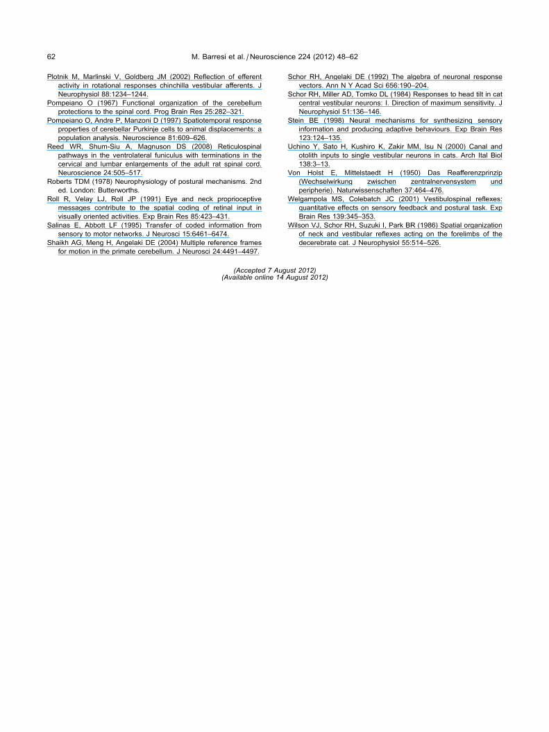
62 M. Barresi et al. / Neuroscience 224 (2012) 48–62
Plotnik M, Marlinski V, Goldberg JM (2002) Reflection of efferent
activity in rotational responses chinchilla vestibular afferents. J
Neurophysiol 88:1234–1244.
Pompeiano O (1967) Functional organization of the cerebellum
protections to the spinal cord. Prog Brain Res 25:282–321.
Pompeiano O, Andre P, Manzoni D (1997) Spatiotemporal response
properties of cerebellar Purkinje cells to animal displacements: a
population analysis. Neuroscience 81:609–626.
Reed WR, Shum-Siu A, Magnuson DS (2008) Reticulospinal
pathways in the ventrolateral funiculus with terminations in the
cervical and lumbar enlargements of the adult rat spinal cord.
Neuroscience 24:505–517.
Roberts TDM (1978) Neurophysiology of postural mechanisms. 2nd
ed. London: Butterworths.
Roll R, Velay LJ, Roll JP (1991) Eye and neck proprioceptive
messages contribute to the spatial coding of retinal input in
visually oriented activities. Exp Brain Res 85:423–431.
Salinas E, Abbott LF (1995) Transfer of coded information from
sensory to motor networks. J Neurosci 15:6461–6474.
Shaikh AG, Meng H, Angelaki DE (2004) Multiple reference frames
for motion in the primate cerebellum. J Neurosci 24:4491–4497.
Schor RH, Angelaki DE (1992) The algebra of neuronal response
vectors. Ann N Y Acad Sci 656:190–204.
Schor RH, Miller AD, Tomko DL (1984) Responses to head tilt in cat
central vestibular neurons: I. Direction of maximum sensitivity. J
Neurophysiol 51:136–146.
Stein BE (1998) Neural mechanisms for synthesizing sensory
information and producing adaptive behaviours. Exp Brain Res
123:124–135.
Uchino Y, Sato H, Kushiro K, Zakir MM, Isu N (2000) Canal and
otolith inputs to single vestibular neurons in cats. Arch Ital Biol
138:3–13.
Von Holst E, Mittelstaedt H (1950) Das Reafferenzprinzip
(Wechselwirkung zwischen zentralnervensystem und
peripherie). Naturwissenschaften 37:464–476.
Welgampola MS, Colebatch JC (2001) Vestibulospinal reflexes:
quantitative effects on sensory feedback and postural task. Exp
Brain Res 139:345–353.
Wilson VJ, Schor RH, Suzuki I, Park BR (1986) Spatial organization
of neck and vestibular reflexes acting on the forelimbs of the
decerebrate cat. J Neurophysiol 55:514–526.
(Accepted 7 August 2012)(Available online 14 August 2012)