Controlled Release Carriers of Growth Factors FGF-2 and TGF 1
Upload
independentCategory
view
0download
0
Effects of low-level laser therapy on theexpression of osteogenic genes related inthe initial stages of bone defects in rats
Kelly Rossetti FernandesDaniel Araki RibeiroNatália Camargo RodriguesCarla TimAnderson Amaro SantosNivaldo Antônio ParizottoHeloisa Selistre de AraujoPatrícia DriussoAna Claudia Muniz Rennó
Downloaded From: http://biomedicaloptics.spiedigitallibrary.org/ on 04/01/2013 Terms of Use: http://spiedl.org/terms
Effects of low-level laser therapy on the expression ofosteogenic genes related in the initial stages of bonedefects in rats
Kelly Rossetti Fernandes,a Daniel Araki Ribeiro,a Natália Camargo Rodrigues,b Carla Tim,b Anderson Amaro Santos,bNivaldo Antônio Parizotto,b Heloisa Selistre de Araujo,c Patrícia Driusso,b and Ana Claudia Muniz Rennóa
aFederal University of São Paulo, Department of Bioscience, Av. Ana Costa, 95, Vila Mathias, Santos, Sao Paulo, 11050-240 BrazilbFederal University of São Carlos, Department of Physiotherapy, RodWashington Luis, Km 235, Monjolinho, São Carlos, São Paulo, 13565-902 Brazil.cFederal University of São Carlos, Department of Physiological Sciences, Rod Washington Luis, Km 235, Monjolinho, São Carlos, São Paulo, 13565-902 Brazil
Abstract. We evaluate the effects of low-level laser therapy (LLLT) on the histological modifications and temporalosteogenic genes expression during the initial phase of bone healing in a model of bone defect in rats. Sixty-fourWistar rats were divided into control and treated groups. Noncritical size bone defects were surgically created at theupper third of the tibia. Laser irradiation (Ga-Al-As laser 830 nm, 30 mW, 0.028 cm2, 1.071 W∕cm2, 1 min and34 s, 2.8 Joules, 100 J∕cm2) was performed for 1, 2, 3, and 5 sessions. Histopathology revealed that treated animalspresented higher inflammatory cells recruitment, especially 12 and 36 h postsurgery. Also, a better tissue organi-zation at the site of the injury, with the presence of granulation tissue and new bone formation was observed ondays three and five postsurgery in the treated animals. The quantitative real time polymerase chain reaction showedthat LLLT produced a significantly increase in mRNA expression of Runx-2, 12 h and three days post-surgery, asignificant upregulation of alkaline phosphatase mRNA expression after 36 h and three days post-surgery and asignificant increase of osteocalcin mRNA expression after three and five days. We concluded that LLLT modulatedthe inflammatory process and accelerated bone repair, and this advanced repair pattern in the laser-treated groupsmay be related to the higher mRNA expression of genes presented by these animals. © 2013 Society of Photo-Optical
Instrumentation Engineers (SPIE) [DOI: 10.1117/1.JBO.18.3.038002]
Keywords: rats; bone regeneration; low-level laser therapy; genetic analysis.
Paper 12564R received Aug. 29, 2012; revised manuscript received Jan. 17, 2013; accepted for publication Feb. 14, 2013; publishedonline Mar. 20, 2013.
1 IntroductionBone fracture healing is a complex biological process thatinvolves the spatial and temporal interaction of various celltypes with large numbers of genes and an extracellular matrix.1
The process of fracture healing can be divided into several stagessuch as immediate injury response, inflammatory phase, intra-membranous and endochondral ossification and, finally, boneremodeling.2
Each step presents distinct histological features but they alldepend on the expression of cell-signaling molecules, synthesisof genes, deposition of extracellular matrix, and bone cell pro-liferation and differentiation.1,2 Moreover, it is well known thateach phase shows distinct histological features, with the markedinteraction of osteoblasts and osteoclasts cells.3 Osteoblastshave an important role in bone formation, being responsible byforming bone tissue, and their activity is influenced by Runx-2,bone morphogenetic proteins (BMPs), alkaline phosphatase(ALP), type I collagen, proteoglycan, bone sialoprotein, andosteocalcin (OC).4–9
In general, bone tissue has the ability of healing by itself.However, under critical conditions as for example, in largerbone defects and fractures with poor vascularization, a delayin the process of healing or even a nonunion can happen.10
Within this context, innovative clinical approaches to accel-erate bone metabolism and to repair damage to bone tissue arebeing developed.11 One promising treatment method is the useof low-level laser therapy (LLLT).11 Several studies suggestthat LLLT can stimulate osteogenesis at the site of fracture, pro-moting a higher deposition of bone mass and accelerating theconsolidation process.11–13
The effects of LLLT in biological tissues can be explained bythe photochemical theory, which offers an explanation for thesensitivity of cells to electromagnetic light.14 Laser irradiationis absorbed by photoreceptors located in the cells, promotingchanges in morphology molecular drum and thereafter. Thisstarts a series of modifications in cellular processes, with theincrease in cell signaling and excitation of components of themitochondrial respiratory chair.15,16 The stimulation of the res-piratory chain alters the redox potential of the cytoplasm andmitochondria, accelerating electron exchange and consequentlyincreasing ATP production. Therefore, one can observe anincrease in cellular metabolism that leads to a stimulation ofDNA and RNA expression, proteins, and enzymes synthesis.17
Stein et al.18 investigated the effects of LLLT (670 nm,1 J∕cm2, 400 mW) on human osteoblast-like cells and observeda significant increase in mRNA expression of ALP after 48 and72 h in the irradiated group as well as an upregulation in themRNA expression of osteopontin after 24 h. Moreover, Fávaro-Pipi et al.19 evaluated the effects of LLLT (830 nm, 30 mW,Address all correspondence to: Ana Claudia Muniz Rennó, Federal University of
São Paulo, Department of Bioscience, Av. Ana Costa, 95, Vila Mathias, Santos,Sao Paulo, Brazil 11050-240. Tel: +55 13 32218058; Fax: +55 13 32232592; E-mail: [email protected] 0091-3286/2013/$25.00 © 2013 SPIE
Journal of Biomedical Optics 038002-1 March 2013 • Vol. 18(3)
Journal of Biomedical Optics 18(3), 038002 (March 2013)
Downloaded From: http://biomedicaloptics.spiedigitallibrary.org/ on 04/01/2013 Terms of Use: http://spiedl.org/terms

50 J∕cm2) on the synthesis of genes in a tibial bone defectmodel. The authors demonstrated a higher expression ofBMP-4 at day 13 post-surgery and an upregulation of mRNAof BMP4, ALP and Runx-2 at day 25 after surgery.19
Although the effects of LLLT have been well described, themolecular and cellular changes induced by this treatment onbone tissue are not fully understood. Furthermore, there is alack in the literature describing the mechanisms by which laseracts in the initial periods in LLLT repair, since most of studieshave evaluated it effects on intermediate and late periods of bonehealing.1,12,13
Therefore, the hypothesis of this study is that LLLT iscapable of improving bone repair in the initial periods of con-solidation by activating the expression of genes related to bonecell proliferation and differentiation. Considering the stimula-tory effects of LLLT on bone regeneration after an injury, theaim of the present study was to evaluate the morphologicalchanges of bone and to measure the temporal pattern of theexpression of osteogenic genes after LLLT during the initialprocess of bone healing. We used histopathology and quantita-tive real time polymerase chain reaction (RT-qPCR) analysis toevaluate the effects of laser therapy irradiation in bone defects intibias of rats at 12 h, 36 h, 3 d and 5 d postsurgery.
2 MethodsThis study was approved by the Committee of Ethics in AnimalResearch at the Federal University of São Paulo, Brazil (0975/10). A total of 64 male Wistar rats (weighing 250þ ∕ − 50 g, 12to 13 weeks) were distributed randomly into two groups: controlor laser irradiated. They were maintained under controlled tem-perature (22þ ∕ − 2°C), light-dark periods of 12 h, and withfree access to water and a commercial diet. All animal handlingand surgical procedures were strictly conducted according theGuiding Principles for the Use of Laboratory Animals. Asdescribed below, noncritical size bone defects were performedon both tibias. Rats were sacrificed at 12 h, 36 h, 3 d and 5 dafter surgery.
2.1 Surgery
Noncritical size bone defects (2.5 mm diameter) were surgicallycreated at the upper third of the tibia (10 mm distal of the kneejoint). Surgery was performed under sterile conditions andgeneral anesthesia induced by intraperitoneal injection of20 mg∕Kg xilazin (Anasedan; Vetbrands, São Paulo, Brazil)and 40 mg∕kg ketamin (Dopalen; Vetbrands, São Paulo,Brazil). After antiasepsia of the skin, the medial compartmentof the tibia was exposed through a longitudinal incision onthe shaved skin and muscle tissue. A standardized 2.5 mm diam-eter bone defect was created using a motorized drill (Dentsply,Maillefer Instruments SA, Ballaigues, Suíça), 12.356 rpm,under copious irrigation with saline solution. The cutaneousflap was replaced and sutured with resorbable polyglactin, andthe antisepsis was performed with povidone iodine. The animalsreceived analgesia (i.m., 0.05 mg∕kg buprenorphine (Temgesic;Reckitt Benckiser Health Care Limited, Schering Plough,Hoddesdon, United Kingdom) and were returned to theircages. The health status of the rats was monitored daily.12,19
2.2 Treatments
The treatments, which started immediately post-surgery, wereperformed for one (12 h), two (36 h), three (3 d) or five
(5 d) sessions, with an interval of 24 h. A low-energy gallium–aluminum–arsenide (Ga-Al-As), laser 830 nm, 30 mW, continu-ous wavelength, 0.028 cm2, 1.071 W∕cm2, 1 min and 34 s,2.8 J, 100 J∕cm2 (Tera laser, DMC Equipamentos, São Carlos,SP, Brazil) was used. The parameters were specified based onthe equipment specification of the manufacturer. The irradiationwas performed at one point, above the area of the injury, throughthe punctual contact technique. On 12 h, 36 h, 3 d and 5 d post-injury, rats were sacrificed individually by carbon dioxideasphyxia. Both tibias were removed for analysis.
2.3 Histopathological Analysis
For the qualitative histopathological analysis, the right tibia wasused. They were removed, fixed in 10% buffer formalin (Merck,Darmstadt, Germany) for 48 h, decalcified in 4% EDTA (Merck,Darmstadt, Germany), and embedded in paraffin blocks. Five-micrometer slices were obtained in serial sections in the longi-tudinal direction and stained with hematoxylin and eosin (H.Estain, Merck). A descriptive qualitative histopathological evalu-ation of the total area of the bone defect was performed by apathologist (blinded to the treatment) under a light microscope(Olympus, Optical Co. Ltd, Tokyo, Japan) at 20×magnification.Any changes in the bone defect, such as presence of wovenbone, bone marrow, inflammatory process, granulation tissue,or even tissues undergoing hyperplastic, metaplastic, and/ordysplastic transformation were investigated per animal.
2.4 Total RNA Isolation and Real-Time PolymeraseChain Reaction
Immediately postmortem, left tibias were dissected (periosteumintact) and rapidly frozen in liquid nitrogen. The region of thecallus was stored (−80°C) until analysis by RT-qPCR. TotalRNA was isolated using standard protocols. Trizol reagent(1 ml, Invitrogen, Carlsbad, California) was added to the sampleand allowed to thaw. The mixture was transferred to a polypro-pylene tube and incubated (room temperature, 5 m). Chloroform(0.2 ml, Sigma, St. Louis, Missouri) was added, mixedvigorously, and the mixture was transferred to a 2 ml tube(Eppendorf, Hamburg, Germany) and centrifuged (28°C, 15 m).The nucleic acid phase was decanted and an equal volume ofRNase-free 70% ethanol was added. Potential DNA contamina-tion was removed by RNase-free DNase I (Invitrogen). RNAintegrity was verified by RNA gel electrophoresis and spectro-photometry. Four genes of interest were selected representingprocesses associated with osteogenesis (Table 1). For eachgene, rat-specific primers were designed for real-time PCRaround exon junctions where possible. All real-time primerswere initially tested against standards and a standard curvewas generated. First strand cDNA was synthesized (M-MLVRT, Invitrogen) from total RNA (1 mg). RT-qPCR reactionswere carried out at 50 ml total volume. Following an initialdenaturing step for each primer (Table 1), the genes of interestwere amplified through 40 cycles (Rotor-Gene, R 3000, RobertResearch, Mortlake, Australia). Gene amplification was mea-sured by SYBR green (Applied Biosystems, Carlsbad,California) fluorescence during the annealing/elongation phase.All samples were run in duplicate and the average was used forfurther analysis. Measures of real-time PCR cycle to thresholdwere normalized to the expression of ribosomal protein S18(RPS18) for each tibia. For comparison between experimentalgroups, RPS18 normalized expression from each tibia was
Journal of Biomedical Optics 038002-2 March 2013 • Vol. 18(3)
Fernandes et al.: Effects of low-level laser therapy on the expression of osteogenic genes related. . .
Downloaded From: http://biomedicaloptics.spiedigitallibrary.org/ on 04/01/2013 Terms of Use: http://spiedl.org/terms

divided by the normalized gene expression from the controlgroup to obtain a fold increase in gene expression of LLLTgroup and control group in 12 h, 36 h, 3 d and 5 d.
2.5 Statistical Analysis
Data are expressed as the mean� standard error of the mean(SEM). The normality of all variables’ distribution was verifiedusing Shapiro–Wilk’s W test. Two-way ANOVA was used toassess the effect of displacement level and time. Post hoc, multi-ple comparisons were made using Tukey’s protected least sig-nificant difference tests with statistical significance definedas p < 0.05.
3 ResultsFrom the 80 animals available for this study, four animals werelost due to an anesthesia-induced respiratory depression. Theremaining animals recovered uneventfully from the surgical pro-cedure and remained in good health. No signs of macroscopicinflammation or adverse tissue responses were observed duringthe experimental period. At the end of the experiment, 152 tibiaswere retrieved and used for both analyses (histopathology andRT-qPCR). For histopathological evaluation, 80 tibias wereused, however four tibias were excluded for animals that diedduring surgery. In addition, four tibias were excluded fromanalysis during histological processing (Table 2). Furthermore,a total of 72 tibias were retrieved, of which 68 of them wereincluded for histopathology analysis. For RT-qPCR, five ani-mals per group were used in a total of 40 tibias.
3.1 Histopathological Analysis
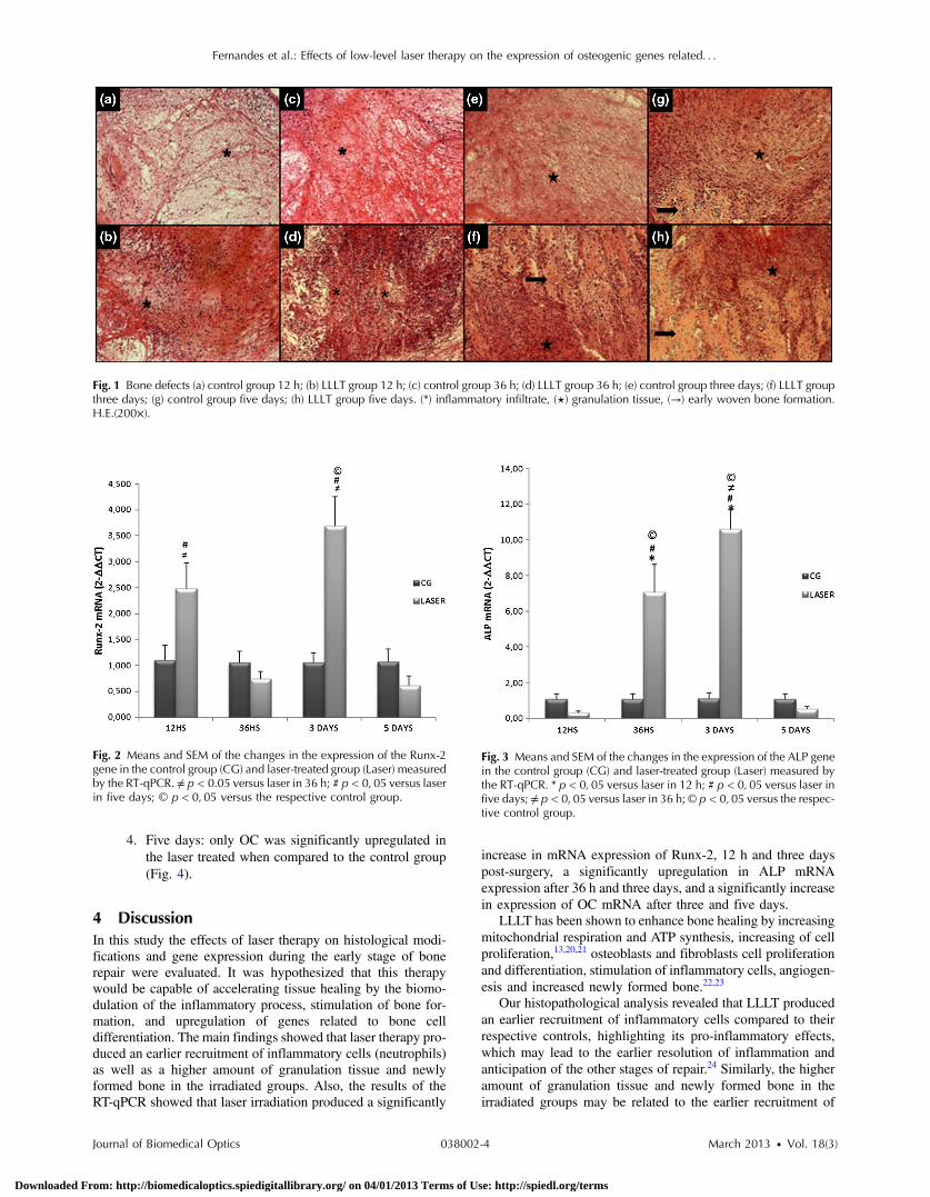
Twelve hours after surgery, for the animals in the control group,the edges of the defect were visible and a moderate amountinflammatory infiltrate, with polymorphonuclear cells (mainlyrepresented by neutrophils) were observed [Fig. 1(a)]. In theLLLT group, the histopathological analysis revealed an intenseamount of inflammatory infiltration, with a higher presence ofpolymorphonuclear cells (neutrophils) when compared to con-trol [Fig. 1(b)].
After 36 h, the control group presented an intense inflamma-tory infiltration, with polymorphonuclear cells [Fig. 1(c)]. In thelaser-irradiated animals, a lower inflammatory response wasobserved when compared to control [Fig. 1(d)].
In addition, on day three, a decrease in the inflammatoryprocess and increased granulation tissue was observed in thecontrol group [Fig. 1(e)]. Also, the irradiated animals displayeda higher amount of granulation tissue, new bone tissue, and neo-angiogenesis, which corresponds to more advanced repair com-pared to controls [Fig. 1(f)].
After five days, histological analysis of the control group[Fig. 1(g)] revealed the presence of granulation tissue andnewly formed bone on half of the animals tested. Figure 1(h)shows that, in the irradiated group, higher amount of granulationtissue and regions of bone remodeling and bone ingrowth werefound in all animals, with a better tissue organization comparedto the control group.
3.2 mRNA Expression of Runx-2, ALP, and OC
Figures 2–4 represent the temporal gene expression of the con-trol and LLLT treated groups.
1. 12 h: of the 3 genes analyzed, reflecting osteogeniccoupling potential in this model, it was observed thatonly Runx-2 was upregulated at this time point in thetreated group compared to control group (Fig. 2).Expression of ALP and OC did not differ between thegroups (Figs. 3 and 4).
2. 36 h: only ALP was consistently and significantlyupregulated in the laser-treated group when comparedto the nontreated animals at this time point. Othergenes did not show significant changes after lasertherapy.
3. Three days: significantly increased Runx-2, ALP andOC expressions were observed in laser treated animalscompared to the control (Figs. 2–4).
Table 1 Primers and indicated annealing temperatures for each gene analyzed.
Gene Forward primer Reverse primer Annealing temperature (°C)
Runx-2 ATGGCCGGGAATGATGAGAA TCTGTCTGTGCCTTCTTGGT 56°C
ALP CGAGCAGGAACAGAAGTTTGC TGGCCAAAAGGCAGTGAATAG 60°C
OC CTGCATTCTGCCTCTCTGACCT GCCGGAGTCTATTCACCACCTT 60°C
RPS18 CTAGTGATCCCCGAGAAGTTTC TGTCTGCTTTCCTCAACACC 60°C
Note: OC, osteocalcin; ALP, alkaline phosphatase; Runx -2, transcriptional factor.
Table 2 Number of tibias, retrieved and used for histological analyses.
Total numberof tibias Tibias retrieved
Tibias usedfor analysis
Control LLLT Control LLLT Control LLLT
12 hours 10 10 8a 10 8a 10
36 hours 10 10 9a 10 9a 9b
3 days 10 10 10 10 9b 10
5 days 10 10 10 9a 8b 9a
aDeviation from number of tibia due to animal death.bDeviation from number of tibias retrieved and were excluded fromanalysis during histophatological processing.
Journal of Biomedical Optics 038002-3 March 2013 • Vol. 18(3)
Fernandes et al.: Effects of low-level laser therapy on the expression of osteogenic genes related. . .
Downloaded From: http://biomedicaloptics.spiedigitallibrary.org/ on 04/01/2013 Terms of Use: http://spiedl.org/terms
4. Five days: only OC was significantly upregulated inthe laser treated when compared to the control group(Fig. 4).
4 DiscussionIn this study the effects of laser therapy on histological modi-fications and gene expression during the early stage of bonerepair were evaluated. It was hypothesized that this therapywould be capable of accelerating tissue healing by the biomo-dulation of the inflammatory process, stimulation of bone for-mation, and upregulation of genes related to bone celldifferentiation. The main findings showed that laser therapy pro-duced an earlier recruitment of inflammatory cells (neutrophils)as well as a higher amount of granulation tissue and newlyformed bone in the irradiated groups. Also, the results of theRT-qPCR showed that laser irradiation produced a significantly
increase in mRNA expression of Runx-2, 12 h and three dayspost-surgery, a significantly upregulation in ALP mRNAexpression after 36 h and three days, and a significantly increasein expression of OC mRNA after three and five days.
LLLT has been shown to enhance bone healing by increasingmitochondrial respiration and ATP synthesis, increasing of cellproliferation,13,20,21 osteoblasts and fibroblasts cell proliferationand differentiation, stimulation of inflammatory cells, angiogen-esis and increased newly formed bone.22,23
Our histopathological analysis revealed that LLLT producedan earlier recruitment of inflammatory cells compared to theirrespective controls, highlighting its pro-inflammatory effects,which may lead to the earlier resolution of inflammation andanticipation of the other stages of repair.24 Similarly, the higheramount of granulation tissue and newly formed bone in theirradiated groups may be related to the earlier recruitment of
Fig. 1 Bone defects (a) control group 12 h; (b) LLLT group 12 h; (c) control group 36 h; (d) LLLT group 36 h; (e) control group three days; (f) LLLT groupthree days; (g) control group five days; (h) LLLT group five days. (*) inflammatory infiltrate, (⋆) granulation tissue, (→) early woven bone formation.H.E.(200×).
Fig. 2 Means and SEM of the changes in the expression of the Runx-2gene in the control group (CG) and laser-treated group (Laser) measuredby the RT-qPCR. ≠ p < 0.05 versus laser in 36 h; # p < 0; 05 versus laserin five days; © p < 0;05 versus the respective control group.
Fig. 3 Means and SEM of the changes in the expression of the ALP genein the control group (CG) and laser-treated group (Laser) measured bythe RT-qPCR. * p < 0; 05 versus laser in 12 h; # p < 0; 05 versus laser infive days; ≠ p < 0;05 versus laser in 36 h; © p < 0;05 versus the respec-tive control group.
Journal of Biomedical Optics 038002-4 March 2013 • Vol. 18(3)
Fernandes et al.: Effects of low-level laser therapy on the expression of osteogenic genes related. . .
Downloaded From: http://biomedicaloptics.spiedigitallibrary.org/ on 04/01/2013 Terms of Use: http://spiedl.org/terms

osteoprogenitor cells and mature osteoblasts, resulting in higherdeposition of newly formed bone.
These results corroborate with several studies that showedpositive effects of laser irradiation on bone cell proliferationand differentiation.13,21,25 Pretel et al.26 found that LLLT(780 nm, 35 mW, 178 J∕cm2, 1,4 J) modulated the initialinflammatory response and anticipated the resolution of theprocess to normal conditions at the earlier periods in a bonedefect model in rats.
Additionally, in vitro and in vivo studies showed that manygenes related to bone repair have been shown to have their tran-scription modified after irradiation with LLLT.27–29
In this study, on the experimental period of 12 h, only Runx-2 mRNA expression was found increased in the LLLT group.Runx-2 is essential for the commitment of early mesenchymalcells to the osteoblasts lineage and differentiation of osteo-blasts.30 In addition, Runx-2 controls the differentiation andfunction of the osteoblast in concert with other factors suchas Osterix by regulating the expression of many osteoblast-related genes such as ALP, OC and osteopontin.31 These resultscorroborate with other studies found in the literature.30–32 Forexample, Fujimoto et al.32 observed that in osteoblastic cell cul-tures, LLLT (830 nm, 1.91 J∕cm2) promoted an increasedexpression of Runx-2 12 h and 48 h after irradiation. The higherexpression of mRNA Runx-2 could be related to the prematuredifferentiation of osteoblast cells at the site of the injury andcould have stimulated the expression of other genes evaluatedin this study (ALP and OC).
Moreover, RT-qPCR analysis demonstrated that laser irradi-ation produced an upregulation of the mRNA of ALP at 36 h andthree days after the surgery. During the process of bone healing,ALP is an important marker of osteoblast differentiation.33 Ourresults suggested that the increase of the mRNA of ALP expres-sion in the laser-treated group may have induced the prematurerecruitment of the osteoblast cells, resulting in a higher deposi-tion of bone matrix. Many authors have demonstrated that LLLTis capable of modulating the expression of the mRNA of ALP atdifferent times.20,28,34 In vitro studies showed that LLLT (904 to910 nm, 200 mW, 6 J∕cm2) applied to human osteoblastic cells
produced an increased mRNA expression of ALP after 10 daysof irradiation.28 However, Khadra et al.27 showed that LLLT(Ga-Al-As, 1, 5 or 3 J∕cm2) did not show any significant differ-ence in the expression of ALP when applied to cells derivedfrom human mandibular bone. In an in vivo study, Fávaro-Pipi et al.29 observed that laser irradiation (830 nm, 50 J∕cm2,1.4 J) produced an increase of ALP mRNA expression in a tibialbone defect model in rats, 25 days after the surgery.
Interestingly, at the third experimental period evaluated, themRNA expression of Runx-2 and OC was also significantlyincreased. Taken as a whole, it appears that this increase inexpression of the genes analyzed may suggest that this resultis related to differentiation of osteoblasts and newly bone for-mation observed in the laser-irradiated animals, supporting thehypothesis that laser therapy showed an osteogenic potential.
Similarly, OC is a marker of bone formation7 and it is themost abundant noncollagenous proteins in bone tissue.5 In ourstudy, the increased expression of OCmRNA after three and fivedays in the irradiated animals appears at the same time with thehigher amount of newly formed bone in the laser groupsobserved in the histological analysis. These results corroboratewith Stein et al.18 who observed an increase in OC mRNAexpression in human osteoblastic cells, 72 h after LLLT appli-cation (670 nm, 2 J∕cm2).
The methodology employed in this study has been used bymany authors.19,23,35 Some limitations of our work should bepointed out. We investigated the effects of LLLT in the expres-sion of ALP, OC and Runx-2 mRNA during the process of bonehealing. It would be interesting to investigate the effects of laserapplication on the expression of other osteogenic factors that arealso involved in bone repair. Also, more quantitative analysisshould be included in future researches such as the quantifica-tion of inflammatory cells and immunohistochemical analysis.In spite of these limitations, the results of this work highlightthe stimulatory effects of laser therapy on bone healing.
Summarizing, this study suggests that the increased expres-sion of osteogenic genes may be responsible by the differentia-tion of osteoblastic cells and stimulation of osteogenesis, whichmay have culminated in the acceleration of repair in the treatedanimals (revealed by the histopathological analysis). This workadvances in the description of molecular and cellular mechanismsof the effects of LLLTon bone repair process, mainly through theincreased expression of genes involved in this process.
5 ConclusionIn conclusion, the present study has shown that LLLT evoked anearlier resolution of the inflammatory process and newly boneformation. In addition, LLLT produced a significant increase ofmRNA expression of Runx-2, ALP and OC, which are involvedin bone repair. Despite these results, further studies are required toinvestigate the mechanisms and molecular pathways stimulatedby LLLT that culminate in the acceleration of bone healing.
References1. K. Sena et al., “Early gene response to low-intensity pulsed ultrasound
in rat osteoblastic cells,” Ultrasound Med. Biol. 31(5), 703–708 (2005).2. R. A. Kayal et al., “Diabetes causes the accelerated loss of cartilage
during fracture repair which is reserved by insulin treatment,” Bone44(2), 357–363 (2009).
3. R. A. Nicola et al., “Effect of low-power GaAlAs laser (660 nm) onbone structure and cell activity, an experimental animal study,”Lasers Med. Sci. 18(2), 89–94 (2003).
Fig. 4 Means and SEM of the changes in the expression of the OC genein the control group (CG) and laser-treated group (Laser) measured bythe RT-qPCR. * p < 0;05 versus laser in 12 h; ≠ p < 0; 05 versus laser in36 h; # p < 0;05 versus laser in five days; © p < 0; 05 versus the respec-tive control group.
Journal of Biomedical Optics 038002-5 March 2013 • Vol. 18(3)
Fernandes et al.: Effects of low-level laser therapy on the expression of osteogenic genes related. . .
Downloaded From: http://biomedicaloptics.spiedigitallibrary.org/ on 04/01/2013 Terms of Use: http://spiedl.org/terms

4. S. L. Song et al., “Temporal expression of proteoglycans in the rat limbduring bone healing,” Gene 379, 92–100 (2006).
5. A. Stein et al., “Low-level laser irradiation promotes proliferation anddifferentiation of human osteoblasts in vitro,” Photomed. Laser Surg.23(2), 161–166 (2005).
6. B. Rath et al., “Compressive forces induce osteogenic gene expressionin calvarial osteoblast,” J. Biomech. 41(5), 1095–1103 (2008).
7. I. Bar et al., “Molecular imaging of the skeleton: quantitative real-timebioluminescence monitoring gene expression in bone repair and devel-opment,” J. Bone Miner. Res. 18(3), 570–578 (2003).
8. G. R. Wohl, D. A. Towler, and M. J. Silva, “Stress fracture healing:fatigue loading of the rat ulna induces upregulation in expression ofosteogenic and angiogenic genes that mimic the intramembranous por-tion of fracture repair,” Bone 44(2), 320–330 (2009).
9. M. Hadjiargyrou et al., “Enhancement of fracture healing by low inten-sity ultrasound,” Clin. Orthop. Relat. Res. 355, 216–229 (1998).
10. G. Victoria et al., “Bone stimulation for fracture healing: what’s all thefuss?,” Indian J. Orthop. 43(2), 117–120 (1998).
11. S. Hamajima et al., “Effect of low-level laser irradiation onosteoglycin gene expression in osteoblasts,” Lasers Med. Sci. 18(2),78–82 (2003).
12. A. L. B. Pinheiro et al., “Biomodulatory effects of LLLTon bone regen-eration,” Laser Therapy 13, 73–79 (2000).
13. Y. A. Vladimirov, A. N. Osipov, and G. I. Klebanov, “Photobiologicalprinciples of therapeutic applications of laser radiation,” Biochemistry69(1), 81–90 (2004).
14. T. I. Karu and S. F. Kolyakov, “Exact action spectra of cellular responsesrelevant to phototherapy,” Photomed. Laser Surg. 23(4), 356–361(2005).
15. T. I. Karu, L. V. Pyatibrat, and G. S. Kalendo, “Cell attachment toextracellular matrices is modulated by pulsed radiation at 820 nmand chemicals that modify the activity of enzymes in the plasma mem-brane,” Lasers Surg. Med. 29(3), 274–281 (2001).
16. S. S. Kitchen and C. J. Partridge, “A review of low level laser therapy,”Physiotherapy 77(3), 161–168 (1991).
17. T. Karu, “Introduction,” in The Science of Low-Power Laser Therapy,T. I. Karu, Ed., pp. xiii–xviii, Gordon and Breach Science Publishers,Amsterdam (1998).
18. A. Stein et al., “Initial effects of low-level laser therapy on growth anddifferentiation of human osteoblast-like cells,” Wien Klin. Wochenschr.120(3–4), 112–117 (2008).
19. E. Fávaro-Pipi et al., “Comparative study of the effects of low-intensitypulsed ultrasound and low-level laser therapy on bone defects in tibiasof rats,” Lasers Med. Sci. 25(5), 727–32 (2010).
20. Y. Ozawa et al., “Low-energy laser irradiation stimulates bone noduleformation at early stages of cell culture in rat calvarial cells,” Bone22(4), 347–354 (1998).
21. T. Ninomiya et al., “Increase of bone volume by a nanosecond pulsedlaser irradiation is caused by a decreased osteoclast number and an acti-vated osteoblast,” Bone 40(1), 140–148 (2007).
22. J. Nissan et al., “Effect of low intensity laser irradiation on surgicallycreated bone defects in rats,” J. Oral Rehabil. 33(8), 619–924 (2006).
23. A. P. Lirani-Galvão, V. Jorgetti, and O. L. da Silva, “Comparative studyof how low-level laser therapy and low-intensity pulsed ultrasoundaffect bone repair in rats,” Photomed. Laser Surg. 24(6), 735–740(2006).
24. A. Lenz, G. A. Franklin, and W. G. Cheadle, “Systemic inflammationafter trauma,” Injury 38(12), 1336–1345 (2007).
25. A. L. B. Pinheiro et al., “Biomodulatory effects of LLLTon bone regen-eration,” Laser Therapy 13, 73–79 (2000).
26. H. Pretel, R. F. Z. Lizarelli, and L. T. O. Ramalho, “Effect of low-levellaser therapy on bone repair: histological study in rats,” Lasers Surg.Med. 39(10), 788–796 (2007).
27. M. Khadra et al., “Enhancement of bone formation in rat calvarial bonedefects using low-level laser therapy,” Oral Surg. Oral Med. OralPathol. Oral Radiol. Endod. 97(6), 693–700 (2004).
28. S. Saracino et al., “Superpulsed laser irradiation increases osteoblastactivity via modulation of bone morphogenetic factors,” Lasers Surg.Med. 41(4), 298–304 (2009).
29. E. Fávaro-Pipi et al., “Low-level laser therapy induces differentialexpression of osteogenic genes during bone repair in rats,”Photomed. Laser Surg. 29(5), 311–317 (2011).
30. T. Komori, “Regulation of bone development and extracellular matrixprotein genes by RUNX2,” Cell Tissue Res. 339(1), 189–195 (2010).
31. B. L. Vaes et al., “Microarray analysis on RUNX-2 deficient mouseembryos reveals novel RUNX-2 functions and target genes during intra-membranous and endochondral bone formation,” Bone 39(4), 724–738(2006).
32. K. Fujimoto et al., “Low-intensity laser irradiation stimulates minerali-zation via increased BMPs in MC3T3-E1 cells,” Lasers Surg. Med.42(6), 519–526 (2010).
33. P. Proff and P. Römer, “The molecular mechanism behind bone remod-elling: a review,” Clin. Oral Investig. 13(4), 355–362 (2009).
34. O. Barushka et al., “Effect of low energy laser (He-Ne) irradiation on theprocess of bone repair in the rat tibia,” Bone 16(1), 47–55 (1995).
35. P. Oliveira et al., “Low-level laser therapy does not modulate the out-comes of a highly bioactive glass-ceramic (Biosilicate) on bone consoli-dation in rats,” J. Mater. Sci. Mater. Med. 21(4), 1379–1384 (2010).
Journal of Biomedical Optics 038002-6 March 2013 • Vol. 18(3)
Fernandes et al.: Effects of low-level laser therapy on the expression of osteogenic genes related. . .
Downloaded From: http://biomedicaloptics.spiedigitallibrary.org/ on 04/01/2013 Terms of Use: http://spiedl.org/terms
Copyright © 2022 FDOKUMEN