Effects of lamellolysin from a parasitoid wasp onDrosophila blood cells in vitro
9
THE JOURNAL OF EXPERIMENTAL ZOOLOGY 257:236-244 (1991) Effects of Lamellolysin From a Parasitoid Wasp on Drosophila Blood Cells In Vitro R.M. RIZKI AND T.M. RIZKI Department of Biology, The University of Michigan, Ann Arbor, Michigan 481 09 ABSTRACT Female parasitoid Leptopilina heterotoma inject a factor, lamellolysin, along with their eggs into the host hemocoel to destroy selectively host hemocytes that encapsulate foreign objects. In parasitized Drosophila melanogaster larvae, these hemocytes (lamellocytes) change from discoidal cells to bipolar cells that no longer adhere to each other to form capsules. To study the effects of lamellolysin on Drosophila lamellocytes in vitro, a giant strain of D. melanogaster was constructed to yield hemolymph with an abundance of lamellocytes. The effect of lamellolysin on the adhesivity of lamellocytes in vitro was demonstrated when the cells were gently rotated in the culture medium. Under these conditions, the bipolar shape of the affected lamellocytes resembled that of lamellocytes in parasitized hosts. When lamellocytes were exposed to lamellolysin in sta- tionary culture medium, the elongation of the bipolar cells continued until they became threadlike. Lamellocytes fragmented in both stationary and rotating culture medium in the presence of lamel- lolysin, although loss of cellular material was more pronounced in the latter. This study demon- strates that lamellolysin acts directly and destructively on lamellocytes. Survival of an endoparasitoid within the hemocoel of an insect host depends on its ability to evade or suppress host cellular defense reac- tions directed against foreign materials. Some parasitoid eggs have surface features that provide passive protection against encapsulation by host hemocytes (Salt, '65; Rotherham, '73; Davies and Vinson, '86). The hemocytes of other host species are inhibited from encapsulating parasitoid eggs by substances that the female parasitoids oviposit along with eggs in the host hemocoel. Among these materials are the viruses in the calyx fluid of Campoletis sonorensis (Edson et al., '81; Davies et al., ,871, the accessory gland secretions of Pim- pla turionella (Osman, '78); the venom and calyx fluid of Apanteles glomeratus (Wago and Kitano, '85; Kitano, '82), and the factor within the acces- sory gland reservoir of Leptopilina heterotoma that selectively destroys the capsule-forming hemocytes of Drosophila rnelanogaster (R. Rizki and T. Rizki, '84). The hemocytes of D. melanogaster larvae that form capsules are discoidal cells called lamello- cytes (Rizki, '57; Rizki and Rizki, '79). Within sev- eral hours after an L. heterotoma female oviposits in a Drosophila larva, the lamellocytes assume a bipolar morphology and lose their adhesiveness (R. Rizki and T. Rizki, '84). The source of the wasp factor responsible for the lamellocyte changes was 0 1991 WILEY-LISS, INC. located by injecting various isolated portions of the female reproductive system into Drosophila larvae. When the contents of a reservoir associ- ated with an accessory gland were injected into Drosophila larvae, the effects on the lamellocytes mimicked those found in parasitized larvae. This organ is referred to as the long gland reservoir, and the factor within this reservoir that alters the lamellocytes was called lamellolysin. The nature of lamellolysin is unknown, but its selectivity for lamellocytes is intriguing. There are no apparent effects on other host cells and organs. Equally in- teresting is the bipolar lamellocyte morphology induced by this factor. Identification of the factor within the long gland reservoir that destroys Drosophila lamello- cytes requires assaying components of the reser- voir for activity against lamellocytes. In vivo as- say involving injection of samples into Drosophila larvae is tedious and time consuming. There are also other drawbacks of this technique, such as difficulties in quantifying the amount of material injected into a larva and loss of hemolymph fol- lowing puncture of the cuticle of anaesthetized larvae. An in vitro assay for lamellolysin would avoid these problems. Furthermore, the kinetics of the changing cell shapes induced by lamelloly- sin would be easier to study in vitro than in vivo. Two methods for observing the effects of long
-
Upload
independent -
Category
Documents
-
view
5 -
download
0
Transcript of Effects of lamellolysin from a parasitoid wasp onDrosophila blood cells in vitro

THE JOURNAL OF EXPERIMENTAL ZOOLOGY 257:236-244 (1991)
Effects of Lamellolysin From a Parasitoid Wasp on Drosophila Blood Cells In Vitro
R.M. RIZKI AND T.M. RIZKI Department o f Biology, The University of Michigan, Ann Arbor, Michigan 481 09
ABSTRACT Female parasitoid Leptopilina heterotoma inject a factor, lamellolysin, along with their eggs into the host hemocoel to destroy selectively host hemocytes that encapsulate foreign objects. In parasitized Drosophila melanogaster larvae, these hemocytes (lamellocytes) change from discoidal cells to bipolar cells that no longer adhere to each other to form capsules. To study the effects of lamellolysin on Drosophila lamellocytes in vitro, a giant strain of D. melanogaster was constructed to yield hemolymph with an abundance of lamellocytes. The effect of lamellolysin on the adhesivity of lamellocytes in vitro was demonstrated when the cells were gently rotated in the culture medium. Under these conditions, the bipolar shape of the affected lamellocytes resembled that of lamellocytes in parasitized hosts. When lamellocytes were exposed to lamellolysin in sta- tionary culture medium, the elongation of the bipolar cells continued until they became threadlike. Lamellocytes fragmented in both stationary and rotating culture medium in the presence of lamel- lolysin, although loss of cellular material was more pronounced in the latter. This study demon- strates that lamellolysin acts directly and destructively on lamellocytes.
Survival of an endoparasitoid within the hemocoel of an insect host depends on its ability to evade or suppress host cellular defense reac- tions directed against foreign materials. Some parasitoid eggs have surface features that provide passive protection against encapsulation by host hemocytes (Salt, '65; Rotherham, '73; Davies and Vinson, '86). The hemocytes of other host species are inhibited from encapsulating parasitoid eggs by substances that the female parasitoids oviposit along with eggs in the host hemocoel. Among these materials are the viruses in the calyx fluid of Campoletis sonorensis (Edson et al., '81; Davies et al., ,871, the accessory gland secretions of Pim- pla turionella (Osman, '78); the venom and calyx fluid of Apanteles glomeratus (Wago and Kitano, '85; Kitano, '82), and the factor within the acces- sory gland reservoir of Leptopilina heterotoma that selectively destroys the capsule-forming hemocytes of Drosophila rnelanogaster (R. Rizki and T. Rizki, '84).
The hemocytes of D. melanogaster larvae that form capsules are discoidal cells called lamello- cytes (Rizki, '57; Rizki and Rizki, '79). Within sev- eral hours after an L. heterotoma female oviposits in a Drosophila larva, the lamellocytes assume a bipolar morphology and lose their adhesiveness (R. Rizki and T. Rizki, '84). The source of the wasp factor responsible for the lamellocyte changes was
0 1991 WILEY-LISS, INC.
located by injecting various isolated portions of the female reproductive system into Drosophila larvae. When the contents of a reservoir associ- ated with an accessory gland were injected into Drosophila larvae, the effects on the lamellocytes mimicked those found in parasitized larvae. This organ is referred to as the long gland reservoir, and the factor within this reservoir that alters the lamellocytes was called lamellolysin. The nature of lamellolysin is unknown, but its selectivity for lamellocytes is intriguing. There are no apparent effects on other host cells and organs. Equally in- teresting is the bipolar lamellocyte morphology induced by this factor.
Identification of the factor within the long gland reservoir that destroys Drosophila lamello- cytes requires assaying components of the reser- voir for activity against lamellocytes. In vivo as- say involving injection of samples into Drosophila larvae is tedious and time consuming. There are also other drawbacks of this technique, such as difficulties in quantifying the amount of material injected into a larva and loss of hemolymph fol- lowing puncture of the cuticle of anaesthetized larvae. An in vitro assay for lamellolysin would avoid these problems. Furthermore, the kinetics of the changing cell shapes induced by lamelloly- sin would be easier to study in vitro than in vivo.
Two methods for observing the effects of long

LAMELLOLYSIN EFFECTS ON CELLS IN VITRO
gland reservoir contents on Drosophila lamello- cytes in vitro were utilized in this study. In the first method, hemocytes were maintained in sta- tionary culture medium so that the changes in individual lamellocytes induced by the wasp fac- tor could be followed for several days. The second method involved rotation of hemocyte samples so that blood cells swirled gently in the liquid me- dium. This technique, which was used to evaluate changes in lamellocyte adhesion as well as mor- phology in the presence of lamellolysin, simulates the circulation of hemolymph in the hemocoel of a Drosophila larva.
MATERIALS AND METHODS Since only the lamellocytes are markedly af-
fected by lamellolysin, it is necessary to have hemolymph samples with many cells of this type for analysis. This requirement is met by using the temperature-sensitive melanotic tumor mutant, tu-Szts, of Drosophila melanogaster as a source of hemocytes (Rizki and Rizki, '80). Lamellocytes are abundant in melanotic tumor larvae but not in larvae whose cellular defense systems have not been challenged (Rizki, '57). Unfortunately, the number of hemocytes obtained from bleeding a single Drosophila larva is small, so hemolymph must be collected from many tu-Szts larvae to as- say a number of fractions separated from the long gland reservoir in a single experiment. To in- crease the lawellocyte yield per larva, the tu-SztS gene was combined with alleles of the giant (gt) gene, which increases larval size by 70% (Lindsley and Grell, '68).
Two alleles of the gt gene were separately com- bined with the tu-Szts allele. The genotypes of the stocks are 1) gt wa tu-Szts and 2) y sc gtxI1 v81wc tu- SztSlw+Y 6 8 and C(1)Dx ywfIw+Y Q 0 (for expla- nation of symbols, see Lindsley and Grell, '68). Matings between females of the former stock and males of the latter stock give viable female larvae of the genotype gt wa tu-Sztsly sc gtx" v81w tu-Szts. The larval life of these gt heterozygotes is more than double that of tu-Szts larvae, and they con- tinue to accumulate lamellocytes during this ex- tended growth period. These larvae are referred to as giant larvae in this paper. The melanotic tumor phenotype is expressed in the giant larvae when they are grown at 27°C just as in tu-Szts larvae raised at this temperature (Fig. 1). Not only does the volume of hemolymph obtained from the giant larvae exceed that of tu-Szts larvae but the hemolymph of these larvae contains an abundance of lamellocytes.
237
Fig. 1. Comparison of the size of a tuSztS larva (left) and a giant larva. The arrows point to the melanotic tumors of the caudal fat bodies.
Giant larvae were raised on cream of wheat1 molasses medium seeded with live Fleischmann's yeast at 27°C. To set up axenic cultures used in some of the studies, flies were allowed to lay eggs on the surface of cream of wheat medium for 15 hr. The eggs were collected from the medium sur- face with a fine brush, rinsed twice in distilled H20, and dechorionated in 5.25% sodium hypo- chlorite (Clorox bleach). After four rinses in dis- tilled H20, they were sterilized by immersion in povidone-iodine (Betadine solution) for 1 min, rinsed twice in sterile distilled Hd20, and trans- ferred to the surface of sterile medium, which con- sisted of 1% agar, 5% glucose, and 5% brewers yeast.
Hemolymph from 8-10-day-old giant larvae was collected in sterile Schneider's culture me- dium (GIBCO) supplemented with 15% fetal calf serum. For some studies hemolymph was col- lected in sterile phosphate buffer (PB) for Drosophila cells (Seecof, '71). Phenylthiourea (0.07%) was added to both solutions to inhibit phenoloxidase activity so the hemolymph samples

238 R.M. RIZKI AND T.M. RIZKI
would not darken. Hemocyte samples intended for long-term observation were set up under sterile conditions so that lamellocyte morphology could be examined for several days in the absence of contaminating microorganisms.
Stationary samples were set up in 7 x 32 mm Sykes chambers with a 1.5 mm Sykes-Moore-0- ring between the lower and upper coverglasses. Each chamber contained 135 p1 of liquid with hemolymph from one or two giant larvae. The cells in these chambers were examined with an Olympus CK2 inverted microscope and photo- graphed with a 35 mm camera.
Hemolymph samples that were gently swirled on a rotary shaker are referred to as rotation sam- ples. A sample (75 p1) was placed in a siliconized flat-bottom depression slide covered with a round 25 mm coverglass held in place over a silicone gasket by petroleum jelly. The slides were placed on the table of a rotary shaker making 70 revolu- tions per minute. Under these conditions, the liq- uid on the slides swirled gently. This degree of movement was used previously to assess the ag- glutination of Drosophila cells treated with lec- tins (Andrews and Rizki, '781, so it was deemed adequate to evaluate the adhesivity of lamello- cytes in vitro. The hemocyte samples were trans- ferred to microscope slides for examination by dark field illumination or phase contrast and pho- tographed with a Polaroid-back camera.
Leptopilina heterotoma (Leiden strain) were bred on tu-Szts larvae. Adult parasitoids were maintained on 50% honey solution in distilled H20 at 18°C. Female wasps were rinsed with 95% alcohol, followed by three rinses in sterile dis- tilled H20 before they were dissected in PB. Long gland reservoirs were collected in 5 pl PB and torn with fine forceps to release their whitish con- tents immediately before it was added to Drosophila hemocyte samples. For each experi- ment, a control sample inoculated with 5 pl PB was included. The parasitoid females used for studies employing sterile cultures were treated with Betadine solution and rinsed in sterile dis- tilled H20 before they were dissected under sterile conditions to isolate reservoirs. The use of trypan blue exclusion to assess lamellocyte viabil- ity followed the procedure given by Mishell and Shiigi ('80).
RESULTS To assess the effects of long gland reservoir
fluid on lamellocytes maintained in quiescent culture medium, aliquots of hemolymph in
Schneider's medium were dispensed to two Sykes chambers under sterile conditions. For each experiment, Leiden reservoir fluid from two to five female wasps was added t o one chamber, and the other chamber was set up as the control to observe the morphology of lamellocytes in Schneider's medium. Both hemocyte samples of each experimental group were examined im- mediately after they were made to confirm that the lamellocytes were normal. Since only one in- verted microscope was available for the study, only the chamber containing reservoir fluid was kept on the microscope stage for intermittent ob- servation. The control sample was examined again after the conclusion of the observations on the treated sample, so the same cells in control samples were not observed during the course of a given experiment.
Lamellocyte clumps are common in hemolymph samples from tu-Szts larvae. Figure 2A shows a typical group of lamellocytes in a sample exam- ined 15 min (t0:15) after the initial (to) introduction of cells and reservoir fluid into the chamber. There was no difference between these cells and cells in the control chamber. At tl:30, there were no apparent changes in the cells in this clump, and the field of observation was moved to photo- graph isolated lamellocytes. Four lamellocytes with the normal discoidal morphology were pho- tographed at tl:40 (Fig. 2B) and when they had assumed a bipolar shape at t2:50 (Fig. 2C). It should be noted that lamellocytes remaining sus- pended in the liquid medium occasionally rotate even though effort is made not to disturb the mi- croscope stage. The lamellocyte at the bottom of the frame in Figure 2C has floated so that its lat- eral profile is apparent in the photograph. At t3:05,
there were no further signs of elongation of these lamellocytes, and the slide was moved to observe other cells.
The elongation of the tip of a single lamellocyte was followed from t 3 0 5 to t3:35. The rate of exten- sion at one end of this lamellocyte was so rapid that the growth of the tip could be discerned by eye while the cell was being observed (Fig. 3A- H). The extension rate of this tip was calculated to be 2 pm/min. Rapidly growing cellular exten- sions were also observed in other samples of lamellocytes exposed to reservoir fluid, but the changes in lamellocyte shape do not always pro- ceed so rapidly. The series of photographs in Fig- ure 3 also illustrates a slight curving of the grow- ing tip. This curving motion may be associated with the growth of the tip itself or may be caused

LAMELLOLYSIN EFFECTS ON CELLS IN VITRO 239
Fig. 2. A Typical aggregate of lamellocytes in a hemo- lymph sample in Schneider’s medium; stationary culture. B,C: Selected lamellocytes monitored from the same culture as in A after 1 hr, 40 min (B), and after 2 hr and 50 min (C), of exposure to lamellolysin. Note the shape of the two lamello- cytes in the center and the upper left corner (arrowheads) in both photographs. The stars serve as position markers. In C, the change in lamellocyte shape is apparent. The bottom lamellocyte of the central group has floated in the culture medium and is seen in its lateral profile. Bar = 50 pm.
by movement within the culture medium gener- ated by floating cells. The field in Figure 3 was photographed 17.5 hr later (Fig. 4A), when the lamellocytes were converted into long threads that had no resemblance to their original dis- coidal morphology. The nuclei remain as sphe- roidal regions within the extremely elongated cells.
In another experiment, the remnants of elong- ated lamellocytes were photographed 27 hr after the sample was set up and again at 47 hr when one of the elongated lamellocytes had separated into two pieces (Fig. 4B,C). Small fragments re- sembling the extending filaments and tips of af- fected lamellocytes were found in other stationary
Fig. 3. Series of photographs of an elongating lamellocyte from the same preparation as Figure 2. The stars (in A and H) represent reference sites to judge the elongation of the tip of the lamellocyte in 30 min (from A through H). As the cell elongates, an overall thinning of the cell body is apparent. The elongating tip to the right has a small, bright contrasting bleb. Such blebs are common in the elongating cells. Bar = 50 pm.
samples examined on the second and third days. Lamellocytes in the absence of reservoir fluid
did not undergo elongation. Sterile control sam- ples were photographed shortly after preparation and at the conclusion of each experiment. By the second day in vitro, the edges of many of the lamellocytes were curved and folded, and the cells remained in clumps.
To study the effects of reservoir fluid concentra- tion on the transformation of lamellocytes, a sam- ple of hemolymph from eight giant larvae was dispensed to four Sykes chambers. Aliquots of re- servoir fluid from 30 Leiden females were distrib- uted to the chambers so that one contained the reservoir fluid equivalent (RFE) to that of 16

240 R.M. RIZKI AND T.M. RIZKI
Fig. 4. A Larger area, showing the same lamellocyte as in Figure 3 photographed 17.5 hr later, when this cell and other lamellocytes in the field had converted to threads. The arrow points to the nucleus of the lamellocyte. Bar = 50 IJ-m. B,C: Two photographs of the same elongating lamellocyte from another stationary culture containing lamellolysin photo- graphed at 27 hr (B) and 47 hr (C). The arrowhead and arrow indicate the same parts of the cell in B and C. The forked (0 region in B has broken near the point of the arrow and rotated in C. The stars are position markers.
wasps, another chamber received 4 RFE, the third had 1 RFE, and no reservoir fluid was added to the fourth chamber that served as the control. The total volume of sample in each chamber was 140 p1, and the series of samples was examined four hours after it was prepared. Two fields with well dispersed lamellocytes were randomly se- lected and photographed. The normal and affected lamellocytes in each photograph were counted to estimate the percentage of affected cells. The total number of lamellocytes counted in 16 RFE was 94, in 4 RFE 68, in 1 RFE 61, and in the control (0 RFE) 40. The percent of affected lamellocytes in the samples was loo%, 96%, 75%, and 0%, respec- tively. The information from this series combined with observations in the above-described studies in which fluid from two reservoirs was added to yield total sample volumes of 135 pl indicates that at least 1 RFE per 70 p1 is required to induce lamellocyte changes under these conditions of in- cubation in vitro.
Not only was there a positive quantitative cor- relation between RFE dosage and affected lamel- locyte frequency, but the quality of effect was more pronounced at higher dosage. In hemolymph samples from parasitized larvae and in hemo- lymph samples treated with reservoir fluid in vitro, an occasional bipolar lamellocyte has more than a single elongating filament at one of the poles. This phenomenon was more frequent at the higher RFEs, so it was quantified by counting the number of filaments at the poles of affected cells. In the 16 RFE sample, 17% of the affected lamellocytes had one filament at one pole and two or more (up to five) filaments at the opposite pole. A t 4 RFE, 5% of the affected lamellocytes had two filaments at one pole and one filament at the other pole. No multifilamentous poles were found in the photographs of the 1 RFE sample.
The plasmatocytes of Drosophila larvae are round, phagocytic cells (Rizki, '57; T. Rizki and R. Rizki, '84) which we found not to be detectably altered by parasitization (R. Rizki and T. Rizki, '84). Plasmatocytes in reservoir-treated samples looked the same as plasmatocytes in control sam- ples for several hours. In older samples contain- ing reservoir fluid, however, there was variation in morphology among the plasmatocytes that was absent in the control samples. To observe individ- ual plasmatocytes for extended periods, two ex- periments with paired samples, one with and one without reservoir fluid, were set up. In each ex- periment, plasmatocytes in both samples were ex- amined and photographed immediately after the

LAMELLOLYSIN EFFECTS ON CELLS IN VITRO 241
Sykes chambers were sealed. In the first experi- ment, photographs of plasmatocytes in the cham- ber with reservoir fluid were taken several hours later, and the same fields were photographed the following day when plasmatocytes in the control sample were also examined. In the second experi- ment, the Sykes chamber containing the control sample was kept on the microscope stage so that the same fields of unperturbed plasmatocytes were photographed on the first and second days of the experiment. Within 5-15 min after the sam- ples were prepared, some plasmatocytes in both control and treated samples spread and flattened against the glass surface of the Sykes chambers. When the lamellocytes in the treated sample showed early signs of elongation at 2 hr, many of the plasmatocytes had spread on the glass sur- face. At 18 hr when the lamellocytes had ex- tended to a thread-like shape and the surround- ing medium contained cellular debris and fragments, some of the flattened plasmatocytes appeared crinkled and had dark and light con- trasting areas, producing a daisy-like appear- ance. The borders of these cells were still smooth, however, and some of the plasmatocytes looked to be unchanged from the previous day. A few plas- matocytes were not spread against the glass sur- face in the older sample. Plasmatocytes in the control sample photographed on the second day looked the same as they had the previous day. The Schneider’s medium in these samples remained clear of debris.
Rotation samples were set up in pairs such that one sample contained reservoir fluid and the other sample did not. In the first experiment, the lamellocytes that had been in Schneider’s me- dium on the rotary shaker for 2.5 hr appeared more vacuolated and folded than fresh lamello- cytes in either Schneider’s medium or PB. There- fore, the samples removed from the rotary shaker were transferred to 0.5 ml Eppendorf tubes and centrifuged for 1 min to pellet the cells. The Schneider’s medium was removed from the tubes and the cells were gently dispersed in 75 pl PB. The cells in PB were returned to depression slides on the rotary shaker for an additional 7 hr before they were reexamined. The morphology of the lamellocytes in these samples that had been in vitro for more than 3.5 hr was similar to that of lamellocytes in fresh samples. In this same exper- iment, hemocyte samples in Schneider’s medium containing the fluid from eight Leiden reservoirs were also transferred to PB after 2.5 hr as de- scribed above. When examined at 3.5 hr, the
lamellocytes in these samples were bipolar simi- lar to those found in stationary samples at 3-4 hr and in hosts parasitized by Leiden females (R. Rizki and T. Rizki, ’84). Therefore, the protocol of an initial 2.5 hr incubation in Schneider’s me- dium followed by a subsequent 1 hr period in PB was adopted for short-term (<4 hr) rotation sam- ples.
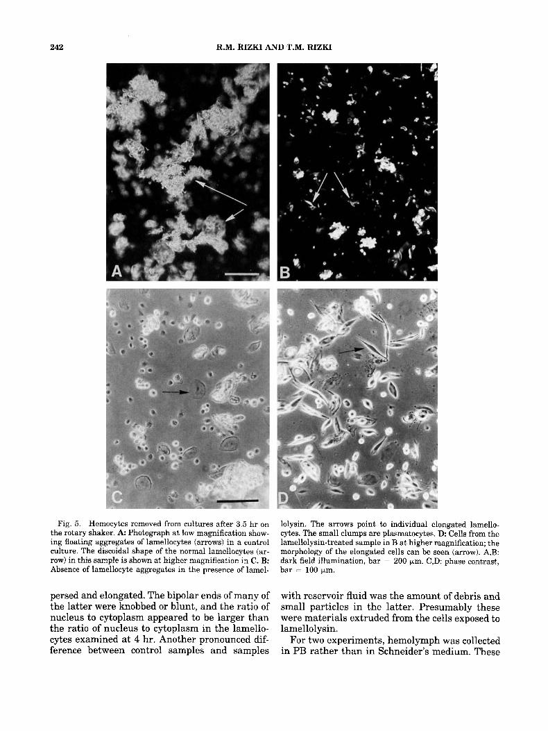
Most of the lamellocytes in control samples on the rotary shaker for 3.5 hr were clumped, indi- cating that the cells had retained their ability to adhere to one another in vitro (Fig. 5A). Individ- ual lamellocytes in these samples had the normal discoidal morphology (Fig. 5C). The lamellocytes exposed to Leiden reservoir fluid under these same conditions did not adhere to each other and had the elongated morphology typical of affected lamellocytes from larvae parasitized by Leiden wasps (Fig. 5B,D). (R. Rizki and T. Rizki, ’84).
To determine the number of reservoirs required to induce lamellocyte changes using the rotation sample procedure, the contents of 16 reservoirs were collected in 20 p1 of PB. A 10 pl aliquot from the sample, i.e., 8 RFE, was tested for activity, and the remaining 10 pl was used to prepare a dilution series containing 4, 2, 1, and 0.5 RFE. The lamellocytes in samples incubated with 4 and 8 RFE were elongated as illustrated in Figure 5D. With 2 RFE, some lamellocytes were elongated, but others appeared normal. Fewer elongated lamellocytes were found in samples with 1 RFE, and the lamellocytes in the final dilution of the series (0.5 RFE) appeared normal with an occa- sional distorted cell. The total volume in a rota- tion sample was 75 pl, so the quantity of reservoir fluid required to produce noticeable lamellocyte effects under these conditions was similar to that found for stationary cultures.
Aliquots of a hemolymph sample were used for a series of four cultures to examine the effects of maintaining lamellocytes in constantly moving medium for 24 hr. Two cultures contained ali- quots of reservoir fluid and two did not. One con- trol sample and one sample with reservoir fluid were removed from the rotary shaker for exami- nation at 4 hr, and the remaining two samples were kept on the rotary shaker for an additional 20 hr before they were examined. At 4 and 24 hr, the lamellocytes in the control samples were dis- coidal and clumped. The lamellocytes in the sam- ple with reservoir fluid for 4 hr were dispersed and elongated, as shown in Figure 5B,D. The lamellocytes that had been exposed to reservoir fluid in rotating medium for 24 hr were also dis-
242 R.M. RIZKI AND T.M. RIZKI
Fig. 5 . Hemocytes removed from cultures after 3.5 hr on the rotary shaker. A Photograph at low magnification show- ing floating aggregates of lamellocytes (arrows) in a control culture. The discoidal shape of the normal lamellocytes (ar- row) in this sample is shown at higher magnification in C. B: Absence of lamellocyte aggregates in the presence of lamel-
lolysin. The arrows point to individual elongated lamello- cytes. The small clumps are plasmatocytes. D. Cells from the lamellolysin-treated sample in B at higher magnification; the morphology of the elongated cells can be seen (arrow). A,B: dark field illumination, bar = 200 pm. C,D: phase contrast, bar = 100 pm.
persed and elongated. The bipolar ends of many of with reservoir fluid was the amount of debris and the latter were knobbed or blunt, and the ratio of small particles in the latter. Presumably these nucleus to cytoplasm appeared to be larger than were materials extruded from the cells exposed to the ratio of nucleus to cytoplasm in the lamello- lamellolysin. cytes examined at 4 hr. Another pronounced dif- For two experiments, hemolymph was collected ference between control samples and samples in PB rather than in Schneider’s medium. These

LAMELLOLYSIN EFFECTS ON CELLS IN VITRO 243
samples were intended for short-term (6 hr) incu- bation only because the buffer does not contain essential cell nutrients present in Schneider’s me- dium. They were also planned for use in cell via- bility studies with trypan blue to avoid the possi- bility of trypan blue staining the serum in Schneider’s medium. Lamellocyte changes result- ing from lamellolysin occurred as rapidly in this buffer as in Schneider’s medium, and the lamello- cytes in PB control samples looked normal. Try- pan blue was added to control and treated cul- tures in PB at 4 hr. Dye was not detected in the lamellocytes, but the nucleoli of the plasmato- cytes in both control and treated samples were lightly colored about 5 min after addition of the dye. Trypan blue was also added to control and reservoir-treated hemocytes that had been in Schneider’s medium for 4 hr. The cells in Schneid- er’s medium responded to the dye the same as the cells in PB. Stationary hemocyte samples that had been in Schneider’s medium for 24 hr and contained threaded lamellocytes were also treated with trypan blue. The threaded lamello- cytes in the samples were not colored with the dye. Materials that appeared to be cell fragments and a few plasmatocytes picked up the dye. It was surprising that the dye did not diffuse into the threaded lamellocytes, which we presumed to be dead. To verify that trypan blue would enter dead lamellocytes, the cells in 24-hr-old cultures were heat killed by placing the chambers containing cells in a 60°C oven for 10 min prior to addition of trypan blue. All cellular material from the heat- treated chambers, including the threaded lamel- locytes, immediately was colored blue.
DISCUSSION The present study confirms that the long gland
reservoir of the L. heterotomu female contains an antilamellocyte factor (R. Rizki and T. Rizki, ’84). It is clear that this factor acts directly on lamello- cytes, since the in vitro system contains only hemocytes and wasp reservoir fluid. Furthermore, the effects of lamellolysin on lamellocytes in vitro parallel the changes seen in these cells in vivo following parasitization by wasps or by injection of reservoir fluid into Drosophila larvae. In all three cases, the lamellocytes assume a bipolar shape and lose their ability to adhere to each other. The loss of adhesivity destroys the func- tional capability of the lamellocytes to adhere to foreign surfaces and to engage in encapsulation of foreign objects. The absence of functional lamello- cytes in a Drosophilu host ensures that parasitoid
eggs remain unencapsulated and free to develop. We presume that the lamellolysin affect on the
shape of the lamellocyte is related to the loss of lamellocyte adhesiveness. However, until the na- ture of lamellolysin is established and its molecu- lar action on lamellocytes is understood, there is no evidence excluding the possibility that lamel- lolysin has more than a single target in the lamel- locyte. Examination of intracellular changes in- volved in the morphological transformation of the lamellocytes following exposure to lamellolysin has been limited to the disposition of the cyto- skeleton (R. Rizki and T. Rizki, ’84). Rear- rangement of microtubule materials occurs dur- ing the modification from a disk to a bipolar shape, but this is expected since the disposition of cytoskeleton dictates cell shape. If the plasticity of the cell surface is lost in the stretching lamello- cyte, then adhesive properties of the cells may be modified as a result of the elongation process in- duced by lamellolysin.
Exposure to higher concentrations of reservoir fluid not only increased the number of affected lamellocytes but also resulted in an increase in lamellocytes with multiple tips extending from the poles. The latter observation suggests that there are multiple targets or receptor sites for lamellolysin on the cell surface. More intensive study of the relationship between lamellolysin concentration and cell morphology is required as well as detailed mapping of the points from which cytoplasmic projections emanate in affected cells. Why the affected cells generally assume a bipolar shape also requires explanation. The reorienta- tion of microtubule components occurring as a consequence of lamellolysin activity must be di- rected along a single or restricted axis to result in a bipolar cell.
The distortion of plasmatocytes in overnight cultures containing reservoir fluid may be a de- layed effect of exposure to lamellolysin or a sec- ondary effect resulting from the release of lytic factors from the distintegrating lamellocytes in the older cultures. Distinction between these alternatives should be possible by using hemo- lymph sample devoid of lamellocytes. No difer- ences between plasmatocytes in control cultures and in cultures with reservoir fluid were detect- able when effects on lamellocytes were apparent in the latter on the first day of treatment. Plas- matocytes in both samples spread against the glass surface within 15 min and the morphology of the flattened cells was the same in the presence or absence of lamellolysin. Plasmatocytes of

244 R.M. RIZKI AND T.M. RIZKI
parasitized larvae also appeared normal (R. Rizki and T. Rizki, '84). The susceptibility of lamello- cytes to lamellolysin may relate to some unique feature of this cell type.
The effect of wasp reservoir fluid on lamellocyte adhesivity can be assessed in vitro when the cul- ture medium is subjected to movement that al- lows contact between the cells. Under these condi- tions, the shape of the elongated lamellocytes closely resembles that of lamellocytes from parasitized hosts (R. Rizki and T. Rizki, '84). The affected cells generally have a single fine tip or a blunt knob at each pole. The accumulation of cel- lular fragments in the culture medium also mim- ics the accumulation of cellular debris found in the hemolymph of parasitized Drosophila larvae. If portions of cytoplasm are shed from the lamello- cytes in circulating hemolymph or culture me- dium, then the ratio of nucleus to cytoplasm should increase. In older rotation samples, the larger volume of the cell occupied by the nucleus was readily apparent. Even under quiescent con- ditions, which apparently do not favor fragmenta- tion, instances of this phenomenon were recorded. Thus there is no doubt that lamellolysin causes destruction of Drosophila lamellocytes. This or- derly destructive process must depend on the vi- tality of the cells, since the affected lamellocytes exclude trypan blue. Transformation of the lamel- locytes from discoidal cells to thread-like cells was observed only when the cells remained undis- turbed in vitro. The circulation of hemocytes in the hemolymph of parasitized hosts must be sufficient to buffet the bulk of the elongating lamellocytes such that their elongating tips break free.
ACKNOWLEDGMENTS This work was supported by NSF grant DCB-
8517807 to R.M.R. and NIH grant GM-37025 to T.M.R. We thank Pearl Johnson for typing the manuscript.
LITERATURE CITED Andrews, C.A., and T.M. Rizki (1978) Studies on lectin-
induced agglutination of Drosophila embryonic cell lines. J. Insect Physiol., 24~9-12.
Davies, D.H., and S.B. Vinson (1986) Passive evasion by eggs of braconid parasitoid Cardiochiles nigriceps of encapsula- tion in vitro by haemocytes of host Heliothis uirescens. Pos- sible role for fibrous layer in immunity. J. Insect Physiol.,
Davies, D.H., M.R. Strand, and S.B. Vinson (1987) Changes in differential haemocyte count and in vitro behavior of plasmatocytes from host Heliothis uirescens caused by Cam- poletis sonorensis polydnavirus. J. Insect Physiol., 33:143- 153.
Edson, K.M., S.B. Vinson, D.B. Stoltz, and M.D. Summers (1981) Virus in a parasitoid wasp: Suppression of the cellu- lar immune response in the parasitoids host. Science,
Kitano, H. (1982) Effect of the venom in the gregarious parasitoid Apanteles glorneratus on its hemocytic encapsu- lation by the host, Pieris. J. Invertebr. Pathol., 40331-67.
Lindsley, D.L., and E.H. Grell (1968) Genetic Variations of Drosophila melanogaster. Carnegie Inst. Wash. Publ. 627, p. 472.
Mishell, B.B., and S.M. Shiigi (1980) Selected Methods in Cellular Immunology. W.H. Freeman and Company, San Francisco, p. 486.
Osman, S.E. (1978) Die Wirkung der Sekrete der weiblichen Genitalanhangsdriisen von Pimpla turionellae L. (Hym., Ichneumonide) auf die Hamocyten und die Einkap- selungsreaktion von Wirtspuppen. Z. Parasitenkd., 57~89- 100.
Rizki, R.M., and T.M. Rizki (1979) Cell interactions in the differentiation of a melanotic tumor in Drosophila. Differ- entiation, 12: 167- 178.
Rizki, R.M. and T.M. Rizki (1984) Selective destruction of a host blood cell type by a parasitoid wasp. Proc. Natl. Acad. Sci. USA, 81 :6154-6158.
Rizki, T.M. (1957) Tumor formation in relation to metamor- phosis in Drosophila melanogaster. J. Morphol., 100:459- 472.
Rizki, T.M., and R.M. Rizki (1980) Developmental analysis of a temperature-sensitive melanotic tumor mutant in Drosophila melanogaster. Wilhelm Rouxs Arch., 189~197- 206.
Rizki, T.M., and R.M. Rizki (1984) The cellular defense sys- tem of Drosophila melanogaster. In: Insect Ultrastructure, Vol., 2. R.C. King and H. Akai, eds. Plenum Publishing Corp., New York.
Rotheram, S. (1973) The surface of the egg of a parasitic in- sect 11. The ultrastructure of the particulate coat on the egg of Nemeritis. Proc. R. SOC. London [Biol.], 183:195-204.
Salt, G. (1965) Experimental studies in insect parasitism XIII. The haemocytic reaction of a caterpillar to eggs of its habitual parasite. Proc. R. SOC. London [Biol.], 162:303- 318.
Seecof, R.L. (1971) Phosphate-buffered saline for Drosophila. Drosophila Inform. Sew., 46:113.
Wago, H., and H. Kitano (1985) Effects of the venom from Apanteles glomeratus on the hemocytes and hemolymph of Pieris rapae cruciuora. Appl. Entomol. Zool., 20~103-110.
32~1003-1010.
21 1 ~582-583.