Effects of different small HSPB members on contractile dysfunction and structural changes in a...
9
Original article Effects of different small HSPB members on contractile dysfunction and structural changes in a Drosophila melanogaster model for Atrial Fibrillation Deli Zhang a, 1 , Lei Ke b, 1 , Katarina Mackovicova a , Johannes J.L. Van Der Want c , Ody C.M. Sibon b , Robert M. Tanguay d , Genevieve Morrow d , Robert H. Henning a , Harm H. Kampinga b , Bianca J.J.M. Brundel a, ⁎ a Department of Clinical Pharmacology, University Institute for Drug Exploration (GUIDE), University of Groningen, University Medical Center Groningen, The Netherlands b Department of Cell Biology, section Radiation and Stress Cell Biology, University Institute for Drug Exploration (GUIDE), University of Groningen, University Medical Center Groningen, The Netherlands c Department of Cell Biology, section Molecular Imaging and Electron Microscopy, University of Groningen, University Medical Center Groningen, The Netherlands d Laboratory of Cell and Developmental Genetics, Department of Molecular Biology, Medical Biochemistry and Pathology, Institut de Biologie Intégrative et des Systèmes and PROTEO, Université Laval, Québec, Canada abstract article info Article history: Received 22 February 2011 Received in revised form 8 June 2011 Accepted 10 June 2011 Available online 13 July 2011 The most common clinical tachycardia, Atrial Fibrillation (AF), is a progressive disease, caused by cardiomyocyte remodeling, which finally results in contractile dysfunction and AF persistence. Recently, we identified a protective role of heat shock proteins (HSPs), especially the small HSPB1 member, against tachycardia remodeling in experimental AF models. Our understanding of tachycardia remodeling and anti- remodeling drugs is currently hampered by the lack of suitable (genetic) manipulatable in vivo models for rapid screening of key targets in remodeling. We hypothesized that Drosophila melanogaster can be exploited to study tachycardia remodeling and protective effects of HSPs by drug treatments or by utilizing genetically manipulated small HSP-overexpressing strains. Tachypacing of Drosophila pupae resulted in gradual and significant cardiomyocyte remodeling, demonstrated by reduced contraction rate, increase in arrhythmic episodes and reduction in heart wall shortening, compared to normal paced pupae. Heat shock, or pre- treatment with HSP-inducers GGA and BGP-15, resulted in endogenous HSP overexpression and protection against tachycardia remodeling. DmHSP23 overexpressing Drosophilas were protected against tachycardia remodeling, in contrast to overexpression of other small HSPs (DmHSP27, DmHSP67Bc, DmCG4461, DmCG7409, and DmCG14207). (Ultra)structural evaluation of the tachypaced heart wall revealed loss of sarcomeres and mitochondrial damage which were absent in tachypaced DmHSP23 overexpressing Drosophila. In addition, tachypacing induced a significant increase in calpain activity, which was prevented in tachypaced Drosophila overexpressing DmHSP23. Tachypacing of Drosophila resulted in cardiomyocyte remodeling, which was prevented by general HSP-inducing treatments and overexpression of a single small HSP, DmHSP23. Thus, tachypaced D. melanogaster can be used as an in vivo model system for rapid identification of novel targets to combat AF associated cardiomyocyte remodeling. © 2011 Elsevier Ltd. All rights reserved. 1. Introduction Atrial Fibrillation (AF) is the most common tachycardia in the clinical setting and it affects patients' cardiovascular function in a progressive and sustained manner [1]. AF is characterized by specific changes in electrical, structural and contractile function of the atrial cardiomyocytes, commonly denoted as ‘remodeling’. Tachycardia remodeling underlies contractile dysfunction and the progressive and intractable nature of AF [2]. Hence, there is great interest in developing anti-remodeling therapies directed at the targets underlying remodeling [3]. However, our understanding of tachycardia remodeling that contributes to AF progression and the effects of anti-remodeling drugs is currently hampered by the lack of suitable genetically manipulatable in vivo models. There are some in vitro cardiomyocyte cell models, using isolated atrial cardiomyocytes or the HL-1 atrial cardiomyocyte cell line [4,5], that can be used for such purposes and that yielded initial suggestions regarding the protective effects of heat shock proteins (HSPs) [4,6,7]. Whereas some of the concepts generated in these in vitro models could be confirmed in experimental canine models for AF [4,8], the precise translation to the in vivo situation is Journal of Molecular and Cellular Cardiology 51 (2011) 381–389 ⁎ Corresponding author at: Department of Clinical Pharmacology, University of Groningen, UMCG, A. Deusinglaan 1, 9713 AV Groningen, The Netherlands. Tel.: + 31 50 3638310; fax: +31 50 3632812. E-mail address: [email protected] (B.J.J.M. Brundel). 1 Both authors contributed equally to this work. 0022-2828/$ – see front matter © 2011 Elsevier Ltd. All rights reserved. doi:10.1016/j.yjmcc.2011.06.008 Contents lists available at ScienceDirect Journal of Molecular and Cellular Cardiology journal homepage: www.elsevier.com/locate/yjmcc
Transcript of Effects of different small HSPB members on contractile dysfunction and structural changes in a...
Journal of Molecular and Cellular Cardiology 51 (2011) 381–389
Contents lists available at ScienceDirect
Journal of Molecular and Cellular Cardiology
j ourna l homepage: www.e lsev ie r.com/ locate /y jmcc
Original article
Effects of different small HSPB members on contractile dysfunction and structuralchanges in a Drosophila melanogaster model for Atrial Fibrillation
Deli Zhang a,1, Lei Ke b,1, Katarina Mackovicova a, Johannes J.L. Van Der Want c, Ody C.M. Sibon b,Robert M. Tanguay d, Genevieve Morrow d, Robert H. Henning a,Harm H. Kampinga b, Bianca J.J.M. Brundel a,⁎a Department of Clinical Pharmacology, University Institute for Drug Exploration (GUIDE), University of Groningen, University Medical Center Groningen, The Netherlandsb Department of Cell Biology, section Radiation and Stress Cell Biology, University Institute for Drug Exploration (GUIDE), University of Groningen,University Medical Center Groningen, The Netherlandsc Department of Cell Biology, section Molecular Imaging and Electron Microscopy, University of Groningen, University Medical Center Groningen, The Netherlandsd Laboratory of Cell and Developmental Genetics, Department of Molecular Biology, Medical Biochemistry and Pathology,Institut de Biologie Intégrative et des Systèmes and PROTEO, Université Laval, Québec, Canada
⁎ Corresponding author at: Department of ClinicalGroningen, UMCG, A. Deusinglaan 1, 9713 AV Groningen3638310; fax: +31 50 3632812.
E-mail address: [email protected] (B.J.J.M1 Both authors contributed equally to this work.
0022-2828/$ – see front matter © 2011 Elsevier Ltd. Aldoi:10.1016/j.yjmcc.2011.06.008
a b s t r a c t
a r t i c l e i n f oArticle history:Received 22 February 2011Received in revised form 8 June 2011Accepted 10 June 2011Available online 13 July 2011
The most common clinical tachycardia, Atrial Fibrillation (AF), is a progressive disease, caused bycardiomyocyte remodeling, which finally results in contractile dysfunction and AF persistence. Recently,we identified a protective role of heat shock proteins (HSPs), especially the small HSPB1 member, againsttachycardia remodeling in experimental AF models. Our understanding of tachycardia remodeling and anti-remodeling drugs is currently hampered by the lack of suitable (genetic) manipulatable in vivo models forrapid screening of key targets in remodeling. We hypothesized that Drosophila melanogaster can be exploitedto study tachycardia remodeling and protective effects of HSPs by drug treatments or by utilizing geneticallymanipulated small HSP-overexpressing strains. Tachypacing of Drosophila pupae resulted in gradual andsignificant cardiomyocyte remodeling, demonstrated by reduced contraction rate, increase in arrhythmicepisodes and reduction in heart wall shortening, compared to normal paced pupae. Heat shock, or pre-treatment with HSP-inducers GGA and BGP-15, resulted in endogenous HSP overexpression and protectionagainst tachycardia remodeling. DmHSP23 overexpressing Drosophilas were protected against tachycardiaremodeling, in contrast to overexpression of other small HSPs (DmHSP27, DmHSP67Bc, DmCG4461,DmCG7409, and DmCG14207). (Ultra)structural evaluation of the tachypaced heart wall revealed loss ofsarcomeres and mitochondrial damage which were absent in tachypaced DmHSP23 overexpressingDrosophila. In addition, tachypacing induced a significant increase in calpain activity, which was preventedin tachypaced Drosophila overexpressing DmHSP23. Tachypacing of Drosophila resulted in cardiomyocyteremodeling, which was prevented by general HSP-inducing treatments and overexpression of a single smallHSP, DmHSP23. Thus, tachypaced D. melanogaster can be used as an in vivo model system for rapididentification of novel targets to combat AF associated cardiomyocyte remodeling.
Pharmacology, University of, The Netherlands. Tel.: +31 50
. Brundel).
l rights reserved.
© 2011 Elsevier Ltd. All rights reserved.
1. Introduction
Atrial Fibrillation (AF) is the most common tachycardia in theclinical setting and it affects patients' cardiovascular function in aprogressive and sustained manner [1]. AF is characterized byspecific changes in electrical, structural and contractile function ofthe atrial cardiomyocytes, commonly denoted as ‘remodeling’.
Tachycardia remodeling underlies contractile dysfunction and theprogressive and intractable nature of AF [2]. Hence, there is greatinterest in developing anti-remodeling therapies directed at thetargets underlying remodeling [3]. However, our understanding oftachycardia remodeling that contributes to AF progression and theeffects of anti-remodeling drugs is currently hampered by the lackof suitable genetically manipulatable in vivo models. There aresome in vitro cardiomyocyte cell models, using isolated atrialcardiomyocytes or the HL-1 atrial cardiomyocyte cell line [4,5],that can be used for such purposes and that yielded initialsuggestions regarding the protective effects of heat shock proteins(HSPs) [4,6,7]. Whereas some of the concepts generated in thesein vitro models could be confirmed in experimental canine modelsfor AF [4,8], the precise translation to the in vivo situation is
382 D. Zhang et al. / Journal of Molecular and Cellular Cardiology 51 (2011) 381–389
hindered due to limited possibilities for genetic manipulations inexperimental models [9] and larger animal models that besidecanine models [10], include AF models in goat [11] and sheep [12].Although these animal models have been very useful in obtainingknowledge about concepts of electrical remodeling and contractiledysfunction, they lack the flexibility of dissecting the underlyingmolecular mechanisms and employing genetic or compoundscreens. In addition, experimental execution is extensive andexpensive. Therefore, we utilize Drosophila melanogaster as an invivo model system for tachycardia remodeling, since it has beenrecognized that Drosophila contains powerful genetics and pro-vides tools to manipulate gene expression in a highly precisespatial and temporal fashion, by the use of a UAS/GAL4 system[13,14]. Furthermore, 85% of the Drosophila genes have humanhomologues [13,15,16], including several genes that have beenassociated with human cardiac diseases, including heart failure,arrhythmias and dilated cardiomyopathy [14,15,17–20]. MoreoverDrosophila contains a pumping heart [13–16]. In the current studywe report on the development of a tachycardia model inDrosophila. In addition, we show, consistent with our previousfindings in in vitro cardiomyocytes [4,6] and in vivo canine modelsfor AF [4,8], that induction of HSPs protects against tachypacing-induced contractile dysfunction. Furthermore, by using bothfunctional and (ultra)structural analyses, it was found that thisprotection is accomplished upon overexpression of a single HSP,DmHSP23, the possible ortholog of human HSPB1. Moreover,overexpression of the other small DmHSP members did not resultin a protective effect. Thus our study demonstrates the feasibilityand power of the tachypaced D. melanogaster as a model in trans-lational research in the field of tachycardia induced heart failure.
2. Materials and methods
2.1. Maintenance of Drosophila melanogaster strains and HSP-inducingtreatments
Drosophila stocks were maintained at 21 °C, during experiments at25 °C, according to standard protocols. The W1118 line was used as acontrol and obtained from Genetic Services Inc. (Massachusetts, USA).Actin-GAL4 (stock #4414) driver strains were obtained from theBloomington Stock Centre (Indiana University, USA). UAS–DmHSP23and UAS–DmHSP27 strains were generated from W1118 geneticbackground and have been described before [21–23]. These UAS strainswere crossed, using standard genetics, with the actin-GAL4 expressingstrains in order to generate strains that contain actin-GAL4 and eitherUAS–DmHSP23 or UAS–DmHSP27 on the same chromosome. Stableubiquitous overexpression of DmHSP23 and DmHSP27 was confirmedbyWesternblottingwith specific antibodies.OtherDrosophila smallHSPoverexpressing strains included were HSP67Bc, CG4461, CG7409,CG14207 [24,25]. These transgenic strains were generated at GeneticServices Inc. by injection of the pUAS vector containing a small HSP ofinterest with a V5 tag, into the W1118 genetic background. Thesetransgenic strains were crossed with actin-GAL4 expressing flies inorder to induce ubiquitous transgenic gene expression of the smallDmHSP of interest in the F1 progeny, which was confirmed byWesternblotting.
Heat treatment was performed at 37 °C for 1 h followed by anovernight recovery at 25 °C. The HSP-inducing compounds Ger-anylgeranylacetone (GGA, 1 mM) and O-(3-piperidino-2-hydroxy-1-propyl)-nicotinic acid amidoxim di-hydrochloride (BGP-15,1 mM) (both kind gifts from Eisai Japan and NP-gene USA,respectively) were freshly dissolved in de-mineralized water and0.5 ml was added to standard Drosophila food for at least 48 hbefore (tachy)pacing. Controls were subjected to de-mineralizedwater only.
2.2. Tachypacing of Drosophila and measurement of heart functionparameters
Early pupae were selected as described before [26]. In short,transparent pupae were selected at entry of the immobile phase (thisphase continues for about 3 h) and were placed on 1% agarose gel inPBS. The basal heart rate in the early pupae was stable at about 2.2 Hz(Supplementary information Fig. 1A). Pupae were tachypaced byusing the C-Pace100™-Culture Pacer (IonOptix Corporation, TheNetherlands) for 0–120 min. Controls were subjected to normal elec-trical field stimulation similar to basal heart rate (2.2 Hz) whereastachypacing was conducted at 2.3 fold basal rate or as indicated. Theheart wall of pupae could follow the pacing rate (Supplementaryinformation Fig. 1B). During interruption of (tachy)pacing for about40 s, spontaneous heart function at each time point was visuallyscored under the light microscope in duplicate periods of 20 s. Inshort, the rate of contraction of the heart was quantified by focusingon the heart wall contrast edges [26] and only regular contractionswere included. During the (tachy)pacing interruption, also time lapsemovies were made and analysed by edge detection software (LeicaMicrosystems, Mannheim, Germany) to determine the heart wallshortening, which indicates the strength of contraction (amplitude ofdiastolic and systolic heart wall contraction), and duration of ar-rhythmic periods (ImageJ). Arrhythmicity index was calculated asthe duration of arrhythmic periods divided by the total duration ofmeasured periods.
2.3. Quantitative PCR
From at least five early pupae per condition, total RNA wasisolated by utilizing the Nucleospin RNA/proteinmini kit (Macherey-Nagel, The Netherlands). First strand cDNA was generated using M-MLV reverse transcriptase (Invitrogen, The Netherlands) using oligo(dT)18 primers (Biologio, The Netherlands). Relative changes intranscription level were determined using the CFX384 Real-TimeSystem C1000 Thermocycler (BioRad, The Netherlands) in combi-nation with SYBR green supermix (Bio-Rad, The Netherlands).Calculations were performed using the comparative CT methodaccording to User Bulletin 2 (Applied Biosystems). Fold inductionwas adjusted using RpL32 transcript levels as a standard. Primer pairsused included the heat inducible DmHSP27 F: CTAGACAGGGTTGT-GAATAAAGAG and R: AAACCGAAGTCATCCTCCAG and DmHSP70AAF: ACCTCAACCTATCCATCAACC and R: GTCTCAATTCCCAATGAAAGTG,and for RpL32 F: CGATCTCGCCGCAGTAAA and R: GCACCAAGCACTT-CATCC. The PCR efficiencies for all primer pairs were between 85%and 100%.
2.4. Western blot analysis
Drosophila pupae or adult flies (five per condition) were quicklylysed on ice in 50 μl SDS sample loading buffer (10% SDS, 50% glycerol,0.33 M Tris–HCl pH 6.8, 10% β-mercaptoethanol, 0.05% bromophenolblue) followed by sonification. After a short spin-down, supernatantwas collected and boiled for 6 min. Proteins were separated on SDS-PAGE 4–20% PreciseTM Protein gels (Thermo Scientific) and trans-ferred onto nitrocellulose membranes (GE Healthcare, The Nether-lands). The membranes were blocked in 5% skim milk (1 h, roomtemperature) and incubated overnight at 4 °C with primary Drosoph-ila anti-DmHSP23, anti-DmHSP27 antibodies [27,28], anti-V5 anti-bodies (Invitrogen, The Netherlands) or anti α-tubulin antibodies(Sigma, The Netherlands). Horseradish peroxidase-conjugated anti-mouse (Santa-Cruz Biotechnology, The Netherlands) was used assecondary antibody. Signals were detected by Super Signal (ThermoScientific, The Netherlands) and quantified by densitometry.
383D. Zhang et al. / Journal of Molecular and Cellular Cardiology 51 (2011) 381–389
2.5. (Ultra) structural evaluation by Light and Electron Microscopy
For morphological evaluation, the head and the abdomen weredissected from the early pupae and the middle segment was im-mediately fixed for at least 2 h at 4 °C in 2% glutaraldehyde (in 0.1 Mcacodylate buffer, pH7.4). Post-fixation was performed for 2 h in 1%osmium tetroxide (supplemented with 1.5% K4Fe(CN)6 in cacodylatebuffer, pH7.4) at 4 °C. After dehydration in ethanol, pupae wereembedded in Epon and semi-thin sections (1 μm)were cut and stainedwith 1% toluidine blue and used for light microscopic evaluation. Toverify the ultrastructural changes, ultrathin sections (60 nm) weremade and stainedwith uranylacetate and lead citrate and examined in aPhilips CM100 electron microscope operating at 60 kV. Changes wereevaluated in six randomly chosen regions by an investigator blinded forDrosophila groups, who (1) calculated the ratio of area positive formyolysis (areas with N10% loss of sarcomeres) compared to total area,using ImagePro software [29]; and (2) scored mitochondrial degener-ation by determining the ratio degenerated versus total mitochondria.
2.6. Calpain activity measurement
Early Drosophila pupae were lysed in a non-denaturing buffer(CelLytic™ MT Cell Lysis Reagent, Sigma, The Netherlands). Proteinconcentration was determined by using Bio-Rad Protein Assay Kit(Bio-Rad, USA). DABCYL-Thr-Pro-Leu-Lys~Ser-Pro-Pro-Pro-Ser-Pro-Arg-EDANS (Calpain Substrate III, Fluorogenic, Merck, Germany)was used as a substrate for calpain. Hereto, protein extract (30 μg)was added to Calpain Substrate III (60 μM final concentration) in
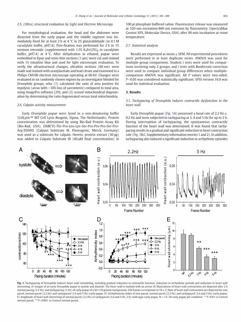
Fig. 1. Tachypacing of Drosophila induces heart wall remodeling, including gradual reductioshortening. A) Images of an early Drosophila pupae in systole and diastole. The heart wall isnormal pacing (2.2 Hz) and tachypacing (5 Hz) of early pupae of aW1118 genetic backgrounpaced, normal paced (2,2 Hz) and tachypaced (3,4 and 5 Hz) early pupae. D) ArrhythmicityE) Amplitude of heart wall shortening of normal paced (2,2 Hz) or tachypaced (3,4 and 5 Hznormal paced, ***Pb0.001 vs Control normal paced.
100 μl phosphate buffered saline. Fluorescence release was measuredat 320-nm excitation/460-nm emission by fluorometry (SpectraMaxGemini XPS, Molecular Device, USA) after 90 min incubation at roomtemperature.
2.7. Statistical analysis
Results are expressed as mean±SEM. All experimental procedureswere performed in at least duplicate series. ANOVA was used formultiple-group comparisons. Student t tests were used for compar-isons involving only 2 groups, and t tests with Bonferroni correctionwere used to compare individual group differences when multiple-comparison ANOVA was significant. All P values were two-sided.Pb0.05 was considered statistically significant. SPSS version 16.0 wasused for statistical evaluation.
3. Results
3.1. Tachypacing of Drosophila induces contractile dysfunction in theheart wall
Early Drosophila pupae (Fig. 1A) possessed a basal rate of 2.2 Hz±0.2 Hz and were subjected to tachypacing at 3, 4 and 5 Hz for up to 2 h.During interruption of tachypacing, the spontaneous contractilefunction of the heart wall was determined. It was found that tachy-pacing results in a gradual and significant reduction in heart contractionrate (Fig. 1B,C, Supplementary informationmovies 1 and 2). In addition,tachypacing also induced a significant induction in arrhythmic episodes
n in contractile function, induction in arrhythmic periods and reduction in heart wallmarked with an arrow. B) Illustrations of heart wall contractions are depicted after 2 hd. 350 frames corresponds to 10 s. C) Rate of heart wall contractions are depicted for nonindex of non-paced, normal paced (2.2 Hz) and tachypaced (3,4 and 5 Hz) early pupae., 2 h) wild-type early pupae. N=13–20 early pupae per condition. ** Pb0.01 vs Control
384 D. Zhang et al. / Journal of Molecular and Cellular Cardiology 51 (2011) 381–389
(Fig. 1B,D, Supplementary information movies 1 and 2). Moreover,increasing frequency of tachypacing induced a gradual reduction in theamplitude of diastolic and systolic heart wall shortening (Fig. 1D). Sincetachypacing at5 Hz, a 2.3 fold rate increase compared to basal heart rate,significantly induced contractile dysfunction of the heart wall, thissetting was used in all following tachypacing experiments.
3.2. Heat shock and the HSP-inducers GGA and BGP-15 increase HSPlevels in Drosophila and protect against tachypacing-induced contractiledysfunction
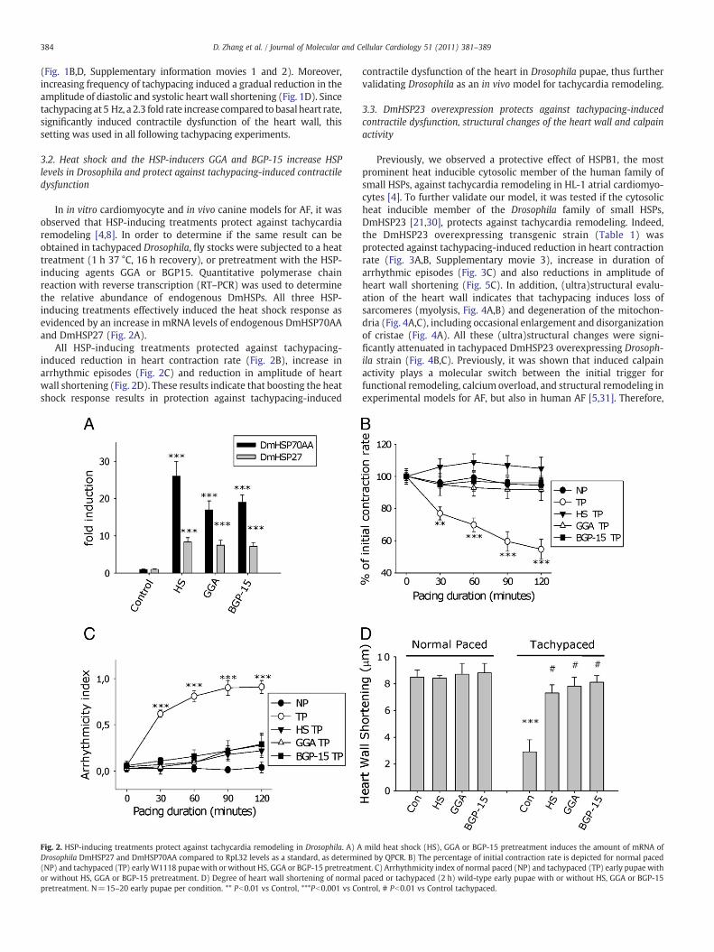
In in vitro cardiomyocyte and in vivo canine models for AF, it wasobserved that HSP-inducing treatments protect against tachycardiaremodeling [4,8]. In order to determine if the same result can beobtained in tachypaced Drosophila, fly stocks were subjected to a heattreatment (1 h 37 °C, 16 h recovery), or pretreatment with the HSP-inducing agents GGA or BGP15. Quantitative polymerase chainreaction with reverse transcription (RT–PCR) was used to determinethe relative abundance of endogenous DmHSPs. All three HSP-inducing treatments effectively induced the heat shock response asevidenced by an increase in mRNA levels of endogenous DmHSP70AAand DmHSP27 (Fig. 2A).
All HSP-inducing treatments protected against tachypacing-induced reduction in heart contraction rate (Fig. 2B), increase inarrhythmic episodes (Fig. 2C) and reduction in amplitude of heartwall shortening (Fig. 2D). These results indicate that boosting the heatshock response results in protection against tachypacing-induced
Fig. 2. HSP-inducing treatments protect against tachycardia remodeling in Drosophila. A) ADrosophila DmHSP27 and DmHSP70AA compared to RpL32 levels as a standard, as determi(NP) and tachypaced (TP) earlyW1118 pupae with or without HS, GGA or BGP-15 pretreatmor without HS, GGA or BGP-15 pretreatment. D) Degree of heart wall shortening of normalpretreatment. N=15–20 early pupae per condition. ** Pb0.01 vs Control, ***Pb0.001 vs Co
contractile dysfunction of the heart in Drosophila pupae, thus furthervalidating Drosophila as an in vivo model for tachycardia remodeling.
3.3. DmHSP23 overexpression protects against tachypacing-inducedcontractile dysfunction, structural changes of the heart wall and calpainactivity
Previously, we observed a protective effect of HSPB1, the mostprominent heat inducible cytosolic member of the human family ofsmall HSPs, against tachycardia remodeling in HL-1 atrial cardiomyo-cytes [4]. To further validate our model, it was tested if the cytosolicheat inducible member of the Drosophila family of small HSPs,DmHSP23 [21,30], protects against tachycardia remodeling. Indeed,the DmHSP23 overexpressing transgenic strain (Table 1) wasprotected against tachypacing-induced reduction in heart contractionrate (Fig. 3A,B, Supplementary movie 3), increase in duration ofarrhythmic episodes (Fig. 3C) and also reductions in amplitude ofheart wall shortening (Fig. 5C). In addition, (ultra)structural evalu-ation of the heart wall indicates that tachypacing induces loss ofsarcomeres (myolysis, Fig. 4A,B) and degeneration of the mitochon-dria (Fig. 4A,C), including occasional enlargement and disorganizationof cristae (Fig. 4A). All these (ultra)structural changes were signi-ficantly attenuated in tachypaced DmHSP23 overexpressing Drosoph-ila strain (Fig. 4B,C). Previously, it was shown that induced calpainactivity plays a molecular switch between the initial trigger forfunctional remodeling, calcium overload, and structural remodeling inexperimental models for AF, but also in human AF [5,31]. Therefore,
mild heat shock (HS), GGA or BGP-15 pretreatment induces the amount of mRNA ofned by QPCR. B) The percentage of initial contraction rate is depicted for normal pacedent. C) Arrhythmicity index of normal paced (NP) and tachypaced (TP) early pupae withpaced or tachypaced (2 h) wild-type early pupae with or without HS, GGA or BGP-15ntrol, # Pb0.01 vs Control tachypaced.
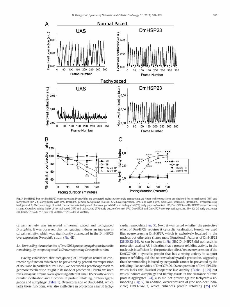
Fig. 3. DmHSP23 but not DmHSP27 overexpressing Drosophilas are protected against tachycardia remodeling. A) Heart wall contractions are depicted for normal paced (NP) andtachypaced (TP, 2 h) early pupae with UAS–DmHSP23 genetic background (no DmHSP23 overexpression, UAS) and with a UAS–actinGAL4–DmHSP23 (DmHSP23) overexpressingbackground. B) The percentage of initial contraction rate is depicted of normal paced (NP) and tachypaced (TP) early pupae of control UAS, DmHSP23 and DmHSP27 overexpressingstrains. C) Arrhythmicity index of normal paced (NP) and tachypaced (TP) early pupae of control UAS, DmHSP23 and DmHSP27 overexpressing strains. N=12–20 early pupae percondition. *Pb0.05, ** Pb0.01 vs Control, ***Pb0.001 vs Control.
385D. Zhang et al. / Journal of Molecular and Cellular Cardiology 51 (2011) 381–389
calpain activity was measured in normal paced and tachypacedDrosophila. It was observed that tachypacing induces an increase incalpain activity, which was significantly attenuated in the DmHSP23overexpressing Drosophila strain (Fig. 4D).
3.4. Unravelling themechanismofDmHSP23protection against tachycardiaremodeling, by comparing small HSP overexpressing Drosophila strains
Having established that tachypacing of Drosophila results in con-tractile dysfunction, which can be prevented by general overexpressionof HSPs and in particular DmHSP23, we next used a genetic approach toget moremechanistic insight in its mode of protection. Hereto, we usedfiveDrosophila strains overexpressing different small HSPswith variouscellular localization and functions in protein refolding, protein aggre-gation and autophagy (Table 1). Overexpression of DmCG4461, whichlacks these functions, was also ineffective in protection against tachy-
cardia remodeling (Fig. 5). Next, it was tested whether the protectiveeffect of DmHSP23 requires it cytosolic localization. Hereto, we usedflies overexpressing DmHSP27, which is exclusively localized in thenucleus but otherwise shares most (functional) features of DmHSP23[28,30,32–34]. As can be seen in Fig. 3B,C DmHSP27 did not result inprotection against AF, indicating that a protein refolding activity in thenucleus is insufficient for theprotective effect. Yet, overexpressionof theDmCG7409, a cytosolic protein that has a strong activity to supportprotein refolding, did also not reveal tachycardia protection, suggestingthat the remodeling induced by tachycardia cannot be prevented by therefolding-like activities of DmCG7409. Overexpression of DmHSP67Bc,which lacks this classical chaperone-like activity (Table 1) [25] butwhich induces autophagy and hereby assists in the clearance of toxicprotein aggregates [24], also did not protect against tachycardia re-modeling (Fig. 5). In addition, overexpression of (the non-heat indu-cible) DmCG14207, which enhances protein refolding [25] and
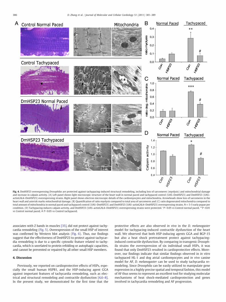
Fig. 4. DmHSP23 overexpressing Drosophilas are protected against tachypacing-induced structural remodeling, including loss of sarcomeres (myolysis) and mitochondrial damageand increase in calpain activity. (A) Left panel shows light microscopic structure of the heart wall in normal paced and tachypaced control (UAS–DmHSP23) and DmHSP23 (UAS–actinGAL4–DmHSP23) overexpressing strains. Right panel shows electron microscopic details of the cardiomyocytes and mitochondria. Arrowheads show loss of sarcomeres in theheart wall and asterisk marks mitochondrial damage. (B) Quantification of ratio myolysis compared to total area of sarcomeres and (C) ratio degenerated mitochondria compared tototal amount of mitochondria in normal paced and tachypaced control (UAS–DmHSP23) and DmHSP23 (UAS–actinGAL4–DmHSP23) overexpressing strains. N=3-5 early pupae percondition. (D) Tachypacing induces calpain activity, and DmHSP23 (UAS–actinGAL4–DmHSP23) overexpressing strains were protected. *Pb0.05 vs Control normal paced, **Pb0.01vs Control normal paced, # Pb0.05 vs Control tachypaced.
386 D. Zhang et al. / Journal of Molecular and Cellular Cardiology 51 (2011) 381–389
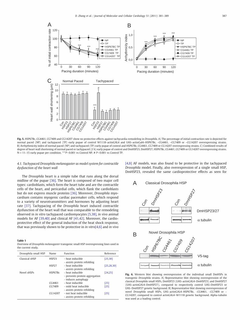
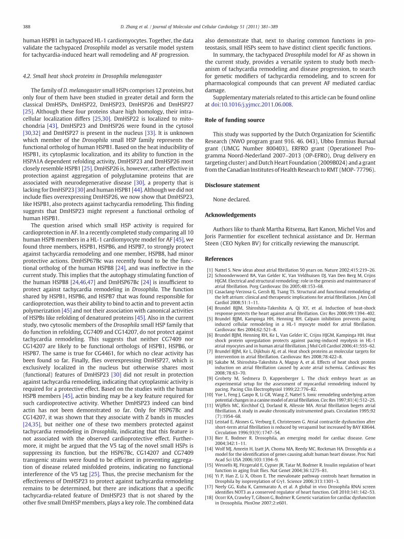
associates with Z bands in muscles [35], did not protect against tachy-cardia remodeling (Fig. 5). Overexpression of the small HSP of interestwas confirmed by Western blot analysis (Fig. 6). Thus, our findingssuggest that the effectiveness of DmHSP23 to protect against tachycar-dia remodeling is due to a specific cytosolic feature related to tachy-cardia, which is unrelated to protein refolding or autophagic capacities,and cannot be prevented or repaired by all other small HSP members.
4. Discussion
Previously, we reported on cardioprotective effects of HSPs, espe-cially the small human HSPB1, and the HSP-inducing agent GGAagainst important features of tachycardia remodeling, such as elec-trical and structural remodeling and contractile dysfunction [4,6–8].In the present study, we demonstrated for the first time that the
protective effects are also observed in vivo in the D. melanogastermodel for tachypacing-induced contractile dysfunction of the heartwall. We observed that both HSP-inducing agents GGA and BGP-15but also a heat shock pretreatment protect against tachypacing-induced contractile dysfunction. By comparing in transgenic Drosoph-ila strains the overexpression of six individual small HSPs, it wasfound that only DmHSP23 resulted in cardioprotective effects. More-over, our findings indicate that similar findings observed in in vitrotachypaced HL-1 and dog atrial cardiomyocytes and in vivo caninemodel for AF, D. melanogaster can be used to study tachycardia re-modeling. Since Drosophila can be easily utilized to manipulate geneexpression in a highly precise spatial and temporal fashion, this modelof AF thus seems to represent an excellent tool for studying molecularmechanisms of heat shock-mediated cardioprotection and genesinvolved in tachycardia remodeling and AF progression.
Pacing duration (minutes)0 30 60 90 120%
of i
nitia
l con
trac
tion
rate
40
60
80
100
120
NPTPHSP67BC TPCG4461 TPCG7409 TPCG14207 TP
A
*********
**
Pacing duration (minutes)0 30 60 90 120
Arr
hyth
mic
ity in
dex
0,0
0,5
1,0
NPTPHSP67BC TP CG4461 TPCG7409 TPCG14207 TP
******
***
***
Con
HSP
23H
SP27
HSP
67BC
CG
4461
CG
7409
CG
1420
7
Con
HSP
23H
SP27
HSP
67BC
CG
4461
CG
7409
CG
1420
7
Hea
rt w
all s
hort
enin
g (μ
m)
0
2
4
6
8
10
12Normal Paced Tachypaced
***
#
C
B
Fig. 5. HSP67Bc, CG4461, CG7409 and CG14207 show no protective effects against tachycardia remodeling in Drosophila. A) The percentage of initial contraction rate is depicted fornormal paced (NP) and tachypaced (TP) early pupae of control W1118–actinGAL4 and UAS–actinGal4–HSP67Bc, –CG4461, –CG7409 or –CG14207 overexpressing strains.B) Arrhythmicity index of normal paced (NP) and tachypaced (TP) early pupae of control and HSP67Bc, CG4461, CG7409 or CG14207 overexpressing strains. C) Combined results ofdegree of heart wall shortening of normal paced or tachypaced (2 h) early pupae of control and DmHSP23, DmHSP27, HSP67Bc, CG4461, CG7409 or CG14207 overexpressing strains.N=11–15 early pupae per condition. ***Pb0.001 vs Control NP, # Pb0.001 vs Control TP.
Classical Drosophila HSP
α tubulin
DmHSP23/27
A
387D. Zhang et al. / Journal of Molecular and Cellular Cardiology 51 (2011) 381–389
4.1. Tachypaced Drosophila melanogaster as model system for contractiledysfunction of the heart wall
The Drosophila heart is a simple tube that runs along the dorsalmidline of the pupae [36]. The heart is composed of two major celltypes: cardioblasts, which form the heart tube and are the contractilecells of the heart, and pericardial cells, which flank the cardioblastsbut do not express muscle proteins [36]. Moreover, Drosophila myo-cardium contains myogenic cardiac pacemaker cells, which respondto a variety of neurotransmitters and hormones by adjusting heartrate [37]. Tachypacing of the Drosophila heart induced contractiledysfunction of the heart wall that was comparable to the remodelingobserved in in vitro tachypaced cardiomyocytes [5,38], in vivo animalmodels for AF [39,40] and clinical AF [41,42]. Moreover, the cardio-protective effect of the general induction of the heat shock response,that was previously shown to be protective in in vitro[4,6] and in vivo
Table 1Overview of Drosophila melanogaster transgenic small HSP overexpressing lines used inthe current study.
Drosophila small HSP Name Function Reference
Classical sHSP HSP23 - heat inducible [25,30]- assists protein refolding
HSP27 - heat inducible [25,28,30]- assists protein refolding
Novel sHSPs HSP67Bc - heat inducible [24,25]- prevents protein aggregation- induces autophagy
CG4461 - heat inducible [25]CG7409 - mild heat inducible [25]
- assists protein refoldingCG14207 - not heat inducible [25]
- assists protein refolding
[4,8] AF models, was also found to be protective in the tachypacedDrosophila model. Finally, also overexpression of a single small HSP,DmHSP23, revealed the same cardioprotective effects as seen for
Novel Drosophila HSP
V5-tag
α tubulin
B
Fig. 6. Western blot showing overexpression of the individual small DmHSPs intransgenic Drosophila strains. A) Representative blot showing overexpression of theclassical Drosophila small HSPs, DmHSP23 (UAS–actinGAL4–DmHSP23) and DmHSP27(UAS–actinGAL4–DmHSP27), compared to respectively control UAS–DmHSP23 orUAS–DmHSP27 genetic background. B) Representative blot showing overexpression ofnovel Drosophila small HSPs, UAS–actinGAL4–HSP67Bc, –CG4461, –CG7409 or –
CG14207, compared to control actinGAL4–W1118 genetic background. Alpha-tubulinwas used as a loading control.
388 D. Zhang et al. / Journal of Molecular and Cellular Cardiology 51 (2011) 381–389
human HSPB1 in tachypaced HL-1 cardiomyocytes. Together, the datavalidate the tachypaced Drosophila model as versatile model systemfor tachycardia-induced heart wall remodeling and AF progression.
4.2. Small heat shock proteins in Drosophila melanogaster
The family ofD.melanogaster small HSPs comprises 12 proteins, butonly four of them have been studied in greater detail and form theclassical DmHSPs, DmHSP22, DmHSP23, DmHSP26 and DmHSP27[25]. Although these four proteins share high homology, their intra-cellular localization differs [25,30]. DmHSP22 is localized to mito-chondria [43], DmHSP23 and DmHSP26 were found in the cytosol[30,32] and DmHSP27 is present in the nucleus [33]. It is unknownwhich member of the Drosophila small HSP family represents thefunctional ortholog of human HSPB1. Based on the heat inducibility ofHSPB1, its cytoplasmic localization, and its ability to function in theHSPA1A dependent refolding activity, DmHSP23 and DmHSP26 mostclosely resemble HSPB1 [25]. DmHSP26 is, however, rather effective inprotection against aggregation of polyglutamine proteins that areassociated with neurodegenerative disease [30], a property that islacking for DmHSP23 [30] andhumanHSPB1 [44]. Althoughwedid notinclude flies overexpressing DmHSP26, we now show that DmHSP23,like HSPB1, also protects against tachycardia remodeling. This findingsuggests that DmHSP23 might represent a functional ortholog ofhuman HSPB1.
The question arised which small HSP activity is required forcardioprotection in AF. In a recently completed study comparing all 10human HSPBmembers in a HL-1 cardiomyocyte model for AF [45], wefound three members, HSPB1, HSPB6, and HSPB7, to strongly protectagainst tachycardia remodeling and one member, HSPB8, had minorprotective actions. DmHSP67Bc was recently found to be the func-tional ortholog of the human HSPB8 [24], and was ineffective in thecurrent study. This implies that the autophagy stimulating function ofthe human HSPB8 [24,46,47] and DmHSP67Bc [24] is insufficient toprotect against tachycardia remodeling in Drosophila. The functionshared by HSPB1, HSPB6, and HSPB7 that was found responsible forcardioprotection, was their ability to bind to actin and to prevent actinpolymerization [45] and not their association with canonical activitiesof HSPBs like refolding of denatured proteins [45]. Also in the currentstudy, two cytosolic members of the Drosophila small HSP family thatdo function in refolding, CG7409 and CG14207, do not protect againsttachycardia remodeling. This suggests that neither CG7409 norCG14207 are likely to be functional orthologs of HSPB1, HSPB6, orHSPB7. The same is true for CG4461, for which no clear activity hasbeen found so far. Finally, flies overexpressing DmHSP27, which isexclusively localized in the nucleus but otherwise shares most(functional) features of DmHSP23 [30] did not result in protectionagainst tachycardia remodeling, indicating that cytoplasmic activity isrequired for a protective effect. Based on the studies with the humanHSPB members [45], actin binding may be a key feature required forsuch cardioprotective activity. Whether DmHSP23 indeed can bindactin has not been demonstrated so far. Only for HSP67Bc andCG14207, it was shown that they associate with Z bands in muscles[24,35], but neither one of these two members protected againsttachycardia remodeling in Drosophila, indicating that this feature isnot associated with the observed cardioprotective effect. Further-more, it might be argued that the V5 tag of the novel small HSPs issuppressing its function, but the HSP67Bc, CG14207 and CG7409transgenic strains were found to be efficient in preventing aggrega-tion of disease related misfolded proteins, indicating no functionalinterference of the V5 tag [25]. Thus, the precise mechanism for theeffectiveness of DmHSP23 to protect against tachycardia remodelingremains to be determined, but there are indications that a specifictachycardia-related feature of DmHSP23 that is not shared by theother five small DmHSPmembers, plays a key role. The combined data
also demonstrate that, next to sharing common functions in pro-teostasis, small HSPs seem to have distinct client specific functions.
In summary, the tachypaced Drosophila model for AF as shown inthe current study, provides a versatile system to study both mech-anism of tachycardia remodeling and disease progression, to searchfor genetic modifiers of tachycardia remodeling, and to screen forpharmacological compounds that can prevent AF mediated cardiacdamage.
Supplementarymaterials related to this article can be found onlineat doi:10.1016/j.yjmcc.2011.06.008.
Role of funding source
This study was supported by the Dutch Organization for ScientificResearch (NWO program grant 916. 46. 043), Ubbo Emmius Bursaalgrant (UMCG Number 800403), ERFRO grant (Operationeel Pro-gramma Noord-Nederland 2007–2013 (OP-EFRO), Drug delivery entargeting cluster) andDutchHeart Foundation (2009B024) and a grantfrom the Canadian Institutes of Health Research to RMT (MOP- 77796).
Disclosure statement
None declared.
Acknowledgements
Authors like to thank Martha Ritsema, Bart Kanon, Michel Vos andJoris Parmentier for excellent technical assistance and Dr. HermanSteen (CEO Nyken BV) for critically reviewing the manuscript.
References
[1] Nattel S. New ideas about atrial fibrillation 50 years on. Nature 2002;415:219–26.[2] Schoonderwoerd BA, Van Gelder IC, Van Veldhuisen DJ, Van Den Berg M, Crijns
HJGM. Electrical and structural remodeling: role in the genesis andmaintenance ofatrial fibrillation. Porg Cardiovasc Dis 2005;48:153–68.
[3] Casaclang-Verzosa G, Gersh BJ, Tsang TS. Structural and functional remodeling ofthe left atrium: clinical and therapeutic implications for atrial fibrillation. J Am CollCardiol 2008;51:1–11.
[4] Brundel BJJM, Shiroshita-Takeshita A, Qi XY, et al. Induction of heat-shockresponse protects the heart against atrial fibrillation. Circ Res 2006;99:1394–402.
[5] Brundel BJJM, Kampinga HH, Henning RH. Calpain inhibition prevents pacinginduced cellular remodeling in a HL-1 myocyte model for atrial fibrillation.Cardiovasc Res 2004;62:521–8.
[6] Brundel BJJM, Henning RH, Ke L, Van Gelder IC, Crijns HJGM, Kampinga HH. Heatshock protein upregulation protects against pacing-induced myolysis in HL-1atrial myocytes and in human atrial fibrillation. J Mol Cell Cardiol 2006;41:555–62.
[7] Brundel BJJM, Ke L, Dijkhuis AJ, et al. Heat shock proteins as molecular targets forintervention in atrial fibrillation. Cardiovasc Res 2008;78:422–8.
[8] Sakabe M, Shiroshita-Takeshita A, Maguy A, et al. Effects of heat shock proteininduction on atrial fibrillation caused by acute atrial ischemia. Cardiovasc Res2008;78:63–70.
[9] Grobety M, Sedmera D, Kappenberger L. The chick embryo heart as anexperimental setup for the assessment of myocardial remodeling induced bypacing. Pacing Clin Electrophysiol 1999;22:776–82.
[10] Yue L, Feng J, Gaspo R, Li GR, Wang Z, Nattel S. Ionic remodeling underlying actionpotential changes in a caninemodel of atrialfibrillation. Circ Res 1997;81(4):512–25.
[11] Wijffels MC, Kirchhof CJ, Dorland R, Allessie MA. Atrial fibrillation begets atrialfibrillation. A study in awake chronically instrumented goats. Circulation 1995;92(7):1954–68.
[12] Leistad E, Aksnes G, Verburg E, Christensen G. Atrial contractile dysfunction aftershort-term atrial fibrillation is reduced by verapamil but increased by BAY K8644.Circulation 1996;93(9):1747–54.
[13] Bier E, Bodmer R. Drosophila, an emerging model for cardiac disease. Gene2004;342:1–11.
[14] Wolf MJ, Amrein H, Izatt JA, Choma MA, Reedy MC, Rockman HA. Drosophila as amodel for the identification of genes causing adult human heart disease. Proc NatlAcad Sci USA 2006;103:1394–9.
[15] Wessells RJ, Fitzgerald E, Cypser JR, Tatar M, Bodmer R. Insulin regulation of heartfunction in aging fruit flies. Nat Genet 2004;36:1275–81.
[16] Yi P, Han Z, Li X, Olson E. The mevalonate pathway controls heart formation inDrosphila by isoprenylation of Gγ1. Science 2006;313:1301–3.
[17] Neely GG, Kuba K, Cammarato A, et al. A global in vivo Drosophila RNAi screenidentifies NOT3 as a conserved regulator of heart function. Cell 2010;141:142–53.
[18] Ocorr KA, Crawley T, Gibson G, Bodmer R. Genetic variation for cardiac dysfunctionin Drosophila. PlosOne 2007;2:e601.
389D. Zhang et al. / Journal of Molecular and Cellular Cardiology 51 (2011) 381–389
[19] Cammarato A, Dambacher CM, Knowles AF, et al. Myosin transducer mutationsdifferentially affect motor function, myofibril structure, and the performance ofskeletal and cardiac muscles. Mol Biol Cell 2008;19:553–62.
[20] Kim IM, Wolf MJ. Serial examination of an inducible and reversible dilatedcardiomyopathy in individual adult Drosophila. PlosOne 2009;4:e7132.
[21] Michaud S, Marin R, Tanguay RM. Regulation of heat shock gene induction andexpression during Drosophila development. Cell Mol Life Sci 1997;53:104–13.
[22] Tanguay RM, Morrow G. Neural expression of small heat shock proteins influenceslongevity and resistance to oxidative stress. Heat shock proteins and the brain:implications for neurodegenerative diseases and neuroprotection. SpringerScience+Business Media; 2008. p. 319–36.
[23] Wang HD, Kazemi-Esfarjani Benzer S. Multiple-stress analysis for isolation ofDrosophila longevity genes. Proc Natl Acad Sci 2004;101:12610–5.
[24] Carra S, Boncoraglio A, Kanon B, et al. Identification of the Drosophila ortholog ofHSPB8: implication of HSPB8 loss of function in protein folding diseases. J BiolChem 2010;285:37811–22.
[25] Vos MJ. Small heat shock proteins; implications for neurodegeneration andlongevity. Thesis. 2009.
[26] Wessells RJ, Bodmer R. Screening assays for heart function mutants in Drosophila.Biotechniques 2004;37:58–66.
[27] Marin R, Valet JP, Tanguay RM. Hsp23 and Hsp26 exhibit distinct spatial andtemporal patterns of constitutive expression in Drosophila adults. Dev Genet1993;14:69–77.
[28] Marin R, Tanguay RM. Stage-specific localization of the small heat shock proteinHsp27 during oogenesis in Drosophila melanogaster. Chromosoma 1996;105:142–9.
[29] Brundel BJJM, Ausma J, Van Gelder IC, et al. Activation of proteolysis by calpainsand structural changes in human paroxysmal and persistent atrial fibrillation.Cardiovasc Res 2002;54:380–9.
[30] Morrow G, Heikkila JJ, Tanguay RM. Differences in the chaperone-like activities ofthe four main small heat shock proteins of Drosophila melanogaster. Cell StressChaperones 2006;11:51–60.
[31] Ke L, Qi XY, Dijkhuis AJ, et al. Calpain mediates cardiac troponin degradation andcontractile dysfunction in atrial fibrillation. J Mol Cell Cardiol 2008;45:685–93.
[32] Beaulieu JF, Arrigo AP, Tanguay RM. Interaction of Drosophila 27,000 Mr heat-shock protein with the nucleus of heat-shocked and ecdysone-stimulated culturecells. J Cell Sci 1989;92:29–36.
[33] Michaud S, Lavoie S, Guimond MO, Tanguay RM. The nuclear localization ofDrosophila Hsp27 is dependent on a monopartite arginine-rich NLS and is
uncoupled from its association to nuclear speckles. Biochem Biophys Acta2008;1783:1200–10.
[34] Marin R, Landry J, Tanguay RM. Tissue-specific posttranslational modification ofthe small heat shock protein HSP27 in Drosophila. Exp Cell Res 1996;223:1–8.
[35] Arndt V, Dick N, Tawo R, et al. Chaperone-assisted selective autophagy is essentialfor muscle maintenance. Curr Biol 2010;20:143–8.
[36] Ward EJ, Skeath JB. Characterization of a novel subset of cardiac cells and theirprogenitors in the Drosophila embryo. Development 2000;127:4959–69.
[37] Johnson E, Sherry T, Ringo J, Dowse H. Modulation of the cardiac pacemaker ofDrosophila: cellular mechanisms. J Comp Physiol 2002;172:227–36.
[38] Sun H, Chartier D, Leblanc N, Nattel S. Intracellular calcium changes andtachycardia-induced contractile dysfunction in canine atrial myocytes. CardiovascRes 2001;49:751–61.
[39] Wakili R, Yeh YH, Yan Qi X, et al. Multiple potential molecular contributors to atrialhypocontractility caused by atrial tachycardia remodeling in dogs. Circ ArrhythmElectrophysiol 2010;3:530–41.
[40] Lenaerts I, Heinzel FR, Driesen RB, et al. Ultrastructural and functional remodelingof the coupling between Ca2+ influx and sarcoplasmic reticulum Ca2+ release inright atrial myocytes from experimental persistent atrial fibrillation. Circ Res2009;105:876–85.
[41] Manning WJ, Silverman DI, Katz SE, et al. Impaired left atrial mechanical functionafter cardioversion: relation to the duration of atrial fibrillation. J Am Coll Cardiol1994;23(7):1535–40.
[42] Daoud EG, Marcovitz P, Knight B, et al. Short-term effect of atrial fibrillation onatrial contractile function in humans. Circulation 1999;99:3024–7.
[43] Morrow G. The small heat shock protein Hsp22 of Drosophila melanogaster is amitochondrial protein displaying oligomeric organization. J Biol Chem 2000;275(40):31204–10.
[44] Vos MJ, Zijlstra MP, Carra S, Sibon OC, Kampinga HH. Small heat shock proteins,protein degradation and protein aggregation diseases. Autophagy 2011;7:101–3.
[45] Ke L, Meijering RAM, Hoogstra-Berends F, et al. HSPB1, HSPB6, HSPB7 and HSPB8protect against RhoA GTPase-induced remodeling in tachypaced HL-1 atrialmyocytes. PLos One 2011;6:e20395.
[46] Carra S, Seguin SJ, Lambert H, Landry J. HspB8 chaperone activity toward poly(Q)-containing proteins depends on its association with Bag3, a stimulator ofmacroautophagy. J Biol Chem 2008;283:1437–44.
[47] Carra S, Brunsting JF, Lambert H, Landry J, Kampinga HH. HspB8 participates inprotein quality control by a non-chaperone-like mechanism that requires eIF2{alpha} phosphorylation. J Biol Chem 2009;284:5523–32.