Effects of cognitive-behavioral therapy on brain activation in specific phobia
11
Effects of cognitive-behavioral therapy on brain activation in specific phobia Thomas Straube, a, * Madlen Glauer, a Stefan Dilger, a Hans-Joachim Mentzel, b and Wolfgang H.R. Miltner a a Department of Biological and Clinical Psychology, Friedrich-Schiller-University, Am Steiger 3,1, D-07743 Jena, Germany b Institute of Diagnostic and Interventional Radiology, Friedrich-Schiller-University, Jena, Germany Received 9 February 2005; revised 3 June 2005; accepted 5 July 2005 Available online 8 August 2005 Little is known about the effects of successful psychotherapy on brain function in subjects with anxiety disorders. The present study aimed to identify changes in brain activation following cognitive-behavioral therapy (CBT) in subjects suffering from specific phobia. Using functional magnetic resonance imaging (fMRI), brain activation to spider videos was measured in 28 spider phobic and 14 healthy control subjects. Phobics were randomly assigned to a therapy-group (TG) and a waiting-list control group (WG). Both groups of phobics were scanned twice. Between scanning sessions, CBT was given to the TG. Before therapy, brain activation did not differ between both groups of phobics. As compared to control subjects, phobics showed greater responses to spider vs. control videos in the insula and anterior cingulate cortex (ACC). CBT strongly reduced phobic symptoms in the TG while the WG remained behaviorally unchanged. In the second scanning session, a significant reduction of hyperactivity in the insula and ACC was found in the TG compared to the WG. These results propose that increased activation in the insula and ACC is associated with specific phobia, whereas an attenuation of these brain responses correlates with successful therapeutic intervention. D 2005 Elsevier Inc. All rights reserved. Keywords: ACC; Insula; Phobia; Therapy; Amygdala; Threat Introduction The most common forms of specific phobia, an anxiety disorder with a high prevalence of approximately 10% in the general population, are related to small animals such as spiders, snakes, or rodents (Fyer, 1998). The functional neuroanatomy associated with symptoms of animal or other kinds of specific phobia is not yet clear. Neuroimaging studies investigating neuronal correlates of the processing of threat in specific phobia have provided mixed results. Several previous positron-emission tomography (PET) studies found increased regional cerebral blood flow (rCBF) in extrastriate visual cortex but not in other brain regions during visual phobogenic stimulation in animal phobics (Fredrikson et al., 1993, 1995; Wik et al., 1993). A PET-study with animal phobics by Rauch et al. (1995) demonstrated activation of the anterior cingulate cortex (ACC), somatosensory cortex, thalamus, and a fronto-temporal region including the anterior insula during symptom provocation induced by tactile imagery of the feared stimulus. Similar results were also reported by other PET studies, which showed increased responses in anterior insula, ACC/ medial frontal cortex, and in the thalamus or midbrain to real feared animals or pictures of these animals (Carlsson et al., 2004; Reiman, 1997). Furthermore, recent functional resonance magnetic imaging (fMRI) studies from our group found significant activation of the insula, ACC, and prefrontal cortex in response to visually presented phobia-related stimuli in animal phobics (Dilger et al., 2003; Straube et al., 2004b, in press). Especially, the findings regarding activation of the insula and ACC are in accordance with brain activation patterns observed during threat processing and symptom provocation in other anxiety disorders. For example, insula activation was shown in social phobia (Straube et al., 2004a, but see Stein et al., 2002; Tillfors et al., 2002), panic disorder (Reiman, 1997), or posttraumatic stress disorder (Osuch et al., 2001; Rauch et al., 1997). Significantly increased activation of the insula associated with provocation of clinically relevant anxiety symptoms was also reported by Rauch et al. (1997), who used pooled data across different anxiety disorders (obsessive – compulsive disorder, specific phobia, posttraumatic stress disorder). In all studies included in the analysis of Rauch et al., activation of the ACC was observed as well. ACC responses during clinically relevant anxiety were also described by other authors (Aouizerate et al., 2004; Boshuisen et al., 2002; Osuch et al., 2001; Pissiota et al., 2002). Furthermore, an involvement of ACC and insula was found during the provocation of anxiety and during evaluation of visual threat signals such as fear conditioned fear stimuli and fearful facial expressions in healthy subjects (for 1053-8119/$ - see front matter D 2005 Elsevier Inc. All rights reserved. doi:10.1016/j.neuroimage.2005.07.007 * Corresponding author. Fax: +49 3641 9 45 142. E-mail address: [email protected] (T. Straube). Available online on ScienceDirect (www.sciencedirect.com). www.elsevier.com/locate/ynimg NeuroImage 29 (2006) 125 – 135
-
Upload
independent -
Category
Documents
-
view
0 -
download
0
Transcript of Effects of cognitive-behavioral therapy on brain activation in specific phobia

www.elsevier.com/locate/ynimg
NeuroImage 29 (2006) 125 – 135
Effects of cognitive-behavioral therapy on brain activation in
specific phobia
Thomas Straube,a,* Madlen Glauer,a Stefan Dilger,a
Hans-Joachim Mentzel,b and Wolfgang H.R. Miltnera
aDepartment of Biological and Clinical Psychology, Friedrich-Schiller-University, Am Steiger 3,1, D-07743 Jena, GermanybInstitute of Diagnostic and Interventional Radiology, Friedrich-Schiller-University, Jena, Germany
Received 9 February 2005; revised 3 June 2005; accepted 5 July 2005
Available online 8 August 2005
Little is known about the effects of successful psychotherapy on brain
function in subjects with anxiety disorders. The present study aimed to
identify changes in brain activation following cognitive-behavioral
therapy (CBT) in subjects suffering from specific phobia. Using
functional magnetic resonance imaging (fMRI), brain activation to
spider videos was measured in 28 spider phobic and 14 healthy control
subjects. Phobics were randomly assigned to a therapy-group (TG) and
a waiting-list control group (WG). Both groups of phobics were
scanned twice. Between scanning sessions, CBT was given to the TG.
Before therapy, brain activation did not differ between both groups of
phobics. As compared to control subjects, phobics showed greater
responses to spider vs. control videos in the insula and anterior
cingulate cortex (ACC). CBT strongly reduced phobic symptoms in the
TG while the WG remained behaviorally unchanged. In the second
scanning session, a significant reduction of hyperactivity in the insula
and ACC was found in the TG compared to the WG. These results
propose that increased activation in the insula and ACC is associated
with specific phobia, whereas an attenuation of these brain responses
correlates with successful therapeutic intervention.
D 2005 Elsevier Inc. All rights reserved.
Keywords: ACC; Insula; Phobia; Therapy; Amygdala; Threat
Introduction
The most common forms of specific phobia, an anxiety disorder
with a high prevalence of approximately 10% in the general
population, are related to small animals such as spiders, snakes, or
rodents (Fyer, 1998). The functional neuroanatomy associated with
symptoms of animal or other kinds of specific phobia is not yet
clear. Neuroimaging studies investigating neuronal correlates of the
processing of threat in specific phobia have provided mixed results.
1053-8119/$ - see front matter D 2005 Elsevier Inc. All rights reserved.
doi:10.1016/j.neuroimage.2005.07.007
* Corresponding author. Fax: +49 3641 9 45 142.
E-mail address: [email protected] (T. Straube).
Available online on ScienceDirect (www.sciencedirect.com).
Several previous positron-emission tomography (PET) studies
found increased regional cerebral blood flow (rCBF) in extrastriate
visual cortex but not in other brain regions during visual
phobogenic stimulation in animal phobics (Fredrikson et al.,
1993, 1995; Wik et al., 1993). A PET-study with animal phobics
by Rauch et al. (1995) demonstrated activation of the anterior
cingulate cortex (ACC), somatosensory cortex, thalamus, and a
fronto-temporal region including the anterior insula during
symptom provocation induced by tactile imagery of the feared
stimulus. Similar results were also reported by other PET studies,
which showed increased responses in anterior insula, ACC/ medial
frontal cortex, and in the thalamus or midbrain to real feared
animals or pictures of these animals (Carlsson et al., 2004; Reiman,
1997). Furthermore, recent functional resonance magnetic imaging
(fMRI) studies from our group found significant activation of the
insula, ACC, and prefrontal cortex in response to visually
presented phobia-related stimuli in animal phobics (Dilger et al.,
2003; Straube et al., 2004b, in press).
Especially, the findings regarding activation of the insula and
ACC are in accordance with brain activation patterns observed
during threat processing and symptom provocation in other anxiety
disorders. For example, insula activation was shown in social
phobia (Straube et al., 2004a, but see Stein et al., 2002; Tillfors et
al., 2002), panic disorder (Reiman, 1997), or posttraumatic stress
disorder (Osuch et al., 2001; Rauch et al., 1997). Significantly
increased activation of the insula associated with provocation of
clinically relevant anxiety symptoms was also reported by Rauch et
al. (1997), who used pooled data across different anxiety disorders
(obsessive–compulsive disorder, specific phobia, posttraumatic
stress disorder). In all studies included in the analysis of Rauch et
al., activation of the ACC was observed as well. ACC responses
during clinically relevant anxiety were also described by other
authors (Aouizerate et al., 2004; Boshuisen et al., 2002; Osuch et
al., 2001; Pissiota et al., 2002). Furthermore, an involvement of
ACC and insula was found during the provocation of anxiety and
during evaluation of visual threat signals such as fear conditioned
fear stimuli and fearful facial expressions in healthy subjects (for

T. Straube et al. / NeuroImage 29 (2006) 125–135126
reviews see Critchley, 2003; Phan et al., 2002; Reiman, 1997). It
has been suggested that areas such as the ACC and anterior insula,
but also the thalamus and prefrontal cortex might be part of a core
anxiety system relevant for sustained evaluation of (potential)
danger and the subjective experience of fear (Critchley, 2003;
Rauch et al., 1997; Reiman, 1997).
Remarkably, numerous functional imaging studies with patients
suffering from specific phobia failed to show amygdala activation
when subjects were exposed to phobia-relevant stimulation
(Fredrikson et al., 1993, 1995; Mountz et al., 1989; Paquette et
al., 2003; Pissiota et al., 2003; Rauch et al., 1995; Reiman, 1997;
Wik et al., 1993, 1997). This is in contrast to the widely
demonstrated role of the amygdala in the processing of fear-related
stimuli and the mediation of fear responses in healthy subjects
(e.g., Anderson et al., 2003; Breiter et al., 1996; Buchel et al.,
1998; Critchley et al., 2002; Morris et al., 1998) and patients with
other anxiety disorders (e.g., Birbaumer et al., 1998; Gilboa et al.,
2004; Liberzon et al., 1999; Rauch et al., 2000; Shin et al.,
1997a,b, 2004; Stein et al., 2002; Straube et al., 2004a; Tillfors et
al., 2001, 2002). Previously, we suggested (Dilger et al., 2003;
Straube et al., in press) that the absence of amygdala activation in
several studies on specific phobia might be due to sustained
periods of symptom provocation, leading to an attenuation of rapid
amygdalar responses (see also Breiter et al., 1996; Buchel et al.,
1998; Keightley et al., 2003; Taylor et al., 2003; Veltman et al.,
2004; Wright et al., 2001). By means of event-related fMRI, we
were able to show increased amygdala activation in spider phobics
during the processing of briefly presented phobia-related pictures
(Dilger et al., 2003; Straube et al., in press; see also Carlsson et al.,
2004). These findings strongly suggest a crucial function of the
amygdala in the initial processing of phobia-related threat and in
the induction of fear, while sustained processing of threat-related
stimuli, such as confrontation with real, imagined, or filmed
feared objects, does not seem to be based on amygdalar activity
(see also Rauch et al., 1997; Walker et al., 2003).
Although the most conclusive indices regarding the functional
neuroanatomy of specific phobia may arise from studies inves-
tigating the neuronal effects of successful therapeutic intervention,
only one such study has been performed so far (Paquette et al.,
2003). In this fMRI study, spider phobics showed a therapy-
induced attenuation of hyperactivity, which had been observed
pretreatment in the parahippocampal gyrus and dorsolateral
prefrontal cortex (DLPFC) in response to spider vs. control videos.
Results were interpreted to reflect a reduction in coping strategies
(DLPFC) and in stimulus-related mnemonic processes (para-
hippocampal gyrus). Remarkably, no phobia-related activation of
the insula, ACC, or other areas was detected. This outcome was
attributed to the fact that threatening visual stimuli might not be
suited to induce activation in these areas. However, this inter-
pretation is not justified by results of several functional imaging
studies (see above), which showed that the (anxious) evaluation of
visual fear-relevant stimuli is associated with activation in the
insula and ACC in phobics and healthy subjects. Although of high
impact for the research regarding neurobiological correlates of
psychotherapy, the study of Paquette and colleagues had the
important limitation that no untreated phobic control group was
included. Since repeated scanning sessions may be associated with
confounding variables such as habituation, anticipation, and
novelty effects (e.g., Johansen-Berg et al., 2002; McGonigle et
al., 2000; Stark et al., 2004), a phobic waiting-list control group
offers the possibility to properly control for these influences.
Furthermore, the videos, which were used in the pre- and
posttreatment scanning sessions, were also presented extensively
between scanning sessions as part of the therapy. Therefore, no
clear conclusion was possible whether the modification of brain
activation was due to effects of the psychotherapy applied, or to
other factors such as stimuli-specific habituation.
The present study aimed at the investigation of brain activation
to phobogenic videos in spider phobics and the effects of
successful therapeutic intervention on these brain responses. The
study included a healthy control group as well as a phobic waiting-
list group. As therapeutic intervention, we administered CBT,
which has been consistently shown to be highly effective in the
reduction of phobic symptoms (e.g., Ost, 1989, 1996; Paquette et
al., 2003). Data analysis was focused on those brain regions that
have been suggested to play a critical role in the processing of
visual threatening stimuli in specific phobia and/or in the mediation
of phobic fear (DLPFC, anterior insula, ACC, thalamus, amygdala,
parahippocampal gyrus, extrastriate cortex).
Methods
Subjects
Twenty-eight female spider phobic subjects and 14 healthy
female control subjects (mean age: 22.07, SD: 1.98) participated in
the study. The participants were recruited by public advertisements.
Subjects were diagnosed as spider phobics prior to the experiment
according to the criteria of the diagnostic and statistic manual of
mental disorders for spider phobia (DSM-IV, American Psychiatric
Association, 1994) as assessed by a structured clinical interview
(Wittchen et al., 1997). In addition, spider phobics had to show
high scores on a spider phobia questionnaire [SPQ (Klorman et al.,
1974); TG (finally analyzed subjects): mean = 22.69, SD = 2.95;
WG (finally analyzed subjects): mean = 22.33, SD = 2.84]. Control
subjects had to be free of any kind of phobia and to show low
scores in the SPQ (mean = 1.57, SD = 1.60). Furthermore,
according to the outcome of the structured clinical interview for
DSM-IV, all subjects were free from additional psychopathological
and neurological disorders. Subjects with spider phobia were
randomly assigned to a therapy-group (TG) and a waiting-list
control group (WG). The groups did not differ in phobia severity
(based on SPQ scores), age, or level of education (all participants
were university students). One subject of the TG did not participate
in the second scanning session leaving a sample of 13 subjects in
this group (mean age: 21.92, SD: 2.02). In the WG, two subjects
had to be excluded due to a missing second scanning session and a
panic attack during scanning, leaving a sample of 12 subjects for
this group (mean age: 21.33, SD: 2.46). Subjects of the healthy
control group (CG) received 6 Euro per hour for participation. The
study was approved by the ethics committee of the University of
Jena and written informed consent was obtained from each
participant prior to the experiment.
Stimuli and tasks
Subjects were exposed to video clips depicting a moving
spider on a grey background or—as baseline condition—a
moving black small synthetic cylinder of comparable size. To
assure high ecological relevance of spider stimuli, a German
house spider (species Tegenaria atrica) most commonly occur-

Table 1
Ratings: descriptive data
Variable CG TG WG
First First Second First Second
Valence
Spider 3.64 (0.39) 8.15 (0.25) 5.46 (0.24) 8.17 (0.27) 8.10 (0.25)
Baseline 3.86 (0.40) 3.93 (0.37) 4.31 (0.38) 4.0 (0.48) 4.10 (0.55)
Arousal
Spider 2.14 (0.39) 7.70 (0.33) 3.15 (0.50) 7.42 (0.36) 7.45 (0.28)
Baseline 1.64 (0.20) 2.08 (0.31) 1.54 (0.31) 2.67 (0.47) 3.10 (0.62)
Fear
Spider 1.07 (0.07) 6.30 (0.65) 2.77 (0.51) 6.25 (0.65) 6.27 (0.62)
Baseline 1.00 (0.00) 1.38 (0.24) 1.15 (0.10) 1.58 (0.26) 2.27 (0.68)
Data are given as mean (standard error of the mean).
T. Straube et al. / NeuroImage 29 (2006) 125–135 127
ring in houses and cellars was used. The baseline control object
had a metallic bottom and was moved by a magnet on the same
grey background the spider had previously been filmed on. Both
objects were filmed from a distance of 1 m. The range and speed
of movements of the cylinder were matched as far as possible to
the movements of the spider. In the scanner, the stimuli were
presented via a back-projection screen on an overhead mirror
(screen size: 11.8 � 11.0 cm). Moving radius of both objects did
not exceed 2 cm. Subjects saw a continuous stream of 5 different
baseline clip sequences alternating with 4 different spider clip
sequences. Each sequence lasted 24 s. The videos showed always
the same spider or control object. Each sequence, however,
displayed different unpredictable movement patterns. During the
second scanning session (after therapy or waiting time), phobics
were also presented with a stream of 5 control and 4 spider
videos displaying the same spider and control object as during the
first scanning session, but movement of both objects were
different as compared to the presentations during the first
scanning session. All subjects were tested before the experiment
proper whether they were able to pay continuous attention to the
videos (using an example of the videos), which was confirmed
for all subjects After the scanning sessions, participants rated the
fear-induction, valence, and arousal during the presentation of the
spider and baseline clips using a 9 point Likert scale (fear: 1 = no
fear to 9 = strongest fear; valence: 1 = most pleasant to 9 = most
unpleasant; arousal: 1 = not arousing to 9 = most arousing). From
one subject of the waiting-list group, ratings after the second
scanning session could not be obtained due to technical problems.
Behavioral data were analyzed by means of repeated measures
analysis of variance (ANOVA) using SPSS (Version 10; SPSS,
INC., Chicago). For post-hoc comparisons, Bonferroni correction
was applied. A probability level of P < 0.05 was considered
statistically significant. All data are expressed as means T SEM
(standard error of mean).
Therapy and therapy outcome measurements
The cognitive-behavioral therapy was based mainly on the rapid
gradual exposure to the feared animals according to Ost (1989,
1996) during two sessions with a duration of 4–5 h each. In the
therapy group, the two sessions were conducted on succeeding days
to the first scanning session, while the waiting group received
therapy after the second scanning session. The CBT started with a
problem analysis and general education of the types and role of
misbeliefs about the dangerousness and properties of spiders and
the major psychological reasons for the maintenance of fear
reactions. Thereafter, the following therapeutic aims were stipu-
lated: (1) to hold a living tarantula for about 10 min; (2) to catch
moving and non-moving spiders at least 10 times with a glass at
different locations within the therapy room; (3) to catch any species
of spiders at least 3 times in the basement of the institute; (4) to
touch a rapid moving house spider. Furthermore, all four treatment
goals had to be fulfilled by subjects without strong feelings of
anxiety. At the end of each exposure, anxiety ratings had to be
below 50 on a scale that ranged from 0 (no fear at all) to 100
(strongest fear). Therapy was given in groups of 2 to 3 subjects by
advanced students of psychotherapy under supervision of experi-
enced psychologists (TS., SD., WHRM.). Gradual exposure started
with the presentation of spider pictures. Then, subjects were
exposed to the skin of a tarantula, followed by exposure to a real
tarantula, which, at the end, had to be touched and to be taken into
the hand. Thereafter, subjects were exposed to rapid moving spiders
whose size increased across succeeding exposures. This procedure
was continued until all therapeutic aims were reached. Participants
were encouraged to catch as many spiders as possible outside the
therapeutic setting and to ‘‘avoid’’ avoidance behavior. All subjects
of the therapy group responded successfully to the therapy and
reached the therapeutic aims. Additional outcome measurements
included ratings of the video presentations (see Results) and
posttherapy scores of the SPQ. For SPQ scores, the ANOVA
showed a significant main effects of scanning session [F(1,23) =
106.8, P < 0.0001] and group [F(1,23) = 27.7, P < 0.0001], and an
interaction of group by session [F(1,23) = 109.2, P < 0.0001]. Post-
hoc tests revealed that posttherapy scores of the TG (mean = 7.69,
SD = 5.38) were significantly reduced compared to pretherapy
scores (t = 19.52, P < 0.0001) and to postwaiting time scores of the
WG (mean = 22.42, SD = 3.6; t = 14.36, P < 0.0001).
fMRI
In the 1.5 T magnetic resonance scanner (‘‘Magnetom Vision
plus’’, Siemens, Medical Systems), one run of 65 volumes was
acquired per session using a T2*-weighted echo-planar sequence
(TE = 50 ms, flip angle = 90-, matrix = 64 � 64, FOV = 192 mm,
TR = 3.9 s). Phobics underwent two sessions separated by an
interval of 2 weeks, except of one TG subject, who had to be
measured with a 3 week interval due to illness. Control subjects
were examined only once. Each volume comprised 38 axial slices
(thickness = 2 mm, no gap, in plane resolution = 3 � 3 mm), which
were acquired using a tilted angel to reduce susceptibility artifacts
in inferior brain areas (Deichmann et al., 2003). The slices covered
the whole brain except for the most superior part of the parietal
cortex. Additionally, a high-resolution T1-weighted anatomical
volume was recorded. Preprocessing and analysis of the functional
data were performed using the software Brain Voyager 2000
(Version 4.9; Brain Innovation, Maastricht, The Netherlands). The
first four volumes of each run were discarded from the analysis to
ensure that steady state tissue magnetization was reached.
The volumes were realigned to the first volume in order to
minimize the effects of head movements on data analysis.
Succeeding data preprocessing comprised spatial (8 mm full-
width half-maximum isotropic Gaussian kernel) as well as
temporal (high pass filter: 3 cycles per run) smoothing.
Anatomical and functional images were coregistered and

T. Straube et al. / NeuroImage 29 (2006) 125–135128
normalized to the Talairach space (Talairach and Tournoux,
1988). Statistical analysis was performed by multiple linear
regression of the signal time course at each voxel. The expected
blood oxygen level-dependent (BOLD) signal change for the
spider videos (=predictor) relative to the baseline videos was
modeled by a canonical hemodynamic response function
(modified gamma function; delta = 2.5, tau = 1.25). Within-
and between-group statistical comparisons were conducted using
a mixed effect analysis, which considers inter-subject variance
and permits population-level inferences. In the first step,
voxelwise statistical maps were generated and the relevant,
planned contrasts of predictor estimates (beta-weights) were
computed for each individual. In the second step, a random
effect group analysis of these individual contrasts was
performed. The results of the analysis were considered statisti-
cally significant for t values with P < 0.005 within the a priori
defined ROIs (see Straube et al., 2004a,b). A cluster threshold
of 5 contiguously activated voxels was used to minimize false
positive results. Only activated voxels (according to the
Table 2
Significant activation to spider vs. control videos in all groups during the first sc
Region WG TG
x y z t value x
ROI
DLPFC R 51 27 20 7.30 51
L �31 40 35 7.02
ACC 3 2 43 7.74 6
Insula R 39 13 3 5.86 35
L �36 19 12 5.1 �36
Amygdala R
L
Parahippocampal g R 36 �47 �7 9.75 33
L �39
Fusiform gyrus R 21 �82 �15 16.83 39
L �33 �83 �14 5.63 �42
Thalamus R 20 �20 7 5.63 22
L �15
Exploratory
DLFC R 3 5 48 5.22 37
L �21 �14 62 8.61 �52
VLPFC R 45 34 13 7.65 54
L �54 32 10 5.59 �39
Precentral gyrus R 39 �9 43 11.57 56
L �41 11 6 7.81 �53
Postcentral gyrus R
Temporal gyri R 50 �62 �2 13.65 50
L �46 �62 �2 7.70 �46
Parietal gyri R 57 �34 28 10.78 24
L �35 �60 47 6.43
Cuneus R 26 �82 13 11.61 27
L �24 �93 0 11.98 �22
Precuneus R 15 �79 38 9.47 24
L �16 �82 38 6.44 �22
Lingual gyrus R 27 �86 �6 15.10 27
L �25 �91 �5 9.67 �22
Occipital gyri R 32 �82 0 16.96 29
L �36 �83 �11 10.44 �42
Basal ganglia R 30 �16 �5 6.76 30
L �30 �11 �3 7.19
DLFC, dorsolateral frontal cortex; DLPFC, dorsolateral prefrontal cortex; g, gyrus
coordinates of maximally activated voxel [Activation threshold: P < 0.005, unco
statistical and cluster thresholds) within the ROIs were used
for further analysis. The following ROIs were defined a priori:
DLPFC, anterior insula, ACC, amygdala, parahippocampal
gyrus, thalamus, and as extrastriate region the fusiform gyrus
(Dilger et al., 2003; Straube et al., in press). The ROIs were
defined using Talairach daemon software (http://www.ric.uthscsa.
edu/projects/talairachdaemon.html). For exploratory analysis out-
side of the ROIs (without cerebellum), thresholds were set at
P < 0.0005 (of at least 5 contiguous voxels).
Results
Behavioral data
First scanning session
Descriptive data for the ratings of valence, arousal, and fear
associated with the stimuli are given in Table 1. Valence and
arousal ratings of the videos showed a significant main effect of
anning session
Controls
y z t value x y z t value
37 20 6.18 57 28 18 3.43
5 46 5.37
�2 �3 6.37
19 12 5.10
24 �9 �11 3.90
�21 �5 �17 5.70
�51 �7 7.11 36 �28 �20 5.50
�35 �23 7.86 �39 �35 �20 9.08
�55 �19 15.49 24 �85 �14 13.24
�80 �14 17.08 �42 �59 �19 16.69
�23 11 6.57
�17 11 4.89 �18 �29 4 5.14
�8 46 6.25
1 39 7.25 �4 43 47 5.05
44 10 5.41 44 25 �4 5.64
25 10 5.35
1 40 5.35 54 �14 34 8.18
1 39 7.25 �45 �8 34 7.41
55 �16 38 7.12
�55 �11 14.45 44 �67 6 18.69
�67 6 16.43 �45 �74 �1 16.60
�57 43 8.23 38 �35 42 6.80
�30 �49 46 8.42
�77 11 8.24 28 �92 �1 7.70
�80 19 6.61 �25 �94 2 10.52
�66 39 10.68 21 �56 43 6.72
�74 25 5.85 �28 �49 48 8.42
�80 �5 22.65 27 �83 �4 20.11
�89 �5 11.36 �22 �84 �10 10.32
�81 �5 22.65 27 �88 �11 20.59
�79 �10 19.88 �43 �74 �3 28.36
�1 0 8.46
; L, left; R, right; VLPFC, ventrolateral prefrontal cortex; (x,y,z), Talairach
rr. (ROI), P < 0.0005, uncorr. (Exploratory), cluster �135 mm3].

Table 3
Significant differences between control subjects and phobicsa in brain
activation to spider vs. control videos during the first scanning session
Region of interest Controls > phobics Phobics > controls
x y z t value x y z t value
ROI
ACC 3 2 43 4.51
Insula R 38 4 0 4.03
L �36 19 12 3.39
Amygdala R
L �28 �8 �19 3.71
Parahippocampal g R 16 �12 �20 4.08
L �30 �11 �17 3.91
Exploratory
Precentral gyrus R 52 �16 35 5.03
Postcentral gyrus R 65 �11 18 4.48
Lingual gyrus L �19 �71 1 4.97
DLFC, dorsolateral frontal cortex; DLPFC, dorsolateral prefrontal cortex;
g, gyrus; L, left; R, right; VLPFC, ventrolateral prefrontal cortex; (x, y, z),
Talairach coordinates of maximally activated voxel [Activation threshold:
P < 0.005, uncorr. (ROI), P < 0.0005, uncorr. (Exploratory), cluster �135
mm3].a There was no significant difference in activation between the TG and
WG.
T. Straube et al. / NeuroImage 29 (2006) 125–135 129
group [valence: F(2,36) = 19.37, P < 0.0001; arousal: F(2,36) =
46.73, P < 0.0001] and object [valence: F(1,36) = 136.31, P <
0.0001; arousal: F(1,36) = 251.89, P < 0.0001] and a significant
interaction of group by object [valence: F(2,36) = 41.24, P <
0.0001; arousal: F(2,36) = 50.15, P < 0.0001]. Post-hoc analysis
revealed that both groups of phobics rated spider videos as
significantly more unpleasant and arousing than control subjects
(TG vs. CG: valence: t = 9.85, P < 0.0001; arousal: t = 12.64,
P < 0.0001; WG vs. CG: valence: t = 9.59, P < 0.0001;
arousal: t = 11.50, P < 0.0001), while no significant differences
between groups were found for the control videos. There was
also no significant difference in the valence and arousal ratings
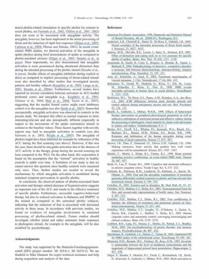
Fig. 1. Increased activation of ACC (A, x,y,z = 2,8,41) and insula (B, x,y,z = 40,11
subjects (CS) during the first scanning session. Statistical parametric maps ( P < 0
plots show the contrasts of parameter estimates (spider vs. baseline; mean T SEM
of spider videos between both groups of phobics (valence: t =
0.035, P > 0.5; arousal: t = 0.57, P > 0.5).
For fear ratings, only phobics were included in the ANOVA,
because controls did not show any variance or only marginal
variance in their fear ratings of the control objects and spiders,
which were rated as not fear-inducing at all (see Table 1). In
phobics, the ANOVA showed a significant main effect of object
[F(1,23) = 125.0, P < 0.0001], which was based on increased
ratings for spider videos as compared to control videos (t = 11.40,
P < 0.0001).
Second scanning session
Descriptive data for the ratings of valence, arousal, and fear
in response to the stimuli are given in Table 1. Ratings of the
clips indicated a significant main effect of group [valence:
F(1,22) = 19.10, P < 0.0001; arousal: F(1,22) = 34.67, P <
0.0001; fear: F(1,22) = 16.32, P < 0.005], object [valence:
F(1,22) = 33.58, P < 0.0001; arousal: F(1,22) = 58.84, P <
0.0001; fear: F(1,22) = 42.58, P < 0.0001], and a significant
interaction of group by object [valence: F(1,22) = 10.24, P <
0.004; arousal: F(1,22) = 12.43, P < 0.005; fear: F(1,22) =
7.68, P < 0.05]. Post-hoc analysis revealed that the WG
compared to the TG rated spiders as significantly more
unpleasant (t = 7.49, P < 0.0001), arousing (t = 7.23, P <
0.0001), and fear-inducing (t = 4.41, P < 0.0001), while no
significant group difference was found for the control object
(valence: t = �0.33, P > 0.5; arousal: t = 2.23, P > 0.05; fear:
t = 1.63, P > 0.1).
fMRI data
First scanning session
The ROI analysis for the contrast spider > baseline revealed
increased brain responses in several areas in control subjects
and phobics (see Table 2). However, only phobics showed
activation in the insula and ACC, while amygdala activation
was restricted to control subjects (see Table 2). For all other
ROIs, increased activation to spider videos was found in all
,3) to spider vs. control videos in phobic subjects (PS) compared to control
.005) are overlaid on a T1 scan (radiological convention: left = right). The
for maximally activated voxel in the ROI).

T. Straube et al. / NeuroImage 29 (2006) 125–135130
groups (see Table 2). Between-group comparisons did not reveal
any significant differences in activation of the ROIs between
both groups of phobic subjects. When activation in a combined
group of phobics was compared with the non-phobic control
subjects, greater responses in the left and right anterior insula
(Table 3; Fig. 1), and in the ACC (Table 3; Fig. 1) were
detected in phobics, while controls showed increased activation
in the left amygdala and bilaterally in the parahippocampal
gyrus (Table 3). Exploratory analysis revealed activation in
several areas in all groups, above all in primary and secondary
visual cortex (Table 2). Between-group comparisons did not
reveal significant differences between both groups of phobic
subjects in these regions. Phobics as compared to non-phobic
control subjects exhibited greater responses in the left extras-
triate visual cortex (lingual gyrus; see Table 3), while control
subjects showed increased activation in the pre- and postcentral
gyri (see Table 3).
Table 4
Significant activation to spider vs. control videos in phobics during the second s
Region WG TG
x y z t value x
ROI
DLPFC R 51 31 20 6.00 43
L �32 45 43 5.94 �51
ACC �6 11 37 4.84
Insula R 39 10 7 5.24 38
L �40 18 7 5.02
Parahippocampal g R 35 �35 �23 10.89 35
L �40 �35 �20 5.19
Fusiform gyrus R 30 �42 �19 12.29 19
L �36 �73 �14 7.89 �36
Thalamus R 14 �17 7 7.48
L �15 �20 10 5.53 �24
Exploratory
DMPFC R
L �11 47 37 4.11
DLFC R 42
L �16 �9 65 5.92 �48
VLPFC R 51 27 15 4.94 41
L �52 45 13 6.33
Precentral gyrus R 47 �5 34 8.92 26
L �36 9 36 7.11 �46
Postcentral gyrus R 45 �26 35 6.34
L �57 �23 36 6.28
Temporal gyri R 38 �72 0 10.18 44
L �52 �65 3 8.40 �48
Parietal gyri R 30 �67 46 16.25 35
L �38 �55 55 7.45 �33
Cuneus R 26 �82 13 8.33 21
L �19
Precuneus R 27 �58 50 10.63 21
L �21
Lingual gyrus R 30 �78 �5 8.66 18
L �25 �88 �6 9.38 �22
Occipital gyri R 38 �73 0 10.18 31
L �25 �88 �7 9.38 �36
Basal ganglia R 14 1 16 7.73
L �27 1 1 6.08 �21
DLFC, dorsolateral frontal cortex; DLPFC, dorsolateral prefrontal cortex; g, gyrus
coordinates of maximally activated voxel [Activation threshold: P < 0.005, uncoa There was no significant result for the contrast TG > WG, except for the cun
Second scanning session
In contrast to the first scanning session, where no differences
between the TG and WG were detected in the ROI analysis,
pronounced differences in brain activation emerged between both
groups at the second scanning time point. The TG showed an
absence of activation to spider videos in the ACC and only a small
cluster of activation in the ventral anterior insula, while the WG
exhibited pronounced responses bilaterally in the insula and in the
ACC (Table 4; Fig. 2). Between-group comparisons demonstrated
stronger activation bilaterally in the insula, thalamus, and in the
ACC in the WG compared to the TG (Table 2; Fig. 2), while the TG
did not exhibit higher activation than the WG in any ROI. In
exploratory analysis, increased activation in the WG compared to
the TG was detected in the left dorsomedial prefrontal cortex
(DMPFC, see Table 4) and left precuneus (Table 4). The TG as
compared to the WG showed greater responses in the right cuneus
(see Table 4).
canning session
WG > TGa
y z t value x y z t value
19 24 5.61
31 24 6.33
�3 11 37 3.66
19 12 5.33 38 4 �2 3.57
�31 5 �14 4.36
�35 �20 6.55
�82 �14 18.85
�82 �14 14.46
17 �14 11 4.48
�26 6 5.28 �9 �13 11 3.71
�16 43 34 4.14
2 40 5.98
0 39 6.91
21 13 5.11
�12 52 6.30
1 37 6.91
�71 2 13.27
�68 �2 11.51
�39 39 9.03
�47 46 6.73
�84 6 9.92
�81 28 8.53
�63 46 8.39
�80 29 8.53 �18 �68 7 4.44
�81 �14 18.85
�84 �9 10.57
�78 �3 17.82
�85 �11 15.58
�3 �3 7.11
; L, left; R, right; VLPFC, ventrolateral prefrontal cortex; (x,y,z), Talairach
rr. (ROI), P < 0.0005, uncorr. (Exploratory), cluster �135 mm3].
eus (x,y,z = 29, �80, 30; t = 5.07).

Fig. 2. Increased activation of ACC (A, x,y,z = 0,13,40) and insula (B, x,y,z = 40,8,�4) to spider vs. control videos in the waiting-list group (WG) compared to
the therapy group (TG) during the second but not the first scanning session. Statistical parametric maps ( P < 0.005) for the second scanning session (WG >
TG) are overlaid on a T1 scan (radiological convention: left = right). The plots show the contrasts of parameter estimates for both scanning sessions (spider vs.
baseline; mean T SEM for maximally activated voxel in the ROI).
T. Straube et al. / NeuroImage 29 (2006) 125–135 131
For those ROIs where decreased activation was found in the TG
compared to the WG, we compared the second with the first
scanning session in the TG using one-tailed t tests. These
comparisons confirmed significant reductions of activation in the
ACC [Talairach coordinates (x,y,z) and statistical values of peak
activation: �3, 8, 34, t = 4.20, P < 0.005], left insula [Talairach
coordinates (x,y,z) and statistical values of peak activation: �39,
22, 7, t = 3.31, P < 0.005], and left thalamus [Talairach coordinates
(x,y,z) and statistical values of peak activation: �9, �17, 11, t =
3.90, P < 0.005]. For the ROIs with differential activation between
the TG and WG, we also contrasted BOLD responses of each
phobic group with BOLD responses of the control subjects from
the first scanning session. These comparisons showed no signifi-
cant differences between control subjects and the TG, while the
WG exhibited increased activation in the right insula [Talairach
coordinates (x,y,z) and statistical values of peak activation: 38, 4,
0, t = 3.59, P < 0.005] and the ACC [Talairach coordinates (x,y,z)
and statistical values of peak activation: 15, 23, 31, t = 4.13, P <
0.005], suggesting a normalization of brain activation in the TG but
not in the WG.
Discussion
The present study revealed that the processing of phobogenic
threat is associated with increased activation in the insula and ACC
in subjects suffering from specific phobia. Most importantly,
successful cognitive-behavioral therapy led to a reduction of
hyperactivity within these brain regions. Thus, in comparison to
subjects of the untreated WG, subjects of the treated TG showed a
marked attenuation of phobic symptomatology and of responses in
the insula and ACC during the second scanning session.
The activation pattern observed in the insula fits theoretical
accounts and increasing empirical evidence that this brain region is
important for pathological but also non-pathological emotional
experiences (for reviews see Critchley, 2003; Phan et al., 2002;
Reiman, 1997). For example, involvement of the insula has been
described in spider phobics in response to real or imagined feared
animals (Rauch et al., 1995; Reiman, 1997), spider pictures (Dilger
et al., 2003; Carlsson et al., 2004; Straube et al., in press), and
spider-related words (Straube et al., 2004b). Significant insula
activation was also demonstrated in social phobics and healthy
subjects during the processing of angry faces (Straube et al.,
2004a), and during symptom provocation in other anxiety
disorders (Osuch et al., 2001; Rauch et al., 1997; Reiman, 1997).
Significantly increased responses of the insula associated with
provocation of clinically relevant anxiety symptoms were reported
by Rauch et al. (1997) when using pooled data across different
anxiety disorders. Furthermore, in healthy subjects, the insular
cortex has been shown to be involved in the recognition and
experience of aversive states such as disgust, fear, or pain (e.g.,
Critchley et al., 2002; Peyron et al., 2002; Phillips et al., 1997;
Reiman, 1997). The insular cortex is also activated by interocep-
tive stimulation and correlates with autonomic activity (Aziz et al.,
2002; Critchley et al., 2003). Above all, the work of Critchley et al.
(2001, 2002) provided strong evidence that the insula might
support the interaction of perceived threat signals and bodily states
of arousal leading to subjective emotional experiences such as the
feeling of fear.
However, although a growing number of studies strongly
implicate a critical involvement of the insula in the processing of
phobia-related threat, several studies failed to find support for this
assumption (e.g., Fredrikson et al., 1993, 1995; Tillfors et al.,
2002; Wik et al., 1993, 1997). Such discrepancies between studies
require further research, in which experimental conditions should
be systematically varied. As suggested elsewhere (Straube et al.,
2004a, in press), an important aspect might be the amount of
subjects’ attentional resources while threatening stimuli are
present. For example, attentional distraction by concurring
cognitive tasks reduces insula responses to threat (e.g., Anderson
et al., 2003; Critchley et al., 2002; Gorno-Tempini et al., 2001;
Straube et al., 2004a). Recently, we showed that activation of the
insula and ACC was even blocked when spider phobics were
requested to solve a demanding cognitive task that was displayed

T. Straube et al. / NeuroImage 29 (2006) 125–135132
in the foreground of phobia-relevant pictures (Straube et al., in
press). Similarly, high processing demand associated with the
coordination of ongoing active behavior (e.g., Tillfors et al., 2002)
could also attenuate or suppress activation of the insula. Finally,
attentional distraction that is self-induced as a kind of avoidance or
coping behavior might lead to an attenuation of insula responses
(see also Paquette et al., 2003).
Even more clearly as for the insula, responses in the ACC were
normalized in the TG at the second scanning time point, whereas
the waiting-list group did not show such an effect. Activation of the
ACC during threat processing has been shown in specific phobia
and in other anxiety disorders (Rauch et al., 1995, 1997; Osuch et
al., 2001; Boshuisen et al., 2002; Pissiota et al., 2002; Aouizerate
et al., 2004; Carlsson et al., 2004). Remarkably, direct stimulation
of the ACC evokes anxiety in humans (Laitinen, 1979) suggesting
a functional relevance of ACC activity in anxiety provocation
studies. In healthy subjects, involvement of the ACC, especially of
its rostral part, has been repeatedly described during the emotional
evaluation of threatening and other salient emotional stimuli (for
reviews see Bush et al., 2000; Phan et al., 2002). Several studies
reported also emotion-related activation of dorsal ACC (for review
see Phan et al., 2002), which has been implicated in cognitive
operations such as attentional control or response selection (for
review see Bush et al., 2000). Recently, Carlsson et al. (2004)
demonstrated augmented rCBF in dorsal ACC to phobogenic vs.
other fear-related pictures in spider phobics during passive view-
ing, suggesting a stimuli-specific, phobia-related increase of
activation in this area. In the present study, ACC activation was
also found rather in the dorsal part of the ACC. Furthermore, the
dorsal ACC has been implicated in the elicitation and control of
sympathetic autonomic arousal (Critchley et al., 2003). Thus, the
ACC activation observed in the present study might be associated
with the sympathetic hyperarousal that has been repeatedly
described to be a main physiological feature during the processing
of phobogenic stimuli in animal phobics (e.g., Hamm et al., 1997;
Globisch et al., 1999; Cuthbert et al., 2003). Successful therapy
strongly reduces these responses (e.g., Antony et al., 2001; Bracht
et al., 1999; Gutberlet and Miltner, 1999, 2001; Hellstrom and Ost,
1995, 1996; Ost, 1996; Ost et al., 2001), which suggests that the
therapy-induced elimination of ACC activation might be coupled
to attenuated fear-associated arousal.
Besides for the insula and ACC, we found stronger activation in
extrastriate visual cortex in phobics than in controls during the first
scanning session. This finding is in accordance with several reports
showing that the processing of threatening visual stimuli is
associated with increased activation in extrastriate visual areas in
phobics (Dilger et al., 2003; Fredrikson et al., 1993, 1995; Straube
et al., 2004a; Wik et al., 1993; but see Paquette et al., 2003) and
healthy controls (e.g., Keightley et al., 2003; Vuilleumier et al.,
2001). However, we found no clear therapy-effect in visual areas.
This suggests that increased activation in these regions is not
necessarily coupled to the fear-relevance of the visual stimulus
during sustained stimulus presentation. Thus, the role of other
factors, such as novelty, attention, and general salience of stimuli,
or an interaction of fear with these factors has to be taken into
account (see also Stark et al., 2004; Ishai et al., 2004).
In the DMPFC and thalamus, the WG showed stronger
activation than the TG at the second scanning time point, whereas
no differences of activation were observed in the first scanning
session. Increased responses in DMPFC has been shown in phobics
during the evaluation of threat-relevant stimulation (Carlsson et al.,
2004; Rauch et al., 1995; Stein et al., 2002; Straube et al., 2004a).
This region has been implicated in anxiety and the general
assessment of the emotional significance of stimuli (Phan et al.,
2002; Simpson et al., 2001). Evidence for a significantly increased
activation of the thalamus during threat is less consistent (for
review see Phan et al., 2002) and seems to be more strongly
associated with the internal induction of emotion during recall or
imagery (e.g., Rauch et al., 1997). The assumed role of the
DMPFC and thalamus in threat processing is partially supported by
the therapy effects found in these areas in the present study.
However, the data for the first scanning session do not confirm this
hypothesis. At least, the observed differences between treated and
untreated phobics indicate that augmented responses of the
DMPFC and thalamus correlate with the maintenance of anxiety
during a repeated confrontation with phobia-related stimuli (see
also Furmark et al., 2002).
In contrast to the study by Paquette et al. (2003), we did not
find activation in DLPFC or parahippocampal gyrus in phobics as
compared with healthy subjects. In parahippocampal gyrus, rather
less activation was detected in phobics. Furthermore, we found no
evidence of a therapy effect in these areas. Activation of the
DLPFC during anxiety provocation paradigms was proposed to
reflect coping strategies and inhibitory control but also anticipatory
anxiety or worrying (e.g., Johanson et al., 1998; Paquette et al.,
2003; Reiman, 1997). Interestingly, a study by Johanson et al.
(1998) suggested that rCBF in prefrontal areas in phobics might
depend on the amount of panic during phobogenic stimulation. In
this study, an increase of prefrontal rCBF was associated with a
higher level of control of anxiety responses, while a decrease was
observed when subjects experienced panic. Therefore, it seems
reasonable that studies with inhomogeneous groups of phobic
subjects in respect to the individual coping or phobic behavior
should not obtain consistent results for the DLPFC.
The other region for which Paquette et al. found a therapy-
induced reduction of hyperactivity was the parahippocampal gyrus.
This area has been strongly associated with memory functions
(Brewer et al., 1998; Wagner et al., 1998), particularly during
processing of emotional stimuli (Kilpatrick and Cahill, 2003).
Although there is evidence for increased parahippocampal activa-
tion in phobics during threat processing (Paquette et al., 2003; Stein
et al., 2002, Straube et al., 2004a), several studies with animal
phobics did not report activation or even deactivation in this region
(e.g., Dilger et al., 2003; Fredrikson et al., 1995; Rauch et al., 1995;
Wik et al., 1993). Eventually, the discrepant findings could be
explained by assuming an inverted U-function of activation
depending on the amount of stress (see Diamond et al., 1992;
Pavlides et al., 1993). Strongest activation would then occur during
an optimal level of emotional arousal (see also Straube et al., 2003),
while high levels of stress or anxiety would decrease neuronal
activity. However, this idea remains to be tested in future studies by
using samples with high variance in emotional reactions or by using
parametric designs allowing the induction of different levels of
anxiety.
The absence of activation in animal phobics in the other ROI in
the medial temporal lobe, the amygdala, is in line with previous
symptom provocation studies that used stimulation paradigms such
as imagery (Rauch et al., 1995), video presentation (Mountz et al.,
1989; Fredrikson et al., 1993, 1995; Wik et al., 1993, 1997;
Paquette et al., 2003), prolonged picture presentation in blocked
designs (Pissiota et al., 2003), or in vivo confrontation (Reiman,
1997). These consistent outcomes, therefore, indicate that sus-
T. Straube et al. / NeuroImage 29 (2006) 125–135 133
tained phobia-related stimulation in specific phobia (in contrast to
social phobia, see Furmark et al., 2002; Tillfors et al., 2001, 2002)
does not seem to be associated with amygdalar activity. The
amygdala, however, has been implicated in the initial processing of
threat and the induction of rapid fear responses (Dilger et al., 2003;
Carlsson et al., 2004; Ohman and Mineka, 2001). In recent event-
related fMRI studies, we showed activation of the amygdala in
spider phobics during brief presentations of spider as compared to
phobia-unrelated pictures (Dilger et al., 2003; Straube et al., in
press). Most importantly, we also demonstrated that amygdala
activation is more pronounced during attentional distraction than
during the attended processing of the phobic stimuli (Straube et al.,
in press). Similar effects of amygdala inhibition during explicit or
direct as compared to implicit processing of threat-related stimuli
were also described by other studies that investigated anxiety
patients and healthy subjects (Keightley et al., 2003; Lange et al.,
2003; Straube et al., 2004a). Furthermore, several studies have
reported an inverse correlation between activation in ACC/medial
prefrontal cortex and amygdala (e.g., Keightley et al., 2003;
Ochsner et al., 2004; Shin et al., 2004; Taylor et al., 2003),
suggesting that the medial frontal cortex might exert inhibitory
control over the amygdala (see also Quirk et al., 2003). Somewhat
unexpectedly, amygdala activation was observed in controls in the
present study. We interpret this effect as normal response to more
interesting/relevant and also perceptually different (especially in
respect to the movements of the spider extremities) stimuli in
contrast to the baseline videos. These factors together with novelty
aspects may lead to amygdala activation in controls (see also
Schwartz et al., 2003; Wright et al., 2003). The amygdala of
phobics might have been inhibited for example by influences of the
ACC during the first scanning (see above). However, if this was
the case, there should be amygdala activation due to the absence of
ACC activity in the therapy group during the second scanning (at
least compared to the WG). On the other hand, this expectation is
based on the assumption that the ‘‘normal’’ activation in healthy
controls is stable over time. A limitation of our study is that we
cannot answer this question since healthy controls were measured
only once. Thus, further studies are needed to reveal the
mechanisms by which amygdala activation is modulated during
sustained symptom provocation in specific phobia.
In conclusion, the observed pattern of phobia-associated brain
activation and therapy-related decrease of hyperactivation suggests
an important role of the ACC and insula in the effective treatment
of specific phobia. Furthermore, successful cognitive-behavioral
therapy led also to reduced activation in thalamus and DMPFC in
the treated as compared to the untreated phobic subjects,
indicating that the reduction of fear is associated with decreased
activity in these areas. In accordance with previous studies, we
found no evidence of amygdala involvement in sustained
processing of phobia-related stimuli. Future studies should
investigate whether initial and rather automatic brain responses
to phobogenic stimuli, for example in the amygdala, will be also
modified by psychotherapy.
Acknowledgments
The study was supported by the Deutsche-Forschungsgemein-
schaft (DFG project number: Mi 265/6-1; Mi 265/6-2). We are
thankful to Mike Hammer for expert technical assistance and help
during acquisition and analysis of the data.
References
American Psychiatric Association, 1994. Diagnostic and Statistical Manual
of Mental Disorders, 4th ed. (DSM-IV). Washington, D.C.
Anderson, A.K., Christoff, K., Panitz, D., De Rosa, E., Gabrieli, J.D., 2003.
Neural correlates of the automatic processing of threat facial signals.
J. Neurosci. 23, 5627–5633.
Antony, M.M., McCabe, R.E., Leeuw, I., Sano, N., Swinson, R.P., 2001.
Effect of distraction and coping style on in vivo exposure for specific
phobia of spiders. Behav. Res. Ther. 39 (10), 1137–1150.
Aouizerate, B., Guehl, D., Cuny, E., Rougier, A., Bioulac, B., Tignol, J.,
Burbaud, P., 2004. Pathophysiology of obsessive–compulsive disorder:
a necessary link between phenomenology, neuropsychology, imagery
and physiology. Prog. Neurobiol. 72, 195–221.
Aziz, Q., Schnitzler, A., Enck, P., 2002. Functional neuroimaging of
visceral sensation. J. Clin. Neurophysiol. 17 (6), 604–612.
Birbaumer, N., Grodd, W., Diedrich, O., Klose, U., Erb, M., Lotze,
M., Schneider, F., Weiss, U., Flor, H., 1998. fMRI reveals
amygdala activation to human faces in social phobics. NeuroReport
9, 1223–1226.
Boshuisen, M.L., Ter Horst, G.J., Paans, A.M., Reinders, A.A., den Boer,
J.A., 2002. rCBF differences between panic disorder patients and
control subjects during anticipatory anxiety and rest. Biol. Psychiatry
52, 126–135.
Bracht, S., Gutberlet, I., Miltner, W.H.R., 1999. Effects of behavioural
therapy intervention on peripheral physiological parameters as well as
subjective estimations of emotional arousal and affective valency during
the processing of phobogenic visual material by clients with adversions
to spiders and snakes. Verhaltenstherapie 9, 6–7.
Breiter, H.C., Etcoff, N.L., Whalen, P.J., Kennedy, W.A., Rauch, S.L.,
Buckner, R.L., Strauss, M.M., Hyman, S.E., Rosen, B.R., 1996.
Response and habituation of the human amygdala during visual
processing of facial expression. Neuron 17, 875–887.
Brewer, J.B., Zhao, Z., Desmond, J.E., Glover, G.H., Gabrieli, J.D., 1998.
Making memories: brain activity that predicts how well visual
experience will be remembered. Science 281, 1185–1187.
Buchel, C., Morris, J., Dolan, R.J., Friston, K.J., 1998. Brain systems
mediating aversive conditioning: an event-related fMRI study. Neuron
20, 947–957.
Bush, G., Luu, P., Posner, M.I., 2000. Cognitive and emotional influences
in anterior cingulate cortex. Trends Cogn. Sci. 4, 215–222.
Carlsson, K., Petersson, K.M., Lundqvist, D., Karlsson, A., Ingvar, M.,
Ohman, A., 2004. Fear and the amygdala: manipulation of awareness
generates differential cerebral responses to phobic and fear-relevant (but
nonfeared) stimuli. Emotion 4, 340–353.
Critchley, H., 2003. Emotion and its disorders. Br. Med. Bull. 65, 35–47.
Critchley, H.D., Mathias, C.J., Dolan, R.J., 2001. Neuroanatomical basis for
first- and second-order representations of bodily states. Nat. Neurosci.
4, 207–212.
Critchley, H.D., Mathias, C.J., Dolan, R.J., 2002. Fear conditioning in
humans: the influence of awareness and autonomic arousal on func-
tional neuroanatomy. Neuron 33, 653–663.
Critchley, H.D., Mathias, C.J., Josephs, O., O’Doherty, J., Zanini, S.,
Dewar, B.K., Cipolotti, L., Shallice, T., Dolan, R.J., 2003. Human
cingulate cortex and autonomic control: converging neuroimaging and
clinical evidence. Brain 126, 2139–2152.
Cuthbert, B.N., Lang, P.J., Strauss, C., Drobes, D., Patrick, C.J., Bradley,
M.M., 2003. The psychophysiology of anxiety disorder: fear memory
imagery. Psychophysiology 40, 407–422.
Deichmann, R., Gottfried, J.A., Hutton, C., Turner, R., 2003. Optimized EPI
for fMRI studies of the orbitofrontal cortex. NeuroImage 19, 430–441.
Diamond, D.M., Bennett, M.C., Fleshner, M., Rose, G.M., 1992. Inverted-
U relationship between the level of peripheral corticosterone and the
magnitude of hippocampal primed burst potentiation. Hippocampus 2,
421–430.
Dilger, S., Straube, T., Mentzel, H.J., Fitzek, C., Reichenbach, J.R., Hecht,
H., Krieschel, S., Gutberlet, I., Miltner, W.H., 2003. Brain activation to

T. Straube et al. / NeuroImage 29 (2006) 125–135134
phobia-related pictures in spider phobic humans: an event-related
functional magnetic resonance imaging study. Neurosci. Lett. 348,
29–32.
Fredrikson, M., Wik, G., Greitz, T., Eriksson, L., Stone-Elander, S.,
Ericson, K., Sedvall, G., 1993. Regional cerebral blood flow during
experimental phobic fear. Psychophysiology 30, 126–130.
Fredrikson, M., Wik, G., Annas, P., Ericson, K., Stone-Elander, S.,
1995. Functional neuroanatomy of visually elicited simple phobic
fear: additional data and theoretical analysis. Psychophysiology 32,
43–48.
Furmark, T., Tillfors, M., Marteinsdottir, I., Fischer, H., Pissiota, A.,
Langstrom, B., Fredrikson, M., 2002. Common changes in cerebral
blood flow in patients with social phobia treated with citalopram or
cognitive-behavioral therapy. Arch. Gen. Psychiatry 59, 425–433.
Fyer, A.J., 1998. Current approaches to etiology and pathophysiology of
specific phobia. Biol. Psychiatry 44, 1295–1304.
Gilboa, A., Shalev, A.Y., Laor, L., Lester, H., Louzoun, Y., Chisin, R.,
Bonne, O., 2004. Functional connectivity of the prefrontal cortex and the
amygdala in posttraumatic stress disorder. Biol. Psychiatry 55, 263–272.
Globisch, J., Hamm, A.O., Esteves, F., Ohman, A., 1999. Fear appears fast:
temporal course of startle reflex potentiation in animal fearful subjects.
Psychophysiology 36, 66–75.
Gorno-Tempini, M.L., Pradelli, S., Serafini, M., Pagnoni, G., Baraldi, P.,
Porro, C., Nicoletti, R., Umita, C., Nichelli, P., 2001. Explicit and
incidental facial expression processing: an fMRI study. NeuroImage 14,
465–473.
Gutberlet, I., Miltner, W.H.R., 1999. Therapeutic effects on differential
electrocortical processing of phobic objects in spider and snake phobics.
Int. J. Psychophysiol. 33 (1), 180.
Gutberlet, I., Miltner, W.H.R., 2001. Therapy-induced changes of cortical
and autonomic functions to phobic, negative, neutral, and positive
stimuli in snake and spider phobics. J. Psychophysiol. 15 (1), 54–55.
Hamm, A.O., Cuthbert, B.N., Globisch, J., Vaitl, D., 1997. Fear and the
startle reflex: blink modulation and autonomic response patterns in
animal and mutilation fearful subjects. Psychophysiology 34, 97–107.
Hellstrom, K., Ost, L.G., 1995. One-session therapist directed exposure vs.
two forms of manual directed self-exposure in the treatment of spider
phobia. Behav. Res. Ther. 33, 959–965.
Hellstrom, K., Ost, L.G., 1996. Prediction of outcome in the treatment
of specific phobia. A cross validation study. Behav. Res. Ther. 34,
403–411.
Ishai, A., Pessoa, L., Bikle, P.C., Ungerleider, L.G., 2004. Repetition
suppression of faces is modulated by emotion. Proc. Natl. Acad. Sci.
U. S. A. 101, 9827–9832.
Johanson, A., Gustafson, L., Passant, U., Risberg, J., Smith, G., Warkentin,
S., Tucker, D., 1998. Brain function in spider phobia. Psychiatry Res.
84, 101–111.
Johansen-Berg, H., Dawes, H., Guy, C., Smith, S.M., Wade, D.T., Matthews,
P.M., 2002. Correlation between motor improvements and altered fMRI
activity after rehabilitative therapy. Brain 125, 2731–2742.
Keightley, M.L., Winocur, G., Graham, S.J., Mayberg, H.S., Hevenor, S.J.,
Grady, C.L., 2003. An fMRI study investigating cognitive modulation
of brain regions associated with emotional processing of visual stimuli.
Neuropsychologia 41, 585–596.
Kilpatrick, L., Cahill, L., 2003. Amygdala modulation of parahippocampal
and frontal regions during emotionally influenced memory storage.
NeuroImage 20, 2091–2099.
Klorman, R., Weerts, T.C., Hastings, J.E., Melamed, B.G., Lang, P.J., 1974.
Psychometric description of some specific-fear questionnaires. Behav.
Ther. 5, 401–409.
Laitinen, L.V., 1979. Emotional responses to subcortical electrical stim-
ulation in psychiatric patients. Clin. Neurol. Neurosurg. 81, 148–157.
Lange, K., Williams, L.M., Young, A.W., Bullmore, E.T., Brammer, M.J.,
Williams, S.C., Gray, J.A., Phillips, M.L., 2003. Task instructions
modulate neural responses to fearful facial expressions. Biol. Psychiatry
53, 226–232.
Liberzon, I., Taylor, S.F., Amdur, R., Jung, T.D., Chamberlain, K.R.,
Minoshima, S., Koeppe, R.A., Fig, L.M., 1999. Brain activation in PTSD
in response to trauma-related stimuli. Biol. Psychiatry 45, 817–826.
McGonigle, D.J., Howseman, A.M., Athwal, B.S., Friston, K.J., Frack-
owiak, R.S., Holmes, A.P., 2000. Variability in fMRI: an examination of
intersession differences. NeuroImage 11, 708–734.
Morris, J.S., Ohman, A., Dolan, R.J., 1998. Conscious and unconscious
emotional learning in the human amygdala. Nature 393, 467–470.
Mountz, J.M., Modell, J.G., Wilson, M.W., Curtis, G.C., Lee, M.A.,
Schmaltz, S., Kuhl, D.E., 1989. Positron emission tomographic
evaluation of cerebral blood flow during state anxiety in simple phobia.
Arch. Gen. Psychiatry 46, 501–504.
Ochsner, K.N., Ray, R.D., Cooper, J.C., Robertson, E.R., Chopra, S.,
Gabrieli, J.D., Gross, J.J., 2004. For better or for worse: neural systems
supporting the cognitive down- and up-regulation of negative emotion.
NeuroImage 23, 483–499.
Ohman, A., Mineka, S., 2001. Fears, phobias, and preparedness: toward an
evolved module of fear and fear learning. Psychol. Rev. 108, 483–522.
Ost, L.G., 1989. One-session treatment for specific phobias. Behav. Res.
Ther. 27, 1–7.
Ost, L.G., 1996. One-session group treatment of spider phobia. Behav. Res.
Ther. 34, 707–715.
Ost, L.G., Svensson, L., Hellstrom, K., Lindwall, R., 2001. One-session
treatment of specific phobias in youths: a randomized clinical trial.
J. Consult. Clin. Psychol. 69, 814–824.
Osuch, E.A., Benson, B., Geraci, M., Podell, D., Herscovitch, P., McCann,
U.D., Post, R.M., 2001. Regional cerebral blood flow correlated with
flashback intensity in patients with posttraumatic stress disorder. Biol.
Psychiatry 50, 246–253.
Paquette, V., Levesque, J., Mensour, B., Leroux, J.M., Beaudoin, G.,
Bourgouin, P., Beauregard, M., 2003. ‘‘Change the mind and you
change the brain’’: effects of cognitive-behavioral therapy on the neural
correlates of spider phobia. NeuroImage 18, 401–409.
Pavlides, C., Watanabe, Y., McEwen, B.S., 1993. Effects of glucocorticoids
on hippocampal long-term potentiation. Hippocampus 3, 183–192.
Peyron, R., Frot, M., Schneider, F., Garcia-Larrea, L., Mertens, P., Barral,
F.G., Sindou, M., Laurent, B., Mauguiere, F., 2002. Role of oper-
culoinsular cortices in human pain processing: converging evidence
from PET, fMRI, dipole modeling, and intracerebral recordings of
evoked potentials. NeuroImage 17, 1336–1346.
Phan, K.L., Wager, T., Taylor, S.F., Liberzon, I., 2002. Functional
neuroanatomy of emotion: a meta-analysis of emotion activation studies
in PET and fMRI. NeuroImage 16, 331–348.
Phillips, M.L., Young, A.W., Senior, C., Brammer, M., Andrew, C., Calder,
A.J., Bullmore, E.T., Perrett, D.I., Rowland, D., Williams, S.C., Gray,
J.A., David, A.S., 1997. A specific neural substrate for perceiving facial
expressions of disgust. Nature 389, 495–498.
Pissiota, A., Frans, O., Fernandez, M., von Knorring, L., Fischer, H.,
Fredrikson, M., 2002. Neurofunctional correlates of posttraumatic stress
disorder: a PET symptom provocation study. Eur. Arch. Psychiatry Clin.
Neurosci. 252, 68–75.
Pissiota, A., Frans, O., Michelgard, A., Appel, L., Langstrom, B., Flaten,
M.A., Fredrikson, M., 2003. Amygdala and anterior cingulate cortex
activation during affective startle modulation: a PET study of fear. Eur.
J. Neurosci. 18, 1325–1331.
Quirk, G.J., Likhtik, E., Pelletier, J.G., Pare, D., 2003. Stimulation of
medial prefrontal cortex decreases the responsiveness of central
amygdala output neurons. J. Neurosci. 23, 8800–8807.
Rauch, S.L., Savage, C.R., Alpert, N.M., Miguel, E.C., Baer, L., Breiter,
H.C., Fischman, A.J., Manzo, P.A., Moretti, C., Jenike, M.A., 1995. A
positron emission tomographic study of simple phobic symptom
provocation. Arch. Gen. Psychiatry 52, 20–28.
Rauch, S.L., Savage, C.R., Alpert, N.M., Fischman, A.J., Jenike, M.A.,
1997. The functional neuroanatomy of anxiety: a study of three
disorders using positron emission tomography and symptom provoca-
tion. Biol. Psychiatry 42, 446–452.
Rauch, S.L., Whalen, P.J., Shin, L.M., McInerney, S.C., Macklin, M.L.,
Lasko, N.B., Orr, S.P., Pitman, R.K., 2000. Exaggerated amygdala

T. Straube et al. / NeuroImage 29 (2006) 125–135 135
response to masked facial stimuli in posttraumatic stress disorder: a
functional MRI study. Biol. Psychiatry 47, 769–776.
Reiman, E.M., 1997. The application of positron emission tomography to
the study of normal and pathologic emotions. J. Clin. Psychiatry 58
(Suppl. 16), 4–12.
Schwartz, C.E., Wright, C.I., Shin, L.M., Kagan, J., Whalen, P.J.,
McMullin, K.G., Rauch, S.L., 2003. Differential amygdalar response
to novel versus newly familiar neutral faces: a functional MRI probe
developed for studying inhibited temperament. Biol. Psychiatry 53,
854–862.
Shin, L.M., Kosslyn, S.M., McNally, R.J., Alpert, N.M., Thompson, W.L.,
Rauch, S.L., Macklin, M.L., Pitman, R.K., 1997. Visual imagery and
perception in posttraumatic stress disorder. A positron emission tomo-
graphic investigation. Arch. Gen. Psychiatry 54, 233–241.
Shin, L.M., McNally, R.J., Kosslyn, S.M., Thompson, W.L., Rauch, S.L.,
Alpert, N.M., Metzger, L.J., Lasko, N.B., Orr, S.P., Pitman, R.K., 1997.
A positron emission tomographic study of symptom provocation in
PTSD. Ann. N. Y. Acad. Sci. 821, 521–523.
Shin, L.M., Orr, S.P., Carson, M.A., Rauch, S.L., Macklin, M.L., Lasko,
N.B., Peters, P.M., Metzger, L.J., Dougherty, D.D., Cannistraro, P.A.,
Alpert, N.M., Fischman, A.J., Pitman, R.K., 2004. Regional cerebral
blood flow in the amygdala and medial prefrontal cortex during
traumatic imagery in male and female Vietnam veterans with PTSD.
Arch. Gen. Psychiatry 61, 168–176.
Simpson, J.R. Jr., Drevets, W.C., Snyder, A.Z., Gusnard, D.A., Raichle,
M.E., 2001. Emotion-induced changes in human medial prefrontal
cortex: II. During anticipatory anxiety. Proc. Natl. Acad. Sci. U. S. A.
98, 688–693.
Stark, R., Schienle, A., Walter, B., Kirsch, P., Blecker, C., Ott, U., Schafer,
A., Sammer, G., Zimmermann, M., Vaitl, D., 2004. Hemodynamic
effects of negative emotional pictures—A test– retest analysis. Neuro-
psychobiology 50, 108–118.
Stein, M.B., Goldin, P.R., Sareen, J., Zorrilla, L.T., Brown, G.G., 2002.
Increased amygdala activation to angry and contemptuous faces in
generalized social phobia. Arch. Gen. Psychiatry 59, 1027–1034.
Straube, T., Korz, V., Balschun, D., Frey, J.U., 2003. Requirement of beta-
adrenergic receptor activation and protein synthesis for LTP-reinforce-
ment by novelty in rat dentate gyrus. J. Physiol. 552, 953–960.
Straube, T., Kolassa, I.T., Glauer, M., Mentzel, H.J., Miltner, W.H., 2004a.
Effect of task conditions on brain responses to threatening faces in
social phobics: an event-related functional magnetic resonance imaging
study. Biol. Psychiatry 56, 921–930.
Straube, T., Mentzel, H.J., Glauer, M., Miltner, W.H., 2004b. Brain
activation to phobia-related words in phobic subjects. Neurosci. Lett.
372, 204–208.
Straube, T., Mentzel, H.J., Miltner, W.H.R., in press. Neural mechanisms of
automatic and direct processing of phobogenic stimuli in specific
phobia. Biol. Psychiatry.
Taylor, S.F., Phan, K.L., Decker, L.R., Liberzon, I., 2003. Subjective rating
of emotionally salient stimuli modulates neural activity. NeuroImage
18, 650–659.
Talairach, J., Tournoux, P., 1988. Co-Planar Stereotactic Atlas of the
Human Brain. Thieme, New York.
Tillfors, M., Furmark, T., Marteinsdottir, I., Fischer, H., Pissiota, A.,
Langstrom, B., Fredrikson, M., 2001. Cerebral blood flow in subjects
with social phobia during stressful speaking tasks: a PET study. Am. J.
Psychiatry 158, 1220–1226.
Tillfors, M., Furmark, T., Marteinsdottir, I., Fredrikson, M., 2002. Cerebral
blood flow during anticipation of public speaking in social phobia: a
PET study. Biol. Psychiatry 52, 1113–1119.
Veltman, D.J., Tuinebreijer, W.E., Winkelman, D., Lammertsma, A.A.,
Witter, M.P., Dolan, R.J., Emmelkamp, P.M., 2004. Neurophysiological
correlates of habituation during exposure in spider phobia. Psychiatry
Res. 132, 149–158.
Vuilleumier, P., Armony, J.L., Driver, J., Dolan, R.J., 2001. Effects of
attention and emotion on face processing in the human brain: an event-
related fMRI study. Neuron 30, 829–841.
Wagner, A.D., Schacter, D.L., Rotte, M., Koutstaal, W., Maril, A., Dale,
A.M., Rosen, B.R., Buckner, R.L., 1998. Building memories: remem-
bering and forgetting of verbal experiences as predicted by brain
activity. Science 281, 1188–1191.
Walker, D.L., Toufexis, D.J., Davis, M., 2003. Role of the bed nucleus of
the stria terminalis versus the amygdala in fear, stress, and anxiety. Eur.
J. Pharmacol. 46, 199–216.
Wik, G., Fredrikson, M., Ericson, K., Eriksson, L., Stone-Elander, S.,
Greitz, T., 1993. A functional cerebral response to frightening visual
stimulation. Psychiatry Res. 50, 15–24.
Wik, G., Fredrikson, M., Fischer, H., 1997. Evidence of altered cerebral
blood-flow relationships in acute phobia. Int. J. Neurosci. 91, 253–263.
Wittchen, H.U., Wunderlich, U., Gruschwitz, S., Zaudig, M., 1997.
Strukturiertes Klinisches Interview fur DSM-IV (Structured clinical
interview for DSM-IV). Gottingen, Hogrefe.
Wright, C.I., Fischer, H., Whalen, P.J., McInerney, S.C., Shin, L.M.,
Rauch, S.L., 2001. Differential prefrontal cortex and amygdala
habituation to repeatedly presented emotional stimuli. NeuroReport
12, 379–383.
Wright, C.I., Martis, B., Schwartz, C.E., Shin, L.M., Fischer, H.H.,
McMullin, K., Rauch, S.L., 2003. Novelty responses and differential
effects of order in the amygdala, substantia innominata, and inferior
temporal cortex. NeuroImage 18, 660–669.