Effects of ascorbic acid on impaired vascular reactivity in aortas isolated from age-matched...
7
Effects of ascorbic acid on impaired vascular reactivity in aortas isolated from age-matched hypertensive and diabetic rats Machha Ajay, Mohd Rais Mustafa ⁎ Department of Pharmacology, Faculty of Medicine, University of Malaya, Kuala Lumpur, 50603, Malaysia Received 16 February 2006; received in revised form 28 April 2006; accepted 2 May 2006 Abstract Impaired vascular reactivity is a hallmark of several cardiovascular diseases that include hypertension and diabetes. This study compared the changes in vascular reactivity in age-matched experimental hypertension and diabetes, and, subsequently, tested whether these changes could be affected directly by ascorbic acid (10 μM). Endothelium-derived nitric oxide (NO) modulation of ascorbic acid effects was also investigated. All the experiments were performed in the presence of a cyclooxygenase inhibitor, indomethacin (10 μM). Results showed that the endothelium- dependent and -independent relaxations induced by acetylcholine (ACh) and sodium nitroprusside (SNP), respectively, were blunted to a similar extent in isolated aortic rings from age-matched spontaneously hypertensive (SHR) (R max : ACh = 72.83 ± 1.86%, SNP = 96.6 ± 1.90%) and diabetic (R max : ACh = 64.09 ± 5.14%, SNP = 95.84 ± 1.41%) rats compared with aortic rings of normal rats (R max : ACh = 89%, SNP = 104.0 ± 1.0%). The α 1 - receptor-mediated contractions induced by phenylephrine (PE) were augmented in diabetic (C max = 148.8 ± 9.0%) rat aortic rings compared to both normal (C max =127±6.9%) and SHR (C max = 118 ± 4.5%) aortic rings. Ascorbic acid pretreatment was without any significant effects on the vascular responses to ACh, SNP and PE in aortic rings from normal rats. Ascorbic acid significantly improved ACh-induced relaxations in SHR (R max = 89.09 ± 2.82%) aortic rings to a level similar to that observed in normal aortic rings, but this enhancement in ACh-induced relaxations was only partial in diabetic aortic rings. Ascorbic acid lacked any effects on SNP-induced relaxations in both SHR and diabetic aortic rings. Ascorbic acid markedly attenuated contractions induced by PE in aortic rings from both SHR (C max = 92.9 ± 6.68%) and diabetic (C max = 116.9 ± 9.4%) rats. Additionally, following inhibition of nitric oxide synthesis with L-NAME, ascorbic acid attenuated PE-induced contractions in all aortic ring types studied. These results suggest that (1) vascular hyper-responsiveness to α 1 -receptor agonists in diabetic arteries is independent of endothelial nitric oxide dysfunction; (2) ascorbic acid directly modulates contractile responses of hypertensive and diabetic rat aortas, likely through mechanisms in part independent of preservation of endothelium-derived nitric oxide. © 2006 Elsevier Inc. All rights reserved. Keywords: Ascorbic acid; Diabetes; Endothelium; Hypertension; Nitric oxide; Vascular reactivity 1. Introduction The endothelium maintains vascular homeostasis through the release of a number of regulatory substances; among which nitric oxide (NO) is most important (Furchgott and Vanhoutte, 1989; Gewaltig and Kojda, 2002). The bioactivity of endothe- lium-derived NO is impaired in cardiovascular disease condi- tions including hypertension and diabetes (Gewaltig and Kojda, 2002; Endemann and Schiffrin, 2004). Vascular production of superoxide anions and its reactive oxygen intermediates such as hydrogen peroxide are increased in hypertension and diabetes (Gewaltig and Kojda, 2002; Endemann and Schiffrin, 2004). Superoxide anions would incapacitate the NO, exacerbate local oxidative stress and ultimately leads to alterations in the vascular tone. Several studies have documented reduced endothelium-mediated vasodilatation to vasodilators, such as acetylcholine, and enhanced vasoconstrictions to adrenergic receptor agonists, such as norepinephrine, in hypertensive and diabetic vessels when compared with physiological vessels (Ting et al., 1996; Dresner et al., 1997; Taddei et al., 1998; Nascimento et al., 2003). However, some evidence suggests that the hyper-responsiveness of diabetic vessels to receptor agonists was independent of endothelial–nitric oxide dysfunction, and it Vascular Pharmacology 45 (2006) 127 – 133 www.elsevier.com/locate/vph ⁎ Corresponding author. Tel.: +60 3 79674952; fax: +60 3 79674791. E-mail address: [email protected] (M.R. Mustafa). 1537-1891/$ - see front matter © 2006 Elsevier Inc. All rights reserved. doi:10.1016/j.vph.2006.05.001
-
Upload
independent -
Category
Documents
-
view
6 -
download
0
Transcript of Effects of ascorbic acid on impaired vascular reactivity in aortas isolated from age-matched...

5 (2006) 127–133www.elsevier.com/locate/vph
Vascular Pharmacology 4
Effects of ascorbic acid on impaired vascular reactivity in aortasisolated from age-matched hypertensive and diabetic rats
Machha Ajay, Mohd Rais Mustafa ⁎
Department of Pharmacology, Faculty of Medicine, University of Malaya, Kuala Lumpur, 50603, Malaysia
Received 16 February 2006; received in revised form 28 April 2006; accepted 2 May 2006
Abstract
Impaired vascular reactivity is a hallmark of several cardiovascular diseases that include hypertension and diabetes. This study compared thechanges in vascular reactivity in age-matched experimental hypertension and diabetes, and, subsequently, tested whether these changes could beaffected directly by ascorbic acid (10 μM). Endothelium-derived nitric oxide (NO) modulation of ascorbic acid effects was also investigated. Allthe experiments were performed in the presence of a cyclooxygenase inhibitor, indomethacin (10 μM). Results showed that the endothelium-dependent and -independent relaxations induced by acetylcholine (ACh) and sodium nitroprusside (SNP), respectively, were blunted to a similarextent in isolated aortic rings from age-matched spontaneously hypertensive (SHR) (Rmax: ACh=72.83±1.86%, SNP=96.6±1.90%) and diabetic(Rmax: ACh=64.09±5.14%, SNP=95.84±1.41%) rats compared with aortic rings of normal rats (Rmax: ACh=89%, SNP=104.0±1.0%). The α1-receptor-mediated contractions induced by phenylephrine (PE) were augmented in diabetic (Cmax=148.8±9.0%) rat aortic rings compared to bothnormal (Cmax=127±6.9%) and SHR (Cmax=118±4.5%) aortic rings. Ascorbic acid pretreatment was without any significant effects on thevascular responses to ACh, SNP and PE in aortic rings from normal rats. Ascorbic acid significantly improved ACh-induced relaxations in SHR(Rmax=89.09±2.82%) aortic rings to a level similar to that observed in normal aortic rings, but this enhancement in ACh-induced relaxations wasonly partial in diabetic aortic rings. Ascorbic acid lacked any effects on SNP-induced relaxations in both SHR and diabetic aortic rings. Ascorbicacid markedly attenuated contractions induced by PE in aortic rings from both SHR (Cmax=92.9±6.68%) and diabetic (Cmax=116.9±9.4%) rats.Additionally, following inhibition of nitric oxide synthesis with L-NAME, ascorbic acid attenuated PE-induced contractions in all aortic ring typesstudied. These results suggest that (1) vascular hyper-responsiveness to α1-receptor agonists in diabetic arteries is independent of endothelial nitricoxide dysfunction; (2) ascorbic acid directly modulates contractile responses of hypertensive and diabetic rat aortas, likely through mechanisms inpart independent of preservation of endothelium-derived nitric oxide.© 2006 Elsevier Inc. All rights reserved.
Keywords: Ascorbic acid; Diabetes; Endothelium; Hypertension; Nitric oxide; Vascular reactivity
1. Introduction
The endothelium maintains vascular homeostasis throughthe release of a number of regulatory substances; among whichnitric oxide (NO) is most important (Furchgott and Vanhoutte,1989; Gewaltig and Kojda, 2002). The bioactivity of endothe-lium-derived NO is impaired in cardiovascular disease condi-tions including hypertension and diabetes (Gewaltig and Kojda,2002; Endemann and Schiffrin, 2004). Vascular production ofsuperoxide anions and its reactive oxygen intermediates such as
⁎ Corresponding author. Tel.: +60 3 79674952; fax: +60 3 79674791.E-mail address: [email protected] (M.R. Mustafa).
1537-1891/$ - see front matter © 2006 Elsevier Inc. All rights reserved.doi:10.1016/j.vph.2006.05.001
hydrogen peroxide are increased in hypertension and diabetes(Gewaltig and Kojda, 2002; Endemann and Schiffrin, 2004).Superoxide anions would incapacitate the NO, exacerbate localoxidative stress and ultimately leads to alterations in thevascular tone. Several studies have documented reducedendothelium-mediated vasodilatation to vasodilators, such asacetylcholine, and enhanced vasoconstrictions to adrenergicreceptor agonists, such as norepinephrine, in hypertensive anddiabetic vessels when compared with physiological vessels(Ting et al., 1996; Dresner et al., 1997; Taddei et al., 1998;Nascimento et al., 2003). However, some evidence suggests thatthe hyper-responsiveness of diabetic vessels to receptor agonistswas independent of endothelial–nitric oxide dysfunction, and it
128 M. Ajay, M.R. Mustafa / Vascular Pharmacology 45 (2006) 127–133
has been proposed that such a defect may depend onsuperoxide-mediated direct alterations in vascular smoothmuscle (Chang et al., 1993a; Weber and Macleod, 1997).
Ascorbic acid (vitamin C) is the main water-solubleantioxidant in human plasma (Frei et al., 1990). Several studieshave demonstrated that ascorbic acid improves endothelium-dependent vasodilatation in hypertension and diabetes, and ithas been proposed that such effects may be due to scavenging ofsuperoxide anions and its reactive oxygen intermediates (Ting etal., 1996; Taddei et al., 1998; Akpaffiong and Taylor, 1998).However, a considerable number of studies also havedemonstrated lack of vascular effects of ascorbic acid indiabetes (May, 2000; Darko et al., 2002). In addition, one studyfound that ascorbic acid modulates venous tone independent ofputative endothelium-derived NO (Grossmann et al., 2001).Recent studies indicate that oral doses of ascorbic acid lowersystolic and mean arterial blood pressure in hypertension anddiabetes (Ceriello et al., 1991; Vasdev et al., 2002; Mullan et al.,2002). Indeed, ascorbic acid has been shown to exertvasodilatation in isolated hypertensive rat aortas, achieved viapreservation of NO (Akpaffiong and Taylor, 1998).
The present study was planned to evaluate the changes invascular reactivity in age-matched experimental hypertensionand diabetes, and, subsequently, to test whether these changescould be directly modulated by ascorbic acid. In addition,endothelium-derived NO modulation of ascorbic acid effectswas also studied. Endothelium-dependent and -independentrelaxations induced by acetylcholine (ACh) and sodiumnitroprusside (SNP), respectively, and α1-receptor-mediatedcontractions induced by phenylephrine (PE) were tested inaortic rings isolated from age-matched spontaneously hyper-tensive rats (SHR), streptozotocin-induced diabetic Wistar–Kyoto (WKY) rats and their age-matched normal WKY rats inthe presence and absence of ascorbic acid using isolated tissuebath experiments.
2. Materials and methods
2.1. Drugs and chemicals
The following drugs were used: L-ascorbic acid, phenyleph-rine–HCl, indomethacin, acetylcholine chloride, streptozotocin,Nω-nitro-L-arginine methyl ester (Sigma Chemical Co., St.Louis, MO., USA), sodium nitroprusside and Krebs salts (BDHLimited and BDH Laboratory Supplies, Poole, England).Except for indomethacin, all the other drug solutions wereprepared fresh on the day of experiment by dissolving weighedamounts of respective drugs in distilled water. Indomethacin(10 mM) stock solution was prepared in 0.5% w/v sodiumcarbonate. The final concentrations were prepared by serialdilutions with distilled water.
2.2. Experimental animals
Male Wistar–Kyoto (WKY) and spontaneously hypertensive(SHR) rats, aged about 12–13 weeks, were used in this study.All the experimental procedures were performed in accordance
with guidelines issued by the University of Malaya AnimalExperimentation Ethics Committee. The rats were maintainedunder controlled room conditions (temperature: 22±2 °C,humidity 30–40%) and had free access to food and water.Diabetes was induced in one group of WKY rats by a singledose (75 mg/kg of body weight, i.p.) of streptozotocin (STZ)dissolved in cold normal saline. Plasmatic glycemia wasexamined 3 days after diabetes induction and the rats wereconsidered as diabetics only if their blood glucose level exceeds17 mmol/l. All the rats were then maintained at the specifiedconditions for 8 weeks prior to being sacrificed.
2.3. Vascular ring preparation and pharmacological studies
The rats were anaesthetized with pentobarbital (60 mg/kg, i.p.). The descending thoracic aorta was excised by midlineincision, cleaned of fat and connective tissues, with care takennot to stretch the vessel excessively or to disturb the luminalsurface of the rings, to ensure the integrity of the endothelium.The aorta was then cut into small rings (3–5 mm in width) andsuspended between two wire stirrups in a jacketed organ bathcontaining 5 ml of normal Krebs physiological solution (KPS)of the following composition (mM): NaCl 118.2, KCl 4.7,CaCl2·2H2O 2.5, KH2PO4 1.2, MgCl2 1.2, glucose 11.7,NaHCO3 25.0 and EDTA 0.026. The bathing solution wasbubbled continuously with a mixture of 95% oxygen and 5%carbon dioxide at 37 °C. The rings were then progressivelystretched to an optimal tension of 1 g and allowed to equilibratefor 45 min. During this period, the bathing solution wasreplaced every 15 min and, if needed, the resting tension wasreadjusted to 1 g. Following the equilibration period, the aorticrings were allowed to achieve maximal tension by repeatedexposure (each for 5 min) to isotonic potassium chloridesolution (high K+, 80 mM). After washout of the responses tohigh K+, the rest of the experimental protocol was performed inthe presence of indomethacin, which was added to the tissuebathing solution to prevent the possible influence of prosta-glandins. The cumulative relaxation responses to ACh(0.1 nM–10 μM) and SNP (10 pM–1 μM) were recorded inPE (1 μM) pre-contracted aortic rings in the presence andabsence (control) of ascorbic acid (10 μM), which was added tobathing solution 20 min prior to PE stimulation. In another setof aortic rings, the cumulative concentration–response curvesfor PE (0.1 nM–10 μM) were recorded in the presence andabsence (control) of ascorbic acid (10 μM) and/or Nω-nitro-L-arginine methyl ester (L-NAME, 10 μM), in the tissue bathingmedium. Ascorbic acid concentration of 10 μM in this studywas selected based on previous investigations showing that invitro incubation with antioxidants (at concentrations≥1 μM)improved impaired endothelium-dependent relaxations inhypertensive or diabetic vessels (Akpaffiong and Taylor,1998; Nascimento et al., 2003).
2.4. Data analysis
The concentrations mentioned in text or in figures representthe final bath concentrations of respective compounds.
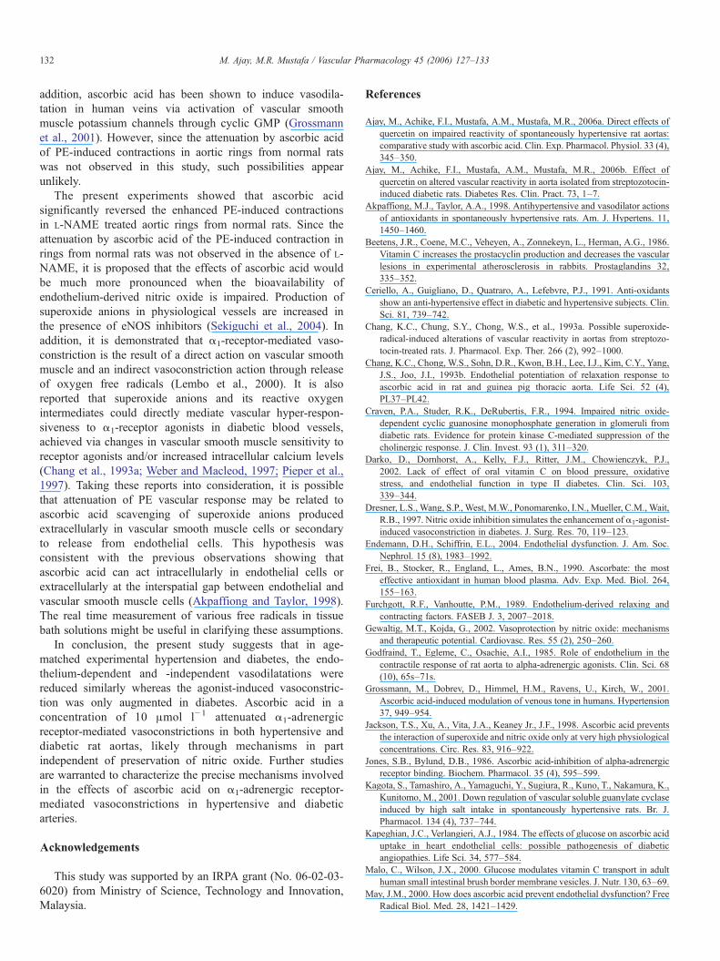
Fig. 1. Endothelium-dependent relaxations induced by acetylcholine (ACh) in control- or ascorbic acid (AA, 10 μM)-pretreated aortic rings isolated from (A) normaland (B) spontaneously hypertensive (SHR) or streptozotocin (STZ)-induced diabetic rats. Symbols represent mean±S.E.M. (n=8 or 9). ⁎p<0.05 versus correspondingnormal aortic rings; #p<0.05 versus corresponding control aortic rings.
129M. Ajay, M.R. Mustafa / Vascular Pharmacology 45 (2006) 127–133
Relaxation responses to cumulative concentrations of ACh andSNP were calculated as percentage inhibition of PE-inducedpeak contraction. Concentration-dependent contractileresponses to PE were recorded as percentage of the maximalcontraction obtained with high K+. All results are given asmean±standard error of mean (S.E.M.). The concentration–response curves for each experimental condition was plotted(Prism version 2.0, Graph Pad software, USA) and from it wasdeduced pEC50 (negative logarithm of median effectiveconcentration) and the maximum agonist-induced relaxation(Rmax) or contraction (Cmax) response values. The observeddifferences were analyzed for statistical significance usingunpaired Student's t-test and one factor ANOVA (Prism version2.0, Graph Pad software, USA). In all the cases, a value ofp<0.05 was considered statistically significant.
3. Results
3.1. ACh- and SNP-induced relaxations
ACh (Fig. 1) and SNP (Fig. 2) concentration-dependentlyrelaxed PE pre-contracted aortic rings from various groups ofrats studied. The relaxant effect of ACh at maximal concentra-tion (10 μM) tested was significantly reduced in aortic ringsfrom SHR and diabetic rats compared with aortic rings fromnormal rats (Table 1). Pretreatment with ascorbic acid did notmodify the vasodilator responses to ACh in aortic rings from
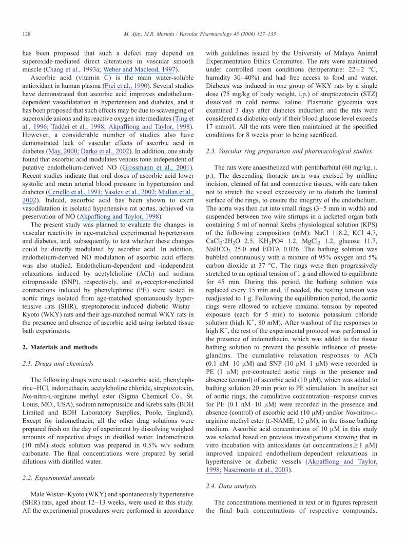
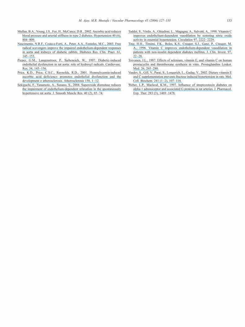
Fig. 2. Endothelium-independent relaxations induced by sodium nitroprusside (SNP)normal and (B) spontaneously hypertensive (SHR) or streptozotocin (STZ)-inducecorresponding normal aortic rings.
normal rats. Ascorbic acid normalized the vasodilator effect ofthe highest concentration of ACh in SHR aortic rings, but onlypartially enhanced in diabetic rat aortic rings. Besides,endothelium-independent vasodilatation to the highest concen-tration of SNP (1 μM) was significantly reduced in aortic ringsfrom both SHR and diabetic rats compared with that attained innormal rat aortic rings (Table 1). Ascorbic acid had no effect onthe vasodilator responses to SNP in aortic rings from eithergroup of rats studied.
3.2. PE-induced contractions
Cumulative addition of PE elicited a concentration-depen-dent contraction in all aortic ring types studied (Fig. 3). Thevasoconstriction of PE at the highest concentration tested(10 μM) was significantly augmented in diabetic rat aortic rings,but remains similar in SHR aortic rings, compared with aorticrings taken from normal rats (Table 2). Ascorbic acid had noeffect on vasoconstrictor responses of PE in aortic rings ofnormal rats. However, ascorbic acid markedly decreased thevasoconstriction of aortic rings of SHR (−26%) and diabetic(−32%) rats to the highest concentration of PE.
To ascertain the involvement of endothelium-derived NO inthe effects of ascorbic acid, we examined the effects ofascorbic acid on PE-induced vasoconstriction in aortic ringsincubated with L-NAME, a nitric oxide synthase (NOS)inhibitor (Fig. 4). Presence of L-NAME significantly increased
in control- or ascorbic acid (AA, 10 μM)-pretreated aortic rings isolated from (A)d diabetic rats. Symbols represent mean±S.E.M. (n=8 or 9). ⁎p<0.05 versus
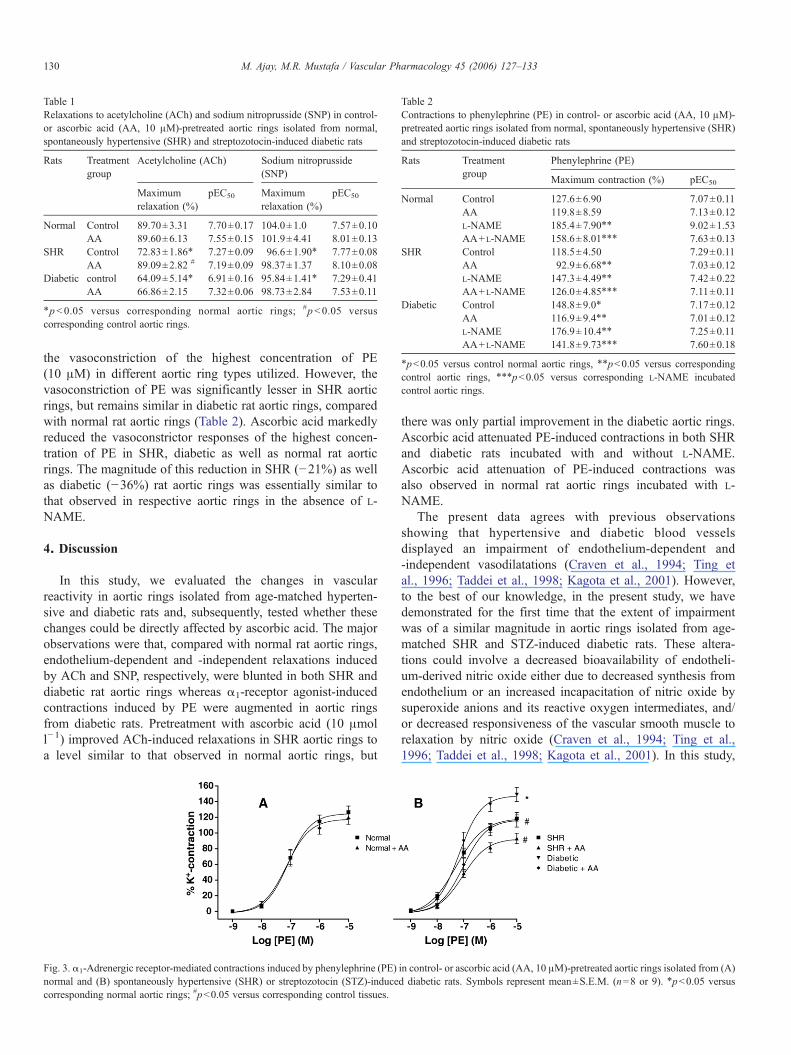
Table 1Relaxations to acetylcholine (ACh) and sodium nitroprusside (SNP) in control-or ascorbic acid (AA, 10 μM)-pretreated aortic rings isolated from normal,spontaneously hypertensive (SHR) and streptozotocin-induced diabetic rats
Rats Treatmentgroup
Acetylcholine (ACh) Sodium nitroprusside(SNP)
Maximumrelaxation (%)
pEC50 Maximumrelaxation (%)
pEC50
Normal Control 89.70±3.31 7.70±0.17 104.0±1.0 7.57±0.10AA 89.60±6.13 7.55±0.15 101.9±4.41 8.01±0.13
SHR Control 72.83±1.86⁎ 7.27±0.09 96.6±1.90⁎ 7.77±0.08AA 89.09±2.82 # 7.19±0.09 98.37±1.37 8.10±0.08
Diabetic control 64.09±5.14⁎ 6.91±0.16 95.84±1.41⁎ 7.29±0.41AA 66.86±2.15 7.32±0.06 98.73±2.84 7.53±0.11
⁎p<0.05 versus corresponding normal aortic rings; #p<0.05 versuscorresponding control aortic rings.
Table 2Contractions to phenylephrine (PE) in control- or ascorbic acid (AA, 10 μM)-pretreated aortic rings isolated from normal, spontaneously hypertensive (SHR)and streptozotocin-induced diabetic rats
Rats Treatmentgroup
Phenylephrine (PE)
Maximum contraction (%) pEC50
Normal Control 127.6±6.90 7.07±0.11AA 119.8±8.59 7.13±0.12L-NAME 185.4±7.90⁎⁎ 9.02±1.53AA+L-NAME 158.6±8.01⁎⁎⁎ 7.63±0.13
SHR Control 118.5±4.50 7.29±0.11AA 92.9±6.68⁎⁎ 7.03±0.12L-NAME 147.3±4.49⁎⁎ 7.42±0.22AA+L-NAME 126.0±4.85⁎⁎⁎ 7.11±0.11
Diabetic Control 148.8±9.0⁎ 7.17±0.12AA 116.9±9.4⁎⁎ 7.01±0.12L-NAME 176.9±10.4⁎⁎ 7.25±0.11AA+L-NAME 141.8±9.73⁎⁎⁎ 7.60±0.18
⁎p<0.05 versus control normal aortic rings, ⁎⁎p<0.05 versus correspondingcontrol aortic rings, ⁎⁎⁎p<0.05 versus corresponding L-NAME incubatedcontrol aortic rings.
130 M. Ajay, M.R. Mustafa / Vascular Pharmacology 45 (2006) 127–133
the vasoconstriction of the highest concentration of PE(10 μM) in different aortic ring types utilized. However, thevasoconstriction of PE was significantly lesser in SHR aorticrings, but remains similar in diabetic rat aortic rings, comparedwith normal rat aortic rings (Table 2). Ascorbic acid markedlyreduced the vasoconstrictor responses of the highest concen-tration of PE in SHR, diabetic as well as normal rat aorticrings. The magnitude of this reduction in SHR (−21%) as wellas diabetic (−36%) rat aortic rings was essentially similar tothat observed in respective aortic rings in the absence of L-NAME.
4. Discussion
In this study, we evaluated the changes in vascularreactivity in aortic rings isolated from age-matched hyperten-sive and diabetic rats and, subsequently, tested whether thesechanges could be directly affected by ascorbic acid. The majorobservations were that, compared with normal rat aortic rings,endothelium-dependent and -independent relaxations inducedby ACh and SNP, respectively, were blunted in both SHR anddiabetic rat aortic rings whereas α1-receptor agonist-inducedcontractions induced by PE were augmented in aortic ringsfrom diabetic rats. Pretreatment with ascorbic acid (10 μmoll−1) improved ACh-induced relaxations in SHR aortic rings toa level similar to that observed in normal aortic rings, but
Fig. 3. α1-Adrenergic receptor-mediated contractions induced by phenylephrine (PE)normal and (B) spontaneously hypertensive (SHR) or streptozotocin (STZ)-inducecorresponding normal aortic rings; #p<0.05 versus corresponding control tissues.
there was only partial improvement in the diabetic aortic rings.Ascorbic acid attenuated PE-induced contractions in both SHRand diabetic rats incubated with and without L-NAME.Ascorbic acid attenuation of PE-induced contractions wasalso observed in normal rat aortic rings incubated with L-NAME.
The present data agrees with previous observationsshowing that hypertensive and diabetic blood vesselsdisplayed an impairment of endothelium-dependent and-independent vasodilatations (Craven et al., 1994; Ting etal., 1996; Taddei et al., 1998; Kagota et al., 2001). However,to the best of our knowledge, in the present study, we havedemonstrated for the first time that the extent of impairmentwas of a similar magnitude in aortic rings isolated from age-matched SHR and STZ-induced diabetic rats. These altera-tions could involve a decreased bioavailability of endotheli-um-derived nitric oxide either due to decreased synthesis fromendothelium or an increased incapacitation of nitric oxide bysuperoxide anions and its reactive oxygen intermediates, and/or decreased responsiveness of the vascular smooth muscle torelaxation by nitric oxide (Craven et al., 1994; Ting et al.,1996; Taddei et al., 1998; Kagota et al., 2001). In this study,
in control- or ascorbic acid (AA, 10 μM)-pretreated aortic rings isolated from (A)d diabetic rats. Symbols represent mean±S.E.M. (n=8 or 9). ⁎p<0.05 versus
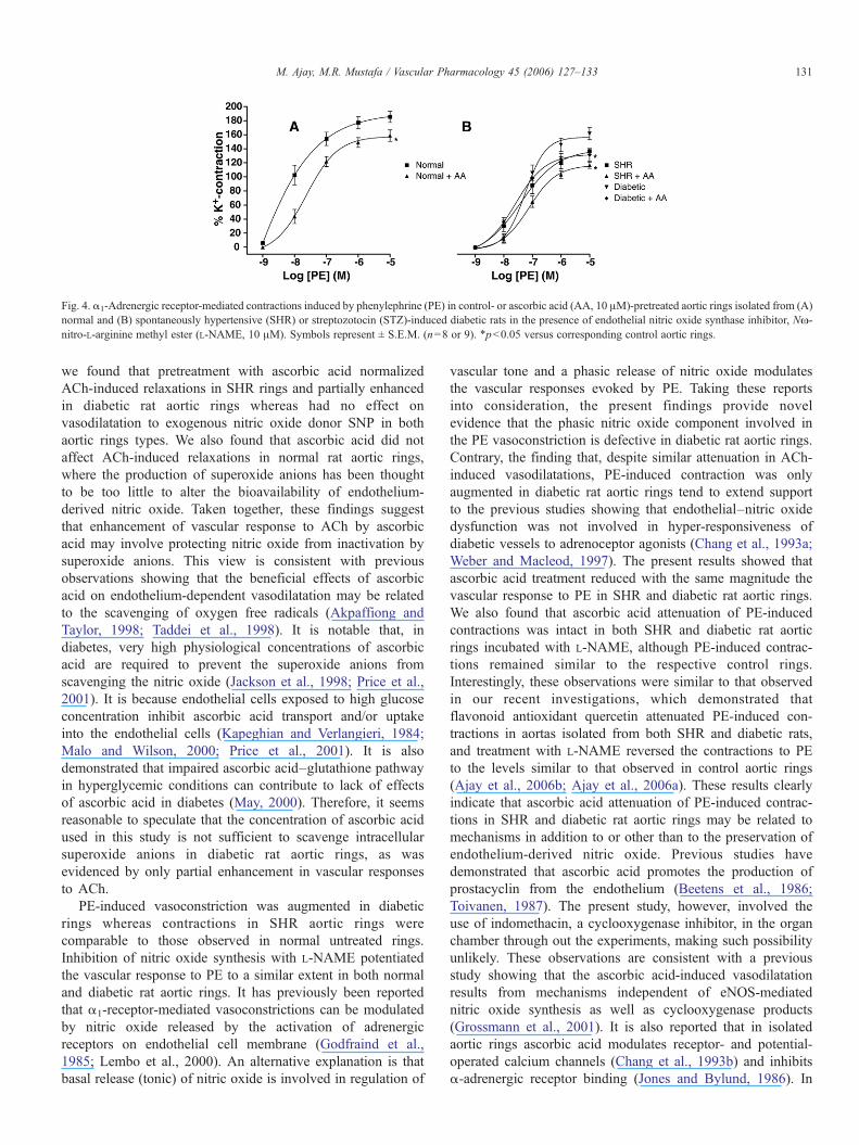
Fig. 4. α1-Adrenergic receptor-mediated contractions induced by phenylephrine (PE) in control- or ascorbic acid (AA, 10 μM)-pretreated aortic rings isolated from (A)normal and (B) spontaneously hypertensive (SHR) or streptozotocin (STZ)-induced diabetic rats in the presence of endothelial nitric oxide synthase inhibitor, Nω-nitro-L-arginine methyl ester (L-NAME, 10 μM). Symbols represent ± S.E.M. (n=8 or 9). ⁎p<0.05 versus corresponding control aortic rings.
131M. Ajay, M.R. Mustafa / Vascular Pharmacology 45 (2006) 127–133
we found that pretreatment with ascorbic acid normalizedACh-induced relaxations in SHR rings and partially enhancedin diabetic rat aortic rings whereas had no effect onvasodilatation to exogenous nitric oxide donor SNP in bothaortic rings types. We also found that ascorbic acid did notaffect ACh-induced relaxations in normal rat aortic rings,where the production of superoxide anions has been thoughtto be too little to alter the bioavailability of endothelium-derived nitric oxide. Taken together, these findings suggestthat enhancement of vascular response to ACh by ascorbicacid may involve protecting nitric oxide from inactivation bysuperoxide anions. This view is consistent with previousobservations showing that the beneficial effects of ascorbicacid on endothelium-dependent vasodilatation may be relatedto the scavenging of oxygen free radicals (Akpaffiong andTaylor, 1998; Taddei et al., 1998). It is notable that, indiabetes, very high physiological concentrations of ascorbicacid are required to prevent the superoxide anions fromscavenging the nitric oxide (Jackson et al., 1998; Price et al.,2001). It is because endothelial cells exposed to high glucoseconcentration inhibit ascorbic acid transport and/or uptakeinto the endothelial cells (Kapeghian and Verlangieri, 1984;Malo and Wilson, 2000; Price et al., 2001). It is alsodemonstrated that impaired ascorbic acid–glutathione pathwayin hyperglycemic conditions can contribute to lack of effectsof ascorbic acid in diabetes (May, 2000). Therefore, it seemsreasonable to speculate that the concentration of ascorbic acidused in this study is not sufficient to scavenge intracellularsuperoxide anions in diabetic rat aortic rings, as wasevidenced by only partial enhancement in vascular responsesto ACh.
PE-induced vasoconstriction was augmented in diabeticrings whereas contractions in SHR aortic rings werecomparable to those observed in normal untreated rings.Inhibition of nitric oxide synthesis with L-NAME potentiatedthe vascular response to PE to a similar extent in both normaland diabetic rat aortic rings. It has previously been reportedthat α1-receptor-mediated vasoconstrictions can be modulatedby nitric oxide released by the activation of adrenergicreceptors on endothelial cell membrane (Godfraind et al.,1985; Lembo et al., 2000). An alternative explanation is thatbasal release (tonic) of nitric oxide is involved in regulation of
vascular tone and a phasic release of nitric oxide modulatesthe vascular responses evoked by PE. Taking these reportsinto consideration, the present findings provide novelevidence that the phasic nitric oxide component involved inthe PE vasoconstriction is defective in diabetic rat aortic rings.Contrary, the finding that, despite similar attenuation in ACh-induced vasodilatations, PE-induced contraction was onlyaugmented in diabetic rat aortic rings tend to extend supportto the previous studies showing that endothelial–nitric oxidedysfunction was not involved in hyper-responsiveness ofdiabetic vessels to adrenoceptor agonists (Chang et al., 1993a;Weber and Macleod, 1997). The present results showed thatascorbic acid treatment reduced with the same magnitude thevascular response to PE in SHR and diabetic rat aortic rings.We also found that ascorbic acid attenuation of PE-inducedcontractions was intact in both SHR and diabetic rat aorticrings incubated with L-NAME, although PE-induced contrac-tions remained similar to the respective control rings.Interestingly, these observations were similar to that observedin our recent investigations, which demonstrated thatflavonoid antioxidant quercetin attenuated PE-induced con-tractions in aortas isolated from both SHR and diabetic rats,and treatment with L-NAME reversed the contractions to PEto the levels similar to that observed in control aortic rings(Ajay et al., 2006b; Ajay et al., 2006a). These results clearlyindicate that ascorbic acid attenuation of PE-induced contrac-tions in SHR and diabetic rat aortic rings may be related tomechanisms in addition to or other than to the preservation ofendothelium-derived nitric oxide. Previous studies havedemonstrated that ascorbic acid promotes the production ofprostacyclin from the endothelium (Beetens et al., 1986;Toivanen, 1987). The present study, however, involved theuse of indomethacin, a cyclooxygenase inhibitor, in the organchamber through out the experiments, making such possibilityunlikely. These observations are consistent with a previousstudy showing that the ascorbic acid-induced vasodilatationresults from mechanisms independent of eNOS-mediatednitric oxide synthesis as well as cyclooxygenase products(Grossmann et al., 2001). It is also reported that in isolatedaortic rings ascorbic acid modulates receptor- and potential-operated calcium channels (Chang et al., 1993b) and inhibitsα-adrenergic receptor binding (Jones and Bylund, 1986). In
132 M. Ajay, M.R. Mustafa / Vascular Pharmacology 45 (2006) 127–133
addition, ascorbic acid has been shown to induce vasodila-tation in human veins via activation of vascular smoothmuscle potassium channels through cyclic GMP (Grossmannet al., 2001). However, since the attenuation by ascorbic acidof PE-induced contractions in aortic rings from normal ratswas not observed in this study, such possibilities appearunlikely.
The present experiments showed that ascorbic acidsignificantly reversed the enhanced PE-induced contractionsin L-NAME treated aortic rings from normal rats. Since theattenuation by ascorbic acid of the PE-induced contraction inrings from normal rats was not observed in the absence of L-NAME, it is proposed that the effects of ascorbic acid wouldbe much more pronounced when the bioavailability ofendothelium-derived nitric oxide is impaired. Production ofsuperoxide anions in physiological vessels are increased inthe presence of eNOS inhibitors (Sekiguchi et al., 2004). Inaddition, it is demonstrated that α1-receptor-mediated vaso-constriction is the result of a direct action on vascular smoothmuscle and an indirect vasoconstriction action through releaseof oxygen free radicals (Lembo et al., 2000). It is alsoreported that superoxide anions and its reactive oxygenintermediates could directly mediate vascular hyper-respon-siveness to α1-receptor agonists in diabetic blood vessels,achieved via changes in vascular smooth muscle sensitivity toreceptor agonists and/or increased intracellular calcium levels(Chang et al., 1993a; Weber and Macleod, 1997; Pieper et al.,1997). Taking these reports into consideration, it is possiblethat attenuation of PE vascular response may be related toascorbic acid scavenging of superoxide anions producedextracellularly in vascular smooth muscle cells or secondaryto release from endothelial cells. This hypothesis wasconsistent with the previous observations showing thatascorbic acid can act intracellularly in endothelial cells orextracellularly at the interspatial gap between endothelial andvascular smooth muscle cells (Akpaffiong and Taylor, 1998).The real time measurement of various free radicals in tissuebath solutions might be useful in clarifying these assumptions.
In conclusion, the present study suggests that in age-matched experimental hypertension and diabetes, the endo-thelium-dependent and -independent vasodilatations werereduced similarly whereas the agonist-induced vasoconstric-tion was only augmented in diabetes. Ascorbic acid in aconcentration of 10 μmol l− 1 attenuated α1-adrenergicreceptor-mediated vasoconstrictions in both hypertensive anddiabetic rat aortas, likely through mechanisms in partindependent of preservation of nitric oxide. Further studiesare warranted to characterize the precise mechanisms involvedin the effects of ascorbic acid on α1-adrenergic receptor-mediated vasoconstrictions in hypertensive and diabeticarteries.
Acknowledgements
This study was supported by an IRPA grant (No. 06-02-03-6020) from Ministry of Science, Technology and Innovation,Malaysia.
References
Ajay, M., Achike, F.I., Mustafa, A.M., Mustafa, M.R., 2006a. Direct effects ofquercetin on impaired reactivity of spontaneously hypertensive rat aortas:comparative study with ascorbic acid. Clin. Exp. Pharmacol. Physiol. 33 (4),345–350.
Ajay, M., Achike, F.I., Mustafa, A.M., Mustafa, M.R., 2006b. Effect ofquercetin on altered vascular reactivity in aorta isolated from streptozotocin-induced diabetic rats. Diabetes Res. Clin. Pract. 73, 1–7.
Akpaffiong, M.J., Taylor, A.A., 1998. Antihypertensive and vasodilator actionsof antioxidants in spontaneously hypertensive rats. Am. J. Hypertens. 11,1450–1460.
Beetens, J.R., Coene, M.C., Veheyen, A., Zonnekeyn, L., Herman, A.G., 1986.Vitamin C increases the prostacyclin production and decreases the vascularlesions in experimental atherosclerosis in rabbits. Prostaglandins 32,335–352.
Ceriello, A., Guigliano, D., Quatraro, A., Lefebvre, P.J., 1991. Anti-oxidantsshow an anti-hypertensive effect in diabetic and hypertensive subjects. Clin.Sci. 81, 739–742.
Chang, K.C., Chung, S.Y., Chong, W.S., et al., 1993a. Possible superoxide-radical-induced alterations of vascular reactivity in aortas from streptozo-tocin-treated rats. J. Pharmacol. Exp. Ther. 266 (2), 992–1000.
Chang, K.C., Chong, W.S., Sohn, D.R., Kwon, B.H., Lee, I.J., Kim, C.Y., Yang,J.S., Joo, J.I., 1993b. Endothelial potentiation of relaxation response toascorbic acid in rat and guinea pig thoracic aorta. Life Sci. 52 (4),PL37–PL42.
Craven, P.A., Studer, R.K., DeRubertis, F.R., 1994. Impaired nitric oxide-dependent cyclic guanosine monophosphate generation in glomeruli fromdiabetic rats. Evidence for protein kinase C-mediated suppression of thecholinergic response. J. Clin. Invest. 93 (1), 311–320.
Darko, D., Dornhorst, A., Kelly, F.J., Ritter, J.M., Chowienczyk, P.J.,2002. Lack of effect of oral vitamin C on blood pressure, oxidativestress, and endothelial function in type II diabetes. Clin. Sci. 103,339–344.
Dresner, L.S., Wang, S.P., West, M.W., Ponomarenko, I.N., Mueller, C.M., Wait,R.B., 1997. Nitric oxide inhibition simulates the enhancement of α1-agonist-induced vasoconstriction in diabetes. J. Surg. Res. 70, 119–123.
Endemann, D.H., Schiffrin, E.L., 2004. Endothelial dysfunction. J. Am. Soc.Nephrol. 15 (8), 1983–1992.
Frei, B., Stocker, R., England, L., Ames, B.N., 1990. Ascorbate: the mosteffective antioxidant in human blood plasma. Adv. Exp. Med. Biol. 264,155–163.
Furchgott, R.F., Vanhoutte, P.M., 1989. Endothelium-derived relaxing andcontracting factors. FASEB J. 3, 2007–2018.
Gewaltig, M.T., Kojda, G., 2002. Vasoprotection by nitric oxide: mechanismsand therapeutic potential. Cardiovasc. Res. 55 (2), 250–260.
Godfraind, T., Egleme, C., Osachie, A.I., 1985. Role of endothelium in thecontractile response of rat aorta to alpha-adrenergic agonists. Clin. Sci. 68(10), 65s–71s.
Grossmann, M., Dobrev, D., Himmel, H.M., Ravens, U., Kirch, W., 2001.Ascorbic acid-induced modulation of venous tone in humans. Hypertension37, 949–954.
Jackson, T.S., Xu, A., Vita, J.A., Keaney Jr., J.F., 1998. Ascorbic acid preventsthe interaction of superoxide and nitric oxide only at very high physiologicalconcentrations. Circ. Res. 83, 916–922.
Jones, S.B., Bylund, D.B., 1986. Ascorbic acid-inhibition of alpha-adrenergicreceptor binding. Biochem. Pharmacol. 35 (4), 595–599.
Kagota, S., Tamashiro, A., Yamaguchi, Y., Sugiura, R., Kuno, T., Nakamura, K.,Kunitomo, M., 2001. Down regulation of vascular soluble guanylate cyclaseinduced by high salt intake in spontaneously hypertensive rats. Br. J.Pharmacol. 134 (4), 737–744.
Kapeghian, J.C., Verlangieri, A.J., 1984. The effects of glucose on ascorbic aciduptake in heart endothelial cells: possible pathogenesis of diabeticangiopathies. Life Sci. 34, 577–584.
Malo, C., Wilson, J.X., 2000. Glucose modulates vitamin C transport in adulthuman small intestinal brush border membrane vesicles. J. Nutr. 130, 63–69.
May, J.M., 2000. How does ascorbic acid prevent endothelial dysfunction? FreeRadical Biol. Med. 28, 1421–1429.
133M. Ajay, M.R. Mustafa / Vascular Pharmacology 45 (2006) 127–133
Mullan, B.A., Young, I.S., Fee, H., McCance, D.R., 2002. Ascorbic acid reducesblood pressure and arterial stiffness in type 2 diabetes. Hypertension 40 (6),804–809.
Nascimento, N.R.F., Costa-e-Forti, A., Peter, A.A., Fonteles, M.C., 2003. Freeradical scavengers improve the impaired endothelium-dependent responsesin aorta and kidneys of diabetic rabbits. Diabetes Res. Clin. Pract. 61,145–153.
Pieper, G.M., Langenstroer, P., Siebeneich, W., 1997. Diabetic-inducedendothelial dysfunction in rat aorta: role of hydroxyl radicals. Cardiovasc.Res. 34, 145–156.
Price, K.D., Price, C.S.C., Reynolds, R.D., 2001. Hyperglycemia-inducedascorbic acid deficiency promotes endothelial dysfunction and thedevelopment o atherosclerosis. Atherosclerosis 158, 1–12.
Sekiguchi, F., Yanamoto, A., Sunano, S., 2004. Superoxide dismutase reducesthe impairment of endothelium-dependent relaxation in the spontaneouslyhypertensive rat aorta. J. Smooth Muscle Res. 40 (2), 65–74.
Taddei, S., Virdis, A., Ghiadoni, L., Magagna, A., Salvetti, A., 1998. Vitamin Cimproves endothelium-dependent vasodilation by restoring nitric oxideactivity in essential hypertension. Circulation 97, 2222–2229.
Ting, H.H., Timimi, F.K., Boles, K.S., Creager, S.J., Ganz, P., Creager, M.A., 1996. Vitamin C improves endothelium-dependent vasodilation inpatients with non-insulin dependent diabetes mellitus. J. Clin. Invest. 97,22–28.
Toivanen, J.L., 1987. Effects of selenium, vitamin E, and vitamin C on humanprostacyclin and thromboxane synthesis in vitro. Prostaglandins Leukot.Med. 26, 265–280.
Vasdev, S., Gill, V., Parai, S., Longerich, L., Gadag, V., 2002. Dietary vitamin Eand C supplementation prevents fructose induced hypertension in rats. Mol.Cell. Biochem. 241 (1–2), 107–114.
Weber, L.P., Macleod, K.M., 1997. Influence of streptozotocin diabetes onalpha-1 adrenoceptor and associated G proteins in rat arteries. J. Pharmacol.Exp. Ther. 283 (3), 1469–1478.