Effects of 24-h and 36-h sleep deprivation on human postural control and adaptation
9
Exp Brain Res (2008) 185:165–173 DOI 10.1007/s00221-007-1143-5 123 RESEARCH ARTICLE EVects of 24-h and 36-h sleep deprivation on human postural control and adaptation M. Patel · S. Gomez · S. Berg · P. Almbladh · J. Lindblad · H. Petersen · M. Magnusson · R. Johansson · P. A. Fransson Received: 1 May 2007 / Accepted: 13 September 2007 / Published online: 12 October 2007 © Springer-Verlag 2007 Abstract This study investigated whether human postural stability and adaptation were aVected by sleep deprivation and the relationship between motor performance and sub- jective scores of sleepiness (visuo-anlogue sleepiness scores, VAS). Postural stability and subjective sleepiness were examined in 18 healthy subjects (mean age 23.8 years) following 24 and 36 h of continued wakefulness, ensured by portable EEG recordings, and compared to a control test where the assessments were made after a normal night of sleep. The responses were assessed using posturography with eyes open and closed, and vibratory proprioceptive stimulations were used to challenge postural control. Pos- tural control was signiWcantly aVected after 24 h of sleep deprivation both in anteroposterior and in lateral directions, but less so after 36 h. Subjective VAS scores showed poor correlation with indicators of postural control performance. The clearest evidence that sleep deprivation decreased pos- tural control was the reduction of adaptation. Also several near falls after 2–3 min during the posturographic tests showed that sleep deprivation might aVect stability through momentary lapses of attention. Access to vision, somewhat, but not entirely reduced the eVect of sleep deprivation. In conclusion, sleep deprivation can be a contributing factor to decreased postural control and falls. Keywords Postural control · Sleep deprivation · Adaptation · Subjective scores · Attention Introduction Chronic sleep restrictions are an endemic in modern society, with large fractions of the population reporting daily sleep below the recommended 8 h per night (National_Sleep_Foundation 2005). This problem is associ- ated with long working hours, commuting, and family responsibilities especially in occupations such as health- care, the military and industrial manufacturing where the potential for sleep-related accidents is also high (Balkin et al. 2004). The frequency of this problem was highlighted in a survey by the National Sleep Foundation in 2005, showing that 60% of drivers in the United States admit to driving while feeling drowsy (National_Sleep_Foundation 2005). Horne and Reyner estimated that 20% of serious motorway collisions in the United Kingdom were due to sleepiness on the basis of surveys from police oYcers and police collision reports (Horne and Reyner 1995). Sleep deprivation may produce eVects normally associated with drunkenness such as a lack of coordination, judgment and reaction time (Williamson and Feyer 2000; Wilson et al. 2006). The similarities between tiredness and drunkenness have been evidenced in driving simulation studies. Wil- liamson and Feyer (2000) found that people who drove after being awake for 17–19 h performed worse than those with a blood alcohol level of 0.05%, which is the legal limit for driving in most western European countries. Although M. Patel · S. Berg · P. Almbladh · J. Lindblad · M. Magnusson · R. Johansson · P. A. Fransson (&) Department of Otorhinolaryngology Head and Neck surgery, Clinical Sciences, Lund, Lund University, 221-85 Lund, Sweden e-mail: [email protected] S. Gomez Faculty of Applied Sciences, University of the West of England, Bristol BS16 1QY, Great Britain H. Petersen Department of Otorhinolaryngology Head and Neck surgery, University of Iceland, Landspitali University Hospital, Reykjavik, Iceland
-
Upload
independent -
Category
Documents
-
view
0 -
download
0
Transcript of Effects of 24-h and 36-h sleep deprivation on human postural control and adaptation

Exp Brain Res (2008) 185:165–173
DOI 10.1007/s00221-007-1143-5RESEARCH ARTICLE
EVects of 24-h and 36-h sleep deprivation on human postural control and adaptation
M. Patel · S. Gomez · S. Berg · P. Almbladh · J. Lindblad · H. Petersen · M. Magnusson · R. Johansson · P. A. Fransson
Received: 1 May 2007 / Accepted: 13 September 2007 / Published online: 12 October 2007© Springer-Verlag 2007
Abstract This study investigated whether human posturalstability and adaptation were aVected by sleep deprivationand the relationship between motor performance and sub-jective scores of sleepiness (visuo-anlogue sleepinessscores, VAS). Postural stability and subjective sleepinesswere examined in 18 healthy subjects (mean age 23.8 years)following 24 and 36 h of continued wakefulness, ensuredby portable EEG recordings, and compared to a control testwhere the assessments were made after a normal night ofsleep. The responses were assessed using posturographywith eyes open and closed, and vibratory proprioceptivestimulations were used to challenge postural control. Pos-tural control was signiWcantly aVected after 24 h of sleepdeprivation both in anteroposterior and in lateral directions,but less so after 36 h. Subjective VAS scores showed poorcorrelation with indicators of postural control performance.The clearest evidence that sleep deprivation decreased pos-tural control was the reduction of adaptation. Also severalnear falls after 2–3 min during the posturographic testsshowed that sleep deprivation might aVect stability throughmomentary lapses of attention. Access to vision, somewhat,
but not entirely reduced the eVect of sleep deprivation. Inconclusion, sleep deprivation can be a contributing factor todecreased postural control and falls.
Keywords Postural control · Sleep deprivation · Adaptation · Subjective scores · Attention
Introduction
Chronic sleep restrictions are an endemic in modernsociety, with large fractions of the population reportingdaily sleep below the recommended 8 h per night(National_Sleep_Foundation 2005). This problem is associ-ated with long working hours, commuting, and familyresponsibilities especially in occupations such as health-care, the military and industrial manufacturing where thepotential for sleep-related accidents is also high (Balkinet al. 2004). The frequency of this problem was highlightedin a survey by the National Sleep Foundation in 2005,showing that 60% of drivers in the United States admit todriving while feeling drowsy (National_Sleep_Foundation2005). Horne and Reyner estimated that 20% of seriousmotorway collisions in the United Kingdom were due tosleepiness on the basis of surveys from police oYcers andpolice collision reports (Horne and Reyner 1995). Sleepdeprivation may produce eVects normally associated withdrunkenness such as a lack of coordination, judgment andreaction time (Williamson and Feyer 2000; Wilson et al.2006). The similarities between tiredness and drunkennesshave been evidenced in driving simulation studies. Wil-liamson and Feyer (2000) found that people who droveafter being awake for 17–19 h performed worse than thosewith a blood alcohol level of 0.05%, which is the legal limitfor driving in most western European countries. Although
M. Patel · S. Berg · P. Almbladh · J. Lindblad · M. Magnusson · R. Johansson · P. A. Fransson (&)Department of Otorhinolaryngology Head and Neck surgery, Clinical Sciences, Lund, Lund University, 221-85 Lund, Swedene-mail: [email protected]
S. GomezFaculty of Applied Sciences, University of the West of England, Bristol BS16 1QY, Great Britain
H. PetersenDepartment of Otorhinolaryngology Head and Neck surgery, University of Iceland, Landspitali University Hospital, Reykjavik, Iceland
123
166 Exp Brain Res (2008) 185:165–173
sleep-related accidents are well documented in tasks requir-ing appropriate cognition and decision making (e.g. driv-ing), tiredness may also be dangerous in daily life activitiesand could increase the likelihood of falling accidents.
Despite the prevalence of sleep deprivation, few studieshave evaluated the postural control performance eVects andsleep-loss-mediated deWcits in performance capacitycaused by sleepiness (Schlesinger et al. 1998; Gribble andHertel 2004; Avni et al. 2006). Although levels of sleepi-ness and fatigue can be subjectively assessed, such evalua-tions may not reXect the objective physiological status ofthe tired person, mainly because subjective scores can bebiased by motivation, personal factors, experience, training,etc. (Avni et al. 2006). Therefore, there is a need to Wndpractical non-invasive objective methods to measure theeVects of sleepiness, especially when reaching critical lev-els involving higher risks for accidents. One possibility isto assess postural stability (Avni et al. 2006; Karita et al.2006).
The eVects of balance perturbations on postural controlcan be studied experimentally by inducing inappropriatesensory information. There are numerous ways of achiev-ing this and one such method involves disturbing somato-sensory aVerents by means of vibrating the calf muscles(Ivanenko et al. 1999; Fransson et al. 2002; Vuillerme et al.2002; Ledin et al. 2004). This action results in an increasedactivation of muscle spindle aVerents (Eklund 1973) signal-ling to the central nervous system that the vibrated muscleis being stretched (Matthews 1986). The increased activityinduced from muscle spindles results in a proprioceptiveillusion of movement. Shortly following muscle vibration,tonic stretch reXexes are elicited that result in increasedbody movements (Goodwin et al. 1972). Repeated musclevibrations over a short period of time result in an increasedability to handle the induced perturbations through posturaladaptation (Fransson et al. 2000; Tjernstrom et al. 2005).
Motor control (Haslam 1984; Frey et al. 2004) and otherfunctions such as cognitive ability, attention and alertness,Wrst scientiWcally investigated by Patrick and Gilbert(1896), have been found to be aVected by sleep deprivation.In support of these Wndings, analysis of brain activity hasshown that the areas of the cerebral cortex that regulateaspects of attention, alertness and cognitive ability, such asthalamus and regions within the pre-frontal cortex, undergodeactivation following 24 h of sleep deprivation (Thomaset al. 2000) and decrease activity further as the period ofsleep deprivation increases (Thomas et al. 2003). Previ-ously, human postural stability has been shown to decreasefollowing 24 h of continued wakefulness (Liu et al. 2001;Gribble and Hertel 2004; Avni et al. 2006; Fabbri et al.2006) or with the addition of an attention-demanding task(Schlesinger et al. 1998). To our knowledge, no previousresearch has been carried out in which postural stability and
postural adaptation have been assessed following both 24(24SDep) and 36 h of sleep deprivation (36SDep) usingvibratory proprioceptive stimulation, and in which theseposturographic assessments have been compared againstsubjective visuo-analogue sleepiness scale (VAS) scoreswhich has been an accepted way of monitoring subjectsleepiness.
The Wrst aim of this study was to investigate whetherpostural stability and postural adaptation, recorded as anter-oposterior and lateral torque variance, diVered between thecontrol tests, 24SDep and 36Sdep, while subjected to pro-prioceptive vibration with eyes open (EO) and eyes closed(EC). The second aim was to determine whether posturalcontrol performance correlated with VAS scores. The Wnalaim was to investigate whether postural stability and adap-tation in anteroposterior and lateral torque variance diVeredwith eyes open and closed between the control tests24SDep and 36SDep.
Methods and materials
Subjects
Eighteen (10 males and 8 females) healthy volunteers, agedbetween 16 and 38 years (mean 23.8 years, SD 4.8 years;mean weight 78.5 kg, SD 18.8 kg and mean height 1.77 m,SD 0.09 m) with no previous history of central nervous dis-eases, performed postural stability tests involving sleepdeprivation. Subjects were instructed not to consume anyalcohol, sleepiness-inducing or revitalizing products, suchas caVeine, 48 h before and during testing. At the time ofexperimentation, no subject was taking any form of medi-cation and signed consent was obtained before any testingbegan. The experiments were all performed in accordanceto the Helsinki declaration of 1975 and approved by thelocal ethical committee.
Equipment
A custom built force platform recorded the forces withsix degrees of freedom and with an accuracy of 0.5 N.A customized computer program controlled the vibratorystimulation and sampled the force platform data at 50 Hz.
The vibrators, had a vibration amplitude of 1.0 mm and afrequency of 85 Hz, were 6 cm long and 1 cm in diameter.The vibrators were placed over the gastrocnemius musclesand secured by elastic straps.
Procedure
Sleep deprivation testing was performed on 2 consecutivedays, days 1 and 2. On day 1, subjects were asked to wake
123
Exp Brain Res (2008) 185:165–173 167
up at 7 am or 8 am (depending on the recording schedule)to begin their sleep deprivation session. The subjects wereinstructed to stay awake, without using stimulants, and goabout their daily routines as normal. Subjects came to thelaboratory at 7 pm or 8 pm on day 1 (12 h into their sleepdeprived state) to be attached with a portable EEG recodingdevice (Embletta™). The EEG recordings were made toensure that the subjects had not fallen asleep during thesleep deprivation period. Signs of sleep were monitoredwith a unilateral electrode measurement set-up using rou-tine electrodes. The EEG device comprised three elec-trodes; an active electrode positioned on the upper temple;a reference electrode positioned on the upper mastoid boneon the opposite side to the active electrode; and a groundelectrode positioned on the mid-forehead. Subjects returnedon day 2 at 7 am or 8 am, 24 h into sleep deprivation, thenagain that evening at 7 or 8 pm, 36 h into sleep deprivationfor posturographic assessment. The EEG equipment wasremoved prior to testing to avoid any possible interferencefrom tactile information from EEG electrodes, the record-ing device and EEG cables. Additionally, it was appraisedthat correct EEG recordings could not be assured during thetests due to the electrical noise produced by the posturo-graphic equipment. The EEG data were stored for oV-lineanalysis before re-attachment. Scoring of wakefulness/sleep was done according to RechtschaVen and Kales(1968). Uninterrupted sleep stage II for more than 2 minwas considered to be sleep.
Subjects were also instructed to provide a subjectivescore using visuo-analogue sleepiness scale (VAS) of alert-ness ranging from “completely alert” to “exhausted, to nearsleep”. Subjects analogue scores were converted into num-bers ranging from 1 to 10, where 1 = “completely alert” and10 = “exhausted to near sleep”.
A control posturographic test (i.e. following a normalnight of sleep) was performed either 1 week before or1 week after the sleep deprivation tests. The test order wasdecided by randomization.
Posturographic assessment
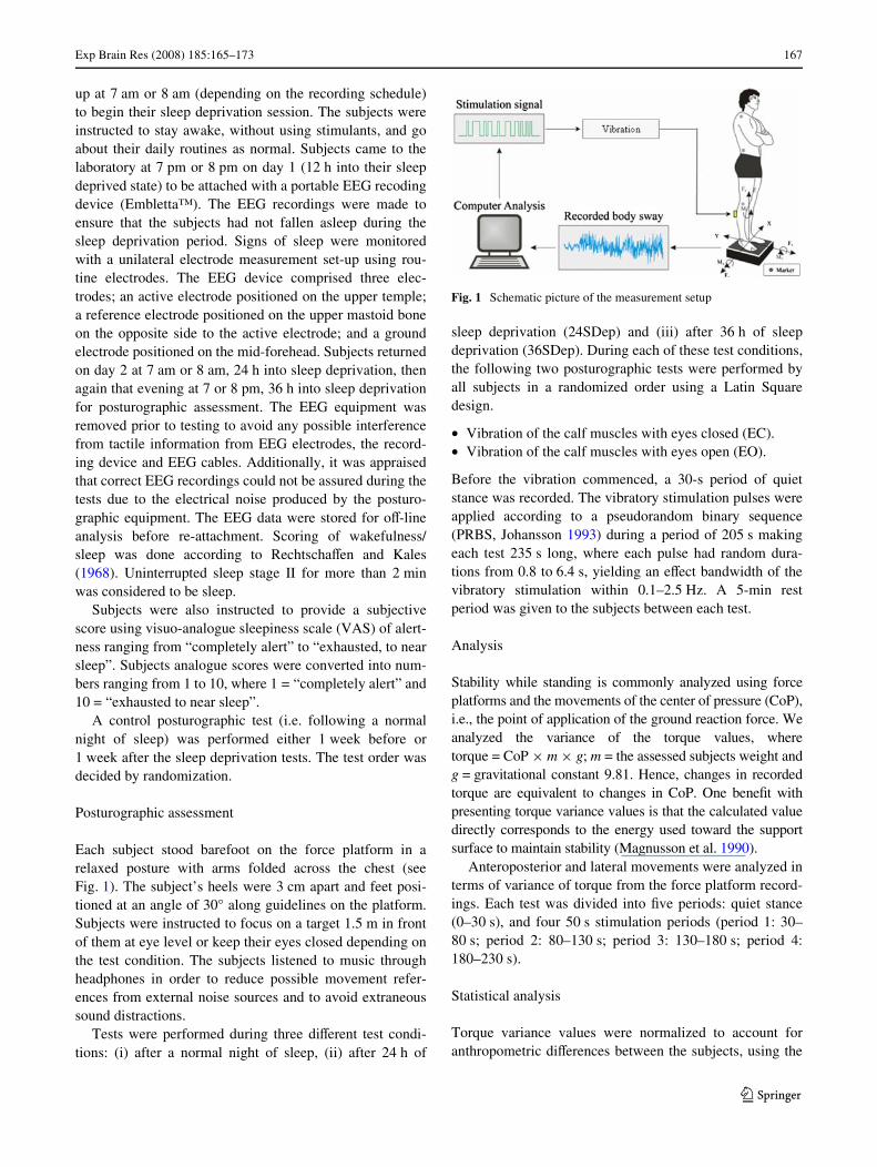
Each subject stood barefoot on the force platform in arelaxed posture with arms folded across the chest (seeFig. 1). The subject’s heels were 3 cm apart and feet posi-tioned at an angle of 30° along guidelines on the platform.Subjects were instructed to focus on a target 1.5 m in frontof them at eye level or keep their eyes closed depending onthe test condition. The subjects listened to music throughheadphones in order to reduce possible movement refer-ences from external noise sources and to avoid extraneoussound distractions.
Tests were performed during three diVerent test condi-tions: (i) after a normal night of sleep, (ii) after 24 h of
sleep deprivation (24SDep) and (iii) after 36 h of sleepdeprivation (36SDep). During each of these test conditions,the following two posturographic tests were performed byall subjects in a randomized order using a Latin Squaredesign.
• Vibration of the calf muscles with eyes closed (EC).• Vibration of the calf muscles with eyes open (EO).
Before the vibration commenced, a 30-s period of quietstance was recorded. The vibratory stimulation pulses wereapplied according to a pseudorandom binary sequence(PRBS, Johansson 1993) during a period of 205 s makingeach test 235 s long, where each pulse had random dura-tions from 0.8 to 6.4 s, yielding an eVect bandwidth of thevibratory stimulation within 0.1–2.5 Hz. A 5-min restperiod was given to the subjects between each test.
Analysis
Stability while standing is commonly analyzed using forceplatforms and the movements of the center of pressure (CoP),i.e., the point of application of the ground reaction force. Weanalyzed the variance of the torque values, wheretorque = CoP £ m £ g; m = the assessed subjects weight andg = gravitational constant 9.81. Hence, changes in recordedtorque are equivalent to changes in CoP. One beneWt withpresenting torque variance values is that the calculated valuedirectly corresponds to the energy used toward the supportsurface to maintain stability (Magnusson et al. 1990).
Anteroposterior and lateral movements were analyzed interms of variance of torque from the force platform record-ings. Each test was divided into Wve periods: quiet stance(0–30 s), and four 50 s stimulation periods (period 1: 30–80 s; period 2: 80–130 s; period 3: 130–180 s; period 4:180–230 s).
Statistical analysis
Torque variance values were normalized to account foranthropometric diVerences between the subjects, using the
Fig. 1 Schematic picture of the measurement setup
123

168 Exp Brain Res (2008) 185:165–173
subject’s squared height and squared weight (Johanssonet al. 1988). The squared nature of the variance algorithmmade it necessary to use normalization with squared param-eters to achieve unit agreement with the standardization.
The Wilcoxon matched-pairs signed-rank test (exactsigned two-tailed, Altman 1991) was used for the statisticalcomparison between test conditions and for the analysis ofvariations over time. The torque variance changes betweenquiet stance and period 1 were evaluated to determine howthe assessed parameters were initially aVected by the bal-ance perturbations evoked by vibratory proprioceptivestimulation compared to the activity during quiet stance.The torque variance changes between periods 1 and 4 wereevaluated to determine how the assessed parameters wereaVected by repeated vibratory stimulation, quantifying pos-sible eVects of adaptation to vibratory proprioceptive stim-ulation. The eVects of sleep deprivation, vision and thecombined eVect of vision and sleep deprivation were ana-lyzed using a GLM univariate ANOVA (general linearmodel univariate analysis of variance) test on log-trans-formed values (Altman 1991). The accuracy of the GLMmodel was evaluated by testing the model residual for nor-mal distribution. Correlation analysis was performedbetween subjective VAS scores and torque variance usingSpearman’s correlation test (Altman 1991).
Normality of distribution was tested with the Shapiro–Wilk test. Non-parametric statistics were used since somevalues were not normally distributed. The statistical anal-ysis was carried out with Bonferroni correction for multi-ple comparisons where no more than two matched paircomparisons were performed for each test. In the analysis,P values < 0.01 were considered to be statistically signiW-cant, except in the GLM analysis, where P values < 0.05were considered to be signiWcant (Altman 1991). How-ever, we present P values < 0.05 in the Wgures for consis-tency. The statistical analysis was performed using SPSS
version 14.0 (SPSS) and Matlab version 7.2 (The Math-Works).
Results
The subjective VAS score increased from a level of 5.2 at24SDep to a level of 6.8 at 36SDep (P < 0.001), where theVAS ranges were deWned as 1 = “completely alert” and10 = “exhausted to near sleep”.
Anteroposterior torque variance
Adaptation of anteroposterior torque variance
Anteroposterior torque variances showed similar trendsduring quiet stance and during the vibration periods, bothwith eyes open (EO) and eyes closed (EC), although themagnitude of the variance values was markedly larger withEC (Fig. 2). In all tests, during the Wrst period of vibratorystimulation (period 1) the torque variances were signiW-cantly larger compared to the quiet stance torque variance(P < 0.001). The increase in torque variance in percentagewith the 10 and 90% conWdence intervals are presentedwithin the square brackets, between quiet stance and period1 were for the diVerent tests: 1,175% [320, 2,030%] (con-trol-EC); 965% [370, 1,560%] (24SDep-EC); 611% [370,850%] (36SDep-EC); 469% [290, 640%] (Control-EO);713% [330, 1,100%] (24SDep-EO); and 341% [240, 440%](36SDep-EO).
In the control tests, after the initial rise in torque vari-ance at period 1, torque variance decreased and stayed lowbetween periods 2 and 4. The extent of adaptation to thevibratory stimulation are presented as percentage decreasein torque variance between periods 1 and 4 with the 10 and90% conWdence intervals presented within the square
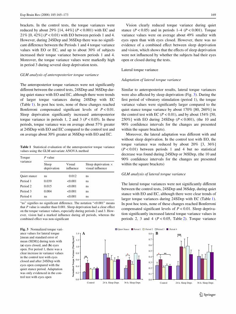
Fig. 2 Normalized torque variance values for anteroposterior torque[mean and standard error of mean (SEM)] during tests with (a) eyesclosed, and (b) eyes open. The variance values have been normalizedwith the subject’s weight and height. Note the diVerences in scale ofthe y-axis of the two graphs. The statistical diVerences found between
quiet stance and period 1, and between periods 1 and 4 are marked withasterisk, where *P < 0.05, **P < 0.01 and ***P < 0.001. For period 1,there was a clear increase in variance values in all tests compared withthe quiet stance period. Adaptation was only evidenced in the controltests
******
***
****
⋅])
m2
************
0.0
1.0
2.0
3.0
4.0
5.0
6.0
7.0
8.0
9.0
Control 24 h. Sleep Depr. 36 h. Sleep Depr.
******
********
gK(/
mN [
ecnai raV
e uqroT
dezil amro
N⋅
*****
***
***
0.0
0.5
1.0
1.5
2.0
2.5
3.0
3.5
4.0
**********
******
******
QuietStance Period 1 Period 2 Period 3 Period 4
Control 24 h. Sleep Depr. 36 h. Sleep Depr.
A B
123
Exp Brain Res (2008) 185:165–173 169
brackets. In the control tests, the torque variances werereduced by about 29% [14, 44%] (P < 0.001) with EC and21% [0, 42%] (P < 0.01) with EO between periods 1 and 4.However, during 24SDep and 36SDep there was no signiW-cant diVerence between the Periods 1 and 4 torque variancevalues with EO or EC, and up to about 30% of subjectsincreased their torque variance between periods 1 and 4.Moreover, the torque variance values were markedly highin period 3 during several sleep deprivation tests.
GLM analysis of anteroposterior torque variance
The anteroposterior torque variances were not signiWcantlydiVerent between the control tests, 24SDep and 36SDep dur-ing quiet stance with EO and EC, although there were trendsof larger torque variances during 24SDep with EC(Table 1). In post hoc tests, none of these changes reachedBonferroni compensated signiWcant levels of P < 0.01.Sleep deprivation signiWcantly increased anteroposteriortorque variance in periods 1, 2 and 3 (P < 0.05). In theseperiods, torque variance was on average about 57% greaterat 24SDep with EO and EC compared to the control test andon average about 30% greater at 36SDep with EO and EC.
Vision clearly reduced torque variance during quietstance (P < 0.05) and in periods 1–4 (P < 0.001). Torquevariance values were on average about 49% smaller witheyes open than with eyes closed. However, there was noevidence of a combined eVect between sleep deprivationand vision, which shows that the eVects of sleep deprivationwere not inXuenced by whether the subjects had their eyesopen or closed during the tests.
Lateral torque variance
Adaptation of lateral torque variance
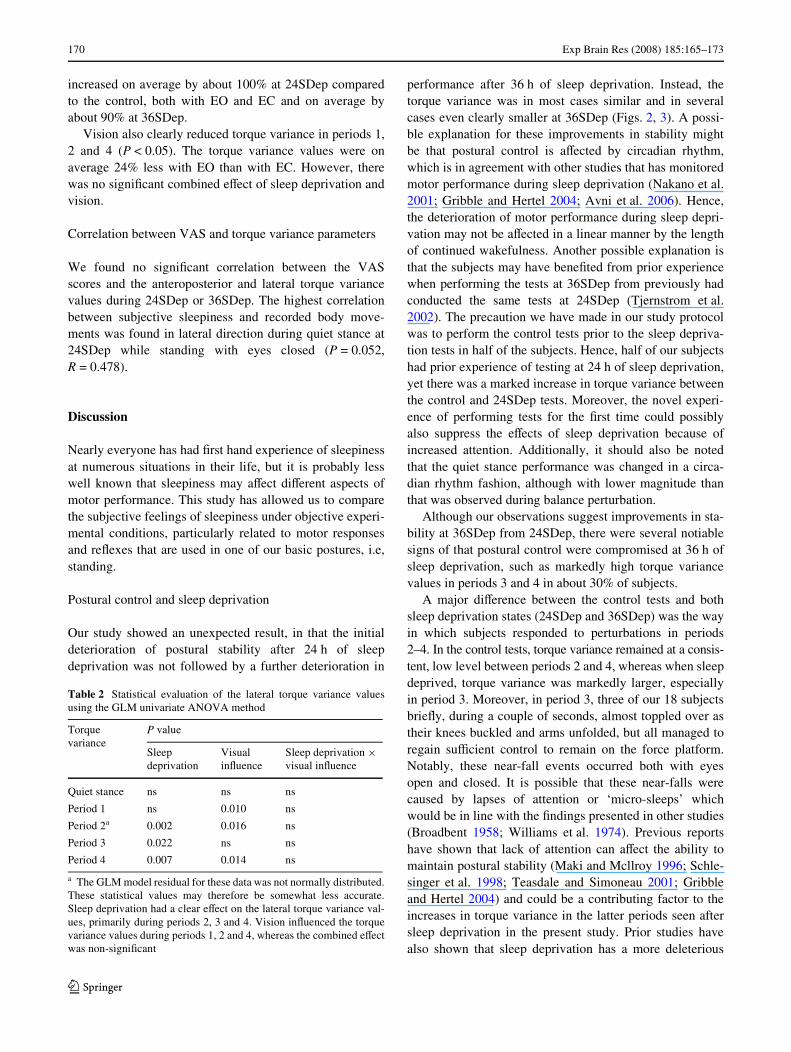
Similar to anteroposterior results, lateral torque varianceswere also aVected by sleep deprivation (Fig. 3). During theWrst period of vibratory stimulation (period 1), the torquevariance values were signiWcantly larger compared to thequiet stance torque variance by about 170% [80, 260%] inthe control test with EC (P < 0.01), and by about 154% [50,250%] with EO during 24SDep (P < 0.001), (the 10 and90% conWdence intervals for the changes are presentedwithin the square brackets).
Moreover, the lateral adaptation was diVerent with andwithout sleep deprivation. In the control test with EO, thetorque variance was reduced by about 20% [3, 36%](P < 0.01) between periods 1 and 4 but no statisticaldecrease was found during 24SDep or 36SDep, (the 10 and90% conWdence intervals for the changes are presentedwithin the square brackets)
GLM analysis of lateral torque variance
The lateral torque variances were not signiWcantly diVerentbetween the control tests, 24SDep and 36Sdep, during quietstance with EO and EC, although there were clear trends oflarger torque variances during 24SDep with EC (Table 1).In post hoc tests, none of these changes reached Bonferronicompensated signiWcant levels of P < 0.01. Sleep depriva-tion signiWcantly increased lateral torque variance values inperiods 2, 3 and 4 (P < 0.05, Table 2). Torque variance
Table 1 Statistical evaluation of the anteroposterior torque variancevalues using the GLM univariate ANOVA method
“ns” signiWes no signiWcant diVerence. The notation “<0.001” meansthat P value is smaller than 0.001. Sleep deprivation had a clear eVecton the torque variance values, especially during periods 2 and 3. How-ever, vision had a marked inXuence during all periods, whereas thecombined eVect was non-signiWcant
Torque variance
P value
Sleep deprivation
Visual inXuence
Sleep deprivation £visual inXuence
Quiet stance ns 0.012 ns
Period 1 0.039 <0.001 ns
Period 2 0.015 <0.001 ns
Period 3 0.004 <0.001 ns
Period 4 ns <0.001 ns
Fig. 3 Normalized torque vari-ance values for lateral torque [mean and standard error of mean (SEM)] during tests with (a) eyes closed, and (b) eyes open. For period 1, there was a clear increase in variance values in the control test with eyes closed and after 24SDep with eyes open compared with the quiet stance period. Adaptation was only evidenced in the con-trol test with eyes open
***
⋅])
m2
0.0
0.2
0.4
0.6
0.8
1.0
1.2
1.4
******
Control 24 h. Sleep Depr. 36 h. Sleep Depr.
gK(/
mN[
ecnairaV
euqroT
dezilamro
N⋅
***
***
0.0
0.1
0.2
0.3
0.4
0.5
0.6
0.7
0.8
******
******
Quiet Stance Period 1 Period 2 Period 3 Period 4
Control 24 h. Sleep Depr. 36 h. Sleep Depr.
A B
123
170 Exp Brain Res (2008) 185:165–173
increased on average by about 100% at 24SDep comparedto the control, both with EO and EC and on average byabout 90% at 36SDep.
Vision also clearly reduced torque variance in periods 1,2 and 4 (P < 0.05). The torque variance values were onaverage 24% less with EO than with EC. However, therewas no signiWcant combined eVect of sleep deprivation andvision.
Correlation between VAS and torque variance parameters
We found no signiWcant correlation between the VASscores and the anteroposterior and lateral torque variancevalues during 24SDep or 36SDep. The highest correlationbetween subjective sleepiness and recorded body move-ments was found in lateral direction during quiet stance at24SDep while standing with eyes closed (P = 0.052,R = 0.478).
Discussion
Nearly everyone has had Wrst hand experience of sleepinessat numerous situations in their life, but it is probably lesswell known that sleepiness may aVect diVerent aspects ofmotor performance. This study has allowed us to comparethe subjective feelings of sleepiness under objective experi-mental conditions, particularly related to motor responsesand reXexes that are used in one of our basic postures, i.e,standing.
Postural control and sleep deprivation
Our study showed an unexpected result, in that the initialdeterioration of postural stability after 24 h of sleepdeprivation was not followed by a further deterioration in
performance after 36 h of sleep deprivation. Instead, thetorque variance was in most cases similar and in severalcases even clearly smaller at 36SDep (Figs. 2, 3). A possi-ble explanation for these improvements in stability mightbe that postural control is aVected by circadian rhythm,which is in agreement with other studies that has monitoredmotor performance during sleep deprivation (Nakano et al.2001; Gribble and Hertel 2004; Avni et al. 2006). Hence,the deterioration of motor performance during sleep depri-vation may not be aVected in a linear manner by the lengthof continued wakefulness. Another possible explanation isthat the subjects may have beneWted from prior experiencewhen performing the tests at 36SDep from previously hadconducted the same tests at 24SDep (Tjernstrom et al.2002). The precaution we have made in our study protocolwas to perform the control tests prior to the sleep depriva-tion tests in half of the subjects. Hence, half of our subjectshad prior experience of testing at 24 h of sleep deprivation,yet there was a marked increase in torque variance betweenthe control and 24SDep tests. Moreover, the novel experi-ence of performing tests for the Wrst time could possiblyalso suppress the eVects of sleep deprivation because ofincreased attention. Additionally, it should also be notedthat the quiet stance performance was changed in a circa-dian rhythm fashion, although with lower magnitude thanthat was observed during balance perturbation.
Although our observations suggest improvements in sta-bility at 36SDep from 24SDep, there were several notiablesigns of that postural control were compromised at 36 h ofsleep deprivation, such as markedly high torque variancevalues in periods 3 and 4 in about 30% of subjects.
A major diVerence between the control tests and bothsleep deprivation states (24SDep and 36SDep) was the wayin which subjects responded to perturbations in periods2–4. In the control tests, torque variance remained at a consis-tent, low level between periods 2 and 4, whereas when sleepdeprived, torque variance was markedly larger, especiallyin period 3. Moreover, in period 3, three of our 18 subjectsbrieXy, during a couple of seconds, almost toppled over astheir knees buckled and arms unfolded, but all managed toregain suYcient control to remain on the force platform.Notably, these near-fall events occurred both with eyesopen and closed. It is possible that these near-falls werecaused by lapses of attention or ‘micro-sleeps’ whichwould be in line with the Wndings presented in other studies(Broadbent 1958; Williams et al. 1974). Previous reportshave shown that lack of attention can aVect the ability tomaintain postural stability (Maki and Mcllroy 1996; Schle-singer et al. 1998; Teasdale and Simoneau 2001; Gribbleand Hertel 2004) and could be a contributing factor to theincreases in torque variance in the latter periods seen aftersleep deprivation in the present study. Prior studies havealso shown that sleep deprivation has a more deleterious
Table 2 Statistical evaluation of the lateral torque variance valuesusing the GLM univariate ANOVA method
a The GLM model residual for these data was not normally distributed.These statistical values may therefore be somewhat less accurate.Sleep deprivation had a clear eVect on the lateral torque variance val-ues, primarily during periods 2, 3 and 4. Vision inXuenced the torquevariance values during periods 1, 2 and 4, whereas the combined eVectwas non-signiWcant
Torque variance
P value
Sleep deprivation
Visual inXuence
Sleep deprivation £visual inXuence
Quiet stance ns ns ns
Period 1 ns 0.010 ns
Period 2a 0.002 0.016 ns
Period 3 0.022 ns ns
Period 4 0.007 0.014 ns
123
Exp Brain Res (2008) 185:165–173 171
eVect on performance in long, monotonous, boring tasksthan on short, interesting ones (Wilkinson 1965; Dingesand Kribbs 1991; Horne 2000). Therefore, our long-dura-tion tests may have provided an appropriate, objectiveapproach for determining the eVects of sleep deprivation. Inaddition, vibration per se may temporarily increase alert-ness in both humans and animals (Magnusson 1986). Thismay explain why the eVect of sleep deprivation on vibra-tory perturbations was less pronounced in period 1.
Although it was previously known that sleep deprivationhas an eVect on postural control, the precise nature of thechange is disputed. Some authors report that postural stabil-ity is aVected in unperturbed standing (Nakano et al. 2001;Gribble and Hertel 2004; Avni et al. 2006; Fabbri et al.2006; Karita et al. 2006). Likewise, we found a smallincrease in quiet stance torque variance in the anteroposte-rior and lateral directions following sleep deprivation.However, we only found a marked decrease in postural sta-bility due to sleep deprivation when we perturbed stanceusing calf vibration. In contrast, Uimonen et al. (1994) alsousing posturography with vibratory proprioceptive stimula-tion to the calf, found no eVects of sleep deprivation onpostural control. However, Uimonen et al. assessed the pos-tural stability during much shorter duration (55 s) and onlyexposed the subjects to Wve vibratory perturbations com-pared with 235 s of recordings and 64 vibratory perturba-tions in each of our tests.
It should be noted that total sleep deprivation, as investi-gated in our study, and chronic sleep restrictions may nothave identical eVects on motor control and postural stability(Haslam 1984). However, decreased postural stability hasalso been reported among subjects with chronic-restrictedsleep hours (Karita et al. 2006). Therefore, it is reasonableto assume that our Wndings may also apply for patients withsevere sleep disorders, such as obstructive sleep apnea syn-drome (OSAS), which is characterized by a stoppage ordecrease in breathing, resulting in inadequate sleep.
Adaptation and sleep deprivation
The ability to adapt and habituate on the basis of priorexperiences is important for human movement control, fallprevention (Eccles 1986; Fransson et al. 2003) and for theability to enhance the performance during various humanactivities (Horak and Nashner 1986; Keshner et al. 1987).In our study, the changes in adaptation presented the clear-est evidence that sleep deprivation signiWcantly aVects pos-tural control. We found that anteroposterior and lateraladaptation clearly diVered under sleep deprivation com-pared to the control tests. The investigated subjects wereclearly able to adapt to the balance perturbations evoked bythe vibratory stimulation in the control tests, but could notdo so signiWcantly when sleep deprived. Some subjects
even increased their torque variance between periods 1 and4. These Wndings showed that sleep deprivation may aVectsome subject’s ability to learn and respond appropriately tobalance perturbations. Integration of information from thevisual, vestibular and somatosensory receptors and motorcoordination are processes known to require attention(Schlesinger et al. 1998; Fabbri et al. 2006), especiallywhen information from any of the sensory systems is notreliable (Redfern et al. 2001). One possible reason for thelack of adaptation during sleep deprivation might be thatthe accompanying decrease in attention led to slower, orinappropriate sensory integration, which also aVected theadaptation processes, i.e., the ability to choose an appropri-ate motor response to enhance balance stability.
Subjective sleepiness and postural control
In this study, we found no correlation between subjectivesleepiness and motor performance. Subjective sleepiness,as assessed by VAS scores, increased markedly from24SDep to 36SDep, although in some instances torque var-iance decreased. Thus, assessments of subjective sleepinessmay not be a reliable indicator of actual postural control(Fabbri et al. 2006). The Wndings presented therefore high-light the value of implementing appropriate regulations forwork durations, particularly in attention-demanding occu-pations, such as long distance driving, because the subjec-tive feeling of sleepiness may not reXect the actualperformance decline. One possible reason for why we wereunable to Wnd a signiWcant correlation between torque vari-ance and subjective VAS scores might be that the mere actof performing a test might momentarily enhance attentionand motivation (Avni et al. 2006). Another explanationcould be that in motor tests that require a high level ofattention such as aerobic tasks, the association between pro-gressive sleep deprivation, subjective sleepiness anddecreased performance might be stronger than that wasdetected in our study (Souissi et al. 2003).
Vision and sleep deprivation
Several of our Wndings support evidence from Edwardsshowing that vision can provide information to assist pos-tural control (Edwards 1946) as sleep deprivation had lesseVect on postural stability with eyes open than with eyesclosed both in anteroposterior and lateral direction. More-over, the statistical analyses showed no combined eVect ofsleep deprivation and vision on postural control, which sug-gests that these two factors possibly act independently.Similar results were found by Karita et al. (2006), in aninvestigation of postural control in subjects with chronicsleep deprivation. Still, the near falls and decreased adapta-tion occurred both in tests with eyes open and eyes closed.
123
172 Exp Brain Res (2008) 185:165–173
Under normal conditions, vision provides an orientationalframe of reference, where any imbalance can be quicklycaptured and an appropriate response can be initiated tomaintain precise postural control. However, the Wndingspresented in this study suggest that although visual infor-mation provides enhanced stability, this additional informa-tion may not at times be suYcient to compensate for thedeterioration of performance caused by sleep deprivation.
References
Altman D (1991) Practical statistics for medical research. Chapman &Hall, New York
Avni N, Avni I, Barenboim E, Azaria B, Zadok D, Kohen-Raz R, Mo-rad Y (2006) Brief posturographic test as an indicator of fatigue.Psychiatry Clin Neurosci 60:340–346
Balkin TJ, Bliese PD, Belenky G, Sing H, Thorne DR, Thomas M,Redmond DP, Russo M, Wesensten NJ (2004) Comparative util-ity of instruments for monitoring sleepiness-related performancedecrements in the operational environment. J Sleep Res 13:219–227
Broadbent DE (1958) Perception and communication. PergamonPress, London
Dinges DF, Kribbs BN (1991) Performing while sleepy: eVects ofexperimentally-induced sleepiness. Wiley, Chichester
Eccles JC (1986) Learning in the motor system. In: Prog Brain Res, vol64. Elsevier Science, Amsterdam, pp 3–18
Edwards AS (1946) Body sway and vision. J Exp Psychol 36:526–536Eklund G (1973) Further studies of vibration-induced eVects on bal-
ance. Ups J Med Sci 78:65–72Fabbri M, Martoni M, Esposito MJ, Brighetti G, Natale V (2006) Pos-
tural control after a night without sleep. Neuropsychologia44:2520–2525
Fransson PA, Johansson R, Hafstrom A, Magnusson M (2000) Meth-ods for evaluation of postural control adaptation. Gait Posture12:14–24
Fransson PA, Tjernström F, Hafström A, Magnusson M, Johansson R(2002) Analysis of short- and long-term eVects of adaptation inhuman postural control. Biol Cybern 86:355–365
Fransson PA, Hafstrom A, Karlberg M, Magnusson M, Tjader A,Johansson R (2003) Postural control adaptation during galvanicvestibular and vibratory proprioceptive stimulation. IEEE TransBiomed Eng 50:1310–1319
Frey DJ, Badia P, Wright KP Jr (2004) Inter- and intra-individual var-iability in performance near the circadian nadir during sleepdeprivation. J Sleep Res 13:305–315
Goodwin GM, McCloskey DI, Matthews PB (1972) Proprioceptiveillusions induced by muscle vibration: contribution by musclespindles to perception? Science 175:1382–1384
Gribble PA, Hertel J (2004) Changes in postural control during a 48-hrsleep deprivation period. Percept Mot Skills 99:1035–1045
Haslam DR (1984) The military performance of soldiers in sustainedoperations. Aviat Space Environ Med 55:216–221
Horak FB, Nashner LM (1986) Central programming of posturalmovements: adaptation to altered support-surface conWgurations.J Neurophysiol 55:1369–1381
Horne J (2000) Neuroscience: images of lost sleep. Nature 403:605–606Horne JA, Reyner LA (1995) Sleep related vehicle accidents. BMJ
310:565–567Ivanenko YP, Talis VL, Kazennikov OV (1999) Support stability inXu-
ences postural responses to muscle vibration in humans. Eur JNeurosci 11:647–654
Johansson R (1993) System modeling and identiWcation. Prentice-Hall, Englewood CliVs
Johansson R, Magnusson M, Akesson M (1988) IdentiWcation ofhuman postural dynamics. IEEE Trans Biomed Eng 35:858–869
Karita K, Nakao M, Nishikitani M, Iwata T, Murata K, Yano E (2006)EVect of overtime work and insuYcient sleep on postural sway ininformation-technology workers. J Occup Health 48:65–68
Keshner EA, Allum JH, Pfaltz CR (1987) Postural coactivation andadaptation in the sway stabilizing responses of normals and pa-tients with bilateral vestibular deWcit. Exp Brain Res 69:77–92
Ledin T, Fransson PA, Magnusson M (2004) EVects of postural distur-bances with fatigued triceps surae muscles or with 20% additionalbody weight. Gait Posture 19:184–193
Liu Y, Higuchi S, Motohashi Y (2001) Changes in postural sway dur-ing a period of sustained wakefulness in male adults. Occup MedLond 51:490–495
Magnusson M (1986) EVect of alertness on the vestibulo-ocular reXexand on the slow rise in optokinetic nystagmus in rabbits. Am J Ot-olaryngol 7:353–359
Magnusson M, Johansson R, Wiklund J (1990) Galvanically inducedbody sway in the anterior-posterior plane. Acta Otolaryngol110:11–17
Maki B, Mcllroy W (1996) InXuence of arousal and attention on thecontrol of postural sway. J Vest Res 6:53–59
Matthews PB (1986) What are the aVerents of origin of the humanstretch reXex, and is it a purely spinal reaction? In: Freund HJ,Büttner U, Cohen B, Noth J (eds) Prog Brain Res, vol 64. Else-vier, Amsterdam, pp 55–66
Nakano T, Araki K, Michimori A, Inbe H, Hagiwara H, Koyama E(2001) Nineteen-hour variation of postural sway, alertness andrectal temperature during sleep deprivation. Psychiatry Clin Neu-rosci 55:277–278
National_Sleep_Foundation (2005) Sleep in America poll 2005.http://www.sleepfoundation.org
Patrick GTW, Gilbert JA (1896) On the eVects of loss of sleep. PsycholRev 3:469–483
RechtschaVen A, Kales A (1968) Manual of standardized terminology,techniques and scoring system for sleep of human subjects. USDepartment of Health, Education and Welfare, NeurologicalInformation Network, Bethesda
Redfern M, Jennings J, Martin C, Furman J (2001) Attention inXuencessensory integration for postural control in older adults. Gait Pos-ture 14(3):211–216
Schlesinger A, Redfern MS, Dahl RE, Jennings JR (1998) Posturalcontrol, attention and sleep deprivation. Neuroreport 9:49–52
Souissi N, Sesboue B, Gauthier A, Larue J, Davenne D (2003) EVectsof one night’s sleep deprivation on anaerobic performance the fol-lowing day. Eur J Appl Physiol 89:359–366
Teasdale N, Simoneau M (2001) Attentional demands for postural con-trol: the eVects of aging and sensory reintegration. Gait Posture14:203–210
Thomas M, Sing H, Belenky G, Holcomb H, Mayberg H, Dannals R,Wagner H, Thorne D, Popp K, Rowland L, Welsh A, BalwinskiS, Redmond D (2000) Neural basis of alertness and cognitive per-formance impairments during sleepiness. I. EVects of 24 h ofsleep deprivation on waking human regional brain activity.J Sleep Res 9:335–352
Thomas M, Sing H, Belenky G, Holcomb H, Mayberg H, DannalsR, Wagner H, Henry N, Thorne D, Popp K (2003) Neural basisof alertness and cognitive performance impairments duringsleepiness: II. EVects of 24 and 72 h of sleep deprivation onwaking human regional brain activity. Thalamus Relat Syst2:199–229
Tjernstrom F, Fransson PA, Hafstrom A, Magnusson M (2002) Adap-tation of postural control to perturbations: a process that initiateslong-term motor memory. Gait Posture 15:75–82
123
Exp Brain Res (2008) 185:165–173 173
Tjernstrom F, Fransson PA, Magnusson M (2005) Improved posturalcontrol through repetition and consolidation. J Vestib Res 15:31–39
Uimonen S, Laitakari K, Bloigu R, Sorri M (1994) The repeatability ofposturographic measurements and the eVects of sleep deprivation.J Vestib Res 4:29–36
Wilkinson RT (1965) Sleep deprivation. Academic, LondonWilliams RL, Karacan I, Hursch CJ (1974) Electroencephalography
(EEG) of human sleep: Clinical applications. Wiley, New YorkWilliamson AM, Feyer AM (2000) Moderate sleep deprivation pro-
duces impairments in cognitive and motor performance equiva-
lent to legally prescribed levels of alcohol intoxication. OccupEnviron Med 57:649–655
Wilson RJ, Fang M, Cooper PJ, Beirness DJ (2006) Sleepiness amongnight-time drivers: relationship to blood alcohol concentrationand other factors. TraYc Inj Prev 7:15–22
Vuillerme N, Danion F, Forestier N, Nougier V (2002) Postural swayunder muscle vibration and muscle fatigue in humans. NeurosciLett 333:131–135
123