Effective Dielectric Properties of Biological Cells: Generalization of the Spectral Density Function...
8
Effective Dielectric Properties of Biological Cells: Generalization of the Spectral Density Function Approach Anatoliy V. Goncharenko* ,†,‡,§ and Yia-Chung Chang † Research Center for Applied Sciences, Academia Sinica, Nankang, Taipei 115, Taiwan, ROC; Department of Physics, National Cheng-Kung UniVersity, Tainan, Taiwan, ROC; and Institute of Semiconductor Physics, National Academy of Sciences of Ukraine, 03028, KyiV, Ukraine ReceiVed: January 23, 2009; ReVised Manuscript ReceiVed: May 20, 2009 We suggest an extension of the spectral density function approach to describe the complex dielectric response of suspensions of arbitrarily shaped particles having a thin shell, in particular, biological cells. The approach is shown to give analytical results in some simple but practically important cases. In the general case, for the 3-phase systems it reduces to determination of the spectral density function for the suspension of a certain kind. Prospects and limitations of the approach, as well as practical examples, are also considered. I. Introduction Today’s dielectric spectroscopy is a powerful tool to study mechanisms of the interaction of electromagnetic fields with biocells, enabling one to consider the influence of various physical and chemical factors on the cell dielectric properties. Actually, dielectric impedance measurements are simple, fast, and not invasive. As a result, the use of the spectroscopy in biological studies and biotechnology industries is increasing. 1 At the same time, in some cases its practical implementation involves difficulties that, in turn, requires a further elaboration of underlying models. Indeed, on the one hand, there are a number of well-developed numerical techniques, such as the finite element method, finite-difference time-domain method, multiple multipole method, rigorous coupled wave analysis, etc. However, numerical techniques are frequently time-consuming, especially due to very different length scales for the cell size and the membrane thickness. For example, although the typical size of the red blood cells is on the order of 10 µm, the membrane thickness is on the order of only 10 nm. In addition, numerical techniques are sometimes unstable and inconvenient when the need arises to investigate behavior of the dielectric response depending on model parameters. On the other hand, there are some simple classical models allowing one to obtain analytical results for the dielectric response. The widely used classical model involves consideration of two confocal el- lipsoidal layers, the cytoplasm and the membrane; an extension to multishell ellipsoid model is also possible. 2 Although such a model considerably simplifies calculations, its practical use is frequently limited due to unrealistic, oversimplified geometry. Real biocells come in a variety of shapes. So, bacteria can take much more complicated shape, such as, for example, rod-like, curved or spiral, croissant, wavy, etc. 3 In these cases the ellipsoidal shape assumption is poor. However, so far only a few (mainly numerical) studies were beyond the scope of the ellipsoidal model. Among them, mention may be made of two methods feasible for dilute suspensions of axially symmetric biocells; 4,5 a review on numerical methods as applied to dielectric mixtures is given by Tuncer et al. 6 So the further progress of the dielectric spectroscopy has motivated a growing need for accurate and efficient modeling allowing one to cor- rectly describe the dielectric properties of the cells. In this paper, we develop the spectral density function approach, pioneered by Bergman 7 and Milton, 8 for studying the biological cell suspensions. At present, the approach is widely used to describe the effective dielectric and optical properties of various inhomogeneous media, including cell suspensions. 9 Its main advantage is that it allows one to separate the material parameters (permittivities and/or conductivities) and the micro- geometrical parameters of the media. At the same time, there is a serious restriction of the method, lying in the fact that its use involves difficulties when dealing with media consisting of more than two phases. Two known exceptions are dilute suspensions of the shelled spheroids 10 and of the graded (or multilayered) spheres. 11 In other words, the spectral representa- tion in its conventional form is applicable for two-phase composite systems only. The primary purpose of this paper is to extend the spectral representation for the effective dielectric constant of two-phase media to more complicated multiphase systems such as biological cell suspensions. II. Basic Formalism Before consideration of three-phase composites, let us first consider the case of the two-phase ones. Actually, there are two forms of the spectral density formalism. Besides, there is Fuchs’ representation, which looks very similar to the above formal- ism. 12 Sometimes this fact leads to some misunderstanding. To avoid confusion, we briefly discuss these issues here. Ronald Fuchs showed that the complex dielectric susceptibil- ity of a small arbitrarily shaped particle can be written as a sum over its normal modes. Making use of this fact, he also obtained an expression for the effective dielectric constant of a dilute suspension of the well-separated particles. 12 It should be noted that Fuchs’ result was derived without taking into account interparticle interactions. The spectral representation for the effective dielectric constant was originally formulated in the form of the sum: 7 * To whom correspondence should be addressed. E-mail: anatolii@ mail.ncku.edu.tw. † Academia Sinica. ‡ National Cheng-Kung University. § National Academy of Sciences of Ukraine. J. Phys. Chem. B 2009, 113, 9924–9931 9924 10.1021/jp900703a CCC: $40.75 2009 American Chemical Society Published on Web 07/01/2009 Downloaded by NATIONAL CHENG KUNG UNIV on July 16, 2009 Published on July 1, 2009 on http://pubs.acs.org | doi: 10.1021/jp900703a
Transcript of Effective Dielectric Properties of Biological Cells: Generalization of the Spectral Density Function...
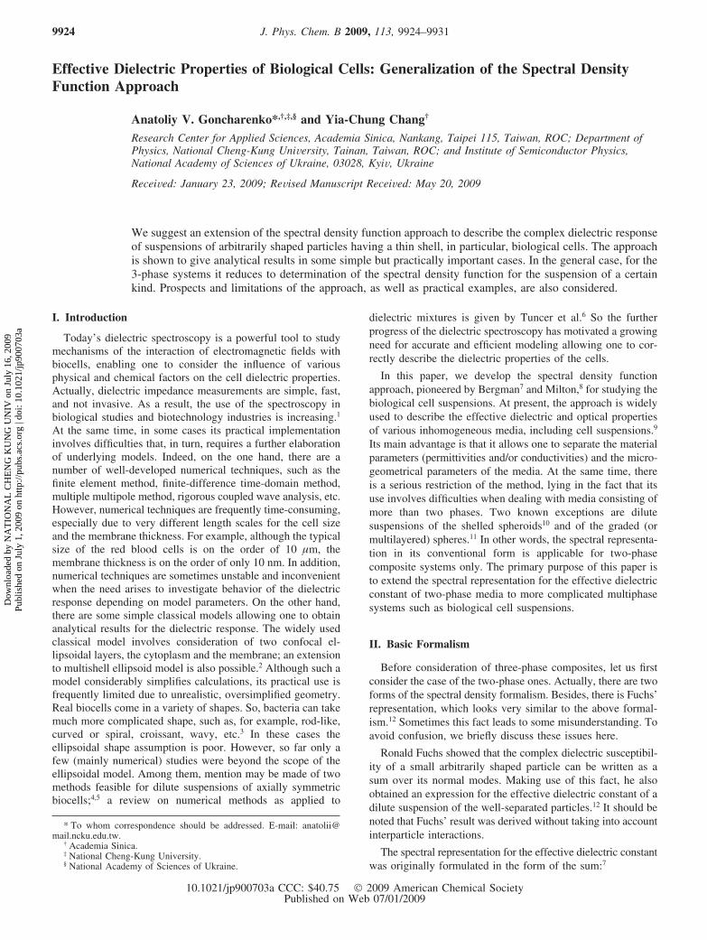
Effective Dielectric Properties of Biological Cells: Generalization of the Spectral DensityFunction Approach
Anatoliy V. Goncharenko*,†,‡,§ and Yia-Chung Chang†
Research Center for Applied Sciences, Academia Sinica, Nankang, Taipei 115, Taiwan, ROC; Department ofPhysics, National Cheng-Kung UniVersity, Tainan, Taiwan, ROC; and Institute of Semiconductor Physics,National Academy of Sciences of Ukraine, 03028, KyiV, Ukraine
ReceiVed: January 23, 2009; ReVised Manuscript ReceiVed: May 20, 2009
We suggest an extension of the spectral density function approach to describe the complex dielectric responseof suspensions of arbitrarily shaped particles having a thin shell, in particular, biological cells. The approachis shown to give analytical results in some simple but practically important cases. In the general case, for the3-phase systems it reduces to determination of the spectral density function for the suspension of a certainkind. Prospects and limitations of the approach, as well as practical examples, are also considered.
I. Introduction
Today’s dielectric spectroscopy is a powerful tool to studymechanisms of the interaction of electromagnetic fields withbiocells, enabling one to consider the influence of variousphysical and chemical factors on the cell dielectric properties.Actually, dielectric impedance measurements are simple, fast,and not invasive. As a result, the use of the spectroscopy inbiological studies and biotechnology industries is increasing.1
At the same time, in some cases its practical implementationinvolves difficulties that, in turn, requires a further elaborationof underlying models. Indeed, on the one hand, there are anumber of well-developed numerical techniques, such as thefinite element method, finite-difference time-domain method,multiple multipole method, rigorous coupled wave analysis, etc.However, numerical techniques are frequently time-consuming,especially due to very different length scales for the cell sizeand the membrane thickness. For example, although the typicalsize of the red blood cells is on the order of 10 µm, themembrane thickness is on the order of only 10 nm. In addition,numerical techniques are sometimes unstable and inconvenientwhen the need arises to investigate behavior of the dielectricresponse depending on model parameters. On the other hand,there are some simple classical models allowing one to obtainanalytical results for the dielectric response. The widely usedclassical model involves consideration of two confocal el-lipsoidal layers, the cytoplasm and the membrane; an extensionto multishell ellipsoid model is also possible.2 Although such amodel considerably simplifies calculations, its practical use isfrequently limited due to unrealistic, oversimplified geometry.Real biocells come in a variety of shapes. So, bacteria can takemuch more complicated shape, such as, for example, rod-like,curved or spiral, croissant, wavy, etc.3 In these cases theellipsoidal shape assumption is poor. However, so far only afew (mainly numerical) studies were beyond the scope of theellipsoidal model. Among them, mention may be made of twomethods feasible for dilute suspensions of axially symmetricbiocells;4,5 a review on numerical methods as applied to
dielectric mixtures is given by Tuncer et al.6 So the furtherprogress of the dielectric spectroscopy has motivated a growingneed for accurate and efficient modeling allowing one to cor-rectly describe the dielectric properties of the cells.
In this paper, we develop the spectral density functionapproach, pioneered by Bergman7 and Milton,8 for studying thebiological cell suspensions. At present, the approach is widelyused to describe the effective dielectric and optical propertiesof various inhomogeneous media, including cell suspensions.9
Its main advantage is that it allows one to separate the materialparameters (permittivities and/or conductivities) and the micro-geometrical parameters of the media. At the same time, thereis a serious restriction of the method, lying in the fact that itsuse involves difficulties when dealing with media consisting ofmore than two phases. Two known exceptions are dilutesuspensions of the shelled spheroids10 and of the graded (ormultilayered) spheres.11 In other words, the spectral representa-tion in its conventional form is applicable for two-phasecomposite systems only. The primary purpose of this paper isto extend the spectral representation for the effective dielectricconstant of two-phase media to more complicated multiphasesystems such as biological cell suspensions.
II. Basic Formalism
Before consideration of three-phase composites, let us firstconsider the case of the two-phase ones. Actually, there are twoforms of the spectral density formalism. Besides, there is Fuchs’representation, which looks very similar to the above formal-ism.12 Sometimes this fact leads to some misunderstanding. Toavoid confusion, we briefly discuss these issues here.
Ronald Fuchs showed that the complex dielectric susceptibil-ity of a small arbitrarily shaped particle can be written as asum over its normal modes. Making use of this fact, he alsoobtained an expression for the effective dielectric constant of adilute suspension of the well-separated particles.12 It should benoted that Fuchs’ result was derived without taking into accountinterparticle interactions.
The spectral representation for the effective dielectric constantwas originally formulated in the form of the sum:7
* To whom correspondence should be addressed. E-mail: [email protected].
† Academia Sinica.‡ National Cheng-Kung University.§ National Academy of Sciences of Ukraine.
J. Phys. Chem. B 2009, 113, 9924–99319924
10.1021/jp900703a CCC: $40.75 2009 American Chemical SocietyPublished on Web 07/01/2009
Dow
nloa
ded
by N
AT
ION
AL
CH
EN
G K
UN
G U
NIV
on
July
16,
200
9Pu
blis
hed
on J
uly
1, 2
009
on h
ttp://
pubs
.acs
.org
| do
i: 10
.102
1/jp
9007
03a
where f1 is the volume fraction of the phase 1, s ) ε0/(ε1 -ε0), and ε0 and ε1 are the complex relative dielectric constantsof the phases (for the sake of definiteness, in the following thephase 0 is considered to be a host or, in biological terms, anextracellular medium). Conventionally, eq 1 is supplementedwith the sum rules ∑nFn ) 1 and ∑nFnsn ) (1 - f1)/3 ) f0/3 .All the complex dielectric constants ε0, ε1 , and εeff may bedefined in terms of the corresponding relative permittivities εk′and conductivities σk as εk ) εk′ - iεk′′ ) ε′k - iσk/εVω, wherek ) 0, 1, 2, eff and εV is the free space permittivity.
Later, Golden, and Papanicolaou13 noticed that the distributionof the poles in eq 1 can be continuous. If so, the integral formof the spectral representation
with g(x) the spectral density function and p(x) ) (s + x)-1,seems to be more convenient. Equation 2 is supplemented with∫0
1g(x) dx ) 1 and ∫01xg(x) dx ) f0/3 . It should be, however,
noted that both forms of the spectral representation, eqs 1 and2, are equivalent if to assume that
where δ(...) is the Dirac delta function.Let us now take a closer look at eq 2. It is easily seen that
p(x) may be considered as a dimensionless polarizability of asingle spheroid having the dielectric constant ε1 and thedepolarization factor x along its axis of revolution. More exactly,p(x) is one of diagonal components of the dimensionlesspolarizability tensor corresponding to orientation of the incidentelectric field along the spheroid symmetry axis. It means thatformally the effective linear response of an arbitrary two-phaseisotropic composite is the same as one of the diagonalcomponents of the effective linear response tensor of a systemof equally oriented and noninteracting spheroids possessing ashape distribution.14 Thus, instead of consideration of a suspen-sion of arbitrarily shaped particles with a complicated geometry,determination of the effective dielectric constant may be reducedto consideration of a relatively simple suspension of shape-distributed spheroids.
Consider now a composite consisting of shelled (core-shell)spheroids. A natural idea suggesting itself now is to extend eq2, substituting p(x) with the dimensionless polarizability p*(x)of the shelled spheroid and f1 with the volume fraction of theshelled spheroid f ) f1 + f2:
where f1 and f2 are the volume fractions of a shell and a core,respectively; the volume fraction of a host is f0 ) 1 - f ) 1 -f2 - f1. To do this, we could introduce an equivalent dielectricconstant εe of the shelled spheroid and then
The value of εe is such that if the dielectric constant of thesurrounding ε0 is equal to it, then the field and the potential atany point in the medium is unperturbed by the introduction ofthe shelled spheroid. Both the size and shape of the equivalentspheroid are considered to be the same as those of the originalshelled spheroid. It should be noted that when using such adefinition, the internal electric field of the equivalent spheroidmay be treated as constant independently of the spheroidconcentration. This is so because the above formalism considersthe spheroids to be noninteracting.
It is clear that doing so allows us to extend the spectralrepresentation to a class of the three-phase composites. Aquestion, of course, appears what the class is. Rigorous andcomprehensive consideration of this problem does not seem tobe straightforward and is beyond the scope of the present paper(we are going to consider it in more detail elsewhere). It isreasonable to assume, however, that this class includes thecore-shell particle composites as a subset. The arguments tosupport this claim can be as follows. Earlier, it was shown thatthere is an equivalence between a shelled (inhomogeneous)particle and a homogeneous one having the same size andshape.15 In other terms, there exists a charge distribution on theexternal surface of the shell that results in the true electricalfield outside the particle. It means that we may introduce anequivalent permittivity of the shelled particle considering it tobe homogeneous. It should be also noted that the validity ofthe above assumption regarding consideration of the permittivityof coated particles in terms of the effective medium theory wassupported experimentally for a dilute suspension of slightlydeformed coated spheres.16 Thus, dealing with a suspension wemay replace the real core-shell particles by the homogeneousones. As to the suspension of the homogeneous particles, wecan now use the spectral density representation.
To proceed further, we need to know the polarizability p* ofthe shelled spheroid. Analytically, the problem can be solvedusing the separation of variables method in spheroidal coordi-nates.17 It is possible, however, only if both spheroids (innerand outer) are to be confocal. The resulting expression fordiagonal components of the polarizability tensor involves thecorresponding depolarization factors of the core (L2) and theshell (L1). In many papers it is mistakenly assumed that L2 )L1. This assumption, in general, is not correct for the confocalspheroids.18,19 Indeed, if L1 and q are specified, the depolarizationfactor L2 of the outer spheroid should depend on the volumeratio of the core to the whole spheroid q ) f2/(f1 + f2). So, L1
≈ L2 only for sphere-like particles, when L1 ≈ L2 ≈ 1/3, or ifq f 1, that is, in the limit of very thin shells. In fact, the latteris indeed the case when dealing with biocells. Due to this, weconsider this issue in more detail.
Let a shelled spheroid have the above-mentioned depolar-ization factors L1 and L2 along its axis of revolution. Then, theequivalent dielectric constant of the spheroid may be writtenas (see, e.g., ref 20, where we have revised an obvious misprintin the numerator)
with s12 ) ε1/(ε2 - ε1), ε1, and ε2 the dielectric constants of theshell and the core, respectively. Furthermore, it is clear that theabove formalism may be easily extended to the case ofmultishelled spheroids using an obvious iterative procedure.
εeff ) ε0(1 + f1 ∑n
Fn
sn - s) (1)
εeff ) ε0[1 + f1 ∫0
1g(x)p(x) dx] (2)
g(x) ) ∑n
Fnδ(x - sn) (3)
εeff ) ε0[1 + f∫0
1g(x)p*(x) dx] (4)
p*(x) ) {[εe(x)/ε0 - 1]-1 + x}-1 (5)
εe ) ε1
qL1 - L2 - q - s12
qL1 - L2 - s12) ε1(1 - q
qL1 - L2 - s12)(6)
Effective Dielectric Properties of Biological Cells J. Phys. Chem. B, Vol. 113, No. 29, 2009 9925
Dow
nloa
ded
by N
AT
ION
AL
CH
EN
G K
UN
G U
NIV
on
July
16,
200
9Pu
blis
hed
on J
uly
1, 2
009
on h
ttp://
pubs
.acs
.org
| do
i: 10
.102
1/jp
9007
03a
Then, equating the depolarization factors of all spheroids, thatis, taking L ≡ x ) L1 ) L2 )... ) LN-1, one has a set of Niterative equations for the determination of the equivalentpermittivity of the N-shelled spheroid
and so on, up to
III. Particular Examples of the Spectral Density Function
For illustration purposes, let us consider some simpleexamples setting particular forms of the spectral densityfunction.
It is easily seen that such a choice results in the well-knownexpression for the effective dielectric constant of the shelledspheres in the dilute limit:21
Such a choice yields an extension of the Maxwell-Garnettapproximation to the shelled spheres
with
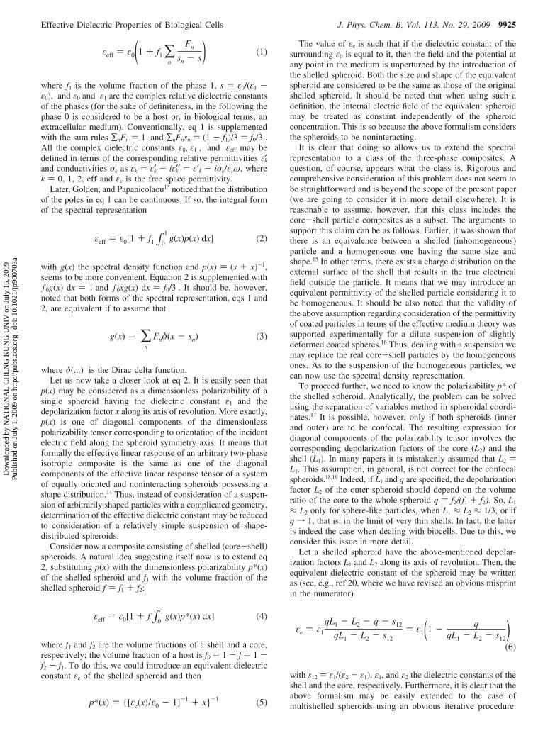
iii. In real practice, the shape of biological cells can varyfrom cell to cell. If so, it looks natural to consider an extendedspectral density function. The simplest example describingthe uniform distribution of ellipsoidal shapes around thespherical shape is the rectangular (step-like) function of theform14
where ∆ is the width of the function (see Figure 1); if now∆ approaches 0, eq 12 transforms into eq 10. As to theeffective dielectric constant, it takes the analytical form
where
and s10 ) (ε1)/(ε0 - ε1). It is worth noting that the parameter∆ may be considered as a measure of cells’ nonsphericity.
iv. To extend the Maxwell-Garnett approximation to asuspension of the randomly oriented shelled spheroids, it isconvenient to adopt the known result for the bare spheroids:22
where
and L is the depolarization factor of spheroids along thesymmetry axis. In this case the effective dielectric constant takesthe form
εe ) ε1[1 +q2
L(1 - q2) + ε1/(ε2e - ε1)] (7a)
ε2e ) ε2[1 +q3
L(1 - q3) + ε2/(ε3e - ε2)] (7b)
εNe ) εN[1 +qN+1
L(1 - qN+1) + εN/(εN+1 - εN)](7c)
i. g(x) ) δ(x - 1/3) (8)
εeff/ε0 ) 1 + 3(f1 + f2) ×(ε1 - ε0)(ε2 + 2ε1) + q(ε2 - ε1)(ε0 + 2ε1)
(ε1 + 2ε0)(ε2 + 2ε1) + 2q(ε2 - ε1)(ε1 - ε0)(9)
ii. g(x) ) δ(x - f0/3) (10)
εeff/ε0 ) 1 +f1 + f2
(ε*-1)-1 + f0/3(11)
ε* ) ε1(1 - 3qqf0 - f0 - 3s12
)
g(x) ) {∆-1, if13
f0(1 - ∆) < x < 13
f0(1 - ∆) + ∆
0, otherwise(12)
Figure 1. Examples of the spectral density function: eq 12 with f0 )0.9, ∆ ) 0.15 (1) and eq 16 with f ) 0.05, R ) 20 (2).
εeff/ε0 ) 1 +f1 + f2
∆×
{12
ln[ f0(1 - ∆) + 3∆]2 + b[3f0(1 - ∆) + 9∆] + 9c
f 02(1 - ∆)2 + 3f0b(1 - ∆) + 9c } -
rtln[2f0(1 - ∆) + 6∆ + 3b - 3t
2f0(1 - ∆) + 6∆ + 3b + 3t·2f0(1 - ∆) + 3b + 3t
2f0(1 - ∆) + 3b - 3t](13)
b ) s01 +qs10 - s12
1 - q, c )
s01s12
1 - q, t ) √b2 - 4c,
r ) b2-
qs10 + s12
1 - q, s01 )
ε0
ε1 - ε0
g(x) ) F1δ(x - s1) + F2δ(x - s2) (14)
s1 ) 14[1 - 2
3f + L + 1
3√(2f - 3 - 3L) - 72f0(1 - L)L]s2 ) 1
4[1 - 23
f + L -
13√(2f - 3 - 3L) - 72f0(1 - L)L],
F1 )1 + 3L - 6s1
6(s2 - s1), F2 )
1 + 3L - 6s2
6(s1 - s2)
9926 J. Phys. Chem. B, Vol. 113, No. 29, 2009 Goncharenko and Chang
Dow
nloa
ded
by N
AT
ION
AL
CH
EN
G K
UN
G U
NIV
on
July
16,
200
9Pu
blis
hed
on J
uly
1, 2
009
on h
ttp://
pubs
.acs
.org
| do
i: 10
.102
1/jp
9007
03a
where
and
v. From the physical point of view, it is more justified tospeak about the continuous and smooth spectral density function.The simplest example resulted from delta function broadeningdue to various physical mechanisms, including both mutualinteraction and shape fluctuation of the particles, is the single-mode approximation in the form of the Beta distribution
where B(...) is the Beta function and � ) R(2 + f)/(1 - f) inthe three-dimensional case23,24 (see Figure 1). Upon substitutionof eq 16 into eq 4, the effective dielectric constant may be easilyobtained; in the two-phase case it is shown to agree withexperimental spectra of brine-saturated rocks.23 The approxima-tion (eq 16) may be considered as a particular case of a moregeneral approximation for the spectral density function in theform of a sum of the Beta distributions.24
vi. For some bioapplications, the Looyenga equation εeff )(∑ifiεi
1/3)3 and Kraszewski equation εeff ) (∑ifiεi1/2)2 are shown
to be useful.25,26 One can show that the spectral density functioncorresponding to these equations in our case may be written as
and
respectively, where φ ) arctan [(ft1/3)/(2f0 + ft1/3)], t ) (1/x) -1. We note that both equations are particular cases of the moregeneral Lichtenecker equation for which the spectral densityfunction is also known.27
vii. One of the most popular is the Bruggeman-Hanaiequation, which has proved to work very well for concentratedsuspensions of spheres with a volume fraction up to 0.8.2 Inour case the equation may be written as f0 ) (εeff - ε*)/(ε0 -ε*)(ε0/εeff)1/3. The spectral density function corresponding to theBruggeman-Hanai equation for two-phase systems is known.28
In our case, the function takes the form
where
and
IV. Practical Examples
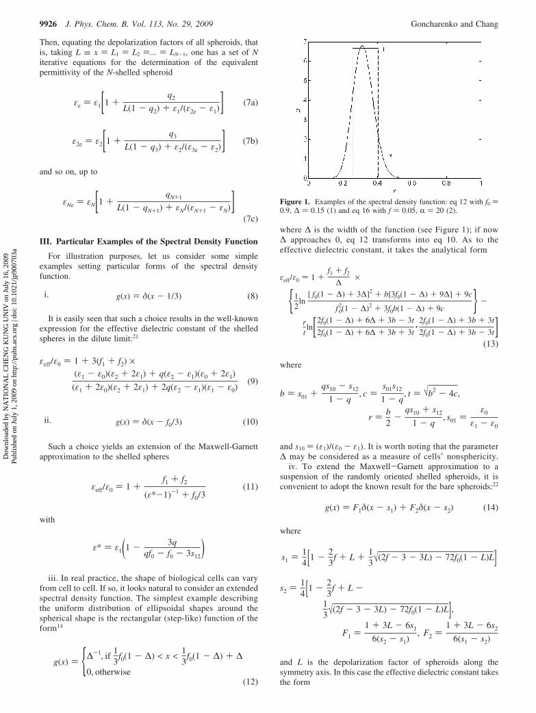
To demonstrate the potential and versatility of our approach,consider some practical examples. The first example shows theeffect of the shape distribution or, more exactly, fluctuation ofthe cell shape around the most probable spherical shape on theeffective dielectric properties of a suspension of human leuko-cytes. This choice is motivated by the fact that normal humanleukocytes are known to have near-spherical shape and, inaddition, may be described in terms of the single-shell model.29,30
To be more specific, consideration is given to a water suspensionof T-lymphocytes. The model parameters have been taken asfollows: the cell volume fraction f ) 0.15, the cell membranethickness d ) 4.5 nm, f1 ) 0.005, ε′0 ) 80, σ0 ) 0.056 S/m, ε1
) (Cs/εv - jGs/εVω)d with Cs ) 12 mF/m2 and Gs ) 100 S/m2,ε′2 ) 74, σ2) 1 S/m. The above cell parameters are typical forhuman T-lymphocytes.30 First, we used eq 11 to compute thefrequency dependencies of the effective permittivity εeff
0 (ω) )εeff′ (ω, ∆ ) 0) and conductivity σeff
0 (ω) ) σeff(ω,∆ ) 0) in thespherical approximation. Then, we used eq 13 and found thesame dependencies at different values of the nonsphericityparameter ∆. The results for the normalized dependenciesεeff′ (ω, ∆)/εeff
0 (ω) and σeff(ω, ∆)/σeff0 (ω) are shown in Figure 2.
We see that at the chosen parameters the effect of thenonsphericity on the effective dielectric response is weak butcan be noticeable; so, at ∆ ) 0.1 (this corresponds to a moderatenonsphericity) a deviation of both εeff′ (ω,∆) and σeff(ω,∆) fromtheir values in the spherical approximation can reach 3%. Atthe same time, both εeff′ (ω, ∆)/εeff
0 (ω) and σeff(ω,∆)/σeff0 (ω) reveal
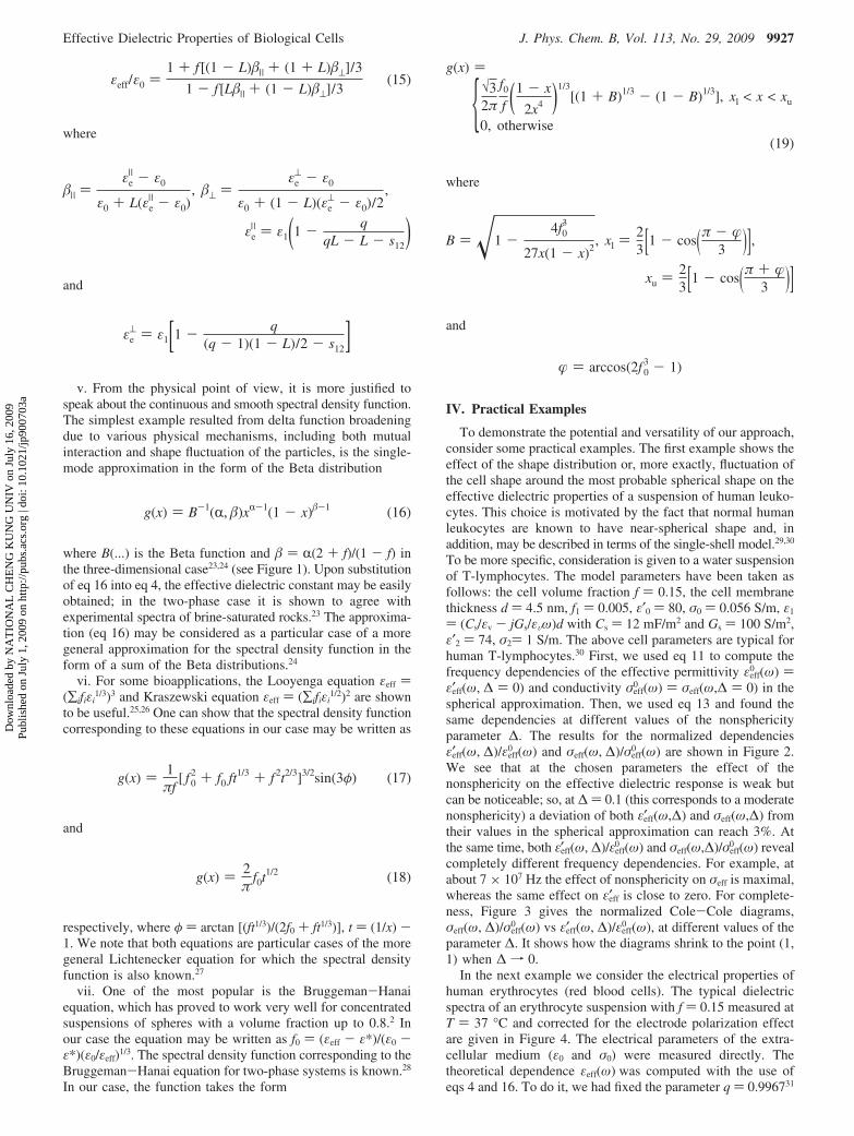
completely different frequency dependencies. For example, atabout 7 × 10⁷ Hz the effect of nonsphericity on σeff is maximal,whereas the same effect on εeff′ is close to zero. For complete-ness, Figure 3 gives the normalized Cole-Cole diagrams,σeff(ω, ∆)/σeff
0 (ω) vs εeff′ (ω, ∆)/εeff0 (ω), at different values of the
parameter ∆. It shows how the diagrams shrink to the point (1,1) when ∆ f 0.
In the next example we consider the electrical properties ofhuman erythrocytes (red blood cells). The typical dielectricspectra of an erythrocyte suspension with f ) 0.15 measured atT ) 37 °C and corrected for the electrode polarization effectare given in Figure 4. The electrical parameters of the extra-cellular medium (ε0 and σ0) were measured directly. Thetheoretical dependence εeff(ω) was computed with the use ofeqs 4 and 16. To do it, we had fixed the parameter q ) 0.996731
εeff/ε0 )1 + f [(1 - L)�| + (1 + L)�⊥]/3
1 - f [L�| + (1 - L)�⊥]/3(15)
�| )εe| - ε0
ε0 + L(εe| - ε0)
, �⊥ )εe
⊥ - ε0
ε0 + (1 - L)(εe⊥ - ε0)/2
,
εe| ) ε1(1 - q
qL - L - s12)
εe⊥ ) ε1[1 - q
(q - 1)(1 - L)/2 - s12]
g(x) ) B-1(R, �)xR-1(1 - x)�-1 (16)
g(x) ) 1πf
[ f02 + f0 ft1/3 + f2t2/3]3/2sin(3φ) (17)
g(x) ) 2π
f0t1/2 (18)
g(x) )
{√32π
f0
f (1 - x
2x4 )1/3[(1 + B)1/3 - (1 - B)1/3], xl < x < xu
0, otherwise(19)
B ) �1 -4f0
3
27x(1 - x)2, xl )
23[1 - cos(π - �
3 )],xu ) 2
3[1 - cos(π + �3 )]
� ) arccos(2f03 - 1)
Effective Dielectric Properties of Biological Cells J. Phys. Chem. B, Vol. 113, No. 29, 2009 9927
Dow
nloa
ded
by N
AT
ION
AL
CH
EN
G K
UN
G U
NIV
on
July
16,
200
9Pu
blis
hed
on J
uly
1, 2
009
on h
ttp://
pubs
.acs
.org
| do
i: 10
.102
1/jp
9007
03a
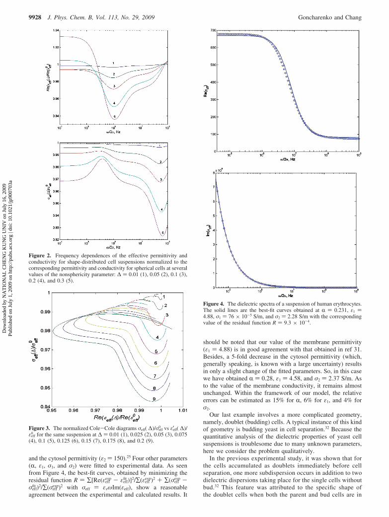
and the cytosol permittivity (ε2 ) 150).25 Four other parameters(R, ε1, σ1, and σ2) were fitted to experimental data. As seenfrom Figure 4, the best-fit curves, obtained by minimizing theresidual function R ) ∑[Re(εeff
exp - εefffit )]2/∑(εeff
exp)2 + ∑(σeffexp -
σefffit )2/∑(σeff
exp)2 with σeff ) ενωIm(εeff), show a reasonableagreement between the experimental and calculated results. It
should be noted that our value of the membrane permittivity(ε1 ) 4.88) is in good agreement with that obtained in ref 31.Besides, a 5-fold decrease in the cytosol permittivity (which,generally speaking, is known with a large uncertainty) resultsin only a slight change of the fitted parameters. So, in this casewe have obtained R ) 0.28, ε1 ) 4.58, and σ2 ) 2.37 S/m. Asto the value of the membrane conductivity, it remains almostunchanged. Within the framework of our model, the relativeerrors can be estimated as 15% for R, 6% for ε1, and 4% forσ2.
Our last example involves a more complicated geometry,namely, doublet (budding) cells. A typical instance of this kindof geometry is budding yeast in cell separation.32 Because thequantitative analysis of the dielectric properties of yeast cellsuspensions is troublesome due to many unknown parameters,here we consider the problem qualitatively.
In the previous experimental study, it was shown that forthe cells accumulated as doublets immediately before cellseparation, one more subdispersion occurs in addition to twodielectric dispersions taking place for the single cells withoutbud.32 This feature was attributed to the specific shape ofthe doublet cells when both the parent and bud cells are in
Figure 2. Frequency dependences of the effective permittivity andconductivity for shape-distributed cell suspensions normalized to thecorresponding permittivity and conductivity for spherical cells at severalvalues of the nonsphericity parameter: ∆ ) 0.01 (1), 0.05 (2), 0.1 (3),0.2 (4), and 0.3 (5).
Figure 3. The normalized Cole-Cole diagrams σeff( ∆)/σeff0 vs εeff′ ( ∆)/
εeff0 for the same suspension at ∆ ) 0.01 (1), 0.025 (2), 0.05 (3), 0.075
(4), 0.1 (5), 0.125 (6), 0.15 (7), 0.175 (8), and 0.2 (9).
Figure 4. The dielectric spectra of a suspension of human erythrocytes.The solid lines are the best-fit curves obtained at R ) 0.231, ε1 )4.88, σ1 ) 76 × 10-5 S/m, and σ2 ) 2.28 S/m with the correspondingvalue of the residual function R ) 9.3 × 10⁻⁴.
9928 J. Phys. Chem. B, Vol. 113, No. 29, 2009 Goncharenko and Chang
Dow
nloa
ded
by N
AT
ION
AL
CH
EN
G K
UN
G U
NIV
on
July
16,
200
9Pu
blis
hed
on J
uly
1, 2
009
on h
ttp://
pubs
.acs
.org
| do
i: 10
.102
1/jp
9007
03a
electrical contact.33 To model the corresponding geometry,we use the two-mode spectral density function of the formg(x) ) ∑i)1
2 CixRi-1(1-x)�i-1 which involves four free pa-rameters.24 For illustration purposes and for comparison withthe above paper by Sekine et al.,33 who made use of theboundary element method, our results are presented in termsof the function T ) f-1Im (1 - εeff/ε0). To obtain a goodagreement, we have fixed the material parameters as in ref32 (ε0 ) ε2 ) 80, ε2 ) 5.5, σ0 ) 0.3 S/m, σ1 ) 0) and thegeometry parameters as R1 ) 0.7, �1 ) 1.44, R2 ) 890, �2
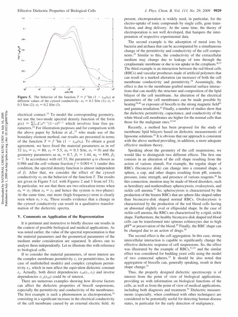
) 7. In accordance with ref 33, the parameter q is chosen as0.986 and the cell volume fraction f ) 0.001, 1 (under thiscondition, the spectral density function is almost independentof f). After that, we consider the effect of the cytosolconductivity σ2 on the behavior of the function T. The resultsare given in Figure 5 (cf. with Figures 2 and 3 from ref 33).In particular, we see that there are two relaxation terms whenσ0 ) σ2 (then ε0 ) ε2 and hence the system is two-phase).At the same time, one more (high-frequency) term is clearlyseen when σ2 > σ0. These results evidence that a change inthe cytosol conductivity can result in a qualitative transfor-mation of the dielectric spectra.
V. Comments on Application of the Representation
It is pertinent and instructive to briefly discuss our results inthe context of possible biological and medical applications. Aswas noted earlier, the value of the spectral representation is thatthe material parameters and the geometrical parameters of themedium under consideration are separated. It allows one toanalyze them independently. Let us illustrate this with referenceto biological cells.
If to consider the material parameters, of most interest arethe complex membrane permittivity ε1 (or permittivities, in thecase of multishelled models) and complex cytoplasm permit-tivity ε2, which in turn affect the equivalent dielectric constantεe. Actually, both direct dependencies εeff(ε1, ε2) and inversedependencies ε1,2(εeff) could be of interest.
There are numerous examples showing how diverse factorscan affect the dielectric properties of biocell suspensions,especially the permittivity and conductivity of the membranes.The first example is such a phenomenon as electroporation,34
consisting in a significant increase in the electrical conductivityof the cell membrane caused by an external electric field. At
present, electroporation is widely used, in particular, for theelectro-uptake of ionic compounds by single cells, gene trans-fection, and drug delivery. At the same time, the theory ofelectroporation is not well developed, that hampers the inter-pretation of respective experimental data.
The second example is the adsorption of metal ions bybacteria and archaea that can be accompanied by a simultaneouschange of the permittivity and conductivity of the cell compo-nents.35 Similar to this, the conductivity of the extracellularmedium may change due to leakage of ions through thecytoplasmatic membrane or due to ion uptake in the cytoplasm.36,37
The third example is an interaction between the red blood cells(RBCs) and vascular prostheses made of artificial polymers thatcan result in a marked alteration (an increase) of both the cellmembrane conductivity and permittivity.38 Assumingly, theeffect is due to the membrane-grafted material surface interac-tions that can modify the structure and composition of the lipidbilayer of the cell membrane. An alteration of the electricalparameters of the cell membranes can be made possible byheating39,40 or exposure of biocells to the strong magnetic field41
and gamma-irradiation.42 Finally, a number of studies show thatthe dielectric permittivity, capacitance, and conductivity of thewhite blood cell membranes are higher for the normal cells thanthose for the malignant ones.43,44
Recently, a method has been proposed for the study ofmembrane lipid bilayers based on dielectric measurements ofliposome solutions.45 It is obvious that our approach is consistentwith the above method providing, in addition, a more adequateeffective medium theory.
Speaking about the geometry of the cell suspensions, wewould like to distinguish two different effects. The first oneconsists in an alteration of the cell shape resulting from theaction of various stimuli. For example, the regular shape ofRBCs (biconcave disk) can change into a crenated shape, asphere, a cup, and other shapes resulting from pH, osmoticpressure, ionic strength, and presence of various reagents.46 Inthis connection, mention may be made of blood disorders, suchas hereditary and nonhereditary spherocytosis, ovalosytosis, andsickle cell anemia.47 So, spherocytosis is characterized by theproduction of the bizarre RBCs that are sphere-like shaped ratherthan biconcave-disk shaped normal RBCs. Ovalocytosis ischaracterized by the production of the red blood cells havingan abnormal slightly oval or ellipsoidal shape. In the case ofsickle-cell anemia, the RBCs are characterized by a rigid, sickleshape. Furthermore, the healthy biconcave-disk shaped red bloodcells can be transformed into spinous echinocytes due to highpH48 or preservation of the blood.49 Finally, the RBC shape canbe changed due to an action of drugs.50
The second effect is the cell aggregation. In this case, strongintercellular interaction is capable to significantly change theeffective dielectric response of cell suspensions. So, the effectwas illustrated by the example of RBCs,51,52 and the similareffect was considered for budding yeast cells using the modelof two connected spheres.33 It should be also noted thataggregation of biocells can, generally speaking, result in theirshape change.53
Thus, the properly designed dielectric spectroscopy is ofinterest from the point of view of biological applications,providing us with information on biological functions of thecells, as well as from the point of view of medical applications,including both diagnosis and treatment.54 Dielectric measure-ments (especially, when combined with other techniques) areconsidered to be potentially useful for detecting human diseasestates, in particular for the early detection of malignancy.
Figure 5. The behavior of the function T ) f-1Im (1 - εeff/ε0) atdifferent values of the cytosol conductivity: σ2 ) 0.2 S/m (1); σ2 )0.3 S/m (2); σ2 ) 0.2 S/m (3).
Effective Dielectric Properties of Biological Cells J. Phys. Chem. B, Vol. 113, No. 29, 2009 9929
Dow
nloa
ded
by N
AT
ION
AL
CH
EN
G K
UN
G U
NIV
on
July
16,
200
9Pu
blis
hed
on J
uly
1, 2
009
on h
ttp://
pubs
.acs
.org
| do
i: 10
.102
1/jp
9007
03a
To conclude this section, it should be noted that our approachcould be also useful for the study of viruses. Indeed, manyviruses are known to have a layered structure including a core(DNAs/RNAs), an envelope, and a capsid layer between them.55
In principle, although it is not always easy and convenient, theirelectrical properties can be considered using dielectric spec-troscopy. At the same time, because the viruses are usuallyranging in size from 20 to 250 nm, their optical properties maybe considered in the long wavelength approximation in thevisible and infrared range, that is, in terms of the effectivemedium, using the spectral density function approach. The samecan also take place for sufficiently small cells in the infraredrange as the approximation is valid.
VI. Concluding Remarks
The main result of this paper tells us how to extend the two-phase spectral representation to three-phase and more compli-cated multiphase systems such as suspensions of biocells, thatis, core-shell particles with a very thin shell or, to be moreexact, with a very low volume fraction of the shell. The questionthen arises, apropos of the present result, what should we do ifthe shell is not very thin.
Before answering this question it should be reminded thatwe have considered the shell thickness to be very small for thesake of convenience of mathematical treatment. As alreadynoted, such an assumption seems to be reasonable in manypractical cases due to very small thickness of real cellmembranes. However, it is not the case when dealing with somemultishell models, as at least one of the shells is not very thin.For example, bacteria, yeast, and plant cells usually have theso-called cell wall (an additional shell) outside the membrane.56
Correspondingly, the dielectric properties of such biocells maybe considered in terms of the two-shell model. Thus, in thiscase we should use eq 6, where L2 is a function of L1. Therelation between the depolarization factors of outer (L1) andinner (L2) confocal spheroids is signified implicitly.57 In thegeneral case, dealing with the multishell model, we should useeq 6 iteratively taking into account that Li is a function of Li-1.
Because the conventional impedance dielectric spectroscopydeals with the measurements on cell suspensions, it gives usassembly averaged (effective) dielectric properties of the cellcollections. At the same time, we usually need some usefulinformation on single cells. So, in the recent years moreemphasis has been placed on the development of varioustechniques feasible for single cell analysis;58 it is the currenttrend in biological and medical researches. Such techniques,while promising, are yet much more complicated and time-consuming than the conventional dielectric spectroscopy. Thus,extracting single cell information from the dielectric spectra ofcell suspensions continues to be relevant. If the mixing rule isknown, the problem becomes trivial because it reduces to astandard fitting procedure for the dielectric parameters of thecells. In the general case, however, it is not so, because thespectral density function is usually unknown. It means thatbefore extracting the cell dielectric parameters we should firstdetermine the function for the cell suspension under consider-ation. Some approaches to this problem have been proposed.59
In general, the spectral density can be determined using relevantexperimental data.60 As was noted earlier,61 this procedurebelongs to the class of the so-called ill-posed problems and acorrect solution is possible only with the use of stabilizationtechniques. Mention may be also made of the work based onan expansion of the modified spectral density function in termsof Legendre polynomials,62 the reconstruction of the spectral
density function using Pade approximation derived from aconstrained minimization problem,63 as well as of Tuncer’sapproach6,64 based on the Monte Carlo integration and con-strained least-squares algorithm. In principle, it seems possiblenot only to extract the spectral density function from experi-mental data, but also to obtain the volume fractions and effectiveshapes of inclusions.64
In summary, an extension of the spectral density functionapproach is proposed and discussed that can be applied tointerpretation of the dielectric spectra of shelled particles suchas biological cells. For illustration, some simple examples ofthe density function, as well as possible applications, areconsidered.
Acknowledgment. The authors are indebted to CesareCametti, who kindly provided us with the dielectric spectra ofsuspensions of red blood cells.
References and Notes
(1) Considering the counts of the term “dielectric spectroscopy” withthe use of ISI’s databases, we have found that the number of the respectivescientific publications grows roughly linearly with time, with a fasteracceleration during the last two years.
(2) Asami, K. Prog. Polym. Sci. 2002, 27, 1617–1659.(3) See e.g. Young, K. D. Microbiol. Mol. Biol. ReV. 2006, 70, 660–
703.(4) Vrinceanu, D.; Gheorghiu, E. Bioelectrochem. Bioenerg. 1996, 40,
167–170, It should be noted that the authors, in essence, have rederivedthe result of Fuchs (see ref 12).
(5) Di Biasio, A.; Ambrosone, L.; Cametti, C. J.Phys. D: Appl. Phys.2009, 42, 025401. The method makes use of an algorithm proposed earlierby Prodan and Prodan (see Ref 15).
(6) Tuncer, E.; Serdyuk, Y. V.; Gubanski, S. M. IEEE Trans. Diel.El. Insul. 2002, 9, 809–828.
(7) Bergman, D. J. Phys. Rep. C 1978, 43, 377–407.(8) Milton, G. W. Appl. Phys. Lett. 1980, 37, 300–302.(9) See e.g., Lei, J.; Wan, J. T. K.; Yu, K. W.; Sun, H. Phys. ReV. E
2001, 64, 012903The relationship between the spectral representation anddielectric relaxation is shown in Tuncer E. J. Phys.: Condens. Matter. 2005,17, L125-L128.
(10) Huang, J. P.; Yu, K. W.; Lei, J.; Sun, H. Commun. Theor. Phys.2002, 38, 113–120.
(11) Dong, L.; Karttunen, M.; Yu, K. W. Phys. ReV. E 2005, 72, 016613.(12) Fuchs, R. Phys. ReV. B 1975, 11, 1732–1740.(13) Golden, K.; Papanicolaou, G. Commun. Math. Phys. 1983, 90, 473–
491.(14) Goncharenko, A. V.; Lozovski, V. Z.; Venger, E. F. J. Phys.:
Condens. Matter 2001, 13, 8217–8234.(15) Prodan, C.; Prodan, E. J. Phys. D: Appl. Phys. 1999, 32, 335–343.(16) Tuncer, E.; Bowler, N.; Youngs, I. J. Physica B 2006, 373, 306–
312.(17) Bohren, C. F.; Huffman, D. R. Absorption and Scattering of Light
by Small Particles; Wiley: New York, 1983.(18) Skryabin, I. L.; Radchik, A. V.; Moses, P.; Smith, G. B. Appl. Phys.
Lett. 1997, 70, 2221–2223.(19) Goncharenko, A. V. Appl. Phys. Lett. 2007, 91, 246101.(20) Miragliotta, J.; Furtak, T. E. Phys. ReV. B 1987, 35, 7382–7391.(21) See e.g., Gao, L. Phys. Lett. A 2003, 318, 119–125.(22) Gao, L.; Wan, J. T. K.; Yu, K. W.; Li, Z. Y. J. Phys.: Condens.
Matt. 2000, 12, 6825–6836.(23) Stroud, D.; Milton, G. W.; De, B. R. Phys. ReV. B 1986, 34, 5145–
5153.(24) Goncharenko, A. V. Chem. Phys. Lett. 2004, 386, 462–468.(25) Bordi, F.; Cametti, C.; Gili, T. J. Non-Cryst. Sol. 2002, 305, 278–
284.(26) Bonicontro, A.; Cametti, C. Coll. Surf. A: Physicochem. Eng. Asp.
2004, 246, 115–120.(27) Goncharenko, A. V.; Lozovski, V. Z.; Venger, E. F. Opt. Commun.
2000, 174, 19–32.(28) Ma, H.; Sheng, P.; Wong, G. K. L. Topics Appl. Phys. 2002, 82,
41–62.(29) Yang, J.; Huang, Y.; Wang, X.; Wang, X. B.; Becker, F. F.;
Gascoyne, P. R. C. Biophys. J. 1999, 76, 3307–3314.(30) Huang, Y.; Wang, X. B.; Gascoyne, P. R. C.; Becker, F. F. Biochim.
Biophys. Acta 1999, 1417, 51–62.(31) Lisin, R.; Ginzburg, B. Z.; Schlesinger, M.; Feldman, Y. Biochim.
Biophys. Acta 1996, 1280, 34–40.
9930 J. Phys. Chem. B, Vol. 113, No. 29, 2009 Goncharenko and Chang
Dow
nloa
ded
by N
AT
ION
AL
CH
EN
G K
UN
G U
NIV
on
July
16,
200
9Pu
blis
hed
on J
uly
1, 2
009
on h
ttp://
pubs
.acs
.org
| do
i: 10
.102
1/jp
9007
03a
(32) Asami, K.; Gheorghiu, E.; Yonezawa, T. Biochim. Biophys. Acta1998, 1381, 234–240.
(33) Sekine, K.; Watanabe, Y.; Hara, S.; Asami, K. Biochim. Biophys.Acta 2005, 1721, 130–138.
(34) Tsong, T. Y. Biophys. J. 1991, 60, 297–306.(35) Bai, W.; Zhao, K.; Asami, K. Coll. Surf. B: Biointerfaces 2007,
58, 105–115.(36) Wal, A.; van der Minor, M.; Norde, W.; Zehnder, A. J. B.; Lyklema,
J. J. Colloid Interface Sci. 1997, 186, 71–79.(37) Yang, L. Talanta 2008, 74, 1621–1629.(38) Basoli, A.; Bordi, F.; Gili, T. J. Biomed. Mater. Res. 2002, 59,
100–109.(39) Bonincontro, A.; Mariutti, G. Phys. Med. Biol. 1988, 33, 557–568.(40) Ivanov, I. T. Biol. Membr. 1993, 10, 160–169.(41) Santini, M. T.; Cametti, C.; Paradisi, S.; Straface, E.; Donelli, G.;
Indovina, P. L.; Malorni, W. Bioelectrochem. Bioelectroenerg. 1995, 36,39–45.
(42) Bonicontro, A.; Cametti, C.; Rosi, A.; Sportelli, L. Intern. J. Radiat.Biol. 1987, 52, 447–457.
(43) Polevaya, Y.; Ermolina, I.; Schlesinger, M.; Ginzburg, B. Z.;Feldman, Y. Biochim. Biophys. Acta 1999, 1419, 257–271.
(44) Ermolina, I.; Polevaya, Y.; Feldman, Y.; Ginzburg, B. Z.; Schlesing-er, M. IEEE Trans. Diel. Electr. Insul. 2001, 8, 253–261.
(45) Merla, C.; Liberti, M.; Apollonio, F.; d’Inzeo, G. Bioelectromagnet.2009, 30, 286–298.
(46) Nakao, M., In Blood Cell Biochemistry. Erythroid Cells; HarrisJ. R., Ed.; Plenum: New York, 1990; Vol. 1, pp 195-226.
(47) Suresh, S. J. Mater. Res. 2006, 21, 1871–1877.(48) Gedde, M. M.; Davis, D. K.; Huestis, W. H. Biophys. J. 1997, 72,
1234–1246.
(49) Hayashi, Y.; Oshige, I.; Katsumoto, Y.; Omori, S.; Yakuda, A.;Asami, K. Phys. Med. Biol. 2008, 53, 295–304.
(50) Deuticke, B.; Grebe, R.; Haest, C. W. M., In Blood CellBiochemistry. Erythroid Cells; Harris J. R., Ed.; Plenum: New York, 1990;Vol. 1, pp 475-530.
(51) Sebastian, J. L.; San Martin, S. M.; Sancho, M.; Miranda, J. M.;Alvarez, G. Phys. ReV. E 2005, 72, 031913.
(52) Asami, K.; Sekine, K. J. Phys. D: Appl. Phys. 2007, 40, 2197–2204.
(53) Santini, M. T.; Cametti, C.; Indovina, P. L.; Morelli, G.; Donelli,G. J. Biomed. Mater. Res. 1997, 35, 165–174.
(54) Feldman, Y.; Ermolina, I.; Hayashi, Y. IEEE Trans. Diel. Electr.Insul. 2003, 10, 728–753.
(55) MacCuspie, R. I.; Nuraje, N.; Lee, S. Y.; Runge, A.; Matsui, H.J. Am. Chem. Soc. 2008, 130, 887–891.
(56) Asami, K. J. Non-Cryst. Sol. 2002, 305, 268–277.(57) See e.g. Goncharenko, A. V.; Chang, Y. C. Chem. Phys. Lett. 2007,
439, 121–126.(58) Morgan, H.; Sun, T.; Holmes, D.; Gawad, S.; Green, N. G. J. Phys.
D: Appl. Phys. 2007, 40, 61–70.(59) Goncharenko, A. V. Phys. ReV. E 2003, 68, 041108.(60) Day, A. R.; Thorpe, M. F. J. Phys.: Condens. Matter 1999, 11,
2551–2568.(61) Cherkaev, E. InV. Prob. 2001, 17, 1203–1218.(62) Pecharroman, C.; Gordillo-Vazquez, F. J. Phys. ReV. B 2006, 74,
035120.(63) Zhang, D.; Cherkaev, E. InV. Prob. 2008, 16, 425–445.(64) Tuncer, E. Phys. ReV. B 2005, 71, 012101.
JP900703A
Effective Dielectric Properties of Biological Cells J. Phys. Chem. B, Vol. 113, No. 29, 2009 9931
Dow
nloa
ded
by N
AT
ION
AL
CH
EN
G K
UN
G U
NIV
on
July
16,
200
9Pu
blis
hed
on J
uly
1, 2
009
on h
ttp://
pubs
.acs
.org
| do
i: 10
.102
1/jp
9007
03a