Effect of Sensory Discrimination Training on Structure and ...
10
Effect of Sensory Discrimination Training on Structure and Function in Patients With Focal Hand Dystonia: A Case Series Nancy N. Byl, PhD, PT, FAPTA, Srikantan Nagajaran, PhD, Alison L. McKenzie, PhD, PT ABSTRACT. Byl NN, Nagajaran S, McKenzie AL. Effect of sensory discrimination training on structure and function in patients with focal hand dystonia: a case series. Arch Phys Med Rehabil 2003;84:1505-14. Objective: To measure the effects of sensorimotor training based on the principles of neuroplasticity for patients with focal hand dystonia. Design: Case series of 3 subjects with focal hand dystonia of the left hand, compared with age-matched normative controls. Setting: Outpatient clinic. Participants: Three consecutive clinic patients—musicians with focal hand dystonia—who described a history of repetitive practice and performance (2 women; ages, 23y and 35y; 1 man; age, 24y). Intervention: Subjects were asked to stop performing the tasks that caused the abnormal movements, to participate in a wellness program (aerobics, postural exercises, stress free hand use), and to carry out supervised, attended, individualized, repetitive sensorimotor training activities at least once week for 12 weeks and reinforced daily at home. Main Outcome Measures: Standard tests documenting so- matosensory hand representation, target-specific hand control, and clinical function. Results: On the affected side, the 3 subjects improved an average of 86.8% on somatosensory hand representation, 117% on target-specific performance, 23.9% on fine motor skills, 22.7% on sensory discrimination, 31.9% on musculoskeletal skills, and 32.3% on independence. All 3 subjects improved 10% or more on 90% of the subtests with 20% improvement on 50% of the subtests. Conclusion: Individuals with focal hand dystonia who have a history of repetitive hand use can improve cortical somato- sensory responses and clinical motor function after individual- ized sensorimotor training consistent with the principles of neural adaptation. Key Words: Dystonia; Focal dystonia; Rehabilitation; So- matosensory disorders. © 2003 by the American Congress of Rehabilitation Medi- cine and the American Academy of Physical Medicine and Rehabilitation O CCUPATIONAL HAND CRAMPS, also referred to as focal hand dystonia, involve loss of inhibition between agonist and antagonist muscles of the hand, disrupting fine motor control at a target task. 1-9 This condition can develop in performing artists, athletes, business executives, and assembly line workers who perform high levels of attended, stereotypi- cal, repetitive movements. 1,4,5,9 There are no specific clinical laboratory tests to confirm the diagnosis of focal hand dystonia, however, loss of inhibition, excessive muscle firing, and inability to release muscle con- tractions are documented with electromyography. 2 Patients with simple target-specific dystonia usually have a normal neurologic examination when performing functional activi- ties. 7,9 Observing the onset of abnormal movements when performing the target task is critical to making the diagnosis of focal hand dystonia. It is not uncommon for patients with focal hand dystonia to report a history of stressful, excessive overuse of the hands, 1,3-5 neuromusculoskeletal trauma (eg, head trauma, radial fractures), 10 degenerative disk disease or cervi- cal injury, 11 biomechanical limitations (eg, poor posture, lim- ited finger spread, decreased forearm and shoulder rota- tion), 12-15 or peripheral nerve entrapments. 16,17 Researchers and clinicians have documented a loss of inhibition between ago- nists and antagonists, 18-20 and abnormal neuronal firing patterns in the motor cortex, 21-26 the basal ganglia, 27-29 the spinal cord, 30 and the somatosensory cortex 31-41 in patients with focal hand dystonia. Focal dystonia is challenging to treat. Traditional interven- tion usually includes peripheral injections of botulinum toxin, 42-47 sometimes also electric stimulation (for cervical dystonia torticollis). 48 More recently, innovative therapeutic programs emphasizing the principles of neuroplasticity appear to be promising. 49,50 Over the last 10 years, research 51-58 has clearly established that the central nervous system is adaptable. Goal-directed, repeated, and rewarded sensory and motor behaviors can drive changes in neural structure and function. 54-57 However, adap- tation of the nervous system is not infinite. If behaviors become stereotypical and nearly simultaneous, the brain may not be able to distinguish and represent the overused part distinctly and precisely. 58 Evidence supporting aberrant learning has been documented in studies with naive nonhuman primates trained to perform daily, highly repetitive, stereotypical, near simultaneous movements of the hand. 34 Ultimately, the mon- keys lost the ability to perform the target task. Electrophysi- ologic mapping revealed a degradation of the somatosensory representation of the hand, including enlarged receptive fields extending across multiple digits and/or across glabrous and dorsal surfaces. 33,34 In healthy musicians, intensive practice of articulated hand movements is usually associated with an in- crease in the somatotopic representation of the hand. 59 How- ever, when dystonic movements develop after excessive over- use, the cortical hand representation may shrink in size and the digits can become dedifferentiated (clumped together at the same location without normal sequencing). 33,34,36,60,61 If focal hand dystonia results from a dedifferentiation of somatosensory structure, then improvement in task-specific From the Department of Physical Therapy and Rehabilitation Science, School of Medicine, UCSF/SFSU Graduate Program in Physical Therapy, University of Cali- fornia, San Francisco, CA (Byl, McKenzie), Department of Radiology, University of California, San Francisco, CA; and School of Physical Therapy, Chapman University, Orange, CA (McKenzie). Presented as a poster the Society of Neuroscience’s annual meeting, November 2001, New Orleans, LA. Supported in part by the School of Medicine, UCSF, REAC Dubois Funds, and the National Institutes of Health (grant no. PO1-NS34835). No commercial party having a direct financial interest in the results of the research supporting this article has or will confer a benefit upon the author(s) or upon any organization with which the author(s) is/are associated. Reprint requests to Nancy Byl, PhD, PT, FAPTA, UCSF Dept of Physical Therapy and Rehabilitation Medicine, 1320 7th Ave, Box 0736, San Francisco, CA 94143- 0736, e-mail: [email protected]. 0003-9993/03/8410-7737$30.00/0 doi:10.1053/S0003-9993(03)00276-4 1505 Arch Phys Med Rehabil Vol 84, October 2003
-
Upload
khangminh22 -
Category
Documents
-
view
3 -
download
0
Transcript of Effect of Sensory Discrimination Training on Structure and ...
Effect of Sensory Discrimination Training on Structure andFunction in Patients With Focal Hand Dystonia: A Case SeriesNancy N. Byl, PhD, PT, FAPTA, Srikantan Nagajaran, PhD, Alison L. McKenzie, PhD, PT
ABSTRACT. Byl NN, Nagajaran S, McKenzie AL. Effectof sensory discrimination training on structure and function inpatients with focal hand dystonia: a case series. Arch Phys MedRehabil 2003;84:1505-14.
Objective: To measure the effects of sensorimotor trainingbased on the principles of neuroplasticity for patients with focalhand dystonia.
Design: Case series of 3 subjects with focal hand dystonia ofthe left hand, compared with age-matched normative controls.
Setting: Outpatient clinic.Participants: Three consecutive clinic patients—musicians
with focal hand dystonia—who described a history of repetitivepractice and performance (2 women; ages, 23y and 35y; 1 man;age, 24y).
Intervention: Subjects were asked to stop performing thetasks that caused the abnormal movements, to participate in awellness program (aerobics, postural exercises, stress free handuse), and to carry out supervised, attended, individualized,repetitive sensorimotor training activities at least once week for12 weeks and reinforced daily at home.
Main Outcome Measures: Standard tests documenting so-matosensory hand representation, target-specific hand control,and clinical function.
Results: On the affected side, the 3 subjects improved anaverage of 86.8% on somatosensory hand representation, 117%on target-specific performance, 23.9% on fine motor skills,22.7% on sensory discrimination, 31.9% on musculoskeletalskills, and 32.3% on independence. All 3 subjects improved10% or more on 90% of the subtests with 20% improvement on50% of the subtests.
Conclusion: Individuals with focal hand dystonia who havea history of repetitive hand use can improve cortical somato-sensory responses and clinical motor function after individual-ized sensorimotor training consistent with the principles ofneural adaptation.
Key Words: Dystonia; Focal dystonia; Rehabilitation; So-matosensory disorders.
© 2003 by the American Congress of Rehabilitation Medi-cine and the American Academy of Physical Medicine andRehabilitation
OCCUPATIONAL HAND CRAMPS, also referred to asfocal hand dystonia, involve loss of inhibition between
agonist and antagonist muscles of the hand, disrupting finemotor control at a target task.1-9 This condition can develop inperforming artists, athletes, business executives, and assemblyline workers who perform high levels of attended, stereotypi-cal, repetitive movements.1,4,5,9
There are no specific clinical laboratory tests to confirm thediagnosis of focal hand dystonia, however, loss of inhibition,excessive muscle firing, and inability to release muscle con-tractions are documented with electromyography.2 Patientswith simple target-specific dystonia usually have a normalneurologic examination when performing functional activi-ties.7,9 Observing the onset of abnormal movements whenperforming the target task is critical to making the diagnosis offocal hand dystonia. It is not uncommon for patients with focalhand dystonia to report a history of stressful, excessive overuseof the hands,1,3-5 neuromusculoskeletal trauma (eg, headtrauma, radial fractures),10 degenerative disk disease or cervi-cal injury,11 biomechanical limitations (eg, poor posture, lim-ited finger spread, decreased forearm and shoulder rota-tion),12-15or peripheral nerve entrapments.16,17Researchers andclinicians have documented a loss of inhibition between ago-nists and antagonists,18-20and abnormal neuronal firing patternsin the motor cortex,21-26the basal ganglia,27-29the spinal cord,30
and the somatosensory cortex31-41 in patients with focal handdystonia.
Focal dystonia is challenging to treat. Traditional interven-tion usually includes peripheral injections of botulinumtoxin,42-47 sometimes also electric stimulation (for cervicaldystonia torticollis).48 More recently, innovative therapeuticprograms emphasizing the principles of neuroplasticity appearto be promising.49,50
Over the last 10 years, research51-58 has clearly establishedthat the central nervous system is adaptable. Goal-directed,repeated, and rewarded sensory and motor behaviors can drivechanges in neural structure and function.54-57 However, adap-tation of the nervous system is not infinite. If behaviors becomestereotypical and nearly simultaneous, the brain may not beable to distinguish and represent the overused part distinctlyand precisely.58 Evidence supporting aberrant learning hasbeen documented in studies with naive nonhuman primatestrained to perform daily, highly repetitive, stereotypical, nearsimultaneous movements of the hand.34 Ultimately, the mon-keys lost the ability to perform the target task. Electrophysi-ologic mapping revealed a degradation of the somatosensoryrepresentation of the hand, including enlarged receptive fieldsextending across multiple digits and/or across glabrous anddorsal surfaces.33,34 In healthy musicians, intensive practice ofarticulated hand movements is usually associated with an in-crease in the somatotopic representation of the hand.59 How-ever, when dystonic movements develop after excessive over-use, the cortical hand representation may shrink in size and thedigits can become dedifferentiated (clumped together at thesame location without normal sequencing).33,34,36,60,61
If focal hand dystonia results from a dedifferentiation ofsomatosensory structure, then improvement in task-specific
From the Department of Physical Therapy and Rehabilitation Science, School ofMedicine, UCSF/SFSU Graduate Program in Physical Therapy, University of Cali-fornia, San Francisco, CA (Byl, McKenzie), Department of Radiology, University ofCalifornia, San Francisco, CA; and School of Physical Therapy, Chapman University,Orange, CA (McKenzie).
Presented as a poster the Society of Neuroscience’s annual meeting, November2001, New Orleans, LA.
Supported in part by the School of Medicine, UCSF, REAC Dubois Funds, and theNational Institutes of Health (grant no. PO1-NS34835).
No commercial party having a direct financial interest in the results of the researchsupporting this article has or will confer a benefit upon the author(s) or upon anyorganization with which the author(s) is/are associated.
Reprint requests to Nancy Byl, PhD, PT, FAPTA, UCSF Dept of Physical Therapyand Rehabilitation Medicine, 1320 7th Ave, Box 0736, San Francisco, CA 94143-0736, e-mail:[email protected].
0003-9993/03/8410-7737$30.00/0doi:10.1053/S0003-9993(03)00276-4
1505
Arch Phys Med Rehabil Vol 84, October 2003

fine motor performance must be contingent on a positive learn-ing paradigm where precise and distinct representations of thedigits are restored. By using a case-study approach, repeatedwith 3 musicians with focal hand dystonia, the present reportdescribes changes in somatosensory evoked responses andclinical function after a sensorimotor training program basedon the principles of neuroplasticity. Compared with controls,subjects with focal hand dystonia were expected to show de-differentiation of the somatosensory cortex and diminishedperformance on clinical, sensory, motor, musculoskeletal, andfunctional independence. Focal hand dystonia subjects werealso expected to improve on all structural and clinical variablesafter sensorimotor training.
METHODS
ParticipantsThree musicians (2 flutists, 1 bagpipe player) with focal
hand dystonia of the left hand were referred to the PeterOstwald Health Program for Performing Artists, University ofCalifornia, San Francisco (UCSF), CA. Two subjects came toSan Francisco to participate in a supervised period of rehabil-itation (subject 1 [flutist] was from New Zealand, subject 2[bagpipe players], from Australia) and 1 subject (subject 3)lived in the San Francisco Bay Area. All flutists had beendiagnosed with focal hand dystonia by a neurologist between 1and 2 years prior to admission to the study. During instrumen-tal play, all 3 subjects presented with complaints of painless,uncontrollable curling of digits 4 (D4) and 5 (D5) on the lefthand. All 3 subjects reported difficulty controlling D4 and D5when D3 was pressing down.
The subjects were a sample of convenience—patients con-secutively admitted to the health program who were able toparticipate in sensorimotor training for a minimum of 10 visits(1–2 sessions/wk), carry out a fitness program 3 days per week,integrate a sensorimotor training program at home, and partic-ipate in extensive testing before and immediately after treat-ment. At the same time, a small grant was awarded for a pilotstudy to evaluate the feasibility and responsiveness of usingmagnetoencephalography to document changes in somatosen-sory-evoked field responses before and after intervention.
The 3 subjects had no known systemic disease. Subject 1 hada previous episode of tendonitis on the involved side andcomplaints of cervical tension. Subject 3 complained of aresting tremor (since birth) that varied in severity in both handsdepending on her level of stress. All subjects were completelyindependent in personal care and household management. Allsubjects had to put musical performance on hold because oftheir hand dystonia. At the time of the study, 1 subject was onmedical disability from the symphony (second flutist), thesecond subject had just returned from intense performance witha traveling bagpipe group, and the third subject had been afull-time music student at a conservatory. The testing proce-dures were explained to each subject and before starting thestudy each gave signed consent according to the protocolapproved by the Committee on Human Research. Ten healthyage-matched controls served as historical reference norms formagnetoencephalography and 30 healthy subjects (21 women,9 men) served as the historical reference norms for clinicalperformance parameters.
Assessment Procedures
All subjects participated in before and after treatment testingusing standardized magnetoencephalography62,63 and clinicalsensory and motor tests.64-73 Scores on standardized tests were
summed into 5 dependent variables: (1) somatosensory struc-ture (amplitude, area of representation, sequential order ofdigits), (2) motor control (task-specific motor control, finemotor control [Purdue Pegboard test, digital reaction time, linetracing accuracy and time]), (3) sensory discrimination (local-ization, 2-point discrimination, graphesthesia, stereognosis),(4) musculoskeletal performance (posture, neural tension, flex-ibility [finger spread, forearm rotation, shoulder external rota-tion], strength of intrinsics to flexor digitorum), and (5) inde-pendence (functional independence, work status). The detailsof test administration for the instruments have been describedpreviously by the test distributors and Byl et al.64-74
The Bioimaging Laboratory of the UCSF Department ofRadiology examined the primary sensory cortex of each subjectusing magnetic source imaging (MSI). The somatosensory-evoked potentials (SEPs) of the somatosensory cortex weremeasured with magnetoencephalophy after 250 air puffs (each15–20psi) were delivered to each segment of each finger.62,63 A37-channel biomagnetometera was used to measure the somato-sensory representation of the hand. This test is considered areliable and valid tool to determine the somatotopic represen-tation of the digits of the hand as well as plotting the locationof tumors or epileptic foci.62,63 Stimulus-related fields are re-corded under a circular sensory area 14.4cm over the primarysensory cortex. The MSI data were fit into a model that as-sumed that the magnetic field was arising from a single equiv-alent-current dipole. The model included selecting a peak re-sponse within 20 to 70ms poststimulus (400–500msinter-stimulus interval), with a signal to noise ratio greater than4, a goodness of fit greater than .95, and a minimal confidencevolume less than 300mm3. Latency, amplitude, and location ofthe digits on the x, y, and z axes were quantified from eachevoked response. Amplitude was plotted over time. The area ofthe hand representation was calculated (formula based on vol-ume of an ellipsoid), and the sequential order of the digits onthe z axis was noted.
Baseline Differences for Controls and PatientsIn the healthy control subjects, no significant differences
existed between the parameters of the somatosensory-evokedresponses on the right compared with the left side. The averagelatency was 50 to 60ms, and the amplitude averaged 50 to 70fTwith the digits sequentially organized from inferior to superioron the z axis. Compared with the unaffected side, the amplitudeof the early phase of the somatosensory-evoked response wasreduced for focal hand dystonia subjects, the area of the handrepresentation was smaller, and digits 1 to 5 were not sequen-tially organized from inferior to superior on the z axis for eitherhand. Compared with controls, the amplitude was lower in theearly phase for those with focal hand dystonia, and the area ofthe hand representation was larger on both sides for the focalsubjects with hand dystonia.
On the clinical tasks, the control subjects performed simi-larly on sensory and fine motor tasks on both sides with digitalmotor reaction time slightly slower for D4 and D5 (bilaterally).Subjects with focal hand dystonia performed motor tasks moreaccurately and efficiently with the unaffected side, but digitalreaction time was similar on both sides. Compared with con-trols, fine motor accuracy and performance speed was slowerfor subjects with focal hand dystonia. Digital reaction timevaried for subjects with focal hand dystonia compared withcontrols (with 1 focal hand dystonia subject performing morequickly, one the same, and one more slowly).
On sensory discrimination tasks, control subjects performedsimilarly on both sides, whereas the subjects with focal handdystonia performed better on the unaffected side. Compared
1506 SENSORIMOTOR TRAINING AND FOCAL HAND DYSTONIA, Byl
Arch Phys Med Rehabil Vol 84, October 2003

with controls, 2 of the subjects with focal hand dystonia per-formed with similar accuracy on the tasks of 2-point discrim-ination, localization, and kinesthesia. The subjects with focalhand dystonia performed with similar accuracy as controls ongraphesthesia and stereognosis but performed the stereognosistask more slowly.
For control subjects, strength of the intrinsic muscles, rangeof motion (ROM), and signs of adverse neural tension werewithin normal limits and similar on the right and left sides.Strength and flexibility were better on the unaffected side forsubjects with focal hand dystonia. Compared with controls,subjects with focal hand dystonia had a lower ratio of strengthin the lumbricals compared with the flexor profundus, signs ofneurovascular entrapment at the thoracic outlet, and decreasedpostural alignment. Focal hand dystonia subjects 1 and 3 hadlimited finger spread between D3-D4 and D4-D5 on the af-fected side (25° on the affected side vs 35°–45° on the unaf-fected side). Subject 1 also had limited shoulder external rota-tion, and subject 3 had decreased supination.
The healthy subjects were completely independent in activ-ities of daily living (ADLs), even though they reported thatphysical complaints interfered with maximum quality of life(rating functional independence an average of 87% out of100%). The subjects with focal hand dystonia were also inde-pendent in ADLs with self-rated functional independence rated
on an ordinal scale as 63%, 78%, and 87%, respectively,subjects 1 through 3.
Medical Diagnosis and Functional ProblemsThe medical diagnosis was focal hand dystonia involving
digits 3 through 5 of the left hand. The handicap was theinvoluntary, uncontrollable movements of digits 4 and 5 on theaffected side primarily while playing their instrument. Subject1 experienced increased tension and curling when resting D4and D5 on any surface in the pronated position (dystonicdystonia), and the other 2 subjects only had difficulty perform-ing the target task (simple dystonia). The disability for all 3subjects was the inability to perform on their instruments. Theseverity of the dystonia for all 3 subjects was rated 2 (able toplay the instrument for short periods with compensatory strat-egies to control for dystonic digits).
PrognosisGiven the intractable nature of focal hand dystonia, the
prognosis for all 3 subjects was guarded. All 3 subjects werecommitted to participating in an intense training program.However, all were worried it might not be possible to return toprofessional performance. In addition, neither the exact amountof sensory stimulation nor the precise time required to modifythe cortical somatosensory hand representation is known. Also,
Table 1: Summary of Outcomes as Percentage and Probability of Change After Treatment
Dependent Variable(no. of subtests)
Improvement (%)Probability (P) Subjects Had
20% Improvement on SubtestAverage P
Subjects AllSubtests
Average PosttestScores vs Controls
(nominal)1 2 3Avg All
FHD Subjects �20% �20%
MSI (2) 86.8% Overall �.625Area of representation 210.0% 33.0% 64.0% �.125 BetterOrder of digits: z axis* 2/3 improved NA SimilarSEP amplitude 40.0% increase when amplitude plotted
over response time�.50 Similar
Motor control of target task �.125 83%–95% of 100%Fine motor control (4) 23.9% �.0625
Digital motor reaction time 14.0% 0.0% �8.0% 0.0 �.125 SimilarMotor accuracy % 31.3% 1.3% 124.0% �.25 SimilarMotor accuracy time 30.0% 9.7% 62.6% �.25 2 similar; 1 slowerPurdue time 4.0% 9.2% 9.1% 0.0 �.125 Slower
Sensory (6) 22.7% �.0022-point discrimination 11.0% 29.0% 32.0% �.25 BetterLocalization (glabrous) 17.0% 15.0% 25.0% �.50 BetterGraphesthesia 33.0% 49.0% 26.0% �.125 BetterKinesthesia 7.0% 15.0% 90.0% �0.5 BetterStereognosis % 45.0% 43.0% 20.0% �.125 BetterStereognosis time 5.0% 4.0% 24.0% �0.5 Slower
Musculoskeletal (7) 31.9% �.0005ROM: finger spread 23.0% 20.0% 32.0% �.125 SimilarROM: supination/pronation 12.5% 14.1% 0.0% 0.0 �.125 SimilarROM: external rotation 33.3% 0.0% 20.0% �.125 SimilarLumbricals: extensors ratio 148.0% 79.0% 40.0% �.125 SimilarPosture 6.0% 6.0% 5.0% 0.0 �.125 SimilarNeural tension 60.0% 80.0% 80.0% �.125 Similar
Independence (2) 32.3% �.0625Independent function 6.9% 3.2% 40.9% �.50 SimilarWork 60.0% 50.0% 33.3% �.125 Lower
NOTE. The probability of 0 to 3 subjects improving more than 20% on a subtest ranged from 0 to P�.125. The probability that 0 to 3 subjectswould improve more than 20% on each subtest within each dependent variable ranged from 0 (no subjects improving on any of the subtests)to P�.00195 (all 3 subjects improving �20% on 3 subtests) to P�.0000038 (all 3 subjects improving on 6 subtests).Abbreviations: Avg, average; FHD, focal hand dystonia; NA, not appropriate.*The z axis is inferior to superior on the somatosensory cotex.
1507SENSORIMOTOR TRAINING AND FOCAL HAND DYSTONIA, Byl
Arch Phys Med Rehabil Vol 84, October 2003

it is not clear whether complete sensory reorganization must beachieved before normal fine motor control can be restored.
Rehabilitation PlanTo create an environment for positive learning, all subjects
were educated about the theory of aberrant learning as 1etiology of focal hand dystonia and the potential for recoverybased on capacity for adaptation in the nervous system. Thepatients were asked to stop all activities that caused abnormalfinger movements (ie, the target task as well as other workrelated tasks or ADLs that triggered abnormal movements) andto implement a health and wellness program by: (1) evaluatingstress and outlining a plan to manage the stress, (2) participat-ing in a fitness program 3 times a week, and (3) practicingstress-free hand movements at target and nontarget tasks.74
Musculoskeletal problems (eg, decreased ROM) were initiallyevaluated and addressed by the therapist in terms of soft tissueand joint mobilization reinforced with a flexibility program inthe gym. Two patients elected to see a massage therapist.
Sensorimotor training behaviors included attended, goal-oriented, rewarded activities, performed normally and accu-rately even in limited range (80%), repeated at regular inter-vals, and progressing in complexity over time. These activities,performed under supervision 1.5 to 2 hours a week, werereinforced with a daily home program (1h/d). Limited skinsurfaces were engaged in sensory tasks involving active (lo-
calization, stereognosis, kinesthesia, stereognosis) and passive(graphesthesia) stimuli. To facilitate controlled hand shapingwithout excessive gripping, rough surfaces were placed on allobjects that were being manipulated, including the target in-strument. To assure broad-based sensory information, patientsworked on hand activities in the prone, supine, sitting, andstanding position. Positive reinforcement was provided by ver-bal, visual, tactile, or auditory feedback. Subjects also mentallyrehearsed normal task performance. A detailed description ofthe intervention program can be reviewed in Byl et al.74
Subject 1 participated in supervised treatment, twice weeklyfor 12 weeks (two 6-wk sessions), subject 2 participated dailyfor 2 weeks, and subject 3 participated in the program for 17weeks (1 session weekly). Consequently, the total number ofvisits with a physical therapist varied: 23 visits for subject 1, 19visits for subject 2, and 23 visits for subject 3.
Research Design and Data Analysis
This study was a single-case design repeated with 3 sequen-tial subjects with focal hand dystonia of the left hand and ahistory of repetitive injury. The following dependent variableswere included in the analysis: somatosensory hand representa-tion, target-specific task performance, fine motor skills, sensorydiscrimination, musculoskeletal performance, and indepen-dence.
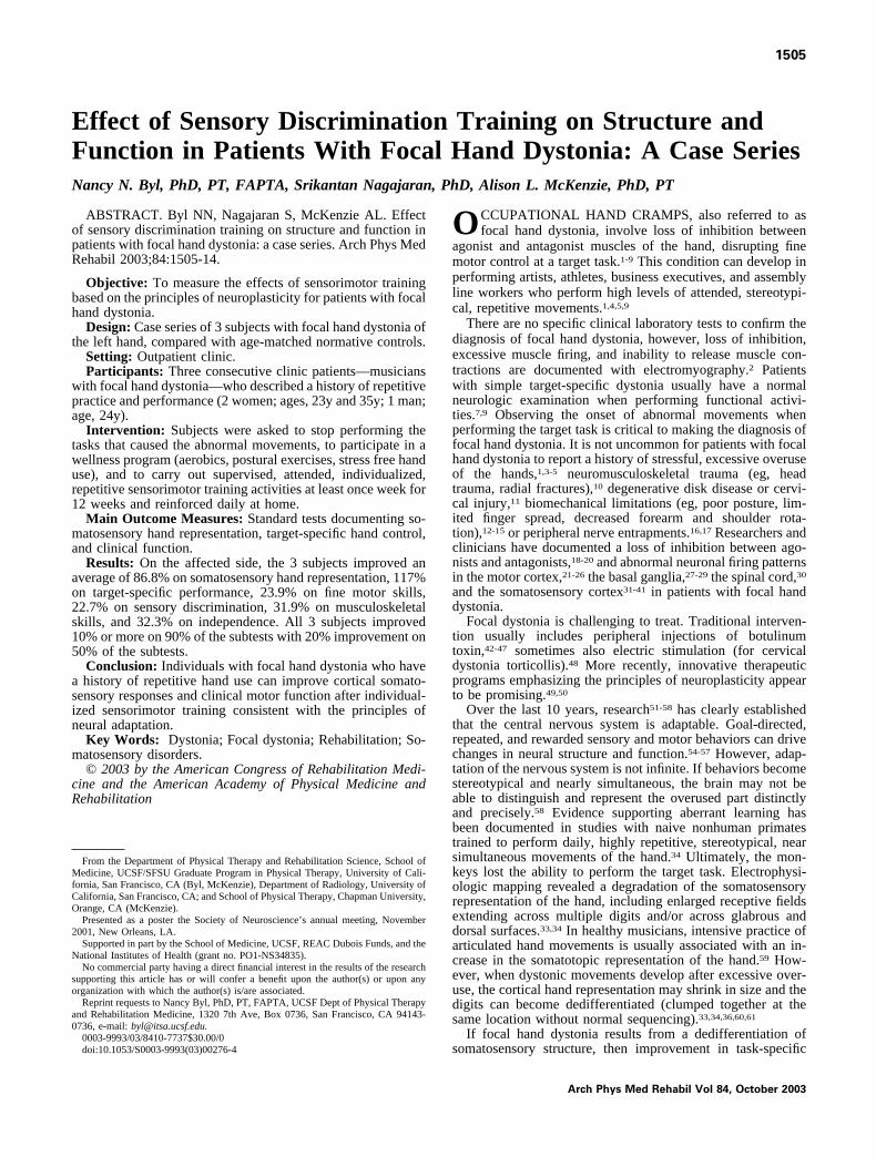
Fig 1. Pre-post changes in somatosensory structure: affected side for with focal hand dystonia subjects. After learning-based sensorimotortraining, there were objective improvements in neural structure, as measured by magnetic source imaging: (A) Increased area of somato-sensory representation of the hand, (B) improved sequencing of the digits from D1 to D5 in 2 of the 3 subjects, (C) similar amplitude andlatency of the somatosensory-evoked field potential between patients with focal hand dystonia and controls.
1508 SENSORIMOTOR TRAINING AND FOCAL HAND DYSTONIA, Byl
Arch Phys Med Rehabil Vol 84, October 2003
The dependent variables were not correlated,74 conse-quently, each dependent variable was considered an indepen-dent family.75 Changes in performance were calculated on eachsubtest as a percentage score and plotted for visual analysis.For descriptive purposes, performance levels of subjects withfocal hand dystonia were compared with reference controls.Inferences were made based on probability theory.75 On eachdependent variable, there was a 50:50 chance that each subjectwould improve or not improve on each subtest. Improvementwas defined as a gain of 20% or more. Digit sequencing couldnot be subjected to the 20% rule. Each dependent variableconsisted of 3 to 6 subtests. Thus, the probability that all 3subjects would improve 20% or more ranged from P less than.0156 (all 3 subjects improving �20% on all 3 subtests) to Pless than .00195 (all 3 subjects improving �20% on all 6subtests). This analysis was applied to determine the signifi-cance of the changes related to sensorimotor training.
RESULTS
Somatosensory-Evoked PotentialsAfter training, the somatosensory-dependent variables im-
proved an average of 86.8% across the 3 subjects (table 1). All3 subjects with focal hand dystonia increased the area of thehand representation by more than 20% and the amplitude of theSEPs by an average of 40%; 2 improved the sequential order-ing of the digits (fig 1). Only 2 parameters could be evaluatedon the 20% rule. The probability that all 3 subjects would
improve on all 6 measurements was P less than .0156. After thetraining, the area of the hand representations was similar on theaffected and unaffected sides, and was larger for subjects withfocal hand dystonia than for controls. Integrated across timeand across subjects with focal hand dystonia, the average SEPamplitude was similar to that of the controls.
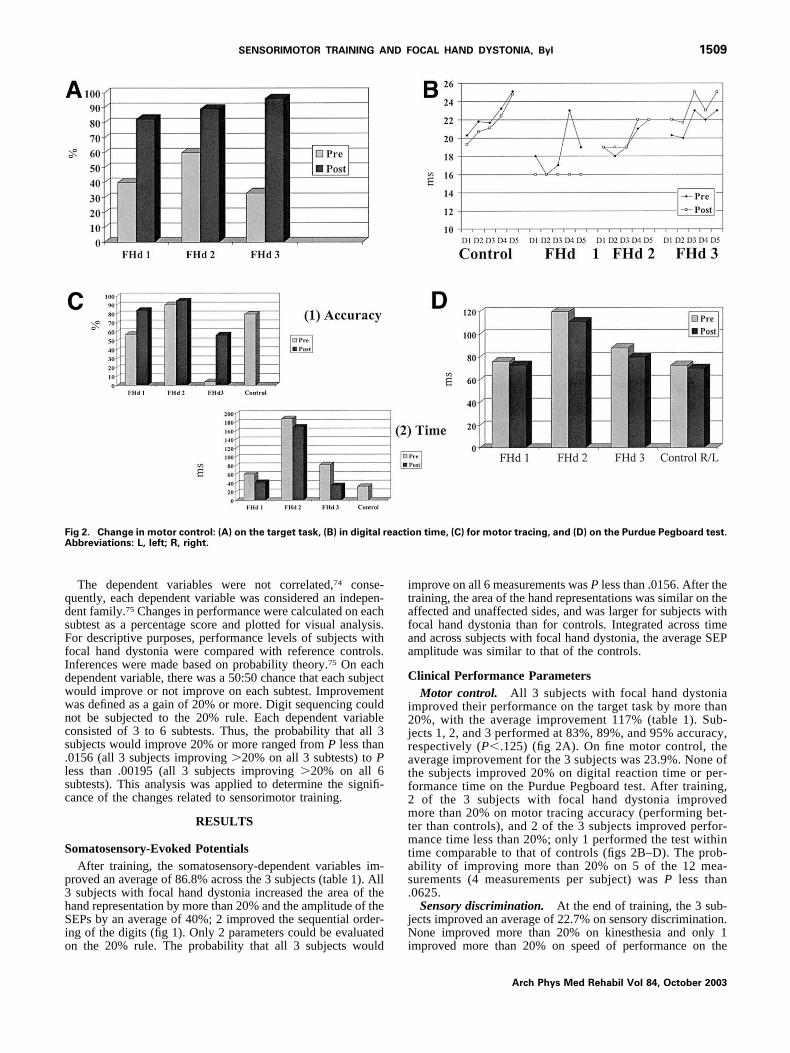
Clinical Performance ParametersMotor control. All 3 subjects with focal hand dystonia
improved their performance on the target task by more than20%, with the average improvement 117% (table 1). Sub-jects 1, 2, and 3 performed at 83%, 89%, and 95% accuracy,respectively (P�.125) (fig 2A). On fine motor control, theaverage improvement for the 3 subjects was 23.9%. None ofthe subjects improved 20% on digital reaction time or per-formance time on the Purdue Pegboard test. After training,2 of the 3 subjects with focal hand dystonia improvedmore than 20% on motor tracing accuracy (performing bet-ter than controls), and 2 of the 3 subjects improved perfor-mance time less than 20%; only 1 performed the test withintime comparable to that of controls (figs 2B–D). The prob-ability of improving more than 20% on 5 of the 12 mea-surements (4 measurements per subject) was P less than.0625.
Sensory discrimination. At the end of training, the 3 sub-jects improved an average of 22.7% on sensory discrimination.None improved more than 20% on kinesthesia and only 1improved more than 20% on speed of performance on the
Fig 2. Change in motor control: (A) on the target task, (B) in digital reaction time, (C) for motor tracing, and (D) on the Purdue Pegboard test.Abbreviations: L, left; R, right.
1509SENSORIMOTOR TRAINING AND FOCAL HAND DYSTONIA, Byl
Arch Phys Med Rehabil Vol 84, October 2003

stereognosis test. The subjects with focal hand dystonia per-formed more accurately than controls on 2-point discrimina-tion, localization, graphesthsia, and stereognosis, but perfor-mance time on the stereognosis task took longer than controls,still requiring approximately twice as much time (figs 3A–D).Over the 6 measurements performed by the 3 focal handdystonia subjects (ie, 18 measures), 11 measurements im-proved more than 20% (P�.0005).
Musculoskeletal performance parameters. At the end oftraining, the 3 subjects with focal hand dystonia improved31.9% in musculoskeletal performance. Two subjects im-proved more than 20% on all musculoskeletal parameters ex-cept posture and forearm rotation. The ratio of strength of thelumbricals to flexor profundus was higher for 2 of the subjectswith focal hand dystonia compared with controls, and allflexibility measurements became similar for focal hand dysto-nia subjects and controls (fig 4). The probability that the 3subjects would improve more than 20% on 11 of the 18measurements was P less than .0005.
Physical independence and work. The average improve-ment in work and independence for the 3 subjects with focalhand dystonia was 30.7%. These subjects were functioning at88%, 89%, and 92% of maximum independence and quality oflife—a level comparable to healthy subjects. Only 1 subjectwith focal hand dystonia improved more than 20% on inde-
pendence. All 3 improved more than 20% in work status. Twosubjects returned to their previous work (one to performancebut on a modified schedule, one to finish studies at the conser-vatory) (fig 5). The probability that the subjects would improvemore than 20% on 4 of the 6 measurements was .0625.
DISCUSSION
The present results add to the evidence that (1) patients withfocal hand dystonia with a history of repetitive overuse canhave measurable degradation of the hand representation in thesomatosensory cortex, (2) they have associated dysfunction insensory processing and fine motor control, and (3) dysfunctioncan be modified with an intervention program based on theprinciples of neuroplasticity. Sensorimotor training had to betailored to each subject, with appropriate progression of taskdifficulty. All subjects made significant progress performingthe target task, but only 2 returned to their musical careers, witha modified performance schedule.
On the affected side, the improvements in SEPs includedincreased amplitude, expanded representation of the hand, andimproved digit order on the z axis. Mean differences in ampli-tude of the SEPs have not been reported in other studies.1,36
Unique to the present study, the amplitude of the SEPs wasintegrated across time and across subjects, revealing noticeable
Fig 3. Change in sensory discrimination: (A) 2-point discrimination, (B) localization, (C) stereognosis accuracy, (D) stereognosis speed, (E)kinesthesia, and (F) grapesthesia.
1510 SENSORIMOTOR TRAINING AND FOCAL HAND DYSTONIA, Byl
Arch Phys Med Rehabil Vol 84, October 2003

variations in the early and late phases. Averaging amplitudesacross subjects would have concealed these differences.
Similar to other clinical studies, all 3 subjects had problemswith some elements of sensory discrimination,36-42 however,before the study they were unaware of these sensory problems.They were also surprised that increasing the cutaneous input totheir affected digits (eg, using tape) had a powerful effect onimproving fine motor control. Increased sensory cues can im-prove the ability of the nervous system to differentiate sensoryinformation and organize a motor response. Interestingly, 1
subject was hyposensitive to light touch, and 2 were hypersen-sitive. In the first case, sensorimotor training had to begin withintensive tactile stimuli to enable adequate discrimination. Inthe latter cases, tactile stimuli had to be lightly delivered withthe subjects positioned where sensory discrimination could beperformed without triggering abnormal muscle contractions(eg, supine with shoulder elevation and forearm pronation).Ultimately, sensorimotor training activities must be integratedinto the position in which the subject performs the target task.
As reported in other studies of hand dystonia,12-15 we alsomeasured limitations in flexibility in the hand and forearm inour subjects. Congenitally or traumatically induced restrictionsin joint or soft-tissue movements can create imbalances inmuscle performance, excessive shortening in 1 muscle andlengthening in a related muscle, as well as abnormal patterns ofmovement with excessive end range loading. The question iswhether these variations in mobility actually cause focal handdystonia10 or simply increase the risk of developing focal handdystonia when an individual is subjected to stressful, excessive,repetitive hand use. In 1 animal study,33,34 nonhuman primateswere trained to perform attended, repetitive, stressful, handopening and closing until they developed dystonic posturingthat interfered with the performance of the target task. Atautopsy, Topp and Byl13 described an anatomic defect of theflexor profundus tendon on D4 of the trained side and D3 onthe untrained side in 1 of the 6 monkeys trained. There were nosigns of motor dysfunction on the untrained side, suggestingthat restricted mobility was a risk factor but not a cause of focalhand dystonia.
Fig 4. Change in muskuloskeletal performance: (A) finger spread (abduction), (B) shoulder external rotation, (C) supination and promotion,(D) lumbricales: extensor digitorum ratio, (E) posture, and (F) neural tension.
Fig 5. Change in independence measures: (A) functional indepen-dence and (B) work status.
1511SENSORIMOTOR TRAINING AND FOCAL HAND DYSTONIA, Byl
Arch Phys Med Rehabil Vol 84, October 2003

Fine motor movements of the hand require sequenced, indi-viduated, fractionated movements.16 Normal motor reactiontime does not ensure skilled fine motor control. Because vol-untary, fine motor digital movements are controlled by variouscortical and subcortical pathways with accurate sensorimotorfeedback, it should not be surprising to find normal motorreaction time in subjects with focal hand dystonia. However,decreased fine motor accuracy and prolonged performance timeshould be expected.
There are significant constraints in a case-study design75,76:small sample size, lack of controls, no random selection orassignment, learning with retesting, the Hawthorne effect, biasin measurement because of lack of blinding, limited statisticalanalysis, and inability to generalize findings to a larger popu-lation. Some of these constraints were minimized in the presentstudy. An independent evaluator, unaware of study objectives,performed MSI; research assistants blinded to group assign-ment performed the clinical measurements; 2 control groupswere included for reference; and probability theory was appliedto provide insight regarding the significance of the measuredchanges. Because responses are individualized, single-subjectdesign repeated over multiple subjects can provide strong ar-guments in support of a theory. Averaging data in large groupscan be seriously biased by variability in measurement, skeweddistribution, and heterogeneity.
The present intervention study included a comprehensiveapproach to sensorimotor training. This type of multitask in-tervention does not isolate the effects of training on a singlesensory task (eg, Braille reading vs graphesthesia). In 1 study(including patients with writer’s cramp),77 Braille readingalone was associated with improved motor performance and areduction in the severity of cramping, but recovery of target-specific motor control was not 100%. Sensory training mayhave to drive changes not only in cortical area 3b but also inother sensory areas, including basal ganglia-thalamic-corticalpathways.
Multisite, controlled, randomized clinical trials are needed toconfirm that sensorimotor retraining normalizes somatosensorystructure, improves sensory discrimination, and restores normalfine motor control at target and nontarget tasks for patients withfocal hand dystonia. These studies should also include genetictesting to determine what proportion of subjects with focalhand dystonia have the DYT1 or other known gene,78-80 andshould combine treatment approaches based on crossover de-signs such as pairing botulinum toxin or limb immobilizationwith sensorimotor retraining. Future studies also must includemore rigorous strategies for assuring patient compliance forhome training activities. Studies are also needed to detail morespecifically training parameters (repetitions needed, spacing ofpractice, reward, progression of difficulty, length of training).
CONCLUSION
Aberrant learning may explain the development of focalhand dystonia in some patients who perform highly repetitive,stressful, stereotypical hand tasks. A conservative interventionstrategy based on the principles of neuroplasticity can improvesomatosensory structure and clinical function. However, 3 to 6months of supervised training, once a week, reinforced with aself-directed home program may not be sufficient to returnmusicians to professional levels of performance. Musiciansmay benefit from behavioral programs designed to help themmeet these objectives: maintain accurate somatosensory feed-back and healthy biomechanical movement strategies, incorpo-rate mental practice as 1 way to reduce the intensity and strainof physical practice, and avoid long periods of stressful, repet-
itive, alternating, stereotypical, near-simultaneous digitalmovements.
References1. Chen R, Hallett M. Focal dystonia and repetitive motion disorders.
Clin Orthop 1998;Jun(351):102-6.2. Cohen L, Hallett M. Hand cramps: Clinical features and electro-
myographic patterns in focal dystonia. Neurology 1988;38:1005-12.
3. Fry H. Overuse syndromes in musicians 100 years ago: an histor-ical review. Med J Aust 1986;146:620-5.
4. Hochberg F, Harris S, Blartert T. Occupational hand cramps:professional disorders of motor control. Hand Inj Sports PerformArts 1990;6:427-8.
5. Jankovic J, Shale H. Dystonia in musicians. Semin Neurol 1989;9:131-5.
6. Rhoad R, Stern P. Writer’s cramp–a focal dystonia: etiology,diagnosis, and treatment. J Hand Surg [Am] 1993;18:541-4.
7. Rothwell J, Oveso J, Day B, Marsden C. Pathophysiology ofdystonias. Adv Neurol 1983;39:851-63.
8. Sheehy M, Marsden C. Writer’s cramp: a focal dystonia. Brain1982;105:461-80.
9. Tubiana R. Incidence: classification of severity and results oftherapy, In: Winspur I, Parry CB, editors. The musician’s hand.London: Martin Dunitz; 1998. p 164-7.
10. Jancovic J. Can peripheral trauma induce dystonia and othermovement disorders? Yes. Mov Disord 2001;16:7-11.
11. Katz R, Williams C. Focal dystonia following soft tissue injury:three case reports with long-term outcomes. Arch Phys MedRehabil 1990;71:345-9.
12. Leijnse JN. Anatomical factors predisposing to focal dystonia inthe musician’s hand: principles, theoretical examples, clinicalsignificance. J Biomech 1996;30:659-69.
13. Topp KS, Byl NN. Movement dysfunction following repetitivehand opening and closing: anatomic analysis in owl monkeys.Mov Disord 1999;14:295-306.
14. Wilson F, Wagner C, Homberg V, Noth J. Interaction of biome-chanical and training factors in musicians with occupationalcramps/focal dystonia [abstract]. Neurology 1991;41(3 Suppl 1):291.
15. Wilson F, Wagner C, Homberg V. Biomechanical abnormalitiesin musicians with occupational cramp/focal dystonia. J Hand Ther1993;6:298-307.
16. Quartarone A, Girlanda P, Risitano G. Focal hand dystonia in apatient with thoracic outlet syndrome. J Neurol Neurosurg Psy-chiatry 1998;65:272-4.
17. Charness ME. Unique upper extremity disorders of musicians. In:Millender LH, Louis DS, Simmons BP, editors. Occupationaldisorders of the upper extremity. New York: Churchill Living-stone; 1992. p 117-51.
18. Chen R, Tsai C, Lu C. Reciprocal inhibition in writer’s cramp.Mov Disord 1995;10:556-61.
19. Nakashima K, Rothwell J, Day B, Thompson P, Shannon K,Marsden C. Reciprocal inhibition between forearm muscles inpatients with writer’s cramp and other occupational cramps,symptomatic hemidystonia and hemiparesis due to stroke. Brain1989;112:681-97.
20. Ridding M, Sheean G, Rothwell J, Inzelberg R, Kujirai T.Changes in the balance between motor cortical excitation andinhibition in focal, task specific dystonia. J Neurol NeurosurgPsychiatry 1995;59:493-8.
21. Chase T, Tamminga C, Burrows H. Positron emission tomo-graphic studies of regional cerebral glucose metabolism in idio-pathic dystonia. Adv Neurol 1988;50:237-41.
22. Defendini R, Fahn S. Magnetic resonance imaging of dystonicstates. Adv Neurol 1988;50:265-75.
23. Deuschl G, Toro C, Matsumoto J, Hallett M. Movement-relatedcortical potentials in writer’s cramp. Ann Neurol 1995;38:861-8.
24. Gilman S, Junck L, Young A. Cerebral metabolic activity inidiopathic dystonia studied with positron emission tomography.Adv Neurol 1988;50:231-6.
1512 SENSORIMOTOR TRAINING AND FOCAL HAND DYSTONIA, Byl
Arch Phys Med Rehabil Vol 84, October 2003

25. Mavroudais N, Caroyer J, Brunko E, Zegers de Beyl D. Abnormalmotor evoked responses to transcranial magnetic stimulation infocal dystonia. Neurology 1995;45:1671-7.
26. Tempel L, Perlmutter J. Abnormal cortical responses in patientswith writer’s cramp. Neurology 1993;43:2252-7.
27. Black KJ, Ongur D, Perlmutter JS. Putamen volume in idiopathicfocal dystonia. Neurology 1998;51:819-24.
28. Karbe H, Holfhof V, Rudolf J. Positron emission tomographydemonstrates frontal cortex and basal ganglia hypometabolism indystonia. Neurology 1992;42:1540-4.
29. Perlmutter JS, Stambuk MK, Markham J. Decreased [18F] piper-one binding in putamen in idiopathic focal dystonia. J Neurosci1997;17:843-50.
30. Kaji R, Rothwell J, Katayama M, et al. Tonic vibration reflex andmuscle afferent block in writer’s cramp. Ann Neurol 1995;38:155-62.
31. Odergren T, Iwasaki N, Borg J, Forssberg H. Impaired sensorymotor integration during grasping in writer’s cramp. Brain 1996;119(Pt 2):569-83.
32. Hallett M. Is dystonia a sensory disorder? Ann Neurol 1995;38:139-40.
33. Byl NN, Merzenich M, Jenkins WM. A primate genesis model offocal dystonia and repetitive strain injury: I. Learning-inducedde-differentiation of the representation of the hand in the primarysomatosensory cortex in adult monkeys. Ann Neurol 1996;47:508-20.
34. Byl NN, Merzenich M, Cheung S, Bedenbaugh P, Nagarajan S,Jenkins W. A primate model for studying focal dystonia andrepetitive strain injury: effects on the primary sensory cortex. PhysTher 1997;77:269-84.
35. Sanger TD, Pascual-Leone A, Tarsy D, Schlaug G. fMRI evidenceof sensory receptive fields spanning multiple fingers in writer’scramp. Mov Disord 2001;16:94-9.
36. Bara-Jiminez W, Catalan M, Hallett M. Abnormal somatosensoryhomunculus in dystonia of the hand. Ann Neurol 1998;44:828-31.
37. Byl NN, Wilson F, Scott P, Oakes A. Sensory dysfunction asso-ciated with repetitive strain injuries of tendinitis and focal handdystonia: a comparative study. J Ortho Sports Phys Ther 1996;23:234-44.
38. Byl NN, Hamati D, Melnick M, Wilson F, McKenzie A. Thesensory consequences of repetitive strain injury in musicians:focal dystonia of the hand. J Back Musculoskeletal Rehabil 1996;7:27-39.
39. Sanger TD, Tarsy D, Pascual-Leone A. Abnormalities of spatialand temporal sensory discrimination in writer’s cramp. Mov Dis-ord 2001;16:94-9.
40. Tinazzi M, Frasson E, Bertolasi L, Fiaschi A, Aglioti S. Temporaldiscrimination of somesthetic stimuli is impaired in dystonic pa-tients. Neuroreport 1999;20:1547-50.
41. Topp KS, Byl NN. Focal hand dystonia. Phys Ther Case Rep1997;1:39-52.
42. Brin M, Fahn S, Moskowitz C, et al. Localized injections ofbotulinum toxin for the treatment of focal dystonia and hemifacialspasm. Mov Disord 1987;2:237-54.
43. Cole R, Hallett M, Cohen L. Double-blind trial of botulinum toxinfor treatment of focal hand dystonia. Mov Disord 1995;10:466-71.
44. Karp B, Cole R, Cohen L, Grill S, Lou JS, Hallet M. Long-termbotulinum toxin treatment of focal hand dystonia. Neurology1994;44:70-6.
45. Pullman S, Greene P, Fahn S, Pedersen S. Approach to thetreatment of limb disorders with botulinum toxin A. Arch Neurol1996;53:617-24.
46. Tsui J, Bhatt M, Calne S, Calne D. Botulinum toxin in thetreatment of writer’s cramp: a double-blind study. Neurology1993;43:183-5.
47. Van den Bergh P, Francart J, Mourin S, Kollmann P, Laterre E.Five-year experience in the treatment of focal movement disorderswith low-dose Dysport botulinum toxin. Muscle Nerve 1995;18:720-9.
48. Goetz C, Penn R, Tanner C. Efficacy of cervical cord stimulationin dystonia. Adv Neurol 1988;50:645-9.
49. Byl NN, Nagarajan S, McKenzie AM. Treatment effectiveness ofpatients with a history of repetitive strain injury and focal hand
dystonia: a planned, prospective follow-up study. J Hand Ther2000;13:289-301.
50. Candia V, Elbert T, Altenmuller E, Rau H, Schafer T, Taub E.Constraint-induced movement therapy for focal hand dystonia inmusicians [letter]. Lancet 1999;253:42-3.
51. Jenkins WM, Merzenich M. Reorganization of neocortical repre-sentations after brain injury: a neurophysiological model of thebases of recovery from stroke. Prog Brain Res 1987;71:249-66.
52. Jenkins WM, Allard T, Nudo R. Cortical representational plastic-ity. In: Raskic P, Singer W, editors. Neurobiology of the neocor-tex. New York: John Wiley & Sons; 1988. p 41-67.
53. Jenkins W, Merzenich M, Ochs M, Allard T, Guic-Robles E.Functional reorganization of primary somatosensory cortex inadult owl monkeys after behaviorally controlled tactile stimula-tion. J Neurophysiol 1990;63:82-104.
54. Nudo R, Wise BS, Fuentes F, Milliken G. Neural substrates for theeffects of rehabilitative training on motor recovery after ischemicinfarct. Science 1996;272:1791-5.
55. Recanzone G, Jenkins W, Hradek G, Merzenich MM. Progressiveimprovement in discriminative abilities in adult owl monkeysperforming a tactile frequency discrimination task. J Neurophysiol1992;67:1015-30.
56. Xerri C, Coq JO, Merzenich MM, Jenkins WM. Experience-induced plasticity of cutaneous maps in the primary somatosen-sory cortex of adult monkeys and rats. J Physiol (Paris) 1996;90:277-87.
57. Byl NN, Merzenich MM. Principles of neuroplasticity: implica-tions for neurorehabilitation and learning. In: Gonzalez EG, MyersSJ, Edelstein JE, Lieberman JS, Downey JA. Downey and Dar-ling’s physiological basis of rehabilitation medicine. Boston: But-terworth-Heinemann; 2001. p 609-28.
58. Wang X, Merzenich MM, Somethethesa K, Jenkins WM. Remod-eling of hand representation in adult cortex determined by timingof tactile stimulation. Nature 1993;378:71-5.
59. Elbert T, Pantev C, Wienbruch C, Rockstroh B, Taub E. Increasedcortical representation of the fingers of the left hand in stringplayers. Science 1995;270:305-7.
60. McKenzie AM, Nagaragan S, Roberts T, Merzenich MM, BylNN. Somatosensory changes and clinical correlates in patientswith focal hand dystonia. Poster presented at: Society for Neuro-science Annual Meeting; 2001 Nov 10-15; San Diego (CA).
61. Byl NN, Merzenick MM, Roberts T, Nagarajan S, McKenzie AM.The correlation of clinical neuromusculoskeletal and central so-matosensory parameters in patients with focal hand dystonia.Poster presented at: Society for Neuroscience Annual Meeting;1998 Nov 7-12; Los Angeles (CA).
62. Roberts T, Poeppel D, Rowley HA. Magnetoencephalography andmagnetic source imaging. Neuropsychiatry Neuropsychol BehavNeurol 1998;11:49-64.
63. Rowley HA, Roberts T. Functional localization by magnetoen-cephalography. Neuroimaging Clin North Am 1995;5:695-710.
64. Ayres A. Sensory integration and praxis tests: SIPT manual. LosAngeles: Western Psychological Assoc; 1989.
65. Bohannon RW. Stopwatch for measuring thumb movement time.Percept Mot Skills 1995;81:122-6.
66. Carlsson AM. Assessment of chronic pain. I. Aspects of thereliability and validity of the visual analogue scale. Pain 1983;16:87-101.
67. Edgelow P, Feinberg S. Controversies and questions: thoracicoutlet syndrome. Calif Workers Compensation Enquirer 1995;7:10-3.
68. Fung S, Byl NN, Melnick M, Callahan P, et al. Functional out-comes: the development of a new instrument to monitor theeffectiveness of physical therapy. Eur J Phys Med Rehabil 1997;7:31-41.
69. Kendall FP, McCreary EK, Provance PG. Muscles: testing andfunction. 4th ed. Philadelphia: Williams & Wilkins; 1993.
70. Instructions and normative data: Purdue Pegboard. Lafayette (IN):Lafayette Instrument Co; 1996.
71. Norkin CC, White DJ. Measurement of joint motiona guide togoniometry. Philadelphia: FA Davis; 1995.
1513SENSORIMOTOR TRAINING AND FOCAL HAND DYSTONIA, Byl
Arch Phys Med Rehabil Vol 84, October 2003

72. Schmidt RT, Toews JV. Grip strength as measured by the Jamardynamometer. Arch Phys Med Rehabil 1970;51:321-7.
73. Shimokata H, Kuzuya F. Two-point discrimination test of the skinas an index of sensory aging. Gerontology 1995;42:267-72.
74. Byl NN, Nagarajan S, McKenzie AM. Effectiveness of sensori-motor training on structure and function in musicians with focalhand dystonia: three case studies. Poster presented at: Society forNeuroscience Annual Meeting; 2000 Nov 7; New Orleans (LA).
75. Maraschuilo LA, Serlin RC. Statistical methods for social andbehavioral sciences. New York: WH Freeman; 1988.
76. Portney LG, Watkins MP. Foundations of clinical research: ap-plications to practice. Upper Saddle River: Prentice Hall; 2000. p223-58.
77. Gerloff C, Corwell B, Chen R, Hallett M, Cohen LG. The role ofthe human motor cortex in the control of complex and simplefinger movement sequences. Brain 1998;121(Pt 9):1695-709.
78. Zeuner KE, Bara-Jimenez W, Noguchi PS, Goldstein SR, Ambro-sia JM, Hallett M. Sensory training for patients with focal handdystonia. Ann Neurol 2002;451:593-8.
79. Leube B, Rudnicki D, Ratzlaff T, Kessler K, Benecke R, Au-burger G. Idiopathic torsion dystonia: assignment of a gene tochromosome 18p in a German family with adult onset, autosomaldominant inheritance and purely focal distribution. Hum MolGenet 1996;5:1673-7.
80. Gasser T, Bove C, Ozelius L, et al. Haplotype analysis at theDYT1 locus in Ashkenazi Jewish patients with occupational handdystonia. Mov Disord 1996;11:163-6.
Suppliera. Magnes®; Biomagnetic Technologies Inc, 9727 Pacific Heights
Blvd, San Diego, CA 92121-3719.
1514 SENSORIMOTOR TRAINING AND FOCAL HAND DYSTONIA, Byl
Arch Phys Med Rehabil Vol 84, October 2003