Effect of Neofabraea Alba on Bark and Wood Anatomy of Fraxinus Spp
10
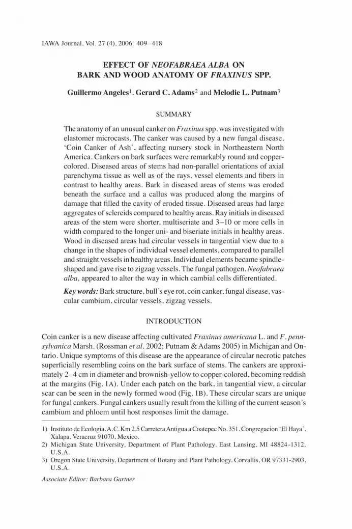
IAWA Journal, Vol. 27 (4), 2006: 409–418 EFFECT OF NEOFABRAEA ALBA ON BARK AND WOOD ANATOMY OF FRAXINUS SPP. Guillermo Angeles 1 , Gerard C. Adams 2 and Melodie L. Putnam 3 SUMMARY The anatomy of an unusual canker on Fraxinus spp. was investigated with elastomer microcasts. The canker was caused by a new fungal disease, ʻCoin Canker of Ashʼ, affecting nursery stock in Northeastern North America. Cankers on bark surfaces were remarkably round and copper- colored. Diseased areas of stems had non-parallel orientations of axial parenchyma tissue as well as of the rays, vessel elements and fibers in contrast to healthy areas. Bark in diseased areas of stems was eroded beneath the surface and a callus was produced along the margins of damage that filled the cavity of eroded tissue. Diseased areas had large aggregates of sclereids compared to healthy areas. Ray initials in diseased areas of the stem were shorter, multiseriate and 3–10 or more cells in width compared to the longer uni- and biseriate initials in healthy areas. Wood in diseased areas had circular vessels in tangential view due to a change in the shapes of individual vessel elements, compared to parallel and straight vessels in healthy areas. Individual elements became spindle- shaped and gave rise to zigzag vessels. The fungal pathogen, Neofabraea alba, appeared to alter the way in which cambial cells differentiated. Key words: Bark structure, bullʼs eye rot, coin canker, fungal disease, vas- cular cambium, circular vessels, zigzag vessels. INTRODUCTION Coin canker is a new disease affecting cultivated Fraxinus americana L. and F. penn- sylvanica Marsh. (Rossman et al. 2002; Putnam & Adams 2005) in Michigan and On- tario. Unique symptoms of this disease are the appearance of circular necrotic patches superficially resembling coins on the bark surface of stems. The cankers are approxi- mately 2– 4 cm in diameter and brownish-yellow to copper-colored, becoming reddish at the margins (Fig. 1A). Under each patch on the bark, in tangential view, a circular scar can be seen in the newly formed wood (Fig. 1B). These circular scars are unique for fungal cankers. Fungal cankers usually result from the killing of the current seasonʼs cambium and phloem until host responses limit the damage. 1) Instituto de Ecologia, A.C. Km 2,5 Carretera Antigua a Coatepec No. 351, Congregacion ʻEl Hayaʼ, Xalapa, Veracruz 91070, Mexico. 2) Michigan State University, Department of Plant Pathology, East Lansing, MI 48824-1312, U.S.A. 3) Oregon State University, Department of Botany and Plant Pathology, Corvallis, OR 97331-2903, U.S.A. Associate Editor: Barbara Gartner
-
Upload
un-lincoln -
Category
Documents
-
view
0 -
download
0
Transcript of Effect of Neofabraea Alba on Bark and Wood Anatomy of Fraxinus Spp
IAWA Journal, Vol. 27 (4), 2006: 409–418
EFFECT OF NEOFABRAEA ALBA ON BARK AND WOOD ANATOMY OF FRAXINUS SPP.
Guillermo Angeles1, Gerard C. Adams2 and Melodie L. Putnam3
SUMMARY
The anatomy of an unusual canker on Fraxinus spp. was investigated with elastomer microcasts. The canker was caused by a new fungal disease, ʻCoin Canker of Ashʼ, affecting nursery stock in Northeastern North America. Cankers on bark surfaces were remarkably round and copper-colored. Diseased areas of stems had non-parallel orientations of axial parenchyma tissue as well as of the rays, vessel elements and fibers in contrast to healthy areas. Bark in diseased areas of stems was eroded beneath the surface and a callus was produced along the margins of damage that filled the cavity of eroded tissue. Diseased areas had large aggregates of sclereids compared to healthy areas. Ray initials in diseased areas of the stem were shorter, multiseriate and 3–10 or more cells in width compared to the longer uni- and biseriate initials in healthy areas. Wood in diseased areas had circular vessels in tangential view due to a change in the shapes of individual vessel elements, compared to parallel and straight vessels in healthy areas. Individual elements became spindle-shaped and gave rise to zigzag vessels. The fungal pathogen, Neofabraea alba, appeared to alter the way in which cambial cells differentiated.
Key words: Bark structure, bull s̓ eye rot, coin canker, fungal disease, vas-cular cambium, circular vessels, zigzag vessels.
INTRODUCTION
Coin canker is a new disease affecting cultivated Fraxinus americana L. and F. penn-sylvanica Marsh. (Rossman et al. 2002; Putnam & Adams 2005) in Michigan and On-tario. Unique symptoms of this disease are the appearance of circular necrotic patches superficially resembling coins on the bark surface of stems. The cankers are approxi-mately 2–4 cm in diameter and brownish-yellow to copper-colored, becoming reddish at the margins (Fig. 1A). Under each patch on the bark, in tangential view, a circular scar can be seen in the newly formed wood (Fig. 1B). These circular scars are unique for fungal cankers. Fungal cankers usually result from the killing of the current season s̓ cambium and phloem until host responses limit the damage.
1) Instituto de Ecologia, A.C. Km 2,5 Carretera Antigua a Coatepec No. 351, Congregacion ̒ El Hayaʼ, Xalapa, Veracruz 91070, Mexico.
2) Michigan State University, Department of Plant Pathology, East Lansing, MI 48824-1312, U.S.A.
3) Oregon State University, Department of Botany and Plant Pathology, Corvallis, OR 97331-2903, U.S.A.
Associate Editor: Barbara Gartner
IAWA Journal, Vol. 27 (4), 2006410 411Angeles, Adams & Putnam — Coin canker in Fraxinus
The causal agent of coin canker is Neofabraea alba (E.J. Guthrie) Verkley (= Pezi-cula alba E.J. Guthrie) which has been verified by studies of morphology, DNA sequences (Rossman et al. 2002) and Kochʼs postulates (Putnam & Adams 2005). However, only the asexual fruiting bodies of the fungus are found on ash, so reports of the pathogen are often under the asexual name, Phylactema vagabunda Desm. The pathogen is one of four species of Neofabraea known to cause bullʼs eye rot on Malus and Pyrus fruit (Henriquez et al. 2004). The different Neofabraea species are diffi-cult to distinguish morphologically and molecular studies using DNA sequences are clarifying the role of each species in bullʼs eye rot (De Jong et al. 2001). For example, N. alba was thought to be present only in Northeastern North America (Gariepy et al. 2003) but is now known to occur in the Pacific Northwest (Henriquez et al. 2004). The name bullʼs eye rot refers to the appearance of the disease on apple fruit following four or more months in cold post-harvest storage (Spotts 1990). The pathogens also cause annual cankers on Malus and Pyrus spp., except for Neofabraea krawtzewii which causes cankers on Populus spp. (De Jong et al. 2001). The cankers on Malus, Pyrus and Populus, however, do not exhibit the circular patches seen in Fraxinus spp. Coin canker is reported from cultivars of Fraxinus spp. that are initially grown in Oregon and shipped to Michigan and Ontario. Disease symptoms generally appear two or more years after transport. The disease causes significant losses as the cankers disfig-ure the stems, decreasing the aesthetic appeal for customers. Additionally, many (5–25) coin cankers often occur along a stem and are believed to seriously disrupt phloem transport. Disease losses may approach $700,000 per year for individual Michigan nurseries. The aetiology of the disease is controversial to the plant industry because it is unfamiliar, and because the Northeastern industry would like to recoup their losses from the Northwestern producers by claiming the trees arrive infected. However, the inoculum most likely originates in the Northeast as the disease symptoms appear in the current yearʼs growth two years after transport. In this study our objective is to compare the anatomy of healthy and diseased portions of stems to describe the symptoms produced on Fraxinus by the infecting agent.
MATERIALS AND METHODS
Healthy and diseased stems of Fraxinus spp. were collected in the field in the state of Michigan, USA. Bark and wood samples of approximately 1.5 cm2 were taken from healthy and diseased areas from the main stem of single-stemmed trees. These were nursery trees approximately six years old with 2.5–3.5 cm stems. Samples were im-mersed in a solution of equal parts of glycerine, water and ethyl alcohol (GAA) in vials, and autoclaved at 1.03 × 10⁻⁴ MPa (15 p.s.i.) for 30 minutes. After the samples reached room temperature, the vials were closed and stored for several weeks.
Microscopy Material was separated for light microscopy and scanning electron microscopy. For light microscopy, samples from the GAA were washed in running tap water and transferred to a 10% aqueous solution of polyethylene glycol of 1500 MW (PEG) in

IAWA Journal, Vol. 27 (4), 2006410 411Angeles, Adams & Putnam — Coin canker in Fraxinus
vials. With the vials open, samples were left in the oven at 65 °C for 48 hours, then transferred to 100% PEG and left in the oven at 65 °C for six hours. Then each sample was placed in a cube of a plastic ice cube tray and kept at room temperature until the PEG solidified to form blocks. The blocks were wrapped individually in two layers of paper towels and stored in a glass desiccator. Blocks were affixed to wood blocks us-ing melted PEG, and sectioned transversally and tangentially at 25 μm thickness with a sliding microtome. Sections were placed on glass microscope slides and tied with cotton thread. After washing in tap water to remove the PEG, sections were stained in safranin-fast green (Ruzin 1999) and mounted in synthetic resin. Microscopical ob-servations were made with a Nikon microscope (Eclipse E600). Digital images were taken with a Nikon Coolpix 600 camera.
SEM Samples used for obtaining sections were washed in running tap water to remove the PEG, and then oven-dried at 65 °C for two days. They were mounted in brass barrels with double-sided adhesive tape and sputter-coated with gold-palladium. Observations were made using a JEOL SEM, model JSM 5600LV. Obtained images were stored digitally.
Microcasting Branch segments 10–12 cm long were split longitudinally in quarters, separating healthy and diseased zones. After drying in the oven at 80 °C for 48 hours, they were placed separately in polyethylene bags of 20 cm long, following the method described by André (2002). After tying the lower end of the tubes, they were filled with a reticu-lant elastomer, Rhodorsil RTV (Rhône-Poulenc, Marseille, France). Leaving the upper portions of the tubes open, they were placed in a wire support inside a glass column (60 × 5 cm) especially designed for this purpose (André 2002). A vacuum of 0.1 MPa (1 atm) was applied for approximately 20 minutes until no more gas bubbles escaped from the samples; then they were brought back to atmospheric pressure. Samples were taken out of the plastic tubes and the excess elastomer was removed from the surface. Afterwards, the samples were placed in the oven at 80 °C for two additional days, then removed from the oven and equilibrated to room temperature. Excess solidified elastomer was cut from one end of each sample with a razor blade to expose the tissue. Samples embedded in elastomer were then immersed in concentrated sulphuric acid in an ice bath to digest the plant tissue. The ice was replaced as it melted during the first hour to avoid overheating of the polysiloxane. After 24 hours, the elastomer moulds were taken out of the sulphuric acid and washed in a 10% solution of sodium bicarbo-nate. This solution was changed several times until no more bubbles were produced. Then, the moulds were washed several times in tap water. The moulds were examined under the compound microscope to identify vessel micro casts. Vessel micro casts were then examined under the SEM after samples were air-dried and sputter-coated with gold-palladium. Observations were made using 15 kV current and digital images were stored as digital files.
IAWA Journal, Vol. 27 (4), 2006412 413Angeles, Adams & Putnam — Coin canker in Fraxinus
✽ ✽
A
B
C D
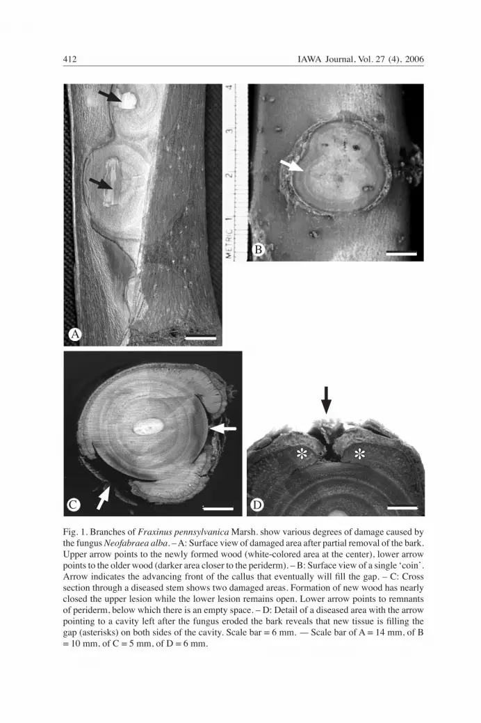
Fig. 1. Branches of Fraxinus pennsylvanica Marsh. show various degrees of damage caused by the fungus Neofabraea alba. – A: Surface view of damaged area after partial removal of the bark. Upper arrow points to the newly formed wood (white-colored area at the center), lower arrow points to the older wood (darker area closer to the periderm). – B: Surface view of a single ̒ coinʼ. Arrow indicates the advancing front of the callus that eventually will fill the gap. – C: Cross section through a diseased stem shows two damaged areas. Formation of new wood has nearly closed the upper lesion while the lower lesion remains open. Lower arrow points to remnants of periderm, below which there is an empty space. – D: Detail of a diseased area with the arrow pointing to a cavity left after the fungus eroded the bark reveals that new tissue is filling the gap (asterisks) on both sides of the cavity. Scale bar = 6 mm. — Scale bar of A = 14 mm, of B = 10 mm, of C = 5 mm, of D = 6 mm.

IAWA Journal, Vol. 27 (4), 2006412 413Angeles, Adams & Putnam — Coin canker in Fraxinus
RESULTS
The effects of the fungal disease were apparent macroscopically on the tangential sur-face of the periderm of stems as round to elliptical scars (cankers) up to 4 cm in diam-eter (Fig. 1A, B). A section cut across one of these scars (Fig. 1C, D) revealed that most of the bark tissue had been eroded. The wood also reacted to this damage (Fig 1D). A callus was produced along the margins of damage that filled the gap created by the fungus (Fig. 1D).
9
✽
✽
✽
✽
A B
C D
Fig. 2. –A: Tangential section through the secondary xylem of a healthy area of the branch shows that rays are homocellular, exclusively of procumbent cells, and 1–3 cells wide (arrow). – B: Tangential section through the secondary xylem shows tissue that has lost its ordered pat-tern in the diseased area. Furthermore, rays of several cells in width have lost their fusiform shape (white arrow) while vessels (asterisks) and fibers (arrowheads) have an undulate pattern. – C: Detail of B, showing a circular vessel almost closing on itself (left), and undulated fibers (arrowhead). – D: Tangential view of wood from a diseased area reveals that some vessel elements (asterisks) are very short and connected to their neighbors laterally. — Scale bar of A & B = 100 μm, of C = 45 μm, of D = 50 μm.

IAWA Journal, Vol. 27 (4), 2006414 415Angeles, Adams & Putnam — Coin canker in Fraxinus
A
B
CD
Fig. 3. Resin microcasts of vessels from healthy and diseased zones. – A: A cast of a vessel segment from a healthy branch reveals that the segment is composed of six parallel and straight vessel elements of similar lengths and diameters (LM). – B: Same as A (SEM). – C: A cast of a vessel segment from a diseased branch shows more than 80 zigzag vessel elements (LM). – D: Scanning electron micrograph of a vessel segment cast from a diseased branch shows that individual vessel elements are much reduced in length and diameter compared to those from healthy branches. — Scale bar of A & B = 100 μm, of C = 160 μm, of D = 200 μm.

IAWA Journal, Vol. 27 (4), 2006414 415Angeles, Adams & Putnam — Coin canker in Fraxinus
X
X f f
f
ph
ph
ph
S
S
n
✽
X
✽
✽
✽
A
B C
D E F
G
H I
trast with the dark background. – C: Same view as B, as seen under bright field. – D: Trans-verse section of a damaged branch, showing the old (X) and newly formed secondary xylem (asterisk) and bark. At the upper left, a new phellogen (arrow), formed as a response to the fungal attack, separates the original bark (formed before wounding) from the new one. – E: Detail of D, showing the phloem of the old bark (ph) and the new phellogen (n). – F: Trans-verse section of healthy bark. Note the predominance of fibers (f); ph = secondary phloem (col-lapsed). – G: Longitudinal section of a damaged branch, showing the xylem (X) and the damaged bark (arrow, right at the ʻcoinʼ). Asterisks indicate the new xylem formed after the damage. – H: Detail of the upper portion of Fig. G, showing the newly formed wood (aster-isk), where vessels are seen in transverse section. Abundant sclereids (S) can be seen in the bark. – I: Detail of sclereids (arrow) shown in H. — Scale bar of A & B = 1 mm, of C = 360 μm, of D = 1.5 mm, of E = 300 μm, of F = 200 μm, of G = 250 μm, of H & I = 370 μm.
Fig. 4. Bark anatomy of healthy and damaged branches. – A: Longitudinal section of a healthy branch, showing xylem (X) and bark. – B: Longitudinal section of a healthy branch, seen with polarized light. Fibers (f) and sclereids (arrow) con-
IAWA Journal, Vol. 27 (4), 2006416 417Angeles, Adams & Putnam — Coin canker in Fraxinus
Tangential sections of the secondary xylem of a healthy zone showed homogeneous rays made exclusively of procumbent cells. The rays were two cells wide and of vari-able height (Fig. 2A). The fibers and vessel elements were parallel in this zone. The diseased zone, on the contrary, showed non-parallel orientations of axial parenchyma tissue as well as of the rays, vessel elements and fibers (Fig. 2B). Rays in that zone were shorter and more than two cells wide (Fig. 2B, C). Vessels in diseased areas (Fig. 2C, D) departed from the parallel orientation observed in the healthy areas due to a change in the shapes of individual vessel elements. Elastomer casts of vessels (Fig. 3) showed that the secondary xylem of diseased stems or branches produced spindle-shaped vessel elements (Fig. 3C, D), which formed zigzag vessels, as described by André (2002). Healthy secondary xylem, in contrast, produced vessel elements whose walls were parallel in longitudinal sections, and were of uniform length along the whole vessel (Fig. 3A, B). The zigzag vessels sometimes gave rise to circular vessels, which are circular in the tangential view (Fig. 2B, C) and which are common in wound tissue (André 2002). The bark of healthy branches shows a single phellogen, a few sclereids in the cortex and several files of fibers (Fig. 4A–C, F). The few sclereids found in the bark form small clusters (Fig. 4B, C). In transverse section, thick bundles of fibers can be seen in the bark of healthy branches (Fig. 4F). Transverse sections of healthy branches (Fig. 4D, E) show that a wound phellogen forms new tissues, which force the original bark (formed previously to wounding) outwards. The fungus destroys large areas of bark (Fig. 4G), leaving the wood under that area exposed. New wood and bark forms later, trying to cover the gap (Fig. 4G). The newly formed bark forms large clusters of sclereids, but no fibers (Fig. 4H, I).
DISCUSSION
The unique symptoms of this disease are the appearance of circular, brown-colored patches on the bark surface of stems. Some weeks after the appearance of the first symp-toms, the margin of the patch cracks and the bark below the circular canker is completely eroded, leaving behind a cavity. The damage soon spreads to the wood beneath the bark canker. The tree responds to this damage by producing a callus-like tissue in the wood, covering the damaged area. The bark produced by the callus is rich in sclereids. The fungus apparently affects both the bark and the vascular cambium of Fraxinus spp. either directly, or by inducing the plant to secrete plant growth substances that act directly on the living tissues. Gibberellins are commonly formed during wound healing and they can induce sclereid formation in the bark. Savidge (1983) reported sclereid formation in callus derived from active cambial explants in Pinus contorta, even though this species normally does not produce sclereids either in the xylem or in the phloem. Ethylene is also a plant growth promoter that forms abundantly as a response to wounding (Aloni 1980). Circular vessels, as we observed in diseased wood, had been reported previously by Aloni & Wolf (1985), Hejnowicz & Kurczyńska (1987), Kurczyńska & Hejnowicz (1991), Larson (1994), Lev-Yadun (1996), and Angeles et al. (2002), among others.
IAWA Journal, Vol. 27 (4), 2006416 417Angeles, Adams & Putnam — Coin canker in Fraxinus
André (2002) defined circular vessels as those vessels in loops, closed on themselves, seen in the tangential plane of the stem, which frequently formed concentric rings. André (2002) proposed that circular vessel form at specific places in stems and roots where two axes join, for example, along the orthogonal lines that separate the files of wood formed by the stem and those of a branch. Circular vessels have also been observed at scars left by shed branches (André 2002) and by wound wood, as in the case reported here. Recently, Rothwell and Lev-Yadun (2005) reported for the first time the pres-ence of circular vessels (which they called indistinctly spiral xylem, circular-patterned secondary xylem, or simply circular secondary xylem) in fossil wood of Archaeopteris, from the upper Devonian (375 million years ago). They claim that this was the earliest clear evidence of polar auxin flow. Zigzag vessels have been defined by André (2002) as those vessels in which one part of their elements are connected ̒ tail-to-head ̓by their terminal or lateral perforations. In tangential or transverse sections these elements are not detectable, since they overlap with neighboring elements. These elements are visible only with microcasting. André (2002) provides wonderful examples of zigzag vessels obtained from nodal zones of tomato (Lycopersicum sp.) and from the insertion zone of the stem and adventitious roots in Ipomoea learii. He points out that these types of vessels are very common at the zone of implantation of the hemiparasite Viscum album with its host Malus sp., and in the galls induced in the rose by Agrobacterium tumefaciens. The adoption of a zigzag path by vessels apparently does not affect water transport, since they do not impair kh or ks values under well-watered conditions in pneumatophores of Laguncu-laria racemosa (L.) Gaertn. (Angeles et al. 2002). The diseased Fraxinus spp. with circular vessels of zigzag elements apparently did not have the risk of dying from embolisms. The coin cankers were annual and when they reached a certain diameter they stopped expanding, so there was no risk of a single canker girdling a tree. However, the cankers caused local damage to the secondary phloem and, in some instances, many cankers occurred in close proximity and coa-lesced, damaging a larger area of stem in the first season. This would have disrupted the distribution of the products of photosynthesis and have a more serious impact on tree health.
ACKNOWLEDGEMENTS
The senior author is indebted to J.P. André for his generosity in training him in microcasting and for the hospitality provided in Antibes and Nice, France. The assistance of Miss Carolina Madero and Mr. Fernando Ortega, of the Instituto de Ecología, A.C., of Xalapa, Veracruz, Mexico, is fully ap-preciated. The hospitality of Dr. Frank Ewers, in whose laboratory this project was initiated, is also appreciated.
REFERENCES
Aloni, R. 1980. Role of auxin and sucrose in the differentiation of sieve and tracheary elements in plant tissue cultures. Planta 150: 255–263.
Aloni, R. & A. Wolf. 1985. Suppressed buds embedded in the bark across the bole and the occur-rence of their circular vessels in Ficus religiosa. Amer. J. Bot. 71: 1060–1066.
IAWA Journal, Vol. 27 (4), 2006418
André, J.-P. 2002. Organisation vasculaire des angiospermes: une vision nouvelle. INRA edi-tions, Paris. 145 pp.
Angeles, G., J. López-Portillo & F. Ortega-Escalona. 2002. Functional anatomy of the second-ary xylem of roots of the mangrove Laguncularia racemosa (L.) Gaertn. (Combretaceae). Trees 16: 338–345.
De Jong, S.N., C.A. Levesque, G.J.M. Verkley, E.C.A. Abeln, J.E. Rahe & G. Braun. 2001. Phylogenetic relationships among Neofabraea species causing tree cankers and bullʼs-eye rot of apple based on DNA sequencing of ITS nuclear rDNA, mitochondrial rDNA and the β-tubulin gene. Mycol. Res. 105: 658–669.
Gariepy, T.D., C.A. Levesque, S.N. de Jong & J.E. Rahe. 2003. Species specific identification of the Neofabraea pathogen complex associated with pome fruits using PCR and multiplex DNA amplification. Mycol. Res. 107: 528–536.
Hejnowicz, Z. & E.U. Kurczyńska. 1987. Occurrence of circular vessels in isolated segments of Fraxinus excelsior. Acta Soc. Bot. Pol. 56: 415–419.
Henriquez, J.L., D. Sugar & R.A. Spotts. 2004. Etiology of bullʼs eye rot of pear caused by Neofabraea spp. in Oregon, Washington, and California. Plant Disease 88: 1134–1138.
Kurczyńska, E.V. & Z. Hejnowicz. 1991. Differentiation of circular vessels in isolated segments of Fraxinus excelsior. Physiol. Plant. 83: 275–280.
Larson, Ph.R. 1994. The vascular cambium: development and structure. In: T.E. Timell (ed.), Springer Series in Wood Science. Springer, New York. 725 pp.
Lev-Yadun, S. 1996. Circular vessels in the secondary xylem of Arabidopsis thaliana. IAWA J. 17: 31–35.
Putnam, M.L. & G.C. Adams. 2005. Phylactema vagabunda causes coin canker of ash (Fraxinus spp.) in North America. Plant Disease 89: 773.
Rossman, A.Y., L.A. Castlebury, G.C. Adams & M.L. Putnam. 2002. Phylactema vagabunda isolated from coin canker of ash trees in Michigan. Plant Disease 86: 442.
Rothwell, G.W. & S. Lev-Yadun. 2005. Evidence of polar auxin flow in 375 million-year-old fossil wood. Amer. J. Bot. 92: 903–906.
Ruzin, S.E. 1999. Plant microtechnique and microscopy. Oxford University Press, New York. 322 pp.
Savidge, R.A. 1983. The role of plant hormones in higher plant cellular differentiation. II. Ex-periments with the vascular cambium, and sclereid and tracheid differentiation in the pine, Pinus contorta. Histochem. J. 15: 447–466.
Spotts, R.A. 1990. Bullʼs eye rot . In: A.L. Jones & H.S. Aldwinckle (eds.), Compendium of apple and pear diseases: 56. American Phytopathological Society, St. Paul, Minnesota.