Application of engineered extracellular vesicles for targeted ...
Upload
independentCategory
view
5download
0
Effect of engineered TiO2 and ZnO nanoparticleson erythrocytes, platelet-rich plasma and giantunilamelar phospholipid vesiclesŠimundić et al.
Šimundić et al. BMC Veterinary Research 2013, 9:7http://www.biomedcentral.com/1746-6148/9/7

Šimundić et al. BMC Veterinary Research 2013, 9:7http://www.biomedcentral.com/1746-6148/9/7
RESEARCH ARTICLE Open Access
Effect of engineered TiO2 and ZnO nanoparticleson erythrocytes, platelet-rich plasma and giantunilamelar phospholipid vesiclesMetka Šimundić1, Barbara Drašler2, Vid Šuštar3, Jernej Zupanc2, Roman Štukelj4, Darko Makovec5, Deniz Erdogmus6,Henry Hägerstrand7, Damjana Drobne2 and Veronika Kralj-Iglič4*
Abstract
Background: Massive industrial production of engineered nanoparticles poses questions about health risks to livingbeings. In order to understand the underlying mechanisms, we studied the effects of TiO2 and ZnO agglomeratedengineered nanoparticles (EPs) on erythrocytes, platelet-rich plasma and on suspensions of giant unilamelarphospholipid vesicles.
Results: Washed erythrocytes, platelet-rich plasma and suspensions of giant unilamelar phospholipid vesicles wereincubated with samples of EPs. These samples were observed by different microscopic techniques. We found thatTiO2 and ZnO EPs adhered to the membrane of washed human and canine erythrocytes. TiO2 and ZnO EPsinduced coalescence of human erythrocytes. Addition of TiO2 and ZnO EPs to platelet-rich plasma caused activationof human platelets after 24 hours and 3 hours, respectively, while in canine erythrocytes, activation of platelets dueto ZnO EPs occurred already after 1 hour. To assess the effect of EPs on a representative sample of giant unilamelarphospholipid vesicles, analysis of the recorded populations was improved by applying the principles of statisticalphysics. TiO2 EPs did not induce any notable effect on giant unilamelar phospholipid vesicles within 50 minutes ofincubation, while ZnO EPs induced a decrease in the number of giant unilamelar phospholipid vesicles that wasstatistically significant (p < 0,001) already after 20 minutes of incubation.
Conclusions: These results indicate that TiO2 and ZnO EPs cause erythrocyte aggregation and could be potentiallyprothrombogenic, while ZnO could also cause membrane rupture.
Keywords: Engineered nanoparticles, Erythrocyte shape, Platelet activation, Thrombosis, Cancer, Dog, Phospholipidvesicles, Biological membrane, Titanium, Zinc oxide
BackgroundWith increasing industrial production of engineerednano and microparticles, questions have been raised ontheir effects on humans and animals [1-4]. Engineeredparticles enter the body by inhalation [5], ingestion [6]and by corrosion of implants [7,8], while possibilities ofinjection of engineered nanoparticles for imaging andtherapeutic purposes are being explored [9,10]. In orderto protect ourselves from their (potentially) harmfuleffects and use them beneficially, their interactions with
* Correspondence: [email protected] Research Group, Faculty of Health Sciences, University ofLjubljana, Ljubljana, SloveniaFull list of author information is available at the end of the article
© 2013 Šimundić et al.; licensee BioMed CentrCommons Attribution License (http://creativecreproduction in any medium, provided the or
living beings should be well studied and the underlyingbiochemical and biophysical mechanisms which takeplace on the mesoscopic and nanoscopic level [11-17]should be better understood.It was observed that engineered nanoparticles can cross
the plasma membrane to be internalized in cells wherethey may cause apoptosis [18] and interact with proteins[19]. It is therefore indicated that studying the interactionof engineered nanoparticles with membranes [20-22] isimportant for the benefit of human and animals. Thesestudies do not require experiments on laboratory animalsor materials derived from them. Engineered nanoparticle-membrane interactions can be studied by focusing onnonspecific biophysical mechanisms that are common in
al Ltd. This is an Open Access article distributed under the terms of the Creativeommons.org/licenses/by/2.0), which permits unrestricted use, distribution, andiginal work is properly cited.

Šimundić et al. BMC Veterinary Research 2013, 9:7 Page 3 of 13http://www.biomedcentral.com/1746-6148/9/7
different types of biological membranes, i.e. on uni-and multilamelar lipid vesicles or selected cell models[20,23,24]. Giant unilamelar phospholipid vesicles lar-ger than 1 μm (GUVs) [25-28] are a convenient modelsystem as they can be observed in motion under a lightmicroscope and enable studies of biological membranestructure [22], phase behaviour [29,30], permeability[31,32] and elasticity [33,34], as well as of their interactionwith macromolecules [35]. Another suitable model isrepresented by mammalian erythrocytes. Since these cellslack internal structure, the membrane properties andinteractions are revealed in the change of cell shape whichcan be observed under a microscope. There are reports inthe literature that nanoparticles can interact with erythro-cytes resulting in shape transformations [20,36] and lysis[37]. Some types of nanoparticles cause formation of re-active oxygen species [38], induce platelet activation, ag-gregation and adhesion [39,40], and increase the risk ofthromboembolic disorders [41,42].In this work we studied the interactions of agglomer-
ated engineered nanoparticles (EPs) with populations ofblood cells and with populations of GUVs. We selectedtwo different types of EPs that are commonly present inthe environment and have the potential to enter thebody and the circulation system, namely TiO2 and ZnOEPs.
ResultsFigures 1 and 2 show TiO2 and ZnO EPs imaged withscanning and transmission electron microscopes and theirsize distributions, respectively. The populations of TiO2
and ZnO EPs in glucose solution as measured with dy-namic light scattering (DLS) method were heterogeneous
Figure 1 TiO2 and ZnO agglomerated engineered nanoparticles (dissomicroscopy (A,C) and by scanning electron microscopy (B,D). Concent
in size (Table 1), with large aggregates present in both sus-pensions tested. DLS measurements of TiO2 and ZnO EPsuspensions in PBS citrate could not be performed due tostrong agglomeration and consequently fast sedimentationof the EPs.When dissolved in PBS or glucose, TiO2 EPs and ZnO
EPs were found to be mildly negatively charged as theydisplayed negative zeta potentials (Table 1). Measure-ments of tested suspensions indicated incipient instabil-ity. ZnO EPs in PBS are the least prone to coalescewhich is in agreement with the measured size of the EPs(Figure 2, Table 1).Figure 3 shows the effect of EPs on washed human
erythrocytes as observed by phase contrast opticalmicroscope. The time of incubation of the erythrocyteswith EPs is indicated in the figure. Incubation with PBS-citrate showed no effect after 3 hours or after 24 hours(Figure 3A, B). TiO2 EPs adhered to the erythrocytemembrane after 3 hours (Figure 3C), while after 24hours clusters of erythrocytes were formed (Figure 3C).ZnO EPs showed no effect after 3 hours (Figure 3E),but after 24 hours clustering of erythrocytes and adhe-sion of EPs to erythrocytes was observed (Figure 3F).Figures 4 and 5 show the interaction between EPs and
washed human erythrocytes as observed by scanningelectron microscopy (SEM). TiO2 and ZnO EPs adheredto the membrane (Figure 4C–F). Singular echinocytescould be observed in samples treated with EPs(Figure 5C–F), but not in control samples (A, B).Figure 6 shows the effect of EPs on platelet-rich
plasma as observed by SEM. Mostly thin disc-like shapescharacteristic of resting platelets can be seen in controlsamples and in samples incubated with TiO2 EPs for
lved in 0.3M glucose solution) imaged by transmission electronration of nanoparticles was 10 μg/ml.

Figure 2 Relative number of agglomerated engineered nanoparticles distributed over size. The height of the column represents thenumber of agglomerated engineered nanoparticles pertaining to a given size interval, divided by the number of all agglomerated engineerednanoparticles measured. Left: the relative number of glucose-suspended TiO2 agglomerated engineered nanoparticles, right: the relative numberof glucose-suspended ZnO agglomerated engineered nanoparticles, as measured by the dynamic light scattering method.
Šimundić et al. BMC Veterinary Research 2013, 9:7 Page 4 of 13http://www.biomedcentral.com/1746-6148/9/7
1 hour and for 3 hours. Incubation with TiO2 EPs for 24hours and incubation with ZnO EPs caused transform-ation to rounded shapes and formation of thin tubularprotrusions characteristic of activated platelets. The ef-fect was stronger with longer incubation times.Figure 7 shows washed erythrocytes of a healthy dog
after incubation with PBS-citrate (A), TiO2 EPs (B) andZnO EPs (C) for 1 hour as observed by SEM. Echino-cytes were formed both in control samples and in sam-ples treated with EPs but we observed no difference inpopulations of erythrocytes between the samples. Pores(marked by an arrow) in the membrane of the untreatederythrocyte (D) and adhesion of a larger TiO2 aggregate(E, black arrow) and of numerous ZnO aggregates (F,white arrow) could be observed. As echinocytic shape oferythrocytes was present in test samples and in controlsample, it was not ascribed to the effect of EPs.Figure 8 shows platelet-rich plasma of a healthy dog
after incubation with PBS-citrate (A), TiO2 EPs (B) andZnO EPs (C) for 1 hour as observed by SEM. Restingplatelets and singular residual erythrocytes were foundin samples incubated in buffer or in a suspension ofTiO2 EPs. Aggregated TiO2 EPs are visible in the sampleincubated with TiO2 (white arrows, B). Incubation in asuspension of ZnO EPs caused transformation of discshaped platelets into a globular form indicating their ac-tivation (white arrow, C) while many residual erythro-cytes (gray arrow, C) could be observed.
Table 1 Characteristic properties of population of suspension
Engineered nanoparticles pH Zeta potential [mV]
TiO2 in PBS- citrate 7,9 −26
ZnO in PBS- citrate 7,9 −29
TiO2 in 0,3 M glucose 4,4 −22
ZnO in 0,3 M glucose 6,6 −15
The DLS measurements of TiO2 and ZnO EP suspensions in glucose could be perforcitrate could not be performed due to fast sedimentation.
In experiments with GUVs, a given volume of suspen-sion containing GUVs was placed on glass slide underthe microscope. After certain time, GUVs in the focalplane at the bottom of the observation chamber werecounted. If GUVs keep their integrity their number atthe bottom of the chamber increases with time. Namely,since they contain heavier sugar molecules than the sur-rounding solution they sediment in the gravitation field.But if nanoparticles are present in the suspension, somevesicles are expected to burst. Therefore the test samplewould contain less GUVs than the control sample.Table 2 shows the average number of GUVs per micro-graph in test samples and in control samples as observedafter 20 and 50 minutes of incubation. As expected, inthe test and in the control samples the number ofdetected GUVs increased with time due to sedimenta-tion of GUVs. Incubation with TiO2 EPs caused no sig-nificant effect on vesicle number after 20 or 50 minutes,i.e. the increase of the number of GUVs in the test and inthe control samples was not statistically significantly differ-ent after 20 minutes and after 50 minutes. In the samplesexposed to ZnO EPs, a considerable (difference = 17%) andstatistically significant (p < 10-3, P = 0,93) decrease of thenumber of GUVs with respect to the control was observedalready after 20 minutes indicating that some GUVs hadbursted. The effect became stronger after 50 minutes. Thedifference in number (28%) was statistically very significant(p < 10-7, P = 1) (Table 2).
s of agglomerated engineered nanoparticles
Stability behaviour Size (measured by DLS)
Incipient instability
Incipient instability
Incipient instability 600-4000 nm (average 1820 nm)
Incipient instability 100 – 1200 nm (average 431 nm)
med, but the DLS measurements of TiO2 and ZnO EP suspensions in PBS
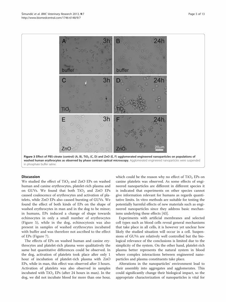
Figure 3 Effect of PBS-citrate (control) (A, B), TiO2 (C, D) and ZnO (E, F) agglomerated engineered nanoparticles on populations ofwashed human erythrocytes as observed by phase contrast optical microscopy. Agglomerated engineered nanoparticles were suspendedin phosphate buffer saline.
Šimundić et al. BMC Veterinary Research 2013, 9:7 Page 5 of 13http://www.biomedcentral.com/1746-6148/9/7
DiscussionWe studied the effect of TiO2 and ZnO EPs on washedhuman and canine erythrocytes, platelet-rich plasma andon GUVs. We found that both TiO2 and ZnO EPscaused coalescence of erythrocytes and activation of pla-telets, while ZnO EPs also caused bursting of GUVs. Wefound the effect of both kinds of EPs on the shape ofwashed erythrocytes in man and in the dog to be minor;in humans, EPs induced a change of shape towardsechinocytes in only a small number of erythrocytes(Figure 5), while in the dog, echinocytosis was alsopresent in samples of washed erythrocytes incubatedwith buffer and was therefore not ascribed to the effectof EPs (Figure 7).The effects of EPs on washed human and canine ery-
throcytes and platelet-rich plasma were qualitatively thesame but quantitative differences could be observed. Inthe dog, activation of platelets took place after only 1hour of incubation of platelet-rich plasma with ZnOEPs, while in man, this effect was observed after 3 hours.Activation of platelets was also observed in samplesincubated with TiO2 EPs (after 24 hours in man). In thedog, we did not incubate blood for more than one hour,
which could be the reason why no effect of TiO2 EPs oncanine platelets was observed. As some effects of engi-neered nanoparticles are different in different species itis indicated that experiments on other species cannotgive information relevant for humans as regards quanti-tative limits. In vitro methods are suitable for testing thepotentially harmful effects of new materials such as engi-neered nanoparticles since they address basic mechan-isms underlying these effects [43].Experiments with artificial membranes and selected
cell types such as blood cells reveal general mechanismsthat take place in all cells, it is however yet unclear howlikely the studied situation will occur in a cell. Suspen-sions of GUVs are relatively well controlled but the bio-logical relevance of the conclusions is limited due to thesimplicity of the system. On the other hand, platelet-richplasma better represents the natural system in bloodwhere complex interactions between engineered nano-particles and plasma constituents take place.Alterations in the nanoparticles' environment lead to
their assembly into aggregates and agglomerates. Thiscould significantly change their biological impact, so theappropriate characterization of nanoparticles is vital for

Figure 4 Effect of PBS-citrate (control) (A, B), TiO2 (C, D) and ZnO (E, F) agglomerated engineered nanoparticles on washed humanerythrocytes as observed by scanning electron microscopy. Agglomerated engineered nanoparticles were suspended in phosphate buffersaline. Arrows point to very large agglomerates of nanoparticles.
Šimundić et al. BMC Veterinary Research 2013, 9:7 Page 6 of 13http://www.biomedcentral.com/1746-6148/9/7
correct interpretation of the toxicological results. There-fore in studies of the biological reactivity of nanoparti-cles, their primary characteristics (characteristics ofindividual particles) and their secondary characteristics(characteristics of aggregates and agglomerates) shouldbe taken into account [44].Larger EPs were observed to interact with the membranes
of washed erythrocytes (Figure 4C–F, Figure 7E). If the sameparticle interacts with the membranes of two cells itcan mediate attractive interaction between the erythrocytesby a bridging interaction. However, besides very large EPs(such as the one shown in Figure 4C–F, Figure 7E), the so-lution contains EPs with a heterogeneous size distribution.Smaller EPs may induce attractive interaction betweenmembranes even without strong binding to the membrane.A possible mechanism of mediating attractive interactionbetween membranes derives from the spatial distribution ofcharge within the mediating particles. Plasma proteins bindto EPs [19] which may affect the interaction of EPs withcells. Also EP-protein complexes may have an internalcharge distribution which differs considerably from the dis-tribution within bare EPs. In general, the structure ofphospholipid headgroups is multipolar, so the headgroup
interface is a source of electric field. Rearrangement of ionsin solution in the vicinity of the interface screens the fieldand creates a gradient [45] in which particles with internaldistribution of charge are free to distribute so as to minimizetheir free energy [46]. This effect is especially energeticallyfavourable when the mediating particles are orientationallyordered between two interacting membranes separated by asmall distance (of the order of nanometers) while it isstronger if the charges within the particles are sepa-rated by a larger distance [47]. The equilibrium config-uration of membranes and mediating molecules appearsas adhesion between membranes.Plasma contains proteins that mediate attractive inter-
action between membranes [47-50]. EPs may enhancethe mediating effect of proteins by forming protein-EPcomplexes with extended charge distribution and expos-ure of positively charged domains that favour interactionwith the negatively charged glycolipid coat of theerythrocyte membrane.We did not observe adhesion of EPs on platelets, even
though the effect of engineered nanoparticles on plate-lets induced an activation which is functionally import-ant. It is possible that EPs undergo changes during the
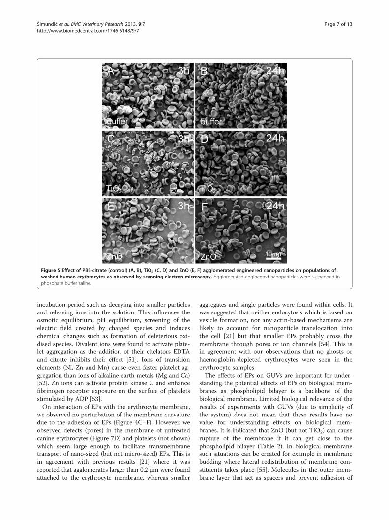
Figure 5 Effect of PBS-citrate (control) (A, B), TiO2 (C, D) and ZnO (E, F) agglomerated engineered nanoparticles on populations ofwashed human erythrocytes as observed by scanning electron microscopy. Agglomerated engineered nanoparticles were suspended inphosphate buffer saline.
Šimundić et al. BMC Veterinary Research 2013, 9:7 Page 7 of 13http://www.biomedcentral.com/1746-6148/9/7
incubation period such as decaying into smaller particlesand releasing ions into the solution. This influences theosmotic equilibrium, pH equilibrium, screening of theelectric field created by charged species and induceschemical changes such as formation of deleterious oxi-dised species. Divalent ions were found to activate plate-let aggregation as the addition of their chelators EDTAand citrate inhibits their effect [51]. Ions of transitionelements (Ni, Zn and Mn) cause even faster platelet ag-gregation than ions of alkaline earth metals (Mg and Ca)[52]. Zn ions can activate protein kinase C and enhancefibrinogen receptor exposure on the surface of plateletsstimulated by ADP [53].On interaction of EPs with the erythrocyte membrane,
we observed no perturbation of the membrane curvaturedue to the adhesion of EPs (Figure 4C–F). However, weobserved defects (pores) in the membrane of untreatedcanine erythrocytes (Figure 7D) and platelets (not shown)which seem large enough to facilitate transmembranetransport of nano-sized (but not micro-sized) EPs. This isin agreement with previous results [21] where it wasreported that agglomerates larger than 0,2 μm were foundattached to the erythrocyte membrane, whereas smaller
aggregates and single particles were found within cells. Itwas suggested that neither endocytosis which is based onvesicle formation, nor any actin-based mechanisms arelikely to account for nanoparticle translocation intothe cell [21] but that smaller EPs probably cross themembrane through pores or ion channels [54]. This isin agreement with our observations that no ghosts orhaemoglobin-depleted erythrocytes were seen in theerythrocyte samples.The effects of EPs on GUVs are important for under-
standing the potential effects of EPs on biological mem-branes as phospholipid bilayer is a backbone of thebiological membrane. Limited biological relevance of theresults of experiments with GUVs (due to simplicity ofthe system) does not mean that these results have novalue for understanding effects on biological mem-branes. It is indicated that ZnO (but not TiO2) can causerupture of the membrane if it can get close to thephospholipid bilayer (Table 2). In biological membranesuch situations can be created for example in membranebudding where lateral redistribution of membrane con-stituents takes place [55]. Molecules in the outer mem-brane layer that act as spacers and prevent adhesion of

Figure 6 Effect of PBS-citrate (control) (A-C), TiO2 (D-F) and ZnO (G-I) agglomerated engineered nanoparticles on human platelet richplasma as observed by scanning electron microscopy. Samples were incubated with agglomerated engineered nanoparticles dissolved inphosphate buffer saline for 1 hour (A,D, G), 3 hours (B, E, H) and 24 hours (C, F, I).
Šimundić et al. BMC Veterinary Research 2013, 9:7 Page 8 of 13http://www.biomedcentral.com/1746-6148/9/7
cells to each other may be depleted in the neck thatforms between the mother cell and the bud. Namely inthe neck the curvature of the membrane is negativewhich is unfavorable for molecules with substantial partsprotruding into the external solution.The basic paradigm of toxicology indicates that the ef-
fect of the substance is dose dependent. However, thisparadigm is only partly relevant for nanoparticles. Instead,nanoparticles follow a nano-specific mode of action whichis not yet completely understood. The behaviour of nano-particles in liquid media is poorly predicted due to the factthat they aggregate and agglomerate already in exposureto media as well as when they enter a biological system.Some authors report that suspensions of nanoparticleswith higher concentrations contain larger aggregates andthus exhibit lower toxic potential ([56] and referencestherein). In addition, aggregates adhere to surfaces outsideor inside organisms and may therefore cause effects with-out even entering cells. Up to now, no unified directionsexist how to test the effects of nanoparticles and how touse the data in human or environmental risk assessment.It is generally accepted that the toxicological tests that areused to assess the toxicity of non-nanosized substancesshould be modified.
ConclusionsWe found that addition of TiO2 and ZnO EPs to erythro-cytes suspended in PBS-citrate caused adhesion of EPs to
the membranes of erythrocytes and erythrocyte coales-cence. Addition of TiO2 and ZnO EPs induced activationof platelets. This indicates that EPs could be potentiallyprothrombogenic. ZnO (but not TiO2) EPs suspended inglucose solution caused a significant decrease of GUVs in-dicating that ZnO EPs (but not TiO2 EPs) could causemembrane rupture. Blood cells and phospholipid vesiclesare convenient systems for the study of membrane proper-ties and their interactions with various substances. Asknowledge on the relevant mechanisms is as yet rudimen-tary, we suggest that further studies using these systemsshould be performed.
MethodsChemicalsThe phospholipid 1-Palmitoyl-2-Oleoyl-sn-Glycero-3-Phospho-choline (POPC) and cholesterol were purchased from AvantiPolar Lipids, Inc. (Alabaster, Al, USA). Sucrose and glucosewere purchased from Sigma–Aldrich (Steinheim, Germany).
Engineered particlesTitanium (IV) oxide nanopowder (TiO2), supplied as apowder of anatase crystalline structure, with guaranteed99,7% purity, and zinc oxide nanopowder (ZnO) werepurchased from Sigma Aldrich (Steinheim, Germany).
Preparation of engineered particle suspensionsFor experiments with washed erythrocytes or platelet-rich plasma, stock suspensions of EPs were prepared in
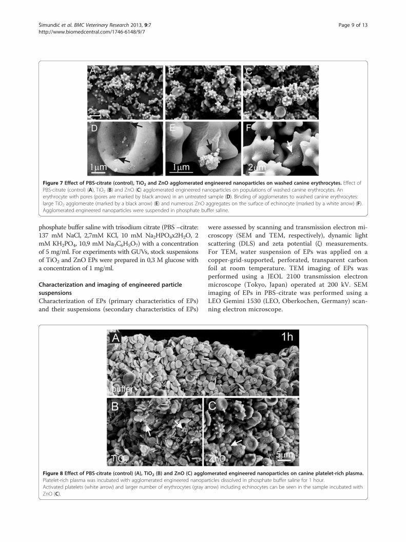
Figure 7 Effect of PBS-citrate (control), TiO2 and ZnO agglomerated engineered nanoparticles on washed canine erythrocytes. Effect ofPBS-citrate (control) (A), TiO2 (B) and ZnO (C) agglomerated engineered nanoparticles on populations of washed canine erythrocytes. Anerythrocyte with pores (pores are marked by black arrows) in an untreated sample (D). Binding of agglomerates to washed canine erythrocytes:large TiO2 agglomerate (marked by a black arrow) (E) and numerous ZnO aggregates on the surface of echinocyte (marked by a white arrow) (F).Agglomerated engineered nanoparticles were suspended in phosphate buffer saline.
Šimundić et al. BMC Veterinary Research 2013, 9:7 Page 9 of 13http://www.biomedcentral.com/1746-6148/9/7
phosphate buffer saline with trisodium citrate (PBS –citrate:137 mM NaCl, 2,7mM KCl, 10 mM Na2HPO4x2H2O, 2mM KH2PO4, 10,9 mM Na3C6H5O7) with a concentrationof 5 mg/ml. For experiments with GUVs, stock suspensionsof TiO2 and ZnO EPs were prepared in 0,3 M glucose witha concentration of 1 mg/ml.
Characterization and imaging of engineered particlesuspensionsCharacterization of EPs (primary characteristics of EPs)and their suspensions (secondary characteristics of EPs)
Figure 8 Effect of PBS-citrate (control) (A), TiO2 (B) and ZnO (C) aggloPlatelet-rich plasma was incubated with agglomerated engineered nanopaActivated platelets (white arrow) and larger number of erythrocytes (gray aZnO (C).
were assessed by scanning and transmission electron mi-croscopy (SEM and TEM, respectively), dynamic lightscattering (DLS) and zeta potential (ζ) measurements.For TEM, water suspension of EPs was applied on acopper-grid-supported, perforated, transparent carbonfoil at room temperature. TEM imaging of EPs wasperformed using a JEOL 2100 transmission electronmicroscope (Tokyo, Japan) operated at 200 kV. SEMimaging of EPs in PBS-citrate was performed using aLEO Gemini 1530 (LEO, Oberkochen, Germany) scan-ning electron microscope.
merated engineered nanoparticles on canine platelet-rich plasma.rticles dissolved in phosphate buffer saline for 1 hour.rrow) including echinocytes can be seen in the sample incubated with

Table 2 Effect of TiO2 and ZnO agglomerated engineered nanoparticles EPs on the average number of giantunilamelar phospholipid vesicles (GUVs) per micrograph
EPs t[min] GUVs Ntest ± SD GUVs Ncontrol ± SD Micrographs Mtest Micrographs Mcontrol Difference in N p P(α = 0,05)
TiO2 20 8,6 ± 3,6 8,7 ± 3,8 151 151 −1% 0,913 0,03
50 13,4 ± 5,8 14,8 ± 7,0 155 148 −10% 0,465 0,51
ZnO 20 5,2 ± 2,3 6,3 ± 2,8 156 145 −17% 3.10-4* 0,93*
50 6,5 ± 3,5 9,1 ± 4,3 155 144 −28% 3.10-8* 1,00*
The table shows the time of incubation (t), the average number of GUVs per micrograph of the test and of the control suspensions (Ntest and Ncontrol, respectively)with their standard deviations (SD), the number of harvested micrographs of the test and of the control suspensions (Mtest and Mcontrol, respectively), thedifference between the number of GUVs in the test and in the control suspensions, the statistical significance (p value) of the t-test subject to the differencebetween the test and the control suspensions, and the corresponding statistical power P at α = 0,05. Asterisks denote that the difference is statistically significantand of sufficient power.
Šimundić et al. BMC Veterinary Research 2013, 9:7 Page 10 of 13http://www.biomedcentral.com/1746-6148/9/7
Dispersed TiO2 and ZnO EPs dissolved in 0,3 M glucose(10 μg/ml) were inspected by DLS using a 3D DLS-SLSspectrometer (LS Instruments, Fribourg, Switzerland).Due to very fast sedimentation of both TiO2 and ZnOEPs, DLS measurements in PBS could not be performed.The zeta potential (ζ) of TiO2 and ZnO EPs suspensionsin both media, PBS-citrate and in 0,3 M glucose, weremeasured at the original concentrations and the pHvalues of the suspensions using a ZetaPals instrument(Brookhaven Instruments Corporation, Holtsville, NY,USA).
Blood samplingBlood was collected from the authors and from 3 healthypet dogs owned by members of the staff of Prva K Kli-nika za male živali d.o.o. Ljubljana. 2,7 ml tubes contain-ing 270 μl trisodium citrate at a concentration 0,109mol/l were used. All the dogs weighed over 15 kg so thevolume of the blood sample (2,7 ml) was small com-pared to the whole volume of the animal. Blood was col-lected by vein puncture into evacuated tubes (BDVacutainers, Becton Dickinson, CA) using a 21-gaugeneedle (length 70 mm, inner radius 0,4 mm) (Micro-lance, Becton Dickinson, NJ, USA). Sampling was per-formed according to the Declaration of Helsinki.Informed consent was obtained from all human partici-pants in this study and written consent to take bloodfrom the dogs was obtained from their owners. Thestudy as regarding both human and animal samples wasapproved by the Slovenian National Medical Ethics Com-mittee, No 117/02/10. No adverse effects on human andanimal donors’ health due to sampling were observed.
Preparation of blood cells for microscopyBlood was processed within 1 hour of sampling. Bloodwas centrifuged in a Centric 400R centrifuge (Domeld.o.o., Železniki, Slovenia) at 150 g and 37°C for 10 min-utes (human) and at 50 g and 37°C for 15 minutes (dog)to separate erythrocytes from platelet-rich plasma. Ery-throcytes were repeatedly washed with PBS citrate bycentrifugation at 1550 g and 37°C for 10 minutes.
Washed erythrocytes or platelet-rich plasma were ali-quotted into equal parts (50 μl in case of erythrocytesand 200 μl in case of platelet-rich plasma). TiO2 or ZnOEPs suspended in PBS-citrate (or PBS-citrate alone forthe control) was added to aliquot in a v/v ratio of 2:1 inthe case of erythrocytes and 3:1 in the case of platelet-rich plasma, 3 and 2 units corresponding to erythrocytesand platelet-rich plasma, respectively. After incubationthe samples were fixed in 0,1% glutaraldehyde, incubatedfor another hour at room temperature and centrifugedat 1550 g and 37°C for 10 minutes. The supernatant wasexchanged with PBS-citrate, samples were vortexed, cen-trifuged at 1550 g and 37°C for 10 minutes and fixed in2% glutaraldehyde for an hour.
Phase contrast microscopy of erythrocytes5 μl of erythrocyte suspension was placed in an observa-tion chamber composed of two cover glasses which wereglued together with nail polish. Samples were observedunder an Olympus GWB BH-2 (Olympus Corporation,Tokyo, Japan) microscope with phase contrast optics atan objective magnification of 400×. Images were takenwith a Canon EOS 450D digital camera (Canon Inc.,Tokyo, Japan).
Scanning electron microscopy of blood cellsFixed samples were washed by exchanging supernatantwith citrated PBS and incubated for 20 minutes at roomtemperature. This procedure was repeated 4 times whilethe last incubation was performed overnight at 8°C.Samples were then post-fixed for 60 min at 22°C in 1%OsO4 dissolved in 0,9% NaCl, dehydrated in a gradedseries of acetone/water (50%-100%, v/v), critical-pointdried, gold-sputtered, and examined using a LEOGemini 1530 (LEO, Oberkochen, Germany) scanningelectron microscope.
Preparation of giant unilamelar phospholipid vesiclesThe electroformation of GUVs was performed at roomtemperature. 40 μl of the lipid mixture of POPC (80%, v/v)and cholesterol (20%, v/v), both dissolved in 2:1

Šimundić et al. BMC Veterinary Research 2013, 9:7 Page 11 of 13http://www.biomedcentral.com/1746-6148/9/7
chloroform/methanol mixture, was spread over two plat-inum electrodes and the solvent was allowed to evapor-ate in low vacuum for two hours. The electrodes werethen placed in the electroformation chamber (a 2 mlEppendorf cup), filled with 2 ml of 0,3 M sucrose solution.An alternating electric field was applied as described in[55]. After electroformation, 600 μl of 0,3M sucrose solu-tion containing GUVs was added to 1 ml of 0,3M glucosesolution.
Experiments with giant unilamelar phospholipid vesiclesand engineered particlesExperiments with the two types of EPs, TiO2 and ZnO,were performed separately, but following the same pro-cedure. After electroformation, the suspension of GUVsin sucrose/glucose solution was mixed by turning theEppendorf cup upside down (five times). The suspensionwas diluted (300 μl of 0,3M glucose solution was addedto 100 μl of original vesicle solution) in order to obtainthe desired concentration of GUVs, which was foundconvenient according to preliminary experiments. Subse-quently, the diluted suspension was aliquotted into 4vials, (81 μl in each vial). Duplicates of test suspensionof EPs (TiO2 or ZnO, both dissolved in 0,3M glucose so-lution with a concentration of 100 μg/ml) and duplicatesof control suspension (0,3M glucose solution) wereadded to GUV suspensions in a volume ratio of 9:1(GUVs: test suspension) to reach the final concentrationof EPs of 10 μg/ml. Vials were turned upside down fivetimes to mix the test suspension with the GUV suspen-sion. The samples (70 μl each) were separately trans-ferred into four CoverWellTM Perfusion chambersPC4L-0,5 (Grace Bio-Labs Sigma-Aldrich, Steinheim,Germany) to create duplicates for the test sample andduplicates for the control sample.The observation chambers with samples were mounted
to the inverted phase contrast light microscope (NikonEclipse TE2000-S, Tokyo, Japan). After pre-defined dura-tions of incubation (20 and 50 minutes), GUV populationsin each chamber were recorded by a Sony CCD videocamera module, model: XC -77 CE (Minato, Japan). Theserecordings consisted of two video sequences each approxi-mately 2 minutes long. During recording, the object glasswas moved to capture images of thousands of GUVs.Using image processing algorithms, the video sequenceswere transformed into large mosaics [57] where each mo-saic contained all the vesicles acquired within the sample.GUVs in mosaics were segmented using a computer-aidedapproach [58].
Statistical analysisHere we introduced improvements to the analysis ofGUV populations. The mosaics were separated intoimages (micrographs) with the size of a single field of
view with the microscope at a given magnification. Usingthe GUVs’ locations inside the mosaics, all vesicles wereassociated with the appropriate micrograph, allowing usto extract information on vesicle density throughout themicrographs composing the mosaic.The numbers of GUVs in a micrograph were averaged
over all micrographs within the test and control samples.Methods of descriptive statistics were used to comparethe test samples and the respective control samples. Weused a two-sided pooled t-test with equal variance. Aprobability of the t-test below 0,05 was considered statis-tically significant. Power analysis was performed in orderto validate the size of the samples. A power larger than0,9 at α = 0.05 indicated a sample of proper size. Thesoftware used for statistical analysis was from R Devel-opment Core Team: R: A language and environment forstatistical computing. R Foundation for Statistical Com-puting, Vienna, Austria. 2008 ISBN 3-900051-07-0, URLhttp://www.R-project.org.
AbbreviationsEPs: Agglomerated engineered nanoparticles; GUVs: Giant unilamelarphospholipid vesicles; POPC: Palmitoyloleoyl phosphatidylcholine;TiO2: Titanium dioxide; ZnO: Zinc oxide; SEM: Scanning electron microscope;TEM: Transmission electron microscope; DLS: Dynamic light scattering.
Competing interestsAuthors declare no competing interests.
Authors’ contributionsMŠ participated by blood sampling, experiments with blood cells, design ofthe study, writing the manuscript and its critical reading, BD prepared EPssuspensions, performed experiments with GUVs, prepared recorded GUVs forstatistical analysis and critically read the manuscript, VŠ and RŠ performedexperiments with blood cells, prepared EPs and blood samples for SEM andcritically read the manuscript, VŠ also performed imaging of EPs and bloodcells by phase contrast microscopy and SEM. JZ participated in experimentswith GUVs, in improving the statistical analysis of GUVs, performed thestatistical analysis and critically read the manuscript. DE participated inimproving the method for statistical analysis of GUVs and critically read themanuscript. DM characterized EPs by TEM imaging, DLS and zeta potentialmeasurements and critically read the manuscript. HH participated in thedesign of the study, performed experiments with blood cells and theirimaging with phase contrast microscopy and SEM and critically read themanuscript. DD participated in the design of the study, writing themanuscript and its critical reading. VKI participated in design of the study,writing the manuscript and its critical reading and in improving the methodfor statistical analysis of GUVs. All authors read and approved the finalmanuscript.
AcknowledgementsThe authors acknowledge the support of the Slovenian Research Agency (J1-4109 and J3-4108).
Author details1Biomedical Research Group, Faculty of Health Sciences, University ofLjubljana and Prva-K Klinika za male živali d.o.o. (Prva-K Clinic for SmallAnimals), Ljubljana, Slovenia. 2Department of Biology, Biotechnical Faculty,University of Ljubljana, Ljubljana, Slovenia. 3Laboratory of Clinical Biophysics,Chair of Orthopaedics, Faculty of Medicine, University of Ljubljana, Ljubljana,Slovenia. 4Biomedical Research Group, Faculty of Health Sciences, Universityof Ljubljana, Ljubljana, Slovenia. 5J Stefan Institute, Ljubljana, Slovenia.6Department of Electrical and Computer Engineering, NortheasternUniversity, Boston, MA, USA. 7Department of Biology, Abo AkademiUniversity, Abo/Turku, Finland.

Šimundić et al. BMC Veterinary Research 2013, 9:7 Page 12 of 13http://www.biomedcentral.com/1746-6148/9/7
Received: 11 September 2012 Accepted: 8 January 2013Published: 11 January 2013
References1. Colvin V: The potential environmental impact of engineered
nanomaterials. Nat Biotechnol 2003, 13:1166–1170.2. Oberdorster G, Oberdorster E, Oberdorster J: Nanotoxicology: An emerging
discipline evolving from studies of ultrafine particles. Environ Health Persp2005, 113:823–839.
3. Iavicoli I, Leso V, Fontana L, Bergamaschi A: Toxicological effects oftitanium dioxide nanoparticles: a review of in vitro mammalian studies.Eur Rev Med Pharmaco 2011, 15:481–508.
4. Reijnders L: Human health hazards of persistent inorganic and carbonnanoparticles. J Mater Sci 2012, 47:5061–5073.
5. Yeates DB, Mauderly JL: Inhaled environmental/occupation irritants andallergens: mechanisms of cardiovascular and systemic responses:introduction. Environ Health Persp 2001, 109:479–481.
6. Böckmann J, Lahl H, Eckert T, Unterhalt B: Blood titanium levels before andafter oral administration of titanium dioxide. Pharmazie 2000, 55:140–143.
7. Robert MU, Tomlinson MJ, Hall DJ, Jacobs JJ: Accumulation in liver andspleen of metal particles generated at non-bearing surfaces in hiparthroplasty. J Arthroplasty 2004, 19:94–101.
8. Manivasagam G, Dhinasekaran D, Rajamanickam A: Biomedical implants:Corrosion and its prevention – a rewiev. Recent Pat Corrosion Sci 2010,2:40–54.
9. Yallapu MM, Jaggi M, Chauhan SC: Scope of nanotechnology in ovariancancer therapeutics. J Ovarian Res 2010, 3:19.
10. Chen Y, Wan Y, Wang Y, Zhang H, Jiao Z: Anticancer efficacyenhancement and attenuation of side effects of doxorubicin withtitanium dioxide nanoparticles. Int J Nanomed 2011, 6:2321–2326.
11. Türkez H, Geyikoglu F: An in vitro blood culture for evaluating thegenotoxicity of titanium dioxide: the responses of antioxidant enzymes.Toxicol Ind Health 2007, 23:19–23.
12. Roiter Y, Ornatska M, Rammohan AR, Balakrishnan J, Heine DR, Minko S:Interaction of nanoparticles with lipid membrane. Nano Lett 2008,8(3):941–944.
13. Kang SJ, Kim BM, Lee YJ, Chung HW: Titanium dioxide nanoparticlestrigger p53-mediated damage response in peripheral bloodlymphocytes. Environ Mol Mutagen 2008, 49:399–405.
14. Dawson KA, Salvati A, Lynch I: Nanotoxicology: nanoparticles reconstructlipids. Nat Nanotechnol 2009, 4:84–85.
15. Hussain S, Boland S, Baeza-Squiban A, Hamel R, Thomassen LC, Martens JA,Billon-Galland MA, Fleury-Feith J, Moisan F, Pairon JC, Marano F: Oxidativestress and proinflammatory effects of carbon black and titanium dioxidenanoparticles: Role of particle surface area and internalized amount.Toxicology 2009, 260:142–149.
16. Corbalan JJ, Medina C, Jacoby A, Malinski T, Radomski MW: Amorphoussilica nanoparticles trigger nitric oxide/peroxynitrite imbalance in humanendothelial cells: inflammatory and cytotoxic effects. Int J Nanomed 2011,6:2821–2835.
17. Yoo KC, Yoon CH, Kwon D, Hyun KH, Woo SJ, Kim RK, Lim EJ, Suh Y, Kim MJ,Yoon TH, Lee SJ: Titanium dioxide induces apoptotic cell death throughreactive oxygen species-mediated Fas upregulation and Bax activation.Int J Nanomed 2012, 7:1203–1214.
18. Park EJ, Yi J, Chung KH, Ryu DY, Choi J, Park K: Oxidative stress andapoptosis induced by titanium dioxide nanoparticles in cultured BEAS-2B cells. Toxicol Lett 2008, 180:222–229.
19. Ruh H, Kühl B, Brenner-Weiss G, Hopf C, Diabaté S, Weiss C: Identificationof serum proteins bound to industrial nanomaterials. Toxicol Lett 2012,208:41–50.
20. Suwalsky M, Villena F, Norris B, Soto MA, Sotomayor CP, Messori L, Zatta P:Structural effects of titanium citrate on the human erythrocytemembrane. J Inorg Biochem 2005, 99:764–770.
21. Rothen-Rutishauser BM, Schurch S, Haenni B, Kapp N, Gehr P: Interactionof fine particles and nanoparticles with red blood cells visualizedwith advanced microscopic techniques. Environ Sci Technol 2006,40:4353–4359.
22. Suwalsky M, Novoa V, Villena F, Sotomayor CP, Aguilar LF, Ronowska A,Szutowicz A: Structural effects of Zn (2+) on cell membranes andmolecular models. J Inorg Biochem 2009, 103:797–804.
23. Zupanc J, Valant J, Drobne D, Kralj-Iglič V, Iglič A: A new approach toanalyse effects of nanoparticles on lipid vesicles. Int J Biomed NanosciNanotechnol 2010, 1:34–51.
24. Zupanc J, Drobne D, Drašler B, Valant J, Iglič A, Kralj-Iglič V, Makovec D,Rappolt M, Sartori B, Kogej K: Experimental evidence for the interaction ofC-60 fullerene with lipid vesicle membranes. Carbon 2012, 50:1170–1178.
25. Angelova MI, Soleau S, Méléard PH, Faucon F, Bothorel P: Preparation ofgiant vesicles by external AC electric fields. Kinetics and applications.Prog Coll Pol Sci 1992, 89:127–131.
26. Peterlin P, Arrigler V: Electroformation in a flow chamber with solutionexchange as a means of preparation of flaccid giant vesicles. ColloidSurface B 2008, 64:77–87.
27. Bagatolli LA, Parasassi T, Gratton E: Giant phospholipid vesicles:comparison among the whole lipid sample characteristics usingdifferent preparation methods: A two photon fluorescence microscopystudy. Chem Phys Lipids 2000, 105:135–147.
28. Pavlič JI, Genova J, Zheliaskova A, Iglič A, Mitov MD: Electroformation ofneutral and negatively charged phospholipid giant vesicles underphysiological conditions. CR Acad Bulg Sci 2010, 63:497–502.
29. Baumgart T, Hunt G, Farkas ER, Webb WW, Feigenson GW: Fluorescenceprobe partitioning between Lo/Ld phases in lipid membranes. BiochimBiophys Acta 2007, 9:2182–2194.
30. Wang B, Zhang L, Bae SC, Granick S: Nanoparticle-induced surfacereconstruction of phospholipid membranes. Proc Natl Acad Sci 2008,105:8171–8175.
31. Peterlin P, Jaklič G, Pisanski T: Determining membrane permeability ofgiant phospholipid vesicles from a series of videomicroscopy images.Meas Sci Technol 2009, 20:055801–055807.
32. Peterlin P, Arrigler V, Haleva E, Diamant H: Law of corresponding states forosmotic swelling of vesicles. Soft Matter 2012, 8(7):2185–2193.
33. Bivas I, Hanusse P, Bothorel P, Lalanne J, Aguerre-Chariol O: An applicationof the optical microscopy to the determination of the curvature elasticmodulus of biological and model membranes. J Physiol Paris 1987,48:855–867.
34. Bivas I: Shape fluctuations of nearly spherical lipid vesicles and emulsiondroplets. Phys Rev E 2010, 81(6):061911.
35. Ambrožič A, Čučnik S, Tomšič N, Urbanija J, Lokar M, Babnik B, Rozman B,Iglič A, Kralj-Iglič V: Interaction of giant phospholipid vesicles containingcardiolipin and cholesterol with ß2-glycoprotein-I and anti-ß2-glycoprotein-I antibodies. Autoimmun Rev 2006, 6:10–15.
36. Khairullina AY, Ol'shanskaya TV, Filimonenko DS, Kozlova NM, Garmaza MY,Slobozhanina EI: Optical, nanostructural, and biophysical properties ofZn-induced changes in human erythrocyte membranes. Opt Spectrosc2011, 110:534–540.
37. Aisaka Y, Kawaguchi R, Watanabe S, Ikeda M, Igisu H: Hemolysis caused bytitanium dioxide particles. Inhal Toxicol 2008, 20:891–893.
38. Pujalté I, Passagne I, Brouillaud B, Tréguer M, Durand E, Ohayon-Courtès C,L'Azou B: Cytotoxicity and oxidative stress induced by different metallicnanoparticles on human kidney cells. Part Fibre Toxicol 2011, 8:10.
39. Gorbet MB, Sefton MV: Biomaterial-associated thrombosis: roles ofcoagulation factors, complement, platelets and leukocytes-rewiev.Biomaterials 2004, 25:5681–5703.
40. Kvietys PR, Grager DN: Role of reactive oxygen and nitrogen species inthe vascular responses to inflammation. Free Radical Biol Med 2012,25:556–592.
41. Vermylen J, Nemmar A, Nemery B, Hoylaerts FM: Ambient air pollution andacute myocardial infarction. J Thromb Haemost 2005, 3(9):1955–1961.
42. Shannahan JH, Kodavanti UP, Brown JM: Manufactured and airbornenanoparticle cardiopulmonary interactions: a review of mechanismsand the possible contribution of mast cells. Inhal Toxicol 2012,24:320–339.
43. Combes RD, Balls M: Integrated testing strategies for toxicity employingnew and existing technologies. Altern Lab Anim 2011, 39:213–225.
44. Valant J, Iavicoli I, Drobne D: The importance of a validated standardmethodology to define in vitro toxicity of nano-TiO2. Protoplasma 2012,249:493–502.
45. Kralj-Iglič V, Iglič A: A simple statistical mechanical approach to the freeenergy of the electric double layer including the excluded volumeeffect. J Phys II 1996, 6:477–491.
46. May S, Iglič A, Reščič J, Maset S, Bohinc K: Bridging like-charged macroionsthrough long divalent rod-like ions. J Phys Chem B 2008, 112:1685–1692.

Šimundić et al. BMC Veterinary Research 2013, 9:7 Page 13 of 13http://www.biomedcentral.com/1746-6148/9/7
47. Urbanija J, Bohinc K, Bellen A, Maset S, Iglič A, Kralj-Iglič V, Kumar PB:Attraction between negatively charged surfaces mediated by sphericalcounterions with quadrupolar charge distribution. J Chem Phys 2008,129:105101.
48. Urbanija J, Tomšič N, Lokar M, Ambrožič A, Čučnik S, Rozman B, KandušerM, Iglič A, Kralj-Iglič V: Coalescence of phospholipid membranes as apossible origin of anticoagulant effect of serum proteins. Chem PhysLipids 2007, 150:49–57.
49. Frank M, Manček-Keber M, Kržan M, Sodin-Šemrl S, Jerala R, Iglič A, RozmanB, Kralj-Iglič V: Prevention of microvesiculation by adhesion of buds tothe mother cell membrane -A possible anticoagulant effect of healthydonor plasma. Autoimmun Rev 2008, 7:240–245.
50. Janša R, Šuštar V, Frank M, Sušanj P, Bešter J, Manček-Keber M, Kržan M, IgličA: Number of microvesicles in peripheral blood and ability of plasma toinduce adhesion between phospholipid membranes in 19 patients withgastrointestinal diseases. Blood Cell Mol Dis 2008, 41:124–132.
51. Spaet TH, Zucker MB: Mechanism of platelet plug formation and role ofadenosine diphosphate. Am J Physiol 1964, 206:1267–1274.
52. Penglis F, Michal F: The induction of blood platelet aggregation bydivalent cations. Experientia 1969, 25:745–746.
53. Trybulec M, Kowalska MA, McLane MA, Silver L, Lu W, Niewiarowski S:Exposure of platelet fibrinogen receptors by zinc ions: role of proteinkinase C. Exp Biol Med 1993, 203:108–116.
54. Porter E, Muller K, Skepper J, Midgley P, Welland M: Uptake of C60 byhuman monocyte macrophages, its localization and implication fortoxicity: studied by high resolution etlectron microskopy and electrontomography. Biomaterials 2006, 2:409–419.
55. Kralj-Iglič V, Gomišček G, Majhenc J, Arrigler V, Svetina S: Myelin-likeprotrusions of giant phospholipid vesicles prepared by electroformation.Colloid Surface A 2001, 181:315–318.
56. Takenaka S, Karg E, Roth C, Schulz H, Ziesenis A, Heinzmann U, Schramel P,Heyder J: Pulmonary and systemic distribution of inhaled ultrafine silverparticles in rats. Environ Health Persp 2001, 109:547–551.
57. Zupanc J, Drobne D, Erdogmus D, Bas E, Valant J, Dobnikar A: Biologicalreactivity of nanoparticles: mosaics from optical microscopy videos ofgiant lipid vesicles. J Biomed Opt 2011, 16(2):26003–26010.
58. Zupanc J, Ster B, Drobne D: Markov random field model for segmentinglarge populations of lipid vesicles from micrographs. J Liposome Res 2011,21(4):315–323.
doi:10.1186/1746-6148-9-7Cite this article as: Šimundić et al.: Effect of engineered TiO2 and ZnOnanoparticles on erythrocytes, platelet-rich plasma and giant unilamelarphospholipid vesicles. BMC Veterinary Research 2013 9:7.
Submit your next manuscript to BioMed Centraland take full advantage of:
• Convenient online submission
• Thorough peer review
• No space constraints or color figure charges
• Immediate publication on acceptance
• Inclusion in PubMed, CAS, Scopus and Google Scholar
• Research which is freely available for redistribution
Submit your manuscript at www.biomedcentral.com/submit
Copyright © 2022 FDOKUMEN