Editorials - International Journal of Cardiovascular Sciences
160
Volume 35 - Number 2 | March / April | ISSN 2359-4802 | ISSN online 2359-5647 Editorials The Pathway to a High Impact Journal and Scopus Indexation – New Achievement of the International Journal of Cardiovascular Sciences Science Gender Gap: Are We in the Right Path? Original Article Implementation of a Telecardiology Service in a Health Unit in the City of Porto Alegre, Brazil: A Pilot Study Editorial Challenges in Telemedicine: Even When the Road is Hard, Never Give up Original Article Is the Wistar Rat a more Suitable Normotensive Control for SHR to Test Blood Pressure and Cardiac Structure and Function? Editorial Are Wistar Rats the Most Suitable Normotensive Controls for Spontaneously Hypertensive Rats to Assess Blood Pressure and Cardiac Structure and Function? Original Article Validation of the Grace Risk Score to Predict In-Hospital and 6-Month Post-Discharge Mortality in Patients with Acute Coronary Syndrome Editorial Risk Scores in Acute Coronary Syndrome: Current Applications and Future Perspectives Original Articles Acute Myocardial Infarction and Percutaneous Coronary Intervention: What does the Epidemiological Data of the Last Years Indicate? Physical Fitness Test (PFT) in Police and Military in Brazil: A Systematic Review Text Messages to Promote Secondary Prevention after Acute Coronary Syndrome (IMPACS trial) Radioprotective Effect of Nigella Sativa Oil on Heart Tissues of Rats Exposed to Irradition Subclinical Systolic Dysfunction during Chemotherapy for Breast Cancer Overview of Cardiovascular Disease Risk Factors in Adults in São Paulo, Brazil: Prevalence and Associated Factors in 2008 and 2015 Atherosclerosis Complications in the Brazilian Population: An Ecological Time Series Study Review Article Effect of Physical Training on Nitric Oxide Levels in Patients with Arterial Hypertension: An Integrative Review Editorial We Need to Talk about Why We don’t Talk about Exercise! Review Article Revisiting the History of Chagas Disease: "Live to tell" Viewpoint Physical Activity and Cardiovascular Health: Practical Strategies to Reduce Sedentary Time in Adult Population Case Reports Surgical Repair of a Ruptured Giant Abdominal Aortic Aneurysm in a 16-Year-Old with Takayasu’s Arteritis: Case Report and Etiological Review Commotio Cordis Secondary to Aggression
-
Upload
khangminh22 -
Category
Documents
-
view
1 -
download
0
Transcript of Editorials - International Journal of Cardiovascular Sciences
Volu
me
35 -
Num
ber
2 | M
arch
/ A
pril
| ISS
N 2
359-
4802
| IS
SN o
nlin
e 2
359-
5647
EditorialsThe Pathway to a High Impact Journal and Scopus Indexation – New Achievement of the International Journal of Cardiovascular Sciences
Science Gender Gap: Are We in the Right Path?
Original ArticleImplementation of a Telecardiology Service in a Health Unit in the City of Porto Alegre, Brazil: A Pilot Study
EditorialChallenges in Telemedicine: Even When the Road is Hard, Never Give up
Original ArticleIs the Wistar Rat a more Suitable Normotensive Control for SHR to Test Blood Pressure and Cardiac Structure and Function?
EditorialAre Wistar Rats the Most Suitable Normotensive Controls for Spontaneously Hypertensive Rats to Assess Blood Pressure and Cardiac Structure and Function?
Original ArticleValidation of the Grace Risk Score to Predict In-Hospital and 6-Month Post-Discharge Mortality in Patients with Acute Coronary Syndrome
EditorialRisk Scores in Acute Coronary Syndrome: Current Applications and Future Perspectives
Original ArticlesAcute Myocardial Infarction and Percutaneous Coronary Intervention: What does the Epidemiological Data of the Last Years Indicate?
Physical Fitness Test (PFT) in Police and Military in Brazil:
A Systematic Review
Text Messages to Promote Secondary Prevention after
Acute Coronary Syndrome (IMPACS trial)
Radioprotective Effect of Nigella Sativa Oil on Heart Tissues
of Rats Exposed to Irradition
Subclinical Systolic Dysfunction during Chemotherapy for
Breast Cancer
Overview of Cardiovascular Disease Risk Factors in Adults
in São Paulo, Brazil: Prevalence and Associated Factors
in 2008 and 2015
Atherosclerosis Complications in the Brazilian Population:
An Ecological Time Series Study
Review ArticleEffect of Physical Training on Nitric Oxide Levels in Patients with Arterial Hypertension: An Integrative Review
EditorialWe Need to Talk about Why We don’t Talk about Exercise!
Review ArticleRevisiting the History of Chagas Disease: "Live to tell"
ViewpointPhysical Activity and Cardiovascular Health: Practical
Strategies to Reduce Sedentary Time in Adult
Population
Case ReportsSurgical Repair of a Ruptured Giant Abdominal Aortic
Aneurysm in a 16-Year-Old with Takayasu’s Arteritis: Case
Report and Etiological Review
Commotio Cordis Secondary to Aggression
SUMARY
145
148
152
159
161
172
174
181
• Editorials
The Pathway to a High Impact Journal and Scopus Indexation – New Achievement of the International Journal of Cardiovascular Sciences ...............................................................................................................
Claudio Tinoco Mesquita
Science Gender Gap: Are We in the Right Path? ................................................................................................................. Gláucia Maria Moraes de Oliveira, Marge Tenorio, Alessandra de Sá Earp Siqueira
• Original Article
Implementation of a Telecardiology Service in a Health Unit in the City of Porto Alegre, Brazil: A Pilot Study ..... Francieli Giachini Esmerio, Silvia Goldmeier, Eduardo Costa Duarte Barbosa, Luis Marcelo Segredo, Rodolfo Silva,
Maria Claudia Irigoyen, Bruna Eibel, Patricia Oliveira Dias
• Editorial
Challenges in Telemedicine: Even When the Road is Hard, Never Give up ................................................................. Erito Marques de Souza Filho and Alexandra Monteiro
• Original Article
Is the Wistar Rat a more Suitable Normotensive Control for SHR to Test Blood Pressure and Cardiac Structure and Function? ............................................................................................................................................
Leonardo Mateus Teixeira de Rezende, Leôncio Lopes Soares, Filipe Rios Drummond, Pedro Zavagli Suarez, Luciano Leite, Joel Alves Rodrigues, Tiago Leal, Lukiya Favarato, Emily Correna Carlo Reis, Evandro Favarato, Miguel Carneiro-Júnior, Antônio José Natali, Cândido Coimbra, Thales Prímola-Gomes
• Editorial
Are Wistar Rats the Most Suitable Normotensive Controls for Spontaneously Hypertensive Rats to Assess Blood Pressure and Cardiac Structure and Function? ........................................................................................................
Nazareth Novaes Rocha
• Original Article
Validation of the Grace Risk Score to Predict In-Hospital and 6-Month Post-Discharge Mortality in Patients with Acute Coronary Syndrome ......................................................................................................................................................
Vítor Boniatti Neves, Raquel Melchior Roman, Tiago Vendruscolo, Gilberto Heineck, Carlos Alberto Santos de Mattos, Eduardo Ilha de Mattos, Luiz Carlos Pereira Bin, Karine de Lima Sírio Boclin, Marcelo Fialho Roman
• Editorial
Risk Scores in Acute Coronary Syndrome: Current Applications and Future Perspectives ....................................... Pedro G. M. de Barros e Silva and Renato D. Lopes
184
191
202
214
220
230
243
253
265
267
• Original Article
Acute Myocardial Infarction and Percutaneous Coronary Intervention: What does the Epidemiological Data of the Last Years Indicate? ............................................................................................................................................................
Gabriel Silvestre Minucci and Samuel Marques dos Reis
Physical Fitness Test (PFT) in Police and Military in Brazil: A Systematic Review .................................................... Bráulio Nascimento Lima, Renato Silveira de Assis Junior, Klebson da Silva Almeida, Leandro Borelli de Camargo,
Ricardo Pablo Passos, Carlos Henrique Prevital Fileni, Divaldo Martins de Souza, Guanis de Barros Vilela Junior
Text Messages to Promote Secondary Prevention after Acute Coronary Syndrome (IMPACS trial) ........................ Luiz Guilherme Passaglia, Luisa Campos Caldeira Brant, José Luiz Padilha da Silva, Bruno Ramos Nascimento,
Antônio Luiz Pinho Ribeiro
Radioprotective Effect of Nigella Sativa Oil on Heart Tissues of Rats Exposed to Irradition ................................... Mehmet Kaplan, Elif Demir, Fethi Yavuz, Gizem Ilgin Kaplan, Mehmet Resit Taysi, Seyithan Taysi, Mehmet Murat Sucu Subclinical Systolic Dysfunction during Chemotherapy for Breast Cancer ................................................................. Geanne Maria Holanda de Menezes Barroso, Júlio César Oliveira Costa Teles, Paulo Victor de Jesus Silva,
Karin Yasmin Santos Fonseca, Vinícius Antônio Santos Aragão, Marília Marques Aquino, Enaldo Vieira de Melo, Karina Oliveira Ferreira, Ronnei José Feitosa de Assis, Michel Fabiano Silva Alves, Antônio Carlos Sobral Sousa, Joselina Luzia Menezes Oliveira
Overview of Cardiovascular Disease Risk Factors in Adults in São Paulo, Brazil: Prevalence and Associated Factors in 2008 and 2015 ............................................................................................................................................................
Jaqueline L. Pereira, Michelle A. de Castro, Jean M. R. S. Leite, Marcelo M. Rogero, Flavia M. Sarti, Chester Luís Galvão César, Moisés Goldbaum, Regina M. Fisberg
Atherosclerosis Complications in the Brazilian Population: An Ecological Time Series Study................................ Emerson de Jesus Silva, Francieudo da Silva Gomes Junior, João Vitor Bezerra Firmiano, Nilson Roberto da Silva, Wendel
Aguiar Carlini, Marcello Barbosa Otoni Gonçalves Guedes, Matheus Rodrigues Lopes, Johnnatas Mikael Lopes
• Review Article
Effect of Physical Training on Nitric Oxide Levels in Patients with Arterial Hypertension: An Integrative Review ..............................................................................................................................................................
Tábata de Paula Facioli, Mariana Colombini Buranello, Eloisa Maria Gatti Regueiro, Renata Pedrolongo Basso-Vanelli, Marina de Toledo Durand
• Editorial
We need to talk about why we don’t talk about exercise ................................................................................................... Fabrício Braga da Silva
• Review Article Revisiting the history of Chagas Disease: “Live to tell” .................................................................................................... Ademir Batista da Cunha and Delma Maria Cunha
283
287
293
• Viewpoint
Physical Activity and Cardiovascular Health: Practical Strategies to Reduce Sedentary Time in Adult Population .......................................................................................................................................................................
Karolinny Borinelli de Aquino Moura, Simone Savaris, Janice Debastiani, Juliana Beust de Lima
• Case Reports
Surgical Repair of a Ruptured Giant Abdominal Aortic Aneurysm in a 16-Year-Old with Takayasu’s Arteritis: Case Report and Etiological Review ......................................................................................................................................
José Maciel Caldas dos Reis, Glauco dos Santos Melo, Murilo Vasconcelos de Oliveira, Felipe Eduardo de Oliveira Santos, Tereza Maria Meireles Fernandes da Silva, Hugo Luis da Silva Ferreira
Commotio Cordis Secondary to Aggression ........................................................................................................................ Luisa Gonçalves, Inês Pires, João Santos, Joana Laranjeira Correia, Davide Moreira, Inês Almeida
ISSN 2359-4802 / IJCS ONLINE: ISSN 2359-5647
EditorCláudio Tinoco Mesquita – Hospital Universitário Antônio Pedro (HUAP), Universidade Federal Fluminense (UFF), Niterói, Rio de Janeiro, RJ – BrazilSocial Media EditorSérgio Emanuel Kaiser – Universidade do Estado do Rio de Janeiro, Rio de Janeiro, RJ – BrazilAssociated EditorsChristianne Brêtas Vieira Scaramello (Multiprofessional Area) – Hospital Universitário Antônio Pedro (HUAP), Universidade Federal Fluminense (UFF), Niterói, Rio de Janeiro, RJ – BrazilClério Francisco Azevedo Filho (Cardiovascular Imaging Area) – Universidade do Estado do Rio de Janeiro (UERJ), Rio de Janeiro, RJ - BrazilGláucia Maria Moraes de Oliveira (Clinical Cardiology Area) – Departamento de Clínica Médica, Faculdade de Medicina (FM), Universidade Federal do Rio de Janeiro (UFRJ), Rio de Janeiro, RJ - BrazilGuilherme Vianna e Silva (Interventionist Cardiology Area) – Texas Heart Institute, USA
BrazilAndréia Biolo – Faculdade de Medicina, Universidade Federal do Rio Grande do Sul (UFRGS), Porto Alegre, RS – BrazilAngelo Amato Vincenzo de Paola – Escola Paulista de Medicina (EPM), Universidade Federal de São Paulo (UNIFESP), São Paulo, SP – BrazilAntonio Cláudio Lucas da Nóbrega – Centro de Ciências Médicas, Universidade Federal Fluminense (UFF), Niterói, Rio de Janeiro, RJ – BrazilAri Timerman – Unidades de Internação, Instituto Dante Pazzanese de Cardiologia (IDPC), São Paulo, SP - BrazilArmando da Rocha Nogueira – Departamento de Clínica Médica, Universidade Federal do Rio de Janeiro (UFRJ), Rio de Janeiro, RJ - BrazilCarísi Anne Polanczyk – Hospital de Clínicas de Porto Alegre, Universidade Federal do Rio Grande do Sul (UFRGS), Porto Alegre, RS – BrazilCarlos Eduardo Rochitte – Departamento de Cardiopneumologia, Hospital das Clínicas da Faculdade de Medicina da Universidade de São Paulo (HCFMUSP), São Paulo, SP – BrazilCarlos Vicente Serrano Júnior – Faculdade de Medicina da Universidade de São Paulo, Instituto do Coração (InCor), São Paulo, SP – BrazilCláudio Gil Soares de Araújo – Instituto do Coração Edson Saad, Universidade Federal do Rio de Janeiro (UFRJ), Rio de Janeiro, RJ - BrazilCláudio Pereira da Cunha – Departamento de Clínica Médica, Universidade Federal do Paraná (UFPR), Paraná, PR – BrazilCláudio Tinoco Mesquita – Hospital Universitário Antônio Pedro (HUAP), Universidade Federal Fluminense (UFF), Niterói, Rio de Janeiro, RJ – BrazilDenílson Campos de Albuquerque – Faculdade de Ciências Médicas, Universidade do Estado do Rio de Janeiro (UERJ), Rio de Janeiro, RJ – BrazilDenizar Vianna Araujo – Departamento de Clínica Médica, Universidade do Estado do Rio de Janeiro (UERJ), Rio de Janeiro, RJ – BrazilEsmeralci Ferreira – Hospital Universitário Pedro Ernesto (HUPE), Universidade do Estado do Rio de Janeiro (UERJ), Rio de Janeiro, RJ - BrazilEvandro Tinoco Mesquita – Hospital Universitário Antônio Pedro (HUAP), Universidade Federal Fluminense (UFF), Niterói, Rio de Janeiro, RJ – BrazilFernando Nobre – Faculdade de Medicina de Ribeirão Preto (FMRP), Universidade de São Paulo, São Paulo, SP – BrazilGabriel Blacher Grossman – Serviço de Medicina Nuclear, Hospital Moinhos de Vento, Porto Alegre, RS – BrazilHenrique César de Almeida Maia – Governo do Distrito Federal (GDF), Brasília, DF - BrazilHumberto Villacorta Júnior – Hospital Universitário Antônio Pedro (HUAP), Universidade Federal Fluminense (UFF), Niterói, Rio de Janeiro, RJ – BrazilIran Castro – Fundação Universitária de Cardiologia (FUC), Instituto de Cardiologia do Rio Grande do Sul (IC), Porto Alegre, RS – BrazilJoão Vicente Vitola – Quanta Diagnóstico e Terapia (QDT), Curitiba, PR – Brazil
José Geraldo de Castro Amino – Sessão Clínica, Instituto Nacional de Cardiologia (INC), Rio de Janeiro, RJ – BrazilJosé Márcio Ribeiro – Clínica Médica (Ambulatório), União Educacional Vale do Aço (UNIVAÇO), Ipatinga, MG - Brazil Leonardo Silva Roever Borges – Departamento de Pesquisa Clínica, Universidade Federal de Uberlândia (UFU), MG – BrazilLeopoldo Soares Piegas – Fundação Adib Jatene, Instituto Dante Pazzanese de Cardiologia (IDPC/FAJ), São Paulo, SP - BrazilLuís Alberto Oliveira Dallan – Serviço Coronariopatias, Instituto do Coração (INCOR), São Paulo, SP - BrazilMarcelo Iorio Garcia – Clínica de Insuficiência Cardíaca, Universidade Federal do Rio de Janeiro (UFRJ), Rio de Janeiro, RJ – BrazilMarcelo Westerlund Montera – Centro de Insuficiência Cardíaca, Hospital Pró Cardíaco (PROCARDIACO), Rio de Janeiro, RJ – BrazilMarcio Luiz Alves Fagundes – Divisão de Arritmia e Eletrofisiologia, Instituto Nacional de Cardiologia Laranjeiras (INCL), Rio de Janeiro, RJ – BrazilMarco Antonio Mota Gomes - Fundação Universitária de Ciências da Saúde Governador Lamenha Filho (UNCISAL), Maceió, AL - BrazilMarco Antonio Rodrigues Torres – Departamento de Medicina Interna, Hospital de Clínicas de Porto Alegre, Porto Alegre, RS – BrazilMarcus Vinicius Bolivar Malachias – Instituto de Pesquisas e Pós-graduação (IPG), Faculdade de Ciências Médicas de Minas Gerais (FCMMG), Belo Horizonte, MG – BrazilMaria Eliane Campos Magalhães – Departamento de Especialidades Médicas, Universidade do Estado do Rio de Janeiro (UERJ), Rio de Janeiro, RJ – BrazilMário de Seixas Rocha – Unidade Coronariana, Hospital Português, Salvador, BA – BrazilMaurício Ibrahim Scanavacca – Unidade Clínica de Arritmia, Instituto do Coração do Hospital das Clínicas da FMUSP, São Paulo, SP – BrazilNadine Oliveira Clausell – Faculdade de Medicina, Universidade Federal do Rio Grande do Sul (UFRGS), Porto Alegre, RS – BrazilNazareth de Novaes Rocha – Centro de Ciências Médicas, Universidade Federal Fluminense, UFF - Rio de Janeiro, RJ – BrazilNelson Albuquerque de Souza e Silva – Departamento de Clínica Médica, Universidade Federal do Rio de Janeiro (UFRJ), Rio de Janeiro, RJ – BrazilPaola Emanuela Poggio Smanio – Seção Médica de Medicina Nuclear, Instituto Dante Pazzanese de Cardiologia (IDPC) São Paulo, SP - BrazilPaulo Cesar Brandão Veiga Jardim – Liga de Hipertensão Arterial, Universidade Federal de Goiás (UFGO), Goiânia, GO – BrazilRonaldo de Souza Leão Lima – Pós-Graduação em Cardiologia, Universidade Federal do Rio de Janeiro (UFRJ), Rio de Janeiro, RJ – Brazil
João Augusto Costa Lima (Integrative Imaging Area) – Johns Hopkins Hospital – Baltimore, USAMiguel Mendes (Ergometric and Cardiac Rehabilitation Area) – Sociedade Portuguesa de Cardiologia, PortugalPedro Adragão (Arrhythmia and Electrophysiology Area) – Hospital da Luz – Lisboa, PortugalEduardo B. Saad (Arrhythmia and Electrophysiology) – Hospital Pró-Cardíaco, Rio de Janeiro, RJ – BrazilRenata Castro (Cardiovascular Physiology Area) – Harvard University, Massachusetts – EUARicardo Mourilhe-Rocha (Heart Failure and Myocardiopathy Area) – Hospital Universitário Pedro Ernesto, Universidade do Estado do Rio de Janeiro (UERJ), Rio de Janeiro, RJ – BrazilFernando Stuardo Wyss Quintana (Hypertension) – Servicios y Tecnología Cardiovascular de Guatemala – GuatemalaMaria Alexandra Arias Mendoza (Ischemic Heart Disease) – Instituto Nacional de Cardiología – Mexico
EDITORIAL BOARD
BIENNIUM BOARD 2022/2023
ADMINIST R AT I V E COU NCIL – M A N DAT E 2022 (BR A ZILI A N SOCIET Y OF CA RDIOLOGY )
North/Northeast RegionNivaldo Menezes Filgueiras Filho (BA)Sérgio Tavares Montenegro (PE)
Eastern RegionDenilson Campos de Albuquerque (RJ)Andréa Araujo Brandão (RJ) – Vice-presidente do Conselho Administrativo
Região PaulistaCelso Amodeo (SP)João Fernando Monteiro Ferreira (SP) – Presidente do Conselho Administrativo
Central RegionCarlos Eduardo de Souza Miranda (MG)Weimar Kunz Sebba Barroso de Souza (GO)
South RegionPaulo Ricardo Avancini Caramori (RS)Gerson Luiz Bredt Júnior (PR)
Editor-in-Chief of the ABC Cardiol (2022-2025)Carlos Eduardo Rochitte
Editor-in-Chief of the IJCS (2022-2025)Claudio Tinoco Mesquita
PRESIDEN TS OF STAT E A N D REGIONAL BR A ZILI A N SOCIET IES OF CA RDIOLOGY
SBC/AL – Pedro Henrique Oliveira de Albuquerque
SBC/BA – Joberto Pinheiro Sena
SBC/DF – Fausto Stauffer Junqueira de Souza
SBC/ES – Tatiane Mascarenhas Santiago Emerich
SBC/GO – Humberto Graner Moreira
SBC/MA – Francisco de Assis Amorim de Aguiar Filho
SBC/MG – Antônio Fernandino de Castro Bahia Neto
SBC/MS – Mauro Rogério de Barros Wanderley Júnior
SBC/NNE – José Albuquerque de Figueiredo Neto
SBC/PB – Guilherme Veras Mascena
SBC/PE – Carlos Japhet Da Matta Albuquerque
SBC/PI – Jônatas Melo Neto
SBC/PR – Olímpio R. França Neto
SOCERJ – Ronaldo de Souza Leão Lima
SBC/RN – Antônio Amorim de Araújo Filho
SOCERGS – Fábio Cañellas Moreira
SOCESP – Ieda Biscegli Jatene
PRESIDENTS OF DEPARTAMENTS AND STUDY GROUPS
SBC/DA – Marcelo Heitor Vieira Assad
SBC/DCC – Bruno Caramelli
SBC/DCC/CP – Cristiane Nunes Martins
SBC/DCM – Maria Cristina Costa de Almeida
SBC/DECAGE – José Carlos da Costa Zanon
SBC/DEIC – Mucio Tavares de Oliveira Junior
SBC/DEMCA – Álvaro Avezum Junior
SBC/DERC – Ricardo Quental Coutinho
SBC/DFCVR – Elmiro Santos Resende
SBC/DHA – Lucélia Batista Neves Cunha Magalhães
SBC/DIC – André Luiz Cerqueira de Almeida
SBCCV – João Carlos Ferreira Leal
SOBRAC – Fatima Dumas Cintra
SBHCI – Ricardo Alves da Costa
DCC/GECIP – Marcelo Luiz da Silva Bandeira
DCC/GECOP – Maria Verônica Câmara dos Santos
DCC/GEPREVIA – Isabel Cristina Britto Guimarães
DCC/GAPO – Luciana Savoy Fornari
DCC/GEAT – Carlos Vicente Serrano Junior
DCC/GECETI – João Luiz Fernandes Petriz
DCC/GEDORAC – Sandra Marques e Silva
DCC/GEECG – Nelson Samesima
DCC/GERTC – Adriano Camargo de Castro Carneiro
DEIC/GEICPED – Estela Azeka
DEIC/GEMIC – Marcus Vinicius Simões
DEIC/GETAC – Silvia Moreira Ayub Ferreira
DERC/GECESP – Marconi Gomes da Silva
DERC/GECN – Lara Cristiane Terra Ferreira Carreira
DERC/GERCPM – Pablo Marino Corrêa Nascimento
Salvador Manoel Serra – Setor de Pesquisa Clínica, Instituto Estadual de Cardiologia Aloysio de Castro (IECAC), Rio de Janeiro, RJ – BrazilSandra Cristina Pereira Costa Fuchs – Departamento de Medicina Social, Universidade Federal do Rio Grande do Sul (UFRGS), Porto Alegre, RS – BrazilTiago Augusto Magalhães – Ressonância Magnética e Tomografia Cardíaca, Hospital do Coração (HCor), São Paulo, SP – BrazilWalter José Gomes – Departamento de Cirurgia, Universidade Federal de São Paulo (UFESP), São Paulo, SP – BrazilWashington Andrade Maciel – Serviço de Arritmias Cardíacas, Instituto Estadual de Cardiologia Aloysio de Castro (IECAC), Rio de Janeiro, RJ – BrazilWolney de Andrade Martins – Centro de Ciências Médicas, Universidade Federal Fluminense (UFF), Niterói, Rio de Janeiro, RJ – Brazil
ExteriorAmalia Peix - Instituto de Cardiología y Cirugía Cardiovascular, Havana – Cuba Amelia Jiménez-Heffernan - Hospital Juan Ramón Jiménez, Huelva – SpainAna Isabel Venâncio Oliveira Galrinho - Hospital Santa Marta, Lisboa – PortugalAna Maria Ferreira Neves Abreu - Hospital Santa Marta, Lisboa – PortugalAna Teresa Timóteo - Hospital Santa Marta, Lisboa – PortugalCharalampos Tsoumpas - University of Leeds, Leeds – EnglandChetal Patel - All India Institute of Medical Sciences, Delhi – IndianEdgardo Escobar - Universidad de Chile, Santiago – Chile
Enrique Estrada-Lobato - International Atomic Energy Agency, Vienna – Austria Erick Alexanderson - Instituto Nacional de Cardiología - Ignacio Chávez, Ciudad de México – México Fausto Pinto - Universidade de Lisboa, Lisboa - Portugal Ganesan Karthikeyan - All India Institute of Medical Sciences, Delhi – IndianGuilherme Vianna e Silva - Texas Heart Institute, Texas – USA Horacio José Faella - Hospital de Pediatría S.A.M.I.C. “Prof. Dr. Juan P. Garrahan”, Caba – ArgentinaJames A. Lang - Des Moines University, Des Moines – USA James P. Fisher - University of Birmingham, Birmingham – England João Augusto Costa Lima - Johns Hopkins Medicine, Baltimore – USA Jorge Ferreira - Hospital de Santa Cruz, Carnaxide, PortugalManuel de Jesus Antunes - Centro Hospitalar de Coimbra, Coimbra – Portugal Marco Alves da Costa - Centro Hospitalar de Coimbra, Coimbra – Portugal Maria João Soares Vidigal Teixeira Ferreira - Universidade de Coimbra, Coimbra – PortugalMassimo Francesco Piepoli - Ospedale “Guglielmo da Saliceto”, Piacenza – ItalyNuno Bettencourt - Universidade do Porto, Porto – PortugalRaffaele Giubbini - Università degli Studi di Brescia, Brescia – ItalyRavi Kashyap - International Atomic Energy Agency, Vienna – Austria Roberto José Palma dos Reis - Hospital Polido Valente, Lisboa – PortugalShekhar H. Deo - University of Missouri, Columbia – USA
INTERNATIONAL JOURNAL OF CARDIOVASCULAR SCIENCES
Volume 35, Nº 2, March/April 2022Indexing: Index Medicus Latino-Americano – LILACS and Scientific Electronic Library Online - SciELO
Commercial DepartmentTelephone Number: (11) 3411-5500 e-mail: [email protected]
Editorial Production SBC - Gerência Científica - Núcleo de Publicações
Desktop Publishing and Graphic DesignSBC - Tecnologia da Informação e Comunicação - Núcleo Interno de Design
Former SOCERJ Magazine (ISSN 0104-0758) up to December 2009; Revista Brasileira de Cardiologia
(print ISSN 2177-6024 and online ISSN 2177-7772) from January 2010 up to December 2014.
International Journal of Cardiovascular Sciences (print ISSN 2359-4802 and online ISSN 2359-5647)
from January 2015.
ÓRGÃO OFICIAL DA SOCIEDADE BRASILEIRA DE CARDIOLOGIA - SBC
PUBLICAÇÃO BIMESTRAL / PUBLISHED BIMONTHLY INTERNATIONAL JOURNAL OF CARDIOVASCULAR SCIENCES
(INT J CARDIOVASC SCI)
This work is available per guidelines from the Creative Commons License. Attribution 4.0 International. Partial or total reproduction of this work is permitted upon citation.
The International Journal of Cardiovascular Sciences (ISSN 2359-4802)is published bimonthly by SBC:
Av. Marechal Câmara, 160 - 3º andar - Sala 33020020-907 • Centro • Rio de Janeiro, RJ • Brazil
Tel.: (21) 3478-2700 e-mail: [email protected]
http://ijcscardiol.org/
"If everyone is moving forward together, then success takes care of itself."
Henry Ford
The International Journal of Cardiovascular Sciences (IJCS) has finally achieved its inclusion in one of the most important abstract and citation databases: Scopus. This international database is sponsored by Elsevier and was launched in 2004. Scopus covers over 25,100 titles (over 23,452 peer-reviewed journals) from approximately 5,000 publishers. IJCS has made some important changes to be accepted in Scopus, including an entirely new and highly interactive website (ijcscardiol.org), a renovation of the editorial board with a more diverse international team of experts in cardiovascular sciences, adoption of open science directives1 and gender equity policies,2 and a high bar for evaluation of articles for publication. These changes contributed to better results concerning the impact of the journal. From 2017 to 2020, IJCS published 390 articles (191 original articles). IJCS articles were cited 493 times. IJCS’s h-index is 13 (Google Scholar), and published articles had 573,736 accesses on SciELO. The rejection rate of IJCS is 46%, and average time for article acceptance is 77 days.
Inclusion in Scopus promotes long-term changes for a scientific journal; there is an increase in internationalization and an improvement in the impact of the articles. Moed et al found that, after a nationally oriented journal is included
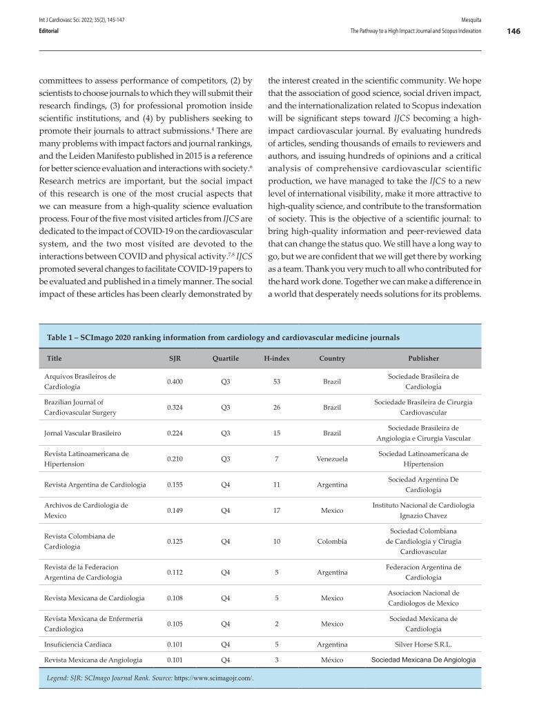
in Scopus, the use of English as publication language and open access status are important determinants of internationalization in the long term.3 IJCS has been compliant to these since 2018. Furthermore, the authors showed that national journals from USA, Japan, Brazil, and Iran evaluated after the year they entered Scopus revealed a broadening of the citation impact compared to the overall average.3 This is important because IJCS now will be ranked in Scopus in the cardiology and cardiovascular medicine subject category and can be compared with its partners. Elsevier publishes three journal metrics based on the Scopus citation database: (1) the Source-Normalized Impact per Paper (SNIP); (2) the Impact per Publication (IPP); and (3) the SCImago Journal Rank (SJR). SJR is used by the Brazilian Ministry of Education to qualify journals for the evaluation of post-graduate programs. SJR accounts for both the number of citations received by a journal and the importance or prestige of the journals from where such citations come.4
Accessing the 2020 SJR rank, we can find 349 journals in the cardiology and cardiovascular medicine subject category, most from North America and Europe. There are 12 journals in this subject from Latin America (Table 1). The best ranked journal from Latin America is Arquivos Brasileiros de Cardiologia (ABC Cardiol), with SJR of 0.400. Like IJCS, ABC Cardiol is sponsored by the Brazilian Society of Cardiology. IJCS will be soon ranked by SCImago, and authors and reader will be able to use the ranking to evaluate the journal’s impact. Our aim is to increase the impact factor of IJCS so that it will be comparable to that of ABC Cardiol, which recently obtained the highest ranking in its history.5
Journal rankings and scores are used for a list of academic and economic activities: (1) by academic
DOI: https://doi.org/10.36660/ijcs.20220030
Mailing Address: Claudio Tinoco MesquitaFaculdade de Medicina da Universidade Federal Fluminense Hospital Universitário Antônio Pedro, Setor de Radiologia R. Marquês de Paraná, 303, 2º andar. Postal Code: 24033-900, Niterói, Rio de Janeiro, RJ – Brazil.E-mail: [email protected]
Int J Cardiovasc Sci. 2022; 35(2), 145-147
145
EDITORIAL
Cardiology; Periodicals as Topic/standards; Abstracting and Indexing as Topic/methods; Editorial Policies; Databases, Bibliographic/trends; Citation Databases; Journal Impact Factor.
Keywords
The Pathway to a High Impact Journal and Scopus Indexation – New Achievement of the International Journal of Cardiovascular Sciences Claudio Tinoco Mesquita1,2,3
Universidade Federal Fluminense,1 Niterói, RJ – Brazil Nuclear Medicine Department – Hospital Pró-Cardíaco,2 Rio de Janeiro, RJ – Brazil Hospital Vitória,3 Rio de Janeiro, RJ – Brazil
committees to assess performance of competitors, (2) by scientists to choose journals to which they will submit their research findings, (3) for professional promotion inside scientific institutions, and (4) by publishers seeking to promote their journals to attract submissions.4 There are many problems with impact factors and journal rankings, and the Leiden Manifesto published in 2015 is a reference for better science evaluation and interactions with society.6 Research metrics are important, but the social impact of this research is one of the most crucial aspects that we can measure from a high-quality science evaluation process. Four of the five most visited articles from IJCS are dedicated to the impact of COVID-19 on the cardiovascular system, and the two most visited are devoted to the interactions between COVID and physical activity.7,8 IJCS promoted several changes to facilitate COVID-19 papers to be evaluated and published in a timely manner. The social impact of these articles has been clearly demonstrated by
the interest created in the scientific community. We hope that the association of good science, social driven impact, and the internationalization related to Scopus indexation will be significant steps toward IJCS becoming a high-impact cardiovascular journal. By evaluating hundreds of articles, sending thousands of emails to reviewers and authors, and issuing hundreds of opinions and a critical analysis of comprehensive cardiovascular scientific production, we have managed to take the IJCS to a new level of international visibility, make it more attractive to high-quality science, and contribute to the transformation of society. This is the objective of a scientific journal: to bring high-quality information and peer-reviewed data that can change the status quo. We still have a long way to go, but we are confident that we will get there by working as a team. Thank you very much to all who contributed for the hard work done. Together we can make a difference in a world that desperately needs solutions for its problems.
Table 1 – SCImago 2020 ranking information from cardiology and cardiovascular medicine journals
Title SJR Quartile H-index Country Publisher
Arquivos Brasileiros de Cardiologia
0.400 Q3 53 BrazilSociedade Brasileira de
Cardiologia
Brazilian Journal of Cardiovascular Surgery
0.324 Q3 26 BrazilSociedade Brasileira de Cirurgia
Cardiovascular
Jornal Vascular Brasileiro 0.224 Q3 15 BrazilSociedade Brasileira de
Angiologia e Cirurgia Vascular
Revista Latinoamericana de Hipertension
0.210 Q3 7 VenezuelaSociedad Latinoamericana de
Hipertension
Revista Argentina de Cardiologia 0.155 Q4 11 ArgentinaSociedad Argentina De
Cardiologia
Archivos de Cardiologia de Mexico
0.149 Q4 17 MexicoInstituto Nacional de Cardiologia
Ignazio Chavez
Revista Colombiana de Cardiologia
0.125 Q4 10 ColombiaSociedad Colombiana
de Cardiologia y Cirugia Cardiovascular
Revista de la Federacion Argentina de Cardiologia
0.112 Q4 5 ArgentinaFederacion Argentina de
Cardiologia
Revista Mexicana de Cardiologia 0.108 Q4 5 MexicoAsociacion Nacional de Cardiologos de Mexico
Revista Mexicana de Enfermeria Cardiologica
0.105 Q4 2 MexicoSociedad Mexicana de
Cardiologia
Insuficiencia Cardiaca 0.101 Q4 5 Argentina Silver Horse S.R.L.
Revista Mexicana de Angiologia 0.101 Q4 3 México Sociedad Mexicana De Angiologia
Legend: SJR: SCImago Journal Rank. Source: https://www.scimagojr.com/.
Int J Cardiovasc Sci. 2022; 35(2), 145-147
146Mesquita
The Pathway to a High Impact Journal and Scopus IndexationEditorial
1. Mesquita C T, Borim D, Rochitte C E. Open Science, Cardiology and 20 years of SciELO (Scientific Electronic Library Online). Int J Cardiovasc Sci.2019;32(3):203-4. doi:10.5935/2359-4802.20190036.
2. Mesquita C T, Lacerda A G. Sex and Gender Equity in Research and Publishing: International Journal of Cardiovascular Sciences endorses SAGER Guidelines. Int J cardiovasc Sci.2021;34(6):597-8. DOI:10.36660/ijcs.20210221
3. Moed HF. Moya-Anegon F, Guerrero-BoteV, Lopez-Illescas c. Are nationally oriented journals indexed in Scopus becoming more international? The effect of publication language and access modality. J Inform.2020;14(2). Doi:10.1016/j.joi.2020.101011
4. Bradshaw CJA,Brook BW, How PLoS One.2016;11(3):e0149852 doi: 10.1371/journal.pone.0149852
5. Rochitte, C. E. Impact factor of 2.0, a new historical record for abc cardiol – many thanks to our cardiology and scientific community. Arq Bras Cardiol.2021; 111(1):266-9. Doi: 10.5935/abc.20180129
6. Hicks D, Wouters P, Waltman L, de Rijcke S, Rafols I. The Leiden Manifesto for research metrics. Nature.2015;520(7548):429-31. Doi: 10.1038/520429a
7. Silva FB, Fonseca B, Domecq F, Facio MR, Prado C, Toledo L, Tuche W. Athletes Health during Pandemic Times: Hospitalization Rates and Variables Related to COVID-19 Prevalence among Endurance Athletes. Int J cardiovasc Sci. 2021;34(3):274-83. doi:10.36660/ijcs.20200208.
8. Araújo SGS. Physical Activity, Exercise and Sports and Covid-19: What Really Matters. Int J Cardiovasc Sci. 2021;34(2):113-5. doi:10.36660/ijcs.20210003.
References
Int J Cardiovasc Sci. 2022; 35(2), 145-147
147Mesquita
The Pathway to a High Impact Journal and Scopus Indexation Editorial
This is an open-access article distributed under the terms of the Creative Commons Attribution License
Women are entering the medical and scientific community in growing numbers, reaching and even surpassing their male counterparts in medical schools. However, a settled imbalance between men and women is still a reality in the international cardiology community despite these recent advances.1 The female presence in Brazilian medical schools was barely noticed until the 1960s; in the following years, there was a gradual increase in the number of women in the medical field, mainly in the first decade of the 21st century (59 % in 2020). In a 2020 demographic analysis of the Brazilian Federal Council of Medicine , men still predominated, accounting for 53.4% of all doctors in the country. Nevertheless, in the age-range below 30-years-old, women are the majority, accounting for 58.5%; the percentage of female doctors is inversely proportional to the increase in the age group, with only 21% of women in the age group above 70 years old.2
In the USA, women represent less than 15% of the cardiology workforce and less than 5% of interventional cardiologists,2 while in Europe, women account for only one-third of cardiologists, and 18% of women are interventionists.3 Currently, Brazil has nearly 500 thousand doctors, 17,802 thousand cardiologists, of which 31.1% are females, and mostly concentrated in the southeast region. In 2018, only 215 (8.6%) of a total of 2,062 cardiovascular surgeons and 7.5% of 970 interventional cardiologists were women.4 The Brazilian Society of Cardiology had two female presidents; and the Cardiovascular Surgery Society, the Interventional Cardiology, and the Federation of Portuguese Language Cardiology Societies had one female president. Also, in the last five years, only one fifth
of the speakers in the annual congress of the Brazilian Cardiology Society were women.
There are more male than female doctors in the private sector (23.9% vs. 14%), and more women in the public and academic sectors (53% vs. 44% of men). Among the professionals who work 20 and 40 hours a week, only 2.7% of the women earn US$ 10,762 per month compared to 13% of men. The likelihood of male doctors earning more than US $ 10,762 is 17%, and of female doctors, only 4%. Wage inequality between genders persist concerning workload, and office and on-call hours.2 In the USA, white women earn 77 cents on the dollar, black women, 79 cents, and Asian women 75 cents comparing with male physicians in their own racial or ethnic groups. Although these data come from academic medical institutions only, they reflect the compensation of 60,000 physicians.5 In an era when half of the medical students are women, these professionals will not succeed unless institutions make a commitment to improve processes and reshape practices and patterns at workplace that have been inadvertently benefited men and detrimental to women; additionally, these practices have upheld the unjustified and deeply troubling gender pay gap.6
Gender gap in science and academic careers is not new. According to US data, less than 30% of the world’s researchers are women. Also, high-status awards and positions are less likely to be given to women in science.7
A Brazilian study8 found that female scientists who hold a productivity scholarship and obtain more funding are at the lower levels of the research ranking system. In addition, only 14% of the Brazilian Academy of Science members were women.8 The authors pointed out several factors that contribute to the underrepresentation of women in higher positions and leadership. However, the primary factor influencing women’s career in science is still an understudied topic: motherhood.8
DOI: https://doi.org/10.36660/ijcs.20220029
Mailing Address: Gláucia Maria Moraes de OliveiraUniversidade Federal do Rio de Janeiro – R. Prof. Rodolpho P. Rocco, 255 – 8° Andar – Sala 6, UFRJ. Postal Code: 21941-913, Cidade Universitária, RJ – BrazilE-mail: [email protected]
Int J Cardiovasc Sci. 2022; 35(2), 148-151
148
EDITORIAL
Cardiovascular Disease; Cardiologists; Women; Ethics; Gender Identity; COVID-19; Pandemic
Keywords
Science Gender Gap: Are We in theRight Path?Gláucia Maria Moraes de Oliveira,1 Marge Tenorio,2 Alessandra de Sá Earp Siqueira2
Universidade Federal do Rio de Janeiro,1 Rio de Janeiro, RJ – BrazilDepartamento de Ciência e Tecnologia da Secretaria de Ciência, Tecnologia e Insumos Estratégicos do Ministério da Saúde (Decit/SCTIE/MS),2 Brasília, DF – Brazil
Another study analyzed the influence of gender, parenthood, and race on academic productivity during the pandemic period based on a survey responded by 3,345 Brazilian academics from various knowledge areas and research institutions. The authors found that male academics, especially those without children, were the least affected group. In contrast, Black women and mothers were the most impacted groups because of the uneven domestic division of labor between men and women, exacerbated during the pandemic.9
Results from the latest Organization for Economic Co-operation and Development (OECD) International Survey of Scientific Authors (ISSA2) showed that women are underrepresented in research careers. On average, across OECD countries, only nearly 40% of all the investigators are women – ranging from 23% in Luxembourg to 56% in Lithuania – and they are considerably less likely to be in leadership positions. Only 30 % of corresponding authors are women. Also, women authors earn on average 5 to 6% less than their male counterparts, even after accounting for individual and job-related characteristics.10
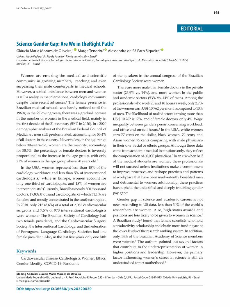
In addition to productivity grants, we can also analyze the Gender Gap in Science through the research supported by the Department of Science and Technology (DECIT) of the Secretariat of Science and Technology and Strategic
Inputs (SCTIE) of the Brazilian Ministry of Health, currently under the leadership of a woman. Figure 1 shows the distribution DECIT/SCTIE/HM-funded research projects from 2010 to 2021 by sex and federative units. There are significantly more projects coordinated by women, mainly in the states of Rio Grande do Sul (RS), Minas Gerais MG), and Bahia (BA). The number of projects coordinated by men was higher in the states of Piaui (PI), Rio Grande do Norte (RN), Roraima (RR) and São Paulo (SP).
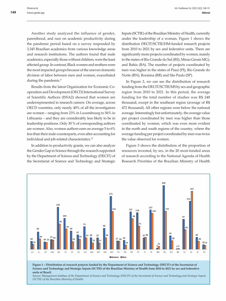
In Figure 2, we can see the distribution of research funding from the DECIT/SCTIE/MS by sex and geographic region from 2010 to 2021. In this period, the average funding for the total number of studies was R$ 248 thousand, except in the southeast region (average of R$ 472 thousand). All other regions were below the national average. Interestingly but unfortunately, the average value per project coordinated by men was higher than those coordinated by women, which was even more evident in the north and south regions of the country, where the average funding per project coordinated by men was twice the value observed for women.
Figure 3 shows the distribution of the proportion of resources invested, by sex, in the 20 most-funded areas of research according to the National Agenda of Health Research Priorities of the Brazilian Ministry of Health
Figure 1 – Distribution of research projects funded by the Department of Science and Technology (DECIT) of the Secretariat of Science and Technology and Strategic Inputs (SCTIE) of the Brazilian Ministry of Health from 2010 to 2021 by sex and federative units of Brazil.Source: Management database of the Department of Science and Technology (DECIT) of the Secretariat of Science and Technology and Strategic Inputs (SCTIE) of the Brazilian Ministry of Health.
Int J Cardiovasc Sci. 2022; 35(2), 148-151
149Oliveira et al.
Science gender gap Editorial
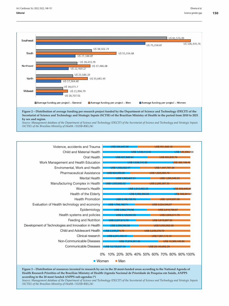
Figure 2 – Distribution of average funding per research project funded by the Department of Science and Technology (DECIT) of the Secretariat of Science and Technology and Strategic Inputs (SCTIE) of the Brazilian Ministry of Health in the period from 2010 to 2021 by sex and region.Source: Management database of the Department of Science and Technology (DECIT) of the Secretariat of Science and Technology and Strategic Inputs (SCTIE) of the Brazilian Ministry of Health. (1US$=R$5,24)
Figure 3 – Distribution of resources invested in research by sex in the 20 most-funded areas according to the National Agenda of Health Research Priorities of the Brazilian Ministry of Health (Agenda Nacional de Prioridade de Pesquisa em Saúde, ANPPS according to the 20 most funded ANPPS sub-agendas (*)Source: Management database of the Department of Science and Technology (DECIT) of the Secretariat of Science and Technology and Strategic Inputs (SCTIE) of the Brazilian Ministry of Health. (1US$=R$5,24)
Violence, accidents and Trauma
Child and Material Health
Oral Health
Work Management and Health Education
Enviromental, Work and Health
Pharmaceutical Assistance
Mental Health
Manufacturing Complex in Health
Women's Health
Health of the Elderly
Health Promotion
Evaluation of Health technology and economy
Epidemiology
Health systems and policies
Feeding and Nutrition
Development of Technologies and Innovation in Health
Child and Adolescent Health
Clinical research
Non-Communicable Diseases
Communicable Diseases
0% 10% 20% 30% 40% 50% 60% 70% 80% 90% 100%
Women Men
US$ 1,039,312.52 US$ 128,306.298
US$ 627,898.44 US$ 600,835.79
US$ 1,638,510.38 US$ 385,198.46
US$ 364,443.56 US$ 551,648.18
US$ 1,082,022.47 US$ 1,447,787.34
US$ 824,085.00 US$ 1,825,085.79
US$ 1,545,407.91 US$ 1,290,549.33
US$ 2,912,962.25 US$ 609,648.94
US$ 3,358,358.85 US$ 588,901.81
US$ 1,033,992.02 US$ 2,295,387.33
US$ 2,198,725.78 US$ 1,818,937.00
US$ 1,768,145.71 US$ 3,024,048,57
US$ 2,862,779.58 US$ 2,818,799.87
US$ 3,129,950.95 US$ 2,609,511.79
US$ 2,207,612.75 US$ 3,615,997.50
US$ 3,559,546.56 US$ 5,093,952.08
US$ 2,235,278.70 US$ 2,235,278.70
US$ 4,251,055.57 US$ 7,991,113.15
US$ 17,874,201.30 US$ 10,369,145.90
US$ 12,736,837.30 US$ 28,145,802.29
Int J Cardiovasc Sci. 2022; 35(2), 148-151
150Oliveira et al.
Science gender gapEditorial
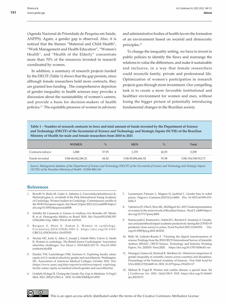
Table 1 – Number of research contracts in force and total amount of funds invested by the Department of Science and Technology (DECIT) of the Secretariat of Science and Technology and Strategic Inputs (SCTIE) of the Brazilian Ministry of Health for male and female researchers from 2010 to 2021
WOMEN % MEN % Total
Contracts inforce 1,888 57.95 1,370 42.05 3,258
Funds invested US$ 68,642,246.21 44.42 US$ 85,896,666.52 55.58 US$ 154,538,912.73
Source: Management database of the Department of Science and Technology (DECIT) of the Secretariat of Science and Technology and Strategic Inputs (SCTIE) of the Brazilian Ministry of Health (1US$=R$5.24)
(Agenda Nacional de Prioridade de Pesquisa em Saúde, ANPPS). Again, a gender gap is observed. Also, it is noticed that the themes “Maternal and Child Health”, “Work Management and Health Education”, “Women's Health”, and “Health of the Elderly” concentrate more than 70% of the resources invested in research coordinated by women.
In addition, a summary of research projects funded by the DECIT (Table 1) shows that the gap persists, since although female researchers hold more contracts, they are granted less funding . The comprehensive depiction of gender inequality in health sciences may provoke a discussion about the sustainability of women’s careers, and provide a basis for decision-makers of health policies.11 The equitable presence of women in advisory
and administrative bodies of health favors the formation of an environment based on societal and democratic principles.12
To change the inequality setting, we have to invest in public policies to identify the flaws and rearrange the relations to value the differences, and make it sustainable and inclusive, in a way that female researchers could reconcile family, private and professional life. Optimization of women’s participation in research projects goes through more investment. Our compelling task is to create a more favorable institutional and healthier environment for women and men, without losing the bigger picture of potentially introducing fundamental changes in the Brazilian society.
1. Borrelli N, Brida M, Cader A, Sabatino J, Czerwińska-Jelonkiewicz K, Shchendrygina A, on behalf of the Pink International Young Academy of Cardiology, Women leaders in Cardiology. Contemporary profile of the WHO European region, Eur Heart J Open.2021;1(1):oeab008 https://doi.org/10.1093/ehjopen/oeab008
2. Scheffer M, Cassenote A, Guerra A, Guilloux AA; Brandão AP, Miotto B, et al. Demografia Médica no Brasil 2020. São Paulo(SP):FMUSP/CFM;2020.312p. ISBN: 978-65-00-12370-8.
3. B u r g e s s S , S h a w E , Z a m a n S . W o m e n i n c a r d i o l o g y . C i r c u l a t i o n . 2 0 1 9 ; 1 3 9 ( 8 ) : 1 0 0 1 - 2 . h t t p s : / / d o i . o r g / 1 0 . 1 1 6 1 /CIRCULATIONAHA.118.037835
4. Sinclair HC, Joshi A, Allen C, Joseph J, Sohaib SMA, Calver A, Smith R. Women in cardiology: The British Junior Cardiologists’ Association identifies challenges. Eur Heart J. 2019;40(3):227-31. Doi;10.1093/eurheart/ehy828
5. Dandar VM, Lautenberger DM, Garrison G. Exploring faculty salary equity at U.S. medical schools by gender and race/ethnicity. Washington, DC: Association of American Medical Colleges, October 2021. Doi: (https://www.aamc.org/data-reports/workforce/report/ exploring-faculty-salary-equity-us-medical-schools-gender-and race/ethnicity)
6. Gottlieb AS,Jagsi R. Closing the Gender Pay Gap in Medicine. N Engl J Med. 2021; 385(27):2501-4. DOI: 10.1056/NEJMp2114955
7. Lunnemann P,Jensen L, Mogens H, Jauffred L. Gender bias in nobel prizes. Palgrave Commun.2019;5(1):e4000. Doi: 10.1057/s41599-019-0256-3
8. Valentova JV, Otta E, Silva ML, McElligott AG. 2017.Underrepresentation of women in the senior levels of Brazilian Science. PeerJ 5: e4000 https://doi.org/10.7717/peerj.4000
9. Staniscuaski F, Kmetzsch L, Soletti R C, Reichert F, Zandonà, E. Gender, race and parenthood impact academic productivity during the COVID-19 pandemic: from survey to action. Front Psychol.2021;12:663252. Doi.org/10.3389/fpsyg.2021.663252
10. Bello M, Galindo-Rueda F. "Charting the digital transformation of science: Findings from the 2018 OECD International Survey of Scientific Authors (ISSA2)", OECD Science, Technology and Industry Working Papers, No. 2020/03 Paris;2020. https://doi.org/10.1787/1b06c47c-en
11. Huanga,J, Gatesa AJ, Sinatrad R , Barabasi AL- Historical comparison of gender inequality in scientific careers across countries and disciplines. Proceedings of the National Academy of Sciences . Proc Natl Acad Sci USA.2020;117(9):4609-16. DOI: 10.1073/pnas.1914221117
12. Mehran R, Vogel B. Women and cardiac disease: a special issue. Int J Cardiovasc Sci. 2021; 34(4):338-9. DOI: https://doi.org/10.36660/ijcs.20210173
References
Int J Cardiovasc Sci. 2022; 35(2), 148-151
151Oliveira et al.
Science gender gap Editorial
This is an open-access article distributed under the terms of the Creative Commons Attribution License
Introduction
In Western European countries and the United States, morbidity and mortality from coronary artery disease (CAD) is about three times higher than cerebrovascular disease. In Brazil, cardiovascular diseases (CVDs) are responsible for about one third of deaths according to DATASUS.1,2 This is particularly important considering that as longevity of populations increases, chronic diseases including CVDs become more frequent.
Porto Alegre, located in the south of Brazil, is the tenth most populous capital in the country, with an estimated 1,484,941 inhabitants. The primary care system in the city covers areas of high social vulnerability and difficult access to specialized services. In this context, reducing geographic barriers and optimizing referrals to secondary and tertiary care would improve the provision of care and favor the local economy.
Telehealth appears as an opportunity to respond to this need using communication with information technology
Int J Cardiovasc Sci. 2022; 35(2), 152-158
152
ORIGINAL ARTICLE
Implementation of a Telecardiology Service in a Health Unit in the City of Porto Alegre, Brazil: A Pilot StudyFrancieli Giachini Esmerio,1 Silvia Goldmeier,1 Eduardo Costa Duarte Barbosa,1 Luis Marcelo Segredo,2 Rodolfo Silva,3 Maria Claudia Irigoyen,1 Bruna Eibel,1,4 Patricia Oliveira Dias1
Instituto de Cardiologia/Fundação Universitária de Cardiologia,1 Porto Alegre, RS – BrazilPrefeitura Municipal de Porto Alegre,2 Porto Alegre, RS – BrazilUniversidade Federal do Rio Grande do Sul – Telessaúde,3 Porto Alegre, RS – BrazilCentro Universitário da Serra Gaúcha,4 Caxias do Sul, RS – Brazil
DOI: https://doi.org/10.36660/ijcs.20200305
Mailing Address: Bruna EibelAvenida Princesa Isabel, 395. Postal Code: 9062000, Porto Alegre, RS – Brazil.E-mail: [email protected]
Manuscript received September 26, 2020; revised manuscript April 19, 2021; accepted August 12, 2021.
Abstract
Background: The implementation of Telecardiology in primary care in the city of Porto Alegre, Brazil, is a viable and promising strategy. It would decrease the distance between patient and specialized professional services by reducing unnecessary referrals and improving the quality of primary care and satisfaction of patients and health professionals.
Objective: To implement a Telecardiology service and assess user satisfaction using the CARDIOSATIS scale.
Methods: This was a pilot study developed by a partnership between the Institute of Cardiology and the Telehealth Center of Rio Grande do Sul. The study was carried out at Eri Flores-Vila Vargas health center in the city of Porto Alegre, from May to October 2019, and included 21 patients attending the health center. The descriptive analysis of data was performed using the SPSS program (Statistical Package for the Sciences) version 23. Data normality was checked using the Kolmogorov-Smirnov test. Statistical significance was set at 10%.
Results: Mean age of participants was 43.8 ± 16.1 years. The most common risk factors in the sample were physical inactivity (81%) and smoking (43%). Most patients had normal electrocardiogram (ECG) readings. The time elapsed from the performance of the ECG test, transmission of the ECG traces to Telehealth, and return of the final ECG report to the health center was 0-7 days. The CARDIOSATIS scale revealed a high prevalence of “very satisfied” users for the general satisfaction domain, and only 14.3% of patients were dissatisfied with their health.
Conclusions: Telecardiology reduced the distance between patient and the specialized professional, with a high level of patient and health professional satisfaction. Our study can serve as a basis for the implementation of a telecardiology network in the city of Porto Alegre in the future.
Keywords: Telemedicine/methods; Cardiovascular Diseases; Electrocardiography; Telemedicine/trends; Telemedicine/ethics.
application for managing online databases. ECG tests were performed using a Wincardio equipment.
Around 10 ECG exams were performed per week. The tests were then attached to the Telessaúde-RS platform, where it was evaluated and approved by a collaborating cardiologist. As this is a study developed within the Brazilian National Telehealth Program (Programa Nacional Telessaúde Brasil Redes), patients needed to be registered in the Teledermatology interface of the Telehealth Platform, adapted to the study. Also, the ECG exams needed to be sent to the teleconsultation center within 72 hours, following the deadline established by the program . It is important to remind that this study was not intended to evaluate data from the return appointment.
For analysis of ECG results, the data were entered into the platform by registered health professionals, using a login and password, which was determined by the management of the Telehealth center, allowing greater security in accessing the platform.
Ethical principles were respected, according to the ethical guidelines for research involving human subjects (Resolution 466/12 of the Brazilian National Health Council). The project was approved by the Research Ethics Committee of the ICFUC and subjected to the Eri Flores- Vila Vargas health center and the Rio Grande do Sul Telehealth center coordinators’ approval.
Statistical analysis
Continuous variables with normal distribution were described as average and standard deviation, and those without normal distribution as median and interquartile range. Normality of the data was checked using the Kolmogorov Smirnov test. Categorical variables were described as absolute and relative frequencies. The level of significance adopted was 10% and the data were analyzed using the SPSS (Statistical Package for the Social Sciences) program version 23.
Results
A total of 21 patients (71.4% women), with suggestive cardiac signs and symptoms were evaluated. Mean age was 43.8±16.1 years, and mean body mass index (BMI) was 27.5±5.9 Kg/m2. Only two patients (10%) had higher education.
Blood pressure was measured at rest according to the 7th Brazilian Guidelines on Arterial Hypertension.10
for information exchange. In Porto Alegre, together with the Federal University of Rio Grande do Sul (UFRGS), the Telehealth center aims to increase healthcare delivery and improve professional satisfaction.
The Porto Alegre the Pontifical Catholic University of Rio Grande do Sul (PUCRS) and the Institute of Cardiology of Rio Grande do Sul (ICFUC-RS) have implemented a telecardiology program, through Tele-ECG, and a special second opinion service to identify susceptible CVD and improve local health care.1,3,4 In this sense, telecardiology emerges as an opportunity to improve local economic, reduce the distance between the patient and specialized health services, avoiding unnecessary referrals,5,6 and improving the quality of care.7,8
Based on these observations, this study aimed at evaluating the implementation of a Telecardiology Network that includes electrocardiogram (ECG) tests in a primary care center in the city of Porto Alegre. Also, we evaluated the level of user satisfaction using the CARDIOSATIS Scale.9
Methods
This pilot study was conducted at Eri Flores Vila Vargas health center in Porto Alegre from May to October to 2019. The study was developed by a partnership between the Institute of Cardiology (responsible for interpreting the electrocardiogram tests) and the Telehealth Center of Rio Grande do Sul that guided the team.
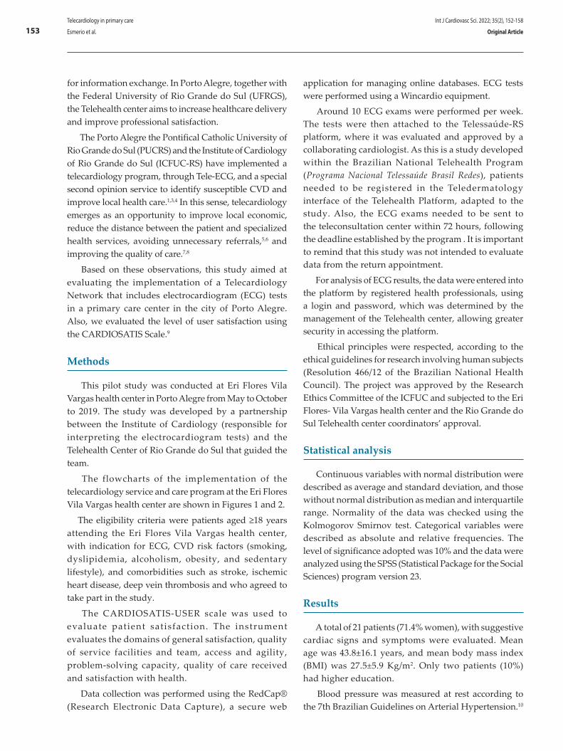
The flowcharts of the implementation of the telecardiology service and care program at the Eri Flores Vila Vargas health center are shown in Figures 1 and 2.
The eligibility criteria were patients aged ≥18 years attending the Eri Flores Vila Vargas health center, with indication for ECG, CVD risk factors (smoking, dyslipidemia, alcoholism, obesity, and sedentary lifestyle), and comorbidities such as stroke, ischemic heart disease, deep vein thrombosis and who agreed to take part in the study.
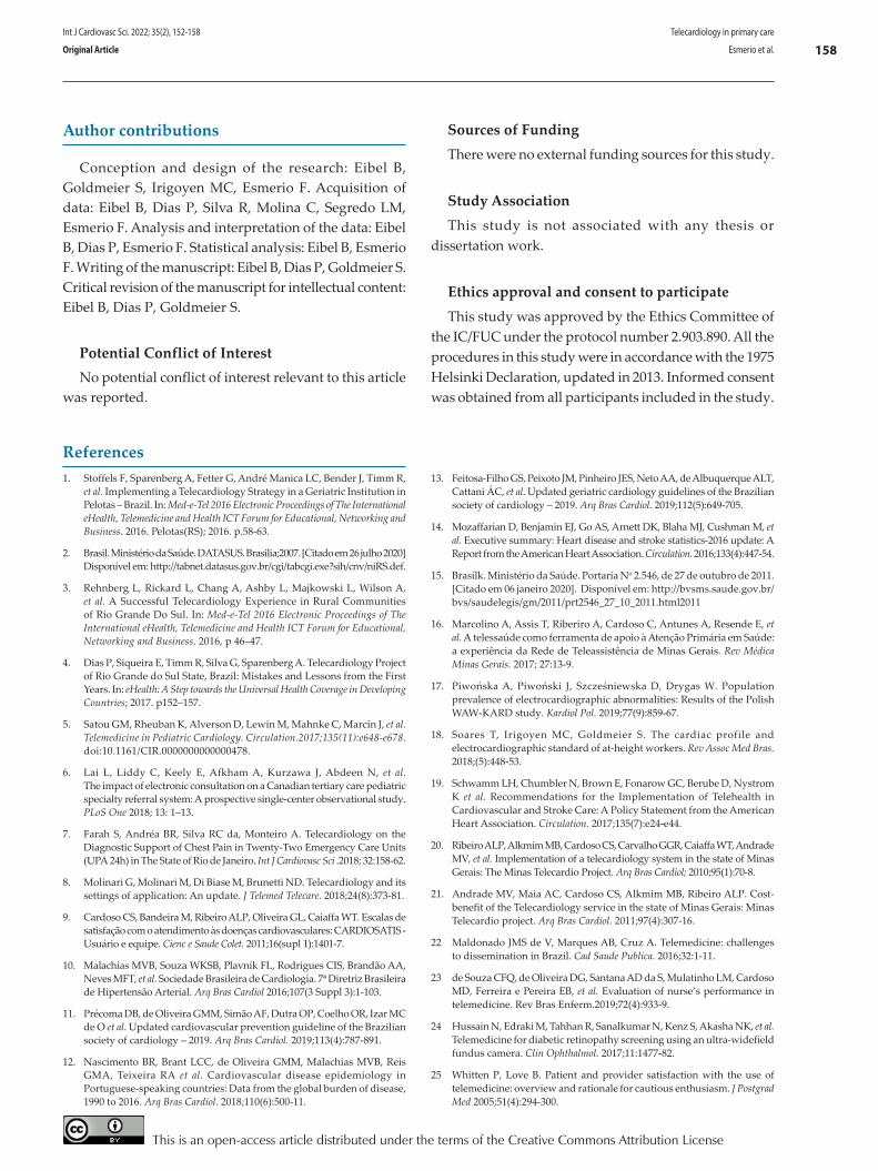
The CARDIOSATIS-USER scale was used to evaluate patient satisfaction. The instrument evaluates the domains of general satisfaction, quality of service facilities and team, access and agility, problem-solving capacity, quality of care received and satisfaction with health.
Data collection was performed using the RedCap® (Research Electronic Data Capture), a secure web
Int J Cardiovasc Sci. 2022; 35(2), 152-158
153Telecardiology in primary care
Esmerio et al. Original Article
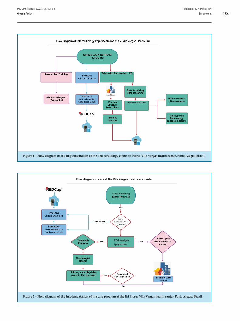
Figure 1 – Flow diagram of the Implementation of the Telecardiology at the Eri Flores Vila Vargas health center, Porto Alegre, Brazil
Comentado [1R0]: Ajuste em fluxograma: Primary care physician sends to the specialist. SUBSTITUIR: Health Unit por Healthcare center. SUBSTITUIR: UBS por Primary care center
Flow diagram of care at the Vila Vargas Healthcare center
Figure 2 – Flow diagram of the Implementation of the care program at the Eri Flores Vila Vargas health center, Porto Alegre, Brazil
Int J Cardiovasc Sci. 2022; 35(2), 152-158
154Telecardiology in primary care
Esmerio et al.Original Article
Mean systolic and diastolic blood pressure was 123.3±7.9 mmHg and 75.5±4.97 mmHg, respectively. The risk factors found were sedentary lifestyle (81%), smoking (43%), dyslipidemia (14.3%), and obesity (5%). The prevalence of diabetes mellitus was 86% in the study group (Table 1).
Analysis of the ECG reports revealed that only four patients had left ventricular hypertrophy and one had left bundle-branch block. Most patients had a normal tracing.
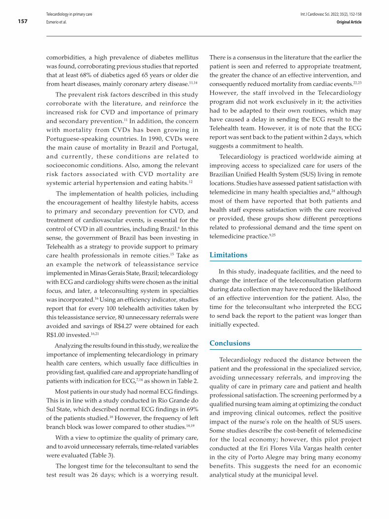
The median time between ECG performance and transmission of the ECG traces to Telehealth for analysis by a specialist was 0 (0-7) day. In a certain period of the data collection, it was necessary to migrate to another teleconsultation interface, the telediagnosis platform. It required adjustment and remote training of the teleconsultant cardiologist, which caused delays in sending the ECG images. The longest time for the
teleconsultant to send the ECG report back to the health center was 26 days. It is worth pointing out that consultation of a cardiologist for a second opinion was not required .. Other results are described in Table 3.
The Cardiosatis Scale was used to assess patient satisfaction with the use of Telemedicine to perform the ECG test. Table 4 shows the percentage distribution of user satisfaction by the Cardiosatis dimensions. In the “general satisfaction” domain, most patients were very satisfied, and 14.3% were dissatisfied with their health.
Discussion
The results in Table 1 show a prevalence of females and an average age of 40 years in our sample. Along with an increasing longevity, mortality rates are decreasing in Brazil, although without the
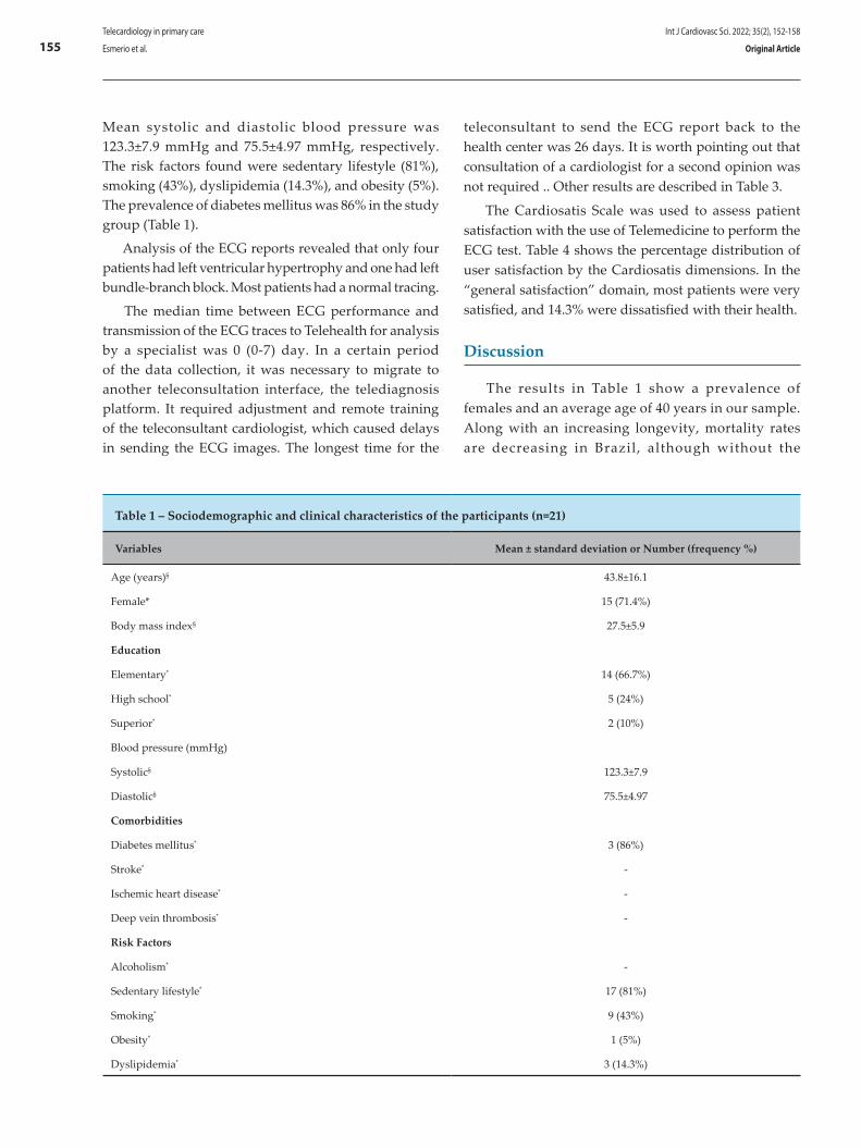
Table 1 – Sociodemographic and clinical characteristics of the participants (n=21)
Variables Mean ± standard deviation or Number (frequency %)
Age (years)§ 43.8±16.1
Female* 15 (71.4%)
Body mass index§ 27.5±5.9
Education
Elementary* 14 (66.7%)
High school* 5 (24%)
Superior* 2 (10%)
Blood pressure (mmHg)
Systolic§ 123.3±7.9
Diastolic§ 75.5±4.97
Comorbidities
Diabetes mellitus* 3 (86%)
Stroke* -
Ischemic heart disease* -
Deep vein thrombosis* -
Risk Factors
Alcoholism* -
Sedentary lifestyle* 17 (81%)
Smoking* 9 (43%)
Obesity* 1 (5%)
Dyslipidemia* 3 (14.3%)
Int J Cardiovasc Sci. 2022; 35(2), 152-158
155Telecardiology in primary care
Esmerio et al. Original Article
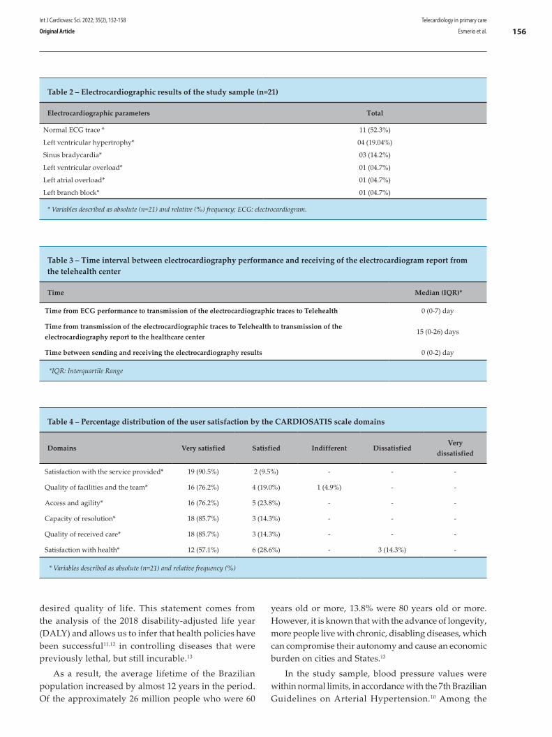
Table 2 – Electrocardiographic results of the study sample (n=21)
Electrocardiographic parameters Total
Normal ECG trace * 11 (52.3%)
Left ventricular hypertrophy* 04 (19.04%)
Sinus bradycardia* 03 (14.2%)
Left ventricular overload* 01 (04.7%)
Left atrial overload* 01 (04.7%)
Left branch block* 01 (04.7%)
* Variables described as absolute (n=21) and relative (%) frequency; ECG: electrocardiogram.
Table 3 – Time interval between electrocardiography performance and receiving of the electrocardiogram report from the telehealth center
Time Median (IQR)*
Time from ECG performance to transmission of the electrocardiographic traces to Telehealth 0 (0-7) day
Time from transmission of the electrocardiographic traces to Telehealth to transmission of the electrocardiography report to the healthcare center
15 (0-26) days
Time between sending and receiving the electrocardiography results 0 (0-2) day
*IQR: Interquartile Range
Table 4 – Percentage distribution of the user satisfaction by the CARDIOSATIS scale domains
Domains Very satisfied Satisfied Indifferent DissatisfiedVery
dissatisfied
Satisfaction with the service provided* 19 (90.5%) 2 (9.5%) - - -
Quality of facilities and the team* 16 (76.2%) 4 (19.0%) 1 (4.9%) - -
Access and agility* 16 (76.2%) 5 (23.8%) - - -
Capacity of resolution* 18 (85.7%) 3 (14.3%) - - -
Quality of received care* 18 (85.7%) 3 (14.3%) - - -
Satisfaction with health* 12 (57.1%) 6 (28.6%) - 3 (14.3%) -
* Variables described as absolute (n=21) and relative frequency (%)
desired quality of life. This statement comes from the analysis of the 2018 disability-adjusted life year (DALY) and allows us to infer that health policies have been successful11,12 in controlling diseases that were previously lethal, but still incurable.13
As a result, the average lifetime of the Brazilian population increased by almost 12 years in the period. Of the approximately 26 million people who were 60
years old or more, 13.8% were 80 years old or more. However, it is known that with the advance of longevity, more people live with chronic, disabling diseases, which can compromise their autonomy and cause an economic burden on cities and States.13
In the study sample, blood pressure values were within normal limits, in accordance with the 7th Brazilian Guidelines on Arterial Hypertension.10 Among the
Int J Cardiovasc Sci. 2022; 35(2), 152-158
156Telecardiology in primary care
Esmerio et al.Original Article
comorbidities, a high prevalence of diabetes mellitus was found, corroborating previous studies that reported that at least 68% of diabetics aged 65 years or older die from heart diseases, mainly coronary artery disease.11,14
The prevalent risk factors described in this study corroborate with the literature, and reinforce the increased risk for CVD and importance of primary and secondary prevention.11 In addition, the concern with mortality from CVDs has been growing in Portuguese-speaking countries. In 1990, CVDs were the main cause of mortality in Brazil and Portugal, and currently, these conditions are related to socioeconomic conditions. Also, among the relevant risk factors associated with CVD mortality are systemic arterial hypertension and eating habits.12
The implementation of health policies, including the encouragement of healthy lifestyle habits, access to primary and secondary prevention for CVD, and treatment of cardiovascular events, is essential for the control of CVD in all countries, including Brazil.6 In this sense, the government of Brazil has been investing in Telehealth as a strategy to provide support to primary care health professionals in remote cities.15 Take as an example the network of teleassistance service implemented in Minas Gerais State, Brazil; telecardiology with ECG and cardiology shifts were chosen as the initial focus, and later, a teleconsulting system in specialties was incorporated.16 Using an efficiency indicator, studies report that for every 100 telehealth activities taken by this teleassistance service, 80 unnecessary referrals were avoided and savings of R$4.27 were obtained for each R$1.00 invested.16,21
Analyzing the results found in this study, we realize the importance of implementing telecardiology in primary health care centers, which usually face difficulties in providing fast, qualified care and appropriate handling of patients with indication for ECG,7,14 as shown in Table 2.
Most patients in our study had normal ECG findings. This is in line with a study conducted in Rio Grande do Sul State, which described normal ECG findings in 69% of the patients studied.18 However, the frequency of left branch block was lower compared to other studies.18,19
With a view to optimize the quality of primary care, and to avoid unnecessary referrals, time-related variables were evaluated (Table 3).
The longest time for the teleconsultant to send the test result was 26 days; which is a worrying result.
There is a consensus in the literature that the earlier the patient is seen and referred to appropriate treatment, the greater the chance of an effective intervention, and consequently reduced mortality from cardiac events.22,23 However, the staff involved in the Telecardiology program did not work exclusively in it; the activities had to be adapted to their own routines, which may have caused a delay in sending the ECG result to the Telehealth team. However, it is of note that the ECG report was sent back to the patient within 2 days, which suggests a commitment to health.
Telecardiology is practiced worldwide aiming at improving access to specialized care for users of the Brazilian Unified Health System (SUS) living in remote locations. Studies have assessed patient satisfaction with telemedicine in many health specialties and,24 although most of them have reported that both patients and health staff express satisfaction with the care received or provided, these groups show different perceptions related to professional demand and the time spent on telemedicine practice.9,25
Limitations
In this study, inadequate facilities, and the need to change the interface of the teleconsultation platform during data collection may have reduced the likelihood of an effective intervention for the patient. Also, the time for the teleconsultant who interpreted the ECG to send back the report to the patient was longer than initially expected.
Conclusions
Telecardiology reduced the distance between the patient and the professional in the specialized service, avoiding unnecessary referrals, and improving the quality of care in primary care and patient and health professional satisfaction. The screening performed by a qualified nursing team aiming at optimizing the conduct and improving clinical outcomes, reflect the positive impact of the nurse's role on the health of SUS users. Some studies describe the cost-benefit of telemedicine for the local economy; however, this pilot project conducted at the Eri Flores Vila Vargas health center in the city of Porto Alegre may bring many economy benefits. This suggests the need for an economic analytical study at the municipal level.
Int J Cardiovasc Sci. 2022; 35(2), 152-158
157Telecardiology in primary care
Esmerio et al. Original Article
1. Stoffels F, Sparenberg A, Fetter G, André Manica LC, Bender J, Timm R, et al. Implementing a Telecardiology Strategy in a Geriatric Institution in Pelotas – Brazil. In: Med-e-Tel 2016 Electronic Proceedings of The International eHealth, Telemedicine and Health ICT Forum for Educational, Networking and Business. 2016. Pelotas(RS); 2016. p.58-63.
2. Brasil. Ministério da Saúde. DATASUS. Brasilia;2007. [Citado em 26 julho 2020] Disponível em: http://tabnet.datasus.gov.br/cgi/tabcgi.exe?sih/cnv/niRS.def.
3. Rehnberg L, Rickard L, Chang A, Ashby L, Majkowski L, Wilson A, et al. A Successful Telecardiology Experience in Rural Communities of Rio Grande Do Sul. In: Med-e-Tel 2016 Electronic Proceedings of The International eHealth, Telemedicine and Health ICT Forum for Educational, Networking and Business. 2016, p 46–47.
4. Dias P, Siqueira E, Timm R, Silva G, Sparenberg A. Telecardiology Project of Rio Grande do Sul State, Brazil: Mistakes and Lessons from the First Years. In: eHealth: A Step towards the Universal Health Coverage in Developing Countries; 2017. p152–157.
5. Satou GM, Rheuban K, Alverson D, Lewin M, Mahnke C, Marcin J, et al. Telemedicine in Pediatric Cardiology. Circulation.2017;135(11):e648-e678. doi:10.1161/CIR.0000000000000478.
6. Lai L, Liddy C, Keely E, Afkham A, Kurzawa J, Abdeen N, et al. The impact of electronic consultation on a Canadian tertiary care pediatric specialty referral system: A prospective single-center observational study. PLoS One 2018; 13: 1–13.
7. Farah S, Andréa BR, Silva RC da, Monteiro A. Telecardiology on the Diagnostic Support of Chest Pain in Twenty-Two Emergency Care Units (UPA 24h) in The State of Rio de Janeiro. Int J Cardiovasc Sci .2018; 32:158-62.
8. Molinari G, Molinari M, Di Biase M, Brunetti ND. Telecardiology and its settings of application: An update. J Telemed Telecare. 2018;24(8):373-81.
9. Cardoso CS, Bandeira M, Ribeiro ALP, Oliveira GL, Caiaffa WT. Escalas de satisfação com o atendimento às doenças cardiovasculares: CARDIOSATIS - Usuário e equipe. Cienc e Saude Colet. 2011;16(supl 1):1401-7.
10. Malachias MVB, Souza WKSB, Plavnik FL, Rodrigues CIS, Brandão AA, Neves MFT, et al. Sociedade Brasileira de Cardiologia. 7ª Diretriz Brasileira de Hipertensão Arterial. Arq Bras Cardiol 2016;107(3 Suppl 3):1-103.
11. Précoma DB, de Oliveira GMM, Simão AF, Dutra OP, Coelho OR, Izar MC de O et al. Updated cardiovascular prevention guideline of the Brazilian society of cardiology – 2019. Arq Bras Cardiol. 2019;113(4):787-891.
12. Nascimento BR, Brant LCC, de Oliveira GMM, Malachias MVB, Reis GMA, Teixeira RA et al. Cardiovascular disease epidemiology in Portuguese-speaking countries: Data from the global burden of disease, 1990 to 2016. Arq Bras Cardiol. 2018;110(6):500-11.
13. Feitosa-Filho GS, Peixoto JM, Pinheiro JES, Neto AA, de Albuquerque ALT, Cattani ÁC, et al. Updated geriatric cardiology guidelines of the Brazilian society of cardiology – 2019. Arq Bras Cardiol. 2019;112(5):649-705.
14. Mozaffarian D, Benjamin EJ, Go AS, Arnett DK, Blaha MJ, Cushman M, et al. Executive summary: Heart disease and stroke statistics-2016 update: A Report from the American Heart Association. Circulation. 2016;133(4):447-54.
15. Brasilk. Ministério da Saúde. Portaria No 2.546, de 27 de outubro de 2011. [Citado em 06 janeiro 2020]. Disponível em: http://bvsms.saude.gov.br/bvs/saudelegis/gm/2011/prt2546_27_10_2011.html2011
16. Marcolino A, Assis T, Riberiro A, Cardoso C, Antunes A, Resende E, et al. A telessaúde como ferramenta de apoio à Atenção Primária em Saúde: a experiência da Rede de Teleassistência de Minas Gerais. Rev Médica Minas Gerais. 2017; 27:13-9.
17. Piwońska A, Piwoński J, Szcześniewska D, Drygas W. Population prevalence of electrocardiographic abnormalities: Results of the Polish WAW-KARD study. Kardiol Pol. 2019;77(9):859-67.
18. Soares T, Irigoyen MC, Goldmeier S. The cardiac profile and electrocardiographic standard of at-height workers. Rev Assoc Med Bras. 2018;(5):448-53.
19. Schwamm LH, Chumbler N, Brown E, Fonarow GC, Berube D, Nystrom K et al. Recommendations for the Implementation of Telehealth in Cardiovascular and Stroke Care: A Policy Statement from the American Heart Association. Circulation. 2017;135(7):e24-e44.
20. Ribeiro ALP, Alkmim MB, Cardoso CS, Carvalho GGR, Caiaffa WT, Andrade MV, et al. Implementation of a telecardiology system in the state of Minas Gerais: The Minas Telecardio Project. Arq Bras Cardiol; 2010;95(1):70-8.
21. Andrade MV, Maia AC, Cardoso CS, Alkmim MB, Ribeiro ALP. Cost-benefit of the Telecardiology service in the state of Minas Gerais: Minas Telecardio project. Arq Bras Cardiol. 2011;97(4):307-16.
22 Maldonado JMS de V, Marques AB, Cruz A. Telemedicine: challenges to dissemination in Brazil. Cad Saude Publica. 2016;32:1-11.
23 de Souza CFQ, de Oliveira DG, Santana AD da S, Mulatinho LM, Cardoso MD, Ferreira e Pereira EB, et al. Evaluation of nurse’s performance in telemedicine. Rev Bras Enferm.2019;72(4):933-9.
24 Hussain N, Edraki M, Tahhan R, Sanalkumar N, Kenz S, Akasha NK, et al. Telemedicine for diabetic retinopathy screening using an ultra-widefield fundus camera. Clin Ophthalmol. 2017;11:1477-82.
25 Whitten P, Love B. Patient and provider satisfaction with the use of telemedicine: overview and rationale for cautious enthusiasm. J Postgrad Med 2005;51(4):294-300.
References
This is an open-access article distributed under the terms of the Creative Commons Attribution License
Author contributions
Conception and design of the research: Eibel B, Goldmeier S, Irigoyen MC, Esmerio F. Acquisition of data: Eibel B, Dias P, Silva R, Molina C, Segredo LM, Esmerio F. Analysis and interpretation of the data: Eibel B, Dias P, Esmerio F. Statistical analysis: Eibel B, Esmerio F. Writing of the manuscript: Eibel B, Dias P, Goldmeier S. Critical revision of the manuscript for intellectual content: Eibel B, Dias P, Goldmeier S.
Potential Conflict of InterestNo potential conflict of interest relevant to this article
was reported.
Sources of FundingThere were no external funding sources for this study.
Study AssociationThis study is not associated with any thesis or
dissertation work.
Ethics approval and consent to participateThis study was approved by the Ethics Committee of
the IC/FUC under the protocol number 2.903.890. All the procedures in this study were in accordance with the 1975 Helsinki Declaration, updated in 2013. Informed consent was obtained from all participants included in the study.
Int J Cardiovasc Sci. 2022; 35(2), 152-158
158Telecardiology in primary care
Esmerio et al.Original Article
“Even when the road is hard, never give up” – Tupac Shakur and The Outlawz
The above quote is from the hit song “Baby Don’t Cry” immortalized by Tupac and The Outlawz. The song is about a young woman who faces challenges and receives encouragement to persevere and not give up. In a similar way, telemedicine faces several challenges on an arduous path. In this way, imbued with a tenacious spirit, its users have had remarkable experiences. In the current issue of the International Journal of Cardiovascular Sciences, Esmerio and colleagues presented a successful application of a telecardiology service. They emphasized that the work contributed to reducing the distance between patients and specialized professionals, and they highlighted a high level of satisfaction for both patients and health Professionals.1 Experiences like this represent the relevant role of telemedicine in providing quality health services and generating value. However, these tools still face important challenges that need to be overcome.
The first challenge is resistance. Some people underestimate the potential of telemedicine, arguing that it damages the doctor-patient relationship and can harm patients. That is not true per se; however, using telemedicine without respecting the ethical dictates of the medical profession would be disastrous. In this context, a fundamental point emerges in order to reduce resistance. It is necessary to increase patients’ and health professionals’ literacy and to design studies that
demonstrate its real benefits in clinical practice, such as the successful example mentioned above. Literacy is equally important in relation to the use of computing resources to access telemedicine tools, which must be user-friendly. Almufleh and colleagues, for example, highlight the need to include telemedicine training in the medical curriculum. They reiterate three fundamental components of a telemedicine curriculum: the issue of technology per se; examination and communication skills; and the medicolegal/licensing aspects.2
The second challenge concerns the limits of telemedicine. It is necessary to specify, from the outset, what kind of benefits can be provided to patients. If, for example, during an appointment, a patient reports chest pain with characteristics of an acute myocardial infarction, it is mandatory for the patient to go to the emergency room for clinical evaluation and complementary tests to rule out or confirm the condition. In this hypothetical case, the teleconsultation did not resolve the complaint; nevertheless, it was fundamental in signaling the need for emergency care. We clearly must not attempt to go beyond what we can deliver. Furthermore, telemedicine is complementary to face-to-face care, and it does not represent a replacement; it must be practiced safely and for a period that is compatible with what the clinical circumstances require.3 It is also necessary to respect limits and explain to the patient that in-person consultation may be necessary. Accordingly, the request to sign a free and informed consent form plays a fundamental role.
The third challenge deals with data security and reliability of electronic systems. This should be a constant concern, as data leakage and access by unauthorized persons could have catastrophic effects on patients’ lives, including worsening their health issues. Specific legislation on the subject has brought great progress, but there is still
DOI: https://doi.org/10.36660/ijcs.20220014
Mailing Address: Erito MarquesUniversidade Federal Rural do Rio de JaneiroRua Governador Roberto da Silveira, w/n. Postal Code 26285-060, Nova Iguaçu, RJ – BrazilE-mail: [email protected]
Int J Cardiovasc Sci. 2022; 35(2), 159-160
159
EDITORIAL
Cardiovascular Disease; Telemedicine/ethics, Telemedicine/methods; Telemedicine/trends; Bioethic; Telehealth; Women; Working; Distance Education; Brazil.
Keywords
Challenges in Telemedicine: Even When the Road is Hard, Never Give upErito Marques de Souza Filho1 and Alexandra Monteiro2
Universidade Federal Rural do Rio de Janeiro,1 Rio de Janeiro, RJ – BrazilUniversidade do Estado do Rio de Janeiro,2 Rio de Janeiro, RJ – BrazilEditorial referring to the article: Implementation of a Telecardiology Service in a Health Unit in the City of Porto Alegre, Brazil: A Pilot Study
a long way to go. In addition, it is worth mentioning that the data should be of good quality (healthy data).4
The fourth major challenge refers to the relationship between telemedicine and other digital tools. It is necessary to understand that artificial intelligence, smartphone apps, electronic health records, blockchain, wearables, and others constitute a continuum of solutions that must be considered in an integrated manner and aligned with the purpose of generating value. In addition, it is essential for telemedicine activities to be properly integrated into the practice of the multidisciplinary health team.5 The main idea is build a sustainable digital ecosystem. If, on the one hand, telemedicine tools have been used successfully in different contexts and are capable of overcoming distances, on the other hand, a significant portion of the Brazilian population cannot yet access these benefits. Thus, the fifth challenge is to expand patients’ access to these tools. This implies several needs, such as expansion of infrastructure and the development of customized management models.
The sixth challenge is about the need for continuous evaluation of the tools and the development of evaluation metrics. In the meantime, the work by Esmerio and colleagues1 exposes something fundamental: the need to assess the degree of satisfaction of the patients for whom the tool was designed. In addition, it is important to validate these tools in different contexts and to encourage sharing of experiences. Last but not least, patient engagement with telemedicine care might help address unmet health needs by more accurately identifying them. In this context, it is important to design more tailored and people-centered services. All this would not only help to cut wasteful spending; more importantly, it would result in healthier and more productive populations.
Resistance, limits to use, security and reliability of systems, the relationship with other digital tools, access, continuous evaluation, and engagement are important challenges to be overcome and must be the object of efforts on the part of the entire community. Surely, it is an arduous task. However, we can quote Tupac again: “Even when the road is hard, never give up”.
1. Esmerio FG, Goldmeier S, Barbosa ECD, Segredo LM, Silva R, Irigoyen MC et al. Implementation of a telecardiology service in a health unit in the city of Porto Alegre, Brazil: A Pilot Study. Int J Cardiovasc Sci. 2022; 35(2):152-158. Doi: https://doi.org/10.36660/ijcs.20200305.
2. Almufleh A, Lee C, Tsang MY, Gin K, Tsang TSM, Nair P. The Need for telemedicine integration into adult cardiology training Curricula in Canada. Can J Cardiol. 2021 Jun;37(6):929-32. doi: 10.1016/j.cjca.2021.03.001.
3. Lopes MACQ, Oliveira GMM, Ribeiro ALP, Pinto FJ, Rey HCV, Zimerman LI, et al. Guideline of the Brazilian Society of Cardiology on Telemedicine
in Cardiology - 2019. Arq Bras Cardiol. 2019 Nov;113(5):1006-56.1056. doi: 10.5935/abc.20190205. PMID: 31800728.
4. Souza Filho EM, Fernandes FA, Soares CLA, Seixas FL, Santos AASMDD, Gismondi RA, et al. Artificial intelligence in cardiology: concepts, tools and challenges - "The Horse is the One Who Runs, You Must Be the Jockey". Arq Bras Cardiol. 2020 Apr;114(4):718-25.doi: 10.36660/abc.20180431.
5. Monteiro A, Grisolia BM, Aguiar J, Santos M. Profile and motivation of women enrolled in the brazilian telemedicine and telehealth postgraduation distance education programme. J Int Soc Telemed eHealth.2019;7:e3-1-e3-4. DOI: https://doi.org/10.29086/JISfTeH.7.e3.
References
This is an open-access article distributed under the terms of the Creative Commons Attribution License
Int J Cardiovasc Sci. 2022; 35(2), 159-160
160Marques & Monteiro
Challenges in TelemedicineEditorial
Introduction
Experimental animals are usually applied in the study of human health and disease, including the use of rodents as experimental models for the investigation of biological phenomenon similar to those observed in humans.1-3 Among them, the SHR is widely used as a model for the investigation of essential hypertension.3-5 For those working with these experimental animals, it
is obvious and mandatory to adopt a control group in their experiments.
In this way, two experimental strains are nowadays used as SHR’s controls, namely Wistar rats (WIS)6-8 or Wistar Kyoto rats (WKY).4,5,9 It is a well-known fact that WKY was the SHR background and most used strain as SHR control in scientific research.8,10,11 However, the WIS strain has also been used.4,5,9,12 Moreover, some previous studies have pointed out limitations in the use of both strains.13-18
Int J Cardiovasc Sci. 2022; 35(2), 161-171
161
ORIGINAL ARTICLE
Is the Wistar Rat a more Suitable Normotensive Control for SHR to Test Blood Pressure and Cardiac Structure and Function?Leonardo Mateus Teixeira de Rezende,1 Leôncio Lopes Soares,1 Filipe Rios Drummond,1 Pedro Zavagli Suarez,1 Luciano Leite,1 Joel Alves Rodrigues,1 Tiago Leal,1 Lukiya Favarato,1 Emily Correna Carlo Reis,1 Evandro Favarato,1 Miguel Carneiro-Júnior,1 Antônio José Natali,1 Cândido Coimbra,2 Thales Prímola-Gomes1
Universidade Federal de Viçosa,1 Viçosa, MG – Brazil Universidade Federal de Minas Gerais,2 Belo Horizonte, MG – Brazil
DOI: https://doi.org/10.36660/ijcs.20200367
Mailing Address: Leonardo RezendeAvenida P H Rolfs, s/n. Postal Code: 36570-000, Viçosa, MG – Brazil. E-mail: [email protected]
Manuscript received November 06, 2020; revised manuscript April 13, 2021; accepted April 26, 2021.
Abstract
Background: There are divergences in the literature regarding the experimental model (Wistar-WIS or Wistar Kyoto-WKY) to be used as a Spontaneously Hypertensive Rat (SHR) control. The characterization of these models in terms of cardiovascular parameters provides researchers with important tools at the time of selection and application in scientific research.
Objective: The aim of this study was to evaluate the use of WIS and WKY as a Spontaneously Hypertensive Rat (SHR) control by assessing the long-term behavior of blood pressure and cardiac structure and function in these strains.
Methods: To this end, WIS, WKY, and SHR underwent longitudinal experiments. Blood pressure and body mass were measured every two weeks from the 8th to the 72nd. Echocardiographic analysis was performed in all groups with 16, 48, and 72 weeks of life. After having applied the normality test, the Two-Way ANOVA of repeated measures followed by the Tukey post hoc test was used. A significance level of 5% was established .
Results: The WIS group showed higher body mass (p<0.05), while the WKY and SHR presented higher body mass variation over time (p<0.05). SHR exhibited increased values of systolic, diastolic, and mean blood pressure when compared to WKY and WIS, whereas the WKY generally showed higher values than WIS (p<0.05). Regarding the cardiac function, SHR showed reduced values, while the WKY presented an early decrease when compared to WIS with aging (p<0.05).
Conclusion: WIS is a more suitable normotensive control for SHR than WKY in experiments to test blood pressure and cardiac structure and function.
Keywords: Hypertension; Laboratory animals; Blood pressure; Heart.
Methods
Animals
Male WIS, WKY, and SHR rats, in their 8th to 72nd week of life, were used for all experiments. Each experimental group was composed of eight animals (n=8), and this number was defined using the sample calculation proposed by Armitage and Berry.23 The animals were housed in collective cages and allocated in a controlled environment with a light/dark cycle (12/12h), temperature at 22 ± 2°C, and had free access to food and water (ad libitum). The animals were obtained from the central biotery of the Federal University of Viçosa (UFV).
Ethical approval
The experiments were conducted in accordance with the Guide for the Care and Use of in Laboratory Animal principles and approved by the UFV Ethics Committee on the Use of Animals (logged under protocol number 09/2018). All procedures were conducted by a veterinarian.
Body mass
Body mass (g) was obtained every two weeks, from the 8th to the 72nd week of life, on an electronic scale (Rochelle, model 3252). To monitor the animals’ weight gain behavior, body mass variation (Δ) was calculated.
Blood pressure
Systolic Blood Pressure (SBP in mmHg) and Diastolic Blood Pressure (DBP in mmHg) were measured using the noninvasive method of tail plethysmography (LE5001; Panlab, Barcelona- Spain), as previously described.24 Briefly, animals were adapted to a tail cuff and a heating apparatus during five consecutive days. After, animals underwent blood pressure measurements each two weeks. Each measurement was performed three times and the median value was used. All measurements were performed by the same researcher in a quiet environment.25 Mean Arterial Pressure (MAP in mmHg) was calculated by the following equation: DBP + 1/3(SBP-DBP).
Echocardiogram
An echocardiographic analysis was performed in all groups with 16, 48, and 72 weeks of life. The animals underwent an anesthesia inhalation (Isoflurane 1.5%
Regarding these limitations, Kurtz & Morris (1987) studied the biological variability of WKY and SHR in two laboratories in the USA over a 20-week period. Differences in growth rate and mean arterial pressure (MAP), i.e., in biological variability in the WKY, were found.17 In a second study, the same group tested the genetic variability of these strains through genomic analysis and found differences between the WKY of different laboratories and even among animals from the same laboratory.16 Evidence also points to the presence of increased sympathetic activity in WKY, as shown by baseline resting catecholamine concentrations similar to the levels found in SHR.14,19 In another study, Aiello et al. performed a series of experiments in the myocardium of WIS, WKY, and SHR, and found higher left ventricle mass: body mass ratio in WKY when compared to WIS, indicating a hypertrophic process.13 The study also found an increase in diastolic papillary muscle stiffness and fibrosis in the left ventricle in the WKY, which was similar to SHR but higher than WIS.13
By contrast, limitations have also been highlighted against the use of WIS. First, it is a fact that WIS is not the SHR background.10 Furthermore, WIS have higher body mass values compared to WKY and SHR.20 This difference brings to light an experimental paradigm when selecting the control group, since when choosing WIS as a control, the researcher will assume that there are two groups with different body weights.21.To understand the growth behavior between WIS, WKY, and SHR, a previous study analyzed their physical development immediately after birth, during suckling and weanling.22 It was found that WIS showed a higher body mass than WKY and SHR. Additionally, WKY presented a body mass similar to SHR at birth and a higher body mass between the 1st and the 6th weeks
of life.22 Searching the literature, there is a lack of data that characterizes the two strains to help scientists to select the appropriate control for their experiments. For example, to the best of our knowledge, no study has been conducted to specifically evaluate the use of WIS as an alternative control for WKY.
Therefore, the present study aimed to evaluate the use of WIS and WKY as an SHR control by assessing the long-term behavior of blood pressure, cardiac structure, and function in these strains. We hypothesized that WIS is a more suitable normotensive control for SHR than WKY in experiments to test blood pressure and cardiac structure and function.
Int J Cardiovasc Sci. 2022; 35(2), 161-171
162Rezende et al.
Is the Wistar rate a more suitable control for SHR?Original Article
and 100% O2 at constant flow rate of 1L/min controlled by calibrated vaporizer; Isoflurane, BioChimio, RJ- Brazil) and placed in a lateral decubitus position. Two-dimensional tests were performed with rapid sampling rate (frame rate) of 120 fps and M-mode, using the ultrasound system (MyLabTM30 - Esaote, Genoa- Italy) and 11.0 MHz nominal frequency transducers (phased array). Two-dimensional transthoracic echocardiography and M-mode were obtained at a scanning speed of 200 mm, adjusted according to heart rate. The images were collected according to the recommendations of the American Society of Echocardiography and stored for further analysis.26 The left ventricle diameter in diastole (LVDd in mm), left ventricle diameter in systole (LVDs in mm), interventricular septum in diastole (IVSd in mm), interventricular septum in systole (IVSs in mm), posterior wall thickness in diastole (PWd in mm), posterior wall thickness in systole (PWs in mm), heart rate (HR in bpm), ejection fraction (EF in %), and shortening fraction (FS in %) were measured using a modified method recommended by the American Society of Echocardiography for three consecutive cardiac cycles. The examinations were performed by a trained researcher through a single-blinded method. Left ventricle mass (LVM in g) was calculated as follows:27 LVM= 0.8 (1.04 (IVSd + LVDd + PWd)3 – (LVDd)3) 0.14. The ratio of LVM to body mass (LVM:BM in mg:g) was calculated as an index of ventricular hypertrophy.
Statistical analysis
The Shapiro-Wilk test was applied to analyze data normality. Two-Way ANOVA of repeated measures, followed by Tukey post hoc tests, was used to analyze body mass, Δ body mass, blood pressure, and echocardiographic results. A significance level of 5% was established. Data are presented as mean ± standard deviation (SD). Statistical procedures were performed using the SAEG (System for Statistical Analysis) software, version 9.1, from UFV.
Results
Body mass and body mass variation. Figure 1A shows the results for body mass. A strain effect for all groups was found. WIS presented a higher body mass than WKY and SHR during the entire period. Moreover, between the 8th and the 20th weeks, WKY presented a higher body mass when compared to SHR. Figure 1B shows the results for body mass variation. A strain effect for all groups was
found. SHR showed higher variation than WIS from the 22nd to the 72nd week and higher variation than WKY at 32th - 50th and 54th - 58th weeks. It was also observed that WKY presented higher variation than WIS at 12th - 16th, 24th - 30th, 34th - 44th, and 60th - 64th weeks.
Blood pressure. There was a strain effect for all groups. Figure 2A exhibits the results of SBP. SHR presented higher SBP than WIS and WKY during the entire experimental period. WKY also presented a higher SBP between the 10th and 16th weeks and in the 20th, 26th and 30th weeks, as well as between the 34th and 72nd weeks. Figure 2B shows the results for DBP. SHR presented a higher DBP than did WIS and WKY in the 8th week. From the 16th week, SHR showed a higher DBP than WIS. In the 18th week, and in-between the 22nd and 72nd weeks, SHR showed a higher DBP than did WKY. WKY presented a higher DBP than did WIS from the week 40 on, specifically in the 40th, 42nd, 46th – 50th, 56th – 62nd, and 70th and 72nd weeks. Figure 2C shows the MAP results. SHR presented a higher MAP value than did WIS during the entire experimental period. Compared to WKY, and in the 8th, 14th, 18th, 22nd, 24th, and 30th – 72nd weeks, SHR presented an increased MAP. WKY presented a higher MAP than did WIS in the 20th, 26th, 28th, 32nd, 40th – 48th, 54th – 58th, 70th, and 72nd weeks. Finally, in the SHR group, a time-dependent increase was observed in SBP, DBP, and MAP from the 28th week on.
Echocardiographic parameters. Figure 3 displays the representative echocardiographic images of animals with 16, 48, and 72 weeks of life. Tab. 1 shows structural and functional echocardiographic results. Concerning cardiac structure, there was a strain effect for all groups for LVDd. WKY presented a lower LVDd than did WIS and SHR in the 16th week. In the 48th and 72nd weeks, WIS presented a higher LVDd than WKY. An aging effect was also observed. WIS and WKY showed an increase in LVDd in the 48th and 72nd weeks when compared to the 16th week. For LVDs, both strain and aging effects were observed. SHR showed higher LVDs than WIS and WKY. WKY presented lower LVDs when compared to WIS in the 16th week. WIS and WKY presented increased LVDs in the 48th and 72nd weeks as compared to the 16th week. A strain effect was observed for PWd. WKY and SHR presented a higher PWd when compared to WIS in the 48th and 72nd weeks. No differences were observed for PWs. When analyzing the thickness of the interventricular septum, no differences were found. For LVM, both strain and aging effects were found. SHR presented higher LVM when compared to WIS and WKY in the 16th, 48th, and
Int J Cardiovasc Sci. 2022; 35(2), 161-171
163Rezende et al.
Is the Wistar rate a more suitable control for SHR? Original Article
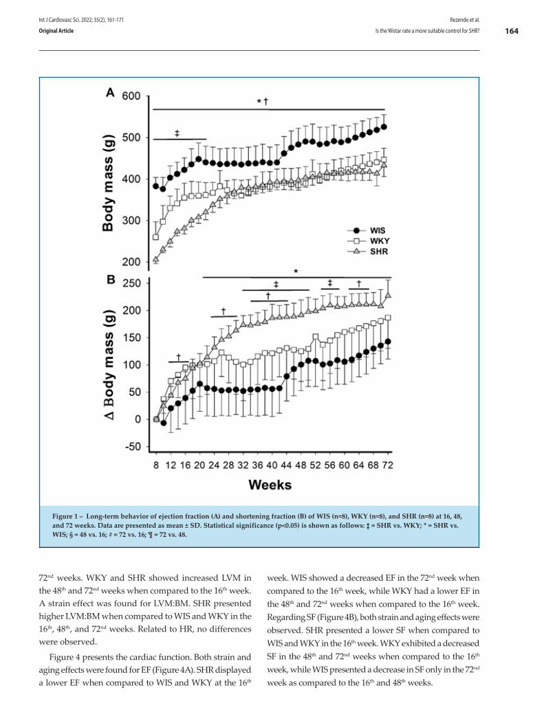
72nd weeks. WKY and SHR showed increased LVM in the 48th and 72nd weeks when compared to the 16th week. A strain effect was found for LVM:BM. SHR presented higher LVM:BM when compared to WIS and WKY in the 16th, 48th, and 72nd weeks. Related to HR, no differences were observed.
Figure 4 presents the cardiac function. Both strain and aging effects were found for EF (Figure 4A). SHR displayed a lower EF when compared to WIS and WKY at the 16th
week. WIS showed a decreased EF in the 72nd week when compared to the 16th week, while WKY had a lower EF in the 48th and 72nd weeks when compared to the 16th week. Regarding SF (Figure 4B), both strain and aging effects were observed. SHR presented a lower SF when compared to WIS and WKY in the 16th week. WKY exhibited a decreased SF in the 48th and 72nd weeks when compared to the 16th week, while WIS presented a decrease in SF only in the 72nd week as compared to the 16th and 48th weeks.
Figure 1 – Long-term behavior of ejection fraction (A) and shortening fraction (B) of WIS (n=8), WKY (n=8), and SHR (n=8) at 16, 48, and 72 weeks. Data are presented as mean ± SD. Statistical significance (p<0.05) is shown as follows: ‡ = SHR vs. WKY; * = SHR vs. WIS; § = 48 vs. 16; # = 72 vs. 16; ¶ = 72 vs. 48.
Int J Cardiovasc Sci. 2022; 35(2), 161-171
164Rezende et al.
Is the Wistar rate a more suitable control for SHR?Original Article
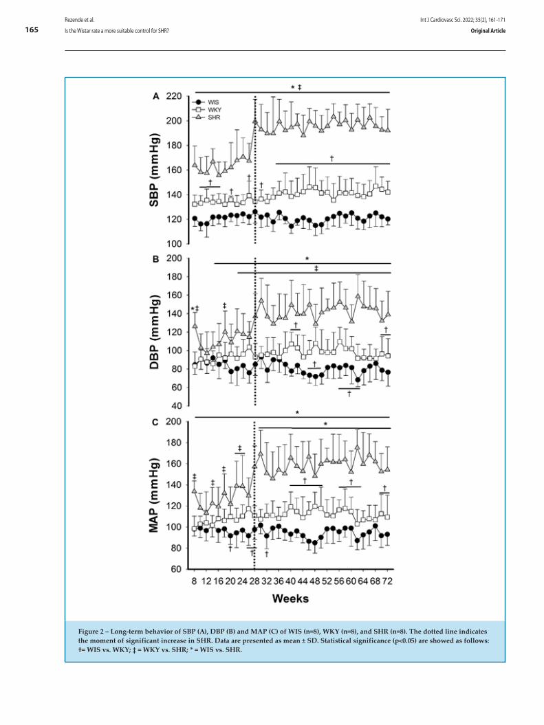
Figure 2 – Long-term behavior of SBP (A), DBP (B) and MAP (C) of WIS (n=8), WKY (n=8), and SHR (n=8). The dotted line indicates the moment of significant increase in SHR. Data are presented as mean ± SD. Statistical significance (p<0.05) are showed as follows: †= WIS vs. WKY; ‡ = WKY vs. SHR; * = WIS vs. SHR.
Int J Cardiovasc Sci. 2022; 35(2), 161-171
165Rezende et al.
Is the Wistar rate a more suitable control for SHR? Original Article
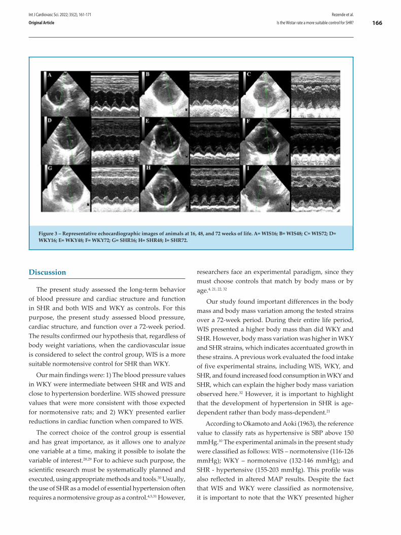
Discussion
The present study assessed the long-term behavior of blood pressure and cardiac structure and function in SHR and both WIS and WKY as controls. For this purpose, the present study assessed blood pressure, cardiac structure, and function over a 72-week period. The results confirmed our hypothesis that, regardless of body weight variations, when the cardiovascular issue is considered to select the control group, WIS is a more suitable normotensive control for SHR than WKY.
Our main findings were: 1) The blood pressure values in WKY were intermediate between SHR and WIS and close to hypertension borderline. WIS showed pressure values that were more consistent with those expected for normotensive rats; and 2) WKY presented earlier reductions in cardiac function when compared to WIS.
The correct choice of the control group is essential and has great importance, as it allows one to analyze one variable at a time, making it possible to isolate the variable of interest.28,29 For to achieve such purpose, the scientific research must be systematically planned and executed, using appropriate methods and tools.30 Usually, the use of SHR as a model of essential hypertension often requires a normotensive group as a control.4,5,31 However,
researchers face an experimental paradigm, since they must choose controls that match by body mass or by age.4, 21, 22, 32
Our study found important differences in the body mass and body mass variation among the tested strains over a 72-week period. During their entire life period, WIS presented a higher body mass than did WKY and SHR. However, body mass variation was higher in WKY and SHR strains, which indicates accentuated growth in these strains. A previous work evaluated the food intake of five experimental strains, including WIS, WKY, and SHR, and found increased food consumption in WKY and SHR, which can explain the higher body mass variation observed here.32 However, it is important to highlight that the development of hypertension in SHR is age-dependent rather than body mass-dependent.21
According to Okamoto and Aoki (1963), the reference value to classify rats as hypertensive is SBP above 150 mmHg.10 The experimental animals in the present study were classified as follows: WIS – normotensive (116-126 mmHg); WKY – normotensive (132-146 mmHg); and SHR - hypertensive (155-203 mmHg). This profile was also reflected in altered MAP results. Despite the fact that WIS and WKY were classified as normotensive, it is important to note that the WKY presented higher
Figure 3 – Representative echocardiographic images of animals at 16, 48, and 72 weeks of life. A= WIS16; B= WIS48; C= WIS72; D=WKY16; E= WKY48; F= WKY72; G= SHR16; H= SHR48; I= SHR72.
Int J Cardiovasc Sci. 2022; 35(2), 161-171
166Rezende et al.
Is the Wistar rate a more suitable control for SHR?Original Article
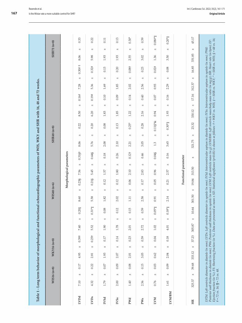
Tabl
e 1
- Lon
g te
rm b
ehav
ior o
f mor
phol
ogic
al a
nd fu
nctio
nal e
choc
ardi
ogra
phic
par
amet
ers
of W
IS, W
KY
and
SHR
with
16,
48
and
72 w
eeks
.
W
IS16
(n=8
)W
KY1
6 (n
=8)
WIS
48 (n
=6)
SH
R48
(n=8
)SH
R72
(n=8
)
Mor
phol
ogic
al p
aram
eter
s
LVD
d7.
10±
0.17
4.95
±0.
39†
7.40
±0.
20‡
8.60
±0.
23§
7.56
±0.
31§†
8.06
±0.
228.
50±
0.16
#7.
28±
0.30
# †
8.06
±0.
33
LVD
s4.
32±
0.22
2.81
±0.
25†
5.52
±0.
31*‡
5.58
±0.
21§
5.45
±0.
44§
5.76
±0.
206.
20±
0.19
#5.
36±
0.32
#5.
98±
0.22
IVSd
1.79
±0.
071.
93±
0.17
1.90
±0.
081.
82±
0.12
1.57
±0.
182.
08±
0.08
1.83
±0.
101.
69±
0.13
1.93
±0.
11
IVSs
2.00
±0.
092.
07±
0.14
1.78
±0.
122.
02±
0.12
1.80
±0.
262.
10±
0.13
1.85
±0.
091.
85±
0.20
1.93
±0.
13
PWd
1.40
±0.
082.
01±
0.23
2.01
±0.
151.
11±
0.06
2.10
± 0
.12†
2.21
±0.
25*
1.22
±0.
142.
02±
0.08
†2.
55±
0.30
*
PWs
2.56
±0.
153.
05±
0.59
2.72
±0.
592.
58±
0.17
2.83
±0.
463.
05±
0.28
2.16
±0.
402.
56±
0.23
3.02
±0.
59
LVM
0.76
±0.
030.
62±
0.04
1.02
±0.
05*‡
0.91
±0.
050.
96±
0.04
§1.
3±
0.11
§*&
0.94
±0.
070.
93±
0.02
#1.
36±
0.08
#*‡
LVM
/BM
1.61
±0.
092.
04±
0.18
4.01
±0.
18*‡
2.14
±0.
212.
57±
0.16
3.65
±0.
30*‡
1.93
±0.
162.
29±
0.08
3.50
±0.
20*‡
Func
tiona
l par
amet
er
HR
325.
37±
39.6
833
3.12
±37
.23
305.
87±
10.6
430
1.50
±19
.84
313.
50±
321.
75±
23.3
233
0.12
±17
.16
312.
37±
16.8
533
1.85
±45
.17
LVD
d: L
eft v
entr
icle
diam
eter
in d
iast
ole (
in m
m);
LVD
s: Le
ft ve
ntric
le di
amet
er in
syst
ole (
in m
m);
IVSd
: Int
erve
ntric
ular
sept
um in
dia
stol
e (in
mm
); IV
Ss: I
nter
vent
ricul
ar se
ptum
in sy
stol
e (in
mm
); PW
d:
Post
erio
r wal
l thi
ckne
ss in
dia
stol
e (in
mm
); PW
s: Po
ster
ior w
all t
hick
ness
in sy
stol
e (in
mm
); LV
M: L
eft v
entr
icle
mas
s (in
g);
LVM
/BM
: Left
ven
tric
le m
ass:b
ody
mas
s rat
io (i
n m
g:g)
; HR:
Hea
rt ra
te (i
n bp
m);
EF:
Ejec
tion
fract
ion
(in %
); FS
: Sho
rten
ing
fract
ion
(in %
). D
ata
are p
rese
nted
as m
ean
± SD
. Sta
tistic
al si
gnifi
canc
e (p<
0.05
) is s
how
n as
follo
ws:
† =
WKY
vs.
WIS
; ‡ =
SH
R vs
. WKY
; * =
SH
R vs
. WIS
; § =
48
vs. 1
6;
# =
72 v
s. 16
; ¶ =
72
vs. 4
8.
Int J Cardiovasc Sci. 2022; 35(2), 161-171
167Rezende et al.
Is the Wistar rate a more suitable control for SHR? Original Article
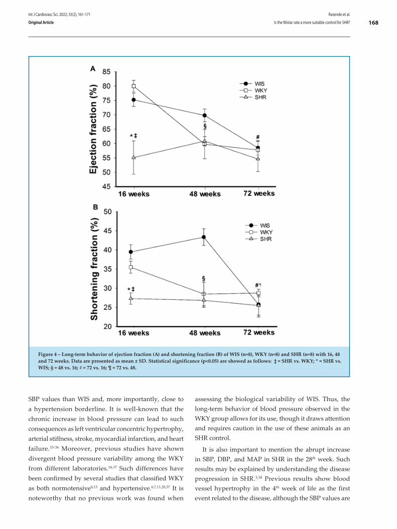
SBP values than WIS and, more importantly, close to a hypertension borderline. It is well-known that the chronic increase in blood pressure can lead to such consequences as left ventricular concentric hypertrophy, arterial stiffness, stroke, myocardial infarction, and heart failure.33-36 Moreover, previous studies have shown divergent blood pressure variability among the WKY from different laboratories.16,17 Such differences have been confirmed by several studies that classified WKY as both normotensive8,13 and hypertensive.6,7,11,20,37 It is noteworthy that no previous work was found when
assessing the biological variability of WIS. Thus, the long-term behavior of blood pressure observed in the WKY group allows for its use, though it draws attention and requires caution in the use of these animals as an SHR control.
It is also important to mention the abrupt increase in SBP, DBP, and MAP in SHR in the 28th week. Such results may be explained by understanding the disease progression in SHR.3,38 Previous results show blood vessel hypertrophy in the 4th week of life as the first event related to the disease, although the SBP values are
Figure 4 – Long-term behavior of ejection fraction (A) and shortening fraction (B) of WIS (n=8), WKY (n=8) and SHR (n=8) with 16, 48 and 72 weeks. Data are presented as mean ± SD. Statistical significance (p<0.05) are showed as follows: ‡ = SHR vs. WKY; * = SHR vs. WIS; § = 48 vs. 16; # = 72 vs. 16; ¶ = 72 vs. 48.
Int J Cardiovasc Sci. 2022; 35(2), 161-171
168Rezende et al.
Is the Wistar rate a more suitable control for SHR?Original Article
still more normotensive in nature.39 Additionally, the prehypertension stage can last up to 4th months of life.3 After, compensated hypertension sets in, in which the SHR reaches the SBP of 150 mmHg with an increase in the thickness of the cardiac walls coupled with the reductions in the left ventricular internal diameter. This structural rearrangement occurs to cope with the stress imposed by pressure overload on the cardiac walls and promote maintenance of systolic function, and may last until the sixth month of life.40,41 Our data show that around the 6th to ,7th month established and balanced hypertension is observed, characterizing the stage of decompensated hypertension.38,41 With disease progression, between the 18th and 24th months of life, SHR reaches the heart failure stage.41 In fact, 20-week-old SHR presents a higher SBP when compared to that of 12-week-old animals,42 and equivalent results have already been demonstrated by others.39,42
Regarding the cardiac structure assessed by echocardiography, it was observed that WKY at 16 weeks presented lower LVDd and LVDs when compared to WIS and SHR. Moreover, LVDd was also lower in WKY than in WIS animals at 48 and 72 weeks of life. Both WIS and WKY presented increased LVDd and LVDs with aging. In addition, WKY and SHR showed a higher PWd when compared to the WIS in weeks 48 and 72. Left ventricular remodeling is a process by which the cardiac chamber undergoes changes in its shape, size, and function, and may occur as a result of either physiology (i.e. physical training) or pathophysiology (i.e. hypertension stimuli).43 Pathophysiological hypertrophy is normally caused by a high blood pressure overload in the cardiac chambers, leading to reductions in the left ventricular diameter, accompanied by increases in the ventricular walls’ width.44
Morphological changes direct ly affect the cardiovascular function. The long-term pathological hypertrophy causes cardiac adverse remodeling of the extracellular matrix, such as increases in collagen content, which promote tissue stiffening, thus affecting the diastolic function and leading to a systolic dysfunction.45 The left ventricle is responsible for blood ejection and its morphology is crucial for pump appropriated functioning.40,46 We demonstrated that the SHR group presented higher values for LVM than did WIS and WKY. With aging, both WKY and SHR exhibited significant increases in LVM when compared to the 16th week. To confirm the pathological hypertrophic process, the LVM/BM ratio was calculated,45 and hypertrophy was not found in the WKY strain. Thus, probably the overload
imposed by the increased SBP was not enough to promote pathological hypertrophy in WKY. However, a previous work found that the hemodynamic cardiac load is more evident in isolated cardiomyocytes than in an entire ventricle.7 In addition to left ventricle analysis, the PWd of the WKY and SHR was larger when compared to the WIS at weeks 48 and 72.
Concerning cardiac function, different from another study that showed late decreases in the cardiac function of SHR over lifetime,31 in the present study, we found decreases in both EF and FS in SHR from the 16th week on. The WKY, however, presented normal values for EF and FS in the 16th week, followed by reductions in the 48th and 72nd weeks. This finding is in agreement with previous studies, showing that WKY had diastolic dysfunction as a consequence of increased pressure overload.13 Finally, cardiac dysfunction was observed in WIS only in the 72nd week.
This work has some limitations. Since our proposal was to verify the animals’ echocardiographic parameters in the 16th week of life, we did not perform an echocardiographic examination in the 8th week, the pre-hypertensive stage. Unfortunately, the lack of this information did not allow the discussion of our results to expand into a broader age range. Furthermore, the animals’ food intake was not monitored, and this factor may have affected the weight gain of the animals. It is possible that differences observed in the body mass may well be related to different values of food intake. However, to confirm such a possibility, future experiments of this nature are warranted.
Conclusions
In conclusion, Wistar rats are a more suitable normotensive control for SHR than Wistar Kyoto rats in experiments to test issues related to blood pressure, cardiac structure, and function in different ages inasmuch as Wistar Kyoto rats exhibit early reductions in cardiac function and blood pressure values in the upper limit of normal blood pressure.
Author contributions
Conception and design of the research: Rezende LMT, Reis ECC, Favarato LSC, Carneiro-Júnior MA, Natali AJ, Coimbra CC, Prímola-Gomes TN. Acquisition of data: Rezende LMT, Reis ECC, Favarato LSC, Soares LL, Drummond FR, Suarez PZ, Leite LB, Rodrigues JA, Leal, TF, Favarato ES. Analysis and interpretation of the data: Rezende
Int J Cardiovasc Sci. 2022; 35(2), 161-171
169Rezende et al.
Is the Wistar rate a more suitable control for SHR? Original Article
LMT, Reis ECC, Favarato LSC, Soares LL, Drummond FR, Suarez PZ, Leite LB, Rodrigues JA, Leal, TF, Favarato ES, Natali AJ, Coimbra, CC. Statistical analysis: Rezende LMT, Soares LL, Drummond FR, Leal TF, Carneiro-Júnior MA, Prímola-Gomes TN. Obtaining financing: Prímola-Gomes TN. Natali, AJ. Writing of the manuscript: Rezende LMT, Prímola-Gomes TN. Natali, AJ. Critical revision of the manuscript for intellectual content : Rezende LMT, Prímola-Gomes TN. Natali AJ, Coimbra CC.
Potential Conflict of InterestNo potential conflict of interest relevant to this article
was reported.
Sources of FundingThis study was supported in part by CNPq (Conselho
Nacional de Desenvolvimento Científico e Tecnológico), CAPES (Coordenação de Aperfeiçoamento de Pessoal de Nível Superior), and FAPEMIG (Fundação de Amparo à Pesquisa do Estado de Minas Gerais). AJ Natali is a CNPq fellow.
Study AssociationThis article is part of the thesis of Doctoral submitted
by Leonardo Mateus Teixeira de Rezende, from Universidade Federal de Viçosa.
Ethics approval and consent to participateThis article does not contain any studies with human
participants or animals performed by any of the authors.
1. Casals JB, Pieri NC, Feitosa ML, Ercolin AC, Roballo KC, Barreto RS, et al. The use of animal models for stroke research: a review. Comp Med. 2011;61(4):305-13.
2. Chatzigeorgiou A, Halapas A, Kalafatakis K, Kamper E. The use of animal models in the study of diabetes mellitus. In Vivo. 2009;23(2):245-58.
3. Doggrell SA, Brown L. Rat models of hypertension, cardiac hypertrophy and failure. Cardiovasc Res. 1998;39(1):89-105. doi: 10.1016/s0008-6363(98)00076-5.
4. Campos HO, Leite LH, Drummond LR, Cunha DN, Coimbra CC, Natali AJ, et al. Temperature control of hypertensive rats during moderate exercise in warm environment. J Sports Sci Med. 2014;13(3):695-701.
5. Drummond LR, Kunstetter AC, Vaz FF, Campos HO, Andrade AGP, Coimbra CC, et al. Brain temperature in spontaneously hypertensive rats during physical exercise in temperate and warm environments. PLoS One. 2016;11(5):e0155919. doi: 10.1371/journal.pone.0155919.
6. Christensen KL. Reducing pulse pressure in hypertension may normalize small artery structure. Hypertension. 1991;18(6):722-7. doi: 10.1161/01.hyp.18.6.722.
7. Deschepper CF, Picard S, Thibault G, Touyz R, Rouleau JL. Characterization of myocardium, isolated cardiomyocytes, and blood pressure in WKHA and WKY rats. Am J Physiol Heart Circ Physiol. 2002;282(1):149-55. doi: 10.1152/ajpheart.00672.2001.
8. Graham DA, Rush JW. Exercise training improves aortic endothelium-dependent vasorelaxation and determinants of nitric oxide bioavailability in spontaneously hypertensive rats. J Appl Physiol (1985). 2004;96(6):2088-96. doi: 10.1152/japplphysiol.01252.2003.
9. Locatelli J, Paiva NCN, Carvalho SHR, Lavorato VN, Gomes LHLS, Castro QJT, et al. Swim training attenuates the adverse remodeling of LV structural and mechanical properties in the early compensated phase of hypertension. Life Sci. 2017;187:42-9. doi: 10.1016/j.lfs.2017.08.014.
10. Okamoto K, Aoki K. Development of a strain of spontaneously hypertensive rats. Jpn Circ J. 1963;27:282-93. doi: 10.1253/jcj.27.282.
11. Prieto I, Segarra AB, de Gasparo M, Martínez-Cañamero M, Ramírez-Sánchez M. Divergent profile between hypothalamic and plasmatic aminopeptidase activities in WKY and SHR. Influence of beta-adrenergic blockade. Life Sci. 2018;192:9-17. doi: 10.1016/j.lfs.2017.11.022.
12. Gomes LHLS, Drummond LR, Campos HO, Rezende LMT, Carneiro-Júnior MA, Oliveira A, et al. Thermoregulation in hypertensive
rats during exercise: effects of physical training. Arq Bras Cardiol. 2019;112(5):534-42. doi: 10.5935/abc.20190050.
13. Aiello EA, Villa-Abrille MC, Escudero EM, Portiansky EL, Pérez NG, de Hurtado MC, et al. Myocardial hypertrophy of normotensive Wistar-Kyoto rats. Am J Physiol Heart Circ Physiol. 2004;286(4):1229-35. doi: 10.1152/ajpheart.00779.2003.
14. Chiueh CC, McCarty R. Sympatho-adrenal hyperreactivity to footshock stress but not to cold exposure in spontaneously hypertensive rats. Physiol Behav. 1981;26(1):85-9. doi: 10.1016/0031-9384(81)90082-2.
15. Collins HL, Loka AM, Dicarlo SE. Daily exercise-induced cardioprotection is associated with changes in calcium regulatory proteins in hypertensive rats. Am J Physiol Heart Circ Physiol. 2005;288(2):532-40. doi: 10.1152/ajpheart.00873.2004.
16. Kurtz TW, Montano M, Chan L, Kabra P. Molecular evidence of genetic heterogeneity in Wistar-Kyoto rats: implications for research with the spontaneously hypertensive rat. Hypertension. 1989;13(2):188-92. doi: 10.1161/01.hyp.13.2.188.
17. Kurtz TW, Morris RC Jr. Biological variability in Wistar-Kyoto rats. Implications for research with the spontaneously hypertensive rat. Hypertension. 1987;10(1):127-31. doi: 10.1161/01.hyp.10.1.127.
18. Langen B, Dost R. Comparison of SHR, WKY and Wistar rats in different behavioural animal models: effect of dopamine D1 and alpha2 agonists. Atten Defic Hyperact Disord. 2011;3(1):1-12. doi: 10.1007/s12402-010-0034-y.
19. Hendley ED, Cierpial MA, McCarty R. Sympathetic-adrenal medullary response to stress in hyperactive and hypertensive rats. Physiol Behav. 1988;44(1):47-51. doi: 10.1016/0031-9384(88)90344-7.
20. Sanada LS, Tavares MR, Neubern MC, Salgado HC, Fazan VP. Can Wistar rats be used as the normotensive controls for nerve morphometry investigations in spontaneously hypertensive rats (SHR)? Acta Cir Bras. 2011;26(6):514-20. doi: 10.1590/s0102-86502011000600018.
21. Hom S, Fleegal MA, Egleton RD, Campos CR, Hawkins BT, Davis TP. Comparative changes in the blood-brain barrier and cerebral infarction of SHR and WKY rats. Am J Physiol Regul Integr Comp Physiol. 2007;292(5):1881-92. doi: 10.1152/ajpregu.00761.2005.
22. Gattone VH 2nd. Body weight of the spontaneously hypertensive rat during the suckling and weanling periods. Jpn Heart J. 1986;27(6):881-4. doi: 10.1536/ihj.27.881.
References
Int J Cardiovasc Sci. 2022; 35(2), 161-171
170Rezende et al.
Is the Wistar rate a more suitable control for SHR?Original Article
23. Armitage P, Berry G, editors. Statistical methods in medical research. 2nd ed. Oxford: Blackwell; 1987.
24. Byrom FB, Wilson C. A plethysmographic method for measuring systolic blood pressure in the intact rat. J Physiol. 1938;93(3):301-4. doi: 10.1113/jphysiol.1938.sp003641.
25. Fink GD. Does Tail-Cuff Plethysmography Provide a Reliable Estimate of Central Blood Pressure in Mice? J Am Heart Assoc. 2017;6(6):e006554. doi: 10.1161/JAHA.117.006554.
26. Sahn DJ, DeMaria A, Kisslo J, Weyman A. Recommendations regarding quantitation in M-mode echocardiography: results of a survey of echocardiographic measurements. Circulation. 1978;58(6):1072-83. doi: 10.1161/01.cir.58.6.1072.
27. Brown L, Fenning A, Chan V, Loch D, Wilson K, Anderson B, et al. Echocardiographic assessment of cardiac structure and function in rats. Heart Lung Circ. 2002;11(3):167-73. doi: 10.1046/j.1444-2892.2002.00148.x.
28. Kinser PA, Robins JL. Control group design: enhancing rigor in research of mind-body therapies for depression. Evid Based Complement Alternat Med. 2013;2013:140467. doi: 10.1155/2013/140467.
29. Pithon MM. Importance of the control group in scientific research. Dental Press J Orthod. 2013;18(6):13-4. doi: 10.1590/s2176-94512013000600003.
30. Gu J, Li XB, Tian LH. Scientific research accomplishment of medical staff. Zhonghua Yi Xue Za Zhi. 2013;93(10):735-7.
31. Slama M, Ahn J, Varagic J, Susic D, Frohlich ED. Long-term left ventricular echocardiographic follow-up of SHR and WKY rats: effects of hypertension and age. Am J Physiol Heart Circ Physiol. 2004;286(1):181-5. doi: 10.1152/ajpheart.00642.2003.
32. Gordon CJ, Phillips PM, Johnstone AF. Impact of genetic strain on body fat loss, food consumption, metabolism, ventilation, and motor activity in free running female rats. Physiol Behav. 2016;153:56-63. doi: 10.1016/j.physbeh.2015.10.025.
33. Chae CU, Pfeffer MA, Glynn RJ, Mitchell GF, Taylor JO, Hennekens CH. Increased pulse pressure and risk of heart failure in the elderly. JAMA. 1999;281(7):634-9. doi: 10.1001/jama.281.7.634.
34. Levy D, Larson MG, Vasan RS, Kannel WB, Ho KK. The progression from hypertension to congestive heart failure. JAMA. 1996;275(20):1557-62.
35. McCrossan ZA, Billeter R, White E. Transmural changes in size, contractile and electrical properties of SHR left ventricular myocytes during compensated hypertrophy. Cardiovasc Res. 2004;63(2):283-92. doi: 10.1016/j.cardiores.2004.04.013.
36. Pawlush DG, Moore RL, Musch TI, Davidson WR Jr. Echocardiographic evaluation of size, function, and mass of normal and hypertrophied rat ventricles. J Appl Physiol. 1993;74(5):2598-605. doi: 10.1152/jappl.1993.74.5.2598.
37. Bomfim GHS, Méndez-López I, Fernández-Morales JC, Padín JF, Jurkiewicz A, Jurkiewicz NH, et al. Electrophysiological properties and augmented catecholamine release from chromaffin cells of WKY and SHR rats contributing to the hypertension development elicited by chronic EtOH consumption. Eur J Pharmacol. 2017;803:65-77. doi: 10.1016/j.ejphar.2017.03.017.
38. Boluyt MO, Bing OH, Lakatta EG. The ageing spontaneously hypertensive rat as a model of the transition from stable compensated hypertrophy to heart failure. Eur Heart J. 1995;16:19-30. doi: 10.1093/eurheartj/16.suppl_n.19.
39. Adams MA, Bobik A, Korner PI. Differential development of vascular and cardiac hypertrophy in genetic hypertension. Relation to sympathetic function. Hypertension. 1989;14(2):191-202. doi: 10.1161/01.hyp.14.2.191.
40. Grossman W. Cardiac hypertrophy: useful adaptation or pathologic process? Am J Med. 1980;69(4):576-84. doi: 10.1016/0002-9343(80)90471-4.
41. Bing OH, Brooks WW, Robinson KG, Slawsky MT, Hayes JA, Litwin SE, et al. The spontaneously hypertensive rat as a model of the transition from compensated left ventricular hypertrophy to failure. J Mol Cell Cardiol. 1995;27(1):383-96. doi: 10.1016/s0022-2828(08)80035-1.
42. Bacharova L, Kyselovic J, Klimas J. The initial stage of left ventricular hypertrophy in spontaneously hypertensive rats is manifested by a decrease in the QRS amplitude/left ventricular mass ratio. Clin Exp Hypertens. 2004;26(6):557-67. doi: 10.1081/ceh-200031835.
43. Ganau A, Devereux RB, Roman MJ, de Simone G, Pickering TG, Saba PS, et al. Patterns of left ventricular hypertrophy and geometric remodeling in essential hypertension. J Am Coll Cardiol. 1992;19(7):1550-8. doi: 10.1016/0735-1097(92)90617-v.
44. Grant C, Greene DG, Bunnell IL. Left ventricular enlargement and hypertrophy. A clinical and angiocardiographic study. Am J Med. 1965;39(6):895-904. doi: 10.1016/0002-9343(65)90111-7.
45. Brower GL, Gardner JD, Forman MF, Murray DB, Voloshenyuk T, Levick SP, et al. The relationship between myocardial extracellular matrix remodeling and ventricular function. Eur J Cardiothorac Surg. 2006;30(4):604-10. doi: 10.1016/j.ejcts.2006.07.006.
46. LINZBACH AJ. Heart failure from the point of view of quantitative anatomy. Am J Cardiol. 1960;5:370-82. doi: 10.1016/0002-9149(60)90084-9.
This is an open-access article distributed under the terms of the Creative Commons Attribution License
Int J Cardiovasc Sci. 2022; 35(2), 161-171
171Rezende et al.
Is the Wistar rate a more suitable control for SHR? Original Article
The first animal models of hypertension involved constriction of renal arteries (the Goldblatt kidney) or parenchyma (the Page kidney). Although these human analogs closely mimicked the pathophysiology of the disease, renovascular hypertension and the Page kidney represent only a small fraction of human hypertension.
Most experimental studies of hypertension using animals have focused on understanding the mechanisms of primary hypertension. Models of primary hypertension have been more difficult to develop. Some models may have greater face validity than others with respect to phenotypic aspects of hypertension such as age at onset, temporal course, severity, variability, and associated comorbidities. Given the clinical importance of hypertension-related target organ damage, it is noteworthy that models exhibiting face validity for left ventricular hypertrophy, metabolic abnormalities, heart failure, renal damage, and stroke (e.g., spontaneously hypertensive rats [SHR] and Dahl salt-sensitive [DSS] rats) are also available. However, other conditions such as spontaneous development of atherosclerosis or acute myocardial infarction are not typically observed in current models.
Several rat genetic models of hypertension have been used in genetic, (patho)physiological, and pharmacological studies. Rat strains exhibiting genetic hypertension include the SHR, DSS rat, the fawn-hooded hypertensive (FHH) rat, the Milan hypertensive strain,
the Lyon hypertensive rat, the Sabra hypertensive rat, the genetically hypertensive rat, and the inherited stress-induced arterial hypertension rat model. Of these, the most studied is the SHR. In the past 10 years, more than 4,500 articles have been indexed in PubMed under the term spontaneously hypertensive rats. In contrast, the next most-cited model, the DSS rat, has been indexed 585 times over the same time span, and the other genetic rat strains indexed less frequently.1
Most of the genetically hypertensive rats have been derived from outbred Wistar or Sprague-Dawley (SD) breeding stocks with selection of hypertension-related traits. The strain of SHR originated in Kyoto, Japan, from the cross of an outbred Wistar male rat, which exhibited spontaneously elevated blood pressure, and a female rat with slightly elevated blood pressure. Subsequent brother-sister mating was continued with selection of animals with systolic blood pressure >150 mm Hg. SHR are the pure line isolated from Wistar rats, which spontaneously showed stable hypertension symptoms in inbreeding offspring and developed high blood pressure at 7–15 weeks of age.2
As controls for the SHR, most workers have used normotensive descendants of Wistar rats of the colony from which the SHR strain was derived (Wistar-Kyoto rats, WKY). But the presumption that WKY are serviceable controls for SHR rests on the tacit assumption that all WKY constitute a single inbred strain. It appears, however, that whereas the National Institutes of Health distributed breeding stocks of SHR after they had been fully inbred (i.e., after 20 generations of brother-sister mating), the breeding stocks of WKY had been distributed before they were fully inbred. Accordingly, the biological variability of WKY may be greater than that of SHR. Kurtz and Morris demonstrated that WKY
DOI: https://doi.org/10.36660/ijcs.20210286
Mailing Address: Nazareth Novaes RochaUniversidade Federal Fluminense. Rua Ernani Mello, 101. Postal Code: 24210-130, RJ – Brazil. E-mail: [email protected]
Int J Cardiovasc Sci. 2022; 35(2), 172-173
172
EDITORIAL
Animal, Rats, Wistar; Epigenesis,genetic; Blood Pressure; Hypertension/genetic; Constriction/pathologic; Renal Artery; Heart Function Tests.
Keywords
Are Wistar Rats the Most Suitable Normotensive Controls for Spontaneously Hypertensive Rats to Assess Blood Pressure and Cardiac Structure and Function?Nazareth Novaes Rocha1
Universidade Federal Fluminense (UFF),1 Niterói, RJ – BrazilEditorial referring to the article: Is the Wistar Rat a more Suitable Normotensive Control for SHR to Test Blood Pressure and Cardiac Structure and Function?
rats, a common genetic control for the SHR, had profound differences in phenotypes (growth rate and blood pressure) when obtained from different commercial suppliers. Moreover, all control strains are necessarily limited in the absence of a complete understanding of the genetic differences between the control and hypertensive strains. This emphasizes the importance of careful identification and consistent use of strains when performing experiments. Equally important are environmental influences on phenotypes.3
Rezende et al.,4 evaluated the use of Wistar rats (WIS) and Wistar WKY as controls for SHR by assessing the long-term behavior of blood pressure and cardiac structure and function in these strains. Their main findings were: 1) blood pressure values in WKY were intermediate between SHR and WIS and close to hypertension borderline. WIS showed pressure values that were more consistent with those expected for normotensive rats; and 2) WKY presented earlier reductions in cardiac function when compared to WIS. Nevertheless, this work had some limitations; the authors did not perform an echocardiographic examination
in the eighth week, i.e., the pre-hypertensive stage. Furthermore, the animals’ food intake was not monitored, which may have affected weight gain, with the WIS group showing higher body mass.4
During the last years there has been enormous growth in the availability and quality of tools for genetic analysis. While such tools emerged more rapidly for humans, they now exist in a well-developed state for rats. However, although it has been recognized that the ideal control would have been a strain of rats identical to the SHR except for the genes for increased blood pressure, this is still away from reality.
The choice of a suitable animal model with spontaneous versus induced hypertension will depend on a number of factors, including the specific experimental question and the investigator’s expertise available resources. No individual model will recapitulate all features of human hypertension and, regarding control models, a consistent use of strain from a reliable and authorized supplier should be considered. All these factors must be taken into account in the design and interpretation of experiments.5
1. Lerman LO, Kurtz TW, Touyz RM, Ellison DH, Chade AR, Crowley SD et al. Animal Models of Hypertension: A Scientific Statement From the American Heart Association. Hypertension. 2019 Jun.;73(6):e87-e120. doi: 10.1161/HYP.0000000000000090.
2. Zhang-James Y, Middleton FA, Faraone SV. Genetic architecture of Wistar-Kyoto rat and spontaneously hypertensive rat substrains from different sources. Physiol Genomics. 2013;45(13):528–38. doi: 10.1152/physiolgenomics.00002.2013.
3. Kurtz TW, Morris RC Jr. Biological variability in Wistar-Kyoto rats: implications for research with the spontaneously hypertensive rat. Hypertension. 1987;10(1):127–31. doi: 10.1161/01.hyp.10.1.127.
4. Rezende LMT, Soares LL, Drummond FR, Suarez PZ, Leite L,
Rodrigues JA et al. Is the Wistar Rat a more Suitable Normotensive
Control for SHR to Test Blood Pressure and Cardiac Structure and
Function? Int J Cardiovasc Sci. 2022; 35(2):161-171. Doi: https://doi.
org/10.36660/ijcs.20200367.
5. Padmanabhan S, Joe B. Towards Precision Medicine for Hypertension:
A Review of Genomic, Epigenomic, and Microbiomic Effects on Blood
Pressure in Experimental Rat Models and Humans. Physiol Rev. 2017
Oct.;1;97(4):1469-528. doi: 10.1152/physrev.00035.2016.
References
Int J Cardiovasc Sci. 2022; 35(2), 172-173
173Rocha
Wistar Rats as Controls for SHR Editorial
This is an open-access article distributed under the terms of the Creative Commons Attribution License
Introduction
Cardiovascular disease is the leading cause of mortality in Brazil and worldwide, and ischemic heart disease accounts for a large portion of this concerning scenario.1 Among its forms of presentation, acute coronary syndrome (ACS) has a wide range of severity.2 However, the use of validated mathematical models of clinical prediction is essential and recommended in national and international guidelines for the management of patients with ACS.3,4
With this stratification, high-risk patients may receive more aggressive antiplatelet and antithrombotic therapy and early invasive intervention, whereas lower-risk patients may receive less aggressive treatments.3,5
Based on the Global Registry of Acute Coronary Events (GRACE) report, the GRACE score was designed with 8 variables analyzed on patient's admission,6 5 semi-quantitative ones (age range, heart rate, systolic blood pressure, plasma creatinine, and Killip class) and 3
Int J Cardiovasc Sci. 2022; 35(2), 174-180
174
ORIGINAL ARTICLE
Validation of the Grace Risk Score to Predict In-Hospital and 6-Month Post-Discharge Mortality in Patients with Acute Coronary SyndromeVítor Boniatti Neves,1 Raquel Melchior Roman,2 Tiago Vendruscolo,2 Gilberto Heineck,2 Carlos Alberto Santos de Mattos,2 Eduardo Ilha de Mattos,2 Luiz Carlos Pereira Bin,1,2 Karine de Lima Sírio Boclin,3 Marcelo Fialho Roman1,2
Faculdade Meridional (IMED),1 Passo Fundo, RS – BrazilHospital de Clínicas de Passo Fundo (HCPF),2 Passo Fundo, RS – Brazil Universidade Estácio de Sá (UNESA),3 Rio de Janeiro, RJ – Brazil
DOI: https://doi.org/10.36660/ijcs.20210019
Mailing Address: Vítor Boniatti NevesRua Senador Pinheiro, 304. Postal Code: 99070-220, Passo Fundo, RS – Brazil E-mail: [email protected]
Manuscript received June 02, 2020; revised manuscript January 19, 2021; accepted April 07, 2021.
Abstract
Background: The wide range of clinical presentations of acute coronary syndrome (ACS) makes it indispensible to use tools for risk stratification and for appropriate risks management; thus, the use of prognosis scores is recommended in the immediat clinical decision-making.
Objective: To validate the Global Registry of Acute Coronary Events (GRACE) score as a predictor of in-hospital and 6-month post-discharge mortality in a population diagnosed with ACS.
Methods: This is a prospective cohort study of consecutive patients diagnosed with ACS between May and December 2018. GRACE scores were calculated, as well as their predictive value for in-hospital and 6-month post-discharge mortality. The validity of the model was assessed by two techniques: discriminative power using the area under the receiver operating characteristic curve (AUC) and goodness-of-fit, using the Hosmer-Lemeshow (HL) test, at the 5% level of significance.
Results: A total of 160 patients were included, mean age 64 (±10.9) years; of which 60% were men. The risk model showed to have satisfactory ability to predict both in-hospital mortality, with an area under the curve (AUC) of 0.76 (95% confidence interval [CI], 0.57-0.95; p = 0.014), and 6-month post-discharge mortality, with AUC of 0.78 (95%CI, 0.62-0.94), p = 0.002. The HL test indicated good-fit for both models of the GRACE score.
Conclusion: In this study, the GRACE risk score for predicting mortality was appropriately validated in patients with ACS, with good discriminative power and goodness-of-fit. The results suggest that the GRACE score is appropriate for clinical use in our setting.
Keywords: Acute Coronary Syndrome; Mortality; Prognosis.
mortality or myocardial infarction so as to facilitate the stratification of patients with ACS.
Based on the GRACE score, patients were classified into low (< 1%), intermediate (1 to 3%), and high risk (> 3%) for in-hospital mortality. For the 6-month post-discharge prognosis, patients were divided into those with low (< 3%), intermediate (3 to 8%), and high mortality risk (> 8%).3,5
Clinical outcomes
The primary outcome was defined as in-hospital and 6-month post-discharge mortality. With regard to secondary outcomes, the accuracy of the GRACE score was assessed in the different presentations of ACS.
Statistical analysis
Categorical data were presented as frequencies (percentages), and continuous variables were presented as mean and standard deviation (SD) or median and interquartile range (IQR). The Kolmogorov-Smirnov test was used to verify of distribution. The level of significance was set at p < 0.05 for all analyses. The discriminative power of the score with regard to in-hospital and 6-month post-discharge was assessed using the C statistics. The area under the receiver operating characteristic curve (AUC) represented the accuracy of the GRACE score in distinguishing survivors from non-survivors. Along with this analysis, cutoff values were identified to define the best prognostic sensitivity and specificity, with their 95% confidence intervals (CI). Goodness-of-fit for the scores was assessed by the Hosmer-Lemeshow test and by the dispersion graph between predicted mortality at each risk decile and the observed mortality. The analysis was performed using the SPSS 20.0, Minitab 16 and MedCalc, version 19.1 software.
Results
Sample characteristics
The sample consisted of 160 patients. Two patients (1.25%) were lost to follow-up, due to absence of outpatient follow-up and telephone contact failure. Demographic and clinical characteristics with regard to the prevalence of cardiovascular risk factors and initial presentation are presented in Table 1.
dichotomic ones: cardiac arrest at admission, ST-segment deviation, and elevation of cardiac markers. The final score can range from 0 to 372.6
Therefore, the present study aimed to assess the value of the GRACE score as a predictor of in-hospital prognosis and 6-month post-discharge prognosis in patients with ACS in our setting.
Methods
Sample Selection
All individuals admitted with a diagnosis of ACS at Hospital de Clínicas de Passo Fundo (HCPF), Brazil, from May to December 2018 were selected. Demographic, clinical, and angiographic variables were prospectively collected. The patients were treated according to the criteria of the attending physician, without intervention from researchers. The study was approved by the Research Ethics Committee of Faculdade IMED, in compliance with the Resolution 466/2012 of the National Health Council.
Inclusion criteria were: age 18 years or older, symptoms suggestive of acute coronary ischemia on admission, and presence of at least one of the following characteristics: changes suggestive of ACS on electrocardiogram (ECG), elevation of serum biochemical markers of myocardial necrosis, and/or documented previous coronary artery disease with angiography showing coronary obstruction ≥ 50%. Patients whose ACS was triggered by secondary factors, such as trauma or surgery, were excluded. No patient refused to participate in the study, and all of them provided informed consent to participate in the research. Hospitalization outcomes were obtained by phone interview or outpatient visit 6 months after hospital discharge.
GRACE Score
The GRACE score was published in 2004 based on the GRACE registry, which was designed to reflect the full of patients with ACS. Data were obtained in 14 countries (Europe, North and South America, Australia, and New Zealand), including 94 hospitals, of which 6 were Brazilian, with a total population of 17142 patients. The aim of the score was to develop a tool to estimate of probability risk of 6-month
Int J Cardiovasc Sci. 2022; 35(2), 174-180
175Neves et al.
Validation of the GRACE Risk Model Original Article
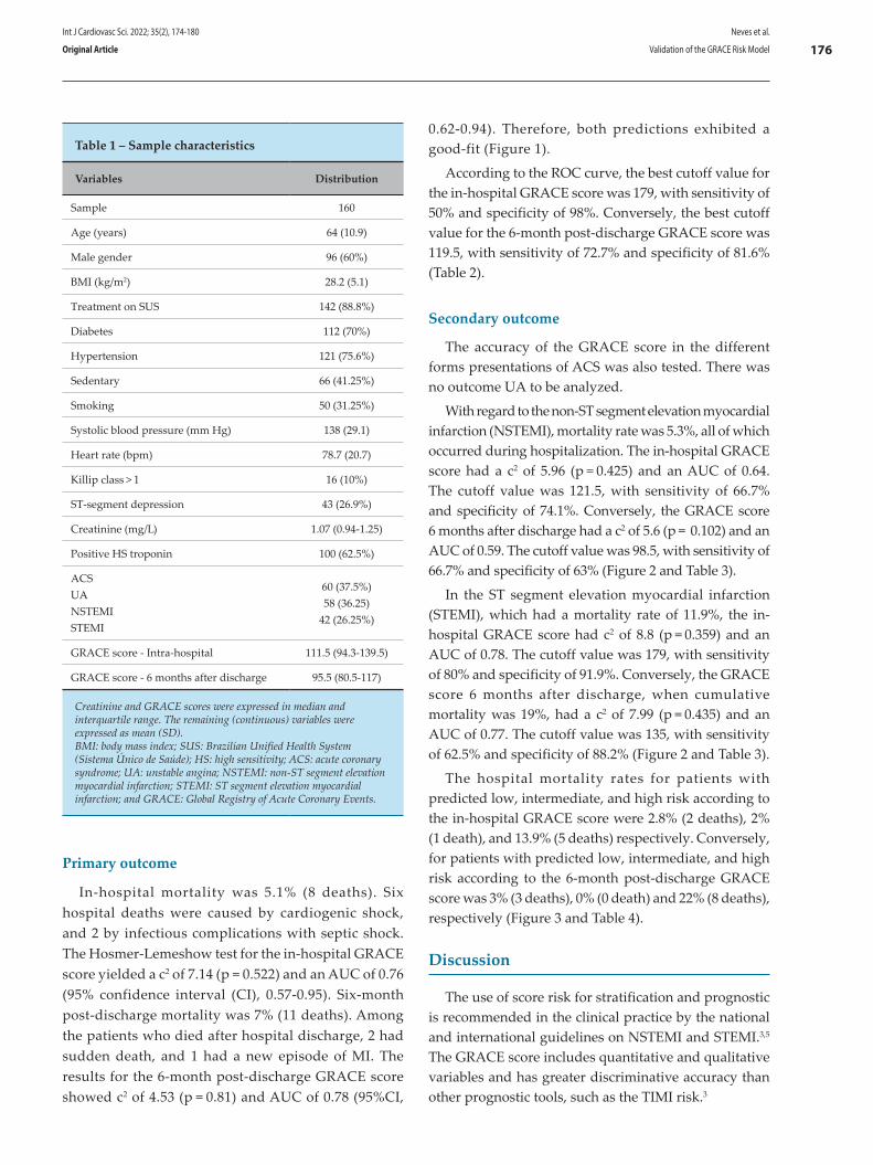
Primary outcome
In-hospital mortality was 5.1% (8 deaths). Six hospital deaths were caused by cardiogenic shock, and 2 by infectious complications with septic shock. The Hosmer-Lemeshow test for the in-hospital GRACE score yielded a c2 of 7.14 (p = 0.522) and an AUC of 0.76 (95% confidence interval (CI), 0.57-0.95). Six-month post-discharge mortality was 7% (11 deaths). Among the patients who died after hospital discharge, 2 had sudden death, and 1 had a new episode of MI. The results for the 6-month post-discharge GRACE score showed c2 of 4.53 (p = 0.81) and AUC of 0.78 (95%CI,
0.62-0.94). Therefore, both predictions exhibited a good-fit (Figure 1).
According to the ROC curve, the best cutoff value for the in-hospital GRACE score was 179, with sensitivity of 50% and specificity of 98%. Conversely, the best cutoff value for the 6-month post-discharge GRACE score was 119.5, with sensitivity of 72.7% and specificity of 81.6% (Table 2).
Secondary outcome
The accuracy of the GRACE score in the different forms presentations of ACS was also tested. There was no outcome UA to be analyzed.
With regard to the non-ST segment elevation myocardial infarction (NSTEMI), mortality rate was 5.3%, all of which occurred during hospitalization. The in-hospital GRACE score had a c2 of 5.96 (p = 0.425) and an AUC of 0.64. The cutoff value was 121.5, with sensitivity of 66.7% and specificity of 74.1%. Conversely, the GRACE score 6 months after discharge had a c2 of 5.6 (p = 0.102) and an AUC of 0.59. The cutoff value was 98.5, with sensitivity of 66.7% and specificity of 63% (Figure 2 and Table 3).
In the ST segment elevation myocardial infarction (STEMI), which had a mortality rate of 11.9%, the in-hospital GRACE score had c2 of 8.8 (p = 0.359) and an AUC of 0.78. The cutoff value was 179, with sensitivity of 80% and specificity of 91.9%. Conversely, the GRACE score 6 months after discharge, when cumulative mortality was 19%, had a c2 of 7.99 (p = 0.435) and an AUC of 0.77. The cutoff value was 135, with sensitivity of 62.5% and specificity of 88.2% (Figure 2 and Table 3).
The hospital mortality rates for patients with predicted low, intermediate, and high risk according to the in-hospital GRACE score were 2.8% (2 deaths), 2% (1 death), and 13.9% (5 deaths) respectively. Conversely, for patients with predicted low, intermediate, and high risk according to the 6-month post-discharge GRACE score was 3% (3 deaths), 0% (0 death) and 22% (8 deaths), respectively (Figure 3 and Table 4).
Discussion
The use of score risk for stratification and prognostic is recommended in the clinical practice by the national and international guidelines on NSTEMI and STEMI.3,5 The GRACE score includes quantitative and qualitative variables and has greater discriminative accuracy than other prognostic tools, such as the TIMI risk.3
Table 1 – Sample characteristics
Variables Distribution
Sample 160
Age (years) 64 (10.9)
Male gender 96 (60%)
BMI (kg/m2) 28.2 (5.1)
Treatment on SUS 142 (88.8%)
Diabetes 112 (70%)
Hypertension 121 (75.6%)
Sedentary 66 (41.25%)
Smoking 50 (31.25%)
Systolic blood pressure (mm Hg) 138 (29.1)
Heart rate (bpm) 78.7 (20.7)
Killip class > 1 16 (10%)
ST-segment depression 43 (26.9%)
Creatinine (mg/L) 1.07 (0.94-1.25)
Positive HS troponin 100 (62.5%)
ACSUANSTEMISTEMI
60 (37.5%)58 (36.25)
42 (26.25%)
GRACE score - Intra-hospital 111.5 (94.3-139.5)
GRACE score - 6 months after discharge 95.5 (80.5-117)
Creatinine and GRACE scores were expressed in median and interquartile range. The remaining (continuous) variables were expressed as mean (SD). BMI: body mass index; SUS: Brazilian Unified Health System (Sistema Único de Saúde); HS: high sensitivity; ACS: acute coronary syndrome; UA: unstable angina; NSTEMI: non-ST segment elevation myocardial infarction; STEMI: ST segment elevation myocardial infarction; and GRACE: Global Registry of Acute Coronary Events.
Int J Cardiovasc Sci. 2022; 35(2), 174-180
176Neves et al.
Validation of the GRACE Risk ModelOriginal Article
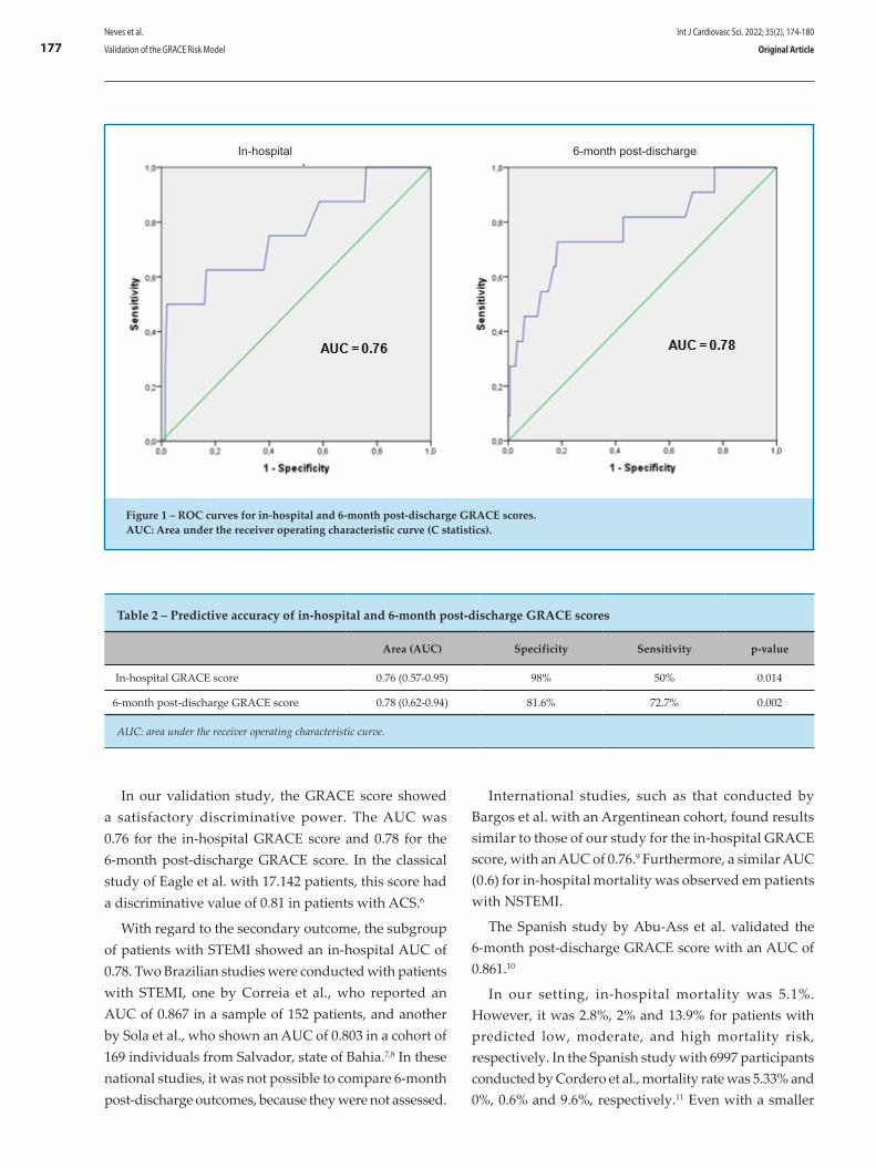
Figure 1 – ROC curves for in-hospital and 6-month post-discharge GRACE scores. AUC: Area under the receiver operating characteristic curve (C statistics).
In our validation study, the GRACE score showed a satisfactory discriminative power. The AUC was 0.76 for the in-hospital GRACE score and 0.78 for the 6-month post-discharge GRACE score. In the classical study of Eagle et al. with 17.142 patients, this score had a discriminative value of 0.81 in patients with ACS.6
With regard to the secondary outcome, the subgroup of patients with STEMI showed an in-hospital AUC of 0.78. Two Brazilian studies were conducted with patients with STEMI, one by Correia et al., who reported an AUC of 0.867 in a sample of 152 patients, and another by Sola et al., who shown an AUC of 0.803 in a cohort of 169 individuals from Salvador, state of Bahia.7,8 In these national studies, it was not possible to compare 6-month post-discharge outcomes, because they were not assessed.
International studies, such as that conducted by Bargos et al. with an Argentinean cohort, found results similar to those of our study for the in-hospital GRACE score, with an AUC of 0.76.9 Furthermore, a similar AUC (0.6) for in-hospital mortality was observed em patients with NSTEMI.
The Spanish study by Abu-Ass et al. validated the 6-month post-discharge GRACE score with an AUC of 0.861.10
In our setting, in-hospital mortality was 5.1%. However, it was 2.8%, 2% and 13.9% for patients with predicted low, moderate, and high mortality risk, respectively. In the Spanish study with 6997 participants conducted by Cordero et al., mortality rate was 5.33% and 0%, 0.6% and 9.6%, respectively.11 Even with a smaller
In-hospital 6-month post-discharge
Table 2 – Predictive accuracy of in-hospital and 6-month post-discharge GRACE scores
Area (AUC) Specificity Sensitivity p-value
In-hospital GRACE score 0.76 (0.57-0.95) 98% 50% 0.014
6-month post-discharge GRACE score 0.78 (0.62-0.94) 81.6% 72.7% 0.002
AUC: area under the receiver operating characteristic curve.
Int J Cardiovasc Sci. 2022; 35(2), 174-180
177Neves et al.
Validation of the GRACE Risk Model Original Article
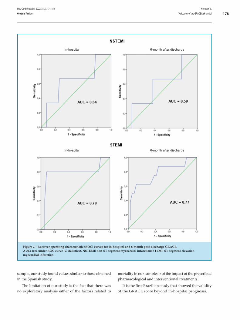
Figure 2 – Receiver operating characteristic (ROC) curves for in-hospital and 6-month post-discharge GRACE. AUC: area under ROC curve (C statistics). NSTEMI: non-ST segment myocardial infarction; STEMI: ST segment elevation myocardial infarction.
In-hospital
In-hospital
6-month after discharge
6-month after discharge
sample, our study found values similar to those obtained in the Spanish study.
The limitation of our study is the fact that there was no exploratory analysis either of the factors related to
mortality in our sample or of the impact of the prescribed pharmacological and interventional treatments.
It is the first Brazilian study that showed the validity of the GRACE score beyond in-hospital prognosis.
Int J Cardiovasc Sci. 2022; 35(2), 174-180
178Neves et al.
Validation of the GRACE Risk ModelOriginal Article
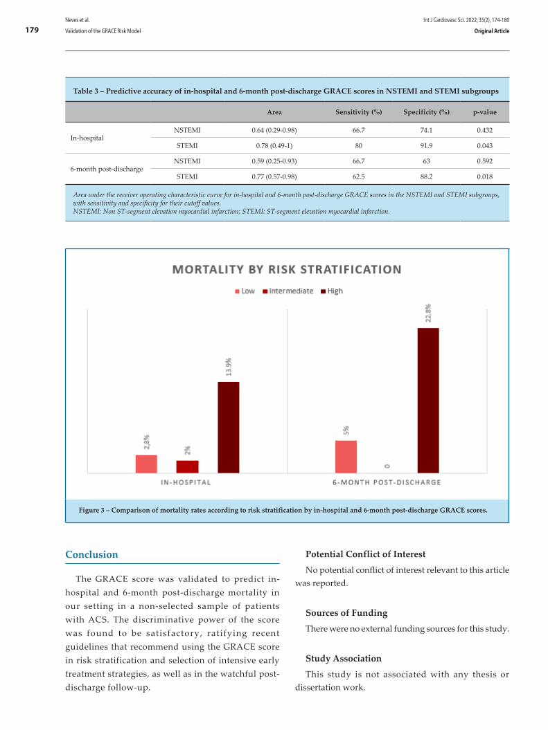
Table 3 – Predictive accuracy of in-hospital and 6-month post-discharge GRACE scores in NSTEMI and STEMI subgroups
Area Sensitivity (%) Specificity (%) p-value
In-hospitalNSTEMI 0.64 (0.29-0.98) 66.7 74.1 0.432
STEMI 0.78 (0.49-1) 80 91.9 0.043
6-month post-dischargeNSTEMI 0.59 (0.25-0.93) 66.7 63 0.592
STEMI 0.77 (0.57-0.98) 62.5 88.2 0.018
Area under the receiver operating characteristic curve for in-hospital and 6-month post-discharge GRACE scores in the NSTEMI and STEMI subgroups, with sensitivity and specificity for their cutoff values. NSTEMI: Non ST-segment elevation myocardial infarction; STEMI: ST-segment elevation myocardial infarction.
Figure 3 – Comparison of mortality rates according to risk stratification by in-hospital and 6-month post-discharge GRACE scores.
Conclusion
The GRACE score was validated to predict in-hospital and 6-month post-discharge mortality in our setting in a non-selected sample of patients with ACS. The discriminative power of the score was found to be satisfactory, ratifying recent guidelines that recommend using the GRACE score in risk stratification and selection of intensive early treatment strategies, as well as in the watchful post-discharge follow-up.
Potential Conflict of Interest
No potential conflict of interest relevant to this article was reported.
Sources of Funding
There were no external funding sources for this study.
Study Association
This study is not associated with any thesis or dissertation work.
Int J Cardiovasc Sci. 2022; 35(2), 174-180
179Neves et al.
Validation of the GRACE Risk Model Original Article
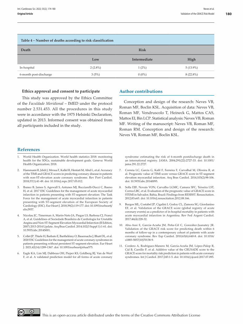
Ethics approval and consent to participateThis study was approved by the Ethics Committee
of the Faculdade Meridional – IMED under the protocol number 2.531.453. All the procedures in this study were in accordance with the 1975 Helsinki Declaration, updated in 2013. Informed consent was obtained from all participants included in the study.
Table 4 – Number of deaths according to risk classification
Death Risk
Low Intermediate High
In-hospital 2 (2.8%) 1 (2%) 5 (13.9%)
6-month post-discharge 3 (5%) 0 (0%) 8 (22.8%)
Author contributions
Conception and design of the research: Neves VB, Roman MF, Boclin KSL. Acquisition of data: Neves VB, Roman MF, Vendruscolo T, Heineck G, Mattos CAS, Mattos EI, Bin LCP. Statistical analysis: Neves VB, Roman MF. Writing of the manuscript: Neves VB, Roman MF, Roman RM. Conception and design of the research: Neves VB, Roman MF, Boclin KSL.
1. World Health Organization. World health statistics 2018: monitoring health for the SDGs, sustainable development goals. Geneva: World Health Organization; 2018.
2. Hammami R, Jdidi J, Mroua F, Kallel R, Hentati M, Abid L, et al. Accuracy of the TIMI and GRACE scores in predicting coronary disease in patients with non-ST-elevation acute coronary syndrome. Rev Port Cardiol. 2018;37(1):41-49. doi: 10.1016/j.repc.2017.05.012.
3. Ibanez B, James S, Agewall S, Antunes MJ, Bucciarelli-Ducci C, Bueno H, et al. 2017 ESC Guidelines for the management of acute myocardial infarction in patients presenting with ST-segment elevation: The Task Force for the management of acute myocardial infarction in patients presenting with ST-segment elevation of the European Society of Cardiology (ESC). Eur Heart J. 2018;39(2):119-177. doi: 10.1093/eurheartj/ehx3937.
4. Nicolau JC, Timerman A, Marin-Neto JA, Piegas LS, Barbosa CJ, Franci A, et al. Guidelines of Sociedade Brasileira de Cardiologia for Unstable Angina and Non-ST-Segment Elevation Myocardial Infarction (II Edition, 2007) 2013-2014 Update. Arq Bras Cardiol. 2014;102(3 Suppl 1):1-61. doi: 10.5935/abc.2014S001.
5. Collet JP, Thiele H, Barbato E, Barthélémy O, Bauersachs J, Bhatt DL, et al. 2020 ESC Guidelines for the management of acute coronary syndromes in patients presenting without persistent ST-segment elevation. Eur Heart J. 2021;42(14):1289-1367. doi: 10.1093/eurheartj/ehaa575.
6. Eagle KA, Lim MJ, Dabbous OH, Pieper KS, Goldberg RJ, Van de Werf F, et al. A validated prediction model for all forms of acute coronary
syndrome: estimating the risk of 6-month postdischarge death in an international registry. JAMA. 2004;291(22):2727-33. doi: 10.1001/jama.291.22.2727.
7. Correia LC, Garcia G, Kalil F, Ferreira F, Carvalhal M, Oliveira R, et al. Prognostic value of TIMI score versus GRACE score in ST-segment elevation myocardial infarction. Arq Bras Cardiol. 2014;103(2):98-106. doi: 10.5935/abc.20140095.
8. Solla DJF, Novais VON, Carvalho LGMC, Camara SFC, Teixeira LST, Correa LBC, et al. Evaluation of the prognostic value of GRACE score in STEMI in Salvador, Bahia, Brazil: Findings from RESISST. Resuscitation. 2012;83:e65. doi: 10.1016/j.resuscitation.2012.08.166.
9. Burgos ML, Costabel JP, Cigalini I, Godoy CL, Zamora SG, Giordanino EF, et al. Validation of the GRACE score (global registry of acute coronary events) as a predictor of in-hospital mortality in patients with acute myocardial infarction in Argentina. Rev Fed Argent Cardiol. 2017;46(4):228-32.
10. Abu-Assi E, García-Acuña JM, Peña-Gil C, González-Juanatey JR. Validation of the GRACE risk score for predicting death within 6 months of follow-up in a contemporary cohort of patients with acute coronary syndrome. Rev Esp Cardiol. 2010;63(6):640-8. doi: 10.1016/s1885-5857(10)70138-9.
11. Cordero A, Rodriguez-Manero M, García-Acuña JM, López-Palop R, Cid B, Carrillo P, et al. Additive value of the CRUSADE score to the GRACE score for mortality risk prediction in patients with acute coronary syndromes. Int J Cardiol. 2017;245:1-5. doi: 10.1016/j.ijcard.2017.07.095.
References
This is an open-access article distributed under the terms of the Creative Commons Attribution License
Int J Cardiovasc Sci. 2022; 35(2), 174-180
180Neves et al.
Validation of the GRACE Risk ModelOriginal Article
Acute Coronary Syndrome (ACS) is the most common cause of death worldwide.1 Nevertheless, ACS represents a heterogenous group of diseases, encompassing since low-risk unstable angina (30-day mortality below 1%), until patients with ST-elevation myocardial infarction (STEMI) and cardiogenic shock (30-day mortality around 50%). Multivariable prediction models have been developed to classify short-term and long-term risk of these patients (Table 1). For patients with the diagnosis of ACS, the TIMI risk score and the Global Registry of Acute Coronary Events (GRACE) score have been largely used in clinical practice; the latter, despite being more complex, has shown better performance as a prognostic tool, including prognostic information not only about the acute phase but also about the risk within six months after the cardiac event.2
Neves et al.,3 analyzed the performance of GRACE score in 160 patients admitted for ACS in a single center in Brazil. The results corroborate the good discrimination and calibration of GRACE score for in-hospital mortality in the Brazilian population and added information regarding its performance for six-month mortality.3 Despite the limited number of events and wide confidence intervals, the consistency of good discrimination and calibration of this score in different populations reinforces this model as an appropriate tool to estimate the risk of patients with ACS.2,3
Once a model has shown good performance in estimating risk, it is important to determine if this information may change the clinical practice. Patients with STEMI usually receive a standard level of care and changes in the approach are made more due to complications (e.g. cardiogenic shock) than to risk scores. However, patients with non-ST elevation ACS are more heterogenous, and risk stratification models have exerted greater influence on decision making (Table 2).4-7 High-risk patients (GRACE score > 140) represent a group of patients who benefit from an invasive approach in the first 24 hours,6,7 meanwhile patients at low risk may be considered for treatment outside the intensive care unit and early discharge.8 Other risk models have been developed, as the one developed by the National Cardiovascular Data Registry (NCDR®), whose accuracy can be further improved by the continuous use of a very large and diverse database.8,9 In addition to the individual risk estimation of patients with ACS, these models can also be used to adjust the risk of mortality in quality-improvement registries using the observed/expected ratio, which is of great value for epidemiological analysis.9
Finally, the use of artificial intelligence as machine learning and the technique of deep learning may represent the next step in risk prediction of patients with ACS with the potential of integrating this information into the decision-making process of diagnosis and treatment.10 Until then, we should consider the traditional prediction models as a support in situations where they could provide useful information to the medical team and the patient about the risk of mortality. Finally, the result of a risk score should never be used apart from medical judgment and the combination of both represents the current good clinical practice.
DOI: https://doi.org/10.36660/ijcs.20220006
Mailing Address: Renato D. Lopes Duke Clinical Research Institute, Box 3850, 2400 Pratt Street, Durham, NC 27705 – USA. Email: [email protected].
Int J Cardiovasc Sci. 2022; 35(2), 181-183
181
EDITORIAL
Acute Coronary Syndrome/mortality; Prognosis; Forecasting; Models, Statistical.
Keywords
Risk Scores in Acute Coronary Syndrome: Current Applications and Future Perspectives Pedro G. M. de Barros e Silva,1,2,3 Renato D. Lopes1,2,4
Brazilian Clinical Research Institute,1 São Paulo, SP – BrazilHospital Samaritano Paulista,2 São Paulo, SP – BrazilCentro Universitário São Camilo,3 São Paulo, SP – BrazilDuke Clinical Research Institute, Duke University School of Medicine,4 Durham, NC – USAEditorial referring to the article: Validation of the Grace Risk Score to Predict In-Hospital and 6-Month Post-Discharge Mortality in Patients with Acute Coronary Syndrome
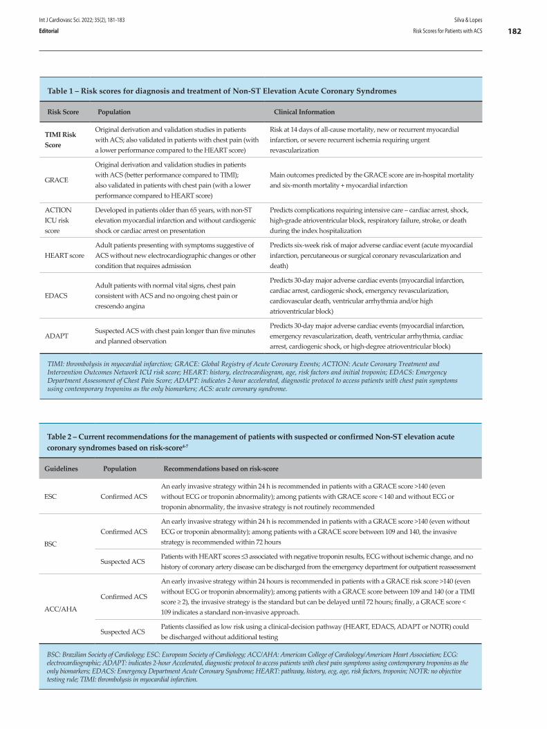
Table 1 – Risk scores for diagnosis and treatment of Non-ST Elevation Acute Coronary Syndromes
Risk Score Population Clinical Information
TIMI Risk Score
Original derivation and validation studies in patients with ACS; also validated in patients with chest pain (with a lower performance compared to the HEART score)
Risk at 14 days of all-cause mortality, new or recurrent myocardial infarction, or severe recurrent ischemia requiring urgent revascularization
GRACE
Original derivation and validation studies in patients with ACS (better performance compared to TIMI); also validated in patients with chest pain (with a lower performance compared to HEART score)
Main outcomes predicted by the GRACE score are in-hospital mortality and six-month mortality + myocardial infarction
ACTION ICU risk score
Developed in patients older than 65 years, with non-ST elevation myocardial infarction and without cardiogenic shock or cardiac arrest on presentation
Predicts complications requiring intensive care – cardiac arrest, shock, high-grade atrioventricular block, respiratory failure, stroke, or death during the index hospitalization
HEART scoreAdult patients presenting with symptoms suggestive of ACS without new electrocardiographic changes or other condition that requires admission
Predicts six-week risk of major adverse cardiac event (acute myocardial infarction, percutaneous or surgical coronary revascularization and death)
EDACSAdult patients with normal vital signs, chest pain consistent with ACS and no ongoing chest pain or crescendo angina
Predicts 30-day major adverse cardiac events (myocardial infarction, cardiac arrest, cardiogenic shock, emergency revascularization, cardiovascular death, ventricular arrhythmia and/or high atrioventricular block)
ADAPTSuspected ACS with chest pain longer than five minutes and planned observation
Predicts 30-day major adverse cardiac events (myocardial infarction, emergency revascularization, death, ventricular arrhythmia, cardiac arrest, cardiogenic shock, or high-degree atrioventricular block)
TIMI: thrombolysis in myocardial infarction; GRACE: Global Registry of Acute Coronary Events; ACTION: Acute Coronary Treatment and Intervention Outcomes Network ICU risk score; HEART: history, electrocardiogram, age, risk factors and initial troponin; EDACS: Emergency Department Assessment of Chest Pain Score; ADAPT: indicates 2-hour accelerated, diagnostic protocol to access patients with chest pain symptoms using contemporary troponins as the only biomarkers; ACS: acute coronary syndrome.
Table 2 – Current recommendations for the management of patients with suspected or confirmed Non-ST elevation acute coronary syndromes based on risk-score4-7
Guidelines Population Recommendations based on risk-score
ESC Confirmed ACSAn early invasive strategy within 24 h is recommended in patients with a GRACE score >140 (even without ECG or troponin abnormality); among patients with GRACE score < 140 and without ECG or troponin abnormality, the invasive strategy is not routinely recommended
BSC
Confirmed ACSAn early invasive strategy within 24 h is recommended in patients with a GRACE score >140 (even without ECG or troponin abnormality); among patients with a GRACE score between 109 and 140, the invasive strategy is recommended within 72 hours
Suspected ACSPatients with HEART scores ≤3 associated with negative troponin results, ECG without ischemic change, and no history of coronary artery disease can be discharged from the emergency department for outpatient reassessment
ACC/AHA
Confirmed ACS
An early invasive strategy within 24 hours is recommended in patients with a GRACE risk score >140 (even without ECG or troponin abnormality); among patients with a GRACE score between 109 and 140 (or a TIMI score ≥ 2), the invasive strategy is the standard but can be delayed until 72 hours; finally, a GRACE score < 109 indicates a standard non-invasive approach.
Suspected ACSPatients classified as low risk using a clinical-decision pathway (HEART, EDACS, ADAPT or NOTR) could be discharged without additional testing
BSC: Brazilian Society of Cardiology; ESC: European Society of Cardiology; ACC/AHA: American College of Cardiology/American Heart Association; ECG: electrocardiographic; ADAPT: indicates 2-hour Accelerated, diagnostic protocol to access patients with chest pain symptoms using contemporary troponins as the only biomarkers; EDACS: Emergency Department Acute Coronary Syndrome; HEART: pathway, history, ecg, age, risk factors, troponin; NOTR: no objective testing rule; TIMI: thrombolysis in myocardial infarction.
Int J Cardiovasc Sci. 2022; 35(2), 181-183
182Silva & Lopes
Risk Scores for Patients with ACSEditorial
1. Ralapanawa U, Sivakanesan R. Epidemiology and the Magnitude of Coronary Artery Disease and Acute Coronary Syndrome: A Narrative Review. J Epidemiol Glob Health. 2021 Jun;11(2):169-77. doi: 10.2991/jegh.k.201217.001.
2. Magalhães LP, D’Ascenzo F, Biondi-Zoccai G, Moretti C, Bollati M, Omedè P, Sciuto F, et al. TIMI, GRACE and alternative risk scores in acute coronary syndromes: a meta-analysis of 40 derivation studies on 216,552 patients and of 42 validation studies on 31,625 patients. Contemp Clin Trials 2012; 33(3):507-14.:1-22. doi: 10.1016/j.cct.2012.01.001.
3. Neves VB, Roman RM, Vendruscolo T, Heineck G, Mattos CA, Mattos EI, et al. Validation of the Grace Risk Score to Predict In-Hospital and 6-Month Post-Discharge Mortality in Patients with Acute Coronary Syndrome. Int J Cardiovasc Sci. 2022; 35(2):174-180. Doi: https://doi.org/10.36660/ijcs.20210019.
4. Amsterdam EA, Wenger NK, Brindis RG, Casey DE Jr, Ganiats TG, Holmes DR Jr, et al. 2014 AHA/ACC Guideline for the Management of Patients with Non-ST-Elevation Acute Coronary Syndromes: a report of the American College of Cardiology/American Heart Association Task Force on Practice Guidelines. J Am Coll Cardiol. 2014 Dec 23;64(24):e139-e228. https://doi.org/10.36660/ijcs.20210019
5. Collet JP, Thiele H, Barbato E, Barthélémy O, Bauersachs J, Bhatt DL, et al; ESC Scientific Document Group. 2020 ESC Guidelines for the management of acute coronary syndromes in patients presenting
without persistent ST-segment elevation. Eur Heart J. 2021 Apr 7;42(14):1289-367. doi: 10.1093/eurheartj/ehaa575.
6. Nicolau JC, Feitosa-Filho G, Petriz JL, Furtado RHM, Précoma DB, Lemke W, et al. Diretrizes da Sociedade Brasileira de Cardiologia sobre Angina Instável e Infarto Agudo do Miocárdio sem Supradesnível do Segmento ST – 2021. Arq Bras Cardiol. 2021;117(1):181-264. doi: 10.36660/abc.20210180.
7. Gulati M, Levy PD, Mukherjee D, Amsterdam E, Bhatt DL, Birtcher KK, et al. 2021 AHA/ACC/ASE/CHEST/SAEM/SCCT/SCMR Guideline for the Evaluation and Diagnosis of Chest Pain: A Report of the American College of Cardiology/American Heart Association Joint Committee on Clinical Practice Guidelines. Circulation. 2021 Nov 30;144(22):e368-e454. doi: 10.1161/CIR.0000000000001029.
8. Guimarães PO, Sampaio MC, Malafaia FL, Lopes RD, Fanaroff AC, de Barros E Silva PGM, et al. Clinical outcomes and need for intensive care after non-ST-segment-elevation myocardial infarction. Eur J Intern Med. 2020 Jun;76:58-63. doi: 10.1016/j.ejim.2020.02.008.
9. de Barros E Silva PGM, Ribeiro HB, Lopes RD, Macedo TA, Conejo F, do Amaral Baruzzi AC, et al. Improvement in quality indicators using NCDR® registries: First international experience. Int J Cardiol. 2018; Sep 15;267:13-5. https://doi.org/10.36660/abc.20190885
10. Wang H, Zu Q, Chen J, Yang Z, Ahmed MA. Application of Artificial Intelligence in Acute Coronary Syndrome: A Brief Literature Review. Adv Ther. 2021 Oct;38(10):5078-86. DOI: 10.1007/s12325-021-01908-2] [Reference Citation Analysis]
References
This is an open-access article distributed under the terms of the Creative Commons Attribution License
Int J Cardiovasc Sci. 2022; 35(2), 181-183
183Silva & Lopes
Risk Scores for Patients with ACS Editorial
Introduction
Coronary artery disease (CAD) is associated with the obstruction of coronary arteries. It has, as an etiopathogenic substrate, atherosclerosis, and as a possible outcome, ischemic syndromes – stable and unstable angina and acute myocardial infarction (AMI). These conditions have varying degrees of impairment involving ischemia and injury, with the possibility that the coronary arteries remain preserved.¹
AMI is a pathological process that initially affects the coronary arteries and is characterized by tissue death in the cardiac muscle. According to the European Society of Cardiology and the 4th universal definition of myocardial
infarction, the diagnosis of AMI is based on acute changes in cardiac enzyme curves, and evidence of ischemia (clinical condition, electrocardiographic alterations, or abnormal angiography).² AMI with ST-segment elevation (STEMI) comprises a condition of transmural ischemia involving ST-segment elevation or left bundle-branch block (LBBB) on the electrocardiogram, with acute elevation of cardiac markers due to total occlusion of a segment of the coronary vascular bed.1-3 Once STEMI is diagnosed, the course of action to be taken will depend on the resources available at the health care center and the time elapsed since the onset of symptoms. For revascularization of the ischemic tissue, intravenous thrombolysis with fibrinolytic agents as well as primary
Int J Cardiovasc Sci. 2022; 35(2), 184-190
184
ORIGINAL ARTICLE
Acute Myocardial Infarction and Percutaneous Coronary Intervention: What does the Epidemiological Data of the Last Years Indicate?Gabriel Silvestre Minucci and Samuel Marques dos ReisUniversidade Federal de São João del-Rei, MG – Brazil.
DOI: https://doi.org/10.36660/ijcs.20200256
Mailing Address: Gabriel MinucciPraça Dom Helvécio, 74. Postal Code: 36307-352, Fábricas, São João del-Rei, MG – Brazil.E-mail: [email protected]
Manuscript received August 21, 2020; revised manuscript December 07, 2020; accepted July 15, 2021.
Abstract
Background: ST-segment elevation acute myocardial infarction (STEMI) is a pathological process that involves cardiac muscle tissue death. Intravenous thrombolysis with fibrinolytics or primary percutaneous coronary intervention (PCI), an invasive technique, can be performed for tissue revascularization. PCI has been preferred as compared to non-invasive methods, although few studies have described its use in Brazil.
Objectives: The aim of the present study was to analyze data on the use of primary PCI and investigate the relevance of hospitalizations for the treatment of STEMI in the country.
Methods: A descriptive, cross-sectional analysis of data from the Brazilian Unified Health system (SUS) Department of Informatics (DATASUS) from 2010 to 2019 was conducted.
Results: Hospitalizations for STEMI represented 0.6% of all hospital admissions in Brazil in the analyzed period, 0.9% of hospital costs, and 2.1% of deaths. The number of hospitalizations due to STEMI was 659,811, and 82,793 for PCIs. Length of hospital stay was 36.0% shorter and mortality rate was 53.3% lower in PCI. The mean cost of PCI was 3.5-fold higher than for treatment of STEMI.
Conclusions: Data on hospitalizations for STEMI treatment in Brazil revealed high hospitalization and mortality rates, elevated costs, and long hospital stay. Although primary PCI is a more expensive and less used technique than other methods, it can reduce the length of hospital stay and mortality in the treatment of STEMI.
Keywords: Cardiovascular Diseases; Myocardial Infarction; Risk Factors; Coronary Diseases; Angioplasty Balloon Coronary/methods; Hospitalization; Mortality.
of hospitals by ownership (public - federal, state, and municipal hospitals, or private (contracted by SUS, philanthropic, and union hospitals).
All procedures were classified according to the codes included in the SUS table, maintaining the original nomenclature: 0303060190 - Treatment of Acute Myocardial Infarction; 0303060280 - Treatment of Acute Coronary Syndrome; 0303060204 - Treatment of Arterial Failure with Critical Ischemia; 0406030049 - Primary Coronary Angioplasty. Codes 0303060280 and 0303060204 were considered as Ischemic Syndromes.
Since the information was obtained from a public domain database, this study did not need ethics committee approval.
Results
Data obtained for the period between 2010 and 2019 are described in Table 1.
The rate of hospital admissions over the years can be viewed in the graph below (Figure 1).
When analyzing the data related to the treatment of STEMI, the year with the highest number of admissions was 2019, with 80,516 hospitalizations, which represented 12.2% of the total. Overall, there was a 40.2% growth between 2010 and 2019. The federal state with the highest number of hospitalizations was São Paulo, with 198,593 admissions, corresponding to 30.1% of all hospital admissions for the treatment of STEMI in the country.
Among the total admissions for STEMI treatment, only 12.5% were for primary referral for PCI. The highest number of procedures was registered in the year 2019, with 11,099 hospitalizations (13.4% of the total number of hospital admissions for primary PCI). A 49% increase in the use of this technique was observed in the analyzed period. The federal state with the highest number of hospitalizations was also São Paulo, with 2,329 admissions, which corresponds to 21.2% of the total.
Regarding the type of hospital (whether public or private), in 46.7% of cases the type of hospitalization for the treatment of STEMI was not recorded; 27.4% (n=180,507) admissions occurred in public hospitals, and 25.9% (n=170,939) of hospitalizations took place in the private hospitals. Data on the use of primary PCI indicated that most procedures were performed in the private sector (31.1%; n=25,717), while 15.3% (n=12,699) took place in the public sector. Note that in 44,377 (53.6%) cases, the type of hospital was not recorded.
percutaneous coronary interventions (PCIs)³ can be carried out.
According to the literature, primary PCI has greater efficacy and better results than fibrinolysis, especially if performed within the first 90 minutes of symptom onset. Despite the similarity with fibrinolytics regarding the need for early intervention for good results, the invasive technique has been preferred over the non-invasive method,⁴ given its association with higher rates of recanalization of the culprit vessel, lower rates of reocclusion and bleeding, in addition to improved ventricular function and survival.⁴-⁷
However, progress has been made in the treatment for the stabilization of atherosclerotic plaques in the coronary arteries. Studies such as COURAGE2, ISCHEMIA, and CAPTIM have reported similar results concerning drug therapy, percutaneous intervention, and revascularization surgery in prospective follow-up of patients after the acute period.⁸,⁹
Thus, the main aim of this study was to analyze data on the use of primary PCI as an alternative in STEMI episodes compared with pharmacological treatment in Brazil. The scarcity of data and the importance of this issue justify studies for a better understanding of the Brazilian reality in this regard. Another objective was to investigate factors related to hospitalizations for the treatment of STEMI – total number of hospitalizations, hospitalization costs, and in-hospital mortality.
Methods
This was a descriptive, cross-sectional study. Data were collected from the “Hospital Admission Authorization” (AIH) form, a document filled out by the physician upon hospital admission recorded in the Hospital Information System (SIH/SUS), available in the free online database of the Brazilian Unified Health System (SUS) Department of Informatics (DATASUS). Data were collected from January to March 2020 and tabulated and analyzed using the TABNET program.
To perform an analysis of a 10-year period, data between January 2010 and December 2019 were examined. We evaluated total hospital admissions in Brazil and hospitalizations for the treatment of ischemic syndromes, STEMI, and for referral for primary coronary angioplasty. The following variables were collected from the records: number of admissions, total hospitalization costs, average cost per admission, mean length of hospital stay, number of deaths, mortality rate, and categories
Int J Cardiovasc Sci. 2022; 35(2), 184-190
185Minucci & Reis
Acute Infarction and Percutaneous Coronary Intervention Original Article

The mean length of hospital stay for primary PCI was 36% shorter compared with the duration of hospitalizations for the treatment of STEMI.
Hospital costs for the treatment of STEMI increased by 62.4% during the analyzed period. The highest hospital costs were observed in São Paulo state, BR$378,783,670.64, corresponding to 31.1% of the total national costs, which, in turn, showed a 10.1% increase over the years. Total hospital expenses for PCI admissions corresponded to 43.9% of the total costs for the treatment of STEMI, which showed a 101.83% increase in the analyzed period. Again, the federal state with the highest costs was São Paulo, with BR$120,791,951.46, accounting for 14.02% of the costs with PCI in the country. The average cost per PCI procedure was 3.5-fold higher than that
for STEMI treatment, with a 13.87% increase during the analyzed period.
Regarding mortality rate, the national in-hospital mortality rate for treatment of STEMI reduced by 15.2% in the period from 2010 to 2019. As for primary PCI, in-hospital mortality rate was 53.33% lower than for STEMI treatment, with a reduction of 17.2% in the analyzed period.
Of the 1,895 types of hospital procedures described in DATASUS, hospital admissions for the treatment of STEMI occupied the 40th position in terms of number of cases when compared to hospitalizations for primary PCI, which ranked the 175th position. Considering the hospital costs per procedure, costs related to the
Table 1 – Data on hospitalizations for the treatment of ischemic syndromes, myocardial infarction with ST elevation (STEMI), and referral for primary percutaneous coronary intervention in Brazil from 2010 to 2019
Total hospitalizations in the country
Hospitalizations due to ischemic syndromes
Hospitalizations for STEMI treatment
Hospitalizations for primary coronary
angioplasty
Number of hospitalizations(nº)
117,122,623 1,573,632 659,811 82,793
Percentage in hospitalizations *
100% 1.34% 0.56% 0.07%
Hospital expenses(Brazilian currency)
R$132,996,086,776.43 R$2,184,781,851.10 R$1,217,030,068.19 R$534,204,650.74
Percentage in value spent *
100% 1.64% 0.91% 0.40%
Number of deaths (No.)
4,631,633 123,806 95,874 5,615
Percentage in deaths *
100% 2.67% 2.06% 0.12%
Mortality rate 4.05% 6.08% 14.53% 6.78%
Average cost per procedure (Brazilian currency)
R$1,135.44 R$4,055.65 R$1,844.54 R$6,452.29
Percentage in average cost
100% 357.18% 162.45% 568.26%
Average length of stay (days)
5.6 6.4 8.3 5.3
Percentage in average length of stay
100% 114.28% 148.21% 94.64%
* In relation to total national values
Int J Cardiovasc Sci. 2022; 35(2), 184-190
186Minucci & Reis
Acute Infarction and Percutaneous Coronary InterventionOriginal Article
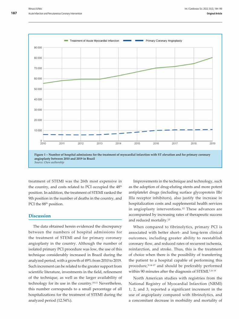
Figure 1 – Number of hospital admissions for the treatment of myocardial infarction with ST elevation and for primary coronary angioplasty between 2010 and 2019 in BrazilSource: Own authorship
treatment of STEMI was the 26th most expensive in the country, and costs related to PCI occupied the 48th position. In addition, the treatment of STEMI ranked the 9th position in the number of deaths in the country, and PCI the 88th position.
Discussion
The data obtained herein evidenced the discrepancy between the numbers of hospital admissions for the treatment of STEMI and for primary coronary angioplasty in the country. Although the number of isolated primary PCI procedure was low, the use of this technique considerably increased in Brazil during the analyzed period, with a growth of 49% from 2010 to 2019. Such increment can be related to the greater support from scientific literature, investments in the field, refinement of the technique, as well as the larger availability of technology for its use in the country.¹º,¹¹ Nevertheless, this number corresponds to a small percentage of all hospitalizations for the treatment of STEMI during the analyzed period (12.54%).
Improvements in the technique and technology, such as the adoption of drug-eluting stents and more potent antiplatelet drugs (including surface glycoprotein IIb/IIIa receptor inhibitors), also justify the increase in hospitalization costs and supplemental health services in angioplasty interventions.¹² These advances are accompanied by increasing rates of therapeutic success and reduced mortality.¹³
When compared to fibrinolytics, primary PCI is associated with better short- and long-term clinical outcomes, including greater ability to reestablish coronary flow, and reduced rates of recurrent ischemia, reinfarction, and stroke. Thus, this is the treatment of choice when there is the possibility of transferring the patient to a hospital capable of performing this procedure,⁵,¹⁴-¹⁷ and should be preferably performed within 90 minutes after the diagnosis of STEMI.5,16-18
North American studies with registries from the National Registry of Myocardial Infarction (NRMI) 1, 2, and 3, reported a significant increment in the use of angioplasty compared with fibrinolytics, and a concomitant decrease in morbidity and mortality of
Int J Cardiovasc Sci. 2022; 35(2), 184-190
187Minucci & Reis
Acute Infarction and Percutaneous Coronary Intervention Original Article
patients with sustained STEMI. This is related to advances in technology and interventional cardiology, justified by investments in technology and greater experience of the medical teams in countries like the United States.19
The growing use of primary PCI in Brazil has been observed since the late 1990s.¹² In a Brazilian study published in 2010, Piegas and Haddad 11 conducted a data survey and showed an increasing performance of PCI in Brazil, with some referral centers presenting mortality rates comparable to international values.¹¹,¹4
In the study by Widimsky et al.,20 conducted in Europe between 2007 and 2008, 37% to 93% of STEMI patients received some kind of reperfusion treatment, varying from country to country, with primary PCI as the most prevalent therapy in most countries.²1 In a study carried out in the United States between 2007 and 2009 with patients with infarction, primary PCI was adopted in 81% of the cases in which reperfusion treatment was applied.²¹
According to the ACCEPT trial, carried out in Brazil between 2011 and 2012, among the 846 patients admitted in hospital centers with STEMI, 83.3% (n=705) received reperfusion therapy, with 10% (71) being treated with thrombolytic agents and 90% (634) with primary angioplasty.²²
Considering that the present study was a national analysis covering different tertiary care hospitals, the collected data indicate that 12.54% of the hospitalizations for STEMI treatment were for primary angioplasty, evidencing a possible contrast in relation to the ACCEPT trial.
Regarding mortality due to primary coronary angioplasty, Canadian studies analyzing data between 2000 and 2005 showed a mortality rate of 1.4%.19 Meanwhile, in North American studies carried out between 1998 and 2000, the mortality rate was 0.78%,²³ whereas other studies conducted in the same country between 2004 and 2017 revealed a mortality rate of 1.27%.24 Data from Brazilian studies reported mortality rates between 2% and 6%,11,25-30 however, the present study found national values close to 6.8%.
In contrast with the findings commonly described in the literature regarding morbidity and mortality rates of the two main revascularization techniques, studies such as CAPTIM, in France, and WEST, in Canada, showed that the mortality rates in one year were not different between the groups of patients who received fibrinolytics and those who underwent primary PCI.
The two studies claim that participants who underwent fibrinolysis within two hours of symptom onset had better one-year survival when compared to those who received PCI in the same period; after two hours, no difference was observed.³¹
As for costs, although percutaneous angioplasty represented only 12.5% of hospitalizations for treatment of STEMI, the procedure corresponded to approximately 43.9% of the total expenses. However, the mortality rate in hospital admissions for primary angioplasty (6.8%) was around 53.3% lower than for the treatment of STEMI (14.53%). Therefore, it is possible to correlate primary PCI with lower mortality rates and higher costs. Nonetheless, we cannot rule out that discrepancy in these numbers may have been influenced by disparities in hospital centers.
The obtained data also showed that the mean length of stay of patients treated with angioplasty (5.3 days) was shorter than that registered for the treatment of STEMI (8.3 days). On the other hand, it is of note that the angioplasty procedure is more costly since it requires specific technologies and interventional cardiology team.
Regarding the relevance of hospitalizations for STEMI in the country, in the present study among the 1,895 types of procedures, the treatment of STEMI occupied significant positions in the national ranking of hospital admissions, costs, and deaths. The impact of cardiovascular diseases and, especially, STEMI on death rates and costs in Brazil is well established in the medical literature. In 2009, cardiovascular ischemic syndromes accounted for more than 7% of all deaths in the country and 19% of the total costs with hospitalizations in the SUS budget.13,32-34
It is important to highlight that there are several limitations regarding the quality of health services in Brazil, which may have influenced the use and effectiveness of different techniques, and hence on data obtained. Among these limitations, the following may be considered: different availability of hemodynamic laboratories; poor coordination of the health network; low efficacy of patient transport; and limited access or low availability of imaging tests, medications, resources, and technologies.34,35
Another aspect that can also be considered a limitation in this study was the dependence on data and designations attributed by the DATASUS platform to the analyzed categories.
Int J Cardiovasc Sci. 2022; 35(2), 184-190
188Minucci & Reis
Acute Infarction and Percutaneous Coronary InterventionOriginal Article

1. Bond MMK. O impacto da angioplastia coronariana prévia na cirurgia de revascularização do miocárdio: na morbimortalidade hospitalar, na qualidade de vida e sobrevivência em um ano.Tese. São Paulo: FMUSP, Instituto Dante de Pazzanese de Cardiologia.
2. Thygesen K, Alpert JS, Jaffe AS, Chaitman BR, Bax JJ, Morrow DA, et al. Fourth universal definition of myocardial infarction (2018). Eur Heart J. 2019; 40(3): 237-69.
3. Souza FNS, Nelson MVR, Gaglianone HC, Kuhn W, Macedo FS, Carvalho AAB (2019). Alterações Morfológica Cardíacas e Sistêmicas no Infarto Agudo do Miocárdio: Relato de Necrópsia. Revista de Patologia do Tocantins. 2019; 6(3): 3-7.
4. Piegas L, Timerman A, Nicolau JC,Mattos LA,Avezum A,et al. Sociedade Brasileira de Cardiologia. III Diretriz sobre tratamento do infarto agudo do miocárdio. Arq Bras Cardiol. 2004;83(Suppl 4):1-86.
5. Piegas LS, Timerman A, Feitosa GS, Nicolau JC, Mattos LAP, Andrade MD, et al.; Sociedade Brasileira de Cardiologia . V Diretriz da Sociedade Brasileira de Cardiologia sobre Tratamento do Infarto Agudo do Miocárdio com Supradesnível do Segmento ST. Arq Bras Cardiol. 2015;105(2 Suppl 1):S1-S121.
6. Nobre F, Serrano Junior CV. Tratado de cardiologia Socesp. São Paulo: Manole, 2005.
7. Feres F, Costa RA, Siqueira D, Costa Jr JRC, Chamié D, Staico R , et al. Diretriz da sociedade brasileira de cardiologia e da Sociedade Brasileira
de Hemodinâmica e Cardiologia Intervencionista sobre intervenção coronária percutânea. Arq Bras Cardiol. 2020;109(1Suppl 1):S1-S81.
8. Feitosa GS, Feitosa-Filho GS, Barbosa P, Silva R, Pereira T, Pilar I, et al. O Tratamento Antiplaquetário por Via Oral na Doença Arterial Coronária–Visão Atual. RCHSI. 2019; 3(4): 203-17.
9. Pesaro AEP, Campos PCGD, Katz M, Corrêa TD, Knobel E. Síndromes coronarianas agudas: tratamento e estratificação de risco. Rev Bras Ter Intensiva. 2008; 20(2): 197-204.
10. Piegas LS, Bittar OJNV, Haddad N. Cirurgia de revascularização miocárdica: resultados do Sistema Único de Saúde. Arq Bras Cardiol. 2009; 93(5): 555-60.
11. Piegas LS, Haddad N. Intervenção Coronariana Percutânea no Brasil: resultados do Sistema Único de Saúde. Arq Bras Cardiol. 2011;96(4):317-24.
12. Mattos LA, Souza AGMR, Pinto IMF, Campos Neto CM, Labrunie A, Alves CR et al. Evolução temporal com a utilização da angioplastia coronariana primária no infarto agudo do miocárdio no Brasil: análise dos preditores de sucesso e dos eventos adversos hospitalares em 9.434 pacientes. Arq Bras Cardiol. 2002;79(4):405-11.
13. Barreto R, Cantarelli MJC, Castello Junior HJ, Gonçalves R, Gioppato S, Guimaraes JBF et al. Resultados da intervenção coronária percutânea primária em pacientes do sistema único de saúde e da saúde suplementar. Rev Bras Cardiol Invasiva. 2011; 19(3):279-65.
References
Conclusion
Based on the collected data, primary coronary angioplasty has been scarcely performed considering all the total number of hospitalizations for the treatment of STEMI. Despite the higher costs, the procedure promotes a shorter hospital stay and a lower mortality rate compared to other treatments for infarction. The significant increase in the use of primary angioplasty (by more than 49% in the past 10 years), concomitant with increases in the cases of STEMI, indicate that this alternative method can be further exploited in Brazil.
The statistical and epidemiological importance of data on STEMI hospitalizations, costs, and mortality rates can assist in measuring the impact of this disease on our society. Thus, the present study is of significant value for public health managers, as well as hospitals and medical teams that apply the PCI technique.
More studies in this field are needed in the country, considering particularities of each region and how health networks are organized to provide PCI for eligible patients in a timely manner and with qualified professionals. From an economic viewpoint, it would be interesting to compare PCI-related costs in Brazil with those in other countries where this technique has been used for a longer time to verify whether there is a trend of cost reduction with the advancement and greater accessibility of the technology required for this intervention.
Potential Conflict of InterestNo potential conflict of interest relevant to this article
was reported.
Sources of FundingThere were no external funding sources for this study.
Study AssociationThis study is not associated with any thesis or
dissertation work.
Ethics approval and consent to participateThis article does not contain any studies with
human participants or animals performed by any of the authors.
Author contributions
Conception and design of the research: Minucci GS, Reis SM. Acquisition of data: Minucci GS, Reis SM. Analysis and interpretation of the data: Minucci GS, Reis SM. Statistical analysis: Minucci GS, Reis SM. Writing of the manuscript: Minucci GS, Reis SM. Critical revision of the manuscript for intellectual content: Minucci GS, Reis SM.
Int J Cardiovasc Sci. 2022; 35(2), 184-190
189Minucci & Reis
Acute Infarction and Percutaneous Coronary Intervention Original Article
14. Huynh T, Perron S, O’Loughlin J, Joseph L, Labrecque M, Tu JV, Théroux P et al. Comparison of primary percutaneous coronary intervention and fibrinolytic therapy in ST-segment–elevation myocardial infarction bayesian hierarchical meta-analysis of randomized controlled trials and observational studies. Circulation 2009;119(24):3101-9.
15. Keeley EC, Boura JA, Grines CL. Primary angioplasty versus intravenous thrombolytic therapy for acute myocardial infarction: a quantitative review of 23 randomised trials. Lancet. 2003; 361(9351):13-20.
16. O'gara PT, Kushner FG, Aschein DD, Casey Jr DE, Chung MK, Lemos JA et. al. ACCF/AHA Guideline for the Management of ST-Elevation Myocardial Infarction: A Report of the American College of Cardiology Foundation/American Heart Association Task Force on Practice Guidelines. Circulation. 2013;127(4):362-425.
17. Steng PF, James SK, Atar D, Badano LP, Blömstrom-Lundqvist C, Borger MA, et al. ESC Guidelines for the management of acute myocardial infarction in patients presenting with ST-segment elevation: The Task Force on the management of ST segment elevation acute myocardial infarction of the European Society of Cardiology (ESC). Eur. Heart J. 2012 Aug; 33(20): 2569–619.
18. Mattos LA, Lemos Neto PA, Rassi A Jr, Marin-Neto JA, Sousa AGMR, Devito FS, et al. Diretrizes da Sociedade Brasileira de Cardiologia – Intervenção Coronária Percutânea e Métodos Adjuntos Diagnósticos em Cardiologia Intervencionista (II Edição – 2008). Arq Bras Cardiol. 2008; 91(6 supl.1):1-58.
19. Jamal SM, Shrive FM, Ghali WA, Knudtson ML; Eisenberg MJ. In-hospital outcomes after percutaneous coronary intervention in Canada:1992/93 to 2000/01. Can J Cardiol. 2003;19(7):782-9.
20. Widimsky P, Wijns W, Fajadet J, De Belder M, Knot J, Aaberge L et al. Reperfusion therapy for ST elevation acute myocardial infarction in Europe: description of the current situation in 30 countries. Eur Heart J. 2010; 31(8):943-57.
21. Peterson ED, Roe MT, Chen AY, Fonarow GC, Lytle BL, Cannon CP et al. The NCDR ACTION Registry–GWTG: transforming contemporary acute myocardial infarction clinical care. Heart. 2010; 96(22):1798-802.
22. Wang R, Neuenschwander FC, Lima Filho A, Moreira CM, Santos ESD, Reis HJL, et al. Uso de Intervenções Baseadas em Evidências na Síndrome Coronária Aguda–Subanálise do Registro ACCEPT. Arq. Bras Cardiol. 2014;102(4):319-26,.
23. Hannan EL, Wu C, Walford G, King SB, Holmes DR Jr, Ambrose JA, et al. Volume-outcome relationships for percutaneous coronary interventions in the stent era. Circulation. 2005; 112(8):1171-9.
24. Peterson ED, Dai D, Delong ER, Brenann JM, Singh M, Rao SV, et al. NCDR Registry Participants. Contemporary mortality risk prediction for percutaneous coronary intervention: results from 588,398 procedures
in the National Cardiovascular Data Registry. J Am Coll Cardiol. 2010; 55(18):1923-32.
25. Oliveira LCS. Disparidades entre os serviços público e privado no uso de terapias de reperfusão para pacientes com IAMCSST: registro VICTIM. Aracaju. Tese. Sergipe: Universidade Federal de Sergipe; 2016.
26. Mallet ALR, Oliveira GMM, Klein CH, Carvalho MRM, Souza SNA. Letalidade e complicações de angioplastias em hospitais públicos no Rio de Janeiro. Rev Saúde Pública. 2009; 43(6): 917-27.
27. Bienert IRC, Rodrigues A, Harada EA, Silva KL, Valente AR, Silva PA et al. Avaliação Temporal dos Procedimentos de Revascularização Coronariana pelo Sistema Único de Saúde (SUS) no Brasil: Um Panorama de 20 Anos. Int J Cardiovasc Sci. 2017; 30(5): 380-90.
28. Cardoso CO, Quadros AS, Voltolini I, Azmus AD, Cardoso CR, Sebben J, et al . Angioplastia primária no infarto agudo do miocárdio: existe diferença de resultados entre as angioplastias realizadas dentro e fora do horário de rotina?. Rev Bras Cardiol Invasiva. 2010; 18(3): 273-80.
29. Mattos LA, Souza AP, Ibraim MF, Campos Neto CDM, Labrunie A, Alves CR, Saad J. Evolução temporal com a utilização da angioplastia coronariana primária no infarto agudo do miocárdio no Brasil: análise dos preditores de sucesso e dos eventos adversos hospitalares em 9.434 pacientes. Arq Bras Cardiol. 2002; 79(4): 405-11.
30. Trevisol DJ, Bianchi BR, Sakae TM, Vinholes DB, Trevisol . Análise de sobrevida em pacientes submetidos à angioplastia coronariana com stent em um hospital da região sul de Santa Catarina. Scientia Medica. 2012; 22(2): 91-6.
31. Westerhout CM, Bonnefoy E, Welsh RC, Steg PG, Boutitie F, Armstrong PW. The influence of time from symptom onset and reperfusion strategy on 1- year survival in ST-elevation myocardial infarction: a pooled analysis of an early fibrinolytic strategy versus primary percutaneous coronary intervention from CAPTIM and WEST. Am Heart J. 2011; 161(2), 283-90.
32. De Lima MSM, Dantas RAN, Dantas DV, Coutinho ICB, De Oliveira SP, Da Costa JM, et al. Trombólise no serviço pré-hospitalar móvel de emergência em vítimas de infarto agudo do miocárdio. Enferm Bras. 2019;18(1):109-15.
33. Teich V, Araujo DV. Estimativa de custo da síndrome coronariana aguda no Brasil. Rev Bras Cardiol. 2011;24(2):85—94.
34. Nicolau JC, Franken M, Lotufo PA, Carvalho AC, Marin Neto JA, Lima FG, et al. Utilização de terapêuticas comprovadamente úteis no tratamento da coronariopatia aguda: comparação entre diferentes regiões brasileiras. Análise do registro brasileiro de síndromes coronarianas agudas (BRACE - Brazilian Registry on Acute Coronary Syndromes). Arq Bras Cardiol. 2012; 98(4):282-9.
35. Quadros AS. Intervenção coronária percutânea após trombólise no infarto agudo do miocárdio: para quem e quando?. Rev Bras Cardiol. Invasiva. 2011;19(4):355-6.
This is an open-access article distributed under the terms of the Creative Commons Attribution License
Int J Cardiovasc Sci. 2022; 35(2), 184-190
190Minucci & Reis
Acute Infarction and Percutaneous Coronary InterventionOriginal Article
Introduction
The Armed Forces are in charge of guaranteeing of the Brazil State, and their performance is based on the existence of the state, enforcing constitutional norms and principles throughout the territory.1 The military police act in direct contact with the civilian population and work to protect their lives and freedom based on their rights and duties.2 The civil police act in the judicial police branch of the criminal justice system.3
The police and military work require a wide range of physical abilities, and basic physical fitness must be displayed by all staff members.4,5 In this context, the
assessment of physical abilities, by means of Physical Fitness Tests (PFTs), is routinely performed among the military personnel, and is also part of the tests for entry into the corps including the civil police.6,7
However, although PFT may be present in military routines and is usually administered to eligible applicants, periodic follow-up examinations are not common. In this sense, studies aimed at clarifying the purposes and applicability of PFTs and contributing to develop standardized protocols seem necessary .
The main objective of this study was to identify in the literature, scientific articles published from inception until May 17, 2020, related to physical fitness tests applied
Int J Cardiovasc Sci. 2022; 35(2), 191-201
191
ORIGINAL ARTICLE
Physical Fitness Test (PFT) in Police and Military in Brazil: A Systematic ReviewBráulio Nascimento Lima,1,2 Renato Silveira de Assis Junior,1 Klebson da Silva Almeida,3 Leandro Borelli de Camargo,1 Ricardo Pablo Passos,1 Carlos Henrique Prevital Fileni,1 Divaldo Martins de Souza,4 Guanis de Barros Vilela Junior1
Universidade Metodista de Piracicaba (UNIMEP),1 Piracicaba, SP – Brazil Anhanguera Educacional Ltda,2 Valinhos, SP – Brazil Universidade da Amazônia (UNAMA),3 Belém, PA – Brazil Universidade do Estado do Pará (UEPA),4 Belém, PA – Brazil
DOI: https://doi.org/10.36660/ijcs.20200299
Mailing Address: Bráulio Nascimento LimaUNIMEP - Secretaria de Pós-GraduaçãoRod. do Açúcar, km- 156. Postal Code: 13400-911, Piracicaba, SP – Brazil.E-mail: [email protected]
Manuscript received September 17, 2020; revised manuscript January 19, 2021; accepted May 18, 2021.
Abstract
Background: Physical Fitness Tests (PFTs) are part of military routines and are usually administered to applicants for the Brazilian corps member, including the civil police.
Objective: To identify in the literature, scientific articles aimed at assessing physical fitness of police and military personnel in Brazil, using PFTs.
Methods: This was a systematic review, using the PRISMA systematization, using the following search keywords "police", "military", "physical fitness test" and " PFT", in English and Portuguese. The databases used were ScienceDirect, PubMed, BVS (Lilacs) and Scielo. Only original works performed with police and military personnel in Brazil were selected, through the application of inclusion and exclusion criteria.
Results: After the screening process, 11 articles were selected from a total of 1,487.
Conclusions: The data collected from the selected articles suggest that older age is related to a decrease in physical fitness, and better performance in the tests is related to a lower risk of comorbidities. Although high-intensity training improves physical fitness and anthropometric data, it is associated with injury rates; physically active lifestyle is associated with better flexibility.
Keywords: Police; Military Personnel; Exercise Test; Brazil.
física”, “Militares” and “Polícia” in BVS (Lilacs); “Health Sciences” and “Biological Sciences” in Scielo.
Data of articles retrieved from the search were exported to the EndNote® program. The systematic review was made using the PRISMA flow diagram.8 In the screening stages, the inclusion and exclusion criteria described in Table 2 were used. The first screening was made based on titles and summary/abstract of the exported citations. The remaining articles were downloaded and included in the EndNote® program for the second screening, based on full reading of the articles.
After the screening phases, a total of seven parameters were extracted from the selected articles – author, year of publication, sample characteristics, methods adopted, tests used, results obtained and main conclusions.
Results
The processes of data acquisition and sorting are shown in a flowchart in Figure 1. The search for articles in the Portuguese language yielded fewer articles than the search for articles in the English. General results of the search in each database are presented in Figure 1.
among the police and military personnel in Brazil. The secondary objective was to present the main results and conclusions obtained from the selected articles and discuss the purposes of the PFTs. This article sought to answer the following guiding questions: 1) What are the most commonly used tests? 2) What did the authors conclude from the results? 3) When available, how were anthropometric parameters assessed, and how did they affect the results?
Methods
This study was approved by PROSPERO (identification code CRD42020186880). First, a search was conducted in the ScienceDirect, PubMed, BVL (Lilacs) and Scielo databases to identify articles on the topic. The first phase of the search conducted on May 17, 2020, and considered all articles published up to that date. The keywords, in Portuguese and in English, used for the search, are listed in Table 1.
The following resources were used in the databases: “Research Articles” in ScienceDirect; “Clinical Trial” and “Randomized Controlled Trial” in PubMed; “Aptidão
Table 1 – Search terms and databases used in review
Keywords by database
ScienceDirect; PubMed; Scielo; BVS (Lilacs)
((police) or (military)) and (fitness test) and not (review)
Scielo; BVS (Lilacs)
((polícia) or (militar)) and ((teste de aptidão física) or (taf)) and not (revisão)
Source: Author.
Table 2 – Inclusion and exclusion criteria for study selection
Inclusion criteria Exclusion criteria
• The study sample was composed of police and/or military personnel in Brazil • Animal experiments
• Studies involving physical fitness tests.• Studies where physical fitness tests were used to assess
patients with diseases or pathological conditions
• Original, complete articles published in scientific journals.• Studies evaluating the performance of police or military
personnel in specific sports or physical exercises
• Only studies with active police and/or military personnel.• Interventions on nutritional supplementation
• Review articles, guidelines, and positioning articles
Source: Author.
Int J Cardiovasc Sci. 2022; 35(2), 191-201
192Lima et al.
Physical Fitness Test (PFT) in Police and Military in BrazilOriginal Article
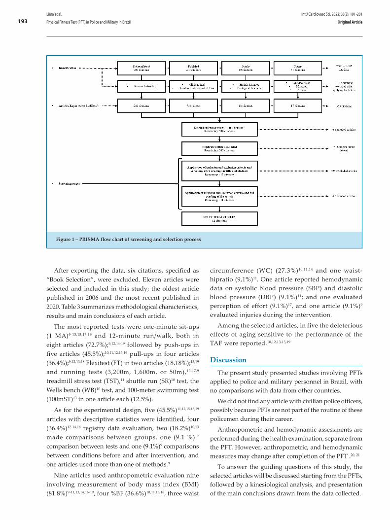
After exporting the data, six citations, specified as “Book Selection”, were excluded. Eleven articles were selected and included in this study; the oldest article published in 2006 and the most recent published in 2020. Table 3 summarizes methodological characteristics, results and main conclusions of each article.
The most reported tests were one-minute sit-ups (1 MA)9-13,15,16,19 and 12-minute run/walk, both in eight articles (72.7%);9,12,14-19 followed by push-ups in five articles (45.5%);10,11,12,15,19 pull-ups in four articles (36.4%);9,12,13,18 Flexitest (FT) in two articles (18.18%);15,19 and running tests (3,200m, 1,600m, or 50m),13,17,9 treadmill stress test (TST),11 shuttle run (SR)10 test, the Wells bench (WB)10 test, and 100-meter swimming test (100mST)13 in one article each (12.5%).
As for the experimental design, five (45.5%)11,12,15,18,19 articles with descriptive statistics were identified, four (36.4%)12-14,16 registry data evaluation, two (18.2%)10,13 made comparisons between groups, one (9.1 %)17 comparison between tests and one (9.1%)9 comparisons between conditions before and after intervention, and one articles used more than one of methods.9
Nine articles used anthropometric evaluation nine involving measurement of body mass index (BMI) (81.8%)9-11,13,14,16-19, four %BF (36.6%)10,11,16,18, three waist
circumference (WC) (27.3%)10,11,14 and one waist-hipratio (9,1%)11. One article reported hemodynamic data on systolic blood pressure (SBP) and diastolic blood pressure (DBP) (9.1%)11; and one evaluated perception of effort (9.1%)17, and one article (9.1%)9 evaluated injuries during the intervention.
Among the selected articles, in five the deleterious effects of aging sensitive to the performance of the TAF were reported.10,12,13,15,19
Discussion
The present study presented studies involving PFTs applied to police and military personnel in Brazil, with no comparisons with data from other countries.
We did not find any article with civilian police officers, possibly because PFTs are not part of the routine of these policemen during their career.
Anthropometric and hemodynamic assessments are performed during the health examination, separate from the PFT. However, anthropometric, and hemodynamic measures may change after completion of the PFT .20, 21
To answer the guiding questions of this study, the selected articles will be discussed starting from the PFTs, followed by a kinesiological analysis, and presentation of the main conclusions drawn from the data collected.
Figure 1 – PRISMA flow chart of screening and selection process
Int J Cardiovasc Sci. 2022; 35(2), 191-201
193Lima et al.
Physical Fitness Test (PFT) in Police and Military in Brazil Original Article
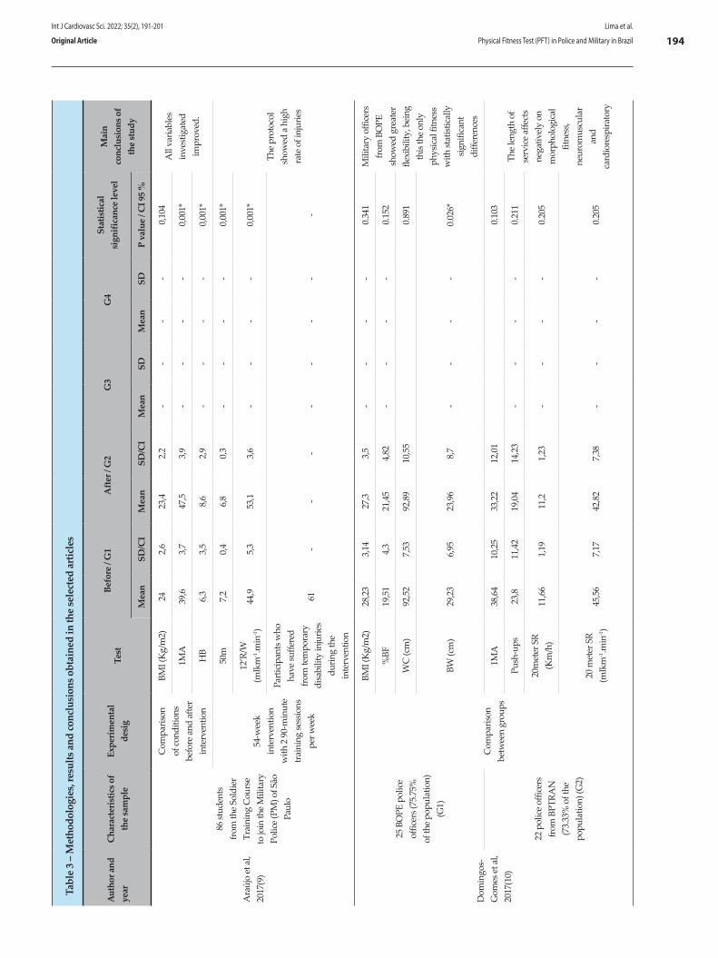
Tabl
e 3
– M
etho
dolo
gies
, res
ults
and
conc
lusi
ons o
btai
ned
in th
e se
lect
ed a
rticl
es
Aut
hor a
nd
year
Cha
ract
eris
tics o
f th
e sa
mpl
eEx
perim
enta
l de
sig
Test
Befo
re /
G1
Afte
r / G
2G
3G
4St
atis
tical
si
gnifi
canc
e le
vel
Mai
n co
nclu
sion
s of
the
stud
yM
ean
SD/C
IM
ean
SD/C
IM
ean
SDM
ean
SDP
valu
e / C
I 95
%
Ara
újo
et a
l, 20
17(9
)
86 st
uden
ts
from
the
Sold
ier
Trai
ning
Cou
rse
to jo
in th
e M
ilita
ry
Polic
e (P
M) o
f São
Pa
ulo
Com
paris
on
of co
nditi
ons
befo
re a
nd a
fter
inte
rven
tion
BMI (
Kg/
m2)
242,
623
,42,
2-
--
-0,
104
All
varia
bles
in
vest
igat
ed
impr
oved
.1M
A39
,63,
747
,53,
9-
--
-0,
001*
HB
6,3
3,5
8,6
2,9
--
--
0,00
1*
54-w
eek
inte
rven
tion
with
2 9
0-m
inut
e tr
aini
ng se
ssio
ns
per w
eek
50m
7,2
0,4
6,8
0,3
--
--
0,00
1*
The
prot
ocol
sh
owed
a h
igh
rate
of i
njur
ies
12’R
/W
(mlk
m-1.m
in-1)
44,9
5,3
53,1
3,6
--
--
0,00
1*
Part
icip
ants
who
ha
ve su
ffere
d fr
om te
mpo
rary
di
sabi
lity
inju
ries
durin
g th
e in
terv
entio
n
61-
--
--
--
-
Dom
ingo
s-G
omes
et a
l, 20
17(1
0)
25 B
OPE
pol
ice
offic
ers (
75.7
5%
of th
e po
pula
tion)
(G
1)
Com
paris
on
betw
een
grou
ps
BMI (
Kg/
m2)
28,2
33,
1427
,33,
5-
--
-0.
341
Mili
tary
offi
cers
fr
om B
OPE
sh
owed
gre
ater
fle
xibi
lity,
bei
ng
this
the
only
ph
ysic
al fi
tnes
s w
ith st
atist
ical
ly
signi
fican
t di
ffere
nces
%BF
19,5
14,
321
,45
4,82
--
--
0.15
2
WC
(cm
)92
,52
7,53
92,8
910
,55
0.89
1
BW (c
m)
29,2
36,
9523
,96
8,7
--
--
0.02
6*
22 p
olic
e of
ficer
s fr
om B
PTRA
N
(73.
33%
of t
he
popu
latio
n) (G
2)
1MA
38,6
410
,25
33,2
212
,01
0.10
3
The
leng
th o
f se
rvic
e af
fect
s ne
gativ
ely
on
mor
phol
ogic
al
fitne
ss,
neur
omus
cula
r an
d ca
rdio
resp
irato
ry
Push
-ups
23,8
11,4
219
,04
14,2
3-
--
-0.
211
20m
eter
SR
(Km
/h)
11,6
61,
1911
,21,
23-
--
-0.
205
20 m
eter
SR
(mlk
m-1.m
in-1)
45,5
67,
1742
,82
7,38
--
--
0.20
5
Int J Cardiovasc Sci. 2022; 35(2), 191-201
194Lima et al.
Physical Fitness Test (PFT) in Police and Military in BrazilOriginal Article
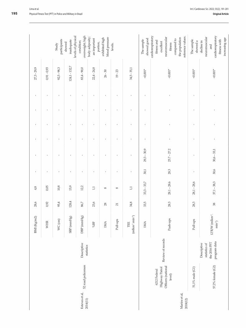
Este
ves e
t al,
2014
(11)
52 ro
ad p
olic
emen
Des
crip
tive
stat
istic
s
BMI (
Kg/
m2)
28,6
4,8
--
--
--
27,3
- 29
,9
Stud
y pa
rtic
ipan
ts
show
ed
inad
equa
te
leve
ls of
phy
sical
co
nditi
on;
over
wei
ght;
high
bo
dy a
dipo
sity;
an
impo
rtan
t po
rtio
n,
exhi
bite
d hi
gh
bloo
d pr
essu
re
leve
ls.
WH
R0,
920,
05-
--
--
-0,
91 -
0,93
WC
(cm
)95
,410
,8-
--
--
-92
,5 -
98,3
SBP
(mm
Hg)
128,
415
,9-
--
--
-12
4,1
- 132
,7
DBP
(mm
Hg)
86,7
12,2
--
--
--
83,4
- 90
,0
%BF
23,6
1,1
--
--
--
22,4
- 24
,8
1MA
288
--
--
--
26 -
30
Pull-
ups
218
--
--
--
19 -
23
TEE
(mlk
m-1.m
in-1)
34,8
1,1
--
--
--
34,5
- 35
,1
Mar
ins e
t al,
2018
(12)
6212
Fed
eral
H
ighw
ay P
atro
l O
ffice
rs (n
atio
nal
leve
l)
Revi
ew o
f rec
ords
1MA
33,5
33,3
– 3
3,7
30,1
29,3
– 3
0,9
--
--
<0,0
01*
The
sam
ple
show
ed g
ood
card
iore
spira
tory
fit
ness
and
ex
celle
nt
neur
omus
cula
r fit
ness
co
mpa
red
to
the
popu
latio
n re
fere
nce
valu
es.
Push
-ups
28.3
28.1
– 2
8.6
28.3
25.7
– 2
7.2
--
--
<0.0
01*
51,1
% m
ale
(G1)
Des
crip
tive
stat
istic
s of
the
2016
PFT
pr
ogra
m d
ata
Pull-
ups
28,3
28,1
– 2
8,6
--
--
--
<0.0
01*
The
sam
ple
show
ed a
de
clin
e in
ne
urom
uscu
lar
and
card
iore
spira
tory
fit
ness
with
in
crea
sing
age
57,2
% fe
mal
e (G
2)12
’R/W
(mlk
m-1.
min
-1)
3837
,1 –
38,
330
,630
,6 –
33,
1-
--
-<0
.001
*
Int J Cardiovasc Sci. 2022; 35(2), 191-201
195Lima et al.
Physical Fitness Test (PFT) in Police and Military in Brazil Original Article

Med
eiro
s et
al, 2
020(
13)
702
PFT
data
fr
om h
ealth
y ad
ults
of t
he
Nav
y Pe
rson
nel
Com
man
d of
the
mun
icip
ality
of R
io
de Ja
neiro
bet
wee
n th
e ye
ars 2
017-
2018
Revi
ew o
f rec
ords
BMI (
Kg/
m2)
25,4
2,9
26,4
4,3
27,5
4-
-≤0
,001
*
Incr
easin
g ag
e ne
gativ
ely
inte
rfer
es w
ith
the
perf
orm
ance
of
stre
ngth
, m
uscl
e en
dura
nce
and
card
iore
spira
tory
ca
paci
ty
1MA
52,5
20,5
52,2
15,1
50,3
11,2
--
0,28
4
91 Y
oung
Adu
lts
(20-
30 y
ears
) (G
1)
Com
paris
on o
f ag
e be
twee
n th
e th
ree
grou
ps
Pull-
ups
10,8
4,6
103,
68,
54,
7-
-0,
000*
243 A
dults
(30-
40
year
s) (G
2)3,
200m
(s)
1282
172,
414
2119
9,8
1567
192
--
≤0,0
01*
368
Mat
ure A
dults
(4
0-50
yea
rs o
ld)
(G3)
100m
ST (s
)14
0,1
45,6
149,
737
,918
0,8
50-
-≤0
,001
*
Oliv
eira
&
Anj
os,
2008
(14)
50,5
23 (3
8.6%
) A
rmy
sold
iers
, m
ale,
hea
lthy
and
who
par
ticip
ated
in
the
2001
TA
F, b
etw
een
Nov
embe
r and
D
ecem
ber.
Revi
ew o
f rec
ords
BMI (
Kg/
m2)
26,2
3,7
24,7
2,8
23,4
2,3
22,3
2p<
0,00
1*
It w
as id
entif
ied
in th
e re
cord
s th
at th
e M
ilita
ry
with
BM
I equ
al
show
ed ra
tio
of sm
alle
r ci
rcum
fere
nce
of th
e ab
dom
en
whe
n ca
rdio
resp
irato
ry
fitne
ss w
as
grea
ter.
62%
of i
ndiv
idua
ls
up to
25
year
s of
age
WC
(cm
)88
,59,
884
,27,
680
,86,
578
,36
<0,0
01*
G1V
O2m
ax ≤
49
(n=1
2290
) (G
1)
49 <
VO
2max
≤ 5
4 (n
=137
37) (
G2)
36 g
arris
ons
54 <
VO
2max
≤ 5
7 (n
=132
54) (
G3)
12’ R
/W
(mlk
m-1.m
in-1)
44,2
4,4
52,1
1,2
55,7
0,9
603
< 0
,000
1*
57 <
VO
2max
(n
=112
42) (
G4)
Int J Cardiovasc Sci. 2022; 35(2), 191-201
196Lima et al.
Physical Fitness Test (PFT) in Police and Military in BrazilOriginal Article
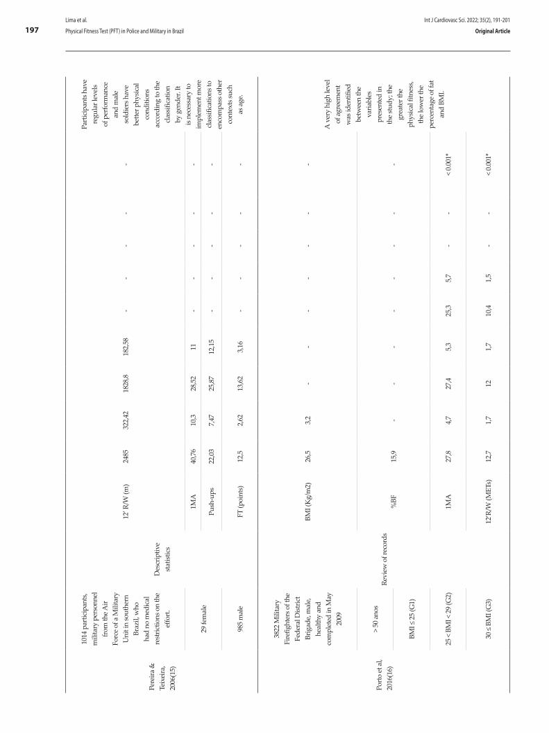
Pere
ira &
Te
ixei
ra,
2006
(15)
1014
par
ticip
ants
, m
ilita
ry p
erso
nnel
fr
om th
e Air
Forc
e of
a M
ilita
ry
Uni
t in
sout
hern
Br
azil,
who
ha
d no
med
ical
re
stric
tions
on
the
effo
rt.D
escr
iptiv
e st
atist
ics
12’ R
/W (m
)24
8532
2,42
1828
,818
2,58
--
--
-
Part
icip
ants
hav
e re
gula
r lev
els
of p
erfo
rman
ce
and
mal
e so
ldie
rs h
ave
bette
r phy
sical
co
nditi
ons
acco
rdin
g to
the
clas
sific
atio
n by
gen
der.
It is
nece
ssar
y to
im
plem
ent m
ore
clas
sific
atio
ns to
en
com
pass
oth
er
cont
exts
such
as
age
.
29 fe
mal
e1M
A40
,76
10,3
28,5
211
--
--
-
Push
-ups
22,0
37,
4725
,87
12,1
5-
--
--
985
mal
eFT
(poi
nts)
12,5
2,62
13,6
23,
16-
--
--
Porto
et a
l, 20
16(1
6)
3822
Mili
tary
Fi
refig
hter
s of t
he
Fede
ral D
istric
t Br
igad
e, m
ale,
he
alth
y an
d co
mpl
eted
in M
ay
2009
Revi
ew o
f rec
ords
BMI (
Kg/
m2)
26,5
3,2
--
--
--
-
A v
ery
high
leve
l of
agr
eem
ent
was
iden
tifie
d be
twee
n th
e va
riabl
es
pres
ente
d in
th
e st
udy;
the
grea
ter t
he
phys
ical
fitn
ess,
the
low
er th
e pe
rcen
tage
of f
at
and
BMI.
> 50
ano
s
%BF
15,9
--
--
--
--
BMI ≤
25
(G1)
25 <
BM
I < 2
9 (G
2)1M
A27
,84,
727
,45,
325
,35,
7-
-<
0.00
1*
30 ≤
BM
I (G
3)12
’R/W
(MET
s)12
,71,
712
1,7
10,4
1,5
--
< 0.
001*
Int J Cardiovasc Sci. 2022; 35(2), 191-201
197Lima et al.
Physical Fitness Test (PFT) in Police and Military in Brazil Original Article
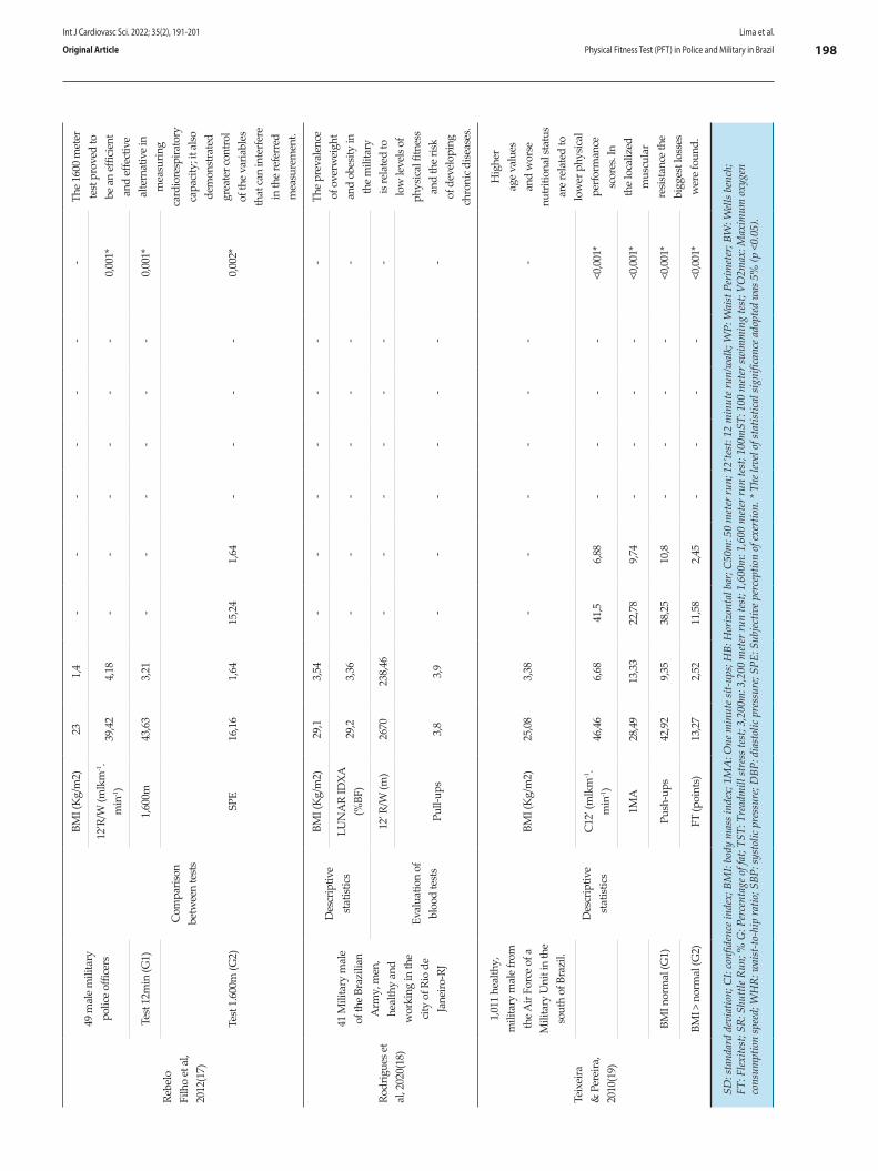
Rebe
lo
Filh
o et
al,
2012
(17)
49 m
ale
mili
tary
po
lice
offic
ers
Com
paris
on
betw
een
test
s
BMI (
Kg/
m2)
231,
4-
--
--
--
The
1600
met
er
test
pro
ved
to
be a
n ef
ficie
nt
and
effe
ctiv
e al
tern
ativ
e in
m
easu
ring
card
iore
spira
tory
ca
paci
ty; i
t also
de
mon
stra
ted
grea
ter c
ontr
ol
of th
e va
riabl
es
that
can
inte
rfer
e in
the
refe
rred
m
easu
rem
ent.
12’R
/W (m
lkm
-1.
min
-1)
39,4
24,
18-
--
--
-0,
001*
Test
12m
in (G
1)1,
600m
43,6
33,
21-
--
--
-0,
001*
Test
1.6
00m
(G2)
SPE
16,1
61,
6415
,24
1,64
--
--
0,00
2*
Rodr
igue
s et
al, 2
020(
18)
41 M
ilita
ry m
ale
of th
e Br
azili
an
Arm
y, m
en,
heal
thy
and
wor
king
in th
e ci
ty o
f Rio
de
Jane
iro-R
J
Des
crip
tive
stat
istic
s
BMI (
Kg/
m2)
29,1
3,54
--
--
--
-Th
e pr
eval
ence
of
ove
rwei
ght
and
obes
ity in
th
e m
ilita
ry
is re
late
d to
lo
w le
vels
of
phys
ical
fitn
ess
and
the
risk
of d
evel
opin
g ch
roni
c dise
ases
.
LUN
AR
IDXA
(%
BF)
29,2
3,36
--
--
--
-
Eval
uatio
n of
bl
ood
test
s
12’ R
/W (m
)26
7023
8,46
--
--
--
-
Pull-
ups
3,8
3,9
--
--
--
-
Teix
eira
&
Per
eira
, 20
10(1
9)
1,01
1 he
alth
y,
mili
tary
mal
e fr
om
the A
ir Fo
rce
of a
M
ilita
ry U
nit i
n th
e so
uth
of B
razi
l.
Des
crip
tive
stat
istic
s
BMI (
Kg/
m2)
25
,08
3,38
--
--
--
-
Hig
her
age
valu
es
and
wor
se
nutr
ition
al st
atus
ar
e re
late
d to
lo
wer
phy
sical
pe
rfor
man
ce
scor
es. I
n th
e lo
caliz
ed
mus
cula
r re
sista
nce
the
bigg
est l
osse
s w
ere
foun
d.
C12
’ (m
lkm
-1.
min
-1)
46,4
66,
6841
,56,
88-
--
-<0
,001
*
1MA
28,4
913
,33
22,7
89,
74-
--
-<0
,001
*
BMI n
orm
al (G
1)Pu
sh-u
ps42
,92
9,35
38,2
510
,8-
--
-<0
,001
*
BMI >
nor
mal
(G2)
FT (p
oint
s)13
,27
2,52
11,5
82,
45-
--
-<0
,001
*
SD: s
tand
ard
devi
atio
n; C
I: co
nfid
ence
inde
x; B
MI:
body
mas
s ind
ex; 1
MA
: One
min
ute s
it-up
s; H
B: H
oriz
onta
l bar
; C50
m: 5
0 m
eter
run;
12’
test
: 12
min
ute r
un/w
alk;
WP:
Wai
st P
erim
eter
; BW
: Well
s ben
ch;
FT: F
lexite
st; S
R: S
huttl
e Run
; % G
: Per
cent
age o
f fat
; TST
: Tre
adm
ill st
ress
test
; 3,2
00m
: 3,2
00 m
eter
run
test
; 1,6
00m
: 1,6
00 m
eter
run
test
; 100
mST
: 100
met
er sw
imm
ing
test
; VO
2max
: Max
imum
oxy
gen
cons
umpt
ion
spee
d; W
HR:
wai
st-to
-hip
ratio
; SBP
: sys
tolic
pre
ssur
e; D
BP: d
iast
olic
pre
ssur
e; SP
E: S
ubjec
tive p
erce
ptio
n of
exer
tion.
* Th
e lev
el of
stat
istic
al si
gnifi
canc
e ado
pted
was
5%
(p <
0.05
).
Int J Cardiovasc Sci. 2022; 35(2), 191-201
198Lima et al.
Physical Fitness Test (PFT) in Police and Military in BrazilOriginal Article
The most reported tests in the selected articles were 1MA and 12-minute walk/run (72.73%). In the 1MA test, the subject lies down on the back, with the knees bent, the soles of the feet resting on the ground, and arms crossed over the chest. Participant’s feet are firmly held down by another person. In the up position, the subject should touch elbows to knee and then return down until their shoulder blades contact the floor. The participant performs as many correct sit ups as possible in 60 seconds.9-13,15,16,19 In the article by Rodrigues et al.18 the numbers of sit ups were considered inadequate, which was related to high values of BMI. In the article by Esteves et al.11 conditioning was also considered inappropriate, however, BMI was classified as normal, and thus, the relationship between BMI and physical fitness was found. In the other articles, the values of the 1MA test were considered satisfactory. The data suggest that BMI may negatively influence the performance of the 1MA test, but it is not decisive.
In the 12-minute run/walk test, participant lines up behind the starting line. After a command, the watch is started, participants should walk or run, and the longest distance covered in 12 minutes is recorded. Then, estimated VO2 Max results are calculated by specific formulas.9,12,14-19 Results of the selected articles suggest that the increase in age, BMI and %BF negatively affect the performance in the 12-minute walk/run test.12,16,18 Also, individuals with better performance had lower BMI and Waist Perimeter (WP).14
The 3,200m, 1,600m, 20m ST and TST (9,1%) were also used for cardiac evaluation. In the 50m, 3,200m and 1,600m tests, the time required to cover the distances of 50m, 3,200m and 1,600m, respectively is recorded.9,13,17 The shuttle run test of running back and forth, repeatedly, a distance of 20 meters, controlled by a sound stimulus.10 TST was performed following a specific protocol, monitored by electrocardiography; the test was performed which made it possible to collect hemodynamic data.11 The VO2max calculations were based on doubly indirect assessments. Despite the methodological differences, these studies supported the statement that cardiorespiratory performance decreases with increasing age.10,12,13,15,19
Push-up test was the third most reported (45.5%). Before beginning the push-up movement, participants assume the front-leaning rest position by placing the hands comfortably and feet together. The body should form a straight line from the shoulders to the ankles,
with the elbows extended. Push-ups are begun by bending the elbows and lowering the entire body until the upper arms are at least parallel to the ground. Then participants return to the starting position by raising the body until the arms are fully extended. The movement is repeated continuously until failure. For women, the exercise may be adapted by performing the exercise with the knees on the floor (knee push-ups). 10-12,15,19 In the test, the number of correct repetitions is counted. The activity load is based on body mass, therefore, the anthropometric characteristics interfere with the test performance. A In that test the number of repetitions is counted to compose the test score and higher values represent the performance. As the activity load is based on body mass, anthropometric characteristics influence the test performance. This is corroborated by the study by Esteves et al.,11 who reported inadequate levels of physical condition in highway police officers. In addition, other studies reinforced the negative effect of age on the performance in push-up tests.10,12,15,19 According to Pereira & Teixeira,15 there should be an age categorization when applying the test and evaluating performance.
In the pull-up test (36.4%), the participant stands below a pull-up bar and, with elbows fully extended, place their hands in pronation in the bar. Then, participant bends the elbows and raises the upper body up toward the bar until the chin is over the bar. The movement is repeated until failure or until completing 60 seconds. An alternative or adapted version consists of maintaining the elbows at 90º in isometry; higher repetition values represent higher scores or better performance.9,12,13,18 This test aims to assess muscle strength or endurance depending on the level of training and body mass.22,23
The 100mST (9.1%) consists of performing a free swimming test in a 50 meter-pool. Participants start by jumping from a platform, touch the opposite end and swim back to the staring edge. Shorter times indicate better performance.13 Medeiros et al.13 also reported a decrease in the performance in the 100mST with increasing age.
Two articles used flexibility tests, the WB test and the FT (9.09%). In the BW test, also known as the sit and reach test, participants sit on the floor with the legs stretched out together, and knees extended. With the hands superimposed and palms facing downwards, participants reach forward along the measuring line as far as possible, performing hip
Int J Cardiovasc Sci. 2022; 35(2), 191-201
199Lima et al.
Physical Fitness Test (PFT) in Police and Military in Brazil Original Article
and trunk flexion. The participant has three attempts to reaches the maximum distance; greater distances correspond to better performance.10 The FT is a test that qualitatively evaluates joint amplitudes in five movements – extension with adduction of the shoulder, horizontal adduction of the shoulder, flexion of the trunk, flexion of the hip and the abduction of the hip. The movements are rated from 0 to 4 and provide a global score called Flexindex. According to Domingos-Gomes et al.,10 flexibility can be influenced by lifestyle, and sedentarism is associated with less flexibility. Although older age can contribute to lower flexibility, it does not appear to be as determinant as in other physical capacities. Higher BMI values are related to lower mobility.15,19
Araújo et al.,9 used a 54-week intervention protocol, including 90 minutes of aerobic, recreational and sports activities, twice a week and during. Results of PFTs and anthropometric assessment showed improvements after the intervention protocol. In that article, a high rate of injuries was also observed during the intervention protocol, which was related to high training intensity.
BMI was the most common anthropometric parameter measured in the studies selected, assessed in nine of the 11 studies included.9-11,13,14,16-19 BMI is suitable for assessing the nutritional status of populations, and widely applied in samples with certain homogeneity.24
The %G can present personalized data of the subjects evaluated. This parameter was assessed in three of the 11 articles included, in 52, 50 and 41 participants. This relatively small number of participants made the individual evaluation viable.10,11,18
Subjective perception of exertion (SPE), also known as the Borg Scale, is not a PFTs, but rather a scale-based instrument used to measure an individual’s effort and exertion during physical work. The instrument was used in one article only.17
Measures involving WC, WHR, SBP and DBP were used to assess the risk of comorbidities.10,11,14 Esteves et al.11 identified, in a sample of highway police officers, low performance in PFTs and worrying data regarding these measurements. Results of the selected articles suggest that better results in PFTs are related to better values of WC, WHR, SBP and DBP, i.e., lower risk of comorbidities.
It is important to note that this review study had some limitations in its construction, such as the scarcity
of articles on the developed theme, as well as the great heterogeneity regarding the protocols applied in the PFTs of the selected studies. Thus, it is evident that there is no standard protocol to be followed in PFT applied to military personnel in Brazil.
Conclusion
The present study brought to l ight studies involving PFTs in police and military personnel in Brazil. The most used tests were 1MA, the 12-minute run walk test, push-ups, pull-ups, the FT, running tests (3,200m, 1,600m, and 50m), TST, SR test, WB test, and 100mST. The tests aimed at assessing cardiorespiratory f i tness, strength, muscular endurance, flexibility, running speed and swimming speed. No articles with civil police officers were found, but only with military police officers. Age and length of service were related to decreased physical fitness, whereas better performance in PFTs were related to decreased risk of comorbidities. Training protocols involving high intensity may be related to significant improvements in PFTs and anthropometric data, however, they can generate high injury rates. More physically active lifestyle can be related to greater flexibility. Further studies involving PFTs with military personnel should be carried out, especially clinical trials, to determine which tests are the most suitable to assess and measure the various physical capacities involved in military service, and allow the development of a single, standardized PFT protocol to assess this population in the various military forces in Brazil.
Author contributions
Conception and design of the research: Lima BN, Assis Junior RS, Vilela Junior GB. Acquisition of data: Lima BN, Assis Junior RS, Vilela Junior GB. Analysis and interpretation of the data: Lima BN, Assis Junior RS, Almeida KS, Camargo LB, Passos RP, Fileni CHP, Souza DM, Vilela Junior GB. Statistical analysis: Lima BN, Vilela Junior GB. Writing of the manuscript: Lima BN. Critical revision of the manuscript for intellectual content: Lima BN, Assis Junior RS, Almeida KS, Camargo LB, Passos RP, Fileni CHP, Souza DM, Vilela Junior GB. Screening of the data: Lima BN, Assis Junior RS, Almeida KS, Camargo LB, Passos RP, Fileni CHP, Souza DM, Vilela Junior GB.
Int J Cardiovasc Sci. 2022; 35(2), 191-201
200Lima et al.
Physical Fitness Test (PFT) in Police and Military in BrazilOriginal Article
1. Campos JGS. A Decretação das Operações de Garantia da Lei e da Ordem na Defesa do Estado Pelas Forças Armadas. Rev Ambient: Gest e Desenvolv. 2019;12(1):64-70. doi: 10.24979/195.
2. Cruz DS, Lara LD. A Importância da Qualidade no Atendimento Prestado Pela Polícia Militar: O que Dizem as Pessoas Atendidas Pelo 10° Batalhão da Polícia Militar do Município de Luziânia-GO. Rev. bras. segur. pública. 2018;11(1)114-24. doi: 10.29377/rebesp.v11i1.
3. Greco R. Atividade Policial: Aspectos Penais, Processuais Penais, Administrativos e Constitucionais. 10th ed. Niterói: Impetus; 2020.
4. Shakibaee A, Rahimi M, Bazgir B, Asgari AR. A Review on Physical Fitness Studies in Military Forces. EBNESINA. 2015;16(4):64-79.
5. Herrador-Colmenero M, Fernández-Vicente G, Ruíz JR. Assessment of Physical Fitness in Military and Security Forces: A Systematic Review. Eur. J. Hum. Mov. 2014(32):3-28.
6. Lentz L, Randall JR, Guptill CA, Gross DP, Senthilselvan A, Voaklander D. The Association Between Fitness Test Scores and Musculoskeletal Injury in Police Officers. Int J Environ Res Public Health. 2019;16(23):4667. doi: 10.3390/ijerph16234667.
7. Wilson JN, Markey CN, Markey PM. Fitness correlates of Obligatory Versus Health Motives for Exercise: An Examination of Men in the Military. Psychol Sport Exerc. 2012;13(4):371-7. doi: 10.1016/j.psychsport.2012.01.002.
8. Liberati A, Altman DG, Tetzlaff J, Mulrow C, Gøtzsche PC, Ioannidis JP, et al. The PRISMA Statement for Reporting Systematic Reviews and Meta-Analyses of Studies That Evaluate Health Care Interventions: Explanation and Elaboration. PLoS Med. 2009;6(7):e1000100. doi: 10.1371/journal.pmed.1000100.
9. Araújo LGM, Sanches M, Turi BC, Monteiro HL. Aptidão Física e Lesões: 54 Semanas de Treinamento Físico com Policiais Militares. Rev Bras Med Esporte. 2017;23(2):98-102. doi: 10.1590/1517-869220172302158877.
10. Domingos-Gomes JR, Oliota-Ribeiro LS, Silva JS, Melo AC, Albuquerque Neto SL, Cirilo-Sousa MS, et al. Comparison of Health-Related Physical Fitness and its Association with the Length of Service Between Special Operations and Traffic Military Police Officers. J Phys Educ. 2017;27(e2743)1-12. doi: 10.4025/jphyseduc.v27i1.2743.
11. Esteves JVDC, Andrade ML, Gealh L, Andreato LV, Moraes SMF. Caracterização da Condição Física e Fatores de risco cardiovascular de Policiais Militares Rodoviários. Rev Andal Med Deporte. 2014;7(2):66-71. doi: 10.1016/S1888-7546(14)70064-4.
12. Marins EF, Ferreira RW, Vecchio FBD. Cardiorespiratory and Neuromuscular Fitness of Federal Highway Police Officers. Rev Bras Med Esporte. 2018;24(6):426-31. doi: 10.1590/1517-869220182406185222.
13. Medeiros FJ, Lourenço JVD, Aedo-Muñoz E, Perez DIV, Santos MAF, Brito CJ, et al. Physical Fitness Test Performance Probability with Increasing Age: Suggestions for Practical Applications in Military Physical Training. Rev bras cineantropom desempenho hum. 2020;22(e71733):1-10. doi: 10.1590/1980-0037.2020v22e71733.
14. Oliveira EAM, Anjos LA. Medidas Antropométricas Segundo Aptidão Cardiorrespiratória em Militares da Ativa, Brasil [Anthropometry and Cardiorespiratory Fitness of Military Men in Active Duty, Brazil]. Rev Saude Publica. 2008;42(2):217-23. doi: 10.1590/s0034-89102008000200005.
15. Pereira ÉF, Teixeira CS. Proposta de Valores Normativos para Avaliação da Aptidão Física em Militares da Aeronáutica. Rev bras educ fís esp. 2006;20(4):249-56. doi: 10.1590/S1807-55092006000400003.
16. Porto LGG, Nogueira RM, Nogueira EC, Molina GE, Farioli A, Junqueira LF Jr, et al. Agreement Between BMI and Body Fat Obesity Definitions in a Physically Active Population. Arch Endocrinol Metab. 2016;60(6):515-25. doi: 10.1590/2359-3997000000220.
17. Rebelo Filho G, Prada JA, Silva GCB, Gayoso Neto JCA, Santos FN, Feitosa RA, et al. Avaliação da Capacidade Cardiorrespiratória (VO2MÁX) em Policiais Militares, com Testes Indiretos. Rev bras ciênc mov. 2012;20(1):5-13. doi: 10.18511/rbcm.v20i1.3019.
18. Rodrigues LC, Fortes MSR, Lippert MAM, Rosa SED, Fernandes Filho J. Visceral Fat, Physical Fitness and Biochemical Markers of Brazilian Military Personnel. Rev Bras Med Esporte. 2020;26(1):21-4. doi: 10.1590/1517-869220202601187736.
19. Teixeira CS, Pereira EF. Aptidão física, idade e estado nutricional em Militares [Physical Fitness, Age and Nutritional Status of Military Personnel]. Arq Bras Cardiol. 2010;94(4):438-43. doi: 10.1590/s0066-782x2010005000005.
20. Chaves LC, Posso MBS. Avaliação Física em Enfermagem. Barueri: Manole; 2012.
21. Heyward VH. Avaliação Física e Prescrição de Exercício: Técnicas Avançadas. 6th ed. Porto Alegre: Artmed; 2011.
22. Marinho BF, Marins JCB. Teste de força/resistência de Membros Superiores: Análise Metodológica e Dados Normativos. Fisioter. mov.. 2012;25(1):219-30. doi: 10.1590/S0103-51502012000100021.
23. Lopes C, Marchetti P. Planejamento e Prescrição do Treinamento Personalizado do Iniciante ao Avançado. 2nd ed. Santa Barbara D'Oeste: Gráfica Mundo; 2018.
24. Nuttall FQ. Body Mass Index: Obesity, BMI, and Health: A Critical Review. Nutr Today. 2015;50(3):117-28. doi: 10.1097/NT.0000000000000092.
References
This is an open-access article distributed under the terms of the Creative Commons Attribution License
Potential Conflict of InterestNo potential conflict of interest relevant to this article
was reported.
Sources of FundingThere were no external funding sources for
this study.
Study AssociationThis study is not associated with any thesis or
dissertation work.
Ethics approval and consent to participateThis article does not contain any studies with human
participants or animals performed by any of the authors.
Int J Cardiovasc Sci. 2022; 35(2), 191-201
201Lima et al.
Physical Fitness Test (PFT) in Police and Military in Brazil Original Article
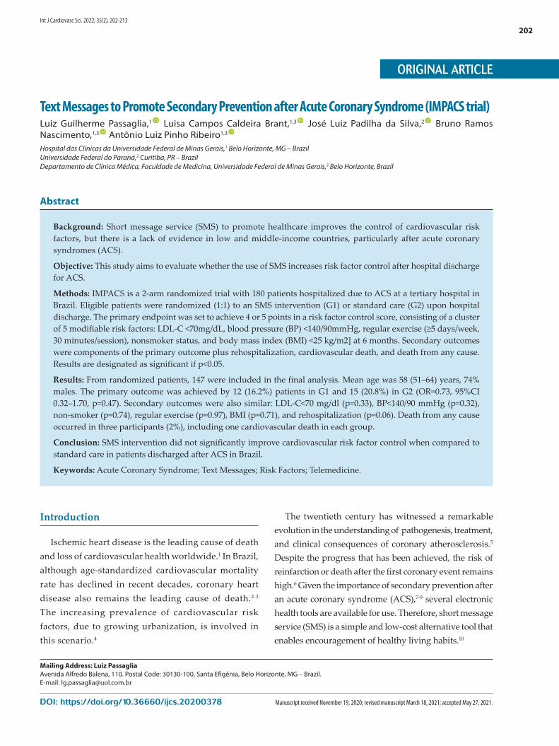
Introduction
Ischemic heart disease is the leading cause of death and loss of cardiovascular health worldwide.1 In Brazil, although age-standardized cardiovascular mortality rate has declined in recent decades, coronary heart disease also remains the leading cause of death.2-3 The increasing prevalence of cardiovascular risk factors, due to growing urbanization, is involved in this scenario.4
The twentieth century has witnessed a remarkable evolution in the understanding of pathogenesis, treatment, and clinical consequences of coronary atherosclerosis.5 Despite the progress that has been achieved, the risk of reinfarction or death after the first coronary event remains high.6 Given the importance of secondary prevention after an acute coronary syndrome (ACS),7-9 several electronic health tools are available for use. Therefore, short message service (SMS) is a simple and low-cost alternative tool that enables encouragement of healthy living habits.10
Int J Cardiovasc Sci. 2022; 35(2), 202-213
202
ORIGINAL ARTICLE
Text Messages to Promote Secondary Prevention after Acute Coronary Syndrome (IMPACS trial)Luiz Guilherme Passaglia,1 Luisa Campos Caldeira Brant,1,3 José Luiz Padilha da Silva,2 Bruno Ramos Nascimento,1,3 Antônio Luiz Pinho Ribeiro1,3
Hospital das Clínicas da Universidade Federal de Minas Gerais,1 Belo Horizonte, MG – BrazilUniversidade Federal do Paraná,2 Curitiba, PR – BrazilDepartamento de Clínica Médica, Faculdade de Medicina, Universidade Federal de Minas Gerais,3 Belo Horizonte, Brazil
DOI: https://doi.org/10.36660/ijcs.20200378
Mailing Address: Luiz PassagliaAvenida Alfredo Balena, 110. Postal Code: 30130-100, Santa Efigênia, Belo Horizonte, MG – Brazil. E-mail: [email protected]
Manuscript received November 19, 2020; revised manuscript March 18, 2021; accepted May 27, 2021.
Abstract
Background: Short message service (SMS) to promote healthcare improves the control of cardiovascular risk factors, but there is a lack of evidence in low and middle-income countries, particularly after acute coronary syndromes (ACS).
Objective: This study aims to evaluate whether the use of SMS increases risk factor control after hospital discharge for ACS.
Methods: IMPACS is a 2-arm randomized trial with 180 patients hospitalized due to ACS at a tertiary hospital in Brazil. Eligible patients were randomized (1:1) to an SMS intervention (G1) or standard care (G2) upon hospital discharge. The primary endpoint was set to achieve 4 or 5 points in a risk factor control score, consisting of a cluster of 5 modifiable risk factors: LDL-C <70mg/dL, blood pressure (BP) <140/90mmHg, regular exercise (≥5 days/week, 30 minutes/session), nonsmoker status, and body mass index (BMI) <25 kg/m2] at 6 months. Secondary outcomes were components of the primary outcome plus rehospitalization, cardiovascular death, and death from any cause. Results are designated as significant if p<0.05.
Results: From randomized patients, 147 were included in the final analysis. Mean age was 58 (51–64) years, 74% males. The primary outcome was achieved by 12 (16.2%) patients in G1 and 15 (20.8%) in G2 (OR=0.73, 95%CI 0.32–1.70, p=0.47). Secondary outcomes were also similar: LDL-C<70 mg/dl (p=0.33), BP<140/90 mmHg (p=0.32), non-smoker (p=0.74), regular exercise (p=0.97), BMI (p=0.71), and rehospitalization (p=0.06). Death from any cause occurred in three participants (2%), including one cardiovascular death in each group.
Conclusion: SMS intervention did not significantly improve cardiovascular risk factor control when compared to standard care in patients discharged after ACS in Brazil.
Keywords: Acute Coronary Syndrome; Text Messages; Risk Factors; Telemedicine.
Randomization
After obtaining informed written consent, data from each patient was entered into an online database (RedCap). A blocked randomization was provided in blocks of four patients each, following the date of patient enrollment, following a uniform 1:1 fashion. Researchers, data collectors, and physicians who provided medical care were blinded to the treatment allocation. Dedicated software developed by the Telehealth Center of UFMG’s University Hospital sent one-way SMS between a server (Microsoft Windows, Redmond, WA, US) and the participant’s mobile phone,17 The software had a bank of 185 text messages that allowed for the identification and scheduling of the submission of SMS on predetermined dates. Interactive communication was not available.
Interventions
The usual care group (IMPACS control group) received standard discharge care after ACS. The intervention group (IMPACS intervention group) also received standard discharge care, plus the SMS intervention program. The usual discharge care in the first 6 months of follow-up after ACS consisted of at least two medical appointments with the attending cardiologist, one appointment with a clinical pharmacist, and one appointment with the physical therapy group. All discharged patients were given the opportunity to participate in the hospital cardiovascular rehabilitation program, which consists of supervised physical exercise for three consecutive months. More appointments could occur, according to the evaluation of the attending health professionals. IMPACS researchers were independent of attending physicians and did not interfere with patient care.
A total of 185 SMS was developed by the research group, offering advice, motivation, and information about medication adherence, increase in regular physical activity, adoption of healthy dietary habits, and smoking cessation (if appropriate). The Intervention group was divided into four subgroups (“modules”), according to baseline characteristics of participants: Module 1, nonsmokers and free of diabetes; Module 2, non-smokers and diabetic patients; Module 3, smokers and non-diabetic patients; Module 4, smokers and diabetic patients. Semi-personalized SMS were sent out foud times per week for six months, with the first SMS being sent immediately after hospital discharge. The system could not inform whether the patients read the messages. No cross-over between modules was permitted, even
A previous randomized study developed in a high-income country, the “TEXT ME” trial,11 showed positive results of using SMS in patients with coronary heart disease. Despite the promising results, there is insufficient evidence to draw definite conclusions about SMS interventions in low and middle-income countries (LMIC), not only because most studies were performed in high-income countries,10-12 but also because significant barriers can hamper the successful application of mobile Health (mHealth) in this setting.13 Furthermore, good adherence to drug therapy is associated with positive health outcomes,14 and treatment adherence after SCA continues to be an important condition to achieve optimal targets.8 The IMPACS (Impact of text Messages to Promote secondary prevention after Acute Corornary Syndrome) study aims to further assess this gap through a randomized clinical trial, evaluating whether the use of SMS increases risk factor control after hospital discharge for ACS at a tertiary hospital in Brazil.
Methods
Design and Participants
The IMPACS trial is a two-arm, parallel, double-blind randomized trial involving patients admitted due to ACS at the University Hospital of Universidade Federal de Minas Gerais (UFMG), a tertiary, public, and general hospital in Belo Horizonte, a capital city located in southeast Brazil. Details of the trial design have been previously published,15 and the protocol was approved by institutional review boards.
Patients of 18 years of age or older, who were hospitalized with a diagnosis of ACS, were discharged for outpatient follow-up, and were able to receive SMS in their own mobile phone, were eligible to participate in this study. Eligible patients were identified by daily assessment in the Coronary Intensive Care Unit and followed up during hospitalization for data collection. Data were collected in standard protocols by previously trained cardiologists and medical students. Exclusion criteria included the refusal or inability to sign the Informed Consent, as well as complete illiteracy (the inability to read and write). Included patients were also participants of the “Good Practice Program in Cardiology / Get With The Guidelines”,16 a joint quality improvement program of the Brazilian Cardiology Society, Ministry of Health (Brazil) and American Heart Association (US).
Int J Cardiovasc Sci. 2022; 35(2), 202-213
203Passaglia et al.
Text messages after acute coronary syndrome Original Article
if the patient stopped smoking or developed diabetes. Examples of text messages can be seen in the previously published trial protocol.15
Outcomes
Outcomes were evaluated 6 (±1) months after hospital discharge, in a pre-scheduled follow-up appointment. The primary endpoint was set to achieve 4 or 5 points in a Risk Factor Control Score, which combined the cluster effect of five main modifiable risk factors for ACS (Low Density Lipoprotein Cholesterol (LDL-C) <70mg/dL, blood pressure <140/90 mmHg, regular exercise [≥5 days/week × 30 minutes of moderate exercise per session], non-smoker status, and body mass index (BMI) <25 kg/m2).
Pre-specified secondary endpoints were: plasma LDL-C levels, level of physical activity (measured by a “direct” question [participants who reported exercising 5 or more days per week × 30 min/d of moderate exercise], and by Portuguese version of the International Physical Activity Questionnaire Short Form (IPAQ-SF)18 - Supplementary Material, which was planned to be validated by using accelerometers in one-fifth of the participants), blood pressure levels, medication adherence "(measured via the "Medida de Adesão aos Tratamentos" - Treatment Adherence Measure [MAT] form19), proportion of non-smokers (self-reported and confirmed by a Carbon Monoxide Meter Breath Test), BMI, rehospitalization, cardiovascular death, and death from any cause. Additional analyses were done using a Health Literacy questionnaire (The Short Assessment of Health Literacy for Portuguese Speaking Adults - SAHLPA-18,20 with a score ranging from 0 to 18, with ≤14 indicating inadequate health literacy), and a follow-up questionnaire (self-reported acceptability and understanding). Both instruments were applied for a better interpretation of trial results due to LMIC’s barriers and particularities.
Statistical methods
A sample size of 160 patients was calculated to provide 80% power to detect a difference of at least 19% between the intervention and the control groups in achieving 4 or more of the 5 modifiable risk factors (Risk Factor Control Score), with a two-sided significance level of 0.05, considering a loss to follow-up of 20 patients. This calculation was based on findings from the TEXT ME trial.11 Pre-specified interim analysis, performed before the end of patient allocation, found follow-up
losses higher than expected, and the sample size was recalculated to 180 patients to maintain an 80% power in the outcome analyses.
Analyses were performed according to the intention-to-treat principle. For the baseline characteristics, continuous variables were summarized as mean± standard deviation (SD) or as median and first and third quartiles (Q1, Q3), as appropriate, and groups compared using unpaired Student’s t-test or Mann-Whitney test, based on the distribution pattern (Shapiro-Wilk test was used for this purpose). Categorical variables were expressed as proportions and groups compared by chi-square test. The primary and secondary outcomes were compared between groups by means of the chi-square test, and the results were presented as odds ratios with 95% confidence intervals (CI). For the additional analyses, the questionnaires were expressed as categorical variables, and the groups were compared by the chi-square test for the primary outcome, when appropriate. A per-protocol analysis was done using the results of the follow-up questionnaire.
A longitudinal post-hoc analysis was carried out considering the baseline values. Marginal models for longitudinal data were adjusted via generalized estimation equations – with independent correlation matrix and robust variance (sandwich estimator). The link function used here was the identity for continuous responses and logit for binary responses, which allows for interpretation in terms of mean differences and odds ratios, respectively. The models included the main effects of group and time in addition to the group x time interaction, which, being statistically significant, would indicate a different evolution of the groups over time. Statistical significance was set at α=0.05 for all analyses.
The statistical analysis was conducted using SPSS Statistics, version 20.1 for Windows (IBM Corp, Armonk, NY, US), and the R statistical software, version 3.6.3, expanded by the packages foreign, tidyverse, ggplot2, gridExtra, and geepack.
Results
From December 2017 to December 2018, 310 eligible patients were screened. A total of 180 patients were randomly assigned to receive either usual care or usual care plus SMS intervention. At ±6 months after hospital discharge, 13 participants in the intervention group (14.4%) and 17 participants in the control group (18.9%) did not attend the scheduled appointment, even though
Int J Cardiovasc Sci. 2022; 35(2), 202-213
204Passaglia et al.
Text messages after acute coronary syndromeOriginal Article
they were personally contacted through their cell phone. Including losses due to death (n=3), a total of 15 patients in the intervention group (16.7%) and 18 patients in the control group (20.0%) did not complete the planned follow-up. The last 6-month follow-up visit was done in June 2019, and 147 patients were included in the primary analysis (Figure 1).
During the index hospitalization, baseline characteristics were similar between groups, including the characterization of ACS, coronary artery disease severity, clinical data, and medications upon discharge (Table 1).
The primary endpoint was achieved by 12 participants (16.2%) in the intervention group and by 15 participants (20.8%) in the control group (p=0.473) (Table 2).
The incidence of secondary endpoints is also shown in Table 2. All pre-specified endpoints were similar between the intervention and control groups, including the LDL-C level <70mg/dL (p=0.335), blood pressure <140x90mmHg (p=0.324), performing regular exercise (more than 150 min/week) (p=0.973), nonsmoker status (p=0.741), BMI <25 Kg/m2 (p=0.710), and medication adherence (p=0.297). Rehospitalization occurred in 39 participants (26%), with a trend to lower hospitalization rates in the intervention group (p=0.062). Death from any cause occurred in three participants (2%) in the entire study, including one cardiovascular death for each group.
At the 6-month follow-up visit, the intervention and control groups had, respectively, similar results in the measures of median LDL-C, mean systolic blood pressure, mean diastolic blood pressure, and median BMI. Medications at 6 months, including aspirin, statin, and beta-blockers, as well as achievement of 3 to 5 points in the Risk Factor Control Score were similar between the groups (Table 3).
In view of a small sample size, a longitudinal post hoc analysis was carried out. Different lines were estimated for each group, connecting baseline and 6-month follow-up data, not assuming baseline equality despite randomization (Figure 2). Taking this model into account, time interaction (effect of time) was observed for the primary outcome and for four of the five secondary components of the primary outcome: LDL-C level <70mg/dL, blood pressure <140x90mmHg, performing regular exercise (more than 150 min/week), nonsmoker status. BMI<25 Kg/m2 was the only outcome without time interaction.
Otherwise, no statistical significance was found when the treatment x time interaction was considered (group effect), which indicates that the intervention and control groups followed lines that did not differ over time.
Regarding the health literacy evaluation, 79 (43.9%) participants in both groups achieved more than 14 points in SAHLPA-18 (Table 1). For the primary outcome, achieving 4 or 5 in a Risk Factor Control Score occurred in 10 participants (15.9%) in the group with adequate literacy and in 17 participants (20.7%) in the group with inadequate health literacy (OR=0.72; 95% CI 0.30-1.70; p=0.456).
The follow-up questionnaire (acceptability and understanding) applied at ±6 months showed that 20 participants in the intervention group (27%) did not receive IMPACS SMS (Table S1 - Supplementary material), by self-report, although the SMS were sent by the software. Given these findings, a per-protocol analysis was performed: participants in the intervention group who confirmed that they had received SMS were compared to the control group plus participants in the intervention group who did not read SMS. The primary and secondary outcomes were similar between the two (Table 4), except for the rate of rehospitalization (p=0.026), which proved to be lower in the intervention group.
The results of the IPAQ-SF and accelerometer users are shown in Supplementary material (Table S2 and S3, respectively). Baseline characteristics of participants who used accelerometers for physical activity analyses were similar between the groups (Table S4 - Supplementary material). Moderate physical activity measured by accelerometer was higher in the intervention group (225 min/week) than in the control group (114 min/week) (Mean Difference 111 min/week; 95% CI 33-189; p=0.007), although IPAQ-SF Questionnaire results were similar between the two.
Discussion
In this randomized clinical trial involving patients hospitalized with ACS and discharged for outpatient follow-up at a public general university hospital in Brazil, the primary composite outcome – achieving 4 to 5 points in a Risk Factor Control Score – was similar between patients under usual care compared to those additionally receiving an SMS intervention program for secondary
Int J Cardiovasc Sci. 2022; 35(2), 202-213
205Passaglia et al.
Text messages after acute coronary syndrome Original Article
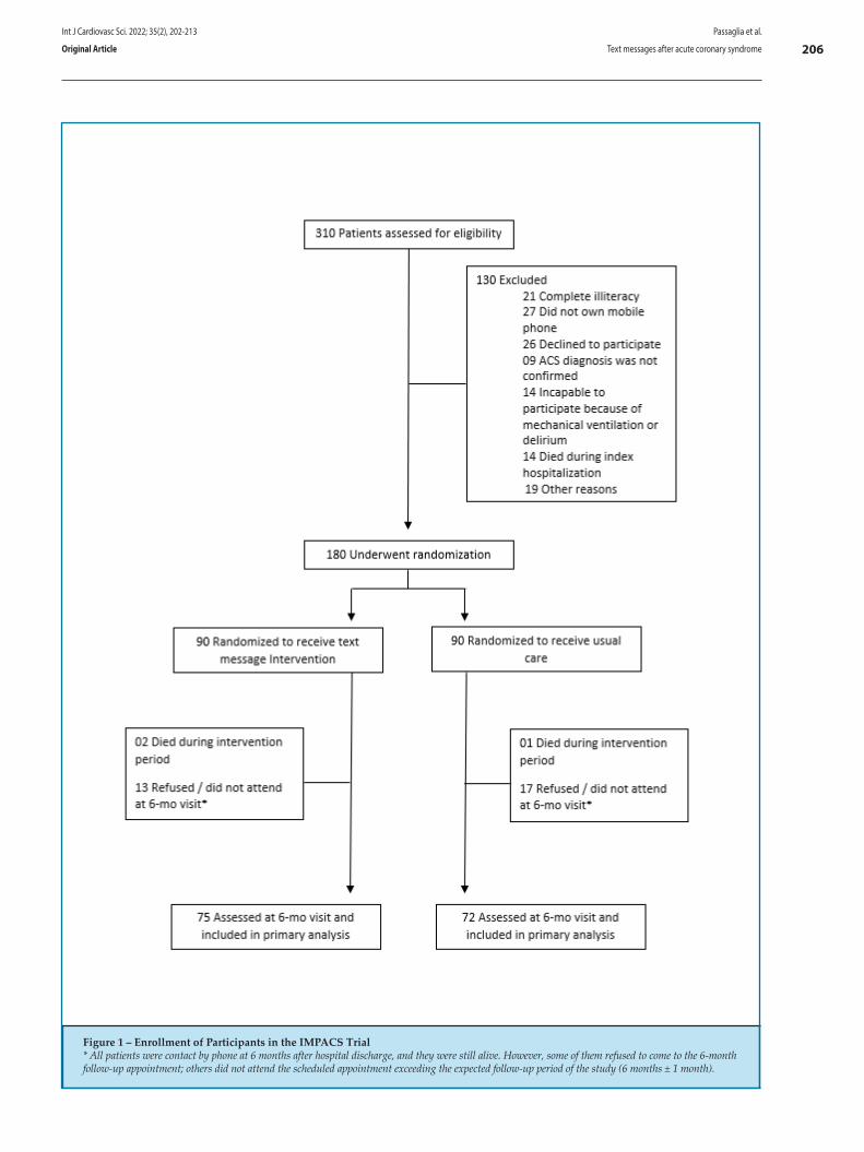
Figure 1 – Enrollment of Participants in the IMPACS Trial* All patients were contact by phone at 6 months after hospital discharge, and they were still alive. However, some of them refused to come to the 6-month follow-up appointment; others did not attend the scheduled appointment exceeding the expected follow-up period of the study (6 months ± 1 month).
Int J Cardiovasc Sci. 2022; 35(2), 202-213
206Passaglia et al.
Text messages after acute coronary syndromeOriginal Article
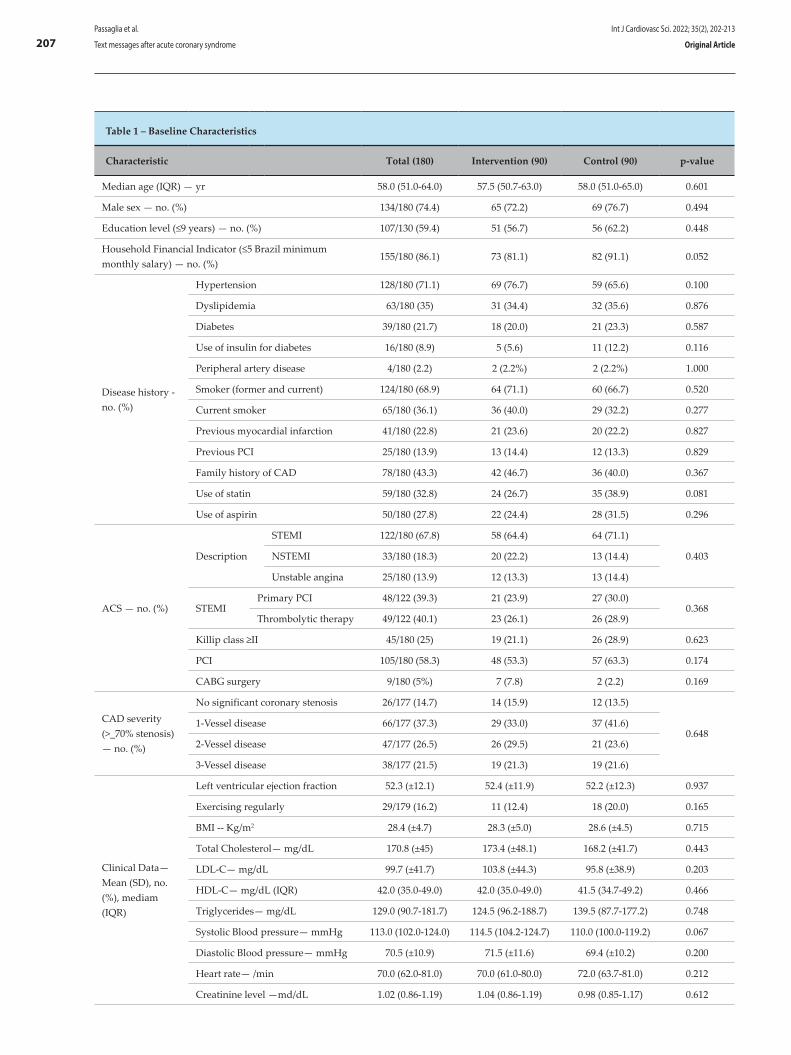
Table 1 – Baseline Characteristics
Characteristic Total (180) Intervention (90) Control (90) p-value
Median age (IQR) — yr 58.0 (51.0-64.0) 57.5 (50.7-63.0) 58.0 (51.0-65.0) 0.601
Male sex — no. (%) 134/180 (74.4) 65 (72.2) 69 (76.7) 0.494
Education level (≤9 years) — no. (%) 107/130 (59.4) 51 (56.7) 56 (62.2) 0.448
Household Financial Indicator (≤5 Brazil minimum monthly salary) — no. (%)
155/180 (86.1) 73 (81.1) 82 (91.1) 0.052
Disease history - no. (%)
Hypertension 128/180 (71.1) 69 (76.7) 59 (65.6) 0.100
Dyslipidemia 63/180 (35) 31 (34.4) 32 (35.6) 0.876
Diabetes 39/180 (21.7) 18 (20.0) 21 (23.3) 0.587
Use of insulin for diabetes 16/180 (8.9) 5 (5.6) 11 (12.2) 0.116
Peripheral artery disease 4/180 (2.2) 2 (2.2%) 2 (2.2%) 1.000
Smoker (former and current) 124/180 (68.9) 64 (71.1) 60 (66.7) 0.520
Current smoker 65/180 (36.1) 36 (40.0) 29 (32.2) 0.277
Previous myocardial infarction 41/180 (22.8) 21 (23.6) 20 (22.2) 0.827
Previous PCI 25/180 (13.9) 13 (14.4) 12 (13.3) 0.829
Family history of CAD 78/180 (43.3) 42 (46.7) 36 (40.0) 0.367
Use of statin 59/180 (32.8) 24 (26.7) 35 (38.9) 0.081
Use of aspirin 50/180 (27.8) 22 (24.4) 28 (31.5) 0.296
ACS — no. (%)
Description
STEMI 122/180 (67.8) 58 (64.4) 64 (71.1)
0.403NSTEMI 33/180 (18.3) 20 (22.2) 13 (14.4)
Unstable angina 25/180 (13.9) 12 (13.3) 13 (14.4)
STEMIPrimary PCI 48/122 (39.3) 21 (23.9) 27 (30.0)
0.368Thrombolytic therapy 49/122 (40.1) 23 (26.1) 26 (28.9)
Killip class ≥II 45/180 (25) 19 (21.1) 26 (28.9) 0.623
PCI 105/180 (58.3) 48 (53.3) 57 (63.3) 0.174
CABG surgery 9/180 (5%) 7 (7.8) 2 (2.2) 0.169
CAD severity (>_70% stenosis) — no. (%)
No significant coronary stenosis 26/177 (14.7) 14 (15.9) 12 (13.5)
0.6481-Vessel disease 66/177 (37.3) 29 (33.0) 37 (41.6)
2-Vessel disease 47/177 (26.5) 26 (29.5) 21 (23.6)
3-Vessel disease 38/177 (21.5) 19 (21.3) 19 (21.6)
Clinical Data—Mean (SD), no. (%), mediam (IQR)
Left ventricular ejection fraction 52.3 (±12.1) 52.4 (±11.9) 52.2 (±12.3) 0.937
Exercising regularly 29/179 (16.2) 11 (12.4) 18 (20.0) 0.165
BMI -- Kg/m2 28.4 (±4.7) 28.3 (±5.0) 28.6 (±4.5) 0.715
Total Cholesterol— mg/dL 170.8 (±45) 173.4 (±48.1) 168.2 (±41.7) 0.443
LDL-C— mg/dL 99.7 (±41.7) 103.8 (±44.3) 95.8 (±38.9) 0.203
HDL-C— mg/dL (IQR) 42.0 (35.0-49.0) 42.0 (35.0-49.0) 41.5 (34.7-49.2) 0.466
Triglycerides— mg/dL 129.0 (90.7-181.7) 124.5 (96.2-188.7) 139.5 (87.7-177.2) 0.748
Systolic Blood pressure— mmHg 113.0 (102.0-124.0) 114.5 (104.2-124.7) 110.0 (100.0-119.2) 0.067
Diastolic Blood pressure— mmHg 70.5 (±10.9) 71.5 (±11.6) 69.4 (±10.2) 0.200
Heart rate— /min 70.0 (62.0-81.0) 70.0 (61.0-80.0) 72.0 (63.7-81.0) 0.212
Creatinine level —md/dL 1.02 (0.86-1.19) 1.04 (0.86-1.19) 0.98 (0.85-1.17) 0.612
Int J Cardiovasc Sci. 2022; 35(2), 202-213
207Passaglia et al.
Text messages after acute coronary syndrome Original Article
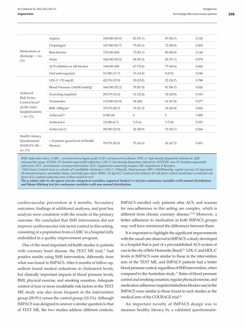
cardiovascular prevention at 6 months. Secondary outcomes, findings of additional analyses, and post hoc analysis were consistent with the results of the primary outcome. We concluded that SMS intervention did not improve cardiovascular risk factor control in this setting, consisting of a population from a LMIC in a hospital fully embedded in a quality improvement program.
One of the most important mHealth studies in patients with coronary heart disease, the TEXT ME trial,11 had positive results using SMS intervention, differently from what was found in IMPACS. After 6 months of follow-up, authors found modest reductions in cholesterol levels, but clinically important impacts in blood pressure levels, BMI, physical exercise, and smoking cessation. Adequate control of four or more modifiable risk factors in the TEXT ME study was also more frequent in the intervention group (28.9%) versus the control group (10.3%). Although IMPACS was designed to answer a similar question to that of TEXT ME, the two studies address different contexts.
IMPACS enrolled only patients after ACS, and reasons for non-adherence in this setting are complex, which is different from chronic coronary disease.21-24 Moreover, a better adherence to medication in both IMPACS groups may well have minimized the differences between them.
It is important to highlight the significant improvements with the usual care observed in IMPACS, a study developed in a hospital that is part of a pre-established ACS system of care in the city of Belo Horizonte, Brazil.25 LDL-C and HDL-C levels in IMPACS were similar to those in the intervention arm of the TEXT ME, and IMPACS patients had a better blood pressure control, regardless of SMS intervention, when compared to the Australian study.11 Rates of blood pressure control and smoking cessation, regular physical exercise, and medication adherence (aspirin/statin/beta-blocker use) in the IMPACS were similar to those found in such studies as the medical arm of the COURAGE trial.26
An important novelty of IMPACS design was to measure healthy literacy by a validated questionnaire.
Medications at discharge — no. (%)
Aspirin 169/180 (93.9) 82 (91.1) 87 (96.7) 0.120
Clopidogrel 147/180 (81.7) 75 (83.3) 72 (80.0) 0.563
Beta-blocker 153/180 (85) 73 (81.1) 80 (88.9) 0.144
Statin 166/180 (92.2) 84 (93.3) 82 (91.1) 0.578
ACE inhibitor or AR blocker 144/180 (80) 67 (74.4) 77 (85.6) 0.062
Oral anticoagulant 21/180 (11.7) 13 (14.4) 8 (8.9) 0.246
Achieved Risk Factor Control Score* (at the index hospitalization) — no. (%)
LDL-C <70 mg/dL 42/176 (23.9) 20 (23.0) 22 (24.7) 0.788
Blood Pressure <140/90 mmHg* 166/180 (92.2) 79 (87.8) 87 (96.7) 0.026
Exercising regularly 29/179 (16.2) 11 (12.4) 18 (20.0) 0.165
Nonsmoker 115/180 (63.9) 54 (60) 61 (67.8) 0.277
BMI <25Kg/m2 37/179 (20.7) 19 (21.3) 18 (20.0) 0.824
Achieved 5 0/180 (0) 0 0 1.000
Achieved 4 12/180 (6.7) 5 (5.6) 7 (7.8) 0.555
Achieved ≥3 59/180 (32.8) 26 (28.9) 33 (36.7) 0.266
Health Literacy Questionnaire (SAHLPA-18)— no. (%)
> 14 points (good level of Health literacy)
79/178 (43.9) 37 (41.6) 42 (47.2) 0.451
BMI: body mass index; CABG - coronary artery bypass graft; CAD: coronary artery disease; HDL-C: high density lipoprotein cholesterol; IQR: interquartile range; STEMI: ST elevation myocardial infarction; LDL-C: low density lipoprotein cholesterol; NSTEMI: non-ST elevation myocardial infarction); PCI: percutaneous coronary intervention; ACE: Angiotensin-converting enzyme; AR: Angiotensin II Receptor; * Risk Factor Control Score is a cluster of 5 modifiable risk factors: LDL-C <70mg/dL, blood pressure (BP) <140/90mmHg, regular exercise (≥5 days/week, 30 minutes/session), nonsmoker status, and body mass index (BMI) <25 kg/m2]. A patient who achieves all risk factor control would have a combined risk factor of 5; a patient achieving none of them would be at 0.The p-values refer to chi-square test for categorical variables, unpaired Student’s t-test for continuous variables with normal distribution, and Mann-Whitney test for continuous variables with non-normal distribution.
Int J Cardiovasc Sci. 2022; 35(2), 202-213
208Passaglia et al.
Text messages after acute coronary syndromeOriginal Article
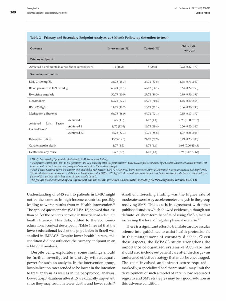
Understanding of SMS sent to patients in LMIC might not be the same as in high-income countries, possibly leading to worse results from m-Health intervention.12 The applied questionnaire (SAHLPA-18) showed that less than half of the patients enrolled in this trial had adequate health literacy. This data, added to the economic-educational context described in Table 1, reveal that the lowest educational level of the population in Brazil was studied in IMPACS. Despite lower health literacy, this condition did not influence the primary endpoint in an additional analysis.
Despite being exploratory, some findings should be further investigated in a study with adequate power for such an analysis. In the intervention group, hospitalization rates tended to be lower in the intention to treat analysis as well as in the per-protocol analysis. Lower hospitalizations after ACS are clinically important, since they may result in fewer deaths and lower costs.8,9
Another interesting finding was the higher rate of moderate exercise by accelerometer analysis in the group receiving SMS. This data is in agreement with other published studies which showed evidence, although not definite, of short-term benefits of using SMS aimed at increasing the level of regular physical exercise.2 7
There is a significant effort to translate cardiovascular science into guidelines to assist health professionals in the management of coronary disease. Given these aspects, the IMPACS study strengthens the importance of organized systems of ACS care that should also include outpatient care after discharge - an underused effective strategy that must be encouraged. The costs involved and infrastructure required – markedly, a specialized healthcare staff – may limit the development of such a model of care in low resourced region,s and SMS strategies may be a good solution in this adverse condition.
Table 2 – Primary and Secondary Endpoint Analyses at 6-Month Follow-up (intention-to-treat)
Outcome Intervention (75) Control (72)Odds Ratio
(95% CI)
Primary endpoint
Achieved 4 or 5 points in a risk factor control score* 12 (16.2) 15 (20.8) 0.73 (0.32-1.70)
Secondary endpoints
LDL-C <70 mg/dL 34/75 (45.3) 27/72 (37.5) 1.38 (0.71-2.67)
Blood pressure <140/90 mmHg 60/74 (81.1) 62/72 (86.1) 0.64 (0.27-1.55)
Exercising regularly 30/75 (40.0) 29/72 (40.3) 0.99 (0.51-1.91)
Nonsmoker* 62/75 (82.7) 58/72 (80.6) 1.15 (0.50-2.65)
BMI <25 Kg/m2 14/75 (18.7) 15/71 (21.1) 0.86 (0.38-1.93)
Medication adherence 66/75 (88.0) 67/72 (93.1) 0.55 (0.17-1.72)
Achieved Risk Factor
Control Score†
Achieved 5 3/75 (4.0) 1/72 (1.4) 2.96 (0.30-29.12)
Achieved 4 9/75 (12.0) 14/72 (19.4) 0.56 (0.23-1.40)
Achieved ≥3 43/75 (57.3) 40/72 (55.6) 1.07 (0.56-2.06)
Rehospitalization 15/77(19.5) 24/73 (32.9) 0.49 (0.23-1.05)
Cardiovascular death 1/77 (1.3) 1/73 (1.4) 0.95 (0.06-15.43)
Death from any cause 2/77 (2.6) 1/73 (1.4) 1.92 (0.17-21.63)
LDL-C: low density lipoprotein cholesterol; BMI: body mass index);* Two patients who said “no” to the question “are you smoking after hospitalization?” were reclassified as smokers by a Carbon Monoxide Meter Breath Test (one patient in the intervention group and one patient in the control group);† Risk Factor Control Score is a cluster of 5 modifiable risk factors: LDL-C <70mg/dL, blood pressure (BP) <140/90mmHg, regular exercise (≥5 days/week, 30 minutes/session), nonsmoker status, and body mass index (BMI) <25 kg/m2]. A patient who achieves all risk factor control would have a combined risk factor of 5; a patient achieving none of them would be at 0.The groups were compared by chi-square test and the results presented as odds ratio, including the 95% confidence interval (95% CI)
Int J Cardiovasc Sci. 2022; 35(2), 202-213
209Passaglia et al.
Text messages after acute coronary syndrome Original Article
Limitations of our study should be considered. First, we decided to use a combined surrogate outcome. Second, most of our patients used simvastatin, 40 mg/day, as the access to a high-intensity statin is limited for patients in the Brazilian public health care system, which may have affected the achievement of LDL-C goals. Third, although the IPAQ-SF is a validated questionnaire, it has important limitations, which have been previously debated,18 and only a few patients could use an accelerometer for a better analysis of their physical exercise level. Fourth, 20 participants (26.6%) in the intervention group reported not having received IMPACS SMS, though our system confirmed that they had been sent, which may have contributed to the loss of study power, raising the possibility of a type II error. The recent widespread use of cellphone messaging apps, instead of SMS – which are charged in some plans – may have additionally accounted
for this. However, different analyses performed in IMPACS were consistent with the primary outcome result, reinforcing our findings. Furthermore, as 13 of the 20 participants (65%) changed their telephone contact number during the study or provided a non-personal phone upon enrollment (Table 4), this limitation is inherent to the use of mHealth tools. In this sense, messaging apps have the advantage of not being linked to mobile numbers. And finally, the results should not be extrapolated to centers with different ACS follow-up care and to other regions of Brazil, especially those with different socioeconomic and healthcare backgrounds.
On the other hand, our study has a number of strengths that should also be considered. First, IMPACS was a randomized trial that targeted a low-income population with low indices of adequate health literacy, which is an important gap, since most SMS studies came
Table 3 – Follow-up Characteristics (at 6-month visit)
Characteristic Total (147) Intervention (75) Control (72) p-value
Medications at
6-month follow up —
no. (%)
Aspirin 135 (91.8) 68 (91.9) 67 (93.1) 0.790
Clopidogrel 116 (78.9) 58 (77.3) 58 (80.6) 0.632
Beta-blocker 125 (85.0) 60 (80.0) 65 (90.3) 0.081
Statin 135 (91.8) 70 (93.3) 65 (90.3) 0.499
ACE inhibitor or AR
blocker126 (85.7) 64 (85.3) 62 (86.1) 0.893
Oral anticoagulant 21 (14.2) 10 (13.3) 11 (15.3) 0.736
Insulin 13 (8.8) 5 (6.7) 8 (11.1) 0.343
Clinical Data —Mean
(SD), no. (%), median
(IQR)
Body mass index 28.0 (26.0-31.0) 28.0 (25.8-31.1) 28.7 (26.0-30.9) 0.576
Total-Cholesterol— mg/
dL150.0 (128.0-176.0) 147.0 (126.0-173.5) 151.0 (129.0-183.0) 0.583
LDL-C— mg/dL 77.0 (57.0-99.0) 77.0 (60.0-100.0) 77.0 (54.0-104.0) 0.815
HDL-C— mg/dL 40.0 (34.0-47.0) 39.0 (34.0-47.0) 41.0 (35.0-49.0) 0.203
Triglycerides— mg/dL 149.5 (110.0-202.2) 150.0 (109.0-200.0) 149.0 (110.0-207.0) 0.795
Systolic Blood
pressure— mm Hg121.2 (±17.3) 121.5 (±19.0) 121.0 (±15.4) 0.860
Diastolic Blood
pressure— mm Hg73.5 (±11.3) 73.7 (±12.3) 73.2 (±10.3) 0.813
Heart rate— /min 67.0 (61.0-76.0) 68.0 (61.0-77.0) 67.0 (61.0-75.2) 0.813
HDL-C: high density lipoprotein cholesterol; IQR: interquartile range; LDL-C: low density lipoprotein cholesterol; ACE: Angiotensin-converting enzyme; AR: Angiotensin II Receptor. The p-values refer to chi-square test for categorical variables, unpaired Student’s t-test for continuous variables with normal distribution, and Mann-Whitney test for continuous variables with non-normal distribution.
Int J Cardiovasc Sci. 2022; 35(2), 202-213
210Passaglia et al.
Text messages after acute coronary syndromeOriginal Article
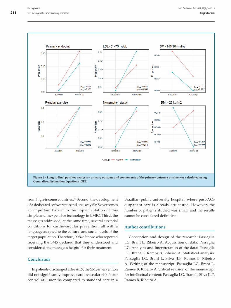
from high-income countries.10 Second, the development of a dedicated software to send one-way SMS overcomes an important barrier to the implementation of this simple and inexpensive technology in LMIC. Third, the messages addressed, at the same time, several essential conditions for cardiovascular prevention, all with a language adapted to the cultural and social levels of the target population. Therefore, 90% of those who reported receiving the SMS declared that they understood and considered the messages helpful for their treatment.
Conclusion
In patients discharged after ACS, the SMS intervention did not significantly improve cardiovascular risk factor control at 6 months compared to standard care in a
Brazilian public university hospital, where post-ACS outpatient care is already structured. However, the number of patients studied was small, and the results cannot be considered definitive.
Author contributions
Conception and design of the research: Passaglia LG, Brant L, Ribeiro A. Acquisition of data: Passaglia LG. Analysis and interpretation of the data: Passaglia LG, Brant L, Ramos B, Ribeiro A. Statistical analysis: Passaglia LG, Brant L, Silva JLP, Ramos B, Ribeiro A. Writing of the manuscript: Passaglia LG, Brant L, Ramos B, Ribeiro A.Critical revision of the manuscript for intellectual content: Passaglia LG, Brant L, Silva JLP, Ramos B, Ribeiro A.
Figure 2 – Longitudinal post hoc analysis – primary outcome and components of the primary outcome p-value was calculated using Generalized Estimation Equations (GEE)
Int J Cardiovasc Sci. 2022; 35(2), 202-213
211Passaglia et al.
Text messages after acute coronary syndrome Original Article
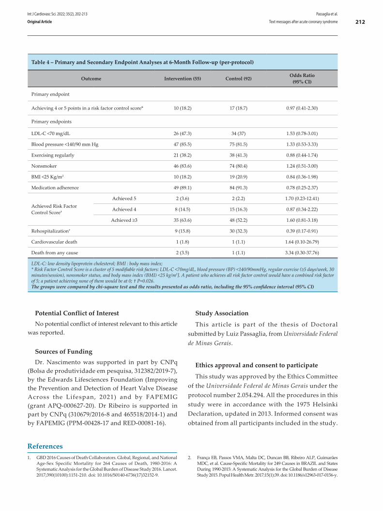
Potential Conflict of InterestNo potential conflict of interest relevant to this article
was reported.
Sources of FundingDr. Nascimento was supported in part by CNPq
(Bolsa de produtividade em pesquisa, 312382/2019-7), by the Edwards Lifesciences Foundation (Improving the Prevention and Detection of Heart Valve Disease Across the Lifespan, 2021) and by FAPEMIG (grant APQ-000627-20). Dr Ribeiro is supported in part by CNPq (310679/2016-8 and 465518/2014-1) and by FAPEMIG (PPM-00428-17 and RED-00081-16).
Study Association
This article is part of the thesis of Doctoral submitted by Luiz Passaglia, from Universidade Federal de Minas Gerais.
Ethics approval and consent to participate
This study was approved by the Ethics Committee of the Universidade Federal de Minas Gerais under the protocol number 2.054.294. All the procedures in this study were in accordance with the 1975 Helsinki Declaration, updated in 2013. Informed consent was obtained from all participants included in the study.
Table 4 – Primary and Secondary Endpoint Analyses at 6-Month Follow-up (per-protocol)
Outcome Intervention (55) Control (92) Odds Ratio (95% CI)
Primary endpoint
Achieving 4 or 5 points in a risk factor control score* 10 (18.2) 17 (18.7) 0.97 (0.41-2.30)
Primary endpoints
LDL-C <70 mg/dL 26 (47.3) 34 (37) 1.53 (0.78-3.01)
Blood pressure <140/90 mm Hg 47 (85.5) 75 (81.5) 1.33 (0.53-3.33)
Exercising regularly 21 (38.2) 38 (41.3) 0.88 (0.44-1.74)
Nonsmoker 46 (83.6) 74 (80.4) 1.24 (0.51-3.00)
BMI <25 Kg/m2 10 (18.2) 19 (20.9) 0.84 (0.36-1.98)
Medication adherence 49 (89.1) 84 (91.3) 0.78 (0.25-2.37)
Achieved Risk Factor Control Score†
Achieved 5 2 (3.6) 2 (2.2) 1.70 (0.23-12.41)
Achieved 4 8 (14.5) 15 (16.3) 0.87 (0.34-2.22)
Achieved ≥3 35 (63.6) 48 (52.2) 1.60 (0.81-3.18)
Rehospitalization† 9 (15.8) 30 (32.3) 0.39 (0.17-0.91)
Cardiovascular death 1 (1.8) 1 (1.1) 1.64 (0.10-26.79)
Death from any cause 2 (3.5) 1 (1.1) 3.34 (0.30-37.76)
LDL-C: low density lipoprotein cholesterol; BMI : body mass index;* Risk Factor Control Score is a cluster of 5 modifiable risk factors: LDL-C <70mg/dL, blood pressure (BP) <140/90mmHg, regular exercise (≥5 days/week, 30 minutes/session), nonsmoker status, and body mass index (BMI) <25 kg/m2]. A patient who achieves all risk factor control would have a combined risk factor of 5; a patient achieving none of them would be at 0; † P=0.026. The groups were compared by chi-square test and the results presented as odds ratio, including the 95% confidence interval (95% CI)
1. GBD 2016 Causes of Death Collaborators. Global, Regional, and National Age-Sex Specific Mortality for 264 Causes of Death, 1980-2016: A Systematic Analysis for the Global Burden of Disease Study 2016. Lancet. 2017;390(10100):1151-210. doi: 10.1016/S0140-6736(17)32152-9.
2. França EB, Passos VMA, Malta DC, Duncan BB, Ribeiro ALP, Guimarães MDC, et al. Cause-Specific Mortality for 249 Causes in BRAZIL and States During 1990-2015: A Systematic Analysis for the Global Burden of Disease Study 2015. Popul Health Metr. 2017;15(1):39. doi: 10.1186/s12963-017-0156-y.
References
Int J Cardiovasc Sci. 2022; 35(2), 202-213
212Passaglia et al.
Text messages after acute coronary syndromeOriginal Article
3. Ribeiro ALP, Duncan BB, Brant LCC, Lotufo PA, Mill JG, Barreto SM. Cardiovascular Health in Brazil: Trends and Perspectives. Circulation. 2016;133(4):422-33. doi: 10.1161/CIRCULATIONAHA.114.008727.
4. Brant LCC, Nascimento BR, Passos VMA, Duncan BB, Bensenõr IJM, Malta DC, et al. Variations and Particularities in Cardiovascular Disease Mortality in Brazil and Brazilian States in 1990 and 2015: Estimates from the Global Burden of Disease. Rev Bras Epidemiol. 2017;20(Suppl 1):116-28. doi: 10.1590/1980-5497201700050010.
5. Zipes DP, Libby P, Bonow RO, Mann DL, Tomaselli GF, editors. Braunwald's Heart Disease: A Textbook of Cardiovascular Medicine. 10th ed. Philadelphia: Elsevier; 2012.
6. Benjamin EJ, Virani SS, Callaway CW, Chamberlain AM, Chang AR, Cheng S, et al. Heart Disease and Stroke Statistics-2018 Update: A Report From the American Heart Association. Circulation. 2018;137(12):e67-e492. doi: 10.1161/CIR.0000000000000558.
7. Roffi M, Patrono C, Collet JP, Mueller C, Valgimigli M, Andreotti F, et al. 2015 ESC Guidelines for the Management of Acute Coronary Syndromes in Patients Presenting Without Persistent ST-Segment Elevation: Task Force for the Management of Acute Coronary Syndromes in Patients Presenting without Persistent ST-Segment Elevation of the European Society of Cardiology (ESC). Eur Heart J. 2016;37(3):267-315. doi: 10.1093/eurheartj/ehv320.
8. Ibanez B, James S, Agewall S, Antunes MJ, Bucciarelli-Ducci C, Bueno H, et al. 2017 ESC Guidelines for the Management of Acute Myocardial Infarction in Patients Presenting with ST-Segment Elevation: The Task Force for the Management of Acute Myocardial Infarction in Patients Presenting with ST-Segment Elevation of the European Society of Cardiology (ESC). Eur Heart J. 2018;39(2):119-77. doi: 10.1093/eurheartj/ehx393.
9. Fleg JL, Forman DE, Berra K, Bittner V, Blumenthal JA, Chen MA, et al. Secondary Prevention of Atherosclerotic Cardiovascular Disease in Older Adults: A Scientific Statement from the American Heart Association. Circulation. 2013;128(22):2422-46. doi: 10.1161/01.cir.0000436752.99896.22.
10. Marcolino MS, Oliveira JAQ, D'Agostino M, Ribeiro ALP, Alkmim MBM, Novillo-Ortiz D. The Impact of mHealth Interventions: Systematic Review of Systematic Reviews. JMIR Mhealth Uhealth. 2018;6(1):e23. doi: 10.2196/mhealth.8873.
11. Chow CK, Redfern J, Hillis GS, Thakkar J, Santo K, Hackett ML, et al. Effect of Lifestyle-Focused Text Messaging on Risk Factor Modification in Patients With Coronary Heart Disease: A Randomized Clinical Trial. JAMA. 2015;314(12):1255-63. doi: 10.1001/jama.2015.10945.
12. Marcolino MS, Oliveira JAQ, Ribeiro ALP. Text messaging for prevention and treatment of cardiovascular disease: time to move on. Heart. 2017;103(12):893-94. doi: 10.1136/heartjnl-2016-311135.
13. Kruse C, Betancourt J, Ortiz S, Valdes Luna SM, Bamrah IK, Segovia N. Barriers to the Use of Mobile Health in Improving Health Outcomes in Developing Countries: Systematic Review. J Med Internet Res. 2019;21(10):e13263. doi: 10.2196/13263.
14. Simpson SH, Eurich DT, Majumdar SR, Padwal RS, Tsuyuki RT, Varney J, et al. A meta-Analysis of The Association between Adherence to Drug Therapy and Mortality. BMJ. 2006;333(7557):15. doi: 10.1136/bmj.38875.675486.55.
15. Passaglia LG, Brant LCC, Nascimento BR, Ribeiro ALP. Impact of Text Messages in a Middle-Income Country to Promote Secondary Prevention After Acute Coronary Syndrome (IMPACS): A Randomized Trial. Medicine. 2019;98(22):e15681. doi: 10.1097/MD.0000000000015681.
16. Sociedade Brasileira de Cardiologia. Programa Boas Práticas Clínicas em Cardiologia [Internet]. São Paulo: 2021 [cited 2021 18 Jun]. Available from: http://www.cardiol.br/boaspraticasclinicas
17. Gusmão LL, Ribeiro ALP, Souza-Silva MVR, Gomes PR, Beleigoli AM, Cardoso CS, et al. Implementation of a Text Message Intervention to Promote Behavioural Change and Weight Loss Among Overweight and Obese Brazilian Primary Care Patients. J Telemed Telecare. 2019;25(8):476-83. doi: 10.1177/1357633X18782092.
18. Lee PH, Macfarlane DJ, Lam TH, Stewart SM. Validity of the International Physical Activity Questionnaire Short Form (IPAQ-SF): A Systematic Review. Int J Behav Nutr Phys Act. 2011;8:115. doi: 10.1186/1479-5868-8-115.
19. Delgado AB, Lima ML. Contributo para a Validação Concorrente de uma Medida de Adesão aos Tratamentos. Psicol. Saude Doenças. 2001;2(2):81-100.
20. Apolinario D, Braga RC, Magaldi RM, Busse AL, Campora F, Brucki S, et al. Short Assessment of Health Literacy for Portuguese-speaking Adults. Rev Saude Publica. 2012;46(4):702-11. doi: 10.1590/s0034-89102012005000047.
21. Bagnall AJ, Yan AT, Yan RT, Lee CH, Tan M, Baer C, et al. Optimal Medical Therapy for Non-ST-segment-elevation Acute Coronary Syndromes: Exploring why Physicians do not Prescribe Evidence-based Treatment and why Patients Discontinue Medications after Discharge. Circ Cardiovasc Qual Outcomes. 2010;3(5):530-7. doi: 10.1161/CIRCOUTCOMES.109.919415.
22. Gencer B, Rodondi N, Auer R, Räber L, Klingenberg R, Nanchen D, et al. Reasons for Discontinuation of Recommended Therapies According to the Patients After Acute Coronary Syndromes. Eur J Intern Med. 2015;26(1):56-62. doi: 10.1016/j.ejim.2014.12.014.
23. Cheng K, Ingram N, Keenan J, Choudhury RP. Evidence of Poor Adherence to Secondary Prevention after Acute Coronary Syndromes: Possible Remedies Through the Application of New Technologies. Open Heart. 2015;2(1):e000166. doi: 10.1136/openhrt-2014-000166.
24. Naderi SH, Bestwick JP, Wald DS. Adherence to Drugs That Prevent Cardiovascular Disease: Meta-Analysis on 376,162 Patients. Am J Med. 2012;125(9):882-7.e1. doi: 10.1016/j.amjmed.2011.12.013.
25. Marcolino MS, Brant LCC, Araujo JG, Nascimento BR, Castro LR, Martins P, et al. Implementation of the Myocardial Infarction System of Care in City of Belo Horizonte, Brazil. Arq Bras Cardiol. 2013;100(4):307-14. doi: 10.5935/abc.20130054
26. Boden WE, O'Rourke RA, Teo KK, Hartigan PM, Maron DJ, Kostuk WJ, et al. Optimal Medical Therapy with or Without PCI for Stable Coronary Disease. N Engl J Med. 2007;356(15):1503-16. doi: 10.1056/NEJMoa070829.
27. Free C, Phillips G, Galli L, Watson L, Felix L, Edwards P, et al. The Effectiveness of Mobile-health Technology-based Health Behaviour Change or Disease Management Interventions for Health Care Consumers: A Systematic Review. PLoS Med. 2013;10(1):e1001362. doi: 10.1371/journal.pmed.1001362.
This is an open-access article distributed under the terms of the Creative Commons Attribution License
*Supplemental MaterialsFor additional information, please click here.
Int J Cardiovasc Sci. 2022; 35(2), 202-213
213Passaglia et al.
Text messages after acute coronary syndrome Original Article
Introduction
Radiotherapy is an important treatment method for a wide variety of malignancies. Ionizing radiation (IR) is known to generate free radicals in irradiated tissues.¹ Mammalian cells have both enzymatic and non-enzymatic cleansing systems to remove reactive oxygen species (ROS) and reactive nitrogen species (RNS), respectively.² An imbalance favoring the prooxidants and disfavoring the antioxidants, potentially leading to damage, has been called “oxidative stress.”³
Efforts to reduce the toxicity of irradiation to normal tissues, organs, and cells have led to the investigation of cytoprotective agents.⁴ Many dietary components may have either direct antioxidant activity, such as flavonoids, melatonin, nigella sativa oil (NSO), and thymoquinone(TQ),⁵ or indirect antioxidant activity, such as zinc,⁶ manganese, and selenium.⁷ NSO is commonly known as ‘black seed’ and has strong antioxidant properties against oxidative damage.⁸ Many studies have reported that it has various pharmacological properties, including
Int J Cardiovasc Sci. 2022; 35(2), 214-219
214
ORIGINAL ARTICLE
Radioprotective Effect of Nigella Sativa Oil on Heart Tissues of Rats Exposed to IrraditionMehmet Kaplan,1 Elif Demir,2 Fethi Yavuz,3 Gizem Ilgin Kaplan,4 Mehmet Resit Taysi,5 Seyithan Taysi,1 Mehmet Murat Sucu1
Gaziantep University School of Medicine, Department of Cardiology,1 Gaziantep - Turkey Division of Biochemistry, Harran University, College of Health,2 Sanlıurfa - TurkeyAdana City Training & Research Hospital, Department of Cardiology,3 Adana - TurkeyErsin Arslan Training & Research Hospital, Department of Internal Medicine,4 Gaziantep - Turkey Faculty of Agriculture, Bingol University,5 Bingol - Turkey
DOI: https://doi.org/10.36660/ijcs.20210055
Mailing Address: Mehmet Kaplan Gaziantep University School of Medicine, Department of Cardiology27310, Gaziantep - TurkeyE-mail: [email protected]
Manuscript received on February 24, 2021; reviewed on June 02, 2021; accepted on September 20, 2021.
Abstract
Background: Various studies are ongoing related to the radioprotective agents. Herbal preparations are currently becoming popular because of their beneficial effects with fewer side effects compared to the synthetic/semi-synthetic medicines, and Nigella sativa oil (NSO) is only one of them.
Objective: To investigate NSO for its antioxidant effects on the heart tissue of rats exposed to ionizing radiation (IR).
Methods: Thirty six male albino Wistar rats, divided into four groups, were designated to group I (IR plus NSO group) that received both 5 Gray of gamma IR to total cranium and NSO; group II (IR alone group) that received IR plus saline, group III (control group of NSO) that received saline and did not receive NSO or IR; group IV (control group) that received only sham IR. Alterations in Total antioxidant status (TAS) and Total oxidant status (TOS), Oxidative stres index (OSI), Sulhydryl group (SH), Lipid hydroperoxide (LOOH), Paraoxonase (PON) levels, Arylesterase (ARE) and Ceruloplasmin (CER) activities in homogenized heart tissue of rats were measured by biochemical methods.
Results: In heart tissue of the rats in the IR alone group (group II) LOOH, TOS and OSI levels were found to be higher, ARE activity and TAS level were found to be lower than all of the other groups (p < 0.01). These results also support that IR increases oxidative stress and NSO's protective effect.
Conclusion: NSO would reduce the oxidative damage in the irradiated heart tissue in the experimental rat model.
Keywords: Rats; Radiation; Radiation, Effects; Nigella Sativa Oil; Plants, Medicinal; Anti-Inflammatory Agents/therapeutic use.
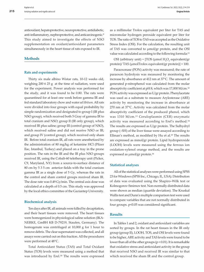
as a millimolar Trolox equivalent per liter for TAS and micromolar hydrogen peroxide equivalent per liter for TOS. The ratio of TOS to TAS was accepted as the Oxidative Stress Index (OSI). For the calculation, the resulting unit of TAS was converted to μmol/gr protein, and the OSI value was calculated according to the following formula:¹¹
OSI (arbitrary unit) = [TOS (μmol H2O2 equivalent/gr protein)/ TAS (μmolTrolox equivalent/gr protein)] × 100.
Paraoxonase (PON) activity was measured; the rate of paraoxon hydrolysis was measured by monitoring the increase by absorbance at 412 nm at 37°C. The amount of generated p-nitrophenol was calculated from the molar absorptivity coefficient at pH 8, which was 17,000 M/cm.¹² PON activity was expressed as U/gr protein. Phenylacetate was used as a substrate to measure Arylesterase (ARE) activity by monitoring the increase in absorbance at 270 nm at 37°C. Activity was calculated from the molar absorptivity coefficient of the produced phenol, which was 1310 M/cm.¹³ Ceruloplasmin (CER) enzymatic activity was measured according to Erel’s method.¹⁴ The results are expressed as U/gr protein. The Sulhydryl group (–SH) of the liver tissue were assayed according to Ellman’s method, as modified by Hu et al.¹⁵ The results are expressed as mmol/gr protein. Lipid hydroperoxide (LOOH) levels were measured using the ferrous ion oxidation-xylenol orange method, and the results are expressed as μmol/gr protein.¹⁶
Statistical analyses
All of the statistical analyses were performed using SPSS 23 for Windows (SPSS Inc., Chicago, IL, USA). Distribution of data was evaluated using the Shapiro–Wilk test or Kolmogorov-Smirnov test. Non-normally distributed data were shown as median (quartile deviation). The Kruskal Wallis test and Dunn's multiple comparison test were used to compare variables that are not normally distributed in four groups. p<0.05 was considered significant.
Results
In Tables 1 and 2, oxidant and antioxidant variables are sorted by groups. In the rat heart tissues in the IR only group (group II), LOOH, TOS, and OSI levels were found to be higher, ARE activity and TAS levels were found to be lower than all of the other groups (p < 0.01). It is remarkable that oxidative stress and antioxidant activity in the group that received NSO and received IR was similar to that which received the sham IR and the control group.
antioxidant, hepatoprotective, neuroprotective, antidiabetic, anti-inflammatory, nephroprotective, and anticarcinogenic.⁹ This study aimed to investigate the effects of NSO supplementation on oxidant/antioxidant parameters simultaneously in the heart tissue of rats exposed to IR.
Methods
Rats and experiments
Thirty six male albino Wistar rats, 10-12 weeks old, weighing 200 6 25 g at the time of radiation, were used for the experiment. Power analysis was performed for the study, and it was found to be 0.80. The rats were quarantined for at least one week before gamma IR and fed standard laboratory chow and water ad libitum. All rats were divided into four groups with equal probability by simple randomization and designated to group I (IR plus NSO group), which received both 5 Gray of gamma IR to total cranium and NSO; group II (IR only group), which received IR plus saline; group III (control group of NSO), which received saline and did not receive NSO or IR; and group IV (control group), which received only sham IR. Before total cranium IR, all rats were anesthetized by the administration of 80 mg/kg of ketamine HCl (Pfizer Ilac, Istanbul, Turkey) and placed on a tray in the prone position. The rats in the IR and the IR plus NSO groups received IR, using the Cobalt-60 teletherapy unit (Picker, C9, Maryland, NY) from a source-to-surface distance of 80 cm by 5 3 5 cm anterior fields with the total cranium gamma IR as a single dose of 5 Gy, whereas the rats in the control and sham control groups received sham IR. The dose rate was 0.49 Gy/min. The central axis dose was calculated at a depth of 0.5 cm. This study was approved by the local ethics committee of the Gaziantep University.
Biochemical analysis
Ten days after IR, all animals were killed by decapitation, and their heart tissues were removed. The heart tissues were homogenized in physiological saline solution (IKA-NERKE, GmBH KB D-79219, Staufen, Germany). The homogenate was centrifuged at 10,000 g for 1 hour to remove debris. The clear supernatant was collected, and all assays were carried out on this fraction. All the procedures were performed at 48°C.
Total Antioxidant Status (TAS) and Total Oxidant Status (TOS) levels were measured using a method that was introduced by Erel.¹⁰ The results were expressed
Int J Cardiovasc Sci. 2022; 35(2), 214-219
215Kaplan et al.
Ionizing radiation and nigella sativa oil Original Article
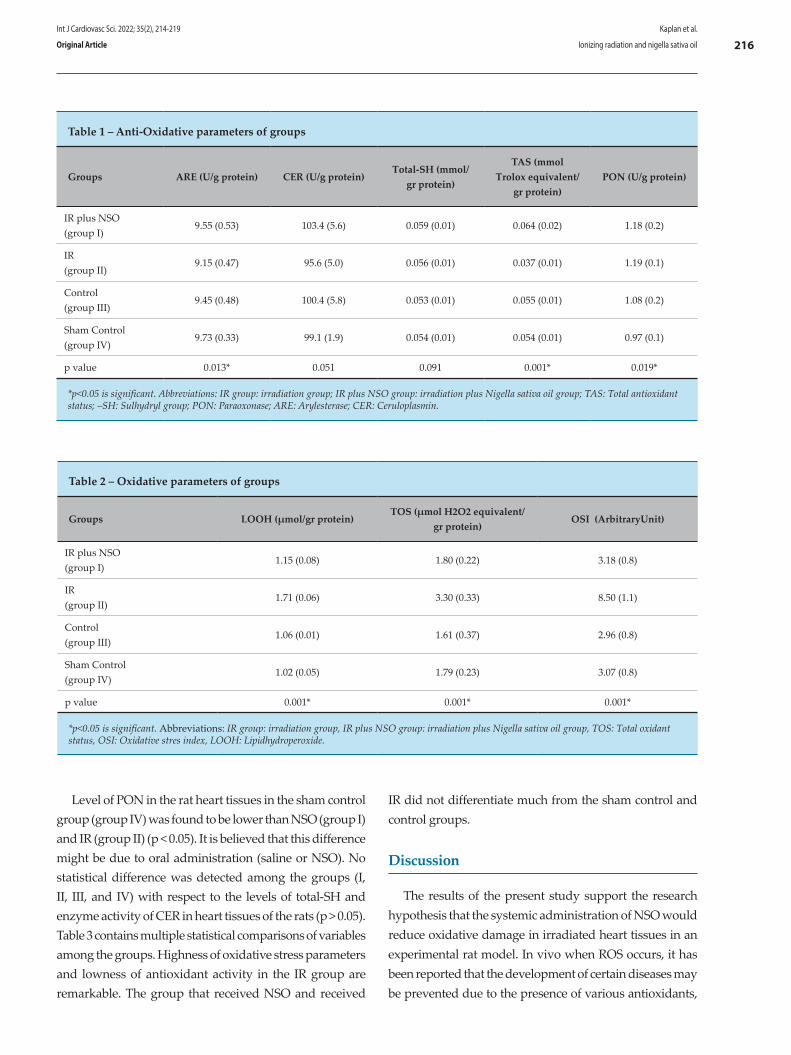
Level of PON in the rat heart tissues in the sham control group (group IV) was found to be lower than NSO (group I) and IR (group II) (p < 0.05). It is believed that this difference might be due to oral administration (saline or NSO). No statistical difference was detected among the groups (I, II, III, and IV) with respect to the levels of total-SH and enzyme activity of CER in heart tissues of the rats (p > 0.05). Table 3 contains multiple statistical comparisons of variables among the groups. Highness of oxidative stress parameters and lowness of antioxidant activity in the IR group are remarkable. The group that received NSO and received
IR did not differentiate much from the sham control and control groups.
Discussion
The results of the present study support the research hypothesis that the systemic administration of NSO would reduce oxidative damage in irradiated heart tissues in an experimental rat model. In vivo when ROS occurs, it has been reported that the development of certain diseases may be prevented due to the presence of various antioxidants,
Table 1 – Anti-Oxidative parameters of groups
Groups ARE (U/g protein) CER (U/g protein)Total-SH (mmol/
gr protein)
TAS (mmol Trolox equivalent/
gr protein)PON (U/g protein)
IR plus NSO(group I)
9.55 (0.53) 103.4 (5.6) 0.059 (0.01) 0.064 (0.02) 1.18 (0.2)
IR(group II)
9.15 (0.47) 95.6 (5.0) 0.056 (0.01) 0.037 (0.01) 1.19 (0.1)
Control(group III)
9.45 (0.48) 100.4 (5.8) 0.053 (0.01) 0.055 (0.01) 1.08 (0.2)
Sham Control(group IV)
9.73 (0.33) 99.1 (1.9) 0.054 (0.01) 0.054 (0.01) 0.97 (0.1)
p value 0.013* 0.051 0.091 0.001* 0.019*
*p<0.05 is significant. Abbreviations: IR group: irradiation group; IR plus NSO group: irradiation plus Nigella sativa oil group; TAS: Total antioxidant status; –SH: Sulhydryl group; PON: Paraoxonase; ARE: Arylesterase; CER: Ceruloplasmin.
Table 2 – Oxidative parameters of groups
Groups LOOH (µmol/gr protein)TOS (µmol H2O2 equivalent/
gr protein)OSI (ArbitraryUnit)
IR plus NSO(group I)
1.15 (0.08) 1.80 (0.22) 3.18 (0.8)
IR(group II)
1.71 (0.06) 3.30 (0.33) 8.50 (1.1)
Control(group III)
1.06 (0.01) 1.61 (0.37) 2.96 (0.8)
Sham Control(group IV)
1.02 (0.05) 1.79 (0.23) 3.07 (0.8)
p value 0.001* 0.001* 0.001*
*p<0.05 is significant. Abbreviations: IR group: irradiation group, IR plus NSO group: irradiation plus Nigella sativa oil group, TOS: Total oxidant status, OSI: Oxidative stres index, LOOH: Lipidhydroperoxide.
Int J Cardiovasc Sci. 2022; 35(2), 214-219
216Kaplan et al.
Ionizing radiation and nigella sativa oilOriginal Article
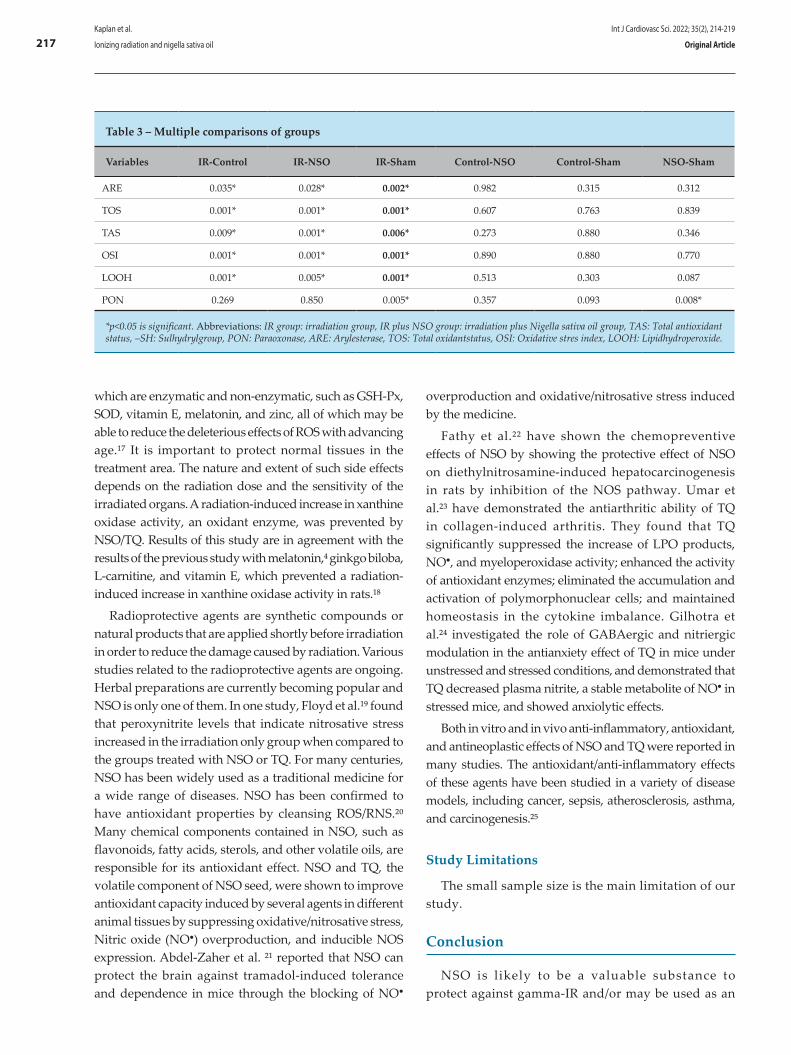
which are enzymatic and non-enzymatic, such as GSH-Px, SOD, vitamin E, melatonin, and zinc, all of which may be able to reduce the deleterious effects of ROS with advancing age.¹⁷ It is important to protect normal tissues in the treatment area. The nature and extent of such side effects depends on the radiation dose and the sensitivity of the irradiated organs. A radiation-induced increase in xanthine oxidase activity, an oxidant enzyme, was prevented by NSO/TQ. Results of this study are in agreement with the results of the previous study with melatonin,⁴ ginkgo biloba, L-carnitine, and vitamin E, which prevented a radiation-induced increase in xanthine oxidase activity in rats.¹⁸
Radioprotective agents are synthetic compounds or natural products that are applied shortly before irradiation in order to reduce the damage caused by radiation. Various studies related to the radioprotective agents are ongoing. Herbal preparations are currently becoming popular and NSO is only one of them. In one study, Floyd et al.¹⁹ found that peroxynitrite levels that indicate nitrosative stress increased in the irradiation only group when compared to the groups treated with NSO or TQ. For many centuries, NSO has been widely used as a traditional medicine for a wide range of diseases. NSO has been confirmed to have antioxidant properties by cleansing ROS/RNS.²⁰ Many chemical components contained in NSO, such as flavonoids, fatty acids, sterols, and other volatile oils, are responsible for its antioxidant effect. NSO and TQ, the volatile component of NSO seed, were shown to improve antioxidant capacity induced by several agents in different animal tissues by suppressing oxidative/nitrosative stress, Nitric oxide (NO●) overproduction, and inducible NOS expression. Abdel-Zaher et al. ²¹ reported that NSO can protect the brain against tramadol-induced tolerance and dependence in mice through the blocking of NO●
overproduction and oxidative/nitrosative stress induced by the medicine.
Fathy et al.²² have shown the chemopreventive effects of NSO by showing the protective effect of NSO on diethylnitrosamine-induced hepatocarcinogenesis in rats by inhibition of the NOS pathway. Umar et al.²³ have demonstrated the antiarthritic ability of TQ in collagen-induced arthritis. They found that TQ significantly suppressed the increase of LPO products, NO●, and myeloperoxidase activity; enhanced the activity of antioxidant enzymes; eliminated the accumulation and activation of polymorphonuclear cells; and maintained homeostasis in the cytokine imbalance. Gilhotra et al.²⁴ investigated the role of GABAergic and nitriergic modulation in the antianxiety effect of TQ in mice under unstressed and stressed conditions, and demonstrated that TQ decreased plasma nitrite, a stable metabolite of NO● in stressed mice, and showed anxiolytic effects.
Both in vitro and in vivo anti-inflammatory, antioxidant, and antineoplastic effects of NSO and TQ were reported in many studies. The antioxidant/anti-inflammatory effects of these agents have been studied in a variety of disease models, including cancer, sepsis, atherosclerosis, asthma, and carcinogenesis.²⁵
Study Limitations
The small sample size is the main limitation of our study.
Conclusion
NSO is likely to be a valuable substance to protect against gamma-IR and/or may be used as an
Table 3 – Multiple comparisons of groups
Variables IR-Control IR-NSO IR-Sham Control-NSO Control-Sham NSO-Sham
ARE 0.035* 0.028* 0.002* 0.982 0.315 0.312
TOS 0.001* 0.001* 0.001* 0.607 0.763 0.839
TAS 0.009* 0.001* 0.006* 0.273 0.880 0.346
OSI 0.001* 0.001* 0.001* 0.890 0.880 0.770
LOOH 0.001* 0.005* 0.001* 0.513 0.303 0.087
PON 0.269 0.850 0.005* 0.357 0.093 0.008*
*p<0.05 is significant. Abbreviations: IR group: irradiation group, IR plus NSO group: irradiation plus Nigella sativa oil group, TAS: Total antioxidant status, –SH: Sulhydrylgroup, PON: Paraoxonase, ARE: Arylesterase, TOS: Total oxidantstatus, OSI: Oxidative stres index, LOOH: Lipidhydroperoxide.
Int J Cardiovasc Sci. 2022; 35(2), 214-219
217Kaplan et al.
Ionizing radiation and nigella sativa oil Original Article
antioxidant against oxidative stress and other severe side effects occurring in the patients treated with radiotherapy.
Author contributions
Conception and design of the research: Kaplan M, Demir E. Acquisition of data: Kaplan M, Demir E, Yavuz F, Kaplan GI, Taysi MR. Analysis and interpretation of the data: Kaplan M, Demir E, Yavuz F, Kaplan GI, Taysi MR. Statistical analysis: Kaplan M, Yavuz F, Taysi MR, Sucu MM. Obtaining financing: Kaplan M. Writing of the manuscript: Kaplan M, Yavuz F, Kaplan GI. Critical revision of the manuscript for intellectual content: Kaplan M, Demir E, Taysi MR, Sucu MM. Supervision / as the major investigador: Kaplan M.
Potential Conflict of Interest
No potential conflict of interest relevant to this article was reported.
Sources of Funding
There were no external funding sources for this study.
Study Association
This study is not associated with any thesis or dissertation work.
Ethics approval and consent to participate
This study was approved by the Ethics Committee on Animal Experiments of the Gaziantep University under the protocol number 2016/15.
1. Prasanna PG, Stone HB, Wong RS,CapalaJacek, Bernhard EJ, Vikram B, et al. Normal tissue protection for improving radiotherapy: where are the gaps? Transl Cancer Res. 2012;(1):35-48.
2. Sies H. Strategies of antioxidant defense. Eur J Biochem. 1993; 215(2):213–9.
3. Cadenas E . Biochemistry of oxygen toxic i ty . Annu Rev Biochem.1989;58:79-110.
4. Taysi S, Memisogullari R, Koc M,Yazıcı AT, Aslankurt M, Gumustekin K, et al. Melatonin reduces oxidative stress in the rat lens due to radiation-induced oxidative injury. Int J Radiat Biol. 2008; 84(10):803–8.
5. Taysi S, Abdulrahman ZK, Okumus S, Demir E, Demir T, Akan M, et al. The radioprotective effect of Nigella sativa on nitrosative stress in lens tissue in radiation-induced cataract in rat. Cutan Ocul Toxicol. 2015; 34 (2):101–6.
6. Taysi S, Cikman O, Kaya A, Demircan B, Gumustekın K, Yılmaz A, et al. Increased oxidant stress and decreased antioxidant status in erythrocytes of rats fed with zinc-deficient diet. Biol Trace Elem Res. 2008;123(1-3):161-7.
7. Pijnenborg R, Vercruysse L, Hanssens M. The uterine spiral arteries in human pregnancy: facts and controversies. Placenta. 2006; 27(9-10):939–58.
8. Salem ML. Immunomodulatory and therapeutic properties of the Nigella sativa L. seed. Immunopharmacol. 2005;5(13-14):1749-70.
9. Ahlatci A, Kuzhan A, Taysi S, Demirtas OC, Alkis HE, Tarakcioglu M, et al. Radiation-modifying abilities of Nigella sativa and thymoquinone on radiation-induced nitrosative stress in the brain tissue. Phytomedicine. 2014; 21(5):740–4.
10. Erel O. A novel automated direct measurement method for total antioxidant capacity using a new generation, more stable ABTS radical cation. Clin Biochem. 2004; 37(4):277–85.
11. Esen C, Alkan BA, Kirnap M, Akgül O, Isıkoglu S, Erel O. The effects of chronic periodontitis and rheumatoid arthritis on serum and gingival crevicular fluid total antioxidant/oxidant status and oxidative stress index. J Periodontol. 2012; 83(6):773–9.
12. Eckerson HW, Wyte CM, La Du BN. The human serum paraoxonase/arylesterase polymorphism. Am J Hum Genet. 1983;35(6):1126-38.
13. Haagen L, Brock A. A new automated method for phenotypingarylesterase (EC 3.1.1.2) based upon inhibition of enzymatic hydrolysis of 4-nitrophenyl acetate by phenyl acetate. Eur J Clin Chem Clin Biochem. 1992; 30(7):391–5.
14. Erel O. Automated measurement of serum ferroxidase activity. Clin Chem.1998; 44(11):2313–9.
15. Ellman GL. Tissue sulfhydryl groups. Arch Biochem Biophys. 1959; 82(1):70–7.
16. Nourooz-Zadeh J. Ferrous ion oxidation in presence of xylenol orange for detection of lipid hydroperoxides in plasma. Methods Enzymol. 1999;300:58-62.
17. Karslioglu I, Ertekin MV, Kocer I, Taysi S, Sezen O, Gepdiremen A, et al. Protective role of intramuscularly administered vitamin E on the levels of lipid peroxidation and the activities of antioxidant enzymes in the lens of rats made cataractous with gammairradiation. Eur J Ophthalmol.2004; 14(6):478–85.
18. Manikandan R, Thiagarajan R, Beulaja S, Chindhu S, Mariammal K, Sudhandiran G, et al. Anti-cataractogenic effect of curcumin and aminoguanidine against selenium-induced oxidative stress in the eye lens of Wistar rat pups: an in vitro study using isolated lens. Chem Biol Interact.2009;18(2):202-9.
19. Taysi S, Okumus S, Ezirmik S, Uzun N, Yılmaz A, Akyüz M, et al. The protective effects of L-carnitine and vitamin E in rat lenses in irradiation-induced oxidative injury. Adv Clin Exp Med. 2011;20(1):15-21.
20. Floyd RA. Neuroinflammatory processes are importantin neurodegenerative diseases: a hypothesis to explain the increased formation of reactive oxygen and nitrogen species as major factors involved in neurodegenerative disease development. Free Radic Biol Med. 1999;6(9-10):1346-55.
21. Abdel-Zaher AO, AbdelRahman MS, Elwasei FM. Protective effect of Nigella sativa oil against tramadol-induced tolerance and dependence in mice: role of nitric oxide and oxidative stress. Neurotoxicology. 2011;32(6):725-33.
22. Fathy M, Nikaido T. In vivo modulation of iNOS pathway in hepatocellular carcinoma by Nigella sativa. Environ Health Prev Med.2013;18:377-85.
References
Int J Cardiovasc Sci. 2022; 35(2), 214-219
218Kaplan et al.
Ionizing radiation and nigella sativa oilOriginal Article

23. Umar S, Zargan J, Umar K, Ahmad S, Katiyar CK, Khan HA. Modulation of the oxidative stress and inflammatory cytokine response by thymoquinone in the collagen induced arthritis in Wistar rats. Chem Biol Interact.2012;197(1):40-6.
24. Gilhotra N, Dhingra D. Thymoquinone produced antianxiety like effects in mice through modulation of GABA and NO levels. Pharmacol Rep. 2011;63(3):660-9..
25. Gholamnezhad Z, Havakhah S, BoskabadyMH. Preclinical and clinical effects of Nigella sativa and its constituent, thymoquinone: A review. J Ethnopharmacol. 2016 Aug 22;190:372-86.
This is an open-access article distributed under the terms of the Creative Commons Attribution License
Int J Cardiovasc Sci. 2022; 35(2), 214-219
219Kaplan et al.
Ionizing radiation and nigella sativa oil Original Article
Introduction
Although most chemotherapy agents have adverse side effects, the most feared, due to its morbidity and mortality, is cardiotoxicity. Its main etiological agents are anthracyclines and trastuzumab, both of which are widely used in breast cancer treatment. 1–3
Several methods have been proposed to diagnose and assess cardiotoxicity secondary to cancer treatment. Currently, determining left ventricular ejection fraction
(LVEF) by echocardiography has been recommended.4–9 However, studies have shown that LVEF, despite being a robust predictor of cardiac events in general, has low sensitivity for detecting changes in LV function,9 with a detectable drop in LVEF occurring only after damage to a large amount of myocardial tissue.5
Large studies have found myocardial strain to be an ideal parameter of myocardial deformation for early detection of subclinical systolic dysfunction, ie, even before cardiotoxicity is diagnosed through a drop in
Int J Cardiovasc Sci. 2022; 35(2), 220-229
220
ORIGINAL ARTICLE
Subclinical Systolic Dysfunction during Chemotherapy for Breast CancerGeanne Maria Holanda de Menezes Barroso,1,2,3 Júlio César Oliveira Costa Teles,1,2 Paulo Victor de Jesus Silva,1,2 Karin Yasmin Santos Fonseca,1,2 Vinícius Antônio Santos Aragão,1,2 Marília Marques Aquino,1,2 Enaldo Vieira de Melo,1,2 Karina Oliveira Ferreira,1,2 Ronnei José Feitosa de Assis,3 Michel Fabiano Silva Alves,3 Antônio Carlos Sobral Sousa,1,4,5 Joselina Luzia Menezes Oliveira1,4,5
Programa de Pós-Graduação em Ciências da Saúde, Universidade Federal de Sergipe (UFS),1 São Cristóvão, SE – BrazilDepartamento de Medicina, Universidade Federal de Sergipe (UFS),2 Aracaju, SE - BrazilRede Primavera,3 Aracajú, SE – BrazilRede D'or São Luiz,4 Aracajú, SE – BrazilHospital Universitário da UFS - Divisão de Cardiologia,5 São Cristóvão, SE – Brazil
DOI: https://doi.org/10.36660/ijcs.20210089
Mailing Address: Júlio César Oliveira TelesRua Jordão de Oliveira, 578. Postal Code: 49037- 330, Aracajú, SE – Brazil.E-mail: [email protected]
Manuscript received February 24, 2021; revised manuscript March 19, 2021; accepted July 15, 2021.
Abstract
Background: Cardiotoxicity is the main complication related to cancer therapy. Studies indicate that global longitudinal strain is an early detector of subclinical dysfunction of the left ventricle, preceding the decline in ejection fraction (EF). However, the reproducibility of such methodology has not been tested outside specialized centers.
Objectives: To assess the frequency of subclinical cardiotoxicity and to compare global longitudinal strain and EF measurements during the clinical course of patients undergoing chemotherapy for breast cancer.
Methods: This was an observational prospective study of 78 adult women who underwent serial echocardiograms (baseline and 1, 3, and 6 months after the beginning of chemotherapy), to evaluate biplane and 3D EF and global longitudinal strain. Cardiotoxicity and subclinical dysfunction were defined according to American Society of Echocardiography/European Association of Cardiovascular Imaging criteria. Statistical significance was set at p < 0.05.
Results: The mean age of the patients was 50.1 ± 11.48 years. The frequency of subclinical cardiotoxicity (defined by global longitudinal strain) was 14.9% after 30 days of chemotherapy, 16.7% after 3 months, and 19.7% after 6 months, compared to 4.5%, 3%, and 6.6%, respectively, when clinical cardiotoxicity was determined according to EF. The group that developed subclinical cardiotoxicity by 30 days (group A) had a higher frequency of clinical cardiotoxicity at 3 months (p=0.028) and a lower mean biplane EF after 30 days (p= 0.036) than the group that showed no evidence of subclinical cardiotoxicity (group B).
Conclusion: Subclinical cardiotoxicity was frequent and began early, being associated with a drop in EF during the clinical course.
Keywords: Breast Neoplasms; Drug Therapy; Ventricular Dysfunction; Echocardiography/ methods; Strain.
a) ACT (doxorubicin, cyclophosphamide and paclitaxel)protocol: doxorubicin 60 mg/m2 and cyclophosphamide 600 mg/m2 administered in 4 cycles every 21 days, followed by paclitaxel 80 mg/m2 in 12 weekly cycles, both for adjuvant and neoadjuvant treatment.
b) EFC (5-fluoracil, epirubicin and cyclophosphamide)protocol: 5-fluoracil 600 mg/m2; epirubicin 90 mg/m2; cyclophosphamide 600 mg/m2 in 4 cycles every 21 days, followed by docetaxel 75 mg/m2, also in 4 cycles every 21 days.
Trastuzumab therapy began in eligible patients after the anthracycline cycles ended. However, none of the patients began using the monoclonal antibody in time. Furthermore, the maximum cumulative dose of anthracycline (doxorubicin, 240 mg/m2, epirubicin, 360 mg/m2) was not exceeded in any patient.12
Doppler echocardiographic procedure
An EPIQ 7 ultrasound system (Philips, Amsterdam, Netherlands) with an X5-1 matrix transducer and Automated Cardiac Motion Quantification (aCMQ) software were used.13 The monitoring protocol for chemotherapy treatment was established by the ASE/EACVI.9
The sector and depth were adjusted for an optimal view of the entire LV myocardium at the highest possible frame rate. The images were acquired at the end of expiration. Three cardiac cycles from AP3L (Three Chamber Longitudinal Apical), AP4L (Four Chamber Longitudinal Apical), and AP2L (Two Chamber Longitudinal Apical) were acquired and recorded for subsequent analysis.14
A LVEF > 53% was considered a normal value.9 LVEF values were obtained by the following methods:
a) Biplane disc (Simpson’s method): aCMQ software was used to determine the AP3L, AP4L, and AP2L strain with automatic delineation of the edge, which was then manually corrected.15
b) Three-dimensional method: volumetric method of greater accuracy that acquires the entire LV, maximizing the temporal resolution without compromising the spatial resolution using the full-volume.15
Two-dimensional LV GLS was calculated using speckle tracking. Cardiac cycles were acquired and selected using aCMQ software. The strain was then calculated beginning with the AP3L cycle, followed
LVEF.6,7,9 However, despite the concrete evidence of its sensitivity, few studies have used the American Society of Echocardiography/European Association of Cardiovascular Imaging (ASE/EACVI) definition of subclinical dysfunction, in which a drop in global longitudinal strain (GLS) is considered a strong predictor of future cardiotoxicity, which is evidenced by LVEF.9
Thus, the objectives of the present study were to assess the incidence of subclinical cardiotoxicity according to the ASE/EACVI criterion, identifying the time of occurrence and associated factors, in addition to comparing GLS with LVEF in the clinical course of patients undergoing chemotherapy for breast cancer.
Methods
Study Design and Population
The population of this observational, longitudinal, analytical, and prospective study consisted of women over 18 years of age with breast cancer who were referred from a variety of public and private institutions to the echocardiography service of a reference hospital prior chemotherapy with anthracyclines. The patients underwent four echocardiographic examinations, the first of which was performed before the initiation of chemotherapy, and the others occurred 30 days, 3 months, and 6 months after chemotherapy began. A tolerance of approximately 15 days was allowed for the date of each exam. The aforementioned intervals for the echocardiograms were based on previous studies assessing the effects of anthracyclines on LV function.10,11
Patients who underwent at least two of the four echocardiographic assessments during the study period were included. Exclusion criteria used were: previous structural heart disease, a subnormal baseline LVEF (< 53%),9 an inadequate acoustic window, and previous chemotherapy or radiotherapy.
Clinical and Socio-demographic Variables
The collected data included clinical information, physical examination results, echocardiographic data, and the proposed treatment;
All patients received anthracycline in one of the two chemotherapy regimens chosen by the responsible oncologist:
Int J Cardiovasc Sci. 2022; 35(2), 220-229
221Barroso et al.
Subclinical systolic dysfunction and chemotherapy Original Article
by the AP4L and AP2L cycles. The measurements were manually adjusted whenever necessary and, after calculating the myocardial deformation in each cycle, the GLS was graphed in a polar map (bull's eye).15
Based on the ASE/EACVI consensus,9 the following definitions and diagnostic criteria for cardiotoxicity were applied:
a) Subclinical cardiotoxicity: > 15% relative reduction in GLS compared to the baseline value;
b) Clinical cardiotoxicity: an absolute reduction in LVEF > 10% to a value < 53%. As in other studies comparing GLS and LVEF, only biplane EF was considered. Moreover, the acoustic window of some patients was inappropriate for three-dimensional EF acquisition.
Patients who developed subclinical cardiotoxicity during the study period were considered group A, and those who did not were considered group B.
All tests were performed by a single experienced observer and the variables were routinely remeasured, with the means used in the analysis. Each echocardiogram was sent to the patient’s oncologist.
Ethical Aspects
This study was approved by the Human Research Ethics Committee of the University Hospital/Federal University of Sergipe (No. 2,659,902, CAAE number 87240718.9.0000.5546) in accordance with Resolution 466/2012 of the National Health Council. All participants provided written informed consent.
Statistical analysis
The study had an initial non-random sample of 82 consecutively selected patients to minimize sampling bias. The Kolmogorov-Smirnov test was used to assess data normality. Numerical variables were described as mean ± standard deviation. Categorical variables and simple and relative frequencies were used with their respective 95% confidence intervals. Pearson's chi-square test or Fisher's exact test was used for categorical variables. A paired Student's t-test was used to compare groups with and without cardiotoxicity. Statistical significance was defined as p < 0.05. In addition, the internal consistency of GLS measurements was analyzed using Cronbach's α. SPSS 23.0 was used for the statistical calculations.
Results
Study population
Of the 82 patients eligible for the study, 3 did not return for any subsequent evaluations and 1 had an inadequate echocardiographic window for baseline values. Thus, 78 patients with breast cancer were included, whose general characteristics are shown in Table 1.
Only 49 of the patients returned for all 3 subsequent assessments (30 days, 3 months, and 6 months). A total of 67, 64, and 61 patients attended the 30-day, 3-month, and 6-month evaluations, respectively.
The mean age of the patients was 50.1 (SD, 11.48) years with a minimum of 21 and a maximum of 77 years. Five (6.4%) patients died between 30 days and 3 months after the start of treatment.
The behavior of echocardiographic parameters that define cardiotoxicity
Each patient was assessed for cardiotoxicity over time in each of the 3 recommended echocardiographic parameters (Table 2). It was observed that subclinical cardiotoxicity occurred earlier and more frequently when defined according to GLS than when defined according to EF, with the 95% CI demonstrating the statistical power of this difference. Furthermore, after an initial peak, the prevalence of cardiotoxicity remained similar over time in all 3 methods (GLS, biplane EF, and 3D EF). The behavior of these echocardiographic parameters over the four periods can be seen in Figure 1.
In addition, the internal reliability of the GLS measurements (baseline, 30 days, 3 months, and 6 months) was assessed, showing a Cronbach's α value of 0.855, which indicates high internal reliability.
Assessment of subclinical cardiotoxicity and GLS
During the first month of treatment, 10 (14.9%) of the 67 patients already met the criteria for subclinical cardiotoxicity (group A), and two also met the criteria for clinical cardiotoxicity. As shown in Table 3, there was no difference in the baseline characteristics between the two groups. However, clinical cardiotoxicity within
Int J Cardiovasc Sci. 2022; 35(2), 220-229
222Barroso et al.
Subclinical systolic dysfunction and chemotherapyOriginal Article
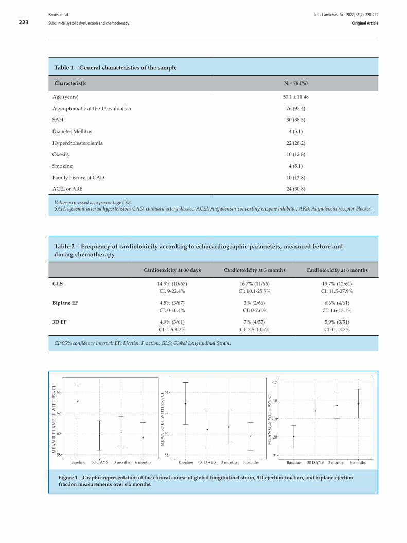
Table 1 – General characteristics of the sample
Characteristic N = 78 (%)
Age (years) 50.1 ± 11.48
Asymptomatic at the 1st evaluation 76 (97.4)
SAH 30 (38.5)
Diabetes Mellitus 4 (5.1)
Hypercholesterolemia 22 (28.2)
Obesity 10 (12.8)
Smoking 4 (5.1)
Family history of CAD 10 (12.8)
ACEI or ARB 24 (30.8)
Values expressed as a percentage (%). SAH: systemic arterial hypertension; CAD: coronary artery disease; ACEI: Angiotensin-converting enzyme inhibitor; ARB: Angiotensin receptor blocker.
Table 2 – Frequency of cardiotoxicity according to echocardiographic parameters, measured before and during chemotherapy
Cardiotoxicity at 30 days Cardiotoxicity at 3 months Cardiotoxicity at 6 months
GLS 14.9% (10/67) CI: 9-22.4%
16.7% (11/66)CI: 10.1-25.8%
19.7% (12/61)CI: 11.5-27.9%
Biplane EF 4.5% (3/67)CI: 0-10.4%
3% (2/66)CI: 0-7.6%
6.6% (4/61)CI: 1.6-13.1%
3D EF 4.9% (3/61)CI: 1.6-8.2%
7% (4/57)CI: 3.5-10.5%
5.9% (3/51)CI: 0-13.7%
CI: 95% confidence interval; EF: Ejection Fraction; GLS: Global Longitudinal Strain.
64 64
-17
-18
-19
-20
-21
62 62
60 60
58 58
BaselineBaseline Baseline
ME
AN
BIP
LA
NE
EF
WIT
H 9
5% C
I
ME
AN
3D
EF
WIT
H 9
5% C
I
ME
AN
GL
S W
ITH
95%
CI
30 DAYS30 DAYS 30 DAYS3 months3 months 3 months6 months6 months 6 months
Figure 1 – Graphic representation of the clinical course of global longitudinal strain, 3D ejection fraction, and biplane ejection fraction measurements over six months.
Int J Cardiovasc Sci. 2022; 35(2), 220-229
223Barroso et al.
Subclinical systolic dysfunction and chemotherapy Original Article
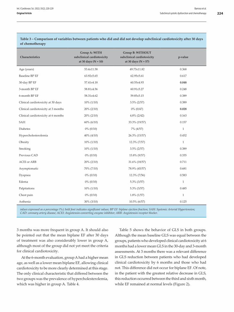
Table 3 – Comparison of variables between patients who did and did not develop subclinical cardiotoxicity after 30 days of chemotherapy
CharacteristicsGroup A: WITH
subclinical cardiotoxicity at 30 days (N = 10)
Group B: WITHOUT subclinical cardiotoxicity
at 30 days (N = 57)p-value
Age (years) 53.4±11.58 49.73±11.82 0.368
Baseline BP EF 63.92±5.65 62.95±5.61 0.617
30-day BP EF 57.41±4.18 60.55±4.93 0.048
3-month BP EF 58.81±4.56 60.91±5.27 0.248
6-month BP EF 58.31±4.62 59.85±5.15 0.389
Clinical cardiotoxicity at 30 days 10% (1/10) 3.5% (2/57) 0.389
Clinical cardiotoxicity at 3 months 20% (2/10) 0% (0/47) 0.028
Clinical cardiotoxicity at 6 months 20% (2/10) 4.8% (2/42) 0.163
SAH 60% (6/10) 33.3% (19/57) 0.157
Diabetes 0% (0/10) 7% (4/57) 1
Hypercholesterolemia 40% (4/10) 26.3% (15/57) 0.452
Obesity 10% (1/10) 12.3% (7/57) 1
Smoking 10% (1/10) 3.5% (2/57) 0.389
Previous CAD 0% (0/10) 15.8% (9/57) 0.335
ACEI or ARB 20% (2/10) 31.6% (18/57) 0.711
Asymptomatic 70% (7/10) 78.9% (45/57) 0.681
Dyspnea 0% (0/10) 12.3% (7/56) 0.583
Edema 0% (0/10) 5.3% (3/57) 1
Palpitations 10% (1/10) 5.3% (3/57) 0.485
Chest pain 0% (0/10) 1.8% (1/57) 1
Asthenia 30% (3/10) 10.5% (6/57) 0.125
values expressed as a percentage (%); bold font indicates significant values; BP EF: biplane ejection fraction; SAH: Systemic Arterial Hypertension; CAD: coronary artery disease; ACEI: Angiotensin-converting enzyme inhibitor; ARB: Angiotensin receptor blocker.
3 months was more frequent in group A. It should also be pointed out that the mean biplane EF after 30 days of treatment was also considerably lower in group A, although most of the group did not yet meet the criteria for clinical cardiotoxicity.
At the 6-month evaluation, group A had a higher mean age, as well as a lower mean biplane EF, allowing clinical cardiotoxicity to be more clearly determined at this stage. The only clinical characteristic that differed between the two groups was the prevalence of hypercholesterolemia, which was higher in group A. Table 4.
Table 5 shows the behavior of GLS in both groups. Although the mean baseline GLS was equal between the groups, patients who developed clinical cardiotoxicity at 6 months had a lower mean GLS in the 30-day and 3-month assessments. At 3 months there was a relevant difference in GLS reduction between patients who had developed clinical cardiotoxicity by 6 months and those who had not. This difference did not occur for biplane EF. Of note, in the patient with the greatest relative decrease in GLS, this reduction occurred between the third and sixth month, while EF remained at normal levels (Figure 2).
Int J Cardiovasc Sci. 2022; 35(2), 220-229
224Barroso et al.
Subclinical systolic dysfunction and chemotherapyOriginal Article
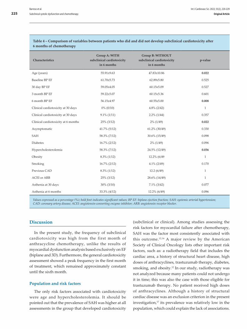
Table 4 – Comparison of variables between patients who did and did not develop subclinical cardiotoxicity after 6 months of chemotherapy
CharacteristicsGroup A: WITH
subclinical cardiotoxicity in 6 months
Group B: WITHOUT subclinical cardiotoxicity
in 6 months p-value
Age (years) 55.91±9.63 47.83±10.86 0.022
Baseline BP EF 61.70±5.73 62.89±5.80 0.525
30 day BP EF 59.05±4.05 60.15±5.09 0.527
3 month BP EF 59.22±5.07 60.15±5.36 0.601
6 month BP EF 56.15±4.97 60.55±5.00 0.008
Clinical cardiotoxicity at 30 days 0% (0/10) 4.8% (2/42) 1
Clinical cardiotoxicity at 30 days 9.1% (1/11) 2.2% (1/44) 0.357
Clinical cardiotoxicity at 6 months 25% (3/12) 2% (1/49) 0.022
Asymptomatic 41.7% (5/12) 61.2% (30/49) 0.330
SAH 58.3% (7/12) 30.6% (15/49) 0.098
Diabetes 16.7% (2/12) 2% (1/49) 0.096
Hypercholesterolemia 58.3% (7/12) 24.5% (12/49) 0.036
Obesity 8.3% (1/12) 12.2% (6/49 1
Smoking 16.7% (2/12) 4.1% (2/49) 0.170
Previous CAD 8.3% (1/12) 12.2 (6/49) 1
ACEI or ARB 25% (3/12) 28.6% (14/49) 1
Asthenia at 30 days 30% (3/10) 7.1% (3/42) 0.077
Asthenia at 6 months 33.3% (4/12) 12.2% (6/49) 0.096
Values expressed as a percentage (%); bold font indicates significant values; BP EF: biplane ejection fraction; SAH: systemic arterial hypertension; CAD: coronary artery disease; ACEI: angiotensin-converting enzyme inhibitor; ARB: angiotensin receptor blocker.
Discussion
In the present study, the frequency of subclinical cardiotoxicity was high from the first month of anthracycline chemotherapy, unlike the results of myocardial dysfunction analysis based exclusively on EF (biplane and 3D). Furthermore, the general cardiotoxicity assessment showed a peak frequency in the first month of treatment, which remained approximately constant until the sixth month.
Population and risk factors
The only risk factors associated with cardiotoxicity were age and hypercholesterolemia. It should be pointed out that the prevalence of SAH was higher at all assessments in the group that developed cardiotoxicity
(subclinical or clinical). Among studies assessing the risk factors for myocardial failure after chemotherapy, SAH was the factor most consistently associated with this outcome.12,16 A major review by the American Society of Clinical Oncology lists other important risk factors, such as: a radiotherapy field that includes the cardiac area, a history of structural heart disease, high doses of anthracyclines, trastuzumab therapy, diabetes, smoking, and obesity.12 In our study, radiotherapy was not analyzed because many patients could not undergo it in time; this was also the case with those eligible for trastuzumab therapy. No patient received high doses of anthracyclines. Although a history of structural cardiac disease was an exclusion criterion in the present investigation,17 its prevalence was relatively low in the population, which could explain the lack of associations.
Int J Cardiovasc Sci. 2022; 35(2), 220-229
225Barroso et al.
Subclinical systolic dysfunction and chemotherapy Original Article
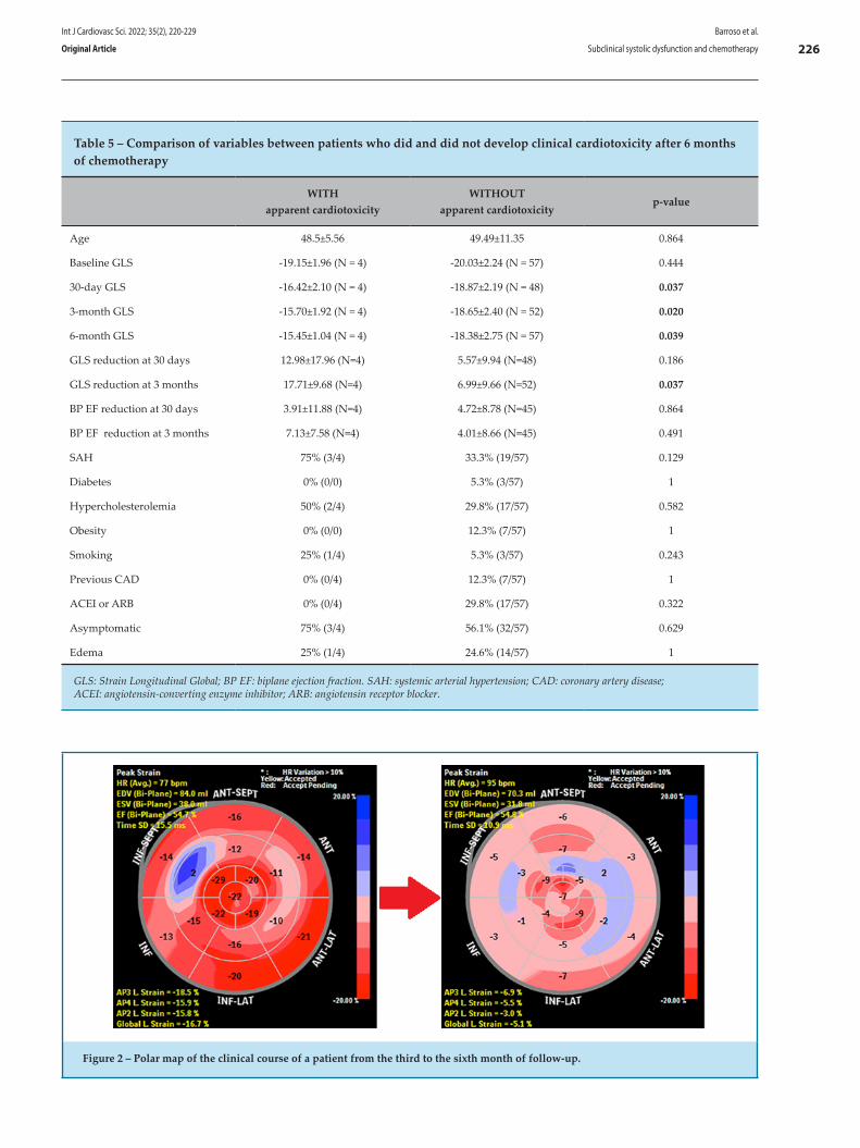
Table 5 – Comparison of variables between patients who did and did not develop clinical cardiotoxicity after 6 months of chemotherapy
WITH apparent cardiotoxicity
WITHOUT apparent cardiotoxicity
p-value
Age 48.5±5.56 49.49±11.35 0.864
Baseline GLS -19.15±1.96 (N = 4) -20.03±2.24 (N = 57) 0.444
30-day GLS -16.42±2.10 (N = 4) -18.87±2.19 (N = 48) 0.037
3-month GLS -15.70±1.92 (N = 4) -18.65±2.40 (N = 52) 0.020
6-month GLS -15.45±1.04 (N = 4) -18.38±2.75 (N = 57) 0.039
GLS reduction at 30 days 12.98±17.96 (N=4) 5.57±9.94 (N=48) 0.186
GLS reduction at 3 months 17.71±9.68 (N=4) 6.99±9.66 (N=52) 0.037
BP EF reduction at 30 days 3.91±11.88 (N=4) 4.72±8.78 (N=45) 0.864
BP EF reduction at 3 months 7.13±7.58 (N=4) 4.01±8.66 (N=45) 0.491
SAH 75% (3/4) 33.3% (19/57) 0.129
Diabetes 0% (0/0) 5.3% (3/57) 1
Hypercholesterolemia 50% (2/4) 29.8% (17/57) 0.582
Obesity 0% (0/0) 12.3% (7/57) 1
Smoking 25% (1/4) 5.3% (3/57) 0.243
Previous CAD 0% (0/4) 12.3% (7/57) 1
ACEI or ARB 0% (0/4) 29.8% (17/57) 0.322
Asymptomatic 75% (3/4) 56.1% (32/57) 0.629
Edema 25% (1/4) 24.6% (14/57) 1
GLS: Strain Longitudinal Global; BP EF: biplane ejection fraction. SAH: systemic arterial hypertension; CAD: coronary artery disease; ACEI: angiotensin-converting enzyme inhibitor; ARB: angiotensin receptor blocker.
Figure 2 – Polar map of the clinical course of a patient from the third to the sixth month of follow-up.
Int J Cardiovasc Sci. 2022; 35(2), 220-229
226Barroso et al.
Subclinical systolic dysfunction and chemotherapyOriginal Article
A further analysis involved the use of angiotensin-converting enzyme inhibitors or angiotensin-receptor blockers, drugs known to provide protection against early decline in global LV function.18 Interestingly, in this study, baseline treatment with angiotensin-converting enzyme inhibitors or angiotensin-receptor blockers did not decrease the occurrence of cardiotoxicity. As shown by Laufer-Perl et al.,19 this is due to the fact that the treatment was not administered to prevent LV dysfunction, but as a therapy for SAH, which would put these patients at greater risk of LV dysfunction.
Definitions and incidence of cardiotoxicity
Several definitions of cardiotoxicity have been developed, making it difficult to standardize and estimate its incidence as an outcome. The present study was based on ASE/EACVI guidelines.9
The incidence of clinical cardiotoxicity in our sample was relatively low, which can be explained, in part, by the fact that our definition was more specific than that of most publications.20,21 Another possible explanation is that our study only included patients treated with anthracycline, while other studies have investigated associations with multiple cardiotoxic drugs.10,21 Such hypotheses are corroborated by the results of studies with designs similar to ours, which obtained similar results regarding the incidence of clinical cardiotoxicity.22,23
In our study, echocardiographic parameters of systolic function during chemotherapy behaved similarly to those of other studies that performed this analysis. The early drop (by 30 days) in GLS and EF (3D and biplane) and their subsequent lower mean values has also been demonstrated in other cohorts that evaluated the cardiotoxicity of chemotherapy.19,21,22
GLS and Subclinical Cardiotoxicity
As for the occurrence of subclinical cardiotoxicity, few studies have evaluated it as a categorical variable (mostly investigating only the behavior of the GLS values themselves) and fewer still have done so according to the ASE/EACVI definition. The frequency of subclinical cardiotoxicity was relatively high in this study, being 15.2% 30 days after the start of chemotherapy and unaccompanied by a significant reduction in EF (biplane or 3D). This agrees with the results of other studies, such as Laufer-Perl et al.,19 and Santoro et al.,22 in which the mean frequency of subclinical cardiotoxicity was approximately 20% at the end of 3 and 12 months of follow-up, respectively.
Another pertinent analysis concerns the relative reduction of GLS and EF. When divided into groups, it was observed that the GLS reduction after 3 months compared to baseline was greater in those who developed clinical cardiotoxicity at 6 months. However, EF did not also drop during the same period, which suggests that this echocardiographic parameter has a lower sensitivity. Similar results were also found in a cohort of 49 Brazilian patients.24 Moreover, between patients with and without clinical cardiotoxicity at 6 months, GLS differed significantly 30 days after the beginning of the chemotherapy and continued to do so in all subsequent assessments, which reinforces the fact that GLS changes are early when myocardial aggression occurs.6
In general, this behavior can be explained by the fact that GLS reductions precede EF reductions, making the former a more sensitive method.6,7,9 Several factors explain why GLS is a better early indicator of cardiotoxicity. In dysfunction related to cancer therapy, certain segments of the myocardium may be more affected than others, leading to early changes in GLS. The unaffected areas of the myocardium can then compensate for the damaged segments, thus preserving LVEF.21 However, GLS, which assesses myocardial deformation, can be measured more accurately.11
Study limitations
One limitation of this study was the short and non-homogeneous follow-up. Thus, an objective assessment of long-term LVEF behavior in relation to the occurrence of subclinical cardiotoxicity was not possible. Furthermore, the low number of patients who appeared for all assessments reduced the statistical power of the results. However, such scenarios are inherent to the study population. Another limitation was the fact that the study was conducted by a single researcher. Nevertheless, the Cronbach's α analysis indicated high internal reliability, which minimized the possibility of measurement bias.
Conclusion
Subclinical cardiotoxicity had a high frequency and began early in this sample. It was associated with a significant drop in EF during the course of treatment but was not associated with known risk factors. The value of assessing GLS at early stages of exposure to anthracyclines was also apparent, which is in line with the ASE/EACVI definition of subclinical cardiotoxicity. It is expected that identifying subclinical cardiotoxicity
Int J Cardiovasc Sci. 2022; 35(2), 220-229
227Barroso et al.
Subclinical systolic dysfunction and chemotherapy Original Article
will be of great value in daily clinical practice, identifying patients at higher risk of overt cardiotoxicity.
Author contributions
Conception and design of the research: Barroso GMHM, Teles JCOC. Acquisition of data: Barroso GMHM, Teles JCOC, Silva PVJ, Ferreira KO, Assis RJF, Alves MFS, Aragão VAS, Fonsêca KYS, Aquino MM. Analysis and interpretation of the data: Teles JCOC, Melo EV, Oliveira, JLM. Statistical analysis: Teles JCOC, Melo EV. Writing of the manuscript: Barroso GMHM, Teles JCOC. Critical revision of the manuscript for intellectual content: Sousa ACS, Oliveira, JLM.
Potential Conflict of InterestNo potential conflict of interest relevant to this article
was reported.
Sources of FundingThis study was funded by own incentive.
Study AssociationThis article is part of the thesis of master submitted
by Geanne Maria Holanda de Menezes Barroso, from Universidade Federal de Sergipe.
Ethics approval and consent to participateThis study was approved by the Ethics Committee
of the Universidade Federal de Sergipe under the protocol number 87240718.9.0000.5546. All the procedures in this study were in accordance with the 1975 Helsinki Declaration, updated in 2013. Informed consent was obtained from all participants included in the study.
1. Zamorano JL, Lancellotti P, Rodriguez Muñoz D, Aboyans V, Asteggiano R, Galderisi M, et al. 2016 ESC Position Paper on cancer treatments and cardiovascular toxicity developed under the auspices of the ESC Committee for Practice Guidelines. Eur Heart J. 2016;37(36):2768–801. doi: 10.1093/eurheartl/ehw211.
2. Chung WB, Youn HJ. Pathophysiology and preventive strategies of anthracycline-induced cardiotoxicity. Korean J Intern Med. 2016;31(4):625–33. doi:10.3904/kjm.2017.158.
3. Oliveira GH, Hardaway BW, Kucheryavaya AY, Stehlik J, Edwards LB, Taylor DO. Characteristics and survival of patients with chemotherapy-induced cardiomyopathy undergoing heart transplantation. J Heart Lung Transplant. 2012;31(8):805–10. doi: 10.1016/j.healun.2012.03.018.
4. Zhang S, Liu X, Bawa-Khalfe T, Lu LS, Lyu YL, Liu LF, et al. Identification of the molecular basis of doxorubicin-induced cardiotoxicity. Nat Med. 2012;18(11):1639–42. doi:10.1038/nm.2919.
5. Santoro C, Arpino G, Esposito R, Lembo M, Paciolla I, Cardalesi C, et al. 2D and 3D strain for detection of subclinical anthracycline cardiotoxicity in breast cancer patients: a balance with feasibility. Eur Heart J Cardiovasc Imaging. 2017;18(8):930–6.doi: 10.1093/ehjci/jex033.
6. Thavendiranathan P, Poulin F, Lim KD, Plana JC, Woo A, Marwick TH. Use of myocardial strain imaging by echocardiography for the early detection of cardiotoxicity in patients during and after cancer chemotherapy: A systematic review. J Am Coll Cardiol. 2014;63(25 PART A):2751–68. doi: 10.1016/j.jacc.2014.01.073.
7. Lorenzini C, Lamberti C, Aquilina M, Rocca A, Cortesi P, Corsi C. Reliability of Left Ventricular Ejection Fraction from Three-Dimensional Echocardiography for Cardiotoxicity Onset Detection in Patients with Breast Cancer. J Am Soc Echocardiogr. 2017;30(11):1103–10. doiu: 10.1016/j.echo.2017.06.025.
8. Gorcsan J, Tanaka H. Echocardiographic assessment of myocardial strain. J Am Coll Cardiol. 2011;58(14):1401–13. doi: 10.1016/j.jacc.2011.06.038.
9. Plana JC, Galderisi M, Barac A, Ewer MS, Ky B, Scherrer-Crosbie M, et al. Expert consensus for multimodality imaging evaluation of adult patients during and after cancer therapy: A report from the American Society
of Echocardiography and the European Association of Cardiovascular Imaging. Eur Heart J Cardiovasc Imaging. 2014;15(10):1063–93. doi:10.1093/ehjci/jeu192.
10. Sawaya H, Sebag IA, Plana JC, Januzzi JL, Ky B, Tan TC, et al. Assessment of echocardiography and biomarkers for the extended prediction of cardiotoxicity in patients treated with anthracyclines, taxanes, and trastuzumab. Circ Cardiovasc Imaging. 2012;5(5):596–603. doi: 10.1161/CRCIMAGING.112.973321.
11. Cardinale D, Colombo A, Bacchiani G, Tedeschi I, Meroni CA, Veglia F, et al. Early detection of anthracycline cardiotoxicity and improvement with heart failure therapy. Circulation. 2015;131(22):1981–8. doi:10.1161/CIRCULATIONAHA.114.013777.
12. Armenian SH, Lacchetti C, Barac A, Carver J, Constine LS, Denduluri N, et al. Prevention and monitoring of cardiac dysfunction in survivors of adult cancers: American society of clinical oncology clinical practice guideline. J Clin Oncol. 2017;35(8):893–911. doi:10.1200/JCO.2016.70.5400.
13. King A, Thambyrajah J, Leng E, Stewart MJ. Global longitudinal strain: A useful everyday measurement? Echo Res Pract. 2016;3(3):85–93. doi 10.1530/ERP-16-0022.
14. Tan TC, Bouras S, Sawaya H, Sebag IA, Cohen V, Picard MH, et al. Time trends of left ventricular ejection fraction and myocardial deformation indices in a cohort of women with breast cancer treated with anthracyclines, taxanes, and trastuzumab. J Am Soc Echocardiogr. 2015;28(5):509–14. doi: 10.1016/j.echo.2015.02.001.
15. Lang RM, Badano LP, Mor-Avi V, Afilalo J, Armstrong A, Ernande L, et al. Recommendations for cardiac chamber quantification by echocardiography in adults: An update from the American society of echocardiography and the European association of cardiovascular imaging. Eur Heart J Cardiovasc Imaging. 2015;16(3):233–70. doi:10.1093/ehji/jev014.
16. Armenian SH, Xu L, Ky B, Sun C, Farol LT, Pal SK, et al. Cardiovascular disease among survivors of adult-onset cancer: A community-based retrospective cohort study. J Clin Oncol. 2016;34(10):1122–30. doi:10.1200/JCO.2015.64.0409.
References
Int J Cardiovasc Sci. 2022; 35(2), 220-229
228Barroso et al.
Subclinical systolic dysfunction and chemotherapyOriginal Article
This is an open-access article distributed under the terms of the Creative Commons Attribution License
17. Swain SM, Whaley FS, Ewer MS. Congestive heart failure in patients treated with doxorubicin: A retrospective analysis of three trials. Cancer. 2003;97(11):2869–79. doi:10.1002/cncr.11407.
18. Gulati G, Heck SL, Ree AH, Hoffmann P, Schulz-Menger J, Fagerland MW, et al. Prevention of cardiac dysfunction during adjuvant breast cancer therapy (PRADA): A 2 × 2 factorial, randomized, placebo-controlled, double-blind clinical trial of candesartan and metoprolol. Eur Heart J. 2016;37(21):1671–80. doi: 10.1093/eurheartj/ehw022.
19. Laufer-Perl M, Derakhshesh M, Milwidsky A, Mor L, Ravid D, Amrami N, et al. Usefulness of Global Longitudinal Strain for Early Identification of Subclinical Left Ventricular Dysfunction in Patients With Active Cancer. Am J Cardiol. 2018;122(10):1784–9.doi:10>1016/j.amjcard.2018.08.019.
20. Khouri MG, Douglas PS, Mackey JR, Martin M, Scott JM, Scherrer-Crosbie M, et al. Cancer therapy-induced cardiac toxicity in early breast cancer addressing the unresolved issues. Circulation. 2012;126(23):2749–63. doi:10.1161/CIRCULATIONAHA.112.100560.
21. Sawaya H, Sebag IA, Plana JC, Januzzi JL, Ky B, Cohen V, et al. Early detection and prediction of cardiotoxicity in chemotherapy-treated patients. Am J Cardiol. 2011;107(9):1375–80. doi:10.1016/j.amjcard.2011.01.006.
22. Santoro C, Esposito R, Lembo M, Sorrentino R, De Santo I, Luciano F, et al. Strain-oriented strategy for guiding cardioprotection initiation of breast cancer patients experiencing cardiac dysfunction. Eur Heart J Cardiovasc Imaging. 2019;20(12):1345–52. doi:10.1093/ehjci/jey108.
23. Charbonnel C, Convers-Domart R, Rigaudeau S, Taksin AL, Baron N, Lambert J, et al. Assessment of global longitudinal strain at lowdose anthracycline-based chemotherapy, for the prediction of subsequent cardiotoxicity. Eur Heart J Cardiovasc Imaging. 2017;18(4):392–401. doi:10.1093/ehjci/jew223.
24. Gripp E A, De Oliveira GE, Feijó LA, Garcia MI, Xavier SS, De Sousa AS. Global longitudinal strain accuracy for cardiotoxicity prediction in a cohort of breast cancer patients during anthracycline and/or trastuzumab treatment. Arq Bras Cardiol. 2018;110(2):140–50.doi:10.5935/abc.20180021.
Int J Cardiovasc Sci. 2022; 35(2), 220-229
229Barroso et al.
Subclinical systolic dysfunction and chemotherapy Original Article
Introduction
Cardiovascular diseases (CVDs) are the main cause of morbidity and mortality worldwide, including in Brazil, where approximately 395,700 deaths (30% of total deaths) registered in 2018 were due to CVDs.1 Considering the negative impact on individual’s health, health systems,
and the economy, monitoring CVD risk factors become imperative for both CVD prevention and control.2 Moreover, many risk factors for CVDs are also risk factors for other non-communicable diseases (e.g., cancer), and are associated with worse outcomes and increased risk of death in patients with infectious diseases (e.g., SARS-CoV-2, responsible for the COVID-19 pandemic).3,4
Int J Cardiovasc Sci. 2022; 35(2), 230-242
230
ORIGINAL ARTICLE
Overview of Cardiovascular Disease Risk Factors in Adults in São Paulo, Brazil: Prevalence and Associated Factors in 2008 and 2015Jaqueline L. Pereira,1 Michelle A. de Castro,2 Jean M. R. S. Leite,1 Marcelo M. Rogero,1 Flavia M. Sarti,3 Chester Luís Galvão César,4 Moisés Goldbaum,5 Regina M. Fisberg1
Department of Nutrition, School of Public Health, University of São Paulo,1 São Paulo, SP - Brazil.School Feeding Coordination, Secretariat of Education of São Paulo,2 São Paulo, SP - Brazil.School of Arts, Sciences and Humanities, University of Sao Paulo,3 São Paulo, SP - Brazil.Department of Epidemiology, School of Public Health, University of São Paulo,4 São Paulo, SP - Brazil.Department of Preventive Medicine, Medical School, University of São Paulo,5 São Paulo, SP - Brazil.
DOI: https://doi.org/10.36660/ijcs.20210076
Mailing Address: Regina Mara FisbergDepartment of Nutrition, School of Public Health, University of São Paulo.Avenida Dr. Arnaldo, 715. Postal Code: 01246-904, Cerqueira Cesar, São Paulo, SP – Brazil. Email: [email protected]
Manuscript received on March 19, 2021; reviewed on July 19, 2021; accepted on September 01, 2021.
Abstract
Background: Cardiovascular diseases (CVDs) are the main cause of morbidity and mortality in Brazil.
Objective: To provide population-based data on prevalence and factors associated with CVD risk factors.
Methods: Individuals aged ≥20 years from two editions of the cross-sectional Health Survey of São Paulo focusing on Nutrition (ISA-Nutrition), performed in Sao Paulo city in 2008 (n=590) and 2015 (n=610), were evaluated for: obesity, central obesity, waist/height ratio, high blood pressure (HBP), dyslipidemia, diabetes, and number of CVD risk factors ≥3. Prevalence was estimated according to complex survey procedures. Factors associated with cardiovascular risk factors were assessed using logistic regression, with statistical significance of p<0.05.
Results: Obesity and older age were associated with higher odds of all cardiovascular risk factors investigated, except for dyslipidemia. HBP was positively associated with being Black/Brown and negatively associated with being physicaly active in leisure time. Women were more likely to have increased adiposity indicators and three or more cardiovascular risk factors than men. Those with higher education had lower chances of having diabetes, HBP and dyslipidemia, and those with higher income had higher chances of having three or more risk factors. Former smokers had higher odds of diabetes, obesity, and high waist/height ratio, and smokers had higher odds of high non-HDL cholesterol levels. From 2008 to 2015, there was an increase (p<0.001) in the prevalence of diabetes (6.9% to 17.3%), HBP (31.9% to 41.8%), dyslipidemia (51.3% to 67.6%), and number of CVD risk factors ≥3 (18.9% to 34.1%).
Conclusion: This study shows increasing prevalence of CVD risk factors in adult population in Sao Paulo and may support the definition of target groups and priority actions on CVD prevention and treatment.
Keywords: Cardiovascular Diseases/epidemiology; Risk Factors; Urban Health Services; Prevalence; Mortality; Morbidity
intake was assessed by two 24-hour recalls. The 2008’s and 2015’s editions comprised 750 individuals (590 aged ≥20 years) and 901 individuals (610 aged ≥20 years), respectively. For this analysis, only individuals aged at least 20 years were evaluated (n=1200).
Anthropometric and cardiometabolic risk factors
Detailed protocols for laboratory, blood pressure and anthropometric measurements were published elsewhere. 10 Body weight , height , and waist circumference were measured in triplicate, and the average of three measures was calculated for analyses. Central obesity was defined as waist circumference ≥88 cm for women and ≥102 cm for men.11 Waist-to-height ratio was considered elevated when ≥0.52 for adult men, ≥0.54 for adult women, and ≥0.55 for older adults.12 According to body mass index (BMI=weight (kg)/height (m)2), participants were categorized as: normal weight, overweight (adults: 25≥BMI>30 kg/m2; older adults: 28<BMI<30 kg/m2),11,13 or obese (BMI≥30 kg/m2).13 High blood pressure was defined as elevated systolic (≥140 mmHg) or diastolic (≥90 mmHg) blood pressure, or use of antihypertensive drugs.14
Diabetes was defined as fasting plasma glucose ≥126 mg/dL, use of oral hypoglycemic drugs or insulin therapy.12 Variables related to dyslipidemia were isolated hypercholesterolemia (low-density lipoprotein cholesterol [LDL-C] ≥160 mg/dL), isolated hypertriglyceridemia (triglycerides ≥160 mg/dL), mixed hyperlipidemia (LDL-C ≥160 mg/dL and triglycerides ≥160 mg/dL), low-HDL (high-density lipoprotein cholesterol [HDL-C] ≤40 mg/dL for men or HDL-C ≤50 mg/dL for women), high non-HDL cholesterol (≥160 mg/dL), and dyslipidemia (elevated LDL-C, elevated triglycerides, low HDL-C, or drug treatment for dyslipidemia).6
Participants were categorized according to the number of CVD risk factors present (less than 3 or equal to or more than 3), that is, diabetes, or high fasting plasma glucose (≥100 mg/dL), or insulin resistance (homeostatic assessment of insulin resistance [HOMA-IR = fasting glucose (mg/dL) x fasting insulin (μU/L)/405) ≥2.71];12 high blood pressure; dyslipidemia; and obesity.
Statistical Analysis
The variables analyzed were: sex (male/female); age (adults: 20-59 years/older adults: ≥60 years); self-
Several factors can be related to the genesis and prognosis of CVDs.3,5,6 Some of them are not modifiable, such as aging, hereditary factors, and sex, while others are related to behavioral factors, such as unhealthy diet, physical inactivity, stress, tobacco use, and harmful use of alcohol. Also, some factors are related to underlying social determinants, such as globalization, urbanization, poverty, air pollution, and access to health services. To some extent, all of them are related to intermediate risk factors that can be evaluated and adequately monitored in primary health care, (e.g. elevated blood glucose levels, high blood pressure, dyslipidemia, overweight and obesity), and indicate an increased risk of developing heart failure, stroke, heart attack, or other complications.3
In the last decades, São Paulo, the largest city of Brazil, has witnessed substantial changes in the lifestyle of its inhabitants and socio-demographic profile, many of them associated with an increase in cardiovascular risk factors.7 This study aimed to evaluate intermediate CVD risk factors, by analysis of population-based data from Sao Paulo, which may support primary and secondary prevention through evidence-based planning of health policies.
Methods
Population and study design
The study assessed data from two cohorts of the cross-sectional Health Survey of São Paulo focusing on Nutrition (ISA-Nutrition study), a sub-sample of the Health Survey of São Paulo (ISA-Capital), carried out in 2008 and 2015. ISA-Capital is a population-based survey aiming to evaluate population health status and use of health services, using a probabilistic sample of individuals aged 12 years or more living in households in the urban area of São Paulo city. Sociodemographic data, and information on morbidity, use of health services, and lifestyle were obtained using a structured questionnaire administered by trained interviewers in the households, to 3,271 individuals (2,086 aged ≥20 years) in 2008, and 4,024 (3,165 aged ≥20 years) in 2015. The samples of both surveys were independent of each other. Details of both editions were previously described.8-10
The ISA-Nutrition aimed at evaluating lifestyle-related CVD risk factors in the population of São Paulo.10 Anthropometric data, blood pressure measurements, and blood samples were obtained by trained nurses during a second visit to the participant’s household. Dietary
Int J Cardiovasc Sci. 2022; 35(2), 230-242
231Pereira et al.
Cardiovascular disease risk factors in São Paulo Original Article
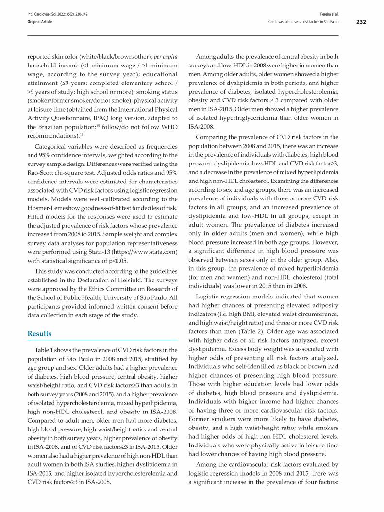
reported skin color (white/black/brown/other); per capita household income (<1 minimum wage / ≥1 minimum wage, according to the survey year); educational attainment (≤9 years: completed elementary school / >9 years of study: high school or more); smoking status (smoker/former smoker/do not smoke); physical activity at leisure time (obtained from the International Physical Activity Questionnaire, IPAQ long version, adapted to the Brazilian population:15 follow/do not follow WHO recommendations).16
Categorical variables were described as frequencies and 95% confidence intervals, weighted according to the survey sample design. Differences were verified using the Rao-Scott chi-square test. Adjusted odds ratios and 95% confidence intervals were estimated for characteristics associated with CVD risk factors using logistic regression models. Models were well-calibrated according to the Hosmer-Lemeshow goodness-of-fit test for deciles of risk. Fitted models for the responses were used to estimate the adjusted prevalence of risk factors whose prevalence increased from 2008 to 2015. Sample weight and complex survey data analyses for population representativeness were performed using Stata-13 (https://www.stata.com) with statistical significance of p<0.05.
This study was conducted according to the guidelines established in the Declaration of Helsinki. The surveys were approved by the Ethics Committee on Research of the School of Public Health, University of São Paulo. All participants provided informed written consent before data collection in each stage of the study.
Results
Table 1 shows the prevalence of CVD risk factors in the population of São Paulo in 2008 and 2015, stratified by age group and sex. Older adults had a higher prevalence of diabetes, high blood pressure, central obesity, higher waist/height ratio, and CVD risk factors≥3 than adults in both survey years (2008 and 2015), and a higher prevalence of isolated hypercholesterolemia, mixed hyperlipidemia, high non-HDL cholesterol, and obesity in ISA-2008. Compared to adult men, older men had more diabetes, high blood pressure, high waist/height ratio, and central obesity in both survey years, higher prevalence of obesity in ISA-2008, and of CVD risk factors≥3 in ISA-2015. Older women also had a higher prevalence of high non-HDL than adult women in both ISA studies, higher dyslipidemia in ISA-2015, and higher isolated hypercholesterolemia and CVD risk factors≥3 in ISA-2008.
Among adults, the prevalence of central obesity in both surveys and low-HDL in 2008 were higher in women than men. Among older adults, older women showed a higher prevalence of dyslipidemia in both periods, and higher prevalence of diabetes, isolated hypercholesterolemia, obesity and CVD risk factors ≥ 3 compared with older men in ISA-2015. Older men showed a higher prevalence of isolated hypertriglyceridemia than older women in ISA-2008.
Comparing the prevalence of CVD risk factors in the population between 2008 and 2015, there was an increase in the prevalence of individuals with diabetes, high blood pressure, dyslipidemia, low-HDL and CVD risk factor≥3, and a decrease in the prevalence of mixed hyperlipidemia and high non-HDL cholesterol. Examining the differences according to sex and age groups, there was an increased prevalence of individuals with three or more CVD risk factors in all groups, and an increased prevalence of dyslipidemia and low-HDL in all groups, except in adult women. The prevalence of diabetes increased only in older adults (men and women), while high blood pressure increased in both age groups. However, a significant difference in high blood pressure was observed between sexes only in the older group. Also, in this group, the prevalence of mixed hyperlipidemia (for men and women) and non-HDL cholesterol (total individuals) was lower in 2015 than in 2008.
Logistic regression models indicated that women had higher chances of presenting elevated adiposity indicators (i.e. high BMI, elevated waist circumference, and high waist/height ratio) and three or more CVD risk factors than men (Table 2). Older age was associated with higher odds of all risk factors analyzed, except dyslipidemia. Excess body weight was associated with higher odds of presenting all risk factors analyzed. Individuals who self-identified as black or brown had higher chances of presenting high blood pressure. Those with higher education levels had lower odds of diabetes, high blood pressure and dyslipidemia. Individuals with higher income had higher chances of having three or more cardiovascular risk factors. Former smokers were more likely to have diabetes, obesity, and a high waist/height ratio; while smokers had higher odds of high non-HDL cholesterol levels. Individuals who were physically active in leisure time had lower chances of having high blood pressure.
Among the cardiovascular risk factors evaluated by logistic regression models in 2008 and 2015, there was a significant increase in the prevalence of four factors:
Int J Cardiovasc Sci. 2022; 35(2), 230-242
232Pereira et al.
Cardiovascular disease risk factors in São PauloOriginal Article
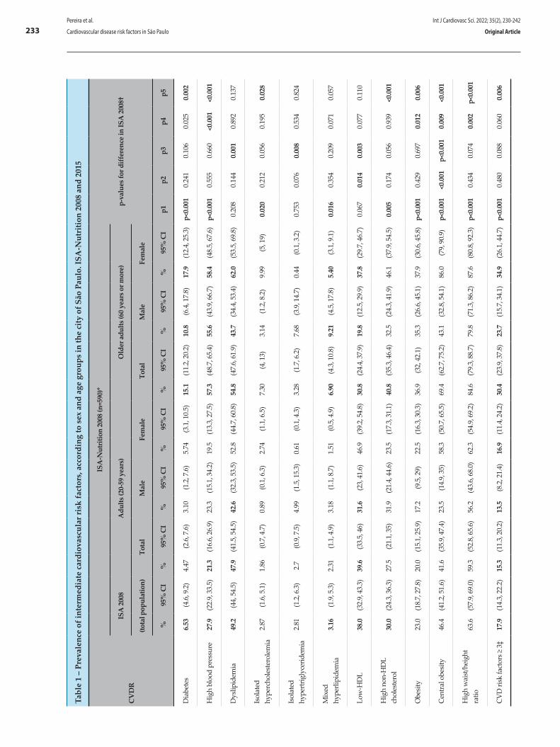
Tabl
e 1
– Pr
eval
ence
of i
nter
med
iate
car
diov
ascu
lar r
isk
fact
ors,
acc
ordi
ng to
sex
and
age
gro
ups
in th
e ci
ty o
f São
Pau
lo. I
SA-N
utri
tion
2008
and
201
5
CV
DR
ISA
-Nut
ritio
n 20
08 (n
=590
)*
p-va
lues
for d
iffer
ence
in IS
A 2
008†
ISA
200
8A
dults
(20-
59 y
ears
)O
lder
adu
lts (6
0 ye
ars o
r mor
e)
(tota
l pop
ulat
ion)
Tota
lM
ale
Fem
ale
Tota
lM
ale
Fem
ale
%95
% C
I%
95%
CI
%95
% C
I%
95%
CI
%95
% C
I%
95%
CI
%95
% C
Ip1
p2p3
p4p5
Dia
bete
s6.
53(4
.6, 9
.2)
4.47
(2.6
, 7.6
)3.
10(1
.2, 7
.6)
5.74
(3.1
, 10.
5)15
.1(1
1.2,
20.
2)10
.8(6
.4, 1
7.8)
17.9
(12.
4, 2
5.3)
p<0.
001
0.24
10.
106
0.02
50.
002
Hig
h bl
ood
pres
sure
27.9
(22.
9, 3
3.5)
21.3
(16.
6, 2
6.9)
23.3
(15.
1, 3
4.2)
19.5
(13.
3, 2
7.5)
57.3
(48.
7, 6
5.4)
55.6
(43.
9, 6
6.7)
58.4
(48.
5, 6
7.6)
p<0.
001
0.55
50.
660
<0.0
01<0
.001
Dys
lipid
emia
49.2
(44,
54.
5)47
.9(4
1.5,
54.
5)42
.6(3
2.3,
53.
5)52
.8(4
4.7,
60.
8)54
.8(4
7.6,
61.
9)43
.7(3
4.4,
53.
4)62
.0(5
3.5,
69.
8)0.
208
0.14
40.
001
0.89
20.
137
Isol
ated
hy
perc
hole
ster
olem
ia2.
87(1
.6, 5
.1)
1.86
(0.7
, 4.7
)0.
89(0
.1, 6
.3)
2.74
(1.1
, 6.5
)7.
30(4
, 13)
3.14
(1.2
, 8.2
)9.
99(5
, 19)
0.02
00.
212
0.05
60.
195
0.02
8
Isol
ated
hy
pert
rigly
cerid
emia
2.81
(1.2
, 6.3
)2.
7(0
.9, 7
.5)
4.99
(1.5
, 15.
3)0.
61(0
.1, 4
.3)
3.28
(1.7
, 6.2
)7.
68(3
.9, 1
4.7)
0.44
(0.1
, 3.2
)0.
753
0.07
60.
008
0.53
40.
824
Mix
ed
hype
rlipi
dem
ia3.
16(1
.9, 5
.3)
2.31
(1.1
, 4.9
)3.
18(1
.1, 8
.7)
1.51
(0.5
, 4.9
)6.
90(4
.3, 1
0.8)
9.21
(4.5
, 17.
8)5.
40(3
.1, 9
.1)
0.01
60.
354
0.20
90.
071
0.05
7
Low
-HD
L38
.0(3
2.9,
43.
3)39
.6(3
3.5,
46)
31.6
(23,
41.
6)46
.9(3
9.2,
54.
8)30
.8(2
4.4,
37.
9)19
.8(1
2.5,
29.
9)37
.8(2
9.7,
46.
7)0.
067
0.01
40.
003
0.07
70.
110
Hig
h no
n-H
DL
chol
este
rol
30.0
(24.
3, 3
6.3)
27.5
(21.
1, 3
5)31
.9(2
1.4,
44.
6)23
.5(1
7.3,
31.
1)40
.8(3
5.3,
46.
4)32
.5(2
4.3,
41.
9)46
.1(3
7.9,
54.
5)0.
005
0.17
40.
056
0.93
9<0
.001
Obe
sity
23.0
(18.
7, 2
7.8)
20.0
(15.
1, 2
5.9)
17.2
(9.5
, 29)
22.5
(16.
3, 3
0.3)
36.9
(32,
42.
1)35
.3(2
6.6,
45.
1)37
.9(3
0.6,
45.
8)p<
0.00
10.
429
0.69
70.
012
0.00
6
Cen
tral
obe
sity
46.4
(41.
2, 5
1.6)
41.6
(35.
9, 4
7.4)
23.5
(14.
9, 3
5)58
.3(5
0.7,
65.
5)69
.4(6
2.7,
75.
2)43
.1(3
2.8,
54.
1)86
.0(7
9, 9
0.9)
p<0.
001
<0.0
01p<
0.00
10.
009
<0.0
01
Hig
h w
aist
/hei
ght
ratio
63.6
(57.
9, 6
9.0)
59.3
(52.
8, 6
5.6)
56.2
(43.
6, 6
8.0)
62.3
(54.
9, 6
9.2)
84.6
(79.
3, 8
8.7)
79.8
(71.
3, 8
6.2)
87.6
(80.
8, 9
2.3)
p<0.
001
0.43
40.
074
0.00
2p<
0.00
1
CV
D ri
sk fa
ctor
s ≥ 3
‡17
.9(1
4.3,
22.
2)15
.3(1
1.3,
20.
2)13
.5(8
.2, 2
1.4)
16.9
(11.
4, 2
4.2)
30.4
(23.
9, 3
7.8)
23.7
(15.
7, 3
4.1)
34.9
(26.
1, 4
4.7)
p<0.
001
0.48
00.
088
0.06
00.
006
Int J Cardiovasc Sci. 2022; 35(2), 230-242
233Pereira et al.
Cardiovascular disease risk factors in São Paulo Original Article
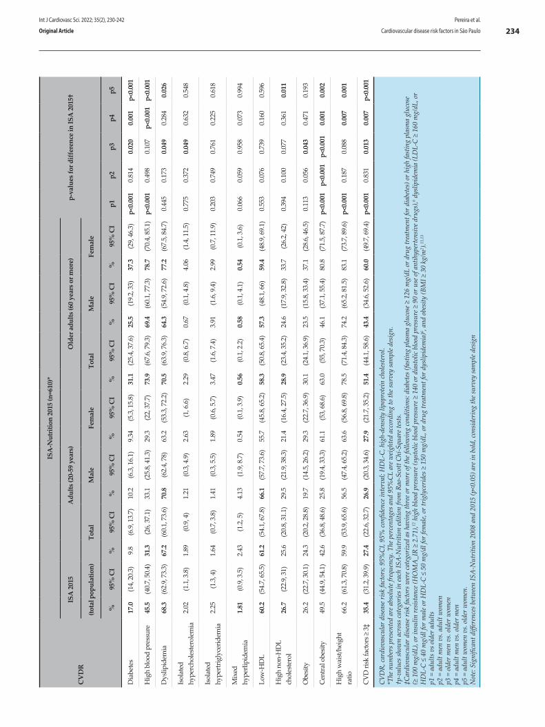
CV
DR
ISA
-Nut
ritio
n 20
15 (n
=610
)*
p-va
lues
for d
iffer
ence
in IS
A 2
015†
ISA
201
5A
dults
(20-
59 y
ears
)O
lder
adu
lts (6
0 ye
ars o
r mor
e)
(tota
l pop
ulat
ion)
Tota
lM
ale
Fem
ale
Tota
lM
ale
Fem
ale
%95
% C
I%
95%
CI
%95
% C
I%
95%
CI
%95
% C
I%
95%
CI
%95
% C
Ip1
p2p3
p4p5
Dia
bete
s17
.0(1
4, 2
0.3)
9.8
(6.9
, 13.
7)10
.2(6
.3, 1
6.1)
9.34
(5.3
, 15.
8)31
.1(2
5.4,
37.
6)25
.5(1
9.2,
33)
37.3
(29,
46.
3)p<
0.00
10.
814
0.02
00.
001
p<0.
001
Hig
h bl
ood
pres
sure
45.5
(40.
7, 5
0.4)
31.3
(26,
37.
1)33
.1(2
5.8,
41.
3)29
.3(2
2, 3
7.7)
73.9
(67.
6, 7
9.3)
69.4
(60.
1, 7
7.3)
78.7
(70.
4, 8
5.1)
p<0.
001
0.49
80.
107
p<0.
001
p<0.
001
Dys
lipid
emia
68.3
(62.
9, 7
3.3)
67.2
(60.
1, 7
3.6)
70.8
(62.
4, 7
8)63
.2(5
3.3,
72.
2)70
.5(6
3.9,
76.
3)64
.3(5
4.9,
72.
6)77
.2(6
7.5,
84.
7)0.
445
0.17
30.
049
0.28
40.
026
Isol
ated
hy
perc
hole
ster
olem
ia2.
02(1
.1, 3
.8)
1.89
(0.9
, 4)
1.21
(0.3
, 4.9
)2.
63(1
, 6.6
)2.
29(0
.8, 6
.7)
0.67
(0.1
, 4.8
)4.
06(1
.4, 1
1.5)
0.77
50.
372
0.04
90.
632
0.54
8
Isol
ated
hy
pert
rigly
cerid
emia
2.25
(1.3
, 4)
1.64
(0.7
, 3.8
)1.
41(0
.3, 5
.5)
1.89
(0.6
, 5.7
)3.
47(1
.6, 7
.4)
3.91
(1.6
, 9.4
)2.
99(0
.7, 1
1.9)
0.20
30.
749
0.76
10.
225
0.61
8
Mix
ed
hype
rlipi
dem
ia1.
81(0
.9, 3
.5)
2.43
(1.2
, 5)
4.13
(1.9
, 8.7
)0.
54(0
.1, 3
.9)
0.56
(0.1
, 2.2
)0.
58(0
.1, 4
.1)
0.54
(0.1
, 3.6
)0.
066
0.05
90.
958
0.07
30.
994
Low
-HD
L60
.2(5
4.7,
65.
5)61
.2(5
4.1,
67.
8)66
.1(5
7.7,
73.
6)55
.7(4
5.8,
65.
2)58
.3(5
0.8,
65.
4)57
.3(4
8.1,
66)
59.4
(48.
9, 6
9.1)
0.55
30.
076
0.73
90.
160
0.59
6
Hig
h no
n-H
DL
chol
este
rol
26.7
(22.
9, 3
1)25
.6(2
0.8,
31.
1)29
.5(2
1.9,
38.
3)21
.4(1
6.4,
27.
5)28
.9(2
3.4,
35.
2)24
.6(1
7.9,
32.
8)33
.7(2
6.2,
42)
0.39
40.
100
0.07
70.
361
0.01
1
Obe
sity
26.2
(22.
7, 3
0.1)
24.3
(20.
2, 2
8.8)
19.7
(14.
5, 2
6.2)
29.3
(22.
7, 3
6.9)
30.1
(24.
1, 3
6.9)
23.5
(15.
8, 3
3.4)
37.1
(28.
6, 4
6.5)
0.11
30.
056
0.04
30.
471
0.19
3
Cen
tral
obe
sity
49.5
(44.
9, 5
4.1)
42.6
(36.
8, 4
8.6)
25.8
(19.
4, 3
3.3)
61.1
(53,
68.
6)63
.0(5
5, 7
0.3)
46.1
(37.
1, 5
5.4)
80.8
(71.
5, 8
7.7)
p<0.
001
p<0.
001
p<0.
001
0.00
10.
002
Hig
h w
aist
/hei
ght
ratio
66.2
(61.
3, 7
0.8)
59.9
(53.
9, 6
5.6)
56.5
(47.
4, 6
5.2)
63.6
(56.
8, 6
9.8)
78.5
(71.
4, 8
4.3)
74.2
(65.
2, 8
1.5)
83.1
(73.
7, 8
9.6)
p<0.
001
0.18
70.
088
0.00
70.
001
CV
D ri
sk fa
ctor
s ≥ 3
‡35
.4(3
1.2,
39.
9)27
.4(2
2.6,
32.
7)26
.9(2
0.3,
34.
6)27
.9(2
1.7,
35.
2)51
.4(4
4.1,
58.
6)43
.4(3
4.6,
52.
6)60
.0(4
9.7,
69.
4)p<
0.00
10.
831
0.01
30.
007
p<0.
001
CVD
R, ca
rdio
vasc
ular
dise
ase r
isk fa
ctor
s; 95
%CI
, 95%
conf
iden
ce in
terv
al; H
DL-
C, h
igh-
dens
ity li
popr
otein
chol
este
rol.
*The
num
bers
pre
sent
ed a
re a
bsol
ute f
requ
ency
. The
per
cent
ages
and
95%
CL a
re w
eight
ed a
ccor
ding
to th
e sur
vey
sam
ple d
esig
n.†p
-val
ues s
how
n ac
ross
cate
gorie
s in
each
ISA
-Nut
ritio
n ed
ition
from
Rao
-Sco
tt Ch
i-Squ
are t
ests
.‡C
ardi
ovas
cula
r dise
ase r
isk fa
ctor
s wer
e cat
egor
ized
as h
avin
g th
ree o
r mor
e of t
he fo
llow
ing
cond
ition
s: di
abet
es (f
astin
g pl
asm
a gl
ucos
e ≥ 1
26 m
g/dL
or d
rug
trea
tmen
t for
dia
bete
s) o
r hig
h fa
stin
g pl
asm
a gl
ucos
e (≥
100
mg/
dL),
or in
sulin
resis
tanc
e (H
OM
A_I
R ≥
2.71
),12 h
igh
bloo
d pr
essu
re (s
ysto
lic b
lood
pre
ssur
e ≥ 1
40 o
r dia
stol
ic b
lood
pre
ssur
e ≥ 9
0 or
use
of a
ntih
yper
tens
ive d
rugs
),6 dys
lipid
emia
(LD
L-C
≥ 16
0 m
g/dL
, or
HD
L-C
≤ 40
mg/
dl fo
r mal
e or H
DL-
C ≤
50 m
g/dl
for f
emal
e, or
trig
lyce
rides
≥ 1
50 m
g/dL
, or d
rug
trea
tmen
t for
dys
lipid
emia
)6 , an
d ob
esity
(BM
I ≥ 3
0 kg
/m2 ).
11,1
3
p1 =
adu
lts v
s old
er a
dults
p2 =
adu
lt m
en v
s. ad
ult w
omen
p3 =
old
er m
en v
s. ol
der w
omen
p4
= a
dult
men
vs.
olde
r men
p5 =
adu
lt w
omen
vs.
olde
r wom
en.
Not
e: Si
gnifi
cant
diff
eren
ces b
etw
een
ISA
-Nut
ritio
n 20
08 a
nd 2
015
(p<0
.05)
are
in b
old,
cons
ider
ing
the s
urve
y sa
mpl
e des
ign
Int J Cardiovasc Sci. 2022; 35(2), 230-242
234Pereira et al.
Cardiovascular disease risk factors in São PauloOriginal Article
Tabl
e 2
– O
dds r
atio
s (95
% co
nfid
ence
inte
rval
) of c
hara
cter
istic
s ass
ocia
ted
with
card
iova
scul
ar d
isea
se ri
sk fa
ctor
s in
the
popu
latio
n of
São
Pau
lo; I
SA-N
utri
tion
2008
and
201
5
Fr
eque
ncy
Mul
tiple
Log
istic
Reg
ress
ion
Mod
els
ISA
-200
8 (n
=590
)IS
A-2
015
(n=6
10)
Dia
bete
sH
igh
bloo
d pr
essu
reD
yslip
idem
ia
%
95%
CI
%95
% C
IO
R95
% C
Ip
OR
95%
CI
pO
R95
% C
Ip
Sex
(vs.
Mal
e)
Fem
ale
59.7
(56.
0, 6
3.3)
47.3
(42.
1, 5
2.6)
1.42
(0.9
7, 2
.07)
0.07
50.
94(0
.71,
1.2
6)0.
693
1.29
(0.9
8, 1
.69)
0.06
8
Age
gro
up (v
s. A
dults
< 6
0 ye
ars)
Old
er a
dults
(≥ 6
0 ye
ars)
22.8
(19.
1, 2
7.1)
33.5
(29.
5, 3
7.8)
3.60
(2.3
5, 5
.52)
p<0.
001
5.93
(4.3
5, 8
.08)
p<0.
001
1.16
(0.8
7, 1
.55)
0.30
2
Self-
repo
rted
ski
n co
lor (
vs. W
hite
)
Blac
k6.
2(4
.1, 9
.5)
9.5
(7.1
, 12.
7)1.
15(0
.63,
2.1
)0.
657
2.01
(1.2
3, 3
.3)
0.00
51.
07(0
.67,
1.7
1)0.
791
Brow
n28
.6(2
2.6,
35.
5)29
.4(2
4.9,
34.
3)0.
69(0
.44,
1.0
8)0.
108
1.58
(1.1
5, 2
.19)
0.00
50.
82(0
.6, 1
.1)
0.18
3
Oth
er2.
5(1
.1, 5
.6)
6.0
(4.1
, 8.7
)1.
10(0
.49,
2.4
3)0.
822
0.59
(0.2
9, 1
.18)
0.13
41.
16(0
.58,
2.3
1)0.
677
Body
Wei
ght S
tatu
s (v
s. n
orm
al w
eigh
t)
Ove
rwei
ght
28.5
(24.
0, 3
3.5)
28.4
(24.
4, 3
2.8)
1.10
(0.6
5, 1
.87)
0.72
11.
52(1
.05,
2.1
9)0.
026
2.08
(1.4
7, 2
.94)
p<0.
001
Obe
sity
24.7
(20.
7, 2
9.1)
26.2
(22.
7, 3
0.1)
2.07
(1.3
8, 3
.11)
p<0.
001
2.35
(1.6
7, 3
.29)
p<0.
001
1.90
(1.3
8, 2
.6)
p<0.
001
Per c
apita
hou
seho
ld in
com
e (v
s. <
1 M
inim
um w
age)
≥ O
ne m
inim
um w
age
66.4
(58.
8, 7
3.2)
53.4
(47.
5, 5
9.3)
1.01
(0.6
9, 1
.47)
0.98
01.
06(0
.79,
1.4
3)0.
700
1.08
(0.8
2, 1
.43)
0.57
8
Educ
atio
n of
hou
seho
lder
(vs.
< H
igh
Scho
ol)
≥ H
igh
Scho
ol51
.8(4
4.5,
59.
0)51
.6(4
5.4,
57.
8)0.
59(0
.39,
0.8
9)0.
013
0.71
(0.5
2, 0
.97)
0.03
10.
63(0
.47,
0.8
5)0.
002
Smok
ing
stat
us (v
s. D
o no
t sm
oke)
Form
er s
mok
er21
.5(1
7.4,
26.
2)20
.7(1
7.2,
24.
8)1.
73(1
.14,
2.6
1)0.
010
1.05
(0.7
4, 1
.47)
0.79
81.
03(0
.75,
1.4
2)0.
851
Smok
er23
.9(1
9.1,
29.
4)17
.6(1
4.5,
21.
3)1.
19(0
.69,
2.0
6)0.
538
1.13
(0.7
7, 1
.67)
0.53
41.
24(0
.86,
1.7
9)0.
244
Phys
ical
Act
ivity
at L
eisu
re ti
me
(vs.
phy
sica
lly in
activ
e)
Phys
ical
ly a
ctiv
e26
.4(2
2.8,
30.
3)20
.8(1
7.0,
25.
2)1.
11(0
.7, 1
.74)
0.65
70.
64(0
.46,
0.9
)0.
009
1.17
(0.8
5, 1
.59)
0.33
3
ISA
yea
r (vs
. 200
8)
2015
--
--
2.87
(1.9
5, 4
.24)
p<0.
001
1.9
(1.4
2, 2
.54)
p<0.
001
2.26
(1.7
2, 2
.97)
p<0.
001
Int J Cardiovasc Sci. 2022; 35(2), 230-242
235Pereira et al.
Cardiovascular disease risk factors in São Paulo Original Article
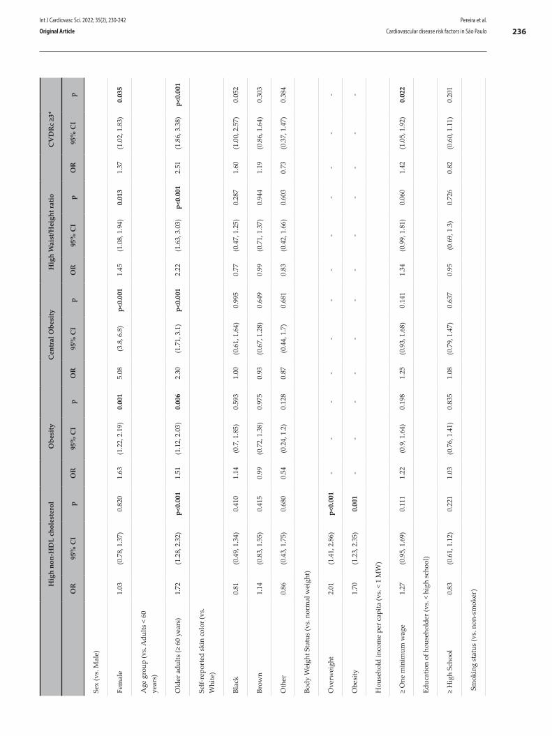
H
igh
non-
HD
L ch
oles
tero
lO
besi
tyC
entr
al O
besi
tyH
igh
Wai
st/H
eigh
t rat
ioC
VD
Rc
≥3*
O
R95
% C
Ip
OR
95%
CI
pO
R95
% C
Ip
OR
95%
CI
pO
R95
% C
Ip
Sex
(vs.
Mal
e)
Fem
ale
1.03
(0.7
8, 1
.37)
0.82
01.
63(1
.22,
2.1
9)0.
001
5.08
(3.8
, 6.8
)p<
0.00
11.
45(1
.08,
1.9
4)0.
013
1.37
(1.0
2, 1
.83)
0.03
5
Age
gro
up (v
s. A
dults
< 6
0 ye
ars)
Old
er a
dults
(≥ 6
0 ye
ars)
1.72
(1.2
8, 2
.32)
p<0.
001
1.51
(1.1
2, 2
.03)
0.00
62.
30(1
.71,
3.1
)p<
0.00
12.
22(1
.63,
3.0
3)p<
0.00
12.
51(1
.86,
3.3
8)p<
0.00
1
Self-
repo
rted
ski
n co
lor (
vs.
Whi
te)
Blac
k0.
81(0
.49,
1.3
4)0.
410
1.14
(0.7
, 1.8
5)0.
593
1.00
(0.6
1, 1
.64)
0.99
50.
77(0
.47,
1.2
5)0.
287
1.60
(1.0
0, 2
.57)
0.05
2
Brow
n1.
14(0
.83,
1.5
5)0.
415
0.99
(0.7
2, 1
.38)
0.97
50.
93(0
.67,
1.2
8)0.
649
0.99
(0.7
1, 1
.37)
0.94
41.
19(0
.86,
1.6
4)0.
303
Oth
er0.
86(0
.43,
1.7
5)0.
680
0.54
(0.2
4, 1
.2)
0.12
80.
87(0
.44,
1.7
)0.
681
0.83
(0.4
2, 1
.66)
0.60
30.
73(0
.37,
1.4
7)0.
384
Body
Wei
ght S
tatu
s (v
s. n
orm
al w
eigh
t)
Ove
rwei
ght
2.01
(1.4
1, 2
.86)
p<0.
001
--
--
--
--
--
--
Obe
sity
1.70
(1.2
3, 2
.35)
0.00
1-
--
--
--
--
--
-
Hou
seho
ld in
com
e pe
r cap
ita (v
s. <
1 M
W)
≥ O
ne m
inim
um w
age
1.27
(0.9
5, 1
.69)
0.11
11.
22(0
.9, 1
.64)
0.19
81.
25(0
.93,
1.6
8)0.
141
1.34
(0.9
9, 1
.81)
0.06
01.
42(1
.05,
1.9
2)0.
022
Educ
atio
n of
hou
seho
lder
(vs.
< h
igh
scho
ol)
≥ H
igh
Scho
ol0.
83(0
.61,
1.1
2)0.
221
1.03
(0.7
6, 1
.41)
0.83
51.
08(0
.79,
1.4
7)0.
637
0.95
(0.6
9, 1
.3)
0.72
60.
82(0
.60,
1.1
1)0.
201
Smok
ing
stat
us (v
s. n
on-s
mok
er)
Int J Cardiovasc Sci. 2022; 35(2), 230-242
236Pereira et al.
Cardiovascular disease risk factors in São PauloOriginal Article
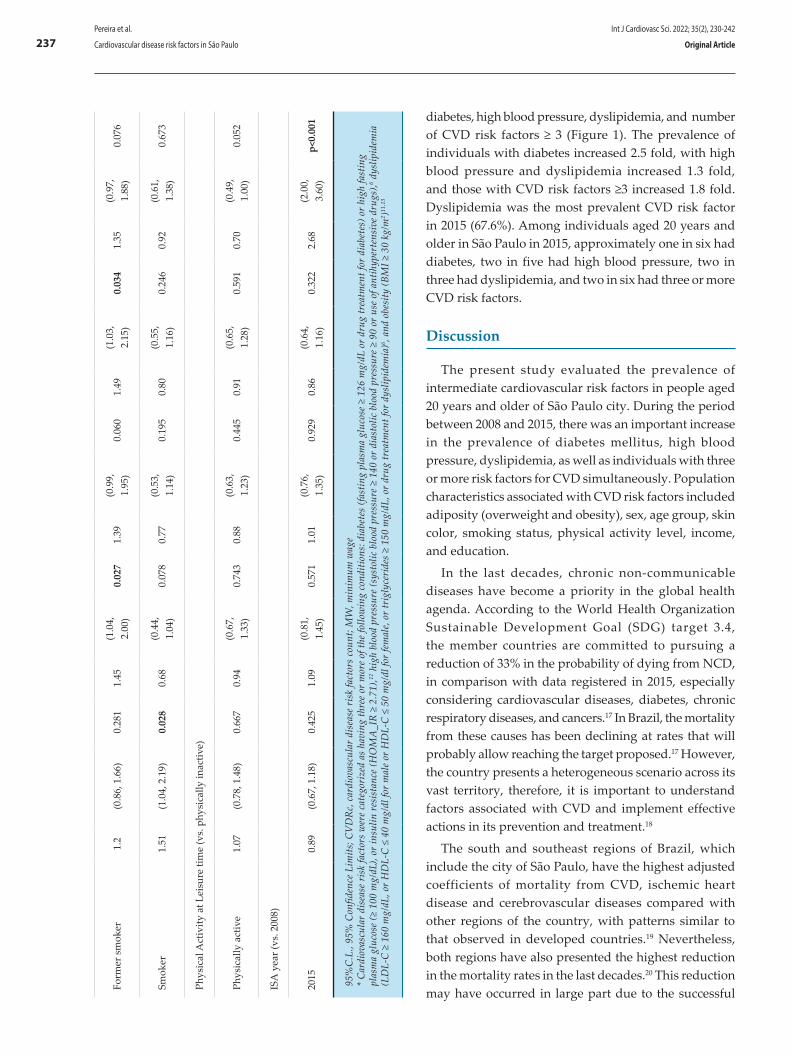
Form
er s
mok
er1.
2(0
.86,
1.6
6)0.
281
1.45
(1.0
4,
2.00
)0.
027
1.39
(0.9
9,
1.95
)0.
060
1.49
(1.0
3,
2.15
)0.
034
1.35
(0.9
7,
1.88
)0.
076
Smok
er1.
51(1
.04,
2.1
9)0.
028
0.68
(0.4
4,
1.04
)0.
078
0.77
(0.5
3,
1.14
)0.
195
0.80
(0.5
5,
1.16
)0.
246
0.92
(0.6
1,
1.38
)0.
673
Phys
ical
Act
ivity
at L
eisu
re ti
me
(vs.
phy
sica
lly in
activ
e)
Phys
ical
ly a
ctiv
e1.
07(0
.78,
1.4
8)0.
667
0.94
(0.6
7,
1.33
)0.
743
0.88
(0.6
3,
1.23
)0.
445
0.91
(0.6
5,
1.28
)0.
591
0.70
(0.4
9,
1.00
)0.
052
ISA
yea
r (vs
. 200
8)
2015
0.89
(0.6
7, 1
.18)
0.42
51.
09(0
.81,
1.
45)
0.57
11.
01(0
.76,
1.
35)
0.92
90.
86(0
.64,
1.
16)
0.32
22.
68(2
.00,
3.
60)
p<0.
001
95%
C.L
., 95
% C
onfid
ence
Lim
its; C
VD
Rc,
car
diov
ascu
lar
dise
ase
risk
fact
ors
coun
t; M
W, m
inim
um w
age
* C
ardi
ovas
cula
r di
seas
e ri
sk fa
ctor
s w
ere
cate
gori
zed
as h
avin
g th
ree
or m
ore
of th
e fo
llow
ing
cond
ition
s: d
iabe
tes
(fast
ing
plas
ma
gluc
ose
≥ 12
6 m
g/dL
or
drug
trea
tmen
t for
dia
bete
s) o
r hi
gh fa
stin
g pl
asm
a gl
ucos
e (≥
100
mg/
dL),
or in
sulin
res
ista
nce
(HO
MA
_IR
≥ 2
.71)
,12 h
igh
bloo
d pr
essu
re (s
ysto
lic b
lood
pre
ssur
e ≥
140
or d
iast
olic
blo
od p
ress
ure
≥ 90
or
use
of a
ntih
yper
tens
ive
drug
s),6 d
yslip
idem
ia
(LD
L-C
≥ 1
60 m
g/dL
, or
HD
L-C
≤ 4
0 m
g/dl
for
mal
e or
HD
L-C
≤ 5
0 m
g/dl
for
fem
ale,
or
trig
lyce
ride
s ≥
150
mg/
dL, o
r dr
ug tr
eatm
ent f
or d
yslip
idem
ia)6 ,
and
obes
ity (B
MI ≥
30
kg/m
2 )11,1
3
diabetes, high blood pressure, dyslipidemia, and number of CVD risk factors ≥ 3 (Figure 1). The prevalence of individuals with diabetes increased 2.5 fold, with high blood pressure and dyslipidemia increased 1.3 fold, and those with CVD risk factors ≥3 increased 1.8 fold. Dyslipidemia was the most prevalent CVD risk factor in 2015 (67.6%). Among individuals aged 20 years and older in São Paulo in 2015, approximately one in six had diabetes, two in five had high blood pressure, two in three had dyslipidemia, and two in six had three or more CVD risk factors.
Discussion
The present study evaluated the prevalence of intermediate cardiovascular risk factors in people aged 20 years and older of São Paulo city. During the period between 2008 and 2015, there was an important increase in the prevalence of diabetes mellitus, high blood pressure, dyslipidemia, as well as individuals with three or more risk factors for CVD simultaneously. Population characteristics associated with CVD risk factors included adiposity (overweight and obesity), sex, age group, skin color, smoking status, physical activity level, income, and education.
In the last decades, chronic non-communicable diseases have become a priority in the global health agenda. According to the World Health Organization Sustainable Development Goal (SDG) target 3.4, the member countries are committed to pursuing a reduction of 33% in the probability of dying from NCD, in comparison with data registered in 2015, especially considering cardiovascular diseases, diabetes, chronic respiratory diseases, and cancers.17 In Brazil, the mortality from these causes has been declining at rates that will probably allow reaching the target proposed.17 However, the country presents a heterogeneous scenario across its vast territory, therefore, it is important to understand factors associated with CVD and implement effective actions in its prevention and treatment.18
The south and southeast regions of Brazil, which include the city of São Paulo, have the highest adjusted coefficients of mortality from CVD, ischemic heart disease and cerebrovascular diseases compared with other regions of the country, with patterns similar to that observed in developed countries.19 Nevertheless, both regions have also presented the highest reduction in the mortality rates in the last decades.20 This reduction may have occurred in large part due to the successful
Int J Cardiovasc Sci. 2022; 35(2), 230-242
237Pereira et al.
Cardiovascular disease risk factors in São Paulo Original Article
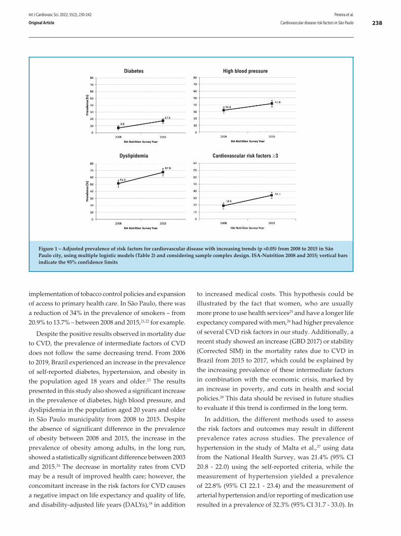
implementation of tobacco control policies and expansion of access to primary health care. In São Paulo, there was a reduction of 34% in the prevalence of smokers – from 20.9% to 13.7% – between 2008 and 2015,21,22 for example.
Despite the positive results observed in mortality due to CVD, the prevalence of intermediate factors of CVD does not follow the same decreasing trend. From 2006 to 2019, Brazil experienced an increase in the prevalence of self-reported diabetes, hypertension, and obesity in the population aged 18 years and older.23 The results presented in this study also showed a significant increase in the prevalence of diabetes, high blood pressure, and dyslipidemia in the population aged 20 years and older in São Paulo municipality from 2008 to 2015. Despite the absence of significant difference in the prevalence of obesity between 2008 and 2015, the increase in the prevalence of obesity among adults, in the long run, showed a statistically significant difference between 2003 and 2015.24 The decrease in mortality rates from CVD may be a result of improved health care; however, the concomitant increase in the risk factors for CVD causes a negative impact on life expectancy and quality of life, and disability-adjusted life years (DALYs),18 in addition
to increased medical costs. This hypothesis could be illustrated by the fact that women, who are usually more prone to use health services25 and have a longer life expectancy compared with men,26 had higher prevalence of several CVD risk factors in our study. Additionally, a recent study showed an increase (GBD 2017) or stability (Corrected SIM) in the mortality rates due to CVD in Brazil from 2015 to 2017, which could be explained by the increasing prevalence of these intermediate factors in combination with the economic crisis, marked by an increase in poverty, and cuts in health and social policies.20 This data should be revised in future studies to evaluate if this trend is confirmed in the long term.
In addition, the different methods used to assess the risk factors and outcomes may result in different prevalence rates across studies. The prevalence of hypertension in the study of Malta et al.,27 using data from the National Health Survey, was 21.4% (95% CI 20.8 - 22.0) using the self-reported criteria, while the measurement of hypertension yielded a prevalence of 22.8% (95% CI 22.1 - 23.4) and the measurement of arterial hypertension and/or reporting of medication use resulted in a prevalence of 32.3% (95% CI 31.7 - 33.0). In
Cardiovascular risk factors ≥3
High blood pressure
Dyslipidemia
Diabetes
Figure 1 – Adjusted prevalence of risk factors for cardiovascular disease with increasing trends (p <0.05) from 2008 to 2015 in São Paulo city, using multiple logistic models (Table 2) and considering sample complex design. ISA-Nutrition 2008 and 2015; vertical bars indicate the 95% confidence limits
Int J Cardiovasc Sci. 2022; 35(2), 230-242
238Pereira et al.
Cardiovascular disease risk factors in São PauloOriginal Article
the study ISA-Capital 2008, the sensitivity of self-reported diabetes was 85.8% (95%CI 70.7–93.8) in older adults and only 42.1% (95%CI 22.4–64.6) in adults.28 Therefore, an advantage of the present study refers to the adoption of direct measurement of risk factors, e.g., anthropometry was assessed by the determination of adiposity measures, while blood pressure and blood samples were used to define high blood pressure, dyslipidemia and diabetes, in combination with information on medication use (confirmed and registered by a nurse in the household visit). This procedure probably resulted in higher values of prevalence compared with other studies based on self-reported information.
Among the characteristics associated with intermediate risk factors for CVD, two should be highlighted: age and adiposity. Aging is associated with progressive loss of tissue and organ function over time, and accumulation of oxidative damage to macromolecules (lipids, DNA, and proteins) by reactive oxygen species, resulting in several acute and chronic pathological processes, such as CVD.29 As we age, another important aspect to be addressed is inflammaging — a state of chronic low-grade systemic inflammation —, which is associated with immunosenescence, metabolic inflammation, and increased insulin resistance and consequently, increased risk of type 2 diabetes.30 In fact, in the present study, older age was associated with higher chances of having diabetes, elevated blood pressure, non-HDL cholesterol and adiposity, and presenting three or more CVD risk factors. In terms of public health, Brazil, including São Paulo municipality, has been experiencing a sharp demographic transition,31 marked by population aging and consequent increase in social and economic burden. Such increase in health care demands may represent a real challenge, which requires public policy planning in health and economy.
Another important challenge for the Brazilian health system is the alarming prevalence of excess adiposity, which leads to an inflammatory condition that is directly involved in the etiology of cardiovascular diseases and type 2 diabetes. It should be noted that one in four people aged 20 years and older in the city of São Paulo is obese and half of the population has high waist circumference. In accordance with previous studies,11,32 excess body weight (overweight and/or obesity) was positively associated with all characteristics associated with CVD risk factors, besides being a risk factor itself. High adiposity, especially obesity, is an important health problem worldwide and, during the last decades, little
progress has been made, considering that no country has shown decreasing trends in population obesity.33
Besides sex, age and adiposity, other conditions were associated with intermediate risk factors for CVD in this study, such as skin color and high blood pressure. Other studies have also shown that black and brown ethinicity is an important risk factor for high blood pressure.34,35 Although hereditary predisposition may be involved,36 socioeconomic conditions and lifestyle may play more important roles in this association.14,37-39 This evidence highlights the importance of the social determinants of health in the context of CVDs,40 such as education, which was inversely associated not only with high blood pressure, but also with diabetes and dyslipidemia in the present study, and is the most consistent social determinant related to CVD outcomes. Lower levels of educational attainment has been associated with higher prevalence of many cardiovascular risk factors, higher incidence of cardiovascular events, and higher cardiovascular mortality, independent of sociodemographic factors.40,41
The risk factors for CVD usually occur together, due to a substantial overlap between disease etiology and mechanisms.42 It has been estimated that, for every 4.5 kg of weight gain, there is a 20% increase in the risk of hypertension.43 In the present study, subjects with overweight and obesity were 1.5 and 2.4 fold, respectively, more likely to have high blood pressure. Besides, weight reduction promotes a decrease in blood pressure both in normotensive and hypertensive individuals.14 The prevalence of subjects with three or more risk factors for CVD increased from 18% to 35% in the city of São Paulo, which may result in increased mortality, functional decline, and lower quality of life in a substantial proportion of the population, leading to an increasing demand of health care services during the following decades.44
Additionaly, the risk factors investigated are also related to the worse health outcomes and increased risk of mortality due to infectious diseases, like the new coronavirus disease (COVID-19).4 Recent studies have shown that obesity, hypertension, diabetes, and cardiovascular disease greatly affect the prognosis of the COVID-19.45,46 Also, social strategies adopted to fight the COVID-19 pandemic (e.g. lockdown and self-isolation) can even worsen the occurrence of obesity and other metabolic diseases due to physical inactivity and anxiety.47,48 Therefore, evaluating and
Int J Cardiovasc Sci. 2022; 35(2), 230-242
239Pereira et al.
Cardiovascular disease risk factors in São Paulo Original Article
adequately controlling these risk factors is a good strategy in public health, since it can be easily made in primary care and have a low cost compared with the management of CVD consequences.3
The main limitation of this study is its cross-sectional design, which limits the evaluation of some associations, such as the higher likelihood of former smokers having obesity and diabetes. Although there is evidence regarding the relationship between smoking cessation and weight gain,49 the results should be interpreted considering the study design, due to the possibility of reverse causality. Additionally, other aspects potentially associated with CVD were not investigated in the present research. Alcoholic beverage intake (yes/no) and diet quality (using the revised version of the Brazilian Healthy Eating Index) were evaluated, but excluded from the models due to the absence of effect or statistical significance. Future research should explore these and other associations in detail using methods specific to CVD risk factors in the Brazilian population.
Despite these limitations, the present study has strengths, such as the use of direct measurements to estimate the evaluated parameters, with methodological rigor, to obtain high-quality information. In addition, important confounders were taken into account in the analysis, such as income, education, and physical activity. Finally, this study evaluated the population in the urban area of the biggest city in Brazil, with more than 12 million habitants50 and a high degree of genetic admixture.51
Conclusion
The prevalence of intermediate CVD risk factors in a population sample from Sao Paulo varied according to non-modifiable (age, sex, skin color) and modifiable characteristics (physical activity, smoking status, income, education). From 2008 to 2015, there was an important increase in the prevalence of diabetes, high blood pressure, dyslipidemia, and invidiuals with three or more CVD risk factors, whereas adiposity parameters (e.g. obesity) had no significant increase, despite their high prevalence. The results may support the selection of target groups and priority actions on CVD prevention and treatment, considering the current health scenario of the high prevalence of CVD, associated with population aging, which exposes epidemiological and mechanistic relationships with cardiometabolic risk factors (abnormal adiposity, dysglycemia, dyslipidemia, and high blood pressure).
Acknowledgements
We acknowledge the contribution of all people involved in ISA-Capital, especially Maria Cecilia Goi Porto Alves, Marilisa Berti de Azevedo Barros, Maria Mercedes Loureiro Escuder, the GAC group (Grupo de Avaliação do Consumo Alimentar – Group of Assessment of Dietary Intake) and the participants of the study.
Author contributions
Conception and design of the research: Pereira JL, Castro MA, Rogero MM, Sarti FM, Fisberg RM. Acquisition of data: Cesar, CLG, Goldbaum M, Fisberg RM. Statistical analysis: Pereira JL, Castro MA, Leite JMRS. Obtaining financing: Fisberg RM, Cesar, CLG, Goldbaum M, Pereira JL. Writing of the manuscript: Pereira JL. Critical revision of the manuscript for intellectual content: Castro MA, Leite JMRS, Rogero MM, Sarti FM, Fisberg RM.
Potential Conflict of InterestNo potential conflict of interest relevant to this
article was reported.
Sources of FundingThis work was supported by the São Paulo
Municipal Health Department (grant number #2013-0.235.936-0), Research Support Foundation of the State of São Paulo (grant numbers #2012/22113-9, #2017/05125-7, and #2019/23985-9), and National Council for Scientific and Technological Development (grant number #472873/2012-1).
Study AssociationThis study is not associated with any thesis or
dissertation work.
Ethics approval and consent to participateThis study was approved by the Ethics Committee
of the Faculdade de Saúde Pública da Universidade de São Paulo under the protocol number 30848914.7.0000.5421. All the procedures in this study were in accordance with the 1975 Helsinki Declaration, updated in 2013. Informed consent was obtained from all participants included in the study.
Int J Cardiovasc Sci. 2022; 35(2), 230-242
240Pereira et al.
Cardiovascular disease risk factors in São PauloOriginal Article
1. Sociedade Brasileira de Cardiologia. Rio de Janeiro; 2020. [Internet] [Cited in 2021 Jan 17] Cardiômetro. Available from: http://www.cardiometro.com.br
2. World Health Organization. (WHO). Noncommunicable diseases country profiles 2018. World Health Organization. Geneva; 2020. [Internet] [Cited in 2021 Jan 17] Available from: http://www.who.int/nmh/publications/ncd-profiles-2018/en/.
3. World Health Organization . (WHO). Geneva; 2017. Cardiovascular diseases (CVDs) – Fact Sheet. [Internet] [Cited in 2021 Jan 17] Available from: https://www.who.int/news-room/fact-sheets/detail/cardiovascular-diseases-(cvds)
4. World Health Organization. (WHO). The impact of the COVID-19 pandemic on noncommunicable disease resources and services: results of a rapid assessment. World Health Organization. Geneva; 2020. [Internet] [Cited in 2021 Jan 17] Available from: https://www.who.int/publications/i/item/ncds-covid-rapid-assessment.
5. O'Donnell CJ, Elosua R. Cardiovascular risk factors. Insights from framingham heart study. Rev Esp Cardiol (English Edition). 2008; 61(3): 299-310.
6. Précoma DB, Oliveira GMM, Simão AF, Dutra OP, Coelho, OR, Izar, MCDO, et al. Atualização da Diretriz de Prevenção Cardiovascular da Sociedade Brasileira de Cardiologia–2019. Arq Bras Cardiol. 2019; 113(4): 787-891.
7. Ferrari TK, Cesar CLG, Alves MCGP, Barros, MBDA, Goldbaum M, Fisberg RM. Estilo de vida saudável em São Paulo, Brasil. Cad Saude Publica. 2017; 33: e00188015.
8. Prefeitura de São Paulo. São Paulo; 2015. Inquérito de Saúde do Município de São Paulo (ISA-Capital) - Health Survey of São Paulo (ISA-Capital). [Internet] [Cited in 2021 Jan 17]http://www.prefeitura.sp.gov.br/cidade/secretarias/saude/epidemiologia_e_informacao/isacapitalsp/.
9. Alves MCGP, Escuder MML, Goldbaum M, Barros MBA, Fisberg R, Cesar CLG. Sampling plan in health surveys, city of São Paulo, Brazil, 2015. Rev Saude Publ. 2018; 52: 81. https://doi.org/10.11606/51518-8787.2018052000471
10. Fisberg RM, Sales CH, Fontanelli MM, Pereira JL, Alves MCGP, Escuder, MML, et al. 2015 Health Survey of São Paulo with Focus in Nutrition: Rationale, Design, and Procedures. Nutrients. 2018; 10(2): 169. doi:10.3390/nu10020169
11. World Health Organization. (WHO). Obesity: Preventing and Managing the Global Epidemic. Report of a WHO Consultation. Geneva; 2000. [Internet] [Cited in 2021 Jan 17] Available from: https://www.who.int/nutrition/publications/obesity/WHO_TRS_894/en/
12. Sociedade Brasileira de Diabetes. Diretrizes da Sociedade Brasileira de Diabetes 2019-2020. São Paulo: SBD; 2019. [Internet] [Cited in 2021 Jan 17] Available from: https://www.diabetes.org.br/profissionais/images/DIRETRIZES-COMPLETA-2019-2020.pdf
13. Lebrão ML, Duarte YAO. (Eds.), SABE–Saúde, Bem-estar e Envelhecimento. O projeto SABE no Município de São Paulo: uma abordagem inicial. 1st ed. Brasília, Brazil: Organização Pan-Americana da Saúde; 2003.
14. Barroso WKS, Rodrigues CIS, Bortolotto LA, Mota-Gomes MA, Brandão AA, Feitosa ADM, et al. Diretrizes Brasileiras de Hipertensão Arterial – 2020. Arq Bras Cardiol. 2020. [Internet] [Cited in 2021 Jan 17] Available from: http://abccardiol.org/wp-content/uploads/2020/11/DBHA-2020.x64000.pdf
15. Matsudo S, Araujo T, Matsudo V, Andrade D, Andrade E, Oliveira LC, et al. Questionário Internacional de Atividade Física (IPAQ): Estudo de validade e reprodutibilidade no Brasil. Rev Bras Ativ Fís Saúde. 2001; 6 Suppl 2: 5-18.
16. World Health Organization. (WHO). Global recommendations on physical activity for health. World Health Organization: Geneva, Switzerland, 2010.
17. NCD Countdown 2030 Collaborators. NCD countdown 2030: worldwide trends in non-communicable disease mortality and progress towards Sustainable Development Goal target 3.4. Lancet 2018; 392(10152):1072-88. doi:10.1016/S0140-6736(18)31992-5.
18. Schmidt MI, Duncan BB, Silva GA, Menezes AM, Monteiro CA, Barreto SM, et al. Chronic non-communicable diseases in Brazil: burden and current challenges. The Lancet. 2011; 377(9781):1949-61. https://doi.org/10.1016/S0140-6736(11)60135-9.
19. Mansur AP, Favarato D. Mortality due to Cardiovascular Diseases in Women and Men in the Five Brazilian Regions, 1980-2012. Arq Bras Cardiol. 2016;107(2):137-46. https://doi.org/10.5935/abc.20160102.
20. Malta DC, Teixeira R, Oliveira GMMD, Ribeiro AL. Mortalidade por Doenças Cardiovasculares Segundo o Sistema de Informação sobre Mortalidade e as Estimativas do Estudo Carga Global de Doenças no Brasil, 2000-2017. Arq Bras Cardiol. 2020; 115(2):152-60. https://doi.org/10.36660/abc.20190867.
21. Brasil.Ministério da Saúde. Secretaria de Vigilância em Saúde, Departamento de Vigilância de Doenças e Agravos não Transmissíveis e Promoção de Saúde. Vigitel Brasil 2008: vigilância de fatores de risco e proteção para doenças crônicas por inquérito telefônico: estimativas sobre frequência e distribuição sociodemográfica de fatores de risco e proteção para doenças crônicas nas capitais dos 26 estados brasileiros e no Distrito Federal em 2008. Brasília;2009.
22. Brasil.Ministério da Saúde. Secretaria de Vigilância em Saúde, Departamento de Vigilância de Doenças e Agravos não Transmissíveis e Promoção de Saúde. Vigitel Brasil 2015: vigilância de fatores de risco e proteção para doenças crônicas por inquérito telefônico: estimativas sobre frequência e distribuição sociodemográfica de fatores de risco e proteção para doenças crônicas nas capitais dos 26 estados brasileiros e no Distrito Federal em 2015. Brasília: Ministério da Saúde; 2016.
23. Brasil. Ministério da Saúde. Secretaria de Vigilância em Saúde, Departamento de Vigilância de Doenças e Agravos não Transmissíveis e Promoção de Saúde. Vigitel Brasil 2019: vigilância de fatores de risco e proteção para doenças crônicas por inquérito telefônico: estimativas sobre frequência e distribuição sociodemográfica de fatores de risco e proteção para doenças crônicas nas capitais dos 26 estados brasileiros e no Distrito Federal em 2019. Brasília;2020.
24. Pereira JL, Vieira DAS, Alves MCGP, Cesar CLG, Goldbaum M, Fisberg RM. Excess body weight in the city of São Paulo: panorama from 2003 to 2015, associated factors and projection for the next years. BMC Public Health. 2018;18(1):1332. https://doi.org/10.1186/s12889-018-6225-8.
25. Malta DC, Bernal RTI, Lima MG, Araújo SSCD, Silva MMAD, Freitas MIDF, Barros MBDA. Noncommunicable diseases and the use of health services: analysis of the National Health Survey in Brazil. Rev Saúde Pública. 2017;51(suppl 1) https://doi.org/10.1590/S1518-8787.
26. Instituto Brasileiro de Geografia e Estatística. Estatística – IBGE. Tábua Completa de Mortalidade para o Brasil 2019 – Breve análise da evolução da mortalidade no Brasil. Rio de Janeiro;2020. [Cited in 2021 Jul 15] Available from: https://biblioteca.ibge.gov.br/visualizacao/periodicos/3097/tcmb_2019.pdf
27. Malta DC, Gonçalves RPF, Machado ÍE, Freitas MIDF, Azeredo C, Szwarcwald CL. Prevalência da hipertensão arterial segundo diferentes critérios diagnósticos, Pesquisa Nacional de Saúde. Rev Bras Epidemiol. 2018;21(supl 1):e180021.https://doi.org/10.1590/1980-549720180021.
28. Fontanelli MM, Teixeira JA, Sales CH, Castro MA, Cesar CLG, Alves MCGP et al. Validation of self-reported diabetes in a representative sample of São Paulo city. Rev Saúde Pública. 2017;51(0):20. doi:org/10.1590/S1518-8787.
29. Liguori I, Russo G, Curcio F, Bulli G, Aran L, Della-Morte D, et al. Oxidative stress, aging, and diseases. Clin Interv Aging. 2018;13;757-72. doi:10.2147/CIA.S158513.
30. Franceschi C, Bonafè M, Valensin S, Olivieri F, De Luca M, Ottaviani E, et al. Inflammation-aging: an evolutionary perspective on immunosenescence. Ann N Y Acad Sci. 2000; 908(1): 244-54. doi:10.1111/j.1749-6632.2000.tb06651.x.
References
Int J Cardiovasc Sci. 2022; 35(2), 230-242
241Pereira et al.
Cardiovascular disease risk factors in São Paulo Original Article
31. United Nations, Department of Economic and Social Affairs, Population Division. World Population Prospects: The 2019 Revision. [Internet] [Cited in 2021 Jan 15] Available from: http://esa.un.org/unpd/wpp/.
32. Afshin A, Forouzanfar MH, Reitsma MB, Sur P, Estep K, Lee A, et al. Health effects of overweight and obesity in 195 countries over 25 years. N Engl J Med. 2017; 377( Suppl 1):13-27. doi:10.1056/NEJMoa1614362.
33. Swinburn BA, Kraak VI, Allender S, Atkins VJ, Baker PI, Bogard JR, et al. The global syndemic of obesity, undernutrition, and climate change: the Lancet Commission report. Lancet. 2019;393(10173):791-846. doi:10.1016/S0140-6736(18)32822-8.
34. Virani SS, Alonso A, Benjamin EJ, Bittencourt MS, Callaway CW, Carson AP, et al., American Heart Association Council on Epidemiology and Prevention Statistics Committee and Stroke Statistics Subcommittee. Heart disease and stroke statistics—2020 update: a report from the American Heart Association. Circulation. 2020; 141(9): e139-e596. doi:10.1161/CIR.0000000000000757.
35. Harada PH, Miname MH, Benseñor IM, Santos RD, Lotufo PA. Familial hypercholesterolemia prevalence in an admixed racial society: Sex and race matter. The ELSA-Brasil. Atherosclerosis. 2018, 277: 273-7. Doi:10.1016/j.atherosclerosis.2018.08.021.
36. Gouveia MH, Bentley AR, Leonard H. Meeks KAC, Ekoru K, Chen G, et al. Trans-ethnic meta-analysis identifies new loci associated with longitudinal blood pressure traits. Sci Rep. 2021;11(1):4075. doi; 10.1038/s41598-83450-3.
37. Lima-Costa MF, Mambrini JVM, Leite MC, Peixoto SV, Firmo JOA, Filho AIL. Socioeconomic Position, But Not African Genomic Ancestry, Is Associated With Blood Pressure in the Bambui-Epigen (Brazil) Cohort Study of Aging. Hypertension. 2016; 67(2):349-55. doi:10.1161/HYPERTENSIONAHA.115.06609.
38. Howard G, Cushman M, Moy CS, Oparil S, Muntner P, Lackland DT, et al. Association of clinical and social factors with excess hypertension risk in black compared with white US adults. JAMA. 2018; 320(13):1338-48. doi:10.1001/jama.2018.13467.
39. Giuffrida A. Racial and ethnic disparities in Latin America and the Caribbean: a literature review. Divers Equal Health Care. 2010;7(2):115–28.
40. Havranek EP, Mujahid MS, Barr DA, Blair IV, Cohen MS, Cruz-Flores S, et al. Social determinants of risk and outcomes for cardiovascular disease:
a scientific statement from the American Heart Association. Circulation. 2015; 132(9): 873-98. doi: 10.1161/CIR.0000000000000228.
41. Cohen AK, Syme SL. Education: a missed opportunity for public health intervention. Am J Public Health. 2013;103(6):997-1001. doi:10.2105/AJPH.2012.300993.
42. Cheung BM, Li C. Diabetes and hypertension: is there a common metabolic pathway?. Curr Atheroscler Rep. 2012;14(2):160-6. doi:10.1007/s11883-013-0227-2.
43. Hall JE, Carmo JM, Silva AA, Wang Z, Hall ME. Obesity-induced hypertension: interaction of neurohumoral and renal mechanisms. Circ Res. 2015; 116(6):991-1006. doi:10.1161/CIRCRESAHA.116.305697.
44. Nunes BP, Batista SRR, Andrade FBD, Souza Junior PRBD, Lima-Costa MF, Facchini LA. Multimorbidity: the Brazilian longitudinal study of aging (ELSI-Brazil). Rev Saúde Públ. 2018;52(Suppl 2):10s. doi:10.11606/S1518-8787.
45. Zheng Z, Peng F, Xu B, Zhao J, Liu H, Peng J, et al. Risk factors of critical & mortal COVID-19 cases: A systematic literature review and meta-analysis. J Infect. 2020; 81(2): e16-e25.
46. Yang J, Zheng Y, Gou X, Pu K, Chen Z, Guo Q, et al. Prevalence of comorbidities and its effects in patients infected with SARS-CoV-2: a systematic review and meta-analysis. Int J Infect Dis. 2020; 94:91-5. doi:10.1016/j.ijid.2020.03.017.
47. Clemmensen C, Petersen MB, Sørensen TIA. Will the COVID-19 pandemic worsen the obesity epidemic?. Nat Rev Endocrinol. 2020; 16(9):469-70. Doi:
48. Hussain A, Mahawar K, Xia Z, Yang W, Shamsi EH. Obesity and mortality of COVID-19. Meta-analysis. Obes Res Clin Pract. 2020; 14(4):295-300. doi: 10.1016/j.orcp.2020.07.002.
49. Filozof C, Fernandez Pinilla MC, Fernández-Cruz A. Smoking cessation and weight gain. Obes Rev.2004; 5(2):95-103. doi:10.1111/j.1467-789X.2004.00131.x.
50. Instituto Brasileiro de Geografia e Estatística. Estatística I-IBdGe–Cidades. 2020. [Internet] [Cited in 2021 Jan 21] Available from: https://cidades.ibge.gov.br/brasil/sp/ sao-paulo/ panorama.
51. Pena SD, Santos FR, Tarazona-Santos E. Genetic admixture in Brazil. Am J Med Genet. 2020;184(4):928-38. doi:10.1002/ajmg.c.31853.
This is an open-access article distributed under the terms of the Creative Commons Attribution License
Int J Cardiovasc Sci. 2022; 35(2), 230-242
242Pereira et al.
Cardiovascular disease risk factors in São PauloOriginal Article
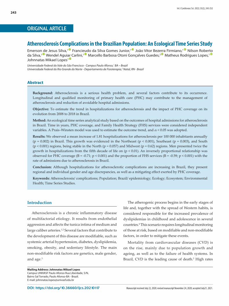
Introduction
Atherosclerosis is a chronic inflammatory disease of multifactorial etiology. It results from endothelial aggression and affects the tunica intima of medium and large caliber arteries.1,2 Several factors that contribute to the development of this disease are modifiable, such as systemic arterial hypertension, diabetes, dyslipidemia, smoking, obesity, and sedentary lifestyle. The main non-modifiable risk factors are genetics, male gender, and age.3
The atherogenic process begins in the early stages of life and, together with the spread of Western habits, is considered responsible for the increased prevalence of dyslipidemias in childhood and adolescence in several countries.4 This scenario requires longitudinal monitoring of those at risk, based on modifiable and non-modifiable factors, in order to mitigate these events.
Mortality from cardiovascular diseases (CVD) is on the rise, mainly due to population growth and ageing, as well as to the failure of health systems. In Brazil, CVD is the leading cause of death.5 High rates
Int J Cardiovasc Sci. 2022; 35(2), 243-252
243
ORIGINAL ARTICLE
Atherosclerosis Complications in the Brazilian Population: An Ecological Time Series StudyEmerson de Jesus Silva,1 Francieudo da Silva Gomes Junior,1 João Vitor Bezerra Firmiano,1 Nilson Roberto da Silva,1 Wendel Aguiar Carlini,1 Marcello Barbosa Otoni Gonçalves Guedes,2 Matheus Rodrigues Lopes,1
Johnnatas Mikael Lopes1
Universidade Federal do Vale do São Francisco - Campus Paulo Afonso,1 BA – BrazilUniversidade Federal do Rio Grande do Norte - Departamento de Fisioterapia,2 Natal, RN - Brazil
DOI: https://doi.org/10.36660/ijcs.20210117
Mailing Address: Johnnatas Mikael LopesCampus UNIVASF Paulo Afonso Rua Liberdade, S/N, Bairro Sal Torrado, Paulo Afonso-BA - Brazil. E-mail: [email protected]
Manuscript received July 22, 2020; revised manuscript November 24, 2020; accepted July21, 2021.
Abstract
Background: Atherosclerosis is a serious health problem, and several factors contribute to its occurrence. Longitudinal and qualified monitoring of primary health care (PHC) may contribute to the management of atherosclerosis and reduction of avoidable hospital admissions.
Objective: To estimate the trend in hospitalizations for atherosclerosis and the impact of PHC coverage on its evolution from 2008 to 2018 in Brazil.
Method: An ecological time series analytical study based on the outcomes of hospital admissions for atherosclerosis in Brazil. Time in years, PHC coverage, and Family Health Strategy (FHS) services were considered independent variables. A Prais–Winsten model was used to estimate the outcome trend, and α < 0.05 was adopted.
Results: We observed a mean increase of 1.81 hospitalizations for atherosclerosis per 100 000 inhabitants annually (p = 0.002) in Brazil. This growth was evidenced in the Northeast (p < 0.001), Southeast (p = 0.003), and South (p < 0.001) regions, being stable in the North (p = 0.057) and Midwest (p = 0.62) regions. Men presented twice the growth in hospitalizations from the fifth decade of life on (p < 0.01). An inversely proportional relationship was observed for PHC coverage (B = -0.71; p < 0.001) and the proportion of FHS services (B = -0.59; p < 0.001) with the rate of admissions due to atherosclerosis in Brazil.
Conclusion: Although hospitalizations for atherosclerotic complications are increasing in Brazil, they present regional and individual gender and age discrepancies, as well as a mitigating effect exerted by PHC coverage.
Keywords: Atherosclerosis/ complications; Population; Brazil/ epidemiology; Ecology; Ecosystem; Environmental Health; Time Series Studies.
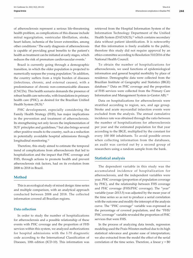
retrieved from the Hospital Information System of the Information Technology Department of the Unified Health System (DATASUS),12 which contains secondary data without patient identification. It is noteworthy that this information is freely available to the public, therefore this study did not require approval by an ethics committee according to Resolution 510/2016 of the National Health Council.
To obtain the number of hospitalizations for atherosclerosis, we used functions of epidemiological information and general hospital morbidity by place of residence. Demographic data were collected from the Brazilian Institute of Geography and Statistics (IBGE) database.13 Data on PHC coverage and the proportion of FHS services were collected from the Primary Care Information and Management System (e-Gestor AB).14
Data on hospitalizations for atherosclerosis were stratified according to region, sex, and age group. Stroke and acute myocardial infarction events were excluded from the analysis. The annual cumulative incidence rate was obtained through the ratio between the number of hospitalizations for atherosclerosis per year and the estimated population for that year according to the IBGE, multiplied by the constant for every 100 000 inhabitants. To avoid possible errors when collecting information from the databases, an audit was carried out by a second group of researchers using a random sample from the bank.
Statistical analysis
The dependent variable in this study was the accumulated incidence of hospitalization for atherosclerosis, and the independent variables were year, PHC coverage (proportion of population coverage by PHC), and the relationship between FHS coverage and PHC coverage (FHS/PHC coverage). The “year” variable (year–2013.5) was adjusted by the mean year of the time series so as not to produce a serial correlation with the outcome and modify the intercept of the analysis curve. The “PHC coverage” variable was expressed as the percentage of covered population, and the “FHS/PHC coverage” variable revealed the proportion of PHC services that were FHS.
In the process of analyzing data trends, regression modeling used the Prais-Winsten method due to its high statistical relevance and greater ease of interpretation; we also extracted from the model the effect of the serial correlation of the time series. Therefore, a linear y = B0
of atherosclerosis represent a serious life-threatening health problem, as complications of this disease include mitral regurgitation, ventricular fibrillation, stroke, heart failure, ischemia of the lower extremities, among other conditions.6 The early diagnosis of atherosclerosis is capable of providing great benefits to the patient’s health as treatment can be initiated at early stages, which reduces the risk of premature cardiovascular events.7
Brazil is currently going through a demographic transition, in which the older population is projected to numerically surpass the young population.8 In addition, the country suffers from a triple burden of diseases (infectious, chronic, and external causes), with a predominance of chronic non-communicable diseases (CNCDs). This health scenario demands the presence of robust health care networks, with foundations in primary health care (PHC), as desired for the Brazilian Unified Health System (SUS).9
PHC development, especially considering the Family Health Strategy (FHS), has major implications in the prevention and treatment of atherosclerosis. Its strengthening not only favors the implementation of SUS’ principles and guidelines,10 but also brings several other positive results to the country, such as a reduction in potentially avoidable hospital admissions through longitudinal monitoring.11
Therefore, this study aimed to estimate the temporal trend of complications from atherosclerosis that led to hospitalization and the impact that PHC coverage and FHS, through actions to promote health and prevent atherosclerosis risk factors, had on its evolution from 2008 to 2018 in Brazil.
Method
This is an ecological study of mixed design: time series and multiple comparison, with an analytical approach conducted between 2008 and 2018. The collected information covered all Brazilian regions.
Data collection
In order to study the number of hospitalizations for atherosclerosis and a possible relationship of these events with PHC coverage and the proportion of FHS services within this system, we analyzed authorizations for hospital admissions with the I-70 diagnostic code according to the International Classification of Diseases, 10th edition (ICD-10). This information was
Int J Cardiovasc Sci. 2022; 35(2), 243-252
244Silva et al.
Atherosclerosis complications in Brazilian PHCOriginal Article
+B1x equation was estimated, where x represented the “year” independent variable.
The mean outcome observed in the period, regardless of the year, was characterized as B0; B1 was the regression coefficient, which informed the mean annual evolution and the slope of the line. The sign of the slope determines an increasing trend (+) or decreasing trend (-) of the outcome. In addition to the equation, the adjusted coefficient of determination (R2
adjus) is presented, which specifies the degree of explanation of the model with the observed data, ranging from 0 to 1.
To verify the impact of the interaction between PHC coverage and the proportion of FHS coverage within PHC services ("PHC–FHS coverage interaction" variable) on the trend of hospitalizations for atherosclerosis in Brazil, we used a generalized estimating equations approach, an extension of generalized linear models (GLM) for correlated data. A robust covariance matrix and an autoregressive (ARIMA) or unstructured working correlation matrix were assumed to estimate the effects of independent variables, depending on the quality of the model based on the quasi-likelihood independence criterion (QIC) of the model. The gamma link function was used to connect independent variables and the outcome in the model. The sign of the model coefficients (B) would indicate the effect of the independent variables and their significance estimated by the Wald chi-squared test (χ2). R software was also used to adjust the polynomial curves and build the GLM. A 5% significance level was considered to minimize type I error in curve adherence and modeling processes. R software was used for data analysis.
Results
Figure 1 shows the pattern of hospitalizations for atherosclerosis in Brazil and allows us to analyze the situation considering this disease. A constant increase is seen in cases in men and an initial decline is observed among women, with subsequent growth from 2012 on. An average increase of 1.81 cases per 100 000 inhabitants (B = 1.81; p = 0.002) is seen in the general population, with cases among men (p < 0.001) growing more than twice those in women (p = 0.019) (Table 1).
When analyzing the time series by the country’s regions, a relatively steady scenario is identified in the Midwest (p = 0.62) and North (p = 0.057) regions, with evidence of a decline in hospitalizations for atherosclerosis among women (B = -0.02; p = 0.013). Conversely, the South
(B = 0.65; p < 0.001), Southeast (B = 0.61; p = 0.003), and Northeast (B = 0.56; p < 0.001) regions showed a growth in hospitalizations for atherosclerosis (Table 1).
Atherosclerosis complications that led to hospitalization were decreasing in men under the age of 40 (p < 0.01) and in women under 50 (p < 0.01). Both sexes showed an increase in hospitalizations starting at the fifth decade of life (p < 0.01). However, between the fifth and eighth decade of life, men showed almost twice the increase in hospitalizations presented by women (Table 2).
Despite the context of increased hospitalizations for atherosclerosis in the Brazilian population, greater PHC coverage and the proportion of FHS services within PHC showed inverse relationships with hospitalization rates (BPHC = -0.71; BFHS/PHC = -0.59), thus exerting a mitigating effect on these acute events in Brazil. These effects were more prominent in the North (BPHC = -1.62; BFHS/PHC = -1.38) and Midwest (BPHC = -0.99; BFHS/PHC = -0.80) regions, followed by the Northeast (BPHC = -0.67; BFHS/PHC = -0.55) and Southeast regions (BPHC = -0.41; BFHS/PHC = -0.31). The South region (BPHC = 0.41; BFHS/PHC = 0.47) was the only one that did not show a mitigating effect of PHC on hospitalizations for atherosclerosis (Table 3).
Discussion
We aimed to estimate the trend of hospitalizations for atherosclerosis in the Brazilian population and the effects of PHC coverage on its evolution. An increase in atherosclerosis complications requiring hospitalization was observed, especially in men and people aged over 50 years. Regional discrepancies were also evident: the South, Southeast, and Northeast regions showed the worst evolutions. However, this scenario is alleviated by the performance of PHC services, which mitigated the growth of these complications.
The increase in hospitalizations for atherosclerosis in the Brazilian population, especially from 2012 on, may be a reflection of 3 main aspects: the progressive population ageing taking place in the current demographic transition9; an increase in Brazilian family incomes accompanied by changes in food consumption and the adoption of urban and less healthy lifestyle habits15; and public health policies limited by funding and public-private partnerships16, which interact with each other and form a complex causal network.
Population ageing is a paradigm that has been experienced since the 1960s–1970s in Brazil and was expressed in this research with the growth of
Int J Cardiovasc Sci. 2022; 35(2), 243-252
245Silva et al.
Atherosclerosis complications in Brazilian PHC Original Article
Acc
umul
ated
inci
denc
e pe
r 100
000
inha
bita
nts
Year
BrazilMenWomen
Figure 1 – Time series of atherosclerotic complications treated in hospitals or hospitals associated with the Unified Health System between 2008 and 2018.
atherosclerotic complications in individuals aged 50 years and older, both men and women. The current challenge seems to be related to reversing the health worsening that men and women over the age of 50 have experienced throughout their lives and that inflates hospitalizations for atherosclerosis in this population.
Only recently has the Brazilian State, through the SUS, been concerned with harmful events produced by population ageing. Therefore, more efficient pharmacological strategies and approaches to therapeutic adherence, along with non-pharmacological actions, will have to be implemented to mitigate harmful events and a reduction in quality of life in this population. This is a generation that requires differentiated care from the health system.
Younger people (under 50 years old) show a decline in atherosclerotic events and this may be directly linked to the adoption of healthier lifestyles in this age group in recent decades, such as a reduction in smoking rates, regular physical activity, and protective health policies for childhood and adolescence. It is important that these
trends continue in the next life cycles and are consolidated in the coming decades in order to counterbalance the effects of aging and an unfavorable social context.
However, the evolution of hospitalizations for atherosclerotic events was not homogeneous in men and women. Complications increased to a lesser extent in women, which is probably due to social aspects of gender (such as the greater tendency of women to seek health services)17 and biological features, such as protective factors for cardiovascular events in fertile women18. Estrogens have a cardiovascular protective effect, act on lipid metabolism, and contribute to the stabilization of atheromatous plaques. Women of reproductive age are at low risk of CVD, especially considering diseases related to the carotids.18 Seeking health monitoring may also be a determining factor for the development of CVD and other diseases, and men present a low adherence to health care practices.17
Another contribution to discrepancies in sex and age arises from ecological/contextual conditions such as those provided by the country’s regions. The regional analysis
Int J Cardiovasc Sci. 2022; 35(2), 243-252
246Silva et al.
Atherosclerosis complications in Brazilian PHCOriginal Article
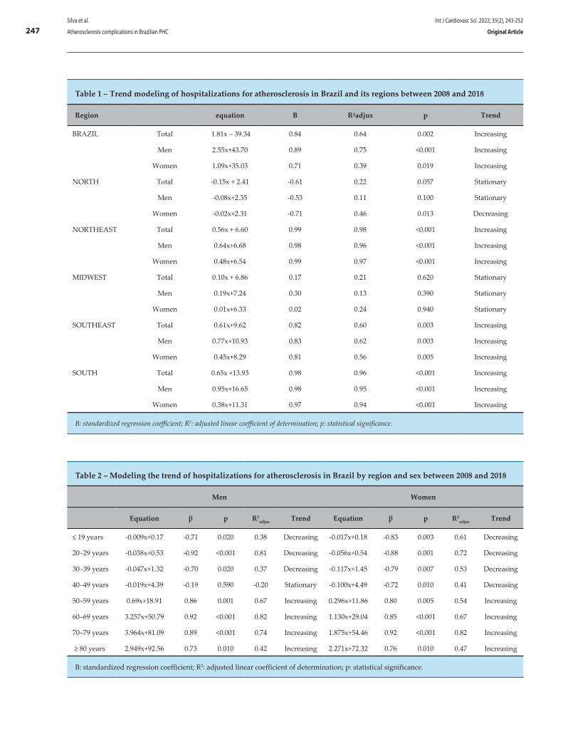
Table 1 – Trend modeling of hospitalizations for atherosclerosis in Brazil and its regions between 2008 and 2018
Region equation Β R²adjus p Trend
BRAZIL Total 1.81x – 39.34 0.84 0.64 0.002 Increasing
Men 2.55x+43.70 0.89 0.75 <0.001 Increasing
Women 1.09x+35.03 0.71 0.39 0.019 Increasing
NORTH Total -0.15x + 2.41 -0.61 0.22 0.057 Stationary
Men -0.08x+2.35 -0.53 0.11 0.100 Stationary
Women -0.02x+2.31 -0.71 0.46 0.013 Decreasing
NORTHEAST Total 0.56x + 6.60 0.99 0.98 <0.001 Increasing
Men 0.64x+6.68 0.98 0.96 <0.001 Increasing
Women 0.48x+6.54 0.99 0.97 <0.001 Increasing
MIDWEST Total 0.10x + 6.86 0.17 0.21 0.620 Stationary
Men 0.19x+7.24 0.30 0.13 0.390 Stationary
Women 0.01x+6.33 0.02 0.24 0.940 Stationary
SOUTHEAST Total 0.61x+9.62 0.82 0.60 0.003 Increasing
Men 0.77x+10.93 0.83 0.62 0.003 Increasing
Women 0.45x+8.29 0.81 0.56 0.005 Increasing
SOUTH Total 0.65x +13.93 0.98 0.96 <0.001 Increasing
Men 0.95x+16.65 0.98 0.95 <0.001 Increasing
Women 0.38x+11.31 0.97 0.94 <0.001 Increasing
Β: standardized regression coefficient; R2: adjusted linear coefficient of determination; p: statistical significance.
Table 2 – Modeling the trend of hospitalizations for atherosclerosis in Brazil by region and sex between 2008 and 2018
Men Women
Equation β p R2adjus Trend Equation β p R2
adjus Trend
≤ 19 years -0.009x+0.17 -0.71 0.020 0.38 Decreasing -0.017x+0.18 -0.83 0.003 0.61 Decreasing
20–29 years -0.038x+0.53 -0.92 <0.001 0.81 Decreasing -0.056x+0.54 -0.88 0.001 0.72 Decreasing
30–39 years -0.047x+1.32 -0.70 0.020 0.37 Decreasing -0.117x+1.45 -0.79 0.007 0.53 Decreasing
40–49 years -0.019x+4.39 -0.19 0.590 -0.20 Stationary -0.100x+4.49 -0.72 0.010 0.41 Decreasing
50–59 years 0.69x+18.91 0.86 0.001 0.67 Increasing 0.296x+11.86 0.80 0.005 0.54 Increasing
60–69 years 3.257x+50.79 0.92 <0.001 0.82 Increasing 1.130x+29.04 0.85 <0.001 0.67 Increasing
70–79 years 3.964x+81.09 0.89 <0.001 0.74 Increasing 1.875x+54.46 0.92 <0.001 0.82 Increasing
≥ 80 years 2.949x+92.56 0.73 0.010 0.42 Increasing 2.271x+72.32 0.76 0.010 0.47 Increasing
Β: standardized regression coefficient; R2: adjusted linear coefficient of determination; p: statistical significance.
Int J Cardiovasc Sci. 2022; 35(2), 243-252
247Silva et al.
Atherosclerosis complications in Brazilian PHC Original Article
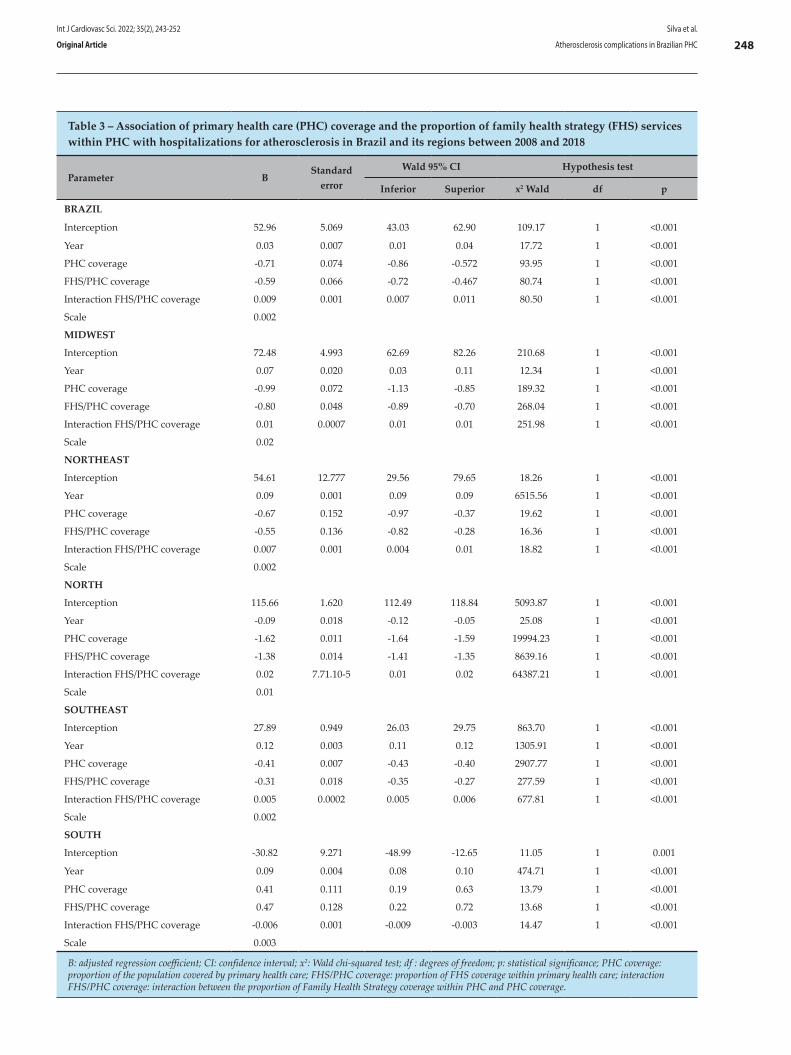
Table 3 – Association of primary health care (PHC) coverage and the proportion of family health strategy (FHS) services within PHC with hospitalizations for atherosclerosis in Brazil and its regions between 2008 and 2018
Parameter BStandard
error
Wald 95% CI Hypothesis test
Inferior Superior x2 Wald df p
BRAZIL
Interception 52.96 5.069 43.03 62.90 109.17 1 <0.001
Year 0.03 0.007 0.01 0.04 17.72 1 <0.001
PHC coverage -0.71 0.074 -0.86 -0.572 93.95 1 <0.001
FHS/PHC coverage -0.59 0.066 -0.72 -0.467 80.74 1 <0.001
Interaction FHS/PHC coverage 0.009 0.001 0.007 0.011 80.50 1 <0.001
Scale 0.002
MIDWEST
Interception 72.48 4.993 62.69 82.26 210.68 1 <0.001
Year 0.07 0.020 0.03 0.11 12.34 1 <0.001
PHC coverage -0.99 0.072 -1.13 -0.85 189.32 1 <0.001
FHS/PHC coverage -0.80 0.048 -0.89 -0.70 268.04 1 <0.001
Interaction FHS/PHC coverage 0.01 0.0007 0.01 0.01 251.98 1 <0.001
Scale 0.02
NORTHEAST
Interception 54.61 12.777 29.56 79.65 18.26 1 <0.001
Year 0.09 0.001 0.09 0.09 6515.56 1 <0.001
PHC coverage -0.67 0.152 -0.97 -0.37 19.62 1 <0.001
FHS/PHC coverage -0.55 0.136 -0.82 -0.28 16.36 1 <0.001
Interaction FHS/PHC coverage 0.007 0.001 0.004 0.01 18.82 1 <0.001
Scale 0.002
NORTH
Interception 115.66 1.620 112.49 118.84 5093.87 1 <0.001
Year -0.09 0.018 -0.12 -0.05 25.08 1 <0.001
PHC coverage -1.62 0.011 -1.64 -1.59 19994.23 1 <0.001
FHS/PHC coverage -1.38 0.014 -1.41 -1.35 8639.16 1 <0.001
Interaction FHS/PHC coverage 0.02 7.71.10-5 0.01 0.02 64387.21 1 <0.001
Scale 0.01
SOUTHEAST
Interception 27.89 0.949 26.03 29.75 863.70 1 <0.001
Year 0.12 0.003 0.11 0.12 1305.91 1 <0.001
PHC coverage -0.41 0.007 -0.43 -0.40 2907.77 1 <0.001
FHS/PHC coverage -0.31 0.018 -0.35 -0.27 277.59 1 <0.001
Interaction FHS/PHC coverage 0.005 0.0002 0.005 0.006 677.81 1 <0.001
Scale 0.002
SOUTH
Interception -30.82 9.271 -48.99 -12.65 11.05 1 0.001
Year 0.09 0.004 0.08 0.10 474.71 1 <0.001
PHC coverage 0.41 0.111 0.19 0.63 13.79 1 <0.001
FHS/PHC coverage 0.47 0.128 0.22 0.72 13.68 1 <0.001
Interaction FHS/PHC coverage -0.006 0.001 -0.009 -0.003 14.47 1 <0.001
Scale 0.003
B: adjusted regression coefficient; CI: confidence interval; x2: Wald chi-squared test; df : degrees of freedom; p: statistical significance; PHC coverage: proportion of the population covered by primary health care; FHS/PHC coverage: proportion of FHS coverage within primary health care; interaction FHS/PHC coverage: interaction between the proportion of Family Health Strategy coverage within PHC and PHC coverage.
Int J Cardiovasc Sci. 2022; 35(2), 243-252
248Silva et al.
Atherosclerosis complications in Brazilian PHCOriginal Article
of hospitalizations for atherosclerosis demonstrates clear differences, probably due to characteristics that are inherent to each country region, which include economic, demographic, cultural and social dimensions.15 Comparing the five Brazilian regions, the South and Southeast had more hospitalizations for atherosclerosis as these regions have historically had greater economic development and were thus associated with contextual effects of urbanization and a precarious lifestyle;19-21 their populations are also older. A smaller effect of PHC coverage was observed in these regions when compared to the others, which limits longitudinal monitoring of individual and collective health necessary for the control of chronic health conditions.
On the other hand, there is a large offer of predominantly private outpatient medical services in the Southeast and South regions of Brazil.22 This scenario is a consequence of the public-private relationship in the health sector of the most economically developed regions of Brazil,16 where a socioeconomic ecosystem pressures the health system to follow models of individual-outpatient-curative actions provided by private services/insurers, whereas less longitudinal and preventive follow-up is offered by the public system, especially PHC. This is driven by less public funding for health protection due to the bias of greater private access to health. However, the objectives of the private subsystem are to minimize expenses, reducing service provision, and to transfer health responsibilities to the individual.16
In addition to economic and organizational aspects of the health system, the Southeast and South regions of Brazil present individual factors such as an atherogenic diet and excess abdominal adiposity,23 a high prevalence of smoking (24.5% among men and 19.7% among women), and a prevalence of hypertension above 30%. Regarding obesity, its prevalence was higher than the national average, and the physical inactivity rate was 41.73%; cardiovascular risk was thus classified as moderate for women (11.8%) and high for men (24.7%).24
In the South, older women presented lack of physical activity as the most prevalent cardiovascular risk factor. Among men, the main cardiovascular risk factor was the limited intake of fruits and vegetables, although alcohol abuse and smoking were also significant.25 The Southeast region had similar results, where men aged 20 to 49 years had 2 or more risk factors for cardiovascular disease,26 mainly obesity and physical inactivity.27
The Northeast region had the greatest PHC coverage in the country. However, this coverage was not able to prevent the progression of complications caused by atherogenic disease, despite mitigating their damage. Greater health service coverage also requires social support actions for the adoption of healthy behaviors, as well as intersectoral measures that improve quality of life and access to food, which are still limited in the SUS.16
The confluence of strong PHC and impacting intersectoral policies could circumvent factors such as low educational and economic levels among PHC users in the Northeast and North of Brazil,28 as CNCDs more intensely affect people from vulnerable groups.29 A study with students from Campina Grande–PB indicated a percentage of regular or occasional smokers of 9.8% and 31.3% of experimental smokers.30 A study in the state of Sergipe indicated that 77.5% of the participants practiced insufficient physical activity, 57.5% drank soft drinks excessively, 15.5% were overweight or obese, and 49.2% claimed to have consumed alcohol in the previous 30 days.31 In the analysis of older age groups, the scenario was also severe, with sedentary lifestyle being reported by 39% of adults and 67.5% of the older population.32
Despite the growing number of hospitalizations for atherosclerosis in Brazil, it is worth highlighting the role of PHC, and specifically FHS services, in the clinical management, management of risk factors, and detection, treatment, and planning of preventive actions. From the perspective that Brazil will have 21.7% of the population aged over 60 years by the year 2040, obesity and diabetes control, as well as individual and collective anti-smoking approaches, become a priority within PHC and FHS practices, such as the promotion of physical activities, dietary guidance, and longitudinal monitoring.33
The adoption of such measures can not only reduce the rate of hospitalizations for CVDs but also minimize spending on health care, given that CVDs are responsible for most costs of hospital admissions in the SUS.34,35 It is stipulated that a 10% reduction in worldwide mortality from CVDs could result in savings of up to 25 billion dollars per year.36 Considering that this is the second leading cause of death in Brazil, the amount saved nationally could be invested in expanding the coverage of the health network, strengthening primary and specialized care and increasing disease prevention and screening.
Int J Cardiovasc Sci. 2022; 35(2), 243-252
249Silva et al.
Atherosclerosis complications in Brazilian PHC Original Article
Based on a more complex approach to the concept of health, valued by the work process and ways of caring within the FHS, the important impact of this health care model on the frequency of CVD in the population is noticed.37 The biological, sociocultural, and economic multi-causality in the manifestation of CNCDs, especially CVDs, indicates the need for action on social determinants of health, in addition to the clinical aspects that are inherent to atherosclerosis and other CVDs. In this sense, the FHS becomes an important health care tool for the Brazilian population.38 Educational interventions aimed at the most affected population evaluated in this study can be valuable instruments for the empowerment of these people and for strengthening social support networks, thus generating capital and social cohesion to reduce risk factors and encourage healthier lifestyle habits.39
As for the limitations of this study, it is possible that the ability to diagnose acute events associated to atherosclerosis without complementary examinations is limited in the most economically vulnerable regions, which could lead to underdiagnosis because of the difficulty in accessing sensitive complementary exams related to the cause of acute events. Furthermore, it is not possible to infer that individuals from a region with more records of atherosclerosis complications are at greater risk of developing it, which would constitute an ecological fallacy. Studies with other methodological designs are important to directly define cause and risk relationships for atherosclerosis.
Conclusion
The incidence of atherosclerosis complications in Brazil is rising, possibly motivated by individual lifestyle and health care aspects as well as ecological conditions related to geographic regionalization and distinct socioeconomic and cultural components within the country. There is an evident need for public health policies that are differentiated by age group and sex due to the different magnitudes of hospitalization trends for atherosclerosis, especially from the fifth decade of life on.
The effect of PHC in mitigating the progression of atherosclerotic complications is noteworthy, being notably influenced by its coverage potential and, possibly, by its quality of care. This result could be expanded with the implementation of comprehensive and intersectoral policies that address conditions of production and work, food, leisure, and physical activity, given that more socioeconomically developed regions had the worst trends in hospitalizations in standardized comparisons.
Author contributions
Conception and design of the research: Silva EJ, Gomes Junior FS, Firmiano JVB, Silva NR, Carlini WA, Lopes JM. Acquisition of data: Silva EJ, Gomes Junior FS, Firmiano JVB, Silva NR, Carlini WA. Analysis and interpretation of the data: Silva EJ, Gomes Junior FS, Firmiano JVB, Silva NR, Carlini WA, Lopes JM, Guedes MBOG, Lopes MR. Statistical analysis: Lopes JM. Writing of the manuscript: Silva EJ, Gomes Junior FS, Firmiano JVB, Silva NR, Carlini WA, Lopes JM, Guedes MBOG, Lopes MR. Critical revision of the manuscript for intellectual content: Lopes JM, Guedes MBOG, Lopes MR.
Potential Conflict of InterestNo potential conflict of interest relevant to this article
was reported.
Sources of FundingThere were no external funding sources for this study.
Study AssociationThis study is not associated with any thesis or
dissertation work.
Ethics approval and consent to participateThis article does not contain any studies with human
participants or animals performed by any of the authors.
Int J Cardiovasc Sci. 2022; 35(2), 243-252
250Silva et al.
Atherosclerosis complications in Brazilian PHCOriginal Article
1. Faludi AA, Izar MCO, Saraiva JFK, Chacra APM, Bianco HT, Afiune Neto A, et al. Atualização da diretriz brasileira de dislipidemias e prevenção da aterosclerose – 2017. Arq Bras Cardiol. 2017; 109(2Supl.1):1-76.
2. Benjamin EJ, Blaha MJ, Chiuve SE, Cushman M, Das SR, Deo R, et al. Heart Disease and Stroke Statistics—2017 Update A Report From the American Heart Association. Circulation. 2017;135(10):e146-e603.
3. Rocha RS, Conti RAS. Risco cardiovascular: abordagem dentro da empresa. Rev Bras Med Trab. 2005;3(1):10-21.
4. Back GI, Coutinho MS, Freitas SF, Pires MM, Zunino JN, Ribeiro RQ. Serum lipids in school kids and adolescents from Florianópolis, SC, Brazil: Healthy Floripa 2040 study. Arq Bras Cardiol. 2005;85 (2):85-91.
5. Macedo LET, Faerstein E. Cholesterol and prevention of atherosclerotic events: limits of a new frontier. Rev Saude Publica. 2017;51:2 doi: .org/10.1590/S15-18-8787.2017051006416
6. Garcia MMO, Lima PRP, Correia LCL. Prognostic value of endothelial function in patients with atherosclerosis: systematic review. Arq Bras Cardiol. 2012; 99(3):857-65.
7. Gambetta JC, Araujo MB, Chiesa P. Dislipemias en la edad pediátrica. Importancia del diagnóstico y tratamiento precoces. Rev Urug Cardiol. 2019; 34(3):208-38.
8. Mendes EV. As redes de atenção à saúde. Cienc Saude Coletiva. 2010; 15(5):2297-305.
9. Moretti PGS, Fedosse E. Family Health Support Center: Impact on Ambulatory Care Sensitive Conditions. Fisioterapia e Pesquisa em D. 2016; 23(3):241-7.
10. Morosini MV, Fonseca AF, Lima LD. Política Nacional de Atenção Básica 2017: retrocessos e riscos para o Sistema Único de Saúde. Saúde em Debate.. 2018; 42(116):11-24.
11. Malta DC, Santos MAS, Stopa SR, Vieira JEB, Melo EA, Reis AAC. A cobertura da Estratégia de Saúde da Família (ESF) no Brasil, segundo a Pesquisa Nacional de Saúde, 2013. Cienc Saude Coletiva. 2016; 21(2):327-38.
12. Brasil, Ministério da Saúde. Banco de dados do Sistema Único de Saúde - DATASUS. Informações de Saúde, Sistema de Informações sobre Mortalidade. Disponível em http://www.datasus.gov.br/catalogo/sim.htm. [acesso em 15 de maio de 2020]
13. IBGE, Instituto Brasileiro de Geografia e Estatística. Panorama das cidades brasileiras [Internet]. Rio de Janeiro: Instituto Brasileiro de Geografia e Estatística; 2020 Disponível em: https://cidades.ibge.gov.br/. [acesso em 26 de agosto de 2020]
14. Brasil, Ministério da Saúde. e-Gestor Atenção Básica. Informação e Gestão da Atenção Básica. Brasília: Secretaria de Atenção Primária à Saúde. Ministério da Saúde. 2017. Disponível em https://egestorab.saude.gov.br/. [acesso em 15 de setembro de 2020]
15. Dedecca CS, Trovão CB, Souza LF. Desenvolvimento e Equidade: desafios do crescimento brasileiro. Novos Estudos CEBRAP. 2014; 98:23-41.
16. Machado CV, Lima LD, Baptista TWF. Health policies in Brazil in times of contradiction: paths and pitfalls in the construction of a universal system. Cad Saude Publica. 2017; 33(suppl2):e00129616.
17. Machin R, Couto MT, Silva GS, Schraiber LB, Gomes R, Santos FW, et al. Concepções de gênero, masculinidade e cuidados em saúde: estudo com profissionais de saúde da atenção primária. Cienc Saude Coletiva. 2011; 16(11):4503-12.
18. Sangiorgi G, Roversi S, Zoccai GB, Modena MG, Servadei F, Ippoliti A, et al. Sex-related differences in carotid plaque features and inflammation. J Vasc Surg. 2013;57(2):338-344.
19. Santos MG, Pegoraro M, Sandrini F, Macuco EC. Risk Factors for the Development of Atherosclerosis in Childhood and Adolescence. Arq Bras Cardiol. 2008; 90(4):301-8.
20. Pereira IFS, Spyrides MHC, Andrade LMB. Estado nutricional de idosos no Brasil: uma abordagem multinível. Cad Saude Publica. 2016; 32(5):e00178814.
21. Simões CF, Lopes WA, Remor JM, Locateli JC, Lima FB, Santos TLC, et al. Prevalence of weight excess in Brazilian children and adolescents: a systematic review. Rev Bras Cineantropom Desempenho Hum. 2018; 20(4):517-31.
22. Scheffer M, Cassenote A, Guilloux AGA, Biancarelli A, Miotto BA, Mainardi GM, et al. Demografia médica no Brasil 2018. São Paulo: Departamento de Medicina Preventiva da Faculdade de Medicina da USP; Conselho Regional de Medicina do Estado de São Paulo; Conselho Federal de Medicina, 2018. [Internet] [Acesso em 2020 13 maio] Disponível em: http://www.epsjv.fiocruz.br/sites/default/files/files/DemografiaMedica2018%20(3).pdf.
23. Beck CC, Lopes AS, Giuliano ICB, Borgatto AF. Fatores de risco cardiovascular em adolescentes de município do sul do Brasil: prevalência e associações com variáveis sociodemográficas. Rev Bras Epidemiol. 2011; 14(1):36-49.
24. Rissardo JP, Caprara ALF, Prado ALC, Leite MTB. Investigation of the cardiovascular risk profile in a south Brazilian city: surveys from 2012 to 2016. Arq Neuro-Psiquiatr. 2018; 76(4):219-24.
25. Medeiros PA, Cembranel F, Figueiró TH, Souza BB, Antes DL, Silva DAS, et al. Prevalence and simultaneity of cardiovascular risk factors in elderly participants of a population-based study in southern Brazil. Rev Bras Epidemiol. 2019; 22:e190064.
26. Eyken EB, Moraes CL. Prevalência de fatores de risco para doenças cardiovasculares entre homens de uma população urbana do Sudeste do Brasil. Cad Saude Publica. 2009; 25(1):111-23.
27. Castro LCV, Franceschini SCC, Priore SE, Pelúzio MCG. Nutrição e doenças cardiovasculares: os marcadores de risco em adultos. Rev Nutr. 2004; 17(3):369-77.
28. Guibu IA, Moraes JC, Guerra Junior AA, Costa EA, Acurcio FA, Costa KS, et al. Main characteristics of patients of primary health care services in Brazil. Rev Saude Publica. 2017; 51(Suppl2):17s.
29. Melo SPS, Cesse EAP, Lira PIC, Rissin A, Cruz RSB, Batista FM. Doenças crônicas não transmissíveis e fatores associados em adultos numa área urbana de pobreza do nordeste brasileiro. Cienc Saude Coletiva. 2019; 24(8):3159-68.
30. Dantas DRG, Machado Neto AS, Matos GS, Figueiredo SG, Pinto IHG, Marques AA, et al. Prevalência e Risco de Tabagismo entre Estudantes do Ensino Médio em Cidade do Nordeste do Brasil. Port J Public Health. 2017; 35(1):44-51.
31. Silva FMA, Smith-Menezes A, Duarte MFS. Consumption of fruits and vegetables associated with other risk behaviors among adolescents in Northeast Brazil. Rev Paul Pediatr. 2016 Sep; 34(3):309-15.
32. Siqueira FV, Facchini LA, Piccini RX, Tomasi E, Thumé E, Silveira DS. et al. Atividade física em adultos e idosos residentes em áreas de abrangência de unidades básicas de saúde de municípios das regiões Sul e Nordeste do Brasil. Cad Saude Publica. 2008; 24(1):39-54.
33. Laslett LJ, Alagona P, Clark BA, Drozda Jr JP, Saldivar F, Wilson SR, et al. The worldwide environment of cardiovascular disease: prevalence, diagnosis, therapy, and policy issues: a report from the American College of Cardiology. J Am Coll Cardiol. 2012 Dec; 60(25 Suppl): S1-49.
References
Int J Cardiovasc Sci. 2022; 35(2), 243-252
251Silva et al.
Atherosclerosis complications in Brazilian PHC Original Article

This is an open-access article distributed under the terms of the Creative Commons Attribution License
34. Schimdt MI, Duncan BB, Azevedo e Silva G, Menezes AM, Monteiro CA, Barreto SM, et al. Doenças crônicas não transmissíveis no Brasil: carga e desafios atuais. Lancet 2011; 377(9781):1949-61. . doi: 10.1590/51413-81232004000400011
35. Song P, Fang Z, Wang H, Cai Y, Rahimi K, Zhu Y, et al. Global and regional prevalence, burden, and risk factors for carotid atherosclerosis: a systematic review, meta-analysis, and modelling study. Lancet Glob Health. 2020; 8(5): e721–29.
36. Smith SC, Collins A, Ferrari R, Holmes Jr DR, Logstrup S, McGhie DV, et al. Our time: a call to save preventable death from cardiovascular
disease (heart disease and stroke). J Am Coll Cardiol. 2012; 60(22):2343-8.
37. Macinko J, Mendonça CS. The Family Health Strategy, a strong model of Primary Health Care that delivers results. Saúde Debate. Rio de Janeiro; 42 (1): 18-37. DOI: 10.1590/0103-11042018s102.
38. Lentsck, MH, Matias TAF. Internações por doenças cardiovasculares e a cobertura da estratégia saúde da família. Rev Latino Am Enferm. 2015;23(4):611-9.
39. Marmot M, Wilkinson RG. Social determinants of health. 2nd ed New York, USA: Oxford University Press; 2011.
Int J Cardiovasc Sci. 2022; 35(2), 243-252
252Silva et al.
Atherosclerosis complications in Brazilian PHCOriginal Article
Abstract
The regular practice of physical exercise as a non-pharmacological treatment of arterial hypertension (AH) has been encouraged due to causing a series of physiological responses in the cardiovascular system, such as the production of vasoactive substances, including nitric oxide (NO). NO is a relaxation factor released by the endothelium, and the decrease in its bioavailability is related to coronary and arterial diseases, such as AH. This study aimed to perform an integrative literature review to elucidate the effect of physical training on NO levels in patients with AH and to establish a relationship between these levels and blood pressure (BP) control. A literature review was was performed by searching PubMed / MEDLINE, Lilacs, Scielo, Cinahl and Embase databases. The search string used was ("arterial hypertension" OR hypertension) AND (exercise OR "physical exercise" OR "aerobic exercise" OR "exercise training" or "physical activity") AND ("nitric oxide"). We included fully available controlled and uncontrolled clinical trials published in English and Portuguese languages in the last 10 years. The review consisted of 16 articles, of which 13 reported an increase in NO production after the physical training intervention, and three studies found no change. In addition, 15 studies observed a reduction in
BP after the intervention. In conclusion, regular practice of physical exercises, advocating moderate intensity, can improve NO bioavailability in pre-hypertensive and hypertensive individuals, which seems to be one of the mechanisms responsible for BP reduction.
Introduction
Arterial hypertension (AH) is characterized as a multifactorial clinical condition and considered one of the main risk factors for cardiovascular morbidity and mortality. In addition to a sustained elevation in blood pressure (BP), AH is also associated with metabolic disorders and functional and structural changes in target organs, which can be aggravated by the presence of other risk factors and is responsible for several other complications.1
The practice of physical exercise as a non-pharmacological therapeutic approach to AH has been increasingly encouraged by health professionals, as it causes many physiological responses in body systems, especially in the cardiovascular system.2 Physical training, when performed regularly, causes important autonomic and hemodynamic adaptations, as well as humoral changes related to the production of vasoactive substances, such as nitric oxide (NO).3 These changes are responsible for the reduction or even normalization of the BP levels in patients with mild to moderate hypertension, using or not using medications.1,4,5
NO, a relaxation factor released by the endothelium,6 is a gaseous mediator responsible for a variety of physiological phenomena,7 and a decrease in its
Blood Pressure; Hypertension; Exercise; Physical Conditioning Human; Nitric Oxide; Endothelium Dependent Relaxing Factors; Vasoactive Substances; Cardiovascular System.
Keywords
Int J Cardiovasc Sci. 2022; 35(2), 253-264
253
REVIEW ARTICLE
Effect of Physical Training on Nitric Oxide Levels in Patients with Arterial Hypertension: An Integrative ReviewTábata de Paula Facioli,1,2,3 Mariana Colombini Buranello,3 Eloisa Maria Gatti Regueiro,1,4 Renata Pedrolongo Basso-Vanelli,1,5 Marina de Toledo Durand1
Universidade de Ribeirão Preto,1 Ribeirão Preto, SP – Brazil. Universidade de São Paulo - Faculdade de Medicina de Ribeirão Preto,2 Ribeirão Preto, SP – Brazil. Universidade de Franca,3 Franca, SP – Brazil. Centro Universitário Barão de Mauá,4 Ribeirão Preto, SP – Brazil. Universidade Federal de São Carlos,5 São Carlos, SP – Brazil.
DOI: https://doi.org/10.36660/ijcs.20200244
Mailing Address: Marina Toledo DurandAv. Costábile Romano, 2201. Postal Code: 14096-900, Ribeirão Preto, São Paulo, SP – Brazil.E-mail: [email protected]
Manuscript received on August 13, 2020; reviewed on November 19, 2020; accepted on January 10, 2021.
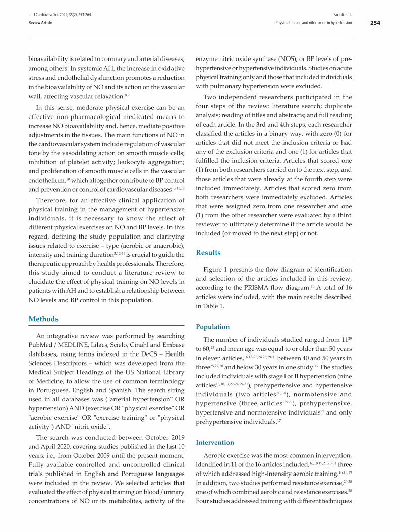
enzyme nitric oxide synthase (NOS), or BP levels of pre-hypertensive or hypertensive individuals. Studies on acute physical training only and those that included individuals with pulmonary hypertension were excluded.
Two independent researchers participated in the four steps of the review: literature search; duplicate analysis; reading of titles and abstracts; and full reading of each article. In the 3rd and 4th steps, each researcher classified the articles in a binary way, with zero (0) for articles that did not meet the inclusion criteria or had any of the exclusion criteria and one (1) for articles that fulfilled the inclusion criteria. Articles that scored one (1) from both researchers carried on to the next step, and those articles that were already at the fourth step were included immediately. Articles that scored zero from both researchers were immediately excluded. Articles that were assigned zero from one researcher and one (1) from the other researcher were evaluated by a third reviewer to ultimately determine if the article would be included (or moved to the next step) or not.
Results
Figure 1 presents the flow diagram of identification and selection of the articles included in this review, according to the PRISMA flow diagram.15 A total of 16 articles were included, with the main results described in Table 1.
Population
The number of individuals studied ranged from 1129 to 60,23 and mean age was equal to or older than 50 years in eleven articles,16,18-22,24,26,29-31 between 40 and 50 years in three25,27,28 and below 30 years in one study.17 The studies included individuals with stage I or II hypertension (nine articles16,18,19,22-24,29-31), prehypertensive and hypertensive individuals (two articles20,31), normotensive and hypertensive (three articles27-29), prehypertensive, hypertensive and normotensive individuals25 and only prehypertensive individuals.17
Intervention
Aerobic exercise was the most common intervention, identified in 11 of the 16 articles included,16,18,19,21,25-31 three of which addressed high-intensity aerobic training.16,18,19 In addition, two studies performed resistance exercise,20,28 one of which combined aerobic and resistance exercises.28 Four studies addressed training with different techniques
bioavailability is related to coronary and arterial diseases, among others. In systemic AH, the increase in oxidative stress and endothelial dysfunction promotes a reduction in the bioavailability of NO and its action on the vascular wall, affecting vascular relaxation.8,9
In this sense, moderate physical exercise can be an effective non-pharmacological medicated means to increase NO bioavailability and, hence, mediate positive adjustments in the tissues. The main functions of NO in the cardiovascular system include regulation of vascular tone by the vasodilating action on smooth muscle cells; inhibition of platelet activity; leukocyte aggregation; and proliferation of smooth muscle cells in the vascular endothelium,10 which altogether contribute to BP control and prevention or control of cardiovascular diseases.3,11,12
Therefore, for an effective clinical application of physical training in the management of hypertensive individuals, it is necessary to know the effect of different physical exercises on NO and BP levels. In this regard, defining the study population and clarifying issues related to exercise – type (aerobic or anaerobic), intensity and training duration3,12-14 is crucial to guide the therapeutic approach by health professionals. Therefore, this study aimed to conduct a literature review to elucidate the effect of physical training on NO levels in patients with AH and to establish a relationship between NO levels and BP control in this population.
Methods
An integrative review was performed by searching PubMed / MEDLINE, Lilacs, Scielo, Cinahl and Embase databases, using terms indexed in the DeCS – Health Sciences Descriptors – which was developed from the Medical Subject Headings of the US National Library of Medicine, to allow the use of common terminology in Portuguese, English and Spanish. The search string used in all databases was ("arterial hypertension" OR hypertension) AND (exercise OR "physical exercise" OR "aerobic exercise" OR "exercise training" or "physical activity") AND "nitric oxide".
The search was conducted between October 2019 and April 2020, covering studies published in the last 10 years, i.e., from October 2009 until the present moment. Fully available controlled and uncontrolled clinical trials published in English and Portuguese languages were included in the review. We selected articles that evaluated the effect of physical training on blood / urinary concentrations of NO or its metabolites, activity of the
Int J Cardiovasc Sci. 2022; 35(2), 253-264
254Facioli et al.
Physical training and nitric oxide in hypertensionReview Article
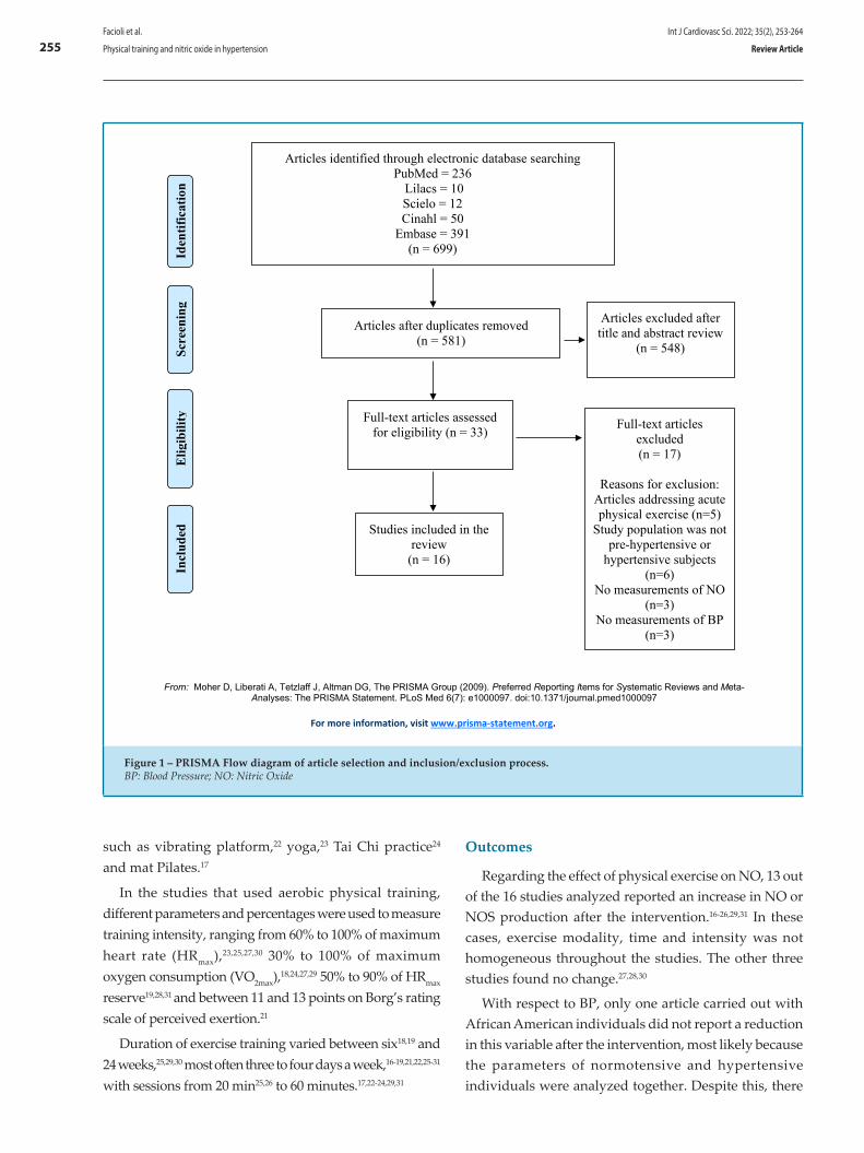
such as vibrating platform,22 yoga,23 Tai Chi practice24 and mat Pilates.17
In the studies that used aerobic physical training, different parameters and percentages were used to measure training intensity, ranging from 60% to 100% of maximum heart rate (HRmax),23,25,27,30 30% to 100% of maximum oxygen consumption (VO2max),18,24,27,29 50% to 90% of HRmax reserve19,28,31 and between 11 and 13 points on Borg’s rating scale of perceived exertion.21
Duration of exercise training varied between six18,19 and 24 weeks,25,29,30 most often three to four days a week,16-19,21,22,25-31 with sessions from 20 min25,26 to 60 minutes.17,22-24,29,31
Outcomes
Regarding the effect of physical exercise on NO, 13 out of the 16 studies analyzed reported an increase in NO or NOS production after the intervention.16-26,29,31 In these cases, exercise modality, time and intensity was not homogeneous throughout the studies. The other three studies found no change.27,28,30
With respect to BP, only one article carried out with African American individuals did not report a reduction in this variable after the intervention, most likely because the parameters of normotensive and hypertensive individuals were analyzed together. Despite this, there
Int J Cardiovasc Sci. 2022; 35(2), 253-264
255Facioli et al.
Physical training and nitric oxide in hypertension Review Article
From: Moher D, Liberati A, Tetzlaff J, Altman DG, The PRISMA Group (2009). Preferred Reporting Items for Systematic Reviews and Meta-Analyses: The PRISMA Statement. PLoS Med 6(7): e1000097. doi:10.1371/journal.pmed1000097
For more information, visit www.prisma-statement.org.
Articles identified through electronic database searching PubMed = 236
Lilacs = 10 Scielo = 12 Cinahl = 50
Embase = 391 (n = 699)
Scre
enin
g In
clud
ed
Elig
ibili
ty
Iden
tific
atio
n
Articles after duplicates removed (n = 581)
Full-text articles assessed for eligibility (n = 33)
Articles excluded after title and abstract review
(n = 548)
Full-text articles excluded (n = 17)
Reasons for exclusion:
Articles addressing acute physical exercise (n=5)
Study population was not pre-hypertensive or
hypertensive subjects (n=6)
No measurements of NO (n=3)
No measurements of BP (n=3)
Studies included in the review
(n = 16)
Figure 1 – PRISMA Flow diagram of article selection and inclusion/exclusion process.BP: Blood Pressure; NO: Nitric Oxide
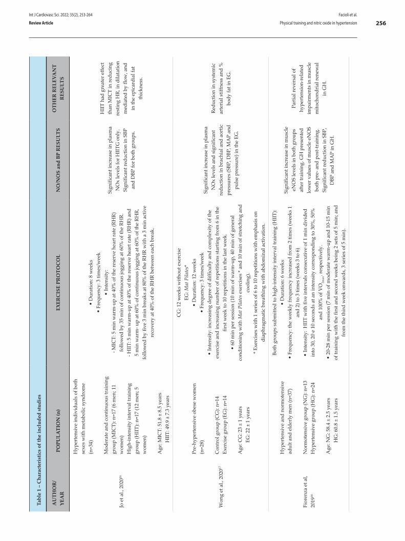
Tabl
e 1
– C
hara
cter
istic
s of
the
incl
uded
stu
dies
AU
THO
R/
YEA
RPO
PULA
TIO
N (n
)EX
ERC
ISE
PRO
TOC
OL
NO
/NO
S an
d BP
RES
ULT
SO
THER
REL
EVA
NT
RES
ULT
S
Jo e
t al.,
202
016
Hyp
erte
nsiv
e in
divi
dual
s of
bot
h se
xes
with
met
abol
ic s
yndr
ome
(n=3
4)
Mod
erat
e an
d co
ntin
uous
trai
ning
gr
oup
(MIC
T): n
=17
(6 m
en; 1
1 w
omen
)H
igh-
inte
nsity
inte
rval
trai
ning
gr
oup
(HII
T): n
=17
(12
men
; 5
wom
en)
Age
: MIC
T: 5
1.8
± 8.
5 ye
ars
H
IIT:
49.
9 ±
7.3
year
s
• D
urat
ion:
8 w
eeks
• Fr
eque
ncy:
3 ti
mes
/wee
k•
Inte
nsity
:- M
ICT:
5 m
in w
arm
-up
at 4
0% o
f the
rese
rve
hear
t rat
e (R
HR)
fo
llow
ed b
y 35
min
of c
ontin
uous
jogg
ing
at 6
0% o
f the
RH
R.- H
IIT:
5 m
in w
arm
-up
at 4
0% o
f the
rese
rve
hear
t rat
e (R
HR)
and
5
min
war
m- u
p at
60%
of c
ontin
uous
jogg
ing
at 6
0% o
f the
RH
R,
follo
wed
by
five
3 m
in b
reak
s at
80%
of t
he R
HR
with
a 3
min
act
ive
reco
very
at 4
0% o
f the
RH
R be
twee
n ea
ch b
reak
.
Sign
ifica
nt in
crea
se in
pla
sma
NO
x le
vels
for H
IITG
onl
y.
Sign
ifica
nt re
duct
ion
in S
BP
and
DBP
for b
oth
grou
ps.
HII
T ha
d gr
eate
r effe
ct
than
MIC
T in
redu
cing
re
stin
g H
R, in
dila
tatio
n m
edia
ted
by fl
ow, a
nd
in th
e ep
icar
dial
fat
thic
knes
s.
Won
g et
al.,
202
017
Pre-
hype
rten
sive
obe
se w
omen
(n
=28)
Con
trol
gro
up (C
G):
n=14
Exer
cise
gro
up (E
G):
n=14
Age
: CG
: 23
± 1
year
s
EG: 2
2 ±
1 ye
ars
CG
: 12
wee
ks w
ithou
t exe
rcis
eEG
: Mat
Pila
tes*
• D
urat
ion:
12
wee
ks
• Fr
eque
ncy:
3 ti
mes
/wee
k•
Inte
nsity
: inc
reas
ing
degr
ee o
f diff
icul
ty a
nd c
ompl
exity
of t
he
exer
cise
and
incr
easi
ng n
umbe
r of r
epet
ition
s st
artin
g fr
om 6
in th
e fir
st w
eek
to 1
0 re
petit
ions
in th
e la
st w
eek.
• 60
min
per
ses
sion
(10
min
of w
arm
-up,
40
min
of g
ener
al
cond
ition
ing
with
Mat
Pila
tes e
xerc
ises
* an
d 10
min
of s
tret
chin
g an
d co
olin
g).
* Exe
rcis
es w
ith 1
ser
ies
of 6
to 1
0 re
petit
ions
with
em
phas
is o
n di
aphr
agm
atic
bre
athi
ng w
ith a
bdom
inal
act
ivat
ion.
Sign
ifica
nt in
crea
se in
pla
sma
NO
x le
vels
and
sig
nific
ant
redu
ctio
n in
bra
chia
l and
aor
tic
pres
sure
s (S
BP, D
BP, M
AP
and
puls
e pr
essu
re) i
n th
e EG
.
Redu
ctio
n in
sys
tem
ic
arte
rial
stif
fnes
s an
d %
bo
dy fa
t in
EG.
Fior
enza
et a
l, 20
1918
Hyp
erte
nsiv
e an
d no
rmot
ensi
ve
adul
t and
eld
erly
men
(n=3
7)
Nor
mot
ensi
ve g
roup
(NG
): n=
13H
yper
tens
ive
grou
p (H
G):
n=24
Age
: NG
: 58.
4 ±
2.5
year
s
HG
: 60.
8 ±
1.5
year
s
Both
gro
ups
subm
itted
to h
igh-
inte
nsity
inte
rval
trai
ning
(HII
T):
• D
urat
ion:
6 w
eeks
•
Freq
uenc
y: th
e w
eekl
y fr
eque
ncy
incr
ease
d fr
om 2
tim
es (w
eeks
1
and
2) to
3 ti
mes
(wee
ks 3
to 6
)•
Inte
nsity
: H
IIT
with
five
inte
rval
s co
nsec
utiv
e of
1 m
in d
ivid
ed
into
30,
20
e 10
sec
onds
at a
n in
tens
ity c
orre
spon
ding
to 3
0%, 5
0%
and
100%
of V
O2m
ax. r
espe
ctiv
ely.
• 20
-28
min
per
ses
sion
(7 m
in o
f mod
erat
e w
arm
-up
and
10-1
5 m
in
of tr
aini
ng w
ith th
e fir
st a
nd s
econ
d w
eeks
bei
ng 2
set
s of
5 m
in; a
nd
from
the
thir
d w
eek
onw
ards
, 3 s
erie
s of
5 m
in).
Sign
ifica
nt in
crea
se in
mus
cle
eNO
S le
vels
in b
oth
grou
ps
afte
r tra
inin
g. G
H p
rese
nted
lo
wer
val
ues
of m
uscl
e eN
OS
both
pre
- and
pos
t-tra
inin
g.
Sign
ifica
nt re
duct
ion
in S
BP,
DBP
and
MA
P in
GH
.
Part
ial r
ever
sal o
f hy
pert
ensi
on-r
elat
ed
impa
irm
ents
in m
uscl
e m
itoch
ondr
ial r
enew
al
in G
H.
Int J Cardiovasc Sci. 2022; 35(2), 253-264
256Facioli et al.
Physical training and nitric oxide in hypertensionReview Article
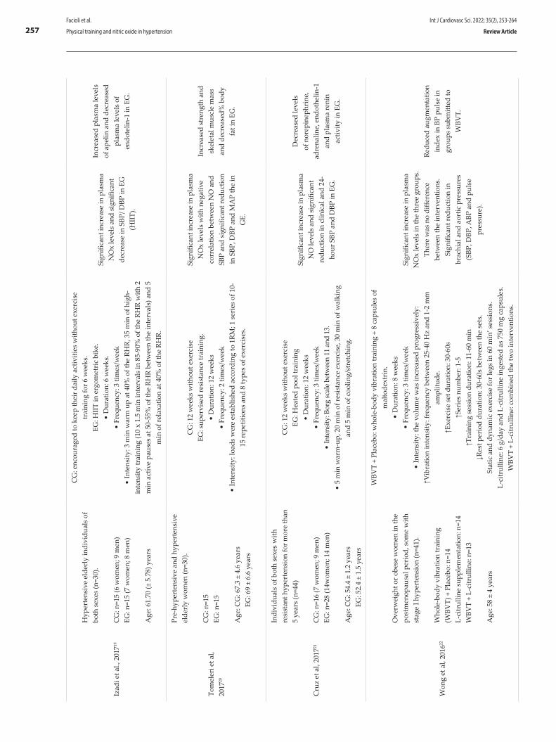
Izad
i et a
l., 2
01719
Hyp
erte
nsiv
e el
derl
y in
divi
dual
s of
bo
th s
exes
(n=3
0).
CG
: n=1
5 (6
wom
en; 9
men
)EG
: n=1
5 (7
wom
en; 8
men
)
Age
: 61.
70 (±
5.7
8) y
ears
CG
: enc
oura
ged
to k
eep
thei
r dai
ly a
ctiv
ities
with
out e
xerc
ise
trai
ning
for 6
wee
ks.
EG: H
IIT
in e
rgom
etri
c bi
ke.
• D
urat
ion:
6 w
eeks
. •
Freq
uenc
y: 3
tim
es/w
eek
• In
tens
ity: 3
min
war
m u
p at
40%
of t
he R
HR,
35
min
of h
igh-
inte
nsity
trai
ning
(10
x 1.
5 m
in in
terv
als
in 8
5-90
% o
f the
RH
R w
ith 2
m
in a
ctiv
e pa
uses
at 5
0-55
% o
f the
RH
R be
twee
n th
e in
terv
als)
and
5
min
of r
elax
atio
n at
40%
of t
he R
HR.
Sign
ifica
nt in
crea
se in
pla
sma
NO
x le
vels
and
sig
nific
ant
decr
ease
in S
BP/ D
BP in
EG
(H
IIT)
.
Incr
ease
d pl
asm
a le
vels
of
ape
lin a
nd d
ecre
ased
pl
asm
a le
vels
of
endo
telin
-1 in
EG
.
Tom
eler
i et a
l, 20
1720
Pre-
hype
rten
sive
and
hyp
erte
nsiv
e el
derl
y w
omen
(n=3
0).
CG
: n=1
5EG
: n=1
5
Age
: CG
: 67.
3 ±
4.6
year
s
EG: 6
9 ±
6.6
year
s
CG
: 12
wee
ks w
ithou
t exe
rcis
e EG
: sup
ervi
sed
resi
stan
ce tr
aini
ng.
• D
urat
ion:
12
wee
ks•
Freq
uenc
y: 2
tim
es/w
eek
• In
tens
ity: l
oads
wer
e es
tabl
ishe
d ac
cord
ing
to 1
RM; 1
ser
ies
of 1
0-15
repe
titio
ns a
nd 8
type
s of
exe
rcis
es.
Sign
ifica
nt in
crea
se in
pla
sma
NO
x le
vels
with
neg
ativ
e co
rrel
atio
n be
twee
n N
O a
nd
SBP
and
sign
ifica
nt re
duct
ion
in S
BP, D
BP a
nd M
AP
the
in
GE.
Incr
ease
d st
reng
th a
nd
skel
etal
mus
cle
mas
s an
d de
crea
sed%
bod
y fa
t in
EG.
Cru
z et
al,
2017
21
Indi
vidu
als
of b
oth
sexe
s w
ith
resi
stan
t hyp
erte
nsio
n fo
r mor
e th
an
5 ye
ars
(n=4
4)
CG
: n=1
6 (7
wom
en; 9
men
)EG
: n=2
8 (1
4wom
en; 1
4 m
en)
Age
: CG
: 54.
4 ±
1.2
year
s
EG: 5
2.4
± 1.
5 ye
ars
CG
: 12
wee
ks w
ithou
t exe
rcis
e EG
: Hea
ted
pool
trai
ning
• D
urat
ion:
12
wee
ks•
Freq
uenc
y: 3
tim
es/w
eek
• In
tens
ity: B
org
scal
e be
twee
n 11
and
13.
•
5 m
in w
arm
-up,
20
min
of r
esis
tanc
e ex
erci
se, 3
0 m
in o
f wal
king
an
d 5
min
of c
oolin
g/st
retc
hing
.
Sign
ifica
nt in
crea
se in
pla
sma
NO
leve
ls a
nd s
igni
fican
t re
duct
ion
in c
linic
al a
nd 2
4-ho
ur S
BP a
nd D
BP in
EG
.
Dec
reas
ed le
vels
of
nor
epin
ephr
ine,
ad
rena
line,
end
othe
lin-1
an
d pl
asm
a re
nin
activ
ity in
EG
.
Won
g et
al,
2016
22
Ove
rwei
ght o
r obe
se w
omen
in th
e po
stm
enop
ausa
l per
iod,
som
e w
ith
stag
e I h
yper
tens
ion
(n=4
1).
Who
le-b
ody
vibr
atio
n tr
aini
ng
(WBV
T) +
Pla
cebo
: n=1
4L-
citr
ullin
e su
pple
men
tatio
n: n
=14
WBV
T +
L-ci
trul
line:
n=1
3
Age
: 58
± 4
year
s
WBV
T +
Plac
ebo:
who
le-b
ody
vibr
atio
n tr
aini
ng +
8 c
apsu
les
of
mal
tode
xtri
n.•
Dur
atio
n: 8
wee
ks•
Freq
uenc
y: 3
tim
es/w
eek
• In
tens
ity: t
he v
olum
e w
as in
crea
sed
prog
ress
ivel
y:↑V
ibra
tion
inte
nsity
: fre
quen
cy b
etw
een
25-4
0 H
z an
d 1-
2 m
m
ampl
itude
.↑E
xerc
ise
set d
urat
ion:
30-
60s
↑Ser
ies
num
ber:
1-5
↑Tra
inin
g se
ssio
n du
ratio
n: 1
1-60
min
↓Res
t per
iod
dura
tion:
30-
60s
betw
een
the
sets
.St
atic
and
dyn
amic
exe
rcis
e fo
r leg
s in
60
min
’ ses
sion
s.L-
citr
ullin
e: 6
g/d
ay a
nd L
-citr
ullin
e in
gest
ed a
s 75
0 m
g ca
psul
es.
WBV
T +
L-ci
trul
line:
com
bine
d th
e tw
o in
terv
entio
ns.
Sign
ifica
nt in
crea
se in
pla
sma
NO
x le
vels
in th
e th
ree
grou
ps.
Ther
e w
as n
o di
ffere
nce
betw
een
the
inte
rven
tions
. Si
gnifi
cant
redu
ctio
n in
br
achi
al a
nd a
ortic
pre
ssur
es
(SBP
, DBP
, ABP
and
pul
se
pres
sure
).
Redu
ced
augm
enta
tion
inde
x in
BP
puls
e in
gr
oups
sub
mitt
ed to
W
BVT.
Int J Cardiovasc Sci. 2022; 35(2), 253-264
257Facioli et al.
Physical training and nitric oxide in hypertension Review Article
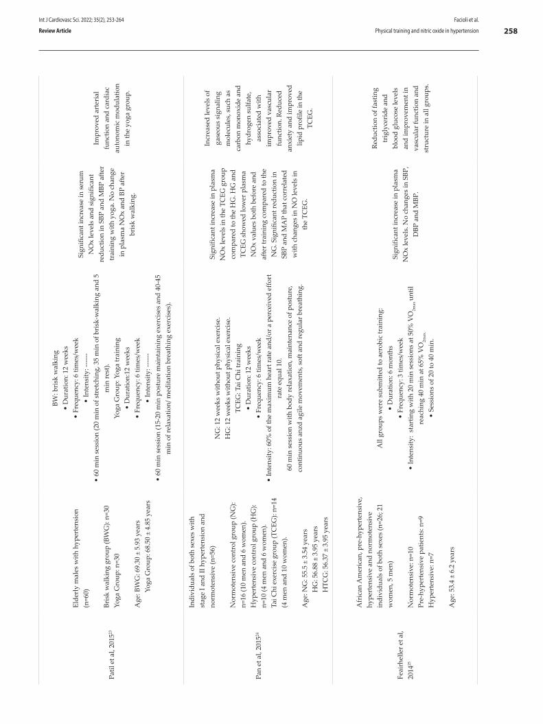
Patil
et a
l, 20
1523
Elde
rly
mal
es w
ith h
yper
tens
ion
(n=6
0)
Bris
k w
alki
ng g
roup
(BW
G):
n=30
Yoga
Gro
up: n
=30
Age
: BW
G: 6
9.30
± 5
.93
year
s
Yoga
Gro
up: 6
8.50
± 4
.85
year
s
BW: b
risk
wal
king
• D
urat
ion:
12
wee
ks
• Fr
eque
ncy:
6 ti
mes
/wee
k •
Inte
nsity
: ----
--•
60 m
in s
essi
on (2
0 m
in o
f str
etch
ing,
35
min
of b
risk
-wal
king
and
5
min
rest
).Yo
ga G
roup
: Yog
a tr
aini
ng•
Dur
atio
n:12
wee
ks
• Fr
eque
ncy:
6 ti
mes
/wee
k•
Inte
nsity
: ----
--•
60 m
in s
essi
on (1
5-20
min
pos
ture
mai
ntai
ning
exe
rcis
es a
nd 4
0-45
m
in o
f rel
axat
ion/
med
itatio
n br
eath
ing
exer
cise
s).
Sign
ifica
nt in
crea
se in
ser
um
NO
x le
vels
and
sig
nific
ant
redu
ctio
n in
SBP
and
MBP
afte
r tr
aini
ng w
ith y
oga.
No
chan
ge
in p
lasm
a N
Ox
and
BP a
fter
bris
k w
alki
ng.
Impr
oved
art
eria
l fu
nctio
n an
d ca
rdia
c au
tono
mic
mod
ulat
ion
in th
e yo
ga g
roup
.
Pan
et a
l, 20
1524
Indi
vidu
als
of b
oth
sexe
s w
ith
stag
e I a
nd II
hyp
erte
nsio
n an
d no
rmot
ensi
ve (n
=56)
Nor
mot
ensi
ve c
ontr
ol g
roup
(NG
): n=
16 (1
0 m
en a
nd 6
wom
en).
Hyp
erte
nsiv
e co
ntro
l gro
up (H
G):
n=10
(4 m
en a
nd 6
wom
en).
Tai C
hi e
xerc
ise
grou
p (T
CEG
): n=
14
(4 m
en a
nd 1
0 w
omen
).
Age
: NG
: 55.
5 ±
3.54
yea
rs
HG
: 56.
88 ±
3.9
5 ye
ars
H
TCG
: 56.
37 ±
3.9
5 ye
ars
NG
: 12
wee
ks w
ithou
t phy
sica
l exe
rcis
e.H
G: 1
2 w
eeks
with
out p
hysi
cal e
xerc
ise.
TCEG
: Tai
Chi
trai
ning
• D
urat
ion:
12
wee
ks•
Freq
uenc
y: 6
tim
es/w
eek
• In
tens
ity: 6
0% o
f the
max
imum
hea
rt ra
te a
nd/o
r a p
erce
ived
effo
rt
rate
equ
al 1
0.
60 m
in s
essi
on w
ith b
ody
rela
xatio
n, m
aint
enan
ce o
f pos
ture
, co
ntin
uous
anz
d ag
ile m
ovem
ents
, sof
t and
regu
lar b
reat
hing
.
Sign
ifica
nt in
crea
se in
pla
sma
NO
x le
vels
in th
e TC
EG g
roup
co
mpa
red
to th
e H
G. H
G a
nd
TCEG
sho
wed
low
er p
lasm
a N
Ox
valu
es b
oth
befo
re a
nd
afte
r tra
inin
g co
mpa
red
to th
e N
G. S
igni
fican
t red
uctio
n in
SB
P an
d M
AP
that
cor
rela
ted
with
cha
nges
in N
O le
vels
in
the
TCEG
.
Incr
ease
d le
vels
of
gase
ous
sign
alin
g m
olec
ules
, suc
h as
ca
rbon
mon
oxid
e an
d hy
drog
en s
ulfa
te,
asso
ciat
ed w
ith
impr
oved
vas
cula
r fu
nctio
n. R
educ
ed
anxi
ety
and
impr
oved
lip
id p
rofil
e in
the
TCEG
.
Feai
rhel
ler e
t al,
2014
25
Afr
ican
Am
eric
an, p
re-h
yper
tens
ive,
hy
pert
ensi
ve a
nd n
orm
oten
sive
in
divi
dual
s of
bot
h se
xes
(n=2
6; 2
1 w
omen
, 5 m
en)
Nor
mot
ensi
ve: n
=10
Pre-
hype
rten
sive
pat
ient
s: n
=9H
yper
tens
ive:
n=7
Age
: 53.
4 ±
6.2
year
s
All
grou
ps w
ere
subm
itted
to a
erob
ic tr
aini
ng:
• D
urat
ion:
6 m
onth
s•
Freq
uenc
y: 3
tim
es/w
eek
• In
tens
ity:
star
ting
with
20
min
ses
sion
s at
50%
VO
2max
unt
il re
achi
ng 4
0 m
in a
t 65%
VO
2max
. •
Sess
ions
of 2
0 to
40
min
.
Sign
ifica
nt in
crea
se in
pla
sma
NO
x le
vels
. No
chan
ges
in S
BP,
DBP
and
MBP
.
Redu
ctio
n of
fast
ing
trig
lyce
ride
and
bl
ood
gluc
ose
leve
ls
and
impr
ovem
ent i
n va
scul
ar fu
nctio
n an
d st
ruct
ure
in a
ll gr
oups
.
Int J Cardiovasc Sci. 2022; 35(2), 253-264
258Facioli et al.
Physical training and nitric oxide in hypertensionReview Article
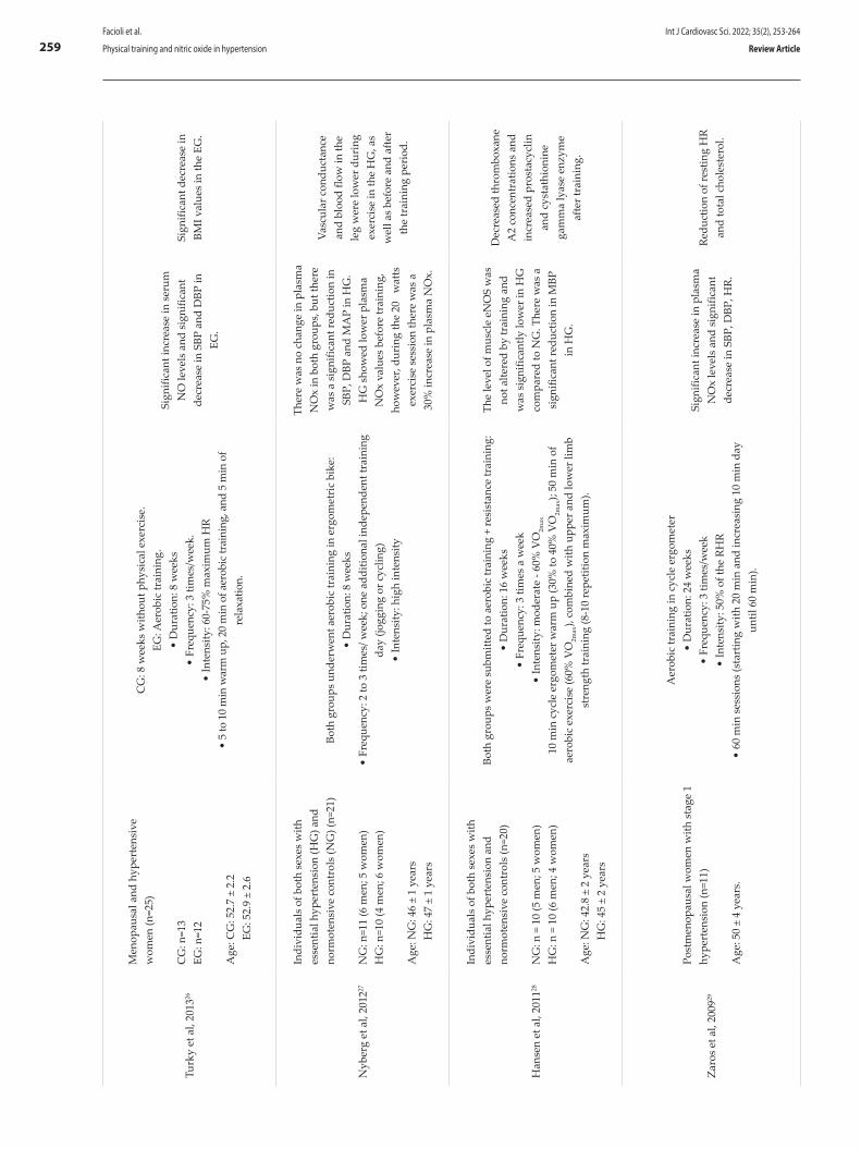
Turk
y et
al,
2013
26
Men
opau
sal a
nd h
yper
tens
ive
wom
en (n
=25)
CG
: n=1
3EG
: n=1
2
Age
: CG
: 52.
7 ±
2.2
EG
: 52.
9 ±
2.6
CG
: 8 w
eeks
with
out p
hysi
cal e
xerc
ise.
EG: A
erob
ic tr
aini
ng.
• D
urat
ion:
8 w
eeks
• Fr
eque
ncy:
3 ti
mes
/wee
k.
• In
tens
ity: 6
0-75
% m
axim
um H
R•
5 to
10
min
war
m u
p, 2
0 m
in o
f aer
obic
trai
ning
, and
5 m
in o
f re
laxa
tion.
Sign
ifica
nt in
crea
se in
ser
um
NO
leve
ls a
nd s
igni
fican
t de
crea
se in
SBP
and
DBP
in
EG.
Sign
ifica
nt d
ecre
ase
in
BMI v
alue
s in
the
EG.
Nyb
erg
et a
l, 20
1227
Indi
vidu
als
of b
oth
sexe
s w
ith
esse
ntia
l hyp
erte
nsio
n (H
G) a
nd
norm
oten
sive
con
trol
s (N
G) (
n=21
)
NG
: n=1
1 (6
men
; 5 w
omen
)H
G: n
=10
(4 m
en; 6
wom
en)
Age
: NG
: 46
± 1
year
s
HG
: 47
± 1
year
s
Both
gro
ups
unde
rwen
t aer
obic
trai
ning
in e
rgom
etri
c bi
ke:
• D
urat
ion:
8 w
eeks
• Fr
eque
ncy:
2 to
3 ti
mes
/ wee
k; o
ne a
dditi
onal
inde
pend
ent t
rain
ing
day
(jogg
ing
or c
yclin
g)•
Inte
nsity
: hig
h in
tens
ity
Ther
e w
as n
o ch
ange
in p
lasm
a N
Ox
in b
oth
grou
ps, b
ut th
ere
was
a s
igni
fican
t red
uctio
n in
SB
P, D
BP a
nd M
AP
in H
G.
HG
sho
wed
low
er p
lasm
a N
Ox
valu
es b
efor
e tr
aini
ng,
how
ever
, dur
ing
the
20
wat
ts
exer
cise
ses
sion
ther
e w
as a
30
% in
crea
se in
pla
sma
NO
x.
Vasc
ular
con
duct
ance
an
d bl
ood
flow
in th
e le
g w
ere
low
er d
urin
g ex
erci
se in
the
HG
, as
wel
l as
befo
re a
nd a
fter
the
trai
ning
per
iod.
Han
sen
et a
l, 20
1128
Indi
vidu
als
of b
oth
sexe
s w
ith
esse
ntia
l hyp
erte
nsio
n an
d no
rmot
ensi
ve c
ontr
ols
(n=2
0)
NG
: n =
10
(5 m
en; 5
wom
en)
HG
: n =
10
(6 m
en; 4
wom
en)
Age
: NG
: 42.
8 ±
2 ye
ars
H
G: 4
5 ±
2 ye
ars
Both
gro
ups
wer
e su
bmitt
ed to
aer
obic
trai
ning
+ re
sist
ance
trai
ning
:•
Dur
atio
n: 1
6 w
eeks
• Fr
eque
ncy:
3 ti
mes
a w
eek
• In
tens
ity: m
oder
ate
- 60%
VO
2max
10 m
in c
ycle
erg
omet
er w
arm
up
(30%
to 4
0% V
O2m
ax);
50 m
in o
f ae
robi
c ex
erci
se (6
0% V
O2m
ax),
com
bine
d w
ith u
pper
and
low
er li
mb
stre
ngth
trai
ning
(8-1
0 re
petit
ion
max
imum
).
The
leve
l of m
uscl
e eN
OS
was
no
t alte
red
by tr
aini
ng a
nd
was
sig
nific
antly
low
er in
HG
co
mpa
red
to N
G. T
here
was
a
sign
ifica
nt re
duct
ion
in M
BP
in H
G.
Dec
reas
ed th
rom
boxa
ne
A2
conc
entr
atio
ns a
nd
incr
ease
d pr
osta
cycl
in
and
cyst
athi
onin
e ga
mm
a ly
ase
enzy
me
afte
r tra
inin
g.
Zaro
s et
al,
2009
29
Post
men
opau
sal w
omen
with
sta
ge 1
hy
pert
ensi
on (n
=11)
Age
: 50
± 4
year
s.
Aer
obic
trai
ning
in c
ycle
erg
omet
er•
Dur
atio
n: 2
4 w
eeks
•
Freq
uenc
y: 3
tim
es/w
eek
• In
tens
ity: 5
0% o
f the
RH
R•
60 m
in s
essi
ons
(sta
rtin
g w
ith 2
0 m
in a
nd in
crea
sing
10
min
day
un
til 6
0 m
in).
Sign
ifica
nt in
crea
se in
pla
sma
NO
x le
vels
and
sig
nific
ant
decr
ease
in S
BP, D
BP, H
R.
Redu
ctio
n of
rest
ing
HR
and
tota
l cho
lest
erol
.
Int J Cardiovasc Sci. 2022; 35(2), 253-264
259Facioli et al.
Physical training and nitric oxide in hypertension Review Article
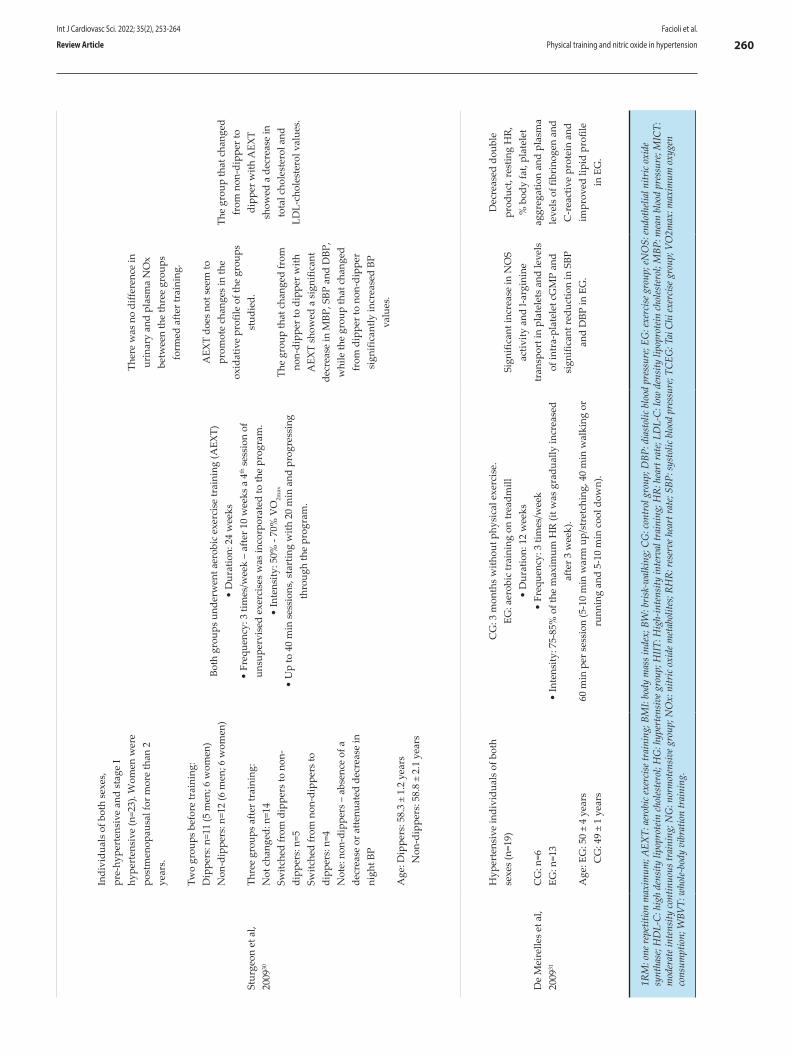
Stur
geon
et a
l, 20
0930
Indi
vidu
als
of b
oth
sexe
s,
pre-
hype
rten
sive
and
sta
ge I
hype
rten
sive
(n=2
3). W
omen
wer
e po
stm
enop
ausa
l for
mor
e th
an 2
ye
ars.
Two
grou
ps b
efor
e tr
aini
ng:
Dip
pers
: n=1
1 (5
men
; 6 w
omen
)N
on-d
ippe
rs: n
=12
(6 m
en; 6
wom
en)
Thre
e gr
oups
afte
r tra
inin
g:N
ot c
hang
ed: n
=14
Switc
hed
from
dip
pers
to n
on-
dipp
ers:
n=5
Switc
hed
from
non
-dip
pers
to
dipp
ers:
n=4
Not
e: n
on-d
ippe
rs –
abs
ence
of a
de
crea
se o
r atte
nuat
ed d
ecre
ase
in
nigh
t BP
Age
: Dip
pers
: 58.
3 ±
1.2
year
s
Non
-dip
pers
: 58.
8 ±
2.1
year
s
Both
gro
ups
unde
rwen
t aer
obic
exe
rcis
e tr
aini
ng (A
EXT)
• D
urat
ion:
24
wee
ks•
Freq
uenc
y: 3
tim
es/w
eek
– af
ter 1
0 w
eeks
a 4
th s
essi
on o
f un
supe
rvis
ed e
xerc
ises
was
inco
rpor
ated
to th
e pr
ogra
m.
• In
tens
ity: 5
0% -
70%
VO
2max
• U
p to
40
min
ses
sion
s, s
tart
ing
with
20
min
and
pro
gres
sing
th
roug
h th
e pr
ogra
m.
Ther
e w
as n
o di
ffere
nce
in
urin
ary
and
plas
ma
NO
x be
twee
n th
e th
ree
grou
ps
form
ed a
fter t
rain
ing.
AEX
T do
es n
ot s
eem
to
prom
ote
chan
ges
in th
e ox
idat
ive
prof
ile o
f the
gro
ups
stud
ied.
The
grou
p th
at c
hang
ed fr
om
non-
dipp
er to
dip
per w
ith
AEX
T sh
owed
a s
igni
fican
t de
crea
se in
MBP
, SBP
and
DBP
, w
hile
the
grou
p th
at c
hang
ed
from
dip
per t
o no
n-di
pper
si
gnifi
cant
ly in
crea
sed
BP
valu
es.
The
grou
p th
at c
hang
ed
from
non
-dip
per t
o di
pper
with
AEX
T sh
owed
a d
ecre
ase
in
tota
l cho
lest
erol
and
LD
L-ch
oles
tero
l val
ues.
De
Mei
relle
s et
al,
2009
31
Hyp
erte
nsiv
e in
divi
dual
s of
bot
h se
xes
(n=1
9)
CG
: n=6
EG: n
=13
Age
: EG
: 50
± 4
year
s
CG
: 49
± 1
year
s
CG
: 3 m
onth
s w
ithou
t phy
sica
l exe
rcis
e.EG
: aer
obic
trai
ning
on
trea
dmill
• D
urat
ion:
12
wee
ks•
Freq
uenc
y: 3
tim
es/w
eek
• In
tens
ity: 7
5-85
% o
f the
max
imum
HR
(it w
as g
radu
ally
incr
ease
d af
ter 3
wee
k).
60 m
in p
er s
essi
on (5
-10
min
war
m u
p/st
retc
hing
, 40
min
wal
king
or
runn
ing
and
5-10
min
coo
l dow
n).
Sign
ifica
nt in
crea
se in
NO
S ac
tivity
and
l-ar
gini
ne
tran
spor
t in
plat
elet
s an
d le
vels
of
intr
a-pl
atel
et c
GM
P an
d si
gnifi
cant
redu
ctio
n in
SBP
an
d D
BP in
EG
.
Dec
reas
ed d
oubl
e pr
oduc
t, re
stin
g H
R,
% b
ody
fat,
plat
elet
ag
greg
atio
n an
d pl
asm
a le
vels
of f
ibri
noge
n an
d C
-rea
ctiv
e pr
otei
n an
d im
prov
ed li
pid
prof
ile
in E
G.
1RM
: one
repe
titio
n m
axim
um; A
EXT:
aer
obic
exer
cise
trai
ning
; BM
I: bo
dy m
ass i
ndex
; BW
: bris
k-w
alki
ng; C
G: c
ontr
ol g
roup
; DBP
: dia
stol
ic b
lood
pre
ssur
e; EG
: exe
rcise
gro
up; e
NO
S: en
doth
elial
nitr
ic o
xide
sy
ntha
se; H
DL-
C: h
igh
dens
ity li
popr
otein
chol
este
rol;
HG
: hyp
erte
nsiv
e gro
up; H
IIT: H
igh-
inte
nsity
inte
rval
trai
ning
; HR:
hea
rt ra
te; L
DL-
C: lo
w d
ensit
y lip
opro
tein
chol
este
rol;
MBP
: mea
n bl
ood
pres
sure
; MIC
T:
mod
erat
e int
ensit
y co
ntin
uous
trai
ning
; NG
: nor
mot
ensiv
e gro
up; N
Ox:
nitr
ic o
xide
met
abol
ites;
RHR:
rese
rve h
eart
rate
; SBP
: sys
tolic
blo
od p
ress
ure;
TCEG
: Tai
Chi
exer
cise
gro
up; V
O2m
ax: m
axim
um o
xyge
n co
nsum
ptio
n; W
BVT:
who
le-bo
dy v
ibra
tion
trai
ning
.
Int J Cardiovasc Sci. 2022; 35(2), 253-264
260Facioli et al.
Physical training and nitric oxide in hypertensionReview Article
was an increase in plasma NO levels and an improvement in vascular structure and function after training.25 On the other hand, three studies showed a decrease in BP, but unrelated to changes in NO or NOS production.27,28,30 In these studies, the hypotensive effect was associated with an improvement in the balance between vasodilator and vasoconstrictor factors, with changes in prostanoids levels,27,28 increased hydrogen sulfide-producing enzyme (cystathionine gamma-lyase) and reduced thromboxane,28 or with decreased levels of total cholesterol and LDL.30
Discussion
In the present review it was verified that physical training was able to increase NO production and reduce BP in hypertensive and prehypertensive individuals. Most studies used an exercise intensity ranging from 60% to 100% of HRmax, 50% to 100% of VO2max, 30% to 90% of HRmax reserve, and between 11 and 13 points on the scale of perceived exertion (Borg). Based on analysis of the relationship between these parameters, we can verify that exercises of intensities of 60-79% of HRmax, 50-74% of VO2max or reserve of HRmax and Borg of 12-13 are considered of moderate intensity.32,33
Based on the literature, approximately 75% of hypertensive individuals when submitted to physical training, mainly of moderate intensity, have reduced BP levels.34 The practice of physical exercise may be responsible for promoting several adaptations, such as attenuation of vascular and cardiac sympathetic activity, decrease in serum levels of vasoconstrictor factors and increase in endothelial dilating factors, resulting in a reduction of peripheral vascular resistance.35,36
The time, frequency and duration of training are also important factors to be considered. Despite the great discrepancy between the training protocols of the selected studies, ranging from 20 to 60 min per session, three to four days per week, and from six to 24 weeks, this did not affect the results on NO concentrations. In this context, the Brazilian Society of Cardiology (Sociedade Brasileira de Cardiologia) recommends that individuals diagnosed with AH initiate regular exercise programs, three to five times a week, in sessions of at least 30 min, with ideal duration between 40 and 50 minutes.1 Furthermore, aerobic exercises are preferred, of light to moderate intensity, between 60% and 80% of HRmax, or between 50% and 70% of VO2max, and complemented by resistance exercises.1,4,37
Dynamic or isotonic resistance training should be performed with caution, since there are still few randomized
and controlled studies with this type of exercise in AH, and its isolated effect on resting BP is not yet well established.38,39 In this case, it is recommended an overload of up to 50-60% of one-repetition maximum (1RM) from two to three times a week, one to three series, 8 to 15 repetitions up to moderate fatigue, and passive breaks of 90 to 120 seconds.1 In this sense, the study by Tomeleri et al.,20 evaluated the effect of resisted exercise – series of 10 to 15 repetitions according to 1RM, twice a week – in pre- and hypertensive women. Although they did not specify the length of breaks and the percentage of RM, the parameters used in this study were consistent with the recommendations of the Hypertension Guideline1 and indicated an increase in plasma NO levels with resistance training.
Three articles included in the present review showed improvement in NO levels and consequent decrease in BP due to increased vascular mechanical stress imposed by high-intensity interval training (HIIT) in hypertensive patients.16,18,19 HIIT consists of alternating short periods of high-intensity aerobic exercise (85-100% VO2max) with active periods of moderate to low intensity exercise. Hence, blood flow varies between high and low intensities, representing a greater challenge to the heart, improving cardiorespiratory fitness.19 The authors justify that in this type of training, the increase in shear stress induces an increase in the apelin pathway, which is positively correlated to the increase in NO production, generating a vasodilatation with a consequent reduction in BP.19 Nevertheless, this type of training is still best suited to healthy adult individuals, as described by the Update of the Cardiovascular Prevention Guideline of the Brazilian Society of Cardiology.40
The shear stress caused by the increased unidirectional blood flow during physical exercise is the main mechanism of improvement of endothelial function.33,41 This mechanical stress produced by the friction between red blood cells and endothelial cells activates endothelial NOS, increasing the production of NO. NO diffuses into the underlying vascular smooth muscle and activates the enzyme guanylate cyclase. This, in turn, induces the cGMP production that activates the metabolic pathways of cGMP-dependent protein kinase G (PKG), causing vascular relaxation.42 Thus, shear stress is considered a powerful stimulus for the release of vasodilator factors produced by the vascular endothelium.41
In addition to its potent vasodilating action, NO can induce other important vascular, renal and cardiac effects, including inhibition of platelet aggregation, modulation of glomerular filtration rate, and an effect on vascular and cardiac remodeling.43 On the other hand, the endogenous reduction of NO synthesis is related to several
Int J Cardiovasc Sci. 2022; 35(2), 253-264
261Facioli et al.
Physical training and nitric oxide in hypertension Review Article
pathophysiological disorders or associated conditions, such as reduction of endothelium-dependent vasodilation in patients with hypertension, hypercholesterolemia, diabetes or arteriosclerosis.44
Studies have shown that the responses in BP control are related to humoral mechanisms, especially with involvement of NO. In fact, in the studies by Firoenza et al.,18 Pan et al.,24 Nyberg et al.,27 and Hansen et al.,28 it was observed that hypertensive individuals had lower levels of muscle eNOS and plasma NO compared to normotensive individuals. In addition, Pan et al.24 and Tomeleri et al.20 demonstrated a negative correlation between NO and BP values. Also, there is evidence that one cause of AH is the presence of products analogous to endothelial L-arginine, which hampers its action on eNOS, resulting in a substantial decrease in NO production.45 Furthermore, the increase in BP is not only caused by elimination of the vasodilating action of NO, but also by elimination of its influence in central regions of the autonomic cardiovascular control, especially of the sympathetic nervous system.45
Therefore, characteristics of physical exercise, i.e., its intensity, duration, frequency, and the muscle groups involved (larger or smaller muscle groups), can be determinant in the greater production of NO and in the control of BP in hypertensive patients.46,47 The increase in NO bioavailability promotes relaxation of smooth muscle cells in the blood vessel wall, leading to an increase in its diameter and a decrease of vascular resistance and systemic BP.6 Besides, the decrease in sympathetic activity induced by physical exercise also suggests that the increase in NO production promotes a buffering action to the low-frequency oscillations in BP, acting in opposition to the vascular sympathetic modulation.48-50
In 2018, Pagan et al.51 published an editorial addressing the role of exercise in endothelial function, with emphasis on NO, and discussed the studies with animal models that obtained improvement of this function associated with increased levels of NO,52,53 also in hypertensive animals.54 The authors emphasized the need to establish better training intensity, type, and duration for this objective. In the present integrative review, a diversity of training parameters in humans was found, as also pointed out by Pagan et al.51 Therefore, among the limitations of this review, we can point
out the lack of information and standardization of tests and training protocols, which made it difficult to interpret the effectiveness of exercise intervention on NO bioavailability.
Therefore, we concluded that the regular practice of physical exercises in pre-hypertensive and hypertensive individuals can increase the bioavailability of NO and, consequently, cause a hypotensive effect. Thus, we can establish a relationship between NO levels and BP control in hypertensive individuals, that is, the greater the NO production, the lower the BP values. However, it is important to note that the higher bioavailability of NO depends on the type – different by controlled, and of moderate intensity – of physical exercise and the muscle mass involved.
Author contributions
Conception and design of the research: Facioli TP, Durand MT. Acquisition of data: Facioli TP, Buranello MC. Analysis and interpretation of the data: Facioli TP, Buranello MC, Durand MT. Writing of the manuscript: Facioli TP, Buranello MC, Durand MT, Regueiro EMG, Vanelli RPB. Critical revision of the manuscript for intellectual content: Durand MT, Regueiro EMG, Vanelli RPB.
Potential Conflict of InterestNo potential conflict of interest relevant to this article
was reported.
Sources of FundingThis study was partially funded by Universidade de
Ribeirão Preto.
Study AssociationThis article is part of the thesis of lato sensu specialization
submitted by Tábata P. Facioli, from Universidade de Ribeirão Preto.
Ethics approval and consent to participateThis article does not contain any studies with human
participants or animals performed by any of the authors.
Int J Cardiovasc Sci. 2022; 35(2), 253-264
262Facioli et al.
Physical training and nitric oxide in hypertensionReview Article
1. Malachias MVB, Souza WK, Plavnik FL, Rodrigues CIS, Brandão AA, Neves MF, et al.Sociedade Brasileira de Cardiologia. 7a.Diretrizes Brasileiras de Hipertensão. Arq Bras Cardiol, 2016;107(3):1-83.
2. Rondon MUPB, Brum PC. Exercício físico como tratamento não farmacológico da hipertensão arterial. Rev Bras Hipertens.2003;10:134-7.
3. Faria TeO, Targueta GP, Angeli JK, Almeida EA, Stefanon I, Vassallo DV, et al. Acute resistance exercise reduces blood pressure and vascular reactivity, and increases endothelium-dependent relaxation in spontaneously hypertensive rats. Eur J Appl Physiol. 2010;110(2):359-66.
4. Whelton SP, Chin A, Xin X, He J. Effect of aerobic exercise on blood pressure: a meta-analysis of randomized, controlled trials. Ann Intern Med. 2002;136(7):493-503.
5. Pescatello LS, Franklin BA, Fagard R, Farquhar WB, Kelley GA, Ray CA, et al. American College of Sports Medicine position stand. Exercise and hypertension. Med Sci Sports Exerc. 2004;36(3):533-53.
6. Furchgott RF, Zawadzki JV. The obligatory role of endothelial cells in the relaxation of arterial smooth muscle by acetylcholine. Nature. 1980; 288(5789):373-60.
7. Garthwaite J, Charles SL, Chess-William SR. Endothelium-derived relaxing factor release on activation of nmda receptors suggests role as intercellular messenger in the brain. Nature. 1988; 336( 6197):385-88.
8. Portaluppi F, Boari B, Manfredini R. Oxidative stress in essential hypertension. Curr Pharm Des. 2004;10(14):1695-8.
9. Ward NC, Hodgson JM, Puddey IB, Mori TA, Beilin LJ, Croft KD. Oxidative stress in human hypertension: association with antihypertensive treatment, gender, nutrition, and lifestyle. Free Radic Biol Med. 2004;36(2):226-32.
10. Ramachandran A, Levonen AL, Brookes PS, Ceaser E, Shiva S, Barone MC, et al. Mitochondria, nitric oxide, and cardiovascular dysfunction. Free Radic Biol Med. 2002;33(11):1465-74.
11. Cubbon RM, Murgatroyd SR, Ferguson C, Bowen TS, Rakobowchuk M, Baliga V, et al. Human exercise-induced circulating progenitor cell mobilization is nitric oxide-dependent and is blunted in South Asian men. Arterioscler Thromb Vasc Biol. 2010;30(4):878-84.
12. Mastelari RB, de Souza HC, Lenhard A, de Aguiar Corrêa FM, Martins-Pinge MC. Nitric oxide inhibition in paraventricular nucleus on cardiovascular and autonomic modulation after exercise training in unanesthetized rats. Brain Res. 2011;1375:68-76.
13. Augeri AL, Tsongalis GJ, Van Heest JL, Maresh CM, Thompson PD, Pescatello LS. The endothelial nitric oxide synthase -786 T>C polymorphism and the exercise-induced blood pressure and nitric oxide responses among men with elevated blood pressure. Atherosclerosis. 2009;204(2):e28-34.
14. Allen JD, Stabler T, Kenjale A, Ham KL, Robbins JL, Duscha BD, et al. Plasma nitrite flux predicts exercise performance in peripheral arterial disease after 3 months of exercise training. Free Radic Biol Med. 2010;49(6):1138-44.
15. Liberati A, Altman DG, Tetzlaff J, Mulrow C, Gøtzsche PC, Ioannidis JP, et al.The prisma statement for reporting systematic reviews and meta-analyses of studies that evaluate health care interventions: explanation and elaboration. Plos med. 2009; 6(7): e1000100. doi:10.1371/journal.pmed.1000100.
16. Jo EA, Cho KI, Park JJ, Im DS, Choi JH, Kim BJ. Effects of high-intensity interval training versus moderate-intensity continuous training on epicardial fat thickness and endothelial function in hypertensive metabolic syndrome. Metab Syndr relat Disord. 2020;18(2): 96-102.
17. Wong A, Figueroa A, Fischer SM, Bagheri R, Park SY. The effects of mat pilates training on vascular function and body fatness in obese young women with elevated blood pressure. Am J Hypertens. 2020; hpaa026. doi:10.1093/ajh/hpaa026.
18. Fiorenza M, Gunnarsson TP, Ehlers TS, Bangsbo J. High-intensity exercise training ameliorates aberrant expression of markers of mitochondrial turnover but not oxidative damage in skeletal muscle of men with essential hypertension. Acta Physiologica.2019; 225(3).
19. Izadi MR, Ghardashi Afousi A, Asvadi Fard M, Babaee Bigi MA. High-intensity interval training lowers blood pressure and improves apelin and nox plasma levels in older treated hypertensive individuals. J Physiol Biochem. 2018;74(1):47-55.
20. Tomeleri CM, Marcori AJ, Ribeiro AS, Gerage AM, Padilha CS, Schiavoni D, et al. Chronic Blood Pressure Reductions and Increments in Plasma Nitric Oxide Bioavailability. Int J Sports Med. 2017;38(4):290-9.
21. Cruz LG, Bocchi EA, Grassi G, Guimaraes GV. Neurohumoral and Endothelial Responses to Heated Water-Based Exercise in Resistant Hypertensive Patients. Circ J. 2017;81(3):339-45.
22. Wong A, Alvarez-Alvarado S, Jaime SJ, Kinsey AW, Spicer MT, Madzima TA, et al. Combined whole-body vibration training and l-citrulline supplementation improves pressure wave reflection in obese postmenopausal women. Appl Physiol Nutr Metab. 2016;41(3):292-7.
23. Patil SG, Aithala MR, Das KK. Effect of yoga on arterial stiffness in elderly subjects with increased pulse pressure: a randomized controlled study. ComplemEnt ther Med. 2015;23( 4):562-9.
24. Pan X, Zhang Y, Tao S. Effects of tai chi exercise on blood pressure and plasma levels of nitric oxide, carbon monoxide and hydrogen sulfide in real-world patients with essential hypertension. Clin Exp Hypertens. 2015;37(1):8-14.
25. Feairheller DL, Diaz KM, Kashem MA, Thakkar SR, Veerabhadrappa P, Sturgeon KM, et al. Effects of moderate aerobic exercise training on vascular health and blood pressure in African Americans. J Clin Hypertens (Greenwich). 2014;16(7):504-10.
26. Turky K, Elnahas N, Oruch R. Effects of exercise training on postmenopausal hypertension: implications on nitric oxide levels. Med J Malaysia. 2013; 68(6):459-464.
27. Nyberg M, Jensen LG, Thaning P, Hellsten Y, Mortensen SP. Role of nitric oxide and prostanoids in the regulation of leg blood flow and blood pressure in humans with essential hypertension: effect of high-intensity aerobic training. J Physiol. 2012;590(6):1481-94.
28. Hansen AH, Nyberg M, Bangsbo J, Saltin B, Hellsten Y. Exercise training alters the balance between vasoactive compounds in skeletal muscle of individuals with essential hypertension. Hypertension. 2011;58(5):943-9.
29. Zaros PR, Pires CE, Bacci M, Moraes C, Zanesco A. Effect of 6-months of physical exercise on the nitrate/nitrite levels in hypertensive postmenopausal women. BMC Womens Health. 2009;9:17.
30. Sturgeon KM, Fenty-Stewart NM, Diaz KM, Brinkley TE, Dowling TC, Brown MD. The relationship of oxidative stress and cholesterol with dipping status before and after aerobic exercise training. Blood Press. 2009;18(4):171-9.
31. de Meirelles LR, Mendes-Ribeiro AC, Mendes MA, da Silva MN, Ellory JC, Mann GE, et al. Chronic exercise reduces platelet activation in hypertension: upregulation of the L-arginine-nitric oxide pathway. Scand J Med Sci Sports. 2009;19(1):67-74.
32. American College of Sports Medicine (ACSM). A quantidade e o tipo recomendados de exercícios para o desenvolvimento e a manutenção da aptidão cardiorrespiratória e muscular em adultos saudáveis. Rev Bras Med Esporte. 1998;4(3):96-106.
33. Borg, G, Ottoson, D. The perception of exertion in physical work. London, England: The Macmillan Press, Ltd. 1986.p.4-7.
34. Hagberg JM, Park JJ, Brown MD. The role of exercise training in the treatment of hypertension: an update. Sports Med. 2000;30(3):193-206.
35. Higashi Y, Yoshizumi M. Exercise and endothelial function: role of endothelium-derived nitric oxide and oxidative stress in healthy subjects and hypertensive patients. Pharmacol ther. 2004;102(1):87-96.
References
Int J Cardiovasc Sci. 2022; 35(2), 253-264
263Facioli et al.
Physical training and nitric oxide in hypertension Review Article
36. Cornelissen VA, Fagard RH. Effects of endurance training on blood pressure, blood pressure-regulating mechanisms, and cardiovascular risk factors. Hypertension. 2005;46(4):667-7.
37. Fletcher GF, Balady GJ, Amsterdam EA, Chaitman B, Eckel R, Fleg J, et al. Exercise standards for testing and training: a statement for healthcare professionals from the American Heart Association. Circulation. 2001;104(14):1694-740.51
38. Fagard RH. Exercise therapy in hypertensive cardiovascular disease. Prog cardiovasc Dis. 2011;53(6):404-11.
39. Pescatello LS, MacDonald HV, Lamberti L, Johnson BT. Exercise for Hypertension: A Prescription Update Integrating Existing Recommendations with Emerging Research. Curr Hypertens Rep. 2015;17(11):87.
40. Précoma D, Oliveira GM, Simão AF, Dutra OP, Coelho DR, Izar MC, et al. Sociedade Brasileira de Cardiologia. Atualização da diretriz de prevenção cardiovascular da sociedade brasileira de cardiologia. Arq Bras Cardiol. 2019;113(4):787-891
41. Kingwell BA. Nitric oxide-mediated metabolic regulation during exercise: effects of training in health and cardiovascular disease. Faseb J. 2000;14(12):1685-96.
42. Noble A. The cardiovascular system. Edinburgh; New York: Churchill Livingstone, 2005.
43. Bartunek J, Weinberg EO, Tajima M, Rohrbach S, Katz SE, Douglas PS, et al. Chronic N (G)-nitro-L-arginine methyl ester-induced hypertension: novel molecular adaptation to systolic load in absence of hypertrophy. Circulation. 2000;101(4):423-9.
44. Moncada S, Higgs A. The l-arginine-nitric oxide pathway. N Engl J Med. 1993;329(27):2002-12.
45. Togashi H, Togashi I, Yoshioka M, Kobayashi T, Yasuda H, Kitabatake A, et al. A central nervous system action of nitric oxide in blood pressure regulation. J Pharmacol Exp Ther. 1992;262(1):343-7.
46. Maiorana A, O’Driscoll G, Dembo L, Cheetham C, Goodman C, Taylor R, et al. Effect of aerobic and resistance exercise training on vascular function in heart failure. Am J Physiol Heart Circ Physiol. 2000;279:H1999-H2005.
47. Green DJ, Maiorana A, O’Driscoll G, Taylor R. Effect of exercise training on endothelium derived nitric oxide function in humans. J Physiol. 2004;561(1):1-25.
48. DiCarlo SE, Zheng H, Collins HL, Rodenbaugh DW, Patel KP. Daily exercise normalizes the number of diaphorase (NOS) positive neurons in the hypothalamus of hypertensive rats. Brain Res. 2002;955(1-2):153-60.
49. Zucker IH, Patel KP, Schultz HD, Li YF,Wang W, Pkiquett RU.Exxercise training in experimental heart failure. Exerc Sport Sci Rev. 2004;32(3):107-11.
50. Higa-Taniguchi KT, Silva FC, Silva HM, Michelini LC, Stern JE. Exercise training-induced remodeling of paraventricular nucleus (nor)adrenergic innervation in normotensive and hypertensive rats. Am J Physiol Regul Integr Comp Physiol. 2007;292(4):R1717-27.
51. Pagan LU, Gomes MJ, Okoshi MP. Função endotelial e exercício físico. Arq Bras Cardiol. 2018;111(4):540-1.
52. Martinez JE, Taipeiro EF, Chies AB. Effects of continuous and accumulated exercise on endothelial function in rat aorta. Arq Bras Cardiol. 2017;108(4):315-22.
53. Mota MM, Silva TLTBD, Macedo FN, Mesquita TRR, Quintans LJJ, Santana-filho VJ, et al. Effects of a single bout of resistance exercise in different volumes on endothelium adaptations in healthy animals. Arq Bras Cardiol. 2017;108(5):436-42.
54. Faria TO, Angeli JK, Mello LGM, Pinto GC, Stefanon i, Vassallo DV, et al. A single resistance exercise session improves aortic endothelial function in hypertensive rats. Arq Bras Cardiol. 2017;108(3):228-36.
This is an open-access article distributed under the terms of the Creative Commons Attribution License
Int J Cardiovasc Sci. 2022; 35(2), 253-264
264Facioli et al.
Physical training and nitric oxide in hypertensionReview Article
We usually avoid subjects that make us feel uncomfortable. As in general these subjects are those we cannot deal with, the consequences of such omission can be really bad. Problems with our children are classical examples of topics we avoid. Published in 2003, “We Need to Talk About Kevin” is a novel written by Lionel Shriver, adapted to the big screen in 2011. The film addresses the vicious cycle of problems caused by the mother who neglects her child with overt psychopathy, culminating in a catastrophe. In this fiction, avoiding the subject cost the lives of many people.
This introductory metaphor was the preamble to the following question: based on available evidence, why do we not have proper conversation about exercise with our patients? Why is physical exercise little addressed in medical visits? Why are healthcare providers not concerned about individual and collective consequences of physical inactivity?
It is, of course, nothing new to affirm that physical inactivity is harmful to health, but at least I bring current information on that – physical inactivity accounts for 7.2% of premature deaths annually.1 In other words, every year, 7.2% of deaths would be avoided if people practiced 150 minutes of moderate-intensity exercise or 75 minutes of vigorous exercise per week (recommendation adopted by the authors). It is worth pointing out that, despite being published in 2022, these data do not include the COVID pandemic.
Most of these deaths (69% or 2.5 million) are in middle-income countries like Brazil. Data from the Health Surveillance Secretary of the Brazilian Ministry of Health published in 20202 showed that 47.2% (95% CI 45.7-48.6%) of Brazilian people are insufficiently physically active. In addition, 14.9% (95%CI 13.9-15.9%) of people are physically inactive, i.e., do not practice any physical exercise, and when the life stage where the risk of chronic diseases attributable to sedentary lifestyle is considered, this percentage is greater than 30%.
The causes of taking such an unhealthy behavior like physical inactivity are multiple. The lack of a structured guidance by primary health care providers is only one of them. However, the problem will persist unless due attention is paid to it. It is to say that (collective) public policies aimed at physical inactivity will occur only if health professionals (individually) counsel their patients about regular physical activity. In the United States, counseling about exercising in primary care has increased,3 although the numbers are still modest (less than one third of medical consultations). Also, the reasons why physical activity is a topic neglected by healthcare professionals are many.4
A reduction in the risk of death associated to regular exercise, at doses recommended, is grounded on the fact that this practice has been shown to decrease the risk of diseases with the highest mortality,5 notably cardiovascular diseases.
Primary and secondary prevention of arterial hypertension is among the mechanisms by which exercise reduces cardiovascular morbidity.6 At the previously mentioned doses, exercise can cause a decrease in systolic pressure by 5-7 mmHg, and a reduction in mortality from coronary disease and stroke by 9% and 14%, respectively.7
DOI: https://doi.org/10.36660/ijcs.20220008
Mailing Address: Fabrício Braga da SilvaLaboratório de Performance Humana. Largo do Ibam, 1. Postal Code: 22271-070. Humaita, Rio de Janeiro – Brazil.Email: [email protected]
Int J Cardiovasc Sci. 2022; 35(2), 265-266
265
EDITORIAL
Blood Pressure; Hypertension; Exercise; Physical Conditioning Human; Nitric Oxide; Endothelium Dependent Relaxing Factors.
Keywords
We need to talk about why we don’t talk about exercise!Fabrício Braga da Silva1,2,3
Laboratório de Performance Humana,1 Rio de Janeiro, RJ – BrazilCasa de Saúde São José,2 Rio de Janeiro, RJ – BrazilConfederação Brasileira de Triathlon,3 Vila Velha, ES – BrazilEditorial referring to the article: Effect of Physical Training on Nitric Oxide Levels in Patients with Arterial Hypertension: An Integrative Review
The benefits of (aerobic and resistance) exercise on arterial pressure are linked to the modulation of the autonomous nervous system and of vascular function and structure.8 In the latter, the increase in nitric oxide (NO) bioavailability plays a very important role. Initially thought of as an atmospheric pollutant, NO was discovered in 1772 by the English chemist Joseph Priestly and it took more than 200 years for its role in the modulation of vascular dilator tone to be identified.9
In this issue of the IJCS, in a systematic review of 16 clinical trials, Facioli et al.10 evaluated the response to different exercise interventions on NO production and its impact on blood pressure. Thirteen studies showed an increase in NO production and 15 reported a reduction in blood pressure levels. Intervention was aerobic exercise in most of studies (11 of 16), and only three used high-intensity exercises. Despite the high heterogeneity among parameters of exercise intensity (60% to 100% of maximum heart rate; 50% to 100% of maximum VO2 etc) and volume (20-60 minutes; 3-4 days per week; 6-24 week-duration), this seemed not to influence NO production.
In summary, the findings of Facioli et al.,10 add to the mass of evidence that supports, explains, and details the benefits of regular exercise for human health.
Every year, 2.5 million people die prematurely due to physical inactivity; any person is shocked by this number. However, being shocked is not synonym of taking action, which reminds me of another film, “Hotel Rwanda”, which addresses the genocide of the Tutsi minority by the Hutu ethnic majority in 1994. In an excerpt from the film, the hotel manager Paul Rusesabagina (Don Cheadle), who saved more than 1,000 other refugees by sheltering them in the hotel, thanked the journalist Jack Daglish (Joaquin Phoenix), for shooting the footage and recording the atrocities his people suffered; he was certain that international help would come after the world sees those scenes. But then Daglish showed him the reality by telling him: “I think if people see this footage, they'll say Oh, my God, that's horrible. And then they'll go on eating their dinners”.
The dissociation between outrage and attitude is proportional to the size of the problem to be solved. In order to talk properly about physical exercise with our patients, we first need to know why we do not talk about it to be able to help them incorporate exercise into their lives.
1. Katzmarzyk PT, Friedenreich C, Shiroma EJ, Lee I-M. Physical inactivity and non-communicable disease burden in low-income, middle-income and high-income countries. Br J Sports Med 2022;56(2):101–106. doi: 10.1136/bjsports-2020-103640.
2. Brasil. Ministério da Saúde. Vigitel Brasil 2020. Brasília; 2021
3. Barnes PM, Schoenborn CA. Trends in adults receiving a recommendation for exercise or other physical activity from a physician or other health professional. NCHS Data Brief. 2012;(86):1-8. PMID: 22617014.
4. Meira DT, Gonçalves LG, Araújo LH, et al. Talk the talk and walk the walk! Association between Weekly Exercise Load and Knowledge about Recommendations for Fighting Innactivity. Int J Cardiovasc Sci 2019;32(5):473-80.
5. Pedersen BK, Saltin B. Exercise as medicine - Evidence for prescribing exercise as therapy in 26 different chronic diseases. Scand J Med Sci Sport 2015;25:1-72. doi: 10.1111/sms.12581.
6. Pinckard K, Baskin KK, Stanford KI. Effects of Exercise to Improve Cardiovascular Health. Front Cardiovasc Med 2019;6:69. doi: 10.3389/fcvm.2019.00069.
7. Alpsoy Ş. Exercise and Hypertension. Adv Exp Med Biol 2020;1228:153-67. doi: 10.1007/978-981-15-1792-1_10.
8. Sabbahi A, Arena R, Elokda A, Phillips SA. Exercise and Hypertension: Uncovering the Mechanisms of Vascular Control. Prog Cardiovasc Dis 2016;59(3):226-34. doi: 10.1016/j.pcad.2016.09.006.
9. Moncada S, Higgs EA. The discovery of nitric oxide and its role in vascular biology. Br J Pharmacol 2006;147 Suppl 1(Suppl 1):S193-201. doi: 10.1038/sj.bjp.0706458.
10. Facioli T de P, Buranello MC, Regueiro EMG, Basso-Vanelli RP, Durand M de T. Effect of Physical Training on Nitric Oxide Levels in Patients with Arterial Hypertension: An Integrative Review. Int J Cardiovasc Sci [Internet]
References
This is an open-access article distributed under the terms of the Creative Commons Attribution License
Int J Cardiovasc Sci. 2022; 35(2), 265-266
266Silva
We need to talk about exerciseEditorial
Abstract
In 1907, Carlos Chagas was designated to fight paludism in the Rio das Velhas region along the Central do Brasil railroad. During his field research, Chagas discovered a hematophagous insect (Panstrongylus megitus) carrying a new trypanosomatide, which he named Trypanosoma cruzi. On April 14th, 1909, he found the same parasite in the blood of a febrile child, submitting the announcement of his discoveries to the Brasil Médico scientific journal.
Here, we discuss the early stages in the establishment of a new human morbid entity during the first decades after its discovery with a definite influence from its discoverer, Carlos Chagas, as well the first collaborators. Moreover, we cover the importance of the Center for the Study and Prophylaxis of Chagas Disease in Bambuí (MG), unraveling the most advanced developments in research within the disease’s habitat and the widening perspectives for modern research that have emerged after the 1960s and continue to improve to this day.
In this revisitation to the history of Chagas disease, we begin at Manguinhos (RJ ), making our way to Lassance (MG), where the discovery took place. Then, we travel back to Rio de Janeiro in the beginning of the twentieth century and Brazilian republic until the current day, revealing milestone publications that settled Chagas disease both as a source of pride for Brazilian medicine and as a challenge with important aspects that remain to be clarified. Any similarities to our country’s politics and economy in the twentieth century are not mere coincidences.
Introduction
Carlos Chagas’s research in the semi-arid region of Minas Gerais began in 1907 with the campaign against paludism in the Rio das Velhas river valley, aiming to save the lives of workers who worked in the expansion of the Central do Brasil railroad. The finding of a hematophagous insect (Panstrongylus megitus), strictly adapted to the domestic environment, with nocturnal habits and commonly known as the “barber bug” or “kissing bug” surprised Chagas, who intensified his research and found, in this insect, a new trypanosomatide named Trypanosoma cruzi. The further identification of T. cruzi in the blood of domestic animals and in the human blood directed Carlos Chagas to systematize a new human morbid entity.
The choice of Carlos Chagas for the noble mission of battling malaria was due in great part to his strong scientific background on the theme with the completion, in Manguinhos, of his doctoral thesis named “Hematological studies of paludism,” presented in 1903. This knowledge prompted him to systematize, in 1905, the household theory of infection by paludism. In 1907, Chagas participated in the group of scientists conducting the most brilliant research at the time, being designated to the anti-paludism campaign in Minas Gerais where, in Lassance, his path became the track for one of the most fascinating discoveries in the history of Medicine. Raquel Lewinsohn1 and Berning2 consider that Carlos Chagas, through his example, his life, and the aspects of his discovery, is unique in the history of Medicine, thus reflecting his international recognition.
Carlos Chagas is the author of an unprecedented fact in the history of Medicine because he included the whole cycle of a disease in his discovery: the etiological agent T. cruzi and its life cycle, the vector insect (kissing bug), its domestic reservoirs, and the pathology.
DOI: https://doi.org/10.36660/ijcs.20200199
Mailing Address: Ademir CunhaAv. Marques de Parana, 303. Postal Code: 24220-900, Niterói, Rio de Janeiro, RJ – Brazil.E-mail: [email protected]
Manuscript received July 16, 2020; revised manuscript January 22, 2021; accepted July 15, 2021.
Int J Cardiovasc Sci. 2022; 35(2), 267-282
267
REVIEW ARTICLE
Chagas Disease/ history; Chagas Disease/ etiology; Trypanosoma Cruzi/ Chagas, Cardiomyopathy.
Keywords
Revisiting the History of Chagas Disease: "Live to tell"Ademir Batista da Cunha and Delma Maria CunhaUniversidade Federal Fluminense, Niterói, RJ – BrazilInstituto Nacional de Cardiologia, Rio de Janeiro, RJ – Brazil
268
It is worth mentioning Oswaldo Cruz’s declaration on Carlos Chagas’ discovery: “The discovery of this illness constitutes the most beautiful example of the power of logic in service of science. To this day, in the domains of biological research, such a complex and brilliant discovery had never been made and, what is more, by a sole researcher.”
To better understand the greatness of the events at the time, it is necessary to revisit the euphoric climate and atmosphere in Manguinhos in the beginning of the twentieth century due to successive discoveries and to the density of projects lining up as needs of a new era of science in Brazil and worldwide. To cite a few examples, a new vaccine against the plague, the pathognomonic liver injury in yellow fever patients, published works on the differentiation between smallpox and variola minor, the treatment of lymphogranuloma venereum, the cure for cutaneous leishmaniasis, the determination of the nuclear division process in amoeba, the discovery of the extra-erythrocyte life cycle of Haemoproteus columbae (responsible for pigeon malaria), the description of hundreds of new species, the study of the biology and morphological characterization of vectors of the main tropical diseases and their therapies, among other great scientific breakthroughs. This way, we can comprehend how the genius of Carlos Chagas is not an isolated factor in the tropics, but instead comes from a solid scientific background and is the product of an institution named Instituto Oswaldo Cruz, which is compared and recognized as equivalent to the institute created by Wilhelm Ostvald in the turn of the century in Munich, where the greatest names in physical chemistry in Europe made residence.
Chagas disease
We can thus systematize the essential factors for comprehending Chagas disease, whose etiological agent is T. cruzi. This organism uses 2 hosts in its life cycle: an invertebrate (hematophagous triatomine) and a vertebrate (mammals, including humans). Being initially an infection of wild animals, this pathology became a zoonosis because the vector insect adapted promptly to the human habitat, especially in regions with poor housing conditions such as the popular wattle and daub houses.
Chagas disease was initially considered a rural endemic and was later characterized by the occupation of the outskirts of large cities, acquiring urban aspects
pressured by agriculture mechanization, which was a great determinant aspect of internal migrations. The most common transmission routes are: natural, transfusion-associated, transplacental, oral, and accidental.
T. cruzi life cycle
By analyzing the biological cycle of this parasite, we observed that it develops within the insect’s gut a few hours after the ingestion of infected blood. Subsequently, the differentiation of trypomastigote life forms into epimastigotes takes place, and these multiply by binary fission. In the insect’s hindgut, these life forms suffer a new differentiation process into metacyclic trypomastigotes, which will be released with the feces when the insect bites the mammal host.
From an experimental viewpoint, after the ingestion of infected blood by the vector, the full T. cruzi life cycle is completed within 30 to 60 days. It is known that different T. cruzi strains determine many differences, whether in the susceptibility of vectors or in their degrees of infectivity, parasitemia, and pathogenicity. In the host, after inoculation, trypomastigotes invade macrophages and other tissues, transforming into amastigotes and multiplying in the cell cytoplasm by binary fission. Various division cycles then take place, where parasites differentiate into trypomastigotes that in turn will be able to invade neighboring cells or even be taken by the blood stream to other tissues after cell rupture.3. (Figure 1)
In the acute phase of human Chagas disease, many parasites are present in the blood circulation; however, with the installation of the chronic phase, there is a tendency towards the disappearance of parasites from the peripheral blood. Carlos Chagas’ discovery has, in its natural progression, very characteristic aspects: by studying the natural history of Chagas disease in Bambuí (MG), Dias observed an incidence of acute cases of 10%3
, and Teixeira4 observed that only 35% of patients in the acute phase presented an overt form of the disease, of which most were mildly symptomatic and went unnoticed.
The acute phase of the disease has a benign character and is mildly symptomatic, requiring observation and complementary examinations for its confirmation. Its clinical course is generally favorable: mortality is low5 (around 5%) throughout the natural history of the disease; the clinical, radiological, and electrocardiographic picture improves in a few months, signaling a return to normality when fever ceases and parasitemia is no longer observed
Int J Cardiovasc Sci. 2022; 35(2), 267-282 Cunha & Cunha
Revisiting the history of Chagas DiseaseReview Article
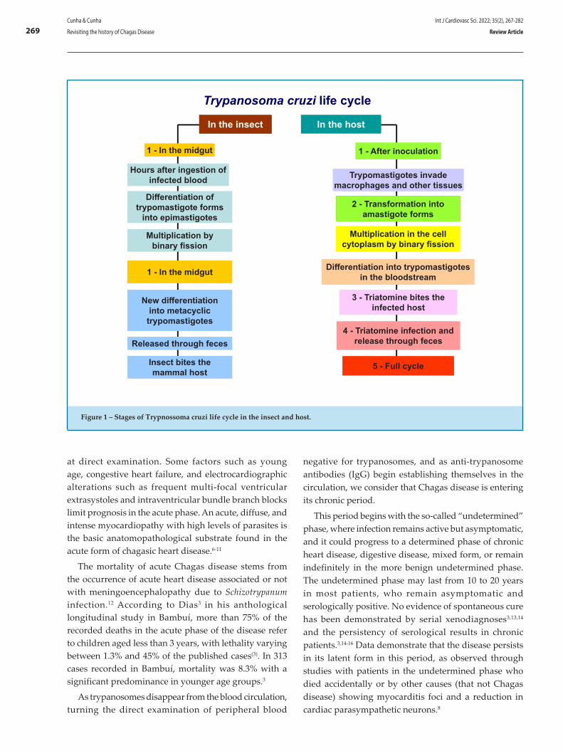
269
Figure 1 – Stages of Trypnossoma cruzi life cycle in the insect and host.
at direct examination. Some factors such as young age, congestive heart failure, and electrocardiographic alterations such as frequent multi-focal ventricular extrasystoles and intraventricular bundle branch blocks limit prognosis in the acute phase. An acute, diffuse, and intense myocardiopathy with high levels of parasites is the basic anatomopathological substrate found in the acute form of chagasic heart disease.6-11
The mortality of acute Chagas disease stems from the occurrence of acute heart disease associated or not with meningoencephalopathy due to Schizotrypanum infection.12 According to Dias3 in his anthological longitudinal study in Bambuí, more than 75% of the recorded deaths in the acute phase of the disease refer to children aged less than 3 years, with lethality varying between 1.3% and 45% of the published cases(3). In 313 cases recorded in Bambuí, mortality was 8.3% with a significant predominance in younger age groups.3
As trypanosomes disappear from the blood circulation, turning the direct examination of peripheral blood
negative for trypanosomes, and as anti-trypanosome antibodies (IgG) begin establishing themselves in the circulation, we consider that Chagas disease is entering its chronic period.
This period begins with the so-called “undetermined” phase, where infection remains active but asymptomatic, and it could progress to a determined phase of chronic heart disease, digestive disease, mixed form, or remain indefinitely in the more benign undetermined phase. The undetermined phase may last from 10 to 20 years in most patients, who remain asymptomatic and serologically positive. No evidence of spontaneous cure has been demonstrated by serial xenodiagnoses3,13,14 and the persistency of serological results in chronic patients.3,14-16 Data demonstrate that the disease persists in its latent form in this period, as observed through studies with patients in the undetermined phase who died accidentally or by other causes (that not Chagas disease) showing myocarditis foci and a reduction in cardiac parasympathetic neurons.8
In the insect In the host
Trypanosoma cruzi life cycle
1 - In the midgut
Hours after ingestion of infected blood
Differentiation of trypomastigote forms
into epimastigotes
Multiplication by binary fission
1 - In the midgut
New differentiation into metacyclic trypomastigotes
Released through feces
Insect bites the mammal host
1 - After inoculation
Trypomastigotes invade macrophages and other tissues
2 - Transformation into amastigote forms
Multiplication in the cell cytoplasm by binary fission
Differentiation into trypomastigotes in the bloodstream
3 - Triatomine bites the infected host
4 - Triatomine infection and release through feces
5 - Full cycle
Int J Cardiovasc Sci. 2022; 35(2), 267-282Cunha & Cunha
Revisiting the history of Chagas Disease Review Article
270
In the longitudinal study by Dias3 in Bambuí, a follow-up of 117 patients with Chagas disease observed that, after 10–20 years of disease progression, 60.3% of patients remained in the undetermined phase; with a mean period of 27 years after the acute phase, 33.9% of patients were still in the undetermined phase.
In general, more than 70% of individuals with Chagas disease aged less than 20 years are in the undetermined chronic phase, according to various field investigations.14,17,18 Patients in the undetermined phase who are in their third decade of life have a risk of progressing to chronic heart disease of around 2% to 5%, according to various longitudinal studies.3,17,19,20
Patients in the undetermined phase present only discrete focal myocarditis at microscopic examination and, according to experimental studies, the cyclic clinical course of these myocarditis foci is related with parasitic stimulus, with resolution by apoptosis of inflammatory cells and reabsorption of the excess extracellular matrix. This progression may happen throughout a long period, without greater repercussions on the patient as long as the immunopathological pattern does not change.
The trigger for progression to a determined phase such as chronic chagasic heart disease is related with multiple factors, among which we include the nature and preferential localization of injuries, the T. cruzi strain, and the extent of the aggression during the acute phase. For comprehending the pathogenesis of Chagas disease, controversial aspects in the literature should be analyzed, such as the direct tissue injury caused by the parasite, microvascular disease, immunological reactions, and the neurogenic theory involving the autonomic nervous system. The transition from the undetermined phase to its determination, whether it be cardiac, digestive, or nervous, constitutes a challenge in the comprehension of this true scientific gap in the history of Chagas disease.
Decades after the infection, 10%–30% of patients present one of two main clinical manifestations: heart disease associated with myocarditis and fibrosis resulting in heart failure, the formation of thrombi and strokes, or the digestive form with clinical manifestations of megacolon and/or megaesophagus, possibly associated with gastrointestinal disturbances such as regurgitation, malnutrition, and severe constipation.
The immune response results in an inflammatory process in the target tissues during the acute phase of infection by T. cruzi and is essential to the control of parasitism and the equilibrium of the parasite/host
relationship, which is observed in most patients with Chagas disease. We have observed that, in around 30% of patients, progressive inflammation results in cardiac and/or digestive dysfunctions. In this process, we highlight the absence of parasites in the affected tissues and the apparent lack of correlation between their presence and that of inflammatory infiltrate in these tissues, which resulted in the elaboration of some theories such as:
The parasympathetic dysautonomia theory
Koberle21, a pioneering scholar studying the autonomic nervous system, adopted in the 1950s the neuron count technique, recording a numeric reduction in parasympathetic nervous cells and thus considering chagasic heart disease to happen due to parasympathetic dysautonomia, with sympathetic predominance.
Autoimmunity
The challenge involving the transition from the undetermined to the determined cardiac phase motivated the hypothesis that “damage to the myocardium would be secondary to a delayed hypersensitivity process directed to the cardiac tissue, mediated by the lymphomononuclear inflammatory infiltrate universally associated with injury. According to this hypothesis, the autoimmune response directed to the myocardium would be caused by a cross-reaction triggered during the immune response against a T. cruzi antigen homologous to cardiac structures.”22
However, Kierszenbaum23 performed an extensive review of the autoimmunity theory, raising doubts about the relevance of autoimmune recognition in the pathogenesis of chronic manifestations of Chagas disease.
Polyclonal activation
Polyclonal B and T lymphocyte activation, observed in the acute phase of the chagasic infection, may be considered a trigger for the pathology found in the chronic phase. The proliferative activity of T cells and the intense polyclonal activation of B cells during the acute phase of an experimental infection by T. cruzi lead to the production of IgM and IgG antibodies with reactivity and multi-reactivity against myosin, myoglobulin, keratin, and other self-proteins. This polyclonal expansion may result from the activation of cell clones that react to a wide variety of parasite antigens or to superantigenic molecules of the parasite. This process may represent a
Int J Cardiovasc Sci. 2022; 35(2), 267-282 Cunha & Cunha
Revisiting the history of Chagas DiseaseReview Article
271
step in the loss of tolerance preceding an autoimmune response in chronic Chagas disease.24
Genetic polymorphism
Infection by T. cruzi with clinical manifestations of Chagas disease varying from asymptomatic patients to those with severe heart failure or of the digestive form, with the formation of megacolon or megaesophagus, already represents strong evidence of the influence of genetic factors in the susceptibility to infection by T. cruzi. Fernandez-Mestre and colleagues, as mentioned by Lannes-Vieira,24 studied the genetic polymorphism of DRB1 and DQB1 molecules in Venezuelan patients with Chagas disease. They observed a decreased frequency of the DRB1*14 and DQB1*0303 alleles in comparison with uninfected individuals, suggesting an independent protective role of these molecules against chronic infection. The study of patients with or without heart disease revealed higher frequencies of DRB1*01, DRB1*08, and DQB1*0501 alleles and a lower frequency of DRB1*1501 in patients with arrhythmia and congestive heart failure. These data suggest that HLA class II genes may be associated with the development of chronic infection and chronic damage to cardiac tissue.
Microvascular alterations
Rossi and colleagues, as cited by Lannes-Vieira,24 reported that microvascular alterations result from thromboembolic processes that play an important part in the origin of chronic chagasic heart disease. The microangiopathy characterized by platelet aggregation and occlusive thrombosis in small vessels of the epicardium and myocardium would cause myonecrosis and focal degeneration with inflammatory infiltrates and contribute to the development of apical aneurysm and cardiomyopathy.
The participation of cytokines
Experimental models have demonstrated that inflammatory cells such as macrophages and T cells, as well as the cytokines produced by them, play important roles in the protective response and immunopathogenesis of parasitic infections. Studies show that cytokines, just as other inflammatory mediators (prostaglandins, thromboxanes, leukotrienes, platelet aggregation factors) play an important role in regulating the immune response during infection by T. cruzi and are involved both in infection resistance and mechanisms related with the progression of Chagas disease.24
Participation of cell adhesion molecules
D’Avila Reis and colleagues, Higuchi and colleagues, and Tostes and colleagues, as mentioned by Lannes-Vieira,24 described a predominance of T CD8+ cells in the myocardium of patients in the chronic phase of Chagas disease; however, the molecular mechanisms determining the prevalence of CD8+ cells in this cardiac tissue remain undiscovered. Most inflammatory cells in the myocardium of patients with chronic Chagas disease express cell adhesion molecules such as LFA-1 (leukocyte function-associated antigen-1; CD11a/CD18), ICAM-1 ligand (intercellular adhesion molecule-1; CD54), CD44 ligand of fibronectin and hyaluronic acid and VLA-4 (Very late antigen-4; CD49d/CD29, α4β1) VCAM-1 ligand (vascular cell adhesion molecule-1;CD106) and fibronectin. This way, D’Avila Reis and colleagues suggest that these molecules would contribute to the progression of the inflammatory reaction by mediating the adhesion of lymphocytes to the endothelium of cardiac tissue vessels activated by cytokines and by being important in cell infiltration and localization at inflammation sites.
Participation of chemokines
Plasmatic chemokine concentrations have been correlated with disease worsening in patients with heart failure. Elevated plasma levels of the CCL2/MCP-1 chemokine, but not of CCL3/MIP1α, were directly correlated with heart damage in patients with chagasic heart disease in a study by Talvani and colleagues, as cited by Lannes-Vieira.24(
Apoptosis
Lopes and colleagues, as cited by Lannes-Vieira,24 showed in their study that programmed cell death by apoptosis in immune cells (including B and T cells) occurs during infection by T. cruzi. Experimental infection models showed a significant loss of T CD4+ cells through an increase in the expression of Fas (CD95) and Fas-ligand (CD95L), with subsequent induction of apoptosis by activation-induced cell death.
Some factors are already consolidated, and we can state that “inflammation persists during the whole chronic phase of Chagas disease, continuously triggering new fibrosis foci.”25 Repeated cycles of parasitic infection and hypersensitivity and autoimmunity phenomena
Int J Cardiovasc Sci. 2022; 35(2), 267-282Cunha & Cunha
Revisiting the history of Chagas Disease Review Article
272
seem to be responsible for perpetuating this picture.26-28 Fibrosis, within the natural course of Chagas disease, represents a new anatomical element that, to a specific extent, may be capable of disrupting the balance of the chagasic heart, possibly triggering heart failure.25
Chronic chagasic heart disease has a slow and variable progression and generally appears in the fourth decade of life, when the first signs of heart failure become apparent.9,29,30. Once established, chronic chagasic heart disease presents itself as a progressive and severe myocardial pathology, exhibiting a varied picture of isolated or combined arrhythmias that has not yet been verified in any other heart disease. This heart pathology has been thoroughly investigated using necropsy material and findings were correlated with clinical and electrocardiographic manifestations, especially thromboembolic phenomena. The rare presence of parasites in sections, as opposed to what is observed in the acute phase, raised discussions about the histopathological diagnosis and interpretation of pathogenesis.
The digestive form
Clinical and epidemiological evidence, reliable serological studies, and a positive serological test for T. c
ruzi lead to the diagnosis of the digestive form of Chagas disease. Anatomical studies contributed decisively to the recognition of the digestive form of Chagas disease, and the conclusive acceptance of a chagasic etiology for megaesophagus and megacolon came with studies by Köberle.31,32 and anatomopathological findings of degenerative injuries of the myenteric plexus in autopsied cases of megaesophagus; these injuries were found not only in dilated segments, but in the whole digestive tract.
The main function of the myenteric plexus in the digestive tract wall is to coordinate motility in its different segments.
Köberle performed his research with autopsies of patients with Chagas disease, naturally infected animals, and through the experimental infection of laboratory animals. In the acute phase of the infection, he observed parasitism of the muscular layer of the digestive tract and an inflammatory process involving the myenteric plexus. In the chronic phase, he observed that denervation was irregular, with variable distribution and intensity.
In the neuron count, performed in the lower third of the esophagus, the reduction in neuron numbers was shown to be very variable. Denervation was a constant in patients with Chagas disease, but it was less intense in cases
where the esophagus appeared normal. He concluded, after comparing cases with and without megaesophagus, that the progression of chagasic esophageal disorder to a typical megaesophagus occurs when denervation reaches a threshold, which was estimated in 90%.
The nervous form
The chronic nervous form of Chagas disease is still not unanimous to this day, even though it was recognized by its discoverer Carlos Chagas and despite the classic study by Köberle in 1967.33
Studies have shown cortical atrophy with or without hydrocephaly, a decreased neuron population and activation of glial cells such as astrocytes and microglia, but no histopathological alterations or even parasitism (as reviewed by Alencar, 1982). Differently from other models, the study by Silva34 using C3H/He mice infected with the Colombian T. cruzi strain showed the presence, even if rare, of the parasite in the chronic phase. However, the absence of inflammation suggests the resolution of inflammatory processes found in the central nervous system during the acute phase of the experimental infection, since all animals survived acute infection.
Specific treatment of Chagas disease
Great variability is seen regarding the types of cases and cure control employed in different studies. Generally, we consider adequate the results for the acute phase and cases of recent infection, mainly among children, who not only tolerate better long-term treatments but also present high cure rates. This was demonstrated by randomized field studies with benznidazole in Brazil (Andrade et al., 1996)35 and Argentina (Sosa Estani et al., 1998).36 The mean estimated parasitological cure rate is around 60% for acute cases and recent infections. Results are considered poor for chronic infections, in the Brazilian experience; in the Southern Cone, results were considered superior, probably owing to a different parasite strain (Silva et al. 1974; Cerisola et al., 1977).37,38
The clinical progression of Chagas disease after specific treatment is controversial and study results are inconclusive due to differences in cases, assessment methods, follow-up periods, and data interpretation.
Macêdo and Silveira39, aiming to assess the electrocardiographic progression of heart disease, studied 171 adults with chronic Chagas disease who
Int J Cardiovasc Sci. 2022; 35(2), 267-282 Cunha & Cunha
Revisiting the history of Chagas DiseaseReview Article
273
were treated with nifurtimox or benznidazole with a follow-up of 7 years and observed electrocardiographic improvements in 6.7% of the cases against 8.8% among untreated patients, with no significant difference between groups. Ianni et al. (1993)40 evaluated 33 adults in the undetermined phase for 8 years and observed electrocardiographic progression in 13.3% of cases treated with benznidazole (n=15) and 0% of patients who received placebo (n=18); since this was a small number of cases, definitive conclusions could not be reached.
It is worth noting that, among 120 cases (adults and children) studied by Miranda el al.,41 progression of the electrocardiogram was observed in 10.5% of patients treated with benznidazole against 63.6% of the placebo group. Patients were monitored for 10 to 16 years, but the combination of results from adults and children and the interpretation of electrocardiograms hampered result analyses.
Carlos Chagas, the man of the twentieth century
It is also vital to know other faces of the man Carlos Chagas and his marriage with Ms. Iris Lobo, a bond that strengthened his fighting spirit and full dedication to research and where he found the support and comprehension indispensable to the life of a researcher in the beginning of the twentieth century.
Iris Lobo was the daughter of a Minas Gerais state senator, Fernando Lobo Leite Pereira, and met Carlos Chagas in one of these weekend social gatherings, sealing a mutual complicity that would remain for their whole lives. In the beginning, their union was not easy due to the objection by the senator’s family; his wife alleged that Chagas’s family had “Black blood.” This was not enough to keep them apart, and in face of Iris’ persistence and a friendly interference by Miguel Couto (who was married to Carlos’ mother’s first cousin), the senator did not resist and authorized the wedding, which happened in July 1904. Iris’ personality was well defined by her son, Carlos Chagas;42 “In moments of glory, she knew how to dim herself, but in terrible moments such as when Carlos Chagas was attacked by the papers at Rio de Janeiro as a public health director or in the episode at the Academy of Medicine, where malevolent and ignorant detractors tried to disparage his work, she stood up to his defense as a hero from a Greek tragedy, not allowing for even a moment that his partner’s spirit be invaded with disheartenment.”
Chagas disease post-Lassance
In Lassance (MG) after his discovery, Chagas continued the studies of this new human morbid entity and, within his anatomopathological studies, it was up to his dedicated colleague Gaspar Vianna to prove the existence of leishmaniform bodies in the myocardium fibers of experimental animals and human beings. This unlocked the perspective for describing the cardiac form of the disease, which was magnificently systematized by Chagas and Villela in 1922 with vital contribution by the Boullite electrocardiograph, which was difficult to manipulate and significantly advanced for the time. The cardiac form would only be modified in 1942 by Laranja, Dias, Nóbrega, and Miranda based on exceptional works performed at the Center for the Study and Prophylaxis of Chagas Disease in Bambuí (MG) under guidance of the Oswaldo Cruz Institute. Among their changes, we highlight the consolidation of the chronic cardiac form (determined phase) and its connection to the acute infection by T. cruzi according to the reproduction of chronic infections in dogs. 43 After this phase, the cardiac form of Chagas disease is defined as progressive, preferentially affecting men aged between 20 and 50 years, with frequent clinical manifestations of disturbances in the formation and conduction of cardiac stimuli and congestive heart failure; anatomically, this form becomes characterized by an extensive inflammatory infiltrate, usually followed by circumscribed injuries in the parietal endocardium, and by the slow development of ischemic myocardial alterations.44
The clinical, electrocardiographic, and prophylaxis-related contribution of Chagas disease studies at Bambuí reach international repercussion and Brazil starts to adopt a prophylaxis model where “Casa de Oswaldo” (the Oswaldo Cruz Institute) is fully invested in fighting Chagas disease, penetrating “Cafuas” (wattle and daub houses) in the region, exterminating triatomines, proposing the construction of decent housing, examining and cataloguing the sick population in a beautiful process of state-coordinated change in established values and health promotion through the correction of perverse social inequalities, especially in the countryside.
The late Dr. Francisco Laranja, in a personal communication to researchers from Rio de Janeiro at his house in Leblon (RJ) in 1981, reported significant information on the historical Center for the Study and Prophylaxis of Chagas Disease at Bambuí, enumerating the contributions by this institution to the knowledge on Chagas disease:
Int J Cardiovasc Sci. 2022; 35(2), 267-282Cunha & Cunha
Revisiting the history of Chagas Disease Review Article
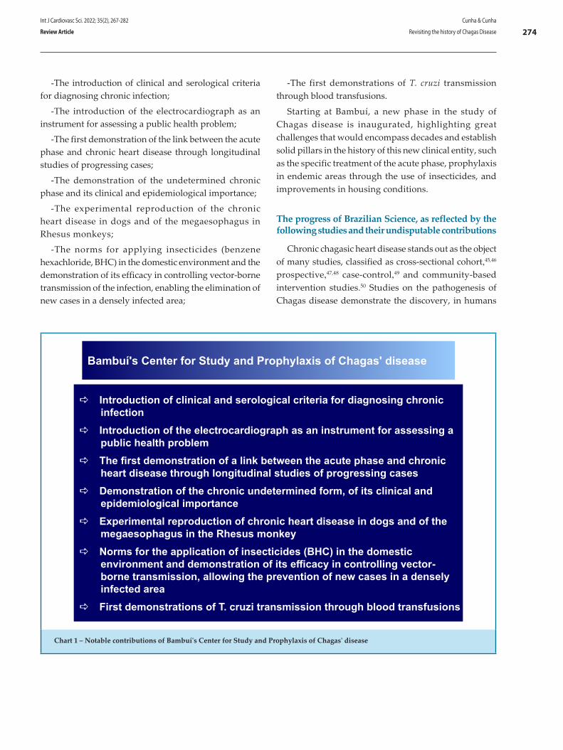
274
-The introduction of clinical and serological criteria for diagnosing chronic infection;
-The introduction of the electrocardiograph as an instrument for assessing a public health problem;
-The first demonstration of the link between the acute phase and chronic heart disease through longitudinal studies of progressing cases;
-The demonstration of the undetermined chronic phase and its clinical and epidemiological importance;
-The experimental reproduction of the chronic heart disease in dogs and of the megaesophagus in Rhesus monkeys;
-The norms for applying insecticides (benzene hexachloride, BHC) in the domestic environment and the demonstration of its efficacy in controlling vector-borne transmission of the infection, enabling the elimination of new cases in a densely infected area;
-The first demonstrations of T. cruzi transmission through blood transfusions.
Starting at Bambuí, a new phase in the study of Chagas disease is inaugurated, highlighting great challenges that would encompass decades and establish solid pillars in the history of this new clinical entity, such as the specific treatment of the acute phase, prophylaxis in endemic areas through the use of insecticides, and improvements in housing conditions.
The progress of Brazilian Science, as reflected by the following studies and their undisputable contributions
Chronic chagasic heart disease stands out as the object of many studies, classified as cross-sectional cohort,45,46 prospective,47,48 case-control,49 and community-based intervention studies.50 Studies on the pathogenesis of Chagas disease demonstrate the discovery, in humans
a Introduction of clinical and serological criteria for diagnosing chronic infection
a Introduction of the electrocardiograph as an instrument for assessing a public health problem
a The first demonstration of a link between the acute phase and chronic heart disease through longitudinal studies of progressing cases
a Demonstration of the chronic undetermined form, of its clinical and epidemiological importance
a Experimental reproduction of chronic heart disease in dogs and of the megaesophagus in the Rhesus monkey
a Norms for the application of insecticides (BHC) in the domestic environment and demonstration of its efficacy in controlling vector-borne transmission, allowing the prevention of new cases in a densely infected area
a First demonstrations of T. cruzi transmission through blood transfusions
Bambuí's Center for Study and Prophylaxis of Chagas' disease
Chart 1 – Notable contributions of Bambuí's Center for Study and Prophylaxis of Chagas' disease
Int J Cardiovasc Sci. 2022; 35(2), 267-282 Cunha & Cunha
Revisiting the history of Chagas DiseaseReview Article
275
or experimental models during the acute phase, of focal inflammatory injuries, severe myocarditis, cardiac myocyte necrosis, and amastigote nests in the heart, as observed by Kosma51 and Tafuri.52
Systematized studies analyzing autonomic function in Chagas disease were performed at Ribeirão Preto Medical School, coordinated by Professor Dalmo Amorim. Following these studies, various faces of autonomic cardiac control appeared in the literature, presented by independent groups in Brazil (Ribeirão Preto and Brasília), Argentina (Córdoba), and Venezuela (Mérida). In general, studies performed in Ribeirão Preto by Amorim et al.,53 and Marin Neto54 and in Brasília by Junqueira et al.55 reflect the functional impairment of the parasympathetic system, while studies from Córdoba (Argentina) performed by Iosa56 and Palmero et al.,57 have shown to be compatible with cardiovascular sympathetic dysfunction.
The analysis of this dysautonomia proceeds with a group from Rio de Janeiro, which has developed a series of studies58-60 reporting, for the first time, the presence of functionally active autoantibodies that reacted against muscarinic cholinergic receptors in patients with Chagas disease and different degrees of cardiac impairment (asymptomatic patients with a normal electrocardiogram/echocardiogram, asymptomatic patients with a normal electrocardiogram and alterations in the echocardiogram, symptomatic patients with alterations in the electrocardiogram/echocardiogram, but no important functional abnormalities, and finally severe symptomatic patients with cardiac conduction and mechanic abnormalities). This group showed that the presence of functional antibodies does not depend on the degree of cardiac impairment.
Animal experiments have shown that antibodies interact with cellular constituents, influencing metabolism and cardiac contractility. The interruption of the beta-adrenergic pathway may affect contraction strength and myocardial relaxation. Leite61 demonstrated that the serum of chronic chagasic patients reduced, in a dose-dependent manner, contraction amplitude in the atrial myocardium of rabbits. Sera of patients without myocardial dysfunction did not significantly alter any of the measured parameters.
Other studies on autoantibodies as the expression of dysautonomia have followed, such as that by Savio-Galimberti and colleagues,62 who observed antibodies with adrenergic properties compromising cardiac muscle function in isolated heart muscle. Another interesting study was published by Hernandez and colleagues,63
who showed antibodies, this time with muscarinic properties, decreasing L-type calcium current via the non-competitive activation of the M2 muscarinic receptor. The impairment of this current by antibodies with adrenergic or muscarinic properties could contribute, at least in part, to the myocardial dysfunction seen in the chronic phase in patients with Chagas disease.
As studies progressed, Gimenez and colleagues64 were able to show cardiac impairment as a consequence of the autoimmune process generated by antibodies with adrenergic or cholinergic properties. They immunized mice with a plasmid coding for the M2 and beta-1 adrenergic receptors. The authors found antibodies not only against the second loop of M2 and beta-1 receptors, but also against the third intracellular loop of the M2 receptor. Through binding assays, they could observe a twofold increment in the expression of M2 receptors, a decrease in beta-1 receptors, and signs of autonomic dysregulation in immunized animals. Other relevant findings observed in these animals were myofibrillar disarray and fibrosis as a consequence of continued exposure to antibodies. The research group in Rio de Janeiro advanced in their studies on dysautonomia and De Carvalho and colleagues,65 in a pioneering study, indicated that a humoral component could be involved in the genesis of arrhythmias in chagasic cardiomyopathy. The authors described that sera from rabbits infected with T. cruzi generated electrocardiographic disturbances in isolated rabbit hearts. Subsequently, the same group confirmed this hypothesis, now showing that antibodies from patients with chronic Chagas disease who had complex arrhythmias decreased the heart rate and caused atrioventricular block in isolated rabbit hearts.66 Still in Rio de Janeiro, Costa and colleagues58 studied the sera of 58 patients with chronic chagasic heart disease and described that some of them, with beta-adrenergic properties, blocked conduction via communicating junctions in cultures of neonatal rat cardiomyocytes, suggesting one more mechanism through which these antibodies might contribute to the occurrence of arrhythmias. Medei and colleagues67 showed, for the first time, that patients with chronic chagasic heart disease who had antibodies with muscarinic properties presented increased QT interval dispersion when compared to patients with chronic disease who did not have them. These antibodies with muscarinic properties, when perfused through isolated hearts under controlled heart rates, increased QT intervals.
Considering that the parasympathetic nervous system only has a discrete effect on coronary circulation, the
Int J Cardiovasc Sci. 2022; 35(2), 267-282Cunha & Cunha
Revisiting the history of Chagas Disease Review Article
276
imbalance between the sympathetic and parasympathetic nervous system with a predominance of sympathetic action on coronary vessels can contribute with microcirculatory constrictions, which would cause possible spastic events and aneurysms.68 Approaches of the anatomical and functional aspects of the coronary circulation and microcirculation in humans and experimental models infected with T. cruzi demonstrated no obstructive injuries capable of inducing myocardial ischemia. The perfusion disturbances observed by these studies are related with microvascular alterations, mostly in patients with chronic Chagas disease with normal coronary arteries at angiographic examination. This would explain regional contractile perfusion dysfunctions that resemble those observed in ischemic heart disease.
Investigations in this direction continued and, in 1997, Carrasco et al.,69 managed to show sympathetic system impairment, with a significant reduction of this component in the sympathovagal balance of patients with chronic Chagas disease, relating it with an increase in myocardial contractile dysfunction.
In 2003, Cunha et al.,70 demonstrated a decrease in heart rate variability expressed through daytime and nighttime SDANN (standard deviation of the mean of all recorded 5-minute intervals [ms]), daytime and nighttime SDNN (standard deviation of all recorded P-P cycles [ms]), daytime SDNN I (mean standard deviation of all recorded 5-minute intervals [ms]), in addition to an increase in daytime and nighttime RMSSD (root mean square of successive differences [ms]) and nighttime pNN50 (proportion [%] of variations of more than 50 ms between normal successive cycles), suggesting that dysautonomia may occur early in Chagas disease.
The complexity of T. cruzi’s biological cycle suggests that infection control involves a combination of adaptive immunological responses by the host that operate in various levels of the immunological system.
Some authors71 indicate that the immunization of mice with a recombinant T. cruzi protein (TcP2β) promotes intense and specific antibody production against the C-terminal portion of the 13-residue epitope (R13 peptide: EEEDDDMGFGLFD) and has a simultaneous β1-adrenergic stimulating activity. Other animals subjected to the same immunization did not provide similar results. These data showed that the R13 epitope could induce an antibody that recognized the second extracellular loop of β1 receptors and could induce ventricular arrhythmias when passively transferred to mice.
Levitus and colleagues72 demonstrated that cloned parasitic peptides (JL5) reacted with sera from patients with Chagas disease. The cloned peptide was identified as the C-terminal portion of the parasite’s ribosomal P protein, which developed cross-reactivity with the host’s ribosomal P protein. This is due to a homologous amino acid sequence covering almost 90% of the parasite and host proteins.
Some authors73 have shown that a cloned parasitic peptide (λgt 11) named JL5 reacted with sera from patients infected by T. cruzi and this peptide was identified as the C-terminal portion of T. cruzi’s ribosomal P protein. Many studies indicate that JL5 has epitopes that promote cross-reactivity with the host’s ribosomal P protein. Most amino acids in this peptide are homologous to the C-terminal portion of the human ribosomal P protein.
In patients with Chagas disease, the allosteric nature of the interaction between IgGs and the muscarinic receptor site was confirmed by observing that these patients’ sera, and not those of healthy donors, increased agonist activity, inducing bradiarrhythmias. This effect disappears in the presence of gallamine, an allosteric antagonist. The main strength of this study was the characterization of the action of antibodies in the serum of patients with Chagas disease with the allosteric interaction occurring in the second extracellular loop of M2 acetylcholine receptors and of their ability to exert an agonist and signal-transducing effect.72,73
In order to establish a cytokine profile for the undetermined and chronic (cardiac) phases of Chagas disease, Vitelli-Avelar and colleagues74 developed methods for calculating the mean percentage of cytokine appearance in leukocytes, establishing a threshold that defined low and high levels of cytokine production and, from there, constructing diagrams that characterized each phase. A low frequency of cytokines, in the undetermined phase, is observed by a reduced frequency of T CD4+ inflammatory cells.
IFNγ levels allowed a distinction between patients in the chronic (cardiac) and undetermined phases of the disease. IFNγ levels were significantly higher in the chronic phase, just as IL-10 and CD8+ levels were significantly higher in the cardiac form of the chronic phase when compared to the control group. In this study, the control group did not present a specific cytokine profile when compared to patients in the undetermined and chronic (cardiac) phases.
We can relate the pathogenesis of acute phase injuries (especially in the initial phase) with the presence of
Int J Cardiovasc Sci. 2022; 35(2), 267-282 Cunha & Cunha
Revisiting the history of Chagas DiseaseReview Article
277
intracellular parasites. The analysis chronic phase pathogenesis is reason for intense controversy, since this phase presents delayed injuries and for a long time it was believed that the presence of parasites was rare. In 1993, Bocchi et al.75 observed that the most common finding after cardiac transplants in patients with Chagas disease was the reactivation of infection by T. cruzi, and Higuchi et al.,76 still in 1993, demonstrated a significant correlation between the presence of T. cruzi and moderate to severe inflammatory infiltrate in cardiac biopsy sections and necropsy examinations of patients with Chagas disease. Later, in 1996, Belloti et al.77 also sought to investigate the presence of parasites in the hearts of patients with chronic Chagas disease, frequently finding these organisms and relating them with the severity of the myocardial inflammatory process, thus strengthening the idea of an important role for the parasite in the pathophysiology of the chronic phase and unlocking a true perspective for specific treatment of the chronic phase of Chagas disease.
Aiming to treat chronic chagasic cardiomyopathy and based on previous studies suggesting that autologous bone marrow stem cell transplant improved cardiac function in patients with chronic chagasic cardiomyopathy, researchers performed a study using cell therapy in chronic chagasic heart disease as an arm of a multi-center randomized cell therapy study in cardiomyopathies. 78
The study evaluated patients aged between 18 and 75 years with chronic chagasic cardiomyopathy, functional class II–IV (New York Heart Association, NYHA), ejection fraction < 35%, who received optimized clinical therapy. They were randomized for intracoronary injection of bone marrow stem cells or placebo. The primary endpoints were differences in ejection fraction 6 months and 1 year after treatment in both groups. This study followed up 234 patients between July 2005 and October 2009.
Their conclusion was that the intracoronary injection of bone marrow stem cells did not improve left ventricular function or quality of life in patients with chronic chagasic cardiomyopathy.
Within this perspective and attending to the needs of researchers of chronic chagasic heart disease, in 2015 the Benefit study79 searched for an answer to the role of benznidazole in chronic chagasic cardiomyopathy; it was conducted as a prospective, multi-center, randomized study involving 2854 patients with chagasic cardiomyopathy who received benznidazole or placebo for 80 days; participants were followed up for a mean period of 5.4 years.
The study demonstrated that therapy with benznidazole in patients with established chronic chagasic heart disease significantly increased conversion rates to negative polymerase chain reaction (PCR) results but did not significantly reduce clinical deterioration during the 5.4-year follow-up period.
Challenges still remain: among them, the search for comprehending autoimmunity based on numerous studies that have been published and stimulated since the demonstration, by Higuchi et al.,76 of the presence of the parasite in hearts of patients with chronic Chagas disease. The logical foundation relies on the parasite presence stimulating a strong immune response, which certainly causes damage and inflammation in the affected cardiac tissues. A molecular mimicry response between parasitic and host molecules may result in cross-reaction with self-molecules and consequently in autoimmunity with autoantibodies and self-reactive cells. Although there is still controversy, autoimmunity may be related with the progression of chronic chagasic heart disease.80).
In this line of thought, Cunha et al.,81 demonstrated with 24 h Holter monitoring that, during a period of mostly parasympathetic activation (02:00–06:00h), a direct and significant correlation between anti-M2 antibodies and SDANN was observed. In the stress test, they found a direct correlation between anti-β1 antibodies and double product.
Among other improvements in the search for comprehending the pathological substrate for chronic chagasic heart disease, we highlight the research on the inheritance and fixation of T. cruzi kDNA minicircles in the genome of patients with Chagas disease and their family members, developed by Aragão. 82
In this study, the lateral and vertical transfer of T. cruzi DNA minicircles (kDNA) was investigated in 26 people from 4 families, and active infection was observed in 5 people with nuclear T. cruzi DNA; evidence of kDNA integration was documented in all these 5 cases. This work explicitly reports the occurrence of kDNA transfer from T. cruzi to humans and expands the knowledge on the inheritance of mutations generated by the integration of minicircle sequences in most host chromosomes. Evidently, these findings are still premature and controversial, but they uncover new perspectives in the pathophysiology and vertical transmission of Chagas disease.
Another study in the same direction was presented by Morini83 and characterized by the demonstration of a protein associated with the T. cruzi kinetoplast, named TcKAP7, which had low molecular weight, a basic
Int J Cardiovasc Sci. 2022; 35(2), 267-282Cunha & Cunha
Revisiting the history of Chagas Disease Review Article
278
nature, and a role in DNA charge neutralization and condensation. By using immunofluorescence, researchers observed that the TcKAP7 protein was located around kinetoplast poles, suggesting it may be involved in late stages of minicircle replication.
Concerns around Chagas disease considering the comprehension of its pathophysiology in the chronic phase, the still unanswered questions regarding its undetermined phase, and particularly an efficient therapy for this phase of the disease are fully justified when observing the astounding numbers, still in the twenty-first century, of 6 million infected people in Latin America and the globalization of Chagas disease, showing thousands of infected people in the USA, Japan, Australia, and Spain.84
According to the World Health Organization (WHO), until 1990 there were 11 million infected people and more than 300 000 cases had been observed in the USA. An expressive increase in oral transmission and in congenital transmission route valorization is noted.85-87
The global incidence of new human infections by T. cruzi has increased in approximately 67%. Only in Brazil, the estimate accounts for 4.6 million infected people.88,89
In the sense of facing all these problems related with Chagas disease and searching for the ideal parameters for solving this challenging problem, in 2014 the London Declaration90 on Neglected Tropical Diseases was signed. It proposed, for 2020, measures aiming at interrupting the main forms of Chagas disease transmission globally, stimulating antiparasitic treatment, the improvement of vigilance systems in affected countries, and easily accessible care to infected patients.
A study performed in 2018 in Yucatan91 questioned the objectives of the London Declaration for 2020 regarding their sufficiency in controlling Chagas disease. This study showed that reductions in domestic vector-borne transmission, congenital transmission, and transmission through blood transfusion may successfully reduce human infection rates (up to 82% in a year), reaching the 2020 objectives, but would still result in 0.5 new acute cases per 2000 people in 5 years.
Progress has been made considering basic research on T. cruzi, especially in the genetic characterization of parasite groups, evidencing their genomic sequence. Zingales,92(in a review on the subject, mentions that T. cruzi is divided into 7 discrete typing units (DTUs), among which are Tcl (Trypanossoma cruzi I), TcVI (Trypanosoma cruzi VI), and Tcbat (bat Trypanosoma cruzi); the latter
is restricted to bats. This review emphasized that the interaction between the parasite and host genetics should have an important role in defining the pathogenesis of Chagas disease, the anti-T. cruzi immunological response, and the chemotherapy response, and it should be considered in future investigations.
The TcI, TcVI, and TcBat DTUs have a yet unknown geographical distribution. TcI is the most spread around the geographical distribution of this parasite, including Brazilian biomes.93
Recently, Vermelho et al.,94 discussed the urgent need for intensifying research on new efficient drugs for treating Chagas disease, reminding that since the 1960s few advances in this area have outdone the only 2 drugs currently in use, benznidazole and nifurtimox. They mention that today, few drugs are going through pre-clinical tests and highlight only 3 classes of compounds that have shown high cure rates in rat infection models: nitroimidazole, the oxaborole DNDi-6148, and proteasome inhibitors (GNF6702).
Considering the educational aspects of Chagas disease, Sanmartino et al.,95 observed that, since the first publication more than 110 years ago, the need for comprehending the complex relationship between Chagas disease and social and environmental aspects beyond the biomedical and epidemiological aspects still remains.
Scenarios in this matter, both rural and Latin American, urban and global, make it clear that education about Chagas disease should include all possible contexts: the location of vectors in Latin America in rural, peri-urban, and urban areas using formal and informal educational environments. The authors stress the requirement for a whole health approach that overcomes the biomedical aspect, including the multi-dimensionality of this matter and providing an educational dialogic perspective, finally allowing individuals and communities to analyze, decide, and lead preventive and promotion actions contextualized to their health.
Conclusion
In our revisitation to the history of Chagas disease, our observations allow us to state that research is progressing and representing the respectability of the Brazilian scientific community; on the other hand, we have noticed the need for improvements in assistance and social, hospital, and preventive care that can cope
Int J Cardiovasc Sci. 2022; 35(2), 267-282 Cunha & Cunha
Revisiting the history of Chagas DiseaseReview Article
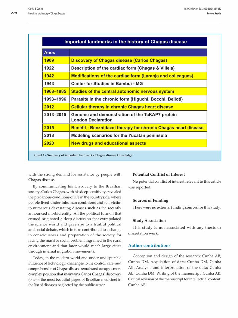
279
with the strong demand for assistance by people with Chagas disease.
By communicating his Discovery to the Brazilian society, Carlos Chagas, with his deep sensitivity, revealed the precarious conditions of life in the countryside, where people lived under inhuman conditions and fell victim to numerous devastating diseases such as the recently announced morbid entity. All the political turmoil that ensued originated a deep discussion that extrapolated the science world and gave rise to a fruitful political and social debate, which in turn contributed to a change in consciousness and preparation of the society for facing the massive social problem ingrained in the rural environment and that later would reach large cities through internal migration movements.
Today, in the modern world and under undisputable influence of technology, challenges to the control, care, and comprehension of Chagas disease remain and occupy a more complex position that maintains Carlos Chagas’ discovery (one of the most beautiful pages of Brazilian medicine) in the list of diseases neglected by the public sector.
Potential Conflict of Interest
No potential conflict of interest relevant to this article was reported.
Sources of Funding
There were no external funding sources for this study.
Study Association
This study is not associated with any thesis or dissertation work.
Author contributions
Conception and design of the research: Cunha AB, Cunha DM. Acquisition of data: Cunha DM, Cunha AB. Analysis and interpretation of the data: Cunha AB, Cunha DM. Writing of the manuscript: Cunha AB. Critical revision of the manuscript for intellectual content: Cunha AB.
Important landmarks in the history of Chagas disease
Anos1909 Discovery of Chagas disease (Carlos Chagas)1922 Description of the cardiac form (Chagas & Villela)1942 Modifications of the cardiac form (Laranja and colleagues)1943 Center for Studies in Bambuí - MG1968–1985 Studies of the central autonomic nervous system1993–1996 Parasite in the chronic form (Higuchi, Bocchi, Belloti)2012 Cellular therapy in chronic Chagas heart disease2013–2015 Genome and demonstration of the TcKAP7 protein
London Declaration2015 Benefit - Benznidazol therapy for chronic Chagas heart disease2018 Modeling scenarios for the Yucatan peninsula2020 New drugs and educational aspects
Chart 2 – Summary of important landmarks Chagas' disease knowledge.
Int J Cardiovasc Sci. 2022; 35(2), 267-282Cunha & Cunha
Revisiting the history of Chagas Disease Review Article
280
1. Lewinsohn R. Carlos Chagasand the Discovery of Chaga’s Disease (American Trypanosomiasis. J R Soc Med. 1981;74(6);451-5.
2. Berning H. : Diagnosi della Malattia di Chagas. Minerva médica,1984;75:693-5.
3. Dias, JCP. Doença de Chagas em Bambui, Minas Gerais, Brasil. Estudo clínico-epidemiológico a partir da fase aguda, entre 1940 e 1982. Tese. Belo-Horizonte, Faculdade de Medicina(UFMG);1982.
4. Teixeira MVLC. Doença de Chagas. Estudo da forma aguda inaparente. Tese.Rio de Janeiro:UFRJ;1977.
5. Perez A, Romanã C. Estado actual de antigos casos de enfermedad de Chagas en una família de Monteros (Tucumán). An Inst Med Reg (Tucumán), 1945;1:214-28.
6. Chagas C. Trypanosomíase americana: forma aguda da moléstia. Mem Inst Oswaldo Cruz .1916; 8:37-65.
7. Dias E, Laranja FS, Nóbrega G. Doença de Chagas. Mem Inst Oswaldo Cruz 1946; 43: 495-582.
8. Andrade Z, Andrade SG. Patologia. In: Brener Z e Andrade Z. Trypanosoma cruzi e doença de Chagas. Rio de Janeiro:Guanabara Koogan ;1979.
9. Laranja FS, Dias E, Nobrega G, Miranda A. Chaga’s disease.A Clinical, Epidemiologic and Pathologic Study. Circulation. 1956;14(6):1035-60.
10. Ferreira HO. Forma aguda da doença de Chagas. In: Cançado JR. Doença de Chagas. Belo Horizonte (MG);1968. p.359-73.
11. Tafuri WL.Alterações ultra estruturais dos componente muscular, intersticial e nervoso do coração, esôfago e intestinos na doença de Chagas experimental e humana.Tese. Belo Horizonte: Universidade Federal de Minas Gerais;1874.
12. Laranja FS, Dias E, Nobrega GC. Manifestações clínicas e diagnóstico da cardiopatia aguda da doença de Chagas. In: 1º Congr Panam Medic, Rio de Janeiro;1946. 17p.
13. Cançado JR.Forma aguda da doença de Chagas. In: Décourt LV, Campos OM. Modernos conhecimentos sobre doença de Chagas. Belo Horizonte, UFMG, Acad Mineira de Medicina;1981.p.13-28.
14. Castro CN. Influência da Parasitemia no quadro clínico da doença de Chagas. Rev Pat Trop. 1980;9: 73-136.
15. Manzullo EC, Darraidou MA, Libonatti O,Rozlosnic J, Bazzano AC. Estudo longitudinal de la cardiopatia chagásica crônica. Buenos Aires :Faculdade de Ciências;1982. 141 p
16. Brener Z. O Parasito: Relações hospedeiro – parasito. In: Brener Z e Andrade Z. Trypanosoma cruzi e doença de Chagas., Rio de Janeiro: Guanabara Koogan;1979. P. 1-41.
17. Macedo VO. Forma indeterminada da doença de Chagas. J Bras Med .1980;38:34-40.
18. Laranja FS, Dias E, Duarte E, Pellegrino J. Observações clínicas e epidemiológicas sobre a moléstia de Chagas no Oeste de Minas Gerais. O Hospital 1951; 40(6):945-88.
19. Brasil A. Evolução e prognóstico da doença de Chagas. Arq Brasil Cardiol. 1965; 18(5):365-80.
20. Puigbó JJ, Nava Rhode JR, Barrios HG, Yepes CG. Cuatro anos de estúdio longitudinal de una comunidade rural com endemicidad chagásica. Bol Ofic Sanit Panam. 1969;48(2):112-20.
21. Koberle F. Cardiopathia parasympathicopriva. Munch Med Wochenschr. 1959;101,1308-10.
22. Cunha-Neto E,Kalil J. A auto-imunidade na doença de Chagas. Rev Soc CardiolEst S Paulo.1994; 4(2):92-100.
23. Kierszenbaum F. Autoimunidade na doença de Chagas: fato ou fantasia? Causa ou consequência? Rev Soc Bras Med Trop. 1985; 18:129-32.
24. Lannes Vieira J. Portal da Doença de Chagas. Laboratório de Biologia das Interações. Rio de Janeiro: Instituto Oswaldo Cruz/Fiocruz,s.d.
25. Blogliolo L. As causas anatômicas da insuficiência cardíaca na cardiopatia (miocardite) chagásica crônica. In:Décourt LV, Campos O M. Modernos conhecimentos sobre doença de Chagas. Belo Horizonte, UFMG, Acad Mineira Medic. 1981; 283-302.
26. Brandão HJS. A lesão neuronal na moléstia de Chagas. . In:Décourt LV, Campos OM. Modernos conhecimentos sobre doença de Chagas. Belo Horizonte, UFMG, Acad Mineira Medic; 1981.p. 65-81.
27. Almeida HO. A cardiopatia em chagásicos crônicos com e sem “megas”- Análise dos papeis da miocardite e da denervação na cardiopatia chagásica crônica. Tese. Uberaba:Fac Med Triângulo Mineiro;1982.
28. Teixeira ARL. Competência imunológica do paciente chagásico. Imunodepressão na forma aguda inaparente. Auto imunidade no hospedeiro imunizado. Tese. Belo Horizonte: Faculdade de Medicina da UFMG;1981. 169p., 169p, 1981.
29. Chagas C, Villela E. Forma Cardíaca da Trypanosomíase Americana. Mem Inst Oswaldo Cruz. 1922;14:5-61.
30. Prata A. Prognóstico e complicações da doença de Chagas. Rev Goiana Med .1959;5:87.
31. Koberle F.Patogenia da moléstia de Chagas:estudo dos órgãos musculares ocos. Rev Goiana Med. 1957;3:155.
32. Koberle F.Enteromegal and cardiomegaly in Chagas’ disease. Gut. 963,4(4):399-405.
33. Koberle F. Aspectos neurológicos da moléstia de Chagas. Arq Neuro-Psiquiatr. 1967;25(3) https://doi.org/10.1590/S0004-282X1967000300001
34. Portal da Doença de Chagas -Fiocruz, 2017.Silva AS. Rio de Janeiro: Faculdade de Medicina (UFRJ)-Departamento de Patologia; 2017.
35. Andrade AL, Zicker F, de Oliveira RM, Almeida Silva S, Luquetti A, Travassos LR, et al. Ensaio randomizado de eficácia de benznidazol no tratamento da infecção precoce do Trypanosoma cruzi. Lancet.1996; 348(9039):1407-13.
36. Sosa ES, Segura EL, Ruiz AM, Velazquez E, Porcel BM, Yampotis C. Eficácia da quimioterapia com benznidazol em crianças na fase indeterminada da doença de Chagas. Am J Trop Med Hyg.1998;59(4):526-9.
37. Silva NN, Kuhn G, Santos JFC, Von Eye G, Chaer JAB. Eficácia e tolerância do nitrofurfurilidene na fase crônica da moléstia de Chagas. Rev Soc Bras Med Trop 1974;88: 325-334
38. Cerisola JA, Silva NN, Prata A, Schenone H, Rohwedder R. Evaluación mediante xenodiagnostico de la efectividad del nifurtimox en la infección chagásica crónica humana. Bol Chil Parasito,1977;l 32: 51-62.
39. Macêdo VO, Silveira CA. Perspectivas da terapêutica específica na doença de Chagas. Experiências na forma indeterminada. Rev Soc Bras Med Trop 1987;1 20 (Supl II): M24-M26.
40. Ianni BM, Arteaga E, Mady C, Barretto ACP, Pileggi F 1993. Uso de benznidazol em chagásicos na forma indeterminada: Resultados a longo prazo. Arq Bras Cardiol 1993; 61 (Supl II): 130.
41. OPAS/OMS. 1998. Tratamiento Etiológico de la Enfermedad de Chagas. Miranda et al. Conclusões de una consulta técnica. OPC/HCP/HCT/140/99, 32 pp. Rev Patol Trop. 1999; 28: 247-79.
42. Carlos Chagas e seus Colaboradores. In: Cançado JR , Chuster M. (org.) Cardiopatia Chagásica. Belo Horizonte. Fundação Carlos Chagas de Pesquisa Médica; 1985.
43. Dias E, Pellegrino J. Chaga’s Heart Disease. A cardiological entity. In:Congress Mondial de Cardiologie. Paris, 1950. Resumes, p.302.A
References
Int J Cardiovasc Sci. 2022; 35(2), 267-282 Cunha & Cunha
Revisiting the history of Chagas DiseaseReview Article
281
44. Laranja FS, Dias E, Nóbrega G, Miranda A. Chagas’Disease. A Clinical, Epidemiologic and Pathologic Study. Circulation,1956;14(6):1035-80.
45. Miguire JH, Mott KE, Lehman JS, Holf R., Muniz TM, Guimarães AC , et al. Relationship of eletrocardiographic abnormalities and seropositivity to Trypanosoma cruzi within a rural Community in northeast Brazil. Am Heart J.1983;105(2):287-94.
46. Puigbó JJ,Nava Rhode JR, Garcia-Barrios H, Suarez JA, Yepez CG. Clinical and epidemiological study of Chronic heart envolvement in Chagas Disease. Bulletin WHO.1966;34:655-69.
47. Maguire JH, Mott KE, Souza JAA, Almeida EC, Ramos NB & Guimarães AC . Eletrocardiographic Classification and abbreviated lead system for population basead studies of Chagas’s Disease 1982; Bull Pan Am Health Organization.1982;16:47-58.
48. Pugliesi C, Lessa I, Filho AS. Estudo da sobrevida na miocardite crônica de Chagas descompensada, Rev Inst Med Trop S Paulo. 1976;18:191-201.
49. Baruffa G, Alcântara Filho A, Aquino Neto JO. Estudo pareado da cardiopatia chagásica no Rio Grande do Sul, Brasil. Mem Inst Oswaldo Cruz,1985; 80(4):457-63.
50. Macedo VO. Influência da exposição à reinfecção na evolução da doença de Chagas. Tese .Salvador: Faculdade de Medicina;1973.
51. Kosma C, Jaffé R; affé W. Estudo experimental sobre a patogenia das miocardites. Arq Bras Cardiol. 1960;13:155-61.
52. Tafuri WL. Patogênese. In: Cançado JR , Chuster M . Cardiopatia Chagásica. Belo Horizonte: Fundação Carlos Chagas; 1985.p.1-10.
53. Amorim DS, Godoy RA, Manso JC, Tanaka A , Gallo Jr L. Effects of acute elevation in blood pressure and of atropine on heart rate in Chagas’ disease. Circulation. 1968; 38(2):289-94.
54. Marin-Neto JA, Maciel BC, Gallo Jr L, Junqueira Jr LF, Amorim DS. Effect of parasympathetic impairment on the haemodinamic response to handgrip in Chagas’ heart disease. Br Heart J. 1986;55(2):204-10.
55. Junqueira Jr LF, Gallo Jr L, Manso JA, Marin-neto JA , Amorim DS. Subtle cardiac autonomic impairment in Chagas’ disease detected by baroreflex sensitivity testing . Braz J Med Biol Res.1985;18(2):171-8.
56. Iosa DJ, Caliero T, Palmero H. Abnormal Hyperventilation test in chronic Chagas’ disease. J Autonom Nerv Syst. 1980;2(1):87-92.
57. Palmero HA, Caieiro TF, Iosa JD. Distinctive abnormal responses to tilting test in chronic Chagas’ disease. Klin Wochen-Schr. 1980;58(23):1307-11.
58. Costa PCS, Fortes FSA, Machado AB, Almeida NAC, Olivares EL, Cabral PR, et al. Sera from chronic chagas’ic patients depress cardiac electrogenesis and conduction. Braz J Med Biol Res. 2000;33(4):439-46.
59. Oliveira SF, Pedrosa RC, Nascimento JHMS, Carvalho ACC, Masuda MO. From chronic Chagasic patients with complex cardiac arrhythmias depress electrogenesis and conduction in isolated rabbit hearts. Circulation. 1997; 96(6):2031-7.
60. Masuda MO, Levin M, Farias de Oliveira S, Santos Costa PC, Bergami PL, Santos Almeida NA, et al. Functionally active cardiac antibodies in chronic Chagas’ disease are specifically blocked by Trypanosoma cruzi antigens. FASEB J. 1998;12(14):1551-5.
61-Leite CM. Estudo de aspectos cardíacos da doença de Chagas’ em modelos experimentais para a fase aguda e a fase crônica.Tese. Rio de Janeiro: Instituto de Biofisica Carlos Chagas Filho; 1999.
62. Savio-Galimberti E, Dos Santos Costa P, de Carvalho AC, Ponce-Hornos JE. Mechanical and energetic effects of chronic chagas’ic patients’ antibodies on rat myocardium. Am J Physiol Heart Circ Physiol. 2004; 287(3):H1239-45.
63. Hernandez CC, Barcellos LC, Gimenez LE, Cabarcas RA, Garcia S, Pedrosa RC,et al. Human chagas’ic IgGs bind to cardiac muscarinic receptors and impair L-type Ca2+ currents. Cardiovasc Res. 2003;58(1):55-65.
64. Gimenez LE, Hernandez CC, Mattos EC, Brandao IT, Olivieri B, Campelo RP, et al. DNA immunizations with M2 muscarinic and beta1 adrenergic receptor coding plasmids impair cardiac function in mice. J Mol Cell Cardiol. 2005; 38(5):703-14.
65. De Carvalho AC, Masuda MO, Tanowitz HB, Wittner M, Goldenberg RCS, Spray DC. Conduction defects and arrythmias in Chagas’ Disease: Possible role of gap junctions and humoral mechanisms. J Cardiovasc Electrophysiol. 1994;98(8):686-98.
66. Farias de Oliveira S, Pedrosa RC, Nascimento JHMS, Campos de Carvalho AC, Masuda MO. Sera from chronic Chagas’ic patients with complex cardiac arrhythmias depress electrogenesis and conduction in isolated rabbit hearts. Circulation. 1997; 96(6):2031-7.
67. Medei E, Pedrosa RC, Benchimol Barbosa PR, Costa PC, Hernandez CC, Chaves EA, et al. Human antibodies with muscarinic activity modulate ventricular repolarization: basis for electrical disturbance. Int J Cardiol. 2007; 115 (3): 373-80.
68. Marin-Neto JA, Simões MV, Ayes-NetoEM, Attab-Santos JL,Gallo Junior L, Amorim DS,et al. A circulação coronária na cardiopatia chagásica crônica. Rev Soc Est São Paulo. 1994;4(2):156-62.
69. Carrasco Guerra HA, Jugo D, Medina R, Castilo C, Miranda P. Eletrocardiograma de alta resolución y variabilidade de la frecuencia cardíaca en pacientes chagásicos crónicos. Arq Inst Cardiol Mex. 1997;67:277-85.
70. Cunha AB, Cunha DM, Pedrosa RC, Flammini F, Silva AJR, Saad EA, et al. Norepinefrina e Variabilidade da frequência cardíaca: Marcadores de disautonomia na cardiopatia chagásica crônica. Rev Port Cardiol 2003; 22 (1): 29-52.
71. Bergami PL, Gomes KA, Levy GV, Grippo V, Baldi A, Levin MJ. The β1 adrenergic effects of antibodies against the C terminal end of the ribosomal P2β protein of Trypanossoma cruzi associate with a specific pattern of epitope recognition. Clin Exper Immunol. 2005;142(1):140-7.
72. Levitus G, JosKowicz MH, Van regenmortel MHV, Levin MJ. Humoral auto-imune response to ribossomal P proteins in Chronic Chagas Heart Disease. Clin Exper Immunol. 1991;85:413-8.
73. Hernandez CC, Barcellos LC, Gimenez LE, Cabarcas RA, Garcia S, Pedrosa RC, et al. Human chagasic IgGs bind to cardiac muscarinic receptors and impair L-type Ca2+ currents. Cardiovasc Res.2003;58(1):55-65.
74. Vitelli-Avelar DM, Satheler-Avelar R, Teixeira Carvalho A, Pinto Dias JC Gontijo ED, Farias AM, Elói- Santos SM, et al. Strategy to Assess the Overal Cytocine Profile of Circulating Leukocytes and its Association with Distinct Clincal Forms of Human Chagas disease. Scand. 2008; 68(5):516-25.
75. Bocchi EA, Bellotti G, Uip D, Kalil J, Higuchi ML, Fiorelli A, et al. Long-term follow up after heart transplantation for Chagas’ disease. Transplantation Proc. 1993;25(Pt 2):1329-30.
76. Higuchi ML, Brito T, Reis MM, Barbosa A. Correlation between Trypanossoma cruzi parasitismo and myocardial inflammatory infiltrate in human chronic chagasic myocarditis: light microscopy and immunohistochemical findings. Cardiovasc Pathol. 1993;2(2):101-6.
77. Bellotti G, Bocchi EA, Moraes AV, Higuchi ML , Marcial MB,Sosa E, et al. In vivo detection of Trypanossoma cruzi antigens in heart of patients with chronic Chagas heart disease. Am Heart J.1996;131(2):301-7.
78. Santos RR, Rasi S, Feitosa G, Grecco OT, A Rassi Jr , Cunha AB,et al. Cell Terapy in Chagas . Cardiomyopathy (Chagas Arm of the Multicenter Randomized Trial of Cell Terapy in Cardiopathies Study). A Multicenter Randomized Trial.Circulation. 2012;125(20):2454-61.
79. Morillo CA, Marin-Neto JA, Avezum A, SosaE A,. Rassi Jr A., et al. Randomized Trial of Benznidazole for Chronic Chagas’Cardiomyopathy N Engl J Med. 2015; 373(14):1295-306.
80. De Bona E,Lidani KCF, Bavia L,Omidian Z,Gremski LH, Sandril TL, et al. Autoimmunity in Chronic ChagasDisease: A Road of Multiple Pathways to Cardiomyopathy? Front Immunol. 2018;9:1842.
81. Cunha DM : O Estudo dos autoanticorpos antirreceptores B1 e anti-M2 na cardiopatia chagásica crônica.Tese.Rio de Janeiro: Instituto Nacional de Infectologia Evandro Chagas, Instituto Oswaldo Cruz;2012.
82. Aragão MMB. Herança e fixação de minicírculos de KDNA de Trypanossoma cruzi no genoma de chagásicos e seus familiares. Dissertação.Brasilia (DF): Universidade de Brasília;2 013.
Int J Cardiovasc Sci. 2022; 35(2), 267-282Cunha & Cunha
Revisiting the history of Chagas Disease Review Article
282
83. Morini SF: Caracterização da TcKAP7: Uma proteína associada ao cineplasto do protozoário Trypanossoma cruzi. Tese. Curitiba: Universidade Federal do Paraná (PR); 2015.
84. Echeverría LE. Roadmap on Chagas disease. Global.2020;15(1)26. Doi:https//org/10.5334/gh.484.29
85. Stimpert KK, Montgomery SP. Physician awareness of Chagas disease, USA (carta). Emerg Infect Dis. 2010 ;16(5):871-2. http://www.cdc.gov/EID/content/16/5/871.
86. World Health Organization (WHO). Chagas Disease. Report of a WHO Expert Committee. WHO.1991;811:1-95. (Technical Series)
87. ANVISA. Secretaria da Vigilância em Saúde. Panorama da Doen ça de Chagas no Brasil, 2019 (Boletim Epidemiológico,36),
88. Moncayo A, Ortiz-Yanine MI. Centennial review. An update on Chagas disease (human American Trypanosomiasis). Ann Trop Med Parasitol. 2006; 100 (8): 663-7.
89. Martins-Melo FR, Ramos AN Jr., Alencar CH, Heukelbach J. Prevalence of Chagas disease in Brasil: a systematic review and meta-analysis. Acta Trop. 2014;130: 167-74. DOI: 10.1016/J. actatropica.2013.10.002.
90. Rick LT, Ricardo EG, Julio AU, Janine R. Rodolfo V. Chagas Disease and the London Declaration on Neglected Tropical Diseases. PLOS Neglected Tropical Diseases.2014;8(10):e321J. | www.plosntds.org
91. Lee BY, Bartsch SM, Skrip L, Hertenstein DL, Avelis CM, Ndelffo – Mbah M,et al. Are The London Declaration’s 2020 goals suficiente to control Chagas disease ?: Modeling scenarios for the Yucatan Peninsula. PLOS Neglected Tropical Diseases.2018 March 19. http://doi.org/10.1371/Journal .pntd.0006337
92. Zingales B: Trypanosoma cruzi genetic diversity: Something new for something known about Chagas disease manifestations, serodiagnosis and drug sensitivity .Acta Tropica.2018;184:38-52.
93. Lima SV. Diversidade de Trypanosoma cruzi TcI e TcII nos Biomas Brasileiros. Tese. Rio de Janeiro:Instituto Oswaldo Cruz; 2014.
94. Vermelho AB, Rodrigues GC, Supuran CT. Why hasn’t there been more progress in new Chagas disease drug Discovery? Expert Opin Drug Discov.2020:15(2):145-58. Doi: 10. 1080/17460441.2020.1681394.
95. Sanmartino M, Mateyca C, Pastorino IC. What are we talking about when we talk about education and Chagas? A systematic review of the issue. Biochim Biophys Acta Mol Basis Dis. 2020;1866(5):165691 DOI: 10.1016/j.bbadis.2020.165.691 PMID: 32006572.
Int J Cardiovasc Sci. 2022; 35(2), 267-282 Cunha & Cunha
Revisiting the history of Chagas DiseaseReview Article
This is an open-access article distributed under the terms of the Creative Commons Attribution License
Introduction
Cardiovascular disease (CVD) is highly prevalent worldwide and is the main cause of death in the general population.1 The practice of physical activity (PA) has emerged as a primary prevention strategy for CVD, with the potential to control risk factors, and promote a better quality of life and longer survival.2
However, despite the scientifically proven benefits, and formal recommendations for the practice of PA for adults,3 (Table 1), published data on the world population are alarming. Recent estimates indicate that one in four (27.5%) adults worldwide and almost half of the population in Brazil do not follow the recommendations made by the World Health Organization (WHO).4 Additionally to physical inactivity, the growing prevalence of sedentary behavior is a current concern5 (Table 2).
Prolonged periods of sedentary behavior have been associated with unfavorable health outcomes. On the other hand, the practice of PA according to the WHO recommendations can attenuate or even eliminate the association between sitting time with all-cause and cardiovascular mortality risk.6 These findings reinforce the importance of reducing the time of sedentary behavior and mainly practicing higher volumes of PA, particularly in individuals who cannot avoid exposure to prolonged periods of sedentary behaviour.7 The current WHO Guidelines on Physical Activity and Sedentary Behavior,
an update of the previous 2010 recommendations,8 in addition to encouraging physical activity practice, strongly recommends reducing the time of sedentary behaviour.3 This is reinforced by the recently published Physical Activity Guidelines for the Brazilian population.9
In 2018, the WHO developed a global action plan named "More Active People for a Healthier World" aimed at reducing by 15% the overall prevalence of physical inactivity among adolescents and adults by 2030.10 In line with plan, based on the available literature, we suggest simple strategies to reduce the time of sedentary behavior and increase the volume of PA.
Physical activity-based health promotion strategies
Considering prolonged sedentary behavior can be deleterious to health, the first strategy is to interrupt these periods and to perform some PA. It has been shown that interrupting sitting position during leisure-time more frequently has a positive effect on mental health11 and metabolic profile.12 Besides that, some experimental studies have shown positive acute responses in vascular health and blood pressure.13,14
An accessible and free method to optimize time is using stairs instead of elevators or escalators. Although some deep-rooted habits (such as the use of escalators) can be difficult to replace, stairs are often found in different places and opportunities for climbing stairs are commonly available and do not involve monetary costs. Regarding this strategy, the available evidence points to enhancing cardiorespiratory fitness and improving the lipid profile.15 In addition, a cohort study has verified a dose-response association between number of floors climbed and all-cause mortality.16
Int J Cardiovasc Sci. 2022; 35(2), 283-286
283
VIEWPOINT
Cardiovascular Diseases/prevention and control; Exercise; Sedentary Behavior; Health Promotion; Activity Physical; Quality of Life; Adult.
Keywords
Physical Activity and Cardiovascular Health: Practical Strategies to Reduce Sedentary Time in Adult PopulationKarolinny Borinelli de Aquino Moura, Simone Savaris, Janice Debastiani, Juliana Beust de LimaUniversidade Federal do Rio Grande do Sul, Porto Alegre, RS – Brazil
DOI: https://doi.org/10.36660/ijcs.20200318
Mailing Address: Juliana Beust de LimaRua Ramiro Barcelos, 2350. Postal Code: 90035-007, Porto Alegre, RS – Brazil.E-mail: [email protected]
Manuscript received October 06, 2020; revised manuscript June 30, 2021; accepted September 01, 2021.
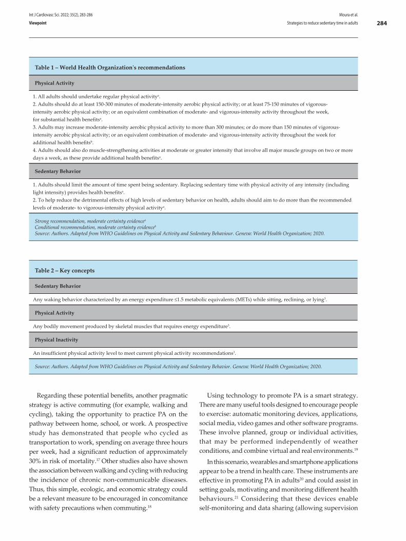
284
Table 1 – World Health Organization's recommendations
Physical Activity
1. All adults should undertake regular physical activitya.2. Adults should do at least 150-300 minutes of moderate-intensity aerobic physical activity; or at least 75-150 minutes of vigorous-intensity aerobic physical activity; or an equivalent combination of moderate- and vigorous-intensity activity throughout the week, for substantial health benefitsa.3. Adults may increase moderate-intensity aerobic physical activity to more than 300 minutes; or do more than 150 minutes of vigorous-intensity aerobic physical activity; or an equivalent combination of moderate- and vigorous-intensity activity throughout the week for additional health benefitsb.4. Adults should also do muscle-strengthening activities at moderate or greater intensity that involve all major muscle groups on two or more days a week, as these provide additional health benefitsa.
Sedentary Behavior
1. Adults should limit the amount of time spent being sedentary. Replacing sedentary time with physical activity of any intensity (including light intensity) provides health benefitsa.2. To help reduce the detrimental effects of high levels of sedentary behavior on health, adults should aim to do more than the recommended levels of moderate- to vigorous-intensity physical activitya.
Strong recommendation, moderate certainty evidencea
Conditional recommendation, moderate certainty evidenceb
Source: Authors. Adapted from WHO Guidelines on Physical Activity and Sedentary Behaviour. Geneva: World Health Organization; 2020.
Table 2 – Key concepts
Sedentary Behavior
Any waking behavior characterized by an energy expenditure ≤1.5 metabolic equivalents (METs) while sitting, reclining, or lying3.
Physical Activity
Any bodily movement produced by skeletal muscles that requires energy expenditure3.
Physical Inactivity
An insufficient physical activity level to meet current physical activity recommendations3.
Source: Authors. Adapted from WHO Guidelines on Physical Activity and Sedentary Behavior. Geneva: World Health Organization; 2020.
Regarding these potential benefits, another pragmatic strategy is active commuting (for example, walking and cycling), taking the opportunity to practice PA on the pathway between home, school, or work. A prospective study has demonstrated that people who cycled as transportation to work, spending on average three hours per week, had a significant reduction of approximately 30% in risk of mortality.17 Other studies also have shown the association between walking and cycling with reducing the incidence of chronic non-communicable diseases. Thus, this simple, ecologic, and economic strategy could be a relevant measure to be encouraged in concomitance with safety precautions when commuting.18
Using technology to promote PA is a smart strategy. There are many useful tools designed to encourage people to exercise: automatic monitoring devices, applications, social media, video games and other software programs. These involve planned, group or individual activities, that may be performed independently of weather conditions, and combine virtual and real environments.19
In this scenario, wearables and smartphone applications appear to be a trend in health care. These instruments are effective in promoting PA in adults20 and could assist in setting goals, motivating and monitoring different health behaviours.21 Considering that these devices enable self-monitoring and data sharing (allowing supervision
Int J Cardiovasc Sci. 2022; 35(2), 283-286 Moura et al.
Strategies to reduce sedentary time in adultsViewpoint
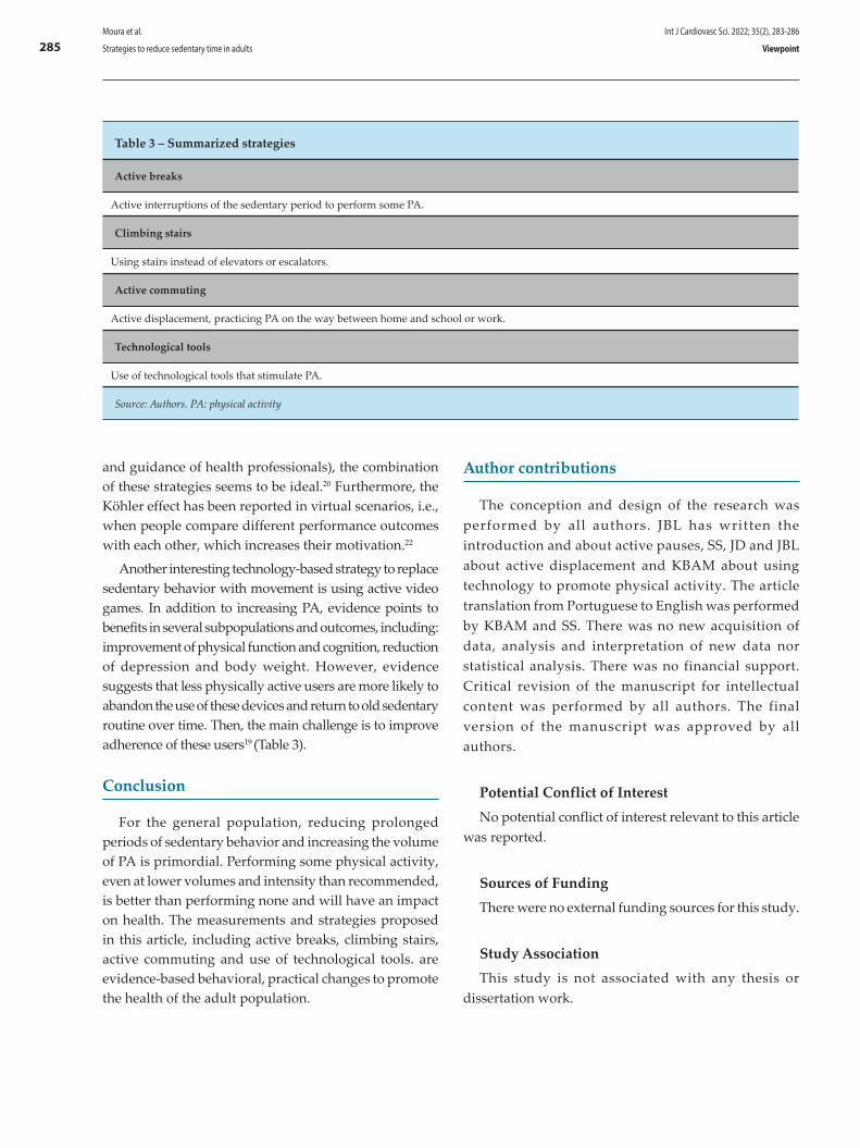
285
and guidance of health professionals), the combination of these strategies seems to be ideal.20 Furthermore, the Köhler effect has been reported in virtual scenarios, i.e., when people compare different performance outcomes with each other, which increases their motivation.22
Another interesting technology-based strategy to replace sedentary behavior with movement is using active video games. In addition to increasing PA, evidence points to benefits in several subpopulations and outcomes, including: improvement of physical function and cognition, reduction of depression and body weight. However, evidence suggests that less physically active users are more likely to abandon the use of these devices and return to old sedentary routine over time. Then, the main challenge is to improve adherence of these users19 (Table 3).
Conclusion
For the general population, reducing prolonged periods of sedentary behavior and increasing the volume of PA is primordial. Performing some physical activity, even at lower volumes and intensity than recommended, is better than performing none and will have an impact on health. The measurements and strategies proposed in this article, including active breaks, climbing stairs, active commuting and use of technological tools. are evidence-based behavioral, practical changes to promote the health of the adult population.
Author contributions
The conception and design of the research was performed by all authors. JBL has written the introduction and about active pauses, SS, JD and JBL about active displacement and KBAM about using technology to promote physical activity. The article translation from Portuguese to English was performed by KBAM and SS. There was no new acquisition of data, analysis and interpretation of new data nor statistical analysis. There was no financial support. Critical revision of the manuscript for intellectual content was performed by all authors. The final version of the manuscript was approved by all authors.
Potential Conflict of InterestNo potential conflict of interest relevant to this article
was reported.
Sources of FundingThere were no external funding sources for this study.
Study AssociationThis study is not associated with any thesis or
dissertation work.
Table 3 – Summarized strategies
Active breaks
Active interruptions of the sedentary period to perform some PA.
Climbing stairs
Using stairs instead of elevators or escalators.
Active commuting
Active displacement, practicing PA on the way between home and school or work.
Technological tools
Use of technological tools that stimulate PA.
Source: Authors. PA: physical activity
Int J Cardiovasc Sci. 2022; 35(2), 283-286Moura et al.
Strategies to reduce sedentary time in adults Viewpoint
286
1. GBD 2017 Causes of Death Collaborators. Global, regional, and national age-sex-specific mortality for 282 causes of death in 195 countries and territories, 1980-2017: a systematic analysis for the Global Burden of Disease Study 2017. Lancet. 2018;392(10159):1736-88.
2. Mendis S, Puska P, Norrving B, World Health Organization. Global atlas on cardiovascular disease prevention and control: Geneva:WHO;2011.
3. World Health Organization (WHO). Guidelines on physical activity and sedentary behaviour. Geneva; 2020.
4. Guthold R, Stevens GA, Riley LM, Bull FC. Worldwide trends in insufficient physical activity from 2011 to 2016: a pooled analysis of 358 population-based surveys with 1.9 million participants. Lancet Glob Health. 2018; 6(10):e1077-e1086.
5. Sedentary Behaviour Research Network. Letter to the editor: standardized use of the terms "sedentary" and "sedentary behaviours". Appl Physiol Nutr Metab. 2012;37(3):540-2.
6. Stamatakis E, Gale J, Bauman A, Ekelund U, Hamer M, Ding D. Sitting time, physical activity, and risk of mortality in adults. J Am Coll Cardiol. 2019;73(16):2062-72.
7. Piercy KL, Troiano RP, Ballard RM, Carlson SA, Fulton JE, Galuska DA, et al. The Physical Activity Guidelines for Americans. JAMA. 2018;320(19):2020-8.
8. World Health Organization. (WHO) Global recommendations on physical activity for health. Geneva; 2010.
9. Ministry of Health of Brazil. Secretariat of Primary Health Care. Health Promotion Department. Physical Activity Guidelines for the Brazilian Population. Brazil; 2021.
10. World Health Organization.(WHO) Global action plan on physical activity 2018-2030. More active people for a healthier world. Geneva:2018.
11. Hallgren M, Nguyen TT, Owen N, Vancampion D, Smith L, Dunstan DW, et al. Associations of interruptions to leisure-time sedentary behaviour with symptoms of depression and anxiety. Transl Psychiatry. 2020;10(1):128.
12. Healy GN, Dunstan DW, Salmon J, Cerin E, Shaw JE, Zimmet PZ, et al. Breaks in sedentary time: beneficial associations with metabolic risk. Diabetes Care. 2008;31(4):661-6.
13. Thosar SS, Bielko SL, Mather KJ, Johnston JD, Wallace JP. Effect of prolonged sitting and breaks in sitting time on endothelial function. Med Sci Sports Exerc. 2015;47(4):843-9.
14. Wheeler MJ, Dunstan DW, Ellis KA, Cerin E, Phillips S, Lambert G, et al. Effect of Morning Exercise With or Without Breaks in Prolonged Sitting on Blood Pressure in Older Overweight/Obese Adults. Hypertension. 2019;73(4):859-67.
15. Boreham CA, Kennedy RA, Murphy MH, Tully M, Wallace WF, Young I. Training effects of short bouts of stair climbing on cardiorespiratory fitness, blood lipids, and homocysteine in sedentary young women. Br J Sports Med. 2005;39(9):590-3.
16. Rey-Lopez JP, Stamatakis E, Mackey M, Sesso HD, Lee IM. Associations of self-reported stair climbing with all-cause and cardiovascular mortality: The Harvard Alumni Health Study. Prev Med Rep. 2019;15:100938.
17. Andersen LB, Schnohr P, Schroll M, Hein HO. All-cause mortality associated with physical activity during leisure time, work, sports, and cycling to work. Arch Intern Med. 2000;160(11):1621-8.
18. Andersen LB. Active commuting is beneficial for health. BMJ Clinical Research.. 2017;357:j1740. Doi:10.1136/bmj1740
19. Tate DF, Lyons EJ, Valle CG. High-Tech Tools for Exercise Motivation: Use and Role of Technologies Such as the Internet, Mobile Applications, Social Media, and Video Games. Diabetes Spectr. 2015; 28(1): 45-54.
20. Hakala S, Rintala A, Immonen J, Karvanen J, Heinonen A, Sjögren T. Effectiveness of physical activity promoting technology-based distance interventions compared to usual care. Systematic review, meta-analysis and meta-regression. Eur J Phys Rehabil Med. 2017; 53(6):953-67.
21. Schoeppe S, Alley S, Van Lippevelde W, Bray NA, Williams SL, Duncan MJ, et al. Efficacy of interventions that use apps to improve diet, physical activity and sedentary behaviour: a systematic review. Int J Behav Nutr Phys Act. 2016;13(1):127.
22. Wattanapisit A, Amaek W, Sukkriang N, Wattanapisit S, Wongsiri S. Perspectives on Using Online Platforms for Promoting Running and Walking Activities. Front Public Health. 2020;8:150.
References
Int J Cardiovasc Sci. 2022; 35(2), 283-286 Moura et al.
Strategies to reduce sedentary time in adultsViewpoint
This is an open-access article distributed under the terms of the Creative Commons Attribution License
Abstract
Takayasu’s arteritis is a type of primary systemic vasculitis that affects medium and large arteries, including the aorta and its main branches, as well as the pulmonary and coronary arteries. Although rare in children, it is the third most common vasculitis in the pediatric population, often with delayed diagnosis due to the nonspecific presentation of clinical symptoms in its initial phase. This is a case of a 16-year-old girl with a giant ruptured abdominal aortic aneurysm, who needed surgery on an emergency basis. The etiological aspects involved in aneurysms in young patients are also addressed.
Introduction
Abdominal aortic aneurysms (AAAs) are rare in the pediatric population.1 They require high clinical suspicion, since they remain asymptomatic for a long period time, and, much like in adults, they also present a serious risk of rupture and death.1,2 These AAAs have also been associated with systemic diseases, developmental abnormalities, and connective tissue disorders.2,3
Takayasu’s arteritis (TA) is a chronic progressive inflammatory disease characterized by granulomatous vasculitis, which involves the aorta and its main
branches.3 Granulomatous transmural inflammation may eventually lead to stenosis or occlusions in the involved vessels. Though aneurysms and aortic dissections are rare events, particularly in the pediatric population and young adults, they are now more frequently being diagnosed. However, TA diagnosis remains a major challenge due to its nonspecific clinical and laboratory features.3,4
An aneurysm is defined by the Society of Vascular Surgery as a localized, permanent dilation of an artery with at least a 50% increase in diameter size as compared to the expected normal diameter of the artery or its proximal segment.5-7 AAAs can be classified according to their relative location to the renal arteries, morphology, diameter, or etiology, and are three to eleven times more common in men than in women.5-7
A ruptured AAA is a major life event that carries a high mortality rate.7 Immediate diagnosis and treatment is essential to improve the outcome because of its time-sensitive nature.7,8
Emergency physicians should maintain a higher rate of suspicion for prompt diagnosis of aortic aneurysm in any individual regardless of age when presented with such symptoms as abdominal pain, especially if associated with hypertension and radiating back pain.8,9
The case presented herein is of particular interest, reporting on a 16-year-old girl with TA who had a ruptured giant abdominal aortic aneurysm. The etiological aspects involved in aneurysms in this population will be reviewed and discussed.
Int J Cardiovasc Sci. 2022; 35(2), 287-292
287
CASE REPORT
Surgical Repair of a Ruptured Giant Abdominal Aortic Aneurysm in a 16-Year-Old with Takayasu’s Arteritis: Case Report and Etiological ReviewJosé Maciel Caldas dos Reis,1 Glauco dos Santos Melo,1 Murilo Vasconcelos de Oliveira,1 Felipe Eduardo de Oliveira Santos,2 Tereza Maria Meireles Fernandes da Silva,3 Hugo Luis da Silva Ferreira4
Division of Vascular and Endovascular Surgery at Gaspar Vianna Clinics Hospital (HCGV),1 Belém, PA - BrazilDivision of General Surgery at Gaspar Vianna Clinics Hospital (HCGV),2 Belém, PA - BrazilPará State University (UEPa),3 Belém, PA - BrazilFederal University of Pará (UFPa),4 Belém, PA - Brazil
Aortic Aneurysm Abdominal/surgery; Takayasu Arteritis; Vasculitis; Young Adult; Child; Mortality.
Keywords
DOI: https://doi.org/10.36660/ijcs.20200198
Mailing Address: José Maciel Caldas dos ReisGaspar Vianna Clinics Hospital (HCGV)Tv. Alferes Costa, s/n. Postal Code: 66083-106, Pedreira, Belém, PA – Brazil. E-mail: [email protected]
Manuscript received July 14, 2020; revised manuscript October 04, 2020; accepted January 03, 2021.
288
Case presentation
The patient was a 16-year-old female with hypertension, who had suffered episodes of palpitations and hypertensive crises over the past two years.
The patient was admitted to the emergency room of the hospital with a sudden onset of abdominal pain, associated with skin and mucosal pallor and back pain. Her pain had begun at home 4 hours before being admitted to the hospital.
The patient had been attending regular nephrological and cardiological follow-up consultations at the institution due to difficult-to-control hypertension and deterioration of renal function. She was also using taking presciption medications and was not a smoker or illicit drug user. There was no family history of aneurysm and no report of trauma, diarrhea, constipation, fever, or symptoms of dysuria or hematuria. She was 1.65 m tall, with a body mass index (BMI) of 24 kg/m2. There were no indications of connective tissue disease with arterial involvement, such as hyperelastic skin, hypermobile joints, or Marfanoid habitus. She reported that 3 months ago she had received the diagnosis of TA, based on clinical and tomographic criteria.
At the initial presentation and physical examination, the patient was experiencing intense abdominal pain. Diffuse abdominal tenderness to palpation was observed, mainly in the lower abdominal region, and the presence of a pulsatile mass in the epigastrium and mesogastrium regions during the abdominal physical examination. She reported that the pain radiated to her back, preventing active movements in the left lower limbs. Her vital signs were stable upon admission, with a heart rate of 115 bpm and a blood pressure of 100/60 mmHg.
Emergency laboratory tests revealed abnormal concentration levels of hemoglobin, 6.2 g/dL; hematocrit, 21%; leukocytes, 12,000/mm3; platelets, 180,000/mm3; Urea, 76; and Creatinine, 2.1. An electrocardiogram showed signs of left ventricular hypertrophy.
A ruptured abdominal aortic aneurysm was suspected, and the patient was immediately referred to the radiology ward, where she underwent an abdominal CT angiography before being transferred to the operating room.
The CT angiography revealed a ruptured abdominal infrarenal aortic aneurysm, which opened to the
retroperitoneum, with a maximum estimated diameter of 11.0 cm (Figure 1). The site of the distal aortic rupture and the bilateral ostial occlusion of the renal artery stood out in the CT angiogram image reconstruction (Figure 2).
In the emergency surgery, a xiphopubic midline abdominal incision was made, the mesentery was exposed, and the intestines were immobilized. Transperitoneal dissection along the ligament of Treitz revealed a giant aneurysm. It was carefully dissected and found to be ruptured in its posterolateral wall on the left, 3 cm above the bifurcation to the iliac arteries, with a significant retroperitoneal hematoma (Figure 3).
The infrarenal aorta and the common iliac artery were dissected for repairs and to apply clamps. The proximal aorta and both iliac arteries were clamped after systemic heparinization.
The aneurysm was opened and the mural thrombus removed. The bifurcation of the juxtarenal aorta and both common iliac arteries presented no aneurysmatic alterations and were used for distal control. As the patient presented no clinical evidence or operative findings compatible with active aortic infection, there was minimal concern with the placement of the prosthetic material. A 20 × 10 mm Dacron graft was used for a bilateral aortoiliac graft using 2-0 polypropylene thread in a continuous suture (Figure 4). The aneurysm sac was sutured over the graft with no leaks. After two days in the intensive care unit, the patient was transferred to the general ward. While in the intensive care unit, she underwent renal replacement therapy with hemodialysis by a catheter inserted into the common femoral vein. She had a total of six sessions of hemodialysis until she was stable and conservative treatment was resumed. The patient was discharged from the hospital on the seventh postoperative day and the follow-up at six months was normal.
Tomographic and intraoperative findings were compatible with the presence of a true aneurysm. The aneurysm walls were thicker than that of the normal aorta. A small fragment from the mural thrombus and the aorta were sent for culture and histopathological analysis. A histopathological examination of the aneurysm with conventional hematoxylin-eosin staining revealed that the aneurysm wall had a disordered structure with nonspecific inflammation in the tunica media and adventitia that was comparable to TA. Both the aneurysm and the thrombus wall cultures were negative for bacteria.
Int J Cardiovasc Sci. 2022; 35(2), 287-292 Reis et al.
Giant abdominal aortic aneurysmCase Report
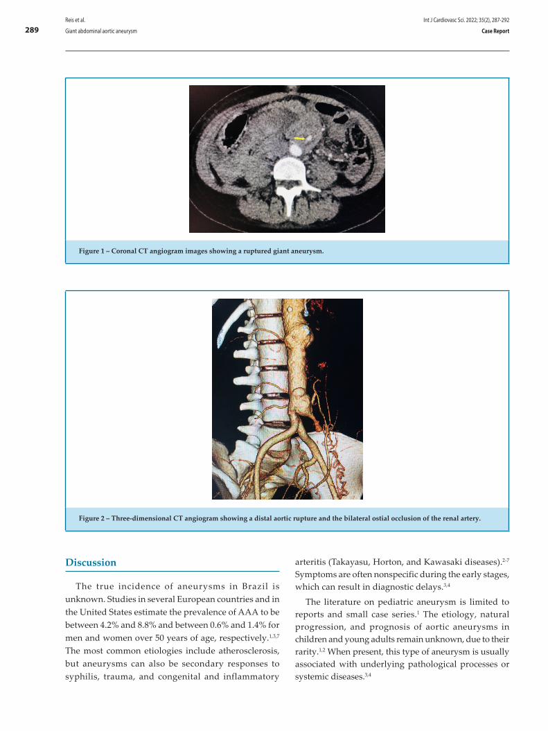
289
Figure 2 – Three-dimensional CT angiogram showing a distal aortic rupture and the bilateral ostial occlusion of the renal artery.
Figure 1 – Coronal CT angiogram images showing a ruptured giant aneurysm.
Discussion
The true incidence of aneurysms in Brazil is unknown. Studies in several European countries and in the United States estimate the prevalence of AAA to be between 4.2% and 8.8% and between 0.6% and 1.4% for men and women over 50 years of age, respectively.1,3,7 The most common etiologies include atherosclerosis, but aneurysms can also be secondary responses to syphilis, trauma, and congenital and inflammatory
arteritis (Takayasu, Horton, and Kawasaki diseases).2-7 Symptoms are often nonspecific during the early stages, which can result in diagnostic delays.3,4
The literature on pediatric aneurysm is limited to reports and small case series.1 The etiology, natural progression, and prognosis of aortic aneurysms in children and young adults remain unknown, due to their rarity.1,2 When present, this type of aneurysm is usually associated with underlying pathological processes or systemic diseases.3,4
Int J Cardiovasc Sci. 2022; 35(2), 287-292Reis et al.
Giant abdominal aortic aneurysm Case Report
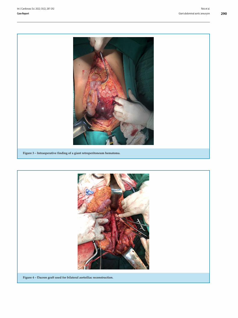
290
Figure 4 – Dacron graft used for bilateral aortoiliac reconstruction.
Figure 3 – Intraoperative finding of a giant retroperitoneum hematoma.
Int J Cardiovasc Sci. 2022; 35(2), 287-292 Reis et al.
Giant abdominal aortic aneurysmCase Report
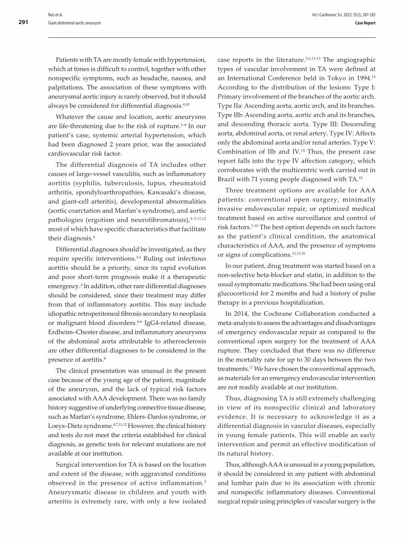
291
Patients with TA are mostly female with hypertension, which at times is difficult to control, together with other nonspecific symptoms, such as headache, nausea, and palpitations. The association of these symptoms with aneurysmal aortic injury is rarely observed, but it should always be considered for differential diagnosis.4,10
Whatever the cause and location, aortic aneurysms are life-threatening due to the risk of rupture.1–4 In our patient’s case, systemic arterial hypertension, which had been diagnosed 2 years prior, was the associated cardiovascular risk factor.
The differential diagnosis of TA includes other causes of large-vessel vasculitis, such as inflammatory aortitis (syphilis, tuberculosis, lupus, rheumatoid arthritis, spondyloarthropathies, Kawasaki’s disease, and giant-cell arteritis), developmental abnormalities (aortic coarctation and Marfan’s syndrome), and aortic pathologies (ergotism and neurofibromatosis),4–7,11,12 most of which have specific characteristics that facilitate their diagnosis.4
Differential diagnoses should be investigated, as they require specific interventions.3,4 Ruling out infectious aortitis should be a priority, since its rapid evolution and poor short-term prognosis make it a therapeutic emergency.4 In addition, other rare differential diagnoses should be considered, since their treatment may differ from that of inflammatory aortitis. This may include idiopathic retroperitoneal fibrosis secondary to neoplasia or malignant blood disorders.4-6 IgG4-related disease, Erdheim–Chester disease, and inflammatory aneurysms of the abdominal aorta attributable to atherosclerosis are other differential diagnoses to be considered in the presence of aortitis.4
The clinical presentation was unusual in the present case because of the young age of the patient, magnitude of the aneurysm, and the lack of typical risk factors associated with AAA development. There was no family history suggestive of underlying connective tissue disease, such as Marfan’s syndrome, Ehlers–Danlos syndrome, or Loeys–Dietz syndrome.4,7,11,12 However, the clinical history and tests do not meet the criteria established for clinical diagnosis, as genetic tests for relevant mutations are not available at our institution.
Surgical intervention for TA is based on the location and extent of the disease, with aggravated conditions observed in the presence of active inflammation.3 Aneurysmatic disease in children and youth with arteritis is extremely rare, with only a few isolated
case reports in the literature.3,6,11-13 The angiographic types of vascular involvement in TA were defined at an International Conference held in Tokyo in 1994.14 According to the distribution of the lesions: Type I: Primary involvement of the branches of the aortic arch. Type IIa: Ascending aorta, aortic arch, and its branches. Type IIb: Ascending aorta, aortic arch and its branches, and descending thoracic aorta. Type III: Descending aorta, abdominal aorta, or renal artery. Type IV: Affects only the abdominal aorta and/or renal arteries. Type V: Combination of IIb and IV.14 Thus, the present case report falls into the type IV affection category, which corroborates with the multicentric work carried out in Brazil with 71 young people diagnosed with TA.15
Three treatment options are available for AAA patients: conventional open surgery, minimally invasive endovascular repair, or optimized medical treatment based on active surveillance and control of risk factors.3-10 The best option depends on such factors as the patient’s clinical condition, the anatomical characteristics of AAA, and the presence of symptoms or signs of complications.12,13,16
In our patient, drug treatment was started based on a non-selective beta-blocker and statin, in addition to the usual symptomatic medications. She had been using oral glucocorticoid for 2 months and had a history of pulse therapy in a previous hospitalization.
In 2014, the Cochrane Collaboration conducted a meta-analysis to assess the advantages and disadvantages of emergency endovascular repair as compared to the conventional open surgery for the treatment of AAA rupture. They concluded that there was no difference in the mortality rate for up to 30 days between the two treatments.17 We have chosen the conventional approach, as materials for an emergency endovascular intervention are not readily available at our institution.
Thus, diagnosing TA is still extremely challenging in view of its nonspecific clinical and laboratory evidence. It is necessary to acknowledge it as a differential diagnosis in vascular diseases, especially in young female patients. This will enable an early intervention and permit an effective modification of its natural history.
Thus, although AAA is unusual in a young population, it should be considered in any patient with abdominal and lumbar pain due to its association with chronic and nonspecific inflammatory diseases. Conventional surgical repair using principles of vascular surgery is the
Int J Cardiovasc Sci. 2022; 35(2), 287-292Reis et al.
Giant abdominal aortic aneurysm Case Report
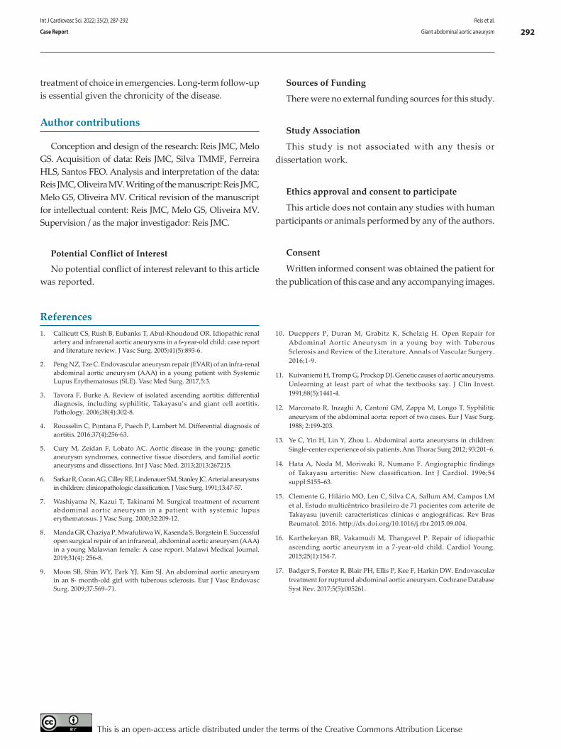
292
treatment of choice in emergencies. Long-term follow-up is essential given the chronicity of the disease.
Author contributions
Conception and design of the research: Reis JMC, Melo GS. Acquisition of data: Reis JMC, Silva TMMF, Ferreira HLS, Santos FEO. Analysis and interpretation of the data: Reis JMC, Oliveira MV. Writing of the manuscript: Reis JMC, Melo GS, Oliveira MV. Critical revision of the manuscript for intellectual content: Reis JMC, Melo GS, Oliveira MV. Supervision / as the major investigador: Reis JMC.
Potential Conflict of Interest
No potential conflict of interest relevant to this article was reported.
Sources of Funding
There were no external funding sources for this study.
Study Association
This study is not associated with any thesis or dissertation work.
Ethics approval and consent to participate
This article does not contain any studies with human participants or animals performed by any of the authors.
Consent
Written informed consent was obtained the patient for the publication of this case and any accompanying images.
1. Callicutt CS, Rush B, Eubanks T, Abul-Khoudoud OR. Idiopathic renal artery and infrarenal aortic aneurysms in a 6-year-old child: case report and literature review. J Vasc Surg. 2005;41(5):893-6.
2. Peng NZ, Tze C. Endovascular aneurysm repair (EVAR) of an infra-renal abdominal aortic aneurysm (AAA) in a young patient with Systemic Lupus Erythematosus (SLE). Vasc Med Surg. 2017,5:3.
3. Tavora F, Burke A. Review of isolated ascending aortitis: differential diagnosis, including syphilitic, Takayasu’s and giant cell aortitis. Pathology. 2006;38(4):302-8.
4. Rousselin C, Pontana F, Puech P, Lambert M. Differential diagnosis of aortitis. 2016;37(4):256-63.
5. Cury M, Zeidan F, Lobato AC. Aortic disease in the young: genetic aneurysm syndromes, connective tissue disorders, and familial aortic aneurysms and dissections. Int J Vasc Med. 2013;2013:267215.
6. Sarkar R, Coran AG, Cilley RE, Lindenauer SM, Stanley JC. Arterial aneurysms in children: clinicopathologic classification. J Vasc Surg. 1991;13:47-57.
7. Washiyama N, Kazui T, Takinami M. Surgical treatment of recurrent abdominal aortic aneurysm in a patient with systemic lupus erythematosus. J Vasc Surg. 2000;32:209-12.
8. Manda GR, Chaziya P, Mwafulirwa W, Kasenda S, Borgstein E. Successful open surgical repair of an infrarenal, abdominal aortic aneurysm (AAA) in a young Malawian female: A case report. Malawi Medical Journal. 2019;31(4): 256-8.
9. Moon SB, Shin WY, Park YJ, Kim SJ. An abdominal aortic aneurysm in an 8- month-old girl with tuberous sclerosis. Eur J Vasc Endovasc Surg. 2009;37:569–71.
10. Dueppers P, Duran M, Grabitz K, Schelzig H. Open Repair for Abdominal Aortic Aneurysm in a young boy with Tuberous Sclerosis and Review of the Literature. Annals of Vascular Surgery. 2016;1-9.
11. Kuivaniemi H, Tromp G, Prockop DJ. Genetic causes of aortic aneurysms. Unlearning at least part of what the textbooks say. J Clin Invest. 1991;88(5):1441-4.
12. Marconato R, Inzaghi A, Cantoni GM, Zappa M, Longo T. Syphilitic aneurysm of the abdominal aorta: report of two cases. Eur J Vasc Surg. 1988; 2:199-203.
13. Ye C, Yin H, Lin Y, Zhou L. Abdominal aorta aneurysms in children: Single-center experience of six patients. Ann Thorac Surg 2012; 93:201–6.
14. Hata A, Noda M, Moriwaki R, Numano F. Angiographic findings of Takayasu arteritis: New classification. Int J Cardiol. 1996;54 suppl:S155–63.
15. Clemente G, Hilário MO, Len C, Silva CA, Sallum AM, Campos LM et al. Estudo multicêntrico brasileiro de 71 pacientes com arterite de Takayasu juvenil: características clínicas e angiográficas. Rev Bras Reumatol. 2016. http://dx.doi.org/10.1016/j.rbr.2015.09.004.
16. Karthekeyan BR, Vakamudi M, Thangavel P. Repair of idiopathic ascending aortic aneurysm in a 7-year-old child. Cardiol Young. 2015;25(1):154-7.
17. Badger S, Forster R, Blair PH, Ellis P, Kee F, Harkin DW. Endovascular treatment for ruptured abdominal aortic aneurysm. Cochrane Database Syst Rev. 2017;5(5):005261.
References
Int J Cardiovasc Sci. 2022; 35(2), 287-292 Reis et al.
Giant abdominal aortic aneurysmCase Report
This is an open-access article distributed under the terms of the Creative Commons Attribution License
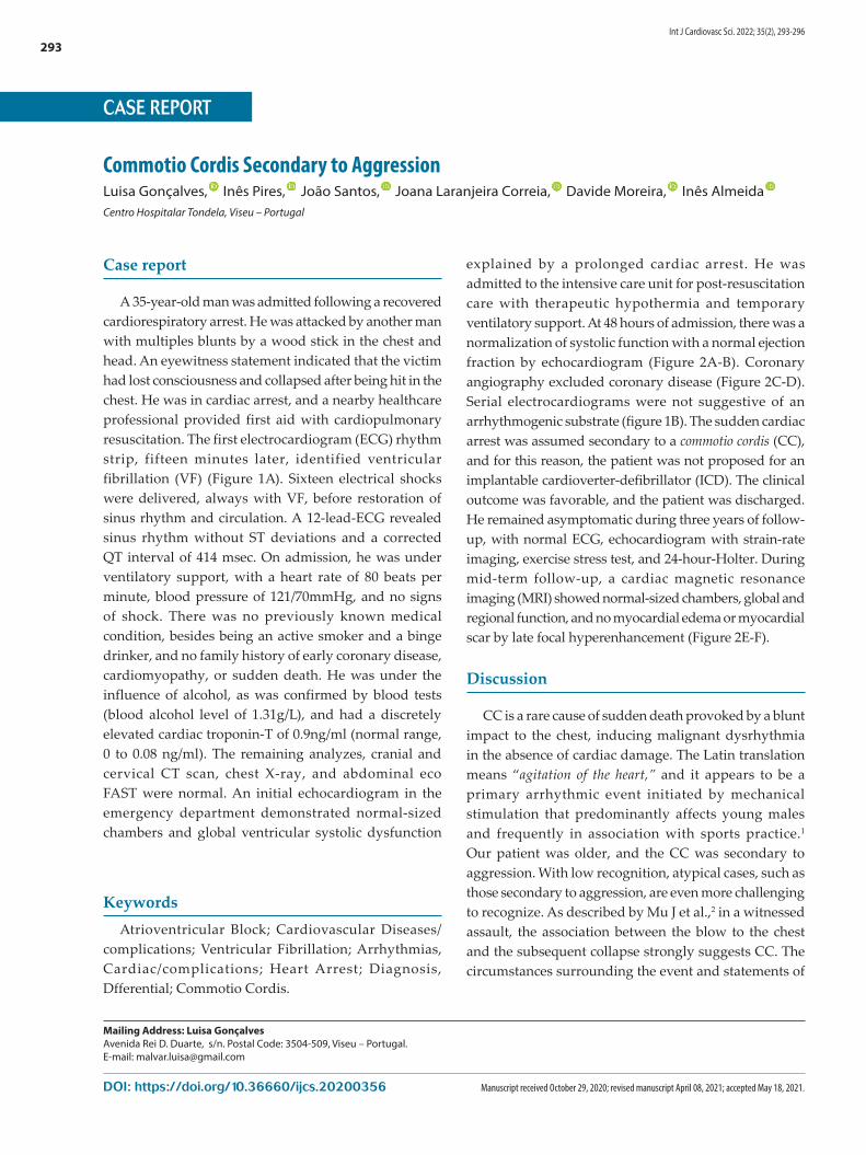
Case report
A 35-year-old man was admitted following a recovered cardiorespiratory arrest. He was attacked by another man with multiples blunts by a wood stick in the chest and head. An eyewitness statement indicated that the victim had lost consciousness and collapsed after being hit in the chest. He was in cardiac arrest, and a nearby healthcare professional provided first aid with cardiopulmonary resuscitation. The first electrocardiogram (ECG) rhythm strip, fifteen minutes later, identified ventricular fibrillation (VF) (Figure 1A). Sixteen electrical shocks were delivered, always with VF, before restoration of sinus rhythm and circulation. A 12-lead-ECG revealed sinus rhythm without ST deviations and a corrected QT interval of 414 msec. On admission, he was under ventilatory support, with a heart rate of 80 beats per minute, blood pressure of 121/70mmHg, and no signs of shock. There was no previously known medical condition, besides being an active smoker and a binge drinker, and no family history of early coronary disease, cardiomyopathy, or sudden death. He was under the influence of alcohol, as was confirmed by blood tests (blood alcohol level of 1.31g/L), and had a discretely elevated cardiac troponin-T of 0.9ng/ml (normal range, 0 to 0.08 ng/ml). The remaining analyzes, cranial and cervical CT scan, chest X-ray, and abdominal eco FAST were normal. An initial echocardiogram in the emergency department demonstrated normal-sized chambers and global ventricular systolic dysfunction
explained by a prolonged cardiac arrest. He was admitted to the intensive care unit for post-resuscitation care with therapeutic hypothermia and temporary ventilatory support. At 48 hours of admission, there was a normalization of systolic function with a normal ejection fraction by echocardiogram (Figure 2A-B). Coronary angiography excluded coronary disease (Figure 2C-D). Serial electrocardiograms were not suggestive of an arrhythmogenic substrate (figure 1B). The sudden cardiac arrest was assumed secondary to a commotio cordis (CC), and for this reason, the patient was not proposed for an implantable cardioverter-defibrillator (ICD). The clinical outcome was favorable, and the patient was discharged. He remained asymptomatic during three years of follow-up, with normal ECG, echocardiogram with strain-rate imaging, exercise stress test, and 24-hour-Holter. During mid-term follow-up, a cardiac magnetic resonance imaging (MRI) showed normal-sized chambers, global and regional function, and no myocardial edema or myocardial scar by late focal hyperenhancement (Figure 2E-F).
Discussion
CC is a rare cause of sudden death provoked by a blunt impact to the chest, inducing malignant dysrhythmia in the absence of cardiac damage. The Latin translation means “agitation of the heart,” and it appears to be a primary arrhythmic event initiated by mechanical stimulation that predominantly affects young males and frequently in association with sports practice.1 Our patient was older, and the CC was secondary to aggression. With low recognition, atypical cases, such as those secondary to aggression, are even more challenging to recognize. As described by Mu J et al.,2 in a witnessed assault, the association between the blow to the chest and the subsequent collapse strongly suggests CC. The circumstances surrounding the event and statements of
Int J Cardiovasc Sci. 2022; 35(2), 293-296
293
CASE REPORT
Commotio Cordis Secondary to AggressionLuisa Gonçalves, Inês Pires, João Santos, Joana Laranjeira Correia, Davide Moreira, Inês AlmeidaCentro Hospitalar Tondela, Viseu – Portugal
Atrioventricular Block; Cardiovascular Diseases/complications; Ventricular Fibrillation; Arrhythmias, Cardiac/complications; Heart Arrest; Diagnosis, Dfferential; Commotio Cordis.
Keywords
DOI: https://doi.org/10.36660/ijcs.20200356
Mailing Address: Luisa GonçalvesAvenida Rei D. Duarte, s/n. Postal Code: 3504-509, Viseu – Portugal.E-mail: [email protected]
Manuscript received October 29, 2020; revised manuscript April 08, 2021; accepted May 18, 2021.
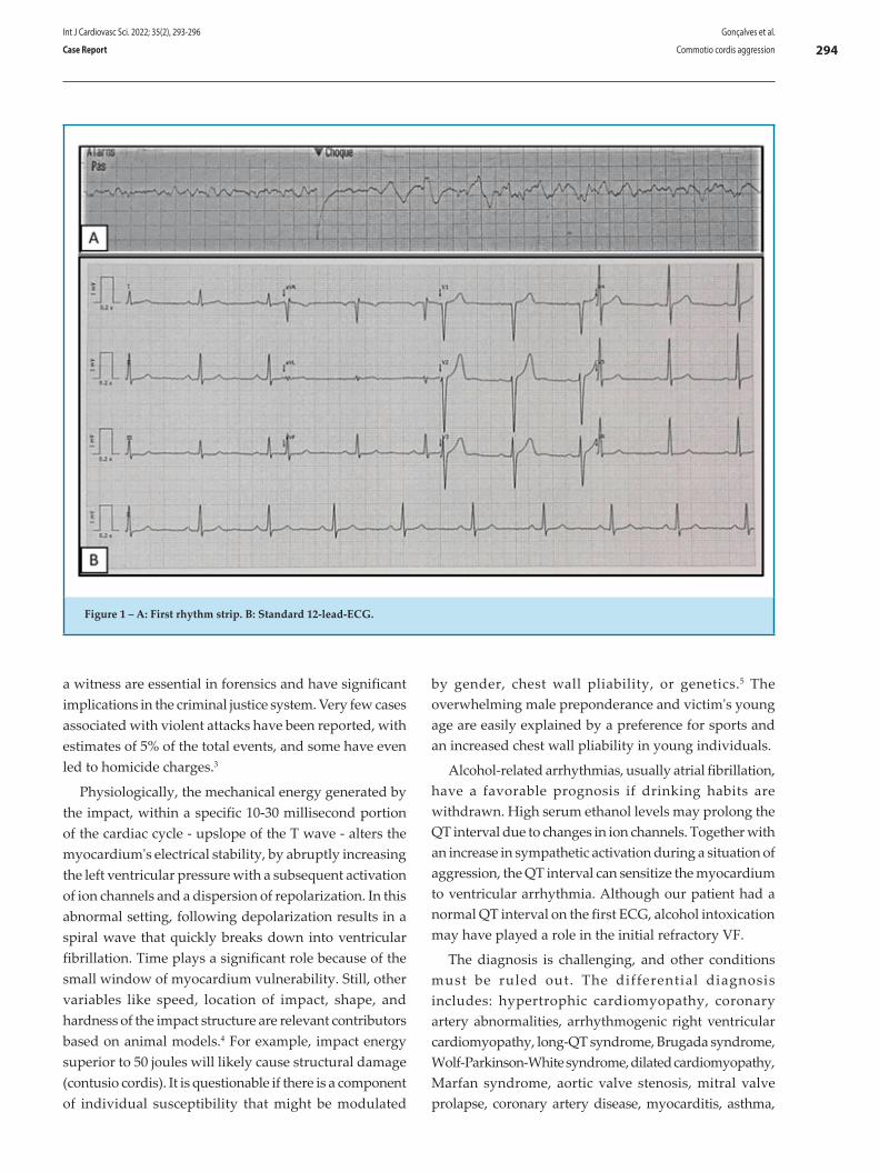
294
a witness are essential in forensics and have significant implications in the criminal justice system. Very few cases associated with violent attacks have been reported, with estimates of 5% of the total events, and some have even led to homicide charges.3
Physiologically, the mechanical energy generated by the impact, within a specific 10-30 millisecond portion of the cardiac cycle - upslope of the T wave - alters the myocardium's electrical stability, by abruptly increasing the left ventricular pressure with a subsequent activation of ion channels and a dispersion of repolarization. In this abnormal setting, following depolarization results in a spiral wave that quickly breaks down into ventricular fibrillation. Time plays a significant role because of the small window of myocardium vulnerability. Still, other variables like speed, location of impact, shape, and hardness of the impact structure are relevant contributors based on animal models.4 For example, impact energy superior to 50 joules will likely cause structural damage (contusio cordis). It is questionable if there is a component of individual susceptibility that might be modulated
by gender, chest wall pliability, or genetics.5 The overwhelming male preponderance and victim's young age are easily explained by a preference for sports and an increased chest wall pliability in young individuals.
Alcohol-related arrhythmias, usually atrial fibrillation, have a favorable prognosis if drinking habits are withdrawn. High serum ethanol levels may prolong the QT interval due to changes in ion channels. Together with an increase in sympathetic activation during a situation of aggression, the QT interval can sensitize the myocardium to ventricular arrhythmia. Although our patient had a normal QT interval on the first ECG, alcohol intoxication may have played a role in the initial refractory VF.
The diagnosis is challenging, and other conditions must be ruled out. The differential diagnosis includes: hypertrophic cardiomyopathy, coronary artery abnormalities, arrhythmogenic right ventricular cardiomyopathy, long-QT syndrome, Brugada syndrome, Wolf-Parkinson-White syndrome, dilated cardiomyopathy, Marfan syndrome, aortic valve stenosis, mitral valve prolapse, coronary artery disease, myocarditis, asthma,
Figure 1 – A: First rhythm strip. B: Standard 12-lead-ECG.
Int J Cardiovasc Sci. 2022; 35(2), 293-296 Gonçalves et al.
Commotio cordis aggressionCase Report
295
Figure 2 – Transthoracic echocardiogram. A: parasternal long-axis view. B: apical 4-chamber view. Coronary angiography. C: Right coronary artery. D: Left coronary artery. Cardiac magnetic resonance imaging. E: cine imaging (SSFP): 4-chamber view. F: late gadolinium enhancement sequences: 4-chamber view.
Int J Cardiovasc Sci. 2022; 35(2), 293-296Gonçalves et al.
Commotio cordis aggression Case Report
296
heat stroke, drug abuse, and a ruptured cerebral artery. Our patient underwent a complete study that excluded all other causes. Contusio cordis is often due to significant chest trauma, originating a contusion of the myocardial muscle, a cardiac chamber rupture, or heart valve disruption.6 Presence of delayed enhancement in cardiac MRI may translate into myocardial structural damage (scar).7 Contusio cordis was dismissed because there was no structural damage on the echocardiogram or scar in cardiac MRI during follow-up.
Increased awareness of this phenomenon in recent decades, especially among those who may be first responders and readily available defibrillators and medical staff on hand on sports grounds, have improved the mortality rate. The survival rate is estimated at 25% if cardiopulmonary resuscitation is initiated within the first three minutes.8 Resuscitation is often unsuccessful, and early defibrillation is critical.1,8 Our patient was lucky to be treated immediately and continuously by a nearby health professional until defibrillation was possible. Although the initial refractory VF, he recovered without sequelae. Given this particular case, the authors suggest that CC's victim should be submitted to aggressive resuscitation, keeping in mind that a normal heart structure and function are presumed.
CC survivors should undergo a comprehensive cardiac evaluation, including a 12-lead-ECG, ambulatory Holter monitoring, ECG stress test, echocardiogram, and cardiac catheterization.9 Electrophysiologic testing and ICD are not generally recommended unless a secondary cause is suspected.9 About 29% of CC survivors had mild to moderate residual neurologic disability or reduced left ventricular ejection fraction on long-term follow-
up.10 During the 3-year follow-up, our patient remained asymptomatic with normal complementary exams.
CC is a rare and frequently fatal event. During a witnessed eve, prompt initiation of cardiopulmonary resuscitation and defibrillation is essential for survival without sequelae. An atypical scenario of sudden death after chest impact secondary to aggression should raise suspicion of CC.
Author contributions
Conception and design of the research: Gonçalves ML, Pires MI, Santos JM. Acquisition of data: Gonçalves ML, Santos JM. Analysis and interpretation of the data: Gonçalves ML, Pires MI. Writing of the manuscript: Gonçalves ML. Critical revision of the manuscript for intellectual content: Gonçalves ML, Pires MI, Santos JM, Correia J, Moreira D, Almeida I.
Potential Conflict of InterestNo potential conflict of interest relevant to this article
was reported.
Sources of FundingThere were no external funding sources for this study.
Study AssociationThis study is not associated with any thesis or
dissertation work.
Ethics approval and consent to participateThis article does not contain any studies with human
participants or animals performed by any of the authors.
1. Cooper S, Woodford NW, Maron BJ, Harris KM, Sheppard MN. A Lethal Blow to the Chest as an Underdiagnosed Cause of Sudden Death in United Kingdom Sports (Football, Cricket, Rugby). Am J Cardiol. 2019;1;124(5):808-11.
2. Mu J, Chen Z, Chen X, Lin W, Dong H. Commotio cordis caused by violence in China: epidemiological characteristics detected at the Tongji Forensic Medical Center. Medicine (Baltimore). 2015;94(51):e2315.
3. Lucena JS, Rico A, Salguero M, Blanco M, Vázquez R, Commotio cordis as a result of a fight: report of a case considered to be imprudent homicide. Forensic Sci Int. 2008;177(1):e1-4.
4. Link MS. Commotio cordis: ventricular fibrillation triggered by chest impact-induced abnormalities in repolarization. Circ Arrhythm Electrophysiol. 2012 Apr;5(2):425-32.
5. Alsheikh-Ali AA, Madias C, Supran S, Link MS. Marked variability in susceptibility to ventricular fibrillation in an experimental commotio cordis model. Circulation. 2010;122:2499-504.
6. Longo Neto GC, Martins WA, Villacorta HJ, Silva EN, Haffner PM, Souza DG. Contusio Cordis Associated with Atrioventricular Block and Tricuspid Regurgitation. Arq Bras Cardiol. 2014;103(2):e22-e25.
7. Vago H, Toth A, Apor A, Maurovich-Horvat P, Toth M, Merkely B. Images in cardiovascular medicine. Cardiac contusion in a professional soccer player: visualization of acute and late pathological changes in the myocardium with magnetic resonance imaging. Circulation. 2010;121(22):2456-61.
8. Maron BJ, Haas TS, Ahluwalia A, Garberich RF, Estes NA3rd, Link MS. Increasing survival rate from commotio cordis. Heart Rhythm. 2013;10: 19-223.
9. Solberg E., Link M.S. (2020) Commotio Cordis. In: Pressler A., Niebauer J. (eds) Textbook of Sports and Exercise Cardiology. New York:Springer; 2020.
10. Maron BJ, Gohman TE, Kyle SB, Estes NA3rd, Link MS. Clinical profile and spectrum of commotio cordis. JAMA. 2002;287(9):1142-6.
References
Int J Cardiovasc Sci. 2022; 35(2), 293-296 Gonçalves et al.
Commotio cordis aggressionCase Report
This is an open-access article distributed under the terms of the Creative Commons Attribution License
4CAPA-ijcs.indd 1 13/03/2018 10:29:17