Editorial introductions
110
Copyright © 2021 Wolters Kluwer Health, Inc. All rights reserved. C URRENT OPINION Editorial introductions Current Opinion in Rheumatology was launched in 1989. It is one of a successful series of review journals whose unique format is designed to provide a systematic and critical assessment of the literature as presented in the many primary journals. The field of Rheumatology is divided into 15 sections that are reviewed once a year. Each section is assigned a Section Editor, a leading authority in the area, who identifies the most important topics at that time. Here we are pleased to introduce the Journal’s Section Editors for this issue. Kenneth G. Saag Kenneth G. Saag, MD, MSc, is Jane Knight Lowe Professor of Medi- cine, Division of Clinical Immu- nology and Rheumatology, at the University of Alabama at Bir- mingham (UAB) in Birmingham, Alabama, USA and Professor of Epidemiology, at the UAB School of Public Health. Dr Saag is a native of Chicago, and following studies in engineering at the University of Michigan, USA, he returned to Chicago for medical school and Internal Medicine Residency at Northwestern Uni- versity, USA. He then traveled to the University of Iowa, USA, for his rheumatology and epidemiology training and remained on the faculty there until moving to UAB in 1998. Dr Saag is the founding Director of the Agency for Health Care Research and Quality Deep South Center for Education and Research on Therapeutics, the UAB Center of Research Translation in Gout and Hyperuricemia, and the UAB Outcomes Research Center. He also serves as a Vice Chair for the UAB Department of Medicine. He has published over 300 peer reviewed manuscripts; has also authored more than 100 reviews, editorials, and book chapters. Recently he published the first edition of the clinical handbook Diagnosis and Management of Osteoporosis. Dr Saag is the Secretary of the American College of Rheumatology (ACR) and Past President of the Board of Trustees of the National Osteoporosis Foundation. Sara K. Tedeschi Dr Tedeschi is a Rheumatologist at Brigham and Women’s Hospi- tal and an Assistant Professor of Medicine at Harvard Medical School, both in Boston, Massa- chusetts, USA. She completed medical school at Vanderbilt Uni- versity School of Medicine in Nashville, Tennessee, USA. Dr Tedeschi then completed both her internal medi- cine residency and rheumatology fellowship train- ing at Brigham and Women’s Hospital and received her MPH from the Harvard T.H. Chan School of Public Health. Her clinical research focuses on risk factors for and outcomes in rheumatic diseases. Dr Tedeschi’s current work which focuses on calcium pyrophosphate crystal deposition disease is funded by the National Institutes of Health. She is Co-Chair of the OMERACT CPPD Working Group and serves on the leadership team of the ACR/EULAR CPPD Classification Criteria working group. George C. Tsokos Dr George C. Tsokos, MD is Pro- fessor of Medicine at Harvard Medical School and Chief, Rheu- matology Division, Beth Israel Deaconess Medical Center in Bos- ton, USA. He trained in Rheuma- tology at the National Institutes of Health, USA. He has served various leadership positions including President of the Clinical Immunology Society, Chair of the Council of the University of Athens and Member or Chair of multiple federal study sections and Editor or Member of editorial boards of scientific journals. 1040-8711 Copyright ß 2021 Wolters Kluwer Health, Inc. All rights reserved. www.co-rheumatology.com
-
Upload
khangminh22 -
Category
Documents
-
view
28 -
download
0
Transcript of Editorial introductions
CURRENTOPINION Editorial introductions
1040-8711 Copyr
Current Opinion in Rheumatology was launched in 1989. It is one of a successful series of review journals whoseunique format is designed to provide a systematic and critical assessment of the literature as presented in the manyprimary journals. The field of Rheumatology is divided into 15 sections that are reviewed once a year. Each sectionis assigned a Section Editor, a leading authority in the area, who identifies the most important topics at that time.Here we are pleased to introduce the Journal’s Section Editors for this issue.
Kenneth G. Saag
Kenneth G. Saag, MD, MSc, is JaneKnight Lowe Professor of Medi-cine, Division of Clinical Immu-nology and Rheumatology, at theUniversity of Alabama at Bir-mingham (UAB) in Birmingham,Alabama, USA and Professor ofEpidemiology, at the UAB Schoolof Public Health.
Dr Saag is a native of Chicago, and followingstudies in engineering at the University of Michigan,USA, he returned to Chicago for medical school andInternal Medicine Residency at Northwestern Uni-versity, USA. He then traveled to the University ofIowa, USA, for his rheumatology and epidemiologytraining and remained on the faculty there untilmoving to UAB in 1998.
Dr Saag is the founding Director of the Agencyfor Health Care Research and Quality Deep SouthCenter for Education and Research on Therapeutics,the UAB Center of Research Translation in Gout andHyperuricemia, and the UAB Outcomes ResearchCenter. He also serves as a Vice Chair for the UABDepartment of Medicine. He has published over 300peer reviewed manuscripts; has also authored morethan 100 reviews, editorials, and book chapters.Recently he published the first edition of the clinicalhandbook Diagnosis and Management of Osteoporosis.
Dr Saag is the Secretary of the American Collegeof Rheumatology (ACR) and Past President of theBoard of Trustees of the National OsteoporosisFoundation.
Copyright © 2021 Wolters Kluwe
ight � 2021 Wolters Kluwer Health, Inc. All rights rese
Sara K. Tedeschi
Dr Tedeschi is a Rheumatologistat Brigham and Women’s Hospi-tal and an Assistant Professor ofMedicine at Harvard MedicalSchool, both in Boston, Massa-chusetts, USA. She completedmedical school at Vanderbilt Uni-versity School of Medicine inNashville, Tennessee, USA. Dr
Tedeschi then completed both her internal medi-cine residency and rheumatology fellowship train-ing at Brigham and Women’s Hospital and receivedher MPH from the Harvard T.H. Chan School ofPublic Health. Her clinical research focuses on riskfactors for and outcomes in rheumatic diseases. DrTedeschi’s current work which focuses on calciumpyrophosphate crystal deposition disease is fundedby the National Institutes of Health. She is Co-Chairof the OMERACT CPPD Working Group and serveson the leadership team of the ACR/EULAR CPPDClassification Criteria working group.George C. Tsokos
Dr George C. Tsokos, MD is Pro-fessor of Medicine at HarvardMedical School and Chief, Rheu-matology Division, Beth IsraelDeaconess Medical Center in Bos-ton, USA. He trained in Rheuma-tology at the National Institutesof Health, USA. He has servedvarious leadership positions
including President of the Clinical ImmunologySociety, Chair of the Council of the University ofAthens and Member or Chair of multiple federalstudy sections and Editor or Member of editorialboards of scientific journals.r Health, Inc. All rights reserved.
rved. www.co-rheumatology.com

Editorial introductions
Dr Tsokos holds a MERIT Award from theNational Institutes of Health and has received sev-eral prestigious awards including the Kirkland, How-ley, Evelyn Hess awards and the Distinguished BasicInvestigator Award from the American College ofRheumatology, the Lupus Insight Award, the CarolNachman International Prize in Rheumatology and
Copyright © 2021 Wolters Kluwer H
www.co-rheumatology.com
the Marian Ropes Physician Achievement Award.Dr Tsokos’ laboratory has opened and led the fieldof molecular abnormalities on immune cells inpatients with SLE and identified previouslyunknown pathways which have served as the basisfor novel treatments which are currently in variousphases of development.
ealth, Inc. All rights reserved.
Volume 33 � Number 2 � March 2021
REVIEW
CURRENTOPINION Global research collaboration in a pandemic-
challenges and opportunities: the COVID-19Global Rheumatology Alliance
Copyrigh
1040-8711 Copyright � 2020 Wolte
a b c,d,e
Philip C. Robinson , Jinoos Yazdany , and Pedro M. MachadoPurpose of review
This review discusses the coronavirus disease-2019 (COVID-19) Global Rheumatology Alliance (GRA), thereason for its formation, the challenges with running the registry, and future opportunities for globalcollaborative research in rheumatology.
Recent findings
The GRA has been successful in collecting and publishing a large volume of case data on patients withrheumatic disease with COVID-19. In addition, the GRA has published reviews, opinion pieces, andpatient-directed summaries of research to further assist in disseminating timely and accurate informationabout COVID-19 in rheumatic diseases. There have been numerous challenges in the journey but they havebeen addressed through a collaborative problem-solving approach.
Summary
The initial objectives of the GRA to describe the outcomes in patients with rheumatic disease whodeveloped COVID-19 have been achieved. There has been extensive use of the data in the clinic and alsoto try and understand the mechanisms of disease and opportunities for drug repurposing. There remainnumerous important areas for research which the GRA will continue to pursue as the pandemic evolves.
Keywords
coronavirus, COVID-19, observational study, outcomes research, registry
INTRODUCTION examining the risk factors for hospitalization in a&&
aUniversity of Queensland Faculty of Medicine, Brisbane, Australia,bDivision of Rheumatology, University of California, San Francisco,California, USA, cCentre for Rheumatology & Department of Neuromus-cular Diseases, University College London, dNational Institute for HealthResearch (NIHR), Biomedical Research Centre (BRC), University Col-lege London Hospitals (UCLH), NHS Foundation Trust and eDepartmentof Rheumatology, Northwick Park Hospital, London North West Univer-sity, Healthcare NHS Trust, London, UK
Correspondence to Philip C. Robinson, MBChB, PhD, University ofQueensland School of Clinical Medicine, Royal Brisbane and Women’s
The global coronavirus pandemic presented a hugechallenge to the rheumatology community andpatients with rheumatic disease. However, it alsoprovided an impetus to create a wide-ranging globalresearch collaboration to address urgent issues [1].The COVID-19 Global Rheumatology Alliance (GRA)was formed in early March following a conversationon Twitter and had an ethics exempted RedCapregistry open for submission 10 days after the projectstarted [2,3]. The mission of the GRA is to collect,analyze, and disseminate information about corona-virus disease-2019 (COVID-19) and rheumatology topatients, physicians, and other relevant groups toimprove the care of patients with rheumatic disease.The vision is to bring together the global rheumatol-ogy community to curate and disseminate accurateand comprehensive knowledge to advance rheuma-tology care during the COVID-19 pandemic.
Hospital, Herston, QLD 4006, Australia. Tel: +61 7 3646 7893;fax: +61 7 3646 1474;e-mail: [email protected], Twitter@philipcrobinson
Curr Opin Rheumatol 2021, 33:111–116
DOI:10.1097/BOR.0000000000000783
ACHIEVEMENTS
To date the GRA has published a descriptive piece onthe first 110 contributed cases [4
&
] as well as a study
t © 2021 Wolters Kluwe
rs Kluwer Health, Inc. All rights rese
series of 600 patients with rheumatic disease [5 ,6].The most recent work is an analysis of almost 4000patients examining risk factors for COVID-19 death[7
&&
]. Early data on the lack of protective effect ofhydroxychloroquine in COVID-19 was also pro-duced to dispel misinformation about the drug[8
&
]. Moreover, a study of the disproportionateimpact of COVID-19 on racial/ethnic minorities
r Health, Inc. All rights reserved.
rved. www.co-rheumatology.com
KEY POINTS
� The COVID-19 Global Rheumatology Alliance was ableto rapidly institute an online case registry to quicklycollect case data on rheumatic patients with COVID-19.
� Although there are limitations in the study design theGRA has been able to develop a good picture of howpatients with rheumatic disease fare with COVID-19and direct further work.
� There remain many unaddressed important questionsfor the field of rheumatology during this pandemic andit is likely the GRA will play a role in helping toaddress these questions.
Epidemiology and health-related services
who have rheumatic diseases has been published[9
&&
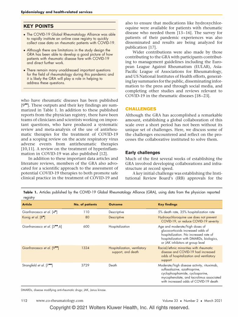
]. These outputs and their key findings are sum-marized in Table 1. In addition to these publishedreports from the physician registry, there have beenteams of clinicians and scientists working on impor-tant questions, who have produced a systematicreview and meta-analysis of the use of antirheu-matic therapies for the treatment of COVID-19and a scoping review on the acute respiratory virusadverse events from antirheumatic therapies[10,11]. A review on the treatment of hyperinflam-mation in COVID-19 was also published [12].
In addition to these important data articles andliterature reviews, members of the GRA also advo-cated for a scientific approach to the assessment ofpotential COVID-19 therapies to both promote safeclinical practice in the treatment of COVID-19 and
Copyright © 2021 Wolters Kluwer H
Table 1. Articles published by the COVID-19 Global Rheumato
registry
Article No. of patients Outcome
Gianfrancesco et al. [4&] 110 Descriptive
Konig et al. [8&] 80 Descriptive
Gianfrancesco et al. [5&&,6] 600 Hospitaliza
Gianfrancesco et al. [9&&] 1324 Hospitalizasupport,
Strangfeld et al. [7&&] 3729 Death
DMARDs, disease modifying anti-rheumatic drugs; JAK, Janus kinase.
112 www.co-rheumatology.com
also to ensure that medications like hydroxychlor-oquine were available for patients with rheumaticdisease who needed them [13–16]. The survey forpatients of their pandemic experiences was alsodisseminated and results are being analyzed forpublication [17].
Wider contributions were also made by thosecontributing to the GRA with participants contribut-ing to management guidelines including the Euro-pean League Against Rheumatism (EULAR), Asia-Pacific League of Associations for Rheumatology,and US National Institutes of Health efforts, generat-ing lay summaries for the public,disseminating infor-mation to the press and through social media, andcompleting other studies and reviews relevant toCOVID-19 in the rheumatic diseases [18–23].
CHALLENGES
Although the GRA has accomplished a remarkableamount, establishing a global collaboration of thisscale over a short period has not been without itsunique set of challenges. Here, we discuss some ofthe challenges encountered and reflect on the pro-cesses the collaborative instituted to solve them.
Early challenges
Much of the first several weeks of establishing theGRA involved developing collaborations and infra-structure at record speed.
A key initial challenge was establishing the Insti-tutional Review Board’s (IRB) approvals for the
ealth, Inc. All rights reserved.
logy Alliance (GRA), using data from the physician reported
Key findings
5% death rate, 35% hospitalization rate
Hydroxychloroquine use does not preventCOVID-19, or reduce COVID-19 severity
tion Age and moderate/high doses ofglucocorticoids increased odds ofhospitalization. No increased rate ofhospitalization with DMARDs, biologics,or JAK inhibitors at group level
tion, ventilatoryand death
Racial/ethnic minorities with rheumaticdisease and COVID-19 had increasedodds of hospitalization and ventilatorysupport
Moderate/high disease activity, rituximab,sulfasalazine, azathioprine,cyclophosphamide, cyclosporine,mycophenolate, and tacrolimus associatedwith increased odds of COVID-19 death
Volume 33 � Number 2 � March 2021
Global research collaboration in a pandemic Robinson et al.
registry. It was important to develop a flexible datacollection system that was easily accessible, secure,and did not require patient consent. During thepandemic, many institutions, including the Univer-sity of California, San Francisco, committed to expe-diting IRB review for COVID-19 related projects.This was critical in allowing the GRA IRB to undergotimely review in less than 48 h. Once the initial IRBapproval was obtained, a large network of collabo-rators around the United States and globally workedto adapt the IRB materials for their individual insti-tutions. For example, investigators working in theUS Veterans Affairs health systems sought individ-ual institutional approvals and eventually appliedfor central approval. Globally, we learned that IRBprocedures differ substantially from country tocountry. Some countries, such as Canada, requiredthat individual institutions have separate IRB appro-vals before participating in the registry. Others, suchas the Philippines or Argentina, could obtain centralapprovals that would apply to all institutions. It wasalso recognized that strategically and due to Euro-pean Union General Data Protection Regulations,which have specific data protection, storage, andprivacy requirements, a separate provider survey wasrequired for Europe, and a partnership with EULARwas established. The final RedCap survey was pro-vided to EULAR so an identical European registrycould be established. The EULAR registry is stored atthe University of Manchester (data processor), withEULAR being the data controller. Data from the twoparallel registries is combined for analysis.
From the outset, the GRA sought to foster globalcollaboration. A key challenge was to develop net-works where none had existed before. Overcomingthis challenge involved using social media in a waythat it had rarely been used in rheumatology [24].Rheumatologists with large followings on platformssuch as Twitter quickly disseminated informationabout the GRA and invited collaborators to commu-nicate on the team platform called Slack. A websitewas also developed (www.rheum-covid.org) thatallowed people to both contribute cases to the reg-istry and access clinical outcome data that had beencollected. It also enabled access to proforma docu-ments for IRB approvals and other logistical tasks.This allowed rapid crowdsourcing of work, includ-ing the IRB approvals mentioned above and facili-tated the recruitment of a series of regional leadsaround the world. This digital infrastructure wascentral to the rapid growth and broad participationin the GRA. A similar endeavor was undertaken byEULAR, who developed their own website (https://www.eular.org/eular_covid19_database.cfm).
One important early difficulty was creating acase report form that requested enough information
Copyright © 2021 Wolters Kluwe
1040-8711 Copyright � 2020 Wolters Kluwer Health, Inc. All rights rese
to adequately assess COVID-19 outcomes and clini-cal and demographic factors, without overburden-ing busy clinicians. After several iterations, wearrived at a balanced form that could be completedrelatively quickly (10–15 min) while providingenough information to allow us to answer the mostpressing questions.
An additional early challenge was ensuring sci-entific rigor and validation despite the compressedproject timelines. There were many initial concernsabout the integrity of the data. Would rheumatol-ogists complete the case report forms accurately?Would there be duplicate entries? Would someonetry to hack the open web-based database platform?Key to overcoming these challenges was havingexperienced data teams in both Europe and theUnited States monitoring registry implementation,performing regular data validity checks, developingalgorithms to remove duplicates, and institutingprocedures to re-contact physicians when data weremissing. National investments in scientific infra-structure were critical to this rapid mobilization.In the United States, work relied on the infrastruc-ture already available through a National Institute ofArthritis, Musculoskeletal and Skin Diseases ClinicalResearch Core, while in Europe, research infrastruc-ture supported by EULAR to support an epidemiol-ogy unit was key.
One key challenge was that many of those con-tributing to the GRA were practicing clinicians.Therefore, at a time when their contributions wereoften needed clinically, there were also the demandsof contributing to the functioning of the GRA. Thesechallenges were addressed with open and honestconversations about what people could andcouldn’t contribute at any given time. In addition,it was quickly realized that the strengths of theassembled group largely were in clinical and epide-miological research. This led to discussions with theAmerican College of Rheumatology (ACR) andEULAR about both what funding might be availableand how that might be best managed. The ACR andEULAR were both able to provide much appreciatedlogistical and administrative support to enable themembers of the GRA to concentrate on the datacollection and analysis efforts.
One aspect of the case collection that enabledrapid increases in case numbers was the integrationof country-specific registry data. Cases from theFrench, German, Italian, Portuguese and Swedishregistries were transferred and merged with datafrom the EULAR database, and registries from coun-tries such as Brazil were merged, reducing duplica-tion of effort. The umbrella of EULAR, a truly pan-European organization fostering a multitude ofactivities in areas of research, patient care, and
r Health, Inc. All rights reserved.
rved. www.co-rheumatology.com 113
Epidemiology and health-related services
education, facilitated the interaction with nationalscientific societies of rheumatology (themselvesmembers of EULAR), whereas legal and administra-tive support from EULAR facilitated practical aspectssuch as setting up data-sharing agreements withrelevant institutions and/or societies. Similarly,research groups globally mobilized quickly to setup data use and transfer agreements to share datawith the GRA. The technical aspects of data transferscan be challenging as the various registries use dif-ferent data collection platforms and formats. Thistask required interaction of technical teams, map-ping of the databases, harmonization efforts, andthe creation of export/import tools.
Later challenges
The limitations of a voluntary physician contrib-uted registry are clear, but in the emerging environ-ment of the early days of the pandemic it was very fitfor purpose. As the pandemic further evolves it isclear that other study designs are required [25]. Theinitial data collected by the GRA was valuable inhelping define risk profiles and provide comparativedata on rheumatic disease patients; however, thereremain significant limitations in the way the projectis structured. These limitations include the conve-nience sampling aspect of the physician registry andthe lack of comparator groups, both COVID-19patients without rheumatic disease and/or under-standing the denominator for the cases that havebeen reported. These issues can potentially beaddressed by utilizing large health systems and sys-tematically assessing all rheumatic disease patientsfor infection. This would enable comparisons to bemade with both rheumatic disease patients who didnot develop COVID-19 and also other patients inthe health system who developed COVID-19 but donot have rheumatic disease. The nature of thesetypes of projects is very resource intensive. Groupsoutside of the GRA are doing some of this type ofwork already. Moreover, the registry does not cap-ture the patient journey, namely long-term COVID-19 outcomes, such as rehabilitation and recoverydata, limiting the ability to view and assess longi-tudinal outcomes beyond hospitalization, ventila-tory support, and mortality.
There is a clear need to improve representationof countries in Latin America, Africa, and Asia in theregistry. This will enable region-specific trends to beobserved. Often in low-resource settings the medi-cations used and alternatives are very different tothose commonly used in Europe and North Americaso there remains a need to have good knowledgeabout the patterns of disease and outcomes across allcontinents and resource settings. The GRA
Copyright © 2021 Wolters Kluwer H
114 www.co-rheumatology.com
instituted a grants scheme, administered throughthe International League of Associations for Rheu-matology, to support investigators in countries thatwere underrepresented in the registry. The purposeof the grants scheme was to provide some support toenable the systematic collection of patients withrheumatic disease who developed COVID-19 toenable them to be entered into the database. Thegrant scheme can also help initiate and supportregionally relevant COVID-19 clinical research inareas that lacked the resources to do this previously.
With the constantly growing database ofreported cases, there is another growing problem.That of data management, there is a need for a dataanalytic infrastructure that can assist multiple inves-tigators with different projects. To address region-specific issues, it is important to be able to delivercountry-specific data to nations that are using theGRA to perform national studies. The two data hubsthat make up the GRA at the University of CaliforniaSan Francisco and the University of Manchesterboth have excellent infrastructure and have beenable to accommodate the growing requests for spe-cific data analytic projects. The GRA has developedtransparent, peer-reviewed policies to assist inassigning data analytic resources to the multipleinvestigators running studies with increasingamounts of information. Ensuring GRA dataremains a global and heavily utilized resource needscareful management to ensure the integrity of thedata is maintained and high-quality research cancontinue to be published.
The funding of the alliance is an issue which willlikely shape the capacity to branch out into newprojects or build on existing projects. Although theinitial enthusiasm from industry to support this casewas very encouraging and much appreciated, it isalmost inevitable that the perceived importance ofthe alliance will track with the trajectory of the pan-demic over time. To branch out into nonpandemicprojects, alternate funding sources will need to befound, and the structure and function of the organi-zation will likely have to change to fit both the newprojects and new funding sources going forward.
OPPORTUNITIES
During the pandemic
There remain many unanswered questions for rheu-matology and patients with rheumatic disease inthis pandemic. The emerging questions encompassimportant areas like vaccines and vaccinations andpost-COVID-19 syndromes. These are both impor-tant areas for future research effort but it remains tobe seen if the GRA, as it is currently configured, is in
ealth, Inc. All rights reserved.
Volume 33 � Number 2 � March 2021
Global research collaboration in a pandemic Robinson et al.
the best situation to lead efforts in these spaces. Bothof these research areas will likely benefit from con-sented research studies with repeated clinical con-tacts and serial collection of data as well asbiospecimens. Therefore it is likely that other studydesigns are going to be better suited to addressingthese important areas. But with the structures andwide network currently in place and a large teamwith in-depth experience of the rheumatic diseasesin the pandemic the GRA is well suited to supportefforts in these spaces.
There is also the opportunity to combine datawith different registries that are capturing COVID-19 data, particularly those with affinities to rheu-matology given mechanistic links in disease patho-genesis or the use of similar drugs (e.g.,inflammatory bowel disease: https://covidibd.org/,psoriasis: https://psoprotect.org/). Differences informat and data elements collected may make itchallenging to combine data from multiple regis-tries, but collaborating across projects represents animportant opportunity to address specific researchquestions (e.g., specific treatments).
After the pandemic
The opportunity to leverage the existing collabora-tion that now exists for further topics is exciting.There is likely to come a time when either the major-ity of the clinical problems presented by the COVID-19 pandemic will have an evidence base to guidethem or the impact of the pandemic will be substan-tially reduced through the uptake of an effectivevaccine(s). It will be interesting to see if there isinterest in building on the success of the alliancein tackling research related to COVID-19 and turnour attention to tackling other global issues thatconfront our speciality. We see the strength of thealliance as being able to leverage a global group ofclinicians to provide cases with wide geographicaldistribution. The advantages of this are that low-frequency events may be able to be collected andcollated in a way that has never been done before. Forexample, cases of rare diseases, or rare manifestationsof diseases, or low-frequency drug side-effects may befuture research directions for the alliance.
CONCLUSION
Although the GRA has achieved much, there ismuch further work to be done. A major success ofthe GRA has been the rapid collaborative mobiliza-tion of the rheumatology community worldwide.The informal feedback from colleagues that thework of the GRA is being used to help cliniciansand patients guide their way through the pandemic
Copyright © 2021 Wolters Kluwe
1040-8711 Copyright � 2020 Wolters Kluwer Health, Inc. All rights rese
is reassuring that we are fulfilling our stated vision.However, there remain many further issues toaddress in the current pandemic. There might bean opportunity to leverage the existing collabora-tion to address some of these topics, whereas otherscan only be addressed by other organizations andmore resource-intensive study designs.
Acknowledgements
We wish to thank all rheumatology providers whoentered data into the registry and patients who completedthe patient survey.
Financial support and sponsorship
The GRA is supported by the American College of Rheu-matology (ACR) and the European League Against Rheu-matism (EULAR). P.M.M. is supported by the NationalInstitute for Health Research (NIHR) University CollegeLondon Hospitals (UCLH) Biomedical Research Centre(BRC). J.Y. is supported by NIH/NIAMS P30AR070155.
Conflicts of interest
J.Y. reports personal fees from AstraZeneca, personal feesfrom Eli Lilly, grants from Pfizer, outside the submittedwork. P.R. reports personal fees from Abbvie, AtomBioscience, Eli Lilly, Janssen, Novartis, Pfizer, Roche,and UCB, and nonfinancial support from BMS, alloutside the submitted work. P.M.M. has received con-sulting and/or speaker’s fees from Abbvie, BMS, Celgene,Eli Lilly, Janssen, MSD, Novartis, Orphazyme, Pfizer,Roche, and UCB, all unrelated to this manuscript.Disclaimer: The views expressed here are those of theauthors, and do not necessarily represent the views of theAmerican College of Rheumatology (ACR), the EuropeanLeague Against Rheumatism (EULAR), the (UK)National Health Service (NHS), the National Institutefor Health Research (NIHR), or the (UK) Department ofHealth, or any other organization.
REFERENCES AND RECOMMENDEDREADINGPapers of particular interest, published within the annual period of review, havebeen highlighted as:
& of special interest&& of outstanding interest1. Robinson PC, Yazdany J. The COVID-19 Global Rheumatology Alliance:collecting data in a pandemic. Nat Rev Rheumatol 2020; 16:293–294.
2. Liew JW, Bhana S, Costello W, et al. The COVID-19 Global RheumatologyAlliance: evaluating the rapid design and implementation of an internationalregistry against best practice. Rheumatology 2020; Epub ahead of print 13August 2020. doi:10.1093/rheumatology/keaa483.
3. Wallace ZS, Bhana S, Hausmann JS, et al. The Rheumatology Communityresponds to the COVID-19 pandemic: the establishment of the COVID-19global rheumatology alliance. Rheumatology 2020; 59:1204–1206.
4.&
Gianfrancesco MA, Hyrich KL, Gossec L, et al. COVID-19 Global Rheuma-tology Alliance Steering Committee Rheumatic disease and COVID-19: initialdata from the COVID-19 Global Rheumatology Alliance provider registries.Lancet Rheumatol 2020; 2:e250–e253.
This is the initial 110 reported patients from the COVID-19 Global rheumatologyalliance physician registry.
r Health, Inc. All rights reserved.
rved. www.co-rheumatology.com 115
Epidemiology and health-related services
5.&&
Gianfrancesco M, Hyrich KL, Al-Adely S, et al. Characteristics associated withhospitalisation for COVID-19 in people with rheumatic disease: data from theCOVID-19 Global Rheumatology Alliance physician-reported registry. AnnRheum Dis 2020; 79:859–866.
This article reports a case series of 600 patients with rheumatic disease and riskfactors for hospitalization because of COVID-19.6. Gianfrancesco M, Yazdany J, Robinson PC. Epidemiology and outcomes of
novel coronavirus 2019 in patients with immune-mediated inflammatorydiseases. Curr Opin Rheumatol 2020; 32:434–440.
7.&&
Strangfeld A, Schafer, M, Milena A, et al. Factors associated with COVID-19-related death in people with rheumatic diseases: results from the COVID-19Global Rheumatology Alliance physician reported registry. Ann Rheum Dis; inpress.
This large case series of 3729 patients reports both demographic and medicationassociation with COVID-19 death.8.&
Konig MF, Kim AH, Scheetz MH, et al. Baseline use of hydroxychloroquine insystemic lupus erythematosus does not preclude SARS-CoV-2 infection andsevere COVID-19. Ann Rheum Dis 2020; 79:1386–1388. Epub ahead ofprint 7 May 2020. doi:10.1136/annrheumdis-2020-217690.
This case series reports the lack of protection from infection or severe disease withthe use of hydroxychloroquine in patients with systemic lupus erythematosus.9.
&&
Gianfrancesco MA, Leykina LA, Izadi Z, et al. Race/ethnicity association withCOVID-19 outcomes in rheumatic disease: data from the COVID-19 GlobalRheumatology Alliance Physician Registry. Arthritis Rheumatol 2020; Epubahead of print 3 November 2020. doi:10.1002/art.41567.
This article reports the disproportionate impact of COVID-19 on racial/ethnicminorities who have rheumatic diseases.10. Putman M, Chock YP, Tam H, et al. Antirheumatic disease therapies for the
treatment of COVID-19: a systematic review and meta-analysis. ArthritisRheumatol 2020; Epub ahead of print 2 August 2020. doi:10.1002/art.41469.
11. Kilian A, Chock YP, Huang IJ, et al. Acute respiratory viral adverse eventsduring use of antirheumatic disease therapies: a scoping review. SeminArthritis Rheum 2020; 50:1191–1201.
12. Amigues I, Pearlman AH, Patel A, et al. Coronavirus disease 2019: investiga-tional therapies in the prevention and treatment of hyperinflammation. ExpertRev Clin Immunol 2020; Epub ahead of print 2020. doi:10.1080/1744666X.2021.1847084.
13. Kim AH, Sparks JA, Liew JW, et al. A rush to judgment? Rapid reporting anddissemination of results and its consequences regarding the use of hydro-xychloroquine for COVID-19. Ann Intern Med 2020; 172:819–821.
Copyright © 2021 Wolters Kluwer H
116 www.co-rheumatology.com
14. Yazdany J, Kim AH. Use of hydroxychloroquine and chloroquine during theCOVID-19 pandemic: what every clinician should know. Ann Intern Med.2020: M20-1334.
15. Sattui SE, Liew JW, Graef ER, et al. Swinging the pendulum: lessons learnedfrom public discourse concerning hydroxychloroquine and COVID-19. ExpertRev Clin Immunol 2020; 16:659–666. Epub ahead of print 3 July 2020.doi:10.1080/1744666X.2020.1792778.
16. Graef ER, Liew JW, Putman MS, et al. Festina lente: hydroxychloroquine, COVID-19 and the role of the rheumatologist. Ann Rheum Dis 2020; 79:734–736.
17. Sirotich E, Dillingham S, Grainger R, et al. Capturing patient-reported out-comes during the COVID-19 pandemic: development of the COVID-19Global Rheumatology Alliance Patient Experience Survey. Arthritis CareRes 2020; 72:871–873.
18. Landewe RB, Machado PM, Kroon F, et al. EULAR provisional recommenda-tions for the management of rheumatic and musculoskeletal diseases in thecontext of SARS-CoV-2. Ann Rheum Dis 2020; 79:851–858.
19. Winthrop KL, Brunton AE, Beekmann S, et al. SARS CoV-2 infection amongpatients using immunomodulatory therapies. Ann Rheum Dis 2020; Epubahead of print 5 August 2020. doi:10.1136/annrheumdis-2020-218580.
20. D’Silva KM, Serling-Boyd N, Wallwork R, et al. Clinical characteristics andoutcomes of patients with coronavirus disease 2019 (COVID-19) and rheu-matic disease: a comparative cohort study from a US ‘hot spot’. Ann RheumDis 2020; 79:1156–1162. Epub ahead of print 26 May 2020. doi:10.1136/annrheumdis-2020-217888.
21. Serling-Boyd N, D’Silva KM, Hsu TY, et al. Coronavirus disease 2019 out-comes among patients with rheumatic diseases 6 months into the pandemic.Ann Rheum Dis 2020; Epub ahead of print 30 November 2020. doi:10.1136/annrheumdis-2020-219279.
22. Robinson PC, Richards D, Tanner HL, et al. Accumulating evidence suggestsanti-TNF therapy needs to be given trial priority in COVID-19 treatment.Lancet Rheumatol 2020. https://www.thelancet.com/journals/lanrhe/article/PIIS2665-9913(20)30309-X/fulltext.
23. Robinson PC, Liew DF, Liew JW, et al. The potential for repurposing anti-TNFas a therapy for the treatment of COVID-19. Med (NY) 2020; https://www.sciencedirect.com/science/article/pii/S2666634020300283.
24. Hausmann JS, Sufka P, Bhana S, et al. Conducting research in a pandemic:the power of social media. Eur J Rheumatol 2020; 7(Suppl 2):S85–S88; https://www.ncbi.nlm.nih.gov/pmc/articles/PMC7431333/.
25. Yazdany J. COVID-19 in rheumatic diseases: a research agenda. ArthritisRheumatol 2020; 72:1596–1599. Epub ahead of print 23 July 2020.doi:10.1002/art.41447.
ealth, Inc. All rights reserved.
Volume 33 � Number 2 � March 2021

REVIEW
CURRENTOPINION Racial, ethnic, and healthcare disparities in
rheumatoid arthritis
Copyrigh
1040-8711 Copyright � 2020 Wolte
a a,b
Kevin Yip and Iris Navarro-MillanPurpose of review
This review highlights the available data describing racial and ethnic health disparities among patients withrheumatoid arthritis in the United States from an epidemiological, disease activity, and widersocioeconomic standpoint.
Recent findings
Despite centralized government initiatives to include more underrepresentative minority populations intoresearch, many of the studies that examined rheumatoid arthritis still fail to include sizeable cohorts ofraces or ethnic groups other than whites. Evidence is slowly mounting that individual, provider, and system-level barriers exist and contribute to unequal care that leads to poorer outcomes amongst patients withrheumatoid arthritis. As rheumatoid arthritis is a progressive disease, early treatment is crucial to delayfunctional decline – a narrow window for many minority patients who are disproportionality affected bydisability.
Summary
To combat the inequality that exists amongst rheumatoid arthritis patients we must focus on whydiscrepancies exist on every level, system, physician, patient, and illness. Further research is needed totease the complex interplay between race, social economic status, medical access, and outcomes toexplain the disparities found in rheumatoid arthritis.
Keywords
access, disability, ethnic, health disparities, healthcare, racial, rheumatoid arthritis, socioeconomic
INTRODUCTION issues related to social determinants of health. This
aHospital for Special Surgery, Division of Rheumatology and bWeillCornell Medicine, Division of General Internal Medicine, New York,New York, USA
Correspondence to Iris Navarro-Millan, MD MPSH, Weill Cornell Medi-cine, Division of General Internal Medicine, 420 East 70th Street, LH –363, New York, NY 10021, USA. Tel: +1 646 962 5896;e-mail: [email protected]
Curr Opin Rheumatol 2021, 33:117–121
DOI:10.1097/BOR.0000000000000782
Health disparity in the United States has multipledimensions and represents differences in health out-comes between different groups of society [1]. Theseare not limited to race and ethnicity, but can also beon the basis of sex, sexual identity, disability, orage [1]. Working toward health equity in societyrequires that every aspect of society is involved toaddress inequalities between social groups so thatthey can attain the maximum level of health amongall people [2]. Since 2010, health disparities havebeen addressed as a priority by the US Department ofHealth and Human Services through the HealthyPeople plan [1].
Racial, ethnic, and access-related health dispar-ities are not the exception among patients withrheumatoid arthritis; one of the most commoninflammatory arthritis in the United States. Never-theless, disparities related to this disease specificallyhave not been studied in as much detail as in otherconditions (e.g., lupus, diabetes); hence, there is nota clear understanding about the magnitude of thisproblem or even the best way to intervene to address
t © 2021 Wolters Kluwe
rs Kluwer Health, Inc. All rights rese
review summarizes the data available to date regard-ing racial and ethnic disparities among rheumatoidarthritis patients within the United States.
EPIDEMIOLOGY
The prevalence of rheumatoid arthritis is approxi-mately 1% in the US population [3,4]. For compari-son, mean age-adjusted prevalence rates suggestthat North Africa, Middle East, and Asia have rela-tively lower prevalence at 0.16%, North Americaand Western Europe at 0.44% with Australasia
r Health, Inc. All rights reserved.
rved. www.co-rheumatology.com
KEY POINTS
� Rheumatoid arthritis research continues topredominantly focus on white populations leaving manyquestions such as epidemiology, disease course, andoutcomes unanswered in racial minority patients.
� Evidence is mounting that the differences observedbetween rheumatoid arthritis in racial groups can beattributed to nonbiological factors.
� Socioeconomic status, differences in therapeuticprescriptions, and access to healthcare revealsignificant health disparities which must be addressedto provide equitable healthcare for all patients withrheumatoid arthritis.
Epidemiology and health-related services
having the highest at 0.46% [5]. In the United States,specific populations have been identified withhigher rates of rheumatoid arthritis including thePima and Pagago Indians of the American Indianpopulation who have an age-adjusted prevalence of5.3% [6]. Although it is traditionally reported thatrheumatoid arthritis affects predominantly whites,it is important to examine the proportion of indi-viduals with rheumatoid arthritis within each racial/ethnic group. According to the 2019 U.S. CensusBureau, the total U.S. population was 328 239 523with 76.3% whites, 13.4% African-American, 18.5%Hispanic or Latino, 5.9% Asian, 2.8% listed as two ormore races, and 0.2% Native American and otherPacific Islander [7].
To date, the majority of the epidemiologic stud-ies, outcomes, and trials of patients with rheumatoidarthritis included primarily white patients or withoutracial information defined [3,4,8]. A recent system-atic review of 240 rheumatoid arthritis Randomizedcontrol trial (RCTs) estimated an overrepresentationof the white population ranging between 74.6% in2010 to 97.0% in 2013 of researched participants[9
&&
]. There is little population-based data regardingthe specific incidence rates and prevalence of rheu-matoid arthritis among individuals who are African-American or Hispanic. This lack of understanding ofthe epidemiology of rheumatoid arthritis by race andethnicity limits our understanding of the burden ofrheumatoid arthritis among different groups of oursociety and the health disparities amongst patients.
DISABILITY AND DISEASE ACTIVITY
Race itself appears to reveal a discrepancy betweenarthritis and related disability. Given the correla-tions between ethnicity, race, social-economic class,and even culturally this can be difficult to interpret[10].
Copyright © 2021 Wolters Kluwer H
118 www.co-rheumatology.com
The differences in reported disability could berelated to differences in treatment for rheumatoidarthritis that could have led to disability. Still, onceindividuals with rheumatoid arthritis are disabled,they might still experience an additional disparity inthe way that rheumatoid arthritis is treated. In arecent study among dual-eligible (Medicare andMedicaid) beneficiaries of Social Security DisabilityInsurance (SSDI), individuals who filed for disabilitybenefits before the retirement age of 65, showedsignificant differences in the use of biological Dis-ease-modifying antirheumatic drug (bDMARDs)amongst races [11
&&
]. African-Americans were leastlikely to receive bDMARDs (49.3%) than whites(53.3%) whereas Hispanics were more likely toreceive bDMARDs (60.9%) [11
&&
]. These differencespersisted after controlling for social determinants ofhealth [11
&&
]. Disability in rheumatoid arthritis hasled to high opioid prescription with more than 66%of SSDI beneficiaries to receive chronic opioids[11
&&
]. This proportion was not different betweenAfrican-Americans and whites beneficiaries of theSSDI by 2014. This data suggest that early disabilitycan result in overreliance on opioids, likely given tohigh level of disability in a group of patients whowere to begin with, highly vulnerable [11
&&
].There are several studies that examined rheuma-
toid arthritis disease activity by race and ethnicity.Greenberg et al. analyzed data from 6008 patientsacross community-based rheumatology clinics overa five-year period in a cross-sectional study [12].Although there was improvement in disease activityacross all racial groups, there were only small differ-ences noted in clinical disease activity index scoresbetween whites 12.38 (11.36–13.4), African-Ameri-cans 13.75 (12.39–15.1, P¼0.007), and Hispanicpatients 13.01 (11.68–14.34, P¼0.179) in anadjusted model accounting for practice setting,treatments, and patient sociodemographic factors[12]. In another cross-sectional study focused onone academic center practice, African-Americanspatients had increased disease activity score (DAS)scores than whites (5.5�1.3 versus 4.3�1.4;P<0.001) and Health Assessment Questionnaire(HAQ) (1.5�0.8 versus 0.9�0.7; P<0.001); how-ever, after accounting for socioeconomic, demo-graphic, and behavioral influences, race was notindependently associated with the reported differ-ences. Hence, to date, there is little data to supportthat individuals from a specific racial or ethnicgroup have more aggressive or higher disease activ-ity in rheumatoid arthritis [13,14].
Jordan et al. analyzed 100 female patientswith rheumatoid arthritis and found that African-Americans had less physical activity and more neg-ative affect compared with their white counterparts
ealth, Inc. All rights reserved.
Volume 33 � Number 2 � March 2021
Racial, ethnic, healthcare disparities in rheumatoid arthritis Yip and Navarro-Millan
despite no difference in pain severity [15]. Theyfound that the two groups used different psycholog-ical coping strategies with white patients more likelyto ignore pain, whereas African-Americans patientsturned to praying and hoping. Regardless of racialbackground, coping statements better predictedpain control suggesting that racial differences incoping strategies may contribute to reported differ-ences [15]. Given the history and racism that Afri-can-Americans and other minorities encountered inthe healthcare system and in their communities,these coping mechanisms could have been shapedby their experiences with unequal treatment andracism that white individuals did not experience.This issue constitutes a problem that is not onlylimited to the healthcare system, but to the politicalsystem and how our society addresses inequalitywithin its members.
SES/ACCESS
A cross-sectional study comparing a cohort of 4730rheumatoid arthritis patients found that whitepatients, despite having a longer disease course hadbetter global health scores and less pain [16]. Level ofeducation attainment, duration of rheumatoidarthritis diagnosis, and number of other comorbid-ities were found to impact the pain ratings of His-panic and African-American patients [16]. There isconvincing data across the world that lower socialeconomic status (SES) (measured in different wayssuch as gross income, occupation, educational level,and area of residence) has been linked to worsedisease in rheumatoid arthritis patients, from diseaseactivity, pain, and disability [17
&
,18,19]. Growingevidence has suggested that low SES evenin childhood has a statistically significant trend(P<0.0001) of increasing the risk of developmentof rheumatoid arthritis; food insecurity (oddsratio¼1.5), young maternal age (<20 versus 20–34years;OR¼1.7), and childhood householdeducation(<12 years versus college degree; OR¼1.7; 95%) [20].
Schmajuk et al. looked at 93 143 Medicare-enrolled patients with rheumatoid arthritis andfound significant correlations of Disease-modifyingantirheumatic drugs (DMARD) use with socioeco-nomic factors [21]. Living in an area of low SES,having low personal income (defined by needingstate assistance for their Medicare Part B), malegender and African-Americans race were all associ-ated with a lower likelihood of being prescribed aDMARD [21]. As early treatment is beneficial forearlier remission, prevention of joint damage anddisease-related disability [22–24], it stands to reasonthat minorities without equitable access to care willaccrue and be exposed to longer uncontrolled
Copyright © 2021 Wolters Kluwe
1040-8711 Copyright � 2020 Wolters Kluwer Health, Inc. All rights rese
disease before therapy and therefore have poorerprognosis and remission rates.
Kerr et al. compared the treatments of 2899patients in the Veteran Affairs Rheumatoid ArthritisRegistry (VARA) in the VA healthcare system (akin to aform of Universal Healthcare in the U.S.) and patientsin the Ethnic Minority Rheumatoid Arthritis Consor-tium (EMRAC) where patients were part of variedhealthcare systems [25]. Notably, in the VARA cohort,there was no difference in biologic use between racialgroups, whereas EMARAC white patients had a higherodds ratioof1.66of receivingbiologics comparedwiththeir nonwhite counterparts [25].
In a longitudinal observational study of 8545patients with rheumatoid arthritis, 43.6% noteddifficulties paying out of pocket medical expenses.Those who had the greatest difficulty with health-care costs were more likely to be on social securitydisability (33.9 versus 10.1%), be a minority race(10.3 versus 5.1%), have worse HAQ Scores (1.5versus 0.9), higher comorbidity index (2.5 versus1.6), less likely to be a college graduate (20.3 versus36.1%) and at the poverty level defined by Healthand Human Services poverty guidelines (51.3 versus12.3%) compared with those who reported ‘no dif-ficulty’ with medical costs. Patients most at finan-cial risk were also those disproportionately affectedthose with the most severe disease and twice as likelyto be a racial minority [26]. This may go some way toattribution of reduced treatment rates and access toeffective therapies in this most vulnerable patientgroup. In addition, out of pocket costs have beenshown to negatively impact adherence to medica-tions in rheumatoid arthritis [27
&
] and representanother barrier to those in lower SES which are madedisproportionately of minority populations espe-cially as they are almost twice as likely as whitepatients to be work-disabled [28].
SYSTEM LEVEL DISPARITIES
Barton et al. [29] examined a diverse racial andimmigrant population served by the same rheuma-tologists that worked at both a University and publicbased clinic. This study found that whites, Englishspeaking, and nonimmigrant patients had lowerDAS-28 and HAQ at a University setting only [29].Notably, these differences were still present afteradjustment for medication use suggesting that ther-apies alone did not account for the discrepancies butinstead suggests the presence of a health disparityon the basis of clinic-level differences. Clinic-leveldifferences could contribute to the health disparitiesobserved in the patients with rheumatoid arthritissuch as variation in time to initial rheumatologycare and access to treatment [29].
r Health, Inc. All rights reserved.
rved. www.co-rheumatology.com 119
Epidemiology and health-related services
Looking at two affiliate rheumatology clinicswithin one medical school, Suarez-Almazor et al.[30] compared their African-American, Hispanicand white rheumatoid arthritis patients and foundthat non-white patients were more likely to be in apublic rather than private clinic (83 versus 18%) andwait significantly longer before being started onDMARD therapy (median of 7 versus 1 year). Inpatients with relatively early disease (<5 years),more whites than non-white patients had alreadytried some form of DMARD prior to their index visit(64 versus 32%) [30]. These findings suggest intrin-sic biases, which prevent the same level of access andcare to racial minorities, are a crucial barrier inrheumatoid arthritis where early treatment can pre-vent long-term joint damage [22–24].
As medicine moves toward an evidence-basedapproach, it is important to reflect upon the long-standing problem to recruit representative popula-tions into research studies. The data and results theygenerate help guide societal guidelines and stand-ards of practice especially as it is often the under-served minorities and often those in lower SESbrackets that miss out on only research opportuni-ties and potential future benefit. Notably, mostlarge-scale studies of rheumatoid arthritis have beenperformed in Western nations, which skews identi-fication of risk factors. Despite making up almost41% of the US population, racial minorities onlyrepresented 16% of the rheumatoid arthritis popu-lation for RCTs [9
&&
]. Concerningly, despite efforts toincrease awareness and participation from bothresearchers and patients, there hasn’t been an obvi-ous improvement of minority representation overthe 10-year period studied (2008–2018) [9
&&
].Given the differences and burden of disease
on non-white populations delineated in this article,it is imperative that we better understand why dis-crepancies exist on every level, system, physician,patient, and illness level, if we are to fix the problem.This will require us to revisit the policies that con-tributed to bring us and our healthcare system tothis unequal state to begin with, so we can rightthis wrong.
Acknowledgements
None.
Financial support and sponsorship
I.N.M. work was supported by K23-AR-068449 from theNational Institute of Arthritis and Musculoskeletal andSkin Diseases.
Conflicts of interest
I.N.M. has received speaker fees from SOBI pharmaceu-ticals. K.Y has no conflicts of interest to declare.
Copyright © 2021 Wolters Kluwer H
120 www.co-rheumatology.com
REFERENCES AND RECOMMENDEDREADINGPapers of particular interest, published within the annual period of review, havebeen highlighted as:
& of special interest&& of outstanding interest1. Healthy People 2020, Foundation health measures, Disparities [Online];2020, December 3rd. Available: https://www.healthypeople.gov/2020/about/foundation-health-measures/Disparities.
2. U.S. Department of Health and Human Services, Office of Minority Health.National Partnership for Action to End Health Disparities. The National Planfor Action Draft as of February 17, 2010. Chapter 1: Introduction [Online];2020, December 3rd. Available: http://www.minorityhealth.hhs.gov/npa/tem-plates/browse.aspx?&lvl=2&lvlid=34.
3. Hunter TM, Boytsov NN, Zhang X, et al. Prevalence of rheumatoid arthritis inthe United States adult population in healthcare claims databases, 2004–2014. Rheumatol Int 2017; 37:1551–1557.
4. Myasoedova E, Crowson CS, Kremers HM, et al. Is the incidence of rheu-matoid arthritis rising?: results from Olmsted County, Minnesota, 1955–2007. Arthritis Rheum 2010; 62:1576–1582.
5. Cross M, Smith E, Hoy D, et al. The global burden of rheumatoid arthritis:estimates from the global burden of disease 2010 study. Ann Rheum Dis2014; 73:1316–1322.
6. Del Puente A, Knowler WC, Pettitt DJ, Bennett PH. High incidence andprevalence of rheumatoid arthritis in Pima Indians. Am J Epidemiol 1989;129:1170–1178.
7. PD US Census Bureau. Annual Estimates of the Resident Population by Sex,Race, and Hispanic Origin for the United States: April 1, 2010 to July 1, 2019(NC-EST2019-SR11H) [Online]; December 3rd. Available: https://www.cen-sus.gov/quickfacts/fact/table/US/PST045219.
8. Lawrence RC, Helmick CG, Arnett FC, et al. Estimates of the prevalence ofarthritis and selected musculoskeletal disorders in the United States. ArthritisRheum 1998; 41:778–799.
9.&&
Strait A, Castillo F, Choden S, et al. Demographic characteristics of partici-pants in rheumatoid arthritis randomized clinical trials: a systematic review.JAMA Netw Open 2019; 2:e1914745.
Metaanalysis examining all rheumatoid arthritis RCTs which included at least oneUS site showing that racial minorities were under-represented in research studies.10. Tennstedt S, Chang BH. The relative contribution of ethnicity versus socio-
economic status in explaining differences in disability and receipt of informalcare. J Gerontol Ser B, Psychol Sci Soc Sci 1998; 53:S61–70.
11.&&
Navarro-Millan I, Rajana M, Lui GE, et al. Racial and ethnic differences inmedication use among beneficiaries of social security disability insurance withrheumatoid arthritis. Semin Arthritis Rheum 2020; 50:988–995.
Amongst rheumatoid arthritis patients on Social Security Disability Insurance,bDMARD, and opiate-prescribing rates differed significantly amongst racial groups.12. Greenberg JD, Spruill TM, Shan Y, et al. Racial and ethnic disparities in
disease activity in patients with rheumatoid arthritis. Am J Med 2013;126:1089–1098.
13. Iren UT, Walker MS, Hochman E, Brasington R. A pilot study to determinewhether disability and disease activity are different in African-American andCaucasian patients with rheumatoid arthritis in St. Louis, Missouri, USA. JRheumatol 2005; 32:602–608.
14. Lopez-Mendez A, Paul WA, Alarcon GS. Rheumatoid arthritis in Americanblacks: a clinical and radiological study. J Rheumatol 1989; 16:1197–1200.
15. Jordan MS, Lumley MA, Leisen JC. The relationships of cognitive coping and paincontrol beliefs to pain and adjustment among African-American and Caucasianwomen with rheumatoid arthritis. Arthritis Care Res 1998; 11:80–88.
16. Bruce B, Fries JF, Murtagh KN. Health status disparities in ethnic minoritypatients with rheumatoid arthritis: a cross-sectional study. J Rheumatol 2007;34:1475–1479.
17.&
Yang G, Bykerk VP, Boire G, et al. Does socioeconomic status affect outcomesin early inflammatory arthritis? Data from a Canadian multisite suspectedrheumatoid arthritis inception cohort. J Rheumatol 2015; 42:46–54.
An incident cohort of 2023 patients found that low SES was associated withhigher disease activity in early rheumatoid arthritis.18. Massardo L, Pons-Estel BA, Wojdyla D, et al. Early rheumatoid arthritis in Latin
America: low socioeconomic status related to high disease activity at base-line. Arthritis Care Res 2012; 64:1135–1143.
19. Harrison MJ, Tricker KJ, Davies L, et al. The relationship between socialdeprivation, disease outcome measures, and response to treatment in pa-tients with stable, long-standing rheumatoid arthritis. J Rheumatol 2005;32:2330–2336.
20. Parks CG, D’Aloisio AA, DeRoo LA, et al. Childhood socioeconomic factorsand perinatal characteristics influence development of rheumatoid arthritis inadulthood. Ann Rheum Dis 2013; 72:350–356.
21. Schmajuk G, Trivedi A, Solomon D, et al. Receipt of disease-modifyingantirheumatic drugs among patients with rheumatoid arthritis in Medicaremanaged care plans. JAMA 2011; 305:480–486.
22. Aga AB, Lie E, Uhlig T, et al. Time trends in disease activity, response andremission rates in rheumatoid arthritis during the past decade: results from theNOR-DMARD study 2000–2010. Ann Rheum Dis 2015; 74:381–388.
ealth, Inc. All rights reserved.
Volume 33 � Number 2 � March 2021
Racial, ethnic, healthcare disparities in rheumatoid arthritis Yip and Navarro-Millan
23. Lard LR, Visser H, Speyer I, et al. Early versus delayed treatment inpatients with recent-onset rheumatoid arthritis: comparison of two cohortswho received different treatment strategies. Am J Med 2001; 111:446–451.
24. Nell VP, Machold KP, Eberl G, et al. Benefit of very early referral andvery early therapy with disease-modifying antirheumatic drugs in patientswith early rheumatoid arthritis. Rheumatology (Oxf, Engl) 2004; 43:906–914.
25. Kerr GS, Swearingen C, Mikuls TR, Yazici Y. ‘Use of biologic therapy in racialminorities with rheumatoid arthritis from 2 US healthcare systems. J ClinRheumatol 2017; 23:12–18.
26. Wolfe F, Michaud K. Out-of-pocket expenses and their burden in patients withrheumatoid arthritis. Arthritis Rheum 2009; 61:1563–1570.
Copyright © 2021 Wolters Kluwe
1040-8711 Copyright � 2020 Wolters Kluwer Health, Inc. All rights rese
27.&
Heidari P, Cross W, Crawford K. Do out-of-pocket costs affect medicationadherence in adults with rheumatoid arthritis? A systematic review. SeminArthritis Rheum 2018; 48:12–21.
This systematic review suggests that out of pocket costs have a negative impact onmedication adherence which impacts patients with low SES the hardest.28. De Roos AJ, Callahan LF. Differences by sex in correlates of work status in
rheumatoid arthritis patients. Arthritis Care Res 1999; 12:381–391.29. Barton JL, Trupin L, Schillinger D, et al. Racial and ethnic disparities in disease
activity and function among persons with rheumatoid arthritis from university-affiliated clinics. Arthritis Care Res 2011; 63:1238–1246.
30. Suarez-Almazor ME, Berrios-Rivera JP, Cox V, et al. Initiation of disease-modifying antirheumatic drug therapy in minority and disadvantaged patientswith rheumatoid arthritis. J Rheumatol 2007; 34:2400–2407.
r Health, Inc. All rights reserved.
rved. www.co-rheumatology.com 121
REVIEW
CURRENTOPINION Scleroderma epidemiology update
Copyright ©
www.co-rheumatology.com
a b
Leonardo Martin Calderon and Janet E. PopePurpose of review
Systemic sclerosis (scleroderma, SSc) is a rare multisystem autoimmune disease characterized byautoantibodies, vasculopathy, and fibrosis of the skin and internal organs. This review aims to provide anoverview and summary of the recent epidemiological studies in systemic sclerosis.
Recent findings
Global trends of scleroderma demonstrate greater prevalence of SSc in European, North, and SouthAmerican patients compared with East Asian patients. However, the greatest prevalence (47 in 100000),was found among the indigenous peoples in Canada. Phenotypical differences exist depending on the ageof presentation with greater internal organ involvement and disease acceleration present in older patients.Sex differences include greater severity of disease expression, relative prevalence of diffuse cutaneous SSc,and organ involvement in males versus females. New studies conflict with previous data reporting greaterproportion of pulmonary arterial hypertension in females. Furthermore, the effect of low median householdincome is demonstrated as a factor increasing risk of death in SSc patients.
Summary
Understanding the epidemiological factors in SSc enables patient care through patient classification,prognostication, and monitoring. Future research may emphasize enrichment of SSc patients in randomizedtrials who are more likely to progress or be treatment responsive, focused screening, and personalizedpatient care through the creation and validation of new SSc criteria and subsets.
Keywords
autoimmunity, epidemiology, mortality, scleroderma, sex differences, systemic sclerosis, systemic sclerosis
aDepartment of Medicine and bDivision of Rheumatology, St. Joseph’sHealthcare, Schulich School of Medicine and Dentistry, University ofWestern Ontario, London, Ontario, Canada
INTRODUCTION
Scleroderma, or systemic sclerosis (SSc), is a rare mul-tisystem autoimmune connective tissue disease char-acterized by vasculopathy with skin and internalorgan fibrosis and autoantibodies [1,2]. SSc is arrangedinto subsets including limited and diffuse cutaneousSSc (lcSSc and dcSSc) [3]. SSc has high morbidity [4,5],decreased quality of life [6], significant societal eco-nomic burden [7,8], and increased mortality [4,5,9].Traditionally, SSc patients are predominantly femalewith an increasing age of onset and at greater risk ofdeveloping lcSSc, peripheral vascular disease, and pul-monary arterial hypertension (PAH) [10,11]. Con-versely, men have greater risk proportionately ofdeveloping dcSSc, with worse interstitial lung disease(ILD) and cardiovascular complications. The purposeof this review is to highlight the newest literaturerelating to the epidemiology of SSc.
Correspondence to Janet E. Pope, MD, Division of Rheumatology,University of Western Ontario, St. Joseph’s Healthcare, 268 GrosvenorSt., London, Ontario, Canada N6A 4V2.E-mail: [email protected]
Curr Opin Rheumatol 2021, 33:122–127
DOI:10.1097/BOR.0000000000000785
DEMOGRAPHICS OF SYSTEMICSCLEROSIS
The classification criteria used to identify SSc casesin population-based studies may vary. Criteria
2021 Wolters Kluwer H
include the preliminary 1980 American College ofRheumatology (ACR) criteria [12], the 2001 LeRoyand Medsger criteria in early SSc [13], and the 2013ACR and European League Against Rheumatism(EULAR) criteria [14]. Differences in sensitivityand specificity between these criteria are well docu-mented [15].
Incidence and prevalence
Demographic parameters in SSc vary with gender,ethnicity, and geography. Zhong et al. [16
&&
] synthe-sized incidence and prevalence patterns of in SScacross North America, Asia, Australia, and South
ealth, Inc. All rights reserved.
Volume 33 � Number 2 � March 2021
KEY POINTS
� Younger age of SSc delineates a greater risk ofdeveloping dcSSc subset. However, disease durationand age increase the risk of death overall.
� SSc is more prevalent in women versus men(approximately 4–1). There is increased organinvolvement and greater risk of developing dcSScin males.
� Adjusted analyses of socioeconomic factors in patientswith SSc demonstrated that a lower median householdincome was associated with an increased riskof mortality.
� The development of new classification criteria usingnew tools, such as proteomics or epigenomics, mayoptimize personalization of SSc patient care, patientmonitoring, prognostication, and researchcohort creation.
Scleroderma epidemiology update Calderon and Pope
America. Original population-based observationalstudies were included. Prevalence in Europeanpopulations was greater (10–35 per 100 000), thanin East Asian populations (3.8–5.6 per 100 000).Prevalence in South America was 29.6 per 100 000.However, the highest prevalence was observed in theindigenous people of Canada (47 per 100 000).Pooled prevalence of SSc was 23 per 100 000. Further-more, incidence in East Asian populations was 1.09–1.5 in 100 000, 1.5 per 100 000 in Australia, 2.1 per100 000 in South America, and in European popula-tions ranged from 0.77 in Netherlands, to 4.3 in Italyper 100 000. This review summarized prevalenceand incidence parameters in SSc worldwide.
Age
SSc age of presentation can vary but commonlyoccurs in middle age with increasing age over time.Importantly, differences in disease severity havebeen described depending on age of presentation.Carreira et al. [17] analyzed patients with early SSc inthree age groups through a cross-sectional analysisof the EULAR Scleroderma Trials and Research data-base (EUSTAR). Patients were identified using the1980 ACR criteria with less than three years from thefirst non-Raynaud’s phenomenon SSc symptom.They categorized age strata as less than 30, 31–59years, and at least 60 years old. The study identified1027 patients of whom 90% were whites with 80%females. Younger patients had higher anti-Scl-70antibodies (53 versus 35 versus 30%) and higherlikelihood of having dcSSc (54 versus 40 versus34%) compared with the medium and older patientstrata. Conversely, older patients were more likely to
Copyright © 2021 Wolters Kluwe
1040-8711 Copyright � 2021 Wolters Kluwer Health, Inc. All rights rese
have lcSSc (58 versus 53 versus 35%) with morecardiac involvement including conduction blocks(15 versus 6 versus 6%) and diastolic dysfunction (26versus 12 versus 3%) comparing descending agestrata, respectively.
Another publication by Moinzadeh et al. [18&
]also reported that older patients had more lcSSc in asubgroup analysis of 3281 patients from the GermanNetwork of Systemic Scleroderma. One quarter oftheir cohort developed SSc at age more than 60years. Within the lcSSc and dcSSc subgroups, theyfound an increased frequency of organ involvementin the lungs and heart in older patients (pulmonaryhypertension in lcSSc and dcSSc, pulmonary fibro-sis, and cardiac involvement in the dcSSc subset)and acceleration of disease progression in olderpatients. Although there were less digital ulcers inthe older onset patients, these observations increaseour understanding that older onset SSc patients mayhave a worse disease course.
Jiang et al. [19&
] performed a systematic reviewexamining the factors associated with PAH in SSc.Studies included had a sample size larger than 20comparing SSc patients with PAH identified by rightheart catheterization to those without PAH. The riskfactors most often cited that were associated withPAH in SSc were older age, lcSSc subset, longerdisease duration, positive anticentromere antibod-ies, and telangiectasia.
These studies demonstrate differences in clinicalfeatures and disease progression secondary to age ofsymptom onset and diagnosis.
Gender differences
A review by Hughes et al. [20&&
] investigated genderdifferences in SSc. Generally, SSc has a female-to-male ratio between 3:1 and 7:1 with some geograph-ical exceptions demonstrating ratio reversal of maleto female from 4.7:1 in North East England to 14.5:1in Tokyo. Males predominantly present with dcSSccompared with females (61 versus 34%). Femaleshave more lcSSc (57%) compared with males(35%). Time to diagnosis of dcSSc after onset ofRaynaud’s phenomenon is slightly longer forwomen than in men (1.1 versus 0.8 years). Menhad more SSc associated cardiomyopathy, left ven-tricular dysfunction, ILD, and scleroderma renalcrisis. PAH is prevalent in both sexes; however,postmenopausal women had more isolated PAH(Group 1 PAH). Some gender differences are corrob-orated by this review including greater severity ofdisease expression, prevalence of dcSSc, and lungand heart organ involvement in males. However,evidence of greater proportion of PAH in femalesremains conflicting.
r Health, Inc. All rights reserved.
rved. www.co-rheumatology.com 123
Epidemiology and health-related services
Hormonal differences
Estrogens, particularly estradiol, have been previ-ously implicated as profibrotic agents in SSc. Ciaffiet al. [21
&
] performed a systematic review investigat-ing the role of sex hormones in SSc. Studies of SSc byany definition/classification were included as werecase reports, case series, cohort studies, and regis-tries. In general, the quality of included studies waspoor. Estrogen may have profibrotic effects and inpostmenopausal women with SSc, there is less skininvolvement. There may be a vasodilatory effect ofestrogen and some data suggest that a hypoestro-genic state increases the risk of developing PAH,whereas hormonal replacement therapy in meno-pause might be protective against PAH. There islikely insufficient data to draw conclusions andconfounding where age is a risk factor for PAH.
Furthermore, Frost et al. [22&
] enrolled and ana-lyzed the estradiol levels of 83 males aged 50 or olderwith recent onset dcSSc (within 2 years of firstsymptoms) and compared them to 37 healthy malecontrols and postmenopausal age-matched womenwith dcSSc. The men with dcSSc had higher levels ofestradiol compared with healthy males and post-menopausal dcSSc at 30.6, 12.9, and 24.2 pg/ml,respectively. High estrogen levels (compared withlow levels) in dcSSc men over the age of 50 wereassociated with more skin fibrosis progression,increased cardiac involvement, and reduced sur-vival. Other studies are needed to corroboratethese findings.
Ethnicity/race
African-American patients have been previouslydescribed to be younger at disease onset with agreater likelihood of having dcSSc, and increasedmortality compared with non-African-Americans[23,24]. Moore et al. [25
&&
] performed a retrospectivestudy comparing African-American versus non-Afri-can-American SSc patients matched for sex, age,disease duration, and SSc subset. Median householdincome derived from residence zip code was used asa surrogate of socioeconomic status (SES). In theunadjusted analysis, African-American ancestrydid demonstrate an elevated hazard ratio of 2.1(P¼0.006) for death during follow-up. However,there were findings that differed from previous pub-lications including no difference in age at initialvisit for SSc and prevalence of dcSSc. African-Ameri-can ancestry when adjusted for age, sex, diseaseduration, SSc subset, and anti-Scl-70 status wasnot predictive of mortality. SES variables, such asmarital status, education, insurance type, employ-ment status, and imputed household income werestudied and a lower household income increased
Copyright © 2021 Wolters Kluwer H
124 www.co-rheumatology.com
mortality. This study demonstrates the impact ofsocioeconomic status on mortality in SSc. Findingsin the literature may differ depending on whetherother important disease and SES factors areadjusted for.
Environmental and occupational exposures
In addition to health behaviors, occupational andenvironmental exposures may account for somedifferences in SSc severity. In a review by Marieet al. [26], environmental and occupational expo-sures implicated in the modulation of the epigeneticdeterminants in SSc development and progressionwere collated. Occupational exposure to crystallinesilica and organic solvents, such as aromatic orchlorinated compounds is strongly associated withSSc development. Exposed patients are at higher riskof developing dcSSc, digital ulcers, and interstitiallung disease. Additionally, exposure to heavy metalsincluding antimony, cadmium, lead, and mercuryseems to increase SSc incidence. Pathogenic mecha-nisms of exposure may include reactive oxygenspecies and endothelial dysfunction. Antimonyand platinum in males, although antimony, mer-cury, lead, cadmium, palladium, and zinc in femaleswere associated with SSc. Occupational differencesmay affect associations in men and women differ-ently. For instance, men are far more likely to workin mining. Some patients receive compensation iftheir employment seems to be a strong risk for thedevelopment of SSc, so taking a detailed occupa-tional history in people with SSc may be important.
Mortality
The mortality rate is greater in SSc than in thegeneral population with the leading causes of deathbeing ILD and PAH [27,28
&
]. Lee et al. [29] performeda metaanalysis for standardized mortality ratios(SMR) in SSc patients. Cohort studies with prede-fined SSc criteria and reporting overall, sex, and/ordisease subtype-specific SMRs were included. TheSMR in SSc patients was 2.8 (95% confidence inter-val 2.2–3.6, P<0.001). No significant differences inSMR between men and women with SSc were found(3.5 and 2.9, respectively). SMR was nearly five timeshigher in the dcSSc subset compared with lcSSc attwo times higher than the age and sex-matchedgeneral population. This study differs from someprevious findings as male sex did not confer anincreased SMR above women with SSc overall(numerically higher but not statistically) and inthe dcSSc subset.
Prognostic factors affecting disease progressionand mortality in dcSSc were studied by Becker et al.
ealth, Inc. All rights reserved.
Volume 33 � Number 2 � March 2021
Scleroderma epidemiology update Calderon and Pope
[30&&
]. Using the EUSTAR database, those earlydcSScs who either had a follow-up visit or diedwithin 12 plus-minus 3 months after baseline wereincluded. Disease progression was defined as eithernew onset renal crisis, decreased forced vital capac-ity greater than or equal to 10%, new left ventricularejection fraction (LVEF) less than 45% or decreasedin LVEF greater than 10% for patients with baselineof lower than 45%, new onset echocardiography-suspected PAH, or death. They found associationsbetween disease progression and older age, activedigital ulcers, lung fibrosis, muscle weakness, andelevated C-reactive protein. This supports the studyof older age onset SSc had a worse prognosis [18
&
].Early skin fibrosis progression was associated withdecreased lung function and worsened survival inpatients with dcSSc using EUSTAR data as reportedby Wu et al. [31].
LIMITED CUTANEOUS SYSTEMICSCLEROSIS
Despite comprising two-third of SSc patients, inclu-sion of lcSSc subset in research is less compared withdcSSc [32]. Although PAH trials often have morelcSSc patients, many trials include only early dcSScor progressive dcSSc patients and ILD trials comprisemore dcSSc patients. Some of this reflects the epi-demiologic differences between the subsets and tri-als to improve skin will include only those withhigher skin scores (i.e., the dcSSc subset). Frantzet al. [33
&
] analyzed observational data from EUSTARto identify factors predictive of progression of skinand lung fibrosis and vasculopathy in patients withlcSSc who met the 2013 ACR/EULAR criteria. A totalof 8013 lcSSc and 4786 dcSSc patients were analyzed.Skin progression was minimal in lcSSc versus dcSSc,at 3.8 and 14.9%, respectively. Nearly half of thelcSSc patients who had skin progression developedproximal or truncal disease and were reclassified asdcSSc. Lung fibrosis increased in lcSSc patients overthree years of follow-up and changes in forced vitalcapacity in lcSSc patients mirrored dcSSc changes.This study illustrates the necessity for pulmonarymonitoring in both subsets of SSc.
CLASSIFICATION/DIAGNOSIS
Systemic sclerosis subtype criteria
The classification and subsetting of SSc by LeRoyet al. [34] has been useful in risk stratification,treatment guidance, prognosis ascertainment, andgrouping of patients for research purposes [35].However, updated classifications guided by newtools, such as metabolomics, proteomics, genomics,
Copyright © 2021 Wolters Kluwe
1040-8711 Copyright � 2021 Wolters Kluwer Health, Inc. All rights rese
transcriptomics, and epigenomics, may optimizepersonalization of SSc patient care. For example,Smeets et al. [36] derived three distinct pathophysi-ological ‘fingerprints’ through antibody clusteringwith associated vascular and inflammatory media-tors. Moreover, Johnson et al. [37
&
] reported on theevolution and ongoing development of new SScclassifications. It was recommended that new clas-sification systems should meet three principalrequirements: enriched research cohorts and com-munication among practitioners; improved clinicalcare with specific investigations, monitoring, andtherapies; improved understanding of prognosiswith special attention to development of internalorgan involvement and survival. An example of anew classification system is presented in Nihtya-nova et al. [38
&
] with seven subtypes, clusteringpatients by skin extent and antibody profiles topredict morbidity and mortality. Nevertheless,new classification schemes will need to be provensuperior, to previous subsets and reliable, feasible,and valid prior to implementation.
Undifferentiated connective tissue disease atrisk for systemic sclerosis
Valentini and Pope [39] described associated fea-tures of patients with Raynaud’s phenomenon atrisk of developing SSc. Antibodies are quite predic-tive of developing SSc in patients with Raynaud’sphenomenon including positive Scl70, anti-nuclearantibody at least 1/320, anticentromere antibodyand avascular areas on nailfold capillaroscopy.There is nonstandardized terminology for undiffer-entiated connective tissue disease at risk for SScincluding ‘prescleroderma’ and other terms.
Antibody profiles and human leukocyteantigen associations in systemic sclerosis
Phenotypical differences in SSc have been proposedto be partially related to genetics and antibodyprofiles. Gourh et al. [40
&
] investigated human leu-kocyte antigen (HLA) and antibody profile subsets inAfrican and European-Americans. HLA-DRB1� 08:04and HLA-DRB1� 11:02 were identified as being ofpredominantly African ancestry, whereas HLA-DRB1� 08:04 was associated with the poor-prognosisantifibrillarin antibody subset of SSc and increasedthe risk of SSc seven times [41]. Additionally, HLA-DPB1� 13:01 was associated with antitopoisomeraseantibody in a third of SSc patients regardless ofancestry suggesting a pathogenetic role beyondancestry. Therefore, this study illustrates the roleof HLA alleles and their associated antibody profilesand contributes to the explanation of why some
r Health, Inc. All rights reserved.
rved. www.co-rheumatology.com 125
Epidemiology and health-related services
studies have observed more severe disease in Afri-can-American patients; however, another study didnot show worse outcomes in this population whenadjusting for SES factors [25
&&
].
CONCLUSION
SSc is a rare multisystemic autoimmune disease withvasculopathy and fibrosis. This review helps to refineepidemiological patterns within SSc. SSc has a pooledprevalence of 22 cases in 100 000 from many studieswith variable rates depending on country and race/ethnicity. Older age onset in SSc is associated withaccelerated disease progression and internal organinvolvement. Novel sex differences in SSc includeratio reversal from the classic female predominancein some specific patient populations and differentdistributions of lcSSc and dcSSc subsets and estradiollevels may impact prognosis in men. Lower house-hold income in SSc is associated with worse outcomesin the USA. Ultimately, new classification subsets,guided by new diagnostic tools and improved epide-miological and biological SSc disease understanding,will facilitate superior and personalized care andresearch within SSc.
Acknowledgements
None.
Financial support and sponsorship
None.
Conflicts of interest
There are no conflicts of interest.
REFERENCES AND RECOMMENDEDREADINGPapers of particular interest, published within the annual period of review, havebeen highlighted as:
& of special interest&& of outstanding interest1. Denton CP, Khanna D. Systemic sclerosis. Lancet 2017; 390:1685–1699.2. Hughes M, Herrick AL. Systemic sclerosis. Br J Hosp Med 2019;
80:530–536.3. Kucharz EJ, Kopec-Medrek M. Systemic sclerosis sine scleroderma. Adv Clin
Exp Med 2017; 26:875–880.4. Al-Dhaher FF, Pope JE, Ouimet JM. Determinants of morbidity and mortality of
systemic sclerosis in Canada. Semin Arthritis Rheum 2010; 39:269–277.5. Mayes MD, Lacey JV Jr, Beebe-Dimmer J, et al. Prevalence, incidence,
survival, and disease characteristics of systemic sclerosis in a large USpopulation. Arthritis Rheum 2003; 48:2246–2255.
6. Hudson M, Thombs BD, Steele R, et al. Health-related quality of life insystemic sclerosis: a systematic review. Arthritis Care Res 2009;61:1112–1120.
7. Morrisroe K. Interstitial lung disease in systemic sclerosis large burden.Pharmacoecon Outcomes News 2019; 842:17–30.
8. Morrisroe K, Stevens W, Sahhar J, et al. The economic burden of systemicsclerosis related pulmonary arterial hypertension in Australia. BMC Pulm Med2019; 19:226.
9. Rubio-Rivas M, Royo C, Simeon CP, et al. Mortality and survival in systemicsclerosis: systematic review and meta-analysis. Semin Arthritis Rheum 2014;44:208–219.
Copyright © 2021 Wolters Kluwer H
126 www.co-rheumatology.com
10. Peoples C, Medsger TA Jr, Lucas M, et al. Gender differences in systemicsclerosis: relationship to clinical features, serologic status and outcomes. JScleroderma Related Disord 2016; 1:204–212.
11. Silman AJ. Epidemiology of scleroderma. Ann Rheum Dis 1991; 50(Suppl 4):846.
12. Masi AT, Rodnan GP, Medsger TA Jr, et al. Preliminary criteria for theclassification of systemic sclerosis (scleroderma). Arthritis Rheum 1980;23:581–590.
13. LeRoy EC, Medsger TA. Criteria for the classification of early systemicsclerosis. J Rheumatol 2001; 28:1573–1576.
14. Van Den Hoogen F, Khanna D, Fransen J, et al. 2013 classification criteria forsystemic sclerosis: an American College of Rheumatology/European Leagueagainst Rheumatism collaborative initiative. Arthritis Rheum 2013;65:2737–2747.
15. Johnson SR. New ACR EULAR guidelines for systemic sclerosis classifica-tion. Curr Rheumatol Rep 2015; 17:32.
16.&&
Zhong L, Pope M, Shen Y, et al. Prevalence and incidence of systemic sclerosis: asystematic review and meta-analysis. Int J Rheum Dis 2019; 22:2096–2107.
This review synthesizes and updates the prevalence and incidence of SSc indifferent regions around the world.17. Carreira PE, Carmona L, Joven BE, et al. Differences associated with age at
onset in early systemic sclerosis patients: a report from the EULAR Scler-oderma Trials and Research Group (EUSTAR) database. Scand J Rheumatol2019; 48:42–51.
18.&
Moinzadeh P, Kuhr K, Siegert E, et al. Older age onset of systemic sclerosis–accelerated disease progression in all disease subsets. Rheumatology 2020;59:3380–3389.
This analysis of a large German cohort highlights the increased severity of diseaseand disease progression depending on age of presentation in SSc.19.&
Jiang Y, Turk MA, Pope JE. Factors associated with pulmonary arterialhypertension (PAH) in systemic sclerosis (SSc). Autoimmun Rev 2020;19:102602; DOI: 10.1016/j.autrev.2020.102602. [ahead of press]
This study details the risk factors associated with development of PAH in SScpatients with the aim of differentiating high risk patients for follow-up.20.&&
Hughes M, Pauling JD, Armstrong-James L, et al. Gender-related differencesin systemic sclerosis. Autoimmun Rev 2020; 19:102494.
This article provides a comprehensive description of the epidemiological patternsobserved in SSc relating to sex differences including pathogenesis, time todiagnosis, disease expression, and mortality.21.&
Ciaffi J, van Leeuwen NM, Schoones JW, et al. Sex hormones and sexhormone-targeting therapies in systemic sclerosis: a systematic literaturereview. Semin Arthritis Rheum 2020; 50:140–148.
This systematic review analyzes vasodilatory and profibrotic effects of estrogens inSSc.22.&
Frost DB, Wolf B, Peoples C, et al. Estradiol levels are elevated in older menwith diffuse cutaneous SSc and are associated with decreased survival.Arthritis Res Ther 2019; 21:85.
This study demonstrates increased levels of estradiol in patients with diffuse SSccompared with healthy controls. This study observes an association of increasedestradiol levels in diffuse SSc patients with cardiac involvement compared withpatients without involvement.23. Nikpour M, Stevens WM, Herrick AL, Proudman SM. Epidemiology of
systemic sclerosis. Best Pract Res Clin Rheumatol 2010; 24:857–869.24. Ranque B, Mouthon L. Geoepidemiology of systemic sclerosis. Autoimmun
Rev 2010; 9:A311–A318.25.&&
Moore DF, Kramer E, Eltaraboulsi R, Steen VD. Increased morbidity andmortality of scleroderma in African Americans compared to non–AfricanAmericans. Arthritis Care Res 2019; 71:1154–1163.
This article investigates the role of African ancestry on disease severity andmortality in patients with SSc. This study presents the importance of lower medianhousehold income as a risk factor for increased mortality risk.26. Marie I. Systemic sclerosis and exposure to heavy metals. Autoimmun Rev
2019; 18:62–72.27. Bergamasco A, Hartmann N, Wallace L, Verpillat P. Epidemiology of systemic
sclerosis and systemic sclerosis-associated interstitial lung disease. ClinEpidemiol 2019; 11:257.
28.&
Orlandi M, Lepri G, Damiani A, et al. One year in review 2020: systemicsclerosis. Clin Exp Rheumatol 2020; 38:3–17.
This review highlights significant literature in SSc regarding pathogenesis, clinicalmanifestations, therapeutics, and patient outcomes in the year 202029. Lee YH. Overall and sex-and disease subtype-specific mortality in patients
with systemic sclerosis. Zeitsch Rheumatol 2019; 78:195–201.30.&&
Becker M, Graf N, Sauter R, et al. Predictors of disease worsening defined byprogression of organ damage in diffuse systemic sclerosis: a EuropeanScleroderma Trials and Research (EUSTAR) analysis. Ann Rheum Dis2019; 78:1242–1248.
This analysis of a large European cohort highlights risk factors associated withdisease progression and organ involvement in SSc.31. Wu W, Jordan S, Graf N, et al. Progressive skin fibrosis is associated with a
decline in lung function and worse survival in patients with diffuse cutaneoussystemic sclerosis in the European Scleroderma Trials and Research(EUSTAR) cohort. Ann Rheum Dis 2019; 78:648–656.
32. Allanore Y. Limited cutaneous systemic sclerosis: the unfairly neglectedsubset. J Scleroderma Relat Disord 2016; 1:241246.
ealth, Inc. All rights reserved.
Volume 33 � Number 2 � March 2021
Scleroderma epidemiology update Calderon and Pope
33.&
Frantz C, Huscher D, Avouac J, et al. Outcomes of limited cutaneous systemicsclerosis patients: results on more than 12,000 patients from the EUSTARdatabase. Autoimmun Rev 2020; 19:102452.
This study describes that despite limited progression of skin fibrosis in localizedSSc compared with diffuse SSc, the rate of organ involvement progression,specifically ILD, is similar in both subtypes.34. LeRoy EC, Black CM, Fleischmajer R, et al. Scleroderma (systemic sclerosis):
classification, subsets and pathogenesis. J Rheumatol 1988; 15:202–205.35. Sobanski V, Lescoat A, Launay D. Novel classifications for systemic sclerosis:
challenging historical subsets to unlock new doors. Curr Opin Rheumatol2020; 32:463–471.
36. Smeets RL, Kersten BE, Joosten I, et al. Diagnostic profiles for precisionmedicine in systemic sclerosis; stepping forward from single biomarkerstowards pathophysiological panels. Autoimmun Rev 2020; 19:102515;doi: 10.1016/j.autrev.2020. [Epub]
37.&
Johnson SR, van den Hoogen F, Devakandan K, et al. Systemic sclerosis:to subset or not to subset, that is the question. Eur J Rheumatol 2020;7(Suppl 3):S222–S227.
This study reports on the utility of current SSc classification criteria and delineatesthe significant factors new classification criteria must meet prior to global clinicaladaptation.
Copyright © 2021 Wolters Kluwe
1040-8711 Copyright � 2021 Wolters Kluwer Health, Inc. All rights rese
38.&
Nihtyanova SI, Sari A, Harvey JC, et al. Using autoantibodiesand cutaneous subset to develop outcome-based diseaseclassification in systemic sclerosis. Arthritis Rheumatol 2020; 72:465–476.
This study elucidates a novel classification of seven sub-groups, using cutaneousinvolvement and antibody profiles, to facilitate disease monitoring and prognos-tication of SSc patients. This study highlights the benefits of developing newclassification criteria for SSc.
39. Valentini G, Pope JE. Undifferentiated connective tissue disease at risk forsystemic sclerosis: which patients might be labeled prescleroderma? Auto-immun Rev 2020; 19:102659.
40.&
Gourh P, Safran SA, Alexander T, et al. HLA and autoantibodies definescleroderma subtypes and risk in African and European Americans andsuggest a role for molecular mimicry. Proc Natl Acad Sci 2020;117:552–562.
This study expands our understanding of disease outcomes and clinical pheno-types as evidenced by the relationship between HLA alleles and SSc antibodies inpatients of European and African ancestry.41. Mehra S, Walker J, Patterson K, Fritzler MJ. Autoantibodies in systemic
sclerosis. Autoimmun Rev 2013; 12:340–354.
r Health, Inc. All rights reserved.
rved. www.co-rheumatology.com 127
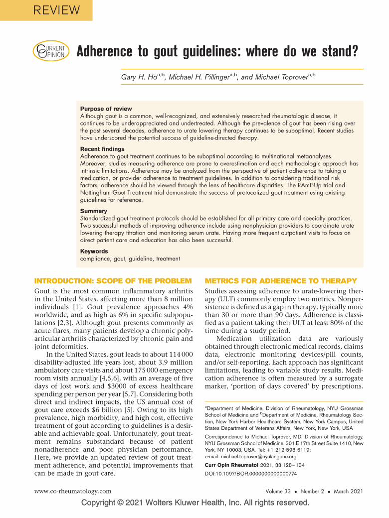
REVIEW
CURRENTOPINION Adherence to gout guidelines: where do we stand?
Copyright ©
www.co-rheumatology.com
a,b a,b a,b
Gary H. Ho , Michael H. Pillinger , and Michael ToproverPurpose of review
Although gout is a common, well-recognized, and extensively researched rheumatologic disease, itcontinues to be underappreciated and undertreated. Although the prevalence of gout has been rising overthe past several decades, adherence to urate lowering therapy continues to be suboptimal. Recent studieshave underscored the potential success of guideline-directed therapy.
Recent findings
Adherence to gout treatment continues to be suboptimal according to multinational metaanalyses.Moreover, studies measuring adherence are prone to overestimation and each methodologic approach hasintrinsic limitations. Adherence may be analyzed from the perspective of patient adherence to taking amedication, or provider adherence to treatment guidelines. In addition to considering traditional riskfactors, adherence should be viewed through the lens of healthcare disparities. The RAmP-Up trial andNottingham Gout Treatment trial demonstrate the success of protocolized gout treatment using existingguidelines for reference.
Summary
Standardized gout treatment protocols should be established for all primary care and specialty practices.Two successful methods of improving adherence include using nonphysician providers to coordinate uratelowering therapy titration and monitoring serum urate. Having more frequent outpatient visits to focus ondirect patient care and education has also been successful.
Keywords
compliance, gout, guideline, treatment
INTRODUCTION: SCOPE OF THE PROBLEM METRICS FOR ADHERENCE TO THERAPY
aDepartment of Medicine, Division of Rheumatology, NYU GrossmanSchool of Medicine and bDepartment of Medicine, Rheumatology Sec-tion, New York Harbor Healthcare System, New York Campus, UnitedStates Department of Veterans Affairs, New York, New York, USA
Correspondence to Michael Toprover, MD, Division of Rheumatology,NYU Grossman School of Medicine, 301 E 17th Street Suite 1410, NewYork, NY 10003, USA. Tel: +1 212 598 6119;e-mail: [email protected]
Curr Opin Rheumatol 2021, 33:128–134
DOI:10.1097/BOR.0000000000000774
Gout is the most common inflammatory arthritisin the United States, affecting more than 8 millionindividuals [1]. Gout prevalence approaches 4%worldwide, and as high as 6% in specific subpopu-lations [2,3]. Although gout presents commonly asacute flares, many patients develop a chronic poly-articular arthritis characterized by chronic pain andjoint deformities.
In the United States, gout leads to about 114 000disability-adjusted life years lost, about 3.9 millionambulatory care visits and about 175 000 emergencyroom visits annually [4,5,6], with an average of fivedays of lost work and $3000 of excess healthcarespending per person per year [5,7]. Considering bothdirect and indirect impacts, the US annual cost ofgout care exceeds $6 billion [5]. Owing to its highprevalence, high morbidity, and high cost, effectivetreatment of gout according to guidelines is a desir-able and achievable goal. Unfortunately, gout treat-ment remains substandard because of patientnonadherence and poor physician performance.Here, we provide an updated review of gout treat-ment adherence, and potential improvements thatcan be made in gout care.
2021 Wolters Kluwer H
Studies assessing adherence to urate-lowering ther-apy (ULT) commonly employ two metrics. Nonper-sistence is defined as a gap in therapy, typically morethan 30 or more than 90 days. Adherence is classi-fied as a patient taking their ULT at least 80% of thetime during a study period.
Medication utilization data are variouslyobtained through electronic medical records, claimsdata, electronic monitoring devices/pill counts,and/or self-reporting. Each approach has significantlimitations, leading to variable study results. Medi-cation adherence is often measured by a surrogatemarker, ‘portion of days covered’ by prescriptions.
ealth, Inc. All rights reserved.
Volume 33 � Number 2 � March 2021
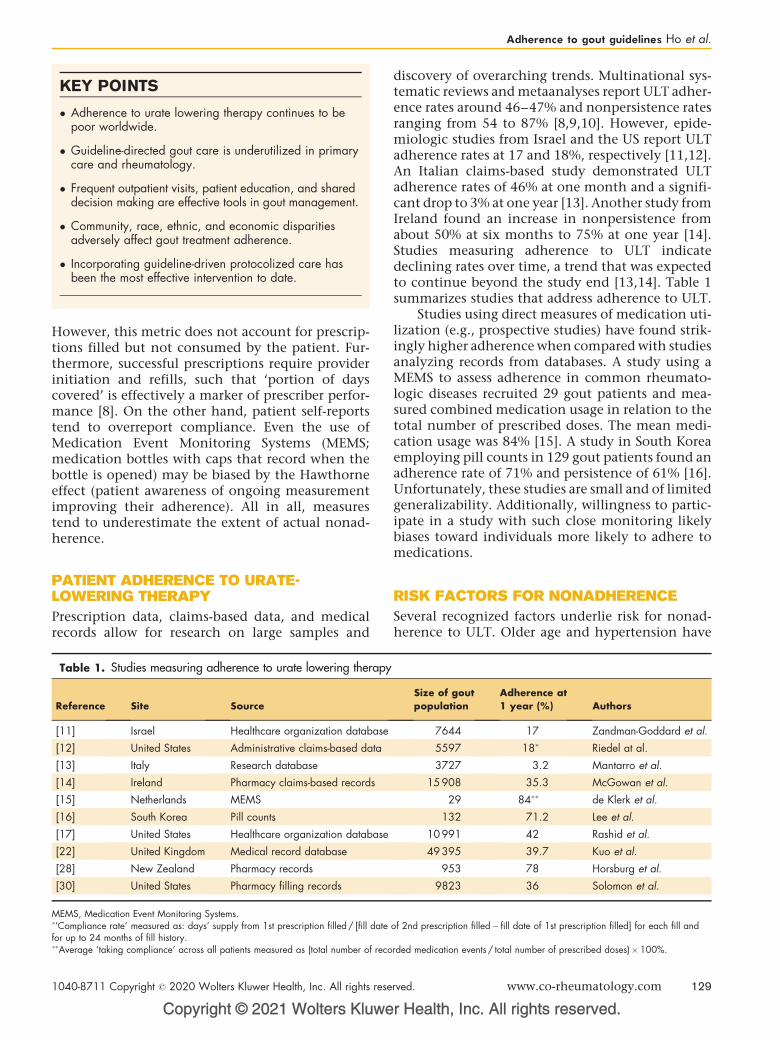
KEY POINTS
� Adherence to urate lowering therapy continues to bepoor worldwide.
� Guideline-directed gout care is underutilized in primarycare and rheumatology.
� Frequent outpatient visits, patient education, and shareddecision making are effective tools in gout management.
� Community, race, ethnic, and economic disparitiesadversely affect gout treatment adherence.
� Incorporating guideline-driven protocolized care hasbeen the most effective intervention to date.
Adherence to gout guidelines Ho et al.
However, this metric does not account for prescrip-tions filled but not consumed by the patient. Fur-thermore, successful prescriptions require providerinitiation and refills, such that ‘portion of dayscovered’ is effectively a marker of prescriber perfor-mance [8]. On the other hand, patient self-reportstend to overreport compliance. Even the use ofMedication Event Monitoring Systems (MEMS;medication bottles with caps that record when thebottle is opened) may be biased by the Hawthorneeffect (patient awareness of ongoing measurementimproving their adherence). All in all, measurestend to underestimate the extent of actual nonad-herence.
PATIENT ADHERENCE TO URATE-LOWERING THERAPY
Prescription data, claims-based data, and medicalrecords allow for research on large samples and
Copyright © 2021 Wolters Kluwe
Table 1. Studies measuring adherence to urate lowering therapy
Reference Site Source
[11] Israel Healthcare organization database
[12] United States Administrative claims-based data
[13] Italy Research database
[14] Ireland Pharmacy claims-based records
[15] Netherlands MEMS
[16] South Korea Pill counts
[17] United States Healthcare organization database
[22] United Kingdom Medical record database
[28] New Zealand Pharmacy records
[30] United States Pharmacy filling records
MEMS, Medication Event Monitoring Systems.�‘Compliance rate’ measured as: days’ supply from 1st prescription filled / [fill datefor up to 24 months of fill history.��Average ‘taking compliance’ across all patients measured as (total number of reco
1040-8711 Copyright � 2020 Wolters Kluwer Health, Inc. All rights rese
discovery of overarching trends. Multinational sys-tematic reviews and metaanalyses report ULT adher-ence rates around 46–47% and nonpersistence ratesranging from 54 to 87% [8,9,10]. However, epide-miologic studies from Israel and the US report ULTadherence rates at 17 and 18%, respectively [11,12].An Italian claims-based study demonstrated ULTadherence rates of 46% at one month and a signifi-cant drop to 3% at one year [13]. Another study fromIreland found an increase in nonpersistence fromabout 50% at six months to 75% at one year [14].Studies measuring adherence to ULT indicatedeclining rates over time, a trend that was expectedto continue beyond the study end [13,14]. Table 1summarizes studies that address adherence to ULT.
Studies using direct measures of medication uti-lization (e.g., prospective studies) have found strik-ingly higher adherence when compared with studiesanalyzing records from databases. A study using aMEMS to assess adherence in common rheumato-logic diseases recruited 29 gout patients and mea-sured combined medication usage in relation to thetotal number of prescribed doses. The mean medi-cation usage was 84% [15]. A study in South Koreaemploying pill counts in 129 gout patients found anadherence rate of 71% and persistence of 61% [16].Unfortunately, these studies are small and of limitedgeneralizability. Additionally, willingness to partic-ipate in a study with such close monitoring likelybiases toward individuals more likely to adhere tomedications.
RISK FACTORS FOR NONADHERENCE
Several recognized factors underlie risk for nonad-herence to ULT. Older age and hypertension have
r Health, Inc. All rights reserved.
Size of goutpopulation
Adherence at1 year (%) Authors
7644 17 Zandman-Goddard et al.
5597 18� Riedel at al.
3727 3.2 Mantarro et al.
15 908 35.3 McGowan et al.
29 84�� de Klerk et al.
132 71.2 Lee et al.
10 991 42 Rashid et al.
49 395 39.7 Kuo et al.
953 78 Horsburg et al.
9823 36 Solomon et al.
of 2nd prescription filled� fill date of 1st prescription filled] for each fill and
rded medication events / total number of prescribed doses)�100%.
rved. www.co-rheumatology.com 129
Crystal deposition diseases
consistently correlated with better medicationadherence [8,9]; such patients tend to have moreexperience managing chronic diseases and takingdaily medications. Interestingly, concomitantdiuretic use and congestive heart failure in one studywere associated with better ULT adherence despitemore difficulty achieving serum urate goal [17].Among a cohort of gout patients in the US Depart-ment of Veterans Affairs healthcare system, a cross-sectional analysis identified multiple comorbid con-ditions that complicated the choice of medicationfor treating acute gout flares and reduce serum urate.Many physicians prescribed specific gout therapies(including colchicine, NSAIDS, glucocorticoids,allopurinol, and probenecid) despite contraindica-tions to the individual medications, raising thepotential for noncompliance due to intolerance oradverse events when an alternative medication mayhave been better tolerated [18].
A disconnect between patient and provider per-spective is another potential factor in treatmentfailure. Parties can complete a visit with differentunderstandings and assumptions, often to the det-riment of the patient. One study identified thatproviders were confident in their treatment of acutegout, their patients’ adherence to ULT, and theirown educational skills. Patients, however, felt thatthey lacked information, particularly about the roleof ULT in gout management [19].
The achievement of serum urate targets acrosspopulations remain elusive partly because the com-plexities of gout treatment are underappreciated[20,21,25]. The concept that intercritical gout isassociated with persistent low-level inflammationand asymptomatic monosodium urate (MSU)deposits, and is not a true remission, is one suchexample. Additionally, knowledge about the diversearray of gout medications is essential for avoidingadverse effects and managing patient expectations.In fact, lack of proper education may lead to loss ofpatient trust in both their physician and medica-tion(s) [22,23].
PHYSICIAN MANAGEMENT OF GOUT:IMPACT ON ADHERENCE
The ‘Management of Gout in the United States: AClaims-based Analysis’ study employed administra-tive claims data to assess gout-related care. Thisstudy found that serum urate testing was performedin less than 70% of patients with chronic gout, andless than 60% of those with acute gout. Further-more, less than 80% of patients with advanced gout(i.e., nontophaceous chronic, tophaceous chronic,or uncontrolled gout) received ULT, and on average,prescriptions for ULT covered less than half of the
Copyright © 2021 Wolters Kluwer H
130 www.co-rheumatology.com
study year. Even a single rheumatology visit duringthe study period was associated with increased fre-quency of serum urate testing, increased number ofULT prescriptions, and reduced emergency depart-ment visits [24
&&
]. A UK study found that only 37%of patients received ULT, and only 18% of theprescriptions were started within six months ofdiagnosis. Timely initiation of ULT and appropriatereferral to a specialist were areas of improvementidentified for general practitioners [25]. These andother data suggest that, even before the question ofadherence is considered, patients with gout arehighly susceptible to underprescribing of ULT.
Oderda et al [26]. studied physician adherence toAmerican College of Rheumatology (ACR) guide-lines and scored physicians from 0 to 8 based onthe categories of targeting a serum urate goal, first-line ULT prescribing, appropriate medication dos-ing, and gout flare prophylaxis while on ULT.Reviewing treatments of 350 chronic gout patientsand their physicians’ perspectives, rheumatologiststended to follow ACR guidelines more closely (5.8/8�1.7 versus 4.3/8�1.7) and were more likely to beclassified as ‘higher adherence’ (64.7 versus 46.3%)than primary care providers. There was a disparitybetween providers’ belief that their patient carefollowed guidelines and their actual practice [26].In one survey of real-world US practice patterns, achart audit was conducted for 124 primary carephysicians and 125 rheumatologists; out of 1245gout patients’ charts reviewed, only 11% achieveddisease control (defined as an average serumurate�6, no flares, and no tophi) [21].
DIFFERENCES ACROSS POPULATIONS
Trends in ULT adherence and practice patterns areusually consistent across countries; however, health-care disparities can further diminish care in subpo-pulations. Lower socioeconomic status and fewerclinic visits have been associated with worse compli-ance [11,27]. More specifically, Singh et al. [27] founda linear correlation between more outpatient visitsand higher odds of reaching goal serum urate as wellas less frequent allopurinol discontinuation.
A New Zealand prescription data analysisdescribed worse ULT adherence among native Maoripopulations at 62% compared with 82% of non-Maori, highlighting the impact of potential health-care disparities in gout [28]. Another community-based study addressing gout care, which included aMaori indigenous population, sent out an informa-tional packet with material reflecting the currentguidelines to a rural medical center. In comparingpractice patterns before and after this educationalpackage, provider education resulted in greater
ealth, Inc. All rights reserved.
Volume 33 � Number 2 � March 2021
Adherence to gout guidelines Ho et al.
conformity to gout guidelines, and a higher percent-age of patients achieving target serum urate [29
&
].Solomon et al. [30] assessed adherence in an
elderly population enrolled in a pharmacy assis-tance program in Pennsylvania. The overall adher-ence rate was 36%, but Black individuals were twiceas likely to be nonadherent [odds ratio (OR) 1.86,95% confidence interval (CI) 1.52–2.27] comparedwith white individuals [30]. In another study explor-ing gout care in Black patients, patients were sepa-rated into high and low-adherence groups to assessdifferences in qualitative factors. Lack of side-effectsfrom ULT, trust in physicians, and gout-relatedhealth literacy facilitated high adherence. Techni-ques for better adherence focused on incorporatingmedications into daily routines and using pillboxes.Suboptimal adherence was attributed to concernsabout medication costs and side-effects, preferencefor alternative medicines, forgetfulness in both tak-ing and refilling medications, lack of belief in ULTeffectiveness, experience of side-effects, pill fatigue,concern for drug interactions, pill size, and othercompeting priorities [31].
GOUT GUIDELINES
In 2020, three professional organizations releasedupdated guidelines for gout management. Treat-to-target remains a strong recommendation from allrheumatologic societies. The ACR recommends ULTandmaintainingserumurate less than6mg/dlovernotarget [32
&
]. There is moderate-to-high-quality evi-dence for the treat-to-target approach as evidencedby target-driven protocols resulting in better adher-ence to ULT, fewer gout flares, and reduction in size oftophi [32
&
,33&&
,34&
,35&&
]. The main pillars of this rec-ommendation are ULT dose titration and frequentassessment of serum urate levels to guide therapy.
The Japanese guideline had already established aserum urate target of less than 6.0 mg/dl in 2011,citing the ‘dissolution limit of uric acid in the bodyfluid’ at 6.4 mg/dl, and went on in 2020 to recom-mend this goal for tophaceous gout [36,37
&
]. TheFrench Society for Rheumatology echoes the recom-mendation for a target serum urate less than 6.0 mg/dl and goes further, recommending a target less than5.0 mg/dl whenever possible [38]. These variousguidelines underscore the importance of maintain-ing serum urate at goal and monitoring serum uratelevels regularly because MSU crystals can take yearsto dissolve. Dual-energy CT studies have shown thatMSU crystal burden is reduced after reaching serumurate goal but MSU deposits may persist beyond twoyears of serum urate control [39,40].
The American College of Physician’s controver-sial guideline to ‘treat to avoid symptoms’ avers that
Copyright © 2021 Wolters Kluwe
1040-8711 Copyright � 2020 Wolters Kluwer Health, Inc. All rights rese
insufficient evidence supports the benefits of ULTtreatment and/or a treat-to-target approach [41].The dissonance between the rheumatology andinternal medicine society guidelines leads to pro-vider confusion, especially as most gout patients,including newly diagnosed cases, are managed byprimary care providers.
INTERVENTIONS TO IMPROVEADHERENCE
Accumulatingevidence supports the utility of includ-ing nonphysician providers in gout care as opposedto primary care or rheumatology alone. Such individ-uals can supplement physician caregivers, providepatient education, regularize follow-up and, in con-trast to primary care physicians, focus exclusively onthe gout treatment. The ACR recommends that non-physician providers use a protocolized approachwhile incorporating the key elements of patient edu-cation and shared decision-making [32
&
]. Table 2summarizes the studies that assess the effect of vari-ous interventions on adherence to ULT.
One approach to increasing patient engagementis using mobile apps. In a study among a NewZealand gout population, patients were randomizedto use a commercially available ‘Gout Central’ app,versus a dietary app as a control (DASH diet app).The primary outcome was user engagement. Impor-tant features of the gout app included a serum uratetracker, a gout flare tracker, an educational section,and a reminder-based function. The 36 users of thegout app were more engaged and reported increasedgout-related knowledge, including awareness ofadverse long-term consequences of gout and hyper-uricemia. However, the two groups did not differ interms of behavioral changes at the end of the two-week study period [42
&
]. Major limitations of thisstudy were its small sample size, short duration, andthe lack of utility of the app for those with infre-quent flares. App development and technologicaladvances more broadly may offer the prospect ofbetter patient engagement and closer serum uratemonitoring.
Mikuls et al [33&&
]. reported a study comparing apharmacist-led gout treatment protocol versus usualcare among 1462 patients who were initiating allo-purinol for gout (RAmP-UP Study). The protocolutilized automated interactive telephone-basedinterventions, which provided reminders andencouragement, and checked adherence. On identi-fication of nonadherence, pharmacists would callpatients as a ‘step-up’ in intervention. This approachresulted in significantly improved allopurinol adher-ence compared with usual care (50 versus 37%; OR1.68; 95% CI 1.30, 2.17). Additionally, the serum
r Health, Inc. All rights reserved.
rved. www.co-rheumatology.com 131
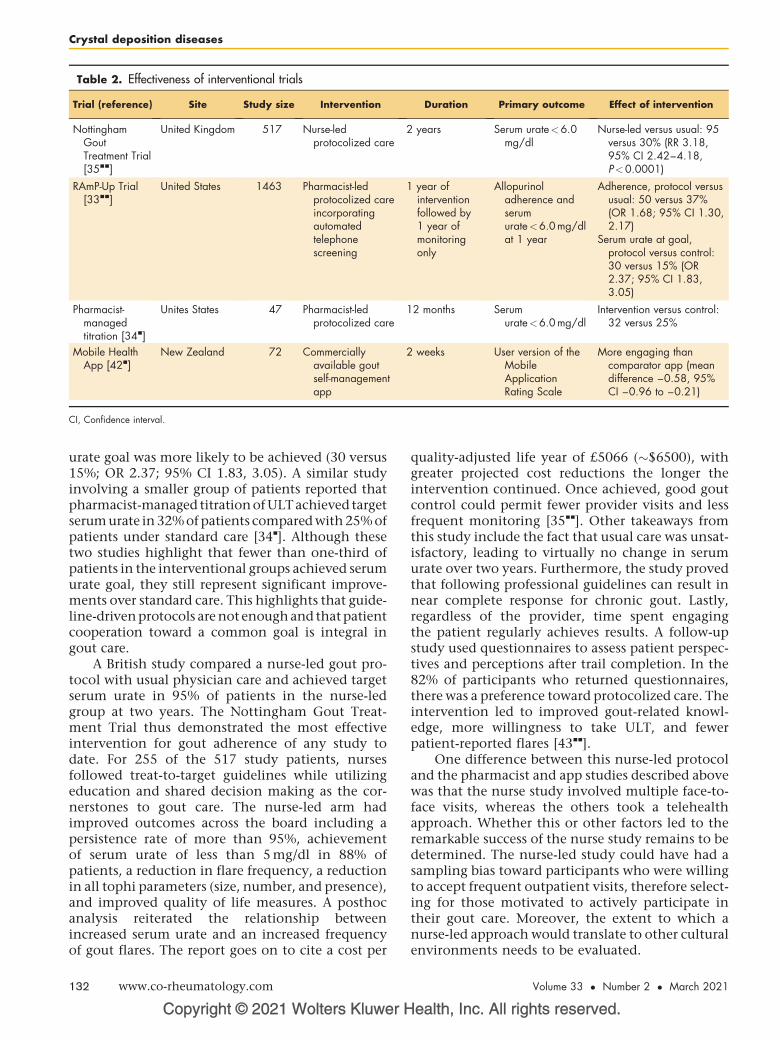
Table 2. Effectiveness of interventional trials
Trial (reference) Site Study size Intervention Duration Primary outcome Effect of intervention
NottinghamGoutTreatment Trial[35&&]
United Kingdom 517 Nurse-ledprotocolized care
2 years Serum urate<6.0mg/dl
Nurse-led versus usual: 95versus 30% (RR 3.18,95% CI 2.42–4.18,P<0.0001)
RAmP-Up Trial[33&&]
United States 1463 Pharmacist-ledprotocolized careincorporatingautomatedtelephonescreening
1 year ofinterventionfollowed by1 year ofmonitoringonly
Allopurinoladherence andserumurate<6.0 mg/dlat 1 year
Adherence, protocol versususual: 50 versus 37%(OR 1.68; 95% CI 1.30,2.17)
Serum urate at goal,protocol versus control:30 versus 15% (OR2.37; 95% CI 1.83,3.05)
Pharmacist-managedtitration [34&]
Unites States 47 Pharmacist-ledprotocolized care
12 months Serumurate<6.0 mg/dl
Intervention versus control:32 versus 25%
Mobile HealthApp [42&]
New Zealand 72 Commerciallyavailable goutself-managementapp
2 weeks User version of theMobileApplicationRating Scale
More engaging thancomparator app (meandifference –0.58, 95%CI –0.96 to –0.21)
CI, Confidence interval.
Crystal deposition diseases
urate goal was more likely to be achieved (30 versus15%; OR 2.37; 95% CI 1.83, 3.05). A similar studyinvolving a smaller group of patients reported thatpharmacist-managed titration of ULT achieved targetserum urate in 32% of patients compared with 25% ofpatients under standard care [34
&
]. Although thesetwo studies highlight that fewer than one-third ofpatients in the interventional groups achieved serumurate goal, they still represent significant improve-ments over standard care. This highlights that guide-line-driven protocols are not enough and thatpatientcooperation toward a common goal is integral ingout care.
A British study compared a nurse-led gout pro-tocol with usual physician care and achieved targetserum urate in 95% of patients in the nurse-ledgroup at two years. The Nottingham Gout Treat-ment Trial thus demonstrated the most effectiveintervention for gout adherence of any study todate. For 255 of the 517 study patients, nursesfollowed treat-to-target guidelines while utilizingeducation and shared decision making as the cor-nerstones to gout care. The nurse-led arm hadimproved outcomes across the board including apersistence rate of more than 95%, achievementof serum urate of less than 5 mg/dl in 88% ofpatients, a reduction in flare frequency, a reductionin all tophi parameters (size, number, and presence),and improved quality of life measures. A posthocanalysis reiterated the relationship betweenincreased serum urate and an increased frequencyof gout flares. The report goes on to cite a cost per
Copyright © 2021 Wolters Kluwer H
132 www.co-rheumatology.com
quality-adjusted life year of £5066 (�$6500), withgreater projected cost reductions the longer theintervention continued. Once achieved, good goutcontrol could permit fewer provider visits and lessfrequent monitoring [35
&&
]. Other takeaways fromthis study include the fact that usual care was unsat-isfactory, leading to virtually no change in serumurate over two years. Furthermore, the study provedthat following professional guidelines can result innear complete response for chronic gout. Lastly,regardless of the provider, time spent engagingthe patient regularly achieves results. A follow-upstudy used questionnaires to assess patient perspec-tives and perceptions after trail completion. In the82% of participants who returned questionnaires,there was a preference toward protocolized care. Theintervention led to improved gout-related knowl-edge, more willingness to take ULT, and fewerpatient-reported flares [43
&&
].One difference between this nurse-led protocol
and the pharmacist and app studies described abovewas that the nurse study involved multiple face-to-face visits, whereas the others took a telehealthapproach. Whether this or other factors led to theremarkable success of the nurse study remains to bedetermined. The nurse-led study could have had asampling bias toward participants who were willingto accept frequent outpatient visits, therefore select-ing for those motivated to actively participate intheir gout care. Moreover, the extent to which anurse-led approach would translate to other culturalenvironments needs to be evaluated.
ealth, Inc. All rights reserved.
Volume 33 � Number 2 � March 2021
Adherence to gout guidelines Ho et al.
In the era of the Coronoa virus disease 2019(COVID-19) pandemic, access to in-person care ismore limited and telehealth will be integrated intoclinical practice for the foreseeable future. Adaptingto these challenges will be critical to gout care whenface-to-face interactions are limited and in a futurewhich technology is more widely employed. Guide-line-driven care and patient engagement should bethe guiding principles whether the focus will turn toapp-based interventions or increasing utilizationof telehealth.
CONCLUSION
Nonadherence remains an important barrier to opti-mal gout care. Modifiable adherence can be sepa-rated into patient adherence to ULT and prescriberadherence to guidelines. As most patients with goutare treated by primary care providers, the questionof which guidelines (if any) providers followremains a concern. A start to addressing factorsunderlying nonadherence would incorporatetreat-to-target protocols for gout patient care.Trained nonphysician providers could be integratedinto the primary care setting and consult with rheu-matology as needed. More innovative approachesare needed to address inadequacies in gout care.
Acknowledgements
None.
Financial support and sponsorship
None.
Conflicts of interest
None. For the purposes of full transparency, M.H.P.reports that he is the recipient of investigator-initiatedgrants from Hikma Pharmaceuticals and Horizon Ther-apeutics.
REFERENCES AND RECOMMENDEDREADINGPapers of particular interest, published within the annual period of review, havebeen highlighted as:
& of special interest&& of outstanding interest1. Singh G, Lingala B, Mithal A. Gout and hyperuricaemia in the USA: prevalenceand trends. Rheumatology (Oxford) 2019; 58:2177–2180.
2. Roddy E, Choi HK. Epidemiology of gout. Rheum Dis Clin North Am 2014;40:155–175.
3. Zhu Y, Pandya BJ, Choi HK. Prevalence of gout and hyperuricemia in the USgeneral population: the National Health and Nutrition Examination Survey2007–2008. Arthritis Rheum 2011; 63:3136–3141.
4. Smith E, Hoy D, Cross M, et al. The global burden of gout: estimates from theGlobal Burden of Disease 2010 study. Ann Rheum Dis 2014;73:1470–1476.
5. Wertheimer A, Morlock R, Becker MA. A revised estimate of the burden ofillness of gout. Curr Ther Res Clin Exp 2013; 75:1–4.
Copyright © 2021 Wolters Kluwe
1040-8711 Copyright � 2020 Wolters Kluwer Health, Inc. All rights rese
6. Garg R, Sayles HR, Yu F, et al. Gout-related healthcare utilization in USemergency departments, 2006 through 2008. Arthritis Care Res (Hoboken)2013; 65:571–577.
7. Shields GE, Beard SM. A systematic review of the economic and humanisticburden of gout. Pharmacoeconomics 2015; 33:1029–1047.
8. Scheepers LE, van Onna M, Stehouwer CD, et al. Medication adherenceamong patients with gout: a systematic review and meta-analysis. SeminArthritis Rheum 2018; 47:689–702.
9. De Vera MA, Marcotte G, Rai S, et al. Medication adherence in gout: asystematic review. Arthritis Care Res (Hoboken) 2014; 66:1551–1559.
10. Yin R, Li L, Zhang G, et al. Rate of adherence to urate-lowering therapy amongpatients with gout: a systematic review and meta-analysis. BMJ Open 2018;8:e017542.
11. Zandman-Goddard G, Amital H, Shamrayevsky N, et al. Rates of adherenceand persistence with allopurinol therapy among gout patients in Israel.Rheumatology (Oxford) 2013; 52:1126–1131.
12. Riedel AA, Nelson M, Joseph-Ridge N, et al. Compliance with allopurinoltherapy among managed care enrollees with gout: a retrospective analysis ofadministrative claims. J Rheumatol 2004; 31:1575–1581.
13. Mantarro S, Capogrosso-Sansone A, Tuccori M, et al. Allopurinol adherenceamong patients with gout: an Italian general practice database study. Int J ClinPract 2015; 69:757–765.
14. McGowan B, Bennett K, Silke C, Whelan B. Adherence and persistence tourate-lowering therapies in the Irish setting. Clin Rheumatol 2016;35:715–721.
15. de Klerk E, van der Heijde D, Landewe R, et al. Patient compliance inrheumatoid arthritis, polymyalgia rheumatica, and gout. J Rheumatol 2003;30:44–54. Erratum in: J Rheumatol 2003;30(2):423.
16. Lee S, So MW. Adherence with urate-lowering therapies among malepatients with gout in a routine clinical setting. Mod Rheumatol 2016;26:950–955.
17. Rashid N, Coburn BW, Wu YL, et al. Modifiable factors associated withallopurinol adherence and outcomes among patients with gout in an inte-grated healthcare system. J Rheumatol 2015; 42:504–512.
18. Keenan RT, O’Brien WR, Lee KH, et al. Prevalence of contraindications andprescription of pharmacologic therapies for gout. Am J Med 2011;124:155–163.
19. Harrold LR, Mazor KM, Velten S, et al. Patients and providers view goutdifferently: a qualitative study. Chronic Illn 2010; 6:263–271.
20. Khanna P, Khanna D, Storgard C, et al. A world of hurt: failure to achievetreatment goals in patients with gout requires a paradigm shift. Postgrad Med2016; 128:34–40.
21. Keenan RT. Limitations of the current standards of care for treating gout andcrystal deposition in the primary care setting: a review. Clin Ther 2017;39:430–441.
22. Coleshill MJ, Aung E, Nguyen AD, et al. Improving adherence to urate-lowering therapy in people living with gout. Int J Rheum Dis 2019;22:542–544.
23. Singh JA. Patient perspectives in gout: a review. Curr Opin Rheumatol 2019;31:159–166.
24.&&
Edwards NL, Schlesinger N, Clark S, et al. Management of gout in theUnited States: a claims-based analysis. ACR Open Rheumatol 2020;2:180–187.
A review of United States administrative claims databases from October 2015to November 2018 of individuals diagnosed with gout. Patients were categor-ized as having acute gout or other advanced forms of gout for analysis.Percentage and frequency of serum urate testing, rheumatology specialistvisits, prescriptions for ULTs, and emergency room visits for gout flares wereevaluated.25. Kuo CF, Grainge MJ, Mallen C, et al. Rising burden of gout in the UK but
continuing suboptimal management: a nationwide population study. AnnRheum Dis 2015; 74:661–667.
26. Oderda GM, Shiozawa A, Walsh M, et al. Physician adherence to ACR gouttreatment guidelines: perception versus practice. Postgrad Med 2014;126:257–267.
27. Singh JA, Hodges JS, Asch SM. Opportunities for improving medication useand monitoring in gout. Ann Rheum Dis 2009; 68:1265–1270.
28. Horsburgh S, Norris P, Becket G, et al. Allopurinol use in a New Zealandpopulation: prevalence and adherence. Rheumatol Int 2014; 34:963–970.
29.&
Stamp LK, Chapman P, Hudson B, et al. The challenges of managing gout inprimary care: results of a best-practice audit. Aust J Gen Pract 2019;48:631–637.
A study assessing practice patterns in a single rural medical practice in NewZealand after the introduction of an informational packet reflecting guidelines ingout management. Achievement of target serum uric acid and frequency of labmonitoring were assessed as outcomes.30. Solomon DH, Avorn J, Levin R, Brookhart MA. Uric acid lowering therapy:
prescribing patterns in a large cohort of older adults. Ann Rheum Dis 2008;67:609–613.
31. Singh JA. Facilitators and barriers to adherence to urate-lowering therapy inAfrican-Americans with gout: a qualitative study. Arthritis Res Ther 2014;16:R82.
r Health, Inc. All rights reserved.
rved. www.co-rheumatology.com 133
Crystal deposition diseases
32.&
FitzGerald JD, Dalbeth N, Mikuls T, et al. 2020 American College of Rheu-matology Guideline for the Management of Gout 2020. Arthritis Care Res(Hoboken) 2020; 72:744–760. Erratum in: Arthritis Care Res (Hoboken)2020;72(8):1187.
Updated American College of Rheumatology guidelines for the management ofgout. A multidisciplinary team answered Population, Intervention, Comparisonand Outcomes questions using systematic literature reviews and network me-taanalyses; the recommendations were graded for strength and quality ofevidence.33.&&
Mikuls TR, Cheetham TC, Levy GD, et al. Adherence and outcomes withurate-lowering therapy: a site-randomized trial. Am J Med 2019;132:354–361.
A site-randomized trial of 1463 gout patients at 116 Kaiser Permanente clinics inCalifornia. The trial compared pharmacist-led intervention to usual care in goutpatients initiating allopurinol. The intervention was implemented for one year andthe patients were monitored for an additional year. The primary outcomes of‘proportion of days covered’ and serum urate at goal were assessed at one andtwo years.34.&
Huang IJ, Liew JW, Morcos MB, et al. Pharmacist-managed titration of urate-lowering therapy to streamline gout management. Rheumatol Int 2019;39:1637–1641.
A Chinese cohort of 47 patients received pharmacist-managed urate-loweringtherapy titration compared with usual care. Serum urate was the primary outcomeat the end of one year.35.&&
Doherty M, Jenkins W, Richardson H, et al. Efficacy and cost-effectiveness ofnurse-led care involving education and engagement of patients and a treat-to-target urate-lowering strategy versus usual care for gout: a randomisedcontrolled trial. Lancet 2018; 392:1403–1412.
A randomized clinical trial of nurse-led care compared with routine care in 517patients residing in Nottingham, United Kingdom. Nurses followed a gout manage-ment protocol and used both telephone and clinic visits to interact with patients.The primary outcome of serum urate less than 0.6 mg/dl was assessed at one andtwo years. Secondary outcomes were flare frequency, presence of tophi, quality oflife, and cost per quality-adjusted life-year gained.
Copyright © 2021 Wolters Kluwer H
134 www.co-rheumatology.com
36. Yamanaka H. Japanese guideline for the management of hyperuricemia andgout: second edition. Nucleosides Nucleotides Nucleic Acids 2011;30:1018–1029.
37.&
Hisatome I, Ichida K, Mineo I, et al. Japanese Society of Gout and Uric &Nucleic Acids 2019 guidelines for management of hyperuricemia and gout,3rd edition. Gout Uric Nucleic Acids 2020; 44:1–40.
Updated Japanese Society of Gout and Uric & Nucleic Acids guidelines focusingon specific gout-related clinical questions. The recommendations were based offan expert panel’s systematic review of the relevant literature.38. Pascart T, Latourte A, Flipo RM, et al. 2020 recommendations from the French
Society of Rheumatology for the management of gout: urate-lowering therapy.Joint Bone Spine 2020; 87:395–404.
39. Dalbeth N, Billington K, Doyle A, et al. Effects of allopurinol dose escalation onbone erosion and urate volume in gout: a dual-energy computed tomographyimaging study within a randomized, controlled trial. Arthritis Rheumatol 2019;71:1739–1746.
40. Ellmann H, Bayat S, Araujo E, et al. Effects of conventional uric acid-loweringtherapy on monosodium urate crystal deposits. Arthritis Rheumatol 2020;72:150–156.
41. Qaseem A, Harris RP, Forciea MA. Management of acute and recurrent gout:a clinical practice guideline from the American College of Physicians. AnnIntern Med 2017; 166:58–68.
42.&
Serlachius A, Schache K, Kieser A, et al. Association between user engage-ment of a mobile health app for gout and improvements in self-care behaviors:randomized controlled trial. JMIR Mhealth Uhealth 2019; 7:e15021.
A study of 72 adults in New Zealand exploring user engagement of a gout care appcompared with a DASH diet app. The trial period was two weeks and assessedchanges in health-related behaviors as a secondary outcome.43.&&
Fuller A, Jenkins W, Doherty M, Abhishek A. Nurse-led care is preferred overGP-led care of gout and improves gout outcomes: results of Nottingham GoutTreatment Trial follow-up study. Rheumatology (Oxford) 2020; 59:575–579.
Participants of the Nottingham Gout Treatment Trial were sent follow-up question-naires enquiring subjectively about gout knowledge, satisfaction with their healthcarepractitioner (nurse or general practitioner), and self-reported adherence to ULT.
ealth, Inc. All rights reserved.
Volume 33 � Number 2 � March 2021
REVIEW
CURRENTOPINION The role of diet in hyperuricemia and gout
Copyrigh
1040-8711 Copyright � 2021 Wolte
a,b a,b,c a,b
Chio Yokose , Natalie McCormick , and Hyon K. ChoiPurpose of review
Although gout’s cardinal feature is inflammatory arthritis, it is closely associated with insulin resistance andconsidered a manifestation of the metabolic syndrome. As such, both gout and hyperuricemia are oftenassociated with major cardiometabolic and renal comorbidities that drive the persistently elevatedpremature mortality rates among gout patients. To that end, conventional low-purine (i.e., low-protein)dietary advice given to many patients with gout warrant reconsideration.
Recent findings
Recent research suggests that several healthy diets, such as the Mediterranean or Dietary Approaches toStop Hypertension (DASH) diets, in combination with weight loss for those who are overweight or obese,can drastically improve cardiometabolic risk factors and outcomes. By treating gout as a part of themetabolic syndrome and shifting our dietary recommendations to these healthy dietary patterns, thebeneficial effects on gout endpoints should naturally follow for the majority of typical gout cases, mediatedthrough changes in insulin resistance.
Summary
Dietary recommendations for the management of hyperuricemia and gout should be approachedholistically, taking into consideration its associated cardiometabolic comorbidities. Several healthy dietarypatterns, many with similar themes, can be tailored to suit comorbidity profiles and personal preferences.
Keywords
diet, gout, hyperuricemia, metabolic syndrome, nutrition
aDivision of Rheumatology, Allergy, and Immunology, bClinical Epidemi-ology Program, Mongan Institute, Massachusetts General Hospital,Boston, Massachusetts, USA and cArthritis Research Canada, Rich-mond, British Columbia, Canada
Correspondence to Chio Yokose, Harvard Medical School, Division ofRheumatology, Allergy, and Immunology, Massachusetts General Hospi-tal, 55 Fruit Street, Bulfinch 165, Boston, MA 02114, USA.Tel: +1 617 726 2870; fax: +1 617 726 2872;e-mail: [email protected]
Curr Opin Rheumatol 2021, 33:135–144
DOI:10.1097/BOR.0000000000000779
INTRODUCTION
Although gout’s cardinal feature is inflammatoryarthritis, its primary underlying cause, hyperurice-mia, is considered a manifestation of the metabolicsyndrome mediated by insulin resistance [1,2].Thus, hyperuricemia and gout are both associatedwith adverse consequences of the metabolic syn-drome, namely cardiometabolic and renal disease[3–5]. To that end, the conventional low-purine(i.e., low-protein) dietary approach focusing on pre-vention of purine-loading can worsen its cardiome-tabolic comorbidities by leading to compensatoryhigher consumption of carbohydrates (includingfructose) and fats (including trans or saturatedfat). Moreover, the long-term effectiveness of alow-purine diet to lower urate levels remains unclearwith its limited palatability and sustainability [1,6].In contrast, there are several preeminent healthydiets that can simultaneously reduce serum urateand the risk of gout and overall cardiometabolic riskby lowering adiposity and insulin resistance. Thesedietary interventions can be applicable to patients atall stages of gout (Fig. 1) and should form a corner-stone of lifestyle counseling for such patients. Thisarticle reviews the relevant scientific rationale and
t © 2021 Wolters Kluwe
rs Kluwer Health, Inc. All rights rese
available data to provide evidence-based dietaryconsiderations for the prevention and managementof hyperuricemia and gout, including the role of dieton gout flares, together with its cardiometaboliccomorbidities holistically.
THE METABOLIC SYNDROME ANDCOMORBIDITIES OF HYPERURICEMIAAND GOUT
Insulin resistance is a key feature of the metabolicsyndrome, and because insulin resistance can reducethe renal excretion of urate [6–9], hyperuricemia and
r Health, Inc. All rights reserved.
rved. www.co-rheumatology.com
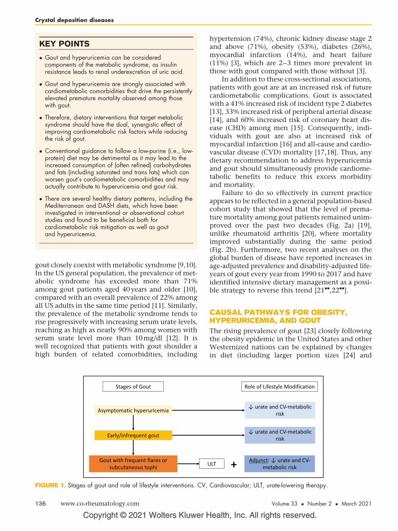
KEY POINTS
� Gout and hyperuricemia can be consideredcomponents of the metabolic syndrome, as insulinresistance leads to renal underexcretion of uric acid.
� Gout and hyperuricemia are strongly associated withcardiometabolic comorbidities that drive the persistentlyelevated premature mortality observed among thosewith gout.
� Therefore, dietary interventions that target metabolicsyndrome should have the dual, synergistic effect ofimproving cardiometabolic risk factors while reducingthe risk of gout.
� Conventional guidance to follow a low-purine (i.e., low-protein) diet may be detrimental as it may lead to theincreased consumption of (often refined) carbohydratesand fats (including saturated and trans fats) which canworsen gout’s cardiometabolic comorbidities and mayactually contribute to hyperuricemia and gout risk.
� There are several healthy dietary patterns, including theMediterranean and DASH diets, which have beeninvestigated in interventional or observational cohortstudies and found to be beneficial both forcardiometabolic risk mitigation as well as goutand hyperuricemia.
Crystal deposition diseases
gout closely coexist with metabolic syndrome [9,10].In the US general population, the prevalence of met-abolic syndrome has exceeded more than 71%among gout patients aged 40 years and older [10],compared with an overall prevalence of 22% amongall US adults in the same time period [11]. Similarly,the prevalence of the metabolic syndrome tends torise progressively with increasing serum urate levels,reaching as high as nearly 90% among women withserum urate level more than 10 mg/dl [12]. It iswell recognized that patients with gout shoulder ahigh burden of related comorbidities, including
Copyright © 2021 Wolters Kluwer H
FIGURE 1. Stages of gout and role of lifestyle interventions. CV,
136 www.co-rheumatology.com
hypertension (74%), chronic kidney disease stage 2and above (71%), obesity (53%), diabetes (26%),myocardial infarction (14%), and heart failure(11%) [3], which are 2–3 times more prevalent inthose with gout compared with those without [3].
In addition to these cross-sectional associations,patients with gout are at an increased risk of futurecardiometabolic complications. Gout is associatedwith a 41% increased risk of incident type 2 diabetes[13], 33% increased risk of peripheral arterial disease[14], and 60% increased risk of coronary heart dis-ease (CHD) among men [15]. Consequently, indi-viduals with gout are also at increased risk ofmyocardial infarction [16] and all-cause and cardio-vascular disease (CVD) mortality [17,18]. Thus, anydietary recommendation to address hyperuricemiaand gout should simultaneously provide cardiome-tabolic benefits to reduce this excess morbidityand mortality.
Failure to do so effectively in current practiceappears to be reflected in a general population-basedcohort study that showed that the level of prema-ture mortality among gout patients remained unim-proved over the past two decades (Fig. 2a) [19],unlike rheumatoid arthritis [20], where mortalityimproved substantially during the same period(Fig. 2b). Furthermore, two recent analyses on theglobal burden of disease have reported increases inage-adjusted prevalence and disability-adjusted life-years of gout every year from 1990 to 2017 and haveidentified intensive dietary management as a possi-ble strategy to reverse this trend [21
&&
,22&&
].
CAUSAL PATHWAYS FOR OBESITY,HYPERURICEMIA, AND GOUT
The rising prevalence of gout [23] closely followingthe obesity epidemic in the United States and otherWesternized nations can be explained by changesin diet (including larger portion sizes [24] and
ealth, Inc. All rights reserved.
Cardiovascular; ULT, urate-lowering therapy.
Volume 33 � Number 2 � March 2021
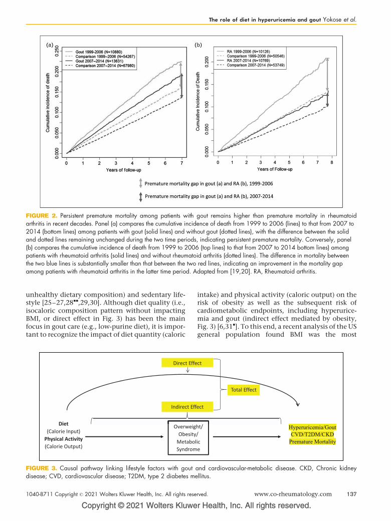
FIGURE 2. Persistent premature mortality among patients with gout remains higher than premature mortality in rheumatoidarthritis in recent decades. Panel (a) compares the cumulative incidence of death from 1999 to 2006 (lines) to that from 2007 to2014 (bottom lines) among patients with gout (solid lines) and without gout (dotted lines), with the difference between the solidand dotted lines remaining unchanged during the two time periods, indicating persistent premature mortality. Conversely, panel(b) compares the cumulative incidence of death from 1999 to 2006 (top lines) to that from 2007 to 2014 bottom lines) amongpatients with rheumatoid arthritis (solid lines) and without rheumatoid arthritis (dotted lines). The difference in mortality betweenthe two blue lines is substantially smaller than that between the two red lines, indicating an improvement in the mortality gapamong patients with rheumatoid arthritis in the latter time period. Adapted from [19,20]. RA, Rheumatoid arthritis.
The role of diet in hyperuricemia and gout Yokose et al.
unhealthy dietary composition) and sedentary life-style [25–27,28
&&
,29,30]. Although diet quality (i.e.,isocaloric composition pattern without impactingBMI, or direct effect in Fig. 3) has been the mainfocus in gout care (e.g., low-purine diet), it is impor-tant to recognize the impact of diet quantity (caloric
Copyright © 2021 Wolters Kluwe
FIGURE 3. Causal pathway linking lifestyle factors with goutdisease; CVD, cardiovascular disease; T2DM, type 2 diabetes me
1040-8711 Copyright � 2021 Wolters Kluwer Health, Inc. All rights rese
intake) and physical activity (caloric output) on therisk of obesity as well as the subsequent risk ofcardiometabolic endpoints, including hyperurice-mia and gout (indirect effect mediated by obesity,Fig. 3) [6,31
&
]. To this end, a recent analysis of the USgeneral population found BMI was the most
r Health, Inc. All rights reserved.
and cardiovascular-metabolic disease. CKD, Chronic kidneyllitus.
rved. www.co-rheumatology.com 137
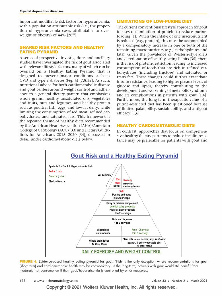
Crystal deposition diseases
important modifiable risk factor for hyperuricemia,with a population attributable risk (i.e., the propor-tion of hyperuricemia cases attributable to over-weight or obesity) of 44% [28
&&
].
SHARED RISK FACTORS AND HEALTHYEATING PYRAMID
A series of prospective investigations and ancillarystudies have investigated the risk of gout associatedwith relevant lifestyle factors, many of which can beoverlaid on a Healthy Eating Pyramid that isdesigned to prevent major conditions such asCVD and type 2 diabetes (Fig. 4) [7,8,32]. As such,nutritional advice for both cardiometabolic diseaseand gout centers around weight control and adher-ence to a general dietary pattern that emphasizeswhole grains, healthy unsaturated oils, vegetablesand fruits, nuts and legumes, and healthy proteinsuch as poultry, fish, eggs, and low-fat dairy, whilelimiting the consumption of red meat, refined car-bohydrates, and saturated fats. This framework isthe repeated theme of healthy diets recommendedby the American Heart Association (AHA)/AmericanCollege of Cardiology (ACC) [33] and Dietary Guide-lines for Americans 2015–2020 [34], discussed indetail under cardiometabolic diets below.
Copyright © 2021 Wolters Kluwer H
FIGURE 4. Evidence-based healthy eating pyramid for gout. �
(short term) and cardiometabolic health may be contradictory. Inmoderate fish consumption if their gout/hyperuricemia is controlle
138 www.co-rheumatology.com
LIMITATIONS OF LOW-PURINE DIET
The current conventional lifestyle approach for goutfocuses on limitation of protein to reduce purine-loading [1]. When the intake of one macronutrientis reduced (e.g., protein), this must be accompaniedby a compensatory increase in one or both of theremaining macronutrients (e.g., carbohydrates andfats). Given the prevalence of Western-style dietsand deterioration of healthy eating habits [35], thereis the risk of protein-restriction leading to increasedconsumption of foods that are rich in refined car-bohydrates (including fructose) and saturated ortrans fats. These changes could further exacerbateinsulin resistance, leading to higher plasma levels ofglucose and lipids, thereby contributing to thedevelopment and worsening of metabolic syndromeand its complications in patients with gout [1,6].Furthermore, the long-term therapeutic value of apurine-restricted diet has been questioned becauseof limited palatability, sustainability, and antigoutefficacy [1,6].
HEALTHY CARDIOMETABOLIC DIETS
In contrast, approaches that focus on comprehen-sive healthy dietary patterns to reduce insulin resis-tance may be preferable for patients with gout and
ealth, Inc. All rights reserved.
Fish is the only exception where recommendations for goutthe long-term, patients with gout would still benefit fromd by other measures.
Volume 33 � Number 2 � March 2021

The role of diet in hyperuricemia and gout Yokose et al.
hyperuricemia to simultaneously address bothgout and cardiometabolic risk factors. Based on inter-ventional and prospective cohort studies, severaldietary patterns have emerged as preeminentapproaches for cardiometabolic health, includingthe Mediterranean (https://www.hsph.harvard.edu/nutritionsource/healthy-weight/diet-reviews/medi-terranean-diet/) and dietary approaches to stophypertension (DASH) (https://www.hsph.harvar-d.edu/nutritionsource/healthy-weight/diet-reviews/dash-diet/) diets. These diets incorporate manyaspects of the aforementioned Healthy Eating Pyra-mid and are endorsed by both the AHA/ACC [33] andDietary Guidelines for Americans 2015–2020(https://health.gov/our-work/food-nutrition/2015–2020-dietary-guidelines/guidelines/) [36]. Below wediscuss in detail the benefits of the Mediterraneanand DASH diets among patients with gout. We alsosummarize several studies which have evaluated thebenefits of weight loss through dietary interventionson the effect of gout and cardiometabolic endpoints,which supports adiposity and insulin resistance asbeing key targets for lifestyle intervention.
The Mediterranean diet
The Mediterranean diet consists of a high intake ofmonounsaturated fat (primarily from olive oil),plant proteins, whole grains, and fish, accompaniedby moderate intake of alcohol and low consumptionof red meat, refined grains, and sweets [37], resem-bling the Healthy Eating Pyramid (Fig. 4). The Med-iterranean diet has been shown to reduce the risk ofCVD events and cardiovascular mortality [38–45],including a 73% lower rate of coronary events and a70% lower rate of total mortality, compared with ausual postinfarct ‘prudent’ diet in secondary preven-tion [45]. Furthermore, two variations of the Medi-terranean diet (one supplemented with olive oil, theother with nuts) were both associated with a morethan 50% lower risk of incident type 2 diabetescompared with a control diet [46]; findings to thiseffect have also been replicated in observationalcohort studies including the Health ProfessionalsFollow-Up Study [47]. In randomized dietary weightloss trials, adherence rates to the Mediterranean diethave been high [38], reaching 85% [48] in one,suggesting that such a dietary strategy may be moresustainable than the conventional low-purine dietcurrently recommended for patients with gout.
Several studies have investigated the beneficialserum urate and gout effects of the Mediterraneandiet. For instance, an ancillary analysis of the Pre-vencion con Dieta Mediterranea trial showed thatparticipants in the highest quintile of adherence tothe Mediterranean diet had 23% lower odds of
Copyright © 2021 Wolters Kluwe
1040-8711 Copyright � 2021 Wolters Kluwer Health, Inc. All rights rese
having hyperuricemia compared with those in thelowest quintile [49]. Furthermore, in a secondaryanalysis of one of the aforementioned dietaryweight loss trials [48], the Mediterranean diet withcalorie restriction resulted in a mean reduction inserum urate from baseline of 0.8 mg/dl for all par-ticipants and 2.1 mg/dl among those with baselinehyperuricemia (serum urate �7 mg/dl) [31
&
].
The Dietary Approaches to StopHypertension diet
The DASH diet, which emphasizes whole grains,fruits, vegetables, and low-fat dairy products, witha high intake of plant protein from legumes andnuts in lieu of animal protein sources, was initiallydeveloped and studied for the management ofhypertension [51–53]. The original DASH andDASH-Sodium (DASH with reduced sodium intake)diets have been shown to significantly lower bothsystolic and diastolic blood pressure as well as totaland LDL cholesterol [52,53]. The OmniHeart trial,which compared a traditional DASH diet with amodified DASH diet with partial substitution ofcarbohydrates for either healthy sources of proteinor monounsaturated fats, showed that in addition tothe blood pressure benefits seen with all three diets,the protein-rich and unsaturated fat-rich dietsresulted in a significantly greater increase in HDLand decrease in triglycerides. The protein-rich dietalso showed a significantly greater decrease in LDLrelative to the traditional DASH diet [54]. A recentancillary analysis of this study looking at serumurate endpoints reported that the protein-rich dietreduced serum urate from baseline to the end of thesix-week feeding period more than the carbohy-drate-rich or unsaturated fat-rich diets (meanchange of �0.12 mg/dl [95% confidence interval(CI), �0.23 to �0.02] for the protein-rich diet, com-pared with 0 mg/dl for both carbohydrate-rich andunsaturated fat-rich diets). However, all three dietsdid significantly reduce serum urate among thosewith baseline hyperuricemia (serum urate �6 mg/dl), (all P�0.003) with no between-group differen-ces [55]. These results are consistent with the notionthat a protein-restricted diet may not necessarily bethe best option for patients with gout.
Furthermore, several interventional and obser-vational cohorts have reported on the benefits of theDASH diet in reducing the risk of CVD [43,56,57],type 2 diabetes [47,58], and mortality [44,59–61].For example, in the Nurses’ Health Study, those withthe highest quintile DASH scores had a 24% lowerrisk of incident CHD and 29% lower risk of CHDmortality compared with those with the lowestquintile DASH scores [57]. Based on this evidence,
r Health, Inc. All rights reserved.
rved. www.co-rheumatology.com 139
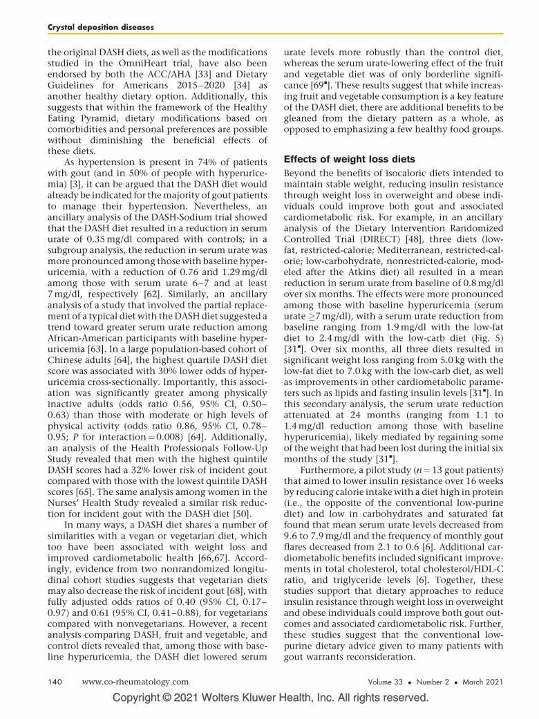
Crystal deposition diseases
the original DASH diets, as well as the modificationsstudied in the OmniHeart trial, have also beenendorsed by both the ACC/AHA [33] and DietaryGuidelines for Americans 2015–2020 [34] asanother healthy dietary option. Additionally, thissuggests that within the framework of the HealthyEating Pyramid, dietary modifications based oncomorbidities and personal preferences are possiblewithout diminishing the beneficial effects ofthese diets.
As hypertension is present in 74% of patientswith gout (and in 50% of people with hyperurice-mia) [3], it can be argued that the DASH diet wouldalready be indicated for the majority of gout patientsto manage their hypertension. Nevertheless, anancillary analysis of the DASH-Sodium trial showedthat the DASH diet resulted in a reduction in serumurate of 0.35 mg/dl compared with controls; in asubgroup analysis, the reduction in serum urate wasmore pronounced among those with baseline hyper-uricemia, with a reduction of 0.76 and 1.29 mg/dlamong those with serum urate 6–7 and at least7 mg/dl, respectively [62]. Similarly, an ancillaryanalysis of a study that involved the partial replace-ment of a typical diet with the DASH diet suggested atrend toward greater serum urate reduction amongAfrican-American participants with baseline hyper-uricemia [63]. In a large population-based cohort ofChinese adults [64], the highest quartile DASH dietscore was associated with 30% lower odds of hyper-uricemia cross-sectionally. Importantly, this associ-ation was significantly greater among physicallyinactive adults (odds ratio 0.56, 95% CI, 0.50–0.63) than those with moderate or high levels ofphysical activity (odds ratio 0.86, 95% CI, 0.78–0.95; P for interaction¼0.008) [64]. Additionally,an analysis of the Health Professionals Follow-UpStudy revealed that men with the highest quintileDASH scores had a 32% lower risk of incident goutcompared with those with the lowest quintile DASHscores [65]. The same analysis among women in theNurses’ Health Study revealed a similar risk reduc-tion for incident gout with the DASH diet [50].
In many ways, a DASH diet shares a number ofsimilarities with a vegan or vegetarian diet, whichtoo have been associated with weight loss andimproved cardiometabolic health [66,67]. Accord-ingly, evidence from two nonrandomized longitu-dinal cohort studies suggests that vegetarian dietsmay also decrease the risk of incident gout [68], withfully adjusted odds ratios of 0.40 (95% CI, 0.17–0.97) and 0.61 (95% CI, 0.41–0.88), for vegetarianscompared with nonvegetarians. However, a recentanalysis comparing DASH, fruit and vegetable, andcontrol diets revealed that, among those with base-line hyperuricemia, the DASH diet lowered serum
Copyright © 2021 Wolters Kluwer H
140 www.co-rheumatology.com
urate levels more robustly than the control diet,whereas the serum urate-lowering effect of the fruitand vegetable diet was of only borderline signifi-cance [69
&
]. These results suggest that while increas-ing fruit and vegetable consumption is a key featureof the DASH diet, there are additional benefits to begleaned from the dietary pattern as a whole, asopposed to emphasizing a few healthy food groups.
Effects of weight loss diets
Beyond the benefits of isocaloric diets intended tomaintain stable weight, reducing insulin resistancethrough weight loss in overweight and obese indi-viduals could improve both gout and associatedcardiometabolic risk. For example, in an ancillaryanalysis of the Dietary Intervention RandomizedControlled Trial (DIRECT) [48], three diets (low-fat, restricted-calorie; Mediterranean, restricted-cal-orie; low-carbohydrate, nonrestricted-calorie, mod-eled after the Atkins diet) all resulted in a meanreduction in serum urate from baseline of 0.8 mg/dlover six months. The effects were more pronouncedamong those with baseline hyperuricemia (serumurate �7 mg/dl), with a serum urate reduction frombaseline ranging from 1.9 mg/dl with the low-fatdiet to 2.4 mg/dl with the low-carb diet (Fig. 5)[31
&
]. Over six months, all three diets resulted insignificant weight loss ranging from 5.0 kg with thelow-fat diet to 7.0 kg with the low-carb diet, as wellas improvements in other cardiometabolic parame-ters such as lipids and fasting insulin levels [31
&
]. Inthis secondary analysis, the serum urate reductionattenuated at 24 months (ranging from 1.1 to1.4 mg/dl reduction among those with baselinehyperuricemia), likely mediated by regaining someof the weight that had been lost during the initial sixmonths of the study [31
&
].Furthermore, a pilot study (n¼13 gout patients)
that aimed to lower insulin resistance over 16 weeksby reducing calorie intake with a diet high in protein(i.e., the opposite of the conventional low-purinediet) and low in carbohydrates and saturated fatfound that mean serum urate levels decreased from9.6 to 7.9 mg/dl and the frequency of monthly goutflares decreased from 2.1 to 0.6 [6]. Additional car-diometabolic benefits included significant improve-ments in total cholesterol, total cholesterol/HDL-Cratio, and triglyceride levels [6]. Together, thesestudies support that dietary approaches to reduceinsulin resistance through weight loss in overweightand obese individuals could improve both gout out-comes and associated cardiometabolic risk. Further,these studies suggest that the conventional low-purine dietary advice given to many patients withgout warrants reconsideration.
ealth, Inc. All rights reserved.
Volume 33 � Number 2 � March 2021
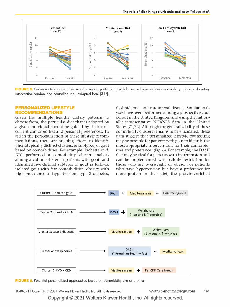
FIGURE 5. Serum urate change at six months among participants with baseline hyperuricemia in ancillary analysis of dietaryintervention randomized controlled trial. Adapted from [31&].
The role of diet in hyperuricemia and gout Yokose et al.
PERSONALIZED LIFESTYLERECOMMENDATIONSGiven the multiple healthy dietary patterns tochoose from, the particular diet that is adopted bya given individual should be guided by their con-current comorbidities and personal preferences. Toaid in the personalization of these lifestyle recom-mendations, there are ongoing efforts to identifyphenotypically distinct clusters, or subtypes, of goutbased on comorbidities. For example, Richette et al.[70] performed a comorbidity cluster analysisamong a cohort of French patients with gout, andidentified five distinct subtypes of gout as follows:isolated gout with few comorbidities, obesity withhigh prevalence of hypertension, type 2 diabetes,
Copyright © 2021 Wolters Kluwe
FIGURE 6. Potential personalized approaches based on comorb
1040-8711 Copyright � 2021 Wolters Kluwer Health, Inc. All rights rese
dyslipidemia, and cardiorenal disease. Similar anal-yses have been performed among a prospective goutcohort in the United Kingdom and using the nation-ally representative NHANES data in the UnitedStates [71,72]. Although the generalizability of thesecomorbidity clusters remains to be elucidated, thesedata suggest that personalized lifestyle counselingmay be possible for patients with gout to identify themost appropriate interventions for their comorbid-ities and preferences (Fig. 6). For example, the DASHdiet may be ideal for patients with hypertension andcan be implemented with calorie restriction forthose who are overweight or obese. For patientswho have hypertension but have a preference formore protein in their diet, the protein-enriched
r Health, Inc. All rights reserved.
idity cluster profiles.
rved. www.co-rheumatology.com 141
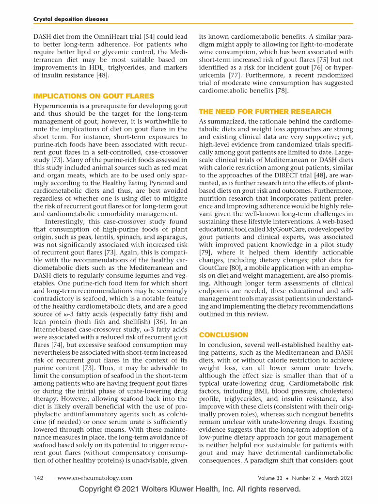
Crystal deposition diseases
DASH diet from the OmniHeart trial [54] could leadto better long-term adherence. For patients whorequire better lipid or glycemic control, the Medi-terranean diet may be most suitable based onimprovements in HDL, triglycerides, and markersof insulin resistance [48].
IMPLICATIONS ON GOUT FLARES
Hyperuricemia is a prerequisite for developing goutand thus should be the target for the long-termmanagement of gout; however, it is worthwhile tonote the implications of diet on gout flares in theshort term. For instance, short-term exposures topurine-rich foods have been associated with recur-rent gout flares in a self-controlled, case-crossoverstudy [73]. Many of the purine-rich foods assessed inthis study included animal sources such as red meatand organ meats, which are to be used only spar-ingly according to the Healthy Eating Pyramid andcardiometabolic diets and thus, are best avoidedregardless of whether one is using diet to mitigatethe risk of recurrent gout flares or for long-term goutand cardiometabolic comorbidity management.
Interestingly, this case-crossover study foundthat consumption of high-purine foods of plantorigin, such as peas, lentils, spinach, and asparagus,was not significantly associated with increased riskof recurrent gout flares [73]. Again, this is compati-ble with the recommendations of the healthy car-diometabolic diets such as the Mediterranean andDASH diets to regularly consume legumes and veg-etables. One purine-rich food item for which shortand long-term recommendations may be seeminglycontradictory is seafood, which is a notable featureof the healthy cardiometabolic diets, and are a goodsource of v-3 fatty acids (especially fatty fish) andlean protein (both fish and shellfish) [36]. In anInternet-based case-crossover study, v-3 fatty acidswere associated with a reduced risk of recurrent goutflares [74], but excessive seafood consumption maynevertheless be associated with short-term increasedrisk of recurrent gout flares in the context of itspurine content [73]. Thus, it may be advisable tolimit the consumption of seafood in the short-termamong patients who are having frequent gout flaresor during the initial phase of urate-lowering drugtherapy. However, allowing seafood back into thediet is likely overall beneficial with the use of pro-phylactic antiinflammatory agents such as colchi-cine (if needed) or once serum urate is sufficientlylowered through other means. With these mainte-nance measures in place, the long-term avoidance ofseafood based solely on its potential to trigger recur-rent gout flares (without compensatory consump-tion of other healthy proteins) is unadvisable, given
Copyright © 2021 Wolters Kluwer H
142 www.co-rheumatology.com
its known cardiometabolic benefits. A similar para-digm might apply to allowing for light-to-moderatewine consumption, which has been associated withshort-term increased risk of gout flares [75] but notidentified as a risk for incident gout [76] or hyper-uricemia [77]. Furthermore, a recent randomizedtrial of moderate wine consumption has suggestedcardiometabolic benefits [78].
THE NEED FOR FURTHER RESEARCH
As summarized, the rationale behind the cardiome-tabolic diets and weight loss approaches are strongand existing clinical data are very supportive; yet,high-level evidence from randomized trials specifi-cally among gout patients are limited to date. Large-scale clinical trials of Mediterranean or DASH dietswith calorie restriction among gout patients, similarto the approaches of the DIRECT trial [48], are war-ranted, as is further research into the effects of plant-based diets on gout risk and outcomes. Furthermore,nutrition research that incorporates patient prefer-ence and improving adherence would be highly rele-vant given the well-known long-term challenges insustaining these lifestyle interventions. A web-basededucational tool called MyGoutCare, codeveloped bygout patients and clinical experts, was associatedwith improved patient knowledge in a pilot study[79], where it helped them identify actionablechanges, including dietary changes; pilot data forGoutCare [80], a mobile application with an empha-sis on diet and weight management, are also promis-ing. Although longer term assessments of clinicalendpoints are needed, these educational and self-management tools may assist patients in understand-ing and implementing the dietary recommendationsoutlined in this review.
CONCLUSION
In conclusion, several well-established healthy eat-ing patterns, such as the Mediterranean and DASHdiets, with or without calorie restriction to achieveweight loss, can all lower serum urate levels,although the effect size is smaller than that of atypical urate-lowering drug. Cardiometabolic riskfactors, including BMI, blood pressure, cholesterolprofile, triglycerides, and insulin resistance, alsoimprove with these diets (consistent with their orig-inally proven roles), whereas such nongout benefitsremain unclear with urate-lowering drugs. Existingevidence suggests that the long-term adoption of alow-purine dietary approach for gout managementis neither helpful nor sustainable for patients withgout and may have detrimental cardiometabolicconsequences. A paradigm shift that considers gout
ealth, Inc. All rights reserved.
Volume 33 � Number 2 � March 2021
The role of diet in hyperuricemia and gout Yokose et al.
as a part of the metabolic syndrome and focuses oncomprehensive dietary patterns as opposed to sin-gular food items is necessary. By focusing our dietaryrecommendations on dietary patterns which havebeen shown to reduce cardiometabolic risk factors,the beneficial effects on gout endpoints shouldnaturally follow for the majority of typical goutcases, mediated through changes in insulin resis-tance. Although diet alone may not supplant theneed for urate-lowering therapy among patientswith gout, it is a powerful adjunctive tool to com-prehensively address the cardiometabolic burdenand premature mortality among patients with gout.
Acknowledgements
None.
Financial support and sponsorship
C.Y. is supported by T32 AR007258 and RheumatologyResearch Foundation Scientist Development Award.N.M. is supported by a Fellowship Award from theCanadian Institutes of Health Research. H.C. is sup-ported by R01 AR065944, P50 AR060772.
Conflicts of interest
C.Y. and N.M. have no conflicts of interest. H.K.C.reports consulting fees from Ironwood, Selecta, Horizon,Takeda, Kowa, Vaxart and research support from Iron-wood, Horizon.
REFERENCES AND RECOMMENDEDREADINGPapers of particular interest, published within the annual period of review, havebeen highlighted as:
& of special interest&& of outstanding interest1. Fam AG. Gout, diet, and the insulin resistance syndrome. J Rheumatol 2002;29:1350–1355.
2. Choi HK, Mount DB, Reginato AM, et al. Pathogenesis of gout. Ann InternMed 2005; 143:499–516.
3. Zhu Y, Pandya BJ, Choi HK. Comorbidities of gout and hyperuricemia in theUS general population: NHANES. Am J Med 2012; 125:679–687.e1.
4. Juraschek SP, Kovell LC, Miller ER 3rd, Gelber AC. Association of kidneydisease with prevalent gout in the United States in 1998–1994 and 2007–2010. Semin Arthritis Rheum 2013; 42:551–561.
5. Juraschek SP, Kovell LC, Miller ER, Gelber AC. Dose-response association ofuncontrolled blood pressure and cardiovascular disease risk factors withhyperuricemia and gout. PLoS One 2013; 8:e56546.
6. Dessein PH, Shipton EA, Stanwix AE, et al. Beneficial effects of weight lossassociated with moderate calorie/carbohydrate restriction, and increasedproportional intake of protein and unsaturated fat on serum urate andlipoprotein levels in gout: a pilot study. Ann Rheum Dis 2000; 59:539–543.
7. Ter Maaten JC, Voorburg A, Heine RJ, et al. Renal handling of urate andsodium during acute physiological hyperinsulinaemia in healthy subjects. ClinSci (Lond) 1997; 92:51–58.
8. Muscelli E, Natali A, Bianchi S, et al. Effect of insulin on renal sodium and uricacid handling in essential hypertension. Am J Hypertens 1996; 9:746–752.
9. Facchini F, Chen YD, Hollenbeck CB, Reaven GM. Relationship betweenresistance to insulin-mediated glucose uptake, urinary uric acid clearance,and plasma uric acid concentration. JAMA 1991; 266:3008–3011.
10. Choi HK, Ford ES, Li C, Curhan G. Prevalence of the metabolic syndrome inpatients with gout: the Third National Health and Nutrition ExaminationSurvey. Arthritis Rheum 2007; 57:109–115.
Copyright © 2021 Wolters Kluwe
1040-8711 Copyright � 2021 Wolters Kluwer Health, Inc. All rights rese
11. Ford ES, Giles WH, Dietz WH. Prevalence of the metabolic syndrome amongUS adults: findings from the third National Health and Nutrition ExaminationSurvey. JAMA 2002; 287:356–359.
12. Choi HK, Ford ES. Prevalence of the metabolic syndrome in individuals withhyperuricemia. Am J Med 2007; 120:442–447.
13. Choi HK, De Vera MA, Krishnan E. Gout and the risk of type 2 diabetes amongmen with a high cardiovascular risk profile. Rheumatology (Oxford) 2008;47:1567–1570.
14. Baker JF, Schumacher HR, Krishnan E. Serum uric acid level and risk forperipheral arterial disease: analysis of data from the multiple risk factorintervention trial. Angiology 2007; 58:450–457.
15. Abbott RD, Brand FN, Kannel WB, Castelli WP. Gout and coronary heartdisease: the Framingham Study. J Clin Epidemiol 1988; 41:237–242.
16. Krishnan E, Baker JF, Furst DE, Schumacher HR. Gout and the risk of acutemyocardial infarction. Arthritis Rheum 2006; 54:2688–2696.
17. Krishnan E, Svendsen K, Neaton JD, et al. Long-term cardiovascularmortality among middle-aged men with gout. Arch Intern Med 2008;168:1104–1110.
18. Choi HK, Curhan G. Independent impact of gout on mortality and risk forcoronary heart disease. Circulation 2007; 116:894–900.
19. Fisher MC, Rai SK, Lu N, et al. The unclosing premature mortality gap in gout:a general population-based study. Ann Rheum Dis 2017; 76:1289–1294.
20. Zhang Y, Lu N, Peloquin C, et al. Improved survival in rheumatoid arthritis: ageneral population-based cohort study. Ann Rheum Dis 2017; 76:408–413.
21.&&
Safiri S, Kolahi AA, Cross M, et al. Prevalence, incidence, and years lived withdisability due to gout and its attributable risk factors for 195 countries andterritories 1990–2017: a systematic analysis of the global burden of diseasestudy. Arthritis Rheumatol 2020; 72:1916–1927.
An analysis of the Global Burden of Disease Study which found increasing burdenof gout worldwide and identified BMI as the major modifiable risk factor thataccounted for the largest proportion of years lived with disability due to gout.22.&&
Xia Y, Wu Q, Wang H, et al. Global, regional and national burden of gout,1990–2017: a systematic analysis of the Global Burden of Disease Study.Rheumatology (Oxford) 2020; 59:1529–1538.
An analysis of the Global Burden of Disease Study which found an increasingburden of gout worldwide, especially among women.23. Zhu Y, Pandya BJ, Choi HK. Prevalence of gout and hyperuricemia in the US
general population: the National Health and Nutrition Examination Survey.Arthritis Rheum 2011; 63:3136–3141.
24. Young LR, Nestle M. The contribution of expanding portion sizes to the USobesity epidemic. Am J Public Health 2002; 92:246–249.
25. Collaboration NCDRF. Worldwide trends in body-mass index, underweight,overweight, and obesity from 1975 to 2016: a pooled analysis of 2416population-based measurement studies in 128.9 million children, adoles-cents, and adults. Lancet 2017; 390:2627–2642.
26. Wang YC, McPherson K, Marsh T, et al. Health and economic burden of theprojected obesity trends in the USA and the UK. Lancet 2011;378:815–825.
27. Hu F. Genetic predictors of obesity. In: Hu F, editor. Obesity epidemiology.New York City: Oxford University Press; 2008.
28.&&
Choi HK, McCormick N, Lu N, et al. Population impact attributable tomodifiable risk factors for hyperuricemia. Arthritis Rheumatol 2020;72:157–165.
Analysis of the National Health and Nutrition Examination Survey which found thatfour modifiable risk factors (BMI, DASH diet, alcohol use, and diuretic use)accounted for a significant proportion of hyperuricemia cases in the US generalpopulation.29. Du Y, Liu B, Sun Y, et al. Trends in adherence to the physical activity
guidelines for Americans for Aerobic Activity and time spent on sedentarybehavior among US adults, 2007 to 2016. JAMA Netw Open 2019;2:e197597.
30. Yang L, Cao C, Kantor ED, et al. Trends in sedentary behavior among the USpopulation. JAMA 2019; 321:1587–1597.
31.&
Yokose C, McCormick N, Rai SK, et al. Effects of low-fat, Mediterranean, orlow-carbohydrate weight loss diets on serum urate and cardiometabolic riskfactors: a secondary analysis of the Dietary Intervention Randomized Con-trolled Trial (DIRECT). Diabetes Care 2020; 43:2812–2820.
A secondary analysis of a dietary interventional trial which found that low-fat,Mediterranean, and low-carbohydrate diets could all similarly reduce serum urateas well as cardiometabolic risk factors, especially among those with baselinehyperuricemia.32. Emmerson B. Hyperlipidaemia in hyperuricaemia and gout. Ann Rheum Dis
1998; 57:509–510.33. Eckel RH, Jakicic JM, Ard JD, et al. 2013 AHA/ACC guideline on lifestyle
management to reduce cardiovascular risk: a report of the American Collegeof Cardiology/American Heart Association Task Force on Practice Guide-lines. J Am Coll Cardiol 2014; 63:2960–2984.
34. Jang WC, Nam YH, Park SM, et al. T6092C polymorphism of SLC22A12gene is associated with serum uric acid concentrations in Korean malesubjects. Clin Chim Acta 2008; 398:140–144.
35. Mellen PB, Gao SK, Vitolins MZ, Goff DC Jr. Deteriorating dietary habitsamong adults with hypertension: DASH dietary accordance, NHANES1988–1994 and 1999–2004. Arch Intern Med 2008; 168:308–314.
r Health, Inc. All rights reserved.
rved. www.co-rheumatology.com 143
Crystal deposition diseases
36. U.S. Department of Health and Human Services, U.S. Department of Agri-culture. 2015–2020 dietary guidelines for Americans, 8th ed.
37. Hu FB. The Mediterranean diet and mortality: olive oil and beyond. N Engl JMed 2003; 348:2595–2596.
38. Estruch R, Ros E, Salas-Salvado J, et al. Primary prevention of cardiovasculardisease with a Mediterranean diet supplemented with extra-virgin olive oil ornuts. N Engl J Med 2018; 378:e34.
39. Fung TT, Rexrode KM, Mantzoros CS, et al. Mediterranean diet and incidenceof and mortality from coronary heart disease and stroke in women. Circulation2009; 119:1093–1100.
40. Filippatos TD, Panagiotakos DB, Georgousopoulou EN, et al. Mediterraneandiet and 10-year (2002–2012) incidence of diabetes and cardiovasculardisease in participants with prediabetes: the ATTICA study. Rev Diabet Stud2016; 13:226–235.
41. Buckland G, Gonzalez CA, Agudo A, et al. Adherence to the Mediterraneandiet and risk of coronary heart disease in the Spanish EPIC Cohort Study. AmJ Epidemiol 2009; 170:1518–1529.
42. Trichopoulou A, Costacou T, Bamia C, Trichopoulos D. Adherence to aMediterranean diet and survival in a Greek population. N Engl J Med2003; 348:2599–2608.
43. Sotos-Prieto M, Bhupathiraju SN, Mattei J, et al. Changes in diet qualityscores and risk of cardiovascular disease among US men and women.Circulation 2015; 132:2212–2219.
44. Sotos-Prieto M, Bhupathiraju SN, Mattei J, et al. Association of changes in dietquality with total and cause-specific mortality. N Engl J Med 2017;377:143–153.
45. de Lorgeril M, Renaud S, Mamelle N, et al. Mediterranean alpha-linolenic acid-rich diet in secondary prevention of coronary heart disease. Lancet 1994;343:1454–1459.
46. Salas-Salvado J, Bullo M, Babio N, et al. Reduction in the incidence of type 2diabetes with the Mediterranean diet: results of the PREDIMED-Reus nutritionintervention randomized trial. Diabetes Care 2011; 34:14–19.
47. de Koning L, Chiuve SE, Fung TT, et al. Diet-quality scores and the risk of type2 diabetes in men. Diabetes Care 2011; 34:1150–1156.
48. Shai I, Schwarzfuchs D, Henkin Y, et al. Weight loss with a low-carbohydrate,Mediterranean, or low-fat diet. N Engl J Med 2008; 359:229–241.
49. Guasch-Ferre M, Bullo M, Babio N, et al. Mediterranean diet and risk ofhyperuricemia in elderly participants at high cardiovascular risk. J Gerontol ABiol Sci Med Sci 2013; 68:1263–1270.
50. Keller S, Lu RS, Zhang L, et al. The Dietary Approaches to Stop Hypertension(DASH) and Mediterranean diets and risk of gout in women: 28-year follow-upof a prospective cohort [abstract]. Arthritis Rheumatol 2017; 69(Suppl. 10).
51. Sacks FM, Campos H. Dietary therapy in hypertension. N Engl J Med 2010;362:2102–2112.
52. Appel LJ, Moore TJ, Obarzanek E, et al. A clinical trial of the effects of dietarypatterns on blood pressure. DASH Collaborative Research Group. N Engl JMed 1997; 336:1117–1124.
53. Sacks FM, Svetkey LP, Vollmer WM, et al. Effects on blood pressure ofreduced dietary sodium and the Dietary Approaches to Stop Hypertension(DASH) diet. DASH-Sodium Collaborative Research Group. N Engl J Med2001; 344:3–10.
54. Appel LJ, Sacks FM, Carey VJ, et al. Effects of protein, monounsaturated fat,and carbohydrate intake on blood pressure and serum lipids: results of theOmniHeart randomized trial. JAMA 2005; 294:2455–2464.
55. Belanger MW, Mukamal K, Miller E, et al. The effects of dietary macronutrientson serum urate: a secondary analysis of the OmniHeart trial [abstract].Arthritis Rheumatol 2020; 72(Suppl. 10). https://acrabstracts.org/abstract/the-effects-of-dietary-macronutrients-on-serum-urate-a-secondary-analysis-of-the-omniheart-trial/ (accessed November 18, 2020).
56. Djousse L, Ho YL, Nguyen XT, et al. DASH score and subsequent risk ofcoronary artery disease: the findings from Million Veteran Program. J Am HeartAssoc 2018; 7:e008089. doi: 10.1161/JAHA.117.008089.
57. Fung TT, Chiuve SE, McCullough ML, et al. Adherence to a DASH-style dietand risk of coronary heart disease and stroke in women. Arch Intern Med2008; 168:713–720.
Copyright © 2021 Wolters Kluwer H
144 www.co-rheumatology.com
58. Liese AD, Nichols M, Sun X, et al. Adherence to the DASH Diet is inverselyassociated with incidence of type 2 diabetes: the insulin resistance athero-sclerosis study. Diabetes Care 2009; 32:1434–1436.
59. Mokhtari Z, Sharafkhah M, Poustchi H, et al. Adherence to the dietaryapproaches to stop hypertension (DASH) diet and risk of total and cause-specific mortality: results from the Golestan Cohort Study. Int J Epidemiol2019; 48:1824–1838.
60. Parikh A, Lipsitz SR, Natarajan S. Association between a DASH-like diet andmortality in adults with hypertension: findings from a population-based follow-up study. Am J Hypertens 2009; 22:409–416.
61. Park YM, Fung TT, Steck SE, et al. Diet quality and mortality risk in metabo-lically obese normal-weight adults. Mayo Clin Proc 2016; 91:1372–1383.
62. Juraschek SP, Gelber AC, Choi HK, et al. Effects of the dietary approaches tostop hypertension (DASH) diet and sodium intake on serum uric acid. ArthritisRheumatol 2016; 68:3002–3009.
63. Juraschek SP, White K, Tang O, et al. Effects of a dietary approach to stophypertension (DASH) diet intervention on serum uric acid in African Amer-icans with hypertension. Arthritis Care Res (Hoboken) 2018;70:1509–1516.
64. Gao Y, Cui LF, Sun YY, et al. Adherence to the dietary approaches to stophypertension (DASH) diet and hyperuricemia: a cross-sectional study. Ar-thritis Care Res (Hoboken) 2020; doi: 10.1002/acr.24150. [Online ahead ofprint]
65. Rai SK, Fung TT, Lu N, et al. The dietary approaches to stop hypertension(DASH) diet, Western diet, and risk of gout in men: prospective cohort study.BMJ 2017; 357:j1794.
66. Tran E, Dale HF, Jensen C, Lied GA. Effects of plant-based diets on weight status:a systematic review. Diabetes Metab Syndr Obes 2020; 13:3433–3448.
67. Satija A, Hu FB. Plant-based diets and cardiovascular health. Trends Cardi-ovasc Med 2018; 28:437–441.
68. Chiu TH, Liu CH, Chang CC, et al. Vegetarian diet and risk of gout in twoseparate prospective cohort studies. Clin Nutr 2020; 39:837–844.
69.&
Juraschek SP, Yokose C, McCormick N, et al. Effects of dietary patterns onserum urate: results from the DASH randomized trial. Arthritis Rheum 2020.
Secondary analysis of a DASH feeding study which found that DASH diet couldreduce serum urate, particularly among those with baseline hyperuricemia.70. Richette P, Clerson P, Perissin L, et al. Revisiting comorbidities in gout: a
cluster analysis. Ann Rheum Dis 2015; 74:142–147.71. Bevis M, Blagojevic-Bucknall M, Mallen C, et al. Comorbidity clusters in
people with gout: an observational cohort study with linked medical recordreview. Rheumatology (Oxford) 2018; 57:1358–1363.
72. Yokose C, Chen-Xu LL, Zhang M, et al. Comorbidity patterns in gout using theUS general population: cluster analysis of the National Health and NutritionExamination Survey. Ann Rheum Dis 2019; 78:A1294.
73. Zhang Y, Chen C, Choi H, et al. Purine-rich foods intake and recurrent goutattacks. Ann Rheum Dis 2012; 71:1448–1453.
74. Zhang M, Zhang Y, Terkeltaub R, et al. Effect of dietary and supplementalomega-3 polyunsaturated fatty acids on risk of recurrent gout flares. ArthritisRheumatol 2019; 71:1580–1586.
75. Neogi T, Chen C, Niu J, et al. Alcohol quantity and type on risk of recurrentgout attacks: an internet-based case-crossover study. Am J Med 2014;127:311–318.
76. Choi HK, Atkinson K, Karlson EW, et al. Alcohol intake and risk of incidentgout in men: a prospective study. Lancet 2004; 363:1277–1281.
77. Choi HK, Curhan G. Beer, liquor, and wine consumption and serum uric acidlevel: the Third National Health and Nutrition Examination Survey. ArthritisRheum 2004; 51:1023–1029.
78. Gepner Y, Golan R, Harman-Boehm I, et al. Effects of initiating moderatealcohol intake on cardiometabolic risk in adults with type 2 diabetes: a 2-yearrandomized, controlled trial. Ann Intern Med 2015; 163:569–579.
79. Khanna P, Berrocal V, An L, Khanna D. Development and pilot testing ofMyGoutCare: a novel web-based platform to educate patients with gout. JClin Rheumatol 2019; 26:320–326.
80. Kang SG, Lee EN. Development and evaluation of a self-managementapplication for patients with gout. Jpn J Nurs Sci 2020; 17:e12285.
ealth, Inc. All rights reserved.
Volume 33 � Number 2 � March 2021
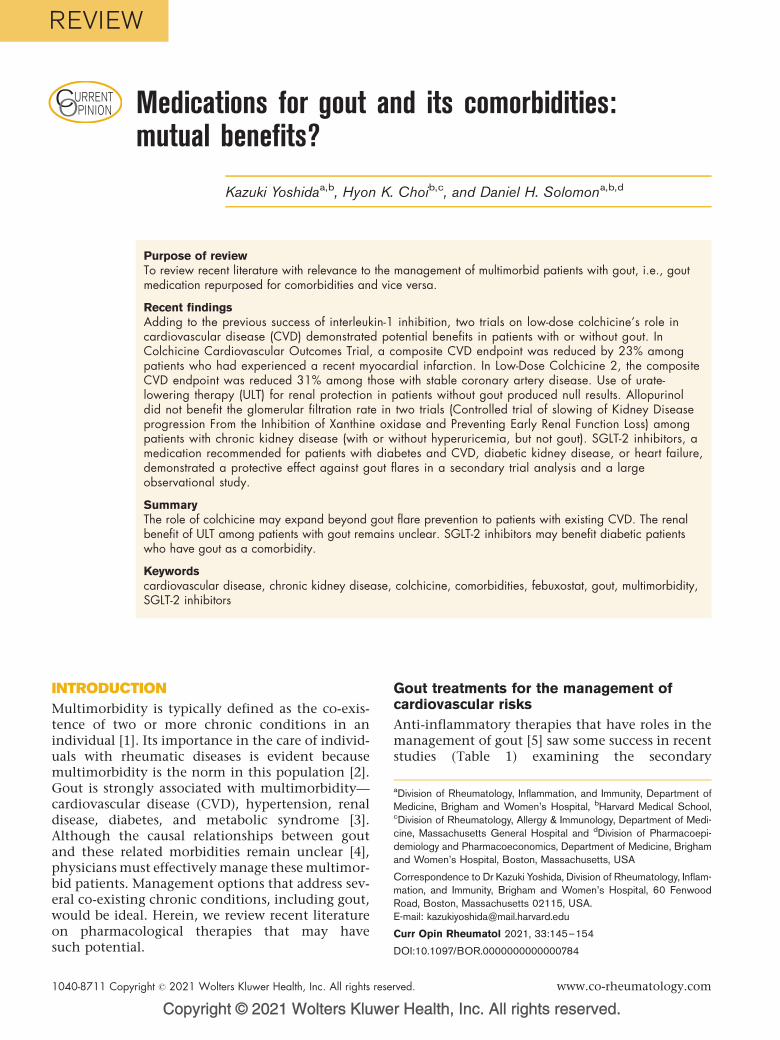
REVIEW
CURRENTOPINION Medications for gout and its comorbidities:
mutual benefits?
Copyrigh
1040-8711 Copyright � 2021 Wolte
a,b b,c a,b,d
Kazuki Yoshida , Hyon K. Choi , and Daniel H. SolomonPurpose of review
To review recent literature with relevance to the management of multimorbid patients with gout, i.e., goutmedication repurposed for comorbidities and vice versa.
Recent findings
Adding to the previous success of interleukin-1 inhibition, two trials on low-dose colchicine’s role incardiovascular disease (CVD) demonstrated potential benefits in patients with or without gout. InColchicine Cardiovascular Outcomes Trial, a composite CVD endpoint was reduced by 23% amongpatients who had experienced a recent myocardial infarction. In Low-Dose Colchicine 2, the compositeCVD endpoint was reduced 31% among those with stable coronary artery disease. Use of urate-lowering therapy (ULT) for renal protection in patients without gout produced null results. Allopurinoldid not benefit the glomerular filtration rate in two trials (Controlled trial of slowing of Kidney Diseaseprogression From the Inhibition of Xanthine oxidase and Preventing Early Renal Function Loss) amongpatients with chronic kidney disease (with or without hyperuricemia, but not gout). SGLT-2 inhibitors, amedication recommended for patients with diabetes and CVD, diabetic kidney disease, or heart failure,demonstrated a protective effect against gout flares in a secondary trial analysis and a largeobservational study.
Summary
The role of colchicine may expand beyond gout flare prevention to patients with existing CVD. The renalbenefit of ULT among patients with gout remains unclear. SGLT-2 inhibitors may benefit diabetic patientswho have gout as a comorbidity.
Keywords
cardiovascular disease, chronic kidney disease, colchicine, comorbidities, febuxostat, gout, multimorbidity,SGLT-2 inhibitors
aDivision of Rheumatology, Inflammation, and Immunity, Department ofMedicine, Brigham and Women’s Hospital, bHarvard Medical School,cDivision of Rheumatology, Allergy & Immunology, Department of Medi-cine, Massachusetts General Hospital and dDivision of Pharmacoepi-demiology and Pharmacoeconomics, Department of Medicine, Brighamand Women’s Hospital, Boston, Massachusetts, USA
Correspondence to Dr Kazuki Yoshida, Division of Rheumatology, Inflam-mation, and Immunity, Brigham and Women’s Hospital, 60 FenwoodRoad, Boston, Massachusetts 02115, USA.E-mail: [email protected]
Curr Opin Rheumatol 2021, 33:145–154
DOI:10.1097/BOR.0000000000000784
INTRODUCTION
Multimorbidity is typically defined as the co-exis-tence of two or more chronic conditions in anindividual [1]. Its importance in the care of individ-uals with rheumatic diseases is evident becausemultimorbidity is the norm in this population [2].Gout is strongly associated with multimorbidity—
cardiovascular disease (CVD), hypertension, renaldisease, diabetes, and metabolic syndrome [3].Although the causal relationships between goutand these related morbidities remain unclear [4],physicians must effectively manage these multimor-bid patients. Management options that address sev-eral co-existing chronic conditions, including gout,would be ideal. Herein, we review recent literatureon pharmacological therapies that may havesuch potential.
t © 2021 Wolters Kluwe
rs Kluwer Health, Inc. All rights rese
Gout treatments for the management ofcardiovascular risks
Anti-inflammatory therapies that have roles in themanagement of gout [5] saw some success in recentstudies (Table 1) examining the secondary
r Health, Inc. All rights reserved.
rved. www.co-rheumatology.com
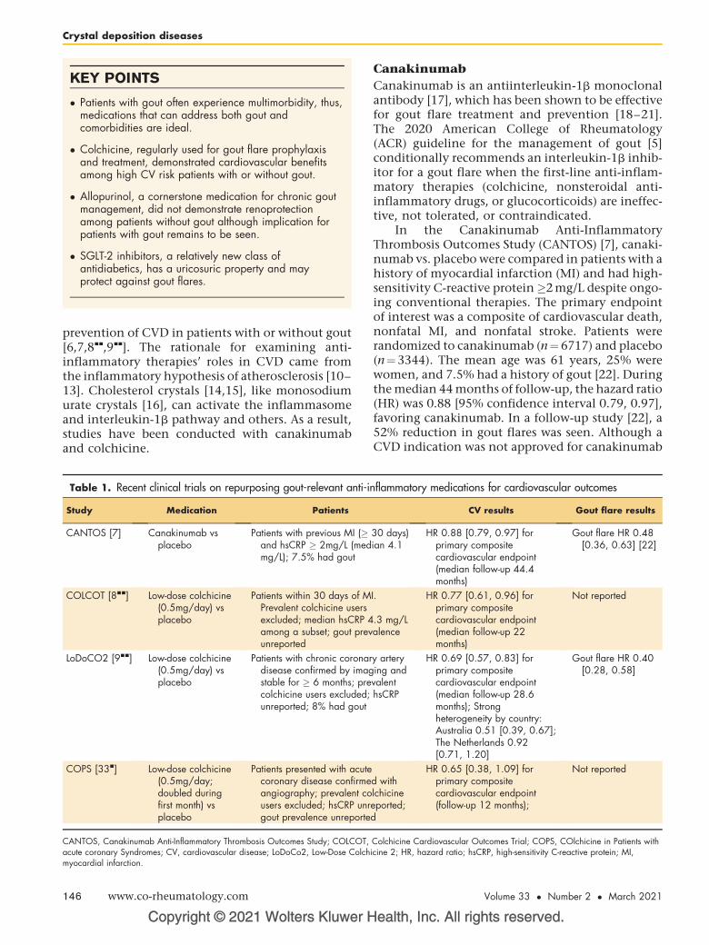
KEY POINTS
� Patients with gout often experience multimorbidity, thus,medications that can address both gout andcomorbidities are ideal.
� Colchicine, regularly used for gout flare prophylaxisand treatment, demonstrated cardiovascular benefitsamong high CV risk patients with or without gout.
� Allopurinol, a cornerstone medication for chronic goutmanagement, did not demonstrate renoprotectionamong patients without gout although implication forpatients with gout remains to be seen.
� SGLT-2 inhibitors, a relatively new class ofantidiabetics, has a uricosuric property and mayprotect against gout flares.
Crystal deposition diseases
prevention of CVD in patients with or without gout[6,7,8
&&
,9&&
]. The rationale for examining anti-inflammatory therapies’ roles in CVD came fromthe inflammatory hypothesis of atherosclerosis [10–13]. Cholesterol crystals [14,15], like monosodiumurate crystals [16], can activate the inflammasomeand interleukin-1b pathway and others. As a result,studies have been conducted with canakinumaband colchicine.
Copyright © 2021 Wolters Kluwer H
Table 1. Recent clinical trials on repurposing gout-relevant anti-in
Study Medication Patients
CANTOS [7] Canakinumab vsplacebo
Patients with previous MI (�and hsCRP � 2mg/L (medmg/L); 7.5% had gout
COLCOT [8&&] Low-dose colchicine(0.5mg/day) vsplacebo
Patients within 30 days of MPrevalent colchicine usersexcluded; median hsCRP 4among a subset; gout prevunreported
LoDoCO2 [9&&] Low-dose colchicine(0.5mg/day) vsplacebo
Patients with chronic coronardisease confirmed by imagstable for � 6 months; precolchicine users excluded;unreported; 8% had gout
COPS [33&] Low-dose colchicine(0.5mg/day;doubled duringfirst month) vsplacebo
Patients presented with acutecoronary disease confirmeangiography; prevalent cousers excluded; hsCRP unrgout prevalence unreporte
CANTOS, Canakinumab Anti-Inflammatory Thrombosis Outcomes Study; COLCOT,acute coronary Syndromes; CV, cardiovascular disease; LoDoCo2, Low-Dose Colchmyocardial infarction.
146 www.co-rheumatology.com
Canakinumab
Canakinumab is an antiinterleukin-1b monoclonalantibody [17], which has been shown to be effectivefor gout flare treatment and prevention [18–21].The 2020 American College of Rheumatology(ACR) guideline for the management of gout [5]conditionally recommends an interleukin-1b inhib-itor for a gout flare when the first-line anti-inflam-matory therapies (colchicine, nonsteroidal anti-inflammatory drugs, or glucocorticoids) are ineffec-tive, not tolerated, or contraindicated.
In the Canakinumab Anti-InflammatoryThrombosis Outcomes Study (CANTOS) [7], canaki-numab vs. placebo were compared in patients with ahistory of myocardial infarction (MI) and had high-sensitivity C-reactive protein�2 mg/L despite ongo-ing conventional therapies. The primary endpointof interest was a composite of cardiovascular death,nonfatal MI, and nonfatal stroke. Patients wererandomized to canakinumab (n¼6717) and placebo(n¼3344). The mean age was 61 years, 25% werewomen, and 7.5% had a history of gout [22]. Duringthe median 44 months of follow-up, the hazard ratio(HR) was 0.88 [95% confidence interval 0.79, 0.97],favoring canakinumab. In a follow-up study [22], a52% reduction in gout flares was seen. Although aCVD indication was not approved for canakinumab
ealth, Inc. All rights reserved.
flammatory medications for cardiovascular outcomes
CV results Gout flare results
30 days)ian 4.1
HR 0.88 [0.79, 0.97] forprimary compositecardiovascular endpoint(median follow-up 44.4months)
Gout flare HR 0.48[0.36, 0.63] [22]
I.
.3 mg/Lalence
HR 0.77 [0.61, 0.96] forprimary compositecardiovascular endpoint(median follow-up 22months)
Not reported
y arterying andvalenthsCRP
HR 0.69 [0.57, 0.83] forprimary compositecardiovascular endpoint(median follow-up 28.6months); Strongheterogeneity by country:Australia 0.51 [0.39, 0.67];The Netherlands 0.92[0.71, 1.20]
Gout flare HR 0.40[0.28, 0.58]
d withlchicineeported;d
HR 0.65 [0.38, 1.09] forprimary compositecardiovascular endpoint(follow-up 12 months);
Not reported
Colchicine Cardiovascular Outcomes Trial; COPS, COlchicine in Patients withicine 2; HR, hazard ratio; hsCRP, high-sensitivity C-reactive protein; MI,
Volume 33 � Number 2 � March 2021
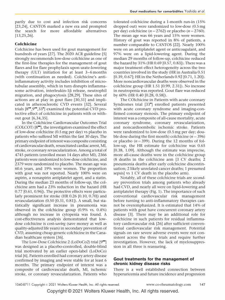
Gout medications for comorbidities Yoshida et al.
partly due to cost and infection risk concerns[23,24], CANTOS marked a new era and promptedthe search for more affordable alternatives[13,25,26].
Colchicine
Colchicine has been used for gout management forhundreds of years [27]. The 2020 ACR guideline [5]strongly recommends low-dose colchicine as one ofthe first-line therapies for the management of goutflares and for flare prophylaxis upon urate-loweringtherapy (ULT) initiation for at least 3–6 months(with continuation as needed). Colchicine’s anti-inflammatory activity includes inhibition of micro-tubular assembly, which in turn disrupts inflamma-some activation, interleukin-1b release, neutrophilmigration, and phagocytosis [28,29]. These sites ofactions are at play in gout flare [30,31] and impli-cated in atherosclerotic CVD events [32]. Severaltrials [8
&&
,9&&
,33&
] examined the potential CVD pro-tective effect of colchicine in patients with or with-out gout [6,34,35].
In the Colchicine Cardiovascular Outcomes Trial(COLCOT) [8
&&
], the investigators examined the effectof low-dose colchicine (0.5mg per day) vs placebo inpatients who suffered MI within the last 30 days. Theprimary endpointof interestwas composite consistingof cardiovasculardeath, resuscitated cardiac arrest, MI,stroke, or coronary revascularization. Among a total of4745 patients (enrolled mean 14 days after MI), 2366patients were randomized to low-dose colchicine, and2379 were randomized to placebo. The mean age was60.6 years, and 19% were women. The proportionwith gout was not reported. Nearly 100% were onaspirin, a nonaspirin antiplatelet agent, and a statin.During the median 23 months of follow-up, the col-chicine arm had a 23% reduction in the hazard (HR0.77 [0.61, 0.96]). The protective effects were particu-larly prominent for stroke (HR 0.26 [0.10, 0.70]) andrevascularization (0.50 [0.31, 0.81]). A small, but sta-tistically significant increase in pneumonia wasobserved in the colchicine group (0.9% vs. 0.4%)although no increase in cytopenia was found. Acost-effectiveness analysis demonstrated that low-dose colchicine is cost-saving (save money as well asquality-adjusted life years) in secondary prevention ofCVD, assuming cheap generic colchicine in the Cana-dian healthcare system [36].
The Low-Dose Colchicine 2 (LoDoCo2) trial [9&&
]was designed as a placebo-controlled, double-blindtrial motivated by an earlier open-label (LoDoCo)trial [6]. Patients enrolled had coronary artery diseaseconfirmed by imaging and were stable for at least 6months. The primary endpoint of interest was acomposite of cardiovascular death, MI, ischemicstroke, or coronary revascularization. Patients who
Copyright © 2021 Wolters Kluwe
1040-8711 Copyright � 2021 Wolters Kluwer Health, Inc. All rights rese
tolerated colchicine during a 1-month run-in (15%dropped out) were randomized to low-dose (0.5 mgper day) colchicine (n¼2762) or placebo (n¼2760).The mean age was 66 years and 15% were women.History of gout was reported in 8% of patients, anumber comparable to CANTOS [22]. Nearly 100%were on an antiplatelet agent or anticoagulant, and97% were on a lipid-lowering agent. During themedian 29 months of follow-up, colchicine reducedthe hazard by 31% (HR 0.69 [0.57, 0.83]). There was amajor treatment effect heterogeneity across the twocountries involved in the study (HR in Australia 0.51[0.39, 0.67]; HR in the Netherlands 0.92 [0.71, 1.20]).More noncardiovascular deaths were observed in thecolchicine group (HR 1.51 [0.99, 2.31]). No increasein neutropenia was reported. Gout flare was reducedby 60% (HR 0.40 [0.28, 0.58]).
The COlchicine in Patients with acute coronarySyndromes trial [33
&
] enrolled patients presentedwith acute coronary syndrome and imaging-con-firmed coronary stenosis. The primary endpoint ofinterest was a composite of all-cause mortality, acutecoronary syndrome, coronary revascularization,and noncardioembolic ischemic stroke. Patientswere randomized to low-dose (0.5 mg per day; dou-ble dose during the first month) colchicine (n¼396)or placebo (n¼399). During the 12 months of fol-low-up, the HR estimate for colchicine was 0.65[0.38, 1.09]. Although the estimate was imprecise,more all-cause deaths were in the colchicine group(8 deaths in the colchicine arm [3 CV deaths; 2pneumonia deaths after early colchicine discontin-uation; 2 likely unrelated cancer deaths; 1 presumedsepsis] vs 1 CV death in the placebo arm).
Notably, all of these colchicine trials are second-ary prevention trials among patients who alreadyhad CVD, and nearly all were on lipid-lowering andantiplatelet therapy (Fig. 1). The importance of suchconventional cardiovascular risk managementbefore turning to anti-inflammatory therapies can-not be overemphasized. It is estimated that 14% ofpatients with gout have concurrent coronary arterydisease [3]. There may be an additional role forcolchicine in such patients for residual inflamma-tory cardiovascular risk [26] after sufficient conven-tional cardiovascular risk management. Potentialsignals on rare severe adverse events were not con-sistent across the three trials and require furtherinvestigation. However, the lack of myelosuppres-sion in all three is reassuring.
Gout treatments for the management ofchronic kidney disease risks
There is a well established connection betweenhyperuricemia and future incidence and progression
r Health, Inc. All rights reserved.
rved. www.co-rheumatology.com 147
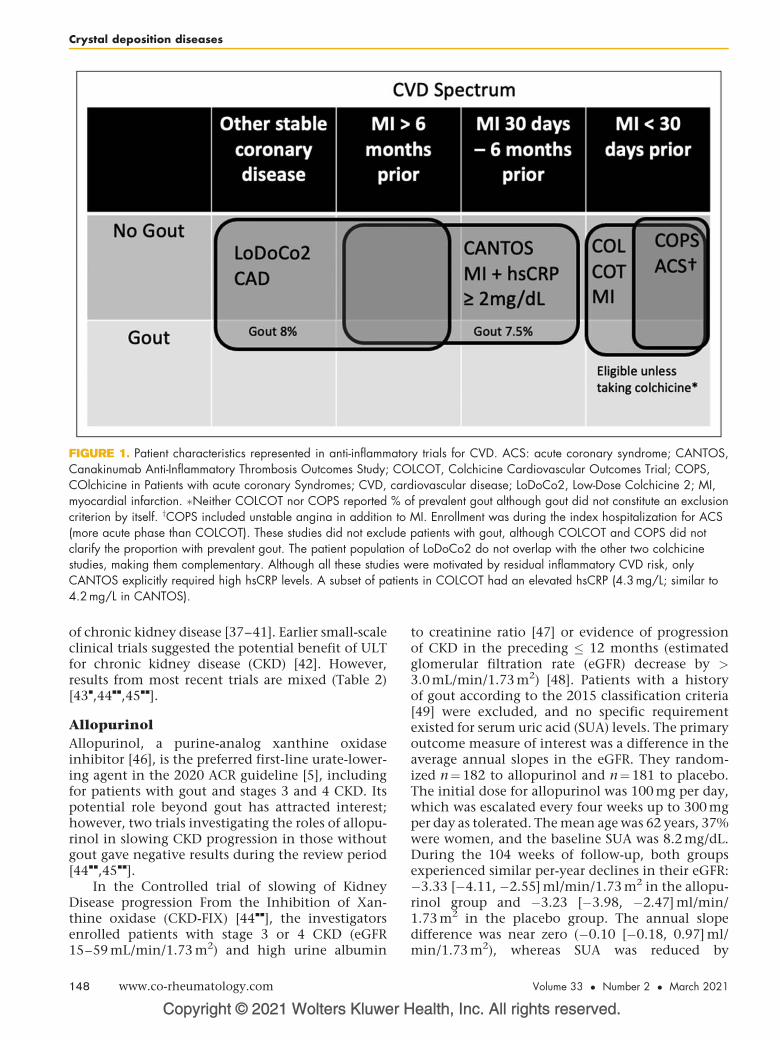
FIGURE 1. Patient characteristics represented in anti-inflammatory trials for CVD. ACS: acute coronary syndrome; CANTOS,Canakinumab Anti-Inflammatory Thrombosis Outcomes Study; COLCOT, Colchicine Cardiovascular Outcomes Trial; COPS,COlchicine in Patients with acute coronary Syndromes; CVD, cardiovascular disease; LoDoCo2, Low-Dose Colchicine 2; MI,myocardial infarction. �Neither COLCOT nor COPS reported % of prevalent gout although gout did not constitute an exclusioncriterion by itself. yCOPS included unstable angina in addition to MI. Enrollment was during the index hospitalization for ACS(more acute phase than COLCOT). These studies did not exclude patients with gout, although COLCOT and COPS did notclarify the proportion with prevalent gout. The patient population of LoDoCo2 do not overlap with the other two colchicinestudies, making them complementary. Although all these studies were motivated by residual inflammatory CVD risk, onlyCANTOS explicitly required high hsCRP levels. A subset of patients in COLCOT had an elevated hsCRP (4.3 mg/L; similar to4.2 mg/L in CANTOS).
Crystal deposition diseases
of chronic kidney disease [37–41]. Earlier small-scaleclinical trials suggested the potential benefit of ULTfor chronic kidney disease (CKD) [42]. However,results from most recent trials are mixed (Table 2)[43
&
,44&&
,45&&
].
Allopurinol
Allopurinol, a purine-analog xanthine oxidaseinhibitor [46], is the preferred first-line urate-lower-ing agent in the 2020 ACR guideline [5], includingfor patients with gout and stages 3 and 4 CKD. Itspotential role beyond gout has attracted interest;however, two trials investigating the roles of allopu-rinol in slowing CKD progression in those withoutgout gave negative results during the review period[44
&&
,45&&
].In the Controlled trial of slowing of Kidney
Disease progression From the Inhibition of Xan-thine oxidase (CKD-FIX) [44
&&
], the investigatorsenrolled patients with stage 3 or 4 CKD (eGFR15–59 mL/min/1.73 m2) and high urine albumin
Copyright © 2021 Wolters Kluwer H
148 www.co-rheumatology.com
to creatinine ratio [47] or evidence of progressionof CKD in the preceding � 12 months (estimatedglomerular filtration rate (eGFR) decrease by >3.0 mL/min/1.73 m2) [48]. Patients with a historyof gout according to the 2015 classification criteria[49] were excluded, and no specific requirementexisted for serum uric acid (SUA) levels. The primaryoutcome measure of interest was a difference in theaverage annual slopes in the eGFR. They random-ized n¼182 to allopurinol and n¼181 to placebo.The initial dose for allopurinol was 100 mg per day,which was escalated every four weeks up to 300 mgper day as tolerated. The mean age was 62 years, 37%were women, and the baseline SUA was 8.2 mg/dL.During the 104 weeks of follow-up, both groupsexperienced similar per-year declines in their eGFR:�3.33 [�4.11, �2.55] ml/min/1.73 m2 in the allopu-rinol group and �3.23 [�3.98, �2.47] ml/min/1.73 m2 in the placebo group. The annual slopedifference was near zero (�0.10 [�0.18, 0.97] ml/min/1.73 m2), whereas SUA was reduced by
ealth, Inc. All rights reserved.
Volume 33 � Number 2 � March 2021
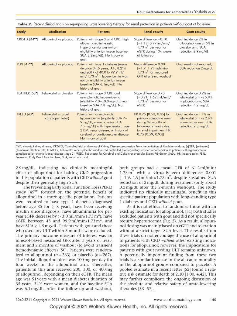
Table 2. Recent clinical trials on repurposing urate-lowering therapy for renal protection in patients without gout at baseline
Study Medication Patients Renal results Gout results
CKD-FIX [44&&] Allopurinol vs placebo Patients with stage 3 or 4 CKD, highalbumin:creatinine ratio.Hyperuricemia was not aneligibility criterion (mean baselineSUA 8.2 mg/dL). No history ofgout.
Slope difference �0.10[�1.18, 0.97]ml/min/1.73 m2 per year foreGFR during 104 weeksof follow-up
Gout incidence 2% inallopurinol arm vs 6% inplacebo arm; SUAreduction 2.9mg/dL
PERL [45&&] Allopurinol vs placebo Patients with type 1 diabetes (meanduration 34.6 years; A1c 8.2%)and eGFR of 40.0 to 99.9 ml/min/1.73m2. Hyperuricemia wasnot an eligibility criterion (meanbaseline SUA 6.1mg/dL). Nohistory of gout.
Mean difference 0.001[�1.9, 1.9] mgl/min/1.73 m2 for measuredGFR after 2-mo washout
Gout results not reported;SUA reduction 2 mg/dL
FEATHER [63&] Febuxostat vs placebo Patients with stage 3 CKD andasymptomatic hyperuricemia(eligibility 7.0–10.0 mg/dL; meanbaseline SUA 7.8 mg/dL). Nohistory of gout.
Slope difference 0.70[�0.21, 1.62] mL/min/1.73 m2 per year foreGFR
Gout incidence 0.9% infebuxostat arm vs 5.9%in placebo arm; SUAreduction 4.2 mg/dL
FREED [43&] Febuxostat vs usualcare (open label)
Patients with asymptomatichyperuricemia (eligibility SUA 7–9 mg/dL; mean baseline SUA7.5 mg/dL) with hypertension, type2 DM, renal disease, or history ofcerebral or cardiovascular disease.No history of gout
HR 0.75 [0.59, 0.95] forprimary composite eventduring 36 months offollow-up primarily dueto renal impairment (HR0.75 [0.59, 0.95])
Gout incidence 1.1% infebuxostat arm vs 2.6%in usual-care arm; SUAreduction 2.3 mg/dL
CKD, chronic kidney disease; CKD-FIX, Controlled trial of slowing of Kidney Disease progression From the Inhibition of Xanthine oxidase; (e)GFR, (estimated)glomerular filtration rate; FEATHER, Febuxostat versus placebo randomized controlled trial regarding reduced renal function in patients with hyperuricemiacomplicated by chronic kidney disease stage 3; FREED, Febuxostat for Cerebral and CaRdiorenovascular Events PrEvEntion StuDy; HR, hazard ratio; PERL,Preventing Early Renal Function Loss; SUA, serum uric acid.
Gout medications for comorbidities Yoshida et al.
2.9 mg/dL, indicating no clinically meaningfuleffect of allopurinol for halting CKD progressionin this population of patients with CKD without goutdespite their generally high SUA.
The Preventing Early Renal Function Loss (PERL)study [45
&&
] focused on the potential benefit ofallopurinol in a more specific population. Patientswere required to have type 1 diabetes diagnosedbefore age 35 for � 8 years, have been receivinginsulin since diagnosis, have albuminuria (or per-year eGFR decrease by > 3.0 mL/min/1.73 m2), haveeGFR between 45 and 99.9 ml/min/1.73 m2, andhave SUA� 4.5 mg/dL. Patients with gout and thosewho used any ULT within 3 months were excluded.The primary outcome measure of interest was aniohexol-based measured GFR after 3 years of treat-ment and 2 months of washout (to avoid transienthemodynamic effects) [50]. Patients were random-ized to allopurinol (n¼263) or placebo (n¼267).The initial allopurinol dose was 100 mg per day forfour weeks in the allopurinol arm. Thereafter,patients in this arm received 200, 300, or 400 mgof allopurinol, depending on their eGFR. The meanage was 51 years with a mean diabetes duration of35 years, 34% were women, and the baseline SUAwas 6.1 mg/dL. After the follow-up and washout,
Copyright © 2021 Wolters Kluwe
1040-8711 Copyright � 2021 Wolters Kluwer Health, Inc. All rights rese
both groups had a mean GFR of 61.2 ml/min/1.73 m2 with a virtually zero difference: 0.001[�1.9, 1.9] ml/min/1.73 m2, despite sustained SUAreduction of 2 mg/dL during treatment (only about0.2 mg/dL after the 2-month washout). The studyindicated no clinically meaningful benefit in thisspecific patient population with long-standing type1 diabetes and CKD without gout.
As it is not ethical to randomize those with anexisting indication for allopurinol, [51] both studiesexcluded patients with gout and did not specificallyrequire hyperuricemia (Fig. 2). As a result, allopuri-nol dosing was mainly based on eGFR and tolerationwithout a strict target SUA level. The results fromthese trials do not encourage the use of allopurinolin patients with CKD without other existing indica-tions for allopurinol; however, the implications forpatients with gout needing ULT remains unknown.A potentially important finding from these twotrials is a similar increase in the all-cause mortalityin the allopurinol groups compared to placebo. Apooled estimate in a recent letter [52] found a rela-tive risk estimate for death of 2.10 [1.00, 4.42]. Thismay further complicate the ongoing discussion ofthe absolute and relative safety of urate-loweringtherapies [53–57].
r Health, Inc. All rights reserved.
rved. www.co-rheumatology.com 149
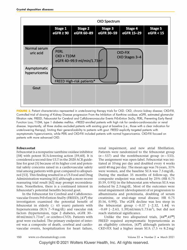
FIGURE 2. Patient characteristics represented in urate-lowering therapy trials for CKD. CKD, chronic kidney disease; CKD-FIX,Controlled trial of slowing of Kidney Disease progression From the Inhibition of Xanthine oxidase; eGFR, estimated glomerularfiltration rate; FREED, Febuxostat for Cerebral and CaRdiorenovascular Events PrEvEntion StuDy; PERL, Preventing Early RenalFunction Loss; T1DM, type 1 diabetes mellitus. �FREED enrolled patients with high risk for cerebro-cardiovascular or renaldisease. Importantly, all three studies excluded patients with existing gout at baseline (i.e., those with a clear indication forurate-lowering therapy), limiting their generalizability to patients with gout. FREED explicitly targeted patients withasymptomatic hyperuricemia, while PERL and CKD-FIX included patients with normal hyperuricemia. CKD-FIX focused onpatients with more advanced CKD.
Crystal deposition diseases
FebuxostatFebuxostat is a nonpurine xanthine oxidase inhibitor[58] with potent SUA-lowering action [59,60]. It isconsidered a second-line ULT in the 2020 ACR guide-line for gout [5] because of its higher cost and poten-tial safety concerns raised in a cardiovascular safetytrial among patients with gout compared to allopuri-nol [53]. This finding resulted in a US Food and DrugAdministration warning [61] although a more recentreassuring trial result [62] may prompt reconsidera-tion. Nonetheless, there is a continued interest infebuxostat’s potential benefits beyond gout.
In the Febuxostat for Cerebral and CaRdioreno-vascular Events PrEvEntion StuDy (FREED), [43
&
] theinvestigators examined the potential benefit offebuxostat in elderly (� 65 years) patients withhyperuricemia (SUA 7–9 mg/dL) and vascular riskfactors (hypertension, type 2 diabetes, eGFR 30–60 ml/min/1.73 m2, or cerebro-CVD). Patients withgout were excluded. The primary endpoint of inter-est was a composite of death, cerebral and cardio-vascular events, hospitalization for heart failure,
Copyright © 2021 Wolters Kluwer H
150 www.co-rheumatology.com
renal impairment, and new atrial fibrillation.Patients were randomized to the febuxostat group(n¼537) and the nonfebuxostat group (n¼533).The assignment was open-label. Febuxostat was ini-tiated at 10 mg per day and doubled every 4 weeksuntil 40 mg per day. The mean age was 76 years, 31%were women, and the baseline SUA was 7.5 mg/dL.During the median 35 months of follow-up, thecomposite endpoint was reduced by 25% (HR 0.75[0.59, 0.95]) in the febuxostat arm whereas SUA wasreduced by 2.3 mg/dL. Most of the outcomes wererenal impairment (development of or progression toalbuminuria and proteinuria, doubling of serumcreatinine, or end-stage renal disease; HR 0.75[0.56, 0.99]). The eGFR decline was less steep inthe febuxostat group (�0.37 [�2.32, 1.44] vs�0.69 [�2.63, 1.39] ml/min/1.73 m2), but did notreach statistical significance.
Unlike the two allopurinol trials, [44&&
,45&&
]FREED required asymptomatic hyperuricemia asan eligibility criterion, [43
&
] although patients inCKD-FIX had a higher mean SUA (7.5 vs 8.2 mg/
ealth, Inc. All rights reserved.
Volume 33 � Number 2 � March 2021
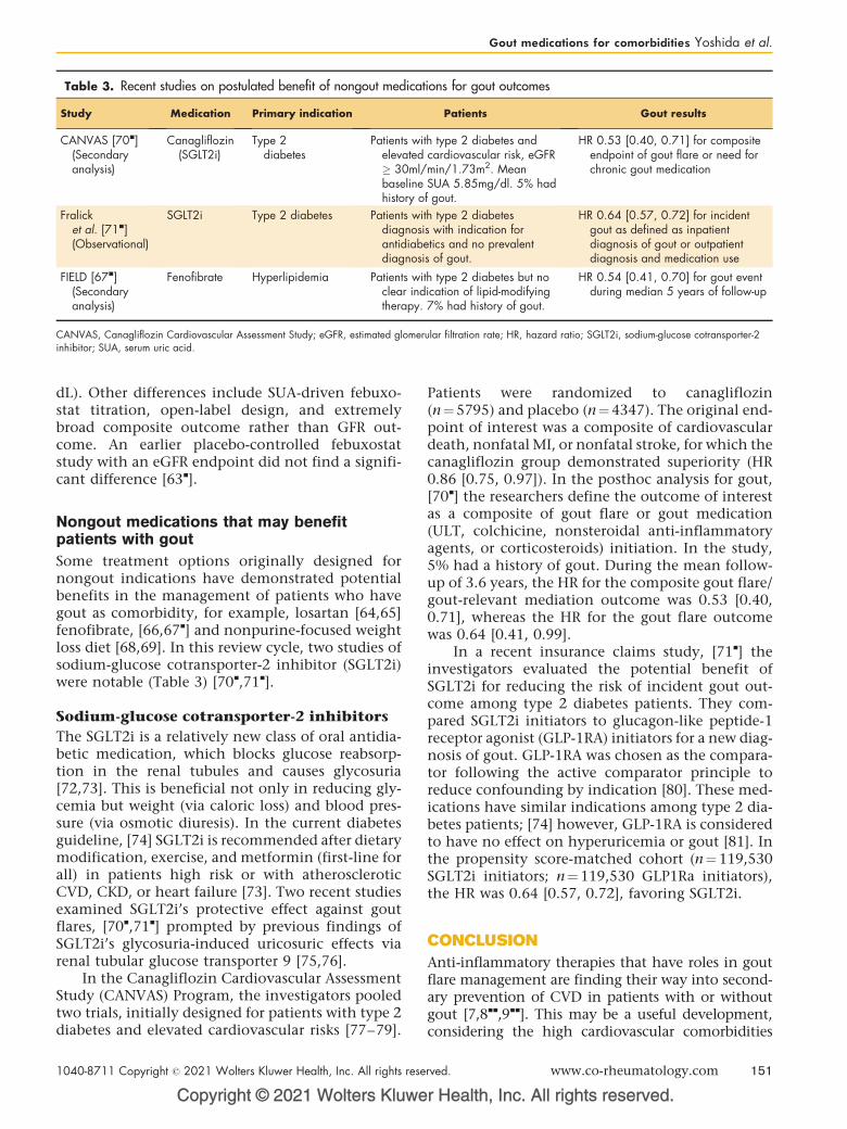
Table 3. Recent studies on postulated benefit of nongout medications for gout outcomes
Study Medication Primary indication Patients Gout results
CANVAS [70&](Secondaryanalysis)
Canagliflozin(SGLT2i)
Type 2diabetes
Patients with type 2 diabetes andelevated cardiovascular risk, eGFR� 30ml/min/1.73m2. Meanbaseline SUA 5.85mg/dl. 5% hadhistory of gout.
HR 0.53 [0.40, 0.71] for compositeendpoint of gout flare or need forchronic gout medication
Fralicket al. [71&](Observational)
SGLT2i Type 2 diabetes Patients with type 2 diabetesdiagnosis with indication forantidiabetics and no prevalentdiagnosis of gout.
HR 0.64 [0.57, 0.72] for incidentgout as defined as inpatientdiagnosis of gout or outpatientdiagnosis and medication use
FIELD [67&](Secondaryanalysis)
Fenofibrate Hyperlipidemia Patients with type 2 diabetes but noclear indication of lipid-modifyingtherapy. 7% had history of gout.
HR 0.54 [0.41, 0.70] for gout eventduring median 5 years of follow-up
CANVAS, Canagliflozin Cardiovascular Assessment Study; eGFR, estimated glomerular filtration rate; HR, hazard ratio; SGLT2i, sodium-glucose cotransporter-2inhibitor; SUA, serum uric acid.
Gout medications for comorbidities Yoshida et al.
dL). Other differences include SUA-driven febuxo-stat titration, open-label design, and extremelybroad composite outcome rather than GFR out-come. An earlier placebo-controlled febuxostatstudy with an eGFR endpoint did not find a signifi-cant difference [63
&
].
Nongout medications that may benefitpatients with gout
Some treatment options originally designed fornongout indications have demonstrated potentialbenefits in the management of patients who havegout as comorbidity, for example, losartan [64,65]fenofibrate, [66,67
&
] and nonpurine-focused weightloss diet [68,69]. In this review cycle, two studies ofsodium-glucose cotransporter-2 inhibitor (SGLT2i)were notable (Table 3) [70
&
,71&
].
Sodium-glucose cotransporter-2 inhibitors
The SGLT2i is a relatively new class of oral antidia-betic medication, which blocks glucose reabsorp-tion in the renal tubules and causes glycosuria[72,73]. This is beneficial not only in reducing gly-cemia but weight (via caloric loss) and blood pres-sure (via osmotic diuresis). In the current diabetesguideline, [74] SGLT2i is recommended after dietarymodification, exercise, and metformin (first-line forall) in patients high risk or with atheroscleroticCVD, CKD, or heart failure [73]. Two recent studiesexamined SGLT2i’s protective effect against goutflares, [70
&
,71&
] prompted by previous findings ofSGLT2i’s glycosuria-induced uricosuric effects viarenal tubular glucose transporter 9 [75,76].
In the Canagliflozin Cardiovascular AssessmentStudy (CANVAS) Program, the investigators pooledtwo trials, initially designed for patients with type 2diabetes and elevated cardiovascular risks [77–79].
Copyright © 2021 Wolters Kluwe
1040-8711 Copyright � 2021 Wolters Kluwer Health, Inc. All rights rese
Patients were randomized to canagliflozin(n¼5795) and placebo (n¼4347). The original end-point of interest was a composite of cardiovasculardeath, nonfatal MI, or nonfatal stroke, for which thecanagliflozin group demonstrated superiority (HR0.86 [0.75, 0.97]). In the posthoc analysis for gout,[70
&
] the researchers define the outcome of interestas a composite of gout flare or gout medication(ULT, colchicine, nonsteroidal anti-inflammatoryagents, or corticosteroids) initiation. In the study,5% had a history of gout. During the mean follow-up of 3.6 years, the HR for the composite gout flare/gout-relevant mediation outcome was 0.53 [0.40,0.71], whereas the HR for the gout flare outcomewas 0.64 [0.41, 0.99].
In a recent insurance claims study, [71&
] theinvestigators evaluated the potential benefit ofSGLT2i for reducing the risk of incident gout out-come among type 2 diabetes patients. They com-pared SGLT2i initiators to glucagon-like peptide-1receptor agonist (GLP-1RA) initiators for a new diag-nosis of gout. GLP-1RA was chosen as the compara-tor following the active comparator principle toreduce confounding by indication [80]. These med-ications have similar indications among type 2 dia-betes patients; [74] however, GLP-1RA is consideredto have no effect on hyperuricemia or gout [81]. Inthe propensity score-matched cohort (n¼119,530SGLT2i initiators; n¼119,530 GLP1Ra initiators),the HR was 0.64 [0.57, 0.72], favoring SGLT2i.
CONCLUSION
Anti-inflammatory therapies that have roles in goutflare management are finding their way into second-ary prevention of CVD in patients with or withoutgout [7,8
&&
,9&&
]. This may be a useful development,considering the high cardiovascular comorbidities
r Health, Inc. All rights reserved.
rved. www.co-rheumatology.com 151
Crystal deposition diseases
that patients with gout suffer [3]. It is important toremember that these trials address residual cardiovas-cular risk remaining after sufficient conventional riskmanagement [26]. ULT, the cornerstone of chronicmanagement of gout, on the other hand, has notdemonstrated the slowing of GFR deterioration inCKD trials [44
&&
,45&&
]. All of the CKD trials reviewedexcluded patients with gout; thus, they do not directlyinform decisions for patients with gout who requireULT. SGLT2i, an antidiabetic medication with cardio-vascular benefit [82], may be relevant for patients withdiabetes and gout to prevent gout flares and to reducethe risk of incident gout in patients with diabetes [83].
Acknowledgements
None.
Financial support and sponsorship
K.Y. have received research support from the Rheuma-tology Research Foundation K Bridge Award and NIHK23AR076453. D.H.S’s work on this manuscript wassupported by NIH P30AR072577.
Conflicts of interest
K.Y. has received consulting fees from OM1, Inc. andsalary support from research contracts to his institutionfrom Corrona for unrelated work. H.K.C. reports researchsupport from Ironwood and Horizon and consulting feesfrom Ironwood, Selecta, Horizon, Takeda, Kowa, andVaxart. D.H.S. receives salary support from researchcontracts to his institution from Abbvie, Amgen, Cor-rona, Genentech, and Janssen. He also receives royaltiesfrom UpToDate on unrelated work.
REFERENCES AND RECOMMENDEDREADINGPapers of particular interest, published within the annual period of review, havebeen highlighted as:
& of special interest&& of outstanding interest1. Akker M, van den. Buntinx F, Knottnerus JA. Comorbidity or multimorbidity. EurJ Gen Pract 1996; 2:65–70. doi:10.3109/13814789609162146.
2. Radner H, Yoshida K, Smolen JS, Solomon DH. Multimorbidity and rheumaticconditions-enhancing the concept of comorbidity. Nat Rev Rheumatol 2014;10:252–256. doi:10.1038/nrrheum.2013.212.
3. Zhu Y, Pandya BJ, Choi HK. Comorbidities of gout and hyperuricemia in theUS general population: NHANES 2007-2008. Am J Med 2012;125:679–687e1. doi:10.1016/j.amjmed.2011.09.033.
4. Sumpter NA, Saag KG, Reynolds RJ, Merriman TR. Comorbidities in gout andhyperuricemia: causality or epiphenomena? Curr Opin Rheumatol 2020;32:126–133. doi:10.1097/BOR.0000000000000691.
5. FitzGerald JD, Dalbeth N, Mikuls T, et al. 2020 American College of Rheu-matology Guideline for the Management of Gout. Arthritis Rheumatol 2020;72:879–895. doi:10.1002/art.41247.
6. Nidorf SM, Eikelboom JW, Budgeon CA, Thompson PL. Low-dose colchicinefor secondary prevention of cardiovascular disease. J Am Coll Cardiol 2013;61:404–410. doi:10.1016/j.jacc.2012.10.027.
7. Ridker PM, Everett BM, Thuren T, et al. Antiinflammatory therapy withcanakinumab for atherosclerotic disease. N Engl J Med 2017;377:1119–1131. doi:10.1056/NEJMoa1707914.
Copyright © 2021 Wolters Kluwer H
152 www.co-rheumatology.com
8.&&
Tardif J-C, Kouz S, Waters DD, et al. Efficacy and safety of low-dosecolchicine after myocardial infarction. N Engl J Med 2019;381:2497–2505. doi:10.1056/NEJMoa1912388.
This large trial of low-dose colchicine vs. placebo in patients with recent MI (goutprevalence unreported) demonstrated a 23% reduction in composite cardiovas-cular endpoint.9.
&&
Nidorf SM, Fiolet ATL, Mosterd A, et al. Colchicine in patients with chroniccoronary disease. N Engl J Med 2020; 383:1838–1847.
This large trial of low-dose colchicine vs. placebo in patients with stable coronaryartery disease (8% with gout) demonstrated a 31% reduction in compositecardiovascular endpoint.10. Ridker PM. Closing the loop on inflammation and atherothrombosis: why
perform the CIRT and CANTOS trials? Trans Am Clin Climatol Assoc 2013;124:174–190.
11. Ridker PM. Moving beyond JUPITER: will inhibiting inflammation reducevascular event rates? Curr Atheroscler Rep 2013; 15:295. doi:10.1007/s11883-012-0295-3.
12. Ridker PM. From C-reactive protein to interleukin-6 to interleukin-1: movingupstream to identify novel targets for atheroprotection. Circ Res 2016;118:145–156. doi:10.1161/CIRCRESAHA.115.306656.
13. Harrington RA. Targeting inflammation in coronary artery disease. N Engl JMed 2017; 377:1197–1198. doi:10.1056/NEJMe1709904.
14. Duewell P, Kono H, Rayner KJ, et al. NLRP3 inflammasomes are required foratherogenesis and activated by cholesterol crystals. Nature 2010;464:1357–1361. doi:10.1038/nature08938.
15. Rajamaki K, Lappalainen J, Oorni K, et al. Cholesterol crystals activatethe NLRP3 inflammasome in human macrophages: a novel link betweencholesterol metabolism and inflammation. PLoS One 2010; 5:e11765.doi:10.1371/journal.pone.0011765.
16. Martinon F, Petrilli V, Mayor A, et al. Gout-associated uric acid crystalsactivate the NALP3 inflammasome. Nature 2006; 440:237–241.doi:10.1038/nature04516.
17. Dhimolea E. Canakinumab. MAbs 2010; 2:3–13.18. So A, De Meulemeester M, Pikhlak A, et al. Canakinumab for the treatment of
acute flares in difficult-to-treat gouty arthritis: results of a multicenter, phase II,dose-ranging study. Arthritis Rheumatol 2010; 62:3064–3076.doi:10.1002/art.27600.
19. Hirsch JD, Gnanasakthy A, Lale R, et al. Efficacy of Canakinumab vs. triamci-nolone acetonide according to multiple gouty arthritis-related health outcomesmeasures. Int J Clin Pract 2014; 68:1503–1507. doi:10.1111/ijcp.12521.
20. Schlesinger N, Alten RE, Bardin T, et al. Canakinumab for acute gouty arthritisin patients with limited treatment options: results from two randomised,multicentre, active-controlled, double-blind trials and their initial extensions.Ann Rheum Dis 2012; 71:1839–1848. doi:10.1136/annrheumdis-2011-200908.
21. Schlesinger N, De Meulemeester M, Pikhlak A, et al. Canakinumab relievessymptoms of acute flares and improves health-related quality of life in patientswith difficult-to-treat Gouty Arthritis by suppressing inflammation: results of arandomized, dose-ranging study. Arthritis Res Ther 2011; 13:1–13R53.doi:10.1186/ar3297.
22. Solomon DH, Glynn RJ, MacFadyen JG, et al. Relationship of interleukin-1b
blockade with incident gout and serum uric acid levels: exploratory analysis ofa randomized controlled trial. Ann Intern Med 2018; 169:535–542.doi:10.7326/M18-1167.
23. Sehested TSG, Bjerre J, Ku S, et al. Cost-effectiveness of Canakinumab forprevention of recurrent cardiovascular events. JAMA Cardiol 2019;4:128–135. doi:10.1001/jamacardio.2018.4566.
24. Miller J. FDA Rejects Novartis Bid to Repurpose Inflammation Drug for HeartAttacks. The Rheumatologist. 2018. https://www.the-rheumatologist.org/arti-cle/fda-rejects-novartis-bid-to-repurpose-inflammation-drug-for-heart-attacks/.[Accessed 12 September 2020]
25. Thompson PL, Nidorf SM. Colchicine: an affordable anti-inflammatory agentfor atherosclerosis. Curr Opin Lipidol 2018; 29:467–473.
26. Ajala ON, Everett BM. Targeting inflammation to reduce residual cardiovas-cular risk. Curr Atheroscler Rep 2020; 22:66. doi:10.1007/s11883-020-00883-3.
27. Hartung EF. History of the use of colchicum and related medicaments in gout;with suggestions for further research. Ann Rheum Dis 1954; 13:190–200.doi:10.1136/ard.13.3.190.
28. Dalbeth N, Lauterio TJ, Wolfe HR. Mechanism of action of colchicine in thetreatment of gout. Clin Ther 2014; 36:1465–1479. doi:10.1016/j.clin-thera.2014.07.017.
29. Pascart T, Richette P. Colchicine in gout: an update. Curr Pharm Des 2018;24:684–689. doi:10.2174/1381612824999180115103951.
30. Pope RM, Tschopp J. The role of interleukin-1 and the inflammasome in gout:implications for therapy. Arthritis Rheumatol 2007; 56:3183–3188.doi:10.1002/art.22938.
31. Dalbeth N, Choi HK, Joosten LAB, et al. Gout. Nat Rev Dis Prim 2019; 5:69.doi:10.1038/s41572-019-0115-y.
32. Libby P. Interleukin-1 beta as a target for atherosclerosis therapy: biologicalbasis of CANTOS and beyond. J Am Coll Cardiol 2017; 70:2278–2289.doi:10.1016/j.jacc.2017.09.028.
ealth, Inc. All rights reserved.
Volume 33 � Number 2 � March 2021
Gout medications for comorbidities Yoshida et al.
33.&
Tong DC, Quinn S, Nasis A, et al. Colchicine in patients with acute coronarysyndrome: The Australian COPS Randomized Clinical Trial. Circulation 2020;142:1890–1900.
This smaller-scale low-dose colchicine vs. placebo in patients who experiencedacute coronary syndrome (gout prevalence unreported) did not demonstrate astatistically significant benefit.34. Solomon DH, Liu C-C, Kuo I-H, et al. Effects of colchicine on risk of
cardiovascular events and mortality among patients with gout: a cohort studyusing electronic medical records linked with Medicare claims. Ann Rheum Dis2016; 75:1674–1679. doi:10.1136/annrheumdis-2015-207984.
35. Hemkens LG, Ewald H, Gloy VL, et al. Cardiovascular effects and safety oflong-term colchicine treatment: cochrane review and meta-analysis. Heart2016; 102:590–596. doi:10.1136/heartjnl-2015-308542.
36. Samuel M, Tardif J-C, Khairy P, et al. Cost-effectiveness of low-dose colchi-cine after myocardial infarction in the colchicine cardiovascular outcomes trial(COLCOT). Eur Heart J Qual Care Clin Outcomes 2020; 1–10; Publishedonline May 14, 2020. doi:10.1093/ehjqcco/qcaa045.
37. Bellomo G, Venanzi S, Verdura C, et al. Association of uric acid with change inkidney function in healthy normotensive individuals. Am J Kidney Dis 2010;56:264–272. doi:10.1053/j.ajkd.2010.01.019.
38. Chonchol M, Shlipak MG, Katz R, et al. Relationship of uric acid withprogression of kidney disease. Am J Kidney Dis 2007; 50:239–247.doi:10.1053/j.ajkd.2007.05.013.
39. Weiner DE, Tighiouart H, Elsayed EF, et al. Uric acid and incident kidneydisease in the community. J Am Soc Nephrol 2008; 19:1204–1211.doi:10.1681/ASN.2007101075.
40. Obermayr RP, Temml C, Gutjahr G, et al. Elevated uric acid increases the riskfor kidney disease. J Am Soc Nephrol 2008; 19:2407–2413. doi:10.1681/ASN.2008010080.
41. Bartakova V, Kuricova K, Pacal L, et al. Hyperuricemia contributes to the fasterprogression of diabetic kidney disease in type 2 diabetes mellitus. J DiabetesComplicat 2016; 30:1300–1307. doi:10.1016/j.jdiacomp.2016.06.002.
42. Bose B, Badve SV, Hiremath SS, et al. Effects of uric acid-lowering therapy onrenal outcomes: a systematic review and meta-analysis. Nephrol Dial Trans-plant 2014; 29:406–413. doi:10.1093/ndt/gft378.
43.&
Kojima S, Matsui K, Hiramitsu S, et al. Febuxostat for Cerebral and CaRdior-enovascular Events PrEvEntion StuDy. Eur Heart J 2019; 40:1778–1786.
This open-label trial of febuxostat vs. usual care among high-risk (hypertension,diabetes, renal disease, or history of cerebro-cardiovascular disease) patients withasymptomatic hyperuricemia (mean SUA 7.5 mg/dL) showed a 25% reduction inbroadly defined composite endpoint, primarily due to reduction in renal impairment.44.&&
Badve SV, Pascoe EM, Tiku A, et al. Effects of allopurinol on the progressionof chronic kidney disease. N Engl J Med 2020; 382:2504–2513.doi:10.1056/NEJMoa1915833.
This trial of allopurinol vs. placebo among nongout stages 3 and 4 chronic kidneydisease patients (mean SUA 8.2 mg/dL) did not demonstrate prevention ofestimate glomerular filtration rate deterioration over 104 weeks of follow up.45.&&
Doria A, Galecki AT, Spino C, et al. Serum urate lowering with allopurinol andkidney function in type 1 diabetes. N Engl J Med 2020; 382:2493–2503.doi:10.1056/NEJMoa1916624.
This trial of allopurinol vs. placebo among nongout type 1 diabetes patients (meanSUA 6.1 mg/dL) did not demonstrate prevention of measured glomerular filtrationrate deterioration at 3 years.46. Pacher P, Nivorozhkin A, Szabo C. Therapeutic effects of xanthine oxidase
inhibitors: renaissance half a century after the discovery of allopurinol.Pharmacol Rev 2006; 58:87–114. doi:10.1124/pr.58.1.6.
47. KDOQI. Clinical practice guidelines and clinical practice recommendationsfor diabetes and chronic kidney disease. Am J Kidney Dis. 2007;49(2 Suppl2): S12–154.
48. Matsushita K, Selvin E, Bash LD, et al. Change in estimated GFR associateswith coronary heart disease and mortality. J Am Soc Nephrol 2009;20:2617–2624. doi:10.1681/ASN.2009010025.
49. Neogi T, Jansen TLTA, Dalbeth N, et al. 2015 Gout classification criteria: anAmerican College of Rheumatology/European League Against Rheumatismcollaborative initiative. Ann Rheum Dis 2015; 74:1789–1798. doi:10.1136/annrheumdis-2015-208237.
50. Bove M, Cicero AFG, Borghi C. The effect of xanthine oxidase inhibitors onblood pressure and renal function. Curr Hypertens Rep 2017; 19:95.doi:10.1007/s11906-017-0793-3.
51. Freedman B. Equipoise and the ethics of clinical research. N Engl J Med1987; 317:141–145. doi:10.1056/NEJM198707163170304.
52. McCormick N, Zhang Y, Choi HK. Allopurinol and Chronic Kidney Disease. NEngl J Med 2020; 383:1689–1690.
53. White WB, Saag KG, Becker MA, et al. Cardiovascular safety of febuxostat orallopurinol in patients with gout. N Engl J Med 2018; 378:1200–1210.doi:10.1056/NEJMoa1710895.
54. Choi H, Neogi T, Stamp L, et al. New perspectives in rheumatology: implica-tions of the cardiovascular safety of febuxostat and allopurinol in patients withgout and cardiovascular morbidities trial and the Associated Food and DrugAdministration Public Safety Alert. Arthritis Rheumatol 2018;70:1702–1709. doi:https://doi.org/10.1002/art.40583.
55. Kim SC, Neogi T, Kang EH, et al. Cardiovascular risks of probenecid versusallopurinol in older patients with gout. J Am Coll Cardiol 2018; 71:994–1004.doi:10.1016/j.jacc.2017.12.052.
Copyright © 2021 Wolters Kluwe
1040-8711 Copyright � 2021 Wolters Kluwer Health, Inc. All rights rese
56. Coburn BW, Michaud K, Bergman DA, Mikuls TR. Allopurinoldose escalationand mortality among patients with gout: A National Propensity-MatchedCohort Study. Arthritis Rheumatol 2018; 70:1298–1307. doi:10.1002/art.40486.
57. Hay CA, Prior JA, Belcher J, et al. Mortality in patients with gout treated withallopurinol: a systematic review and meta-analysis. Arthritis Care Res 2020;Published online April 14, 2020. doi:10.1002/acr.24205.
58. Takano Y, Hase-Aoki K, Horiuchi H, et al. Selectivity of febuxostat, a novelnonpurine inhibitor of xanthine oxidase/xanthine dehydrogenase. Life Sci2005; 76:1835–1847. doi:10.1016/j.lfs.2004.10.031.
59. Kamatani N, Naoyuki K, Fujimori S, et al. An allopurinol-controlled, multicenter,randomized, open-label, parallel between-group, comparative study of febuxo-stat (TMX-67), a nonpurine-selective inhibitor of xanthine oxidase, in patientswith hyperuricemia including those with gout in Japan: phase 2 exploratoryclinical study. J Clin Rheumatol 2011; 17(4 Suppl 2):S44–49. doi:10.1097/RHU.0b013e31821d352f.
60. Lin Y-J, Lin S-Y, Lin C-H, et al. Evaluation of urate-lowering therapyin hyperuricemia patients: a systematic review and Bayesian network meta-analysis of randomized controlled trials. Clin Rheumatol 2020;39:1633–1648.
61. FDA. FDA adds Boxed Warning for increased risk of death with gout medicineUloric (febuxostat). FDA Drug Safety Communication 2019; https://www.fda.gov/drugs/drug-safety-and-availability/fda-adds-boxed-warning-in-creased-risk-death-gout-medicine-uloric-febuxostat. [Accessed 9 September2020]
62. Mackenzie IS, Ford I, Nuki G, et al. Long-term cardiovascular safety offebuxostat compared with allopurinol in patients with gout (FAST): a multi-centre, prospective, randomised, open-label, non-inferiority trial. Lancet 2020;396:1745–1757.
63.&
Kimura K, Hosoya T, Uchida S, et al. Febuxostat therapy for patients withStage 3 CKD and asymptomatic hyperuricemia: a randomized trial. Am JKidney Dis 2018; 72:798–810. doi:10.1053/j.ajkd.2018.06.028.
This trial comparing febuxostat and placebo among patients with stage 3 chronickidney disease and asymptomatic hyperuricemia, the estimated glomerular filtra-tion rate did not significantly differ.64. Miao Yan. Ottenbros Stefan A, Laverman Goos D, et al. Effect of a reduction in
uric acid on renal outcomes during losartan treatment. Hypertension 2011;58:2–7. doi:10.1161/HYPERTENSIONAHA.111.171488.
65. Choi HK, Soriano LC, Zhang Y, Rodrıguez LAG. Antihypertensivedrugs and risk of incident gout among patients with hypertension: popula-tion based case-control study. BMJ 2012; 344:d8190. doi:10.1136/bmj.d8190.
66. Jung J-Y, Choi Y, Suh C-H, et al. Effect of fenofibrate on uric acid levelin patients with gout. Sci Rep 2018; 8:16767. doi:10.1038/s41598-018-35175-z.
67.&
Waldman B, Ansquer J-C, Sullivan DR, et al. Effect of fenofibrate onuric acid and gout in type 2 diabetes: a post-hoc analysis of the rando-mised, controlled FIELD study. Lancet Diabetes Endocrinol 2018;6:310–318.
In this secondary analysis of a trial of fenofibrate vs placebo among patients withtype 2 diabetes, a 46% reduction in incident gout was observed.68. Yokose C, McCormick N, Rai SK, et al. Effects of low-fat, mediterranean, or
low-carbohydrate weight loss diets on serum urate and cardiometabolic riskfactors: a secondary analysis of the Dietary Intervention Randomized Con-trolled Trial (DIRECT). Diabetes Care 2020; 43:2812–2820.
69. Shai I, Schwarzfuchs D, Henkin Y, et al. Weight loss with a low-carbohydrate,mediterranean, or low-fat diet. N Engl J Med 2008; 359:229–241.doi:10.1056/NEJMoa0708681.
70.&
Li J, Badve SV, Zhou Z, et al. The effects of canagliflozin on gout in type 2diabetes: a posthoc analysis of the CANVAS Program. Lancet Rheumatol2019; 1:e220–e228. doi:10.1016/S2665-9913(19)30078-5.
In this re-analysis of canagliflozin (sodium-glucose cotransporter-2 inhibitor fordiabetes) trials, 47% reduction in a composite endpoint of gout diagnosis or theinitiation of gout medication.71.&
Fralick M, Chen SK, Patorno E, Kim SC. Assessing the risk for gout withsodium-glucose cotransporter-2 inhibitors in patients with type 2 diabetes: apopulation-based cohort study. Ann Intern Med 2020; 172:186–194.doi:10.7326/M19-2610.
This large observational study of insurance claims showed a 36% reduction inincident gout among new users of sodium-glucose cotransporter-2 inhibitors (oralantidiabetic) compared to new users of glucagon-like peptide-1 receptor agonists(injectable antidiabetic).72. Bailey CJ. Uric acid and the cardio-renal effects of SGLT2 inhibitors. Diabetes
Obes Metab 2019; 21:1291–1298. doi:10.1111/dom.13670.73. Tentolouris A, Vlachakis P, Tzeravini E, et al. SGLT2 inhibitors: a review of their
antidiabetic and cardioprotective effects. Int J Environ Res Public Health2019; 16:1–27. doi:10.3390/ijerph16162965.
74. American Diabetes Association. Pharmacologic Approaches to GlycemicTreatment: Standards of Medical Care in Diabetes-2020. Diabetes Care.2020;43(Suppl 1):S98–S110.
75. Zhao Y, Xu L, Tian D, et al. Effects of sodium-glucose co-transporter 2(SGLT2) inhibitors on serum uric acid level: a meta-analysis of randomizedcontrolled trials. Diabetes Obes Metab 2018; 20:458–462. doi:10.1111/dom.13101.
r Health, Inc. All rights reserved.
rved. www.co-rheumatology.com 153
Crystal deposition diseases
76. Chino Y, Samukawa Y, Sakai S, et al. SGLT2 inhibitor lowers serum uric acidthrough alteration of uric acid transport activity in renal tubule by increasedglycosuria. Biopharm Drug Dispos 2014; 35:391–404. doi:10.1002/bdd.1909.
77. Neal B, Perkovic V, Mahaffey KW, et al. Canagliflozin and cardiovascular andrenal events in type 2 diabetes. N Engl J Med 2017; 377:644–657.doi:10.1056/NEJMoa1611925.
78. Neal B, Perkovic V, de Zeeuw D, et al. Rationale, design, and baselinecharacteristics of the Canagliflozin Cardiovascular Assessment Study (CAN-VAS) – a randomized placebo-controlled trial. Am Heart J 2013;166:217–223e11. doi:10.1016/j.ahj.2013.05.007.
79. Neal B, Perkovic V, Matthews DR, et al. Rationale, design and baselinecharacteristics of the CANagliflozin cardioVascular Assessment Study-Renal(CANVAS-R): A randomized, placebo-controlled trial. Diabetes Obes Metab2017; 19:387–393. doi:10.1111/dom.12829.
Copyright © 2021 Wolters Kluwer H
154 www.co-rheumatology.com
80. Yoshida K, Solomon DH, Kim SC. Active-comparator design and new-userdesign in observational studies. Nat Rev Rheumatol 2015; 11:437–441.doi:10.1038/nrrheum.2015.30.
81. Tonneijck L, Muskiet MHA, Smits MM, et al. Effect of immediate and pro-longed GLP-1 receptor agonist administration on uric acid and kidneyclearance: Posthoc analyses of four clinical trials. Diabetes Obes Metab2018; 20:1235–1245. doi:10.1111/dom.13223.
82. Shivakumar O, Sattar N, Wheeler DC. Sodium-glucose cotransporter 2inhibitor effects on cardiovascular outcomes in chronic kidney disease.Nephrol Dial Transplant 2020; 35(Supplement_1):i43–i47. doi:10.1093/ndt/gfz292.
83. Neogi T. Repurposing antidiabetic medications for gout: the potential ofsodium glucose co-transporter 2 inhibitors. Lancet Rheumatol 2019;1:E197–E199.
ealth, Inc. All rights reserved.
Volume 33 � Number 2 � March 2021
REVIEW
CURRENTOPINION COVID-19 and autoimmune diseases
Copyrigh
1040-8711 Copyright � 2020 Wolte
a b a,c
Yu Liu , Amr H. Sawalha , and Qianjin LuPurpose of review
The aim of this study was to evaluate the relationship between infection with SARS-CoV-2 andautoimmunity.
Recent findings
Coronavirus disease 2019 (COVID-19) is an infectious disease caused by severe acute respiratorysyndrome (SARS) associated coronavirus 2 (SARS-CoV-2). Although most of the infected individuals areasymptomatic, a proportion of patients with COVID-19 develop severe disease with multiple organ injuries.Evidence suggests that some medications used to treat autoimmune rheumatologic diseases might havetherapeutic effect in patients with severe COVID-19 infections, drawing attention to the relationshipbetween COVID-19 and autoimmune diseases. COVID-19 shares similarities with autoimmune diseases inclinical manifestations, immune responses and pathogenic mechanisms. Robust immune reactionsparticipate in the pathogenesis of both disease conditions. Autoantibodies as a hallmark of autoimmunediseases can also be detected in COVID-19 patients. Moreover, some patients have been reported todevelop autoimmune diseases, such as Guillain–Barre syndrome or systemic lupus erythematosus, afterCOVID-19 infection. It is speculated that SARS-CoV-2 can disturb self-tolerance and trigger autoimmuneresponses through cross-reactivity with host cells. The infection risk and prognosis of COVID-19 in patientswith autoimmune diseases remains controversial, but patient adherence to medication regimens to preventautoimmune disease flares is strongly recommended.
Summary
We present a review of the association between COVID-19 and autoimmune diseases, focusing onsimilarities in immune responses, cross-reactivity of SARS-CoV-2, the development of autoimmune diseasesin COVID-19 patients and the risk of COVID-19 infection in patients with preexisting autoimmuneconditions.
Keywords
autoimmune diseases, COVID-19, cross-reactivity, molecular mimicry, SARS-CoV-2
aDepartment of Dermatology, Second Xiangya Hospital, Central SouthUniversity, Hunan Key Laboratory of Medical Epigenetics, Changsha,Hunan, PR China, bDepartments of Pediatrics, Medicine, and Immunol-ogy, and Lupus Center of Excellence, University of Pittsburgh, Pittsburgh,Pennsylvania, USA and cInstitute of Dermatology, Chinese Academy ofMedical Sciences and Peking Union Medical College, Nanjing, China
Correspondence to Qianjin Lu, MD, PhD, #139 Renmin Middle Rd,Changsha, Hunan 410011, P.R. China. Tel: +86 731 85295860;fax: +86 731 85533525; e-mail: [email protected]; Amr H.Sawalha, MD, 7123 Rangos Research Center, 4401 Penn Avenue,Pittsburgh, PA 15224, USA. Tel: +1 412 692 8140;fax: +1 412 412 692 5054; e-mail: [email protected].
Curr Opin Rheumatol 2021, 33:155–162
DOI:10.1097/BOR.0000000000000776
INTRODUCTION
Since December 2019, a novel infection namedcoronavirus disease 2019 (COVID-19) broke out inWuhan, China, and has been sweeping across theglobe. COVID-19 was officially declared a pandemicby WHO on 11 March 2020 [1]. The disease is causedby a newly identified strain of severe acute respira-tory syndrome (SARS) associated coronavirus, whichwas named SARS-CoV-2 after SARS-CoV that causedthe epidemic of SARS in 2002 [2].
SARS-CoV-2 belongs to the coronavirus family,which are enveloped viruseswitha sphericalmorphol-ogy and a single-stranded RNA (ssRNA) genome [3].The spike glycoproteins (S protein) cross through thepeplos of the virus and form a crown-like surface [4].Through the receptor binding domain (RBD) locatedintheS1 subunitof theSprotein, theviruscanligate tothe host cell receptor angiotensin-converting enzyme2 (ACE2) and invade into the cell [5–7].
t © 2021 Wolters Kluwe
rs Kluwer Health, Inc. All rights rese
In many cases, hosts infected by SARS-CoV-2present with flu-like symptoms, such as fever,fatigue and dry cough. Headache, myalgia, sorethroat, nausea and diarrhoea can also be seen inpatients with COVID-19 [8,9]. Shortness of breath
r Health, Inc. All rights reserved.
rved. www.co-rheumatology.com
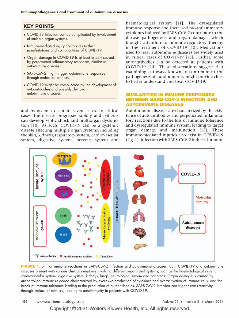
KEY POINTS
� COVID-19 infection can be complicated by involvementof multiple organ systems.
� Immune-mediated injury contributes to themanifestations and complications of COVID-19.
� Organ damage in COVID-19 is at least in part causedby perpetuated inflammatory responses, similar toautoimmune diseases.
� SARS-CoV-2 might trigger autoimmune responsesthrough molecular mimicry.
� COVID-19 might be complicated by the development ofautoantibodies and possibly de-novoautoimmune diseases.
Immunopathogenesis and treatment of autoimmune diseases
and hypoxemia occur in severe cases. In criticalcases, the disease progresses rapidly and patientscan develop septic shock and multiorgan dysfunc-tion [10]. As such, COVID-19 can be a systemicdisease affecting multiple organ systems, includingthe skin, kidneys, respiratory system, cardiovascularsystem, digestive system, nervous system and
Copyright © 2021 Wolters Kluwer H
FIGURE 1. Similar immune reactions in SARS-CoV-2 infection adiseases present with various clinical symptoms involving differencardiovascular system, digestive system, kidneys, lungs, neurologuncontrolled immune response characterized by excessive producbreak of immune tolerance leading to the production of autoantibthrough molecular mimicry, leading to autoimmunity in patients w
156 www.co-rheumatology.com
haematological system [11]. The dysregulatedimmune response and increased pro-inflammatorycytokines induced by SARS-CoV-2 contribute to thedisease pathogenesis and organ damage, whichbrought attention to immune-regulatory therapyin the treatment of COVID-19 [12]. Medicationsused to treat autoimmune diseases are widely usedin critical cases of COVID-19 [13]. Further, someautoantibodies can be detected in patients withCOVID-19 [14]. These observations suggest thatexamining pathways known to contribute to thepathogenesis of autoimmunity might provide cluesto better understand and treat COVID-19.
SIMILARITIES IN IMMUNE RESPONSESBETWEEN SARS-COV-2 INFECTION ANDAUTOIMMUNE DISEASES
Autoimmune diseases are characterized by the exis-tence of autoantibodies and perpetuated inflamma-tory reactions due to the loss of immune toleranceand dysregulated immune system, leading to targetorgan damage and malfunction [15]. Theseimmune-mediated injuries also exist in COVID-19(Fig. 1). Infection with SARS-CoV-2 induces immune
ealth, Inc. All rights reserved.
nd autoimmune diseases. Both COVID-19 and autoimmunet organs and systems, such as the haematological system,ical system and pancreas. Organ damage is caused bytion of cytokines and overactivation of immune cells, and theodies. SARS-CoV-2 infection can trigger cross-reactivityith COVID-19.
Volume 33 � Number 2 � March 2021
COVID-19 and autoimmune diseases Liu et al.
reactions, which might have important implica-tions in the development of vaccine strategiesagainst this virus [16]. T cell immunity plays acentral role in the control of SARS-CoV-2 infection.Antigen-specific CD4þ and CD8þ T cells and neu-tralizing antibody responses play protective rolesagainst SARS-CoV-2, while impaired adaptiveimmune responses such as scarcity of naive T cellsmay lead to poor disease outcomes [17].
In clinical laboratory tests, lymphopenia (lym-phocyte count�1.0 x 10⁹ /l) is associated with severeillness in COVID-19 patients and might be a prog-nostic factor for disease severity and mortality [18–21]. Another notable haemocytological change isneutrophilia and associated excessive neutrophilextracellular traps, which paralleled lung injury insevere COVID-19 patients [12]. Therefore, theimmune response is a double-edged sword inCOVID-19, with outcomes affected by the degreeof cytokine imbalance and activation of immunecells. Excessive production and release of pro-inflam-matory cytokines and chemokines can cause severeorgan damage in critical cases, which is observed inautoimmune diseases as well. In COVID-19 patients,pro-inflammatory cytokines and chemokines,including interleukin (IL)-1, IL-2, IL-6, IL-8, IL-10,IL-17, IL-18, CXCL10 and CCL2, increased signifi-cantly and the expression levels of some of thesecytokines, such as IL-1, IL-6, IL-10 and IL-18, havebeen demonstrated to be associated with diseaseseverity [22–25]. Similar to autoimmune diseases,damage-associated molecular patterns (DAMPs) alsoparticipate in the pathogenesis of COVID-19 and arerelated to disease outcome. Chen et al. [26] revealedthat serum levels of S100A8/A9 and HMGB1increased significantly in patients with severeCOVID-19 and that significant elevation of the twoDAMPs was associated with higher mortality.
Activation and infiltration of immune cells par-ticipate in the pathogenesis of organ injury inpatients with COVID-19. Macrophage activationsyndrome (MAS) could be a continuum of cytokinestorm syndrome leading to life-threatening compli-cations in COVID-19 [27]. In this condition, acti-vated macrophages will produce excessive pro-inflammatory cytokines, polarize into the inflam-matory M1 phenotype and exhibit cytotoxic dys-function [28]. Recently, Conti et al. [29] proposedthat SARS-CoV-2 activated mast cells could releasehistamine to increase IL-1 levels to initiate cytokinestorm and aggravate lung injury. Woodruff et al. [30]found extrafollicular B cell activation in critically illpatients with COVID-19, similar to what has beenobserved in autoimmunity. Further, extrafollicularB cell activation correlated strongly with the pro-duction of high concentrations of SARS-CoV-2
Copyright © 2021 Wolters Kluwe
1040-8711 Copyright � 2020 Wolters Kluwer Health, Inc. All rights rese
specific neutralizing antibodies and poor diseaseoutcome [30]. Peripheral blood B-cell subpopula-tions are altered during COVID-19. In COVID-19patients, atypical memory B-cells (CD21lo/CD27�/CD10�) expanded significantly, while classicalmemory B-cells (CD21þ/CD27þ/CD10�) were signif-icantly reduced [31]. Analysis of immune profiles ofsevere COVID-19 patients revealed an increasedproportion of mature natural killer (NK) cells anddecreased proportion of T-cell numbers [32].
Similar to some autoimmune and immune-mediated thromboinflammatory diseases, includinglupus, antiphospholipid syndrome and ANCA-asso-ciated vasculitis, neutrophil activation and neutro-phil extracellular trap production (NETosis) appearto have a pathogenic role in COVID-19. Zuo et al.[33
&
] reported increased markers of NETs in serafrom patients with COVID-19, and significantlymore in patients requiring mechanical ventilation.In-vitro experiments demonstrated that sera fromCOVID-19 patients triggered NETosis in normalneutrophils, similar to sera from patients with anti-phospholipid syndrome [33
&
,34].In severe and critical cases, immunomodulatory
drugs and biological agents targeting pro-inflamma-tory cytokines have been applied to contain therobust immune response in COVID-19. Corticoste-roids, JAK inhibitors, IL-1 blockade and IL-6 receptorantagonists, which are familiar to rheumatologists,have been used to treat COVID-19 patients [35–38].Similarities in immunopathogenesis of COVID-19and autoimmune diseases are summarized inTable 1.
MOLECULAR MIMICRY AND SARS-COV-2
The production of autoantibodies is a key feature ofautoimmune diseases. However, the underlyingmechanisms are complicated and still not fullyunderstood. Molecular mimicry by infectiouspathogens is believed to be one of the mechanisms[39]. Viral infection can disturb immunologic toler-ance by exposure of antigen epitopes that elicitcross-reactive antibodies. There are a large numberof reports indicating antigenic mimicry betweenviral and human proteins. Perhaps one of the mostestablished examples of molecular mimicry in auto-immunity is the immune response to Epstein–Barrvirus (EBV) in lupus patients [40]. An abnormalimmune repose to Epstein–Barr virus Nuclear Anti-gen-1 (EBNA-1) can induce an autoimmuneresponse targeting the Sm and Ro autoantigen sys-tems [41]. Cross-reactivity between anti-EBNA-1antibodies and myelin basic protein in patients withmultiple sclerosis has also been demonstrated [42].Moreover, EBNA-1 showed structural similarity with
r Health, Inc. All rights reserved.
rved. www.co-rheumatology.com 157
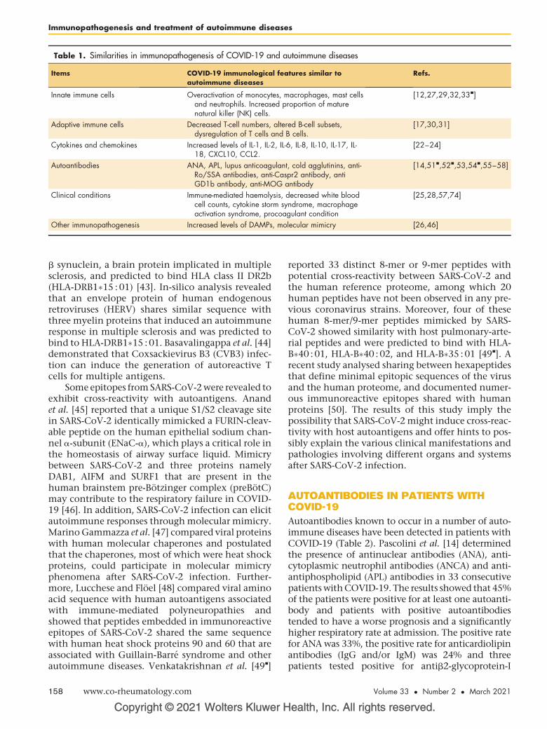
Table 1. Similarities in immunopathogenesis of COVID-19 and autoimmune diseases
Items COVID-19 immunological features similar toautoimmune diseases
Refs.
Innate immune cells Overactivation of monocytes, macrophages, mast cellsand neutrophils. Increased proportion of maturenatural killer (NK) cells.
[12,27,29,32,33&]
Adaptive immune cells Decreased T-cell numbers, altered B-cell subsets,dysregulation of T cells and B cells.
[17,30,31]
Cytokines and chemokines Increased levels of IL-1, IL-2, IL-6, IL-8, IL-10, IL-17, IL-18, CXCL10, CCL2.
[22–24]
Autoantibodies ANA, APL, lupus anticoagulant, cold agglutinins, anti-Ro/SSA antibodies, anti-Caspr2 antibody, antiGD1b antibody, anti-MOG antibody
[14,51&,52&,53,54&,55–58]
Clinical conditions Immune-mediated haemolysis, decreased white bloodcell counts, cytokine storm syndrome, macrophageactivation syndrome, procoagulant condition
[25,28,57,74]
Other immunopathogenesis Increased levels of DAMPs, molecular mimicry [26,46]
Immunopathogenesis and treatment of autoimmune diseases
b synuclein, a brain protein implicated in multiplesclerosis, and predicted to bind HLA class II DR2b(HLA-DRB1�15 : 01) [43]. In-silico analysis revealedthat an envelope protein of human endogenousretroviruses (HERV) shares similar sequence withthree myelin proteins that induced an autoimmuneresponse in multiple sclerosis and was predicted tobind to HLA-DRB1�15 : 01. Basavalingappa et al. [44]demonstrated that Coxsackievirus B3 (CVB3) infec-tion can induce the generation of autoreactive Tcells for multiple antigens.
Some epitopes from SARS-CoV-2 were revealed toexhibit cross-reactivity with autoantigens. Anandet al. [45] reported that a unique S1/S2 cleavage sitein SARS-CoV-2 identically mimicked a FURIN-cleav-able peptide on the human epithelial sodium chan-nel a-subunit (ENaC-a), which plays a critical role inthe homeostasis of airway surface liquid. Mimicrybetween SARS-CoV-2 and three proteins namelyDAB1, AIFM and SURF1 that are present in thehuman brainstem pre-Botzinger complex (preBotC)may contribute to the respiratory failure in COVID-19 [46]. In addition, SARS-CoV-2 infection can elicitautoimmune responses through molecular mimicry.Marino Gammazza et al. [47] compared viral proteinswith human molecular chaperones and postulatedthat the chaperones, most of which were heat shockproteins, could participate in molecular mimicryphenomena after SARS-CoV-2 infection. Further-more, Lucchese and Floel [48] compared viral aminoacid sequence with human autoantigens associatedwith immune-mediated polyneuropathies andshowed that peptides embedded in immunoreactiveepitopes of SARS-CoV-2 shared the same sequencewith human heat shock proteins 90 and 60 that areassociated with Guillain-Barre syndrome and otherautoimmune diseases. Venkatakrishnan et al. [49
&
]
Copyright © 2021 Wolters Kluwer H
158 www.co-rheumatology.com
reported 33 distinct 8-mer or 9-mer peptides withpotential cross-reactivity between SARS-CoV-2 andthe human reference proteome, among which 20human peptides have not been observed in any pre-vious coronavirus strains. Moreover, four of thesehuman 8-mer/9-mer peptides mimicked by SARS-CoV-2 showed similarity with host pulmonary-arte-rial peptides and were predicted to bind with HLA-B�40 : 01, HLA-B�40 : 02, and HLA-B�35 : 01 [49
&
]. Arecent study analysed sharing between hexapeptidesthat define minimal epitopic sequences of the virusand the human proteome, and documented numer-ous immunoreactive epitopes shared with humanproteins [50]. The results of this study imply thepossibility that SARS-CoV-2 might induce cross-reac-tivity with host autoantigens and offer hints to pos-sibly explain the various clinical manifestations andpathologies involving different organs and systemsafter SARS-CoV-2 infection.
AUTOANTIBODIES IN PATIENTS WITHCOVID-19
Autoantibodies known to occur in a number of auto-immune diseases have been detected in patients withCOVID-19 (Table 2). Pascolini et al. [14] determinedthe presence of antinuclear antibodies (ANA), anti-cytoplasmic neutrophil antibodies (ANCA) and anti-antiphospholipid (APL) antibodies in 33 consecutivepatients with COVID-19. The results showed that 45%of the patients were positive for at least one autoanti-body and patients with positive autoantibodiestended to have a worse prognosis and a significantlyhigher respiratory rate at admission. The positive ratefor ANA was 33%, the positive rate for anticardiolipinantibodies (IgG and/or IgM) was 24% and threepatients tested positive for antib2-glycoprotein-I
ealth, Inc. All rights reserved.
Volume 33 � Number 2 � March 2021
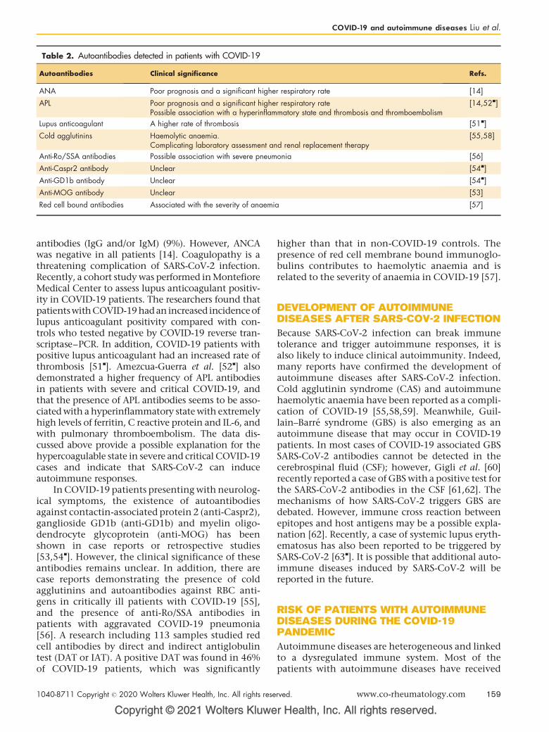
Table 2. Autoantibodies detected in patients with COVID-19
Autoantibodies Clinical significance Refs.
ANA Poor prognosis and a significant higher respiratory rate [14]
APL Poor prognosis and a significant higher respiratory ratePossible association with a hyperinflammatory state and thrombosis and thromboembolism
[14,52&]
Lupus anticoagulant A higher rate of thrombosis [51&]
Cold agglutinins Haemolytic anaemia.Complicating laboratory assessment and renal replacement therapy
[55,58]
Anti-Ro/SSA antibodies Possible association with severe pneumonia [56]
Anti-Caspr2 antibody Unclear [54&]
Anti-GD1b antibody Unclear [54&]
Anti-MOG antibody Unclear [53]
Red cell bound antibodies Associated with the severity of anaemia [57]
COVID-19 and autoimmune diseases Liu et al.
antibodies (IgG and/or IgM) (9%). However, ANCAwas negative in all patients [14]. Coagulopathy is athreatening complication of SARS-CoV-2 infection.Recently, a cohort study was performed in MontefioreMedical Center to assess lupus anticoagulant positiv-ity in COVID-19 patients. The researchers found thatpatientswith COVID-19had an increased incidence oflupus anticoagulant positivity compared with con-trols who tested negative by COVID-19 reverse tran-scriptase–PCR. In addition, COVID-19 patients withpositive lupus anticoagulant had an increased rate ofthrombosis [51
&
]. Amezcua-Guerra et al. [52&
] alsodemonstrated a higher frequency of APL antibodiesin patients with severe and critical COVID-19, andthat the presence of APL antibodies seems to be asso-ciated with a hyperinflammatory state with extremelyhigh levels of ferritin, C reactive protein and IL-6, andwith pulmonary thromboembolism. The data dis-cussed above provide a possible explanation for thehypercoagulable state in severe and critical COVID-19cases and indicate that SARS-CoV-2 can induceautoimmune responses.
In COVID-19 patients presenting with neurolog-ical symptoms, the existence of autoantibodiesagainst contactin-associated protein 2 (anti-Caspr2),ganglioside GD1b (anti-GD1b) and myelin oligo-dendrocyte glycoprotein (anti-MOG) has beenshown in case reports or retrospective studies[53,54
&
]. However, the clinical significance of theseantibodies remains unclear. In addition, there arecase reports demonstrating the presence of coldagglutinins and autoantibodies against RBC anti-gens in critically ill patients with COVID-19 [55],and the presence of anti-Ro/SSA antibodies inpatients with aggravated COVID-19 pneumonia[56]. A research including 113 samples studied redcell antibodies by direct and indirect antiglobulintest (DAT or IAT). A positive DAT was found in 46%of COVID-19 patients, which was significantly
Copyright © 2021 Wolters Kluwe
1040-8711 Copyright � 2020 Wolters Kluwer Health, Inc. All rights rese
higher than that in non-COVID-19 controls. Thepresence of red cell membrane bound immunoglo-bulins contributes to haemolytic anaemia and isrelated to the severity of anaemia in COVID-19 [57].
DEVELOPMENT OF AUTOIMMUNEDISEASES AFTER SARS-COV-2 INFECTION
Because SARS-CoV-2 infection can break immunetolerance and trigger autoimmune responses, it isalso likely to induce clinical autoimmunity. Indeed,many reports have confirmed the development ofautoimmune diseases after SARS-CoV-2 infection.Cold agglutinin syndrome (CAS) and autoimmunehaemolytic anaemia have been reported as a compli-cation of COVID-19 [55,58,59]. Meanwhile, Guil-lain–Barre syndrome (GBS) is also emerging as anautoimmune disease that may occur in COVID-19patients. In most cases of COVID-19 associated GBSSARS-CoV-2 antibodies cannot be detected in thecerebrospinal fluid (CSF); however, Gigli et al. [60]recently reported a case of GBS with a positive test forthe SARS-CoV-2 antibodies in the CSF [61,62]. Themechanisms of how SARS-CoV-2 triggers GBS aredebated. However, immune cross reaction betweenepitopes and host antigens may be a possible expla-nation [62]. Recently, a case of systemic lupus eryth-ematosus has also been reported to be triggered bySARS-CoV-2 [63
&
]. It is possible that additional auto-immune diseases induced by SARS-CoV-2 will bereported in the future.
RISK OF PATIENTS WITH AUTOIMMUNEDISEASES DURING THE COVID-19PANDEMIC
Autoimmune diseases are heterogeneous and linkedto a dysregulated immune system. Most of thepatients with autoimmune diseases have received
r Health, Inc. All rights reserved.
rved. www.co-rheumatology.com 159
Immunopathogenesis and treatment of autoimmune diseases
or are receiving immunomodulatory medications orbiological agents. During the pandemic of COVID-19, a proportion of the autoimmune disease patientssuspended their medication due to fear of the immu-nosuppressive effect of medications or lack of avail-abilities [64], and decreased medical visits because ofconcerns of the contagious nature of SARS-CoV-2[65]. However, disrupted continuity of medical careand medication nonadherence are associated withrheumatologic disease flares and worsened diseaseactivity [66]. Therefore, building a reliable telemed-icine platform and education on medication adher-ence should be strongly recommended.
Since the beginning of this pandemic, infectionrisk in patients with autoimmune diseases has beena subject of interest [67
&
,68,69]. The results of across-sectional study conducted in northeast Italyindicated that autoimmune disease patients had asimilar rate of infection of SARS-CoV-2 comparedwith the general population [70]. Another Italianstudy performed in Milan also confirmed that auto-immune disease is not a risk factor of being positivefor COVID-19 [71]. To the contrary, the results of amulticentre retrospective study conducted in Hubei,China, indicated that patients with autoimmunediseases might be more susceptible to SARS-CoV-2infection compared with controls. Further, thisstudy examined family members of the patients thatresided at the same environment during the out-break as controls [72]. Of interest, the study fromMilan indicated that patients with autoimmunediseases do not have a worse prognosis comparedwith non-autoimmune disease individuals [71].However, a Spanish study revealed that hospitalizedpatients with autoimmune diseases have a moresevere course of COVID-19 [73]. At this time, untilmore data become available, it is crucial to empha-size the importance of physical distancing, wearingmasks and frequent hand washing for everyone andespecially in our patients with autoimmune dis-eases. Adherence to medications is also very impor-tant to prevent flares of autoimmune diseases thatmight result in organ damage.
CONCLUSION
COVID-19 is a novel pandemic that has had signifi-cant global health consequences. Similar to systemicautoimmune diseases, COVID-19 can present withheterogeneous and systemic clinical manifestations.To some extent, there are similarities in the immuneresponse in both disease conditions, and organdamage in COVID-19 appears to be largelyimmune-mediated, similar to autoimmune diseases.The SARS-CoV-2 virus can disturb self-tolerance ofhost antigens at least in part through molecular
Copyright © 2021 Wolters Kluwer H
160 www.co-rheumatology.com
mimicry. Indeed, the development of autoantibod-ies and sometimes organ-specific (e.g. GBS) or sys-temic (e.g. SLE-like disease) autoimmunity has beenobserved in COVID-19. Overall, more data areneeded to further understand the relationshipbetween COVID-19 and autoimmunity and charac-terize the risk and severity of COVID-19 in patientswith preexisting autoimmune diseases.
Acknowledgements
None.
Financial support and sponsorship
This study is supported by grants from the NationalNatural Science Foundation for Young Scientists ofChina (Grant No. 81502732) to Y.L., and an urgentgrant of Hunan Province for fighting against coronavirusdisease- 2019 epidemic (2020SK3005) to Q.L. A.H.S issupported by the National Institutes of Health (NIH)grants number R01AI097134 and R01AR070148, andthe Lupus Research Alliance.
Conflicts of interest
There are no conflicts of interest.
REFERENCES AND RECOMMENDEDREADINGPapers of particular interest, published within the annual period of review, havebeen highlighted as:
& of special interest&& of outstanding interest1. Pollard C, Morran M, Nestor-Kalinoski A. The COVID-19 pandemic: a globalhealth crisis. Physiol Genomics 2020.
2. Domingues R, Lippi A, Setz C, et al. SARS-CoV-2, immunosenescence andinflammaging: partners in the COVID-19 crime. Aging 2020; 12:18778–18789.
3. Hopfer H, Herzig M, Gosert R, et al. Hunting coronavirus by transmissionelectron microscopy: a guide to SARS-CoV-2-associated ultrastructuralpathology in COVID-19 tissues. Histopathology 2020.
4. De P, Bhayye S, Kumar V, Roy K. In silico modeling for quick prediction ofinhibitory activity against 3CL enzyme in SARS CoV diseases. J Biomol StructDynamics 2020; 1–27.
5. Yu F, Xiang R, Deng X, et al. Receptor-binding domain-specific humanneutralizing monoclonal antibodies against SARS-CoV and SARS-CoV-2.Signal Transduc Target Ther 2020; 5:212.
6. Yi C, Sun X, Ye J, et al. Key residues of the receptor binding motif in the spikeprotein of SARS-CoV-2 that interact with ACE2 and neutralizing antibodies.Cell Mol Immunol 2020; 17:621–630.
7. Hoffmann M, Kleine-Weber H, Schroeder S, et al. SARS-CoV-2 cell entrydepends on ACE2 and TMPRSS2 and is blocked by a clinically provenprotease inhibitor. Cell 2020; 181:271–280e278.
8. Bai Y, Xu Y, Wang X, et al. Advances in SARS-CoV-2: a systematic review.Eur Rev Med Pharmacol Sci 2020; 24:9208–9215.
9. Rothan H, Byrareddy S. The epidemiology and pathogenesis of coronavirusdisease (COVID-19) outbreak. J Autoimmun 2020; 109:102433.
10. Schettino M, Pellegrini L, Picascia D, et al. Clinical characteristics of COVID-19 patients with gastrointestinal symptoms in Northern Italy: a single-centercohort study. Am J Gastroenterol 2020.
11. Qian S, Hong W, Lingjie-Mao, et al. Clinical characteristics and outcomes ofsevere and critical patients with 2019 novel coronavirus disease (COVID-19)in Wenzhou: a retrospective study. Front Med 2020; 7:552002.
12. Wang J, Li Q, Yin Y, et al. Excessive neutrophils and neutrophil extracellulartraps in COVID-19. Front Immunol 2020; 11:2063.
13. Esmaeilzadeh A, Elahi R. Immunobiology and immunotherapy of COVID-19: aclinically updated overview. J Cell Physiol 2020.
14. Pascolini S, Vannini A, Deleonardi G, et al. COVID-19 and immunologicaldysregulation: can autoantibodies be useful? Clin Transl Sci 2020.
ealth, Inc. All rights reserved.
Volume 33 � Number 2 � March 2021
COVID-19 and autoimmune diseases Liu et al.
15. Hejrati A, Rafiei A, Soltanshahi M, et al. Innate immune response in systemicautoimmune diseases: a potential target of therapy. Inflammopharmacology2020; 28:1421–1438.
16. Singh A, Thakur M, Sharma L, Chandra K. Designing a multiepitope peptidebased vaccine against SARS-CoV-2. Sci Rep 2020; 10:16219.
17. Rydyznski Moderbacher C, Ramirez S, Dan J, et al. Antigen-specific adaptiveimmunity to SARS-CoV-2 in acute COVID-19 and associations with age anddisease severity. Cell 2020; 183:996–1012.e19.
18. Lancman G, Mascarenhas J, Bar-Natan M. Severe COVID-19 virus reactiva-tion following treatment for B cell acute lymphoblastic leukemia. J HematolOncol 2020; 13:131.
19. Setiati S, Harimurti K, Safitri E, et al. Risk factors and laboratory test resultsassociated with severe illness and mortality in COVID-19 patients: a sys-tematic review. Acta Med Indones 2020; 52:227–245.
20. Ziadi A, Hachimi A, Admou B, et al. Lymphopenia in critically ill COVID-19patients: a predictor factor of severity and mortality. Int J Lab Hematol 2020.
21. Ciceri F, Castagna A, Rovere-Querini P, et al. Early predictors of clinical outcomesof COVID-19 outbreak in Milan, Italy. Clin Immunol 2020; 217:108509.
22. Satıs H, Ozger H, Aysert Yıldız P, et al. Prognostic value of interleukin-18 andits association with other inflammatory markers and disease severity inCOVID-19. Cytokine 2020; 137:155302.
23. Vassallo M, Manni S, Pini P, et al. Patients with Covid-19 exhibit differentimmunological profiles according to their clinical presentation. Int J Infect Dis2020; 101:174–179.
24. Azar M, Shin J, Kang I, Landry M. Diagnosis of SARS-CoV-2 infection in thesetting of cytokine release syndrome. Expert Rev Mol Diagn 2020.
25. Sun Y, Dong Y, Wang L, et al. Characteristics and prognostic factors ofdisease severity in patients with COVID-19: the Beijing experience. J Auto-immun 2020; 112:102473.
26. Chen L, Long X, Xu Q, et al. Elevated serum levels of S100A8/A9 andHMGB1 at hospital admission are correlated with inferior clinical outcomes inCOVID-19 patients. Cell Mol Immunol 2020; 17:992–994.
27. Conti P, Caraffa A, Gallenga C, et al. Coronavirus-19 (SARS-CoV-2) inducesacute severe lung inflammation via IL-1 causing cytokine storm in COVID-19:a promising inhibitory strategy. J Biol Regulat Homeost Agents 2020; 34:.
28. Wampler Muskardin T. Intravenous Anakinra for macrophage activationsyndrome may hold lessons for treatment of cytokine storm in the settingof coronavirus disease 2019. ACR Open Rheumatol 2020; 2:283–285.
29. Conti P, Caraffa A, Tete G, et al. Mast cells activated by SARS-CoV-2 releasehistamine which increases IL-1 levels causing cytokine storm and inflammatoryreaction in COVID-19. J Biol Regul Homeost Agents 2020; 34:1629–1632.
30. Woodruff M, Ramonell R, Nguyen D, et al. Extrafollicular B cell responsescorrelate with neutralizing antibodies and morbidity in COVID-19. Nat Im-munol 2020; 21:1506–1516.
31. Oliviero B, Varchetta S, Mele D, et al. Expansion of atypical memory B cells is aprominent feature of COVID-19. Cell Mol Immunol 2020; 17:1101–1103.
32. Varchetta S, Mele D, Oliviero B, et al. Unique immunological profile in patientswith COVID-19. Cell Mol Immunol 2020.
33.&
Zuo Y, Yalavarthi S, Shi H, et al. Neutrophil extracellular traps in COVID-19.JCI Insight 2020; 5:.
This study demonstrates a possible role for neutrophils and NETs in the patho-geneis of COVID-19.34. Ali RA, Gandhi AA, Meng H, et al. Adenosine receptor agonism protects
against NETosis and thrombosis in antiphospholipid syndrome. Nat Commun2019; 10:1916.
35. Kaminski M, Sunny S, Balabayova K, et al. Tocilizumab therapy of COVID-19:a comparison of subcutaneous and intravenous therapies. Int J Infect Dis2020.
36. Liu Y, Chang C, Lu Q. Management strategies for patients with autoimmunediseases during the COVID-19 pandemic: a perspective from China. Eur JRheumatol 2020; 7:S94–S96.
37. Canziani L, Trovati S, Brunetta E, et al. Interleukin-6 receptor blocking withintravenous tocilizumab in COVID-19 severe acute respiratory distress syn-drome: a retrospective case-control survival analysis of 128 patients. JAutoimmunity 2020; 114:102511.
38. Iglesias-Julian E, Lopez-Veloso M, de-la-Torre-Ferrera N, et al. High dosesubcutaneous Anakinra to treat acute respiratory distress syndrome second-ary to cytokine storm syndrome among severely ill COVID-19 patients. JAutoimmun 2020; 115:102537.
39. Reyes-Castillo Z, Valdes-Miramontes E, Llamas-Covarrubias M, Munoz-ValleJ. Troublesome friends within us: the role of gut microbiota on rheumatoidarthritis etiopathogenesis and its clinical and therapeutic relevance. Clin ExpMed 2020.
40. Harley JB, James JA. Everyone comes from somewhere: systemic lupus erythe-matosus and Epstein-Barr virus induction of host interferon and humoral anti-Epstein-Barr nuclear antigen 1 immunity. Arthritis Rheum 2010; 62:1571–1575.
41. Jog NR, Young KA, Munroe ME, et al. Association of Epstein-Barr virusserological reactivation with transitioning to systemic lupus erythematosus inat-risk individuals. Ann Rheum Dis 2019; 78:1235–1241.
42. Jog NR, McClain MT, Heinlen LD, et al. Epstein Barr virus nuclear antigen 1(EBNA-1) peptides recognized by adult multiple sclerosis patient sera induceneurologic symptoms in a murine model. J Autoimmun 2020; 106:102332.doi: 10.1016/j.jaut.2019.102332.
Copyright © 2021 Wolters Kluwe
1040-8711 Copyright � 2020 Wolters Kluwer Health, Inc. All rights rese
43. Ramasamy R, Mohammed F, Meier U. HLA DR2b-binding peptides fromhuman endogenous retrovirus envelope, Epstein-Barr virus and brain proteinsin the context of molecular mimicry in multiple sclerosis. Immunol Lett 2020;217:15–24.
44. Basavalingappa R, Arumugam R, Lasrado N, et al. Viral myocarditis involvesthe generation of autoreactive T cells with multiple antigen specificities thatlocalize in lymphoid and nonlymphoid organs in the mouse model of CVB3infection. Mol Immunol 2020; 124:218–228.
45. Anand P, Puranik A, Aravamudan M, et al. SARS-CoV-2 strategically mimicsproteolytic activation of human ENaC. eLife 2020; 9:.
46. Lucchese G, Floel A. Molecular mimicry between SARS-CoV-2 and respira-tory pacemaker neurons. Autoimmun Rev 2020; 19:102556.
47. Marino Gammazza A, Legare S, Lo Bosco G, et al. Human molecularchaperones share with SARS-CoV-2 antigenic epitopes potentially cap-able of eliciting autoimmunity against endothelial cells: possible role ofmolecular mimicry in COVID-19. Cell Stress Chaperones 2020;25:737–741.
48. Lucchese G, Floel A. SARS-CoV-2 and Guillain-Barre syndrome: molecularmimicry with human heat shock proteins as potential pathogenic mechanism.Cell Stress Chaperones 2020; 25:731–735.
49.&
Venkatakrishnan A, Kayal N, Anand P, et al. Benchmarking evolutionarytinkering underlying human-viral molecular mimicry shows multiple host pul-monary-arterial peptides mimicked by SARS-CoV-2. Cell Death Discov 2020;6:96.
This is one of several articles suggesting molecular mimickry as a possiblemechnism underlying the development of immune-mediated manifestations inCOVID-19.50. Kanduc D. From anti-SARS-CoV-2 immune responses to COVID-19 via
molecular mimicry. Antibodies (Basel) 2020; 9:.51.&
Reyes Gil M, Barouqa M, Szymanski J, et al. Assessment of lupus antic-oagulant positivity in patients with coronavirus disease 2019 (COVID-19).JAMA Netw Open 2020; 3:e2017539.
This study and reference [51] below describe the presence of antiphospholipdantibodies in COVID-19 patients, which provides an insight to thrombo-inflam-matory features observed in COVID-19.52.&
Amezcua-Guerra L, Rojas-Velasco G, Brianza-Padilla M, et al. Presence ofantiphospholipid antibodies in COVID-19: case series study. Ann Rheum Dis2020.
This study and reference [50] above describe the presence of antiphospholipdantibodies in COVID-19 patients, which provides an insight to thromboinflamma-tory features observed in COVID-19.53. Pinto A, Carroll L, Nar V, et al. CNS inflammatory vasculopathy with antimyelin
oligodendrocyte glycoprotein antibodies in COVID-19. Neurol NeuroimmunolNeuroinflamm 2020; 7:.
54.&
Guilmot A, Maldonado Slootjes S, Sellimi A, et al. Immune-mediated neuro-logical syndromes in SARS-CoV-2-infected patients. J Neurol 2020.
This study summarizes immune-mediated neurological complications in 349patients with COVID-19.55. Jensen C, Wilson S, Thombare A, et al. Cold agglutinin syndrome as a
complication of Covid-19 in two cases. Clin Infect Pract 2020; 7:100041.56. Fujii H, Tsuji T, Yuba T, et al. High levels of anti-SSA/Ro antibodies in COVID-
19 patients with severe respiratory failure: a case-based review: high levels ofanti-SSA/Ro antibodies in COVID-19. Clin Rheumatol 2020.
57. Berzuini A, Bianco C, Paccapelo C, et al. Red cell-bound antibodies andtransfusion requirements in hospitalized patients with COVID-19. Blood2020; 136:766–768.
58. Maslov D, Simenson V, Jain S, Badari A. COVID-19 and cold agglutininhemolytic anemia. TH Open 2020; 4:e175–e177.
59. Patil N, Herc E, Girgis M. Cold agglutinin disease and autoimmune hemolyticanemia with pulmonary embolism as a presentation of COVID-19 infection.Hematol Oncol Stem Cell Ther 2020.
60. Gigli G, Vogrig A, Nilo A, et al. HLA and immunological features of SARS-CoV-2-induced Guillain-Barre syndrome. Neurol Sci 2020; 41:3391–3394.
61. Finsterer J, Scorza F, Fiorini A. SARS-CoV-2 associated Guillain-Barresyndrome in 62 patients. Eur J Neurol 2020.
62. Uncini A, Vallat J, Jacobs B. Guillain-Barre syndrome in SARS-CoV-2 infec-tion: an instant systematic review of the first six months of pandemic. J NeurolNeurosurg Psychiatry 2020; 91:1105–1110.
63.&
Bonometti R, Sacchi M, Stobbione P, et al. The first case of systemic lupuserythematosus (SLE) triggered by COVID-19 infection. Eur Rev Med Phar-macol Sci 2020; 24:9695–9697.
This case report suggests the development of new-onset lupus as a complicationof COVID-19.64. Khabbazi A, Kavandi H, Paribanaem R, et al. Adherence to medication in
patients with rheumatic diseases during COVID-19 pandemic. Ann RheumDis 2020.
65. Chuah S, Teh C, Wan Mohd Akbar S, et al. Impact of COVID-19 pandemicon hospitalisation of patients with systemic lupus erythematosus (SLE):report from a tertiary hospital during the peak of the pandemic. Ann RheumDis 2020.
66. Hassen L, Almaghlouth I, Hassen I, et al. Impact of COVID-19 outbreak onrheumatic patients’ perceptions and behaviors: a cross-sectional study. Int JRheum Dis 2020; 23:1541–1549.
r Health, Inc. All rights reserved.
rved. www.co-rheumatology.com 161
Immunopathogenesis and treatment of autoimmune diseases
67.&
Sawalha AH, Zhao M, Coit P, Lu Q. Epigenetic dysregulation of ACE2and interferon-regulated genes might suggest increased COVID-19susceptibility and severity in lupus patients. Clin Immunol 2020;215:108410.
This study suggests that epigenetic dysregulation of host genes might be relaventto the pathogenesis of COVID-19.68. Sawalha AH, Manzi S. Coronavirus disease-2019: implication for the care and
management of patients with systemic lupus erythematosus. Eur J Rheumatol2020; 7:S117–S120.
69. Sawalha AH. Patients with lupus are not protected from COVID-19. AnnRheum Dis 2020.
70. Zen M, Fuzzi E, Astorri D, et al. SARS-CoV-2 infection in patients withautoimmune rheumatic diseases in northeast Italy: a cross-sectional studyon 916 patients. J Autoimmun 2020; 112:102502.
Copyright © 2021 Wolters Kluwer H
162 www.co-rheumatology.com
71. Murtas R, Andreano A, Gervasi F, et al. Association between autoimmunediseases and COVID-19 as assessed in both a test-negative case-control andpopulation case-control design. Auto Immun Highlights 2020; 11:15.
72. Zhong J, Shen G, Yang H, et al. COVID-19 in patients with rheumatic diseasein Hubei province, China: a multicentre retrospective observational study.Lancet Rheumatol 2020; 2:e557–e564.
73. Pablos J, Galindo M, Carmona L, et al. Clinical outcomes of hospitalisedpatients with COVID-19 and chronic inflammatory and autoimmune rheumaticdiseases: a multicentric matched cohort study. Ann Rheum Dis 2020;79:1544–1549.
74. Voicu S, Delrue M, Chousterman B, et al. Imbalance between procoagulantfactors and natural coagulation inhibitors contributes to hypercoagulability inthe critically ill COVID-19 patient: clinical implications. Eur Rev Med Phar-macol Sci 2020; 24:9161–9168.
ealth, Inc. All rights reserved.
Volume 33 � Number 2 � March 2021
REVIEW
CURRENTOPINION Double-negative T cells in autoimmune diseases
Copyrigh
1040-8711 Copyright � 2020 Wolte
Hao Li and George C. Tsokos
Purpose of review
TCRabþCD4-CD8- double-negative T (DNT) cells, a principal subset of mature T lymphocytes, have beenclosely linked with autoimmune/inflammatory conditions. However, controversy persists regarding theirontogeny and function. Here, we present an overview on DNT cells in different autoimmune diseases toadvance a deeper understanding of the contribution of this population to disease pathogenesis.
Recent findings
DNT cells have been characterized in various chronic inflammatory diseases and they have been proposedto display pathogenic or regulatory function. The tissue location of DNT cells and the effector cytokines theyproduce bespeak to their active involvement in chronic inflammatory diseases.
Summary
By producing various cytokines, expanded DNT cells in inflamed tissues contribute to the pathogenesis of avariety of autoimmune inflammatory diseases. However, it is unclear whether this population represents astable lineage consisting of different subsets similar to CD4þ T helper cell subset. Better understanding ofthe possible heterogeneity and plasticity of DNT cells is needed to reveal interventional therapeuticopportunities.
Keywords
autoimmune diseases, double negative T cells, heterogeneity, ontogeny
Division of Rheumatology and Clinical Immunology, Beth Israel Deacon-ess Medical Center and Harvard Medical School, Boston, Massachu-setts, USA
Correspondence to Hao Li, MD, PhD, Harvard University, Boston02115, MA, USA. E-mail: [email protected],[email protected]
Curr Opin Rheumatol 2021, 33:163–172
DOI:10.1097/BOR.0000000000000778
INTRODUCTION
The most important hallmark of immune disordersis the activation and accumulation of T lympho-cytes, the majority of which express both alpha andbeta chains of the T cell receptor (TCR) and aretherefore referred as ab T cells [1]. Among ab T cells,CD4þ helper or CD8þ cytotoxic T cells are mostprevalent subsets [2]. However, a small populationof ab T cells which do not express both CD4 andCD8, termed ‘double negative’ T (DNT) cells [3,4],have been considered to contribute to the patho-physiology of a series of autoimmune diseases [4].
DNT cells were initially identified and charac-terized in lpr and gld mice (deficiency of either Fas orFas ligand) in which lymphoproliferative syndromedeveloped due to impaired Fas-mediated apoptosis[5–9]. The massively expanded DNT cells results inthe lymphadenopathy and splenomegaly, whichleads to the early hypothesis that DNT cells areimmunopathogenic [5]. Later on, expanded DNTcells were observed in patients with differentimmune disorders, including autoimmune lympho-proliferative syndrome (ALPS) [10,11], systemiclupus erythematosus (SLE) [12,13] and Sjogren’ssyndrome [14,15]. Although double-negative T cellsonly represent a small portion of ab T cells com-pared with either CD4þ or CD8þ T cells in normal
t © 2021 Wolters Kluwe
rs Kluwer Health, Inc. All rights rese
individuals [5,16], the expansion of double-negativeT cells in various autoimmune diseases and thepresence of DNT cells at sites of injury in differentinflammatory conditions strongly suggest their crit-ical roles in inflammation [4]. However, our under-standing of DNT cell ontogeny and function stillremains limited [3–5,17].
We propose that the discrepancy on the differ-entiation and function of DNT cells could beexplained by the heterogeneity and plasticity of thistype of cells.
ONTOGENY OF DOUBLE-NEGATIVE T CELLS
In healthy individuals, DNT cell only comprise asmall portion of ab T cells and are considered quies-cent [5,4]. ab T cells are derived from the developingprogenitors within the thymus, the thymocytes.
r Health, Inc. All rights reserved.
rved. www.co-rheumatology.com
KEY POINTS
� DNT cells are expanded in various chronicinflammatory diseases and they display pathogenic orregulatory function.
� DNT cells are present in inflamed tissues and produceeffector cytokines through which they exercisetheir function.
� It is unclear whether they represent a distinct lineage orthey originate from single positive cells, whether theyrepresent a homogenous group of cells and whetherthey display plasticity.
Immunopathogenesis and treatment of autoimmune diseases
Developing thymocytes undergo a series of matura-tion steps before egressing from the thymus [18] andthe earliest developing thymocytes lack the expres-sion of the coreceptors CD4þ and CD8þ and aretermed double-negative population [18,19], whichleads to the hypothesis that peripheral DNT cells mayrepresent primitive ab T cells, which originate in thethymus but escape the late development followed bymigration to the periphery (Fig. 1a) [3,20]. For late-stage thymocyte development, TCR signal strengthand duration determine the lineage commitment toeither CD4þ or CD8þ T cells. Typically, lower inten-sity TCR signals lead to full maturation of eitherCD4þ or CD8þ T cells, while cells with high TCRstrength are deleted during the development to avoidautoimmunity [21,22]. This process has been wellrecapitulated by in-vitro cultured thymocytes inthe presence of cortical epithelial cells [23]. Consid-ering the fact that CD4þ or CD8þ expression is
Copyright © 2021 Wolters Kluwer H
Late DN DP Immature
SP
Thymus
Peripheral
DNT
A
FasLFa
Peripheral
(a) (b)
FIGURE 1. Ontogeny of double-negative T cells. (a) Peripheral DDP thymocytes through the downregulation of both CD4þ and CD8signals escape AICD and give rise to DNT cells. Right: Cytokine sig
164 www.co-rheumatology.com
essential in augmenting TCR signalling by stabilizinginteractions between TCR-MHC complex [24,25], it isreasonable to postulate that low or negative expres-sion of CD4þ and CD8þ coreceptors protects thymo-cytes away from high-intensity TCR signallingmediated depletionand promotes their thymic egress[26,27]. In contrast to low concentrations of ligandsthat induce maturation to single positive thymocyte,double-positive thymocytes cocultured with corticalepithelial cells loaded with high concentrations ofhigh affinity ligands acquire DNT phenotype withdownregulation of both CD4þ and CD8þ [20,28].
However, there is sufficient evidence to suggestthat DNT cells are generated in the periphery. Forexample, DNT cells can develop in thymectomizedmice reconstituted with T cell depleted bone mar-row cells [29]. The fact that mice deficient in b2-microglobulin have reduced DNT cell lymphopro-liferation [30] and polyclonal DNT cells regain CD8þ
expression in lymphopenic environment [31], indi-cates that DNT may derive from peripheral matureCD8þ T cells [4]. Similar evidence was generatedfrom human studies [32,33]. First, gene expressionpattern analysis revealed that DNT display moresimilarities with CD8þ rather than CD4þ T cells[32]. Second, the analysis of Va and b usage ofTCR revealed the high similarity between CD8þ Tand DNT in patients with ALPS [34]. The dysregu-lated DNT cell homeostasis in lpr, gld mice and ALPSpatients [35–37] has directed the attention to defec-tive apoptosis mediated by Fas dependent pathway[38,39]. The loss-of-function mutations in the Faspathway in T cells lead to impaired apoptosis afterrepeated TCR engagement [9,11]. Activation-
ealth, Inc. All rights reserved.
Mature CD4/CD8
DNT
AICD
popto�c blebs
CD4/
CD8
Mature CD8
s Mature CD4/CD8
Ac�va�on
Mature CD8
Ac�va�on
Apopto�c blebs CD8 CD8
CD4/
CD8
DNT
CytokineCytokine receptor
AICD
prolifera�on
NT cells derive directly from immature DN thymocytes or fromþ. (b) Left: Activated CD8þ T cells without proper apoptoticnal inputs help the conversion from CD8þ T cells to DNT cells.
Volume 33 � Number 2 � March 2021
Double-negative T cells in autoimmune diseases Li and Tsokos
induced cell death (AICD), a Fas/FasL dependentnegative regulator of activated T cells upon repeatedTCR stimulation [40,41], is important for the main-tenance of T-cell homeostasis and abnormalities inthis process may result in autoimmunity [42]. Theevidence above depicts a possible model for thepathogenic DNT cell expansion in autoimmunityin which autoreactive CD8þ T cells skip antigeninduced AICD by losing CD8þ, and execute theirpathogenic role in vivo [5,4]. Along this line, TCRabþ
CD8 T cells lost their CD8þ expression upon stimu-lation with high concentration of anti-CD3 in vitro[32,33]. Adoptively transferred CD8þ T cells withtransgenic TCR acquired DNT-like phenotype afterencountering exogenous or endogenous antigens invivo [16,43,44
&&
]. Moreover, increased Ki67 expres-sion, narrowed TCR Vb repertoire usage and dilutedT-cell receptor excision circles (TRECs) observed inDNT cells indicated the clonal proliferation andexpansion possibly driven by endogenous self-anti-gens [44
&&
,45,46,47&
,48,49]. Of note, in-vivo antigenadministration to MHC class I-restricted TCR trans-genic mice on lpr background resulted in expansionof DNT cells [33], which further supported the con-cept that the expanded DNT cells under chronicinflammation might derive from antigen activatedCD8 T cells [4,17]. However, it remains a mysterywhether the absence of proper apoptotic signals oraddition of supportive signals such as cytokines helpactivated CD8þ T cells escape AICD and acquire DNTcell phenotype (Fig. 1b).
To date, the controversy on the origin of DNTcells continues. There are several possible scenarios,which are worth of attention: double-negative Tcells directly originate from those immature dou-ble-negative thymocytes, which could not recognizeMHC class I or MHC class II molecules but for somereason are not appropriately depleted in thymic-positive selection. Double-negative T cells representa unique lineage, which is selected by recognition ofneither class I nor MHC class II but certain unknownMHC-like molecules. There are different types ofDNT cells with either intrathymic or extrathymicorigin, a model which we favour most, as it fits thebest for the above augments [5,17].
DOUBLE-NEGATIVE T CELLS, THE EVIL ORTHE ANGEL IN INFLAMMATION
Under naive status, DNT cells represent a minorpopulation in total ab T cells with unrecognizedroles in immune system. However, the lupus likesymptoms in lpr or gld mice and disease-associatedexpansion of DNT cells lead to the supposition thatDNT cells are assigned a pro-inflammatory role [6–9,50]. The findings that DNT cells are also expanded
Copyright © 2021 Wolters Kluwe
1040-8711 Copyright � 2020 Wolters Kluwer Health, Inc. All rights rese
in patients with various inflammatory rheumaticdisorders including ALPS and SLE reinforce thisconcept [11,12]. Evidence has emerged that sup-ports the pathogenic role of DNT cells [4]. Ex-vivoanalysis on the cytokine profiles of DNT cells fromvarious murine models has shown the great abilityto produce various inflammatory cytokines, includ-ing interleukin (IL)-2, IL-4, TNFa and IL-17A[31,43,51]. Similar results were also achieved fromstudies in humans with diverse autoimmune dis-eases [4]. In addition, DNT cells provide help to Bcells to enhance autoantibody production in vitro[12]. Immune cell infiltration is generally consid-ered as a major contributor of tissue damages duringchronic inflammation [52]. Along this line, DNTcells are present in inflamed kidney[13,44
&&
,48,47&
], skin [53], salivary gland [14], enthe-ses [54] and ischemic brain [55
&
], which suggeststhey present good therapeutic targets to controlinflammation in various diseases.
The activation of T cells requires signallingthrough TCR and the coreceptors CD4þ and CD8þ
are essential augmenting TCR signalling[1,24,25,56]. It has been argued that the cognateTCR-antigen interaction without proper augmenta-tion by CD4þ and CD8þ molecules is sufficient todrive DNT cell activation in vivo. Mice with concom-itant deficiency of both CD4þ and CD8þ developedinflammatory responses and immunopathologycompared with wild-type mice during acute Staph-ylococcal enterotoxin B infection (SEB) [57]. Ofnote, chronic exposure to SEB precipitated alupus-like inflammatory disease characterized bylympho-monocytic infiltration in multiple tissuesalong with production of autoantibodies in thesedouble gene deficient mice [57]. Interestingly, dis-ease development was accompanied with the expan-sion of DNT cells [57]. In line with their response toSEB, in the lung of mice challenged with live vaccinestrain (LSV) of Francisella tularensis, DNT cells rep-resent the major responding T cell subset [58]. Also,in HIV-infected patients, DNT cells represent a sig-nificant portion of the cellular viral load in T cells[59,60], which suggest in vivo they might functionsimilar to CD4þ T cells.
In contrast to above studies, evidence has beengenerated suggesting that at least subsets of DNTcells exert regulatory activity [17,46]. In skin or bonemarrow allograft murine model, DNT cells werecapable of suppressing syngeneic CD4þ or CD8þ Tcells in both Fas-dependent and Fas-independentmanners [17]. In addition, DNT cells were also capa-ble of inhibiting natural killer (NK) cell-mediatedrejection of allogenic bone marrow through per-forin-dependent killing [61]. In agreement withtheir role in transplantation, a number of studies
r Health, Inc. All rights reserved.
rved. www.co-rheumatology.com 165
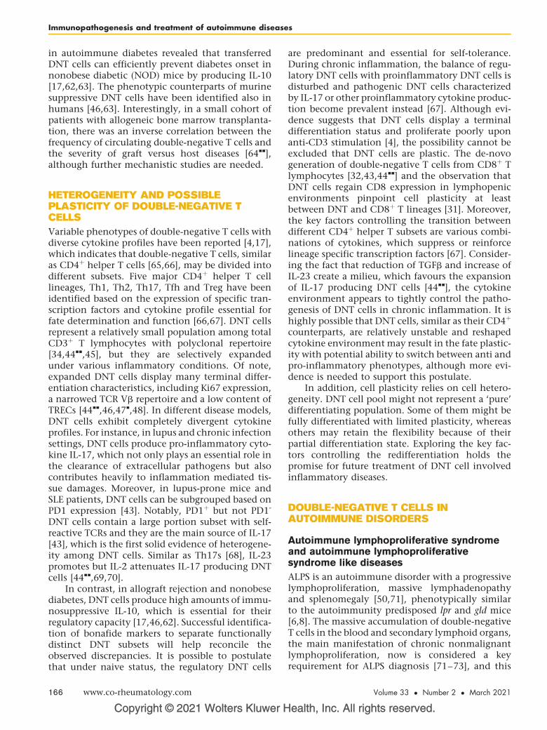
Immunopathogenesis and treatment of autoimmune diseases
in autoimmune diabetes revealed that transferredDNT cells can efficiently prevent diabetes onset innonobese diabetic (NOD) mice by producing IL-10[17,62,63]. The phenotypic counterparts of murinesuppressive DNT cells have been identified also inhumans [46,63]. Interestingly, in a small cohort ofpatients with allogeneic bone marrow transplanta-tion, there was an inverse correlation between thefrequency of circulating double-negative T cells andthe severity of graft versus host diseases [64
&&
],although further mechanistic studies are needed.
HETEROGENEITY AND POSSIBLEPLASTICITY OF DOUBLE-NEGATIVE TCELLS
Variable phenotypes of double-negative T cells withdiverse cytokine profiles have been reported [4,17],which indicates that double-negative T cells, similaras CD4þ helper T cells [65,66], may be divided intodifferent subsets. Five major CD4þ helper T celllineages, Th1, Th2, Th17, Tfh and Treg have beenidentified based on the expression of specific tran-scription factors and cytokine profile essential forfate determination and function [66,67]. DNT cellsrepresent a relatively small population among totalCD3þ T lymphocytes with polyclonal repertoire[34,44
&&
,45], but they are selectively expandedunder various inflammatory conditions. Of note,expanded DNT cells display many terminal differ-entiation characteristics, including Ki67 expression,a narrowed TCR Vb repertoire and a low content ofTRECs [44
&&
,46,47&
,48]. In different disease models,DNT cells exhibit completely divergent cytokineprofiles. For instance, in lupus and chronic infectionsettings, DNT cells produce pro-inflammatory cyto-kine IL-17, which not only plays an essential role inthe clearance of extracellular pathogens but alsocontributes heavily to inflammation mediated tis-sue damages. Moreover, in lupus-prone mice andSLE patients, DNT cells can be subgrouped based onPD1 expression [43]. Notably, PD1þ but not PD1-
DNT cells contain a large portion subset with self-reactive TCRs and they are the main source of IL-17[43], which is the first solid evidence of heterogene-ity among DNT cells. Similar as Th17s [68], IL-23promotes but IL-2 attenuates IL-17 producing DNTcells [44
&&
,69,70].In contrast, in allograft rejection and nonobese
diabetes, DNT cells produce high amounts of immu-nosuppressive IL-10, which is essential for theirregulatory capacity [17,46,62]. Successful identifica-tion of bonafide markers to separate functionallydistinct DNT subsets will help reconcile theobserved discrepancies. It is possible to postulatethat under naive status, the regulatory DNT cells
Copyright © 2021 Wolters Kluwer H
166 www.co-rheumatology.com
are predominant and essential for self-tolerance.During chronic inflammation, the balance of regu-latory DNT cells with proinflammatory DNT cells isdisturbed and pathogenic DNT cells characterizedby IL-17 or other proinflammatory cytokine produc-tion become prevalent instead [67]. Although evi-dence suggests that DNT cells display a terminaldifferentiation status and proliferate poorly uponanti-CD3 stimulation [4], the possibility cannot beexcluded that DNT cells are plastic. The de-novogeneration of double-negative T cells from CD8þ Tlymphocytes [32,43,44
&&
] and the observation thatDNT cells regain CD8 expression in lymphopenicenvironments pinpoint cell plasticity at leastbetween DNT and CD8þ T lineages [31]. Moreover,the key factors controlling the transition betweendifferent CD4þ helper T subsets are various combi-nations of cytokines, which suppress or reinforcelineage specific transcription factors [67]. Consider-ing the fact that reduction of TGFb and increase ofIL-23 create a milieu, which favours the expansionof IL-17 producing DNT cells [44
&&
], the cytokineenvironment appears to tightly control the patho-genesis of DNT cells in chronic inflammation. It ishighly possible that DNT cells, similar as their CD4þ
counterparts, are relatively unstable and reshapedcytokine environment may result in the fate plastic-ity with potential ability to switch between anti andpro-inflammatory phenotypes, although more evi-dence is needed to support this postulate.
In addition, cell plasticity relies on cell hetero-geneity. DNT cell pool might not represent a ‘pure’differentiating population. Some of them might befully differentiated with limited plasticity, whereasothers may retain the flexibility because of theirpartial differentiation state. Exploring the key fac-tors controlling the redifferentiation holds thepromise for future treatment of DNT cell involvedinflammatory diseases.
DOUBLE-NEGATIVE T CELLS INAUTOIMMUNE DISORDERS
Autoimmune lymphoproliferative syndromeand autoimmune lymphoproliferativesyndrome like diseases
ALPS is an autoimmune disorder with a progressivelymphoproliferation, massive lymphadenopathyand splenomegaly [50,71], phenotypically similarto the autoimmunity predisposed lpr and gld mice[6,8]. The massive accumulation of double-negativeT cells in the blood and secondary lymphoid organs,the main manifestation of chronic nonmalignantlymphoproliferation, now is considered a keyrequirement for ALPS diagnosis [71–73], and this
ealth, Inc. All rights reserved.
Volume 33 � Number 2 � March 2021
Double-negative T cells in autoimmune diseases Li and Tsokos
elevation results from a primary defect in Fas-medi-ated apoptosis [9,11,41,72]. In patients who developsome features of ALPS but do not fulfil the diagnosticcriteria for ALPS, mutations in other components ofpathways central to lymphocyte growth, activationand apoptosis have been identified including Cas-pase-8 and FADD [71,74]. These have been groupedinto ALPS-like diseases and some patients in thiscategory have increased DNT cells also [71,74].
Interestingly, CDR3 sequencing has revealed asignificant overlap of TCR Vb-Jb transcripts betweenDNT cells and CD8þ T cells from ALPS patients[34,49], which strongly suggest the at least partialCD8þ origin of DNT cells in ALPS. The concept thatDNT cells contribute to autoimmune symptoms inALPS patients and autoimmunity predisposed lprand gld mice comes from the following facts: Theprogressive expansion of DNT cells is closely associ-ated with disease development [75]. The presence ofautoantibodies in most ALPS patients correlateswith the number of double-negative T cells[76,77]. Effective treatment ameliorates autoim-mune symptoms in ALPS with significant elimina-tion of abnormal DNT cells [78–80]. Although theelevation of DNT cells in ALPS is not in dispute,further evidence is needed to validate theirpathophysiological significance.
Systemic lupus erythematosus
SLE is a clinically heterogeneous autoimmune dis-ease with systemic inflammation and organ damage[81]. Various T cell abnormalities were reported andthe expansion of DNT cells represents a prominentone [12,13,82]. The early observation that SLEpatients have expanded numbers of DNT cells inthe peripheral blood and this expansion correlateswith disease activity leads to the supposition thatexpanded DNT cells contribute to the pathophysi-ology of SLE [13,83]. The first evidence was from in-vitro co-culture assays, which clearly demonstratedthat DNT cells provide help to B cells to promoteantibody production [12]. IL-17A, a pro-inflamma-tory cytokine, has been documented with crucialcontribution for systemic inflammation and tissuedamage in SLE [13,84–86]. The findings that DNTcells represent a major source of IL-17A in SLEpatients reinforced the concept on DNT cell patho-genesis in SLE [4,13]. Moreover, DNT cell invasion inthe kidneys of patients with lupus nephritis has alsobeen recorded [37]. A series of experiments reportedfrom our laboratory have demonstrated that a largeportion of expanded double-negative T cells in SLEwere derived from self-reactive CD8þ T cells[16,31,32,44
&&
]. Self-antigens derived from apopto-tic cells can activate self-reactive CD8þ T cells,
Copyright © 2021 Wolters Kluwe
1040-8711 Copyright � 2020 Wolters Kluwer Health, Inc. All rights rese
which give rise to double-negative T cells throughthe downregulation of CD8 expression on the cellsurface. These cells displayed acquired proliferatingor proliferated phenotype (Ki67 expression, dilutedTREC and narrowed TCR repertoire) [44
&&
]. CD8þ
Treg cells have been described as CD8þ T cells spe-cific for antigen delivered to immune-privilegedsites and to control the effector T-cell responsesby CD8þ and perforin dependent killing [87–89].The whole process of conversion from CD8þ T cellsinto DNT cells contributes to the pathogenesis oflupus based on the loss of CD8þ-dependent immu-nosuppressive potentials and the acquisition of abil-ity to produce different pro-inflammatory cytokinesand especially IL-17 [44
&&
]. In addition to TCR sig-nalling and coreceptor signalling, cytokines providethe third signal for T cell activation and surviving[90]. The fact that skewed inflammatory cytokineenvironment in lupus favours the expansion of DNTcells suggests that cytokines compensate reducedTCR signalling strength due to the loss of CD8þ
for cell activation and survival [44&&
].
Sjogren’s syndrome, psoriasis, axialspondylarthritis and other rheumaticdiseases
Sjogren’s syndrome is a systemic autoimmune dis-ease characterized by lymphocytic infiltration insalivary and tear glands [91]. Sjogren’s syndromemay occur as primary disease but most often occursin the context of other autoimmune disorders [91],including SLE and rheumatoid arthritis. Similar as inSLE, DNT cells are expanded and become the mainsource of IL-17 in patients with primary Sjogren’ssyndrome [14,15,92]. The expansion of DNT cellscorrelates well with disease activity and IL-17þ DNTcell infiltration was detected in inflamed salivaryglands [14,15].
Psoriasis is a complex inflammatory skin diseasecharacterized by immune cell infiltration to the skin[93]. IL-17 producing DNT cell infiltration wasfound in the epidermis of mice with induced psori-asis [94] and patients with plaque-type psoriasis [53].Axial spondylarthritis is another chronic inflamma-tory disease, which affects primarily the spine andthe sacroiliac joints [95] but shares many geneticfeatures with psoriasis [96]. Interestingly, in a widelyaccepted murine model of spondyloarthropathy, IL-23RþDNT cells were detected in the inflamed enthe-ses [54]. Again, these observations reinforce theperception that DNT cells contribute heavily topathogenesis of many inflammatory rheumatoiddisorders. Furthermore, DNT cells are expanded ina subset of paediatric patients with various autoim-mune diseases including mixed connective tissue
r Health, Inc. All rights reserved.
rved. www.co-rheumatology.com 167
Immunopathogenesis and treatment of autoimmune diseases
disease (MCTD), juvenile idiopathic arthritis (JIA),juvenile dermatomyositis [97] and Behcet’s disease[98], although additional investigations are requiredfor their precise role in these patients.
Type 1 diabetes
Type 1 diabetes (T1D) is an organ-specific autoim-mune disease with severe loss of pancreatic b cells[99]. Both CD4þ and CD8þ T cells play distinct andhighly pathogenic roles in b cell destruction [100].In contrast to the pathogenesis of DNT cells ininflammatory rheumatoid disorders listed above, anumber of studies have demonstrated the immuno-suppressive ability of DNT cells and their ability toinhibit the development of autoimmune diabetes[3,17]. First, a progressive loss of DNT cells with agewas observed in nonobese diabetic (NOD) mice [62].Second, adoptive transfer of DNT cells efficientlyinhibited the development of autoimmune diabetesin several different diabetic mouse models[63,101,102]. Third, transfer of NOD CD8þ T cellsresulted in diabetes but co-transfer of NOD CD8þ Tcells with DNT cells did not, which indicates thatDNT cells act directly on pathogenic T cells toexercise their immunosuppressive function[17,62]. However, controversies remain on thenature of immunosuppression of DNT cells. Distinctmechanisms with different molecules involved havebeen proposed for DNT cell mediated suppressionincluding elimination of effector T cells by eitherFas/FasL-mediated apoptosis [103,104] or perforinmediated killing [46,102,105,106] and modulationon antigen presenting cells by producing IL-10[62,63] or IFNg [46,107,108]. Of note, both IL-10and IFNg function as a double-edged sword in auto-immune diseases [109,110] and the immune envi-ronments determine whether they are beneficial ordetrimental. Therefore, more mechanistic studiesare needed.
THERAPEUTIC INTERVENTIONSTARGETING DOUBLE-NEGATIVE T CELLS
In autoimmune diseases wherein expanded DNTcells display distinct pathogenic capacity their selec-tive ablation or specific modulation of the processesthat render them less pathogenic should be consid-ered for therapeutic purposes. More attentionshould be given to the design of specific drugs ableto limit the expansion pathogenic DNT cells or ifpossible favour regulatory DNT activation. In lightof understanding of DNT cell generation in lupus,more and more approaches directly or indirectlytargeting DNT cells have been tested. In lupus-pronemice and SLE patients, a large portion of DNT cells
Copyright © 2021 Wolters Kluwer H
168 www.co-rheumatology.com
were derived from antigen-stimulated CD8þ T cells.The activation-induced chromatin remodelling andepigenetic silencing on various promoters andenhancers of Cd8 locus might be responsible forthe de-novo generation of DNT cells from CD8þ Tcells. As expected, the methylome of DNT cellsaffirmed hypermethylation on regulatory elementsof Cd8 locus [111]. In brief, the transcription factorcAMP responsive element modulator (CREM)aorchestrates DNA methyltransferase (DNMT)3aand histone methyltransferase G9a to directlyenhance DNA and histone methylation on Cd8locus [112,113], which results in stable epigeneticsilencing [113,114]. Accordingly, genetic deficiencyof Crem in lupus-prone mice significantly amelio-rates disease manifestations by reducing IL-17þDNTcells [115]. DNA methylation patterns in SLE T cellsare complex with both hypomethylated and hyper-methylated cytosine-guanine sites [116,117]. Gen-eralized DNA hypomethylation in CD4þ T cells hasbeen well linked to the disease manifestation[118,119]. Surprisingly, in contrast to systemicdelivery of 5-azacytidine [120], a DNA methyltrans-ferase inhibitor, which profoundly augments dis-ease progression [121], its targeted delivery toCD8þ T cells using a nanolipogel delivery systemsignificantly ameliorated disease severity in lupusprone mice by restraining the expansion of patho-genic DNT cells [122]. This result is consistent withthe proposition that CD8þ T cells acquire pro-inflammatory DNT cell phenotype throughenhanced DNA methylation mediated CD8þ loss[4]. In line with these observations, well controlledCD8þ expression on CD8þ T cells and DNT cells byproper modulation of epigenetic modification onCd8 locus should present a valuable therapeuticstrategy for the treatment of autoimmune diseasewith involvement of DNT cells (Fig. 2a).
The expanded DNT cells in human and micewith defective Fas-mediated pathway depictedanother picture, in which double-negative T cellswere derived from mature T cells with failed apo-ptosis [5]. Along this line, DNT cells with resistanceto AICD could be generated in vitro from Fas-suffi-cient T-cells with repeated anti-CD3 stimulation[32,33]. However, further studies are warranted tovalidate whether addition of FasL or other apoptosisinducing molecules could modulate the generationof DNT cells as expected in vitro and in vivo (Fig. 2b),as the controversies persist on the therapeutic valuesof FasL in autoimmune disease [123]. Fas and FasLplay essential immunosuppressive roles in control-ling T cell homeostasis, as recorded with the devel-opment of autoimmunity in lpr or gld mice [5].Paradoxically, Fas also plays a proinflammatory rolein certain settings, as lpr or gld mice are resistant to
ealth, Inc. All rights reserved.
Volume 33 � Number 2 � March 2021
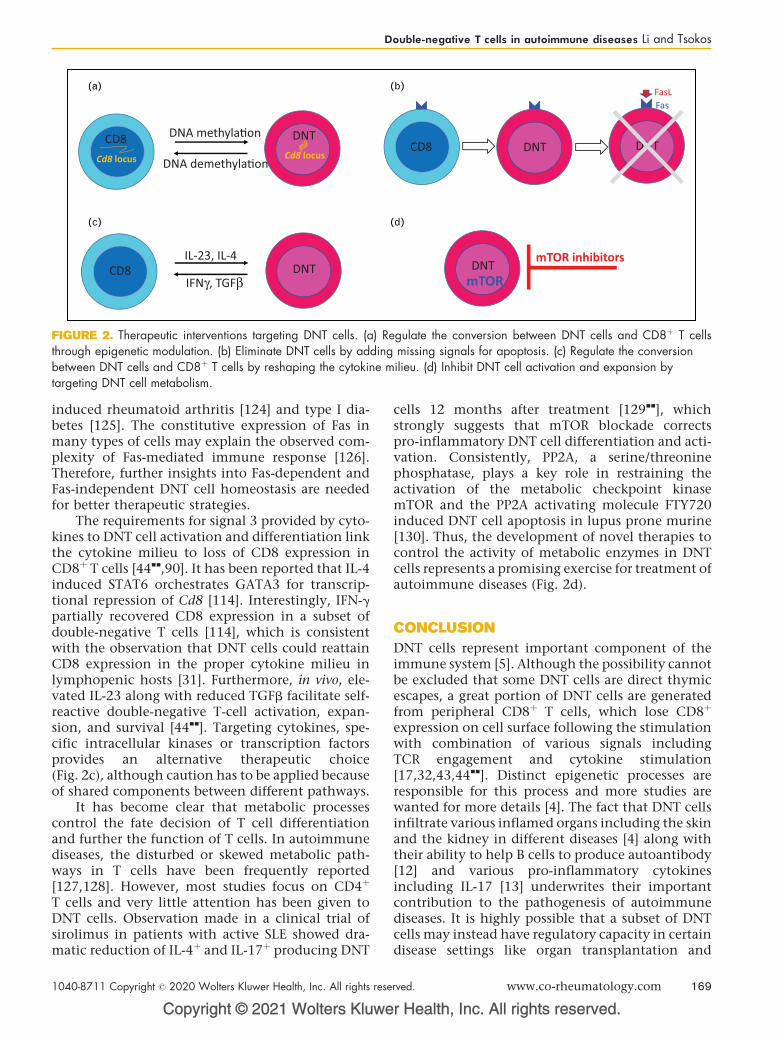
CD8 DNT
Cd8 locus Cd8 locusDNA demethyla�on
DNA methyla�onCD8 DNT DNT
FasFasL
CD8 DNTIFNγ, TGFβ
IL-23, IL-4DNT
mTOR
mTOR inhibitors
(a) (b)
(c) (d)
FIGURE 2. Therapeutic interventions targeting DNT cells. (a) Regulate the conversion between DNT cells and CD8þ T cellsthrough epigenetic modulation. (b) Eliminate DNT cells by adding missing signals for apoptosis. (c) Regulate the conversionbetween DNT cells and CD8þ T cells by reshaping the cytokine milieu. (d) Inhibit DNT cell activation and expansion bytargeting DNT cell metabolism.
Double-negative T cells in autoimmune diseases Li and Tsokos
induced rheumatoid arthritis [124] and type I dia-betes [125]. The constitutive expression of Fas inmany types of cells may explain the observed com-plexity of Fas-mediated immune response [126].Therefore, further insights into Fas-dependent andFas-independent DNT cell homeostasis are neededfor better therapeutic strategies.
The requirements for signal 3 provided by cyto-kines to DNT cell activation and differentiation linkthe cytokine milieu to loss of CD8 expression inCD8þ T cells [44
&&
,90]. It has been reported that IL-4induced STAT6 orchestrates GATA3 for transcrip-tional repression of Cd8 [114]. Interestingly, IFN-gpartially recovered CD8 expression in a subset ofdouble-negative T cells [114], which is consistentwith the observation that DNT cells could reattainCD8 expression in the proper cytokine milieu inlymphopenic hosts [31]. Furthermore, in vivo, ele-vated IL-23 along with reduced TGFb facilitate self-reactive double-negative T-cell activation, expan-sion, and survival [44
&&
]. Targeting cytokines, spe-cific intracellular kinases or transcription factorsprovides an alternative therapeutic choice(Fig. 2c), although caution has to be applied becauseof shared components between different pathways.
It has become clear that metabolic processescontrol the fate decision of T cell differentiationand further the function of T cells. In autoimmunediseases, the disturbed or skewed metabolic path-ways in T cells have been frequently reported[127,128]. However, most studies focus on CD4þ
T cells and very little attention has been given toDNT cells. Observation made in a clinical trial ofsirolimus in patients with active SLE showed dra-matic reduction of IL-4þ and IL-17þ producing DNT
Copyright © 2021 Wolters Kluwe
1040-8711 Copyright � 2020 Wolters Kluwer Health, Inc. All rights rese
cells 12 months after treatment [129&&
], whichstrongly suggests that mTOR blockade correctspro-inflammatory DNT cell differentiation and acti-vation. Consistently, PP2A, a serine/threoninephosphatase, plays a key role in restraining theactivation of the metabolic checkpoint kinasemTOR and the PP2A activating molecule FTY720induced DNT cell apoptosis in lupus prone murine[130]. Thus, the development of novel therapies tocontrol the activity of metabolic enzymes in DNTcells represents a promising exercise for treatment ofautoimmune diseases (Fig. 2d).
CONCLUSION
DNT cells represent important component of theimmune system [5]. Although the possibility cannotbe excluded that some DNT cells are direct thymicescapes, a great portion of DNT cells are generatedfrom peripheral CD8þ T cells, which lose CD8þ
expression on cell surface following the stimulationwith combination of various signals includingTCR engagement and cytokine stimulation[17,32,43,44
&&
]. Distinct epigenetic processes areresponsible for this process and more studies arewanted for more details [4]. The fact that DNT cellsinfiltrate various inflamed organs including the skinand the kidney in different diseases [4] along withtheir ability to help B cells to produce autoantibody[12] and various pro-inflammatory cytokinesincluding IL-17 [13] underwrites their importantcontribution to the pathogenesis of autoimmunediseases. It is highly possible that a subset of DNTcells may instead have regulatory capacity in certaindisease settings like organ transplantation and
r Health, Inc. All rights reserved.
rved. www.co-rheumatology.com 169
Immunopathogenesis and treatment of autoimmune diseases
nonobese diabetes [17]. A growing understanding ofDNT cell origin and functional features hasprompted the consideration of therapeuticapproaches including targeted reopening of theCD8 locus, precise modulation of cell activationand survival, inhibition of proinflammatory meta-bolic pathways and blockade of the inflammatorymilieu which enables their generation or enablingtheir demise.
A number of questions needs urgent attention. Aclear characterization of DNT subsets is neededthrough novel spectral cytometry and single cellsequencing technologies. The factors which enablethe expansion of proinflammatory or regulatoryDNT cells in various diseases need to be defined.Using advanced protocols, including Slide-Seq[131], the exact interaction between DNT cellsand other immune cells or tissue resident cellsshould be defined. Prospective clinical studies areneeded to define their appearance during the evo-lution of the disease process. Such studies mayreveal that certain characteristic of DNT cells inthe periphery can serve as biomarkers of organinflammation and disease activity.
Acknowledgements
None.
Financial support and sponsorship
This worked was supported by grants RO1 AI085567 toG.C.T. and T32 DK007199 to H.L.
Conflicts of interest
There are no conflicts of interest.
REFERENCES AND RECOMMENDEDREADINGPapers of particular interest, published within the annual period of review, havebeen highlighted as:
& of special interest&& of outstanding interest1. Davis MM, Boniface JJ, Reich Z, et al. Ligand recognition by alpha beta T cellreceptors. Annu Rev Immunol 1998; 16:523–544.
2. Miceli MC, Parnes JR. The roles of CD4 and CD8 in T cell activation. SeminImmunol 1991; 3:133–141.
3. D’Acquisto F, Crompton T. CD3þCD4-CD8- (double negative) T cells:saviours or villains of the immune response? Biochem Pharmacol 2011;82:333–340.
4. Brandt D, Hedrich CM. TCRalphabeta(þ)CD3(þ)CD4(�)CD8(�) (doublenegative) T cells in autoimmunity. Autoimmun Rev 2018; 17:422–430.
5. Martina MN, Noel S, Saxena A, et al. Double negative (DN) alphabeta T cells:misperception and overdue recognition. Immunol Cell Biol 2015;93:305–310.
6. Morse HC 3rd, Davidson WF, Yetter RA, et al. Abnormalities induced by themutant gene Ipr: expansion of a unique lymphocyte subset. J Immunol 1982;129:2612–2615.
7. Davidson WF, Dumont FJ, Bedigian HG, et al. Phenotypic, functional, andmolecular genetic comparisons of the abnormal lymphoid cells of C3H-lpr/lprand C3H-gld/gld mice. J Immunol 1986; 136:4075–4084.
8. Roths JB, Murphy ED, Eicher EM. A new mutation, gld, that produceslymphoproliferation and autoimmunity in C3H/HeJ mice. J Exp Med 1984;159:1–20.
Copyright © 2021 Wolters Kluwer H
170 www.co-rheumatology.com
9. Cheng J, Zhou T, Liu C, et al. Protection from Fas-mediated apoptosis by asoluble form of the Fas molecule. Science 1994; 263:1759–1762.
10. Sneller MC, Straus SE, Jaffe ES, et al. A novel lymphoproliferative/auto-immune syndrome resembling murine lpr/gld disease. J Clin Invest 1992;90:334–341.
11. Fisher GH, Rosenberg FJ, Straus SE, et al. Dominant interfering Fas genemutations impair apoptosis in a human autoimmune lymphoproliferativesyndrome. Cell 1995; 81:935–946.
12. Shivakumar S, Tsokos GC, Datta SK. T cell receptor alpha/beta expressingdouble-negative (CD4�/CD8�) and CD4þ T helper cells in humans aug-ment the production of pathogenic anti-DNA autoantibodies associated withlupus nephritis. J Immunol 1989; 143:103–112.
13. Crispin JC, Oukka M, Bayliss G, et al. Expanded double negative T cells inpatients with systemic lupus erythematosus produce IL-17 and infiltrate thekidneys. J Immunol 2008; 181:8761–8766.
14. Alunno A, Bistoni O, Bartoloni E, et al. IL-17-producing CD4-CD8- T cells areexpanded in the peripheral blood, infiltrate salivary glands and are resistant tocorticosteroids in patients with primary Sjogren’s syndrome. Ann Rheum Dis2013; 72:286–292.
15. Alunno A, Carubbi F, Bistoni O, et al. CD4(�)CD8(�) T-cells in primarySjogren’s syndrome: association with the extent of glandular involvement. JAutoimmun 2014; 51:38–43.
16. Rodriguez-Rodriguez N, Apostolidis SA, Fitzgerald L, et al. Pro-inflammatoryself-reactive T cells are found within murine TCR-alphabeta(þ) CD4(�)CD8(�) PD-1(þ) cells. Eur J Immunol 2016; 46:1383–1391.
17. Juvet SC, Zhang L. Double negative regulatory T cells in transplantation andautoimmunity: recent progress and future directions. J Mol Cell Biol 2012;4:48–58.
18. Anderson G, Jenkinson EJ. Lymphostromal interactions in thymic develop-ment and function. Nat Rev Immunol 2001; 1:31–40.
19. Spits H. Development of alphabeta T cells in the human thymus. Nat RevImmunol 2002; 2:760–772.
20. Mixter PF, Russell JQ, Morrissette GJ, et al. A model for the origin of TCR-alphabetaþ CD4-CD8- B220þ cells based on high affinity TCR signals. JImmunol 1999; 162:5747–5756.
21. Kurd N, Robey EA. T-cell selection in the thymus: a spatial and temporalperspective. Immunol Rev 2016; 271:114–126.
22. Klein L, Kyewski B, Allen PM, Hogquist KA. Positive and negative selection ofthe T cell repertoire: what thymocytes see (and don’t see). Nat Rev Immunol2014; 14:377–391.
23. Takahama Y. Journey through the thymus: stromal guides for T-cell devel-opment and selection. Nat Rev Immunol 2006; 6:127–135.
24. Li QJ, Dinner AR, Qi S, et al. CD4 enhances T cell sensitivity to antigen bycoordinating Lck accumulation at the immunological synapse. Nat Immunol2004; 5:791–799.
25. Holler PD, Kranz DM. Quantitative analysis of the contribution of TCR/pepMHC affinity and CD8 to T cell activation. Immunity 2003; 18:255–264.
26. Starr TK, Jameson SC, Hogquist KA. Positive and negative selection of Tcells. Annu Rev Immunol 2003; 21:139–176.
27. Kreslavsky T, Kim HJ, Koralov SB, et al. Negative selection, not receptorediting, is a physiological response of autoreactive thymocytes. J Exp Med2013; 210:1911–1918.
28. Wang R, Wang-Zhu Y, Grey H. Interactions between double positivethymocytes and high affinity ligands presented by cortical epithelial cellsgenerate double negative thymocytes with T cell regulatory activity. Proc NatlAcad Sci U S A 2002; 99:2181–2186.
29. Ford MS, Zhang ZX, Chen W, Zhang L. Double-negative T regulatory cellscan develop outside the thymus and do not mature from CD8þ T cellprecursors. J ‘Immunol 2006; 177:2803–2809.
30. Zhang Y, Wang H, Li J, et al. Intracellular zinc release and ERK phosphor-ylation are required upstream of 12-lipoxygenase activation in peroxynitritetoxicity to mature rat oligodendrocytes. J Biol Chem 2006; 281:9460–9470.
31. Rodriguez-Rodriguez N, Flores-Mendoza G, Apostolidis SA, et al. TCR-alpha/beta CD4(�) CD8 (�) double negative T cells arise from CD8(þ) Tcells. J Leukoc Biol 2020; 108:851–857.
32. Crispin JC, Tsokos GC. Human TCR-alpha betaþ CD4- CD8- T cells canderive from CD8þ T cells and display an inflammatory effector phenotype.J Immunol 2009; 183:4675–4681.
33. Mehal WZ, Crispe IN. TCR ligation on CD8þ T cells creates double-negativecells in vivo. J Immunol 1998; 161:1686–1693.
34. Bristeau-Leprince A, Mateo V, Lim A, et al. Human TCR alpha/betaþ CD4-CD8- double-negative T cells in patients with autoimmune lymphoprolifera-tive syndrome express restricted Vbeta TCR diversity and are clonally relatedto CD8þ T cells. J Immunol 2008; 181:440–448.
35. Mohamood AS, Bargatze D, Xiao Z, et al. Fas-mediated apoptosis regulatesthe composition of peripheral alphabeta T cell repertoire by constitutivelypurging out double negative T cells. PLoS One 2008; 3:e3465.
36. Teachey DT, Manno CS, Axsom KM, et al. Unmasking Evans syndrome: T-cellphenotype and apoptotic response reveal autoimmune lymphoproliferativesyndrome (ALPS). Blood 2005; 105:2443–2448.
37. Shirai T, Abe M, Yagita H, et al. The expanded populations of CD4-CD8- Tcell receptor alpha/betaþ T cells associated with the lpr and gld mutationsare CD2. J Immunol 1990; 144:3756–3761.
ealth, Inc. All rights reserved.
Volume 33 � Number 2 � March 2021
Double-negative T cells in autoimmune diseases Li and Tsokos
38. Suda T, Takahashi T, Golstein P, Nagata S. Molecular cloning and expressionof the Fas ligand, a novel member of the tumor necrosis factor family. Cell1993; 75:1169–1178.
39. Watanabe-Fukunaga R, Brannan CI, Copeland NG, et al. Lymphoprolifera-tion disorder in mice explained by defects in Fas antigen that mediatesapoptosis. Nature 1992; 356:314–317.
40. Green DR, Droin N, Pinkoski M. Activation-induced cell death in T cells.Immunol Rev 2003; 193:70–81.
41. Nagata S. Apoptosis by death factor. Cell 1997; 88:355–365.42. Zhang J, Xu X, Liu Y. Activation-induced cell death in T cells and auto-
immunity. Cell Mol Immunol 2004; 1:186–192.43. Rodriguez-Rodriguez N, Apostolidis SA, Penaloza-MacMaster P, et al. Pro-
grammed cell death 1 and Helios distinguish TCR-alphabetaþ double-negative (CD4-CD8-) T cells that derive from self-reactive CD8 T cells. JImmunol 2015; 194:4207–4214.
44.&&
Li H, Adamopoulos IE, Moulton VR, et al. Systemic lupus erythematosusfavors the generation of IL-17 producing double negative T cells. NatCommun 2020; 11:2859.
An important update on the expansion of DNT cells in the inflammatory milieu ofvarious autoimmune diseases.45. Herron LR, Eisenberg RA, Roper E, et al. Selection of the T cell receptor
repertoire in Lpr mice. J Immunol 1993; 151:3450–3459.46. Fischer K, Voelkl S, Heymann J, et al. Isolation and characterization of human
antigen-specific TCR alpha betaþ CD4(�)CD8- double-negative regulatoryT cells. Blood 2005; 105:2828–2835.
47.&
Sadasivam M, Noel S, Lee SA, et al. Activation and proliferation of PD-1(þ)kidney double-negative T cells is dependent on nonclassical MHC proteinsand IL-2. J Am Soc Nephrol 2019; 30:277–292.
This study provides further evidence on the pathogenesis of DNT cells in tissueinjury.48. Martina MN, Noel S, Saxena A, et al. Double-negative alphabeta T cells are
early responders to AKI and are found in human kidney. J Am Soc Nephrol2016; 27:1113–1123.
49. Rensing-Ehl A, Volkl S, Speckmann C, et al. Abnormally differentiated CD4þor CD8þ T cells with phenotypic and genetic features of double negative Tcells in human Fas deficiency. Blood 2014; 124:851–860.
50. Fleisher TA. The autoimmune lymphoproliferative syndrome: an experiment ofnature involving lymphocyte apoptosis. Immunol Res 2008; 40:87–92.
51. Zlotnik A, Godfrey DI, Fischer M, Suda T. Cytokine production by mature andimmature CD4-CD8- T cells. Alpha beta-T cell receptorþ CD4-CD8- T cellsproduce IL-4. J Immunol 1992; 149:1211–1215.
52. Nathan C, Ding A. Nonresolving inflammation. Cell 2010; 140:871–882.53. Brandt D, Sergon M, Abraham S, et al. TCR(þ)CD3(þ)CD4(�)CD8(�)
effector T cells in psoriasis. Clin Immunol 2017; 181:51–59.54. Sherlock JP, Joyce-Shaikh B, Turner SP, et al. IL-23 induces spondyloarthro-
pathy by acting on ROR-gammatþ CD3þCD4-CD8- entheseal resident Tcells. Nat Med 2012; 18:1069–1076.
55.&
Meng H, Zhao H, Cao X, et al. Double-negative T cells remarkably promoteneuroinflammation after ischemic stroke. Proc Natl Acad Sci U S A 2019;116:5558–5563.
An important article demonstrating the presence of DNT cells in neuroinflamma-tion.56. Artyomov MN, Lis M, Devadas S, et al. CD4 and CD8 binding to MHC
molecules primarily acts to enhance Lck delivery. Proc Natl Acad Sci U S A2010; 107:16916–16921.
57. Chowdhary VR, Krogman A, Tilahun AY, et al. Concomitant disruption of CD4and CD8 genes facilitates the development of double negative alphabetaTCR(þ) peripheral T cells that respond robustly to Staphylococcal super-antigen. J Immunol 2017; 198:4413–4424.
58. Jia Q, Lee BY, Bowen R, et al. A Francisella tularensis live vaccine strain(LVS) mutant with a deletion in capB, encoding a putative capsular biosynth-esis protein, is significantly more attenuated than LVS yet induces potentprotective immunity in mice against F. tularensis challenge. Infect Immun2010; 78:4341–4355.
59. DeMaster LK, Liu X, VanBelzen DJ, et al. A subset of CD4/CD8 double-negative T cells expresses HIV proteins in patients on antiretroviral therapy. JVirol 2015; 90:2165–2179.
60. Meziane O, Salahuddin S, Pham TNQ, et al. HIV infection and persistence inpulmonary mucosal double negative T-cells in vivo. J Virol 2020.
61. Su Y, Huang X, Wang S, et al. Double negative Treg cells promote non-myeloablative bone marrow chimerism by inducing T-cell clonal deletion andsuppressing NK cell function. Eur J Immunol 2012; 42:1216–1225.
62. Duncan B, Nazarov-Stoica C, Surls J, et al. Double negative (CD3þ 4-8-)TCR alphabeta splenic cells from young NOD mice provide long-lastingprotection against type 1 diabetes. PLoS One 2010; 5:e11427.
63. Hillhouse EE, Beauchamp C, Chabot-Roy G, et al. Interleukin-10 limits theexpansion of immunoregulatory CD4-CD8- T cells in autoimmune-pronenonobese diabetic mice. Immunol Cell Biol 2010; 88:771–780.
64.&&
Hillhouse EE, Thiant S, Moutuou MM, et al. Double-negative T cell levelscorrelate with chronic graft-versus-host disease severity. Biol Blood MarrowTransplant 2019; 25:19–25.
This study suggests the immunoregulatory potential of DNT cells in vivo.65. Bluestone JA, Mackay CR, O’Shea JJ, Stockinger B. The functional plasticity
of T cell subsets. Nat Rev Immunol 2009; 9:811–816.
Copyright © 2021 Wolters Kluwe
1040-8711 Copyright � 2020 Wolters Kluwer Health, Inc. All rights rese
66. Zhou L, Chong MM, Littman DR. Plasticity of CD4þ T cell lineage differ-entiation. Immunity 2009; 30:646–655.
67. Zhu J, Paul WE. Heterogeneity and plasticity of T helper cells. Cell Res 2010;20:4–12.
68. Teng MW, Bowman EP, McElwee JJ, et al. IL-12 and IL-23 cytokines: fromdiscovery to targeted therapies for immune-mediated inflammatory diseases.Nat Med 2015; 21:719–729.
69. Mizui M, Koga T, Lieberman LA, et al. IL-2 protects lupus-prone mice frommultiple end-organ damage by limiting CD4-CD8- IL-17-producing T cells. JImmunol 2014; 193:2168–2177.
70. Kyttaris VC, Zhang Z, Kuchroo VK, et al. Cutting edge: IL-23 receptordeficiency prevents the development of lupus nephritis in C57BL/6-lpr/lprmice. J Immunol 2010; 184:4605–4609.
71. Teachey DT. New advances in the diagnosis and treatment of autoimmunelymphoproliferative syndrome. Curr Opin Pediatr 2012; 24:1–8.
72. Price S, Shaw PA, Seitz A, et al. Natural history of autoimmune lymphopro-liferative syndrome associated with FAS gene mutations. Blood 2014;123:1989–1999.
73. Neven B, Magerus-Chatinet A, Florkin B, et al. A survey of 90 patients withautoimmune lymphoproliferative syndrome related to TNFRSF6 mutation.Blood 2011; 118:4798–4807.
74. Worth A, Thrasher AJ, Gaspar HB. Autoimmune lymphoproliferative syn-drome: molecular basis of disease and clinical phenotype. Br J Haematol2006; 133:124–140.
75. Bleesing JJ, Brown MR, Novicio C, et al. A composite picture of TcRalpha/beta(þ) CD4(�)CD8(�) T Cells (alpha/beta-DNTCs) in humanswith autoimmune lymphoproliferative syndrome. Clin Immunol 2002;104:21–30.
76. Li P, Huang P, Yang Y, et al. Updated understanding of autoimmunelymphoproliferative syndrome (ALPS). Clin Rev Allergy Immunol 2016;50:55–63.
77. Lisco A, Wong CS, Price S, et al. Paradoxical CD4 lymphopenia in auto-immune lymphoproliferative syndrome (ALPS). Front Immunol 2019;10:1193.
78. Teachey DT, Obzut DA, Axsom K, et al. Rapamycin improves lymphoproli-ferative disease in murine autoimmune lymphoproliferative syndrome (ALPS).Blood 2006; 108:1965–1971.
79. Teachey DT, Greiner R, Seif A, et al. Treatment with sirolimus results incomplete responses in patients with autoimmune lymphoproliferative syn-drome. Br J Haematol 2009; 145:101–106.
80. Kossiva L, Theodoridou M, Mostrou G, et al. Mycophenolate mofetil as analternate immunosuppressor for autoimmune lymphoproliferative syndrome. JPediatr Hematol Oncol 2006; 28:824–826.
81. Tsokos GC. Systemic lupus erythematosus. N Engl J Med 2011;365:2110–2121.
82. Masuda T, Ohteki T, Abo T, et al. Expansion of the population of doublenegative CD4-8- T alpha beta-cells in the liver is a common feature ofautoimmune mice. J Immunol 1991; 147:2907–2912.
83. Dean GS, Anand A, Blofeld A, et al. Characterization of CD3þ CD4- CD8-(double negative) T cells in patients with systemic lupus erythematosus:production of IL-4. Lupus 2002; 11:501–507.
84. Hsu HC, Yang P, Wang J, et al. Interleukin 17-producing T helper cells andinterleukin 17 orchestrate autoreactive germinal center development inautoimmune BXD2 mice. Nat Immunol 2008; 9:166–175.
85. Amarilyo G, Lourenco EV, Shi FD, La Cava A. IL-17 promotes murine lupus. JImmunol 2014; 193:540–543.
86. Zhang Z, Kyttaris VC, Tsokos GC. The role of IL-23/IL-17 axis in lupusnephritis. J Immunol 2009; 183:3160–3169.
87. Sharabi A, Mozes E. Harnessing regulatory T cells for the therapy of lupusand other autoimmune diseases. Immunotherapy 2009; 1:385–401.
88. Dinesh RK, Skaggs BJ, La Cava A, et al. CD8þ Tregs in lupus, autoimmunity,and beyond. Autoimmun Rev 2010; 9:560–568.
89. Mishra S, Liao W, Liu Y, et al. TGF-beta and Eomes control the homeostasisof CD8þ regulatory T cells. J Exp Med 2021; 218:e20200030.
90. Curtsinger JM, Schmidt CS, Mondino A, et al. Inflammatory cytokines providea third signal for activation of naive CD4þ and CD8þ T cells. J Immunol1999; 162:3256–3262.
91. Voulgarelis M, Tzioufas AG. Pathogenetic mechanisms in the initiation andperpetuation of Sjogren’s syndrome. Nat Rev Rheumatol 2010;6:529–537.
92. Alunno A, Carubbi F, Bartoloni E, et al. Unmasking the pathogenic role of IL-17 axis in primary Sjogren’s syndrome: a new era for therapeutic targeting?Autoimmun Rev 2014; 13:1167–1173.
93. Lowes MA, Suarez-Farinas M, Krueger JG. Immunology of psoriasis. AnnuRev Immunol 2014; 32:227–255.
94. Ueyama A, Imura C, Fusamae Y, et al. Potential role of IL-17-producing CD4/CD8 double negative alphabeta T cells in psoriatic skin inflammation in aTPA-induced STAT3C transgenic mouse model. J Dermatol Sci 2017;85:27–35.
95. Sieper J, Poddubnyy D. Axial spondyloarthritis. Lancet 2017; 390:73–84.96. Wellcome Trust Case Control C. Australo-Anglo-American Spondylitis C.
Burton PR, et al. Association scan of 14,500 nonsynonymous SNPs in fourdiseases identifies autoimmunity variants. Nat Genet 2007; 39:1329–1337.
r Health, Inc. All rights reserved.
rved. www.co-rheumatology.com 171
Immunopathogenesis and treatment of autoimmune diseases
97. Tarbox JA, Keppel MP, Topcagic N, et al. Elevated double negative T cells inpediatric autoimmunity. J Clin Immunol 2014; 34:594–599.
98. Ling E, Shubinsky G, Press J. Increased proportion of CD3þCD4-CD8-double-negative T cells in peripheral blood of children with Behcet’s disease.Autoimmun Rev 2007; 6:237–240.
99. Ilonen J, Lempainen J, Veijola R. The heterogeneous pathogenesis of type 1diabetes mellitus. Nat Rev Endocrinol 2019; 15:635–650.
100. Pugliese A. Autoreactive T cells in type 1 diabetes. J Clin Invest 2017;127:2881–2891.
101. Liu T, Cong M, Sun G, et al. Combination of double negative T cells andantithymocyte serum reverses type 1 diabetes in NOD mice. J Transl Med2016; 14:.
102. Dugas V, Beauchamp C, Chabot-Roy G, et al. Implication of the CD47pathway in autoimmune diabetes. J Autoimmun 2010; 35:23–32.
103. ZhangZX,MaY,Wang H,et al.Double-negativeTcells, activatedby xenoantigen,lyse autologous B and T cells using a perforin/granzyme-dependent, Fas-Fasligand-independent pathway. J Immunol 2006; 177:6920–6929.
104. Young KJ, Yang L, Phillips MJ, Zhang L. Donor-lymphocyte infusion inducestransplantation tolerance by activating systemic and graft-infiltrating double-negative regulatory T cells. Blood 2002; 100:3408–3414.
105. Zhang D, Yang W, Degauque N, et al. New differentiation pathway fordouble-negative regulatory T cells that regulates the magnitude of immuneresponses. Blood 2007; 109:4071–4079.
106. Voelkl S, Gary R, Mackensen A. Characterization of the immunoregulatoryfunction of human TCR-alphabetaþ CD4- CD8- double-negative T cells. EurJ Immunol 2011; 41:739–748.
107. Zhang ZX, Yang L, Young KJ, et al. Identification of a previously unknownantigen-specific regulatory T cell and its mechanism of suppression. Nat Med2000; 6:782–789.
108. Zhang L, Thomson AW. New partners for tolerogenic dendritic cells. Am JTransplant 2011; 11:2003–2004.
109. Kelchtermans H, Billiau A, Matthys P. How interferon-gamma keeps auto-immune diseases in check. Trends Immunol 2008; 29:479–486.
110. Lalani I, Bhol K, Ahmed AR. Interleukin-10: biology, role in inflammation andautoimmunity. Ann Allergy Asthma Immunol 1997; 79:469–483.
111. Renauer PA, Coit P, Sawalha AH. The DNA methylation signature of humanTCRalphabetaþCD4-CD8- double negative T cells reveals CG demethyla-tion and a unique epigenetic architecture permissive to a broad stimulatoryimmune response. Clin Immunol 2015; 156:19–27.
112. Hedrich CM, Crispin JC, Rauen T, et al. cAMP responsive element modulator(CREM) alpha mediates chromatin remodeling of CD8 during the generationof CD3þ CD4- CD8- T cells. J Biol Chem 2014; 289:2361–2370.
113. Hedrich CM, Rauen T, Crispin JC, et al. cAMP-responsive element modulatoralpha (CREMalpha) trans-represses the transmembrane glycoprotein CD8and contributes to the generation of CD3þCD4-CD8- T cells in health anddisease. J Biol Chem 2013; 288:31880–31887.
114. Harland KL, Day EB, Apte SH, et al. Epigenetic plasticity of Cd8a locusduring CD8(þ) T-cell development and effector differentiation and repro-gramming. Nat Commun 2014; 5:3547.
Copyright © 2021 Wolters Kluwer H
172 www.co-rheumatology.com
115. Yoshida N, Comte D, Mizui M, et al. ICER is requisite for Th17 differentiation.Nat Commun 2016; 7:12993.
116. Jeffries MA, Dozmorov M, Tang Y, et al. Genome-wide DNA methylationpatterns in CD4þ T cells from patients with systemic lupus erythematosus.Epigenetics 2011; 6:593–601.
117. Chung SA, Nititham J, Elboudwarej E, et al. Genome-wide assessment ofdifferential DNA methylation associated with autoantibody production insystemic lupus erythematosus. PLoS One 2015; 10:e0129813.
118. Sawalha AH. Epigenetics and T-cell immunity. Autoimmunity 2008;41:245–252.
119. Hedrich CM, Tsokos GC. Epigenetic mechanisms in systemic lupus erythe-matosus and other autoimmune diseases. Trends Mol Med 2011;17:714–724.
120. Christman JK. 5-Azacytidine and 5-aza-2’-deoxycytidine as inhibitors of DNAmethylation: mechanistic studies and their implications for cancer therapy.Oncogene 2002; 21:5483–5495.
121. Yoshida H, Yoshida M, Merino R, et al. 5-Azacytidine inhibits the lpr gene-induced lymphadenopathy and acceleration of lupus-like syndrome in MRL/MpJ-lpr/lpr mice. Eur J Immunol 1990; 20:1989–1993.
122. Li H, Tsokos MG, Bickerton S, et al. Precision DNA demethylation amelio-rates disease in lupus-prone mice. JCI Insight 2018; 3:e120880.
123. Rossin A, Miloro G, Hueber AO. TRAIL and FasL functions in cancer andautoimmune diseases: towards an increasing complexity. Cancers (Basel)2019; 11:.
124. Tu-Rapp H, Hammermuller A, Mix E, et al. A proinflammatory role for Fas injoints of mice with collagen-induced arthritis. Arthritis Res Ther 2004;6:R404–R414.
125. Itoh N, Imagawa A, Hanafusa T, et al. Requirement of Fas for the developmentof autoimmune diabetes in nonobese diabetic mice. J Exp Med 1997;186:613–618.
126. Strasser A, Jost PJ, Nagata S. The many roles of FAS receptor signaling in theimmune system. Immunity 2009; 30:180–192.
127. Sharabi A, Tsokos GC. T cell metabolism: new insights in systemic lupuserythematosus pathogenesis and therapy. Nat Rev Rheumatol 2020;16:100–112.
128. Yang Z, Matteson EL, Goronzy JJ, Weyand CM. T-cell metabolism inautoimmune disease. Arthritis Res Ther 2015; 17:29.
129.&&
Lai ZW, Kelly R, Winans T, et al. Sirolimus in patients with clinically activesystemic lupus erythematosus resistant to, or intolerant of, conventionalmedications: a single-arm, open-label, phase 1/2 trial. Lancet 2018;391:1186–1196.
An important article demonstrating the metabolic processes control DNT cellhomeostasis.130. Okazaki H, Hirata D, Kamimura T, et al. Effects of FTY720 in MRL-lpr/lpr
mice: therapeutic potential in systemic lupus erythematosus. J Rheumatol2002; 29:707–716.
131. Rodriques SG, Stickels RR, Goeva A, et al. Slide-seq: a scalable technologyfor measuring genome-wide expression at high spatial resolution. Science2019; 363:1463–1467.
ealth, Inc. All rights reserved.
Volume 33 � Number 2 � March 2021
REVIEW
CURRENTOPINION Immunopathogenesis of skin injury in systemic
lupus erythematosus
Copyrigh
1040-8711 Copyright � 2020 Wolte
a a,b
Grace A. Hile and J. Michelle KahlenbergPurpose of review
Skin injury is the most common clinical manifestation of SLE and is disfiguring, difficult to treat, andincompletely understood. We provide an overview of recently published articles covering theimmunopathogenesis of skin injury in SLE
Recent findings
Skin of SLE has an inherent susceptibility to apoptosis, the cause of which may be multifactorial. Chronic IFNoverexpression leads to barrier disruption, infiltration of inflammatory cells, cytokine production, and releaseof autoantigens and autoantibody production that result in skin injury. Ultraviolet light is the most importantCLE trigger and amplifies this process leading to skin inflammation and potentially systemic disease flares.
Summary
The pathogenesis of skin injury in CLE is complex but recent studies highlight the importance of mechanismsdriving dysregulated epidermal cell death likely influenced by genetic risk factors, environmental triggers(UV light), and cytotoxic cells and cellular signaling.
Keywords
apoptosis, cutaneous lupus, interferon, photosensitivity, systemic lupus
aDepartment of Dermatology and bDivision of Rheumatology, Departmentof Internal Medicine, University of Michigan, Ann Arbor, Michigan, USA
Correspondence to J. Michelle Kahlenberg, MD, PhD, Division of Rheu-matology, Department of Internal Medicine and Department of Derma-tology, University of Michigan, Ann Arbor, MI 48109, USA.Tel: +1 734 763 8025; e-mail: [email protected]
Curr Opin Rheumatol 2021, 33:173–180
DOI:10.1097/BOR.0000000000000770
INTRODUCTION
Systemic lupus erythematosus (SLE) is a complexmultiorgan autoimmune disease that results in tissueinflammation and damage. Skin inflammation,referred to as cutaneous lupus erythematosus orCLE, is the most common organ to be involved,and occurs in up to 93% of patients with SLE [1].CLE is the first sign of systemic disease in up to 25% ofcases [1]. Skin lesions can be divided into clinicalsubtypes according to the D€usseldorf criteria basedon acuity of inflammation (acute CLE, subacute CLE,chronic CLE) or by histologic appearance of biopsyspecimens. Histologically, CLE is characterized by alichenoid pattern (interface dermatitis) with superfi-cial and deep dermal lymphocytic infiltrate and scat-tered apoptotic keratinocytes. Immune complexdeposition, termed the ‘lupus band’, along the der-moepidermal junction [2] is also found in CLE lesionsand can also be detected more in sun-exposed than insun-unexposed skin of SLE patients [3]. Recent find-ings in CLE have uncovered important cellular andmolecular mechanisms driving skin inflammationand dysregulated epidermal cell death and highlighta need for further investigation. In this review, we willdiscuss our current understanding of genetic andenvironmental risk factors and role of cytotoxic cells
t © 2021 Wolters Kluwe
rs Kluwer Health, Inc. All rights rese
and cellular signaling in the development of skininjury in SLE.
PATHOGENESIS OF SKIN INJURY INSYSTEMIC LUPUS ERYTHEMATOSUS
Genetic risk factors
Similar to SLE, many identified genetic risk factorsfor CLE contribute to the function of the immunesystem, which has expanded our understanding ofthe pathogenesis of skin injury. CLE has been asso-ciated with a number of genes and gene productslinked to various cellular and molecular pathways,including signaling and adhesive interactions,uptake of complement-coated particles and patho-gens, defective clearance of immune complexes, and
r Health, Inc. All rights reserved.
rved. www.co-rheumatology.com
KEY POINTS
� Cutaneous lupus erythematosus (CLE) is the mostcommon manifestation of SLE and is characterized byimmune-mediated skin injury.
� Mechanisms of skin injury in CLE are multifactorial butinvolve chronic IFN overexpression, infiltration ofinflammatory cells, cytokine production, andautoantibody production.
� Exacerbated epidermal cell death and inflammationprime SLE/CLE skin for inflammatory responses.
� UV light is the most important CLE trigger and amplifiesepidermal injury potentially leading to systemicdisease flares.
Immunopathogenesis and treatment of autoimmune diseases
DNA degradation, which all serve to promote theinitiation of autoimmunity [4]. Specific alleles ingenes associated with skin injury include ITGAM(also known as CD11b), which is associated withan increased risk of discoid lupus erythematosus [5],and FCGR2A (encoding low-affinity IgG Fc regionreceptor IIa), which is associated with increasedsusceptibility to a malar rash [5]. Mutations inTREX1 (encoding 30 repair exonuclease 1) have beenlinked to chilblain lupus and are thought to increaseinterferon production secondary to increasednucleic acid sensing [6]. Polymorphisms in the IFNKgene that encodes for IFNk, a type-I IFN, may alsoconfer susceptibility to CLE [7]. In addition to tradi-tional genetic variation, differences in expression ofnoncoding RNAs may also impact disease but fur-ther validation is required [8
&
].
Environmental risk factors
Ultraviolet light
Sensitivity to sunlight and ultraviolet light (UV) is acharacteristic feature of lupus erythematosus, with areported frequency of 60–80% [9,10]. Photosensitiv-ity is well known to be the most important environ-mental trigger for skin injury [11] and potentiallysystemic disease flares [12,13]. UV light directly indu-ces production of chemokines and cytokines, indi-rectlycauses keratinocyteapoptosisandnecrosis, andcauses displacement of nucleoproteins to the cellsurface. These pathways all likely contribute to skininjury and inflammation in SLE and CLE throughincreasing recruitment of immune cells, productionof inflammatory signals, and increasing autoantigenproduction and antibody binding to keratinocytes.
UV light directly increases cytokine productionas it triggers keratinocytes and immune cells to
Copyright © 2021 Wolters Kluwer H
174 www.co-rheumatology.com
release IFN, TNFa, transforming growth factor(TGF)-b, IL-1a/b, IL-6, IL-8, IL-10, and IL-17 [14–17]. These cytokines contribute to skin injury byincreasing release of additional inflammatory cyto-kines and by recruiting inflammatory cells, both ofwhich lead to local tissue inflammation and injury.UV irradiation increases expression of chemokine(CXC motif) ligand CXCL 9, CXCL10, and CXCL11and (C-C motif) CCL27, which are the most highlyexpressed chemokines in CLE [18]. In addition, evenin healthy skin, UV light induces upregulation ofnucleoproteins that serve as autoantigens in SLEpatients, which suggests that chronic or intenseUV exposure can increase the propensity for auto-antigen exposure [19
&
].UV irradiation causes DNA damage and apopto-
sis of keratinocytes. Nonlesional keratinocytes fromlupus patients have been shown to have increasedcell death following UVB irradiation compared withcontrol suggesting a predisposition to skin injuryfollowing UV exposure [20]. Chronic type I IFNexposure in lupus skin promotes increased apoptosisof keratinocytes after UVB stimulation and blockadeof IFN signaling reverses heightened cell death inSLE keratinocytes [20]. Loss of EGFR signaling is alsoa feature of CLE lesions [21
&
], which contributes toheightened keratinocyte apoptotic response to UVBin SLE patients [22] (See Fig. 1).
Lupus-prone murine models have also demon-strated differences in UV responses compared withcontrol. These models suggest a role for enhancednucleic acid-triggered IFN induction in the skin as amechanism. Rapid induction of type I IFN responsesin the skin occur after UVB exposure [23
&&
]. Lupusprone Trex1�/�mice or MRL/lpr mice develop CLE-like lesions following direct injection of UV-modi-fied DNA [24]. MRL/lpr mice have increased suscep-tibility to UV-mediated DNA release compared withwild-type [25]. Mice with an extra copy of TLR7(BXSB males) develop lethal lupus nephritis follow-ing high dose UVB exposure; however, this does notinduce a flare in female littermates or in NZB/NZWF1 mice [25]. The molecular and cellular sig-naling underlying these differences remains poorlyunderstood, intriguingly, cutaneous UV exposureincreases renal expression of IFN-regulated genes[23
&&
], suggesting IFNs may be a link between cuta-neous and systemic inflammation.
One proposed mechanism of how UV-inducedskin injury may lead to systemic inflammation isthrough the increased autoantigen production inthe epidermis. Recent transcriptional data supportupregulation of key autoantigens after UV exposureincluding Ro52 [19
&
] and IF116, Sm, RNP, Ku, andribosomal-P [25–27]. It is proposed that antigenupregulation coupled with increased keratinocyte
ealth, Inc. All rights reserved.
Volume 33 � Number 2 � March 2021
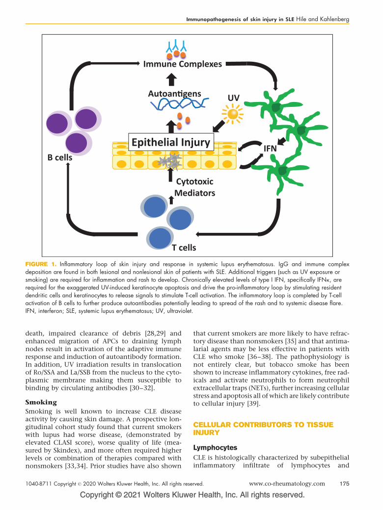
Autoan�gens
Immune Complexes
IFN
T cells
Epithelial Injury
Cytotoxic Mediators
B cells
UV
FIGURE 1. Inflammatory loop of skin injury and response in systemic lupus erythematosus. IgG and immune complexdeposition are found in both lesional and nonlesional skin of patients with SLE. Additional triggers (such as UV exposure orsmoking) are required for inflammation and rash to develop. Chronically elevated levels of type I IFN, specifically IFNk, arerequired for the exaggerated UV-induced keratinocyte apoptosis and drive the pro-inflammatory loop by stimulating residentdendritic cells and keratinocytes to release signals to stimulate T-cell activation. The inflammatory loop is completed by T-cellactivation of B cells to further produce autoantibodies potentially leading to spread of the rash and to systemic disease flare.IFN, interferon; SLE, systemic lupus erythematosus; UV, ultraviolet.
Immunopathogenesis of skin injury in SLE Hile and Kahlenberg
death, impaired clearance of debris [28,29] andenhanced migration of APCs to draining lymphnodes result in activation of the adaptive immuneresponse and induction of autoantibody formation.In addition, UV irradiation results in translocationof Ro/SSA and La/SSB from the nucleus to the cyto-plasmic membrane making them susceptible tobinding by circulating antibodies [30–32].
Smoking
Smoking is well known to increase CLE diseaseactivity by causing skin damage. A prospective lon-gitudinal cohort study found that current smokerswith lupus had worse disease, (demonstrated byelevated CLASI score), worse quality of life (mea-sured by Skindex), and more often required higherlevels or combination of therapies compared withnonsmokers [33,34]. Prior studies have also shown
Copyright © 2021 Wolters Kluwe
1040-8711 Copyright � 2020 Wolters Kluwer Health, Inc. All rights rese
that current smokers are more likely to have refrac-tory disease than nonsmokers [35] and that antima-larial agents may be less effective in patients withCLE who smoke [36–38]. The pathophysiology isnot entirely clear, but tobacco smoke has beenshown to increase inflammatory cytokines, free rad-icals and activate neutrophils to form neutrophilextracellular traps (NETs), further increasing cellularstress and apoptosis all of which are likely contributeto cellular injury [39].
CELLULAR CONTRIBUTORS TO TISSUEINJURY
Lymphocytes
CLE is histologically characterized by subepithelialinflammatory infiltrate of lymphocytes and
r Health, Inc. All rights reserved.
rved. www.co-rheumatology.com 175
Immunopathogenesis and treatment of autoimmune diseases
plasmacytoid dendritic cells (pDCs). The lympho-cytic infiltrate consists of subpopulation of gran-zyme Bþ T cells, which are considered directmarkers of cytotoxic damage and central to kerati-nocyte death [40–42]. Granzyme B (GrB) is a serineprotease that primes for apoptosis through caspaseactivation and cleavage of substrates that initiateDNA fragmentation [43]. Cytotoxic T-cell ligands/receptors, such as CXCL10/CXCR3 are highlyexpressed in lupus skin [44] and also upregulatedon Th1 cells following stimulation with IFNg [44],which is noted to present in discoid lesion more sothan other CLE subtypes [21
&
]. Type I IFNs alsoinfluence this process as GrB expression in lympho-cytes is induced following IFNa stimulation by pDCs[45]. In addition, there is a strong correlationbetween epidermal and dermal IFN-regulated geneexpression and the number of lesional GrB-positivelymphocytes [46]. Type I IFNs stimulate develop-ment of cytotoxic lymphocytes and could makekeratinocytes more prone cytotoxic attack [47].Although cytotoxic T-cell autoreactivity to kerati-nocyte-expressed proteins may drive skin injury incutaneous LE lesions, the role of other T-lympho-cyte subtypes is less clear.
INNATE IMMUNE CELLS
Innate immune cells, including pDCs and neutro-phils, contribute to tissue damage by releasinginflammatory mediators and by failing to effectivelyclear cellular debris.
pDCs are a specialized population of bone mar-row-derived cells that synthesize large amounts oftype I IFN, IL-6, and TNFa in response to ligandsthat engage TLR7, TLR9, and immune complexes[48,49]. pDCs are rare in normal skin but are abun-dant in CLE lesions [50–52]. pDCs may contributeto cellular injury following UV exposure as UVlight increases infiltration of pDCS in exposedskin of lupus patients vs. healthy controls [53].pDCs have been shown to be critically involvedin the pathogenesis of lupus erythematosus asdepletion of pDCs in mice before disease initiationameliorated autoimmunity [51]. Additionally, arecent phase I study utilizing an antibody againstBDCA2, a pDC-specific receptor, has shown prom-ise in improving CLE disease activity [54]. Furtherstudy of contributions of pDCs to SLE and CLEis needed.
Neutrophils may also contribute to tissue dam-age in CLE. As in many inflammatory states, includ-ing oxidative stress, infections or autoimmunity,neutrophils undergo a mechanism of cell deathcalled neutrophil extracellular traps or ‘NETosis’in which they extrude nuclear material [55
&&
]. NETs
Copyright © 2021 Wolters Kluwer H
176 www.co-rheumatology.com
have been detected in the skin of patients withlupus [55
&&
] and likely contribute to immune acti-vation by forming of immune complexes and stim-ulation of IFNa by pDCs [55
&&
] leading to tissuedamage and accumulation of cellular debris.Intriguingly, neutrophils are increased in nonle-sional skin prior to lesion onset in a murine CLEmodel [56]. Studies have shown that activatedphagocytic cells, including monocytes and macro-phages, are increased in CLE lesions [57
&&
] andimportantly have an impaired ability to clear apo-ptotic debris [58]. Accumulation of cellular debrisfollowing cell death results in an increased propen-sity for autoantigen exposure and risk of productionof circulating autoantibodies.
MEDIATORS OF TISSUE DAMAGE
Fas and FasL
Mande et al. recently developed an inducible modelof murine autoimmunity that presents with skinlesions similar to CLE. In this model, transfer of Tcells specific for antigen expressed by MHC II-posi-tive cells resulted in FasL-dependent killing of ker-atinocytes and CLE lesion induction. Interestingly,this model required tissue damage via gamma irra-diation in order for CLE lesions to occur [59]. Acti-vated or damaged keratinocytes express T-cellchemokines, such as CXCL9, CXCL10, andCXCL11. Keratinocytes also produce IFN kappa(IFNk), which may contribute to the strong Th1phenotype and infiltration of immune cells. FasLwas also shown to be upregulated in human CLEbiopsies [59]. Further, in the Mande et al. model,blockade of the type I IFN receptor prevented devel-opment of skin disease and the accumulation ofdermal pDCs, suggesting an IFN-dependent feed-back loop in the maintenance of skin injury, evenwhen the phenotype is driven primarily by T cells.
Tumor necrosis factor a
Tumor necrosis factor (TNF) a is increased in boththe skin and serum of patients with CLE [60,61] andin the skin is upregulated by UVB exposure, in partby IL-1a signaling [62,63]. TNF increases inflam-mation and skin injury by producing cytokines,chemokines and adhesion molecules, such as IL-1, IL-6, CXCL8, CCL20, selectins, and ICAM-1,which increase leukocytes into lesional skin. InSCLE, an increase in the autoantigen Ro52, whichis associated with photosensitivity, is increasedfollowing TNFa treatment [64]. Paradoxically,TNF a antagonists have been reported to causelupus-like skin disease [65]. Thus, further research
ealth, Inc. All rights reserved.
Volume 33 � Number 2 � March 2021
Immunopathogenesis of skin injury in SLE Hile and Kahlenberg
is needed regarding this pathway’s role in the path-ogenesis of CLE.
Tumor necrosis factor-like weak inducer ofapoptosis
Another TNF-family cytokine more recentlyrecognized to be key in propagating skin injuryin SLE is TNF-like weak inducer of apoptosis(TWEAK) [66,67]. TWEAK functions as a solublecytokine that signals through its sole receptor,fibroblast growth factor-inducible 14 (Fn14) [68].Mechanistic studies have elucidated a role in cellproliferation, cell death, inflammation, and tissuerepair [69,70]. Notably, the expression of TWEAK/Fn14 pathway is increased in the setting of tissueinjury and disease [71] and may contribute to skininjury in lupus via cell death, promotion of inflam-matory cytokines, and UV sensitivity. A recentstudy showed that keratinocytes stimulated withTWEAK produce RANTES, in an Fn14-dependentmanner and also demonstrate NFkB-dependentenhanced apoptosis [72]. In vivo, MRL/lpr Fn14KOmice were shown to have significantly attenuatedskin lesions compared with the FN14 wildtypemice that displayed epidermal thickening andinterface dermatitis similar to CLE [72]. Further,MRL/lpr FN14KO mice develop less severe skinlesions with reduced inflammatory infiltrates fol-lowing UVB treatment compared with FN14 wild-type mice [72]. The role of the TWEAK/Fn14pathway in skin injury of primary human kerati-nocytes and in patients with CLE needsfurther investigation.
Type I interferons
Type I IFNs are central to the development of CLEand contribute to tissue injury by upregulating cyto-kines, chemokines, adhesion molecules as well asrecruiting other inflammatory cells leading to aproinflammatory loop [53,73]. Type I IFNs areexpressed in the epidermis and dermis of skinlesions, with IFN a10 and IFNk being significantlyoverexpressed [20]. Sources of IFNs are likely kera-tinocytes (for IFNk) and pDCs, which produce type IIFN in response to immune complexes [74]. Addi-tionally, type I IFNs upregulate cytotoxic proteins,such as perforin and granzyme B via IFNg in T cells,the apoptosis receptor cluster of differentiation 96(CD95) and TNF-related apoptosis-inducing ligand(TRAIL) [45,75,76]. TRAIL is a keratinocyte-pro-duced IFN-dependent chemokine that is upregu-lated in CLE skin [76]. TRAIL’s proapoptoticreceptor is also found on keratinocytes in CLElesions and is upregulated upon stimulation with
Copyright © 2021 Wolters Kluwe
1040-8711 Copyright � 2020 Wolters Kluwer Health, Inc. All rights rese
IFNa [76]. UV light, the main trigger of keratinocyteapoptosis and skin injury, also overstimulates pDCsto produce IFNs in SLE compared with controls [77].Chronic overexpression of IFNk promotes this pro-cess, as it is required for UVB-induced apoptosis ofkeratinocytes [20].
Type II interferons
The role of type II IFNs (IFNg) in the pathogenesisCLE skin injury is not entirely clear. Th1 cells andnatural killer (NK) cells stimulated by specific anti-gens secrete IFNg. Stimulating keratinocytes withIFNg increases terminal differentiation and inhibitscell growth by modulating function of B cells, Tcells, and macrophages [78]. IFNg also induces pro-duction of the chemokines CXCL9, CXCL10, andCXCL11 [18]. IFNg and chemokine CXCL10 areincreased in discoid lesions [44]. However, blockadeof IFNg decreased CXCL10 but did not change dis-ease activity [79]. More research is needed into therole of IFNg in CLE.
Type III interferons
Type III IFNs or ‘l’ IFNs have been shown to beelevated in the epidermis of CLE lesions [80]. Inaddition, CLE patients with active skin disease havebeen shown to have elevated IFNl that correlatedwith CXCL9 levels [80]. IFN l may contribute to skininjury by increasing recruitment of cytotoxic T cellsto the epidermis resulting in cellular death andinflammation [81,82]. Further study of type III IFNsin skin injury in SLE is warranted.
Autoantibodies
Autoantibodies and immune complexes are centralto tissue injury in SLE [83
&
,84], and likely play animportant role in the skin. In CLE, immune complexdeposition of IgG and IgM at the dermoepidermaljunction in lesional and nonlesional skin [85] isconsidered diagnostic of SLE. The murine modelMRL-Faslpr/lpr (also known as MRL/lpr) characterizedby high levels of autoantibodies, spontaneouslydevelop lupus-like skin injury that appears histolog-ically identical to human SLE and shows a immu-nofluorescent ‘lupus band’ [86,87]. Specifically, IgGmay be key in skin injury as injection of lupus sera –from either lupus-prone mice or patients with SLE –into the dermis of wild type mice result in aninflammatory response, which was not observedafter control serum, isotonic saline, or IgG-depletedsera [88]. Antibodies against galectin 3 and ribo-somal protein P0 are related to the developmentof skin lesions in SLE [89]. Anti-Ro52 is associated
r Health, Inc. All rights reserved.
rved. www.co-rheumatology.com 177
Immunopathogenesis and treatment of autoimmune diseases
with an increased risk of photosensitive rash [90], andRo52 is expressed highly in skin and infiltrating cellsafter UV-induced skin injury in SLE patients [91,92].
CONCLUSION
The pathogenesis of skin injury in CLE is complex(see Fig. 1), but recent evidence supports a role fordysregulated epidermal cell death. Further researchwill be needed to understand these molecular path-ways to develop targeted treatment modalities andimprove disease outcomes.
Acknowledgements
None.
Financial support and sponsorship
G.A.H. was funded in part by the National Institute ofArthritis and Musculoskeletal Diseases of the NationalInstitutes of Health under award T32- AR007197.J.M.K. is supported by the National Institute of Arthritisand Musculoskeletal and Skin Diseases of the NationalInstitutes of Health under awards R01-AR071384,K24AR076975, and P30-AR075043, the LupusResearch Alliance, the Doris Duke Charitable Founda-tion under a physician scientist development award, theRheumatology Research Foundation under an Investiga-tor Award, and the A. Alfred Taubman Medical ResearchInstitute and the Parfet Emerging Scholar Award.
Conflicts of interest
J.M.K. has served on advisory boards for AstraZeneca, EliLilly, Bristol Myers Squibb, Avion Pharmaceuticals, Pro-vention Bio, Aurinia Pharmaceuticals, and BoehringerIngleheim. She has received grant funding from BristolMyerse Squibb/Celgene and Q32 Bio. G.A.H does nothave any conflicts of interest.
REFERENCES AND RECOMMENDEDREADINGPapers of particular interest, published within the annual period of review, havebeen highlighted as:
& of special interest&& of outstanding interest1. Kuhn A, Ruland V, Bonsmann G. Cutaneous lupus erythematosus: update oftherapeutic options part I. J Am Acad Dermatol 2011; 65:e179–e193.
2. Provost TT. Lupus band test. Int J Dermatol 1981; 20:475–481.3. Reich A, Marcinow K, Bialynicki-Birula R. The lupus band test in systemic
lupus erythematosus patients. Ther Clin Risk Manag 2011; 7:27–32.4. Rullo OJ, Tsao BP. Recent insights into the genetic basis of systemic lupus
erythematosus. Ann Rheum Dis 2013; 72(Suppl 2):ii56–ii61.5. Sanchez E, Nadig A, Richardson BC, Freedman BI. Phenotypic associations
of genetic susceptibility loci in systemic lupus erythematosus. Ann Rheum Dis2011; 70:1752–1757.
6. T€ungler V, Silver RM, Walkenhorst H, G€unther C. Inherited or de novomutation affecting aspartate 18 of TREX1 results in either familial chilblainlupus or Aicardi-Goutieres syndrome. Br J Dermatol 2012; 167:212–214.
7. Harley IT, Niewold TB, Stormont RM. The role of genetic variation nearinterferon-kappa in systemic lupus erythematosus. J Biomed Biotechnol2010; 2010:706825.
Copyright © 2021 Wolters Kluwer H
178 www.co-rheumatology.com
8.&
Xuan J, Xiong Y, Shi L. Do lncRNAs and circRNAs expression profilesinfluence discoid lupus erythematosus progression?-a comprehensive ana-lysis. Ann Transl Med 2019; 7:728.
RNA-sequencing analysis of DLE showing IncRNAS and circRNAs influenceprogression.9. Biazar C, Sigges J, Patsinakidis N; EUSCLE co-authors. Cutaneous lupus
erythematosus: first multicenter database analysis of 1002 patients from theEuropean Society of Cutaneous Lupus Erythematosus (EUSCLE). Autoim-mun Rev 2013; 12:444–454.
10. Foering K, Chang AY, Piette EW. Characterization of clinical photosensi-tivity in cutaneous lupus erythematosus. J Am Acad Dermatol 2013;69:205–213.
11. Kuhn A, Wenzel J, Weyd H. Photosensitivity apoptosis, and cytokines in thepathogenesis of lupus erythematosus: a critical review. Clin Rev AllergyImmunol 2014; 47:148–162.
12. Pirner K, Rubbert A, Salinger R. Significance of ultraviolet light in thepathogenesis of systemic lupus erythematosus: case report and discussionof the literature]. Z Rheumatol 1992; 51:20–24.
13. Fruchter O, Edoute Y. First presentation of systemic lupus erythematosusfollowing ultraviolet radiation exposure in an artificial tanning device. Rheu-matology (Oxford) 2005; 44:558–559.
14. Kock A, Schwarz T, Kirnbauer R. Human keratinocytes are a source for tumornecrosis factor alpha: evidence for synthesis and release upon stimulationwith endotoxin or ultraviolet light. J Exp Med 1990; 172:1609–1614.
15. Takashima A, Bergstresser PR. Impact of UVB radiation on the epidermalcytokine network. Photochem Photobiol 1996; 63:397–400.
16. Kuhn A, Sontheimer RD. Cutaneous lupus erythematosus: molecularand cellular basis of clinical findings. Curr Dir Autoimmun 2008;10:119–140.
17. Sontheimer C, Liggitt D, Elkon KB. Ultraviolet B irradiation causes stimulatorof interferon genes-dependent production of protective type i interferon inmouse skin by recruited inflammatory monocytes. Arthritis Rheumatol 2017;69:826–836.
18. Meller S, Winterberg F, Gilliet M. Ultraviolet radiation-induced injury, chemo-kines, and leukocyte recruitment: an amplification cycle triggering cutaneouslupus erythematosus. Arthritis Rheum 2005; 52:1504–1516.
19.&
Katayama S, Panelius J, Koskenmies S. Delineating the healthy human skin UVresponse and early induction of interferon pathway in cutaneous lupuserythematosus. J Invest Dermatol 2019; 139:2058.e4–2061.e4.
Photoprovocation study of CLE, SCLE, and healthy control comparing biopsiesand RNA-sequencing of exposed vs. unexposed skin.20. Sarkar MK, Hile GA, Tsoi LC. Photosensitivity and type I IFN responses in
cutaneous lupus are driven by epidermal-derived interferon kappa. AnnRheum Dis 2018; 77:1653–1664.
21.&
Berthier CC, Tsoi LC, Reed TJ. Molecular profiling of cutaneous lupus lesionsidentifies subgroups distinct from clinical phenotypes. J Clin Med 2019;8:1244.
Large cohort comparing differentially expressed genes in CLE subtypes.22. Shipman WD, Chyou S, Ramanathan A. A protective Langerhans cell-kera-
tinocyte axis that is dysfunctional in photosensitivity. Sci Transl Med 2018;10:eaa9527.
23.&&
Skopelja-Gardner S, An J, Tai J, Tanaka L. The early local and systemic type Iinterferon responses to ultraviolet B light exposure are cGAS dependent. SciRep 2020; 10:7908.
Epidermal UV exposure increases IFN signature not only in skin but blood andkidneys. The early IFN-I signature was dependent on cGAS activation.24. Gehrke N, Mertens C, Zillinger T. Oxidative damage of DNA confers resis-
tance to cytosolic nuclease TREX1 degradation and potentiates STING-dependent immune sensing. Immunity 2013; 39:482–495.
25. Scholtissek B, Zahn S, Maier J. Immunostimulatory endogenous nucleic acidsdrive the lesional inflammation in cutaneous lupus erythematosus. J InvestDermatol 2017; 137:1484–1492.
26. Golan DT, Borel Y. Increased photosensitivity to near-ultraviolet light in murineSLE. J Immunol 1984; 132:705–710.
27. Mondini M, Vidali M, Airo P, et al. Role of the interferon-inducible gene IFI16 inthe etiopathogenesis of systemic autoimmune disorders. Ann N Y Acad Sci2007; 1110:47–56.
28. Shi ZR, Cao CX, Tan GZ. The association of serum antiribosomal P antibodywith clinical and serological disorders in systemic lupus erythematosus: asystematic review and meta-analysis. Lupus 2015; 24:588–596.
29. Kuhn A, Herrmann M, Kleber S, et al. Accumulation of apoptotic cells in theepidermis of patients with cutaneous lupus erythematosus after ultravioletirradiation. Arthritis Rheum 2006; 54:939–950.
30. Yu C, Chang C Zhang J. Immunologic and genetic considerations of cuta-neous lupus erythematosus: a comprehensive review. J Autoimmunity 2013;41: 34–45.
31. Lawley W, Doherty A, Denniss S, et al. Rapid lupus autoantigen relocalizationand reactive oxygen species accumulation following ultraviolet irradiation ofhuman keratinocytes. Rheumatology (Oxford) 2000; 39:253–261.
32. Jones SK. Ultraviolet radiation (UVR) induces cell-surface Ro/SSA antigenexpression by human keratinocytes in vitro: a possible mechanism for the UVRinduction of cutaneous lupus lesions. British Journal of Dermatology. 1992;126: 546–553.
ealth, Inc. All rights reserved.
Volume 33 � Number 2 � March 2021
Immunopathogenesis of skin injury in SLE Hile and Kahlenberg
33. Wang B, Dong X, Yuan Z, et al. SSA/Ro antigen expressed on membrane ofUVB-induced apoptotic keratinocytes is pathogenic but not detectable insupernatant of cell culture. Chinese Med J 1999; 112:512–515.
34. Piette EW, Foering KP, Chang AY. Impact of smoking in cutaneous lupuserythematosus. Arch Dermatol 2012; 148:317–322.
35. Kuhn A, Sigges J, Biazar C; EUSCLE coauthors. Influence of smoking ondisease severity and antimalarial therapy in cutaneous lupus erythematosus:analysis of 1002 patients from the EUSCLE database. Br J Dermatol 2014;171:571–579.
36. Moghadam-Kia S, Chilek K, Gaines E. Cross-sectional analysis of a colla-borative Web-based database for lupus erythematosus-associated skin le-sions: prospective enrollment of 114 patients. Arch Dermatol 2009;145:255–260.
37. Kreuter A, Gaifullina R, Tigges C. Lupus erythematosus tumidus: response toantimalarial treatment in 36 patients with emphasis on smoking. Arch Der-matol 2009; 145:244–248.
38. Jewell ML, McCauliffe DP. Patients with cutaneous lupus erythematosus whosmoke are less responsive to antimalarial treatment. J Am Acad Dermatol2000; 42:983–987.
39. Rahman P, Gladman DD, Urowitz MB. Smoking interferes with efficacy ofantimalarial therapy in cutaneous lupus. J Rheumatol 1998; 25:1716–1719.
40. White PC, Hirschfeld J, Milward MR. Cigarette smoke modifies neutrophilchemotaxis, neutrophil extracellular trap formation and inflammatory re-sponse-related gene expression. J Periodontal Res 2018; 53:525–535.
41. Higaki Y, Yamada O, Okamura T. Granzyme-B-containing lymphocyte invol-vement in epidermal injury in graft-versus-host disease. Dermatology 2001;202:94–98.
42. Shimizu M, Higaki Y, Higaki M. The role of granzyme B-expressing CD8-positive T cells in apoptosis of keratinocytes in lichen planus. Arch DermatolRes 1997; 289:527–532.
43. Grassi M, Capello F, Bertolino L. Identification of granzyme B-expressing CD-8-positive T cells in lymphocytic inflammatory infiltrate in cutaneous lupuserythematosus and in dermatomyositis. Clin Exp Dermatol 2009;34:910–914.
44. Russell JH, Ley TJ. Lymphocyte-mediated cytotoxicity. Annu Rev Immunol2002; 20:323–370.
45. Wenzel J, Zahn S, Mikus S. The expression pattern of interferon-inducibleproteins reflects the characteristic histological distribution of infiltrating im-mune cells in different cutaneous lupus erythematosus subsets. Br J Dermatol2007; 157:752–757.
46. Bose A, Baral R. IFNalpha2b stimulated release of IFNgamma differentiallyregulates T cell and NK cell mediated tumor cell cytotoxicity. Immunol Lett2007; 108:68–77.
47. Wenzel J, Uerlich M, Worrenkamper E. Scarring skin lesions of discoid lupuserythematosus are characterized by high numbers of skin-homing cytotoxiclymphocytes associated with strong expression of the type I interferon-induced protein MxA. Br J Dermatol 2005; 153:1011–1015.
48. Lang KS, Recher M, Junt T. Toll-like receptor engagement converts T-cellautoreactivity into overt autoimmune disease. Nat Med 2005; 11:138–145.
49. Kim JM, Park SH, Kim HY. A plasmacytoid dendritic cells-type I interferon axisis critically implicated in the pathogenesis of systemic lupus erythematosus.Int J Mol Sci 2015; 16:14158–14170.
50. Davison LM, Jorgensen TN. New treatments for systemic lupus erythematosuson the horizon: targeting plasmacytoid dendritic cells to inhibit cytokineproduction. J Clin Cell Immunol 2017; 8:.
51. Farkas L, Beiske K, Lund-Johansen F. Plasmacytoid dendritic cells (naturalinterferon- alpha/beta-producing cells) accumulate in cutaneous lupus er-ythematosus lesions. Am J Pathol 2001; 159:237–243.
52. Rowland SL, Riggs JM, Gilfillan S. Early, transient depletion of plasmacytoiddendritic cells ameliorates autoimmunity in a lupus model. J Exp Med 2014;211:1977–1991.
53. Tomasini D, Mentzel T, Hantschke M. Plasmacytoid dendritic cells: an over-view of their presence and distribution in different inflammatory skin diseases,with special emphasis on Jessner’s lymphocytic infiltrate of the skin andcutaneous lupus erythematosus. J Cutan Pathol 2010; 37:1132–1139.
54. Reefman E, Kuiper H, Limburg PC. Type I interferons are involved in thedevelopment of ultraviolet B-induced inflammatory skin lesions in systemiclupus erythaematosus patients. Ann Rheum Dis 2008; 67:11–18.
55.&&
Furie R, Werth VP, Merola JF. Monoclonal antibody targeting BDCA2 ame-liorates skin lesions in systemic lupus erythematosus. J Clin Invest 2019;129:1359–1371.
Randomized, double-blinded, placebo-controlled clinical trial of monoclonal anti-body that binds blood DC antigen 2 (BDCA2) and inhibits production of IFNresults in improvement of CLE. This study suggests targeting pDCs may bebeneficial for patients with cutaneous manifestations of SLE.56. Villanueva E, Yalavarthi S, Berthier CC. Netting neutrophils induce endothelial
damage, infiltrate tissues, and expose immunostimulatory molecules in sys-temic lupus erythematosus. J Immunol 2011; 187:538–552.
57.&&
Han X, Vesely MD, Yang W. PD-1H (VISTA)-mediated suppression ofautoimmunity in systemic and cutaneous lupus erythematosus. Sci TranslMed 2019; 11:eaax1159.
Novel murine model lacking inhibitory receptor VISTA or programmed death-1homolog (PD-1H) that mimics human systemic and cutaneous lupus. Neutrophilsincrease in prelesional skin, indicating a role in driving CLE lesions.
Copyright © 2021 Wolters Kluwe
1040-8711 Copyright � 2020 Wolters Kluwer Health, Inc. All rights rese
58. Chong BF, Tseng LC, Kim A. Differential expression of BAFF and itsreceptors in discoid lupus erythematosus patients. J Dermatol Sci2014; 73:216–224.
59. Herrmann M, Voll RE, Zoller OM. Impaired phagocytosis of apoptotic cellmaterial by monocyte-derived macrophages from patients with systemic lupuserythematosus. Arthritis Rheum 1998; 41:1241–1250.
60. Mande P, Zirak B, Ko WC, et al. Fas ligand promotes an inducible TLR-dependent model of cutaneous lupus-like inflammation. J Clin Invest 2018;128:2966–2978.
61. Maczynska I, Millo B, Ratajczak-Stefanska V. Proinflammatory cytokine (IL-1beta, IL-6, IL-12, IL-18 and TNF-alpha) levels in sera of patients withsubacute cutaneous lupus erythematosus (SCLE). Immunol Lett 2006;102:79–82.
62. Zampieri S, Alaibac M, Iaccarino L. Tumour necrosis factor alpha is expressedin refractory skin lesions from patients with subacute cutaneous lupuserythematosus. Ann Rheum Dis 2006; 65:545–548.
63. Bashir MM, Sharma MR, Werth VP. UVB and proinflammatorycytokines synergistically activate TNF-alpha production in keratinocytesthrough enhanced gene transcription. J Invest Dermatol 2009;129:994–1001.
64. Stannard JN, Kahlenberg JM. Cutaneous lupus erythematosus: updates onpathogenesis and associations with systemic lupus. Curr Opin Rheumatol2016; 28:453–459.
65. Gerl V, Hostmann B, Johnen C. The intracellular 52-kd Ro/SSA autoantigen inkeratinocytes is up-regulated by tumor necrosis factor alpha via tumornecrosis factor receptor I. Arthritis Rheum 2005; 52:531–538.
66. Shovman O, Tamar S, Amital H, et al. Diverse patterns of anti-TNF-a-inducedlupus: case series and review of the literature. Clin Rheumatol 2018;37:563–568.
67. Campbell S, Burkly LC, Gao HX. Proinflammatory effects of TWEAK/Fn14interactions in glomerular mesangial cells. J Immunol 2006;176:1889–1898.
68. Winkles JA. The TWEAK-Fn14 cytokine-receptor axis: discovery, biology andtherapeutic targeting. Nat Rev Drug Discov 2008; 7:411–425.
69. Wiley SR, Winkles JA. TWEAK, a member of the TNF superfamily, is amultifunctional cytokine that binds the TweakR/Fn14 receptor. CytokineGrowth Factor Rev 2003; 14:241–249.
70. Zheng TS, Burkly LC. No end in site: TWEAK/Fn14 activation and auto-immunity associated- end-organ pathologies. J Leukoc Biol 2008;84:338–347.
71. Burkly LC, Michaelson JS, Zheng TS. TWEAK/Fn14 pathway: an immuno-logical switch for shaping tissue responses. Immunol Rev 2011;244:99–114.
72. Burkly LC, Michaelson JS, Hahm K. TWEAKing tissue remodeling by amultifunctional cytokine: role of TWEAK/Fn14 pathway in health and disease.Cytokine 2007; 40:1–16.
73. Doerner JL, Wen J. Xia. TWEAK/Fn14 signaling involvement in the pathogen-esis of cutaneous disease in the MRL/lpr model of spontaneous lupus. J InvestDermatol 2015; 135:1986–1995.
74. Robinson ES, Werth VP. The role of cytokines in the pathogenesis ofcutaneous lupus erythematosus. Cytokine 2015; 73:326–334.
75. Savarese E, Chae OW, Trowitzsch S. U1 small nuclear ribonucleoproteinimmune complexes induce type I interferon in plasmacytoid dendritic cellsthrough TLR7. Blood 2006; 107:3229–3234.
76. Zella D, Barabitskaja O, Casareto L. Recombinant IFN-alpha (2b) increasesthe expression of apoptosis receptor CD95 and chemokine receptors CCR1and CCR3 in monocytoid cells. J Immunol 1999; 163:3169–3175.
77. Zahn S, Rehkamper C, Ferring-Schmitt S. Interferon-a stimulates TRAILexpression in human keratinocytes and peripheral blood mononuclear cells:implications for the pathogenesis of cutaneous lupus erythematosus. Br JDermatol 2011; 165:1118–1123.
78. Zahn S, Graef M, Patsinakidis N, et al. Ultraviolet light protection by asunscreen prevents interferon-driven skin inflammation in cutaneous lupuserythematosus. Exp Dermatol 2014; 23:516–518.
79. Saunders NA, Jetten AM. Control of growth regulatory and differentiation-specific genes in human epidermal keratinocytes by interferon gamma.Antagonism by retinoic acid and transforming growth factor beta 1. J BiolChem 1994; 269:2016–2022.
80. Werth VP, Fiorentino D, Sullivan BA. Brief report: pharmacodynamics, safety,and clinical efficacy of AMG 811, a human anti-interferon-g antibody, inpatients with discoid lupus erythematosus. Arthritis Rheumatol 2017;69:1028–1034.
81. Zahn S, Rehkamper C, K€ummerer BM. Evidence for a pathophysiological roleof keratinocyte-derived type III interferon (IFNl) in cutaneous lupus erythe-matosus. J Invest Dermatol 2011; 131:133–140.
82. Yoneyama H, Matsuno K, Zhang Y. Evidence for recruitment of plasmacytoiddendritic cell precursors to inflamed lymph nodes through high endothelialvenules. Int Immunol 2004; 16:915–928.
83.&
Goel RR, Wang X, O’Neil LJ. Interferon lambda promotes immune dysregula-tion and tissue inflammation in TLR7-induced lupus. Proc Natl Acad Sci U S A2020; 117:5409–5419.
Murine lupus model showing IFNl receptor deficiency reduces immune activationand associated organ damage in skin and kidneys without effects on autoantibodyproduction.
r Health, Inc. All rights reserved.
rved. www.co-rheumatology.com 179
Immunopathogenesis and treatment of autoimmune diseases
84. Tsokos GC. Systemic lupus erythematosus. N Engl J Med 2011;365:2110–2121.
85. Rahman A, Isenberg DA. Systemic lupus erythematosus. N Engl J Med 2008;358:929–939.
86. Kuhn A, Landmann A. The classification and diagnosis of cutaneous lupuserythematosus. J Autoimmun 2014; 48-49:14–19.
87. Furukawa F, Tanaka H, Sekita K. Dermatopathological studies on skin lesionsof MRL mice. Arch Dermatol Res 1984; 276:186–194.
88. Kanauchi H, Furukawa F, Imamura S. Characterization of cutaneous infiltratesin MRL/lpr mice monitored from onset to the full development of lupuserythematosus-like skin lesions. J Invest Dermatol 1991; 96:478–483.
Copyright © 2021 Wolters Kluwer H
180 www.co-rheumatology.com
89. Deng GM, Liu L, Kyttaris VC, Tsokos GC. Lupus serum IgG induces skininflammation through the TNFR1 signaling pathway. J Immunol 2010;184:7154–7161.
90. Shi ZR, Tan GZ, Meng Z. Association of antiacidic ribosomal protein P0 andantigalectin 3 antibodies with the development of skin lesions in systemiclupus erythematosus. Arthritis Rheumatol 2015; 67:193–203.
91. Sontheimer RD, Maddison PJ, Reichlin M. Serologic and HLA associations insubacute cutaneous lupus erythematosus, a clinical subset of lupus erythe-matosus. Ann Intern Med 1982; 97:664–671.
92. Oke V, Vassilaki I, Espinosa A. High Ro52 expression in spontaneous and UV-induced cutaneous inflammation. J Invest Dermatol 2009; 129:2000–2010.
ealth, Inc. All rights reserved.
Volume 33 � Number 2 � March 2021
REVIEW
CURRENTOPINION New therapeutic approaches in systemic
lupus erythematosus
Copyrigh
1040-8711 Copyright � 2020 Wolte
a,b a a
Antonios G.A. Kolios , Nobuya Yoshida , and George C. TsokosPurpose of review
This review gives an overview of the recently published clinical trials in systemic lupus erythematosus (SLE).
Recent findings
Our continuously improving understanding of the cellular and molecular mechanisms, which are involved inthe pathogenesis of SLE, has inspired the performance of multiple clinical trials in an attempt to modifyrecognized targets. Here, we summarize results obtained from recent trials, which used monoclonalantibodies blocking cytokines, blockers of costimulatory molecules or deleting immune cells, small druginhibitors of kinases and replenishment of cytokines.
Summary
The therapeutic options for patients with SLE grow continuously and in parallel it raises the need forpathogenetic mechanism-based precision medicine so that we may select the right treatment for the rightpatient.
Keywords
biological therapies, systemic lupus erythematosus, therapy, treatment update
aDepartment of Medicine, Beth Israel Deaconess Medical Center, Har-vard Medical School, Boston, Massachusetts, USA and bDepartment ofImmunology, University Hospital Zurich, Zurich, Switzerland
Correspondence to Antonios G.A. Kolios, Department of Medicine, BethIsrael Deaconess Medical Center, Harvard Medical School, Boston, MA02215, USA. E-mail: [email protected]
Curr Opin Rheumatol 2021, 33:181–189
DOI:10.1097/BOR.0000000000000772
INTRODUCTION
Systemic lupus erythematosus (SLE) is multiorganautoimmune disease characterized by loss ofimmune tolerance leading to organ inflammation[1]. The prevalence varies from 20 to 150 cases per100,000 population. Pathogenesis has been linkedto abnormalities in innate and adaptive immunesystem induced by environmental and hormonaltriggers in genetically susceptible individuals [2].
Genetic, environmental and hormonal factorsact on various components of the innate and adap-tive immune system. Gene copy variants or Singlenucleotide polymorphisms influence the expressionof many genes involved in the immune response.Environmental factors, including UV light, drugsand products of the microbiome alter T and B-cellresponses as well as the function of innate cells byengaging Toll-like receptors. Hormones and genesdefined by the X chromosome contribute to diseaseexpression by altering the function of lymphocytesand cells of the innate immune response. All path-ways eventually lead to loss of tolerance of B andT cells to autoantigens, which are present in abun-dance because of increased apoptotic rates of cellsand defects in processes responsible for their clear-ance. The T-cell response to autoantigens is aberrantin terms of early and late signaling events and results
t © 2021 Wolters Kluwe
rs Kluwer Health, Inc. All rights rese
in misbalanced levels of cytokines, includingdecreased IL-2 and increased IL-17 production. T cellsalso through distinct pathways acquire increasedability to invade tissues and contribute to the inflam-matory response. B cells in response to cognateand noncognate (cytokine) interaction with T cellsproduce autoantibodies. Cells of the innate immuneresponse under the influence of the involved patho-genic factors produce cytokines [including interferon(IFN)-a] or through the direct interaction withlymphocytes contribute in a major way to the inflam-matory organ-damaging response. Although severalpathways operate in each individual, the relativecontribution of each pathway varies from person toperson [2].
Clinically, SLE can affect any organ, includingthe kidney (60%), skin (70%), muscles and joints(85%), the blood (50%), as well as central and
r Health, Inc. All rights reserved.
rved. www.co-rheumatology.com
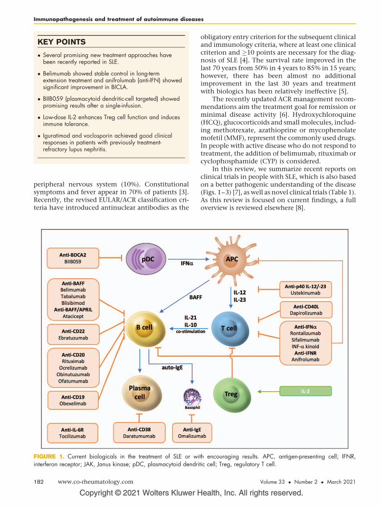
KEY POINTS
� Several promising new treatment approaches havebeen recently reported in SLE.
� Belimumab showed stable control in long-termextension treatment and anifrolumab (anti-IFN) showedsignificant improvement in BICLA.
� BIIB059 (plasmacytoid dendritic-cell targeted) showedpromising results after a single-infusion.
� Low-dose IL-2 enhances Treg cell function and inducesimmune tolerance.
� Iguratimod and voclosporin achieved good clinicalresponses in patients with previously treatment-refractory lupus nephritis.
Immunopathogenesis and treatment of autoimmune diseases
peripheral nervous system (10%). Constitutionalsymptoms and fever appear in 70% of patients [3].Recently, the revised EULAR/ACR classification cri-teria have introduced antinuclear antibodies as the
Copyright © 2021 Wolters Kluwer H
FIGURE 1. Current biologicals in the treatment of SLE or wiinterferon receptor; JAK, Janus kinase; pDC, plasmacytoid dendri
182 www.co-rheumatology.com
obligatory entry criterion for the subsequent clinicaland immunology criteria, where at least one clinicalcriterion and �10 points are necessary for the diag-nosis of SLE [4]. The survival rate improved in thelast 70 years from 50% in 4 years to 85% in 15 years;however, there has been almost no additionalimprovement in the last 30 years and treatmentwith biologics has been relatively ineffective [5].
The recently updated ACR management recom-mendations aim the treatment goal for remission orminimal disease activity [6]. Hydroxychloroquine(HCQ), glucocorticoids and small molecules, includ-ing methotrexate, azathioprine or mycophenolatemofetil (MMF), represent the commonly used drugs.In people with active disease who do not respond totreatment, the addition of belimumab, rituximab orcyclophosphamide (CYP) is considered.
In this review, we summarize recent reports onclinical trials in people with SLE, which is also basedon a better pathogenic understanding of the disease(Figs. 1–3) [7], as well as novel clinical trials (Table 1).As this review is focused on current findings, a fulloverview is reviewed elsewhere [8].
ealth, Inc. All rights reserved.
th encouraging results. APC, antigen-presenting cell; IFNR,tic cell; Treg, regulatory T cell.
Volume 33 � Number 2 � March 2021
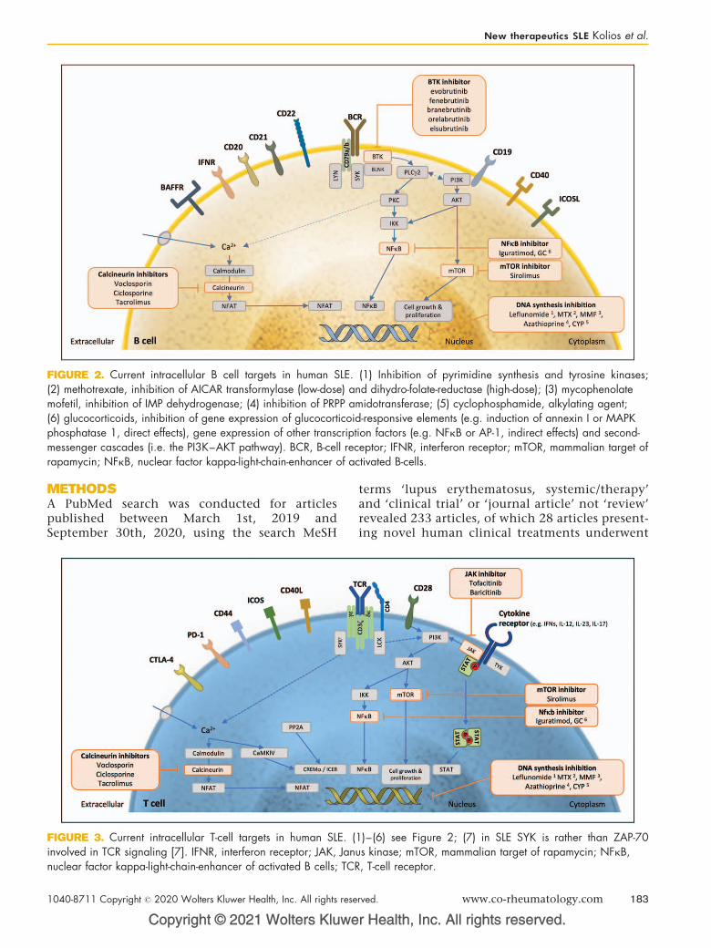
FIGURE 2. Current intracellular B cell targets in human SLE. (1) Inhibition of pyrimidine synthesis and tyrosine kinases;(2) methotrexate, inhibition of AICAR transformylase (low-dose) and dihydro-folate-reductase (high-dose); (3) mycophenolatemofetil, inhibition of IMP dehydrogenase; (4) inhibition of PRPP amidotransferase; (5) cyclophosphamide, alkylating agent;(6) glucocorticoids, inhibition of gene expression of glucocorticoid-responsive elements (e.g. induction of annexin I or MAPKphosphatase 1, direct effects), gene expression of other transcription factors (e.g. NFkB or AP-1, indirect effects) and second-messenger cascades (i.e. the PI3K–AKT pathway). BCR, B-cell receptor; IFNR, interferon receptor; mTOR, mammalian target ofrapamycin; NFkB, nuclear factor kappa-light-chain-enhancer of activated B-cells.
New therapeutics SLE Kolios et al.
METHODSA PubMed search was conducted for articlespublished between March 1st, 2019 andSeptember 30th, 2020, using the search MeSH
Copyright © 2021 Wolters Kluwe
FIGURE 3. Current intracellular T-cell targets in human SLE. (involved in TCR signaling [7]. IFNR, interferon receptor; JAK, Jannuclear factor kappa-light-chain-enhancer of activated B cells; TCR
1040-8711 Copyright � 2020 Wolters Kluwer Health, Inc. All rights rese
terms ‘lupus erythematosus, systemic/therapy’and ‘clinical trial’ or ‘journal article’ not ‘review’revealed 233 articles, of which 28 articles present-ing novel human clinical treatments underwent
r Health, Inc. All rights reserved.
1)–(6) see Figure 2; (7) in SLE SYK is rather than ZAP-70us kinase; mTOR, mammalian target of rapamycin; NFkB,, T-cell receptor.
rved. www.co-rheumatology.com 183
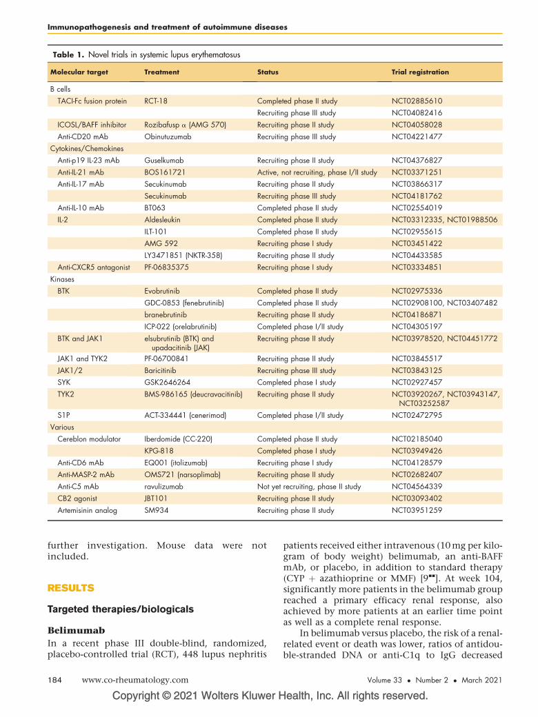
Table 1. Novel trials in systemic lupus erythematosus
Molecular target Treatment Status Trial registration
B cells
TACI-Fc fusion protein RCT-18 Completed phase II study NCT02885610
Recruiting phase III study NCT04082416
ICOSL/BAFF inhibitor Rozibafusp a (AMG 570) Recruiting phase II study NCT04058028
Anti-CD20 mAb Obinutuzumab Recruiting phase III study NCT04221477
Cytokines/Chemokines
Anti-p19 IL-23 mAb Guselkumab Recruiting phase II study NCT04376827
Anti-IL-21 mAb BOS161721 Active, not recruiting, phase I/II study NCT03371251
Anti-IL-17 mAb Secukinumab Recruiting phase II study NCT03866317
Secukinumab Recruiting phase III study NCT04181762
Anti-IL-10 mAb BT063 Completed phase II study NCT02554019
IL-2 Aldesleukin Completed phase II study NCT03312335, NCT01988506
ILT-101 Completed phase II study NCT02955615
AMG 592 Recruiting phase I study NCT03451422
LY3471851 (NKTR-358) Recruiting phase II study NCT04433585
Anti-CXCR5 antagonist PF-06835375 Recruiting phase I study NCT03334851
Kinases
BTK Evobrutinib Completed phase II study NCT02975336
GDC-0853 (fenebrutinib) Completed phase II study NCT02908100, NCT03407482
branebrutinib Recruiting phase II study NCT04186871
ICP-022 (orelabrutinib) Completed phase I/II study NCT04305197
BTK and JAK1 elsubrutinib (BTK) andupadacitinib (JAK)
Recruiting phase II study NCT03978520, NCT04451772
JAK1 and TYK2 PF-06700841 Recruiting phase II study NCT03845517
JAK1/2 Baricitinib Recruiting phase III study NCT03843125
SYK GSK2646264 Completed phase I study NCT02927457
TYK2 BMS-986165 (deucravacitinib) Recruiting phase II study NCT03920267, NCT03943147,NCT03252587
S1P ACT-334441 (cenerimod) Completed phase I/II study NCT02472795
Various
Cereblon modulator Iberdomide (CC-220) Completed phase II study NCT02185040
KPG-818 Completed phase I study NCT03949426
Anti-CD6 mAb EQ001 (itolizumab) Recruiting phase I study NCT04128579
Anti-MASP-2 mAb OMS721 (narsoplimab) Recruiting phase II study NCT02682407
Anti-C5 mAb ravulizumab Not yet recruiting, phase II study NCT04564339
CB2 agonist JBT101 Recruiting phase II study NCT03093402
Artemisinin analog SM934 Recruiting phase II study NCT03951259
Immunopathogenesis and treatment of autoimmune diseases
further investigation. Mouse data were notincluded.
RESULTS
Targeted therapies/biologicals
Belimumab
In a recent phase III double-blind, randomized,placebo-controlled trial (RCT), 448 lupus nephritis
Copyright © 2021 Wolters Kluwer H
184 www.co-rheumatology.com
patients received either intravenous (10 mg per kilo-gram of body weight) belimumab, an anti-BAFFmAb, or placebo, in addition to standard therapy(CYP þ azathioprine or MMF) [9
&&
]. At week 104,significantly more patients in the belimumab groupreached a primary efficacy renal response, alsoachieved by more patients at an earlier time pointas well as a complete renal response.
In belimumab versus placebo, the risk of a renal-related event or death was lower, ratios of antidou-ble-stranded DNA or anti-C1q to IgG decreased
ealth, Inc. All rights reserved.
Volume 33 � Number 2 � March 2021
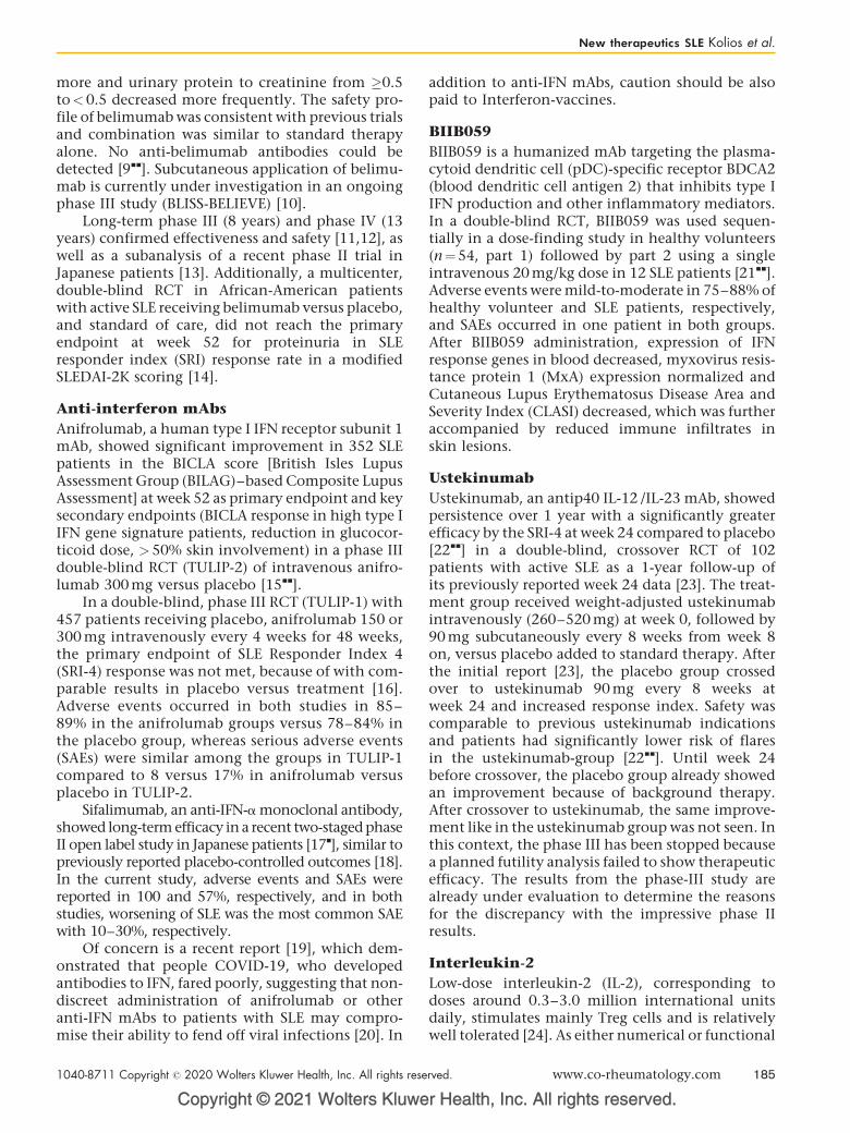
New therapeutics SLE Kolios et al.
more and urinary protein to creatinine from �0.5to<0.5 decreased more frequently. The safety pro-file of belimumab was consistent with previous trialsand combination was similar to standard therapyalone. No anti-belimumab antibodies could bedetected [9
&&
]. Subcutaneous application of belimu-mab is currently under investigation in an ongoingphase III study (BLISS-BELIEVE) [10].
Long-term phase III (8 years) and phase IV (13years) confirmed effectiveness and safety [11,12], aswell as a subanalysis of a recent phase II trial inJapanese patients [13]. Additionally, a multicenter,double-blind RCT in African-American patientswith active SLE receiving belimumab versus placebo,and standard of care, did not reach the primaryendpoint at week 52 for proteinuria in SLEresponder index (SRI) response rate in a modifiedSLEDAI-2K scoring [14].
Anti-interferon mAbs
Anifrolumab, a human type I IFN receptor subunit 1mAb, showed significant improvement in 352 SLEpatients in the BICLA score [British Isles LupusAssessment Group (BILAG)–based Composite LupusAssessment] at week 52 as primary endpoint and keysecondary endpoints (BICLA response in high type IIFN gene signature patients, reduction in glucocor-ticoid dose, >50% skin involvement) in a phase IIIdouble-blind RCT (TULIP-2) of intravenous anifro-lumab 300 mg versus placebo [15
&&
].In a double-blind, phase III RCT (TULIP-1) with
457 patients receiving placebo, anifrolumab 150 or300 mg intravenously every 4 weeks for 48 weeks,the primary endpoint of SLE Responder Index 4(SRI-4) response was not met, because of with com-parable results in placebo versus treatment [16].Adverse events occurred in both studies in 85–89% in the anifrolumab groups versus 78–84% inthe placebo group, whereas serious adverse events(SAEs) were similar among the groups in TULIP-1compared to 8 versus 17% in anifrolumab versusplacebo in TULIP-2.
Sifalimumab, an anti-IFN-a monoclonal antibody,showed long-term efficacy in a recent two-staged phaseII open label study in Japanese patients [17
&
], similar topreviously reported placebo-controlled outcomes [18].In the current study, adverse events and SAEs werereported in 100 and 57%, respectively, and in bothstudies, worsening of SLE was the most common SAEwith 10–30%, respectively.
Of concern is a recent report [19], which dem-onstrated that people COVID-19, who developedantibodies to IFN, fared poorly, suggesting that non-discreet administration of anifrolumab or otheranti-IFN mAbs to patients with SLE may compro-mise their ability to fend off viral infections [20]. In
Copyright © 2021 Wolters Kluwe
1040-8711 Copyright � 2020 Wolters Kluwer Health, Inc. All rights rese
addition to anti-IFN mAbs, caution should be alsopaid to Interferon-vaccines.
BIIB059
BIIB059 is a humanized mAb targeting the plasma-cytoid dendritic cell (pDC)-specific receptor BDCA2(blood dendritic cell antigen 2) that inhibits type IIFN production and other inflammatory mediators.In a double-blind RCT, BIIB059 was used sequen-tially in a dose-finding study in healthy volunteers(n¼54, part 1) followed by part 2 using a singleintravenous 20 mg/kg dose in 12 SLE patients [21
&&
].Adverse events were mild-to-moderate in 75–88% ofhealthy volunteer and SLE patients, respectively,and SAEs occurred in one patient in both groups.After BIIB059 administration, expression of IFNresponse genes in blood decreased, myxovirus resis-tance protein 1 (MxA) expression normalized andCutaneous Lupus Erythematosus Disease Area andSeverity Index (CLASI) decreased, which was furtheraccompanied by reduced immune infiltrates inskin lesions.
Ustekinumab
Ustekinumab, an antip40 IL-12 /IL-23 mAb, showedpersistence over 1 year with a significantly greaterefficacy by the SRI-4 at week 24 compared to placebo[22
&&
] in a double-blind, crossover RCT of 102patients with active SLE as a 1-year follow-up ofits previously reported week 24 data [23]. The treat-ment group received weight-adjusted ustekinumabintravenously (260–520 mg) at week 0, followed by90 mg subcutaneously every 8 weeks from week 8on, versus placebo added to standard therapy. Afterthe initial report [23], the placebo group crossedover to ustekinumab 90 mg every 8 weeks atweek 24 and increased response index. Safety wascomparable to previous ustekinumab indicationsand patients had significantly lower risk of flaresin the ustekinumab-group [22
&&
]. Until week 24before crossover, the placebo group already showedan improvement because of background therapy.After crossover to ustekinumab, the same improve-ment like in the ustekinumab group was not seen. Inthis context, the phase III has been stopped becausea planned futility analysis failed to show therapeuticefficacy. The results from the phase-III study arealready under evaluation to determine the reasonsfor the discrepancy with the impressive phase IIresults.
Interleukin-2
Low-dose interleukin-2 (IL-2), corresponding todoses around 0.3–3.0 million international unitsdaily, stimulates mainly Treg cells and is relativelywell tolerated [24]. As either numerical or functional
r Health, Inc. All rights reserved.
rved. www.co-rheumatology.com 185
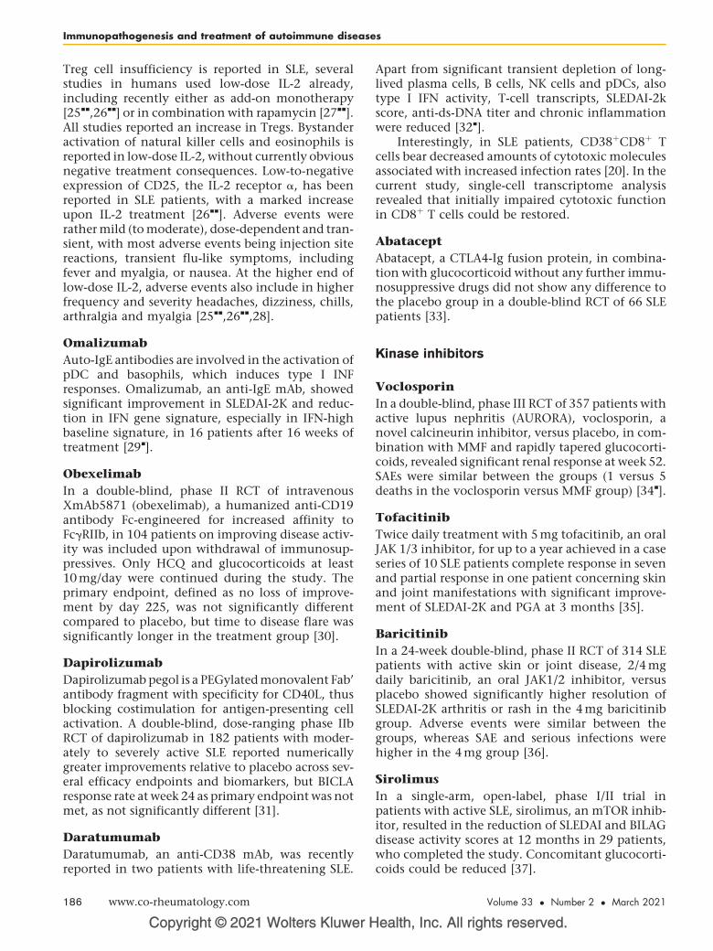
Immunopathogenesis and treatment of autoimmune diseases
Treg cell insufficiency is reported in SLE, severalstudies in humans used low-dose IL-2 already,including recently either as add-on monotherapy[25
&&
,26&&
] or in combination with rapamycin [27&&
].All studies reported an increase in Tregs. Bystanderactivation of natural killer cells and eosinophils isreported in low-dose IL-2, without currently obviousnegative treatment consequences. Low-to-negativeexpression of CD25, the IL-2 receptor a, has beenreported in SLE patients, with a marked increaseupon IL-2 treatment [26
&&
]. Adverse events wererather mild (to moderate), dose-dependent and tran-sient, with most adverse events being injection sitereactions, transient flu-like symptoms, includingfever and myalgia, or nausea. At the higher end oflow-dose IL-2, adverse events also include in higherfrequency and severity headaches, dizziness, chills,arthralgia and myalgia [25
&&
,26&&
,28].
Omalizumab
Auto-IgE antibodies are involved in the activation ofpDC and basophils, which induces type I INFresponses. Omalizumab, an anti-IgE mAb, showedsignificant improvement in SLEDAI-2K and reduc-tion in IFN gene signature, especially in IFN-highbaseline signature, in 16 patients after 16 weeks oftreatment [29
&
].
Obexelimab
In a double-blind, phase II RCT of intravenousXmAb5871 (obexelimab), a humanized anti-CD19antibody Fc-engineered for increased affinity toFcgRIIb, in 104 patients on improving disease activ-ity was included upon withdrawal of immunosup-pressives. Only HCQ and glucocorticoids at least10 mg/day were continued during the study. Theprimary endpoint, defined as no loss of improve-ment by day 225, was not significantly differentcompared to placebo, but time to disease flare wassignificantly longer in the treatment group [30].
Dapirolizumab
Dapirolizumab pegol is a PEGylated monovalent Fab’antibody fragment with specificity for CD40L, thusblocking costimulation for antigen-presenting cellactivation. A double-blind, dose-ranging phase IIbRCT of dapirolizumab in 182 patients with moder-ately to severely active SLE reported numericallygreater improvements relative to placebo across sev-eral efficacy endpoints and biomarkers, but BICLAresponse rate at week 24 as primary endpoint was notmet, as not significantly different [31].
Daratumumab
Daratumumab, an anti-CD38 mAb, was recentlyreported in two patients with life-threatening SLE.
Copyright © 2021 Wolters Kluwer H
186 www.co-rheumatology.com
Apart from significant transient depletion of long-lived plasma cells, B cells, NK cells and pDCs, alsotype I IFN activity, T-cell transcripts, SLEDAI-2kscore, anti-ds-DNA titer and chronic inflammationwere reduced [32
&
].Interestingly, in SLE patients, CD38þCD8þ T
cells bear decreased amounts of cytotoxic moleculesassociated with increased infection rates [20]. In thecurrent study, single-cell transcriptome analysisrevealed that initially impaired cytotoxic functionin CD8þ T cells could be restored.
Abatacept
Abatacept, a CTLA4-Ig fusion protein, in combina-tion with glucocorticoid without any further immu-nosuppressive drugs did not show any difference tothe placebo group in a double-blind RCT of 66 SLEpatients [33].
Kinase inhibitors
Voclosporin
In a double-blind, phase III RCT of 357 patients withactive lupus nephritis (AURORA), voclosporin, anovel calcineurin inhibitor, versus placebo, in com-bination with MMF and rapidly tapered glucocorti-coids, revealed significant renal response at week 52.SAEs were similar between the groups (1 versus 5deaths in the voclosporin versus MMF group) [34
&
].
Tofacitinib
Twice daily treatment with 5 mg tofacitinib, an oralJAK 1/3 inhibitor, for up to a year achieved in a caseseries of 10 SLE patients complete response in sevenand partial response in one patient concerning skinand joint manifestations with significant improve-ment of SLEDAI-2K and PGA at 3 months [35].
Baricitinib
In a 24-week double-blind, phase II RCT of 314 SLEpatients with active skin or joint disease, 2/4 mgdaily baricitinib, an oral JAK1/2 inhibitor, versusplacebo showed significantly higher resolution ofSLEDAI-2K arthritis or rash in the 4 mg baricitinibgroup. Adverse events were similar between thegroups, whereas SAE and serious infections werehigher in the 4 mg group [36].
Sirolimus
In a single-arm, open-label, phase I/II trial inpatients with active SLE, sirolimus, an mTOR inhib-itor, resulted in the reduction of SLEDAI and BILAGdisease activity scores at 12 months in 29 patients,who completed the study. Concomitant glucocorti-coids could be reduced [37].
ealth, Inc. All rights reserved.
Volume 33 � Number 2 � March 2021
New therapeutics SLE Kolios et al.
Leflunomide
In an RCT over 24 weeks in 100 Chinese patientswith proliferative lupus nephritis low-dose leflu-nomide (40 mg/day for 3 days, followed by 20 mg/day), an inhibitor of pyrimidine synthesis andtyrosine kinases showed similar complete and par-tial remission rates for induction treatment as wellas adverse events compared to intravenous CYP(0.8–1.0 g per monthly), both in addition to pred-nisone [38
&
].
Immunomodulators
Iguratimod
Iguratimod, an NF-kB inhibitor preventing B-celldifferentiation, reducing autoantibody levels andplasma cells, showed promising results with 93%response in an investigational study of 14 therapy-refractory lupus nephritis patients at week 24 [39
&&
].
Lymphokine-based vaccines
In a 36-week phase IIb, double-blind RCT with 185adults with mild-to-moderate active SLE, an INF-akinoid vaccine induced neutralizing anti-IFN-a2bserum antibodies in 91% of treated patients andreduced significantly the IFN gene signature.Although the primary endpoint did not statisticallydiffer between treatment and placebo in the modi-fied BICLA response, patients were able to signifi-cantly reduce concomitant glucocorticoids. Adverseevents in the IFN-K group included infections andinjection site reactions with upper respiratory tractinfections and arthralgia were thrice more commonand nasopharyngitis twice more commen in IFN-Kgroup versus placebo; SAEs were more common inthe placebo group [40
&
].
Mesenchymal stem cells
In an investigator-initiated trial, 21 treatmentrefractory SLE patients underwent umbilical-cord-derived mesenchymal stem cell (U-MSC) transplan-tation, which leads to significant upregulation ofperipheral blood CD1cþ dendritic cells by expres-sion of FLT3L [41
&&
]. The short-term group of 10patients at 1-month follow-up showed remarkableimprovements in SLEDAI. The long-term follow-upgroup at 6 months included 11 patients; twopatients achieved an SLEDAI complete response,seven a partial response and two progressed. Patientswere able to reduce glucocorticoid dose and con-comitant immunosuppression. It was previouslyshown that allogenic U-MSC also shows an immu-noregulatory effect in suppressing T cells and B cells,
Copyright © 2021 Wolters Kluwe
1040-8711 Copyright � 2020 Wolters Kluwer Health, Inc. All rights rese
including IgG production, as well as inducing Tregsand macrophage phagocytic activity. Larger studieswould need to evaluate the composite biologicaleffect of U-MSC on all cell types.
Ongoing registered clinical trials in systemiclupus erythematosus
Several molecular targets, which are reviewed else-where [8,42], are currently under investigationbecause a relevant association to disease pathogen-esis has been claimed. Table 1 summarizes noveltreatments in clinical studies, which can eithertarget immune cells directly (e.g. anti-CD20 mAbsand anti-CD6 mAb), relevant cytokines (e.g. IL-23),intracellular pathways (e.g. BTK or JAK), comple-ment system (e.g. anti-MASP-2 mAb and anti-C5mAb), or cytokine treatments (e.g. IL-2 andmuteins).
CONCLUSION
Treatment of SLE is still a challenging endeavor,which is related to its clinical and pathogeneticheterogeneity. A step toward patient-centered pre-cision medicine should require the inclusion ofadditional diagnostic steps to define the right subsetof patients who will benefit from any given biologic/small drug, which targets a specific pathway.
In the last year, several new and promisingtherapies were developed and much more areawaited in the near future. These interventionseither inhibit the inflammatory axis or enhanceregulatory immune mechanisms.
As patients with SLE are inherently immuno-compromised [43] and most of the novel therapeu-tics interfere with the required immune response topathogens, vigilance for possible increased infectionrates should be heightened. It was discussed abovethat IFN mAbs and vaccine may exaggerate the riskfor severe COVID-19 [19].
Biologics used in clinical trials should be able toaccomplish what they have been prepared for. Yet,there are certain contrivances, which may preventthem to accomplish the expected mechanistic inter-vention. For example, IL-2 does not trigger a robustsignaling response in T cells from people with SLE asit does in normal T cells [44]. B-cell depleting anti-bodies do not deplete B cells in lupus-prone mice aseffectively as in normal mice [45], which suggeststhat B-cell depletion may not be achieved in allcompartments in SLE patients who are administeredB-cell-depleting antibodies.
It is apparent that none of the identified patho-genetic pathways is present in all patients with SLEwho carry a bona fide clinical diagnosis of SLE.
r Health, Inc. All rights reserved.
rved. www.co-rheumatology.com 187
Immunopathogenesis and treatment of autoimmune diseases
Accordingly, targeting of any pathway will benefitonly a subset of patients, which may not deliver theexpected effect in the study population. Further, it ispossible that although a targeted pathway may beoperational in a patient with SLE, it may not be thedriving pathway and therefore, its correction maynot deliver the expected effect both at the immu-nopathogenic and clinical levels. The need for pre-cision medicine in people with SLE cannot beoverstated and is long overdue.
Acknowledgements
None.
Financial support and sponsorship
The work in the Tsokos laboratory is funded by the NIH.
Conflicts of interest
There are no conflicts of interest.
REFERENCES AND RECOMMENDEDREADINGPapers of particular interest, published within the annual period of review, havebeen highlighted as:
& of special interest&& of outstanding interest1. Tsokos GC. Systemic lupus erythematosus. N Engl J Med 2011;365:2110–2121.
2. Tsokos GC. Autoimmunity and organ damage in systemic lupus erythema-tosus. Nat Immunol 2020; 21:605–614.
3. Kaul A, Gordon C, Crow MK, et al. Systemic lupus erythematosus. Nat RevDis Primers 2016; 2:16039.
4. Aringer M, Costenbader K, Daikh D, et al. 2019 European LeagueAgainst Rheumatism/American College of Rheumatology classificationcriteria for systemic lupus erythematosus. Ann Rheum Dis 2019;78:1151–1159.
5. Murphy G, Isenberg DA. New therapies for systemic lupus erythematosus:past imperfect, future tense. Nat Rev Rheumatol 2019; 15:403–412.
6. Fanouriakis A, Kostopoulou M, Alunno A, et al. 2019 Update of the EULARrecommendations for the management of systemic lupus erythematosus. AnnRheum Dis 2019; 78:736–745.
7. Grammatikos AP, Ghosh D, Devlin A, et al. Spleen tyrosine kinase (Syk)regulates systemic lupus erythematosus (SLE) T cell signaling. PLoS One2013; 8:e74550.
8. Vukelic M, Li Y, Kyttaris VC. Novel treatments in lupus. Front Immunol 2018;9:2658.
9.&&
Furie R, Rovin BH, Houssiau F, et al. Two-year, randomized, controlled trial ofbelimumab in lupus nephritis. N Engl J Med 2020; 383:1117–1128.
Belimumab in addition to standard of care showed significant better renal outcomeat week 104.10. Teng YKO, Bruce IN, Diamond B, et al. Phase III, multicentre, randomised,
double-blind, placebo-controlled, 104-week study of subcutaneous belimu-mab administered in combination with rituximab in adults with systemic lupuserythematosus (SLE): BLISS-BELIEVE study protocol. BMJ Open 2019;9:e025687.
11. van Vollenhoven RF, Navarra SV, Levy RA, et al. Long-term safety and limitedorgan damage in patients with systemic lupus erythematosus treated withbelimumab: a phase III study extension. Rheumatology (Oxford) 2020;59:281–291.
12. Wallace DJ, Ginzler EM, Merrill JT, et al. Safety and efficacy of belimumab plusstandard therapy for up to thirteen years in patients with systemic lupuserythematosus. Arthritis Rheumatol 2019; 71:1125–1134.
13. Tanaka Y, Bass D, Chu M, et al. Organ system improvements in Japanesepatients with systemic lupus erythematosus treated with belimumab: a sub-group analysis from a phase 3 randomized placebo-controlled trial. ModRheumatol 2020; 30:313–320.
14. D’Cruz D, Maksimowicz-McKinnon K, Oates J, et al. Efficacy and safety ofbelimumab in patients of black race with systemic lupus erythematosus:results from the EMBRACE study. Lupus Sci Med 2019; 6:. doi: 10.1136/lupus-2019-lsm.200.
Copyright © 2021 Wolters Kluwer H
188 www.co-rheumatology.com
15.&&
Morand EF, Furie R, Tanaka Y, et al. Trial of anifrolumab in active systemiclupus erythematosus. N Engl J Med 2020; 382:211–221.
Anifrolumab showed significant improvement in BICLA after 52 weeks.16. Furie RA, Morand EF, Bruce IN, et al. Type I interferon inhibitor anifrolumab in
active systemic lupus erythematosus (TULIP-1): a randomised, controlled,phase 3 trial. Lancet Rheumatol 2019; 1:E208–E219.
17.&
Takeuchi T, Tanaka Y, Matsumura R, et al. Safety and tolerability of sifalimu-mab, an antiinterferon-a monoclonal antibody, in Japanese patients withsystemic lupus erythematosus: a multicenter, phase 2, open-label study.Mod Rheumatol 2020; 30:93–100.
Sifalimumab showed the long-term efficacy in Japanese patients.18. Khamashta M, Merrill JT, Werth VP, et al. Sifalimumab, an antiinterferon-alpha
monoclonal antibody, in moderate to severe systemic lupus erythematosus: arandomised, double-blind, placebo-controlled study. Ann Rheum Dis 2016;75:1909–1916.
19. Bastard P, Rosen LB, Zhang Q, et al. Auto-antibodies against type I IFNs inpatients with life-threatening COVID-19. Science 2020; 370:eabd4585.
20. Katsuyama E, Suarez-Fueyo A, Bradley SJ, et al. The CD38/NAD/SIRTUIN1/EZH2 axis mitigates cytotoxic CD8 T cell function and identifies patients withSLE prone to infections. Cell Rep 2020; 30:112–123e4.
21.&&
Furie R, Werth VP, Merola JF, et al. Monoclonal antibody targeting BDCA2ameliorates skin lesions in systemic lupus erythematosus. J Clin Invest 2019;129:1359–1371.
BIIB059, a pDC-targeting mAb, reduced the expression of IFN response genes inblood and CLASI and normalized MxA expression after a single infusion.22.&&
van Vollenhoven RF, Hahn BH, Tsokos GC, et al. Maintenance of efficacy andsafety of ustekinumab through one year in a phase II multicenter, prospective,randomized, double-blind, placebo-controlled crossover trial of patients withactive systemic lupus erythematosus. Arthritis Rheumatol 2020;72:761–768.
Belimumab in an 8-year extension study showed stable disease in 87.7% ofpatients.23. van Vollenhoven RF, Hahn BH, Tsokos GC, et al. Efficacy and safety of
ustekinumab, an IL-12 and IL-23 inhibitor, in patients with active systemiclupus erythematosus: results of a multicentre, double-blind, phase 2, rando-mised, controlled study. Lancet 2018; 392:1330–1339.
24. Rosenzwajg M, Lorenzon R, Cacoub P, et al. Immunological and clinicaleffects of low-dose interleukin-2 across 11 autoimmune diseases in a single,open clinical trial. Ann Rheum Dis 2019; 78:209–217.
25.&&
He J, Zhang R, Shao M, et al. Efficacy and safety of low-dose IL-2 in thetreatment of systemic lupus erythematosus: a randomised, double-blind,placebo-controlled trial. Ann Rheum Dis 2020; 79:141–149.
Low-dose IL-2 was able to enhance Treg function in SLE.26.&&
Humrich JY, von Spee-Mayer C, Siegert E, et al. Low-dose interleukin-2 therapyin refractory systemic lupus erythematosus: an investigator-initiated, single-centre phase 1 and 2a clinical trial. Lancet Rheumatol 2019; 1:e44–54.
Low-dose IL-2 was able to enhance Treg function in SLE.27.&&
Zhao C, Chu Y, Liang Z, et al. Low dose of IL-2 combined with rapamycinrestores and maintains the long-term balance of Th17/Treg cells in refractorySLE patients. BMC Immunol 2019; 20:32.
Low-dose IL-2 was able to enhance Treg function in SLE.28. Mahmoudpour SH, Jankowski M, Valerio L, et al. Safety of low-dose sub-
cutaneous recombinant interleukin-2: systematic review and meta-analysis ofrandomized controlled trials. Sci Rep 2019; 9:7145.
29.&
Hasni S, Gupta S, Davis M, et al. Safety and tolerability of omalizumab: arandomized clinical trial of humanized anti-IgE monoclonal antibody in sys-temic lupus erythematosus. Arthritis Rheumatol 2019; 71:1135–1140.
Omalizumab reduced INF-signature and improved disease activity.30. Merrill J, June J, Koumpouras F, et al. FRI0176 phase 2, double-blind,
randomized, placebo-controlled study of a reversible B cell inhibitor,XMAB15871, in systemic lupus erythematosus (SLE). Ann Rheum Dis2019; 78:761–762.
31. Furie R, Bruce IN, Dorner T, et al. FRI0195 efficacy and safety of dapiroli-zumab pegol (DZP) in patients with moderately to severely active systemiclupus erythematosus (SLE): a randomised, placebo (PBO)-controlled study.Ann Rheum Dis 2019; 78:775–776.
32.&
Ostendorf L, Burns M, Durek P, et al. Targeting CD38 with daratumumab inrefractory systemic lupus erythematosus. N Engl J Med 2020; 383:1149–1155.
The first report of daratumumab, a plasma-cell targeted therapy, shows promisingresults in two treatment-refractory SLE patients.33. Thanou A, Arriens C, Aberle T, et al. FRI0213 randomized placebo controlled
trial of abatacept for nonorgan threatening systemic lupus with backgroundmedications withdrawn. Ann Rheum Dis 2019; 78:786.
34.&
Onno Teng YK, Parikh SV, Saxena A, et al. O11 AURORA phase 3 studydemonstrates voclosporin statistical superiority over standard of care in lupusnephritis (LN). Lupus Sci Med 2020; 7. doi: 10.1136/lupus-2020-eurolupus.24.
Voclosporin, a novel calcineurin inhibitor, revealed significant renal response in aphase III study in combination with MMF and low-dose glucocorticoids.35. You H, Zhang G, Wang Q, et al. Successful treatment of arthritis and rash with
tofacitinib in systemic lupus erythematosus: the experience from a singlecentre. Ann Rheum Dis 2019; 78:1441–1443.
36. Wallace DJ, Furie RA, Tanaka Y, et al. Baricitinib for systemic lupus erythe-matosus: a double-blind, randomised, placebo-controlled, phase 2 trial.Lancet 2018; 392:222–231.
ealth, Inc. All rights reserved.
Volume 33 � Number 2 � March 2021
New therapeutics SLE Kolios et al.
37. Lai ZW, Kelly R, Winans T, et al. Sirolimus in patients with clinically activesystemic lupus erythematosus resistant to, or intolerant of, conventional medica-tions: a single-arm, open-label, phase 1/2 trial. Lancet 2018; 391:1186–1196.
38.&
Zhang M, Qi C, Zha Y, et al. Leflunomide versus cyclophosphamide in theinduction treatment of proliferative lupus nephritis in Chinese patients: arandomized trial. Clin Rheumatol 2019; 38:859–867.
Leflunomide versus intravenous CYP in addition to glucocorticoid in proliferativelupus nephritis showed similar remission and adverse event rates.39.&&
Kang Y, Yan Q, Fu Q, et al. Iguratimod as an alternative induction therapy forrefractory lupus nephritis: a preliminary investigational study. Arthritis ResTher 2020; 22:65.
Iguratimod showed 93% response in previously therapy-refractory lupus nephritispatients.40.&
Houssiau FA, Thanou A, Mazur M, et al. IFN-a kinoid in systemic lupuserythematosus: results from a phase IIb, randomised, placebo-controlledstudy. Ann Rheum Dis 2020; 79:347–355.
INF-a kinoid vaccine did not meet the primary endpoint in a modified BICLAresponse, but concomitant glucocorticoids could have been significantly reduced.
Copyright © 2021 Wolters Kluwe
1040-8711 Copyright � 2020 Wolters Kluwer Health, Inc. All rights rese
41.&&
Yuan X, Qin X, Wang D, et al. Mesenchymal stem cell therapy induces FLT3Land CD1c(þ) dendritic cells in systemic lupus erythematosus patients. NatCommun 2019; 10:2498.
A small trial of umbilical-cord-derived mesenchymal stem cell transplantationshowed promising results in SLEDAI improvements and reduction of concomitantglucocorticoids.42. Klavdianou K, Lazarini A, Fanouriakis A. Targeted biologic therapy for systemic
lupus erythematosus: emerging pathways and drug pipeline. BioDrugs 2020;34:133–147.
43. Grammatikos AP, Tsokos GC. Immunodeficiency and autoimmunity: les-sons from systemic lupus erythematosus. Trends Mol Med 2012;18:101–108.
44. Comte D, Karampetsou MP, Kis-Toth K, et al. Brief Report: CD4þ T cellsfrom patients with systemic lupus erythematosus respond poorly toexogenous interleukin-2. Arthritis Rheumatol 2017; 69:808–813.
45. Chen J, Crispin JC, Tedder TF, et al. B cells contribute toischemia/reperfusion-mediated tissue injury. J Autoimmun 2009;32:195–200.
r Health, Inc. All rights reserved.
rved. www.co-rheumatology.com 189
REVIEW
CURRENTOPINION Update on the cellular pathogenesis of lupus
Copyright ©
www.co-rheumatology.com
a,b b c
Noemie Gensous , Andrea Boizard-Moracchini , Estibaliz Lazaro ,Christophe Richezb,d, and Patrick Blancob,ePurpose of review
Aberrations in the innate and in the adaptive arms of the immune system play both important roles in theinitiation and progression of systemic lupus erythematosus (SLE). The aim of this study was to provide anupdate on the most recent findings on the cellular pathogenesis of SLE. Our overview focused particularlyon results obtained over the last 18 months.
Recent findings
Recent observations have provided an improved understanding of the importance of low-densitygranulocytes, a highly proinflammatory subset of neutrophils. We also highlighted in this work recentdescriptions of the various cellular sources associated with the interferon signature. In addition, novelcontributions have also developed our understanding of the potential importance of extrafollicular T–B-cellinteractions in SLE pathogenesis. Finally, the role of recently described B and T-cell subsets, that is, atypicalmemory B cells, T-peripheral helper cells, and Th10 T cells, were also reviewed.
Summary
Recent findings in the cellular pathogenesis of SLE give a deeper comprehension of previously describedmechanisms which drive SLE pathogenesis and shed light on novel players in immune dysregulation thatcould help to identify potential therapeutic targets.
Keywords
autoimmunity, B cells, systemic lupus erythematosus, T cells, type I interferon
aDepartment of Internal Medicine and Clinical Immunology, CHU Bor-deaux (Groupe Hospitalier Saint-Andre), bUMR/CNRS 5164, Immuno-ConcEpT, CNRS, University of Bordeaux, cDepartment of InternalMedicine, CHU Bordeaux (Groupe Hospitalier Haut Leveque), Pesac,
INTRODUCTION
Systemic lupus erythematosus (SLE) is a chronic sys-temic autoimmune disease, characterized by a loss oftolerance toward nuclear components, leading toautoantibody production, immune complex forma-tion, and multiorgan damage. The pathogenesis ofSLE is complex, involving genetic, environmental,and hormonal factors [1]. Important progress has beenmade over the last decades toward the understandingof the underlying cellular pathophysiology of SLE.Dysregulation of multiple immune cell lineages,belonging to the adaptive but also to the innateimmune systems, has been identified as contributorto the expression of systemic autoimmunity. In thisreview, we will summarize recent insights into thecellular pathogenesis of SLE and provide an update onthe most recent findings involving the different cellu-lar compartments of the immune system.
dDepartment of Rheumatology and eDepartment of Immunology, CHUBordeaux (Groupe Hospitalier Pellegrin), Bordeaux, France
Correspondence to Patrick Blanco, Department of Immunology, CHUBordeaux (Groupe Hospitalier Pellegrin), Bordeaux, France. E-mail:[email protected]
Curr Opin Rheumatol 2021, 33:190–196
DOI:10.1097/BOR.0000000000000775
HEMATOPOIETIC STEM AND PROGENITORCELLS
Hematopoietic stem and progenitor cells are themost primitive multipotent population that giverise to all blood cell types. Their transcriptomic
2021 Wolters Kluwer H
analysis in the NZB/W F1 mouse model of lupusrevealed a reprogramming of these cells towardmyeloid lineage, with expanded frequencies of com-mon myeloid progenitors [2]. When compared withhuman SLE hematopoietic progenitor cells, similardata were observed with clear evidence of dysregu-lated differentiation. Thus, in lupus patients withsevere disease, CD34þ cells were found to haveenhanced proliferation, cell differentiation, andtranscriptional activation of cytokines and chemo-kines that drive differentiation toward myeloid/granulocytic lineage [2]. This inappropriate myeloidversus lymphoid balance may contribute to the
ealth, Inc. All rights reserved.
Volume 33 � Number 2 � March 2021

KEY POINTS
� There is a significant heterogeneity amongst SLE LDGsand the majority of them exhibit a highproinflammatory phenotype with proinflammatoryeffects on T cells.
� Plasmacytoid dendritic cells, which are key producersof type I interferon, can be activated through FcgRIIareceptors and taurine is crucial for their metabolism.
� Extrafollicular T–B-cell interactions appear to be involvedin the pathogenesis of SLE, as suggested by data onDnase1l3�/� mice model and by the expansion of T-peripheral helper cells observed in SLE patients.
� In the presence of oxidized mitochondrial DNA, naıveCD4þ T can differentiate into Th10 T cells, whichprovide B-cell help through IL-10 and succinate.
Update on the cellular pathogenesis of lupus Gensous et al.
dysregulation of subsequent immune responses par-ticularly during the early biologic phase of the dis-ease, as it could feed the immune system withnuclear-derived antigens.
DYSREGULATION OF INNATE IMMUNITY:ROLE OF NEUTROPHILS
Owing to the presence of self-reactive autoantibod-ies, SLE has long been considered as predominantlyassociated with a dysfunction of the adaptiveimmune responses. However, over the past years,several elements of the innate immune system havebeen identified as major contributors to the diseasepathogenesis [3].
In recent years, several lines of evidence haveimplicated neutrophils in autoimmune diseases andspecially in lupus pathogenesis [4]. Neutrophils ofSLE patients exhibit an increased capacity toundergo a specific form of spontaneous cell death,called NETosis, constituting thereby neutrophilextracellular traps (NETs). NETs are networks ofdecondensed chromatin and associated granularcomponents, histones and cytoplasmic proteins,released into the extracellular space. NETs caninduce endothelial damage and favor thrombosisand, harboring autoantigens, they are an importantsource of autoantigens to trigger autoimmunity.Additionally, SLE patients are characterized by thepresence of a specific subset of circulating low-den-sity granulocytes (LDGs), which are phenotypically,functionally and transcriptionally distinct from theother neutrophil subsets. LDGs are highly proin-flammatory, as they are important producers ofNETs and are able to secrete proinflammatory cyto-kines (type I interferons [IFN], tumor necrosis factor[TNF]-a, and IFN-g).
Copyright © 2021 Wolters Kluwe
1040-8711 Copyright � 2020 Wolters Kluwer Health, Inc. All rights rese
Performing in-depth transcriptomic and epige-netic analysis of SLE LDGs, autologous normal-den-sity neutrophils, and healthy control neutrophils,Mistry et al. [5
&
] recently identified significant tran-scriptional and epigenetic heterogeneity amongstSLE LDGs. Thus, they were able to clearly delineatetwo subpopulations of intermediate-mature andimmature neutrophils, identified as LDGmat andLDGimm, characterized by distinct transcriptionalprofiles and epigenetic landscapes. The majorityof SLE LDGs was of intermediate-mature nature,with several functional pathogenic features suchas NETs formation, capacity to undergo chemotaxis,and enhanced ability to phagocytosis. This subset,with a high proinflammatory phenotype, couldthereby be responsible of the association with organdamage associated with neutrophils.
Another recent study further explored in detail thepathogenic potential of SLE LDGs, with an immuno-phenotypic, morphological and functional character-izationof this neutrophil subset inawell-characterizedSLEcohort [6
&
]. SLELDGs,whichexhibitedheightenedsurface expression of several activation markers, werefound to exert proinflammatory effects on T cells, astheir supernatants induced proinflammatory cytokineproduction (IFN-g, TNF-a, and lymphotoxin-a) fromCD4þ T cells. These supernatants did not suppress T-cell proliferation, in contrast to supernatants of nor-mal-density granulocytes. Additionally, LDG preva-lence, elevated in SLE patients as compared withhealthy controls, was associated with the type I IFNgene signature, as well as with disease activity, asassessed by the SLE disease activity index (SLEDAI)score [6
&
]. The association between higher numbersof LDGs with disease activity and low complementlevels was further confirmed in another cohort ofpatientswithSLE [7]. Finally,LDGswerealso identifiedas potential biomarkers of cardiovascular risk in SLEpatients with subclinical atherosclerosis [8].
Frangou et al. [9] explored the exact molecularmechanisms that lead to NETs release in SLE andidentified autophagy as a key pathway in this phe-nomenon. Neutrophils from patients with active SLEexhibited, in the context of proinflammatory micro-environment, increased basal autophagy levels lead-ing to excessive NETs production. Mechanistically,the induction of autophagy was under the depen-dence of the hypoxia and stress-response proteinDNA damage inducible transcript 4/regulated indevelopment and DNA damage responses 1. Explor-ing the protein composition of SLE NETs, the authorsalso revealed abundant expression of bioactive tissuefactor and interleukin-17A (IL-17A), which promotedthrombin generation and fibrosis, thereby contribut-ing to tissue injury in skin and kidneys [9]. Overall,those studies place LDGs as key contributors to organ
r Health, Inc. All rights reserved.
rved. www.co-rheumatology.com 191
Immunopathogenesis and treatment of autoimmune diseases
damage during acute flares, but also to long-termcardiovascular comorbidity.
INTERFERONS
Type I IFNs, which affect multiple components ofthe immune system, are considered to be key mol-ecules in the pathogenesis of SLE [10]. Overexpres-sion of type I IFN inducible genes, known as the IFNsignature, and high concentrations of circulatingtype I IFN are characteristic of the disease, correlat-ing with several parameters of lupus severity. Mech-anistically, a lot of research has been carried outduring the last decade and is still ongoing on theidentification of the precise cellular sources andmechanisms of type I IFN activity in SLE.
Applying the technique of single-cell RNAsequencing to renal biopsies from patients with lupusnephritis, Der et al. [11] confirmed the presence of anoverexpression of type I IFN-related genes in lupuspatients as compared with healthy controls. Interest-ingly, this signature was specifically identified in thesubset of tubular cells. The renal IFN signature corre-latedwith the oneobserved inkeratinocytes obtainedfrom skin biopsies of the same patients, and predictedthe response to lupus nephritis treatment six monthsafter the biopsy, thereby identifying a potential pow-erful biomarker for this disease.
Plasmacytoid dendritic cells (pDCs) are consid-ered as key producers of type I IFN in SLE. Theiractivation by nucleic acid-containing immune com-plexes, via interaction with Toll-like receptors (TLRs),has been well demonstrated, but further studies havetried to identify other pathways involved in theirpriming. A recent study examined the involvementof Fcg receptor in this phenomenon [12]. AlthoughIgG-immune complexes appeared to inhibit type IIFN production in healthy individuals by an inhibi-tory signaling through FcgRIIa on dendritic cells, SLEpatients with lupus nephritis displayed an increasedresponsivity to the stimulation by the same type ofimmune complexes, resulting in elevated productionlevels of type I IFN and IL-1b [12]. In addition, arecent study examined the metabolic regulation oftype I IFN production by pDCs in the context ofautoimmunity [13
&
]. Taurine metabolism was foundcritical in type I IFN production by pDCs. Taurinelevels were found higher in SLE patients’ sera ascompared with healthy controls or rheumatoidarthritis patients, with a positive correlation betweentaurine content, disease activity, and the expressionof IFN signature genes. In lupus-prone mice, taurinesupplementation led to a more severe disease withpromotion of type I IFN-induced genes expression,lymphocytes activation, increased autoantibodiesproduction, and increased proteinuria [13
&
].
Copyright © 2021 Wolters Kluwer H
192 www.co-rheumatology.com
Other cellular sources have been pinpointed asassociated with the IFN signature. Thus, monocyteswere identified as potent producers of IFNa in SLEpatients, with a positive correlation between thefrequency of IFNa-producing monocytes and SLE-disease activity [14]. Mechanistically, production ofIFNa was under the dependency of the cyclic guano-sine monophosphate - adenosine monophosphatesynthase (cGAS) and stimulator of IFN genes (STING)(cGAS-STING) pathway stimulation. Inhibition ofthe mechanistic target of the rapamycin (mTOR)pathway downregulated theenhancedSTING expres-sion and its downstream molecules and suppressedthe subsequent IFNa production by monocytes [14].
Recently, the IFN signature has also been asso-ciated with innate lymphoid cells (ILCs), which areimmune cells belonging to the lymphoid lineagebut which do not express antigen-specific receptors[15]. Alterations in the frequency and in the func-tion of ILCs have been implicated in the pathophys-iology of different rheumatic diseases. Blokland et al.[15] identified an association between the type I IFNsignature in SLE patients and a decreased cell fre-quency of ILC2 and ILC3 subsets with an elevatedexpression of Fas (CD95) on their surface.
Type II (IFN-g) and type III (IFN-l) IFNs may alsoplay a role in SLE pathogenesis. In a large cohort ofSLE patients, Oke et al. [16] performed simultaneousmeasurement of peripheral levels of all three IFNsubtypes and demonstrated higher levels in patientsas compared with controls (IFN-a, IFN-g, and IFN-l1). Interestingly, it was observed different clinicaland biological SLE features according to the type ofelevated IFN, suggesting that IFNs contribute to theheterogeneity of SLE clinical manifestations. Thus,high type I IFN activity was associated with activemucocutaneous inflammation, whereas high levelsof IFN-g were more associated with nephritis andarthritis [16]. Both type I and type III IFNs appearedto have a possible complementary role in lupusdisease pathogenesis [17]. Hiorton et al. [17] dem-onstrated that RNA-containing immune complexeshave the capacity to induce type III IFN productionby pDCs, especially in the small subset which pro-duces type I IFN as well, connecting thereby the typeI and type III IFN production in pDCs and suggestingsimilar mechanisms at work.
Given its importance in SLE pathogenesis, type IIFN has been considered as a potential interestingtarget for the development of new therapies. Thishas been emphasized by the recently publishedresults of the TULIP-2 phase 3 trial, which reportedthe therapeutic benefit in SLE patients of anifrolu-mab, a fully human monoclonal antibody targetingtype I IFN receptor subunit 1 which inhibits signal-ing by all type I IFNs [18].
ealth, Inc. All rights reserved.
Volume 33 � Number 2 � March 2021
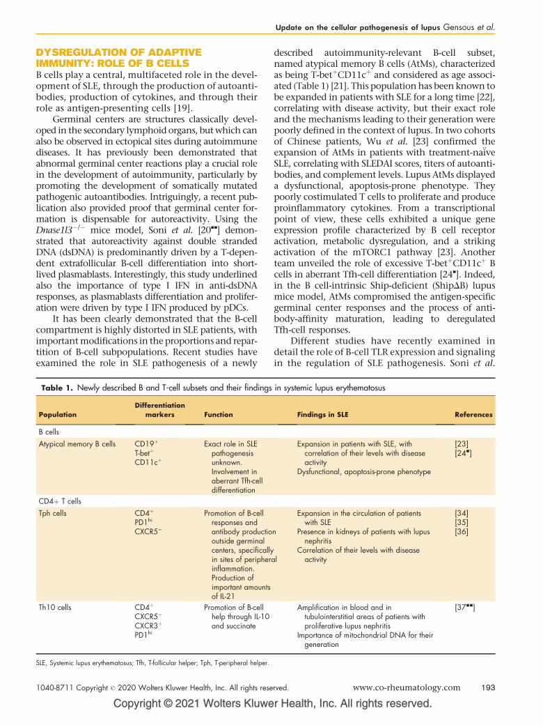
Update on the cellular pathogenesis of lupus Gensous et al.
DYSREGULATION OF ADAPTIVEIMMUNITY: ROLE OF B CELLSB cells play a central, multifaceted role in the devel-opment of SLE, through the production of autoanti-bodies, production of cytokines, and through theirrole as antigen-presenting cells [19].
Germinal centers are structures classically devel-oped in the secondary lymphoid organs, but which canalso be observed in ectopical sites during autoimmunediseases. It has previously been demonstrated thatabnormal germinal center reactions play a crucial rolein the development of autoimmunity, particularly bypromoting the development of somatically mutatedpathogenic autoantibodies. Intriguingly, a recent pub-lication also provided proof that germinal center for-mation is dispensable for autoreactivity. Using theDnase1l3�/� mice model, Soni et al. [20
&&
] demon-strated that autoreactivity against double strandedDNA (dsDNA) is predominantly driven by a T-depen-dent extrafollicular B-cell differentiation into short-lived plasmablasts. Interestingly, this study underlinedalso the importance of type I IFN in anti-dsDNAresponses, as plasmablasts differentiation and prolifer-ation were driven by type I IFN produced by pDCs.
It has been clearly demonstrated that the B-cellcompartment is highly distorted in SLE patients, withimportant modifications in the proportions and repar-tition of B-cell subpopulations. Recent studies haveexamined the role in SLE pathogenesis of a newly
Copyright © 2021 Wolters Kluwe
Table 1. Newly described B and T-cell subsets and their findings
PopulationDifferentiation
markers Function
B cells
Atypical memory B cells CD19þ
T-betþ
CD11cþ
Exact role in SLEpathogenesisunknown.Involvement inaberrant Tfh-celldifferentiation
CD4þ T cells
Tph cells CD4þ
PD1hi
CXCR5–
Promotion of B-cellresponses andantibody productiooutside germinalcenters, specificallyin sites of peripherainflammation.Production ofimportant amountsof IL-21
Th10 cells CD4þ
CXCR5�
CXCR3þ
PD1hi
Promotion of B-cellhelp through IL-10and succinate
SLE, Systemic lupus erythematosus; Tfh, T-follicular helper; Tph, T-peripheral helper.
1040-8711 Copyright � 2020 Wolters Kluwer Health, Inc. All rights rese
described autoimmunity-relevant B-cell subset,named atypical memory B cells (AtMs), characterizedas being T-betþCD11cþ and considered as age associ-ated (Table 1) [21]. This population has been known tobe expanded in patients with SLE for a long time [22],correlating with disease activity, but their exact roleand the mechanisms leading to their generation werepoorly defined in the context of lupus. In two cohortsof Chinese patients, Wu et al. [23] confirmed theexpansion of AtMs in patients with treatment-naiveSLE, correlating with SLEDAI scores, titers of autoanti-bodies, and complement levels. Lupus AtMs displayeda dysfunctional, apoptosis-prone phenotype. Theypoorly costimulated T cells to proliferate and produceproinflammatory cytokines. From a transcriptionalpoint of view, these cells exhibited a unique geneexpression profile characterized by B cell receptoractivation, metabolic dysregulation, and a strikingactivation of the mTORC1 pathway [23]. Anotherteam unveiled the role of excessive T-betþCD11cþ Bcells in aberrant Tfh-cell differentiation [24
&
]. Indeed,in the B cell-intrinsic Ship-deficient (ShipDB) lupusmice model, AtMs compromised the antigen-specificgerminal center responses and the process of anti-body-affinity maturation, leading to deregulatedTfh-cell responses.
Different studies have recently examined indetail the role of B-cell TLR expression and signalingin the regulation of SLE pathogenesis. Soni et al.
r Health, Inc. All rights reserved.
in systemic lupus erythematosus
Findings in SLE References
Expansion in patients with SLE, withcorrelation of their levels with diseaseactivity
Dysfunctional, apoptosis-prone phenotype
[23][24&]
n
l
Expansion in the circulation of patientswith SLE
Presence in kidneys of patients with lupusnephritis
Correlation of their levels with diseaseactivity
[34][35][36]
Amplification in blood and intubulointerstitial areas of patients withproliferative lupus nephritis
Importance of mitochondrial DNA for theirgeneration
[37&&]
rved. www.co-rheumatology.com 193
Immunopathogenesis and treatment of autoimmune diseases
[20&&
] demonstrated that anti-dsDNA responses werepromoted by TLR9 in Dnase1l3�/� mice, in a syner-gistic and partially redundant way with TLR7.Accordingly, it had been previously demonstratedthat TLR9 deficiency led to a loss of anti-dsDNAautoantibodies, but its effects on disease severityhave appeared to be opposite. Indeed, in differentmodel of lupus, a protective role of TLR9 has beensuggested, as TLR9 deficiency resulted in decreasedsurvival with exacerbated nephritis [25]. To go fur-ther in the exploration of these paradoxical results,Tilstra et al. [26] evaluated the effects of TLR9 expres-sion in multiple target populations to explore pos-sible cell type specific roles. The effect of TLR9 losswas evaluated using a conditional Tlr9 knockout(Tlr9fl) in B cells (CD19-Cre), dendritic cells(CD11c-Cre), neutrophils (MRP8-Cre), and myeloidcells (both macrophages and neutrophils) (LysM-Cre). Strikingly, only B-cell-specific Tlr9 deficiencyresulted in an acceleration of lupus nephritis and inan alteration of the autoantibody response, whereasits overexpression led to an amelioration of thedisease in murphy roths large/lpr mice. Accordingto this study, B-cell TLR9 expression appears therebyto be both necessary and sufficient to modulate SLEpathogenesis [26].
Regarding TLR7 expression, it was demonstratedthat its high expression in SLE patients, driven by agenetic polymorphism (rs3853839 C/G), was associ-ated with a more active disease and with an upre-gulation of IFN-responsive genes [27]. Moreover,this overexpression correlated with the expansionof newly formed transitional B cells and promotedautoantibodies production [27].
DYSREGULATION OF ADAPTIVEIMMUNITY: ROLE OF T CELLS
T lymphocytes (CD4þ, CD8þ, double negative) arekey players in SLE disease initiation and mainte-nance, through the help provided to B cells, thesecretion of proinflammatory cytokines and theaccumulation of autoreactive memory T cells [28].The function of the majority of T-cell subsets in SLEhave been extensively studied in recent years, withspecial interest in CD4þ effector proinflammatorysubpopulations (such as Th17), in Tfh cells or in T-regulatory (Treg) lymphocytes.
T-regulatory cells
Defects in the CD4þFoxp3þ Treg compartment havebeen demonstrated in SLE patients. Although mostreports agree on reduced numbers or impaired func-tion of circulating Tregs [29], some groups havefound increased Treg levels in SLE patients as
Copyright © 2021 Wolters Kluwer H
194 www.co-rheumatology.com
compared with healthy controls. Thus, in a recentstudy, examining the phenotype and function ofTreg cells in SLE patients, an increase in circulatingCD4þFoxp3þ T cells was reported, correlating withdisease activity [30]. Interestingly, the increased Tregcells in peripheral blood were mainly derived fromthymus Treg, as determined by the completely deme-thylated status at the Treg-specific demethylatedregion of the Foxp3 gene. Functionally, these cellsdemonstrated a unique dichotomic phenotype, withon one hand, an upregulated expression of IL-17A,and on the other hand, immunosuppressive abilitiescomparable to that of healthy controls [30]. Thisfeature of Th17 cells in the Treg cell subset was alsopreviously observed by Kato et al. [31]. AnalyzingCD4þCD25þLAG3þ T-cell subset, which was foundsignificantly increased in SLE patients, authors dem-onstrated that these cells expressed mRNA of bothFOXP3 and RORC, and produced both IL-17 andFOXP3. Yet, they lacked suppressive capacity, incontrast to the subsequent study [30,31].
Concerning the CD8þ Treg cells subset, Denget al. [32] recently demonstrated that the adoptivetransfer of CD8þCD103þ regulatory T cells(CD8þCD103þ iTregs) was able in MRL/lpr mice,to attenuate glomerular endothelial cell injury, bylowering renal deposition of IgG/C3 and thus reduc-ing renal pathological lesions.
B-cell helper T-cell subsets
During the last decade, numerous studies have pro-vided novel insights regarding the identification ofdistinct T-cell subsets and their role in providing B-cell help in the context of SLE (Table 1). In the lastfew months, special interest has been focused on therole of T peripheral helper (Tph) cells in SLE patho-genesis. Tph cells were initially described by Raoet al. [33] in 2017 as PD1hiCXCR5–CD4þ T cells,expanded in joints and blood of patients with sero-positive rheumatoid arthritis. These cells promote B-cell responses and antibody production outside ger-minal center, specifically in sites of peripheralinflammation. This population was found, in differ-ent cohorts, markedly expanded in the circulationof patients with SLE as compared with healthy con-trols [34–36]. Tph cells were also identified in kid-neys of patients with lupus nephritis, where theycorrelated with B-cell numbers [35]. Their levelscorrelated with markers of clinical disease activity,such as SLEDAI score, autoantibodies titers, or com-plement levels, and they were able to produceimportant amounts of IL-21 (similar to the onesproduced by Tfh cells), a key cytokine for B-cell help[34–36]. The signals important for Tph generationin the context of SLE remain to be discovered.
ealth, Inc. All rights reserved.
Volume 33 � Number 2 � March 2021
Update on the cellular pathogenesis of lupus Gensous et al.
Additionally, Caielli et al. [37&&
] also expandedthe spectrum of B-cell helper T cells, by the descrip-tion of a novel CD4þT-cell population, amplified inblood and in the tubulointerstitial areas ofpatients with proliferative lupus nephritis.These cells, characterized by the phenotypeCD4þCXCR5�CXCR3þPD1hi and designated asTh10 cells by the authors, were distinct from classicTfh cells and from Tph cells. They did not provide B-cell help through IL-21 but through a unique mech-anism involving IL-10 and succinate. Strikingly, theauthors demonstrated the importance of mito-chondrial DNA (mtDNA) in the generation of thissubset. Thus, T cells resulting from coculturingnaıve CD4þ T cells with pDCs activated by oxidizedmtDNA (a TLR9 ligand) had a characteristic pheno-type, distinct from those of T cells originated fromcultures with dinucleotide of cytosine and guaninewith adenosine-stimulated pDCs (a distinct TLRligand). The ‘oxidized mtDNA T cells’ secreted highlevels of IL-10, IL-3, and succinate, and were able toinduce the differentiation of B cells into plasma-blasts [37
&&
].Altogether, these studies have permitted the
identification of new T-cell subsets which drivepathogenic B-cell responses in SLE, especiallythrough extrafollicular T–B-cell interactions. Theidentification of these subsets may provide interest-ing new targets from the design of novel therapies.
Double negative T cells
A recent study has elaborated on the molecularmechanisms behind the expansion of ab
CD4�CD8� double-negative T lymphocytes in SLEpatients [38
&
]. Remarkably, the authors demon-strated that loss of splenic marginal zone macro-phages, which are usually crucial for establishingimmune tolerance, generated an inflammatorymilieu, with impairment of apoptotic cells clearanceand alteration of the cytokine profile (elevated IL-23, reduced TGFb). This proinflammatory environ-ment, with self-antigens derived from unclearedcellular debris, provoked in turn the activation,expansion and survival of double-negative IL-17producing T cells, resulting from the conversionof self-reactive CD8þ T cells [38
&
].
CONCLUSION
SLE is a heterogeneous disease with a complexpathogenesis, which is still far away to be fullyunderstood. Dysregulated immune responses havebeen extensively studied and within the last fewmonths, new insights have been obtained oninnate and adaptive immune cells’ abnormalities,
Copyright © 2021 Wolters Kluwe
1040-8711 Copyright � 2020 Wolters Kluwer Health, Inc. All rights rese
especially regarding the role of neutrophils, type Iinterferon, B cells, and T cells. A clearer comprehen-sion of the mechanisms driving SLE pathogenesiswill help to identify potential safer and moreeffective treatments.
Acknowledgements
None.
Financial support and sponsorship
None.
Conflicts of interest
There are no conflicts of interest.
REFERENCES AND RECOMMENDEDREADINGPapers of particular interest, published within the annual period of review, havebeen highlighted as:
& of special interest&& of outstanding interest1. Tsokos GC. Autoimmunity and organ damage in systemic lupus erythema-tosus. Nat Immunol 2020; 21:605–614.
2. Grigoriou M, Banos A, Filia A, et al. Transcriptome reprogramming andmyeloid skewing in haematopoietic stem and progenitor cells in systemiclupus erythematosus. Ann Rheum Dis 2020; 79:242–253.
3. Herrada AA, Escobedo N, Iruretagoyena M, et al. Innate immune cells’contribution to systemic lupus erythematosus. Front Immunol 2019; 10:772.
4. Smith CK, Kaplan MJ. The role of neutrophils in the pathogenesis of systemiclupus erythematosus. Curr Opin Rheumatol 2015; 27:448–453.
5.&
Mistry P, Nakabo S, O’Neil L, et al. Transcriptomic, epigenetic, and functionalanalyses implicate neutrophil diversity in the pathogenesis of systemic lupuserythematosus. Proc Natl Acad Sci USA 2019; 116:25222–25228.
This study revealed phenotypic, functional, and pathogenic neutrophil heteroge-neity in SLE patients, correlating with specific clinical features.6.&
Rahman S, Sagar D, Hanna RN, et al. Low-density granulocytes activate Tcells and demonstrate a nonsuppressive role in systemic lupus erythemato-sus. Ann Rheum Dis 2019; 78:957–966.
This study demonstrated that SLE low-density granulocytes have an activatedphenotype and do not suppress T cells.7. van den Hoogen LL, van der Linden M, Meyaard L, et al. Neutrophil extra-
cellular traps and low-density granulocytes are associated with the interferonsignature in systemic lupus erythematosus, but not in antiphospholipidsyndrome. Ann Rheum Dis 2019; 79:e135. doi:10.1136/annrheumdis-2019-215781.
8. Lopez P, Rodrıguez-Carrio J, Martınez-Zapico A, et al. Low-density granulo-cytes and monocytes as biomarkers of cardiovascular risk in systemic lupuserythematosus. Rheumatology (Oxford) 2020; 59:1752–1764.
9. Frangou E, Chrysanthopoulou A, Mitsios A, et al. REDD1/autophagy pathwaypromotes thromboinflammation and fibrosis in human systemic lupus erythe-matosus (SLE) through NETs decorated with tissue factor (TF) and inter-leukin-17A (IL-17A). Ann Rheum Dis 2019; 78:238–248.
10. Thorlacius GE, Wahren-Herlenius M, Ronnblom L. An update on the role oftype I interferons in systemic lupus erythematosus and Sjogren’s syndrome.Curr Opin Rheumatol 2018; 30:471–481.
11. Der E, Suryawanshi H, Morozov P, et al. Tubular cell and keratinocyte single-cell transcriptomics applied to lupus nephritis reveal type I IFN and fibrosisrelevant pathways. Nat Immunol 2019; 20:915–927.
12. Newling M, Fiechter RH, Sritharan L, et al. Dysregulated Fcg receptor IIa-induced cytokine production in dendritic cells of lupus nephritis patients. ClinExp Immunol 2020; 199:39–49.
13.&
Li J, Ding H, Meng Y, et al. Taurine metabolism aggravates the progression oflupus by promoting the function of plasmacytoid dendritic cells. Arthritis &Rheumatology 2020; 72:2106–2117.
This study demonstrated the involvement of taurine metabolism in the developmentof SLE through the enhancement of plasmacytoid dendritic cells mediated IFN-Iproduction.14. Murayama G, Chiba A, Kuga T, et al. Inhibition of mTOR suppresses IFNa
production and the STING pathway in monocytes from systemic lupuserythematosus patients. Rheumatology 2020; 59:2992–3002.
15. Blokland SL, van den Hoogen LL, Leijten EF, et al. Increased expression of Fason group 2 and 3 innate lymphoid cells is associated with an interferon
r Health, Inc. All rights reserved.
rved. www.co-rheumatology.com 195
Immunopathogenesis and treatment of autoimmune diseases
signature in systemic lupus erythematosus and Sjogren’s syndrome. Rheu-matology (Oxford) 2019; 58:1740–1745.
16. Oke V, Gunnarsson I, Dorschner J, et al. High levels of circulating interferonstype I, type II and type III associate with distinct clinical features of activesystemic lupus erythematosus. Arthritis Res Ther 2019; 21:107.
17. Hjorton K, Hagberg N, Pucholt P, et al. The regulation and pharmacologicalmodulation of immune complex induced type III IFN production by plasma-cytoid dendritic cells. Arthritis Res Ther 2020; 22:130.
18. Morand EF, Furie R, Tanaka Y, et al. Trial of anifrolumab in active systemiclupus erythematosus. N Engl J Med 2020; 382:211–221.
19. Dorner T, Giesecke C, Lipsky PE. Mechanisms of B cell autoimmunity in SLE.Arthritis Res Ther 2011; 13:243.
20.&&
Soni C, Perez OA, Voss WN, et al. Plasmacytoid dendritic cells and type Iinterferon promote extrafollicular B cell responses to extracellular self-DNA.Immunity 2020; 52:1022–1038.e7.
In the Dnase1l3�/� mice model, anti-DNA autoreactivity was driven by extrafolli-cular B-cell differentiation into short-lived plasmablasts in a T-dependent mechan-ism facilitated by type I interferon.21. Rubtsova K, Rubtsov AV, Thurman JM, et al. B cells expressing the transcription
factor T-bet drive lupus-like autoimmunity. J Clin Invest 2017; 127:1392–1404.22. Jacobi AM, Reiter K, Mackay M, et al. Activated memory B cell subsets
correlate with disease activity in systemic lupus erythematosus: delineation byexpression of CD27, IgD, and CD95. Arthritis Rheum 2008; 58:1762–1773.
23. Wu C, Fu Q, Guo Q, et al. Lupus-associated atypical memory B cells aremTORC1-hyperactivated and functionally dysregulated. Ann Rheum Dis2019; 78:1090–1100.
24.&
Zhang W, Zhang H, Liu S, et al. Excessive CD11cþTbetþ B cells promoteaberrant TFH differentiation and affinity-based germinal center selection inlupus. Proc Natl Acad Sci USA 2019; 116:18550–18560.
This study revealed the role of excessive T-betþCD11cþB cells in aberrant Tfh-celldifferentiation during SLE.25. Christensen SR, Shupe J, Nickerson K, et al. Toll-like receptor 7 and TLR9
dictate autoantibody specificity and have opposing inflammatory and regu-latory roles in a murine model of lupus. Immunity 2006; 25:417–428.
26. Tilstra JS, John S, Gordon RA, et al. B cell-intrinsic TLR9 expression isprotective in murine lupus. J Clin Invest 2020; 130:3172–3187.
27. Wang T, Marken J, Chen J, et al. High TLR7 expression drives the expansion ofCD19þCD24hiCD38hi transitional B cells and autoantibody production inSLE patients. Front Immunol 2019; 10:1243.
Copyright © 2021 Wolters Kluwer H
196 www.co-rheumatology.com
28. Suarez-Fueyo A, Bradley SJ, Tsokos GC. T cells in systemic lupus erythe-matosus. Curr Opin Immunol 2016; 43:32–38.
29. Li W, Deng C, Yang H, Wang G. The regulatory T cell in active systemic lupuserythematosus patients: a systemic review and meta-analysis. Front Immunol2019; 10:159.
30. Hanaoka H, Nishimoto T, Okazaki Y, et al. A unique thymus-derived regulatoryT cell subset associated with systemic lupus erythematosus. Arthritis ResTher 2020; 22:88.
31. Kato R, Sumitomo S, Tsuchida Y, et al. CD4þCD25þLAG3þ T cells with afeature of Th17 cells associated with systemic lupus erythematosus diseaseactivity. Front Immunol 2019; 10:1619.
32. Deng W, Xu M, Meng Q, et al. CD8þCD103þ iTregs inhibit the progressionof lupus nephritis by attenuating glomerular endothelial cell injury. Rheuma-tology (Oxford) 2019; 58:2039–2050.
33. Rao DA, Gurish MF, Marshall JL, et al. Pathologically expanded peripheral Thelper cell subset drives B cells in rheumatoid arthritis. Nature 2017;542:110–114.
34. Lin J, Yu Y, Ma J, et al. PD-1þCXCR5-CD4þ T cells are correlated with theseverity of systemic lupus erythematosus. Rheumatology (Oxford) 2019;58:2188–2192.
35. Bocharnikov AV, Keegan J, Wacleche VS, et al. PD-1hiCXCR5– T peripheralhelper cells promote B cell responses in lupus via MAF and IL-21. JCI Insight2019; 4:e130062.
36. Makiyama A, Chiba A, Noto D, et al. Expanded circulating peripheralhelper T cells in systemic lupus erythematosus: association with diseaseactivity and B cell differentiation. Rheumatology (Oxford) 2019; 58:1861–1869.
37.&&
Caielli S, Veiga DT, Balasubramanian P, et al. A CD4þ T cell populationexpanded in lupus blood provides B cell help through interleukin-10 andsuccinate. Nat Med 2019; 25:75–81.
In this study, authors described a novel CD4þ T-cell population, named Th10 Tcells, whose differentiation is promoted by oxidized mitochondrial DNA and whichprovide B-cell help through IL-10 and succinate.38.&
Li H, Adamopoulos IE, Moulton VR, et al. Systemic lupus erythematosusfavors the generation of IL-17 producing double negative T cells. Nat Com-mun 2020; 11:2859.
The authors demonstrated that loss of splenic marginal zone macrophagesgenerated an inflammatory milieu which provoked in turn the activation, expansion,and survival of double negative IL-17-producing T cells.
ealth, Inc. All rights reserved.
Volume 33 � Number 2 � March 2021

REVIEW
CURRENTOPINION Cellular aspects of the pathogenesis of lupus
nephritis
Copyrigh
1040-8711 Copyright � 2020 Wolte
a b b
Anthony Chang , Marcus R. Clark , and Kichul KoPurpose of review
Lupus nephritis is a common severe manifestation of systemic lupus erythematosus. Despite recent advancesin therapeutics and understanding of its pathogenesis, there are still substantial unmet needs. This reviewdiscusses recent discoveries in these areas, especially the role of tubulointerstitial inflammation (TII) in lupusnephritis.
Recent findings
Non-white ethnicity is still a major risk and poor prognostic factor in lupus nephritis. TII and fibrosis havebeen found to be associated with worse renal outcome but the current lupus nephritis treatment guidelinesand trials are based on the degree of glomerular inflammation. In combination with mycophenolate mofetil,a B-cell-targeted therapy (belimumab) and a calcineurin inhibitor (voclosporin) have shown efficacy inrecent lupus nephritis trials. However, response rates have been modest. While lupus glomerulonephritisresults from immune complex deposition derived from systemic autoantibodies, TII arises from complexprocesses associated with in situ adaptive cell networks. These include local antibody production, andcognate or antigen-induced interactions between T follicular helper cells, and likely other T-cell populations,with antigen presenting cells including B cells, myeloid dendritic cells and plasmacytoid dendritic cells.
Summary
Better understanding of the pathogenesis of TII will identify novel therapeutic targets predicted to improveoutcomes in our patients with lupus nephritis.
Keywords
adaptive immunity, lupus nephritis, pathogenesis, tubulointerstitial inflammation
aDepartment of Pathology and bSection of Rheumatology, Department ofMedicine and Gwenn Knapp Center for Lupus and ImmunologyResearch, University of Chicago, Chicago, Illinois, USA
Correspondence to Kichul Ko, MD, Section of Rheumatology, Depart-ment of Medicine and Gwenn Knapp Center for Lupus and ImmunologyResearch, University of Chicago, 5841 S. Maryland Ave., MC0930,Chicago, IL 60637, USA. E-mail: [email protected]
Curr Opin Rheumatol 2021, 33:197–204
DOI:10.1097/BOR.0000000000000777
INTRODUCTION
Lupus nephritis is one of the most common severemanifestations of systemic lupus erythematous(SLE), occurring in up to 50% of SLE patients duringthe course of the disease [1,2,3
&
,4&
,5,6]. Lupusnephritis occurs in racial/ethnic minorities atincreased frequency in the United States [1,3
&
,7],and this trend has also been found in pediatricpopulation [8] as well as in other parts of the world[4
&
,8]. Despite the dramatic renal survival improve-ment in lupus nephritis in the 1970–1980s, this hasnot changed much since then [9–11]. Still up to 50%of lupus nephritis cases progress to end-stage renaldisease (ESRD) [10,12], and lupus nephritis, espe-cially when it results in ESRD, is one of the mostimportant predictor of mortality in SLE [6,13,14].There is an urgent need to better understand thedisease pathogenesis and develop new therapeuticapproaches.
In this review, we discuss the new discoveries inpathogenic mechanisms in lupus nephritis espe-cially those involving the tubulointerstitial space
t © 2021 Wolters Kluwe
rs Kluwer Health, Inc. All rights rese
in kidneys along with recent clinical trials inlupus nephritis.
DEMOGRAPHIC AND SEROLOGICALPREDICTORS OF POOR PROGNOSIS
In addition to non-white race/ethnicity, youngerage at the time of SLE diagnosis is another risk factorfor development of lupus nephritis [2,7], along withmale sex [6,15] although this difference betweensexes may not be seen in all ethnic groups [16].The majority of lupus nephritis cases occur either
r Health, Inc. All rights reserved.
rved. www.co-rheumatology.com
KEY POINTS
� A B-cell-targeted therapy, belimumab and calcineurininhibitor, voclosporin have shown success in recentclinical trials for lupus nephritis.
� TII and fibrosis are poor prognostic factors inlupus nephritis.
� TII is associated with in situ adaptive immunity which isdistinct from systemic autoimmunity which is related toglomerular process in lupus nephritis.
Immunopathogenesis and treatment of autoimmune diseases
at or in the first 5 years of SLE diagnosis, and thetime to lupus nephritis development is also shorterin non-white patients [2,3
&
,17].Lupus nephritis patients with non-white ethnic-
ities especially those with African ancestry have aworse renal prognosis [12,18,19], and these findingsmay be due to both biological and socioeconomicfactors [18–20]. Serologically, positive anti–Sjogren’s-syndrome-related antigen A antibodies [12], low com-plements [18] and high-serum creatinine [10,12]correlate with poor renal outcome.
HISTOLOGY AND CLASSIFICATION OFLUPUS NEPHRITIS
The framework of the lupus nephritis classificationhas always centered around glomerular alterationsand first was formalized under the auspices of theWorld Health Organization in 1974. Refinements tothe classification have occurred in every subsequentdecade and the latest 2018 International Society ofNephrology/Renal Pathology Society (ISN/RPS) ver-sion maintains most of the original framework [21].Mesangial immune complex deposition dependingon its severity is diagnosed as minimal mesangial(Class I) or mesangial (Class II) lupus nephritis. Thepresence of active or chronic glomerular lesions arediagnosed as focal (Class III) or diffuse (Class IV)lupus nephritis. Membranous (Class V) lupusnephritis (mLN) can be present in conjunction withclass III or IV lupus nephritis. When membranousand mesangial lupus nephritis are both present, theclass V diagnosis supersedes class II. End-stage (ClassVI) lupus nephritis represents the presence of morethan 90% global glomerulosclerosis.
The following are active glomerular lesions: Glo-merular basement membrane (GBM) break, fibri-noid necrosis, cellular or fibrocellular crescent,endocapillary hypercellularity, prominent immunecomplex deposition in the form of ‘wire-loop’ orhyaline pseudothrombi, or karryorhexis. The rup-ture of the GBM leads to fibrinoid necrosis and
Copyright © 2021 Wolters Kluwer H
198 www.co-rheumatology.com
crescent formation. The remaining active lesionsare related to immune complex deposition or theinflammatory response to these glomerular immunecomplexes.
Given that the lupus nephritis classificationcategories are fairly crude, the addition of theNational Institute of Health (NIH) activity andchronicity indices provides additional granularinformation regarding the degree of active andchronic lupus renal disease, which is pertinent forfocal and diffuse lupus nephritis [21]. For example, alupus nephritis biopsy with 5 or 45% active glomer-ular lesions would both be considered focal lupusnephritis. Likewise, a biopsy with 50% comparedwith 90% active glomerular lesions would both beconsidered diffuse lupus nephritis.
The NIH activity index considers the followingsix parameters: Cellular and/or fibrocellular cres-cents, fibrinoid necrosis, wire-loop and/or hyaline‘thrombi,’ neutrophils/karyorrhexis and interstitialinflammation. These individual parameters areassessed and the extent of glomerular involvementis determined using these categories of 0, 1–24, 25–50 or more than 50%, which are assigned a semi-quantitative score of 0–3 points. The cellular/fibro-cellular crescent and fibrinoid necrosis scores areboth multiplied by two for a score of 24 total points.The NIH chronicity index assesses four parameters:Global glomerulosclerosis, fibrous crescents, tubularatrophy, and interstitial fibrosis and a sum of thesefour categories (0–3 points for each) is a score of 12total points.
The extent of activity reflects ongoing injurythat may be responsive to therapeutic intervention.Chronicity indicates scarring and irreversible dam-age that is unlikely to respond to therapy.
THE PROGNOSTIC IMPORTANCE OFTUBULOINTERSTITIAL INFLAMMATIONAND TUBULOINTERSTITIAL SCARRING
Tubulointerstitial inflammation (TII) repeatedly hasbeen demonstrated to be an important pathologicparameter, but remains overshadowed by the glo-merulocentric approach to the clinical managementof lupus nephritis. Hill et al. [22] first identifiedinterstitial inflammation as ‘one of the pivotal var-iables’ in lupus nephritis which has been confirmedin several more recent studies. In fact, we foundthat TII was more predictive than the glomerularinjury [23].
The degree of interstitial fibrosis and tubularatrophy (IF/TA) is the pathologic parameter thatbest predicts clinical outcomes in lupus nephritis[23,24,25
&&
], and this is true for many renal diseases.In fact, tubulointerstitial scarring was more
ealth, Inc. All rights reserved.
Volume 33 � Number 2 � March 2021
Cellular aspects of the pathogenesis of lupus nephritis Chang et al.
important than the other three glomerular parame-ters that were ultimately included in the Oxford IgAnephropathy classification. Of note, interstitialinflammation was also a significant pathologicparameter, but did not provide any informationbeyond tubulointerstitial scarring so it was elimi-nated from the final classification [21]. While thiswas implemented for practical purposes, the omis-sion of TII in IgA nephropathy has a similar inadver-tent effect of shifting attention back to glomeruli.
African ancestry which is one of the most impor-tant predictors of poor renal outcome has been shownto be associated with presence of moderate to severeTII which in turn correlates with presence of IF/TA[23,26]. Therefore, current classifications largely fail tocapture the prognostically important histological fea-tures which are especially frequently found in lupusnephritis patients with African ancestry.
RECENT CLINICAL TRIALS WITH LUPUSNEPHRITIS
Treatment of lupus nephritis has made incrementalprogress in the past few decades. Despite the tubu-loinsterstitial process being an important prognosticfactor, patients with lupus nephritis continue to bestratified based on the glomerulocentric ISN/RPSclassification criteria [21] for therapeutic strategiesto be determined [27
&&
,28]. For proliferative lupusnephritis (pLN, Classes III and IV) either with orwithout mLN (Class V), the induction regimen tra-ditionally had involved intravenous cyclophospha-mide (CYC) with glucocorticoids based on the NIHtrials [29]; however, in the past 2 decades, low-doseCYC and mycophenolate mofetil (MMF) haveemerged as noninferior but safer alternatives [30–33]. In fact, MMF has shown higher efficacy overmonthly intravenous CYC in pLN patients, espe-cially in groups mostly comprised of African ances-try or Hispanic ethnicity [31,32]. MMF andazathioprine are both the mainstay medicationsfor pLN maintenance after induction [28,34,35],whereas MMF has shown superiority over azathio-prine in one international study [34].
Pure mLN cases have often been included insmall numbers in the previous large trials for lupusnephritis [29–33], and thus, data for pure mLNtreatment are limited. Both the American Collegeof Rheumatology and European Union LeagueAgainst Rheumatism recommend MMF along withglucocorticoids as an induction therapy for puremLN [27
&&
,28]. Of note, CYC, MMF and azathioprineare all used as off-label therapies in lupus nephritis.
Numerous other therapies have been studied inlupus nephritis and most failed to show positiveresults [36,37]. Despite many setbacks in recent
Copyright © 2021 Wolters Kluwe
1040-8711 Copyright � 2020 Wolters Kluwer Health, Inc. All rights rese
years, different medications especially in combina-tion against various targets are rigorously beingresearched, and are starting to demonstrate promis-ing results. Tacrolimus, a calcineurin inhibitor,either alone or in combination with MMF hasshown efficacy in lupus nephritis in Chinesepatients [38,39]. Voclosporin, another calcineurininhibitor which is not yet approved by the UnitedStates Food and Drug Administration, in combina-tion with MMF showed superiority over MMF alonein phase 2 and 3 multiethnic trials [40
&&
,41&&
].Despite the failure of rituximab, a monoclonalanti-CD20 antibody in a large randomized con-trolled trial for lupus nephritis [42], another B-cell-targeted therapy, belimumab which targets B-cell activating factor showed positive results in aphase 3 trial in combination with MMF [43
&&
]. Whilethe successful results from these novel approachesare encouraging, the renal response rates are stillwell below 50%, and their efficacy is unclear inpatients of African ancestry due to the small sizeof this cohort in the trials [40
&&
,41&&
,43&&
]. Therefore,there are great unmet needs in lupus nephritisresearch.
LUPUS GLOMERULONEPHRITIS AS AMANIFESTATION OF SYSTEMICAUTOIMMUNITY
Lupus nephritis is often equated with glomerulone-phritis. Canonically, lupus glomerulonephritis isdriven by immune complex-mediated inflamma-tion in which autoantibodies and preformedimmune complexes deposit in glomeruli and causeinflammation. Much work has focused on the role ofanti-double stranded (ds) DNA antibodies. Indeed,serum titers of anti-dsDNA antibodies correlate withproliferative glomerulonephritis [44]. Elegant stud-ies have demonstrated that anti-dsDNA antibodiescan form immune complexes with DNA-wrappednucleosomes, and these can then directly deposit inglomeruli [44–46]. Alternatively, anti-dsDNA anti-bodies can directly bind glomeruli. This could bebecause of cross-reactivity, or recognizing DNAbound to collagen or chromatin fragments boundto lamin and collagen [45]. Anti-dsDNA antibodieshave been purified from nephritic kidneys [47,48].Importantly, infused anti-dsDNA antibodies canbind glomeruli and induce glomerulonephritis innonautoimmune mice [49,50]. This provides adirect pathogenic link between anti-dsDNA anti-bodies and glomerulonephritis. Significantly, notall anti-dsDNA antibodies are likely to be nephrito-genic [51]. Therefore, there must be specific bio-physical features, beyond just binding dsDNA,that confer pathogenicity.
r Health, Inc. All rights reserved.
rved. www.co-rheumatology.com 199
Immunopathogenesis and treatment of autoimmune diseases
More recent work has implicated other immu-nological mechanisms in lupus glomerulonephritis.Among these are neutrophils and their ability toextrude DNA/protein nets [52–54]. Oxidized mito-chondrial DNA released by neutrophils and othercells, also drives local inflammation [55].
Furthermore, lupus is associated with increasedcirculating levels of an activated subset of neutro-phils, low-density granulocytes (LDGs), that gener-ate nets (NETosis) more easily than other neutrophilpopulations [56
&
,57&
]. These LDGs, presumably, pro-vide for abundant nets that drive systemic inflam-mation and breaking of both B and T-cell tolerance.Neutrophils respond to multiple inflammatorystimuli, including interferons and autoantibodies,which can make them more likely to undergo NETo-sis. Therefore, it is unclear if neutrophil dysregula-tion is a secondary manifestation, or primary cause,of lupus. They might be more important is amplifi-cation of systemic inflammation rather than ininitiating disease. Furthermore, in mice, neutrophilscan also repress inflammation and B-cell activation[58] and neutrophils play multiple homeostaticroles [52]. This suggests the contributions of neu-trophils to lupus might be complex.
Recent transcriptomic studies of peripheralblood have revealed seven different lupus subsets[59]. Most notably, a predominance of neutrophiltranscripts, a neutrophil signature, correlated withactive nephritis. However, neutrophils are not acharacteristic histologic feature of lupus nephritis.Indeed, they are only occasionally seen in veryinflamed glomeruli and are essentially absent fromthe tubulointersititum (discussed below). Therefore,it is likely that neutrophilia and NETosis are featureof systemic autoimmunity that contribute to a gen-eral inflammatory state and loss of systemic toler-ance.
There is also a relationship between lupus activ-ity, titers of anti-dsDNA antibodies and circulatinglevels of T follicular helper (Tfh) cells [60,61]. Tfhcells are specialized for providing help to B cells ingerminal center light zones [62]. Indeed, it has beendemonstrated in mice that Tfh cells help limit anddirect somatic hypermutation and affinity matura-tion [63]. In human peripheral blood, at least threedifferent Tfh-like populations have been detectedthat differ in their in-vitro activities [64]. However,none of these populations are directly comparablewith those Tfh cells in germinal centers. Whilecorrelated with lupus activity, Tfh cells are notobserved in glomeruli [65]. However, as discussedbelow, they might play an important role in TII.
Indeed, for all the peripheral immune mecha-nisms that have been associated with lupus glomer-ulonephritis, the histological findings in glomeruli
Copyright © 2021 Wolters Kluwer H
200 www.co-rheumatology.com
are rather nonspecific and consist of mostly effectorT cells and macrophages. We propose this reflectssystemic autoimmunity and a linear pathogenicmodel. In other words, neutrophils, Tfh cells, plas-macytoid dendritic cells (pDCs) and associated pro-cesses contribute to glomerulonephritis by breakingtolerance and inducing inflammation. Then, theproducts of autoimmunity and inflammation areexported to glomeruli in the form of T cells andantibodies. In this model, there are no amplificationloops that involve those processes active in glomer-uli.
TUBULOINTERSTITIAL INFLAMMATIONASSOCIATED WITH IN-SITU ADAPTIVECELL NETWORKS
In contrast to glomerular inflammation, inflamma-tion in the tubulointerstitium is complex and inmany cases organized into structures reminiscent ofthose observed in secondary lymphoid organs [66].In an earlier study, closely packed T : B aggregateswere observed in about half of patients while five of70 had germinal center-like structures includingclearly formed light and dark zones, follicular den-dritic cell networks and discrete areas of proliferat-ing B cells [67]. Indeed, sampling of these germinalcenters using laser capture microscopy and exten-sive sequencing revealed strong clonal expansionand ongoing somatic hypermutation. Given thatthese were diagnostic needle biopsies, providing avery small sample size, we likely underestimated theprevalence of tertiary lymphoid neogenesis. Thesedata clearly demonstrate in situ antigen-driven selec-tion which has not been observed, and indeed isunlikely to occur, in inflamed glomeruli.
Furthermore, it is likely that a restricted number,and classes of antigens, drive in situ B-cell selectionin lupus TII. Indeed, cloning and expressing anti-bodies expressed by clonally expanded intrarenal Bcells revealed that the majority expressed antibodiesthat bound cytoplasmic, ubiquitously expressedantigens [68]. Among these, most directly boundvimentin. In contrast, across eight patients we didnot find clonal expansion of B cells expressing anti-dsDNA antibodies.
Vimentin is an intermediate cytosolic filamentand has been thought to be a structural protein.However, mice with a deletion in the gene encodingvimentin are phenotypically normal [69]. Further-more, vimentin is strongly upregulated by someinflammatory and injured cells. Indeed, vimentinis highly expressed throughout the inflamed lupustubulointerstitium [68]. In activated macrophages,vimentin is secreted and presented on the cell sur-face suggesting roles other than that of a structural
ealth, Inc. All rights reserved.
Volume 33 � Number 2 � March 2021
Cellular aspects of the pathogenesis of lupus nephritis Chang et al.
protein [70]. Furthermore, vimentin might be aproinflammatory molecule sensed by Dectin-1[71]. These data suggest that tolerance is broken insitu to molecular patterns of inflammation. Thisprovides a potential in situ feedforward mechanismin which inflammation elicits local adaptive immu-nity leading to antibody deposition and moreinflammation.
In a cross-sectional cohort, serum anti-vimentinantibodies (AVAs) correlated with TII severity [72
&
].Furthermore, high-titer AVAs in lupus nephritispatients predict a poor response to both MMF andMMF and rituximab therapy. Significantly, AVAserum titers did not correlate closely with otherautoantibodies and, in contrast to anti-dsDNA anti-body titers, did not change substantially with ther-apy. These data suggest that serum AVAs provide ameasure of TII in the periphery that is prognosticallymeaningful and different than that provided byother antibody specificities. We would propose thatwhile serum anti-dsDNA antibodies reflect mecha-nisms relevant to glomerulonephritis, AVAs capturea TII pathogenic process. In the periphery, selectionis for antibodies to DNA or RNA protein complexes.Work in mice has shown these specificities to bedependent on Toll-like receptor signaling [73]. Incontrast, vimentin is a protein antigen to which Bcell responses should be fully dependent upon T-cell help.
Indeed, in addition to B cells, there are Tfh cellswithin the inflamed tubulointerstitium [65]. TheseTfh cells are mature, with high levels of IL-21 indi-cating they have recently provided productive helpto B cells. Furthermore, these intrarenal Tfh cells arein intimate contact with B cells forming compleximmunological synapses consistent with ongoingcognate help. These data suggest that in additionto recognizing antigen, intrarenal B cells are gettingcritical costimulation from cognate T cells. Thesetwo signals are predicted to provide the necessarystimulation for full in situ activation and differenti-ation. Indeed, our analysis indicates that the T : Baggregates that are often observed histologicallyrepresent collections of Tfh cells providing help toB cells. Significantly, within these aggregates, thereis relatively little proliferation (unpublished obser-vation). Rather, proliferation is seen within theintrarenal plasmablast pool. We propose that thereis selection within T : B aggregates for cells thatsubsequently differentiate into plasmablasts.
Different Tfh-cell populations have been identi-fied in lupus. Notable is a population of T cells inperipheral blood, and in the inflamed tubulointer-stitium, that can activate B cells via IL-10 and succi-nate [74
&
]. In contrast to canonical Tfh cells, thesecells do not rely on IL-21 for B-cell activation. In
Copyright © 2021 Wolters Kluwe
1040-8711 Copyright � 2020 Wolters Kluwer Health, Inc. All rights rese
rheumatoid synovium and the blood of lupuspatients, peripheral T helper cells have beendescribed which differ from Tfh cells in the chemo-kine receptors they express and possibly in theirunderlying molecular programming [75,76]. There-fore, the heterogeneity of T-cell helper pools acrossblood and tissue, and their relative importance inactivating B cells in different diseases, remain tobe understood.
SLE is thought to be the canonical B-cell-drivensystemic autoimmune disease and therefore mucheffort has been directed to defining how autoreac-tive B cells are selected and activated. However,recent single cell (sc) RNA-Seq experiments haverevealed great complexity in the cells infiltratingthe lupus kidney including populations of CD8þT cells, natural killer cells, conventional dendriticcells, macrophages and pDCs [77
&&
]. In anotherrelated study that captured both immune cellsand renal tubular/stroma cells, it was clear thatimportant ligand/receptor pairs mediate communi-cation between immune cells and their environ-ment [78
&&
]. This complex web of interactionslikely drive in situ inflammation and fibrosis. Thesestudies were done on relatively few biopsies and thedata were reported in aggregate. We do not know ifall cells are present in all biopsies. Furthermore, wedo not know the spatial or functional relationshipsbetween these different cell populations. Unravel-ing how these cells independently, and as part ofimmune cell networks, mediate TII and fibrosis willalmost certainly reveal new therapeutic targets.
These scRNA-Seq studies also provided insightsinto the phenotypes of the B cells infiltrating theinflamed kidney. Notably, double negative (DN,CD27-IgD-) B cells appeared common [77
&&
]. Thesecells have been studied in the periphery, accumulatewith age and have been associated with autoimmu-nity [79,80]. One of the markers they express is thetranscription factor, T-bet, which most commonlylies downstream of Toll-like receptor activation [81].Therefore, DN cells likely arise from activation path-ways commonly implicated in lupus includingthose involved in the generation of anti-dsDNAand antiribonucleoprotein antibodies. However,the antibody repertoire of these DN cells is notknown. It will be important to resolve intrarenal Bcell heterogeneity and to determine if DN cells, orother in situ B-cell populations, express AVAs.
The most common lymphocyte population inthe kidney are CD4þ T cells. While some of theseare Tfh cells, the majority are not (unpublished obser-vation). The role of these non-Tfh CD4þ T cells is stillnot clear. However, at least one mechanism by whichthey are activated in situ has been identified. pDCs arecanonically primary sources of interferon alpha
r Health, Inc. All rights reserved.
rved. www.co-rheumatology.com 201
Immunopathogenesis and treatment of autoimmune diseases
(IFNa). However, it is now clear that pDC subsets canpresent antigens [82]. Significantly, it appears thatantigen presentation and IFNa secretion by pDCs aremutually exclusive states; at any one time, a pDCdoes one or the other. However, single-cell studieshave demonstrated that the same cell can, over time,do both functions [83
&
]. In recent studies, we haveused confocal microscopy and deep machine learn-ing to demonstrate that pDCs are important in situantigen presenting cells (APCs) in lupus nephritis[84
&&
]. Indeed, in most patients studied, they weremore important APCs than classical CD11cþ den-dritic cells. Significantly, these cells did not expressmarkers of antigen presenting pDCs in the periphery.Therefore, the relationship between peripheral andintrarenal pDCs is unclear. Furthermore, how in situ,pDCs form different functional populations is notknown. There is a great deal to learn about the in situAPCs in lupus nephritis.
The above results demonstrate that many celltypes, and many immunological mechanisms,mediate lupus TII. Furthermore, they appear to bequite different than those associated with lupusglomerulonephritis. This includes both the cellsinvolved and the antigen specificities driving lym-phocyte selection in each renal compartment. Atthe time of clinical presentation, glomerular and TIIare markedly different. However, inflammation ineach compartment might arise from similar initiat-ing mechanisms. Indeed, we propose that a systemicautoimmune diathesis is likely required for TII todevelop. Furthermore, inflammation in the tworenal compartments could be functionally related.Indeed, tubulointerstitial hypoxia, which has beenrelated to glomerular inflammation [66], is a featureof both human and mouse lupus TII [85].
The vast majority of lupus therapies are predi-cated on a model of systemic autoimmunity whoserelevance might be limited to glomerulonephritis. Toreveal new therapeutic targets, that are likely to alterthe natural history of lupus nephritis, it will requireidentifying important intrarenal mechanisms driv-ing TII, fibrosis and ultimately renal failure.
CONCLUSION
Despite the recent scientific advances in lupusnephritis, there is still substantial room for improve-ment. Currently, the focus of lupus nephritis, both inclinical trials and mechanistic studies, is still focusedon lupus glomerulonephritis rather than TII. Asdescribed in this review, there is a tale of two patho-genic mechanisms in lupus nephritis, one involvingthe glomeruli and systemic autoimmunity, andthe other involving the tubulointersitium and insitu local adaptive immunity. Understanding the
Copyright © 2021 Wolters Kluwer H
202 www.co-rheumatology.com
pathogenesis of TII and tubulointerstitial scarringwill reveal new and better therapeutic targets whichwill diminish mortality and improve the quality oflife for those afflicted with lupus nephritis.
Acknowledgements
None.
Financial support and sponsorship
M.R.C. gets support from grants from the NationalInstitute of Health (U19 AI082724) and Departmentof Defense (LRI180083), both of which are relevant tothis article.
Conflicts of interest
A.C. is a consultant and on the speaker’s bureau forAlexion Pharmaceuticals Inc.
REFERENCES AND RECOMMENDEDREADINGPapers of particular interest, published within the annual period of review, havebeen highlighted as:
& of special interest&& of outstanding interest1. Alarcon GS, McGwin G Jr, Petri M, et al. Baseline characteristics of amultiethnic lupus cohort: PROFILE. Lupus 2002; 11:95–101.
2. Bastian HM, Roseman JM, McGwin G Jr, et al. Systemic lupus erythematosusin three ethnic groups. XII. Risk factors for lupus nephritis after diagnosis.Lupus 2002; 11:152–160.
3.&
Maningding E, Dall’Era M, Trupin L, et al. Racial and ethnic differences in theprevalence and time to onset of manifestations of systemic lupus erythema-tosus: the california lupus surveillance project. Arthritis Care Res (Hoboken)2020; 72:622–629.
The article addresses the clinical differences in systemic lupus erythematousamong four major ethnic groups in the United States in the 21st century.4.&
Hernandez Cruz B, Alonso F, Calvo Alen J, et al. Differences in clinicalmanifestations and increased severity of systemic lupus erythematosusbetween two groups of Hispanics: European Caucasians versus Latin Amer-ican mestizos (data from the RELESSER Registry). Lupus 2020; 29:27–36.
The article highlights the clinical heterogeneity amongst Hispanic lupus patients.5. Mok CC, Wong RW, Lau CS. Lupus nephritis in Southern Chinese patients:
clinicopathologic findings and long-term outcome. Am J Kidney Dis 1999;34:315–323.
6. Hanly JG, O’Keeffe AG, Su L, et al. The frequency and outcome of lupusnephritis: results from an international inception cohort study. Rheumatology(Oxford) 2016; 55:252–262.
7. Seligman VA, Lum RF, Olson JL, et al. Demographic differences in thedevelopment of lupus nephritis: a retrospective analysis. Am J Med 2002;112:726–729.
8. Hiraki LT, Benseler SM, Tyrrell PN, et al. Ethnic differences in pediatricsystemic lupus erythematosus. J Rheumatol 2009; 36:2539–2546.
9. Tektonidou MG, Dasgupta A, Ward MM. Risk of end-stage renal disease inpatients with lupus nephritis, 1971–2015: a systematic review and bayesianmeta-analysis. Arthritis Rheumatol 2016; 68:1432–1441.
10. Faurschou M, Dreyer L, Kamper AL, et al. Long-term mortality and renaloutcome in a cohort of 100 patients with lupus nephritis. Arthritis Care Res(Hoboken) 2010; 62:873–880.
11. Ward MM. Changes in the incidence of endstage renal disease due to lupusnephritis in the United States. J Rheumatol 2009; 36:63–67.
12. Korbet SM, Schwartz MM, Evans J, et al. Severe lupus nephritis: racialdifferences in presentation and outcome. J Am Soc Nephrol 2007;18:244–254.
13. Mok CC, Kwok RC, Yip PS. Effect of renal disease on the standardizedmortality ratio and life expectancy of patients with systemic lupus erythema-tosus. Arthritis Rheum 2013; 65:2154–2160.
14. Bernatsky S, Boivin JF, Joseph L, et al. Mortality in systemic lupus erythe-matosus. Arthritis Rheum 2006; 54:2550–2557.
15. Tan TC, Fang H, Magder LS, Petri MA. Differences between male and femalesystemic lupus erythematosus in a multiethnic population. J Rheumatol 2012;39:759–769.
ealth, Inc. All rights reserved.
Volume 33 � Number 2 � March 2021
Cellular aspects of the pathogenesis of lupus nephritis Chang et al.
16. Murphy G, Isenberg D. Effect of gender on clinical presentation in systemiclupus erythematosus. Rheumatology (Oxford) 2013; 52:2108–2115.
17. Mahajan A, Amelio J, Gairy K, et al. Systemic lupus erythematosus, lupusnephritis and end-stage renal disease: a pragmatic review mapping diseaseseverity and progression. Lupus 2020; 29:1011–1020.
18. Adler M, Chambers S, Edwards C, et al. An assessment of renal failure in anSLE cohort with special reference to ethnicity, over a 25-year period.Rheumatology (Oxford) 2006; 45:1144–1147.
19. Contreras G, Lenz O, Pardo V, et al. Outcomes in African Americans andHispanics with lupus nephritis. Kidney Int 2006; 69:1846–1851.
20. Barr RG, Seliger S, Appel GB, et al. Prognosis in proliferative lupus nephritis:the role of socio-economic status and race/ethnicity. Nephrol Dial Transplant2003; 18:2039–2046.
21. Bajema IM, Wilhelmus S, Alpers CE, et al. Revision of the International Societyof Nephrology/Renal Pathology Society classification for lupus nephritis:clarification of definitions, and modified National Institutes of Health activityand chronicity indices. Kidney Int 2018; 93:789–796.
22. Hill GS, Delahousse M, Nochy D, et al. A new morphologic index for theevaluation of renal biopsies in lupus nephritis. Kidney Int 2000;58:1160–1173.
23. Hsieh C, Chang A, Brandt D, et al. Predicting outcomes of lupus nephritis withtubulointerstitial inflammation and scarring. Arthritis Care Res (Hoboken)2011; 63:865–874.
24. Broder A, Mowrey WB, Khan HN, et al. Tubulointerstitial damage predicts endstage renal disease in lupus nephritis with preserved to moderately impairedrenal function: a retrospective cohort study. Semin Arthritis Rheum 2018;47:545–551.
25.&&
Leatherwood C, Speyer CB, Feldman CH, et al. Clinical characteristics andrenal prognosis associated with interstitial fibrosis and tubular atrophy (IFTA)and vascular injury in lupus nephritis biopsies. Semin Arthritis Rheum 2019;49:396–404.
The article is one of several recent publications that demonstate the prognosticimportance of tubulointerstitial scarring in lupus nephritis, highlighting the unmetneeds to study its process and find potential therapeutic targets.26. Londono Jimenez A, Mowrey WB, Putterman C, et al. Brief report: tubuloin-
terstitial damage in lupus nephritis: a comparison of the factors associatedwith tubulointerstitial inflammation and renal scarring. Arthritis Rheumatol2018; 70:1801–1806.
27.&&
Fanouriakis A, Kostopoulou M, Cheema K, et al. 2019 Update of the JointEuropean League Against Rheumatism and European Renal Association-European Dialysis and Transplant Association (EULAR/ERA-EDTA) recom-mendations for the management of lupus nephritis. Ann Rheum Dis 2020;79:713–723.
The article illustrates helpful updated recommendations for the management oflupus nephritis by European League Against Rheumatism and European RenalAssociation-European Dialysis and Transplant Association. All recommendationsare graded by their literature evidence and outlined in a comprehensive manner.Moreover, tubulointerstitial lesions were recognized as being associated with pooroutcome.28. Hahn BH, McMahon MA, Wilkinson A, et al. American College of Rheuma-
tology guidelines for screening, treatment, and management of lupus ne-phritis. Arthritis Care Res (Hoboken) 2012; 64:797–808.
29. Austin HA 3rd, Klippel JH, Balow JE, et al. Therapy of lupus nephritis.Controlled trial of prednisone and cytotoxic drugs. N Engl J Med 1986;314:614–619.
30. Houssiau FA, Vasconcelos C, D’Cruz D, et al. Immunosuppressive therapy inlupus nephritis: the Euro-Lupus Nephritis Trial, a randomized trial of low-doseversus high-dose intravenous cyclophosphamide. Arthritis Rheum 2002;46:2121–2131.
31. Ginzler EM, Dooley MA, Aranow C, et al. Mycophenolate mofetil or intrave-nous cyclophosphamide for lupus nephritis. N Engl J Med 2005;353:2219–2228.
32. Appel GB, Contreras G, Dooley MA, et al. Mycophenolate mofetil versuscyclophosphamide for induction treatment of lupus nephritis. J Am SocNephrol 2009; 20:1103–1112.
33. Rathi M, Goyal A, Jaryal A, et al. Comparison of low-dose intravenouscyclophosphamide with oral mycophenolate mofetil in the treatment of lupusnephritis. Kidney Int 2016; 89:235–242.
34. Dooley MA, Jayne D, Ginzler EM, et al. Mycophenolate versus azathioprine asmaintenance therapy for lupus nephritis. N Engl J Med 2011; 365:1886–1895.
35. Houssiau FA, D’Cruz D, Sangle S, et al. Azathioprine versus mycophenolatemofetil for long-term immunosuppression in lupus nephritis: results from theMAINTAIN Nephritis Trial. Ann Rheum Dis 2010; 69:2083–2089.
36. Trotter K, Clark MR, Liarski VM. Overview of pathophysiology and treatment ofhuman lupus nephritis. Curr Opin Rheumatol 2016; 28:460–467.
37. Parikh SV, Almaani S, Brodsky S, Rovin BH. Update on lupus nephritis: corecurriculum. Am J Kidney Dis 2020; 76:265–281.
38. Mok CC, Ying KY, Yim CW, et al. Tacrolimus versus mycophenolate mofetilfor induction therapy of lupus nephritis: a randomised controlled trial and long-term follow-up. Ann Rheum Dis 2016; 75:30–36.
39. Liu Z, Zhang H, Liu Z, et al. Multitarget therapy for induction treatment of lupusnephritis: a randomized trial. Ann Intern Med 2015; 162:18–26.
Copyright © 2021 Wolters Kluwe
1040-8711 Copyright � 2020 Wolters Kluwer Health, Inc. All rights rese
40.&&
Rovin BH, Solomons N, Pendergraft WF 3rd, et al. A randomized, controlleddouble-blind study comparing the efficacy and safety of dose-ranging vo-closporin with placebo in achieving remission in patients with active lupusnephritis. Kidney Int 2019; 95:219–231.
This Phase 2 trial shows the efficacy of voclosporin in treatment of lupus nephritisin combination with mycophenolate mofetil. The results of this study led to a Phase3 trial.41.&&
Arriens C, Polyakova S, Adzerikho I, et al. OP0277 aurora phase 3 studydemonstrates voclosporin statistical superiority over standard of care in lupusnephritis (LN). Ann Rheum Dis 2020; 79(Suppl 1):172–173.
This abstract shows the results of a Phase 3 trial demonstrating the efficacy ofvoclosporin in treatment of lupus nephritis when it’s combined with mycophenolatemofetil. The medication is currently under the United States Food and DrugAdministration Priority Review.42. Rovin BH, Furie R, Latinis K, et al. Efficacy and safety of rituximab in patients
with active proliferative lupus nephritis: the Lupus Nephritis Assessment withRituximab study. Arthritis Rheum 2012; 64:1215–1226.
43.&&
Furie R, Rovin BH, Houssiau F, et al. Two-year, randomized, controlled trial ofbelimumab in lupus nephritis. N Engl J Med 2020; 383:1117–1128.
The study demonstrates the efficacy of belimumab in combination with standard ofcare therapy (cyclophosphamide or mycophenolate moeftil) in treatment of lupusnephritis without increased safety signals. The results could provide evidence for anovel combined approach in management of lupus nephritis.44. Rekvig OP. The dsDNA, anti-dsDNA antibody, and lupus nephritis: what we
agree on, what must be done, and what the best strategy forward could be.Front Immunol 2019; 10:1104.
45. Mjelle JE, Rekvig OP, Fenton KA. Nucleosomes possess a high affinity forglomerular laminin and collagen IV and bind nephritogenic antibodies inmurine lupus-like nephritis. Ann Rheum Dis 2007; 66:1661–1668.
46. Fenton KA, Tømmeras B, Marion TN, Rekvig OP. Pure anti-dsDNA mAbs needchromatin structures to promote glomerular mesangial deposits in BALB/cmice. Autoimmunity 2010; 43:179–188.
47. Winfield JB, Faiferman I, Koffler D. Avidity of anti-DNA antibodies in serum andIgG glomerular eluates from patients with systemic lupus erythematosus.Association of high avidity antinative DNA antibody with glomerulonephritis. JClin Invest 1977; 59:90–96.
48. Xie C, Liang Z, Chang S, Mohan C. Use of a novel elution regimen reveals thedominance of polyreactive antinuclear autoantibodies in lupus kidneys. Ar-thritis Rheum 2003; 48:2343–2352.
49. Ehrenstein MR, Katz DR, Griffiths MH, et al. Human IgG anti-DNA antibodiesdeposit in kidneys and induce proteinuria in SCID mice. Kidney Int 1995;48:705–711.
50. Krishnan MR, Wang C, Marion TN. Anti-DNA autoantibodies initiate experi-mental lupus nephritis by binding directly to the glomerular basement mem-brane in mice. Kidney Int 2012; 82:184–192.
51. Lou H, Wojciak-Stothard B, Ruseva MM, et al. Autoantibody-dependentamplification of inflammation in SLE. Cell Death Dis 2020; 11:729.
52. Nicolas-Avila JA, Adrover JM, Hidalgo A. Neutrophils in homeostasis, immu-nity, and cancer. Immunity 2017; 46:15–28.
53. Wigerblad G, Kaplan MJ. NETs spread ever wider in rheumatic diseases. NatRev Rheumatol 2020; 16:73–74.
54. Garcia-Romo GS, Caielli S, Vega B, et al. Netting neutrophils are majorinducers of type I IFN production in pediatric systemic lupus erythematosus.Sci Transl Med 2011; 3:73ra20.
55. Lood C, Blanco LP, Purmalek MM, et al. Neutrophil extracellular trapsenriched in oxidized mitochondrial DNA are interferogenic and contributeto lupus-like disease. Nat Med 2016; 22:146–153.
56.&
Kegerreis BJ, Catalina MD, Geraci NS, et al. Genomic identification of low-density granulocytes and analysis of their role in the pathogenesis of systemiclupus erythematosus. J Immunol 2019; 202:3309–3317.
This study shows that there are elevated levels of low-density granulocytes in lupusthat can lead to increased NET formation.57.&
Mistry P, Nakabo S, O’Neil L, et al. Transcriptomic, epigenetic, and functionalanalyses implicate neutrophil diversity in the pathogenesis of systemic lupuserythematosus. Proc Natl Acad Sci U S A 2019; 116:25222–25228.
This study also shows that there are elevated levels of low-density granulocytes inlupus that can lead to NET formation.58. Huang X, Li J, Dorta-Estremera S, et al. Neutrophils regulate humoral auto-
immunity by restricting interferon-gamma production via the generation ofreactive oxygen species. Cell Rep 2015; 12:1120–1132.
59. Banchereau R, Hong S, Cantarel B, et al. Personalized immunomonitoringuncovers molecular networks that stratify lupus patients. Cell 2016;165:551–565.
60. Kim SJ, Lee K, Diamond B. Follicular helper T cells in systemic lupuserythematosus. Front Immunol 2018; 9:1793.
61. Choi JY, Ho JH, Pasoto SG, et al. Circulating follicular helper-like T cells insystemic lupus erythematosus: association with disease activity. ArthritisRheumatol 2015; 67:988–999.
62. De Silva NS, Klein U. Dynamics of B cells in germinal centres. Nat RevImmunol 2015; 15:137–148.
63. Victora GD, Schwickert TA, Fooksman DR, et al. Germinal center dynamicsrevealed by multiphoton microscopy with a photoactivatable fluorescentreporter. Cell 2010; 143:592–605.
r Health, Inc. All rights reserved.
rved. www.co-rheumatology.com 203
Immunopathogenesis and treatment of autoimmune diseases
64. Morita R, Schmitt N, Bentebibel S-E, et al. Human blood CXCR5þCD4þ Tcells are counterparts of T follicular cells and contain specific subsets thatdifferentially support antibody secretion. Immunity 2011; 34:108–121.
65. Liarski V, Kaverina N, Chang A, et al. Quantitative cell distance mapping inhuman nephritis reveals organization of in situ adaptive immune responses.Sci Trans Med 2014; 6:230ra46.
66. Clark MR, Trotter K, Chang A. The pathogenesis and therapeutic implicationsof tubulointerstitial inflammation in human lupus nephritis. Semin Nephrol2015; 35:455–464.
67. Chang A, Henderson SG, Brandt D, et al. In situ B cell-mediated immuneresponses and tubulointerstitial inflammation in human lupus nephritis. JImmunol 2011; 186:1849–1860.
68. Kinloch AJ, Chang A, Ko K, et al. Vimentin is a dominant target of in situhumoral immunity in human lupus tubulointerstitial nephritis. Arthritis Rheu-matol 2014; 66:3359–3370.
69. Colucci-Guyon E, Portier MM, Dunia I, et al. Mice lacking vimentin develop andreproduce without an obvious phenotype. Cell 1994; 79:670–694.
70. Mor-Vaknin N, Punturieri A, Sitwala K, Markovitz DM. Vimentin is secreted byactivated macrophages. Nat Cell Biol 2003; 5:59–63.
71. Thiagarajan PS, Yakubenko VP, Elsori DH, et al. Vimentin is an endogenousligand for the pattern recognition receptor Dectin-1. Cardiovasc Res 2013;99:494–504.
72.&
Kinloch AJ, Cascino MD, Dai J, et al. Antivimentin antibodies: a uniqueantibody class associated with therapy-resistant lupus nephritis. Lupus2020; 29:569–577.
The study suggests that anti-vimentin antibodies, distinct from more conventionalantinuclear antibodies can identify lupus nephritis patients who are resistant tostandard therapy.73. Christensen SR, Shupe J, Nickerson K, et al. Toll-like receptor 7 and
TLR9 dictate autoantibody specificity and have opposing inflammatoryand regulatory roles in a murine model of lupus. Immunity 2006;25:417–428.
74.&
Caielli S, Veiga DT, Balasubramanian P, et al. A CD4(þ) T cell populationexpanded in lupus blood provides B cell help through interleukin-10 andsuccinate. Nat Med 2019; 25:75–81.
Here, the authors demonstrate that certain follicular helper T cells found in bloodand inflamed tubulointerstium of lupus patients can activate B cells through IL-10and succinate.
Copyright © 2021 Wolters Kluwer H
204 www.co-rheumatology.com
75. Rao DA, Gurish MF, Marshall JL, et al. Pathologically expanded peripheral Thelper cell subset drives B cells in rheumatoid arthritis. Nature 2017;542:110–114.
76. Bocharnikov AV, Keegan J, Wacleche VS, et al. PD-1hiCXCR5-T peripheralhelper cells promote B cell responses in lupus via MAF and IL-21. JCI Insight2019; 4:e130062.
77.&&
Arazi A, Rao DA, Berthier CC, et al. The immune cell landscape in kidneys ofpatients with lupus nephritis. Nat Immunol 2019; 20:902–914.
This is the first study to reveal the cellular complexity of lupus nephritis.78.&&
Der E, Suryawanshi H, Morozov P, et al. Tubular cell and keratinocyte single-cell transcriptomics applied to lupus nephritis reveal type I IFN and fibrosisrelevant pathways. Nat Immunol 2019; 20:915–927.
Here, the authorits used extensive single-cell RNA-Seq to identify potentialinflammatory and fibrogenic pathways between immune and renal resident cells.79. Knox JJ, Myles A, Cancro MP. T-bet(þ) memory B cells: generation, function,
and fate. Immunol Rev 2019; 288:149–160.80. Jenks SA, Cashman KS, Zumaquero E, et al. Distinct effector B cells induced
by unregulated Toll-like receptor 7 contribute to pathogenic responses insystemic lupus erythematosus. Immunity 2018; 49:725–39.e6.
81. O’Neill SK, Veselits ML, Zhang M, et al. Endocytic sequestration of the B cellantigen receptor and Toll-like receptor 9 in anergic B cells. Proc Natl Acad SciU S A 2009; 106:6262–6267.
82. Villani AC, Satija R, Reynolds G, et al. Single-cell RNA-seq reveals new typesof human blood dendritic cells, monocytes, and progenitors. Science 2017;356:273–284.
83.&
Abbas A, Vu Manh TP, Valente M, et al. The activation trajectory of plasma-cytoid dendritic cells in vivo during a viral infection. Nat Immunol 2020;21:983–997.
The study suggests that the same plasmacytoid dendritic cell can secrete type Iinterferon and activate T cells over time.84.&&
Liarski V, Sibley A, van Panhuys N, et al. Quantifying in situ adaptive immunecell cognate interactions in humans. Nat Immunol 2019; 20:503–513.
The study demonstrates a novel technique that can identify cognate cell-to-cellinteractions on par with two-photon microscopy. It shows that plasmacytoiddendritic cells are important in situ antigen presenting cells in the inflammedtubulointerstitium in human lupus nephritis.85. Chen PM, Wilson PC, Shyer JA, et al. Kidney tissue hypoxia dictates T cell-
mediated injury in murine lupus nephritis. Sci Transl Med 2020; 12:eaay1620.
ealth, Inc. All rights reserved.
Volume 33 � Number 2 � March 2021
REVIEW
CURRENTOPINION New lupus criteria: a critical view
Copyrigh
1040-8711 Copyright � 2020 Wolte
a b,c
Martin Aringer and Sindhu R. JohnsonPurpose of review
To review the validation of the European League Against Rheumatism (EULAR)/American College ofRheumatology (ACR) 2019 classification criteria for systemic lupus erythematosus (SLE).
Recent findings
Positive antinuclear antibodies, which constitute the obligatory entry criterion of the EULAR/ACR criteria, werefound in the vast majority of SLE patients worldwide, with 97% (94–100%) of patients antinuclear antibodiespositive in studies investigating EULAR/ACR criteria performance. Combined over the publications, EULAR/ACR criteria sensitivity was 92% (range 85–97%). Specificity varied more relevantly, with the publicationspublished after the EULAR/ACR 2019 criteria showing 93% (83–98%) specificity. Of particular relevance isthe good performance of the EULAR/ACR criteria seen in pediatric SLE as well as in early SLE.
Summary
The new classification criteria have been investigated in an impressive number of cohorts worldwide,adding to the data from the EULAR/ACR criteria project cohort. It is critical to strictly keep to the attributionrule, that items are only counted if there is no more likely alternative explanation than SLE, the domainstructure, where only the highest weighted item in a domain counts, and the limitation to highly specifictests for antibodies to double-stranded DNA.
Keywords
classification criteria, cutaneous lupus erythematosus, pediatric systemic lupus erythematosus,systemic lupus erythematosus
aDivision of Rheumatology, Department of Medicine III, University MedicalCenter and Faculty of Medicine Carl Gustav Carus at the TU Dresden,Dresden, Germany, bDivision of Rheumatology, Department of Medicine,Toronto Western Hospital, Mount Sinai Hospital and cInstitute of HealthPolicy, Management and Evaluation, University of Toronto, Toronto,Ontario, Canada
Correspondence to Prof. Martin Aringer, Division of Rheumatology,Department of Medicine III, University Medical Center and Faculty ofMedicine Carl Gustav Carus at the TU Dresden, Fetscherstrasse 74,01037 Dresden, Germany. Tel: +49 351 458 4422;fax: +49 351 458 5801; e-mail: [email protected]
Curr Opin Rheumatol 2021, 33:205–210
DOI:10.1097/BOR.0000000000000771
INTRODUCTION
One year has passed since the publication of theEuropean League Against Rheumatism/AmericanCollege of Rheumatology (EULAR/ACR) 2019 clas-sification criteria for systemic lupus erythematosus(SLE) [1,2]. The new SLE criteria have introducedsome structural changes. First, antinuclear anti-bodies (ANA) were shifted to the position of anobligatory entry criterion. Second, criteria itemswere weighted, with weights ranging from 2 to10. Third, the items were ordered in domains, onlythe highest weighted out of each is counted.Fourth, individual exceptions for singular itemswere replaced with one attribution rule thatexcludes items from counting that are more likelyexplained by another cause than SLE. Two ruleshave not changed from the previous criteria sets[3–5], but may still need reiteration: All criteriacount if they have ever been fulfilled, that is,historical items and items not present at the sametime are still fully honored. Second, at least oneclinical criterion is needed, which is also consti-tuted by hematology criteria.
A goal of the EULAR/ACR classification criteriaproject was to increase sensitivity as compared with
t © 2021 Wolters Kluwe
rs Kluwer Health, Inc. All rights rese
the ACR criteria, but maintain specificity. This wasbased on data that the Systemic Lupus InternationalCollaborative Centers (SLICC) group 2012 criteriahad increased sensitivity, but at the same time lostspecificity, at least when compared with relevantdisease controls [6]. In the validation cohort, theEULAR/ACR 2019 classification criteria achieved asensitivity of 96% and a specificity of 93%, whichfavorably compared with the ACR 1997 criteria(83% sensitivity, 93% specificity) and the SLICC2012 criteria (97% sensitivity, 84% specificity). Asubanalysis on SLE patients by ethnicity, sex, and
r Health, Inc. All rights reserved.
rved. www.co-rheumatology.com
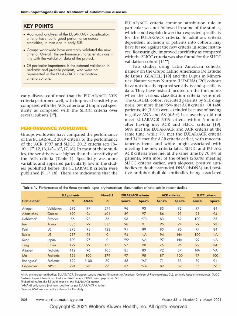
KEY POINTS
� Additional analyses of the EULAR/ACR classificationcriteria have found good performance acrossethnicities, in men and in early SLE.
� Groups worldwide have externally validated the newcriteria. Overall, the performance characteristics are inline with the validation data of the project.
� Of particular importance is the external validation inpediatric and juvenile patients, who were notrepresented in the EULAR/ACR classificationcriteria cohorts.
Immunopathogenesis and treatment of autoimmune diseases
early disease confirmed that the EULAR/ACR 2019criteria performed well, with improved sensitivity ascompared with the ACR criteria and improved spec-ificity as compared with the SLICC criteria overseveral subsets [7
&
].
PERFORMANCE WORLDWIDE
Groups worldwide have compared the performanceof the EULAR/ACR 2019 criteria to the performanceof the ACR 1997 and SLICC 2012 criteria sets [8–10,11
&&
,12,13,14&
–16&
,17,18]. In most of these stud-ies, the sensitivity was higher than the sensitivity ofthe ACR criteria (Table 1). Specificity was morevariable, and appeared particularly low in the stud-ies published before the EULAR/ACR criteria werepublished [9,17,18]. There are indications that the
Copyright © 2021 Wolters Kluwer H
Table 1. Performance of the three systemic lupus erythematosus c
SLE patients Non-SLE EU
First author n ANA% n Se
Aringer Validation 696 99 574
Adamichou Greece 690 94 401
Dahlstroma Sweden 56 98 56
Lee Korea 335 99 337
Petri US 293 98 423
Rubio US 217 96 0
Suda Japan 100 97 0 b
Teng China 199 99 175
Aljaberi Pediatric 112 96 105
Ma Pediatric 156 100 379
Rodriguesa Pediatric 122 c100 89
Gegenavaa NPSLE 294 96 66
ANA, antinuclear antibodies; EULAR/ACR, European League Against Rheumatism/ASystemic Lupus International Collaborative Centers; NPSLE, neuropsychiatric SLE.aPublished before the full publication of the EULAR/ACR criteria.bANA directly tested (not ‘ever positive’ as per EULAR/ACR criteria).cPositive ANA were an entry criterion for this study.
206 www.co-rheumatology.com
EULAR/ACR criteria common attribution rule inparticular was not followed in some of the studies,which could explain lower than expected specificityfor the EULAR/ACR criteria. In addition, criteriadependent inclusion of patients into cohorts mayhave biased against the new criteria in some instan-ces. Reassuringly, improved specificity as comparedwith the SLICC criteria was also found for the SLICCvalidation cohort [11
&&
].Two studies using Latin American cohorts,
namely on the Grupo Latino Americano De Estudiode Lupus (GLADEL) [19] and the Lupus in Minori-ties: Nature versus Nurture (LUMINA) [20] cohortshave not directly reported sensitivity and specificitydata. They have instead focused on the timepointswhen the various classification criteria were met.The GLADEL cohort recruited patients by SLE diag-nosis, but more than 95% met ACR criteria. Of 1480patients, 49 (3.3%) were excluded because of havingnegative ANA and 68 (6.5%) because they did notmeet EULAR/ACR 2019 criteria within 6 monthsafter having met ACR and SLICC criteria [19].58% met the EULAR/ACR and ACR criteria at thesame time, while 7% met the EULAR/ACR criteriaand 34% met the ACR criteria earlier, with mucocu-taneous items and white origin associated withmeeting the new criteria later. SLICC and EULAR/ACR criteria were met at the same time by 70.8% ofpatients, with most of the others (28.6%) meetingSLICC criteria earlier, with alopecia, positive anti-bodies to double-stranded DNA (dsDNA) and posi-tive antiphospholipid antibodies being associated
ealth, Inc. All rights reserved.
lassification criteria sets in recent studies
LAR/ACR criteria ACR criteria SLICC criteria
ns% Spec% Sens% Spec% Sens% Spec%
96 93 83 93 97 84
89 97 86 93 91 94
93 a73 83 82 100 75
98 91 96 94 99 93
91 89 83 96 97 84
94 NA 94 NA 100 NA
92 NA 97 NA 99 NA
97 90 75 96 92 84
85 83 72 87 NA NA
97 98 87 100 97 100
88 a67 71 83 89 91
87 a74 89 89 85 76
merican College of Rheumatology; SLE, systemic lupus erythematosus; SLICC,
Volume 33 � Number 2 � March 2021
New lupus criteria Aringer and Johnson
with SLICC criteria being met earlier. The LUMINAcohort included patients by ACR criteria. 14% ofthese patients met the EULAR/ACR and 25% theACR criteria earlier, with white patients those whomet the new criteria later [20]. While the authorsreported not being able to include all of the EULAR/ACR criteria variables, it is not clear why their ana-logue to sensitivity was lower for the EULAR/ACRcriteria than in other cohorts.
One additional aspect was disease severity, alsotermed ominosity [1,2]. Several studies have nowshown that those patients whose SLE fulfilled theEULAR/ACR criteria were more prone to severe dis-ease and to damage than those who did not[12,21,22]. While this is outside the legitimate spec-trum of classification criteria employment and incontrast with the fact that criteria items accumulateover time [23], Teng et al. [24] have even argued thatthe criteria were reflective of disease activity.
EARLY SYSTEMIC LUPUSERYTHEMATOSUS
Better performance in classifying early SLE was also agoal of the EULAR/ACR classification criteria project[1,2], which also led to the early SLE cohort study[25]. The EULAR/ACR validation cohort subanalysison the patients in the early years of their diseasesuggest that this goal was met [7
&
]. In the first year ofSLE, the ACR 1997 criteria had 56% sensitivity, ascompared with 89% of both the EULAR/ACR 2019and the SLICC criteria, with a constant level speci-ficity of 92% for each. In the second and third year ofdisease, ACR criteria sensitivity increased to 81%,but EULAR/ACR criteria sensitivity to 89% andSLICC criteria sensitivity to 87%. Specificity was95% for the ACR and 96% for the EULAR/ACRcriteria, as compared with 88% for the SLICC criteria[7
&
]. Similarly, at the time of physician diagnosis ofSLE, Adamichou et al. [8] found 69% sensitivity forthe ACR 1997 criteria, 75% sensitivity for theEULAR/ACR 2019 criteria and 74% sensitivity forthe SLICC criteria. The respective specificities were96% for the ACR, 98% for the EULAR/ACR, and 97%for the SLICC criteria.
PEDIATRIC SYSTEMIC LUPUSERYTHEMATOSUS
In the early phases of the EULAR/ACR criteria project,pediatricians were actively involved [26]. Half of thepediatricians favored one set of criteria for adult andpediatric SLE. Data on pediatric and juvenile SLEpatients, that is, those who developed SLE beforetheir 19th birthday, are of particular relevance. Theavailable data [15
&
,16&
,17] show improved sensitivity
Copyright © 2021 Wolters Kluwe
1040-8711 Copyright � 2020 Wolters Kluwer Health, Inc. All rights rese
of the EULAR/ACR criteria as compared with the ACRcriteria, with the two studies published after the fullpublication of the EULAR/ACR criteria showingmaintained specificity (Table 1).
ANTINUCLEAR ANTIBODIES AS ANOBLIGATORY ENTRY CRITERION
One of the novel features of the new criteria was therepositioning of ANA as an obligatory entry crite-rion. To reiterate the rationale beyond specificity:there were essentially three options to deal withANA. To keep them as a specific criterion appearedquestionable, given the dismal specificity of the test.To leave them completely out would have discardedan important clinical test with implications forteaching and training. The third possibility was tochange their position to the equivalent of thescreening test ANA constitute in clinical practice.This was supported by a majority of expert in theDelphi exercise who felt at least not completelycomfortable with diagnosing SLE in patients whowere always ANA negative [26]. Meta-regressionanalysis of a systematic literature review concluded97.8% of SLE patients were positive for ANA at a titerof 1 : 80 or higher, with the lower limit of the 95%confidence interval at 96.8% [27]. The decision toreposition ANA as an entry criterion was also sup-ported by international SLE expert consensus [28].
Nevertheless, the argument was brought for-ward that ANA as an obligatory entry criterionwould exclude a small but relevant proportion ofpatients. This would obviously be a real issue fordiagnosis, but it is important to reiterate that classi-fication criteria should not be used as a diagnostictool, and certainly not abused for withholding treat-ment from patients [6]. In contrast, for classifica-tion, exclusion of patients would only become anissue if the group was large or would systematicallyimpair the study of relevant subgroups. In theEULAR/ACR criteria cohorts, the percentage ofANA positive patients was always above 99%. Theexternal data so far support sufficient ANA sensitiv-ity. In fact, all the cohorts recently studied for thecomparison of the criteria sets showed 94–100% ofpatients ANA positive (Table 1). Moreover, at thetime of diagnosis, 97% of 115 Sudanese SLE patientswere ANA positive [29]. Importantly, this alsoapplied to pediatric and juvenile SLE and to earlydisease. An analysis of SLE patients negative for ANAat a titer of 1 : 160 or higher on HEp-2 cells, in theSLICC inception cohort was 6.2% [30].
Frodlund et al. [31] found that 13% of theirpatients lost ANA positivity over time, which reit-erates the importance of ANA ever positive as anentry criterion in the EULAR/ACR criteria. The more
r Health, Inc. All rights reserved.
rved. www.co-rheumatology.com 207
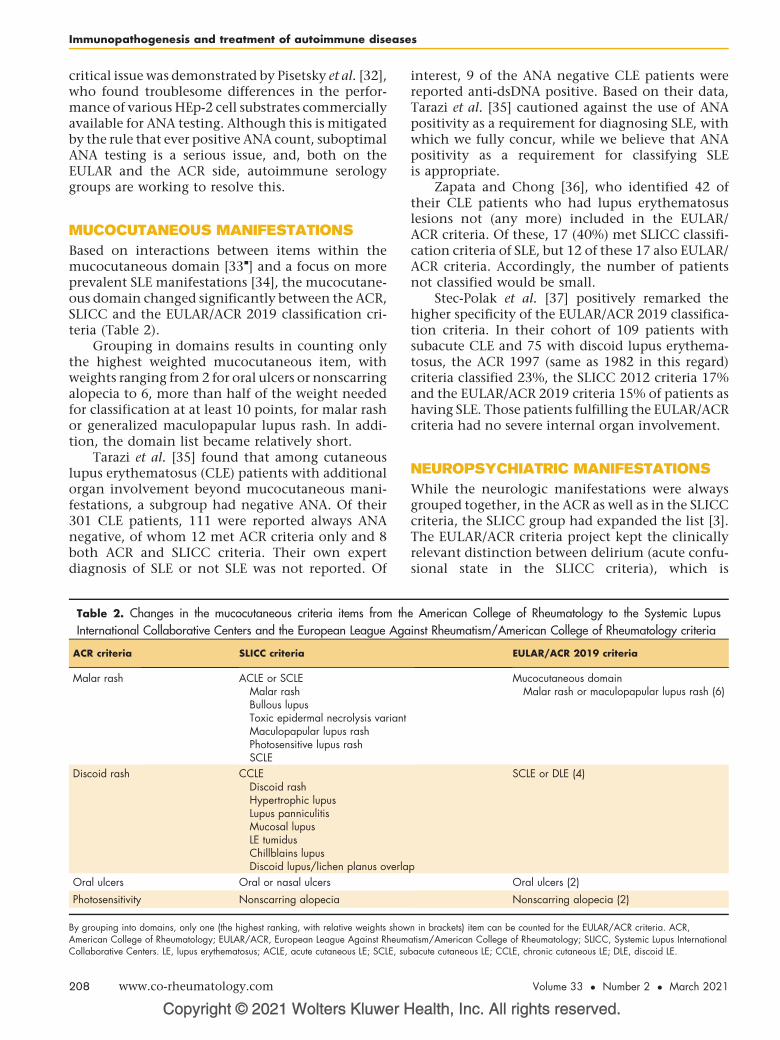
Immunopathogenesis and treatment of autoimmune diseases
critical issue was demonstrated by Pisetsky et al. [32],who found troublesome differences in the perfor-mance of various HEp-2 cell substrates commerciallyavailable for ANA testing. Although this is mitigatedby the rule that ever positive ANA count, suboptimalANA testing is a serious issue, and, both on theEULAR and the ACR side, autoimmune serologygroups are working to resolve this.
MUCOCUTANEOUS MANIFESTATIONS
Based on interactions between items within themucocutaneous domain [33
&
] and a focus on moreprevalent SLE manifestations [34], the mucocutane-ous domain changed significantly between the ACR,SLICC and the EULAR/ACR 2019 classification cri-teria (Table 2).
Grouping in domains results in counting onlythe highest weighted mucocutaneous item, withweights ranging from 2 for oral ulcers or nonscarringalopecia to 6, more than half of the weight neededfor classification at at least 10 points, for malar rashor generalized maculopapular lupus rash. In addi-tion, the domain list became relatively short.
Tarazi et al. [35] found that among cutaneouslupus erythematosus (CLE) patients with additionalorgan involvement beyond mucocutaneous mani-festations, a subgroup had negative ANA. Of their301 CLE patients, 111 were reported always ANAnegative, of whom 12 met ACR criteria only and 8both ACR and SLICC criteria. Their own expertdiagnosis of SLE or not SLE was not reported. Of
Copyright © 2021 Wolters Kluwer H
Table 2. Changes in the mucocutaneous criteria items from th
International Collaborative Centers and the European League Aga
ACR criteria SLICC criteria
Malar rash ACLE or SCLEMalar rashBullous lupusToxic epidermal necrolysis variantMaculopapular lupus rashPhotosensitive lupus rashSCLE
Discoid rash CCLEDiscoid rashHypertrophic lupusLupus panniculitisMucosal lupusLE tumidusChillblains lupusDiscoid lupus/lichen planus overla
Oral ulcers Oral or nasal ulcers
Photosensitivity Nonscarring alopecia
By grouping into domains, only one (the highest ranking, with relative weights showAmerican College of Rheumatology; EULAR/ACR, European League Against RheumCollaborative Centers. LE, lupus erythematosus; ACLE, acute cutaneous LE; SCLE, su
208 www.co-rheumatology.com
interest, 9 of the ANA negative CLE patients werereported anti-dsDNA positive. Based on their data,Tarazi et al. [35] cautioned against the use of ANApositivity as a requirement for diagnosing SLE, withwhich we fully concur, while we believe that ANApositivity as a requirement for classifying SLEis appropriate.
Zapata and Chong [36], who identified 42 oftheir CLE patients who had lupus erythematosuslesions not (any more) included in the EULAR/ACR criteria. Of these, 17 (40%) met SLICC classifi-cation criteria of SLE, but 12 of these 17 also EULAR/ACR criteria. Accordingly, the number of patientsnot classified would be small.
Stec-Polak et al. [37] positively remarked thehigher specificity of the EULAR/ACR 2019 classifica-tion criteria. In their cohort of 109 patients withsubacute CLE and 75 with discoid lupus erythema-tosus, the ACR 1997 (same as 1982 in this regard)criteria classified 23%, the SLICC 2012 criteria 17%and the EULAR/ACR 2019 criteria 15% of patients ashaving SLE. Those patients fulfilling the EULAR/ACRcriteria had no severe internal organ involvement.
NEUROPSYCHIATRIC MANIFESTATIONS
While the neurologic manifestations were alwaysgrouped together, in the ACR as well as in the SLICCcriteria, the SLICC group had expanded the list [3].The EULAR/ACR criteria project kept the clinicallyrelevant distinction between delirium (acute confu-sional state in the SLICC criteria), which is
ealth, Inc. All rights reserved.
e American College of Rheumatology to the Systemic Lupus
inst Rheumatism/American College of Rheumatology criteria
EULAR/ACR 2019 criteria
Mucocutaneous domainMalar rash or maculopapular lupus rash (6)
p
SCLE or DLE (4)
Oral ulcers (2)
Nonscarring alopecia (2)
n in brackets) item can be counted for the EULAR/ACR criteria. ACR,atism/American College of Rheumatology; SLICC, Systemic Lupus Internationalbacute cutaneous LE; CCLE, chronic cutaneous LE; DLE, discoid LE.
Volume 33 � Number 2 � March 2021
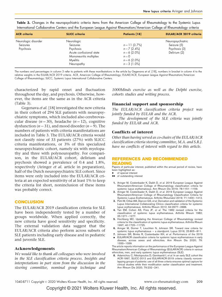
Table 3. Changes in the neuropsychiatric criteria items from the American College of Rheumatology to the Systemic Lupus
International Collaborative Centers and the European League Against Rheumatism/American College of Rheumatology criteria
ACR criteria SLICC criteria Patients [18] EULAR/ACR 2019 criteria
Neurologic disorderSeizuresPsychosis
NeurologicSeizuresPsychosisAcute confusional stateMononeuritis multiplexMyelitisNeuropathy
n¼11 (3.7%)n¼7 (2.4%)n¼6 (2.0%)n¼0n¼6 (2.0%)n¼3 (1.0%)
NeuropsychiatricSeizure (5)Psychosis (3)Delirium (2)
The numbers and percentages in column 3 refer to patients with these manifestations in the article by Gegenava et al. [18], numbers in bracket in column 4 to therelative weights in the EULAR/ACR 2019 criteria. ACR, American College of Rheumatology; EULAR/ACR, European League Against Rheumatism/AmericanCollege of Rheumatology; SLICC, Systemic Lupus International Collaborative Centers.
New lupus criteria Aringer and Johnson
characterized by rapid onset and fluctuationthroughout the day, and psychosis. Otherwise, how-ever, the items are the same as in the ACR criteria(Table 3).
Gegenava et al. [18] investigated the new criteriain their cohort of 294 SLE patients with neuropsy-chiatric symptoms, which included also cerebrovas-cular disease (n¼30), headache (n¼12), cognitivedysfunction (n¼31), and mood disorder (n¼9). Thenumbers of patients with criteria manifestations areincluded in Table 3. The EULAR/ACR criteria wouldnot classify nine of 33 patients (27%) with SLICCcriteria manifestations, or 3% of this specializedneuropsychiatric cohort, namely six with myelopa-thy and three with polyneuropathy. For compari-son, in the EULAR/ACR cohort, delirium andpsychosis showed a prevalence of 0.4 and 1.8%,respectively (Aringer et al. article in preparation),half of the Dutch neuropsychiatric SLE cohort. Sinceitems were only included into the EULAR/ACR cri-teria at an expected sensitivity of at least 1% to keepthe criteria list short, noninclusion of these itemswas probably correct.
CONCLUSION
The EULAR/ACR 2019 classification criteria for SLEhave been independently tested by a number ofgroups worldwide. When applied correctly, thenew criteria have good sensitivity and specificity.The external validation data suggest that theEULAR/ACR criteria also perform across subsets ofSLE patients including early disease and in pediatricand juvenile SLE.
Acknowledgements
We would like to thank all colleagues who were involvedin the SLE classification criteria process. Insights andinterpretations in part stem from the discussion in thesteering committee, nominal group technique and
Copyright © 2021 Wolters Kluwe
1040-8711 Copyright � 2020 Wolters Kluwer Health, Inc. All rights rese
1000Minds exercise as well as the Delphi exercise,cohorts studies and writing process.
Financial support and sponsorship
The EULAR/ACR classification criteria project wasjointly funded by EULAR and the ACR.
The development of the SLE criteria was jointlyfunded by EULAR and ACR.
Conflicts of interest
Other than having served as co-chairs of the EULAR/ACRclassification criteria steering committee, M.A. and S.R.J.have no conflicts of interest with regard to this article.
REFERENCES AND RECOMMENDEDREADINGPapers of particular interest, published within the annual period of review, havebeen highlighted as:
& of special interest&& of outstanding interest1. Aringer M, Costenbader K, Daikh D, et al. 2019 European League AgainstRheumatism/American College of Rheumatology classification criteria forsystemic lupus erythematosus. Ann Rheum Dis 2019; 78:1151–1159.
2. Aringer M, Costenbader K, Daikh D, et al. 2019 European League AgainstRheumatism/American College of Rheumatology Classification Criteria forSystemic Lupus Erythematosus. Arthritis Rheumatol 2019; 71:1400–1412.
3. Petri M, Orbai AM, Alarcon GS, et al. Derivation and validation of the SystemicLupus International Collaborating Clinics classification criteria for systemiclupus erythematosus. Arthritis Rheum 2012; 64:2677–2686.
4. Tan EM, Cohen AS, Fries JF, et al. The 1982 revised criteria for theclassification of systemic lupus erythematosus. Arthritis Rheum 1982;25:1271–1277.
5. Hochberg MC. Updating the American College of Rheumatology revisedcriteria for the classification of systemic lupus erythematosus. Arthritis Rheum1997; 40:1725–11725.
6. Aringer M, Dorner T, Leuchten N, Johnson SR. Toward new criteria forsystemic lupus erythematosus – a standpoint. Lupus 2016; 25:805–811.
7.&
Johnson SR, Brinks R, Costenbader KH, et al. Performance of the 2019EULAR/ACR classification criteria for systemic lupus erythematosus in earlydisease, across sexes and ethnicities. Ann Rheum Dis 2020; 79:1333–1339.
The article reports information on the performance of the European League AgainstRheumatism/American College of Rheumatology (EULAR/ACR) criteria in variousethnicities, men, and early systemic lupus erythematosus (SLE).8. Adamichou C, Nikolopoulos D, Genitsaridi I, et al. In an early SLE cohort the
ACR-1997, SLICC-2012 and EULAR/ACR-2019 criteria classify nonover-lapping groups of patients: use of all three criteria ensures optimal capture forclinical studies while their modification earlier classification and treatment.Ann Rheum Dis 2020; 79:232–241.
r Health, Inc. All rights reserved.
rved. www.co-rheumatology.com 209
Immunopathogenesis and treatment of autoimmune diseases
9. Dahlstrom O, Sjowall C. The diagnostic accuracies of the 2012 SLICCcriteria and the proposed EULAR/ACR criteria for systemic lupus erythema-tosus classification are comparable. Lupus 2019; 28:778–782.
10. Lee EE, Lee EB, Park JK, et al. Performance of the 2019 European LeagueAgainst Rheumatism/American College of Rheumatology classification criteriafor systemic lupus erythematosus in Asian patients: a single-centre retrospec-tive cohort study in Korea. Clin Exp Rheumatol 2020; 38:1075–1079.
11.&&
Petri M, Goldman DW, Alarcon GS, et al. A Comparison of 2019 EULAR/ACR SLE classification criteria with two sets of earlier SLE classificationcriteria. Arthritis Care Res (Hoboken) 2020; May 20 Epub. doi: 10.1002/acr.24263.
Performance data on the EULAR/ACR classification criteria from the SystemicLupus International Collaborative Centers validation cohort.12. Rubio J, Krishfield S, Kyttaris VC. Application of the 2019 European League
Against Rheumatism/American College of Rheumatology systemic lupuserythematosus classification criteria in clinical practice: a single centerexperience. Lupus 2020; 29:421–425.
13. Suda M, Kishimoto M, Ohde S, Okada M. Validation of the 2019 ACR/EULARclassification criteria of systemic lupus erythematosus in 100 Japanesepatients: a real-world setting analysis. Clin Rheumatol 2020; 39:1823–1827.
14.&
Teng J, Ye J, Zhou Z, et al. A comparison of the performance of the 2019European League Against Rheumatism/American College of Rheumatologycriteria and the 2012 Systemic Lupus International Collaborating Clinicscriteria with the 1997 American College of Rheumatology classificationcriteria for systemic lupus erythematous in new-onset Chinese patients. Lupus2020; 29:617–624.
Important external validation study on the EULAR/ACR criteria in 199 new onsetChinese SLE patients.15.&
Aljaberi N, Nguyen K, Strahle C, et al. The performance of the new 2019-EULAR/ACR classification criteria for systemic lupus erythematosus in chil-dren and young adults. Arthritis Care Res (Hoboken) 2020; Aug 25 Epub. doi:10.1002/acr.24430.
Important external validation of the EULAR/ACR criteria in pediatric and juvenileSLE patients.16.&
Ma M, Hui-Yuen JS, Cerise JE, et al. Validation of the 2017 Weighted criteriacompared to the 1997 ACR and the 2012 SLICC in Pediatric Systemic LupusErythematosus. Arthritis Care Res (Hoboken) 2020; 72:1597–1601.
Second external validation study in pediatric and juvenile SLE.17. Rodrigues FA, Felix Rodrigues MC, Sztajnbok FR, et al. Comparison among
ACR1997, SLICC and the new EULAR/ACR classification criteria in child-hood-onset systemic lupus erythematosus. Adv Rheumatol 2019; 59:20.
18. Gegenava M, Beaart HJL, Monahan RC, et al. Performance of the proposedACR-EULAR classification criteria for systemic lupus erythematosus (SLE) ina cohort of patients with SLE with neuropsychiatric symptoms. RMD Open2019; 5:e000895.
19. Pons-Estel GJ, Ugarte-Gil MF, Harvey GB, et al. Applying the 2019 EULAR/ACR lupus criteria to patients from an established cohort: a Latin Americanperspective. RMD Open 2020; 6:e001097. doi: 10.1136/rmdopen-2019-001097.
20. Ugarte-Gil MF, Pons-Estel GJ, Harvey GB, et al. Applying the 2019 EULAR/ACR lupus criteria to patients from the LUMINA cohort. Arthritis Care Res(Hoboken) 2020; Jun 29 Epub. doi: 10.1002/acr.24367.
21. Carneiro AC, Ruiz MM, Freitas S, Isenberg D. A comparison of threeclassification criteria sets for Systemic Lupus Erythematosus – a studylooking at links to outcome and mortality. Arthritis Care Res (Hoboken)2020; 72:1611–1614.
22. Ugarte-Gil MF, Pons-Estel GJ, Griffin R, et al. Patients who do not fulfill the2019 EULAR/ACR criteria for systemic lupus erythematosus accrue lessdamage. Arthritis Care Res (Hoboken) 2020; Apr 15 Epub. doi: 10.1002/acr.24213.
Copyright © 2021 Wolters Kluwer H
210 www.co-rheumatology.com
23. Aringer M, Costenbader K, Dorner T, Johnson SR. SLE classification criteriaitems are not dependent on current activity but may accumulate over time.Response to: ‘Do 2019 European League against rheumatism/AmericanCollege of Rheumatology classification criteria for systemic lupus erythema-tosus also indicate the disease activity?’ by Teng et al. Ann Rheum Dis 2020;Mar 2 Epub. doi: 10.1136/annrheumdis-2020-217105.
24. Teng J, Zhou Z, Wang F, et al. Do 2019 European League Against Rheu-matismm/American college of rheumatology classification criteria for systemiclupus erythematosus also indicate the disease activity? Ann Rheum Dis 2020;Mar 2 Epub. doi: 10.1136/annrheumdis-2020-217017.
25. Mosca M, Costenbader KH, Johnson SR, et al. Brief report: how do patientswith newly diagnosed systemic lupus erythematosus present? A multicentercohort of early systemic lupus erythematosus to inform the development ofnew classification criteria. Arthritis Rheumatol 2019; 71:91–98.
26. Schmajuk G, Hoyer BF, Aringer M, et al. Multicenter Delphi exercise to identifyimportant key items for classifying systemic lupus erythematosus. ArthritisCare Res (Hoboken) 2018; 70:1488–1494.
27. Leuchten N, Brinks R, Hoyer A, et al. Performance of antinuclear antibodies(ANA) for classifying systemic lupus erythematosus (SLE): a systematicliterature review and meta-regression of diagnostic data. Ann Rheum Dis2015; 74:809–810. (abstract SAT0415).
28. Johnson SR, Khanna D, Daikh D, et al. Use of consensus methodology todetermine candidate items for systemic lupus erythematosus classificationcriteria. J Rheumatol 2019; 46:721–726.
29. Elbagir S, Elshafie AI, Elagib EM, et al. Sudanese and Swedish patients withsystemic lupus erythematosus: immunological and clinical comparisons.Rheumatology (Oxford) 2020; 59:968–978.
30. Choi MY, Clarke AE, St Pierre Y, et al. Antinuclear antibody-negative systemiclupus erythematosus in an international inception cohort. Arthritis Care Res(Hoboken) 2019; 71:893–902.
31. Frodlund M, Wettero J, Dahle C, et al. Longitudinal antinuclear antibody (ANA)seroconversion in systemic lupus erythematosus: a prospective study ofSwedish cases with recent-onset disease. Clin Exp Immunol 2020;199:245–254.
32. Pisetsky DS, Spencer DM, Lipsky PE, Rovin BH. Assay variation in thedetection of antinuclear antibodies in the sera of patients with establishedSLE. Ann Rheum Dis 2018; 77:911–913.
33.&
Touma Z, Cervera R, Brinks R, et al. Associations among classification criteriaitems within systemic lupus erythematosus. Arthritis Care Res (Hoboken)2020; 72:1820–1826.
The association study demonstrated that items within a domain are not fullyindependent of each other.34. Tedeschi SK, Johnson SR, Boumpas D, et al. Developing and refining new
candidate criteria for systemic lupus erythematosus classification: aninternational collaboration. Arthritis Care Res (Hoboken) 2018;70:571–581.
35. Tarazi M, Gaffney RG, Kushner CJ, et al. Cutaneous lupus erythematosuspatients with a negative antinuclear antibody meeting the American College ofRheumatology and/or Systemic Lupus International Collaborating ClinicsCriteria for Systemic Lupus Erythematosus. Arthritis Care Res (Hoboken)2019; 71:1404–1409.
36. Zapata L Jr, Chong BF. Exclusion of cutaneous lupus erythematosus subtypesfrom the 2019 European League Against Rheumatism/American College ofRheumatology classification criteria for systemic lupus erythematosus: com-ment on the article by Aringer et al. Arthritis Rheumatol 2020;72:1403–1403.
37. Stec-Polak M, Matyja-Bednarczyk A, Wojas-Pelc A, Pastuszczak M. Higherspecificity of the new EULAR/ACR 2019 criteria for diagnosing systemiclupus erythematosus in patients with biopsy-proven cutaneous lupus. Clin ExpRheumatol 2020; Sep 16 Epub.
ealth, Inc. All rights reserved.
Volume 33 � Number 2 � March 2021
REVIEW
CURRENTOPINION The latest in systemic lupus erythematosus-
accelerated atherosclerosis: related mechanismsinform assessment and therapy
Copyrigh
1040-8711 Copyright � 2020 Wolte
a b,c
Brenna D. Appleton and Amy S. MajorPurpose of Review
Accelerated atherosclerosis is a significant comorbidity and the leading cause of death for patients withsystemic lupus erythematosus (SLE). It is now apparent that SLE-accelerated atherosclerosis is not drivensolely by traditional cardiovascular risk factors, adding complexity to disease characterization andmechanistic understanding. In this review, we will summarize new insights into SLE-acceleratedatherosclerosis evaluation, treatment, and mechanism.
Recent findings
Recent work highlights the need to incorporate inflammatory biomarkers into cardiovascular disease (CVD)risk assessments. This is especially true for SLE patients, in which mechanisms of immune dysfunction likelydrive CVD progression. There is new evidence that commonly prescribed SLE therapeutics hinderatherosclerosis development. This effect is achieved both by reducing SLE-associated inflammation and bydirectly improving measures of atherosclerosis, emphasizing the interconnected mechanisms of the twoconditions.
Summary
SLE-accelerated atherosclerosis is most likely the consequence of chronic autoimmune inflammation.Therefore, diligent management of atherosclerosis requires assessment of SLE disease activity as well astraditional cardiovascular risk factors. This supports why many of the therapeutics classically used to controlSLE also modulate atherosclerosis development. Greater understanding of the mechanisms underlying thiscondition will allow for the development of more targeted therapeutics and improved outcomes for SLEpatients.
Keywords
atherosclerosis, cardiovascular disease, risk assessment, SLE-accelerated atherosclerosis, systemic lupuserythematosus
aDepartment of Pathology, Microbiology and Immunology, VanderbiltUniversity, bDepartment of Medicine, Division of Rheumatology andImmunology, Vanderbilt Medical Center and cTennessee Valley Health-care System, U.S. Department of Veterans Affairs, Nashville, Tennessee
Correspondence to Amy S. Major, Vanderbilt University, 1161 21stAvenue South, A-3314Medical Center North, Nashville,TN 37232, USA.Tel: +615 957 2496; e-mail: [email protected]
Curr Opin Rheumatol 2021, 33:211–218
DOI:10.1097/BOR.0000000000000773
INTRODUCTION
Systemic lupus erythematosus (SLE) is a chronic auto-immune disease affecting over 1.5 million Americansand at least 5 million individuals worldwide. Patientsare predominantly female, and are two to three timesmore likely to be women of color. SLE is characterizedby autoantibody producing B cells and dysfunctionalCD4þ T cells, and can result in end organ damage tothe kidneys, joints, and cardiovascular system [1].Life expectancy for SLE patients has improved butremains lower than thatof the general population [2],and the leading cause of death among SLE patients iscardiovascular disease (CVD) driven by acceleratedatherosclerosis [3–7]. It is estimated that women withSLE between the ages of 35 and 44 years have a 50-foldincreased risk of myocardial infarction comparedwith age- and sex-matched controls [8]. Even if we
t © 2021 Wolters Kluwe
rs Kluwer Health, Inc. All rights rese
assume that this estimate is inflated due to the factthat healthy, premenopausal women are typicallyprotected from CVD development [9], postmeno-pausal women with SLE are still five times more likelyto develop atherosclerosis than healthy, age-matched controls [10]. Statin therapy has beenshown to provide less cardiovascular protection in
r Health, Inc. All rights reserved.
rved. www.co-rheumatology.com
KEY POINTS
� Accelerated atherosclerosis is the leading cause ofdeath among systemic lupus erythematosus(SLE) patients.
� Cardiovascular disease risk assessment in SLE patientsmust integrate inflammatory as well as traditionalcardiovascular risk factors in order to get anaccurate assessment.
� Many of the immune mechanisms that drive SLE alsopromote atherosclerosis.
� Many of the existing SLE treatments hold promise forimproving SLE-accelerated atherosclerosis.
Immunopathogenesis and treatment of autoimmune diseases
SLE patients compared to the general population[11], suggesting the SLE-accelerated atherosclerosisis not entirely lipid dependent. In this review, wehighlight recent findings regarding mechanismsdriving SLE-accelerated atherosclerosis as well asthe best techniques to evaluate and treat this disease.
DISEASE RISK ASSESSMENT
Atherosclerosis is associated with a number of riskfactors and the likelihood of a related adverse event(coronary heart disease, stroke, peripheral arterydisease, or heart failure) occurring in the long termin the general population is often determined usingcomposite measurements such as the FraminghamRisk Score [12] or Systematic Coronary Risk Evalua-tion (SCORE) [13]. The Framingham Risk Score is a10-year predictive measurement that uses sex, age,total and high-density lipoprotein (HDL) choles-terol levels, systolic blood pressure, hypertension,smoking, and diabetes status to determine an indi-vidual’s CVD risk [12]. SCORE utilizes many of thesame metrics, but also incorporates geography as avariable to consider whether an individual’s countryas a whole has rising or falling CVD rates [13]. In SLEpatients, these scores may inaccurately predict anindividual’s true risk of CVD [14,15]. This is likelydue to several factors; the lack of consideration ofinflammatory variables that are increased in SLE, theheterogeneity of the disease, and our incompleteunderstanding of disease mechanism. Therefore,improved methods for predicting risk in SLE arerequired to ensure appropriate management ofCVD and other life-threatening comorbidities.
Techniques for improved risk assessment
One strategy for adjusting the Framingham riskscore in SLE has been to simply multiply the
Copyright © 2021 Wolters Kluwer H
212 www.co-rheumatology.com
traditional score by a factor of two [16]. A similartechnique was successfully applied to rheumatoidarthritis patients, who also experience increasedrates of CVD related death [17,18]. Unfortunately,given the heterogeneous nature of SLE, a blanketcorrection factor may oversimplify the contributionof autoimmune factors to CVD risk. Therefore, itbecame necessary to develop an SLE-centered car-diovascular risk score that uses traditional cardio-vascular risk factors like age, gender, systolic bloodpressure, cholesterol, smoking, and diabetes, butalso SLE-specific risk factors. Variables such as timesince SLE diagnosis, corticosteroid and hydroxy-chloroquine use, C3 and C4 levels, anti-doublestranded DNA (dsDNA) titer, proteinuria, diseaseactivity score (SELENA-SLEDAI score), estimatedglomerular filtration rate, and history of lupus anti-coagulant and anticardiolipin were evaluated. In thesubjects of this study, only disease activity score, C3level, and lupus anticoagulant titer were predictiveof cardiovascular outcomes [19
&&
]. Using the SLE-specific risk score incorporating these select varia-bles, Petri et al. determined that some SLE patientswere appropriately assessed by the traditional Fra-mingham risk score, whereas others, particularlythose with higher SLEDAI scores, had their 10-yearrisk underestimated by as much as a factor of 10[19
&&
]. In agreement with these findings, a separate7-year surveillance study found no difference in theprogression of subclinical atherosclerosis betweenthe control group and SLE patients with mild, well-controlled disease [20], highlighting the need to useSLE-specific criteria and disease activity level in CVDrisk assessment.
InaccurateCVD risk assessment is especiallyprev-alent in young SLE patients (�45 years). Thesepatients are not likely to experience adverse cardio-vascular events within 10 years (the range in whichclassical scoring methods are designed to predict),but still have elevated risk compared to their healthy,age-matched counterparts, including a 50-foldincrease in the risk of myocardial infarction [8,21].Clinical studies have shown that coronary arterycalcification (CAC), a known predictor of CVD mor-tality [22], is increased in SLE patients anywhere from2.8 to 4 times compared to controls and may be anaccurate predictor of outcomes [23,24]. These studieswere performed using older, mostly white cohorts(72% and 65%, respectively), perhaps not accuratelyrepresenting the predominately nonwhite SLE-patient population. Gartshteyn et al. studied SLEpatients aged 18 to 65 years, who were primarilyAfrican American and Hispanic, and had no clinicalCVD. They found that CAC severity increased withage in patients, and that CAC was significantly moreprevalent in young SLE patients compared to age-
ealth, Inc. All rights reserved.
Volume 33 � Number 2 � March 2021
The latest in SLE-accelerated atherosclerosis Appleton and Major
matched controls, 32% versus 9.6%, respectively[25]. In a separate study, a related, although indepen-dent condition, aorta calcification (AC), was found tooccur earlier, at a higher incidence, and in a widerrange of ages than CAC in SLE patients highlightingits importance in predicting atherosclerosis develop-ment [26].
Collectively, studies indicate risk assessment forSLE-accelerated atherosclerosis needs to be distinctfrom that of standard atherosclerosis. By integratingmore inflammatory variables into predictive equa-tions and/or adopting regular monitoring of CACand AC, SLE patients will experience improved carewith more timely therapeutic interventions.
Biomarkers
In addition to improved techniques to assess ath-erosclerosis development in SLE patients, there hasalso been progress in terms of new biomarkers toidentify potentially high CVD risk patients. Bio-markers associated with SLE-accelerated atheroscle-rosis include proinflammatory HDL (piHDL),increased circulating leptin, elevated homocysteine,and the presence of soluble tumor necrosis factor-like weak inducer of apoptosis (sTWEAK) [27].
Soluble CD163
A biomarker that was previously associated withincreased glomerular inflammation in SLE patientsis the soluble form of the scavenger receptor CD163(sCD163). sCD163 was found at high levels in bothserum and urine of SLE patients with more CD163þ
macrophage glomerular infiltrate and severe inflam-mation (r¼0.635) as part of lupus nephritis [28].CD163 expression on macrophages has typicallybeen thought of as an indication of alternative acti-vation and an anti-inflammatory phenotype [29].However, recent findings demonstrated CD163þ
macrophages actually promote atherosclerotic pla-que progression in both humans and mice via aCD163/HIF1a/VEGF-A pathway [30]. The involve-ment of CD163þ macrophages in both SLE and ath-erosclerosis suggest expression of this scavengerreceptor may be an underlying mechanism of SLE-accelerated atherosclerosis and that its soluble formcould be a biomarker of the disease. This was tested inone study of 63 SLE patients whose carotid athero-sclerotic plaque was measured by ultrasound andserum sCD163 levels monitored both at baseline ofthe study and during follow-up. Results indicatedthat serum sCD163 is elevated in SLE patients com-pared to controls, and is further increased in patientswith carotid plaque. Additionally, higher levels ofsCD163 correlated with the development of newcarotid plaque over time and positively associated
Copyright © 2021 Wolters Kluwe
1040-8711 Copyright � 2020 Wolters Kluwer Health, Inc. All rights rese
with subclinical atherosclerosis in SLE independentof classic cardiovascular risk factors [31
&
]. Theseresults indicate sCD163 is a promising biomarker ofSLE-accelerated atherosclerosis.
FcgRIIA polymorphism
Fcg receptors (FcgRs) are expressed on antigen pre-senting cells, such as macrophages, dendritic cells(DCs), and B cells, and bind to the Fc domain of IgGantibodies. Depending on whether the FcgR is acti-vating or inhibitory, it will promote pro- or anti-inflammatory responses [32]. In mouse models, acti-vating FcgRs are proatherogenic [33]. Functionalpolymorphism in the activating Fcg receptor IIA(FcgRIIA) have previously been associated with SLEand lupus nephritis in humans [34,35]. In heparin-induced thrombocytopenia, a functional polymor-phism (single amino acid substitution H131R) inFcgRIIA caused a hyperactive platelet phenotypecharacterized by increased expression of P-selectinand CD40 ligand, greater binding to annexin V, andincreased formation of platelet-leukocyte complexes[36–40]. A similar phenotype was described in SLEwhere hyperactive platelets promote vascular patho-genesis by activating endothelial cells [41]. Based onthese works, Clancy et al. investigated whether theH131R FcgRIIA polymorphism impacted cardiovas-cular outcomes in SLE patients. The H131R allelicvariant was found in a similar proportion (�40%) ofboth the SLE patient and healthy control popula-tions. When SLE patients were assessed by carotidultrasound, 58% of SLE patients with the allelic vari-ant had carotid plaque compared to only 25% of SLEpatients with the ancestral variant. Yet, the FcgRIIAallelic variant did not associate with carotid plaque inheathy control subjects [42
&
]. This suggests theH131R functional polymorphism confers CVD riskspecifically in the context of SLE and could poten-tially act as a genetic CVD biomarker in patients.
Appropriate CVD risk assessment for the SLEpatient population has long been needed. The tech-niques and biomarkers outlined above have thepotential to improve CVD risk detection in patients,hopefully resulting in prompt therapeutic interven-tion and prevention of devastating cardiovascularoutcomes.
MECHANISM AND TREATMENT
Treatments for SLE are limited in comparison toother chronic illnesses and therapeutic advance-ment has been relatively slow, in part because oflack of understanding of disease mechanisms.Unfortunately, the impact of some of the mostcommon SLE therapeutics specifically on SLE-accel-erated atherosclerosis has been understudied. The
r Health, Inc. All rights reserved.
rved. www.co-rheumatology.com 213
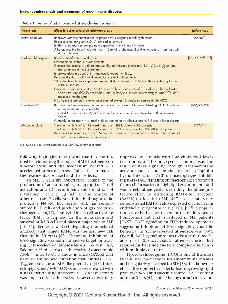
Table 1. Review of SLE-accelerated atherosclerosis treatments
Treatment Effect in SLE-accelerated atherosclerosis References
BAFF inhibition Improves SLE responder index in patients with ongoing B cell dysfunctionReduces circulating anti-dsDNA antibodies in miceInhibits antibody and complement deposition in the kidney in miceAtheroprotective in animals with low (<5mmol/L) cholesterol and atherogenic in animals with
high cholesterol
[52,54&&]
Hydroxychloroquine Reduces interferon-a productionLessens aortic stiffness in SLE patientsCorrects lipoprotein profile (increases HDL and lowers cholesterol, LDL, VLDL, triglycerides,
and cylomicrons) in SLE patientsImproves glycemic control in nondiabetic women with SLEReduces the risk of all thrombovascular events in SLE patientsSLE patients with carotid plaque are less likely to be using HCQ than those with no plaque
(63% vs. 82.3%)Long term HCQ treatment in ApoE-/- mice with pristane-induced SLE reduces atherosclerotic
lesion size, anti-dsDNA antibodies, total leukocyte numbers, macrophages, and DCs, andincreases lymohocytes
HDL from SLE patients is more functional following 12 weeks of treatment with HCQ.
[58–66,67&,70&]
Low-dose IL-2 IL-2 treatment reduces renal inflammation and activation of kidney-infiltrating CD4þ T cells in amouse model of lupus nephritis
Targeted IL-2 treatment in ApoE-/- mice reduces the size of pre-established atheroscleroticlesions
Currently under study in clinical trials to determine its effectiveness in SLE and atherosclerosis
[96&,97–99]
Mycophenolate Treatment with MMF for 12 weeks improves HDL function in SLE patientsTreatment with MMF for 12 weeks improves CVD biomarkers like sTWEAK in SLE patientsReduces atherosclerosis in Ldlr-/- B6.Sle1.2.3 bone marrow chimeras and limits recruitment of
CD4þ T cells to atherosclerotic lesions
[70&,71]
SLE, systemic lupus erythematosus; VLDL, very low-density lipoprotein.
Immunopathogenesis and treatment of autoimmune diseases
following highlights recent work that has contrib-uted to determining the impact of SLE treatments onatherosclerosis and the mechanism behind SLE-accelerated atherosclerosis. Table 1 summarizesthe treatments discussed and their effects.
In SLE, B cells are hyperactive leading to theproduction of autoantibodies, inappropriate T cellactivation and DC recruitment, and inhibition ofregulatory T cells (Tregs) [43]. In the context ofatherosclerosis, B cells were initially thought to beprotective [44,45], but recent work has demon-strated B2 B cells and production of IgG are proa-therogenic [46,47]. The cytokine B-cell activatingfactor (BAFF) is required for the maturation andsurvival of B2 B cells and plays a major role in SLE[48–51]. Benlysta, a B-cell-depleting monoclonalantibody that targets BAFF, was the first new SLEtherapy in 50 years [52]. Therefore, inhibition ofBAFF signaling seemed an attractive target for treat-ing SLE-accelerated atherosclerosis. To test this,Saidoune et al. crossed atherosclerosis-susceptibleApoE�/� mice to Qa-1 knock-in mice (D227K) thathave an amino acid mutation that hinders CD8þ
Tregs and develop an SLE-like phenotype [53]. Inter-estingly, when ApoE-/- D227K mice were treated witha BAFF neutralizing antibody, SLE disease activitywas impaired but atherosclerosis severity was only
Copyright © 2021 Wolters Kluwer H
214 www.co-rheumatology.com
improved in animals with low cholesterol levels(<5 mmol/L). This unexpected finding was theresult of BAFF signaling through transmembraneactivator and calcium modulator and cyclophilinligand interactor (TACI) on macrophages. Inhibit-ing BAFF-TACI signaling on macrophages promotedfoam cell formation in high lipid environments andwas largely atherogenic, overriding the atheropro-tective effect of disrupting BAFF-BAFF receptor(BAFFR) on B cells in SLE [54
&&
]. A separate studydemonstrated BAFFR is also expressed on circulatingendothelial progenitor cells (EPCs) [55
&
], a popula-tion of cells that are meant to maintain vascularhomeostasis but that is reduced in SLE patients[56,57]. BAFF signaling on EPCs induced apoptosissuggesting inhibition of BAFF signaling could bebeneficial in SLE-accelerated atherosclerosis [55
&
].Overall, BAFF signaling seems to be a critical mech-anism of SLE-accelerated atherosclerosis, butrequires further study due to its complex interactionwith multiple cell types.
Hydroxychloroquine (HCQ) is one of the mostwidely used medications for autoimmune diseasesand is regularly prescribed for SLE [58]. It is known toelicit atheroprotective effects like improving lipidprofiles [59–61] and glycemic control [62], lesseningaortic stiffness [63], and reducing thrombotic events
ealth, Inc. All rights reserved.
Volume 33 � Number 2 � March 2021
The latest in SLE-accelerated atherosclerosis Appleton and Major
[64,65]. There is a documented negative correlationbetween HCQ treatment and atherosclerosis in SLE[66], but the mechanism has not been elucidated. Bystudying pristane-induced SLE in atherosclerosis-susceptible ApoE-/- mice, Liu et al. showed long-termHCQ treatment reduced atherosclerosis largelythrough reversal of SLE-associated autoinflamma-tion. Specifically, HCQ treatment lowered anti-dsDNA antibodies and reduced total leukocytes,macrophages, and DCs [67
&
]. Pristane-induced SLEis known to reduce lymphocyte numbers in thespleen by triggering their apoptosis [68]. This phe-notype was also reversed by HCQ treatment in boththe aorta and the spleen which authors hypothesizeincreased the numbers of atheroprotective Tregs
[67&
]. This mechanistic study provides some insightto how HCQ may also be beneficial in treating SLE-accelerated atherosclerosis.
Although typically considered to be atheropro-tective due to its anti-inflammatory properties, HDLin SLE patients can become piHDL. SLE patientswith carotid artery plaque are more likely to havepiHDL than SLE patients without plaque [69], mak-ing it an interesting biomarker for SLE-acceleratedatherosclerosis [27]. Little is known about the effectsof regularly prescribed SLE disease-modifying thera-pies like mycophenolate (MMF), azathioprine(AZA), and HCQ on piHDL. In a recent observationalstudy, SLE patients starting a new disease-modifyingtherapy (MMF, AZA, or HCQ) had plasma collectedand disease activity measured at baseline, 6 weekspost initiation of therapy, and 12 weeks post initia-tion of therapy [70
&
]. MMF and HCQ significantlyimproved HDL function over the course of the12-week treatment, although not to normal levels,whereas AZA had no effect on HDL function. MMFalso significantly improved other biomarkers asso-ciated with plaque and intima-media thicknessprogression in SLE, suggesting MMF could havepotential to treat both SLE associated glomerulone-phritis and accelerated atherosclerosis [70
&
].Findings from our own work demonstrate that
MMF treatment reduces atherosclerosis in Ldlr-/-
B6.Sle1.2.3 bone marrow chimeric mice (which spon-taneously develop SLE and are atherosclerosis prone)and inhibits CD4þ T cell activation and recruitmentto atherosclerotic plaques [71]. In general, T cells areknown to significantly contribute to the develop-ment of atherosclerosis [72,73], with different subsetsplaying unique roles. For instance, Th1 cells arewidely accepted to be proatherogenic [74–76],whereas functional Tregs are atheroprotective[77,78]. T cells are also critical in SLE where theyare long-lived, hyperactive, and produce proinflam-matory cytokines like IFN-g and IL-17 [79–82]. Muchof thework fromourgrouphas shown the importance
Copyright © 2021 Wolters Kluwe
1040-8711 Copyright � 2020 Wolters Kluwer Health, Inc. All rights rese
of T cells in mouse models of SLE-accelerated athero-sclerosis [83–85], and our findings are in agreementwith results from human studies [86,87].
Previous results from Laurence Morel’s groupidentified the lupus susceptibility gene, Pbx1d, asoverexpressed in CD4þ T cells from SLE patientsand using mouse models of SLE described its rolein expanding follicular helper T cells (Tfh) andimpairing Treg homeostasis [88,89]. Given the impor-tance of CD4þ T cells in atherosclerosis, recent workexplored the effect of Pbx1d transgenic CD4þ T cellsin atherosclerosis in Ldlr�/� bone marrow chimeras.Pbx1d overexpression in CD4þ T cells resulted inthicker arterial walls and larger necrotic cores withinlesions, which is thought to be an indication ofplaque instability and severity. Recipients of Pbx1dtransgenic bone marrow had expanded Tfh cells andimpaired Tregs, that was increased with western dietfeeding [90
&&
], indicating a lupus susceptibility allelecan drive atherosclerosis, while at the same time,dyslipidemia can enhance autoimmune phenotypes.
Impaired IL-2 signaling in T cells due todecreased IL-2 production in SLE and its contribu-tion to disease pathogenesis is well known [91–94].Recent findings identify a regulatory subunit(PPP2R2D) of protein phosphatase 2A (an enzymewhich is increased in T cells from SLE patients andleads to decreased IL-2 production) that limits theaccessibility of the IL-2 gene and other transcriptionfactors important to IL-2 expression via chromatinremodeling. PPP2R2D is increased in T cells fromSLE patients and PPP2R2D deficiency in T cellsprevents autoimmunity in an imiquimod-inducedlupus-like mouse model [95
&
]. A separate groupdemonstrated that treating (NZB � NZW) F1 SLEmice with IL-2 reduced renal inflammation andlessened the activity of kidney-infiltrating CD4þ Tcells [96
&
]. Interestingly, IL-2 delivered specificallyto atherosclerotic lesions through fusion to fibro-nectin targeting antibody reduced plaque size byactivating and expanding Tregs [97]. This suggeststhat IL-2 supplementation could be beneficial inboth SLE and atherosclerosis, a hypothesis that iscurrently being tested in low-dose IL-2 treatmentclinical trials [98,99].
In order to determine how the risk of athero-sclerotic vascular events (AVE) in SLE patients haschanged over time in response to new therapeuticstrategies, Urowitz et al. compared two cohorts ofnewly identified (enrolled in the study within12 months of diagnosis) SLE patients. Cohort 1included patients who entered the University ofToronto Lupus Clinic between 1975 and 1987 andwere then followed through 1992, whereas Cohort 2included patients entering between 1999 and 2011and were followed through 2016. Over the course of
r Health, Inc. All rights reserved.
rved. www.co-rheumatology.com 215
Immunopathogenesis and treatment of autoimmune diseases
study SLE disease activity, treatment regimens,blood pressure, cholesterol, blood sugar, smokingstatus, and AVE were monitored. Patients in Cohort2 had significantly less AVE than Cohort 1 over the17-year study period. Additionally, patients inCohort 2 had lower SLE disease burdens, smokedless and had longer periods of time with normalblood pressure, cholesterol, and glucose thanpatients in Cohort 1, demonstrating interventionsfor both SLE and CVD have improved over time[100
&
].Understanding how current treatment regimens
effect SLE-associated atherosclerosis is an importantgoal that will provide near immediate benefit toindividuals living with the disease. Of equal impor-tance, is the more long-term goal of elucidating themechanisms that drive SLE-accelerated atheroscle-rosis. Although findings made in this area may notimmediately translate to the clinic, they will even-tually lead to more targeted therapeutics forpatients. These works suggest a highly intercon-nected, positive feedback loop between progressionof SLE and atherosclerosis, and may provide furtherinsight as to why therapeutics designed to treat oneimpact the other.
CONCLUSION
SLE-accelerated atherosclerosis is a complex diseasein which immunological and cardiovascular mech-anisms are intertwined to drive pathogenesis.Although recent work has begun to elucidate thisinterconnected relationship, further study is neededto gain a complete mechanistic understanding ofthe disease and to identify potential therapeutictargets. In the meantime, the field has grown inits understanding of how to best evaluate currentSLE patients for their cardiovascular risk, integratingtraditional cardiovascular risk factors with informa-tion about inflammatory disease activity. Thisimprovement in disease risk assessment will leadto better monitoring of high-risk patients as wellas improved treatment regimens.
Acknowledgements
None.
Financial support and sponsorship
Support provided by the Veterans Association (VA MeritAward I01BX002968 to A.S.M) and the National Insti-tutes of Health (NIAID R01AI153167 to A.S.M andNHLBI 1F31HL154569-01 to B.D.A).
Conflicts of interest
There are no conflicts of interest.
Copyright © 2021 Wolters Kluwer H
216 www.co-rheumatology.com
REFERENCES AND RECOMMENDEDREADINGPapers of particular interest, published within the annual period of review, havebeen highlighted as:
& of special interest&& of outstanding interest1. Ward MM, Pyun E, Studenski S. Causes of death in systemic lupuserythematosus. Long-term followup of an inception cohort. Arthritis Rheum1995; 38:1492–1499.
2. Urowitz MB, Gladman DD, Tom BDM, et al. Changing patterns in mortalityand disease outcomes for patients with systemic lupus erythematosus. JRheumatol 2008; 35:2152–2158.
3. Thomas G, Mancini J, Jourde-Chiche N, Sarlon G, et al. Mortality associatedwith systemic lupus erythematosus in France assessed by multiple-cause-of-death analysis. Arthritis Rheumatol 2014; 66:2503–2511.
4. Bartels CM, Buhr KA, Goldberg JW, et al. Mortality and cardiovascularburden of systemic lupus erythematosus in a US population-based cohort. JRheumatol 2014; 41:680–687.
5. Urowitz MB, Gladman D, Ibanez D, et al. Clinical manifestations and coronaryartery disease risk factors at diagnosis of systemic lupus erythematosus: datafrom an international inception cohort. Lupus 2007; 16:731–735.
6. Manzi S, Meilahn EN, Rairie JE, et al. Age-specific incidence rates ofmyocardial infarction and angina in women with systemic lupus erythema-tosus: comparison with the Framingham study. Am J Epidemiol 1997;145:408–415.
7. Giannelou M, Mavragani CP. Cardiovascular disease in systemic lupuserythematosus: a comprehensive update. J Autoimmun 2017; 82:1–12.
8. Manzi S, Selzer F, Sutton-Tyrrell K, et al. Prevalence and risk factors of carotidplaque in women with systemic lupus erythematosus. Arthritis Rheum 1999;42:51–60.
9. Mendelsohn ME, Karas RH. Molecular and cellular basis of cardiovasculargender differences. Science 2005; 308:1583–1587.
10. Urowitz MB, Bookman AAM, Koehler BE, et al. The bimodal mortality patternof systemic lupus erythematosus. Am J Med 1976; 60:221–225.
11. Petri MA, Kiani AN, Post W, et al. Lupus atherosclerosis prevention study(LAPS). Ann Rheum Dis 2011; 70:760–765.
12. D’Agostino RB, Vasan RS, Pencina MJ, et al. General cardiovascular riskprofile for use in primary care: The Framingham heart study. Circulation 2008;117:743–753.
13. Conroy RM, Pyorala K, Fitzgerald AP, et al. Estimation of ten-year risk of fatalcardiovascular disease in Europe: the SCORE project. Eur Heart J 2003;24:987–1003.
14. Esdaile JM, Abrahamowicz M, Grodzicky T, et al. Traditional Framingham riskfactors fail to fully account for accelerated atherosclerosis in systemic lupuserythematosus. Arthritis Rheum 2001; 44:2331–2337.
15. Bruce IN, Urowitz MB, Gladman DD, et al. Risk factors for coronary heartdisease in women with systemic lupus erythematosus: the Toronto risk factorstudy. Arthritis Rheum 2003; 48:3159–3167.
16. Urowitz MB, Ibanez D, Su J, Gladman DD. Modified framingham risk factorscore for systemic lupus erythematosus. J Rheumatol 2016; 43:875–879.
17. Agca R, Heslinga SC, Rollefstad S, et al. EULAR recommendations forcardiovascular disease risk management in patients with rheumatoid arthritisand other forms of inflammatory joint disorders: 2015/2016 update. AnnRheum Dis 2016; 76:17–28.
18. Wahlin B, Innala L, Magnusson S, et al. Performance of the expandedcardiovascular risk prediction score for rheumatoid arthritis is not superiorto the ACC/AHA risk calculator. J Rheumatol 2019; 46:130–137.
19.&&
Petri MA, Barr E, Magder LS. Development of a systemic lupus erythema-tosus cardiovascular risk equation. Lupus Sci Med 2019; 6:e000346.
This study describes a SLE-specific cardiovascular risk equation that combinesSLE inflammatory variables like disease activity score, C3 concentration, andlupus anticoagulant titer with traditional cardiovascular risk criteria. The devel-opment of this SLE-specific score corrects the underestimation of risk typicallyassociated with the use of Framingham or SCORE predictions on SLEpatients.20. Ajeganova S, Gustafsson T, Lindberg L, et al. Similar progression of carotid
intima-media thickness in 7-year surveillance of patients with mild SLE andcontrols, but this progression is still promoted by dyslipidaemia, lower HDLlevels, hypertension, history of lupus nephritis and a higher prednisoloneusage in patients. Lupus Sci Med 2020; 7:e000362.
21. Bonow RO. Should coronary calcium screening be used in cardiovascularprevention strategies? N Engl J Med 2009; 361:990–997.
22. Detrano R, Guerci AD, Carr JJ, et al. Coronary calcium as a predictor ofcoronary events in four racial or ethnic groups. N Engl J Med 2008; 358:1336–1345.
23. Asanuma Y, Oeser A, Shintani AK, et al. Premature coronary-artery athero-sclerosis in systemic lupus erythematosus. N Engl J Med 2003; 349:2407–2415.
24. Kiani AN, Magder LS, Post WS, et al. Coronary calcification in SLE:Comparison with the multi-ethnic study of atherosclerosis. Rheumatology2015; 54:1976–1981.
ealth, Inc. All rights reserved.
Volume 33 � Number 2 � March 2021
The latest in SLE-accelerated atherosclerosis Appleton and Major
25. Gartshteyn Y, Braverman G, Mahtani S, et al. Prevalence of coronary arterycalcification in young patients with SLE of predominantly Hispanic andAfrican-American descent. Lupus Sci Med 2019; 6:e000330.
26. Hu L, Chen Z, Jin Y, et al. Incidence and predictors of aorta calcification inpatients with systemic lupus erythematosus. Lupus 2019; 28:275–282.
27. McMahon M, Skaggs BJ, Grossman JM, et al. A panel of biomarkers isassociated with increased risk of the presence and progression of athero-sclerosis in women with systemic lupus erythematosus. Arthritis Rheumatol2014; 66:130–139.
28. Endo N, Tsuboi N, Furuhashi K, et al. Urinary soluble CD163 level reflectsglomerular inflammation in human lupus nephritis. Nephrol Dial Transplant2016; 31:2023–2033.
29. Abraham NG, Drummond G. CD163-mediated hemoglobin-heme uptakeactivates macrophage HO-1, providing an antiinflammatory function. CircRes 2006; 99:911–914.
30. Guo L, Akahori H, Harari E, et al. CD163þmacrophages promote angiogen-esis and vascular permeability accompanied by inflammation in atherosclero-sis. J Clin Invest 2018; 128:1106–1124.
31.&
David C, Divard GD, Abbas R, et al. Soluble CD163 is a biomarker foraccelerated atherosclerosis in systemic lupus erythematosus patients atapparent low risk for cardiovascular disease. Scand J Rheumatol 2020;49:33–37.
This work identifies a positive correlation between sCD163 and atherosclerosisprogression in SLE patients. As sCD163 is already known as a biomarker of SLEseverity, this work highlights it is truly a marker of SLE-accelerated atherosclerosis.32. Nimmerjahn F, Ravetch JV. Fc-receptors as regulators of immunity. Adv
Immunol 2007; 96:179–204.33. Hernandez-Vargas P, Ortiz-Munoz G, Lopez-Franco O, et al. Fcg receptor
deficiency confers protection against atherosclerosis in apolipoprotein Eknockout mice. Circ Res 2006; 99:1188–1196.
34. Manger K, Repp R, Spriewald BM, et al. Fcg receptor IIa polymorphism inCaucasian patients with systemic lupus erythematosus: association withclinical symptoms. Arthritis Rheum 1998; 41:1181–1189.
35. Brown EE, Edberg JC, Kimberly RP. Fc receptor genes and the systemiclupus erythematosus diathesis. Autoimmunity 2007; 40:567–581.
36. Arepally G, McKenzie SE, Jiang X-M, et al. FcgRIIA H/R131 polymorphism,subclass-specific IgG antiheparin/platelet factor 4 antibodies and clinicalcourse in patients with heparin-induced thrombocytopenia and thrombosis.Blood 1997; 89:370–375.
37. Duffau P, Seneschal J, Nicco C, et al. Platelet CD154 potentiates interferon-a secretion by plasmacytoid dendritic cells in systemic lupus erythematosus.Sci Transl Med 2010; 2:47ra63.
38. Nagahama M, Nomura S, Ozaki Y, et al. Platelet activation markers andsoluble adhesion molecules in patients with systemic lupus erythematosus.Autoimmunity 2001; 33:85–94.
39. Lood C, Amisten S, Gullstrand B, et al. Platelet transcriptional profile andprotein expression in patients with systemic lupus erythematosus: up-reg-ulation of the type I interferon system is strongly associated with vasculardisease. Blood 2010; 116:1951–1957.
40. Joseph JE, Harrison P, Mackie IJ, et al. Increased circulating platelet-leuco-cyte complexes and platelet activation in patients with antiphospholipidsyndrome, systemic lupus erythematosus and rheumatoid arthritis. Br JHaematol 2001; 115:451–459.
41. Nhek S, Clancy R, Lee KA, et al. Activated platelets induce endothelial cellactivation via an interleukin-1( pathway in systemic lupus erythematosus.Arterioscler Thromb Vasc Biol 2017; 37:707–716.
42.&
Clancy R, El Bannoudi H, Rasmussen SE, et al. Human low-affinity IgGreceptor Fc(RIIA polymorphism H131R associates with subclinical athero-sclerosis and increased platelet activity in systemic lupus erythematosus. JThromb Haemost 2019; 17:532–537.
This work highlights how genetic polymorphisms can confer risk in the context ofSLE while not impacting healthy individuals. SLE patients with the H131Rfunctional polymorphism in FcgRIIA are more likely to have carotid plaque whilethere is no association with allele variant in healthy controls.43. Sanz I, Lee FE-H. B cells as therapeutic targets in SLE. Nat Rev Rheumatol
2010; 6:326–337.44. Major AS, Fazio S, Linton MF. B-lymphocyte deficiency increases athero-
sclerosis in LDL receptor-null mice. Arterioscler Thromb Vasc Biol 2002;22:1892–1898.
45. Ait-Oufella H, Herbin O, Bouaziz JD, et al. B cell depletion reduces thedevelopment of atherosclerosis in mice. J Exp Med 2010; 207:1579–1587.
46. Kyaw T, Tay C, Khan A, et al. Conventional B2 B cell depletion ameliorateswhereas its adoptive transfer aggravates atherosclerosis. J Immunol 2010;185:4410–4419.
47. Kyaw T, Tay C, Hosseini H, et al. Depletion of B2 but not B1a B cells in BAFFreceptor-deficient ApoE-/- mice attenuates atherosclerosis by potently ame-liorating arterial inflammation. PLoS One 2012; 7:1–10.
48. Vincent FB, Morand EF, Schneider P, MacKay F. The BAFF/APRIL system inSLE pathogenesis. Nat Rev Rheumatol 2014; 10:365–373.
49. Mackay F, Woodcock SA, Lawton P, et al. Mice transgenic for BAFF developlymphocytic disorders along with autoimmune manifestations. J Exp Med1999; 190:1697–1710.
50. Zhang J, Roschke V, Baker KP, et al. Cutting edge: a role for B lymphocytestimulator in systemic lupus erythematosus. J Immunol 2001; 166:6–10.
Copyright © 2021 Wolters Kluwe
1040-8711 Copyright � 2020 Wolters Kluwer Health, Inc. All rights rese
51. Stohl W, Metyas S, Tan SM, et al. B lymphocyte stimulator overexpression inpatients with systemic lupus erythematosus: longitudinal Observations.Arthritis Rheum 2003; 48:3475–3486.
52. Stohl W, Hilbert DM. The discovery and development of belimumab: the anti-BLyS-lupus connection. Nat Biotechnol 2012; 30:69–77.
53. Kim HJ, Verbinnen B, Tang X, et al. Inhibition of follicular T-helper cells by CD8þregulatory T cells is essential for self tolerance. Nature 2010; 467:328–332.
54.&&
Saidoune F, Even G, Lamri Y, et al. Effects of BAFF neutralization onatherosclerosis associated with systemic lupus erythematosus. ArthritisRheumatol 2020; doi: 10.1002/art.41485. [Epub ahead of print]
Results from this study are particularly surprising given the integral role BAFFsignaling plays in the activation of proatherogenic B2 B cells. Findings indicate inlow-lipid conditions, BAFF inhibition is predictably atheroprotective, but in highlipid environments it is atherogenic due to a counter function in macrophages.55.&
Spinelli FR, Barbati C, Cecarelli F, et al. B lymphocyte stimulator modulatesnumber and function of endothelial progenitor cells in systemic lupuserythematosus. Arthritis Res Ther 2019; 21:245.
This includes the first description of BAFF receptor on endothelial progenitor cellswhich causes an increase apoptosis in response to BAFF signaling. These findingssuggest the anti-BAFF SLE biologic, Benlysta �, might also improve SLE-acceleratedatherosclerosis by restoring a cell population important for vascular homeostasis.56. Asahara T, Murohara T, Sullivan A, et al. Isolation of putative progenitor
endothelial cells for angiogenesis. Science 1997; 275:964–967.57. Moonen JRAJ, de Leeuw K, van Seijen XJGY, et al. Reduced number and
impaired function of circulating progenitor cells in patients with systemiclupus erythematosus. Arthritis Res Ther 2007; 9:1–10.
58. Costedoat-Chalumeau N, Dunogue B, Morel N, et al. Hydroxychloroquine: amultifaceted treatment in lupus. Press Medicale 2014; 43(6P2): e167–e180.
59. Durcan L, Winegar DA, Connelly MA, et al. Longitudinal evaluation oflipoprotein variables in systemic lupus erythematosus reveals adversechanges with disease activity and prednisone and more favorable profileswith hydroxychloroquine therapy. J Rheumatol 2016; 43:745–750.
60. Wallace DJ, Metzger AL, Stecher VJ, et al. Cholesterol-lowering effect ofhydroxychloroquine in patients with rheumatic disease: reversal of deleter-ious effects of steroids on lipids. Am J Med 1990; 89:322–326.
61. Cairoli E, Rebella M, Danese N, et al. Hydroxychloroquine reduces low-density lipoprotein cholesterol levels in systemic lupus erythematosus: alongitudinal evaluation of the lipid-lowering effect. Lupus 2012;21:1178–1182.
62. Penn SK, Kao AH, Schott LL, et al. Hydroxychloroquine and glycemia inwomen with rheumatoid arthritis and systemic lupus erythematosus. J Rheu-matol 2010; 37:1136–1142.
63. Selzer F, Sutton-Tyrrell K, Fitzgerald S, et al. Vascular stiffness in women withsystemic lupus erythematosus. Hypertension 2001; 37:1075–1082.
64. Jung H, Bobba R, Su J, et al. The protective effect of antimalarial drugs onthrombovascular events in systemic lupus erythematosus. Arthritis Rheum2010; 62:863–868.
65. Kaiser R, Cleveland CM, Criswell LA. Risk and protective factors forthrombosis in systemic lupus erythematosus: results from a large, multiethniccohort. Ann Rheum Dis 2009; 68:238–241.
66. Roman MJ, Shanker B-A, Davis A, et al. Prevalence and correlates ofaccelerated atherosclerosis in systemic lupus erythematosus. N Engl JMed 2003; 349:2399–2406.
67.&
Liu T, Shi N, Zhang S, et al. Systemic lupus erythematosus aggravatesatherosclerosis by promoting IgG deposition and inflammatory cell imbal-ance. Lupus 2020; 29:273–282.
This work provides some of the first in vivo mechanistic evidence to explain the wellestablished atheroprotective effect of HCQ treatment in SLE patients.68. Calvani N, Caricchio R, Tucci M, et al. Induction of apoptosis by the
hydrocarbon oil pristane: implications for pristane-induced lupus. J Immunol2005; 175:4777–4782.
69. McMahon M, Grossman J, Skaggs B, et al. Dysfunctional proinflammatoryhigh-density lipoproteins confer increased risk of atherosclerosis in womenwith systemic lupus erythematosus. Arthritis Rheum 2009; 60:2428–2437.
70.&
McMahon M, Skaggs B, Grossman J, et al. Comparison of PREDICTSatherosclerosis biomarker changes after initiation of new treatments inpatients with SLE. Lupus Sci Med 2019; 6:e000321.
This study is the first to demonstrate that HCQ treatment in SLE patients improvesHDL function, a finding that adds to a list of other mechanisms by which HCQtreatment is atheroprotective in SLE.71. van Leuven SI, Mendez-Fernandez YV, Wilhelm AJ, et al. Mycophenolate
mofetil but not atorvastatin attenuates atherosclerosis in lupus-prone LDLr-/-mice. Ann Rheum Dis 2012; 71:408–414.
72. Zhou X, Nicoletti A, Elhage R, Hansson GK. Transfer of CD4 þ T cellsaggravates atherosclerosis in immunodeficient apolipoprotein e knockoutmice. Circulation 2000; 102:2919–2922.
73. Zhou X, Robertson AKL, Rudling M, et al. Lesion development and responseto immunization reveal a complex role for CD4 in atherosclerosis. Circ Res2005; 96:427–434.
74. Gupta S, Pablo AM, Jiang XC, et al. IFN-gamma potentiates atherosclerosisin ApoE knock-out mice. J Clin Invest 1997; 99:2752–2761.
75. Whitman SC, Ravisankar P, Elam H, Daugherty A. Exogenous interferon-(enhances atherosclerosis in apolipoprotein E�/� mice. Am J Pathol 2000;157:1819–1824.
r Health, Inc. All rights reserved.
rved. www.co-rheumatology.com 217
Immunopathogenesis and treatment of autoimmune diseases
76. Buono C, Binder CJ, Stavrakis G, et al. T-bet deficiency reduces athero-sclerosis and alters plaque antigen-specific immune responses. Proc NatlAcad Sci 2005; 102:1596–1601.
77. Klingenberg R, Gerdes N, Badeau RM, et al. Depletion of FOXP3þ reg-ulatory T cells promotes hypercholesterolemia and atherosclerosis. J ClinInvest 2013; 123:1323–1334.
78. de Boer OJ, van der Meer JJ, Teeling P, et al. Low numbers of FOXP3 positiveregulatory T cells are present in all developmental stages of human athero-sclerotic lesions. PLoS One 2007; 2:e779.
79. Crispın JC, Apostolidis SA, Finnell MI, Tsokos GC. Induction of PP2A B(, aregulator of IL-2 deprivation-induced T-cell apoptosis, is deficient in systemiclupus erythematosus. PNAS 2011; 108:12443–12448.
80. Dawisha SM, Gmelig-Meyling F, Steinberg AD. Assessment of clinicalparameters associated with increased frequency of mutant T cells in patientswith systemic lupus erythematosus. Arthritis Rheum 1994; 37:270–277.
81. Zhang Z, Kyttaris VC, Tsokos GC. The role of IL-23/IL-17 axis in lupusnephritis. J Immunol 2009; 183:3160–3169.
82. Budagyan VM, Bulanova EG, Sharova NI, et al. The resistance of activated T-cells from SLE patients to apoptosis induced by human thymic stromal cells.Immunol Lett 1998; 60:1–5.
83. Wilhelm AJ, Rhoads JP, Wade NS, Major AS. Dysregulated CD4þ T cellsfrom SLE-susceptible mice are sufficient to accelerate atherosclerosis inLDLr-/- mice. Ann Rheum Dis 2015; 74:778–785.
84. Stanic AK, Stein CM, Morgan AC, et al. Immune dysregulation acceleratesatherosclerosis and modulates plaque composition in systemic lupus er-ythematosus. Proc Natl Acad Sci U S A 2006; 103:7018–7023.
85. Braun Na, Wade NS, Wakeland EK, Major AS. Accelerated atherosclerosisis independent of feeding high fat diet in systemic lupus erythematosus-susceptible LDLr(-/-) mice. Lupus 2008; 17:1070–1078.
86. van Leuven SI, van Wijk DF, Volger OL, et al. Mycophenolate mofetilattenuates plaque inflammation in patients with symptomatic carotid arterystenosis. Atherosclerosis 2010; 211:231–236.
87. Mengya Z, Hanyou M, Dong L, et al. Th17/Treg imbalance induced byincreased incidence of atherosclerosis in patients with systemic lupuserythematosus (SLE). Clin Rheumatol 2013; 32:1045–1052.
88. Cuda CM, Li S, Liang S, et al. Pre-B cell leukemia homeobox 1 is associatedwith lupus susceptibility in mice and humans. J Immunol 2012; 188:604–614.
89. Choi S-C, Hutchinson TE, Titov AA, et al. The lupus susceptibility gene Pbx1regulates the balance between follicular helper T cell and regulatory T celldifferentiation. J Immunol 2016; 197:458–469.
90.&&
Li W, Elshikha AS, Cornaby C, et al. T cells expressing the lupus suscept-ibility allele Pbx1d enhance autoimmunity and atherosclerosis in dyslipidemicmice. JCI Insight 2020; 5:e138274.
This study provides the first evidence of a reciprocal relationship between a lupussusceptibility allele that drives autoimunity and atheroslcerosis severity. Over-expression of lupus susceptibility allele Pbx1d in CD4þ T cells increases athero-sclerosis severity while atherosclerosis-related dyslipidemia promotes heightenedautoimmunity.
Copyright © 2021 Wolters Kluwer H
218 www.co-rheumatology.com
91. Katsiari CG, Kyttaris VC, Juang YT, Tsokos GC. Protein phosphatase 2A is anegative regulator of IL-2 production in patients with systemic lupus erythe-matosus. J Clin Invest 2005; 115:3193–3204.
92. Sunahori K, Juang Y-T, Kyttaris VC, Tsokos GC. Promoter hypomethylationresults in increased expression of protein phosphatase 2A in T Cells frompatients with systemic lupus erythematosus. J Immunol 2011; 186:4508–4517.
93. Crispın JC, Apostolidis SA, Finnell MI, Tsokos GC. Induction of PP2A Bb, aregulator of IL-2 deprivation-induced T-cell apoptosis, is deficient in sys-temic lupus erythematosus. Proc Natl Acad Sci U S A 2011; 108:12443–12448.
94. Apostolidis SA, Rodrıguez-Rodrıguez N, Suarez-Fueyo A, et al. PhosphatasePP2A is requisite for the function of regulatory T cells. Nat Immunol 2016;17:556–564.
95.&
Pan W, Sharabi A, Ferretti A, et al. PPP2R2D suppresses IL-2 productionand Treg function. JCI Insight 2020; 5:e138215.
This work identifies a regulatory subunit of PP2A that controls IL-2 transcription inT cells by limiting accessibility to the gene through chromatin remodeling. As PP2Anegatively impacts SLE severity and cannot be systemically inhibited due to itsubiquitous expression, identification of regulatory subunits like the one describedcould allow for the development of more targeted therapeutics.96.&
Rose A, von Spee-Mayer C, Kloke L, et al. IL-2 therapy diminishes renalinflammation and the activity of kidney-infiltrating CD4þ T cells in murinelupus nephritis. Cells 2019; 8:1234.
This study provides rationale for the currently ongoing low-dose IL-2 clinical trials inSLE by deomonstrating IL-2 administration in mice with lupus nephritis reducesdisease severity by inhibiting conventional CD4þ T cell hyperactivity and expand-ing Tregs.97. Dietrich T, Hucko T, Schneemann C, et al. Local delivery of IL-2 reduces
atherosclerosis via expansion of regulatory T cells. Atherosclerosis 2012;220:329–336.
98. Humrich JY, von Spee-Mayer C, Siegert E, et al. Low-dose interleukin-2therapy in refractory systemic lupus erythematosus: an investigator-initiated,single-centre phase 1 and 2a clinical trial. Lancet Rheumatol 2019;1:e44–e54.
99. Zhao TX, Kostapanos M, Griffiths C, et al. Low-dose interleukin-2 inpatients with stable ischaemic heart disease and acute coronary syn-dromes (LILACS): protocol and study rationale for a randomised, double-blind, placebo-controlled, phase I/II clinical trial. BMJ Open 2018;8:e022452.
100.&
Urowitz MB, Su J, Gladman DD. Atherosclerotic vascular events insystemic lupus erythematosus: an evolving story. J Rheumatol 2020;47:66–71.
This work takes a broad view of SLE-accelerated atherosclerosis and eval-uates whether treatment has improved over time. Results indicate prematureatherosclerotic vascular events have declined in the modern era, suggestingcurrent interventions for both SLE and CVD are more effective than priorstrategies.
ealth, Inc. All rights reserved.
Volume 33 � Number 2 � March 2021