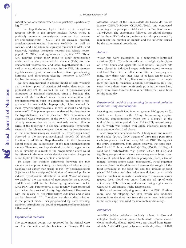
Dental Fast Track: Prenatal Enamel Growth, Incisor Eruption, and Weaning in Human Infants
Early weaning by maternal prolactin inhibition leads to higher neuropeptide Yand astrogliosis in the hypothalamus of the adult rat offspring
Viviane Younes-Rapozo1,2, Egberto G. Moura1, Alex C. Manhaes2, Nayara Peixoto-Silva1,Elaine de Oliveira1 and Patricia C. Lisboa1*1Laboratorio de Fisiologia Endocrina, Departamento de Ciencias Fisiologicas, Instituto de Biologia Roberto Alcantara
Gomes, Universidade do Estado do Rio de Janeiro, 58 Andar, Av. 28 de Setembro, 87, Rio de Janeiro, RJ 20551-031, Brazil2Laboratorio de Neurofisiologia, Instituto de Biologia Roberto Alcantara Gomes, Universidade do Estado do Rio de Janeiro,
Rio de Janeiro, Brazil
(Submitted 12 June 2014 – Final revision received 16 October 2014 – Accepted 31 October 2014)
Abstract
The suppression of prolactin production with bromocriptine (BRO) in the last 3 d of lactation reduces milk yield (early weaning) and
increases the transfer of leptin through the milk, causing hyperleptinaemia in pups. In adulthood, several changes occur in the offspring
as a result of metabolic programming, including overweight, higher visceral fat mass, hypothyroidism, hyperglycaemia, insulin resistance,
hyperleptinaemia and central leptin resistance. In the present study, we investigated whether overweight rats programmed by early
weaning with maternal BRO treatment have hypothalamic alterations in adulthood. We analysed the expression of neuropeptide Y
(NPY), cocaine- and amphetamine-regulated transcript (CART), pro-opiomelanocortin (POMC) and a-melanocyte-stimulating hormone
(a-MSH) by immunohistochemistry in the following hypothalamic nuclei: medial and lateral arcuate nucleus (ARC); paraventricular nucleus
(PVN); lateral hypothalamus (LH). Additionally, we sought to determine whether these programmed rats exhibited hypothalamic inflam-
mation as indicated by astrogliosis. NPY immunostaining showed a denser NPY-positive fibre network in the ARC and PVN (þ82 % in both
nuclei) of BRO offspring. Regarding the anorexigenic neuropeptides, no difference was found for CART, POMC and a-MSH. The number of
astrocytes was higher in all the nuclei of BRO rats. The fibre density of glial fibrillary acidic protein was also increased in both medial and
lateral ARC (6·06-fold increase and 9·13-fold increase, respectively), PVN (5·75-fold increase) and LH (2·68-fold increase) of BRO rats. We
suggest that early weaning has a long-term effect on the expression of NPY as a consequence of developmental plasticity, and the presence
of astrogliosis indicates hypothalamic inflammation that is closely related to overweight and hyperleptinaemia observed in our model.
Key words: Lactation: Developmental plasticity: Central obesity: Neuropeptides: Reactive astroglia
It has been shown that the lactation period is a critical stage
of development, and that early weaning could permanently
affect the progeny due to malnutrition or changes in neural
and hormonal status(1–7). The developing progeny has a
large potential to adapt to nutritional or hormonal changes,
and the process that leads to long-term alterations has been
referred to as ‘developmental plasticity’ or ‘programming’(8,9).
Our group has developed a model of programming based
on early weaning, in which milk yield is reduced during
the last 3 d of lactation through the inhibition of prolactin
with the administration of bromocriptine (BRO), a type 2
dopaminergic receptor agonist. The reduction in maternal
milk production causes malnutrition in pups and increases the
transfer of leptin through the milk, causing hyperleptinaemia
in pups(10). These alterations are sufficient to induce several
metabolic changes in adulthood as a result of metabolic
programming, including overweight, higher visceral fat mass,
hypothyroidism, hyperglycaemia, insulin resistance, dyslipi-
daemia, and increased medullary adrenal function and serum
glucocorticoid concentration(1,2,11). In addition, these animals
also exhibit hyperleptinaemia and central leptin resistance(1).
Disturbances in leptin levels caused by altered perinatal
nutrition lead to long-term consequences for energy meta-
bolism and body mass in adult life(12). Our group has
shown that leptin administered during the lactation period
can programme for overweight in the adult rat(13). One expla-
nation is that leptin may affect the formation and function
of hypothalamic circuitries when administered during the
* Corresponding author: Dr P. C. Lisboa, fax þ5521 28688029, email [email protected]
Abbreviations: a-MSH, a-melanocyte-stimulating hormone; ARC, arcuate nucleus; BRO, bromocriptine; CART, cocaine- and amphetamine-regulated
transcript; GFAP, glial fibrillary acidic protein; lARC, lateral arcuate nucleus; LH, lateral hypothalamus; mARC, medial arcuate nucleus; NPEW,
non-pharmacological early weaning; NPY, neuropeptide Y; P, postnatal day; POMC, pro-opiomelanocortin; PVN, paraventricular nucleus; TRH,
thyrotropin-releasing hormone.
British Journal of Nutrition, page 1 of 10 doi:10.1017/S0007114514003882q The Authors 2015
Bri
tish
Journ
alof
Nutr
itio
n
critical period of lactation when neural plasticity is particularly
high(14–17).
In the hypothalamus, leptin binds to its long-form
receptor Ob-Rb in the arcuate nucleus (ARC), where it
positively regulates anorexigenic neurons that release
pro-opiomelanocortin (POMC) and its cleavage product,
a-melanocyte-stimulating hormone (a-MSH), as well as
cocaine- and amphetamine-regulated transcript (CART), and
negatively regulates orexigenic neurons that release neuro-
peptide Y (NPY) and agouti-related peptide(18,19). These
ARC neurons project their axons to other hypothalamic
nuclei such as the paraventricular nucleus (PVN) and the
dorsomedial, ventromedial and lateral hypothalamus (LH), as
well as to extrahypothalamic areas. In the PVN, this circuitry
regulates anorexigenic neurons such as corticotropin-releasing
hormone and thyrotropin-releasing hormone (TRH)(19,20)
involved in energy expenditure.
We have demonstrated in another model of early weaning
that the interruption of lactation 3 d earlier than usual, on
postnatal day (P) 18, without the use of pharmacological
substances or maternal separation, using a bandage that
covers all the mothers’ teats, causes malnutrition and
hypoleptinaemia in pups; in adulthood, the progeny is pro-
grammed for overweight, hyperphagia, higher visceral fat
mass, hypertriacylglycerolaemia as well as insulin and leptin
resistance(6). This phenotype was evidenced by alterations in
the hypothalamus, such as increased NPY expression and
decreased CART expression in the PVN(7). The two models
of early weaning that we have previously studied differ in
terms of the following: (1) neonatal leptinaemia (hyperlepti-
naemia in the pharmacological model and hypoleptinaemia
in the non-pharmacological model); (2) hyperphagia (only
observed in the non-pharmacological model); (3) thyroid
function in adulthood (hypothyroidism in the pharmaco-
logical model and euthyroidism in the non-pharmacological
model). Therefore, we hypothesised that the changes in the
neural circuitry as a result of the programming effect could
be different in the two models despite the similar changes in
serum leptin levels and effects in adulthood.
To assess the possible differences between the two
models, in the present study, we investigated whether early
weaning (3 d earlier than usual, at P18) by pharmacological
(injections of bromocriptine) inhibition of maternal prolactin
induces hypothalamic alterations in adult Wistar offspring.
We analysed the expression of neuropeptides NPY, POMC,
a-MSH and CART in the following hypothalamic nuclei:
ARC; PVN; LH. Furthermore, it has recently been proposed
that before the onset of obesity, hypothalamic inflammation
with the release of pro-inflammatory cytokines and gliosis
can be observed(21–23). Therefore, we also studied whether
in the present model, rats programmed by early weaning
exhibited astrogliosis that could be suggestive of hypothalamic
inflammation.
Experimental methods
The experimental design was approved by the Animal Care
and Use Committee of the Instituto de Biologia Roberto
Alcantara Gomes of the Universidade do Estado do Rio de
Janeiro (CEUA/048/2010; CEUA/061/2011), and conducted
according to the principles established in the Brazilian Law no.
11.794/2008. The experiments followed the ethical doctrine
of the three ‘R’s (reduction, refinement and replacement)(24),
minimising the number of animals and the suffering caused
by the experimental procedures.
Animals
Wistar rats were maintained in a temperature-controlled
vivarium (25 ^ 18C) with an artificial dark–light cycle (lights
on 07.00 hours and lights off 19.00 hours). Pregnant rats
were placed in individual cages with free access to water
and food. To avoid the influence of litter size on program-
ming, only dams with litter sizes of at least ten to twelve
pups were used. At birth, litters were adjusted to six male
pups per dam to maximise lactation performance. In a few
cases where there were no six male pups in the same litter,
pups were cross-fostered from other litters that were born
on the same day.
Experimental model of programming by maternal prolactininhibition using bromocriptine
Lactating rats were divided into two groups: BRO group (n 7),
which was treated with 0·5 mg bromo-a-ergocryptine
(Novartis) intraperitoneally, twice per d (1 mg/d), at the
end of the lactation period, from P18 to P21; control group
(n 7), which was treated with saline injections using the
same protocol described above.
After progenitor separation (at P21), body mass and relative
food intake (g/100 g body weight) of three male pups from
each group were monitored every 7 d until P180. Throughout
the entire experiment, both groups received the same stan-
dard Nuvilabw chow, with 1466 kJ/100 g (350·5 kcal/100 g) of
solid food (carbohydrate 55 g, protein 22·5 g, fat 4·5 g and
8 g fibre; composition: calcium carbonate; maize bran; soya-
bean meal; wheat bran; dicalcium phosphate; NaCl; vitamin/
mineral premix; amino acids; antioxidants). Food ingestion
was calculated as the difference between the weight of the
food remaining in the food bin and the amount of food
placed 7 d before and that value was divided by 4, which
was the number of animals in each cage. To measure serum
glucose level, blood was drawn from the tail tip of each
animal after 12 h of fasting and assessed using a glucometer
(Accu-Chek Advantage; Roche Diagnostics).
BRO and control offspring were killed at P180. Further-
more, one rat offspring per litter, which was randomly
chosen from the three rats from the same litter maintained
in the same cage, was used for immunohistochemistry.
Antibodies
Anti-NPY (rabbit polyclonal antibody, diluted 1:1000) and
anti-glial fibrillary acidic protein (anti-GFAP) (mouse mono-
clonal antibody, diluted 1:200) were purchased from Sigma-
Aldrich. Anti-CART (goat polyclonal antibody, diluted 1:100)
V. Younes-Rapozo et al.2
Bri
tish
Journ
alof
Nutr
itio
n
and anti-POMC (rabbit polyclonal antibody, diluted 1:100)
were purchased from Santa Cruz Biotechnology, Inc. Anti-
a-MSH (sheep polyclonal antibody, diluted 1:10 000) was
purchased from EMD Millipore Corporation.
The application of primary antibodies was followed by the
application of appropriate secondary antibodies, which were
purchased from Molecular Probes (Invitrogen) and used at
1:400 dilution: donkey anti-rabbit conjugated with Alexa
Fluor 488; donkey anti-mouse conjugated with Alexa Fluor
555; donkey anti-goat conjugated with Alexa Fluor 555;
donkey anti-sheep conjugated with Alexa Fluor 488.
Immunohistochemistry
BRO and control offspring were perfused at P180 (n 7 rats
per group). The rats were fasted for 12 h. They were anaes-
thetised with Avertinw (0·3 mg/kg intraperitoneally) and
intracardially perfused with a saline solution followed by
4 % paraformaldehyde and then by paraformaldehyde plus
10 % sucrose. The brain was sectioned at 20mm using a cryo-
tome (Hyrax C25; Zeiss) and stored at 2208C. All coronal
sections containing the hypothalamus starting from the
bregma (21·88 mm), according to Paxinos & Watson(25),
were collected on gelatinised slides. For immuno-
histochemical procedures, the sections were treated with
a 0·3 % PBS–Triton solution followed by incubation with
a blocking solution (5 % bovine serum albumin), and then
immunolabelling with primary antibodies was performed.
Immunoreactivity was visualised by incubation with an
appropriate secondary antibody, and the sections were
counterstained with 40,6-diamidino-2-phenylindole, dihydro-
chloride (DAPI) (diluted 1:5000; Sigma-Aldrich). The slides
were mounted in ProLong Gold Antifade Reagent
(Invitrogen, Molecular Probes). In control procedures,
omission of primary antibodies with the inclusion of the
secondary antibody produced no labelling.
Image capturing was performed using an epifluorescence
microscope (Olympus BX-40). For quantification procedures,
we used captured images of four coronal sections from each
animal. Each selected section was separated from other
selected sections by 100mm. The following hypothalamic
nuclei were analysed: medial ARC (mARC) and lateral ARC
(lARC); PVN; LH.
Anti-NPY and anti-a-MSH antibodies specifically label
neuron fibres, CART labels fibres and cell bodies, and anti-
GFAP labels astrocyte processes and cell bodies. For the
quantification analysis of immunostaining, we used Image-
Pro Plus (version 4.5; Media Cybernetics, Inc.). The seg-
mentation tool was initially used to better differentiate the
background from actual anti-NPY, anti-CART, anti-a-MSH or
anti-GFAP staining. Because the cut-off point was selected
by the experimenter (who was blinded to the group assign-
ments), the segmentation tool procedure was repeated three
times on separate occasions for each image. The grey-scale
image (brighter pixels indicating stronger labelling) obtained
from this procedure was used in the quantification of the
average pixel brightness density of the entire image. Then,
we calculated the average pixel density that represents the
fibre density of the three treated images from each original
image, and this final result was used as the datum.
For the quantification of POMC, CART and GFAP, the cells
positive for each marker were counted in the captured
images (four sections counterstained with DAPI per nucleus
per animal) of the selected hypothalamic nuclei. Because indi-
vidual cells can be easily identified by perinuclear labelling,
the number of positive cells associated with each marker
was determined.
Statistical analysis
Data are presented as means with their standard errors. Body
weight, food intake and glycaemia data were analysed by
Student’s t test. Regarding immunohistochemistry data, to
reduce the likelihood of type 1 statistical errors that might
result from the repeated testing of the global dataset of each
immunostaining procedure, results for the density of NPY,
CART, a-MSH and GFAP immunostaining in seven animals
from each group (control and BRO) were first evaluated using
a global repeated-measures ANOVA. Separate repeated-
measures ANOVA were performed for each immunostaining
procedure. Group (control or BRO) was considered as the
between-subjects factor. Number of nuclei (NPY: ARC, PVN
and LH; CART: ARC, PVN and LH; a-MSH: PVN and LH; GFAP:
mARC, lARC, PVN and LH) was considered as the within-
subjects factor. Data regarding the number of positive cells
showing CART and GFAP immunostaining (considering GFAP
as a marker of astroglia) in seven animals from each group
were also subjected to repeated-measures ANOVA using the
same between-subjects factor. Number of nuclei (CART: ARC,
PVN and LH; GFAP: mARC, lARC, PVN and LH) was considered
the within-subjects factor. Regarding POMC, data regarding the
number of positive cells in the lARC were subjected to a univari-
ate ANOVA using group as the between-subjects factor. For
simplicity, results based only on the averaged univariate F tests
are reported. Lower-order ANOVA were used whenever
significant effects or interactions were observed in the global
repeated-measures ANOVA. For main effects, significance was
considered at P,0·05. For significant interactions at P,0·1,
we also examined whether lower-order main effects were
detectable after the subdivision of interactive factors(26).
Results
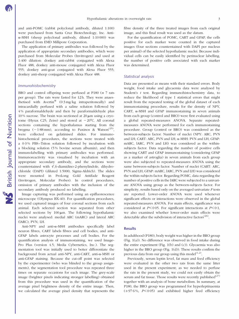
In adulthood (P180), body weight was higher in the BRO group
(Fig. 1(a)). No difference was observed in food intake during
the entire experiment (Fig. 1(b) and (c)). Glycaemia was also
higher in the BRO group (Fig. 1(d)). These results confirm the
previous data from our group using this model(1,2).
Previously, serum leptin level, fat mass and feed efficiency
were evaluated in the other two rats from the same litter
used in the present experiment; as we needed to perfuse
the rats in the present study, we could not easily obtain the
serum and fat tissue. Those results were recently published(27)
together with an analysis of bone metabolism. In summary, at
P180, the BRO group was programmed for hyperleptinaemia
(þ97·6 %, P,0·05) and exhibited higher feed efficiency
Hypothalamic alterations in overweight rats 3
Bri
tish
Journ
alof
Nutr
itio
n
(þ10 %, P,0·05) and higher visceral fat mass (retroperitoneal:
þ85 % and epididymal adipose tissue: þ58 %, P,0·05).
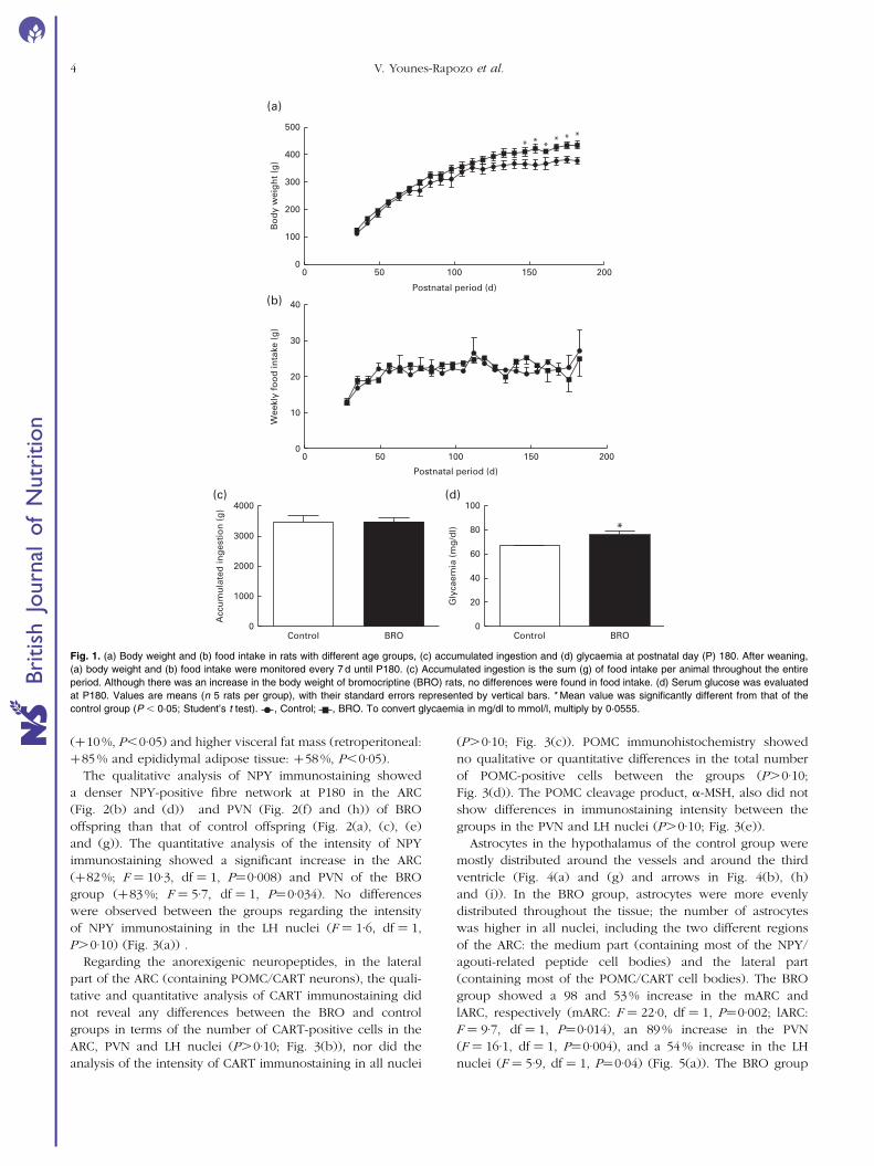
The qualitative analysis of NPY immunostaining showed
a denser NPY-positive fibre network at P180 in the ARC
(Fig. 2(b) and (d)) and PVN (Fig. 2(f) and (h)) of BRO
offspring than that of control offspring (Fig. 2(a), (c), (e)
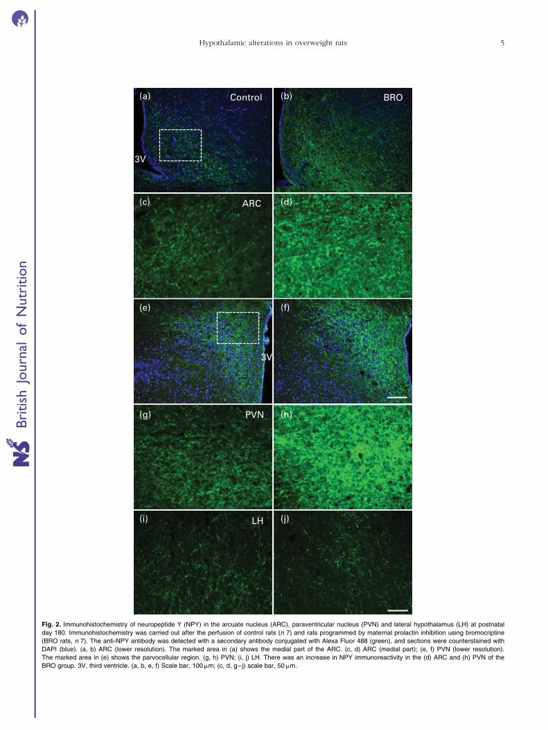
and (g)). The quantitative analysis of the intensity of NPY
immunostaining showed a significant increase in the ARC
(þ82 %; F ¼ 10·3, df ¼ 1, P¼0·008) and PVN of the BRO
group (þ83 %; F ¼ 5·7, df ¼ 1, P¼0·034). No differences
were observed between the groups regarding the intensity
of NPY immunostaining in the LH nuclei (F ¼ 1·6, df ¼ 1,
P.0·10) (Fig. 3(a)) .
Regarding the anorexigenic neuropeptides, in the lateral
part of the ARC (containing POMC/CART neurons), the quali-
tative and quantitative analysis of CART immunostaining did
not reveal any differences between the BRO and control
groups in terms of the number of CART-positive cells in the
ARC, PVN and LH nuclei (P.0·10; Fig. 3(b)), nor did the
analysis of the intensity of CART immunostaining in all nuclei
(P.0·10; Fig. 3(c)). POMC immunohistochemistry showed
no qualitative or quantitative differences in the total number
of POMC-positive cells between the groups (P.0·10;
Fig. 3(d)). The POMC cleavage product, a-MSH, also did not
show differences in immunostaining intensity between the
groups in the PVN and LH nuclei (P.0·10; Fig. 3(e)).
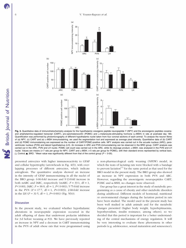
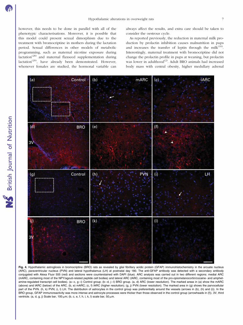
Astrocytes in the hypothalamus of the control group were
mostly distributed around the vessels and around the third
ventricle (Fig. 4(a) and (g) and arrows in Fig. 4(b), (h)
and (i)). In the BRO group, astrocytes were more evenly
distributed throughout the tissue; the number of astrocytes
was higher in all nuclei, including the two different regions
of the ARC: the medium part (containing most of the NPY/
agouti-related peptide cell bodies) and the lateral part
(containing most of the POMC/CART cell bodies). The BRO
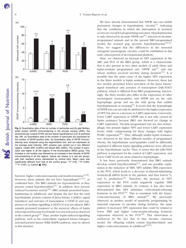
group showed a 98 and 53 % increase in the mARC and
lARC, respectively (mARC: F ¼ 22·0, df ¼ 1, P¼0·002; lARC:
F ¼ 9·7, df ¼ 1, P¼0·014), an 89 % increase in the PVN
(F ¼ 16·1, df ¼ 1, P¼0·004), and a 54 % increase in the LH
nuclei (F ¼ 5·9, df ¼ 1, P¼0·04) (Fig. 5(a)). The BRO group
Control BRO0
1000
2000
3000
4000
Acc
um
ula
ted
ing
esti
on
(g
)
Control BRO0
20
40
60
80
100
*
Gly
caem
ia (
mg
/dl)
0 50 100 150
******
2000
100
200
300
400
500
Postnatal period (d)
Bo
dy
wei
gh
t (g
)
0 50 100 150 2000
10
20
30
40
Postnatal period (d)
Wee
kly
foo
d in
take
(g
)
(a)
(b)
(c) (d)
Fig. 1. (a) Body weight and (b) food intake in rats with different age groups, (c) accumulated ingestion and (d) glycaemia at postnatal day (P) 180. After weaning,
(a) body weight and (b) food intake were monitored every 7 d until P180. (c) Accumulated ingestion is the sum (g) of food intake per animal throughout the entire
period. Although there was an increase in the body weight of bromocriptine (BRO) rats, no differences were found in food intake. (d) Serum glucose was evaluated
at P180. Values are means (n 5 rats per group), with their standard errors represented by vertical bars. * Mean value was significantly different from that of the
control group (P , 0·05; Student’s t test). , Control; , BRO. To convert glycaemia in mg/dl to mmol/l, multiply by 0·0555.
V. Younes-Rapozo et al.4
Bri
tish
Journ
alof
Nutr
itio
n
(a) (b)
(c) (d)
(e) (f)
(g) (h)
(i) (j)
3V
3V
Control BRO
ARC
PVN
LH
Fig. 2. Immunohistochemistry of neuropeptide Y (NPY) in the arcuate nucleus (ARC), paraventricular nucleus (PVN) and lateral hypothalamus (LH) at postnatal
day 180. Immunohistochemistry was carried out after the perfusion of control rats (n 7) and rats programmed by maternal prolactin inhibition using bromocriptine
(BRO rats, n 7). The anti-NPY antibody was detected with a secondary antibody conjugated with Alexa Fluor 488 (green), and sections were counterstained with
DAPI (blue). (a, b) ARC (lower resolution). The marked area in (a) shows the medial part of the ARC. (c, d) ARC (medial part); (e, f) PVN (lower resolution).
The marked area in (e) shows the parvocellular region. (g, h) PVN; (i, j) LH. There was an increase in NPY immunoreactivity in the (d) ARC and (h) PVN of the
BRO group. 3V, third ventricle. (a, b, e, f) Scale bar, 100mm; (c, d, g–j) scale bar, 50mm.
Hypothalamic alterations in overweight rats 5
Bri
tish
Journ
alof
Nutr
itio
n
presented astrocytes with higher immunoreactivity to GFAP
and cellular hypertrophy (arrowheads in Fig. 4(f)), with over-
lapping processes of different astrocytes, which indicate
astrogliosis. The quantitative analysis showed an increase
in the intensity of GFAP immunostaining in all the nuclei of
the BRO group: 6·06-fold increase and 9·13-fold increase in
both mARC and lARC, respectively (mARC: F ¼ 33·1, df ¼ 1,
P,0·001; lARC: F ¼ 96·9, df ¼ 1, P,0·001); 5·75-fold increase
in the PVN (F ¼ 17·7, df ¼ 1, P¼0·003); 2·68-fold increase
in the LH (F ¼ 31·5, df ¼ 1, P¼0·001) (Fig. 5(b)).
Discussion
In the present study, we evaluated whether hypothalamic
alterations in neuropeptide expression occurred in the
adult offspring of dams that underwent prolactin inhibition
for 3 d before weaning at P21. We have previously reported
an increase in NPY and a decrease in CART immunostaining
in the PVN of adult obese rats that were programmed using
a non-pharmacological early weaning (NPEW) model, in
which the teats of lactating rats were blocked with a bandage
to prevent lactation(7) for the same period as that used for the
BRO model in the present study. The BRO group also showed
an increase in NPY expression in both PVN and ARC.
However, regarding the anorexigenic neuropeptides CART,
POMC and a-MSH, no changes were observed.
Our group has a great interest in the study of metabolic pro-
gramming as a cause of obesity and other metabolic disorders
during adulthood. Different models of hormonal, nutritional
or environmental changes during the lactation period in rats
have been studied. The model used in the present study has
been well studied in adult animals and for the metabolic
changes presented (higher body weight, hyperleptinaemia,
hypothyroidism, insulin resistance, among others), so we
decided that this period is important for a better understand-
ing of the central mechanisms of energy regulation. It will
be very interesting to evaluate these phenomena in other
periods (e.g. adolescence, sexual maturation and senescence);
ARC0
50
100
150
PO
MC
-po
siti
ve c
ell n
um
ber
PVN LH0
2
4
6
8
α-M
SH
imm
un
ost
ain
ing
inte
nsi
ty
(a)
(b)
(d) (e)
(c)
ARC PVN LH0
10
20
30
*
*
NP
Y im
mu
no
stai
nin
g in
ten
sity
ARC PVN LH0
50
100
150
*
CA
RT
-po
siti
ve c
ell n
um
ber
ARC PVN LH0
5
10
15
CA
RT
imm
un
ost
ain
ing
inte
nsi
ty
Fig. 3. Quantitative data of immunohistochemistry analysis for the hypothalamic orexigenic peptide neuropeptide Y (NPY) and the anorexigenic peptides cocaine-
and amphetamine-regulated transcript (CART), pro-opiomelanocortin (POMC) and a-melanocyte-stimulating hormone (a-MSH) in rats at postnatal day 180.
Quantification was performed by photomicrography of different hypothalamic nuclei taken from four coronal sections of each animal. To analyse the neuron fibres
of (a) NPY, (c) CART and (e) a-MSH immunostaining, we used the segmentation tool and expressed as average pixel intensity. Quantitative data of (b) CART
and (d) POMC immunostaining are expressed as the number of CART/POMC-positive cells. NPY analysis was carried out in the arcuate nucleus (ARC), para-
ventricular nucleus (PVN) and lateral hypothalamus (LH). An increase in ARC and PVN immunostaining can be observed in the BRO group. CART analysis was
carried out in the ARC, PVN and LH nuclei. POMC cell count was carried out in the ARC, while its cleavage product, a-MSH, was analysed in the PVN and LH
nuclei. Values are means (n 7 rats per group for NPY, CART and a-MSH; n 5 rats per group for POMC), with their standard errors represented by vertical bars.
, Control; , BRO. * Mean value was significantly different from that of the control group (P , 0·05).
V. Younes-Rapozo et al.6
Bri
tish
Journ
alof
Nutr
itio
n
however, this needs to be done in parallel with all of the
phenotypic characterisations. Moreover, it is possible that
this model could present sexual dimorphism due to the
treatment with bromocriptine in mothers during the lactation
period. Sexual differences in other models of metabolic
programming, such as maternal nicotine exposure during
lactation(28) and maternal flaxseed supplementation during
lactation(29), have already been demonstrated. However,
whenever females are studied, the hormonal variable can
always affect the results, and extra care should be taken to
consider the oestrous cycle.
As reported previously, the reduction in maternal milk pro-
duction by prolactin inhibition causes malnutrition in pups
and increases the transfer of leptin through the milk(10).
Interestingly, maternal treatment with bromocriptine did not
change the prolactin profile in pups at weaning, but prolactin
was lower in adulthood(2). Adult BRO animals had increased
body mass with central obesity, higher medullary adrenal
mARC lARCControl
BRO
Control
BRO
PVN LH
(a) (b) (c)
(d) (e) (f)
(g) (h) (i)
(j) (k) (l)
3V
3V
Fig. 4. Hypothalamic astrogliosis in bromocriptine (BRO) rats as revealed by glial fibrillary acidic protein (GFAP) immunohistochemistry in the arcuate nucleus
(ARC), paraventricular nucleus (PVN) and lateral hypothalamus (LH) at postnatal day 180. The anti-GFAP antibody was detected with a secondary antibody
conjugated with Alexa Fluor 555 (red) and sections were counterstained with DAPI (blue). ARC analysis was carried out in two different regions: medial ARC
(mARC, containing most of the NPY/agouti-related peptide cell bodies) and lateral ARC (lARC, containing most of the pro-opiomelanocortin/cocaine- and amphet-
amine-regulated transcript cell bodies). (a–c, g–i) Control group; (b–d, j– l) BRO group. (a, d) ARC (lower resolution). The marked areas in (a) show the mARC
(above) and lARC (below) of the ARC. (b, e) mARC; (c, f) lARC (higher resolution); (g, j) PVN (lower resolution). The marked area in (g) shows the parvocellular
part of the PVN. (h, k) PVN; (i, l) LH. The distribution of astrocytes in the control group was preferentially around the vessels (arrows in (b), (h) and (i)). In the
BRO group, GFAP immunoreactivity was more intense and astrocyte processes were thicker than those observed in the control group (arrowheads in (f)). 3V, third
ventricle. (a, d, g, j) Scale bar, 100mm; (b, c, e, f, h, i, k, l) scale bar, 50mm.
Hypothalamic alterations in overweight rats 7
Bri
tish
Journ
alof
Nutr
itio
n
function, higher corticosteronaemia and insulin resistance(2,29).
However, these animals did not have hyperphagia(1,2,11), as
confirmed here. The BRO animals are hypometabolic as they
present central hypothyroidism(11). In addition, they present
reduced locomotor activity(30). BRO animals presented hyper-
leptinaemia in adulthood, and despite the fact that the total
hypothalamic protein content of Janus kinase 2 (JAK2), signal
transducer and activator of transcription 3 (STAT-3) and sup-
pressor of cytokine signalling 3 (SOCS-3) was not altered, BRO
animals presented resistance to the anorectic effects of leptin
when injected because food intake was not reduced as observed
in the control group(1). Thus, another leptin-induced signalling
pathway, such as the extracellular regulated kinase/mitogen-
activated protein kinase (ERK/MAPK) pathway, may be altered
in this situation.
We have already demonstrated that NPEW rats can exhibit
permanent changes in hypothalamic circuitry(7), indicating
that the conditions in which the interruption in lactation
occurs are crucial for programmingoutcomes. Hypoleptinaemia
is only observed in neonate NPEW rats(6), whereas in nicotine-
programmed animals and in the present BRO-programmed
model, the weaned pup presents hyperleptinaemia(10,31).
Thus, we suggest that the differences in the neuronal
orexigenic/anorexigenic circuitry could be established in this
early critical period of developmental plasticity.
Here, we observed an increase in NPY expression in the
ARC and PVN of the BRO group, which is a characteristic
that is also present in two other models of adult obese and
leptin-resistant programmed rats: NPEW rats(7) and rats
whose mothers received nicotine during lactation(32). It is
possible that the main cause of the higher NPY expression
in the three models is leptin resistance. However, these last
two models presented lower activation of the Janus kinase/
signal transducer and activator of transcription (JAK/STAT)
pathway, which is different from BRO programming. Interest-
ingly, the three models also differ in the expression of other
anorexigenic neuropeptides, and NPEW rats are the only
hyperphagic group and are the only group that exhibit
hypoleptinaemia at weaning(6). It seems that the hyperphagia
of NPEWrats can not only be attributed to the higher expression
of NPY but also to a decrease in CART expression. Conversely,
lower CART expression in NPEW rats is not only caused by
leptin resistance because BRO rats showed no change in
CART expression. Nicotine-programmed rats are also normo-
phagic but present higher NPY and lower CART expression
levels, while compensating for these changes with higher
POMC expression(32). Thus, although similar leptin resistance
was observed in the three different models of programmed
obesity, the hypothalamic neuropeptides could be differently
regulated if different leptin signalling pathways were affected
in the hypothalamic nuclei. Thus, it seems that the JAK/STAT
pathway is important for the control of CART expression, and
lower CART levels are more related to hyperphagia.
It has been previously demonstrated that BRO animals
develop central hypothyroidism(11), and one possible expla-
nation is the reduction of TRH. NPY inhibits TRH expression
in the PVN, which leads to a decrease in thyroid-stimulating
hormone-b mRNA levels in the pituitary and thus lowers T4
and T3 production(33). Therefore, the increase in NPY
expression in the PVN could negatively regulate TRH
expression in BRO animals. In contrast, it has also been
demonstrated that NPY stimulates corticotropin-releasing
hormone in the PVN(34). This is consistent with the hyper-
corticosteronaemia of BRO animals(2). We previously
observed, in another model of metabolic programming by
maternal exposure to nicotine during lactation, the same
pattern of increased NPY expression that could be related to
lower TRH and higher corticotropin-releasing hormone
expression observed in the PVN(32). This observation is
reinforced by the fact that in that nicotine exposure
model, the offspring exhibit central hypothyroidism and
higher corticosteronaemia in adulthood(28,35).
mARC lARC PVN LH0
5
10
15
******
**
**
GFA
P im
mu
no
stai
nin
g in
ten
sity
mARC lARC PVN LH0
20
40
60
80
100
(a)
(b)
*
****
*
Ast
rocy
te c
ell n
um
ber
Fig. 5. Quantitative data of the (a) number of astrocytes and (b) glial fibrillary
acidic protein (GFAP) immunostaining in the arcuate nucleus (ARC), the
paraventricular nucleus (PVN) and the lateral hypothalamus (LH) at postnatal
day 180. (a) Quantitative data of the number of astrocytes are expressed as
the number of GFAP-positive cells. (b) Quantitative data of GFAP immuno-
staining were analysed using the segmentation tool, and are expressed as
the average pixel intensity. ARC analysis was carried out in two different
regions: medial ARC (mARC) and lateral ARC (lARC). The number of astro-
cytes was higher in all the regions of the bromocriptine (BRO) group. This
increase in cell number was followed by an increase in the intensity of GFAP
immunostaining in all the regions. Values are means (n 5 rats per group),
with their standard errors represented by vertical bars. Mean value was
significantly different from that of the control group: * P,0·05, **P,0·005,
*** P,0·001. , Control; , BRO.
V. Younes-Rapozo et al.8
Bri
tish
Journ
alof
Nutr
itio
n
Leptin concentration is critical to hypothalamic develop-
ment, which occurs mostly after birth in rats. Projections
from the ARC to the PVN and LH during development are
completed at P16(14,15,36), just 2 d before lactation interruption
in BRO offspring and the onset of the anorexigenic effect of
leptin, which only starts after the third postnatal week in
rats(14). Several studies have already shown that malnutrition
during critical stages of hypothalamic development can per-
manently affect the organisation or this brain region. Rocha
et al.(37) showed that malnutrition during early life (from P1
to P10) causes a delay in leptin peak during lactation as
well as in NPY projection from the ARC to the PVN.
In the present study, we demonstrated significant hypo-
thalamic astrogliosis, which is suggestive of inflammation
in various parts of the ARC, PVN and LH nuclei. Some
characteristics that define reactive astrogliosis are progres-
sive alterations in molecular expression, progressive cellular
hypertrophy, and, in severe cases, proliferation and scar
formation(38). Here, we showed astrocytes with hypertrophy
of the cell body and processes, disruption of individual
domains (overlapping of astrocyte processes), and an
increased number of these cells, which suggest reactive
astrogliosis(38). This is consistent with the idea that hypothala-
mic inflammation is closely related to obesity(21–23,39). Indeed,
hyperleptinaemia can induce astrogliosis both in vitro and
in vivo. It has been suggested that the increase in GFAP
expression and alterations in astrocyte morphology can
modulate leptin distribution and signalling in neurons(40).
The deletion of leptin receptors in astrocytes alters cell mor-
phology and increases its synaptic inputs on POMC and
agouti-related peptide neurons. In addition, the anorexigenic
effect of leptin is reduced, indicating an active role of glial
cells in feeding behaviour(41).
Obesity also induces astrogliosis and increases the
synthesis of inflammatory cytokines, such as IL-6 and PGE2,
by astrocytes in the mouse(42), demonstrating the effective
role of astrocytes in the development of hypothalamic inflam-
mation. Obese animals programmed by neonatal overfeeding
exhibit hypothalamic inflammation based on increased acti-
vation of microglia, and this occurs during development
and persists until adulthood, demonstrating that glial cells
can also exhibit long-term alterations due to developmental
plasticity(43). Our BRO-programmed model produces higher
visceral fat mass and hyperleptinaemia, the findings that
can be associated with hypothalamic astrogliosis. Because
the NPEW model is hyperleptinaemic in adulthood but hypo-
leptinaemic at P21, it will be interesting to evaluate GFAP
expression in the NPEW model to determine whether this
gliosis is dependent on leptin concentration in early life or
only in adulthood.
In summary, our data provide evidence regarding the
relevance of different anorexigenic hypothalamic pathways
for the diverse neonatal imprinting effects that are observed
in different animal models of early weaning. In contrast,
NPY seems to be generally increased in different imprinting
models. The relationship between those neuronal circuitry
changes and astrogliosis reported here is unknown. However,
both changes can be fundamental for the obese phenotype.
Therefore, our data support the hypothesis that a short-
ened lactation period leads to long-term effects on central
neuropeptide expression and gliosis as a consequence of
developmental plasticity.
Acknowledgements
The authors thank Ms Monica Moura, Mr Ulisses Siqueira
and Mr Nilton S. V. Franca for technical assistance in the
laboratory.
The present study was financially supported by the Con-
selho Nacional de Desenvolvimento Cientıfico e Tecnologico
– CNPq (National Council for Scientific and Technological
Development), Fundacao Carlos Chagas Filho de Amparo a
Pesquisa do Estado do Rio de Janeiro – FAPERJ (Carlos
Chagas Filho Research Foundation of the State of Rio de
Janeiro) and Coordenacao de Aperfeicoamento de Pessoal
de Nıvel Superior – CAPES (Coordination for the Enhance-
ment of Higher Education Personnel). V. Y.-R. and N. P.-S.
were recipients of CAPES fellowships.
The authors’ contributions are as follows: V. Y.-R., E. G. M.,
A. C. M., E. d. O. and P. C. L. designed the experiments; V. Y.-R.
and N. P.-S. were responsible for animal programming and
immunohistochemistry. All authors participated in the ana-
lysis, interpretation of the data, elaboration and revision of
the manuscript, and contributed to and approved the final
manuscript.
The authors declare that there is no conflict of interest.
References
1. Bonomo IT, Lisboa PC, Pereira AR, et al. (2007) Prolactininhibition in dams during lactation programs for overweightand leptin resistance in adult offspring. J Endocrinol 192,339–344.
2. de Moura EG, Bonomo IT, Nogueira-Neto JF, et al. (2009)Maternal prolactin inhibition during lactation programsfor metabolic syndrome in adult progeny. J Physiol 587,4919–4929.
3. Kikusui T & Mori Y (2009) Behavioural and neurochemicalconsequences of early weaning in rodents. J Neuroendo-crinol 21, 427–431.
4. dos Santos Oliveira L, de Lima DP, da Silva AA, et al. (2010)Early weaning programs rats to have a dietary preference forfat and palatable foods in adulthood. Behav Processes 86,75–80.
5. Lisboa PC, Pires L, de Oliveira E, et al. (2010)Prolactin inhibition at mid-lactation influences adiposityand thyroid function in adult rats. Horm Metab Res 42,562–569.
6. Lima N da S, de Moura EG, Passos MC, et al. (2011) Earlyweaning causes undernutrition for a short period andprogrammes some metabolic syndrome components andleptin resistance in adult rat offspring. Br J Nutr 105,1405–1413.
7. Younes-Rapozo V, Moura EG, Lima NS, et al. (2012) Earlyweaning is associated with higher neuropeptide Y (NPY)and lower cocaine- and amphetamine-regulated transcript(CART) expressions in the paraventricular nucleus (PVN)in adulthood. Br J Nutr 28, 2286–2295.
8. Barker DJ (2003) The developmental origins of adult disease.Eur J Epidemiol 18, 733–736.
Hypothalamic alterations in overweight rats 9
Bri
tish
Journ
alof
Nutr
itio
n
9. Gluckman PD & Hanson MA (2007) Developmental plasticityand human disease: research directions. J Intern Med 261,461–471.
10. Bonomo IT, Lisboa PC, Passos MC, et al. (2005) Prolactininhibition in lactating rats changes leptin transfer throughthe milk. Horm Metab Res 37, 220–225.
11. Bonomo IT, Lisboa PC, Passos MC, et al. (2008) Prolactininhibition at the end of lactation programs for a centralhypothyroidism in adult rat. J Endocrinol 198, 331–337.
12. Teixeira C, Passos M, Ramos C, et al. (2002) Leptin serumconcentration, food intake and body weight in rats whosemothers were exposed to malnutrition during lactation.J Nutr Biochem 13, 493–498.
13. de Oliveira-Cravo C, Teixeira CV, Passos MC, et al. (2002)Leptin treatment during the neonatal period is associatedwith higher food intake and adult body weight in rats.Horm Metab Res 34, 400–405.
14. Bouret SG, Draper SJ & Simerly RB (2004) Trophic action ofleptin on hypothalamic neurons that regulate feeding.Science 30, 108–110.
15. Bouret SG & Simerly RB (2004) Leptin and developmentof hypothalamic feeding circuits. Endocrinology 145,2621–2626.
16. Pinto S, Roseberry AG, Liu H, et al. (2004) Rapid rewiring ofarcuate nucleus feeding circuits by leptin. Science 304,110–115.
17. Coupe B, Amarger V, Grit J, et al. (2010) Nutritionalprogramming affects hypothalamic organization and earlyresponse to leptin. Endocrinology 151, 702–713.
18. Valassi E, Scacchi M & Cavagnini F (2008) Neuroendocrinecontrol of food intake. Nutr Metab Cardiovasc Dis 18,158–168.
19. Sanchez-Lasheras C, Konner AC & Bruning JC (2010) Inte-grative neurobiology of energy homeostasis-neurocircuits,signals and mediators. Front Neuroendocrinol 31, 4–15.
20. Schwartz MW, Woods SC, Porte D Jr, et al. (2000) Centralnervous system control of food intake. Nature 404, 661–671.
21. Velloso LA, Araujo EP & de Souza CT (2008) Diet-induced inflammation of the hypothalamus in obesity.Neuroimmunomodulation 15, 189–193.
22. Buckman LB, Thompson MM, Moreno HN, et al. (2013)Regional astrogliosis in the mouse hypothalamus in responseto obesity. J Comp Neurol 15, 1322–1333.
23. Garcıa-Caceres C, Yi CX & Tschop MH (2013) Hypothalamicastrocytes in obesity. Endocrinol Metab Clin North Am 42,57–66.
24. Marques RG, Morales MM & Petroianu A (2009) Brazilianlaw for scientific use of animals. Acta Cir Bras 24, 69–74.
25. Paxinos G & Watson C (1998) The Rat Brain in StereotaxicCoordinates, 4th ed. Sydney: Academic Press.
26. Snedecor GW & Cochran WG (1967) Statistical Methods,6th ed. Ames, IA: Iowa University Press.
27. de Albuquerque Maia L, Lisboa PC, de Oliveira E, et al.(2014) Bone metabolism in obese rats programmed byearly weaning. Metabolism 63, 352–364.
28. Pinheiro CR, Oliveira E, Trevenzoli IH, et al. (2011) Develop-mental plasticity in adrenal function and leptin productionprimed by nicotine exposure during lactation: gender differ-ences in rats. Horm Metab Res 43, 693–701.
29. Troina AA, Figueiredo MS, Moura EG, et al. (2010) Maternalflaxseed diet during lactation alters milk composition andprograms the offspring body composition, lipid profile andsexual function. Food Chem Toxicol 48, 697–703.
30. Peixoto-Silva N, Conceicao EP, Carvalho JC, et al. (2014)Does bromocriptine play a role in decreasing oxidativestress for early weaned programmed obesity? Life Sci 95,14–21.
31. Fraga MC, Moura EG, Silva JO, et al. (2011) Maternal pro-lactin inhibition at the end of lactation affects learning/memory and anxiety-like behaviors but not novelty-seekingin adult rat progeny. Pharmacol Biochem Behav 100,165–173.
32. Younes-Rapozo V, Moura EG, Manhaes AC, et al. (2013)Maternal nicotine exposure during lactation alters hypothala-mic neuropeptides expression in the adult rat progeny. FoodChem Toxicol 58, 158–168.
33. Vella KR, Ramadoss P, Lam FS, et al. (2011) NPY and MC4Rsignaling regulate thyroid hormone levels during fastingthrough both central and peripheral pathways. Cell Metab14, 780–790.
34. Sarkar S & Lechan RM (2003) Central administrationof neuropeptide Y reduces a-melanocyte-stimulating hor-mone-induced cyclic adenosine 50-monophosphate responseelement binding protein (CREB) phosphorylation in pro-thyrotropin-releasing hormone neurons and increasesCREB phosphorylation in corticotropin-releasing hormoneneurons in the hypothalamic paraventricular nucleus.Endocrinology 144, 281–291.
35. Oliveira E, Moura E, Santos-Silva A, et al. (2009) Short- andlong-term effects of maternal nicotine exposure duringlactation on body adiposity, lipid profile, and thyroid func-tion of rat offspring. J Endocrinol 202, 397–405.
36. Grove KL & Smith MS (2003) Ontogeny of the hypothalamicneuropeptide Y system. Physiol Behav 79, 47–63.
37. Rocha ML, Fernandes PP, Lotufo BM, et al. (2014) Under-nutrition during early life alters neuropeptide Y distributionalong the arcuate/paraventricular pathway. Neuroscience256, 379–391.
38. Sofroniew MV & Vinters HV (2010) Astrocytes: biology andpathology. Acta Neuropathol 119, 7–35.
39. Tomassoni D, Nwankwob IE, Gabrielli MG, et al. (2013)Astrogliosis in the brain of obese Zucker rat: a model ofmetabolic syndrome. Neurosci Lett 543, 136–141.
40. Pan W, Hsuchou H, Xu C, et al. (2011) Astrocytesmodulate distribution and neuronal signaling of leptin inthe hypothalamus of obese A vy mice. J Mol Neurosci 43,478–484.
41. Kim JG, Suyama S, Koch M, et al. (2014) Leptin signalingin GFAP-expressing adult glia cells regulates hypothalamicneuronal circuits and feeding. Nat Neurosci 17, 908–910.
42. Chikuma T, Yoshimoto T, Ohba M, et al. (2009) Interleukin-6induces prostaglandin E2 synthesis in mouse astrocytes.J Mol Neurosci 39, 175–184.
43. Ziko I, Luca S, Dinan T, et al. (2014) Neonatal overfeedingalters hypothalamic microglial profiles and central responsesto immune challenge long-term. Brain Behav Immun 41,32–43.
V. Younes-Rapozo et al.10
Bri
tish
Journ
alof
Nutr
itio
n
Copyright © 2022 FDOKUMEN