Unbalance and harmonics detection in induction motors using an optical fiber sensor
Upload
independentCategory
view
2download
0
Early Low Protein Diet Aggravates Unbalance betweenAntioxidant Enzymes Leading to Islet DysfunctionNicolas Theys, Andre Clippe, Thomas Bouckenooghe, Brigitte Reusens, Claude Remacle*
Laboratory of Cell Biology, Institute of Life Sciences, Universite catholique de Louvain, Louvain-la-Neuve, Belgium
Abstract
Background: Islets from adult rat possess weak antioxidant defense leading to unbalance between superoxide dismutase(SOD) and hydrogen peroxide-inactivating enzymatic activities, catalase (CAT) and glutathione peroxidase (GPX) rendingthem susceptible to oxidative stress. We have shown that this vulnerability is influenced by maternal diet during gestationand lactation.
Methodology/Principal Findings: The present study investigated if low antioxidant activity in islets is already observed atbirth and if maternal protein restriction influences the development of islet antioxidant defenses. Rats were fed a controldiet (C group) or a low protein diet during gestation (LP) or until weaning (LPT), after which offspring received the controldiet. We found that antioxidant enzymatic activities varied with age. At birth and after weaning, normal islets possessed anefficient GPX activity. However, the antioxidant capacity decreased thereafter increasing the potential vulnerability tooxidative stress. Maternal protein malnutrition changed the antioxidant enzymatic activities in islets of the progeny. At 3months, SOD activity was increased in LP and LPT islets with no concomitant activation of CAT and GPX. This unbalancecould lead to higher hydrogen peroxide production, which may concur to oxidative stress causing defective insulin geneexpression due to modification of critical factors that modulate the insulin promoter. We found indeed that insulin mRNAlevel was reduced in both groups of malnourished offspring compared to controls. Analyzing the expression of such criticalfactors, we found that c-Myc expression was strongly increased in islets from both protein-restricted groups compared tocontrols.
Conclusion and Significance: Modification in antioxidant activity by maternal low protein diet could predispose topancreatic islet dysfunction later in life and provide new insights to define a molecular mechanism responsible forintrauterine programming of endocrine pancreas.
Citation: Theys N, Clippe A, Bouckenooghe T, Reusens B, Remacle C (2009) Early Low Protein Diet Aggravates Unbalance between Antioxidant Enzymes Leadingto Islet Dysfunction. PLoS ONE 4(7): e6110. doi:10.1371/journal.pone.0006110
Editor: Kathrin Maedler, University of Bremen, Germany
Received March 29, 2009; Accepted May 25, 2009; Published July 1, 2009
Copyright: � 2009 Theys et al. This is an open-access article distributed under the terms of the Creative Commons Attribution License, which permitsunrestricted use, distribution, and reproduction in any medium, provided the original author and source are credited.
Funding: This work was supported by the Parthenon Trust (London, UK), the European Project ERNEST (Food-CT-2005-007036, 6th Frame Programme), theBelgian Fonds National de la Recherche Scientifique, Fonds pour la Recherche dans l’Industrie et l’Agriculture. The funders had no role in study design, datacollection and analysis, decision to publish, or preparation of the manuscript.
Competing Interests: The authors have declared that no competing interests exist.
* E-mail: [email protected]
Introduction
Oxidative stress (OS) is an important factor in the process of
pancreatic b-cell dysfunction which leads to diabetes [1]. Reactive
oxygen species (ROS) and/or reactive nitrogen species (RNS) are
formed during both pro-inflammatory cytokine-mediated b-cell
aggression in Type 1 diabetes mellitus (T1DM) and glucolipotoxi-
city-mediated b-cell dysfunction in Type 2 diabetes mellitus
(T2DM) [1–4]. Indeed, there is strong evidence for ROS and RNS
participating in the pathogenesis of T1DM while only the
participation of ROS can be described as a mechanism of b-cell
dysfunction in T2DM [2,5,6]. OS features pathological relevance
in the appearance of diabetes at adult age because pancreatic islets
possess particularly weak antioxidant defense activity [7,8].
Therefore, the ability of OS to damage pancreatic islets and
markedly blunt insulin secretion is not surprising.
The risk of developing metabolic diseases in adulthood is
influenced not only by genetic and lifestyle factors but also by
environmental factors acting in early life [9]. Epidemiological
studies have related that a poor intra-uterine environment may
program susceptibility in the progeny to later development of the
metabolic syndrome (rev in [10]).
Most of our work to date focused on the influence of a protein
restriction (Low Protein, LP) during gestation or gestation and
lactation for the offspring. LP diet modified the process of islet cell
development leading to impairment in islet cell expansion with a
smaller b-cell mass at birth [11–14]. When the LP diet was
maintained until weaning, this deficiency was more pronounced
[11–13]. Moreover, maternal LP diet seems to program an
increased vulnerability of the endocrine pancreas [15–17]. A
proteome analysis carried out on in vitro neoformed fetal islets
showed that ten proteins involved in protein folding and
chaperoning were modified, possibly testifying cell stress [18].
Using micro-array analysis, we demonstrated that more than 10%
of the gene expression was significantly altered by the maternal
low protein diet in fetal islets including antioxidant enzymes
modified expression [19]. Furthermore, islets from fetal and adult
progeny of dams fed a low protein diet showed a higher rate of
PLoS ONE | www.plosone.org 1 July 2009 | Volume 4 | Issue 7 | e6110

apoptosis after cytokines or oxidative stress aggression in in vitro
experiments [15,16]. Higher NOu production by islets from adult
LP offspring could be an important factor to explain this
subsequent cell death [17]. Taken together, these findings suggest
that the LP diet increased susceptibility to OS that renders the b-
cell more vulnerable and prone to apoptosis.
In addition, the reprogramming of mitochondrial function was
proposed as a key adaptation enabling a fetus to survive in a
limited environment (rev in [20]). Finally, mitochondrial dysfunc-
tion occurs and leads to increased production of ROS and
eventually to OS if the defense systems of the cell are overwhelmed
[21].
Adult pancreatic islets are equipped with lower antioxidant
defenses than other cell types. In the present study, we
investigated (1) if low antioxidant capacity is already observed
at birth by measuring the antioxidant enzyme activity, (2) the
evolution of these activities during post-natal life. Finally (3), the
influence of protein restriction during early life on b-cells
antioxidant defense and the control of insulin gene expression
were assessed.
Results
Litter size and body weightNo difference in litter size was observed between the groups (on
average C = 11.960.7, LP = 11.760.5 pups per litter). The low
protein diet caused a significantly smaller mean fetal and newborn
body weight as shown in Table 1. The importance of growth
retardation was related to the time-window of exposure to the low
protein diet. At weaning and at 28 days, LPT offspring was growth
restricted compared to C and LP groups, which remained until 3
months of age. Conversely, the body weight gain of the LP
progeny was higher compared to C offspring.
Blood analysisTable 2 shows fasting blood parameters at 3 months. Compared
to C group, insulin level was lower in LPT group whereas the
lower value in LP rats did not reach statistical significance. Plasma
glucose levels were unaffected in both LP and LPT groups.
Offspring of dam fed a low protein diet showed indeed an
improved glucose tolerance which is reflected by a significantly
lower HOMA-R index. Despite non significant difference in
antioxidant potential in plasma of the three groups of rats, the
protein nitrosylation was significantly higher in both LP and LPT
animals.
Antioxidant enzymatic activities in fetal pancreatic isletsAntioxidant activities were measured in islets and compared
with those in the liver. Like in previous work [7,8], the liver was
used as a reference tissue since its antioxidant capacity is well
documented. As shown in Figure 1, fetal pancreatic islets possessed
less SOD and CAT activity than fetal liver. GPX activity was
similar in islets and liver at this age.
Antioxidant enzymatic activities in postnatal pancreaticislets
Antioxidant enzymatic activity was analyzed in postnatal
pancreatic islets isolated at 5 days, 28 days, 6 weeks and 3 months
of age. Values obtained for pancreatic islets were compared to
those in the liver. Pancreatic islets showed a higher SOD activity at
5 days than at subsequent time points whereas the liver showed an
opposite evolution (Figure 2A). Thus, throughout life, islets
exhibited a lower SOD activity than liver and this difference
was more pronounced with age.
At all ages, CAT activity was very low in islets and we did not
observe any significant change from 5 day- to 3 month-old rats
(Figure 2B). Compared to liver where the activity was higher in
neonate, pancreatic islets had already at birth less than one
percent of the liver CAT activity.
Islets exhibited a higher GPX activity at 28 days (Figure 2C),
compared to earlier and later ages. At this stage, GPX capacity in
islets was significantly higher than in liver at the same age. In the
liver, GPX activity was much higher at 5 days than later in life.
Consequences of a low protein diet on antioxidantenzymatic activities in fetal pancreatic islets
In islets from fetuses of dams fed a low protein diet during
gestation, SOD and CAT activities were unchanged compared
to controls, whereas GPX activity was significantly lower
Table 1. Body weights of fetuses (21.5 days of gestation) andpostnatal rats.
C LP LPT
Fetus(n = 73–101) 5.6660.06 5.3860.05**
5 days(n = 50–78) 14.860.3 14.160.2* 11.460.2*** {{{
21 days(n = 50–78) 54.560.7 54.160.8 27.960.5*** {{{
28 days(n = 28–47) 8861.3 8761.3 5761.1*** {{{
6 weeks(n = 16–29) 19764.1 19063.4 14364.9*** {{{
3 months(n = 16–29) 38167.4 417610* 344611** {{{
Values are means6SEM.*p,0.05, **p,0.01, ***p,0.001 compared with control group.{{{p,0.001 compared to LP group.doi:10.1371/journal.pone.0006110.t001
Table 2. Blood analysis in 3 month-old male rats.
C LP LPT
Plasma insulin (pmol/l) 141.7610.7 111.967.6 87.466.4**
Plasma glucose (mmol/l) 5.160.14 5.160.24 5.460.35
Homeostasis model assessment insulin-resistance index (HOMA-R) 4.760.4 3.660.2* 2.960.2**
Total peroxyl-radical trapping potential (mM Trolox equiv.) 91.4612.8 87.265.5 70.364.9
Nitrotyrosine levels (pmol/mg protein) 0.1960.02 0.4160.03** 0.5460.02**
Values are means6SEM of 6 to 8 animals in each group.*p,0.05, **p,0.01 vs C.doi:10.1371/journal.pone.0006110.t002
Maternal Diet and Antioxidant
PLoS ONE | www.plosone.org 2 July 2009 | Volume 4 | Issue 7 | e6110

(Figure 3). Gene expression corresponding to these enzymes was
similar in islets from protein restricted and control fetuses (data
not shown). The consequences of low protein diet were different
in liver, where CAT and GPX activity was unchanged whereas
SOD activity was reduced in LP fetuses compared to controls
(Figure 3).
Figure 1. Antioxidant enzyme activity in fetal pancreatic islets and liver. (A) SOD, (B) CAT, (C) GPX. Results are expressed as means6SEM;n = 5–6 independent experiments. ***p,0.001 islets vs liver.doi:10.1371/journal.pone.0006110.g001
Figure 2. Postnatal evolution of antioxidant enzyme activity in pancreatic islets and liver. (A) SOD, (B) CAT, (C) GPX. Results are expressedas means6SEM; n = 4–7 independent experiments. *p,0.05, **p,0.01, ***p,0.001 vs 5 days; £p,0.05, ££p,0.01, £££p,0.001 vs 28 days within thesame tissue.doi:10.1371/journal.pone.0006110.g002
Figure 3. Effect of low protein diet on fetal pancreatic islets and liver antioxidant enzyme activity, SOD (white bars), CAT (blackbars), GPX (grey bars). Data are expressed as fold change in activity relative to C; n = 5–6 independent experiments. *p,0.05 vs controls.doi:10.1371/journal.pone.0006110.g003
Maternal Diet and Antioxidant
PLoS ONE | www.plosone.org 3 July 2009 | Volume 4 | Issue 7 | e6110

Consequences of a low protein diet on antioxidantenzymatic activities in postnatal pancreatic islets
SOD activity was lower in pancreatic islets from LP newborns
(Figure 4A). However, from 28 days, a significant higher SOD
activity was measured in islets from LP animals compared to
controls. At 3 months, islets obtained from LPT offspring
presented a two folds higher SOD activity. In liver, protein
restriction led to a lower SOD capacity in LP and LPT pups. No
more difference was observed thereafter except at 3 months, when
this activity was increased in LPT rats.
Particularly in LPT islets, CAT activity was in a range reaching
the detection limit and was so low for many samples that it could
not be detected (Figure 4B). Conversely, CAT activity was
significantly upregulated in liver from 28 days to 3 months for
both LP and LPT offspring.
GPX activity was altered in islets and liver from LP and LPT
rats (Figure 4C). It was lower in pancreatic islets from LP newborn
while at 28 days, this decline was observed in LPT as well as in LP
pancreatic islets. This strong reduction was no more found
thereafter. Such lower activity was also detected in liver,
particularly in neonates and at 3 months in both protein-restricted
progeny compared to controls.
Consequences of a low protein diet on antioxidant geneexpression
Parallel measurements of antioxidant enzyme mRNA at 3
months enabled to compare expression to activity. A higher SOD1
transcript was observed in islets from 3 month-old low protein
progeny whereas SOD2 was slightly increased only in LPT islets
(Figure 5A/B). The level of catalase expression was about 30% in
islets from LP and LPT animals compared to controls (Figure 5C).
GPX1 mRNA was higher in pancreatic islets from LP rats but
unchanged in LPT (Figure 5D).
Consequences of a low protein diet on peroxiredoxincapacity
Peroxiredoxins constitute a less known family of peroxidase.
PRDX activity was not detectable in pancreatic islets at any age as
well as in the fetal liver. In liver, PRDX activity was lower at birth
and at 3 months in LP and LPT animals compared to controls
(Figure 6). PRDX1 and PRDX2 gene expression was modified in
LP islets (Figure 7A/B) whereas only PRDX2 transcripts tended to
be upregulated in LPT islets compared to C. In the liver, PRDX1
mRNA level tended to be reduced in LP (p = 0.07) and was
significantly downregulated in LPT rats (p,0.01; Data not
shown).
Since the liver is a hematopoietic organ during gestation and
early postnatal period, we analyzed to which cell type the lower
PRDX activity can be attributed. Immunostaining for the different
isoforms of peroxiredoxins (1 to 5) was assessed on liver sections
from 5 day-old rats and showed no stained hematopoietic cells.
Thus, the activity measured in liver from newborn was attributed
to hepatocytes. Immunostaining for PRDX1 is presented in
Figure 8.
Consequences of a low protein diet on PDX-1, c-Myc andinsulin gene expression
Because the level of SOD activity was increased in islets from
LP and LPT offspring with no concomitant activation of the
hydrogen peroxide-inactivating enzymes CAT and GPX, this
unbalance could lead to oxidative stress through a higher
hydrogen peroxide production. Exposure of pancreatic islets to
oxidative stress causes defective insulin gene expression due to
modification of at least two critical factors that modulate the
insulin promoter : PDX-1 [22] and c-Myc [23]. Analyzing the
expression of these factors (Figure 9), we found that PDX-1
mRNA level was unchanged while c-Myc expression increased
strongly in islets from LP and LPT progeny. Insulin mRNA level
was reduced in LP and LPT rats compared to controls.
Discussion
ROS and RNS species are described as contributors of b-cell
dysfunction during the development of diabetes [1,2,4]. Every cell
is equipped with antioxidant defense systems that react with
reactive species. Superoxide dismutases (SOD) are the antioxidant
enzymes that catalyze the dismutation of the highly reactive
superoxide anion to less reactive hydrogen peroxide. Peroxides can
be inactivated by catalase (CAT), glutathion peroxidase (GPX) or
peroxiredoxin (PRDX). However, pancreatic islets are weakly
protected against the toxicity of ROS and RNS as the level of their
antioxidant defense has been found to be very low, leading to
unbalance between the two antioxidant enzymatic scavenger
systems, superoxide dismutases and hydrogen peroxide-inactivat-
ing systems, namely CAT and GPX [7,8,24–26].
The first objective of the present study was to investigate the
evolution of the antioxidant activity of pancreatic islets already just
before birth until 3 months. The results were compared to
antioxidant capacity of the liver, used as a reference tissue [7,8].
Fetal islets did not feature the unbalanced antioxidant potential
described for adult animals. Just before birth, pancreatic islets
exhibited a much lower level of SOD and CAT activity than in
liver but, a similar GPX activity. Although catalase activity was
already negligible in fetal islets, a significant peroxidase capacity
through GPX may be protective against the oxidative challenge
caused by delivery [27,28].
Postnatal evolution of antioxidant capacity showed that SOD,
CAT and GPX remained particularly weak in pancreatic islets
throughout life compared to that in liver. Our findings for adult
animals support previous observations comparing the relative
values between islets and liver [8]. All antioxidant activities
varied with age and the time-course was different regarding the
tissue. Weaning is a step where most of the nutritional changes
occur and the b-cells have to deal with a change from a high fat
diet to a high glucose diet. We showed that in newborn, GPX
activity was low and that it increased until 28 days to diminish
thereafter. It is known that when GPX activity is experimentally
increased to the levels found in other tissues, b-cell are protected
from OS [26]. Therefore, the GPX increase may be beneficial
for the b-cell before this nutritional transition. In addition, it is
known that a postnatal wave of apoptosis occurs during lactation
in the normal developing endocrine pancreas [29,30]. The peak
of apoptosis was observed at 2 weeks of age during which 60%
of pre-existing b-cells died [31]. Although the functional
significance of this remodeling phase is still unknown for b-
cells, similar waves of apoptosis which coincide with changing
function were described for other cell types [32]. In the
pancreas, this wave may prepare the b-cell mass to the
nutritional challenge taking place at weaning. Thus, immediately
after the remodeling, pancreatic islets acquire a significant
intrinsic hydrogen peroxide-inactivating potential which may
provide enhanced protection [26,33].
At each age, pancreatic islets exhibited an extremely weak
catalase capacity in the range of 1 or 2% of that in the liver.
Catalase overexpresion in cultured insulin producing cells as well
as in b-cells from transgenic mice provided protection against
oxidative injury [24,34]. Many authors discussed the relative
Maternal Diet and Antioxidant
PLoS ONE | www.plosone.org 4 July 2009 | Volume 4 | Issue 7 | e6110

importance of catalase and other peroxidases in removing H2O2.
Catalase is not expected to intervene in eliminating low levels of
H2O2 but plays an important role in the acquisition of tolerance to
oxidative stress [35,36]. With such a low catalase capacity already
at birth, the b-cells should be particularly susceptible to oxidative
stress. Indeed, an appropriate level seems to be necessary to
challenge high concentration of H2O2 produced during hypergly-
cemia or cytokines aggression [24,26,34,37,38].
Figure 4. Postnatal effect of low protein diet on antioxidant enzyme activity in pancreatic islets and liver, in LP (grey) and LPT(black) progeny. (A) SOD, (B) CAT, (C) GPX. Data are expressed as fold change in activity relative to C; n = 4–7 independent experiments. *p,0.05,**p,0.01, ***p,0.001, {p = 0.06 vs controls.doi:10.1371/journal.pone.0006110.g004
Maternal Diet and Antioxidant
PLoS ONE | www.plosone.org 5 July 2009 | Volume 4 | Issue 7 | e6110

Experimental and clinical evidence support the notion that
intrauterine malnutrition can contribute to the development of
metabolic disease in adult life [9,10]. Low protein diet impairs the
development of fetal pancreas and has long-term consequences on
its secretory capacity [11,14]. Our study investigated a possible
mechanism by which the antioxidant unbalance may contribute to
the early programming of islets by exposure to a low protein diet.
First, we confirmed previous observations showing a significant
effect of maternal low protein diet on weight and plasma
composition of the offspring [16]. At 3 months, progeny of dams
fed a low protein diet during early life presented a lower fasting
plasma insulin concentration with a normal fasting glucose level,
Figure 5. SOD1 (A), SOD2 (B), CAT (C), GPX1 (D) gene expression in pancreatic islets from 3 month-old rats. Results are expressed asmeans6SEM; n = 6–7 independent experiments. *p,0.05, **p,0.01 vs C;doi:10.1371/journal.pone.0006110.g005
Figure 6. Peroxiredoxin activity in liver from C (white bars), LP (grey bars), LPT (black bars). Results are expressed as means6SEM; n = 4–5 independent experiments. *p,0.05, ** p,0.01 vs C;doi:10.1371/journal.pone.0006110.g006
Maternal Diet and Antioxidant
PLoS ONE | www.plosone.org 6 July 2009 | Volume 4 | Issue 7 | e6110

which may be explained by the improvement of peripheral insulin
sensitivity [39,40]. However, it is known that glucose intolerance
occurs later in life in these animals [41,42].
We demonstrated that GPX activity was significantly reduced in
fetal LP islets. This contrasts with the significant GPX activity
observed in control fetal islets which is probably important to
tolerate oxidative challenge caused by delivery. Islets from fetus of
dams fed a low protein diet during gestation are known to be more
vulnerable to cytotoxic aggression [15]. Indeed, in vitro exposure of
fetal islets to a chemical generator of NOu or to cytokines induced
apoptosis in b-cell with a significantly greater impact in LP than in
C islets [15]. Ten proteins involved in protein folding and
chaperoning were found to be altered by the low protein diet in
proteomic analysis [18] and data provided by microarray analysis
are congruent with an increased sensitivity to oxidative stress in
islets from LP fetuses [19]. Moreover, b-cell mass was significantly
lower in LP fetuses compared to controls [11]. Although specific
targets such as proliferation, vascularization, and growth factors
have been identified as involved in the lower b-cell mass in LP
progeny [11–14,43], the participation of oxidative stress can not
be excluded.
The switch to a normal diet after delivery caused a reduction of
islet antioxidant capacity in the newborn, which was not observed
when the low protein diet was maintained in LPT pups. Gluckman
et al. [44] suggested that a mismatch between suboptimal fetal
environment and a richer environment really encountered after
Figure 7. PRDX1 (A), PRDX2 (B), PRDX3 (C) and PRDX5 (D) gene expression in pancreatic islets from 3 month-old rats. Results areexpressed as means6SEM; n = 6–7 independent experiments. *p,0.05, **p,0.01 vs C;doi:10.1371/journal.pone.0006110.g007
Figure 8. Immunostaining of PRDX1 in liver from 5 day-old newborn control, LP and LPT rats. Hepatocytes (arrows head) appear inbrown staining while hematopoietic cells (black arrows) are unstained for PRDX1 for both groups.doi:10.1371/journal.pone.0006110.g008
Maternal Diet and Antioxidant
PLoS ONE | www.plosone.org 7 July 2009 | Volume 4 | Issue 7 | e6110

birth is detrimental. However, in the liver, the duration of
exposure to protein restriction did not modify the consequence at
5 days, when both LP and LPT pups presented a lower
antioxidant potential than the controls.
In the adult, islets from LP and LPT animals featured a higher
SOD activity compared to controls whereas CAT activity was very
low. It should be noted that LP offspring recovered a body weight
similar to control at weaning whereas LPT offspring remained
significantly leaner throughout life. Then, the catch-up growth per
se in LP animals did not influence the antioxidant potential in islets
but, it was shown by others [45], that the reduction in litter size
after delivery, 4 pups instead of 8 in our study, to maximize catch-
up growth of LP progeny was associated to a lower SOD
expression. The same discrepancy is also true for PRDX
expression, which shall be discussed below. Those contradictions
demonstrate the sensitivity of the enzyme response to nutritional
manipulations during the lactating period. In a mouse model,
overexpression of MnSOD resulting in overproduction of H2O2,
was found responsible for damaging the b-cells through overriding
the limited hydrogen peroxide-inactivating capacity [46]. An
increase in SOD activity can be an adaptation to mitochondrial
dysfunction in islets from LP and LPT progeny. Using a model of
uteroplacental insufficiency, Simmons et al. [21] proposed that a
reprogramming of mitochondrial function occurred in pancreatic
islets as a consequence of a limited energy environment during
early life. This reprogramming led to mitochondrial dysfunction
which increased production of mitochondrial ROS and MnSOD
expression. Modification of mitochondrial function, like a much
higher ROS production, was also observed in islets from LP
offspring (Theys, unpublished data).
Contrary to the liver where the higher SOD activity in LPT
animals was associated to a higher catalase potential, no
concomitant increase in any peroxidase potential was observed
in islets from LP and LPT rats. Independently of the malnutrition,
we know that the vulnerability of normal b-cells is attributed to
their unbalance through SOD and hydrogen peroxide-inactivating
enzymes [8]. Maternal malnutrition increased this unbalance and
probably through that way, enhanced the vulnerability of fetal and
adult islets, as reported previously [16,17].
The method for measuring PRDX activity [47] was not
sensitive enough to face the limited quantity of available protein
in pancreatic islets. PRDX1 gene expression was lower in LP islets
whereas PRDX2 was higher and PRDX-3 and -5 mRNA levels
similar in LP and LPT islets compared to C. It should be noted
that, as mentioned above, when forced catch-up growth was
induced after delivery, offspring of dams fed a LP diet did not
present the same modification of PRDX gene expression [45].
Exposure to oxidative stress can cause a decrease in insulin gene
expression via loss of the transcription factor PDX-1 [22].
Although PDX-1 mRNA level was unchanged in our study,
insulin gene was underexpressed in both LP and LPT islets. Elouil
et al. [23] showed that H2O2 reproduced the effect of
hyperglycemia on islet mRNA expression of several genes like c-
Myc in absence of high glucose concentration. In 3 month-old
rats, we observed a strong overexpression of c-Myc in LP and LPT
islets compared to controls. It should be noted that high expression
of c-Myc in b-cells downregulated insulin gene expression [48] and
stimulated apoptosis in rodent b-cells [49]. These modifications
may contribute to explain the lower plasma insulin and pancreatic
insulin content observed in LP pups [11] as well as the 3 fold-
higher apoptotic rate in islets from offspring of dams fed a low
protein diet during early life [17].
In conclusion, antioxidant enzymatic capacities of the pancre-
atic islets of normal rats were very low, already at birth compared
to those in the liver. A temporary efficient GPX activity
counterbalancing SOD capacity in islets was observed in fetus
and after weaning when important modifications in nutritional
status occur. Maternal low protein diet altered the islets
antioxidant capacity of the progeny. At birth, the changes due
to LP diet should render islets more vulnerable to oxidative stress.
In adult, we detected an increased unbalance between superoxide-
radical-inactivating enzymes and very low hydrogen peroxide-
inactivating enzymes, independently of the postnatal continuation
of the malnutrition. It is conceivable that those modifications
correspond to adaptations to mitochondrial dysfunction. Future
studies are in progress to specifically address the role of
mitochondria in the consequences of the maternal malnutrition.
Materials and Methods
AnimalsAdult virgin females Wistar rats (Janvier, Le Genest St Isle,
France) were caged overnight with males (four females to one
male), and copulation was verified the next morning by detection
of spermatozoa in the vaginal smear. Midnight was considered as
the time of mating. Pregnant females were then housed
individually under controlled conditions (25uC; 14:10 h light:dark
cycle) and had free access to their respective diets and to water.
Rats were fed either a control diet containing 20% protein (C
group) or an isocaloric low protein diet containing 8% protein
during gestation only (LP group) or until weaning (LPT group).
The composition of these diets has been described previously [11].
At birth (LP group) or after weaning (LPT group), offspring
received the control diet. After parturition, all litters were
standardized randomly to 8 pups. Results are obtained from male
Figure 9. PDX1 (A), cMyc (B) and INS (C) gene expression in pancreatic islets from 3 month-old male rats. Results are expressed asmeans6SEM; n = 6 independent experiments. *p,0.05, **p,0.01 vs C;doi:10.1371/journal.pone.0006110.g009
Maternal Diet and Antioxidant
PLoS ONE | www.plosone.org 8 July 2009 | Volume 4 | Issue 7 | e6110

offspring. All procedures were carried out in accordance with
‘‘Principles of laboratory animal care’’ (NIH publication no. 85-
23, revised 1985) and with the approval of the animal ethics
committee of the Universite catholique de Louvain, Belgium.
Islet collectionPancreases from about 10 newborns (5 day-old) were first
digested with collagenase (Sigma, St. Louis, MO, USA). After
washing, the digest was resuspended in HBSS (Hank’s Balanced
Salt Solution, pH 7.4) containing 1% foetal bovine serum (Gibco,
Paisley, UK). For the other ages (28 days, 6 weeks and 3 months),
after obstruction of the junction of the common bile duct with
duodenum, a catheter was introduced into the bile duct close to the
liver. Collagenase P (Roche, Mannhein, Germany) was injected
into the duct to distend the pancreas. The pancreas containing
collagenase was laid down into a tube and placed in 37uC water
bath to allow digestion of the exocrine tissue. After washing, islets
were isolated by hand picking and immediately homogenised in
PBS-1% Triton buffer. Islets from 21.5 day-old foetuses (F21.5)
were obtained after 7 days of culture as described previously [18].
Liver collectionLivers were removed rapidly after decapitation, washed in
HBSS to remove contaminating blood, frozen and stored at
280uC until utilized. Frozen tissues were homogenized as
described previously [50].
Fixation and tissue processing for immunohistochemistryLivers from 5 day-old rats were fixed in 0.2% glutaraldehyde
22% paraformaldehyde in PBS 0.1 M, dehydrated and embed-
ded in paraffin. Tissue sections (7 mM) were collected on poly-L-
lysine-coated glass slides.
ImmunohistochemistryTissue sections were submitted to a 12-min microwave
treatment in citrate buffer to induced epitope retrieval and
incubated 30 min with a blocking buffer (3% casein in Tris-
buffered saline) before a 4uC overnight incubation with the
primary antibodies. Second antibodies were incubated for 1 h at
room temperature and revealed by diaminobenzidine (Vector
Laboratories, Compiegne, France). Primary antibodies were rabbit
polyclonal anti-peroxiredoxins (1 to 5) antibodies and were used at
1:1000 dilution (Autogen Bioclear, Wiltshire, UK). Secondary
antibodies were pig anti-rabbit coupled to peroxidase and used at
1:1000. Controls were made by omitting the primary antibody.
Antioxidant enzyme activitiesSuperoxide dismutase (SOD) activity was measured using the
chemiluminescence of imidazolopyrazinone molecule (mCLZm)
when this reacts with O22. This method was adapted from Jansens
et al. [51] allowing to detect very low activity in samples.
Exploiting the mCLZm chemiluminescence property, total SOD
activity was estimated by the inhibition of light production in
conditions where the enzymatically generated O22 was the rate
limiting factor of the luminescence. In these circumstances, SOD
competed with mCLZm for O22, and the luminescence was
proportional to SOD activity in the sample. The injection of 50 ml
of an hypoxanthine solution (34 mg ml21) into a mixture
composed of 25 ml of diluted sample, 165 ml of a 100 mM
phosphate buffer (pH 7.8) containing 0.6 mM EDTA and 0.01%
bovine serum albumin (BSA), 5 ml 0.8 mM mCLZm and 5 ml
xanthine oxidase (34.5 mU ml21) initiated the light emission.
SOD activity was calculated using a standard curve established
with SOD1 purified from bovine erythrocytes (Sigma-Aldrich,
Bornem, Belgium) in PBS-1% Triton buffer.
Catalase (CAT) activity measurement was carried out using a
microplate luminometer (Berthold, Oak Ridge, TN, USA)
equipped with two 50 ml injectors. A first injection of a 10 mM
H2O2 solution initiated its reduction by the CAT contained in the
diluted sample, which was added to a 100 ml phosphate buffer
100 mM (pH 7.8) containing 0.6 mM EDTA. After 30 minutes of
incubation, the injection of a solution containing 20 mM luminol
and 11.6 mU ml21 horseradish peroxydase produced an emission
of light whose intensity was proportional to the H2O2 remaining in
the mixture. Catalase activity in the sample was obtained using
standard curve established with purified bovine liver catalase
(Sigma-Aldrich) solubilized in the homogenization buffer.
Glutathione peroxidase (GPX) activity was measured according
to the spectrophotometric method of Paglia and Valentine [52].
Diluted samples (15 ml) were added to 115 ml of a 50 mM Tris-
HCl buffer (pH 7.6) containing 0.1 mM EDTA, 0.14 mM
NADPH, 1 mM GSH and 1 U ml21 glutathione reductase. The
consumption of NADPH was monitored at 340 nm after the
addition of 0.2 mM tert-butyl hydroperoxide (15 ml) in a
spectrophotometer (SpectraMax 190, Molecular Devices, Sunny-
vale, USA). GPX activity was estimated from a standard construct
with GPX purified from bovine erythrocytes.
Thioredoxin peroxidase activity was assayed following the
technique of Kim et al. [47] using recombinant (rec) yeast (y)
thioredoxin/thioredoxin reductase system. Both enzymes were
produced in E.Coli. Briefly, the spectrophotometric assay was
carried out in a 100 ml reaction mixture containing PBS 0.1 M,
500 mM NADPH, 4 mM rec yTXN, 2 mM rec yTXNRD, and
sample. The reaction was initiated by adding H2O2 at the final
concentration of 33 mM. NADPH oxidation was monitored by
following absorbance at 340 nm for 30 min at 37uC. Thioredoxin
peroxidase activity was estimated from a standard construct with
recombinant PRDX5 and expressed in equivalent nM PRDX5.
Glucose and insulin measurementsBlood samples were collected from 3 month-old rats in tubes
containing heparin and used for preparation of plasma. For
measurement of glucose concentration, 50 ml of blood were added
to 500 ml HClO4 (0.33 mol/l) for protein precipitation. Plasma
glucose concentration was determined by the glucose oxidase
colorimetric method (Stanbio, Boerne, TX, USA). Plasma insulin
concentration was measured by ELISA test using the Mercodia
Ultrasensitive Rat Insulin ELISA Kit (Uppsala, Sweden). Insulin
resistance was calculated by a homeostasis model assessment
(HOMA-R) = [fasting serum insulin (mU/l) times fasting blood
glucose (mmol/l)]/22.5.
Plasma oxidative markersBlood samples were collected from 3 month-old rats in tubes
containing EDTA, used for preparation of plasma. Plasma samples
were stored at 280uC until analysis. Plasma nitrotyrosine levels
were assessed with a commercial ELISA kit (HyCult Biotechnol-
ogy, Uden, Holland).
Plasma total peroxyl radical antioxidant trappingpotential (TRAP)
This measure is based on the protection afforded by plasma
antioxidants against the decay of luminol luminescence emission
during a controlled peroxidation reaction initiated by AAPH (2-29-
Azobis(2-methylpropionamidine)dihydrochloride). Reaction mix-
ture (100 ml) contained plasma diluted in phosphate buffer
Maternal Diet and Antioxidant
PLoS ONE | www.plosone.org 9 July 2009 | Volume 4 | Issue 7 | e6110
(100 mM, pH 7.4) was laid down on 96 well plate. A luminometer
(Berthold) equipped with two injectors added successively 50 ml
luminol (20 mM) and AAPH (6 mM). The decay of luminol
luminescence was monitored every 30 sec during 30 min. The
results were standardized using Trolox (Sigma-Aldrich) as a
reference. TRAP values were calculated from the length of the lag-
phase due to the sample compared with that of Trolox and were
expressed as mM of Trolox equivalent.
Real-Time RT-PCRThe level of mRNA expression was measured by real-time
reverse transcription (RT)-PCR. Total RNA was extracted from
islets/liver with the NucleoSpinH RNAII/L (Macherey-Nagel,
Hoerdt, France). All RNA used for quantitative real-time gene
analysis met the minimum requirement of at least a 1.8 ratio of
18S:28S rRNA. The RNA aliquots were stored at 280uC before
use. First-strand cDNAs were synthesised from 1 mg DNA-free
total RNA by RT using random hexamers and SuperScript III
enzyme (InVitrogen, Paisley, UK). The template concentration
per reaction represented one tenth of the cDNA reaction
performed on 1 mg total RNA. Amplification was achieved in
20 ml reaction mixture containing 2 ml cDNA, 25 pmol/l of each
oligonucleotide primer (Sigma-Aldrich, Bornem, Belgium) and 26Sybr Green Master Mix (Eurogentec, Seraing, Belgium). Oligo-
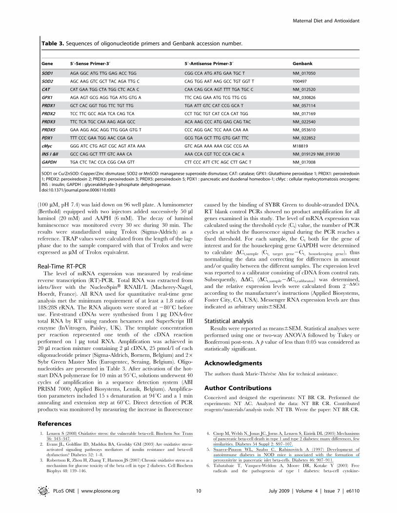
nucleotides are presented in Table 3. After activation of the hot-
start DNA polymerase for 10 min at 95uC, solutions underwent 40
cycles of amplification in a sequence detection system (ABI
PRISM 7000; Applied Biosystems, Lennik, Belgium). Amplifica-
tion parameters included 15 s denaturation at 94uC and a 1 min
annealing and extension step at 60uC. Direct detection of PCR
products was monitored by measuring the increase in fluorescence
caused by the binding of SYBR Green to double-stranded DNA.
RT blank control PCRs showed no product amplification for all
genes examined in this study. The level of mRNA expression was
calculated using the threshold cycle (Ct) value, the number of PCR
cycles at which the fluorescence signal during the PCR reaches a
fixed threshold. For each sample, the Ct both for the gene of
interest and for the housekeeping gene GAPDH were determined
to calculate DCt,sample (Ct, target gene2Ct, housekeeping gene), thus
normalizing the data and correcting for differences in amount
and/or quality between the different samples. The expression level
was reported to a calibrator consisting of cDNA from control rats.
Subsequently, DDCt (DCt,sample2DCt,calibrator) was determined,
and the relative expression levels were calculated from 22DDCt
according to the manufacturer’s instructions (Applied Biosystems,
Foster City, CA, USA). Messenger RNA expression levels are thus
indicated as arbitrary units6SEM.
Statistical analysisResults were reported as means6SEM. Statistical analyses were
performed using one or two-way ANOVA followed by Tukey or
Bonferroni post-tests. A p value of less than 0.05 was considered as
statistically significant.
Acknowledgments
The authors thank Marie-Therese Ahn for technical assistance.
Author Contributions
Conceived and designed the experiments: NT BR CR. Performed the
experiments: NT AC. Analyzed the data: NT BR CR. Contributed
reagents/materials/analysis tools: NT TB. Wrote the paper: NT BR CR.
References
1. Lenzen S (2008) Oxidative stress: the vulnerable beta-cell. Biochem Soc Trans
36: 343–347.
2. Evans JL, Goldfine ID, Maddux BA, Grodsky GM (2003) Are oxidative stress-
activated signaling pathways mediators of insulin resistance and beta-cell
dysfunction? Diabetes 52: 1–8.
3. Robertson R, Zhou H, Zhang T, Harmon JS (2007) Chronic oxidative stress as a
mechanism for glucose toxicity of the beta cell in type 2 diabetes. Cell Biochem
Biophys 48: 139–146.
4. Cnop M, Welsh N, Jonas JC, Jorns A, Lenzen S, Eizirik DL (2005) Mechanisms
of pancreatic beta-cell death in type 1 and type 2 diabetes: many differences, few
similarities. Diabetes 54 Suppl 2: S97–107.
5. Suarez-Pinzon WL, Szabo C, Rabinovitch A (1997) Development of
autoimmune diabetes in NOD mice is associated with the formation of
peroxynitrite in pancreatic islet beta-cells. Diabetes 46: 907–911.
6. Tabatabaie T, Vasquez-Weldon A, Moore DR, Kotake Y (2003) Free
radicals and the pathogenesis of type 1 diabetes: beta-cell cytokine-
Table 3. Sequences of oligonucleotide primers and Genbank accession number.
Gene 59-Sense Primer-39 59-Antisense Primer-39 Genbank
SOD1 AGA GGC ATG TTG GAG ACC TGG CGG CCA ATG ATG GAA TGC T NM_017050
SOD2 AGC AAG GTC GCT TAC AGA TTG C CAG TGG AAT AAG GCC TGT GGT T Y00497
CAT CAT GAA TGG CTA TGG CTC ACA C CAA CAG GCA AGT TTT TGA TGC C NM_012520
GPX1 AGA AGT GCG AGG TGA ATG GTG A TTC CAG GAA ATG TCG TTG CG NM_030826
PRDX1 GCT CAC GGT TGG TTC TGT TTG TGA ATT GTC CAT CCG GCA T NM_057114
PRDX2 TCC TTC GCC AGA TCA CAG TCA CCT TGC TGT CAT CCA CAT TGG NM_017169
PRDX3 TTC TCA TGC CAA AAG AGA GCC ACA AAG CCC ATG GAG CAG TAC NM_022540
PRDX5 GAA AGG AGC AGG TTG GGA GTG T CCC AGG GAC TCC AAA CAA AA NM_053610
PDX1 TTT CCC GAA TGG AAC CGA GA GCG TGA GCT TTG GTG GAT TTC NM_022852
cMyc GGG ATC CTG AGT CGC AGT ATA AAA GTC AGA AAA AAA CGC CCG AA M18819
INS I &II GCC CAG GCT TTT GTC AAA CA AAA CCA CGT TCC CCA CAC A NM_019129 NM_019130
GAPDH TGA CTC TAC CCA CGG CAA GTT CTT CCC ATT CTC AGC CTT GAC T NM_017008
SOD1 or Cu/ZnSOD: Copper/Zinc dismutase; SOD2 or MnSOD: managanese superoxide dismutase; CAT: catalase; GPX1: Glutathione peroxidase 1; PRDX1: peroxiredoxin1; PRDX2: peroxiredoxin 2; PRDX3: peroxiredoxin 3; PRDX5: peroxiredoxin 5; PDX1 : pancreatic and duodenal homeobox-1; cMyc : cellular myelocytomatosis oncogene;INS : insulin; GAPDH : glyceraldehyde-3-phosphate dehydrogenase.doi:10.1371/journal.pone.0006110.t003
Maternal Diet and Antioxidant
PLoS ONE | www.plosone.org 10 July 2009 | Volume 4 | Issue 7 | e6110
mediated free radical generation via cyclooxygenase-2. Diabetes 52:
1994–1999.7. Lenzen S, Drinkgern J, Tiedge M (1996) Low antioxidant enzyme gene
expression in pancreatic islets compared with various other mouse tissues. Free
Radic Biol Med 20: 463–466.8. Tiedge M, Lortz S, Drinkgern J, Lenzen S (1997) Relation between antioxidant
enzyme gene expression and antioxidative defense status of insulin-producingcells. Diabetes 46: 1733–1742.
9. Hales CN, Barker DJ, Clark PM, Cox LJ, Fall C, Osmond C, Winter PD (1991)
Fetal and infant growth and impaired glucose tolerance at age 64. BMJ 303:1019–1022.
10. Remacle C, Dumortier O, Bol V, Goosse K, Romanus P, Theys N,Bouckenooghe T, Reusens B (2007) Intrauterine programming of the endocrine
pancreas. Diabetes Obes Metab 9 Suppl 2: 196–209.11. Snoeck A, Remacle C, Reusens B, Hoet JJ (1990) Effect of a low protein diet
during pregnancy on the fetal rat endocrine pancreas. Biol Neonate 57:
107–118.12. Petrik J, Reusens B, Arany E, Remacle C, Coelho C, Hoet JJ, Hill DJ (1999) A
low protein diet alters the balance of islet cell replication and apoptosis in thefetal and neonatal rat and is associated with a reduced pancreatic expression of
insulin-like growth factor-II. Endocrinology 140: 4861–4873.
13. Boujendar S, Reusens B, Merezak S, Ahn MT, Arany E, Hill D, Remacle C(2002) Taurine supplementation to a low protein diet during foetal and early
postnatal life restores a normal proliferation and apoptosis of rat pancreaticislets. Diabetologia 45: 856–866.
14. Dumortier O, Blondeau B, Duvillie B, Reusens B, Breant B, Remacle C (2007)Different mechanisms operating during different critical time-windows reduce
rat fetal beta cell mass due to a maternal low-protein or low-energy diet.
Diabetologia 50: 2495–2503.15. Merezak S, Hardikar AA, Yajnik CS, Remacle C, Reusens B (2001) Intrauterine
low protein diet increases fetal beta-cell sensitivity to NO and IL-1 beta: theprotective role of taurine. J Endocrinol 171: 299–308.
16. Merezak S, Reusens B, Renard A, Goosse K, Kalbe L, Ahn MT, Tamarit-
Rodriguez J, Remacle C (2004) Effect of maternal low-protein diet and taurineon the vulnerability of adult Wistar rat islets to cytokines. Diabetologia 47:
669–675.17. Goosse K, Bouckenooghe T, Balteau M, Reusens B, Remacle C (2009)
Implication of nitric oxide in the increased islet-cells vulnerability of adultprogeny from protein-restricted mothers and its prevention by taurine.
J Endocrinol 200: 177–187.
18. Sparre T, Reusens B, Cherif H, Larsen MR, Roepstorff P, Fey SJ, Mose LP,Remacle C, Nerup J (2003) Intrauterine programming of fetal islet gene
expression in rats-effects of maternal protein restriction during gestation revealedby proteome analysis. Diabetologia 46: 1497–1511.
19. Reusens B, Sparre T, Kalbe L, Bouckenooghe T, Theys N, Kruhoffer M,
Orntoft TF, Nerup J, Remacle C (2008) The intrauterine metabolicenvironment modulates the gene expression pattern in fetal rat islets: prevention
by maternal taurine supplementation. Diabetologia 51: 836–845.20. Simmons RA (2006) Developmental origins of diabetes: the role of oxidative
stress. Free Radic Biol Med 40: 917–922.21. Simmons RA, Suponitsky-Kroyter I, Selak MA (2005) Progressive accumulation
of mitochondrial DNA mutations and decline in mitochondrial function lead to
beta-cell failure. J Biol Chem 280: 28785–28791.22. Robertson RP (2004) Chronic oxidative stress as a central mechanism for
glucose toxicity in pancreatic islet beta cells in diabetes. J Biol Chem 279:42351–42354.
23. Elouil H, Cardozo AK, Eizirik DL, Henquin JC, Jonas JC (2005) High glucose
and hydrogen peroxide increase c-Myc and haeme-oxygenase 1 mRNA levels inrat pancreatic islets without activating NFkappaB. Diabetologia 48: 496–505.
24. Gurgul E, Lortz S, Tiedge M, Jorns A, Lenzen S (2004) Mitochondrial catalaseoverexpression protects insulin-producing cells against toxicity of reactive oxygen
species and proinflammatory cytokines. Diabetes 53: 2271–2280.
25. Lortz S, Gurgul-Convey E, Lenzen S, Tiedge M (2005) Importance ofmitochondrial superoxide dismutase expression in insulin-producing cells for
the toxicity of reactive oxygen species and proinflammatory cytokines.Diabetologia 48: 1541–1548.
26. Tanaka Y, Tran PO, Harmon J, Robertson RP (2002) A role for glutathioneperoxidase in protecting pancreatic beta cells against oxidative stress in a model
of glucose toxicity. Proc Natl Acad Sci U S A 99: 12363–12368.
27. Pallardo FV, Sastre J, Asensi M, Rodrigo F, Estrela JM, Vina J (1991)Physiological changes in glutathione metabolism in foetal and newborn rat liver.
Biochem J 274(Pt 3): 891–893.28. Vento M, Asensi M, Sastre J, Lloret A, Garcia-Sala F, Vina J (2003) Oxidative
stress in asphyxiated term infants resuscitated with 100% oxygen. J Pediatr 142:
240–246.
29. Finegood DT, Scaglia L, Bonner-Weir S (1995) Dynamics of beta-cell mass in
the growing rat pancreas. Estimation with a simple mathematical model.Diabetes 44: 249–256.
30. Scaglia L, Cahill CJ, Finegood DT, Bonner-Weir S (1997) Apoptosis participates
in the remodeling of the endocrine pancreas in the neonatal rat. Endocrinology138: 1736–1741.
31. Petrik J, Arany E, McDonald TJ, Hill DJ (1998) Apoptosis in the pancreatic isletcells of the neonatal rat is associated with a reduced expression of insulin-like
growth factor II that may act as a survival factor. Endocrinology 139:2994–3004.
32. Trudeau JD, Dutz JP, Arany E, Hill DJ, Fieldus WE, Finegood DT (2000)
Neonatal beta-cell apoptosis: a trigger for autoimmune diabetes? Diabetes 49:1–7.
33. Robertson RP, Harmon JS (2007) Pancreatic islet beta-cell and oxidative stress:the importance of glutathione peroxidase. FEBS Lett 581: 3743–3748.
34. Chen H, Li X, Epstein PN (2005) MnSOD and catalase transgenes demonstratethat protection of islets from oxidative stress does not alter cytokine toxicity.
Diabetes 54: 1437–1446.
35. Izawa S, Inoue Y, Kimura A (1996) Importance of catalase in the adaptiveresponse to hydrogen peroxide: analysis of acatalasaemic Saccharomyces
cerevisiae. Biochem J 320(Pt 1): 61–67.
36. Rhee SG, Yang KS, Kang SW, Woo HA, Chang TS (2005) Controlled
elimination of intracellular H(2)O(2): regulation of peroxiredoxin, catalase, and
glutathione peroxidase via post-translational modification. Antioxid RedoxSignal 7: 619–626.
37. Kaneto H, Katakami N, Kawamori D, Miyatsuka T, Sakamoto K, et al. (2007)Involvement of oxidative stress in the pathogenesis of diabetes. Antioxid Redox
Signal 9: 355–366.
38. Wu L, Nicholson W, Knobel SM, Steffner RJ, May JM, Piston DW, Powers AC
(2004) Oxidative stress is a mediator of glucose toxicity in insulin-secreting
pancreatic islet cell lines. J Biol Chem 279: 12126–12134.
39. Ozanne SE, Smith GD, Tikerpae J, Hales CN (1996) Altered regulation of
hepatic glucose output in the male offspring of protein-malnourished rat dams.Am J Physiol 270: E559–E564.
40. Ozanne SE, Wang CL, Coleman N, Smith GD (1996) Altered muscle insulinsensitivity in the male offspring of protein-malnourished rats. Am J Physiol 271:
E1128–E1134.
41. Hales CN, Desai M, Ozanne SE, Crowther NJ (1996) Fishing in the stream ofdiabetes: from measuring insulin to the control of fetal organogenesis. Biochem
Soc Trans 24: 341–350.
42. Fernandez-Twinn DS, Wayman A, Ekizoglou S, Martin MS, Hales CN, et al.
(2005) Maternal protein restriction leads to hyperinsulinemia and reduced
insulin-signaling protein expression in 21-mo-old female rat offspring.Am J Physiol Regul Integr Comp Physiol 288: R368–R373.
43. Boujendar S, Arany E, Hill D, Remacle C, Reusens B (2003) Taurinesupplementation of a low protein diet fed to rat dams normalizes the
vascularization of the fetal endocrine pancreas. J Nutr 133: 2820–2825.
44. Gluckman PD, Lillycrop KA, Vickers MH, Pleasants AB, et al. (2007) Metabolic
plasticity during mammalian development is directionally dependent on early
nutritional status. Proc Natl Acad Sci U S A 104: 12796–12800.
45. Tarry-Adkins JL, Chen JH, Smith NS, Jones RH, Cherif H, et al. (2009) Poor
maternal nutrition followed by accelerated postnatal growth leads to telomereshortening and increased markers of cell senescence in rat islets. FASEB J.
46. Zraika S, Aston-Mourney K, Laybutt DR, Kebede M, et al. (2006) Theinfluence of genetic background on the induction of oxidative stress and
impaired insulin secretion in mouse islets. Diabetologia 49: 1254–1263.
47. Kim JA, Park S, Kim K, Rhee SG, Kang SW (2005) Activity assay ofmammalian 2-cys peroxiredoxins using yeast thioredoxin reductase system. Anal
Biochem 338: 216–223.
48. Laybutt DR, Weir GC, Kaneto H, Lebet J, Palmiter RD, et al. (2002)
Overexpression of c-Myc in beta-cells of transgenic mice causes proliferation and
apoptosis, downregulation of insulin gene expression, and diabetes. Diabetes 51:1793–1804.
49. Van de CM, Kefas BA, Cai Y, Heimberg H, Scott DK, et al. (2003) Prolongedculture in low glucose induces apoptosis of rat pancreatic beta-cells through
induction of c-myc. Biochem Biophys Res Commun 312: 937–944.
50. El KI, Remacle C, Reusens B (2006) The regulation of IGFs and IGFBPs by
prolactin in primary culture of fetal rat hepatocytes is influenced by maternal
malnutrition. Am J Physiol Endocrinol Metab 291: E835–E842.
51. Janssens BJ, Childress JJ, Baguet F, Rees JF (2000) Reduced enzymatic
antioxidative defense in deep-sea fish. J Exp Biol 203: 3717–3725.
52. Paglia DE, Valentine WN (1967) Studies on the quantitative and qualitative
characterization of erythrocyte glutathione peroxidase. J Lab Clin Med 70:158–169.
Maternal Diet and Antioxidant
PLoS ONE | www.plosone.org 11 July 2009 | Volume 4 | Issue 7 | e6110
Copyright © 2022 FDOKUMEN