Connexin 43 Reverses Malignant Phenotypes of Glioma Stem Cells by Modulating E-Cadherin
ARTICLE IN PRESS
0344-0338/$ - se
doi:10.1016/j.pr
�CorrespondiE-mail addre
Pathology – Research and Practice 204 (2008) 809–815
www.elsevier.de/prp
ORIGINAL ARTICLE
E-cadherin, b-catenin adhesion complex and relation to matrilysin
expression in pT3 rectosigmoid cancers
Fatma Husniye Dileka,�, Nevin Topaka, Fatma Aktepea, Onder S-ahina,Kadir Serkan Turelb, Dursun Ali S-ahinb, Osman Nuri Dilekb
aDepartment of Pathology, School of Medicine, Kocatepe University, PK: 70, 03100 Afyonkarahisar, TurkeybDepartment of Surgery, School of Medicine, Kocatepe University, PK: 70, 03100 Afyonkarahisar, Turkey
Received 13 February 2008; accepted 19 May 2008
Presented at the European Congress of Pathology, Istanbul, Turkey, September 8–13, 2007
Abstract
E-cadherin/b-catenin complex has a critical role in cell–cell adhesion. b-Catenin is a critical component of the highlyconserved Wnt signaling pathway that regulates cell proliferation and differentiation. Wnt signaling leads to thestabilization of cytosolic b-catenin and to translocation to the nucleus, where it binds with T-cell factor and promotesthe transcription and changes in target gene expression, including matrix metalloproteinases.
In this study, we analyzed paraffin-embedded specimens from 42 patients with pT3 rectosigmoid cancer forE-cadherin, b-catenin, and matrix metalloproteinase-7(MMP-7, matrilysin) expression using immunohistochemistry.Seventy-four and 79% of tumors expressed b-catenin and E-cadherin, respectively. Nuclear expression of b-cateninwas detected only in 26.1% of tumors. Forty-five percent of the rectosigmoid cancers showed strong expression ofMMP-7. It was revealed that membranous or cytoplasmic b-catenin expression was significantly related to E-cadherinand MMP-7 expression. No significant association was seen between E-cadherin, b-catenin, or MMP-7 expressionand some clinicopathologic features. Our results may contribute to the functional interaction between b-catenin andMMP-7. Further studies on Wnt/b-catenin and MMP-7 gene activity and protein expression are necessary to betterunderstand the pathogenesis of colorectal carcinoma.r 2008 Elsevier GmbH. All rights reserved.
Keywords: Colorectal carcinoma; b-catenin; E-cadherin; MMP-7
Introduction
Matrix metalloproteinases (MMPs) constitute anenzyme family capable of degrading extracellularmatrix. This group of enzymes has been extensivelystudied in both physiological and pathological condi-tions, where matrix degradation and matrix turnover are
e front matter r 2008 Elsevier GmbH. All rights reserved.
p.2008.05.010
ng author. Tel.: +90505 4004006.
ss: [email protected] (F.H. Dilek).
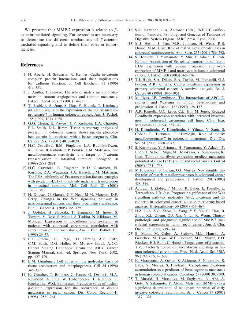
key features. Most notably, MMPs have been exten-sively investigated with regard to tumor invasion andmetastasis [2,27]. Matrix metalloproteinase-7 (MMP-7matrilysin), a member of MMPs, is thought to bedirectly involved in the processes of growth, invasion,metastasis, and prognosis of colorectal cancer[19,20,25,30] (Figs. 1 and 2).
E-cadherin is a transmembrane glycoprotein thataids in the facilitation of cell–cell adhesion processesvia association with cytoskeletal proteins, a-, b-,
ARTICLE IN PRESS
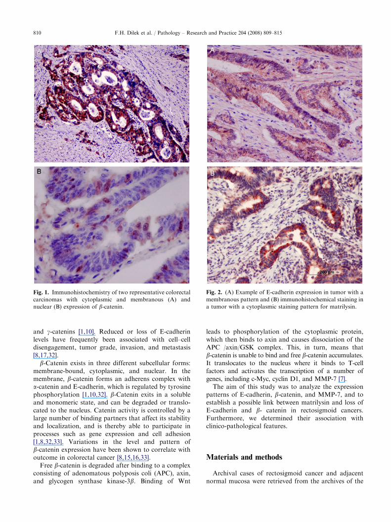
Fig. 1. Immunohistochemistry of two representative colorectal
carcinomas with cytoplasmic and membranous (A) and
nuclear (B) expression of b-catenin.
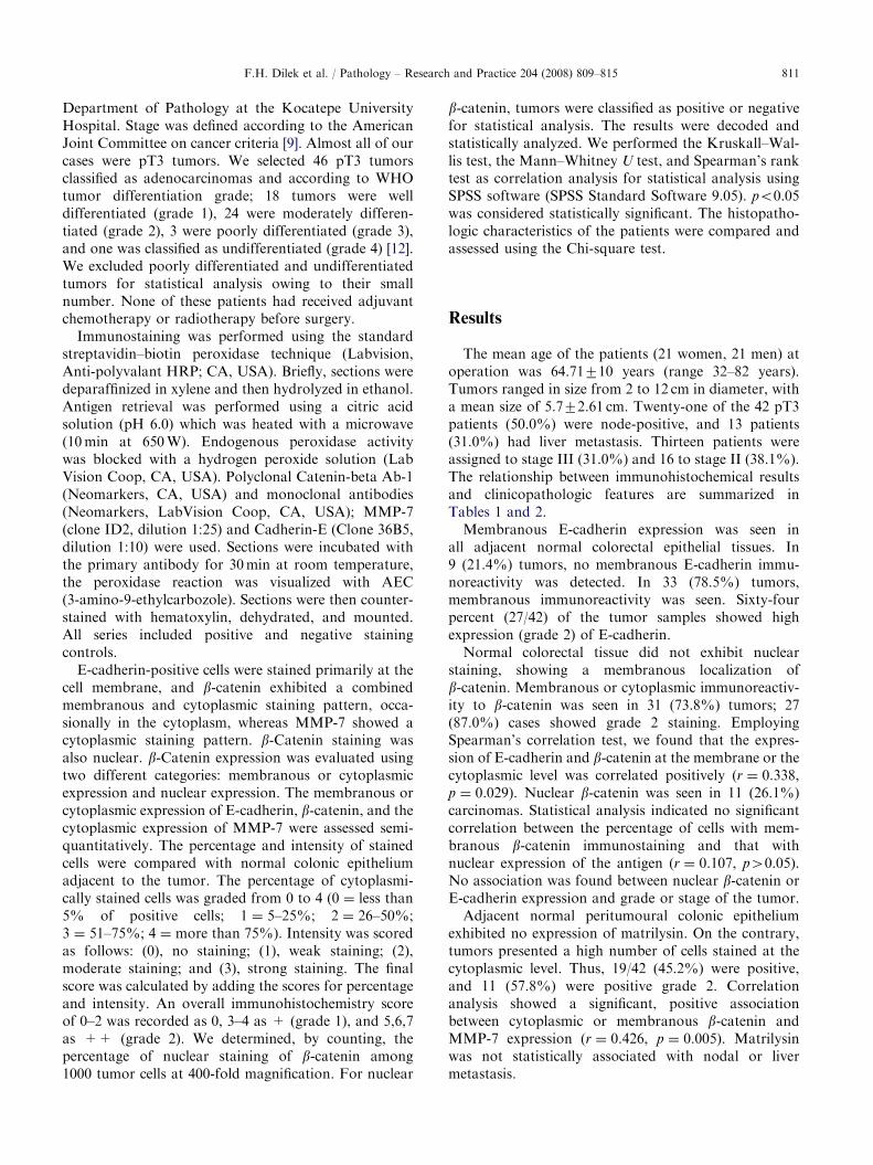
Fig. 2. (A) Example of E-cadherin expression in tumor with a
membranous pattern and (B) immunohistochemical staining in
a tumor with a cytoplasmic staining pattern for matrilysin.
F.H. Dilek et al. / Pathology – Research and Practice 204 (2008) 809–815810
and g-catenins [1,10]. Reduced or loss of E-cadherinlevels have frequently been associated with cell–celldisengagement, tumor grade, invasion, and metastasis[8,17,32].
b-Catenin exists in three different subcellular forms:membrane-bound, cytoplasmic, and nuclear. In themembrane, b-catenin forms an adherens complex witha-catenin and E-cadherin, which is regulated by tyrosinephosphorylation [1,10,32]. b-Catenin exits in a solubleand monomeric state, and can be degraded or translo-cated to the nucleus. Catenin activity is controlled by alarge number of binding partners that affect its stabilityand localization, and is thereby able to participate inprocesses such as gene expression and cell adhesion[1,8,32,33]. Variations in the level and pattern ofb-catenin expression have been shown to correlate withoutcome in colorectal cancer [8,15,16,33].
Free b-catenin is degraded after binding to a complexconsisting of adenomatous polyposis coli (APC), axin,and glycogen synthase kinase-3b. Binding of Wnt
leads to phosphorylation of the cytoplasmic protein,which then binds to axin and causes dissociation of theAPC /axin/GSK complex. This, in turn, means thatb-catenin is unable to bind and free b-catenin accumulates.It translocates to the nucleus where it binds to T-cellfactors and activates the transcription of a number ofgenes, including c-Myc, cyclin D1, and MMP-7 [7].
The aim of this study was to analyze the expressionpatterns of E-cadherin, b-catenin, and MMP-7, and toestablish a possible link between matrilysin and loss ofE-cadherin and b- catenin in rectosigmoid cancers.Furthermore, we determined their association withclinico-pathological features.
Materials and methods
Archival cases of rectosigmoid cancer and adjacentnormal mucosa were retrieved from the archives of the
ARTICLE IN PRESSF.H. Dilek et al. / Pathology – Research and Practice 204 (2008) 809–815 811
Department of Pathology at the Kocatepe UniversityHospital. Stage was defined according to the AmericanJoint Committee on cancer criteria [9]. Almost all of ourcases were pT3 tumors. We selected 46 pT3 tumorsclassified as adenocarcinomas and according to WHOtumor differentiation grade; 18 tumors were welldifferentiated (grade 1), 24 were moderately differen-tiated (grade 2), 3 were poorly differentiated (grade 3),and one was classified as undifferentiated (grade 4) [12].We excluded poorly differentiated and undifferentiatedtumors for statistical analysis owing to their smallnumber. None of these patients had received adjuvantchemotherapy or radiotherapy before surgery.
Immunostaining was performed using the standardstreptavidin–biotin peroxidase technique (Labvision,Anti-polyvalant HRP; CA, USA). Briefly, sections weredeparaffinized in xylene and then hydrolyzed in ethanol.Antigen retrieval was performed using a citric acidsolution (pH 6.0) which was heated with a microwave(10min at 650W). Endogenous peroxidase activitywas blocked with a hydrogen peroxide solution (LabVision Coop, CA, USA). Polyclonal Catenin-beta Ab-1(Neomarkers, CA, USA) and monoclonal antibodies(Neomarkers, LabVision Coop, CA, USA); MMP-7(clone ID2, dilution 1:25) and Cadherin-E (Clone 36B5,dilution 1:10) were used. Sections were incubated withthe primary antibody for 30min at room temperature,the peroxidase reaction was visualized with AEC(3-amino-9-ethylcarbozole). Sections were then counter-stained with hematoxylin, dehydrated, and mounted.All series included positive and negative stainingcontrols.
E-cadherin-positive cells were stained primarily at thecell membrane, and b-catenin exhibited a combinedmembranous and cytoplasmic staining pattern, occa-sionally in the cytoplasm, whereas MMP-7 showed acytoplasmic staining pattern. b-Catenin staining wasalso nuclear. b-Catenin expression was evaluated usingtwo different categories: membranous or cytoplasmicexpression and nuclear expression. The membranous orcytoplasmic expression of E-cadherin, b-catenin, and thecytoplasmic expression of MMP-7 were assessed semi-quantitatively. The percentage and intensity of stainedcells were compared with normal colonic epitheliumadjacent to the tumor. The percentage of cytoplasmi-cally stained cells was graded from 0 to 4 (0 ¼ less than5% of positive cells; 1 ¼ 5–25%; 2 ¼ 26–50%;3 ¼ 51–75%; 4 ¼ more than 75%). Intensity was scoredas follows: (0), no staining; (1), weak staining; (2),moderate staining; and (3), strong staining. The finalscore was calculated by adding the scores for percentageand intensity. An overall immunohistochemistry scoreof 0–2 was recorded as 0, 3–4 as + (grade 1), and 5,6,7as ++ (grade 2). We determined, by counting, thepercentage of nuclear staining of b-catenin among1000 tumor cells at 400-fold magnification. For nuclear
b-catenin, tumors were classified as positive or negativefor statistical analysis. The results were decoded andstatistically analyzed. We performed the Kruskall–Wal-lis test, the Mann–Whitney U test, and Spearman’s ranktest as correlation analysis for statistical analysis usingSPSS software (SPSS Standard Software 9.05). po0.05was considered statistically significant. The histopatho-logic characteristics of the patients were compared andassessed using the Chi-square test.
Results
The mean age of the patients (21 women, 21 men) atoperation was 64.71710 years (range 32–82 years).Tumors ranged in size from 2 to 12 cm in diameter, witha mean size of 5.772.61 cm. Twenty-one of the 42 pT3patients (50.0%) were node-positive, and 13 patients(31.0%) had liver metastasis. Thirteen patients wereassigned to stage III (31.0%) and 16 to stage II (38.1%).The relationship between immunohistochemical resultsand clinicopathologic features are summarized inTables 1 and 2.
Membranous E-cadherin expression was seen inall adjacent normal colorectal epithelial tissues. In9 (21.4%) tumors, no membranous E-cadherin immu-noreactivity was detected. In 33 (78.5%) tumors,membranous immunoreactivity was seen. Sixty-fourpercent (27/42) of the tumor samples showed highexpression (grade 2) of E-cadherin.
Normal colorectal tissue did not exhibit nuclearstaining, showing a membranous localization ofb-catenin. Membranous or cytoplasmic immunoreactiv-ity to b-catenin was seen in 31 (73.8%) tumors; 27(87.0%) cases showed grade 2 staining. EmployingSpearman’s correlation test, we found that the expres-sion of E-cadherin and b-catenin at the membrane or thecytoplasmic level was correlated positively (r ¼ 0.338,p ¼ 0.029). Nuclear b-catenin was seen in 11 (26.1%)carcinomas. Statistical analysis indicated no significantcorrelation between the percentage of cells with mem-branous b-catenin immunostaining and that withnuclear expression of the antigen (r ¼ 0.107, p40.05).No association was found between nuclear b-catenin orE-cadherin expression and grade or stage of the tumor.
Adjacent normal peritumoural colonic epitheliumexhibited no expression of matrilysin. On the contrary,tumors presented a high number of cells stained at thecytoplasmic level. Thus, 19/42 (45.2%) were positive,and 11 (57.8%) were positive grade 2. Correlationanalysis showed a significant, positive associationbetween cytoplasmic or membranous b-catenin andMMP-7 expression (r ¼ 0.426, p ¼ 0.005). Matrilysinwas not statistically associated with nodal or livermetastasis.
ARTICLE IN PRESS
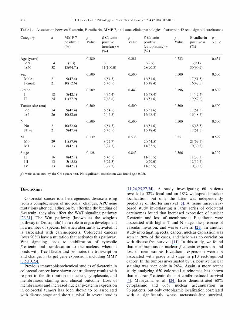
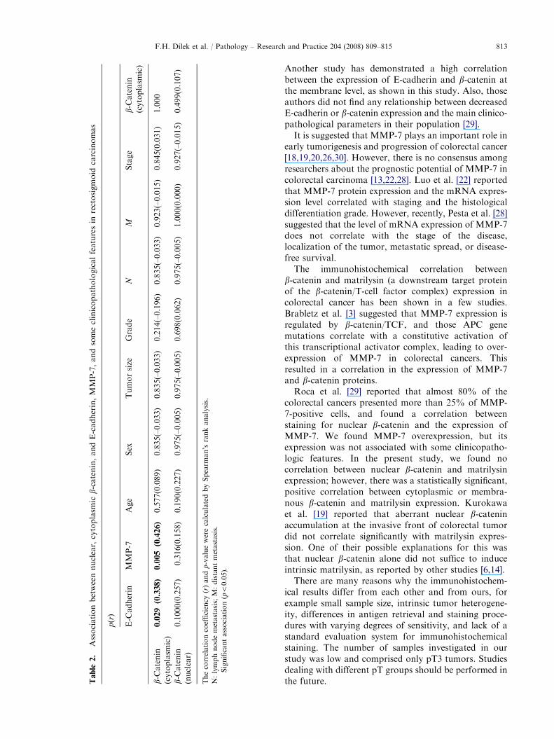
Table 1. Association between b-catenin, E-cadherin, MMP-7, and some clinicopathological features in 42 rectosigmoid carcinomas
Category n MMP-7
positive n
(%)
p-
Value
b-Cateninpositive
(nuclear) n
(%)
p-
Value
b-Cateninpositive
(cytoplasmic) n
(%)
p-
Value
E-cadherin
positive n
(%)
p-
Value
Age (years) 0.380 0.281 0.723 0.634
o50 4 1(5.3) 0 3(9.7) 3(9.1)
X50 38 18(94.7.) 11(100.0) 28(90.3) 30(90.9)
Sex 0.500 0.500 0.500 0.500
Male 21 9(47.4) 6(54.5) 16(51.6) 17(51.5)
Female 21 10(52.6) 5(45.5) 15(48.4) 16(48.5)
Grade 0.589 0.443 0.196 0.602
I 18 8(42.1) 4(36.4) 15(48.4) 14(42.4)
II 24 11(57.9) 7(63.6) 16(51.6) 19(57.6)
Tumor size (cm) 0.500 0.500 0.500 0.500
o5 14 9(47.4) 6(54.5) 16(51.6) 17(51.5)
X5 26 10(52.6) 5(45.5) 15(48.4) 16(48.5)
N 0.500 0.500 0.500 0.500
N0 21 10(52.6) 6(54.5) 16(51.6) 16(48.5)
N1–2 21 9(47.4) 5(45.5) 15(48.4) 17(51.5)
M 0.139 0.538 0.251 0.579
M0 29 11(57.9) 8(72.7) 20(64.5) 23(69.7)
M1 13 8(42.1) 3(27.3) 11(35.5) 10(30.3)
Stage 0.128 0.843 0.566 0.302
II 16 8(42.1) 5(45.5) 11(35.5) 11(33.3)
III 13 3(15.8) 3(27.3) 9(29.0) 12(36.4)
IV 13 8(42.1) 3(27.3) 11(35.5) 10(30.3)
p’s were calculated by the Chi-square test. No significant association was found (p40.05).
F.H. Dilek et al. / Pathology – Research and Practice 204 (2008) 809–815812
Discussion
Colorectal cancer is a heterogeneous disease arisingfrom a complex series of molecular changes. APC genemutations alter cell adhesion by affecting the binding ofb-catenin; they also affect the WnT signaling pathway[26,31]. The Wnt pathway (known as the winglesspathway in Drosophila) has a role in organ developmentin a number of species, but when aberrantly activated, itis associated with carcinogenesis. Colorectal cancers(over 90%) have a mutation that activates this pathway.Wnt signaling leads to stabilization of cytosolicb-catenin and translocation to the nucleus, where itbinds with T-cell factor and promotes the transcriptionand changes in target gene expression, including MMP[3,5,10,23].
Previous immunohistochemical studies of b-catenin incolorectal cancer have shown contradictory results withrespect to the distribution of nuclear, cytoplasmic, andmembranous staining and clinical outcome. Loss ofmembranous and increased nuclear b-catenin expressionin colorectal tumors has been shown to be associatedwith disease stage and short survival in several studies
[11,24,25,27,34]. A study investigating 60 patientsrevealed a 32% focal and an 18% widespread nuclearlocalization, but only the latter was independentlypredictive of shorter survival [5]. A tissue microarray-based study investigating a large series of colorectalcarcinomas found that increased expression of nuclearb-catenin and loss of membranous E-cadherin wereassociated with higher T and N stage, the presence ofvascular invasion, and worse survival [21]. In anotherstudy investigating rectal cancer, nuclear expression wasseen in 20% of the cases, and there was no correlationwith disease-free survival [11]. In this study, we foundthat membranous or nuclear b-catenin expression andloss of membranous E-cadherin expression were notassociated with grade and stage in pT3 rectosigmoidcancer. In the tumors investigated by us, positive nuclearstaining was seen only in 26%. Again, a more recentstudy analyzing 650 colorectal carcinomas has shownthat nuclear b-catenin did not confer reduced survival[4]. Maruyama et al. [24] have demonstrated 68%cytoplasmic and 66% nuclear accumulation in96 patients, but only cytoplasmic localization correlatedwith a significantly worse metastasis-free survival.
ARTICLE IN PRESS
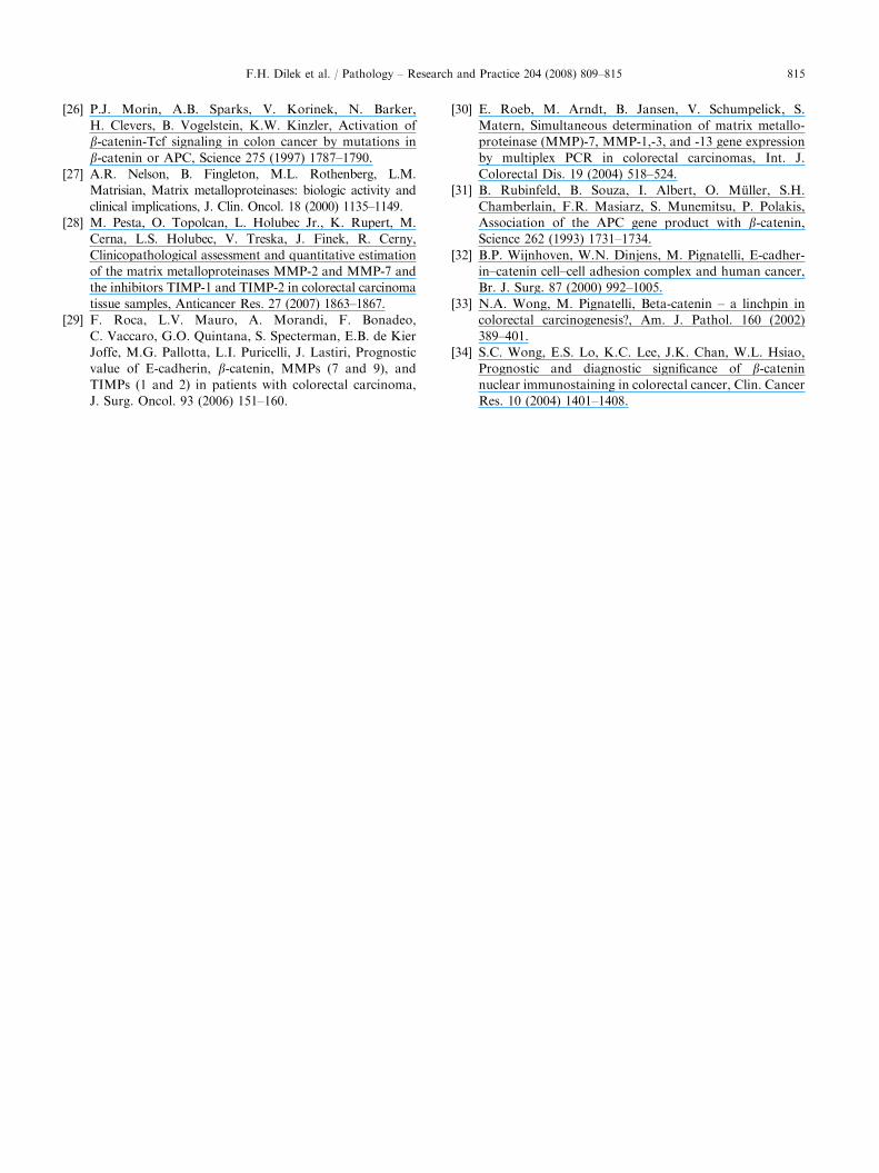
Table
2.
Associationbetweennuclear,cytoplasm
icb-catenin,andE-cadherin,MMP-7,andsomeclinicopathologicalfeaturesin
rectosigmoid
carcinomas
p(r)
E-C
adherin
MMP-7
Age
Sex
Tumorsize
Grade
NM
Stage
b-Catenin
(cytoplasm
ic)
b-Catenin
(cytoplasm
ic)
0.029� (0.338)
0.005� (0.426)
0.577(0.089)
0.835(–0.033)
0.835(–0.033)
0.214(–0.196)
0.835(–0.033)
0.923(–0.015)
0.845(0.031)
1.000
b-Catenin
(nuclear)
0.1000(0.257)
0.316(0.158)
0.190(0.227)
0.975(–0.005)
0.975(–0.005)
0.698(0.062)
0.975(–0.005)
1.000(0.000)
0.927(–0.015)
0.499(0.107)
Thecorrelationcoefficiency
(r)and
p-valuewerecalculatedbySpearm
an’srankanalysis.
N:lymphnodemetastasis;M:distantmetastasis.
�Significantassociation(po0.05).
F.H. Dilek et al. / Pathology – Research and Practice 204 (2008) 809–815 813
Another study has demonstrated a high correlationbetween the expression of E-cadherin and b-catenin atthe membrane level, as shown in this study. Also, thoseauthors did not find any relationship between decreasedE-cadherin or b-catenin expression and the main clinico-pathological parameters in their population [29].
It is suggested that MMP-7 plays an important role inearly tumorigenesis and progression of colorectal cancer[18,19,20,26,30]. However, there is no consensus amongresearchers about the prognostic potential of MMP-7 incolorectal carcinoma [13,22,28]. Luo et al. [22] reportedthat MMP-7 protein expression and the mRNA expres-sion level correlated with staging and the histologicaldifferentiation grade. However, recently, Pesta et al. [28]suggested that the level of mRNA expression of MMP-7does not correlate with the stage of the disease,localization of the tumor, metastatic spread, or disease-free survival.
The immunohistochemical correlation betweenb-catenin and matrilysin (a downstream target proteinof the b-catenin/T-cell factor complex) expression incolorectal cancer has been shown in a few studies.Brabletz et al. [3] suggested that MMP-7 expression isregulated by b-catenin/TCF, and those APC genemutations correlate with a constitutive activation ofthis transcriptional activator complex, leading to over-expression of MMP-7 in colorectal cancers. Thisresulted in a correlation in the expression of MMP-7and b-catenin proteins.
Roca et al. [29] reported that almost 80% of thecolorectal cancers presented more than 25% of MMP-7-positive cells, and found a correlation betweenstaining for nuclear b-catenin and the expression ofMMP-7. We found MMP-7 overexpression, but itsexpression was not associated with some clinicopatho-logic features. In the present study, we found nocorrelation between nuclear b-catenin and matrilysinexpression; however, there was a statistically significant,positive correlation between cytoplasmic or membra-nous b-catenin and matrilysin expression. Kurokawaet al. [19] reported that aberrant nuclear b-cateninaccumulation at the invasive front of colorectal tumordid not correlate significantly with matrilysin expres-sion. One of their possible explanations for this wasthat nuclear b-catenin alone did not suffice to induceintrinsic matrilysin, as reported by other studies [6,14].
There are many reasons why the immunohistochem-ical results differ from each other and from ours, forexample small sample size, intrinsic tumor heterogene-ity, differences in antigen retrieval and staining proce-dures with varying degrees of sensitivity, and lack of astandard evaluation system for immunohistochemicalstaining. The number of samples investigated in ourstudy was low and comprised only pT3 tumors. Studiesdealing with different pT groups should be performed inthe future.
ARTICLE IN PRESSF.H. Dilek et al. / Pathology – Research and Practice 204 (2008) 809–815814
We presume that MMP-7 expression is related to b-catenin-mediated signaling. Future studies are necessaryto determine the different mechanisms of b-catenin-mediated signaling and to define their roles in tumor-igenesis.
References
[1] H. Aberle, H. Schwartz, R. Kemler, Cadherin–catenin
complex: protein interactions and their implications
for cadherin function, J. Cell Biochem. 61 (1996)
514–523.
[2] J. Anitha, T. George, The role of matrix metalloprotei-
nases in tumour angiogenesis and tumour metastasis,
Pathol. Oncol. Res. 7 (2001) 14–23.
[3] T. Brabletz, A. Jung, S. Dag, F. Hlubek, T. Kirchner,
b-Catenin regulates the expression of the matrix metallo-
proteinase-7 in human colorectal cancer, Am. J. Pathol.
155 (1999) 1033–1038.
[4] G.G. Chung, E. Provost, E.P. Kielhorn, L.A. Charette,
B.L. Smith, D.L. Rimm, Tissue microarray analysis of
b-catenin in colorectal cancer shows nuclear phospho-
beta-catenin is associated with a better prognosis, Clin.
Cancer Res. 7 (2001) 4013–4020.
[5] H.C. Crawford, B.M. Fingleton, L.A. Rudolph-Owen,
K.J. Goss, B. Rubinfeld, P. Polakis, L.M. Matrisian, The
metalloproteinase matrilysin is a target of b-catenintransactivation in intestinal tumours, Oncogene 18
(1999) 2883–2891.
[6] H.C. Crawford, B. Fingleton, M.D. Gustavson, N.
Kurpios, R.A. Wagenaar, J.A. Hassell, L.M. Matrisian,
The PEA subfamily of Ets transcription factors synergies
with b-catenin-LEF-1 to activate matrilysin transcription
in intestinal tumours, Mol. Cell Biol. 21 (2001)
1370–1383.
[7] H. Doucas, G. Garcea, C.P. Neal, M.M. Manson, D.P.
Berry, Changes in the Wnt signalling pathway in
gastrointestinal cancers and their prognostic significance,
Eur. J. Cancer 41 (2005) 365–379.
[8] J. Gofuku, H. Shiozaki, T. Tsujinaka, M. Inoue, S.
Tamura, Y. Doki, S. Matsui, S. Tsukita, N. Kikkawa, M.
Monden, Expression of E-cadherin and a-catenin in
patients with colorectal carcinoma: correlation with
cancer invasion and metastasis, Am. J. Clin. Pathol. 111
(1999) 29–37.
[9] F.L. Greene, D.L. Page, I.D. Fleming, A.G. Fritz,
C.M. Balch, D.G. Haller, M. Morrow (Eds.), AJCC:
Cancer Staging Handbook: From the AJCC Cancer
Staging Manual, sixth ed, Springer, New York, 2002,
pp. 127–129.
[10] B.M. Gumbiner, Cell adhesion: the molecular basis of
tissue architecture and morphogenesis, Cell 84 (1996)
345–357.
[11] K. Gunther, T. Brabletz, C. Kraus, O. Dworak, M.A.
Reymond, A. Jung, W. Hohenberger, T. Kirchner, F.
Kockerling, W.G. Ballhausen, Predictive value of nuclear
b-catenin expression for the occurrence of distant
metastases in rectal cancer, Dis. Colon Rectum 41
(1998) 1256–1261.
[12] S.R. Hamilton, L.A. Aaltonen (Eds.), WHO Classifica-
tion of Tumours. Pathology and Genetics of Tumours of
Digestive System Organs, IARC press, Lyon, 2000.
[13] M.J. Heslin, J. Yan, M.R. Johnson, H. Weiss, R.B.
Diasio, M.M. Urist, Role of matrix metalloproteinases in
colorectal carcinogenesis, Ann. Surg. 233 (2001) 786–792.
[14] S. Horiuchi, H. Yamamoto, Y. Min, Y. Adachi, F. Itoh,
K. Imai, Association of Ets-related transcriptional factor
E1AF expression with tumour progression and over-
expression of MMP-1 and matrilysin in human colorectal
cancer, J. Pathol. 200 (2003) 568–576.
[15] T.J. Hugh, S.A. Dillon, B.A. Taylor, M. Pignatelli, G.J.
Poston, A.R. Kinsella, Cadherin–catenin expression in
primary colorectal cancer: A survival analysis, Br. J.
Cancer 80 (1999) 1046–1051.
[16] M. Ilyas, I.P. Tomlinson, The interactions of APC, E-
cadherin and b-catenin in tumour development and
progression, J. Pathol. 182 (1997) 128–137.
[17] A.R. Kinsella, G.C. Lepts, C.L. Hill, M. Jones, Reduced
E-cadherin expression correlates with increased invasive-
ness in colorectal carcinoma cell lines, Clin. Exp.
Metastasis 12 (1994) 335–342.
[18] H. Kirimlioglu, V. Kirimlioglu, S. Yilmaz, V. Sagir, S.
Coban, E. Turkmen, F. Hilmioglu, Role of matrix
metalloproteinase-7 in colorectal adenomas, Dig. Dis.
Sci. 51 (2006) 2068–2072.
[19] S. Kurokawa, Y. Arimura, H. Yamamoto, Y. Adachi, T.
Endo, T. Sato, T. Suga, M. Hosokawa, Y. Shinomura, K.
Imai, Tumour matrilysin expression predicts metastatic
potential of stage I (pT1) colon and rectal cancers, Gut 54
(2005) 1751–1758.
[20] M.F. Leeman, S. Curran, G.I. Murray, New insights into
the roles of matrix metalloproteinases in colorectal cancer
development and progression, J. Pathol. 201 (2003)
528–534.
[21] A. Lugli, I. Zlobec, P. Minoo, K. Baker, L. Tornillo, L.
Terracciano, J.R. Jass, Prognostic significance of the Wnt
signalling pathway molecules APC, b-catenin and E-
cadherin in colorectal cancer: a tissue microarray-based
analysis, Histopathology 50 (2007) 453–464.
[22] H.Z. Luo, Z.G. Zhou, L. Yang, Y.Y. Yu, C. Tian, B.
Zhou, X.L. Zheng, Q.J. Xia, Y. Li, R. Wang, Clinico-
pathologic and prognostic significance of MMP-7 (ma-
trilysin) expression in human rectal cancer, Jpn. J. Clin.
Oncol. 35 (2005) 739–744.
[23] B. Mann, M. Gelos, A. Siedow, M.L. Hanski, A.
Gratchev, M. Ilyas, W.F. Bodmer, M.P. Moyer, E.O.
Riecken, H.J. Buhr, C. Hanski, Target genes of b-catenin-T cell factor/lymphoid-enhancer-factor signaling in hu-
man colorectal carcinomas, Proc. Natl. Acad. Sci. USA
96 (1999) 1603–1608.
[24] K. Maruyama, A. Ochiai, S. Akimoto, S. Nakamura, S.
Baba, Y. Moriya, S. Hirohashi, Cytoplasmic b-cateninaccumulation as a predictor of hematogenous metastasis
in human colorectal cancer, Oncology 59 (2000) 302–309.
[25] T. Masaki, H. Matsuoka, M. Sugiyama, N. Abe, A.
Goto, A. Sakamoto, Y. Atomi, Matrilysin (MMP-7) as a
significant determinant of malignant potential of early
invasive colorectal carcinomas, Br. J. Cancer 84 (2001)
1317–1321.
ARTICLE IN PRESSF.H. Dilek et al. / Pathology – Research and Practice 204 (2008) 809–815 815
[26] P.J. Morin, A.B. Sparks, V. Korinek, N. Barker,
H. Clevers, B. Vogelstein, K.W. Kinzler, Activation of
b-catenin-Tcf signaling in colon cancer by mutations in
b-catenin or APC, Science 275 (1997) 1787–1790.
[27] A.R. Nelson, B. Fingleton, M.L. Rothenberg, L.M.
Matrisian, Matrix metalloproteinases: biologic activity and
clinical implications, J. Clin. Oncol. 18 (2000) 1135–1149.
[28] M. Pesta, O. Topolcan, L. Holubec Jr., K. Rupert, M.
Cerna, L.S. Holubec, V. Treska, J. Finek, R. Cerny,
Clinicopathological assessment and quantitative estimation
of the matrix metalloproteinases MMP-2 and MMP-7 and
the inhibitors TIMP-1 and TIMP-2 in colorectal carcinoma
tissue samples, Anticancer Res. 27 (2007) 1863–1867.
[29] F. Roca, L.V. Mauro, A. Morandi, F. Bonadeo,
C. Vaccaro, G.O. Quintana, S. Specterman, E.B. de Kier
Joffe, M.G. Pallotta, L.I. Puricelli, J. Lastiri, Prognostic
value of E-cadherin, b-catenin, MMPs (7 and 9), and
TIMPs (1 and 2) in patients with colorectal carcinoma,
J. Surg. Oncol. 93 (2006) 151–160.
[30] E. Roeb, M. Arndt, B. Jansen, V. Schumpelick, S.
Matern, Simultaneous determination of matrix metallo-
proteinase (MMP)-7, MMP-1,-3, and -13 gene expression
by multiplex PCR in colorectal carcinomas, Int. J.
Colorectal Dis. 19 (2004) 518–524.
[31] B. Rubinfeld, B. Souza, I. Albert, O. Muller, S.H.
Chamberlain, F.R. Masiarz, S. Munemitsu, P. Polakis,
Association of the APC gene product with b-catenin,Science 262 (1993) 1731–1734.
[32] B.P. Wijnhoven, W.N. Dinjens, M. Pignatelli, E-cadher-
in–catenin cell–cell adhesion complex and human cancer,
Br. J. Surg. 87 (2000) 992–1005.
[33] N.A. Wong, M. Pignatelli, Beta-catenin – a linchpin in
colorectal carcinogenesis?, Am. J. Pathol. 160 (2002)
389–401.
[34] S.C. Wong, E.S. Lo, K.C. Lee, J.K. Chan, W.L. Hsiao,
Prognostic and diagnostic significance of b-cateninnuclear immunostaining in colorectal cancer, Clin. Cancer
Res. 10 (2004) 1401–1408.
Copyright © 2022 FDOKUMEN