Dual-purpose sample trap for on-line strong cation-exchange chromatography/reversed-phase liquid...
8
Journal of Chromatography A, 1070 (2005) 193–200 Dual-purpose sample trap for on-line strong cation-exchange chromatography/reversed-phase liquid chromatography/tandem mass spectrometry for shotgun proteomics Application to the human Jurkat T-cell proteome Dukjin Kang a , Hyungwook Nam b , Yu-Sam Kim b , Myeong Hee Moon a, ∗ a Department of Chemistry, Yonsei University, Seoul 120-749, South Korea b Protein Network Research Center (PNRC), Department of Biochemistry, Yonsei University, Seoul 120-749, South Korea Received 23 October 2004; received in revised form 2 February 2005; accepted 16 February 2005 Available online 8 March 2005 Abstract A dual-purpose sample-trapping column is introduced for the capacity enhancement of proteome analysis in on-line two-dimensional nanoflow liquid chromatography (strong cation-exchange chromatography followed by reversed-phase liquid chromatography) and tandem mass spectrometry. A home-made dual trap is prepared by sequentially packing C 18 reversed-phase (RP) particles and SCX resin in a silica capillary tubing (1.5 cm × 200 m I.D. for SCX, 0.7 cm × 200 m for RP) ended with a home-made frit and is connected to a nanoflow column having a pulled tip treated with an end frit. Without having a separate fraction collection and concentration process, digested peptide mixtures were loaded directly in the SCX part of the dual trap, and the SCX separation of peptides was performed with a salt step elution initiated by injecting only 8 L of NH 4 HCO 3 solution from the autosampler to the dual trap. The fractionated peptides at each salt step were directly transferred to the RP trap packed right next to the SCX part for desalting, and a nanoflow LC–MS–MS run was followed. During the sample loading–SCX fractionation–desalting, flow direction was set to bypass the analytical column to prevent contamination. The entire 2D-LC separation and MS–MS analysis were automated. Evaluation of the technique was made with an injection of 15 g peptide mixtures from human Jurkat T-cell proteome, and the total seven salt step cycles followed by each RPLC run resulted in an identification of 681 proteins. © 2005 Elsevier B.V. All rights reserved. Keywords: Proteomics; Liquid chromatography–mass spectrometry; Two-dimensional; Dual trap; Nanoflow liquid chromatography; Tandem mass spectrometry; Jurkat T-cells 1. Introduction Proteome analysis requires a comprehensive use of tech- niques including protein/peptide separation, mass spectro- metric characterization, and bioinformatics for identification. Recent advances in mass spectrometry have boosted the pos- sibility of characterizing complex mixtures of proteins and biomolecules [1,2]. Especially, tandem mass spectrometry (MS) has become a reliable and highly selective method for ∗ Corresponding author. Fax: +82 2 364 7050. E-mail address: [email protected] (M.H. Moon). characterizing peptides/proteins [3,4]. Prior to mass spectro- metric analysis, complicated proteins/peptides mixtures are required to be separated or simplified by some means. Among many separation techniques available, two-dimensional elec- trophoresis (2DE) has been widely used to isolate proteins from mixtures based on the differences in molar mass and pI [5–7]. While 2DE has been extensively utilized due to the simplicity and the advantages in isolating thousands of protein spots, it usually requires a large amount of sample loading, has difficulties in handling hydrophobic proteins and in quantitation, and is labor intensive [7–9]. Recently, liquid chromatography has been combined with mass spectrometry 0021-9673/$ – see front matter © 2005 Elsevier B.V. All rights reserved. doi:10.1016/j.chroma.2005.02.058
-
Upload
independent -
Category
Documents
-
view
1 -
download
0
Transcript of Dual-purpose sample trap for on-line strong cation-exchange chromatography/reversed-phase liquid...

Journal of Chromatography A, 1070 (2005) 193–200
Dual-purpose sample trap for on-line strong cation-exchangechromatography/reversed-phase liquid chromatography/tandem
mass spectrometry for shotgun proteomicsApplication to the human Jurkat T-cell proteome
Dukjin Kanga, Hyungwook Namb, Yu-Sam Kimb, Myeong Hee Moona, ∗a Department of Chemistry, Yonsei University, Seoul 120-749, South Korea
b Protein Network Research Center (PNRC), Department of Biochemistry, Yonsei University, Seoul 120-749, South Korea
Received 23 October 2004; received in revised form 2 February 2005; accepted 16 February 2005Available online 8 March 2005
Abstract
mensionaln nd tandemm ilicac lumnh e mixturesw n initiatedb directlyt e samplel ire 2D-LCsh proteins.©
K ectrometry;J
1
nmRsb(
o-s are
ongelec-teinsandtos of
mpleand
metry
0d
A dual-purpose sample-trapping column is introduced for the capacity enhancement of proteome analysis in on-line two-dianoflow liquid chromatography (strong cation-exchange chromatography followed by reversed-phase liquid chromatography) aass spectrometry. A home-made dual trap is prepared by sequentially packing C18 reversed-phase (RP) particles and SCX resin in a s
apillary tubing (1.5 cm× 200�m I.D. for SCX, 0.7 cm× 200�m for RP) ended with a home-made frit and is connected to a nanoflow coaving a pulled tip treated with an end frit. Without having a separate fraction collection and concentration process, digested peptidere loaded directly in the SCX part of the dual trap, and the SCX separation of peptides was performed with a salt step elutioy injecting only 8�L of NH4HCO3 solution from the autosampler to the dual trap. The fractionated peptides at each salt step were
ransferred to the RP trap packed right next to the SCX part for desalting, and a nanoflow LC–MS–MS run was followed. During thoading–SCX fractionation–desalting, flow direction was set to bypass the analytical column to prevent contamination. The enteparation and MS–MS analysis were automated. Evaluation of the technique was made with an injection of 15�g peptide mixtures fromuman Jurkat T-cell proteome, and the total seven salt step cycles followed by each RPLC run resulted in an identification of 6812005 Elsevier B.V. All rights reserved.
eywords:Proteomics; Liquid chromatography–mass spectrometry; Two-dimensional; Dual trap; Nanoflow liquid chromatography; Tandem mass spurkat T-cells
. Introduction
Proteome analysis requires a comprehensive use of tech-iques including protein/peptide separation, mass spectro-etric characterization, and bioinformatics for identification.ecent advances in mass spectrometry have boosted the pos-ibility of characterizing complex mixtures of proteins andiomolecules[1,2]. Especially, tandem mass spectrometryMS) has become a reliable and highly selective method for
∗ Corresponding author. Fax: +82 2 364 7050.E-mail address:[email protected] (M.H. Moon).
characterizing peptides/proteins[3,4]. Prior to mass spectrmetric analysis, complicated proteins/peptides mixturerequired to be separated or simplified by some means. Ammany separation techniques available, two-dimensionaltrophoresis (2DE) has been widely used to isolate profrom mixtures based on the differences in molar masspI [5–7]. While 2DE has been extensively utilized duethe simplicity and the advantages in isolating thousandprotein spots, it usually requires a large amount of saloading, has difficulties in handling hydrophobic proteinsin quantitation, and is labor intensive[7–9]. Recently, liquidchromatography has been combined with mass spectro
021-9673/$ – see front matter © 2005 Elsevier B.V. All rights reserved.oi:10.1016/j.chroma.2005.02.058
194 D. Kang et al. / J. Chromatogr. A 1070 (2005) 193–200
through an electrospray ionization (ESI) interface to sepa-rate and characterize peptide mixtures that are digested fromproteome[10–13]. In most cases of LC–ESI-MS technique,a tandem mass spectrometric analysis is used since this pro-vides an unambiguous identification of peptides and proteinsby examining a collision-induced dissociation (CID) patternof a single peptide. However, complexity in peptide mix-tures still remains a barrier toward a complete identification.This has led to the utilization of a jointed separation tech-nique which is normally preceded by strong cation-exchange(SCX) chromatography prior to reversed-phase liquid chro-matography (RPLC) in order to spread the number of peptidesinjected to RPLC, so called as two-dimensional LC (2D-LC),either on-line or off-line[14–22]. SCX separation is based onthe difference between charges of peptides and RPLC differ-entiates peptides according to variations in hydrophobicity.
Two-dimensional chromatographic separations have be-come an effective means of resolving a complicated pep-tide mixture with an increased loading capacity, and haveexpanded the dynamic range of protein identification num-ber through a sequential RPLC–MS–MS analysis followedby several salt step gradients to displace peptides from SCXcolumn. The so-called MUDPIT (multidimensional analy-sis of proteins identification technology) on-line approachwas introduced by Yates III and co-workers[15,16], andthe two-dimensional separation approach in a single col-u tiono ci wni d-v r saltg ion,a s byh -p thee , mosto s forp tainp andw
gy isi se-q ep ctedp so-l salts lutedf tt iente Sa mul-t nglyi tionw celli ionsi
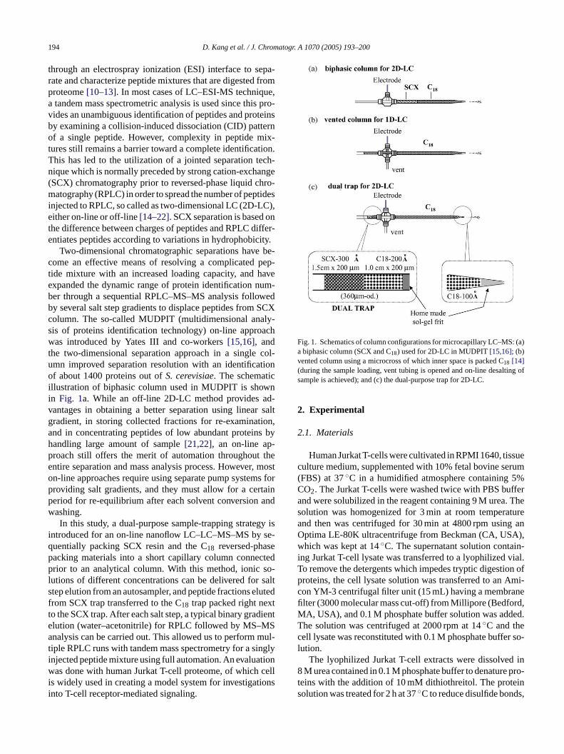
Fig. 1. Schematics of column configurations for microcapillary LC–MS: (a)a biphasic column (SCX and C18) used for 2D-LC in MUDPIT[15,16]; (b)vented column using a microcross of which inner space is packed C18 [14](during the sample loading, vent tubing is opened and on-line desalting ofsample is achieved); and (c) the dual-purpose trap for 2D-LC.
2. Experimental
2.1. Materials
Human Jurkat T-cells were cultivated in RPMI 1640, tissueculture medium, supplemented with 10% fetal bovine serum(FBS) at 37◦C in a humidified atmosphere containing 5%CO2. The Jurkat T-cells were washed twice with PBS bufferand were solubilized in the reagent containing 9 M urea. Thesolution was homogenized for 3 min at room temperatureand then was centrifuged for 30 min at 4800 rpm using anOptima LE-80K ultracentrifuge from Beckman (CA, USA),which was kept at 14◦C. The supernatant solution contain-ing Jurkat T-cell lysate was transferred to a lyophilized vial.To remove the detergents which impedes tryptic digestion ofproteins, the cell lysate solution was transferred to an Ami-con YM-3 centrifugal filter unit (15 mL) having a membranefilter (3000 molecular mass cut-off) from Millipore (Bedford,MA, USA), and 0.1 M phosphate buffer solution was added.The solution was centrifuged at 2000 rpm at 14◦C and thecell lysate was reconstituted with 0.1 M phosphate buffer so-lution.
The lyophilized Jurkat T-cell extracts were dissolved in8 M urea contained in 0.1 M phosphate buffer to denature pro-teins with the addition of 10 mM dithiothreitol. The proteinsolution was treated for 2 h at 37◦C to reduce disulfide bonds,
mn improved separation resolution with an identificaf about 1400 proteins out ofS. cerevisiae. The schemati
llustration of biphasic column used in MUDPIT is shon Fig. 1a. While an off-line 2D-LC method provides aantages in obtaining a better separation using linearadient, in storing collected fractions for re-examinatnd in concentrating peptides of low abundant proteinandling large amount of sample[21,22], an on-line aproach still offers the merit of automation throughoutntire separation and mass analysis process. Howevern-line approaches require using separate pump systemroviding salt gradients, and they must allow for a cereriod for re-equilibrium after each solvent conversionashing.In this study, a dual-purpose sample-trapping strate
ntroduced for an on-line nanoflow LC–LC–MS–MS byuentially packing SCX resin and the C18 reversed-phasacking materials into a short capillary column connerior to an analytical column. With this method, ionic
utions of different concentrations can be delivered fortep elution from an autosampler, and peptide fractions erom SCX trap transferred to the C18 trap packed right nexo the SCX trap. After each salt step, a typical binary gradlution (water–acetonitrile) for RPLC followed by MS–Mnalysis can be carried out. This allowed us to perform
iple RPLC runs with tandem mass spectrometry for a sinjected peptide mixture using full automation. An evaluaas done with human Jurkat T-cell proteome, of which
s widely used in creating a model system for investigatnto T-cell receptor-mediated signaling.
D. Kang et al. / J. Chromatogr. A 1070 (2005) 193–200 195
and the reduced thiol groups were alkylated by adding iodoac-etamide to a total concentration of 20 mM. The reaction wascarried out in the dark at 0◦C for 2 h. After an alkylation re-action, excess cysteine (∼40×) was added to react away theremaining iodoacetamide. The mixture was then diluted to atotal concentration of 1.0 M urea by adding phosphate bufferfor tryptic digestion. A proteomics grade trypsin from Sigma(St. Louis, MO, USA) was added at a concentration ratio of1:50 (protein:trypsin), and the mixture was incubated for 24 hat 37◦C. After digestion, TLCK was added to stop digestionat a slight excess to the number of moles of peptides. Thedigested mixture was finally desalted by using an Oasis HLBcartridge from Waters (Milliford, MA, USA) and dried byan Autospin 314U vacuum centrifuge from BioTron (Seoul,South Korea).
2.2. Two-dimensional LC with MS–MS
Nanoflow LC separation was carried out with an UltimatePump system from Dionex (Sunnyvale, CA, USA). The ana-lytical column (150 mm× 75�m) and the dual trap columnshown inFig. 1c were prepared in-house. The tip at the end ofcapillary tubing (75�m I.D., 360�m O.D.) from PolymicroTechnology LLC (Phoenix, AZ, USA) was pulled by flamewith a tip diameter of around 10�m. The empty column tipwas filled with a sol–gel frit by the following procedure. Thet /v)m ob-t Ko-r elfiM n,C Thed tub-ii lier. Itw ,ac oro,M andt rossa odet de-s
ys-t ered quisi-t 0 to2 cans(
2
ing aM -Prot
human database. The mass tolerance between the measuredmonoisotopic mass and the calculated mass was 1.0 u for themolar mass of a precursor peptide and 1.0 u for the massof peptide fragment ions. Only those peptides were selectedyielding larger than a minimum Mascot score of 29, whichindicates identification at the 95% confidence level for thissearch.
3. Results and discussion
The on-line LC–LC–MS–MS using a dual-purpose trapmethod is based on the combination of the biphasic columnmethod[15,16]in Fig. 1a and the vented column method[14]as shown inFig. 1b. While the biphasic column provides amerit of 2D-LC–LC separation on-line, it requires a seriesof column clean up process after each salt step. In the caseof using the vented column method, sample can be enrichedand purified prior to separation in analytical column for 1D-LC–MS–MS. With the dual trap method shown inFig. 1c,ionic solutions of different concentrations can be deliveredfor salt step elution from an autosampler with the vent valveopen, and peptide fractions eluted from SCX trap transferredto the C18 trap packed right next to the SCX trap. After eachsalt step, a typical binary gradient elution (water–acetonitrile)for RPLC can be carried out.Fig. 2 shows the schematicv hep rapw stedh ot ringtt led endo thata Thev lel CXt . Att thefl suree for8 theva e thes out tor ned int ed int ep-a po-s .B Theg de-f 2%B in-t ned
ip of column was immersed very shortly into the 1:4 (vixture of formamide and potassium silicate solutions
ained from R.S. Chem. (Hwasung, Kyeong-gi, Southea) and then was baked at 100◦C for 3 h to make a sol–grit. Then, the pulled tip capillary with an end frit (∼1 mmn length) was packed with methanol slurry of 5�m 100A
agic C18AQ from Michrom BioResources Inc. (AuburA, USA) at a constant pressure (1000 psi) of He.ual-purpose trapping column was made with a silica
ng (200�m I.D., 360�m O.D.) in which the end frit (2 mmn length) was prepared as the same way explained earas packed with 5�m 200A Magic C18AQ for 1.0 cm at firstnd then packed with 5�m 300A Polysulfoethy ATM strongation-exchange resin from The Nest Group Inc. (SouthbA, USA) for 1.5 cm. The dual-purpose trapping column
he analytical column were connected via a PEEK microcs shown inFig. 1c and a gold wire was used as an electr
o supply an electrospray ionization voltage of 2.5 kV ascribed in the literature[12,24].
A QSTAR mass spectrometer model from Applied Biosems (Foster City, CA, USA) was utilized. Peptide ions wetected in a data-dependent analysis mode. The ac
ion method involves one MS precursor scan from 30000 amu followed by three data-dependent MS–MS s35% normalized collision energy).
.3. Data processing
The acquired MS–MS spectra were analyzed by usascot Search program and then compared with a Swiss
iew of the on-line LC–LC system used in this study. Terformance of on-line LC–LC–MS–MS using a dual tas tested with human Jurkat T-cell proteome. A digeuman Jurkat T-cell protein mixture (15�g) was loaded ont
he SCX part of the dual trap from an autosampler. Duhe sample loading (valve position A ofFig. 2) onto the SCXrap, a flow rate of 4�L/min was applied from the sampelivery pump with the vent valve open, located at thef the waste line connected to the PEEK microcross, sony non-retaining impurities were removed via the vent.alve configuration A shown inFig. 2 was used for sampoading and also for the delivery of salt solution to the Srap from the autosampler for salt step gradient elutionhe valve configuration A (with the vent valve open), allow is expected to direct to the vent due to the high presxerted from the analytical column. After sample loadingmin, the 10-port valve was turned to block the end ofent tubing (position B: 36◦ turn from position A inFig. 2)nd was then ready for RPLC gradient separation. Beforalt step elution began, a breakthrough run was carriedesolve some peptides that were not expected to be retaihe SCX trap during the sample loading, but were retainhe C18 part of the dual trap. The binary gradient RPLC sration was carried out by varying the mobile phase comition of: (A) 3% acetonitrile (ACN) in water; and (B) ACNoth mobile phases contained 0.1% (v/v) formic acid.radient began with an increase to 5% B (from 2% B at
ault) over 4 min and it ramped to 20% for 60 min, and to 3for 20 min. Then, it was raised to 80% B for 2 min, ma
ained for 20 min, down to 2% B during 3 min, and maintai

196 D. Kang et al. / J. Chromatogr. A 1070 (2005) 193–200
Fig. 2. Schematic representation of the dual trap two-dimensional nanoflowLC setup. Salt step elution was made by delivering salt solution to the dualtrap using autosampler and each RPLC run was followed. Sample was ini-tially loaded to the SCX part of the dual trap with the valve position Afollowed by a breakthrough run (RPLC gradient) with the position B. At po-sition B, the vent tubing connection to the valve is blocked (expressed withx). After the breakthrough run, valve position was turned back to positionA for salt step elution. Eight microliters of 3 mM NH4HCO3 solution wasdelivered to the dual trap from autosampler for 8 min and during the saltelution, peptides desorbed from the SCX trap were transferred to RP partof the dual trap. Then, the valve position was changed to B and an RPLCgradient run was performed. For additional salt step elution and RPLC run,steps 3 and 4 shown inFig. 2were repeated.
for at least 20 min for column re-conditioning. The flow rateduring the gradient separation was kept at 200 nL/min andthe eluted peptides were directly electrosprayed into a massspectrometer. After the breakthrough run, the 10-port valvewas changed to position A with the vent tubing open for thesalt step elution. The first salt step fractionation was accom-plished by delivering 8�L of 3 mM ammonium bicarbonate(NH4HCO3) solution contained in a microvial from the au-tosampler to the SCX trap at 4�L/min. Desorbed peptidesfrom the first salt step elution were readily trapped in the C18trap right next to the SCX trap. By delivering only a smallamount of salt solution from the autosampler, salt deliveryand desalting were made in sequence by the sample deliv-ery pump over the 8 min of salt step elution. After the firstsalt step elution, the valve position was turned to carry outa gradient RPLC run (position B). The pump flow deliveredto the analytical column was set at 200 nL/min and the bi-nary gradient separation in RPLC was performed. After thefirst salt step elution followed by a gradient RPLC run, theprocedure was repeated for consecutive salt steps by increasing the concentration of NH4HCO3 solution to 6, 10, 15, 20,50, and 500 mM. The last salt step was to deplete all peptidesfrom SCX trap. In this experiment, a total of eight RPLC runs
were performed. With the use of a dual-purpose sample trapdirectly connected to the microcross, the entire system oper-ation is automated and the current setup minimizes the use ofa complicated valve operation as well as a separate columnwashing process after each SCX run needed for on-line two-dimensional LC systems. During RPLC separation, a home-made pulled tip column having a frit at the tip was utilized, asshown inFig. 1c. By installing a 1 mm long sol–gel frit at theend of the pulled tip, the column blocking that occasionallyformed during electrospray ionization due to insoluble solidsat the open tip disappeared. Formation of bubble can be ob-served at the end of a pulled tip column without frit in somecases (at a very low flow rate) and this can interfere with MSanalysis. It can be removed when the in-line degassing andthe high pressure mixing of mobile phases are integrated toHPLC system. Another remedy is to fill the microcross withpacking materials. However, at the current experiment using200 nL/min., bubble formation was not observed to interfereMS analysis.
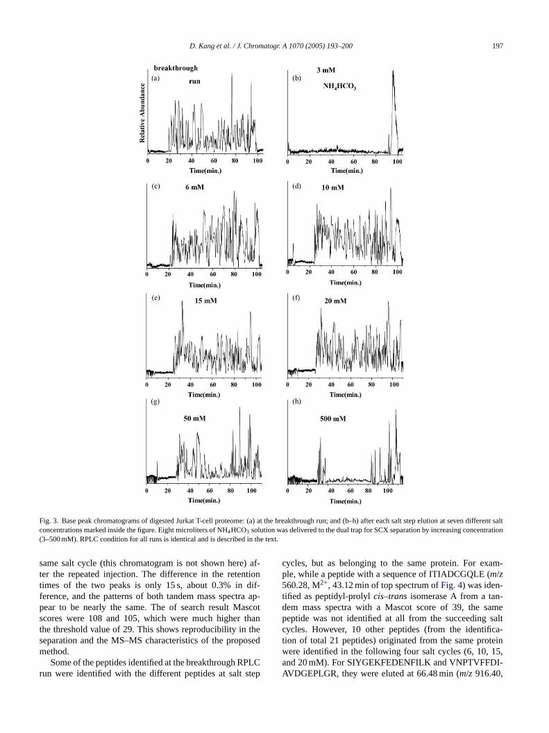
Fig. 3shows the base peak chromatograms of a 7-cycle on-line 2-D-LC–MS analysis of the peptide mixtures of humanJurkat T-cell proteome along with an initial breakthrough run.The first RPLC run inFig. 3a was obtained right after thesample loading in the SCX fraction. It demonstrated that therewas a considerable amount of components which were notretained in the SCX trap but were trapped in the RP part of thed fromt rchf akso nics
N klyb d thec olu-t salts.T lts ceptf ever,t ought wasi SCXt ls ntra-t dea theS thint nates
asss T-M tin( asss onp so the
-
ual trap. However, 57 unique peptides were identifiedhe numerous peaks inFig. 3a through the database seaor the corresponding MS–MS spectra. Most of the pebserved inFig. 3a are thought to be due to some non-iomall molecules or peptide fragments.
The first salt step began by delivering 8�L of 3 mMH4HCO3 solution from the autosampler to displace weaound peptides from the SCX trap to the RP region, anarrier liquid (the same as solvent A) delivering the salt sion band washed the dual trap to remove any remaininghe RPLC run (Fig. 3b) immediately following the first satep elution did not show a significant peptide peak exor the huge transient peak at the end of gradient; howhe database search did yield 12 unique proteins, althhese are single peptide hit. When the salt concentrationncreased to 6 mM, peptides began desorbing from therap as shown by RPLC separation inFig. 3c. The additionaalt steps were carried out by increasing the salt conceion, as shown inFig. 3d–g, and the final step was mat 500 mM in order to detach all remaining peptides inCX trap. Most of the peptides were shown to elute wi
he concentration range of 6–50 mM ammonium bicarboolution.
Fig. 4 shows an example of two tandem mpectra obtained for the peptide K.DLYANTVLSGGTYPGIADR.M (m/z 1107.98, doubly charged) from ac
cytoplasmic 1) eluted at the salt cycle of 6 mM. The mpectra at the top ofFig. 4 was obtained at the parent ieak eluted at 76.14 min inFig. 3c, and the bottom wabtained from a chromatographic peak at 75.89 min in
D. Kang et al. / J. Chromatogr. A 1070 (2005) 193–200 197
Fig. 3. Base peak chromatograms of digested Jurkat T-cell proteome: (a) at the breakthrough run; and (b–h) after each salt step elution at seven different saltconcentrations marked inside the figure. Eight microliters of NH4HCO3 solution was delivered to the dual trap for SCX separation by increasing concentration(3–500 mM). RPLC condition for all runs is identical and is described in the text.
same salt cycle (this chromatogram is not shown here) af-ter the repeated injection. The difference in the retentiontimes of the two peaks is only 15 s, about 0.3% in dif-ference, and the patterns of both tandem mass spectra ap-pear to be nearly the same. The of search result Mascotscores were 108 and 105, which were much higher thanthe threshold value of 29. This shows reproducibility in theseparation and the MS–MS characteristics of the proposedmethod.
Some of the peptides identified at the breakthrough RPLCrun were identified with the different peptides at salt step
cycles, but as belonging to the same protein. For exam-ple, while a peptide with a sequence of ITIADCGQLE (m/z560.28, M2+, 43.12 min of top spectrum ofFig. 4) was iden-tified as peptidyl-prolylcis–trans isomerase A from a tan-dem mass spectra with a Mascot score of 39, the samepeptide was not identified at all from the succeeding saltcycles. However, 10 other peptides (from the identifica-tion of total 21 peptides) originated from the same proteinwere identified in the following four salt cycles (6, 10, 15,and 20 mM). For SIYGEKFEDENFILK and VNPTVFFDI-AVDGEPLGR, they were eluted at 66.48 min (m/z 916.40,

198 D. Kang et al. / J. Chromatogr. A 1070 (2005) 193–200
Fig. 4. MS–MS spectra of the peptide K.DLYANTVLSGGTT-MYPGIADR.M (m/z 1107.98, doubly charged) from actin (cyto-plasmic 1) obtained from repeated experiments. The peptide was eluted atthe salt cycle of 6 mM NH4HCO3.
M2+) and 92.13 min (m/z 973.52, M2+), respectively, in the10 mM salt step LC run. Both were identified to be fromthe same peptidyl-prolylcis–trans isomerase A with searchscores of 94 and 75, respectively. The same peptides ap-peared in the next salt step (15 mM) at 66.01 min (m/z611.34,M3+), and 91.44 min (m/z 973.51, M2+), respectively. Thedifference in the retention times for the nearby runs wasabout 0.7%. This showed that same peptides were elutedat nearby salt steps. It also suggested that there was a pos
sibility of a decreased chance of detecting other peptideions due to the repeated detection of identified peptides inthe previous salt step. Moreover, the peptide ITIADCGQLEdid not at all appear throughout the entire salt steps exceptin the breakthrough run, but it was nonetheless identifiedwith a similar retention time (44.39 min,m/z 560.26, M2+,Mascot score = 43) when a single-dimensional LC–MS–MSanalysis was performed. However, those 10 peptides iden-tified from the 2D-LC–LC–MS–MS experiments were noteven identified at all in the single-dimensional LC–MS–MSanalysis.
The MS–MS spectra obtained from each RPLC runyielded an identification of 441 different peptides, and thenumber of proteins was found to be 234 listed inTable 1.Among these numbers, 94 proteins were identified as multi-ple peptides. The difference between the number of proteinsand the number of new proteins in each cycle represents thenumber of proteins eluted in nearby salt step runs. The num-ber of proteins identified in each run was the highest at thefirst salt step, and it decreased to a level of 102 in the finalcycle, as listed inTable 1. The total number of proteins iden-tified from a 15�g injection of digested human Jurkat T-cellproteome was 681. When a single dimension LC–MS–MSrun is used, the number of proteins identified from a 3�ginjection was only 222, as listed inTable 1. For the single-dimensional LC–MS–MS analysis, a trapping column packedw theo linga foro them mes
las-s nly4 sult-i Sa witht mh uchl onal
Table 1Coverage of peptide/protein identification from single-dimensional LC–MS–M dproteins from Jurkat T-cell
1D LC–MS–MS (3�g) On-line LC–LC–
0 mMa 3 mMa
Number of peptides 650 57 12Number of proteins 222 57 12(Multiple hits)b 126 0 0Number of new proteins 57 11Number of proteins (cumulative) 57 68
Total 222 681a Concentration of NH4HCO3.b Number of identified proteins with multiple peptides.
-
ith C18 only was used instead of the dual column. Sincen-line 2D-LC technique provides the advantage of handsmaller amount of protein digests than that required
ff-line SCX fractionation, the dual trap method still haserit of being able to deal with small amount of proteo
ample.Table 2lists the number of identified proteins that are c
ified according to their molecular function and types. O52 proteins were classified from the total 681 proteins re
ng from 15�g injection using the on-line LC–LC–MS–Mpproach, and 229 proteins were not clearly identified
heir origin or function. The number of proteins frouman Jurkat T-cell obtained by this approach is m
arger than that identified by a conventional two-dimensi
S and on-line LC–LC–MS–MS using a dual trap from 15�g injection of digeste
MS–MS
6 mMa 10 mMa 15 mMa 20 mMa 50 mMa 500 mMa
441 418 429 412 329 167234 203 210 204 197 102
(94) (92) (87) (89) (78) (30)221 109 78 85 76 44289 398 476 561 637 681

D. Kang et al. / J. Chromatogr. A 1070 (2005) 193–200 199
Table 2Number of identified proteins of human Jurkat T-cell obtained at two differ-ent injection amounts
Categories Number of proteins classified
Cytoskeleton 66Heat shock proteins 32HnRNP 21Membrane proteins 25Metabolic enzyme 55Nucleosome 37Nuclear proteins 32Proteosome 20Ribosome 84Signaling molecules 26Spliceosome 24T-complex proteins 10Translation factor 20Others 229
Total 681
electrophoresis-matrix assisted laser desorption ionisation-mass spectrometry (2DE-MALDI-MS)[23].
4. Conclusions
In this article, a fully automated on-line two-dimensionalLC system with a dual-purpose sample trap was introducedfor the separation of peptide mixtures from human JurkatT-cell and for the improvement of ESI-tandem MS analy-sis. It has been demonstrated that the dual trap on-line 2D-LC–LC–MS–MS method described in this study can be ap-plicable to the proteome analysis in an unattended way. Espe-cially, the current method has several advantages over othermultidimensional chromatographic methods since compli-cated valve systems are avoided along with a minimizedplumbing and thus, a very small amount of peptides can beefficiently separated into two dimensions with the reducedfear of a possible loss during the passage through the tubingconnections. With the use of a dual trap directly connectedto a home-made nanoflow RPLC column with a pull tip, theon-line fractionation of peptides by charge in the SCX trapcan be simply obtained prior to the analytical column witha minimization of dead volume. The dual trap 2D-LC setuppractically obviates the necessity of feeding salt solution toM shinga y in-ja olu-t willr tiont n isu ini-m d byi pu-r SCXt .
One difficulty in the use of a dual trap system came froma build-up of system pressure in the dual trap during the ini-tial sample loading and the delivery of salt solution at a highspeed. In this study, the SCX trap was packed in 200�mI.D. capillary tubing for 1.5 cm long in order to reduce thesystem pressure during sample loading. Since the volume ofSCX packing in this dual trap was about 50% larger thanthat of a typical SCX packed in an integrated (SCX and RP)column (4 cm of SCX part, 100�m I.D.) in literature[16],sample loading capacity of the dual trap was expected to ac-commodate a sufficient amount of protein digests (more than100�g) based on the volume of SCX trap. However, whensalt solution was delivered to the SCX trap at an increasedrate (∼4�L/min), the maximum loading capacity was 50�gof protein digests used in this study. When it is needed forloading sample and salt step elution at an increased rate, thesubsequent increase of backpressure needs to be decreasedby incorporating a larger inner diameter tubing in the prepa-ration of dual trap or a special device. When a large diametertubing is used for a dual trap, length of the total trap devicemust be minimized to reduce inaccurate gradient conditionof RPLC mobile phase. In this work, it was focused to im-plement the dual trap system for the possible use of on-line2D-LC/tandem MS toward proteomics analysis.
The idea utilized in this study can be expanded to incor-porate an affinity based stationary phase such as may be usedf C)i pep-t tion,w t isn ededt iveryr temp
A
alth2 ofK s theg oun-d C) atY TechI
R
se,
uer,
iol.
74
S since it removes a separate process for column wafter each use of salt solution. This can be achieved b
ecting a small volume (only 8�L) of salt solution from anutosampler, and the delivery liquid following the salt s
ion band washes remaining salts in the dual trap. Thisemove a column re-equilibrium period after each salt eluo column when an integrated biphasic (SCX–RP) columsed as in MUDPIT. In addition, it is advantageous to mize a chance of deteriorating analytical column cause
mpurities or cell debris left in protein digests even afterification since peptide mixtures are loaded to a separaterap initially and injected to analytical column after SCX
or the immobilized metal affinity chromatography (IMAnto the dual trap for the on-line screening of phosphoides and for the simultaneous washing of metal soluhich is an essential eluent for affinity separation buot desirable for an MS analysis. Further studies are ne
o expand peptide throughput and to increase the delate of salt solution to the dual trap by reducing sysressure.
cknowledgements
This study was supported by a grant from the Korea He1 R&D Project, Ministry of Health & Welfare, Republicorea (03-PJ1-PG6-GP01-0002). Y.-S.K. acknowledgerant support from the Korea Science and Engineering Fation through Protein Network Research Center (PNRonsei University. The authors also thank to Proteome
nc. for the access to MS facilities.
eferences
[1] J.B. Fenn, M. Mann, C.K. Meng, S.F. Wong, C.M. WhitehouScience 246 (1989) 64.
[2] R. Aebersold, D.R. Goodlett, Chem. Rev. 101 (2001) 269.[3] D.F. Hunt, J.R. Yates III, J. Shabanowitz, S. Winston, C.R. Ha
Proc. Natl. Acad. Sci. U.S.A. 83 (1986) 6233.[4] S.P. Gygi, Y. Rochon, B.R. Franza, R. Aebersold, Mol. Cell. B
19 (1999) 1720.[5] N.L. Anderson, N.G. Anderson, Proc. Natl. Acad. Sci. U.S.A.
(1977) 5421.
200 D. Kang et al. / J. Chromatogr. A 1070 (2005) 193–200
[6] T. Rabilloud, Proteomics 2 (2002) 3.[7] Y.M. Cho, S.H. Bae, B.K. Choi, S.Y. Cho, C.W. Song, J.K. Yoo,
Y.K. Paik, Proteomics 3 (2003) 1883.[8] T.C. Hunter, N.L. Andon, A. Koller, J.R. Yates III, P.A. Haynes, J.
Chromatogr. B 782 (2002) 165.[9] R. Zhang, C.S. Sioma, R.A. Thomson, L. Xiong, F.E. Regnier, Anal.
Chem. 74 (2002) 3662.[10] L.J. Deterding, M.A. Moseley, K.B. Tomer, J.W. Jorgenson, J. Chro-
matogr. 554 (1991) 73.[11] J.K. Eng, A.L. McCormack, J.R. Yates III, J. Am. Soc. Mass Spec-
trom. 5 (1994) 876.[12] C.L. Gatlin, G.R. Kleeman, L.G. Hays, A.J. Link, J.R. Yates III,
Anal. Biochem. 263 (1998) 93.[13] H. Lee, J. Griffin, S.P. Gygi, B. Rist, R. Aebersold, Anal. Chem. 74
(2002) 4353.[14] L.J. Licklider, C.C. Thoreen, J. Peng, S.P. Gygi, Anal. Chem. 74
(2002) 3076.[15] A.J. Link, J. Eng, D.M. Shieltz, E. Carmack, G.J. Mize, D.R.
Morris, B.M. Garvik, J.R. Yates III, Nat. Biotechnol. 17 (1999)676.
[16] M.P. Washburn, D. Wolters, J.R. Yates III, Nat. Biotechnol. 19 (2001)242.
[17] D.A. Wolters, M.P. Washburn, J.R. Yates III, Anal. Chem. 73 (2001)5683.
[18] W.H. McDonald, R. Ohi, D.T. Miyamoto, T.J. Mitchinson, J.R. YatesIII, Int. J. Mass Spectrom. 219 (2002) 245.
[19] G.J. Opiteck, K.C. Lewis, J.W. Jorgenson, R.J. Anderegg, Anal.Chem. 69 (1997) 1518.
[20] M.T. Davis, J. Beierle, E.T. Bures, M.D. McGinley, J. Mort, J.H.Robinson, C.S. Sphar, W. Yu, R. Luethy, S.D. Patterson, J. Chro-matogr. B 752 (2001) 281.
[21] J. Peng, J.E. Elias, C.C. Thoreen, L.J. Licklider, S.P. Gygi, J. Pro-teome Res. 2 (2003) 43.
[22] Y. Shen, J.M. Jacobs, D.G. Camp II, R. Fang, R.J. Moore, R.D.Smith, W. Xiao, R.W. Davis, R.G. Tompkins, Anal. Chem. 76 (2004)1134.
[23] B. Thiede, F. Siejak, C. Dimmler, P.R. Jungblut, T. Ruel, Elec-trophoresis 21 (2000) 2713.
[24] M.H. Moon, S. Myung, M. Plasencia, A.E. Hilderbrand, D.E. Clem-mer, J. Proteome Res. 2 (2003) 589.