Resolving subtle stratigraphic features using ... - CiteSeerX
Upload
khangminh22Category
view
2download
0
Citation: Khandpur, S.; Ahuja, R.
Drug-Induced vs. Viral
Maculopapular Exanthem—
Resolving the Dilemma.
Dermatopathology 2022, 9, 164–171.
https://doi.org/10.3390/
dermatopathology9020021
Academic Editors: Joyce Lee and
M. Ramam
Received: 26 February 2022
Accepted: 30 April 2022
Published: 7 May 2022
Publisher’s Note: MDPI stays neutral
with regard to jurisdictional claims in
published maps and institutional affil-
iations.
Copyright: © 2022 by the authors.
Licensee MDPI, Basel, Switzerland.
This article is an open access article
distributed under the terms and
conditions of the Creative Commons
Attribution (CC BY) license (https://
creativecommons.org/licenses/by/
4.0/).
Review
Drug-Induced vs. Viral Maculopapular Exanthem—Resolvingthe DilemmaSujay Khandpur * and Rhea Ahuja
Department of Dermatology and Venereology, All India Institute of Medical Sciences, New Delhi 110029, India;[email protected]* Correspondence: [email protected]
Abstract: Maculopapular exanthem is a commonly encountered presentation in routine clinicalpractice, and differentiation between its two most common etiologies, i.e., viral- and drug-induced,often poses a diagnostic dilemma. Clinical, hematological and biochemical investigations are seldomreliable in distinguishing between a drug reaction and a viral exanthem. Certain key histopathologicalfeatures such as the presence of a moderate degree of spongiosis, extensive basal cell damage withmultiple necrotic keratinocytes and dermal infiltrate rich in eosinophils or lymphocytes and histio-cytes may favor a drug exanthem, while distinctive epidermal cytopathic changes and lymphocyticvasculitis point towards a viral etiology. Similarly, notable immunohistochemical markers such asIL-5, eotaxin and FAS ligand may support a diagnosis of a drug-induced maculopapular eruption.Histopathological and immunohistochemical evaluations may help in distinguishing between thetwo etiologies when faced with a clinical overlap, especially in patients on multiple essential drugswhen drug withdrawal and rechallenge is not feasible.
Keywords: maculopapular; exanthema; drug; viral
1. Introduction
Maculopapular exanthem is characterized by an acute and generalized eruption oferythematous macules and papules without overlying scaling. It is a common presentationin day-to-day clinical practice, and differentiating between its two most common etiologies,i.e., viral- and drug-induced, is often perplexing. An eruption occurring after drugs havingbeen initiated for treatment of an underlying infection further adds to the diagnosticdilemma. Establishing an etiological diagnosis aids the clinician in deciding whether tointerrupt a culprit drug or to continue a wrongly incriminated and possibly life-savingdrug. This review aims to distinguish between viral- and drug-induced exanthem, with aspecial focus on histopathological features.
2. Clinical Features
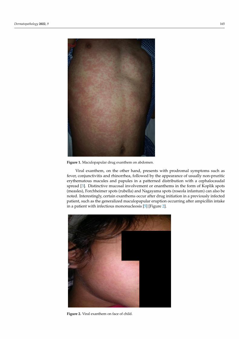
Maculopapular eruption is the most common presentation, accounting for around90% of cases among 1317 patients with cutaneous drug hypersensitivity reactions [1]. Thecommonly implicated drugs include antibiotics such as beta-lactams and suphonamides,anti-convulsants and non-steroidal anti-inflammatory drugs [2]. The cutaneous eruptionusually occurs within 7 to 10 days (range 5 to 21 days) after treatment, starting from thetrunk and proximal extremities. Distinctive features of a drug-induced exanthem includethe presence of pruritus, confluence of the rash on dependent areas, facial involvementand purpuric lesions on lower extremities [3]. A temporal correlation with drug intakeis important to ascertain causation, and in a previous study re-appearance of the rash ondrug provocation was an essential diagnostic criterion for defining definite drug-inducedexanthem [4] [Figure 1].
Dermatopathology 2022, 9, 164–171. https://doi.org/10.3390/dermatopathology9020021 https://www.mdpi.com/journal/dermatopathology
Dermatopathology 2022, 9 165Dermatopathology 2022, 9, 2
Figure 1. Maculopapular drug exanthem on abdomen.
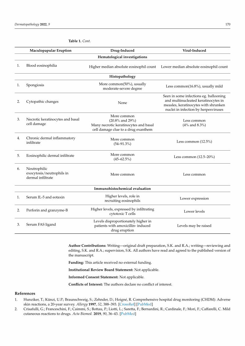
Viral exanthem, on the other hand, presents with prodromal symptoms such as fever, conjunctivitis and rhinorrhea, followed by the appearance of usually non-pruritic erythematous macules and papules in a patterned distribution with a cephalocaudal spread [3]. Distinctive mucosal involvement or enanthems in the form of Koplik spots (measles), Forchheimer spots (rubella) and Nagayama spots (roseola infantum) can also be noted. Interestingly, certain exanthems occur after drug initiation in a previously infected patient, such as the generalized maculopapular eruption occurring after ampicillin intake in a patient with infectious mononucleosis [5] [Figure 2].
Figure 2. Viral exanthem on face of child.
Figure 1. Maculopapular drug exanthem on abdomen.
Viral exanthem, on the other hand, presents with prodromal symptoms such asfever, conjunctivitis and rhinorrhea, followed by the appearance of usually non-pruriticerythematous macules and papules in a patterned distribution with a cephalocaudalspread [3]. Distinctive mucosal involvement or enanthems in the form of Koplik spots(measles), Forchheimer spots (rubella) and Nagayama spots (roseola infantum) can also benoted. Interestingly, certain exanthems occur after drug initiation in a previously infectedpatient, such as the generalized maculopapular eruption occurring after ampicillin intakein a patient with infectious mononucleosis [5] [Figure 2].
Dermatopathology 2022, 9, 2
Figure 1. Maculopapular drug exanthem on abdomen.
Viral exanthem, on the other hand, presents with prodromal symptoms such as fever, conjunctivitis and rhinorrhea, followed by the appearance of usually non-pruritic erythematous macules and papules in a patterned distribution with a cephalocaudal spread [3]. Distinctive mucosal involvement or enanthems in the form of Koplik spots (measles), Forchheimer spots (rubella) and Nagayama spots (roseola infantum) can also be noted. Interestingly, certain exanthems occur after drug initiation in a previously infected patient, such as the generalized maculopapular eruption occurring after ampicillin intake in a patient with infectious mononucleosis [5] [Figure 2].
Figure 2. Viral exanthem on face of child. Figure 2. Viral exanthem on face of child.

Dermatopathology 2022, 9 166
3. Hematological and Biochemical Investigations
Blood eosinophilia may support a diagnosis of drug-induced exanthem. Singh et al.found the median absolute eosinophil count to be higher than normal in patients with drugexanthem, whereas they were within normal limits in those with viral exanthem, althoughthe difference was not statistically significant [4]. Yawalaker et al. have also describedthat cutaneous drug eruptions may be associated with blood and tissue eosinophilia [6].Although blood eosinophilia is not pathognomonic for maculopapular drug exanthem, thedegree of eosinophilia usually correlates with the severity of drug reactions, more oftenin drug reactions with eosinophilia and systemic symptoms (DRESS) [7]. Biochemicalinvestigations such as hepatic and renal function panels are usually normal, unless thepatient is suffering from DRESS syndrome.
C-reactive protein levels also do not play a significant role in establishing an etiologicaldiagnosis of maculopapular eruption. Although the levels are usually higher in drugexanthem compared to viral exanthem, the difference is not statistically significant [4,8].
4. Histopathology
Lever’s Textbook of Dermatology and Mckee’s Pathology of Skin have emphasizedthat the histological findings of an exanthematous drug eruption are often indistinguishablefrom those of viral exanthems, although the presence of eosinophils may favor a drugreaction [9,10]. Ackerman also believed that “drugs can elicit any of the nine basic patternsof inflammatory diseases in the skin, and none of those patterns is specific for a drug erup-tion” [11]. Nonetheless, various studies have highlighted certain histopathological pointersfor both drug- and viral-induced exanthema, albeit they may not be pathognomonic.
Seitz et al. in their study on 91 patients of drug-induced exanthema described themost common reaction pattern to be the combined type of spongiotic and interface pattern,followed by the perivascular (15/26), spongiotic (10/13), vacuolar interface (6/10) andlichenoid interface patterns (4/5). Most importantly, no specific reaction pattern could besignificantly attributed to drug-induced or non-drug-induced exanthem. Parakeratosis waspresent in a slightly but not significantly higher proportion of non-drug-induced exanthemthan drug-induced exanthem. No significant differences in occurrence of dyskeratotickeratinocytes, extent of lymphocyte exocytosis or density of the dermal lymphocyticinfiltrate were observed. Moreover, there was no significant difference in the presenceof papillary dermal edema, accompanying vasculitis or extravasation of erythrocytes. Acomparison of drug-induced exanthema and non-drug-induced exanthem did not yield asignificant difference in the number of eosinophils within the dermal infiltrate. The resultsregarding the sensitivity (62.9%), specificity (41.1%) and positive (40.7%) and negativepredictive values (69.7%) of a correct diagnosis showed that no valid conclusions can bedrawn from histologic evaluations of skin biopsy specimens [12].
However, other authors have noted certain distinctive findings that may point towardseither a drug-induced or a viral exanthem [Figures 3 and 4]. We will now highlight theimportant differences proceeding from the epidermis to the dermis.
4.1. Spongiosis
Naim et al. while evaluating 60 biopsy specimens noted that spongiosis was the mostconsistent feature (97%) in maculopapular drug eruptions, and it was mostly focal [13].Other authors also corroborated this finding, with spongiosis being noted in significantlyhigher number of drug eruption cases (50%) compared to viral exanthem (17%), however itwas of a moderate-severe degree [4]. Spongiosis may be associated with inflammatory cellexocytosis variably noted in 38–100% cases [13,14]. Usually eosinophilic or lymphocyticexocytosis is noted, but invasion by solitary neutrophils is an important clue towards adrug etiology [13]. The presence of follicular spongiosis is uncommon, present in up to 8%of exanthems of either etiology and cannot differentiate between the two [4,13].

Dermatopathology 2022, 9 167Dermatopathology 2022, 9, 4
Figure 3. (a) Drug exanthem to tranexamic acid, showing spongiosis, focal vacuolar interface changes with papillary dermal perivascular and interstitial infiltration of lympho-histiocytes and eosinophils (H&E ×40). (b) Drug exanthem to rifampicin, showing parakeratosis, spongiosis, necrotic keratinocytes in upper layers of the epidermis, lymphocytic exocytosis and papillary dermal infiltration of lymphocytes and eosinophils (H&E ×400).
Figure 4. Viral exanthem showing normal epidermis with sparse perivascular infiltration of lymphocytes and occasional eosinophils (H&E ×40).
4.1. Spongiosis Naim et al. while evaluating 60 biopsy specimens noted that spongiosis was the most
consistent feature (97%) in maculopapular drug eruptions, and it was mostly focal [13]. Other authors also corroborated this finding, with spongiosis being noted in significantly higher number of drug eruption cases (50%) compared to viral exanthem (17%), however it was of a moderate-severe degree [4]. Spongiosis may be associated with inflammatory cell exocytosis variably noted in 38–100% cases [13,14]. Usually eosinophilic or lymphocytic exocytosis is noted, but invasion by solitary neutrophils is an important clue towards a drug etiology [13]. The presence of follicular spongiosis is uncommon, present in up to 8% of exanthems of either etiology and cannot differentiate between the two [4,13].
4.2. Viral Cytopathic Changes Some viral exanthems can be recognized by their distinctive features, such as
ballooning and multinucleated keratinocytes in measles and keratinocytes with shrunken
Figure 3. (a) Drug exanthem to tranexamic acid, showing spongiosis, focal vacuolar interface changeswith papillary dermal perivascular and interstitial infiltration of lympho-histiocytes and eosinophils(H&E ×40). (b) Drug exanthem to rifampicin, showing parakeratosis, spongiosis, necrotic ker-atinocytes in upper layers of the epidermis, lymphocytic exocytosis and papillary dermal infiltrationof lymphocytes and eosinophils (H&E ×400).
Dermatopathology 2022, 9, 4
Figure 3. (a) Drug exanthem to tranexamic acid, showing spongiosis, focal vacuolar interface changes with papillary dermal perivascular and interstitial infiltration of lympho-histiocytes and eosinophils (H&E ×40). (b) Drug exanthem to rifampicin, showing parakeratosis, spongiosis, necrotic keratinocytes in upper layers of the epidermis, lymphocytic exocytosis and papillary dermal infiltration of lymphocytes and eosinophils (H&E ×400).
Figure 4. Viral exanthem showing normal epidermis with sparse perivascular infiltration of lymphocytes and occasional eosinophils (H&E ×40).
4.1. Spongiosis Naim et al. while evaluating 60 biopsy specimens noted that spongiosis was the most
consistent feature (97%) in maculopapular drug eruptions, and it was mostly focal [13]. Other authors also corroborated this finding, with spongiosis being noted in significantly higher number of drug eruption cases (50%) compared to viral exanthem (17%), however it was of a moderate-severe degree [4]. Spongiosis may be associated with inflammatory cell exocytosis variably noted in 38–100% cases [13,14]. Usually eosinophilic or lymphocytic exocytosis is noted, but invasion by solitary neutrophils is an important clue towards a drug etiology [13]. The presence of follicular spongiosis is uncommon, present in up to 8% of exanthems of either etiology and cannot differentiate between the two [4,13].
4.2. Viral Cytopathic Changes Some viral exanthems can be recognized by their distinctive features, such as
ballooning and multinucleated keratinocytes in measles and keratinocytes with shrunken
Figure 4. Viral exanthem showing normal epidermis with sparse perivascular infiltration of lympho-cytes and occasional eosinophils (H&E ×40).
4.2. Viral Cytopathic Changes
Some viral exanthems can be recognized by their distinctive features, such as bal-looning and multinucleated keratinocytes in measles and keratinocytes with shrunkennuclei with margination of the nucleoplasm in infections by herpesviruses, although thesechanges are relatively rare. Liersch et al. noted certain distinct histopathological featuresin measles characterized by multinucleated keratinocytes and individual and groupednecrotic keratinocytes in the epidermis with pronounced folliculo-sebaceous and acrosy-ringeal involvement [15].
4.3. Necrotic Keratinocytes and Basal Cell Damage
This finding is also noted more often in drug-induced (21% and 29%) than in viralexanthem (4% and 8%). The presence of many necrotic keratinocytes and severe basal celldamage is an important clue towards a drug’s etiology [4]. Weyer et al. have shown thatthe most common histopathological pattern of maculopapular drug eruption is vacuolar

Dermatopathology 2022, 9 168
interface dermatitis, noted in 97% of cases [16]. The degree of interface change is highlyvariable, ranging from slight vacuolar damage with few necrotic keratinocytes to severevacuolar alterations with many necrotic keratinocytes, even at higher levels in the epidermis.However, severe interface changes are more commonly observed in a severe cutaneousadverse drug reaction such as erythema multiforme, Stevens–Johnson syndrome and toxicepidermal necrolysis rather than maculopapular drug eruption.
4.4. Chronic Dermal Inflammatory Infiltrate
A lymphocytic and histiocytic dermal infiltrate was observed in a significantly higherproportion of drug exanthema (62.5%) compared to viral exanthema (12.5%) biopsies [4].The infiltrate may be perivascular, interstitial or associated with necrotic keratinocytes atthe dermo-epidermal junction. The lymphocytes may be atypical and large, as noted in16.7–35% of biopsy samples [4,14].
4.5. Eosinophilic Dermal Infiltrate
This finding is noticed in a significantly higher proportion of drug exanthem (52%)compared to viral exanthem (12.5%) biopsies [4]. Weyer et al. in their study on mac-ulopapular eruption found that besides eosinophils, neutrophils are also an importantconstituent of the dermal infiltrate. Although eosinophils are more common, because theyare seen in such a wide variety of diseases, they are less distinctive for drug eruptions. Aninfiltrate of neutrophils is rarer but of greater diagnostic importance. Sparse perivascularand interstitial infiltration of neutrophils and eosinophils with subtle vacuolar changes atthe dermoepidermal junction is virtually diagnostic of a drug eruption [16].
4.6. Lymphocytic Vasculitis
Viral exanthem may be associated with lymphocytic vasculitis, a finding which isuncommon in drug exanthema [16]. A dense perivascular cuff of lymphocytes with RBC ex-travasation has also been observed in COVID-associated maculopapular eruption, althoughmore commonly in a livedo-like or erythema-multiforme-like clinical presentation [17].
Other discernible features such as hyperkeratosis, parakeratosis, fibrin globules in thestratum corneum, acanthosis, colloid bodies in the papillary dermis, congested capillariesand pigment incontinence may be observed more commonly in biopsies from maculopapu-lar drug eruption compared to viral exanthem, although these alone may not be sufficientin differentiating the two [4].
5. Immunohistochemical Techniques
Attempts have been made to histologically diagnose drug-induced exanthem anddifferentiate it from viral exanthem using immunohistochemical techniques. Using CD4,CD8, FAS ligand, IL-5 and eotaxin markers, differentiation between the two entities havebeen made. FAS–ligand in the serum and tissue is elevated in most cases of maculopapulardrug exanthem, but it is normal in viral exanthem [18,19].
IL-5 and eotaxin cause activation and recruitment of eosinophils, thereby contribut-ing to the development of skin inflammation in drug-induced maculopapular exanthems.Yawalkar et al. performed an IHC analysis of the dermal mononuclear infiltrate in drug ex-anthem biopsies and compared it with normal skin in control subjects. IL-5 expression wasmainly found among mononuclear cells of the inflammatory infiltrate. Immunoreactivityfor eotaxin, RANTES (regulated on activation, normal T cell expressed and secreted), IL-8and to a lesser extent monocyte chemotactic protein-3 were also seen in the mononuclearcells. In addition, resident cells (i.e., endothelial cells and keratinocytes) also demonstratedpositivity for eotaxin, RANTES and IL-8 [6].
The chronic dermal inflammatory infiltrate in a drug exanthem is primarily composedof CD3+ T cells. Within T-lymphocytes, CD4+ T cells are noted predominantly in theperivascular location, whereas equal proportions of CD4+ and CD8+ T cells are located atthe dermoepidermal junction and in the epidermis [20]. Up to 20% of the infiltrating T cells

Dermatopathology 2022, 9 169
in drug-induced maculopapular exanthem express perforin and granzyme B, which areimportant components of cell-mediated cytotoxic reaction. In addition to T cells, eosinophilsare also present within the dermal infiltrate. These may also contribute to the generation oftissue damage via the release of various toxic granule proteins, such as eosinophilic cationicprotein, major basic protein and eosinophil peroxidase.
Bellini et al. performed an IHC evaluation of the cutaneous cytokine expressionin maculopapular eruptions (36 patients with drug exanthem, 30 patients with viral orbacterial exanthem). They also noted a higher number of IL-5-, perforin- and granzyme-B-positive cells in drug-induced exanthem. The distribution of cytokine positivity graduallydecreased with an increase in the duration between the onset of the exanthem and timingof the biopsy. In contrast, FAS-L, IL-10 and INF-γ were expressed in biopsies from bothgroups (~40%), so their levels were non-discriminatory. However, FAS-L levels weredisproportionately raised (~90%) in those patients with amoxicillin-induced exanthem, soit may represent a differentiating tool only for an amoxicillin-induced rash [21].
In addition to drug exanthem, IHC may help confirm a suspicion of viral exanthem,e.g., the staining of skin biopsies with anti-measles virus (MeV) nucleoprotein and anti-MeVphosphoprotein can be of great value in confirming the diagnosis of this exanthem [15].
6. Conclusions
Maculopapular exanthem is a commonly encountered presentation in routine clinicalpractice, and differentiation between its two most common causes, i.e., viral- and drug-induced, may be perplexing. Clinical features are often overlapping, although temporalcorrelations with drug intake and drug rechallenge may point towards a drug-inducedmaculopapular eruption. Besides blood eosinophilia, other hematological and biochemicalinvestigations are often unreliable in distinguishing between the two etiologies. Certainkey histopathological features such as the presence of a moderate degree of spongiosis,extensive basal cell damage with multiple necrotic keratinocytes and dermal infiltrate richin eosinophils or lymphocytes and histiocytes may favor a drug exanthem, while distinctiveepidermal cytopathic changes and lymphocytic vasculitis point towards a viral etiology.Similarly, some immunohistochemical markers such as IL-5, eotaxin and FAS ligand maysupport a diagnosis of a drug-induced maculopapular eruption. The important differencesbetween drug-induced and viral induced maculoapapular exanthem are highlighted inTable 1.
Table 1. Differentiation between drug-induced and viral maculopapular exanthem.
Maculopapular Eruption Drug-Induced Viral-Induced
Clinical features
1. Prodromal features Usually absent Fever, conjunctivitis, rhinorrhea,myalgia, arthralgia
2. Rash Pruritic, may be confluent on dependantareas ± facial involvement
Usually non-pruritic, with patterneddistribution ± enanthems
3. Distribution Haphazard distribution. Starts fromtrunk and proximal extremities Cephalocaudal spread
4. Temporal co-relationWithin 7–10 days after drug intake,improves on withdrawal, reappears
on re-challengeNone
Dermatopathology 2022, 9 170
Table 1. Cont.
Maculopapular Eruption Drug-Induced Viral-Induced
Hematological investigations
1. Blood eosinophilia Higher median absolute eosinophil count Lower median absolute eosinophil count
Histopathology
1. Spongiosis More common(50%), usuallymoderate-severe degree Less common(16.8%), usually mild
2. Cytopathic changes None
Seen in some infections eg. ballooningand multinucleated keratinocytes in
measles, keratinocytes with shrunkennuclei in infection by herpesviruses
3. Necrotic keratinocytes and basalcell damage
More common(20.8% and 29%)
Many necrotic keratinocytes and basalcell damage clue to a drug exanthem
Less common(4% and 8.3%)
4. Chronic dermal inflammatoryinfiltrate
More common(54–91.3%) Less common (12.5%)
5. Eosinophilic dermal infiltrate More common(45–62.5%) Less common (12.5–20%)
6. Neutrophilicexocytosis/neutrophils indermal infiltrate
More common Less common
Immunohistochemical evaluation
1. Serum IL-5 and eotaxin Higher levels, role inrecruiting eosinophils Lower expression
2. Perforin and granzyme-B Higher levels, expressed by infiltratingcytotoxic T cells Lower levels
3. Serum FAS ligandLevels disproportionately higher inpatients with amoxicillin- induced
drug eruptionLevels may be raised
Author Contributions: Writing—original draft preparation, S.K. and R.A.; writing—reviewing andediting, S.K. and R.A.; supervision, S.K. All authors have read and agreed to the published version ofthe manuscript.
Funding: This article received no external funding.
Institutional Review Board Statement: Not applicable.
Informed Consent Statement: Not applicable.
Conflicts of Interest: The authors declare no conflict of interest.
References1. Hunziker, T.; Künzi, U.P.; Braunschweig, S.; Zehnder, D.; Hoigné, R. Comprehensive hospital drug monitoring (CHDM): Adverse
skin reactions, a 20-year survey. Allergy 1997, 52, 388–393. [CrossRef] [PubMed]2. Crisafulli, G.; Franceschini, F.; Caimmi, S.; Bottau, P.; Liotti, L.; Saretta, F.; Bernardini, R.; Cardinale, F.; Mori, F.; Caffarelli, C. Mild
cutaneous reactions to drugs. Acta Biomed. 2019, 90, 36–43. [PubMed]

Dermatopathology 2022, 9 171
3. Doshi, B.R.; Manjunathswamy, B.S. Maculopapular drug eruption versus maculopapular viral exanthem. Indian J. Drugs Dermatol.2017, 3, 45–47. [CrossRef]
4. Singh, S.; Khandpur, S.; Arava, S.; Rath, R.; Ramam, M.; Singh, M.; Sharma, V.K.; Kabra, S.K. Assessment of histopathologicalfeatures of maculopapular viral exanthem and drug-induced exanthem. J. Cutan. Pathol. 2017, 44, 1038–1048. [CrossRef][PubMed]
5. Coskey, R.J.; Paul, L. Ampicillin Sensitivity in Infectious Mononucleosis. Arch. Dermatol. 1969, 100, 717–719. [CrossRef] [PubMed]6. Yawalkar, N.; Shrikhande, M.; Hari, Y.; Nievergelt, H.; Braathen, L.R.; Pichler, W.J. Evidence for a role for IL5 and eotaxin
in activating and recruiting eosinophils in drug-induced cutaneous eruptions. J. Allergy Clin. Immunol. 2000, 106, 1171–1176.[CrossRef] [PubMed]
7. Yang, J.; Yang, X.; Li, M. Peripheral Blood Eosinophil counts predict the prognosis of drug eruptions. J. Investig. Allergol. Clin.Immunol. 2013, 23, 248–255. [PubMed]
8. Tabak, F.; Murtezaoglu, A.; Tabak, O.; Ozaras, R.; Mete, B.; Kutlubay, Z.; Mert, A.; Öztürk, R. Clinical features and etiology ofadult patients with Fever and rash. Ann. Dermatol. 2012, 24, 420–425. [CrossRef] [PubMed]
9. Hiatt, K.; Horn, T. Cutaneous Toxicities of Drugs. In Histopathology of the Skin, 10th ed.; Elder, D., Ed.; J. B. Lippincott: Philadelphia,PA, USA, 2009; p. 312.
10. Brinster, N. Cutaneous adverse reactions to drugs. In McKee’s Pathology of the Skin, 4th ed.; Mckee, P., Ed.; Elsevier: Amsterdam,The Netherlands, 2012; p. 593.
11. Ackerman, A.B.; Chongchitnant, N.; Sanchez, J.; Guo, Y.; Bennin, B.; Reichel, M.; Randall, M.B. Histologic Diagnosis of InflammatorySkin Diseases, 2nd ed.; Williams & Wilkins: Baltimore, ML, USA, 1997; p. 317.
12. Seitz, C.S.; Rose, C.; Kerstan, A.; Trautmann, A. Drug-induced exanthems: Correlation of allergy testing with histologic diagnosis.J. Am. Acad. Dermatol. 2013, 69, 721–728. [CrossRef] [PubMed]
13. Naim, M.; Weyers, W.; Metze, D. Histopathologic features of exanthemtous drug eruptions of the macular and papular type. Am.J. Dermatopathol. 2011, 33, 695–704. [CrossRef] [PubMed]
14. Ortonne, N.; Valeyrie-Allanore, L.; Bastuji-Garin, S.; Wechsler, J.; De Feraudy, S.; Duong, T.-A.; Delfau-Larue, M.-H.; Chosidow, O.;Wolkenstein, P.; Roujeau, J.-C. Histopathology of drug rash with eosinophilia and systemic symptoms syndrome: A morphologicaland phenotypical study. Br. J. Dermatol. 2015, 173, 50–58. [CrossRef] [PubMed]
15. Liersch, J.; Omaj, R.; Schaller, J. Histopathological and Immunohistochemical Characteristics of Measles Exanthema: A Study of aSeries of 13 Adult Cases and Review of the Literature. Am. J. Dermatopathol. 2019, 41, 914–923. [CrossRef] [PubMed]
16. Weyers, W.; Metze, D. Histopathology of drug eruptions-general criteria, common patterns, and differential diagnosis. Dermatol.Pract. Concept. 2011, 1, 33–47. [CrossRef] [PubMed]
17. Gianotti, R.; Recalcati, S.; Fantini, F.; Riva, C.; Milani, M.; Dainese, E.; Boggio, F. Histopathological study of a broad spectrumof skin dermatoses in patients affected or highly suspected of infection by COVID-19 in the northern part of italy: Analysis ofthe many faces of the viral-induced skin diseases in previous and new reported cases. Am. J. Dermatopathol. 2020, 42, 564–570.[CrossRef] [PubMed]
18. Stur, K.; Karlhofer, F.M.; Stingl, G. Soluble fas ligand a discriminating feature between drug Induced skin eruptions and viralexanthems. J. Investig. Dermatol. 2007, 127, 802–807. [CrossRef] [PubMed]
19. Wang, E.C.; Lee, J.S.; Tan, A.W.; Tang, M.B.Y. Fas-ligand staining in non-drug and drug-induced maculopapular rashes. J. Cutan.Pathol. 2011, 38, 196–201. [CrossRef] [PubMed]
20. Hari, Y.; Frutig-Schnyder, K.; Hurni, M.; Yawalkar, N.; Zanni, M.P.; Schnyder, B.; Kappeler, A.; Von Greyerz, S.; Braathen, L.R.;Pichler, W.J. T-cell involvement in cutaneous drug eruptions. Clin. Exp. Allergy 2001, 31, 1398–1408. [CrossRef] [PubMed]
21. Bellini, V.; Pelliccia, S.; Lisi, P. Drug- and virus- or bacteria-induced exanthems: The role of immunohistochemical staining forcytokines in differential diagnosis. Dermatitis 2013, 24, 85–90. [CrossRef] [PubMed]
Copyright © 2022 FDOKUMEN