Draft "Safety Training for Industrial Radiographers." - Nuclear ...
134
. s. - " . . . . .. - . NOTE TO: Document Control Room 016 $/cgk g g. /W b v ' '* ; b) FROM: Please place' the attached doc aent in the PDR using the following file a.nd file points: - PDR File Related Documents (SelectOne) (Enter if appropriate) . Proposed Rule (PR) ACRS Minutes No. Reg. Guide Proposed Rule (PR) Draft Rep- Guide Draft Reg. Guide Petition (PRM) Reg. Guide Effective Rule (RM) Petition (PRM) Effective Rule (RM) Federal Register Notice SD Task No. NUREG Report / R - /.2 3 / C Cor.':act No. ' Subject; +kIu -)Mctstu u /&t ' 0Avbut nial bn(YFc / ms, d J f(y,g lstcq~b)L lALs? i Y 'O'G YiL ? C .. ; I ' 1930 141 ,. . .. ~/ 8002060 0 ya : 1
-
Upload
khangminh22 -
Category
Documents
-
view
3 -
download
0
Transcript of Draft "Safety Training for Industrial Radiographers." - Nuclear ...
.
s.- ". . .
.
.. -
.
NOTE TO: Document ControlRoom 016
$/cgk g g. /W b v ' '* ; b)FROM:
Please place' the attached doc aent in the PDR using the following file a.nd
file points: -
PDR File Related Documents(SelectOne) (Enter if appropriate)
.
Proposed Rule (PR) ACRS Minutes No.Reg. Guide Proposed Rule (PR)Draft Rep- Guide Draft Reg. GuidePetition (PRM) Reg. GuideEffective Rule (RM) Petition (PRM)
Effective Rule (RM)Federal Register NoticeSD Task No.NUREG Report / R - /.2 3 /CCor.':act No. '
Subject; +kIu -)Mctstu u /&t'
0Avbut nial bn(YFc / ms,d J
f(y,g lstcq~b)L lALs? i Y 'O'G YiL ? C
..
;
I
'
1930 141,.
.
.. ~/8002060 0 ya
:
1

.
i +/ ro - p y d p i/z/eoDraft o+ 1'
'
def cf NVM6|CR -|2 2/
'
SAFETY TRAINING ,
FOR
INDUSTRIAL RADIOGRAPHERS
James Phelps, Principal InvestigatorJose Martin
Gilbert BrownGeorge Chabot
Nuclear Engineering DepartmentUniversity of Lowell. .
,\ Lowell, hssachusetts 01854 ,' . . ,, ,, _ .
. l . '#- Il ''
and ''
" ~
r. m, -
Stephen A. McGuirs '
fN._-'~~; . .,
; ', * '. _. ',i ..,
Occupational Health Standards Branch -'- <'"/ -'' -
_ s
Office of Standards Development e'' --
U.S. Nuclear Regulatory Commission ('- "-., % 'Washington, D.C. 20555 s , '.~
..anuscript Submitted: '*
'
Date Published: ~ .._ _._ '
..
'
.,
- )D* *
]D ' * ~D}'
9 l hoJu.A.ktru.m. J.kw o Ju'
- .
"
^ Prepared forOccupational Health Standards Branchs
Office of Standards Development. , . . ,
U.S. Nuclear Regulatory Commission-.
'. Washington, DC 20555,
'
-..%
1930 142

A
.,
3/f7y
-
TABLE OF CONTENTS
1. Introduction
2. ^ttt~...:t!: (Cdded
3. Radiation and Radioactivity
4. Radiation Dose: Units and Quantities
5. Hazards of Exposure to Radiation
6. Methods of Controlling Radiation Dose: Time, Distance and Shielding
7. Radiation Detection: Ins 6ruments, Survey Techniques and Dosimeters
8. Federal and State Regulations
9. Operating Procedures
10. Emergency Procedures
11. Case Histories of Radiography Accidents
klo SS a f,
.
4
1930 143

a/r/77-
-
PREFACE
This safety training manual is intended for persons training to become indus-trial radiographers.
Industrial radiography is an essential activity in our industrial wurld.Undetected flaws in airplanes, submarines, pipes, and power plants, forexample, may lead to disasters. Those flaws can be detected with radiationthat cars penetrate metal objects and allow us to take pictures of the insideof those objects.
All powerful tools pose hazards, however. Pictures of the inside of a steelpipe are possible not only because radiation penetrates the steel, but alsobecause the radiation interacts with the steel. The same radiation that psne-trates and interacts with steel also penetrates and interacts with livingthings, and causes damage.
Because radiati a poses hazards, society regulates its use to protect theworkers and the public.
u.s.n radicisctoxbIndustrial radiography is regulated by the U.S. Nuclear Regulatory Commissionr
or, in some states by the states themselves.
NRC regulations require that radiographer; receive safety training and allstates that regulate radiography have an equivalent requirement. Section 34.31of NRC regulations states that no person can act as a radiograoher until suchperson "has been instructed in the subjects ouitTined in Appendix A of this part
-
and shall have demonstrated understanding thereof."
The following is the list of subjects outlined in that Appendix A and otherparagraphs of $34.31(a) of NRC's regulations:
I. FUNDAMENTALS OF RADIATION SAFETY
A. Characteristics of gamma radiation
B. Units of radiation dose (mrem) and quantity of radioactivity(curie)
C. Hazards of exposure to radiation
0. Levels of radiation from licensed material
E. Methods of controlling radiation dose
1. Working time2. Working distances3. Shielding
'
45 1930 144

.
II. RADIATION DETECTION INSTRUMENTATION TO BE USED
A. Use of radiation survey instruments
1. Operation2. Calibration3. Limitations
B. Survey techniques
C. Use of personnel monitoring equipment
1. Film badges and thermoluminescent dosimeters2. Pocket dosimeters
III. RADIOGRAPHIC EQUIPMENT TO BE USED
A. Remote handling equipment
B. Radiographic exposure devices
C. Storage conteiners
IV. INSPECTION AND MAINTENANCE PERFORMED BY THE RADIOGRAPHER
V. CASE HISTORIES OF RADIOGRAPHY ACCIDENTS
VI. PERTINENT FEDERAL REGULATIONS [from $34.31(a)(2)]
VII. THE LICENSEE'S WRITTEN OPERATING AND EMERGENCY PROCEDURES[from 534.31(a)(2)]
This manual is intended as an aid in safety training in these subjects. Themanual is designed to be used in a classroom course taught by a qualified indi-vidual. As a minimum, the course should be a 30-hour course, supplemented bypractical experiments.
The manual does not address Item III, the nature of the operation, and thelicensee's own procedures. The licensee must teach you how to use his equip-ment properly.
The manual cannot cover all possible operations, and therefore, it cannot pre-pare a person adequately in the licensee's operating and emergency procedures.The manual will point out the existence of such procedures, but the licenseemust instruct you concerning his own.
. In other words, this manual will not teach you how to take pictures, or how toconduct your employer's procedures. The manual will help you learn ,the funda-mentals for working more safely.
iii
1930 145
*,
'
Upon completion of the safety training course, the student should be given atest to determine if he has adequately understood the training given.
If the student's test grade is satisfactory, he and his instructor should fillout a certificate such as the one shown in Appendix of this manual to certifythat he has been trained. The test given the studenTand the signed certificateshould be kept in the licensee's files as proof that the radiographer hasreceived the training required in 34.31(a) of HRC's regulations.
.
\
1930 146iv
'

.
f C 62;U !||lf**
O.
INTRODUCTION
The purpose of this manual is to provide training to aid you in working.
safely as you develop the skills necessary to become a radiographer. This
training is important not only to help you to understand the principles involved
with the use of radiation, but also to help you to prevent radiography accidents.
Accidents with radiation can lead to serious consequences. By training you to
be aware of these consequences and the cause and prevention of accidents,
your employer benefits himself and all of his employees.
The Consequences of Radiography Accidents
Any industrial occupation has its associated hazards. For a radiographer,
exposure to radiation is an important occupational hazard. A radiography
accident can result in an overexposure. An overexposure is usually described
as a radiation exposure in excess of the legal limits established by the Nuclear
Regulatory Commission (NRC). It is important for you to realize that any
radiation exposure received unnecessarily is an overexposure.
The consequences of an overexposure can be very grave. As you may
already know, a radiography source is some radioactive material enclosed in
a small stainless steel capsule. These sources are very strong. If held in
the hand, a typical source will cause radiation burns in seconds _. If you
were to stand one foot away from this source, exposure for several minutes
might result in radiation sickness. Longer exposure times can be lethal. Even
if the consequences of an overexposure are not seen immediately (acute effects),
long term effects, such as cancer, can occur sometime later on.
In the past, there have been fatalities and loss-of-limb due to radiation
exposures both in the United States and in foreign countries. But the safety
record has improved over the years. Since 1971, there have been no deaths,
.
1
1930 147~

.
. -.
,
-2-
near deaths, or II!nesses due to the short term effects of radiation in indus-
trial radiography licensed by the NRC. However, amputation of fingers and'
other portions of the body have been caused by radiographic sources in the
United States. Amputation of hands, legs, and portions of the torso have
been caused by sources in other countries.
Generally, the radiation exposures experienced have not been life threat-
ening due to the acute effects of radiation. It is uncertain if these overex-
posures will result in future cancers. Although radiography sources have
occasionally caused serious injuries to the extremeties, few other construction-
type industries can point to as small an incidence of loss-of-limb. Many other
industries have poor safety records as compared to industrial radiography.
The Cause of Radiography Accidents
The root of any accident is carelessness. Carelessness may be the result
of boredom, illness, personal problems, tiredness, lack of proper communication,
poor training, or a number of other factors. As an act of carelessness, some-
one either does something wrong or fails to do something required. Radiography
accidents are usually the result of three failures:
1. The source is left exposed when it should not be.
2. A required radiation survey to assure proper radiation levels is
omitted u inadequately done.
3. The source, once , has been retracted into the safe, shielded
position, is not locked into place.
These are a number of reasons why a sou.a may be left exposed, but
an exposed source does not necessarily have to result in an overexposure.
Radiation surveys are performed for the sole purpose of assuring that
radiation levels are safe. Radiation is not detected by human senses. It
M30 148.
.

. .,
- 3-
is tasteless, odorless, noiseless, invisible and it cannot be felt. The only
method of measuring radiation is with a properly working radiation detector.
Failure to perform a required survey is sheer carelessness. Guesswork and
taking chances will only get you, or someone else, a free ride to the hospital.
The Prevention of Radiography Accidents
The prevention of radiography accidents can be accomplished with the
help of some knowledge followed by good common sense. Everyone is born
with common sense. We all.use it from time to time. But safety awareness
is not an occasional need. Safety must be practiced whenever you are on
the job.
Many radiography accidents are the result of a failure to follow established
procedures. Procedures are written so that you may accomplish work in a
safe and efficient manner. Follow the procedure. If, for some reason, you
feel that a procedure is inadequate, it is your responsibility to bring it to
the attention of your supervisor.
As part of your job you will be required to evalur.., radiological conditions
and make judgments that affect the safety of other workers. This is especially
true in emergency situations. The ability to make these decisions comes from
a combination of training and experience. This manual was written to help
provide the training necessary to perform your job safely and to make good
decisions. Once you have learned the concepts and skills associated with
your job, safety awareness and accident prevention become a matter of exer-
cising good common sense.
,
1930 149

'
/2/3//79'
A/o Chapf6" d
1930 160
..

4
-, _ , . . .
L FUNDAMENTALS OF RADIOACTIVITY
This chapter deals with radioactivity. Wo are going to present some basic termsand definitions and introduce concepts that are in industrial radiography.
1. Structure of Matter
The first thing to keep in mind is that all matter is composed of atoms. Thereare 92 different kind of naturally occurring atoms. Each different kind of atomis called an element. Oxygen, uranium, iron, gold, and cobalt are examples ofelements. Sometimes a material consists of several atoms that are chemicallycombined into molecules. Water, for instance, consists of molecules that areeach made up of two hydrogen atoms and one oxygen atom.
An individual atom is extremely small, so small that, for example, an ounce ofwater contains about a trillion-trillion atoms. In spite of their incrediblysmall size, atoms can be studied and scientists have been able to determinethe basic " building blocks" that make up an atom. These " building block"particles are known as neutrons, protons, and electrons.
Neutrons and protons make up the core of nucleus of an atom. These particlesare almost equal in weight, and together thay account for almost all of anatom's weight. The electrons are very light particles: it takes about 1,800electrons to equal the weight of one proton or neutron. The electrons rotatearound the nucleus, similar to the way the moon rotates around the earth, butof course on a much smaller scale. These electrons are often called orbitalelectrons because they move in orbit-like paths around the nucleus.
Protons and electrons are electrically charged. The proton carries a singleunit of positive charge whereas the electron, in spite of its lesser weight,carries an equal amount of negative charge. Neutrons have no electrical charge.
The number of protons in the nucleus of an atom is equal to the number ofelectrons revolving about the nucleus, and this number is referred to as theatomic number. Since an atom has as many negatively charged electrons aspositively cnarged protons, the atom as a whole is uncharged.
There is no fixed relationship between the number of neutrons and protons inthe nucleus; however, most atoms have slightly more neutrons than protons.The total number of neutrons plus protons in the nucleus is called the atomicmass number.
The number of electrons in an atos determines its chemical properties, how anatom combines with other atoms to form molecules. All atoms of a particularelement have the same number of electrons and protons. For example, allhydrogen atoms have one electron and one proton. Oxygen atoms have eightelectrons and eight protons while uranium atoms 92 electrons and 92 protons.On the other hand, atoms of the same element can have different numbers ofneutrons. For example, oxygen-16 has eight protons and eight neutrons; oxvoen-17has eight protons and nine neutrons. Here 16 is the total number of neutronsplus protons for oxygen-16, while 17 is the number of number of neutrons plusprotons in oxygen-17.
1930 1513-1- ..

.
Isotopes
Atoms with the same number of protons but with a different number of neutronsare known as isotooes of the element. Thus, oxygen-16 and oxygen-17 are differentisotopes of oxygen. Similarly, uranium-235 and uranium-238 are differentisotopes of uranium. The uranium-235 atom has 92 protons, 92 electrons, and146 neutrons.
Today, there are many notations in common use for representing different isotopes.For example,
f ridium-192, Ir-192, and Ir.
All mean the same thing; that is, an atom with 77 electrons, 77 protons, and192 - 77 = 115 neutrons.
The number of neutrons in an atom does not affect its chemical properties.(Remember, the chemical properties are determined by the number of electrons.)Thus, all isotopes of a particular element exhibit the same chemical behavior.However, some isotopes of a given element will be radioactive while others maybe stable.
2. What is Radioactivity?
Radioactivity is the emission of radiation from the rucleus of a atom.
Certain isotopes are radioactive while others are not. Some radioactive isotopesare found in nature but the majority are man-made, including all radioactiveelements used in industrial radiography, except for radium-226.
Although every element has at least one isotope that is radioactive, mostisotopes are not radioactive. The nuclei (nuclei is pural for nucleus) ofradioactive isotopes are unstable and they break apart, emitting radiationin the process. This process is called radioactive decay, which is oftenreferred to simply as decay. Atoms that decay are called radioisotooes.Atoms that do not decay are referred to as stable isotopes. Some atoms areradioactive because some combinations of neutrons and protons are not stable.
The main types of radiation emitted by radioisotopes are alpha rays or particles,beta rays or particles, and gamma rays.
Alpha Particles
Alpha particles are emitted principally by heavy elements such as radium.Heavy elements are elements that have a very large number of protons andneutrons in the nucleus. An alpha particle consists of two protons and twoneutrons.
Alpha particles do not penetrate very far in matter. The dead layer of skincovering your body or a few inches of air is ample to stop most alpha particles;consequently, they are not a hazard if they remain outside your body. However,
3-2
1930 152

*,
radioactive elements which ehei? alpha particles are harmful if they are swallowedor inhaled.
Beta Particles
A beta particle is an energetic electron esitted from the nucleus of a radio-active atom. Beta particles are more penetrating than alpha particles, butthey can be stopped by a thin piece of sheet metal or a few millimeters ofbody tissue. Thus beta particles can harm your skin but they cannot harmtissue deeper in your body unless they are swallowed or inhaled.
Gamma Rays
Gamma rays are a type of radiation that can penetrate deep into your body orcan penetrate fairly thick pieces of metal. The safety problems with radicgraphysources arise because of the gama rays that they emit. Gamma rays have nomass and carry no electrical charge, but they do carry energy. They are identicalin nature at radiowaves, microwaves, visible light, ultra violet light, andx-rays, except that gamma rays have much more energy. For example, a typicalgamma ray may have a trillion times more energy than a radfowave. Gamma rays,radiowaves, microwaves, visible light, ultra violet light, and x-rays are allexamples of what scientists refer to as electromagnetic waves.
The more energetic the gamma ray, the mor penetrating it is. Radioactive sourcesused in industrial radiography usually emit both beta and gamma rays. But it isnot the beta particle that we are interested in, because the beta particles donot penetrate the metal capsule containing the radioactive source. Because oftheir penetrating power, gamna rays are used in industrial radiography.
The energy of gama rays is normally specified in terms of a unit called theelectron volt. The electron volt is often abbreviated e.v. The gama raysemitted from some sources have thousands or even millions of electron voltseach. The symbol for a thousand electron volts is kev while the symbol for amillion electron volts is MeV. Thus,
1 kev = 1,000 e.v.
and
1 MeV = 1,000,000 e.v.
A Co-60 source emits two gamma rays having an energis of 1.33 and 1.17 MeV whilea Cs-137 source emits .66 MeV gama rays. The Co-60 gama rays are thus moreenergetic and more penetrating than the Cs-137 gamma rays.
There are several ways in which gama rays interact with matter. In some cases,the gamma ray hits an electron and gives the electron all of its energy. Thegamma ray, since it consists only of energy in the first place, simply vanishes.Its energy is given to the electron, and this energy causes the electron to flyaway with considerable velocity. Thus, a negatively charged electron andpositively charged atom (ion) results. This is called an ion-pair. See Figure3.1. The high velocity electron has sufficient energy to knock other electronsfrom the orbits of other atoms, and it goes on its way producing secondaryion pairs until all of its energy is lost.
3-3
9. b

.
.
Figure 3.1
1930 154
3-4
.
.
-,
In other cases, only a part of the gamma ray energy is transferred to theelectron, and the gamma ray " staggers" on in a weakened condition. The electronspeeds away as before, but does not move quite as fast as before. The electronproduces secondary ionization as before, but produces fewer secondary electrons.The weakened gamma ray continues on until it interacts again. In this kind ofenergy loss the weakened gamma ray has a different direction of flight that theoriginal gamma ray. The weakened gamma ray is frequently referred to as " scatteredradiation." By this mechanism, the direction of gamma rays in a beam may bechanged, so that scattered radiation may appear to go around corners and behind" shadow" type (partial shields;
Activity
As a radiographer, you will have t know how " hot" (that is, how radioactive)your source is, so that you car determine correct exposure times for yourpictures and proper shielding procedure to protect people from the source.
Each time an atom in your source decays, radiation is released. The activity
of a radioactive sample is defined as the number of radioactive atoms that decayor disintegrate per unit of time.
The unit most widely used for giving the amount of activity a radioactive samplepossesses is the curie. The curie is abbreviated Ci and is defined as 37 billiondisintegrations per second (dps), or
1 Ci = 37,000,000,000 dps.
For convenience, 37,000,000,000 can be written as 3.7 x 10¹⁰ which is readthree paint seven times ten to the tenth. The notation 1010 means a "1" followedby ten zeroes. Thus,
3.7 x 1010 = 3.7 x 10,000,000,000 = 37,000,000,000.
Similarly, 9.2 x 103 = 9.2 x 1,000 = 9,200 and 8.5 x 102 = 8.5 x 100 = 850.Using this notation,
1 Ci = 3.7 x 10¹⁰ dps.
A typical source that you will use in your radiography work may have 1 to 100curies.
Half-Life
Each time a radioactive atom in a source decays (and this happens millions oftimes each second for most sources), the source gets slightly less radioactivebecause some of the radioactive material has decayed away. Not all of theradioactive atoms decay at once. The only thing for certain is that eventuallyevery atom will decay.
One of the unique characteristics of each kind of radioisotope is the timerequired for one-half of the initial number of unstable atoms to decay. PLetime recuired for one-half of the initial number of unstable atoms to decay
is known as the half-iife and is given by the symbol T The half-life ofaradioisotopeisapropertythatcannotbealteredbyhky. means.
'
'
1930 1553-5

.
Understanding the meaning of half-life is one of the concepts you shouldmaster.
If the number of radioactive atoms in a source is reduced by half, then theradiation from the surce will also be reduced by half. After one half-life,the activity of a radioactive source will be half its initial activity. Aftertwo half-lives, the activity would be reduced by 1/2 x 1/2 = 1/4. Similarly,after three half-lives, the activity would be reduced by 1/2 x 1/2 x 1/2 = 1/8,and so on. After ten half-lives, the activity would be less than one thousandthof the original activity.
Tre concept of half-life is illustrated in Figure 3.2 which is called a decaycurve. It shows how the activity decreases with time for a source that has anoriginal activity of 80 Ci and a half-life of I day.
Mathematicians refer to a curve of this shape as an exponential curve. Forthis reason, radioactive decay is often referred to as exponential decay.
Of all the hundreds of radioisotopes that exist, only a few are important foruse in industrial radiography. These are listed in the following table, alongwith their half-lives.
RADI0 ISOTOPES USED IN INDUSTRIAL RADIOGRAPHY
Name Symbol Half-Life
Cobal t-60 Co-60 5.3 yearsThulium-170 Tm-170 129 daysIridium-192 Ir-192 74.2 daysCesium-137 Cs-137 30 yearsYtterbium-169 Yb-169 32 days
Let us now look at some typical examples of radioactive decay calculation.
Example 1
A source of Ir-192 has an initial activity of 10 Ci. Find the activity ofthe source after 148.4 days.
Solution.
Each successive half-life reduces the present activity by one-half. Thus, twohalf-lives reduce the activity to 1/2 x 1/2, or 1/4 of the original; threehalf-lives reduce it to 1/2 x 1/2 x 1/2, or 1/8 of the original and so on. So,if we estimate how many half-lives have elapsed since the initial activity ofthe source was 10 C1, we can calculate the factor by which the activity has beenreduced.
i930 1563-6

l.
.
.
.
.
DECAY OFA RAD /CACTIVE A1ATEi?/AL W/7"MA 24 HR.HALFL/FESD C;
- 2 0 0 . ./',.F(
..*. .
'. ..
' . ,'
*.*.
\.*.
.'.
Q '.'.>
~*~
'. 4 0 -C,.- g| f f;/.t/LtFC I*fittC *3i.,$ '
y 2 -|* ri::gr.:ics.cnviro'0'uiuai 7:.i i.ur:.UA . MAS
.) .
9 '. r. :,: ro sen w": .,.
..
, ..
AC, 1 .**o .
n, i . . . ' *2.0 C.'
r..*.. + ,,V.m , o c.,m, ,- 3.a ,,/ c?7-,,,,n,,c,. = - ... ...e.e,. ., ,a
-v. ,
. . . . . ~ . ~ .i..... x.. .... . . ..i.... .. .......s,,g,, , , .
o.f .,
.
. t s e. i za<-
.~\ - ,,-
.
{. 1 2 " *- 1 "? g
.i r - '' + =
.
'4 g 24 a... | :t. n. 3i-~
*-
^ >
4
Dooms *TI M 5.
FIGURE 3-2DECAY OF A RADI0 ACTIVE MATERIAL WITH A 24-HR. HALF LIFE
'l930 1573-7.
._

.
The half-life of Ir-192 is known to be 74.2 days. So,
t 14t!. 42.0 half-lives have elapsed.= =
T 1/2 74.2
of tfie source, then the activity after 148.4 days is? s the initial activityIt A is the activity after time t has elapsed and A i
A(Ao)(1/2)2t =
Ao 10 Ci= 2.5 Cf.
t (A,)(1/2)(1/2) = (A,)(1/4) =A = =
4 4
3. Use of Graphs
The solution to the above problem was simplified due to the fact that theelapsed time was exactly two half-lives. How would one find the activityremaining after 2.5 half-lives, or after 3.2 half-lives? If the elapsed timeis not an even number of half-lives, then it is easiler to use graphs.
Usually decay curves are plotted on a special type of graph paper calledsemiologarithmic (semilog) paper. The advantage of using semilog paper isthat the decay curve becomes a straight line. Figure 3.3 shows a decay curvedrawn on semilog paper. The line slowing the activity for the source atdifferent times is a straight line.
The disadvantage of semilog paper is that you have to be a little more carefulin reading the scale. Notice that the numbers on the vertical axis are notuniformly spaced. This is called a logarithmic r,cale.
Examoel 2
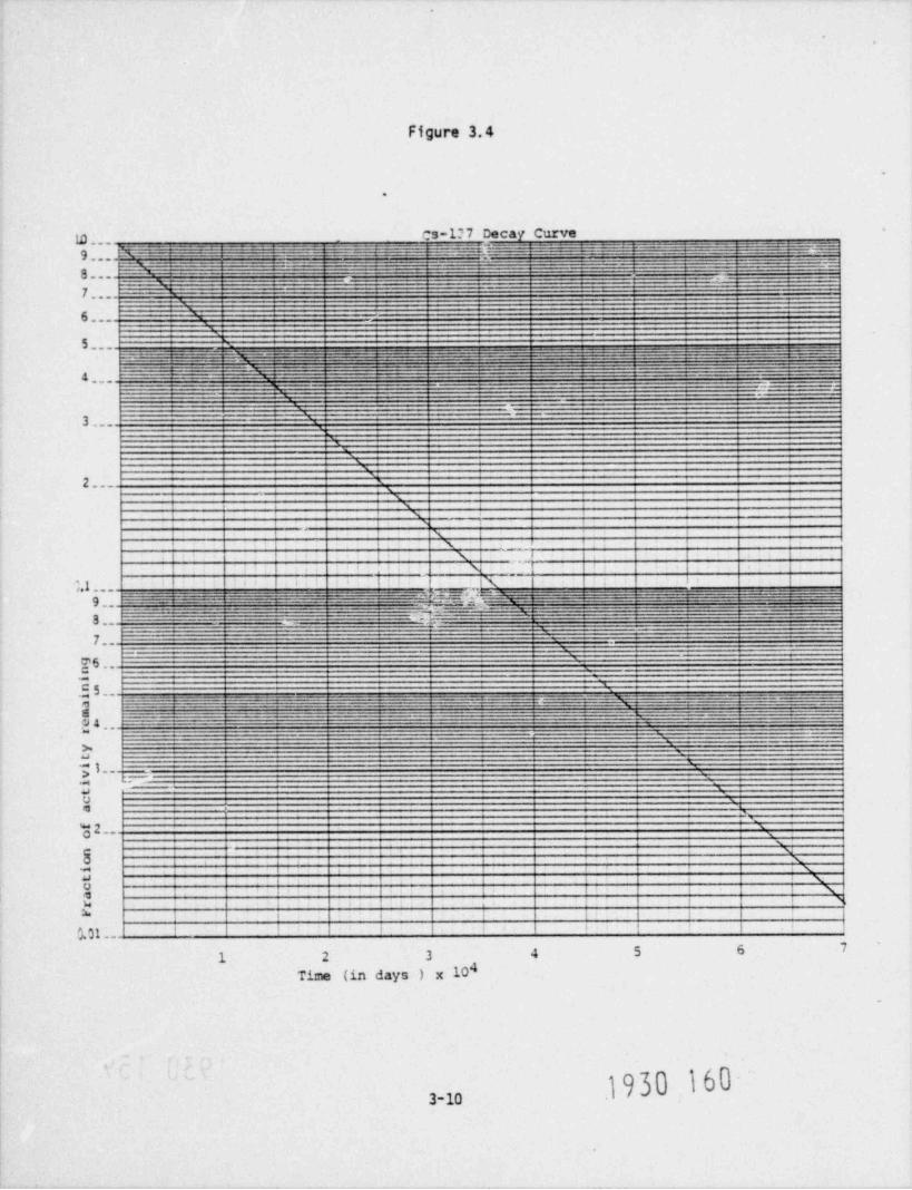
A Co-60 source has an initial activity of 15 Cf. Use Figure 3.4 to find theacitivities after 74, 365, and 3940 days.
We see that 74 days is only a small percentage of a half-life, and so theactivity will be decreased by only a small amount. For 365 days, we still
- - have much less than a half-life, so the activity will be decreased by a bitmore than it was at 74 days, but considerably less than by a factor of 2. For3,935 days, we have more than 2 half-lives, so the activity should be decreasedby a factor just slightly more than 4.
Solutions to the problem are given below.
(a) 74 days, is what part of a year? It is 74/365 year, or 0.2 year.
Using Figure 3.4, we can estimate that the fraction of remaining activity isabout 0.97. We multiply this fraction 0.97 times the original activity tofind the activity after 74 days:
0.97 x 15 Ci = 14.6 Ci
~*M30 158

'
.
Figure 3.3
p_, Ir-192 Decav Curve3 ,,E'NJ--- __=.-- ___. _ ___-_._____ _____ -_ _ __, _
. _ _ _ _ _ _ _ _ - _ _ _ . 2----f. ~ _~..ii'_~ Z Z ' _ _E' i~'Zli i __ __ fifMJN .~2 l . 'i_.fg.__ k..
- . _ _ _ _ _ _ _=__._n - _. __.
-
- ,
7...'
.-
6...,
5... _ . . _ - . - - .. _ - . . . ' , _ _ _ . _ _ _ _ _ . . _ __ - _ _ . _ . - _ __ . . _ _ . _ _ _ _-- t-._--._____._f._y_.m=____-._
.___..
__._____._=_-_._1__-...:+...__. = _ _ . . _ _ _ _ _ _ _ .- _ . - . _ _ _
.. . ___ z .. x=-_ _ _ _ = ._. s.
2______
_ 4 _ _ _ _ _ -_ . . - - - - _ _ . -- - _ - --:n.
. .-~=:-..~_..----3.._
'' - ' '
p ,_ . . -
b;'
,I XIq I N
s... sL 1
s i 3 1
X I
l N%
, x .
1 1 N e.
1 II 6 AJ l
i t | | | i\ s |i i i i N_ t I0,1. ,
),,t _ __. . . g--------N_ _ _ _ _ _ _ _ _ ___
8.. --'
_-..
7.. '.'76 --e.
m
m'c
}5~~-f% ==. ~=? == -C _ _*
_
~~~ ~ :_L_ ::=_-- ~7 ~ }~;Li?'_Q ~ ~-
-.__.___._r--
u 4 . 4-- _ _ _ _ _ _ __ _ _ _ _ __ ._ _s;-" _
.
>.
~3..3e
a01
'"o ..2
_t
x '* .me ;aoq i *i
"w i , . , , , ,
* . 4 i i i i
!, a . i ... v 1. . . . ,
50 100 150 200 250 300 350Time in days
.
e
. 1930 159,'
s
3-9
-.
Figure 3.4
.
.
rs-1?7 Decay Curveg[[[ V- -
.. - - . _ --J-R _-- ..- '-fi=LY G .-_~-- - - - _l ' ~ ~]-- -N =f {_- ~ }._7ZMQ9 :: 5 _J.CZ_i- , - _ . . . _ _ . - - - - . - . - - - __-
3,,, ==-s _ _____ . _ . . __ _
.__ _._ .
7...
6.._ \
$---'
-- ... _ _ -_u ===---- - - - - - - - - - -------- - - - - - - - - - - - - -
____---- -
- . . - _ _ _4.. - *
. .-
. .
3...' ^
'.x'x
2.... I
'
sx
I N\
, sa t i \ ! i
i .x i.
.i
,
! I | \ l _I I I
t i N-
4 6
e i iN i> Ii ,,.g---. - _ _ _ _ _ . _ . _ . . . _ . _ . . _ . . _ . -_
== --
3._..,-<
---E ---
_~
. ---- --.-e.r.Ca a =- =. .x=.w - - . - - --- - - - - - -9_. ----_-.r--.=-- - --- - - _.. - - - - _ - - - - -n -
3.._
,-
_
7..__
T6...
c. 5 -- . . . . .
- _ - . __. . - - , . - . . - _ _ . - - - - _ - _ _ _ _- - . ' , -- - - - . - - - - - . - . - - . - - - -
g__, . _ . - _ - ._ _ _ . - .. _ _ . _ . . . _ - . .. - _ - - . _ __ _ __y 2.
_.-
; _--.
3 4,, ______ _
>, -
a
.ee j " t--'
3'a
U ','e,
* 2-- 'xOs
h 1 1
\V a ii Nwe a e
M i i \e
i i f a ' 3 \w , , , , , s
3. i i . + i i
i i i i i ieg t ,,
1 2 3 4 5 6 7
4Time (in days ) x 10.
3-10 -1930 160-

.
This is a reasonable answer, because it is just slightly smaller than theoriginal 15 C1.
(b) Using F'gare 3.4, we can estimate that the fraction of remainingactivity after 1 year is about 0.88, so:
0.88 x 15 Ci = 13.2 Ci
Again, this answer is reasonable since it is smaller than the remaining activityafter 74 days, but not anywhere near the activity after one half-life, whichwould be 7.5 Ci, or one-half of the original activity.
(c) 3940 days is equal to 10.8 years.
Using Figure 3.4, the fraction of remaining activity atfer 10.8 years is about0.24, so:
0.24 x 15 Ci = 3.6 Ci
9e know that after two half-lives the remaining activity would have been one-quarter of 15 curies or:
15 Ci= 3.6 Ci
4
We also know that 3,940 days is just slightly longer than two half-lives, sowe would expect the remaining activity to be slightly less than 3.75 Ci. Ouranswer seems reasonable.
Specific Decay Curves
Most of the radioactive sources that you will work with will have a decaycurve made up specifically for the source. Figures 3.3 and 3.4 are the decaycurves for a Cs-137 and an Ir-192 source, respectively. These isotopes, alongwith Co-60, are the most common ones used in radiography. Figure 3.11 showsthe relative rates of decay of these three source materials. Note that Ir-192,with the shortest half-life, dscays much faster than Co-60, and Co-60 in turn,decays faster than the long-lived Cs-137.
It is somewhat simpl . to use a decay curve for a specific source than to use oneshowing only the fraction of activity remaining after various times since thevertical scale gives the source activity directly for any date rather than thefraction of original activity remaining after some number of days. Thus, nocalculations are required in using these graphs. You just read off the activitydirectly for the correct date. However, care must still be taken when readingthe logarithmic (vertical) scale.
Examole 3
Find the activity of a 20 curie Ir-192 source after 74 and 365 days. Theactivities can be read directly from Figure 3.3. After 74 days, 10.1 Ci remains.After 365 days, only .03 C1 remains.
1930 1613- 2

.
This is more than the initial activity of the Co-60 source of Example 2. Yet,after 365 days, the Co-60 source still had 13.2 /:i while the activity of the.
Ir-192 source was down to .03 Cf. This is a: .. the fact that the half-lifeof Ir-192 is much less than the half-life of Co-60.
i
.
QUESTIONS - CHAPTER 3
1. The activity of a source of iridium-192 will drop from 100 curies to25 cur'is in a period of 148.4 days. What is the half-life of iridium-192?
2. If you have 2 sources of 2 curies each and place them together, what isthe activity of the combined sources?
3. The half life of Tm-170 is 130 days. If we start with a source of Tm-170having an activity of 50 curies, in how long would the activity of thesource be less than 10 curies?
.
1930 162
3- 12

=~ -,.. ,
.
4 RADIATION DOSE: UNITS AND QUANTITIES
Radioactivity (or, simply, activity) is the number of radicactive atoms thatdisintegrate in a second. It is a measure of the strength of a radioactivesource. However, the activity of a source does not directly tell how muchradiation the source will deliver to people. Obviously, an active source whichis far frem you will not expose you to as much radiation as one which is close.Also, different types of atoms give off different types of radiation. Sometypes of radiation will be more hazardous to you than seme other types.
_
What is important to you is & much radiation your body receives to.There are three quantities that are commonly used to describe the amount ofradiation present. These are exposure, absorbed dose, and dose equivalent.These quantities are related to eacn other, but they are different. Sometimesthey are used interchangeably. This is not strictly correct, although forgama ray radiography the quantities are usually about equivalent.
1. Excesure
" Exposure" is a measure of the interaction of air with gamma or x-rays." Notethat " exposure" can be used only for gamma or x-rays, not for beta or alphaparticles or other types of radiation. Note also that it applies only to air.It does not apply to other materials.
When radiation interacts with air, some of the atoms making up the air become" ionized"--in other words, some electrons dissociate themselves from the atomsof the air.
Exposure is a measurement of how much ionization has occurred._ _
The unit for exposure is the roentgen (abbreviated as R or r). It was one ofthe earliest units useu in radiation work.
The roentgen has two limitations that limit its practicality. First of all,
there are other types of radiation besides gamma and x-rays. Also, people arenot made up of air. The advantage of the roentgen is that it is easy to measure.An air fiibd detector merely measures the electrical charge produced when radia-tion ionizes the air.
. _An.importan'. thing for you to remember about the reentgen is that, since itapplies only to x-rays or gamma radiation, any instrument with a scale whichreads " roentgens" is one which is intended for x-rays or gamma radiation.
Jit
Note that " exposure" really has two different definitions. One definition isthe very technical definition given here: a measure af the ionization in aircaused by gamma or x-rays. However, " exposure" also has a commoc usage: beingsubjected or exposed to some hazardous substance, for example. Thus we cansay, " Exposure to enlorine gas is dangerous." Or "He was exposed to beta radia-tion," even though he is not made of air and beta radiation is different fromgama or x-rays.
4-1
1930 163
.
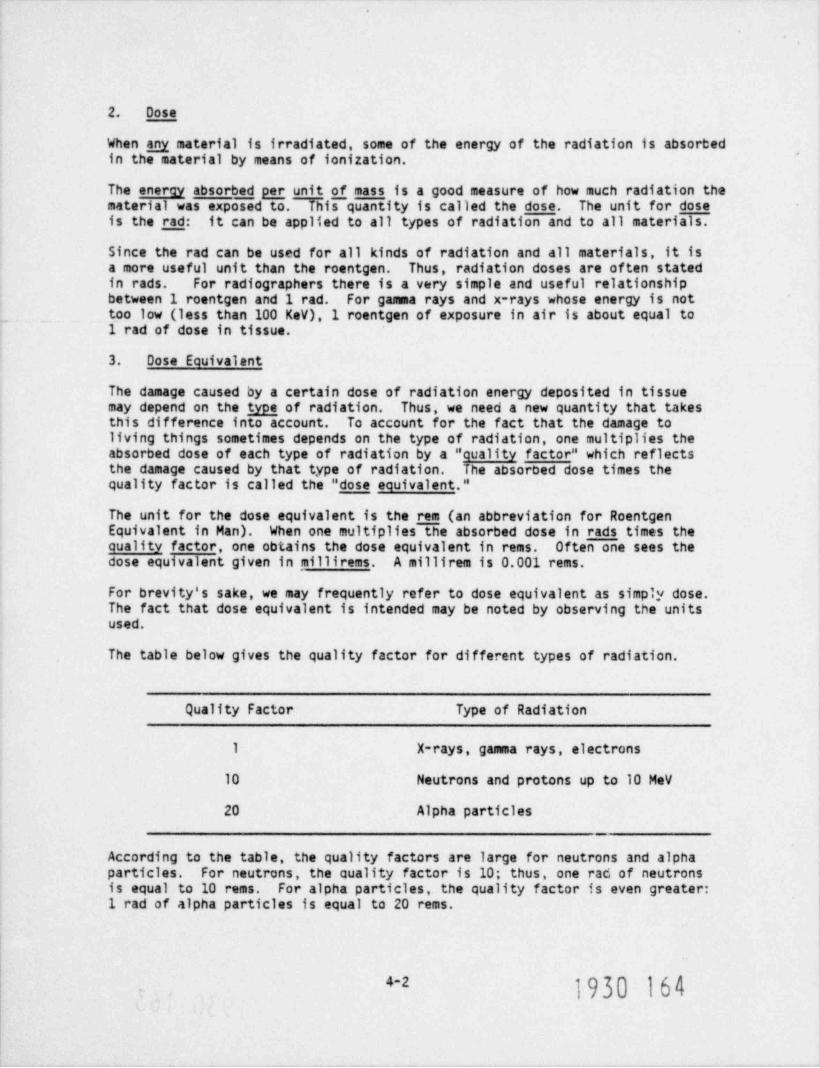
2. Dose
When any material is irradiated, some of the energy of the radiation is absorbedin the material by means of ionization.
The energy absorbed per unit g mass is a good measure of how much radiation thematerial was exposed to. This quantity is called the dose. The unit for doseis the rad: it can be applied to all types of radiation and to all materials.
Since the rad can be used for all kinds of radiation and all materials, it isa more useful unit than the roentgen. Thus, radiation doses are often statedin rads. For radiographers there is a very simple and useful relationshipbetween 1 roentgen and 1 rad. For gamma rays and x-rays whose energy is not
_.too low (less than 100 kev),1 roentgen of exposure in air is about equal to1 rad of dose in tissue.
3. Oose Equivalent
The damage caused by a certain dose of radiation energy deposited in tissuemay depend on the type _ of radiation. Thus, we need a new quantity that takesthis difference into account. To account for the fact that the damage toliving things sometimes depends on the type of radiation, one multiplies theabsorbed dose of each type of radiation by a " quality factor" which reflectsthe damage caused by that type of radiation. The absorbed dose times thequality factor is called the " dose equivalent."
The unit for the dose equivalent is the rem (an abbreviation for RoentgenEquivalent in Man). When one multipliesThe absorbed dose in rads times thecuality factor, one obtains the dose equivalent in rems. Often one sees thedose equivalent given in millirems. A millirem is 0.001 rems.
For brevity's sake, we may frequently refer to dose equivalent as simply dose.The fact that dose equivalent is intended may be noted by observing the unitsused.
The table below gives the quality factor for different types of radiation.
Quality Factor Type of Radiation
1 X-rays, gamma rays, electrons
10 Neutrons and protons up to 10 MeV
20 Alpha particles__
According to the table, the quality factors are large for neutrons and alphaparticles. For neutrons, the cuality factor is 10; thus, one rad of neutronsis equal to 10 rems. For alpha particles, the quality factor is even greater:1 rad of alpha particles is equal to 20 rems.
1930 164e2

.
Not.2, honever, that for x-rays and gamma rays, the quality factor is 1. Thus,for this type of radiatien,
1 rad = 1 roentgen.1 rem =
No wonder these units are often used interchangeably in many circumstances! Infact, in industrial radiography, most of the time you will be concerned withgamma radiation. For the sake of brevity, we may frequently refer to doseequivalent as simply dose. Even tabla and graphs for gamma radiation willgive data in terms of "R", where "R_" may be read as rem, or rad, or roentgen.If, in some specific case, dose equivalent is intended, this should be notedby writing " rem."
.
A radiation detection instrument which really measures exposure in roentgenssimultaneously tells you the biologically important quantity, the dose equiva-lent in millirems.
,
4. Exoosure and Dose Rates
It is often important to know how fast radiation exposure or dose is beingreceived. For example, we may want to know, "What dose will I receive if Istand here for one hour?" Exposure, dose, and dose equivalent can each bestated as a rate. Thus, it is common to see rates such as roentgens / hour,rems / hour, millirems / hour, and so forth. One roengten/ hour means that aperson standing there for one hour will receive an exposure of one roentgen.
5. Characteristics of Gamma and X-ravs
For gamma radiation, you may see data in terms of "R/ hour"; that is, roentgens /hour, or rads / hour, or rems / hour. You may also see "mR/ hour", where "m" standsfor " milli", or one-thousandth. To convert mR/ hour to R/ hour, one divides by'. , 0 0 0. Another possibility is "mR/ min", or "mR/ minute": to convert "mR/ min"to "mR/ hour" you must multiply by 60, because there are 60 minutes in an hour.
To be able to convert exposure rates from one unit to another is very important,of course. For example, how do you convert a reading in mR/ min to R/ hour?Obviously, you multiply by 60 and divide by 1,000. To convert R/hr to mR/ min,you perform the inverse operation; that is, you divide by 60 and multiply by1,000.
(Please note that you may have other types of units for exposure and dose rates.For example, the time may be given in seconds and you will have a unit such asmR/sec. If you know that, at a certain position, exposure is 1 mR/sec, what isthe exposure in terms of mR/ min? Clearly, 60 mR/ min. What is it in terms ofmR/ hour? Clearly, 3,600 mR/ hour, And in tenns of R/hr? 3.6 R/ hour, of course.)
Although the activity, or strength, of a source is given in terms of curies, aso source are more energetic. The1 curie source of one radioisotope like Co
table below gives the gamma radiation level at a distance of one meter (or 100centimeters, or approximately 3.28 feet) from different sources.
4-3

.
Approximate Gamma Radiation Level at One Meterfrom One Curie Source of Certain Radioisotopes
Sodium-124 1.93 R/hr.Cobalt-60 1.33Iron-59 0.644Iridium-192 0.51Cesium-137 0.36Zirc-65 0.3CGold-198 0.25Iodine-131 0.23
These are the radiation levels for 1 curie sources. For any given source, you-
should multiply these levels by the source activity. For example, for 10 curiesources, you multiply these levels by 10. For a 10 curie Coso source, the gammaradiatioa level at a distance of one meter from the source is 13.3 R/hr. What isthe level one meter from a 100 millicurie Cs-135 source? Clearly, 0.1 x 0.36 =0.036 R/hr, or 36 mR/hr, er 0.6 mR/ min, or 0.01 mR/sec.
You should also note that these radiation levels are given for a distance of 1meter away from the sources. The levels are different at other distances.When, in Chapter 6, we study the effect of distance on exposure, you will seethat at larger distances the radiation levels are lower, while for smallerdistances, the radiation levels are higher; in fact, they can be much, muchhigher.
Because of the importance of the effect of distance on exposure, we shall devotea whole section to it in Chapter 6 when we shall introduce the " inverse squarelaw."
6. Radiation Dose from Natural Sources
Is it true that radiation is basically " manmade" and " artificial?" No, notat all. The human race has always been exposed to radiation from naturallyoccurring sources.
Is it true that everybody is constantly exposed to radiation in the environment?
Yes. Everybody in the world receives a small amount of radiation exposure atall times. Radiation is given off constantly by radioactive materials allaround us -- in the ground, in the walls of buildings, and even in our bod'es.In addition, the earth is bombarded by radiation from the sun and from outeespace, known as cosmic radiation. These low levels of radiation do not haveany noticeable effect on the health of individuals.
4-4

.
The exact amount of radiation received by a person depends on where the personlives. Persons living at high altitudes receive more cosmic rouiation thanpersons living near sear level. Then too, some ground areas nave higherradiatien levels than others. Figure 4-1 below illustrates the appropriateaverage for persons in the United States.
FRC/A00TER SPN.F.
FROA\ BulLDING gmrem//r.MATERIALS / -
O-30 mrem /yr. 4 "
N 2..
aFRCbA INSIDE. , 'THE BCDT'
' <'
l3 [4'. k'
mrem /yc2
.
\ FRCtA. .'
THE GROUND'
M.%60 ~.,i'
._ _
Radiation exposures (in millirems) receivedin one year from tne environment
Source: U.S. Environmental Protection Agency, " Radiological Quality of theEnvironment," EPA-520/1-77-009, May, 1977, Washington, D.C.
Figure 4-1
.,
1930 1674-5

.
The total of 125 millirems per year is an estimated average for the UnitedStates. The exact dose varies with locality. This is due to differences inthe amounts of natural radioactive material present and to variation in theintensity of cosmic rays with altitude and geographical position. For example,cosmic radiation increases by a factor of 2 in going from sea 1,9 vel to 10,000feet. Latitude also has an effect; for example, the dose rate increases byabout 15% in going from the equator to Vancouver, Canada. Reported naturalbackground radiation levels for the United States range from about 90 to200 millirems per year. In some parts of the world, such as certain regionsof India and Brazil, much higher levels prevail. Radiation from thorium-bearing sands in these areas makes the external environmental absorbed dose10 to 30 times the world average.
7. Radiation Oose to the Pubife from Man-Made Sources
People are also exposed to man-made sources of radiation. Examples of man-maderadiation are medical x-rays, fallout from nuclear weapon tests, radiation fromcolor television sets, radiation from radium or tritium luminous dial wrist-watches and clocks, and radiatian from uranium false teeth.
The largest man made source of exposure to radiation in the public is medicalexposure, such as chest x-rays and dental x-rays. The average dose equivalentper person per year in the United States from medical use of radiation isapproximately 74 mrem. A person undergoing a gastrointestinal series of x-raysmay receive several rems. A person undergoing cancer therapy may receive smalldoses, a large lo al dose (up to hundreds of rems).
The doses from sources of man-made radiation are shown in Table 4.1.
8. Occupational Radiation Doses
Radioactive materials are used in various industrial and commercial applications.Individuals tnat work in fields where radioactive materials are used are exposedto a radiation environment during their occupation. Some of these occupationsare medicine, radiography, nuclear reactors, waste disposal, and radar.
There are approximately _ _ radiation workers per in the United States or aboutone out of each workers. The average annual individual occupational doseamong some workers regulated by the NRC is about 0.8 (?) rem / year.
Some average doses for certain types of workers are shown in Table 4-2.
1930 168,,

.
Tab 1( 4.1
Whole-Body Doses From Man-Made Sources of RadiationAnnual Average Ovee U.S. Population
OoseSource (mrem)
Medical *** 74.(Primarily diagnostic x-rays)
Fallout * 4.4~ ~ ~ ' -
Uranium Fuel Cycle * 0.01(Including enrichment, trans-portation, reprocessing, and
_
reactor operations)
Consumer Products ** 1. 5(largest fraction is from TV
sets)
Building Materials ** 3.5(There is an additional 5 mrem lungdose to the pcpulation as a resultof burning natural gas which con-tains natural radon)
"U.S. Environmental Protection Agency, Raolological Quality of the Environ-ment, EPA-520/1-77-009, Washington, D.C., 1977.
** National Council on Radiation Production and Measurements, Radiation Expo-cure from Consumer Products and Miscellaneous Sources, NCRP Report No. 56,Washington, D.C., November 1977.
*** National Academy of Sciences-National Research Council, The Effects onPopulations of Exposure to Low Levels of Ionizing Radiation (NationalAcademy of Sciences-National Research Council, Washington, D.C., 1972).
" "1930 169
4-7

,
Table 4.2
Average Annual Doses (rem) of Workers with Measurable Dosesat Some NRC-Licensed Facilities *
Number ofIndividuals with
Category Measurable Dose Average Dose_
Power Reactors 44,233 0.74
Industrial Radiography 6,197 0.51
Fuel Processing andReprocessing 7,004 0.25
Manufacturing and Distributionof Radioisotopes 2,459 0.54
* Note: Only workers with measurable doses included. Workers with no measur-able dose are assumed to be not actively engaged in radiation work.
Reference: Earbara Brooks, " Occupational Radiation Exposure - Tenth AnnualRiport - 1977." NRC Report NUREG-0463, 1978.
} 9 f)0 170~
4-8

.
QUESTIONS - CHAPTER 4
1. Explain briefly where background radiation comes from.
2. What are some of the factors that determine the natural background?
3. What are the differences between exposure, absorbed dose and dose equivalent?
4. Match the following quantities with the corresponding unit.
(1) Exposure (a) rad
(2) Dose (b) rem
(3) Dose Equivalent (c) MeV
(4) energy (d) roentgen
5. Explain briefly how gamma rays lose their energy through interationwith matter.
1930 171
4-9
..
5. HAZARDS OF EXPOSURE TO RADIATION
What is the danger of radiation to me?
At this point in the course, this is a reasonable question for you to ask.
When we are exposed to radiation, the radiation interacts with the individualmolecules of the individual cells that make up our body. These interactionscan cause changes in the chemical structure of the cell molecules. Thesechanges may be harmful to the cell. Some cells may be affected so that theirability to reproduce normally may be impaired. Some cells may die. Othersmay be totally unaffected. If radiation doses are very large, so many cellswill be damaged or killed that the person may die. If a very large radiationdose is delivered to only part of the body, such as the hand, amputation maybe necessary. If radiation doses are smaller, many of the cells can repairwhatever damage has been done. However, in some cases, the affected cells mayreproduce, but in an uncontrolled manner, eventually causing the growth of acancer tumor. If the damaged cell is a reproductive cell, a sperm cell or anovarian cell, the result can be a child with a genetic defect. It is conserva-tively assumed that any amount of radiation, no matter how little, has somesmall chance of causing a harmful change.
As you may know, large doses of radiation are used as part of a therapy programfor some cancer patients. Even though radiation may cause cancer in healthycells and have other side effects, when used to treat cancer patients the maindamage is done to the tumor. Actually both healthy cells and cancer cells aredamaged and begin to repair themselves as soon as the treatment ends. However,the rapidly dividing cancer cells have a greater chance of being destroyedbecause they are more sensitive to radiation during the dividing process. Inany case, this type of therapy is only given to those patients who are alreadyseriously ill. Other than for such cases, exposure to radiation is notbeneficial.
What will happen if I am exposed? What can I expect to feel?
Again, these are fair questions to ask. First, you will feel nothing. Radia-tion cannot be detected by any of your senses. Only your survev meter can tellyou if radiation is present. And, primarily, the degree of raciation injurydepends upon the total amount of radiation absorbed in the body, the length oftime it took to receive the given dose, and the part of the body exposed.
The types of radiation injury that occur can be divided into short-term anddelayed effects.
1930 1723- 3.

',
5.1 Short-Term Effects from Whole-Body Exoosure
Short-term effects are effects that become evident within minutes to withinweeks of the exposure. If you are exposed to a large amount of radiationduring a short period of time you might die within a few days or you might nothave any detectable effects, depending on the amount of radiation received.
It is impossible to say exactly how much radiation will kill any specific indi-vidual, because we all vary slightly in our resistance to any attack on thebody, whether it is by radiation, electricity, poison, injury, or disease. Itis quite certain, however, that no human being could survive 1,000 rads oftotal body radiation delivered in a day, and essentially certain that no onecould survive 1,000 rads of total body radiation delivered in less than a week.
The portion of the total body exposed and the length of time involved in expo-sure are most important. The effect of 1,000 rads of radiation energy absorbedby the total body is by no means the same thing as 1,000 rad:; delivered to asmall portion of the body any more than a third degree burn of the palm of thehand is the same thing as a third degree burn to a large area of the body.
Similarly, the short space of time is an important part of the consideration.A short space of time is about 24 hours. The ability of the body to withstanda harmful agent is, of course, increased if the same amount of the agent givento the body it spread out over a longer period of time. Whiskey can be toxic,but many people, apparently without any demonstrable injury, can drink an ounceof whiskey each evening before dinner over an extended period of time. If,
however, a person attempts to consume a 3 month's quota of whiskey in one sit-ting, he will probably die of alcoholic poisoning before the body has had suffi-cient time to recover from the toxic effects.
The median lethal dose, or 50-30, is the dose that would result in 50% of thepeople so exposed dying within 30 days. This is a statistical concept. If
gly 2peopleweresoexposed,bothcoulddieorbothcouldsurvive. The50-30 for penetrating external radiation is about 400 to 500 rads delivered
to the total body in 24 hours or less.
This means that if a large group of people were subjected to 450 rems of totalbody radiation within a 24-hour period, approximately 50% of these people woulddie within 30 days, and the other 50% would recover. The life span of recoveredpersons may be reduced by this large exposure, although this is uncertain.There would be an increased statistical probability that such individuals mightincur cancers after 20 or 30 years.
As a result of 100 to 200 rems of total body radiation in a short space of time,we would expect nausea, fatigue, vomiting, diarrhea, and loss of body hair, butno fatalities. These symptoms indicate the onset of the acute radiation syndromeor radiation sickness. These symptoms are also present in those people receivinghigner ooses incluoing people fatally exposed.
At about 50 rems of total body radiation in a short space of time, there may beslight temporary blood changes which would reverse themselves with the passageof time. The person would have no observable symptoms which he would noticehimself.
,
1930 1735-2
.

At '5 rems of total body radiation in a short space of time, we might detectonly some abnormalities in cell chrcmosomes when they were viewed under amicroscope.
Table 5.1 summarizes the expected symptoms and effec'.s of total body dose ofradiation delivered in a short space of time.
Remember that all of these figures are on the basis of total body radiationwithin a short space of time. We should note that no radiographer in theUnited States has ever died as the result of the acute effects of ', hole bodyexposure to radiation. However, radiographic sources have caused such deaths,as discussed in case history number 10 in Chapter 11.
,
5.2 Radiation Burns 4
Inaccidents involving radiographic sources it ismore common for a large dose tobe delivered unevenly to the body rather than spread out evenly over the entirebody. In severe accidents a part of the body receives a radiation dose greatenough to cause "radiatin burns." Usually the hands and fingers receive theburns, but occasionally other parts of the body receive the " burns." These" burns" to the hands result when a radiographer toucnes or almost touches a
. source for a few seconds. The temperature of the source is not high, but theradiation right near the source is extremely intense. The " burns" are causedby radiation, not heat.
The nature of radiographic sources is such that the radiation intensity decreasesvery rapidly as you move away from the source. This decrease in radiation inten-sity at you move further from the source will be discussed in detail in the nextchapter.
When a radiographer puts his hand near enough to a source to cause " burns," theradiation dose to the rest of his body is usually not enough to cause him toget the " radiation sickness," that was discussed previously. However, therehave been cases where people exposed to radiography sources at some distancefor longer times have died from " radiation sickness" but never had " radiationburns." (See Case 10 in Chapter 11.)
Radiation burns first become evident when the doso to that portion of the bodyexceeds perhaps about 200 rads. A slight reddening of the skin will occur. Theperson receiving the burns will not feel any burning or pain while he is beingexposed to the radiation nor will the reddening appear immediately. The firstreddening usually appears several hours after exposure to the radiation, andfades after some further hours or days. The reddening =sy be associated witha feeling of warmth or itching. At 200 rads the reddening may be so slightthat it is not noticable, especially on dark complexioned people. At 600 radsthe reddening should be fairly evident, but will still disappear after somehours or days. At much higher doses the initial reddening will appear withina few minutes. Then it will disappear and reappear several times. If you havebeen performing radiography, an unexplained redness on your skin may be a sign ithat you have received a severe radiation overexposure.
1930 174s3

)s '
da lr a
tm0 epo5 l es
bcoo axmt veo
l r ras eshvd stceia bc gvr oeear f mmu0 of oaS( Nesd
)s sd e ea g l
r n ba a
0 h v0 c r1 e
d so o bt o o
l
l s b rad esva t ht /ir h t cv g oer0 i fu5 l ofS( S Ne ;
ase ~t
- l tht u-) t kl orh oiss i f ai prg bl ud m oe ;sai apoa on wft i e mi nr vo s oaedw novoe s sl ol i cei
l0 ,a m o a g r p ;f rtb5 ae o l rnhrro yspca2 eh t eit uo l s eb sr p ;nl pl s eerfoo ur m reee l s kl on* rt aa y s i ger;ao i noi
S P ni s m a f o e pl l upS s dy o h; sg rE ld e a r t ed a;e ysyoN aa ldd e p ft n ;h n t rhbK vr bn h m oiahria et hC i iat t y t t rkr vndtI v0 s s o s sesl ose ooelS r0 sgr spsam d cmt a
u1 oni o o opeeeno e aeN S( Pif N N Lanhhom R3ch1 O
. I
5 T fA ) o te I s f h; su
l D d ol g - t a uob A a an e ue oba R r sri es oh i aT e s sel r l o mr r )
F l0 g m onee pn r eysO b5 nns o l eev r ;f a st mi5 iir t gf e u;roit ieS s t u p ; f pno dh el rM so i ao m rdd il n g raO ot meh y i nn; ;kl o ;i ot0T P oh s aaah esaite mr5P s vrw h t g pt aw o4M ld re r esl an ao nmY aa ,af e ft sa ho;mrf i( rS vr ai h oiee r smho o
edt t t nh rsdat hsfi
v0 s s o sek ot el s t er0 udr spal moefds as%u3 ani o opel e pl nno ea0S( Naf N L awi Hsbial Dc5
-m
da- nel
w r o gf ;
de nro an te nf i emshid hl) a het r n gbe t sthor ,a i
ar gs mo poh ebo nr o ,smth w )om ii t na eat %r tf pi e e h e u r fh0pr i m hl noe ot0mo mn y;rpormv a1I oi ssrrt e e se
s v eau dff sdyld a r si p e n o o l
aa ,e ead uu , l ;bvr ah hc ,d nt ta
i ers t se doa dhbv0 srr oeegnoio i0or0 uau msaiot r pi ru7 aio ooahkl ah aepS( Ndh Nscrsbmt Rw( - D.euD - %
_ C . L-ret e k) k k kf r y et e e e,
Au a en e e es D We W W Weo tmp t ta d d hix s sl n r tTE 1 1( 2 3 4
r , , ,, , e = - ?. . > ..
.
W9 /p-o ! I ~ " i"% 'te
* f- -_1 Y~ W~
* - m.
d 4 A,. (3 n %-~,.
- -
}r ;. . ...
. .-
==; * * *~
-.E.!r-w.
A. ~ijf" .
' ,i:1. _. ,
y ..
-.
-- . nt-
$n". n; :. . .. ",Se~
3. e.
'.) .: ..--.c d = -3, - r.
- '< (,
.
. .% ' .., ~ . $r,t .h .: ::?-
.~.~*
:. .
- T =ir'
-,
. . - .t . . = - .-.,
H**.,,.- .f % *; 'T*. , ''
.:',
: 1?-*. .. .- A''. : .,iv,.f.77 '
s
[,- .,i.'.~ ' .-
- - *,
. .. -
%-- wnk.:. .-
.
' 2: 4 EM.=.....p.-.
. 5<.
FE.0 ~1. d5!2iEl-
m) o .,. . ,..n ., o .., . g ,,,, c
w ;g -= .. s, .
. ' ''. .; . . . ..
'J,5 ' .*q 3-
E24; i y, . .. - :.'
v -.:
.L . -- ' ..
. . . -. -.
$' *
"Ab. w' -a-s' '..n. : ''fy.
' '
:.g:a.,>?.>-. r
;.. o .,-
.
.. o.., ,.
. a.,
.. . ,
A.
:.= .
ia. ....
. . _
c,, % . . , ., , . .:. . . ..
, - . . . _ ..
1930 176

y._ s- 2 s)r
w a w a ,_ p o . u - x <, ~LA) & G, IQ n.e 'M L _ :=>
...
0
..
e r
6
-1 e'
%
., . [''
~. ,
*% , 'p., -
- - .:,',.Y i, '.** ''r7 5 "'3 '~
,-
',,f.. .- .- , . ., ,- '
, , * * *'* i . . 2.a
,} ,* e * } - , , j e .9f d /w 'A ) @ **, * , og
*
ry''$$ggg%7''-: .h. +s - , .S'.Myt"* ~ .;.4en.m.. : . . : v. ...
i'^ 3gO e ; .,- . !,
*.,
~
' ' : ,];.'t;.4, ..
,
y, ,.,..:. * ..
,, ,_ ""
t {f'g-),.]T,. ,. , . -
' " . . ~ . - -?:. ,..
,
,, .
e
'
*
I. *.e*- .-
. -
=
..
r
e
S
a-4;O;Y.
Q.?$ ."
r . .c- ..- ,, , ... .
..
1930 177
.
The initial reddening will soon fade, and the skin will appear normal. However,reddening will reappear at some later time. Depending on the dose this laterreddening may appear perhaps as early as a week or as late as three weeks afterthe exposure. This time the reddening may last for 3 or 4 weeks.
Some loss of hair will also occur if doses exceed perhaps about 200 rads. Thehair loss becomes noticable two or three weeks after the exposure. Above 600rads loss of hair may be permanent. Below 600 rads hair should regrow, but maybe gray.
Below about 600 rads the reddening and hair loss are the only symptoms that areexpected. From doses below 600 rads recovery should be fairly complete, andmedical care is not necessary.
At doses exceeding 1000 rads serious tissue damage can result. At these dosesthe secondary reddening will erupt into painful burns. Blisters will form andbreaks open leaving raw, painful wounds which are vulnerable to infection.Swelling, tenderness, and inflammation will occur. The need for medical caredepends on the size and severity of the burn. The visible damage may be healedwithin a month or so. Delayed symptoms from deeper tissue damage may occurduring the next several months. Some permanent dmage to the tissue, such asthinning of the skin and scarring of the underlying tissue may occur. Thisdamage will predispose the person to cancer of the skin.
At 5000 rads, there results a burn resembling a scalding or chemical burn. Painoccurs promptly and is intense. The burned areas may be very slow at healing ormay not heal without amputation of some tissue or skin grafting. Future medicalproblems with such highly exposed tissue can be expected.
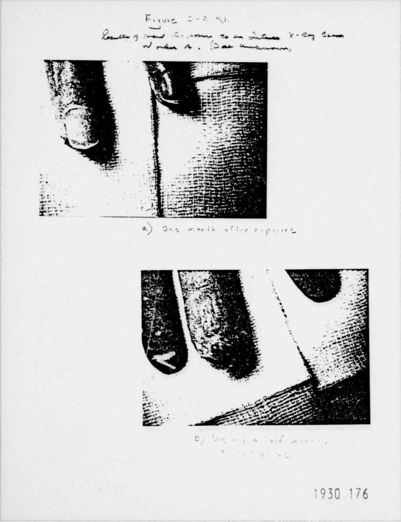
These doses of thousands of rada sound high; It must be remembered, though, thatat close distances to a radiography 100 Curie 192Ir source in contact with theskin can yield a hand dose of 5,000 rads in less than one minute. A 60Co samesource of the same size would produce a dose of 5,000 rads in about 15 seconds.A radiography source must never be touched or handled directly - not even fora few seconds. Figures 5.1 and 5.2 show radiation damage suffered by twoworkers whose hands were overexposed to x-rays.
5.3 Cataracts 5
Cataracts are a cloudiness or apacity of the lens of the eye which cause loss- of vision or even blindness. Large radiation exposures can cause cataracts. A
- - slight degree of apacity can be caused by a single dose of 200 rads or 400 radsdelivered over several weeks. At 500 rads in a single dose a more seriouscataract condition will occur. Cataracts are not caused by prolonged exposureto radiation at normal occupational levels. Radiation induced cataracts aregenerally distinguishable from cataracts due to other causes and do not occurin radiation workers except for some very rare accident cases.
5-7

'
.
5.4 Delayed Effects - Cancer and Genetic Defects in Offsoring
Delayed effects take years, decades, and sometimes generations to becomeapparent, and the specific radiation injuries are consequently more difficultto identify. These effects include initiation of cancer in the exposed peopleand genetic defects in the decendents of such exposed people. The difficultyin identify;ag radiation as the cause of these delayed effects is that cancerand genetic defects occur in any population, even if you had not been exposedto radiation beyond background levels. The fact that radiation beyond back-ground may indeed cause delayed effects can only be ceduced by animal experi-ments or by studying large groups of people who have been exposed to radiationfor various reasons.
If you are exposed to radiation, you may never experience any observable injury.However, the chance that you will is increased. For example, if you receive aradiation dose high enough to increase your likelihood of coming down withleukemia, there would still be a high probability that you will not get thedisease, since the increased probability is still rather small. This isgenerally true for all of the delayed effects, whether brought on by a suddenexposure to a large dose of radiation, or exposures w small doses over severalyears. An analogy to this situation would be if you work in an office and oneof your coworkers became sick with the flu. You may or may not get the flu.However, even if no one in your office has the bug, you may still catch it.And if you did catch the flu, tnd your coworker was ill, you could not be cer-tain that he was the reason f',r your illness.
If a ) regnant woman is employed as a radiation worker, special considerationsshco'I apply to doses which she may receive. The growing fetus is believed tobe ms r. more sensitive to radiation than is an adult human being. The earlypart of the pregnancy (first three months) is felt to be the most criticalperiod. During this time the unborn child is growing and changing more dramati-cally than at any other time in its life. The single cell formed at conceptionrapidly divides. Cells take on a variety of specialized functions, all theinternal organs are formed, and the human body is taking shape. Any eventwhich might change or interrupt this early development could have serioushealth effects on the child. Childhood leukemia and other possible cancershave been connected with radiation exposure to developing fetuses as a resultof medical irradiation of expectant mothers.
The amount of risk involved in receiving low-level exposure to radiation hasbeen extensively studied. The Biological Effects of Ionizing Radiation (BEIR)Committee of the National Academy of Sciences conducted an extensive study ofthe effect of low-level radiation exposures. One conclusion was that it isalmost impossible to demonstrate a cause effect relationship between radiationand cancer at dose rates within the limits established by the NRC.
The Committee reviewed both experimental animal data and results of large scaleepidemiological studies of human population exposed to radiation. Most of theresults which showed a relationship between dose and effect were for quite highdose- (typically greater than 100 rem). When the doses were plotted againstthe number of effects observed (e.g., leukemia), a rather straight line wasobtained. Although there were no data for the low dose levels, the Committeefelt that it was conservativc to extend this straight line to the low doserange. In this way, one could predict the number of cases of a particular
5-8
1930 179

.
.
cancer which one might expect to see if a large population was exposed to agiven dose. The Committee intended that these risk estimates might be usefulin reviewing the effectiveness of radiation safety programs. They mightprovide further guidance in judging the impact of certain existent or plannedmodern technologies.
Considering lethal cancers of all types, the Committee estimated that a wholebody dose of one rem received by an individual would increase that person'schances of contracting such cancer by about one out of 10,000. The normalincidence from all sources of lethal cancer in the population is about one outof six. Said slightly differently, if a group of 10,000 people each receivedone rem of whole body dose, we would expect one extra cancer in that group overits lifetime. This one would be in addition to the approximately 1,700 whichwould result without any additional exposure to radiation. We might also notethat, using the BEIR Committee's risk estimates, background radiation mightaccount for about 2,000 to 4,000 cancer deaths per year in this country.Cancer from all causes kills about 350,000 people per year.
Genetic effects of radiation were also studied by the BEIR group. It was con-cluded that the genetic risks to all generations were slightly less than thecancer risks associated with exposure to radiation.
The United Nations Scientific Committee 2 on the Effects of Atomic Radiation hasalso reviewed epidemiological data and arrived at similar conclusions as theBEIR report.
Some recent studies of the effects of low level radiation, especially theMancuso3 report, have claimed that the BEIR risk estimates are not conservativeenough. Considerable controversy exists at the present time reca' ding thistopic. Since the risks of radiation induced cancer is small, vt.; large popula-tions would be required to determine what the increased risk is for low doses.It is cuestionable when, if ever, this question will be absolutely resolved.
How have radiation dose limits been set?
The effects of radiation on man have been of concern to the scientific communityfor well over 50 years. The oldest of the sc %ntific bodies that still haveresponsibility in this field are the Internatical Commission on RadiologicalProtection (ICRP), formed in 1928, anc the National Council on Raalation Protec-tion and Measurements (NCRP), formed in 1929. Botn codies are composea of inai-viaual scientists.
After World War II and the advent of nuclear weapons and nuclear power, interestin the effects of radiation increased. Many studies of the effects of radiationwere started, including studies of atomic-bomb survivors and many large-scaleanimal experiments. A high level of scientific research on radiation effectshas continued to this day.
The results of these scientific studies have been reviewed periodically bypanels of scientists seeking to estimate the risks from exposure to radiation.In 1955, the president of the National Academy of Sciences (NAS) appointed agroup of scientists to conduct an appraisal of the effects of radiation onliving organisms. That study, called " Biological Effects of Atomic Radiation,"led to a series of reports issued from 1956 to 1963.
1930 1805-9

*.
Also in 1955, the General Assembly of the United Nations established the UNScientific Committee on the Effects of Atomic Radiation (UNSCEAR). UNSCEARhas periodically issued reports, the latest in 19/7, which have served asreviews of worldwide scientific information and opinion concerning human expo-sure to atomic radiation.
In 1959 the U.S. government formed the Federal Sadiation Council (FRC) toprovide a federal policy on human exposure to radiation. In 1964, at therequest of FRC, the National Academy of Sci.nces - National Research Councilestablished a scientific committee on the Bi> logical Effects of Ionizing Radia-tions (BEIR) which continues in existence today. The BEIR committee has issuedthree major reports on the effects of radiation, in 1972, 1977, and 1979. Thefindings of the latest BEIR report will be discussed here because this is thelatest work of a major authorative scientific committee.
The BEIR committee is currently composed of 22 prominent scientists. Thesescientists have evaluated the massive research results on radiation effects toproduce their reports for the National Academy of Sciences - National ResearchCouncil. The BEIR committee risk estimates form the basis of the dose limitguidelines issued by the U.S. Environmental Protection Agency. (The EPAabsorbed the functions of the FRC in a 1970 government reorganization.) Thenregulatory agencies, such as the U.S. Nuclear Regulatory Commission, implementthe EPA guidelines in their recommendations.
Thus, the radiation protection limits are set by government agencies which haveenforcement powers, but the limits derive from committees of scientists who havestudied the effects of radiation on living organisms.
How certain can you be that the present radiation limits are soundly based?You can be fairly certain. The limits are based on more than 50 years of care-ful study, billions of dollars of scientific research efforts, the delibera-tions of committees of scientists in the U.S. and abroad, and the carefulreview of the scientists' work by U.S. government officials who have been givenby Congress the responsibility to protect the public health and safety.
The BEIR committee also studied the claims of some people that radiation effectsare considerably greater than has been estimated by the generally acceptedlinear-nonthreshold model of risk.* The entire committee found no substancein theories that effects may be proportionally greater at low doses than athigh doses. Thus, the claims that radiation is much more dangerous than hadbeen assumed in setting radiation protection limits found no support in thiscommittee of scientists.
"Specifically, the BEIR committee studied: (1) the work of Mancuso, Stewart,and Kneale on exposed workers at the Hanford works in Richland, Washington;(2) the studies of Irvin J. Bross on people exposeu to medical x-rays; (3) theNajarian and Colton studies of Portmouth, New Hampshire Naval Shipyard workers;(4) comments by Ernest Sternglass on effects of fallout from weapons tests;and (5) studies of Victor Archer on variation of cancer rates with backgroundradiation.
,
5-101930 181

REFERENCES
1. National Academy of Sciences - National Research Council, The Effects onPopulations of Exposure to Low Levels of Ionizing Radiation, Washington,1972.
2. United Nations Scientific Committee on the Effects of Atomic Radiation,Ionizing Radiation: Levels and Effects, United Nations, New York, 1972.
3. Mancuso, T. F. , A. Stewart, A. Kneale, " Radiation Exposure of HanfordWorkers Dying of Cancer and Other Causes," Health Physics 33, 369, 1977. -
4. Sources of information on radiation burns are: (1) K. Z. Morgan andJ. E. Turner, editors, Principles of Radiation Protection, John Wiley andSons,Inc.,NewYork,1967,pages425,426,461,and46'2T(2)"ThePrin-ciples and General Procedures for Handling Emergency and Accidental Expo-sures of Workers," ICRP Publication 28, Pergamon Press, Oxford, June 1977,page 15; and (3) "Non-Stochastic Effects Resulting from Localized Irradia-tion," draft of April 10, 1979, United Nations Scientific Committee on theEffects of Atomic Radiation, unpublished, paragraphs 185 to 188.
5. Source of information on cataracts: "Non-Stochastic Effects Resultingfrom Localized Irradiation," draft of April 10, 1979, United NationsScientific Committee on the Effects of Atomic Radiation, unpublished,paragraphs 232 to 234.
1930 182.
5-11
QUESTIONS - CHAPTER 5
1. A team of 4 radiographers accidentally received a total body radiationdose of 55 rads each during an eight-hour shift. What would be the symp-toms these radiographers should expect to have? How many of them wouldmost probably survive?
M. Is it safe to receive 25 . ms of total body radiation over a short periodof time? Explain.
3. You have been accidentally exposed to a radiation field whose insensity youdon't know. On that same day, you have a bad headache, high fever, anddizziness. Would you be concerned? What should you do?
Y. In your opinion, which is worse: exposure toI 200 R/hr field for ten minutes,or exposure to a 20 R/hr field for three hours? Why?
$ What is a safe radiation level?
f
,
1930 1835-12

.
(22iSide c $ /;Wj-
6. HOW TIME, DISTANCE AND SHIELDING AFFECT EXPOSURES
-
1. Introduction
As a prospective radiographer, you may find that the last section, on thebiological effects of radiation, was not reassuring. No one will guaranteethat there is a " threshold" amount of radiation exposure below which radiationcan do no damage. On the other hand, radiation is everywhere, so there is noway to escape it, even if you decide not to become a radiographer.
We all like straioht answers and guarantees "with no strings attached." Indealing with radioslo.y we may have to settle for guarantees that are lessthan absolute. If you hear phrases like "a little bit pregnant," you assumethat someone is trying to make a joke. On the othcr hand, you are probablywilling to believe that somebody is "a little bit sunburned." With radiationthe question might be, "how about a little bit exposed?" Is that more like" pregnant" or more like " sunburned?" To help you answer this question, we willgive you a hint. Have you ever heard about someone playing "a little game ofRussian roulette?" Well, imagine that exposure to radiation is a " game," alittle like Russian roulette, except that the " bullets" may have delayed action.
If the reference to this noble sport sends a chill down your spine, you willfind no comfort in learning that you have already been practicing it. Infact, you were practicing it before you were born, because radiation is allaround us, and it was all around us, and it was all arnund our ancestors, fromthe beginning of time. You have no choice; you have to play the game. There-fore, you should decide to play wisely and pull the trigger as seldom as youcan. In other words, you want to keep your exoosure to radiation as low as youcan.
Some people who work in the industrial radiography field may find that theabove paragraph is too alarming, and the comparison with Russian rouletteis misleading. If you will pardon the pun, "we will stick by our guns."
Once we sent questionnaires to radiographers asking about their experiences andobservations. The respondents were not to identify themselves, so the answerscould be candid. One respondent admitted that he had been overexposed fourtimes. He referred to himself as one of "those who had made it." Well, we donot want you to try to "make it!" Often, there may be no official witness whenyou are operating with sources. Even if you were to be overexposed, if theoverexposure was not severe, you would not feel any differently. It would notbe like a blow with a hammer that hurts right away, but which you will probablyforget in a few days. When you got exposed, you pulled the trigger, and youmay have been hit by one of those " delayed action bullets."
,
2. Time, Distance and Shielding
Chances are that you, by yourself, would be able to guess the three simplerules which may be obeyed when working with radioactive sources. These are:
6-11930 184

.
/
(1) Don't stay near the source any longer than you have to;(2) Stay as far away from the source as you can; and(3) Shield yourself.,
They make sense, don't they? Those three rules refer, respectively, to time,distance and shielding. Although the first two rules require no specialknowledge, let us go into a] of them in some detail, see Figure 6.1.
2.1. Time
The less time you spend near a radioactive source, the better. This seemspretty straightforward--an almost obvious rule. Unfortunately, it may be hardto understand the implications of a simple rule like this, precisely becausethe rule is "so simple."
A difficulty arises, for example, when one tries to illustrate the rule by ananalogy with another more familiar situation.
A Candle Analogy
For aample, you know that you can move your hand quickly above a lit candlewhich would give a burn if you were to keep your hand on it for a longer time.This would seem like a good analogy except for some important differences.
a. If the candle is burning your hand, you feel the pain and you manageto get your hand away from the candle quickly. In fact, you may havenothing to decide on the matter. Your hand may be out of the candle'sway before you know what happened. The avoidance movement is a reflexreaction. Unfortunately, there is no such thing as a radiation reflex.
b. You can tell a lit candle when you see one, but you cannot tell aradioactive substance from a nonradioactive one unless you use a devicewhich detects radiation. (Imagine that the device is not operatingand you don't know it!)
c. You can grab a candle by its cold base, and place it where you wantit. There is no such thing as a " cold end" of a radioactive piecieof matter. The piece is " hot" all over, and here we use " hot" inthe sense of " radioactive." It is also " hot" anywhere in its vicinity,so you cannot touch it. In fact, we will see that, because you needa shield between the source and you, you cannot readily tell wherethe source is. Imagine a blind person who is moving in a room wherethere is a lit candle.
d. A candle burn is likely to be minor and will eventually heal completely.A radiation exposure can be a much more serious matter. You willprobably feel nothing at first; in fact, you may die some years laterin a car accident without ever feeling the worse for your exposure.On the other hand, if the exposure is great enough, burns can developdays later. Even if no burns develop, it is possible that your chancesof eventually falling ill with a degenerative type of disease, likecancer, may increase as a result of your exposure.
1930 1856-2.

.
..
TI M E D ISTAN C E SWlELOINC;
his $ $ bwL., ~ . . . . . . . . . . % . .r... .ov.c. w .. .,,. ......LESS TIME SPENT (sRE ATER DISTANCE SEWINC SulELCING
-
NEAR CCUQCE - LESS FCCM SCUCCE-LESS FCCM SCUCCE - LESS-1:!ADIATIC N RECEI VE D CACIA?!CN R CCEI VEC RAOfATICN 4ECEI VEO.
. . .-
-
Fiqure 6-1
How Time, Oistance and shielding affect exposure.
.
6-3.

'
.
A Suntan Analogy
Consider suntanning. The basic difference between sunshine and radiation hassome important consequences, as one may well expect. Before we go into someof those consequences, however, we wish to point out one aspect that sunlightand gamma emission from a radiography source have in common: they are bothradiation, and the amount of energy that radiation can transfer to a bodydepends on the time during which the body is exposed to that radiation.
A fair-skinned fellow starting his summer vacation at the beach is careful notto spend too much time in the sun, at least during the first few days, as heknows he will be sorry if he does. Less time means less exposure, just likein the case of exposure to a radiography source. The difference between sunand the radiation completely overwhelms the analogy, however. For example,some people are less sensitive to the sun than others. A frail dark-skinnedpretty maid may stay in the sun long after a 300 pound hulk of a fair-skinnedfootball player has wisely gone indoors. Melanin, a pigment in the skin, pro-tects the maid better.
There is no such thing as melanin against gamma rays. Everybody suffers fromradiation. Male or female, brainy or brawny, fair or dark, radiation may hurtyou. Also, in a delayed action type of effect, obviously, the younger you are,the more time you are expected to live, and the more likely it is that some ofthe symptoms of the delayed action may show up. Radiation may hurt anybody,however, and therefore, unless you don't really care about tomorrow, you donot want to be irradiated.
The same melanin that helped the dark-skinned beauty enjoy the sun may helpthe jock after M gradually developed a suntan. At the end of the summer, hemay even be able to fall asleep on the sand without regretting it. This bringsup another basic difference between the resistance to radiation from a radiog-raphy source and sunshine.
No one develops a resistance to radiation.
In short, the more radiation to which you c cs exposed, the worse off you are.You may not realize it, but worse off you wili be. Anyone who thinks that hehas developed a resistance to radiation is suffering from a very sad misunder-standing. If you know of anyone with such delusions, please be helpful.Explain the facts of life to that someone, and keep him away from radiation.
So, there is only one thing in common to all radiation: the longer you stayin a place where there is radiation--what the technical people call a "radia-tion field"--the more you will be exposed, see Figure 6.2.
Some Numbers
You are probably getting impatient with these many words, and you probably wantus to write down some numbers to see what is, in fact, the effect of time onthe dose.
M30 18754

.
.
.
.
_
{ ) 10d c:r [:ar hr. gggggg gg
~
, y-1 hour 100mr b g
A i Ga Radiatica1F 2|: cars 200ar- Exposure
,
Ie. . ;
b L- Oiiours 400c:r ;. -- m 3-.
I,
., -
'01:ours 8.00c:r - 7
-
[s -
Figure 6.2Time and exposure.
,_
6-5

In fact, you already have the knowledge to evaluate this effect, because, whenlearning the units of dose equivalent and dose equivalent rates, you learnedabout rems and rems per time, respectively. If you have a dose equivalent rateand an exposure time,
dose equivalent dose equivalent rate x time.=
For example, suppose that you know that the dose rate at a distance of 10 cmfrom a 90 curie source of cobalt-60 is approximately 2 rems / minute. The doseto somebody's hand, if that hand stays 10 cm from the source for 1 minute issimply:
[*te2 x 1 minute = 2 rems or 2000 mremsu
If that hand stays 10 cm from the source for 1 second, that is, 1/60 of aminute, the total dose to the hand will be:
2 x minutes " rem = 0.033 rem or 33 mremg, 60 30
However, if it stayed for 3 hours, that is,180 minutes, the total dose to thehand would be:
2 x 180 minutes n 360 remsg,
This is a large number indeed. Of course, most of the body of the person whois exposed may be further away than 10 cm from the source. But if the sourcewas in somebody's pocket, for three hours some part of the body would be muchmore exposed than 360 rems. To give a sense of what the effect may be, we willtake the 360 rems to be representative to the whole body. A person exposed at10 cm to this source for 3 hours may actually die from hemorrhage or infection,maybe 1 or 2 months after he gets exposed. The person exposed at 10 cm for1 second will not show any physical effects at all in the short-term.
The above calculation was made for a 1 curie cobalt-60 source. If the sourcewere a 100 curie source and the dose at 10 cm was delivered to a sensitive partof the body like to stomach exposure during 1.8 minutes could be lethal. Fora 500 curie source, 20 seconds would do it.
Does the above calculation show that an exposure of 1 minute to a 1 curie Co-60source is harmless?
Absolutely not! We stated that such an exposure would not result in anyimmediate physical effect. We said nothing about delayed effects cancer andgenetic effects. From Chapter 5 you may remember that there is no amount ofdose that is considered harmless.
1930 1896-6

-
.
2.2. Distance - The Inverse Square Law
You probably realize that increasing your distance from a source will decreasethe amount of radiation you receive.
You may help intuition by remembering that radiation is a form of energy thatis being emitted, and that energy does not " disappear." Energy raay beabsorbed in matter. It may also " spread out" and become less intense.
A Madman's Analogy
Suppose a madman is randomly shooting a revolver in the middle of the townsquare. Also suppose that he is not aiming at anything in particular. Youknow that the closer you are to the madman, the more likely you are to get hitby one of his bullets, especially if there are no buildings between you andhis revolver.
The precise effect is complicated by such factors as your general orientationtowards the direction of the bullets, and the fact that bullets may penetrateor ricochet eff the pavement. It would be a simple matter for you to calculatethe effect of distance on your possible chances of remaining healthy if youwere to think of yourself as an astronaut in the vicinity of a space pistol.Suppose the pistol is shooting one bullet per second. Those bullets are travel-ling away from the pistol. If the pistol was shot in a round space bubble,one bullet from inside per second would be hitting the bubble, regardless ofhow large the bubble is.
You may know that the surface area, S, of a sphere depends on the radius, r,of that sphere, according to the formula,
S = 4nr3
Thus, if one sphere has twice the radius of a second sphere, it has four timesthe surface area.
Assume that the bullets pass through the larger sphere. The number that passthrough some area in this larger sphere will be only one-fourth as great. If
that area represents you, you reduce your exposure. You reduce it to one-fourthif you double your distaric from the source, or to one-ninth if you triple yourdistance, see Figures 6.3 and 6.4.
In general, one may state that exposure goes down as the souare of the distancefrom a source. Specifically, exposure is proportional to the inverse of thesquare of the distance. This is called the inverse square law.Radiation is like a Madman's Bullets
The picture we have asked you to imagine is very similar to the one that applies.
to small sources of "hard" radiation in air, and it is possible to state thatthe exposure rate due to a radioactive source of small dimensions varies asthe inverse of the square of the distance from the source. Quantities thatare proportional to the exposure rate, such as the dose equivalent for a cer-tain time of. exposure, will also vary as the inverse of the square of thedistance.
1930 190
6-7

*
.
_
Let us return to the example of the 90 curie Co-60 source. Assume the sourceis a " point source;" that is, a source occupying only a very small volume. Wehad stated that a part of the body 100 cm away from the source for 1 minutewould be exposed to 2 rems. What would be the equivalent dose for a 1 minuteexposure 10 meters (about 33 feet) away fromt he source? We should apply anequation of the form of the " bullet operation." The new distance is 10 timesas large as 1 meter. The square of 10 is 100 and, thus, the equivalent doseat this distance is 100 times smaller than the original, namely,
2 rems = 0.02 rems = 20 mrems.100
By applying the inverse square law, you will be able to calculate the radiationlevels at arbitrary distances from any of the sources listed in Chapter 4,regardless of their activity. For example, what is the radiation level at adistance of 3 meters away from a 50 curie Iridium-92 (Ir-92) source?
From the table in Chapter 4, the dose rate for a 1 curie source of Ir-92 is0.51 R/hr at 1 meter. For a 50 curie source, we multiply times 50, to get areference dose rate of 25.5 R/hr at one meter. At a distance of three meters,we must divide the reference rate by the square of 3; that is, by 3 x 3 = 9.Since 25.5 divided by 9 is approximately 2.83333, you know that the dose rate3 meters away from a 50 curie Ir-92 source is approximately 2.8 R/hr, or 47mR/ min.
(These numbers are approximate, and it does not make sense to give the answerwith too many decimal places. The reference of 0.51 R/hr at 1 meter is not anexact number either. In general, if you reference number has two figures,your answer should not have more than two figures. With the new calcuiators,it is very easy to give answers which appear to be correct to many decimalplaces, but the extra numbers are meaningless. In fact, the use of too manydecimals in an answer is a good indication that the person who is giving theanswar is misusing his calculator. So if an " expert" tells you that a radiationlevel at a certain spot is something like 2.83333 R/hr, you have every right tomistrust that " expert".)
Since you are now in a position to calculate radiation levels by yourself,this manual will not give a detailed table of exposures as a function ofdistance for different sources. Instead, the following chart gives you someuseful isotopes, so you can check that you are not very much "off the mark"in your own calculations.
1930 191
6-8i i

.
..
RADIATION LEVELS AT VARIOUS DISTANCES, in mR/ minute
Co-60 Sources
-----Source Strength (Activity), Curies--- --
Distance,in meters ICi 10C1 SOCi 100Ci
0.5 88.7 887 4,440 8,8701 22.2 222 1,110 2,2202 5.54 55.4 277 5545 0.887 8.87 44.4 88.7 -
10 0.222 2.22 11.1 22.220 0.0554 0.554 2.77 22.250 0.00887 0.0887 0.444 0.887
100 0.00221 0.0222 0.111 0.222
Ir-197 Sources-----Source Strength (Activity), Curies------
Distance,in meters M 10Ci 50Ci 10001
0.5 34 340 1,700 3,400. - - - - - - _1 _ _ _ 8.5 85 430 850
2 2.1 21 110 2105 0.34 3.4 17 34
10 0.085 '0.85 4.3 8.5- - - - - 20 . O.021 0.21 1.06 2.1
50 0.0034 0.034 0.17 0.34100 0.00085 0.0085 0.043 0.085
Cs-137 Sources
-----Source Strength (Activity), Curies------
Distance,in meters ici 10Ci 50Cf 100Ci
0.5 24 240 1,200 2,4001 6.0 60 300 6002 1. 5 15 75 1505 0.24 2.4 12 24
10 0.06 0.6 3.0 6.020 0.015 0.15 0.75 1. 550 0.0024 0.024 0.12 0.24
100 0.00060 0.0060 0.030 0.06
1930 192
6-9. -
.
.
In other words, every time you multiply the distance by some factor, you dividethe dose by the square of that same factor. If your distance to the pointsource is halved, your dose goes up the square of two, that is, it quadruples.
So, you can see that distance is very effective to reduce exposure. That iswhy the NRC regulations require that you should make sure that nobody comesclose to a source.
,
The inverse square law works in two ways, though: when you approach a source,exposure increases rapidly.
As an example to illustrate how important it is not to be close to an unshieldedsource, consider what would happen if one. got close to a 100 curie iridium-92source. At ,1_ meter, the exposure from such source is approximately 51 R/hr.What happens if you come as close at 2 millimeters from the center of thesource? One meter is equal to 1,000 millimeters, or 500 times as great as 2millimeters. The square of 500 is 250,000. The exposure becomes 51 R/hr x250,000 = 12,750,000 R/hr, an astronomical figure!
There is still another side to the inverse square law that deserves your attention.
We saw that at a distance of 100 centimeters from the source the dose was 1/100of the dose at 10 centimeters. If you go out 100 centimeters further, however,the dose will not drop as dramatically. The new distance, 100 + 100 = 200 cm,is only 200/100 = 2 times as large as 100 centimeters was, and thus these last100 centimeters only reduced the dose by a factor of (2) X (2) or 4. Thus,you should keep as far away from an unshielded source as possible.
We should point out to you that this inverse square law relationship is onlyvalid for point sources (small volume sources) such as the source you use inradiography. It is not true for a large radiation source. For such sourcesthe radiation dose rate decreases much more slowly as you move away from thesource. If you are working near a radioactive tank at a nuclear power plantyou may find that you cannot reduce your radiation dose very much by movingaway from the tank. If the tank is fairly large, the radiation dose rate maybe fairly constant throughout the room.
2.3. Shielding
Let us return to the example of the madman shooting in the town square. Youknow that if there were some concrete buildings around the square, it might bebetter for you to find cover behind them than to run away while exposed. Thickconcrete structures will stop the bullets: they act as a " shield." A flimsypartition, on the other hand, may offer little or no protection.
Most of the radiation sources encountered in industrial radiography are emittersof gamma rays. In some cases, thick concrete walls are used to shield againstgamma rays. In other cases, such as a radiographic device, other materials suchas lead are used. Physically, what makes a material effective in stopping gammarays on its density and atomic number. The higher the density and atomic number,the more effective the shield. The atomic number is important because it isthe electrons in the shield that mainly stop the gamma rays.
.
6-10
1930 193

.
.
x f$ 0
/h '
\ % -.
R THE AREA 0F A SPHEREoIS PROPORTIONAL
TO THE SOUARE-
0 BULLET
ANALOGY OF THE RA01US;< ~
h-
R2 THEREFORE,eI /
rP+ c,wvred y yA
^^ '^7' "
$' 15 INVERSELY4.q F > ,
a% PROPORTIONALgTO THE SOUARE OF
>_ _ .
> YOUR DISTANCE- -- - -
} RADIATIONTO THE SOURCE,
ILLUSTRATION OF THE INVERSE SOUARE LAN
g}Q }hkFigure 6.3
, ,
6-11

.
$iff5 'hf.
wmu t' -
. i h t d S.li D (}'
. s ~nunw,m.w u.m;e
i '% ms
\h_ ._.-
-l
/' 'g_ __
'
e\
@w@r@r@7,,@,,dQ"'"
@ Mi___ x.
,- - .
. -
|
|.2....."j /
|5- N,
1 - ~~ }
g , --g ,
N'-.,
,
~N,'lN'.
,
:3
.. , .$*
',
Figure 6.4
)}h6-12

*.
The effectiveness of a shield can be evaluated in terms of its " half-valuethickness" or " half-value layer." This is the thickness that will reducegamma radiation by half. The effectiveness will also depead on how energeticthe gamma rems are and ttis depends on the source itself. The table belowshows some approximate values of half-thicknesses for concrete and lead fordifferent sources of gamma rays.
Source Gamma Energies (MeV) Half-Thickness (cm)Concrete Lead
Cobalt-60 1.17, 1.33 6.9 1. 6Cesium-137 0.66 5.3 0.9Iridium-192 0.30 - 0.61 7.8 0.25
For example, 1.6 cm of lead will reduce the dose from a cobalt-60 source by 2;3.2 cm will reduce it by 4; 4.8 cm will reduce it by 8; 6.4 cm by 16. Notethat although a wide slab of lead 6.4 cm thick appears massive, it may not besufficient to make a " shielded" location safe forever. For example, for thecase which we derived for a 3-hour exposure to a ene curie Co-60 source, theabove result would indicate that the total exposure to a shielded hand wouldbe 360/16 = 22.5 rems. This would be an overexposure to radiation. The doseto the hand exceeds the NRC's quarterly limit to extremities of 18-3/4 rems.
Shielding is a more complicated matter than the example given above would indi-cate. Also, you would havt . combination of effects because of the inversesquare distance and the shielding. The example does indicate, however, thatyou want to have as much shielding as possible, while you want to be as faraway as possible from the radioactive sources, and that you never want to beexposed to radiation for a longer time than you have to.
Additional information on half-value layers are given in Figures 6.5, 6.6 and6. 7.
3. Internal Exposure
The discussion of shielding has referred only to gamma rays, because gamma radia-tion is more penetrating than either beta or alpha radiation, and whatever stops
-
the gammas will stop alpha and beta particles as well. Shielding only protects
-
you from radiation from sources outside of your body, however. If, somehow,some radioactive matter is introduced in your body--in the form, say, of radio-active dust that you breathe or ingest--then no external shielding will help.
The materials used as sources in industrial radiography are so strongly radio--'~ active that if you have to worry about internal exposure at all, you are already
in trouble.
Fortunately, the likelihood of internal exposure is rather remote in the fieldof industrial radiography because the sources are sealed. The Nuclear Regula-tory Commission requires that radiography sources should be tested for leaksat least every 6 months. If 0.005 microcuries (5 x 10 8 curies) or more ofremovable contamination is present, the source is considered " leaking," and itmust be removed from use and reported to the NRC.
1930 1966-13
.
'.
Independently of the required periodic tests, any accident involving radioac-tive isctope equipment is a potential hazard, both internal and external. Theisotope contaitar may be broken in an accident, and radioactive material released.(This is particularly true of cesium sources, which tend to be easily spreadfrom damaged containers, because they are generally cesium salts, whereas cobaltand iridium are generally in metalic form.
Any sign of leaking from a source, any amcunt of contamination, and a y accidentinvolving the source is an emergency situation. Proper surveying proceduresshould warn you of this dangerous condition. In case of an indication of sourceleakage, you shauld secure the area and make sure that nobody approaches thesource, and then your radiation safety officers should be contacted for furtherairectics..
6-14
1930 197

.
.
Half.value Layers
i i a
First Second Third
t t tRelative Number 3 24 Rays 12 Rays 6 Raysof Garnma Rays
= _ _ = - - - - - _ _= =
:_ _ _ =_ :: __. : :=__=- - = _ _ = _ - =
3 Rays=--- - ----- =~ _ ~ _ ~~ = :: _ -_ __- _- ~~ =
-- - ^^^^-7^ ^ ^
: 1:-___ _ _---- __ __ ____ __ -_ _ _ - _ - . _ ,
_
__ _
_--
_
:_______---_ _
__
~~__ _ ___=: ~- _:_____ =:_ _ _ _ _^:__- ___ y
----^^^z- M^
:_ _ _-- _ :- _ -_:-_^ -:_- _:::_ :_-.
_ : _ _ _- _ _ ::_ :_z 4^
_ _ _ _ - __1___- _ _ _n____ y^ ^ ^ ^ ^ ^ ' ^ ^_ ' _ _ _ ' _^_ ' _ __ ___
^' ^ ^^ ^ ^^^ ^ ^ ^ ^ ^^^^
___^^^^'_=%^_^_^;_^'__- .----- -- - - ~ - - - - ~ - - O- - - - ~ - - - - -
-_ . __ _ . ' _ __
_ ._ :_ ::+__ .. __- ---__::_ _- ::-___- y
_- _^: : : _ _ _-_ :_ =
_-____-___:_^- _ __:____ : ^:__-_-':- #^
_. _ _ _- --_ _ _- _-_________ ___ _ - _ _ _ _ _ _ _ . .
_^_ ::: - . ._-_: &-- -
^_ _ _^ : :_ ^ ^ - : __ - _z __-_-- - _ - - ^-^ ^ %^_?^
^
-
_
- . _ :_ _ :_z :- _ - :._-- - - - --- - -- -- _
Radiation 720 mr/hr 360 mr/hr 180 mr/hr
Intensity1,440 mr/hr
A
PProsimote Hal(.volue Layers for SM,gg;,3 goosography Sources (including build up)..
.
Figure 6.51930 198
.
"'
6-15

RADIOGRAPHYSOURCE HALF-VALUE LA'!ER THICKNESS (in cm)
147 lbs.Lead Iron Concrete cu. ft.
Co-60 1.24 2.21 6.86
Ra-226 1.42 2.31 7.37
Cs-137 0.64 1.73 5.33
4.83I r-192 0.48 -
Figure 6.6Table of half value lays for some materials
..
6-16

. R'Gliffisau IS.hi@lidqv (!E F.lRipM $@70ilais f5$!@$$ ;
CONCREig MTERgw %:.. y :.:. Q. ... M _ @ %.
.-... -..,n -.. ... .- ...
. . . . .. .. . . . .[k' .*.* .- .*;.~~.~~k'...**.***P.'' W E T # ^~<
- .
i:'M
>:'!.i.itFN.?:' .%. :QU.:..:'?|::|:'. .'$::-dWQ,~, __.-
.M. 6%";"'
:'
_
|%|i in T:;fii-:;' ..ii.'i? 3.*iJ st. =-
-
-
*u , ;rM.-" N .R. %.F-.ipy
.-i- "_#- M
P:')!< #X- _~ .#,._ .-
% : ||/"I '.MA-f/'
..A.~.f }=~~
g ggg,[m'N'
e. .-:+#.. f.r.,.:r.w- -
s
LEDD- - - -
Frattag 12.
-_
7YPICAl. [.Ci'ECT OF ADDING SUCCESS /VEHALF 14ALUE LA YERS OFSH/ ELD /NG
R,:D/A770N p'2 2 < s s r x h' ah'wo| | 3 1 1f.EVEl. | | | \
25600 12,000|' 6,40003,200|/ COO | 800400 200"f" ~!* ' "'/''
| "'/h t ~/h *@ ~# ~@ Amax. th mers8 TIME
k'
204000||22;100 5y00 25600,m,eco|6.400,3,200gcoo! c00t400 2go"Y "Y "'/h arryk spej% mj'h mjh mofh n.f mf moy%
1 2 3 4 s 4 7 3 9 gg -g g=, y a
f6" LEA!) |||EE7Tjfb7f.F 19L 05!.A YER
FOR 2MEM GAMMA RAD /ATION)
Figure 6.71930 200
' 6-17
QUESTIONS - CHAPTER 6
1. A radiographer was working with a 20-curie Co-60 source and he accidentallyleft the source exposed when he was setting up to take another picture.While he was doing that he was *20 cm from the source for about 5 minutes.If the dose equivalent rate for 20-curie Co-60 at 20 cm away is 25 rems /minute, what would be the symptoms that the radiographer should have dueto the exposure?
2. NRC regulations state that the radiation field cannot exceed 2mR in onehour in the unrestricted area. Assume you are performing radiography100 feet from an unrestricted area. You are using a 60-curie Ir-192source and each shot requires a 1.5 minute exposure of the source. Howmany exposures can be made in two hours at this location. The exposurerate from an Ir-192 source at one foot from the source is 5.9R per hourper curie. Show all calculations.
3. A company for which you work as a radiographer decided to build a radiog-raphy cell with its walls made either out of lead or concrete. (Seedisgram) The cell is to be used only for radiography with a 50-curieIr-192 source which is to be located at the center.
You were asked to estimate whether a 25-inch thick concrete wall or a4-inch-thick lead wall construction is preferable (concrete half-valuethickness = 2.7 inches lead half-value thickness = 0.49 i.1ches). Whatwould your answer be?
4. A calibrated Co-60 source is used to calibrate survey meters. The sourceis known to deliver 1.50 R/hr at one meter. When you were trying tocalibrate your meter you placed it 2 meters away from the source and itshowed you a reading of 390 mR/ hours. Is your meter accurately calibratedor not?
5. A very important quantity used in radiography in addition to the half-value thickness is the tenth value thickness or layer, which is the amountof material required to reduce radiation to one-tenth its original value.Suppose we have two hypothetical materials A and B. A has a half-valuethickness of 5 cm and B has a tenth-value thickness of 15 cm for a givengamma source. If we have to shield this source with 20 cm of either mate-rial A or 8 and our only concern is the radiation level, which materialdo you think we should use?
6. What is the radiation intensity at 5 feet from a 30 curie cobalt-60 source?
'~ *1930 201

(t/11$50M Oh | 9) $D
7; RADIATION DETECTION: INSTRUMENTS, SURVEY TECHNIQUES, AND DOSIMETERS
I
This chapter will describe the typical instruments used in detecting andmeasuring radiation and how you should use them.
1. Description of Survey Instrument
a. General
The two types of survey meters usually used by radiographers are calledionization chamber (ion chamber) survey meters and Geiger-Muller (G-M)survey meters. Both of these instruments use detectors known as" gas -filled detectors". The detector is the part of the instrument whichsenses the radiation. Detectors in common use are cylinder-shaped hollowtubes, filled with gas. They are usually located inside the instrumentCase.
Figure 7.1 shows a cross-sectional view of a typical detector.
All gas-filled detectors sense iadia~tTon~through'a~pr6ce~ss calle'd-~~~~
ionization. In this process, eleWirfcil charges are produced when~
~~
radiation, such as gamma rays, interacts in the detector wall and gas. Aproper voltage is placed between the two electrodes (shown as anode andcathode in Figure 7.1) by the use of batteries and other electronics in theinstrument. This voltage allows the electrical charges to be collected atthe electrodes. The collection of charge can be measured by connecting thedetector to proper electronics. The final electrical signal is fed to ameter. The meter responds by showing a movement of a needle across a scalewhich is usually marked and n.unbered (See Ffgures 7} 2-}hrough"7(.6{.]- -]- -
_ __ _
b. Ion Chamber Instruments
Ion chamber type instruments are in common use. The ion chamberde,tector is usually filled with air. The voltages applied to the detectorare rather low, and only the electrical charges produced by the radiationinteractions are collected. These instruments are usually calibrated toread in roentgens per hour or millircentgens per hour (recall that theroentgen is a unit of charge produ~cid In ~alr~ 5F gahihia ~radiatfon~ ~or x-rays) .The size of the electrical signal at the metef depends directly on the
-~
amount of electrical charge produced in the detector by the radiation.
Air-filled ion chamber instruments can measure exposure rate quiteaccurately. They usually have a wide range, often from a few mR/ hour toseveral thousand mR/ hour. Also a well-designed ion chamber instrumentyields reliable readings even when exposed to various gamma ray energies.Thus, exposure rates for different sources, e.g., Co-60, Cs-137, and Ir-192,can be measured with good confidence.
,
.
i930 202-
-

.
.
.
.
.
hedekea.) Cylin d ca I ?. E :
6 -)
..
De f e c.bvb} CreJJ ZecYion e$ ,.$^"
n e.d ig, velf s . e .'o e ila .
,_ t| eC*rs0 4 (:s%eie)
%|: - i.+.
-gss|c
. _- .
asth ret ta y - a
-,
/e.s:8.. v.n .,, e..:..n . t e la C .-ai.)
FIGURE 7.1hETECToR
RADIATICN :T::TI:|: 'Is:::
7~ 1930 203

*.
.
c. Geiger-Muller Instrumenta
The Geiger-Muller (G-M) detector is also very popular. It differs fromthe ion chamber in that it operates at a higher voltage, and the gas fillingis not air. The high voltage causes the electrical charges to move veryrapidly. Some of these collide with other molecules of gas. The gasmolecules which are struck may be ionized in the collision, and moreelectrical charges are produced. A cascade of collisions and chargeproduction occurs, and a large electrical pulse is produced in thedetector. This cascade of events and the large pulse occur whether only afew or many electrical charges were produced in the detector by radiation. -
There is no connection between the size of the electrical signal reachingthe meter and the amount of electrical charge produced by the radiation inthe detector.
For this reason, a G-M detector is not as reliable as an ion chamberinstrument to measure exposure rates. Errors can be made if the instrumentis calibrated using a source such as Co-60, and the instrument is then used
,
to measure exposure rate from a source such as Ir-192.
Some manufacturers have adjusted G-M detectors to give a fairly,realiable_ measure of exposure rate for photons gamma rays) "from aboht ' ~~_0.1_MeV_to_IJ MeV.
G-M instruments usually are more sensitive (i.e., can measure lowerexposure rates) than ion chambers. At the same time, they may not be ableto measure as high exposure rates as ion chambers. Some older G-Minstruments have the bad property of reading zero in an intense radiationfield. This is due to the gas being fully ionized. In this condition thedetector is not able to record any events occurring in it. Such instrumentsshould not be used in high radiation fields. Most modern G-M instrumentshave been changed electronically to avoid this problem.
2. Calibration of Instruments1
The NRC requires radiographers to use survey instruments calibratedwithin the precsding three months. The calibration of a detector requires asource of radiation whose energy and activity are known. The source isplaced in a fixed position and the instrument to be calibrated is placed ata fixed distance at which the exposure rate is known. The internalelectronics are then adjusted to produce the desired reading on theinstrument. On instruments that have several ranges, as most do today, eachrange is calibrated independently of the other ranges. The acceptedprocedure is to calibrate the instrument at two points on each range. Thesepoints should be separated by at least 50% of the full-scale reading. Thus,one point might be at 2S% of full-scale and the other point at 75% of
,
full-scale.
i930 204

-.
3. Proper Care and Operation of Survey Instrumer.ts
The portable r,urvey instrument is the most important means you will haveas a radiographer.for avoiding accidents.1 overexposures to radiation.Properly used, the survey meter will tell you what the radiation exposurerate is at any work location. While portable survey instruments can takesome physical abuse, they can be damaged by rough handling. An instrumentshould never be dropped or left for long periods of time in areas of highmoisture.
If you think that an instrument might have been damaged, check itsresponse to a radiation check source before using it in your work.
The most common cause of instrument failure is bad batteries. Thecondition of the batteries should be checked when an instrument is selectedfor use. Many instruments have a battery or circuit test position. When
- " ~ ~
the switch is moved to this position, the meter needle should fall within amarked range on the meter. When the survey work has been completed and aninstrument will not be needed for some time, switch it off.
Instruments may f ail to respond to radiation even in some cases when thebatteries are in good condition. For this reason, it is always a good ideato check the instrument response to radiation prior to beginning work withexposed sources. Some instruments have a small radiation source built intothe instrument. Moving a switch to the source check position should give aresponse within a range specified by the manufacturer on the meter. If yourinstrument does not have this feature, use any convenient source (e.g., theshielded radiography source) to find out whether the instrument doesrespond. If the instrument does not respond as expe=ted, you must assumethat it is not operating properly. In such a case, you should return theinstrument for maintenance and obtain a properly operating instrument.
Host survey instruments in use today have several ranges of operation.The ranges are selected using a range selection switch. Figure 7.2 shows atypical meter face and range selector switch. As you can see, the rangeselector has five positions: off, zero, X100, X10, and XI. In the "off"position, the instrument is inactive. In the "zero" position, the meter is"zerced"; that is, the zero-adjust knob shown to the right of the rangeswitch is rotated until the meter needle rests on "0". If the meter is notset on zero when no radiation field is present, it will give false readingswhen the instrument is used in a radiation field. It may read either low orhigh depending on how the zero point was adjusted. The numbers 100, 10, and1 shown on the range switch are multiplying factors (the "X" symbol impliesmultiplication by the factor). The meter reading, multiplied by the numberat which the switch is set, is the exposure rate in mR/hr. Figure 7.3 showsthe instrument reading when the range switch is set at each of the threepositions and the needle indicates a particular value on the meter.
.
1930 205

.
.
.f'.
],
+ G
~
\ m
9
,
.
*
.
-
O
,
t cif R '* 5'*U,
. /oxe
s/ f [ ~f f'} C A 5|al?,, "*P1s n aivt
, , c ,. - ,s _ &..,
:
[;', ~~_##_
en Pa n a e. :n;t L:".
x i '' ** e * * o u s :-;e ..)*
:g'2 M f d' ,
Twi+c t, I
I
I ?
i
!i
.
y
FIGURE 7.$2.
SURVEY METER WITH FIVE-?OSITION RAffGE SWITCH
1930 2067-8

b .
K -
-NE
/0 O%
% 9/
.
4wj n. oi a o-
;.~w :. s n'
yR =zu n I- 2-
,iV -
-
t.Jn g
#
\ ri * .i~-s
A"\, n- uy, Mo
J
L.A
./ ' <**
%$ e.-
4-
f ** Y.wy s.- .
x. ,5% m+ < 0 2 *I 5 *. Q* .#3 i u: jT / o .
/ t. ,~
^ >1N .f/ ~,
%''v * b, *
ooc4
M --
T:.s a
e% ~
/.]uN-/ fil
f '8d,
l= *. NXG e g
t- vC / ;; .
: N9 .*-;,*
s *z\ S. e'
+' 3 :a 4.IGURE 7./:\-
4 s%c DMETER READING w w-
_ m.C
i
1930 2077-9

..
Figure 7.4 shows a six-position switch which might be found on othersurvey instruments. The " bat" position is a battery check position. Themeter should read in the indicated range when the switch is set in thisposition.
Figure 7.5 shows another type of range selection switch found on someinstr uments . In this case, the number shown for each switch position is nota multiplier. The number represents the full-scale value of exposure ratewhen the switch is set at a particular position. Note that the meter faceshows two scales. The lower scale is used when the range switch is set atany number that is a multiple of 3. The upper scale is used for all other
_. switch positions._. _ _ _ _ .
Besides the multirange instruments, there are also wide rangeinstruments in use that have a single range. These instruments, instead ofhaving numbers evenly spaced on the meter space, use a logarithmic scale.Using this scale, it is possible to cover a range from about 1 mR/hr to1,000 mR/hr. The meter is not as easy to read as the multirange type.These instruments of ten have a second switch position which allows readoutin R/hr rather than mR/hr to 1,000 R/hr. Figure 7.6 shows the meter faceand switch for such an instrument.
Any instrument for use in industrial radiography must be able to measureexposure rates at least as low as 2 mR/hr and at least at high as 1 R/hr(1,000 mR/hr).
A properly calibrated and operable instrument must be available and usedany time that radiography is performed.
When using an instrument, follow a few simple rules and you will berewarded by instruments you can trust,
1. Turn the instruments on about 30 seconds before you will need touse them. This is recommended by some, but not all, manufacturers.
2. Switch to the battery (circuit) check position, if the instrumenthas one. Note that the meter needle shows batteries (circuit) areoperating.
3. Switch to the source check position, if the instrument has one.Note that the instrument responds.
4. Zero the meter if this capability exists.
5. Enter the radiation area with the instrument on the most sensitive -
scale; this will allow you to better see the increase'in levels.
6. As the levels increase, change to a higher scale. If, after acertain point, you do not see an indication on the meter, leavethe area and check the survey meter again
08
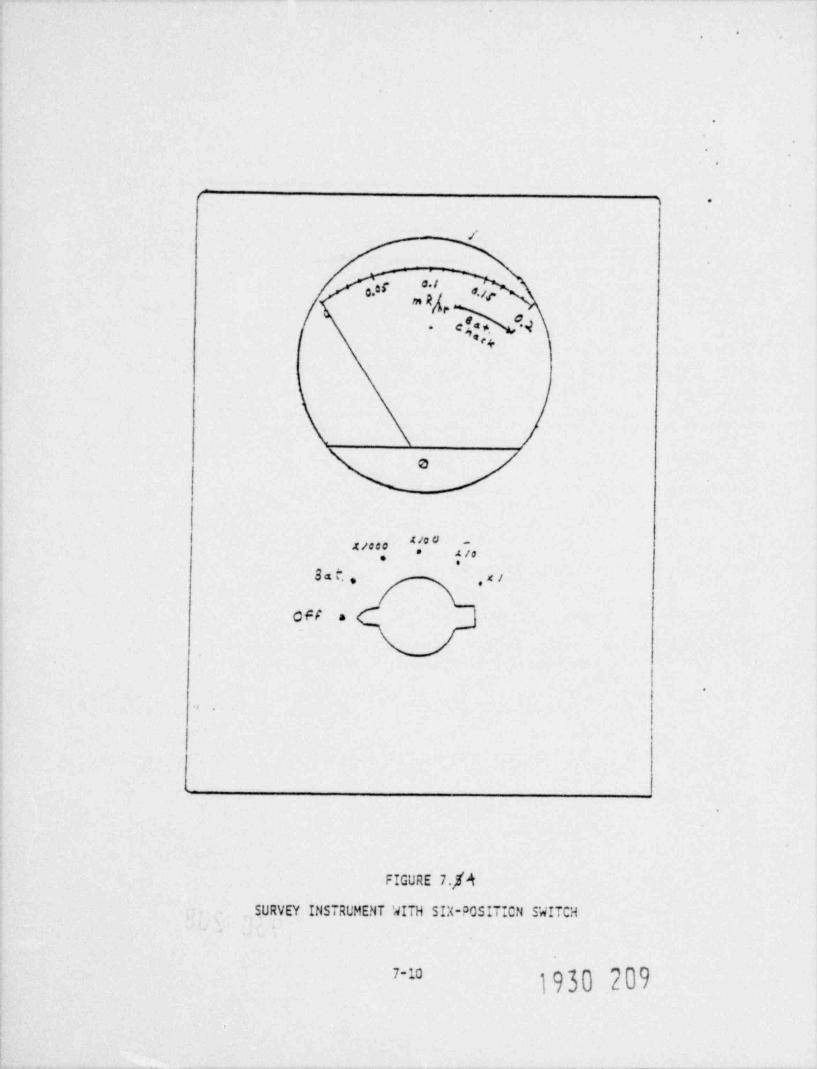
.
.
.
r .
/
**o.3C o.jp
4...
' '# ~~.t/000 * z io.
sa . . ,xi
0FF a
.
't
FIGURE 7.f4
SURVEY INSTRUMENT WITH SIX-POSIT!CN SWITCH
1930 2097-
.
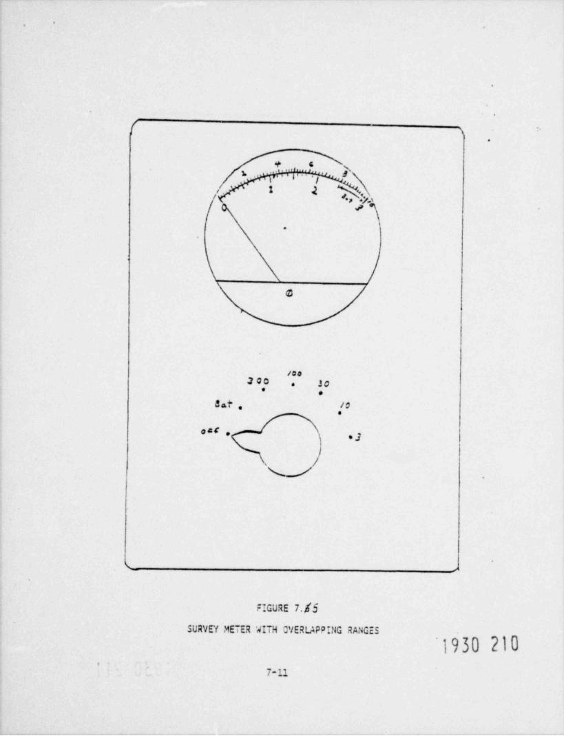
'.
.
.
[~
>.
+|''5 x
''|'..s
>y .
.
O
/CD200 30.
* e
S at . ."cat ,
, 3
J.
v -
k s
FIGURE 7.83
SURVEY METER WITH CVERUPPING RANGES
1930 210*
-
7-11
1

-.
.
f 3 .
.
*'
,
% 'b,
m%eR/kr*ct,eu;r*skeek e
09C e* !
\
i'
!.
. .
! .
L
FIGURE 7./6
SURVEY METER WITH LCGARITHMIC SCALE
1930.2117-u

'
.
4. Instrument Surveys s
According to 10 CFR 20, a radiation survey is "an evaluation of the radiationhazards incident to the production, use, release, disposal, or presence of radio-active materials under a specific set of conditions. When appropriate, such evalua-tion includes a physical survey of the location of materials and equipment, andmeasurements of levels of radiation or concentrations of radioactive material present".
To quote from the NRC publications, NUREG-0495, Public Meeting on Radiation Safety for :Industrial Radiographers, "the recognition and assessment of the hazard is theessential purpose of making a survey". Actual measurements with properly operatingsurvey instruments are the best way of recognizing and assessing a possible hazard.Regulating agencies pay close attention in their inspection to both survey recordsand to the actual use of survey meters by radiographers on the job.
Additional infor=ation in NUREG-0495 shows that in the years 1971 through 1977,42 incidents occurred in which 46 individuals engaged in industrial radiographyreceived whole-body doses greater than 5 rems. At least 28 of the 42 incidentscould have been avoided if the radiographer had performed a proper survey.
Proper use of the survey instrument is the most effective way of avoiding extremeradiation exposure of the radiogracher and others. Procedures and training provided
by your employer will provide specific instructions regarding radiation surveys.However, there are some radiation surveys com=on to all operations in radiography,and these will be discussed here.
a. Stored-Source Survey
When the source is to be removed from storage for use in the field, the sourceshield should be surveyed. Since the radiographer does not necessarily have controlof the source when it is in storage, it is possible that the source is not properlypositioned in its storage container. Before the source is transported, the containershould be surveyed. If radiation levels higher than expected are measured, you should
_ follow your employer's procedures to assure that the source will be safe to transport.
b. Surveys at Time of Set-Up and Exposures
When the source has been removed from its container and set up for use in thefield, the radiographer must do an area survey. The major reason for the surveyis to confirm the restricted area boundary. The radiation exposure rate outside ofthis boundary must be less than 2 mR/hr. This survey also defines the boundaries ofndiation areas (greater than 5 mR/hr) and high radiation areas (greater than 100mR/hr). These boundaries must be posted with proper signs (Caution Radiation Areaand Caution High Radiation Area). This survey need not be repeated af ter the firstexposure if subsequent exposures use about the same source-target configuration. Ifa change in shif ts occurs, this radiation survey should be repeated before the firstfilm exposure on the new shift. If the radiography is being done in a permanentradiography facility, the survey to confirm the unrestricted area boundary is notnecessary.
The radiographer has the responsibility to control access to the restricted area.He must have the boundary clearly marked off to be sure that people in the vicinityknow the area is restricted and do not enter during exposure periods. Some portionsof the restricted boundary area may not be visible to the radiographer. He must makesure that such locations are well marked and posted. During the exposure, he shallmake occasional visual checks of such areas to make sure that no one has entered therestricted area.
1930 212.

,
c. Post-Exposure Survey
After each exposure has been completed, the radiographer must complete aradiation survey of the exposure device. This survey is to determine thatthe source has been returned safely to the fully shielded position in theexposure device. In performing this survey, you would walk towards theexposure device with your survey instrument on a low range. If you notethat the exposure is much higher than expected for the source in itsshielded position, you should not advance any further. You should recheckyour instrument and make additional measurements at a safe distance todetermine whether the source is still out. Follow your emergency proceduresif the source is out and cannot be retracted.
If the readings are as expected, you should approach the back side ofthe exposure device, switching to a higher range as necessary. You shouldsurvey the back and both sides and then reach over the device from behind tosurvey the guide tube connection. You should not move from the back of thedevice to the front to survey the guide tube connection. It is possible forthe source to be stuck at this connection point, and you could beoverexposed by moving to the front. If readings appear normal, you shouldsurvey along the guide tube (if one exists) to determine that the source hasnot stuck in the tube (see Figure 7.7).
Conducting croper surveys with a calibrated survey meter is your bestdefense against overexposures. You should be thoroughly familiar with theinstruments available to you. You should develop confidence in their properuse and take appropriate care of them in use. Proper use of survey
. instruments can prevent accidental exposure to radiation.
d. Survey Prior to Securing Exposure Device
After the source has been returned to the exposure device and work iscompleted, the device must be secured. Securing the exposure device or itscontainer requires locking it. A survey of the locked device or itscontainer is required. The survey is to ensure that the source is in itsproperly shielded condition before the container is secured. Writtenrecords of this survey must be kept (10 CFR 34.43).
5. Personnel Dosimetry
a. General Types of Dosimeters in Use
By requirements of Part 34 of Title 10 of the Code of FederalRegulations, every working radiographer must be equipped with a self-readingpocket dosime.ter and a film badge or thermoluminescent dosimeter (TLD). Thepocket dosimeter provides an on-the-spot measurement of exposure. The filmbadge or TLD must be processed by the employer or an outside contractor.Another type of dosimeter that is occasionally used, but not presentlyrequired, is the alarming pocket dosimeter. This device can be very useful,since it gives an audible alarm if the exposure rate is above some presetvalue. In other designs, the alarm sounds if the accumulated exposureexceeds a preset value. Alarming dosimeters are available in a variety oftypes. Many of those on the market will not tolerate much physical abuse;care must be exercised in use in work situations where they may be banged
1930 213,

. <,
.
FIGURE 7 7 7 _
-
.-:
SURVEY OF PROJECTION DEVICE AFTER SCURCE RETRACTICN .
O
"1. Approach from back side of sourcacontainer. Sur/ey back side.
.
2. Survey both sides of container.
k-, y.d \ E
3. Survey tcpe of container as ycu reachover it. Remain behind contain andwith instrument beyond front edge ofcontainer sur/ey the guide tubeconnection. g
hO W.
4. Sur/ey the guide tube.
o
J
1930 214-7-g

'
.
around or dropped. Many of these devices also cannot be readily checked inthe field for proper operability. Low batteties may result in no alarmsounding or not sounding when it should. As noted below, an alarmingdosimeter should never be relied on as a primary indicator of a radiationproblem.
Most personnel dosimeters in use are passive devices. They accumulateexposure to provide a record of what dose the wearer has received. They areafter-the-fact devices. That is, the readout is a measure of what dose hasalready accrued. Thus, while the dosimeter is a necessary device to measuretotal personnel expostre, it does not replace the survey meter. The surveyins tr ument tells you the exposure rate at any instant; properly used, itwill give valuable information necessary to avoid excessive exposures. Youmight argue that a personnel device like the self-reading pocket dosimeteror the alarming dosimeter could frequently replace the survey meter. Thisis not the case.
If a proper survey is not done, you could easily walk into a situationin which the source was exposed at a time when you thought it wasretracted. You might check your pocket dosimeter after a short time andfind it is off-scale. Walking into a high-level field would cause thedosimeter to alarm but would not necessarily prevent you from receiving asignificant exposure before you reacted to the alarm.
b. Pocket Dosimeter
The self-reading pocket dosimeter, shown in Figure 7.8, is a rathersimple device. It is basically an air-filled ionization chamber. Attachedto the central electrode is a fine quartz fiber, usually coated withplatinum. A voltage is placed across the electrode and the wall of thechamber. The quartz fiber is free to move except at the point where it isattached to the electrode. When the dosimeter is charged, the fiber takeson the same charge as the central electrode and is repelled from theelectrode. When you look through one end of the dosimeter, you see theimage of the quartz fiber. The image is projected on a scale that isdivided into segments. Each segment of the scale usually represents 10 mR(see Figure 7.9). When the dosimeter is fully charged before use, the imageof the fiber is made to rest at the "O" position on the scale.
If the dosimeter is exposed to ionizing radiation, the chamber willdischarge according to how much exposure occur 3. As the chamber discharges,the fiber image moves upward on the scale.
The dosimeter used by radiographers must have a full-scale reading of atleast 200 mR. Pocket dosimeters allow you to monitor your exposure as youwork. Your pocket dosimeter should be read frequently during radiographyoperations. While the dosimeters are quite rugged, they can be damaged bybeing dropped or if they are struck by a hard object. Such an event maycause the dosimeter to discharge and show an off-scale reading ~.
~
All pocketdosimeters will lose electric charge by leakage even when no radi~a~ tion is - ~ ~
i930 215

.,
.
.
.
i-.
.
M s .%I . .p las tis u sil Q u a o tz' F.* b e r Gradaafr] Sc ale
1 ) }
I|
._ g I; .f\ fb,
} r \}lwl a. s
,t / % P' \ |,
. wIo$ 3b )I
''S' *1 L ens Eye::enEle cten d e.
1930 216 .
FIGURE 7./3
CROSS SECTION OF POCKET COSIMETF'
7-17

.
,
,a .w. .
*2- %
L .- n -*:- cg .-
.a s. .' ,% .m.-
.
u .~,. : . ..,s a ... ,,
r . . , . .. - s-q ,.
. . . . ...
S % =-E ?|, _x. 1
., .
0 g .s ,_ ,
o 7 - . . , . u. s3tm e-ksw %,.
4. ;6v o~ s,s
'
- s:uoC.
c~ .
..**s
4 I,.% o.
c.<.- s .
3 ~.-.u,e , ,sy , . ..
I. '_-- y.f.<:,D---
u!. a: I s .4. m
-
-%-
g 4
.--J A. %' s. 4* s w: g. ; .
.4~ . .
*"''& O k .at*
u 4 -- c L. .
1 c- e4v
-
':>
t''|'c
Do '-_ 5
.- - +e- ..
H 7T l&2*---,eo- % .
&e, ,-
- w= -
. r- - -=
1 2.:. <-%1- . -.*
4''*"- 4 Q. ~
> ,. s. :.=. c.e, -. s .
; ; / u =r ZA
i- $. .2 Y,- -.P9 o .-
C .a -
. ..
m.,e
FIGijRE 7. "49
1930 2177-13
.
present. If a dosimeter is working properly this natural leaking willbe small and will not affect the readings over the course of a workingday. As a result of aging or of mechanical damage, however, a -
dosimeter might leak charge quite rapidly. In such a case, the 3
dosimeter might be in such bad shape that it will not hold a charge atall; or it might produce a distinct drift of the fiber across thescale. Such a drift might produce falsely high readings on thedosimeter. When using a pocket dosimeter it is important to check thereading frequently. It takes only a few seconds to note the reading.If you get in the habit of doing this often you will be able to detecta faulty dosimeter. You will also have a better awareness of your own .
dose and will be better able to avoid excessive exposure. If yourdosimeter goes off-scale you will lose time on the job and frequentreading of the dosimeter will also avoid this.
Following is a list of procedures that should be followed in using_
self-reading pocket douimeters.
1. The dosimeter must be charged at least once a day or at thestart of each shift.
2. Firmly clip your dosimeter to your clothing, preferably in afront pocket, and always wear it while conducting operations.
3. Read your dosimeter frequently during operations. You mustrecord your pocket dosimeter reading daily.
4. If you drop your dosimeter or suspect you might hcve damagedit in some other way, check the reading to see if it appearsnormal.
5. If your dosimeter reads off-scale, you must have your filmbadge or TLD processed. Your employer must investigate thecause of your off-scale reading,
c. Film Badge Dosimeter
The film badge dosimeter contains a piece of film similar to thefilm used in making radiographs. Ionizing radiation darkens thefilm. The darker the image, the higher the exposure. In order toproduce the proper response and to allow the processor to interpretthe response correctly, the film must be worn in a proper badgeholder. Figure 7.10 shows a typical film badge. The metal absorbershelp the processor to tell whether the exposure was caused by high orlow energy photons. This is useful information to obtain the correctexposure. The open window part of the badge holder allows beta -
particles to penetrate the film. Beta radiation is generally not ofconcern in radiography operations.
_
t
i930 218.
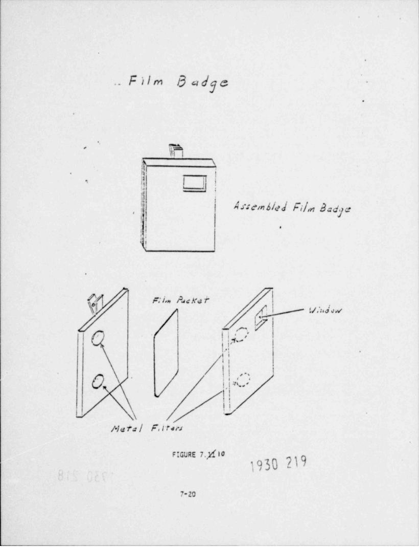
e
S
.. F ilm B ad eg -
.
e
eFehh
$ '
!1-
-
U i
k* j| A.c:embled fil,n Badge|
i
<1
*h
9e
;d
.
r, I'an hacNa f~'
!tj,;,4 a,,e
\f- r.,k |,
\i.'
ii
/ !
\ , ' ~ '.. g .
:n....
n
7.je ra l F. rr<n.
7-20

.
-.
The film badge readings will form the basis of the radiographer'spermanent dose record. It is, therefore, essential that the filmbadge be worn at all times while you are working. In the event of anaccidental overexposure in which your pocket dosimeter goes off-scale,your film would provide the only first-hand information about the dose
' ~..
received.
Film badges are rugged, but they can be damaged. If the papercovering on the film is broken, the film will be ruined by exposure tolight. Film can also be damaged and yield wrong results if it issubjected to high temperatures or high humidity for long periods.Leaving a film badge in a closed automobile on a hot smmer day canproduce fogging of the film so that an estimate of radiation exposureis impossible. Submerging a film badge in water or laundering it willalso make an estimate of exposure impossible. The followingrecommendations apply to the use of film dosimeters.
1. Firmly clip your badge to the f ront side of the major trunkof the body (between waist and neck) and wear it at alltimes during operations.
2. Do not unnecessarily expose the film to high temperatures orhumidity.
3. If you suspect that you have damaged your film badge, stopactivities. Submit your badge to your employer. Do notbegin work with sources until you have been reissued a filmbadge.
4. If your pocket dosimeter reads off-scale during radiographicoperations, stop your work. Submit your film badge to youremployer for immediate processing. Do not start work withsources until your employer allows you to resume work.
5. Routine badge processing is done on a regular schedule.Know the schedule and have your badge available forprocessing.
d. Thermoluminescent Dosimeters
Thermoluminescent dosimeters (TLDs) are used by some employers inplace of film. TLDs are crystalline materials. They have the,
property of being able to store energy deposited in them by ionizingradiation. This energy can be measured by heating the dosimeter(af ter irradiation) and measuring energy released in the form oflight. A special TLD reader is required to obtain this information.The light output from the dosimeter is a measure of the dose orexposure. TLDs are worn in a badge holder, similar to a film badgeholer in appearance.
The same recommendations apply to the use of TLDs as noted abovefor film, although TLDs are not as sensitive to temperature orhumidity.
.
100 220

.
',
e. Alarming Dosimeters
There has been, and there still is, a good deal of controversyregarding the use of alarming dosimeters by radiographers. Thesedosimeters have the obvious advantage that they audibly alert theradiographer to a possible exposure problem. Some employers do issuethem, along with self-reading pocket dosimeters and film badges orTLDs, to radiographers. The greatest problems has been withreliability of the alarming dosimeters. Most designs to not seem ableto take much physical abuse. Even routine use in some difficultradiographic procedures may be too severe for some of the devices.The failure rate has been relatively high, and many people avoid theiruse for this reason. Presently, review of the capabilities of many ofthese devices now on the market is continuing. It seems likely thatdesigns will be forthcoming that will provide the requiredreliability. There have been discussions among people in the businessand in regulating agencies related to requiring the use of alarmingdosimeters. In the future, we may find that their use will berequired. An alarming dosimeter must never be used to replace thesurvey meter. It simply provides one additional source of informationfor the protection of the radiographer.
Personnel Dosimetry Recordkeeping
As noted above, daily records of pocket dosimeter readings must bekept. Reports of film badge or TLD readings must also be maintainedby the employer. These records may be inspected by the NRC or theState.
A radiographer's permanent dose record is maintained onappropriate forms. Current exposure information is usually maintainedon NRC Form 5 or equivalent. These forms are shown in Appendix .
Since a radiation worker's average yearly whole-body dose (averagedover years since age 18) cannot exceed,by law, 5 rems, it is necessaryto maintain this exposure history.
.
1930 221

-.
CUESTIONS
1. Record the reading for each of the following survey meters.A. B. C. f:
p . . . ;. . . . ; 3. . . .p . . ., g ;,.......,4,
L
3.,. ia7. .,m . , e,* .= 74 .' .,* ., " ; -e.
, , , , ,
I L /2. Record the readings for each of the following dosimeters.
A. B. C.
/ ... 4p.3 mi.. 9.J.... | /, . . ~ .A( . 2 ,
/ \ /\ d v'
3. Two radiographers are working together in L.t. ting up an exposure. Whentaking a survey before the exposure, each radiographer uses a different
-- _ . survey meter. One of them records a reading of 150 mR/hr while the other' ' ~ ~ - - records a reading of 250 mR/hr. Oc you think they should proceed with
the exposure? (Give your reason as to why they should or not.)
4. After you have finished an exposure and v: ,le you are setting up the nextone your dosimeter falls out of your poeiret. When you pick it up you seethat it reads off scale. Is it safe to vssume that this is a result of
3 its fall to the ground and to continue vitn the next exposure or not? Why?
5. What is the primary function of the film badge?
6. List three occasions for conducting surveys.
Vn7. If you ace a radiographer working on site and your film badge geas wet
accidentally, what would you do?
8. Should you let other radiography workers use your film badge? Explain..
9. What are the advantages and disadvantages of an alarming dosimeter?
.
1930 222

f P. y|Jo'3 H of||Ilf|gQ-.
8. FEDERAL AND STATE REGULATIONS.s
"... Any person who willfully violates any provision of the Actor any regulation or order issued thereunder may be guilty of acrime and, upon conviction, may be punished by fine or imprison-ment or both, as provided by law." 10 CFR 520.601
This chapter deals with the Federal and State regulations that govern theradiography industry. Your company's Operating and Emergency Procedureshave been written to conform with these regulations. (In the next two chtpters
you will be directed to study these procedures.) We include at this point thehighlights of some of the more relevant regulations. Knowing the regulationswill give you a better feeling for why your company's procedures are written asthey are.
Understanding the regulations and following approved procedures may not necessarilylead to better radiographs, but will lead to a safer working environment. Andthat, after all, is the purpose of this manual - safety, not only to yourself,but to the general public.
Another reason for studying this chapter carefully is given in the sentenceunderneath the chapter title. Not following a company procedure may mean yourjob. Not following a regulation may mean jail.
The remainder of this chapter is divided into 5 sections. Section 1 identifieswho the regulators are. Section 2 summarizes the regulations of the NuclearRegulatory Commission as they pertain to industrial radiography. The transporta-tion of radioactive materials (in particular radioisotope sources) is treatedin Nction 3. Section 4 discusses the role of the American Society for Non-destructive Testing. The last section treats, in general terms, the relationshipthat exists between employers and employees and how an understanding of eachothers needs can lead to improved safety.
1. Who regulates you?
Under the Occ'ipational Safety and Health Act (OSHA) of 1970, most businessesmust meet general working safety regulations, specified by the Department ofLabor in Title 29 of the Code of Federal Regulations. Furthermore, due to the
special hazards associated with radiation, the United States Congress passed alaw giving the NRC the responsibility of regulating the use of radioisotopesproduced by the fissioning of uranium and plutonium such as iridium-192 andcobalt-60 which are widely used in industrial radiography.*
.
"NRC has no authority to regulate naturally occurring radioisotopes such asradium or accelerator produced isotopes. The individual states have authority
to regulate their use.,
8-11
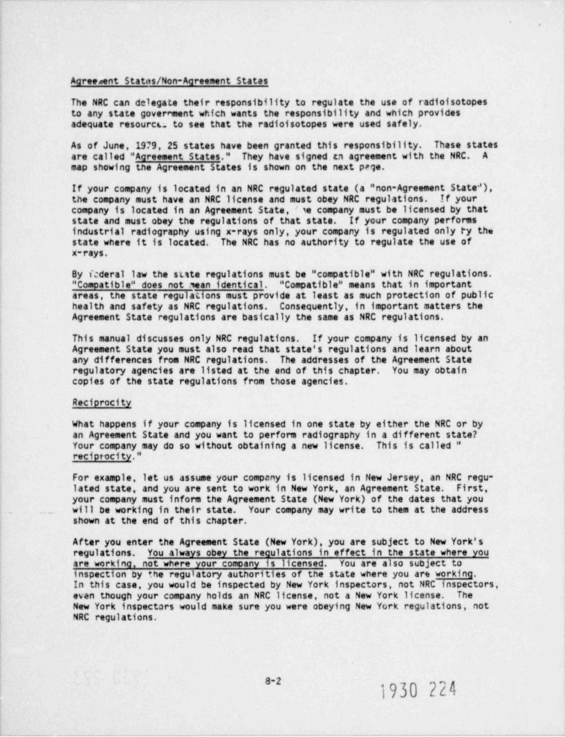
.
Agreenent Statos/Non-Agreement States
The NRC can delegate their responsibility to regulate the use of radioisotopesto any state government which wants the responsibility and which providesadequate resourcu to see that the radioisotopes were used safely.
As of June, 1979, 25 states have been granted this responsibility. These statesare called " Agreement States." They have signed an agreement with the NRC. Amap showing the Agreement States is shown on the next prge.
If your company is located in an NRC regulated state (a "non-Agreement State'),the company must have an NRC license and must obey NRC regulations. If your
company is located in an Agreement State, se company must be licensed by thatstate and must obey the regulations of that state. If your company performsindustrial radiography using x-rays only, your company is regulated only by thestate where it is located. The NRC has no authority to regulate the use ofx-rays.
By fcderal law the sute regulations must be " compatible" with NRC regulations." Compatible" does not sean identical. " Compatible" means that in importantareas, the state regulations must provide at least as much protection of publichealth and safety as NRC regulations. Consequently, in important matters theAgreement State regulations are basically the same as NRC regulations.
This manual discusses only NRC regulations. If your company is licensed by anAgreement State you must also read that state's regulations and learn aboutany differences from NRC regulations. The addresses of the Agreement Stateregulatory agencies are listed at the end of this chapter. You may obtaincopies of the state regulations from those agencies.
Reciprocity
What happens if your company is licensed in one state by either the NRC or byan Agreement State and you want to perform radiography in a different state?Your company may do so without obtaining a new license. This is called "reciprocity."
For example, let us assume your company is licensed in New Jersey, an NRC regu-lated state, and you are sent to work in New York, an Agreement State. First,
your company must inform the Agreement State (New York) of the dates that you.. will be working in their state. Your company may write to them at the address
shown at the end of this chapter.
After you enter the Agreement State (New York), you are subject to New York'sregulations. You always obey the regulations in effect in the state where youare working, not where your comoany is licensed. You are also subject toinspection by the regulatory authorities of the state where you are working.In this case, you would be inspected by New York inspectors, not NRC inspectors,even though your company holds an NRC license, not a New York license. TheNew York inspectors would make sure you were obeying New York regulations, notNRC regulations.
8-21930 224

-.
Noto that under " reciprocity" your company is limited to performing radiographyto 180 days per year in a state where it is not licensed. Usually, if thecompany keeps radioactive sources in a state where it is not licensed for morethan 180 days, it must obtain a license from the authority responsible in thatstate. The following states, however, have periods different from 180 gjdays:
Off-shore work sites
If you perform radiography offshore on a platform or other structure attachedto submerged lands of the continental shelf beyond the 3-mile territorial limit,you are subject to NRC regulation even if you are off the shore of an AgreementState and your company is licensed by an Agreement State. To perform such off-
-
shore radiography, your company must notify the NRC when and where you will be~
performing that radiography. You are then subject to NRC regulations and NRCinspections of that radiography. However, Agrtement State licensees on a boaton the high seas would remain under Agreement State jurisdiction no matter howfar out they went.
2. Code of Federal Regulations
The Act of Congress that gave NRC the authority to regulate industrial radiog-raphy using radioisotopes gave NRC the authority to issue regulations. Theseregulations are laws. It is illegal for a person subject to them to disobeythem. /folations of these regulations can result in loss of your company'slicense, monetary fines, or even imprisonment for the most severe and willfulviolations.
The book containing the NRC's regulations is called Title 10 of the Code ofFederal Requiations. This is often abbreviated "10 CFR".
There are other Federal rules which must be followed as well. For example,Title 45 of the Code of Federal Regulations (49 CFR) deals with the regulationsof the Department of Transportation. Parts of this Title deal with the trans-portation of hazardous materials, including the radioisotope sources used inindustrial radiography. The transportation of radioactive materials is thesubject of Section 8.3.
Title 10 is compossJ of many separate parts. In particular, Part 19, Part 20,and Part 34 contain especially important information for the radiographyindustry. In fact, exact copies of each of these parts must be provided toyou by your company.* What follows is a summary of these three parts, as wellas a brief description of Parts 21, 30, 71, radiography. These summaries aresimplified. If you have specific questions, you should ask someone familiarwith the regulations, such as your company's Safety Officer, or read theregulations themselves.
-
sFurthermore, your company must post copies of their licence and any amendments,the operating and emergency procedures, and Parts 19, 20 and 34.
1930 2258-3
' N
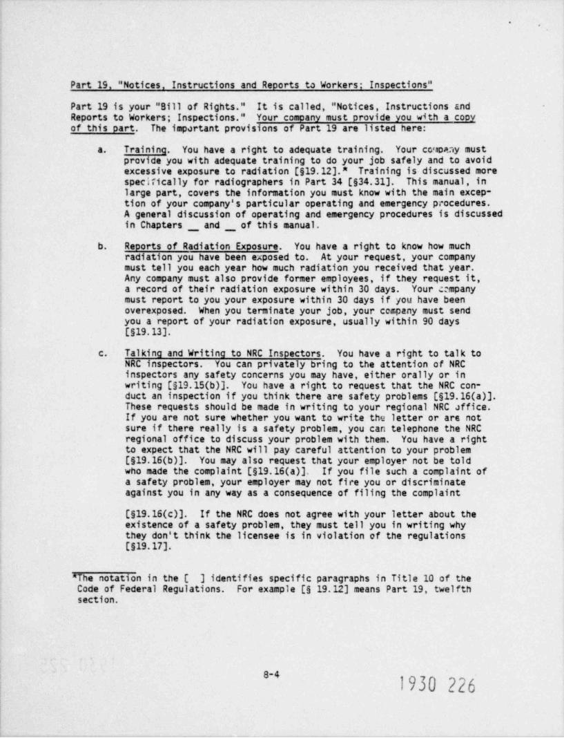
*.
Part 19, " Notices, Instructions and Reports to Workers; Insoections"
Part 19 is your " Bill of Rights." It is called, " Notices, Instructions andReports to Workers; Inspections." Your comoany must provide you with a copyof this part. The important provisions of Part 19 are listed here:
a. Training. You have a right to adequate training. Your co.toaay mustprovide you with adequate training to do your job safely and to avoidexcessive exposure to radiation [S19.12].* Training is-discussed morespecifically for radiographers in Part 34 [$34.31]. This manual, inlarge part, covers the information you must know with the main excep-tion of your company's particular operating and emergency procedures.A general discussion of operating and emergency procedures is discussedin Chapters _ and _ of this manual.
b. Reports of Radiation Exposure. You have a right to know how muchradiation you have been exposed to. At your request, your companymust tell you each year how much radiation you received that year.Any company must also provide former employees, if they request it,a record of their radiation exposure within 30 days. Your e mpanymust report to you your exposure within 30 days if you have beenoverexposed. When you terminate your job, your company must sendyou a report of your radiation exposure, usually within 90 days[$19.13].
c. Talking and Writing to NRC Inspectors. You have a right to talk toNRC inspectors. You can privately bring to the attention of NRCinspectors any safety concerns you may have, either orally or inwriting [S19.15(b)]. You have a right to request that the NRC con-duct an inspection if you think there are safety problems [S19.16(a)].These requests should be made in writing to your regional NRC affice.If you are not sure whether you want to write the letter or are notsure if there really is a safety problem, you can telephone the NRCregional office to discuss your problem with them. You have a rightto expect that the NRC will pay careful attention to your problem[S19.16(b)]. You may also request that your employer not be toldwho made the complaint [S19.16(a)]. If you file such a complaint ofa safety problem, your employer may not fire you or discriminateagainst you in any way as a consequence of filing the complaint
[S19.16(c)]. If the NRC does not agree with your letter about theexistence of a safety problem, they must tell you in writing whythey don't think the licensee is in violation of the regulations
[$19.17].
"The notation in the [ ] identifies specific paragraphs in Title 10 of theCode of Federal Regulations. For example [6 19.12] means Part 19, twelfthsection.
8-41930 226

-.
Part 20, " Standards for Protection Against Radiation"
Part 20 discusses basic rules for radiation safety. It is called " Standardsfor Protection Against Radiation." Actually, for industrial radiography, Part ..
34, to be discussed later, discusses many requirements for radiation safety in d
greater detail than does Part 20. For example, Part 34 gives very specificadvice on surveys, personnel monitoring, and protecting the public from gettingtoo much radiation. We will discuss here only those sections of Part 20 that
are not covered in more detail in Part 34.
a. Radiation Dose Limits. The NRC has quarterly (3-month) dose limits.It does not have annual, monthly, or weekly limits. The quarterly .
limits are listed here [from $20.101 and 120.102]:
External Radiation Dose Limits for Adults:
Whole body 1 or 3 remsHands, forearms, feet, ankles 18-3/4 remsSkin of the whole body 7-1/2 rems
The whole-body dose is normally what your film badge or TLD badgereads. It is considered to be a measure of the amount of penetratingradiation that has been received by a large portion of your body,particularly the parts important from a radiation protection pointof view, such as the bone marrow where leukemia would originate, orthe gonads, where genetic damage to offspring would originate.
The quarterly limit on whole-body radiation is 1.25 rems if youremployer does not wish to consider your past exposure histo.y. Thequarterly limit on whole-body radiation is 3 rems per quarter if youremployer is willing to look back into your past radiation exposurehistory and determine that your average annual dose since age 18 isless than 5 rems per year. The equation for your maximum lifetimedose is:
Maximum Permitted Lifetime Dose = 5 (N-18) rems, where N is yourpresent age.
If you are under the age of 18, the dose Timits are one-tenth (10%)of the amount allowed an adult.* For example, a minor, under 18 yearsold is permitted a quarterly whole body dcsa of only 0.125 rems or125 millirems [920.104].
Note that the NRC permits siigher limits, 18-3/4 rems, to the body'sextremities, such as the hands [$20.101]. Still you cannot touch aradiographic saurce since the dose to your hands would be much higherthan that limit. -
" Note, however, that Department of Labor regulations prohibit individuals underthe age of 18 from working in occupations involving exposure to radiation[29 CFR 9570.120 and $570.57]. Ycu cannot work as a radiographer if you areless than 18 years old.
8-5
1930 227

-.
The skin dose limit is rarely of interest to radiographers. It isimportant where there is a high beta dose and little gamma dose.Beta rays can penetrate the skin but will reach less than one centi-meter into the body so they do not contribute to the'"whole bodydose." Radiographic sealed sources do not emit beta rays. The betarays cannot penetrate the metal capsule.
b. Restricted areas. A " restricted area" is an area to which thelicensee restricts access fo= the purpose of radiation protection[$20.3(a)(14]. Restricted ;eas are established to protect the publicfrom radiation exposure. (ou cannot let anyone into a restrictedarea unless he has been told of the presence of radiation in the areaand told what to do to avoid exposure [S19.12]. If you are a radiog-rapher at a field site, you will frequently use ropes to mark offthe restricted area and keep people from entering. If people ignorethe ropes, you should be prepared to approach them and tell them thata radiation source is present and that they should keep away.
c. Unrestricted Area. An " unrestricted area is an area to which accessis not restricted. The maximum doses allowed to an individual standingin any area to which access is not restricted (an " unrestricted area")is 2 mrem in any one hour or 100 mrem in any one week [S20.105(b)].
d. Radiation Areas and High Radiation Areas. Definitions and Posting.A " radiation area" is defined as an area in which radiation existsso that an individual could receive a dose in excess of 5 mrem inany one hour or 100 mrem in five days [$20.f_02(b)(2)]. Radiationareas must be posted with signs saying " CAUTION, RADIATION AREA"(sign can also say " DANGER") and displaying the radiation symbol[$20.203(b)]. In the performance of radiography, the radiation areawill be not very different in size to the restricted area. Thereforeit is often practical to post the radiation area signs at the 2 mremin an hour boundary established for the restricted area and not havea separate restricted area and a separate radiation area.
If the dose to an individual could exceed 100 mrem in an hour the areais a high radiation area [$20.202(b)(2)]. High radiation areas mustbe posed with a sign saying " CAUTION, HIGH RADIATION AREA" and display-ing the radiation symbol.
e. Receiving Radioactive Sources. Licensees must promptly pick up pack-ages from shippers as soon as they are notified the packages areready [620.205(a)].
f. Theft or Loss of Sources. Radiographic sources can be very dangerousto a member of the public who does not understand the danger or theprecautions necessary with a radiographic source. You must immediatelynotify your supervisor so that the company may immediately notifythe regional NRC office by telephone of the loss or theft of a radio-graphic source [S20.402(a)].
8-6

-.
g. Notifications of Radiation Overexoosures. Your company must notifythe NRC regional office immediately by telephone if any individualis overexposed to radiation exceeding 25 rems whole body or 375 remsto the hands or feet [$20.403(a)(1)]. You must notify the NRC regional
- office within 24 hours of a radiation overexposure exceeding 5 rems , 2whole body or 75 rems to the hands or feet [520.403(b)(1)]. Lesseroverexposures must be reported within 30 days [920.405].
Part 34, " Licenses for Radiography and Radiation Safety Requirements for Radio-Graphic Operations"
If Part 19 is your " Bill of Rights," Part 34 is your " Bill of Responsibilities." .
Actually, you have no direct responsibility to the NRC to obey the regulations.The licensee, your employer, has the sole responsibility in the eyes of theNRC to see that the regulations are obeyed. But since you are the ,9erson onthe spot and the person who must actually do the work to meet the regulations,you become responsible to your employer to see that the regulations are obeyed.
We mentioned that Part 19 forbids your employer from firing or discriminatingagainst you if you complain to the NRC about safety problems. However, thereis nothing in the regulations to prevent your employer from firing you becauseyou have failed to.do a required radiation survey.'
The basic provisions of Part 34 that you must follow are discussed here.
a. Surveys. The most important thing that you must do to protect your-self and the people near you is to perform adequate radiation surveys.You must never work without an operable survey instrument [$34.43(a)].The survey instrument must have been calibrated within the previousthree months [$34.24].
You absolutely must perform the following surveys:
(1) A survey of the exposure device after each radiographic expo-sure to make sure that the source is in its shielded position
[$34.43(b)].
(2) A survey of the restricted area to make sure no member of thepublic is exposed to a dose of more than 2 mrem in any one hour[520.201(b) and $20.105(b)(1)].
(3) A survey of storage sheds to make sure no member of the publiccould be exposed to a dose of more than 2 mrem in any one houror 100 mrem in any 7 consecutive days [520.201(b) and620.105(b)(1) and (2)].
b. Posting of Signs. Signs are posted to warn other people that radia- -
tion is present in the area and that they should be careful to avoid
the radiation [$34.42]. Ropes are often used with the signs althoughthe regulations do not specifically require ropes. You must postRadiation Area signs, generally at the restricted area boundary, andyou must post High Radiation Area signs anywhere the dose is suffi-cient to expose anyone to a dose of 100 mrem in an hour.
' *~'1930 229

*.
c. Security for the Hich Radiation Area. You are responsible to seethat no one enters the high radiation area while a source is exposed[$34.41]. If it is possible for anyone to enter the high radiationarea you must maintain direct visual surveillance of the area andprevent them from entering. Surveillance may not be necessary ifthere are other means of preventing an individual from being exposed.For example, if an area is locked so no one can enter it, surveillanceis not necessary [534.41]. Surveillance also is not necessary ifthe source will automatically retract when someone approaches or ifthere is an alarm system present that will warn both the person andyou that the source is being approached [534.41 and $20.203(c)(2)].
d. Personnel Monitoring. Whenever you work with a radiographic source,_ _ _
you must have both a pocket dosimeter and, in addition, either afilm badge or TLD badge [634.33]. You must read the pocket dosim-eter every day and record the results (634.33]. It is also a verygood idea to read it several times during the day to make sure youare not accidentally irradiating yourself.
e. Locking of Exposure Devices. After a ch exposure, the source mustbe secured in the device (not necessarily locked with a key) to makesure it is in a safely shielded position [S34.22]. In addition, youmust lock the device with a key whenever it will not be under yourdirect surveillance or control [S34.22]. You cannot leave a radio-graphic source unattended and unlocked so that anyone who happensalong could crank out the source and expose himself or someone else.
f. Storace of Sources. You must protect the radiographic exposuredevice from being stolen, tampered with, or accidentally removed[634.23]. You must not leave them unattended. Lock them in a securestorage area or truck before you leave them unattended. Both thestorage area and the truck must be posted with a sign saying " CAUTION,RADIOACTIVE MATERIALS" and bearing the radiation symbol.
g. Leak Testing. Radiographic sources must be leak tested every 6 months[$34.25].
h Quarterly Inventory. Every 3 months all the radiographic sources inyour company must be accounted for [534.26].
i. Inspection and Maintenance of Radiograohic Excosure Devices. Youmust check your device each day before you use it to make sure it isin good working order [$34.28]. You must also have it comprehen-sively inspected and maintained every 3 months [$34.28].
j. Training. You must study and learn the subjects covered in thistraining manual. In addition, you must study and learn your ownlicensee's operating and emergency procedures [634.31].
In addition to Parts 19, 20, and 34, some other parts are of interest to radiog-raphers, although lesser interest. They are mentioned here:
. 1930 2308-8
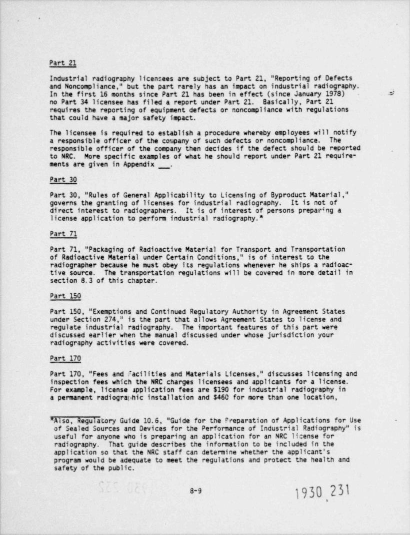
.
Part 21
Industrial radiography licen::ees are subject to Part 21, " Reporting of Defectsand Noncompliance," but the part rarely has an impact on industrial radiography.In the first 16 months since Part 21 has been in effect (since January 1978) Sno Part 34 licensee has filed a report under Part 21. Basically, Part 21requires the reporting of equipment defects or noncompliance with regulationsthat could have a major safety impact.
The licensee is required to establish a procedure whereby employees will notifya responsible officer of the company of such defects or noncompliance. Theresponsible officer of the company then decides if the defect should be reported ,
to NRC. More specific examples of what he should report under Part 21 require-ments are given in Appendix _ .
Part 30
Part 30, " Rules of General Applicability to Licensing of Byproduct Material,"governs the granting of licenses for industrial radiography. It is not ofdirect interest to radiographers. It is of interest of persons preparing alicense application to perform industrial radiography.*
Part 71
Part 71, " Packaging of Radioactive Material for Transport and Transportationof Radioactive Material under Certain Conditions," is of interest to theradiographer because he must obey its regulations whenever he ships a radioac-tive source. The transportation regulations will be covered in more detail insection 8.3 of this chapter.
Part 150
Part 150, " Exemptions and Continued Regulatory Authority in Agreement Statesunder Section 274," is the part that allows Agreement States to license andregulate industrial radiography. The important features of this part werediscussed earlier when the manual discussed under whose jurisdiction yourradiography activities were covered.
Part 170
Part 170, " Fees and Facilities and Materials Licenses," discusses licensing andinspection fees which the NRC charges licensees and applicants for a license.For example, license application fees are $190 for industrial radiography ina permanent radiographic installation and $460 for more than one location,
^Also, Regulatory Guide 10.6, " Guide for the Preparation of Applications for Use -
of Sealed Sources and Devices for the Performance of Industrial Radiography" isuseful for anyone who is preparing an application for an NRC license forradiography. That guide describes the information to be included in theapplication so that the NRC staff can determine whether the applicant'sprogram would be adequate to meet the regulations and protect the health andsafety of the public.
,
jg}Q 23}8-9
,

.
including field radiography. License renewals are $150 and $460, respectively.Amendments are $40 and $110, respectively. Annual inspection fees are $720per year and $980 per year, respectively.
3. Transportation of Radioative Materials
The transportation of radioactive materials is one of the more highly regulatedaspects of industrial radiography. As a radiographer, you will be involved withthe transportation of radioactive materials every time you take your camera outinto the field. Even if all your radiographs are taken in-house, you will stillbe involved in transportation when, for example, one of the radioactive sourcesis to be sent back to the source manufacturer.
For industrial radiography, the principal source of federal regulations per-taining to the transport of radioactive materials are found in the Departmentof Transportation's " Hazardous Materials Regulations", Parts 100 through 199 ofTitle 49 of the Code of Federal Regulations (49 CFR 100-199), as well as thepreviously mentioned 10 CFR 71.
The responsibility for safety in all interstate transportation, except postalshipments,* of radioactive material (such as radioisotope sources used inindustrial radiography) lies with the Department of Transportation (DOT). Theresponsibility for safety in intrastate transportation of radioactive materialslies with the NRC and Agreement States. In most cases the regulations of the00T have been adopted by each of the states so that for example, the rulesgoverning the packaging, labeling and marking of radioactive shipments areessentially the same for both interstate and intrastate transportation. In anyevent you must follow the specific local regulations.
What is presented in the remainder of this section is derived from the 00T'sregulations. For more specific information, you should consult with yourcompany's Safety Officer. Your Company's procedures for transporting radioactivematerials are written to be consistent with all federal and state regulations.
Packaging Requirements
The primary consideration for achievement of safety in the transportation ofradioactive materials is the use of proper packaging for the specific radioactivematerials to be transported. In order to determine the proper package, youneed to know:
a. if the radioactive material is "special form" or " normalform," and,
b. the amount of radioactivity involved.
These topics will be discussed in the following paragraphs.
* Postal shipments come under the jurisdiction of the U.S. Postal Service,39 CFR 123.
1930 2328-10.

',
Special Form Radioactive Material
What is meant by "special form" radioactive materials? Special form materialsare defined as materials, which if released from a package, might only presenta hazard due to direct, external radiation. The spread of contamination isminimized, if not eliminated, because of the high physical integrity of the
, g
radioactive material. A radiography source which is doubly encapsulated in ahigh strength metal such as stainless steel with high integrity welds is anexample of a special form material.
Normal Form Radioactive Material
" Normal form" radioactive materials are those materials which do not qualify -
as special form. An example of this type of normal form material is wastematerial, such as contaminated towels, which are placed in plastic bags, or
-- radioactve powder of liquid within a breakable or non-fire resistant package.You will not normally encounter such materials in radiography.
The remainder of this chapter will deal mainly with special form material.This is of most interest to you as a radiographer since the radiography sourceswhich you will encounter will be special form material.
Types of Quantities
There are three definitions of quantities of radioactive materials dependingon the amount of curies involved: Type A, Type B, and "Large Quantities."With special form materials, the curie limits are as follows:
Type A: less than 20 curies
Type B: between 20 and 5,000 curies
"Large Quantity": greater than 5,000 curies.
Most radiography sources fit into the Type 8 classification and require Type Bpackaging.
Type B Packaging
A " Type B" package is one that is designed to withstand certain serious accidentdamage conditionc with basically no loss of shielding capability. In particular,the Type 8 packages for a radiography source must pass the following tests withoutfailure:
1. A 30 foot free fall onto an unyielding surface;
2. A puncture test consisving of a 40 inch drop onto,
a 6 inch diameter steel pin;
3. Exposure to temperatures of 1475 degrees F for 30 minutes tosimulate fire conditions.
1930 2338-11-

.
Fortunately, you don't have to design such a container. But at least youknow what one is. In fact, it turrs out that many radiographic exposuredevices, when the source is locked in the shielded position, can serve as aType B package.
Control of Radiation
Even though the radioactive material is packaged properly, there will stillsome gamma radiation from the package. To avoid excessive exposure to radiationand to alert those people who will be handling the package, or other opersonssuch as the driver of the truck, certain radiation limits and labelling criteriahave been set.
Radiation Limits - Transport Index
The regulations give limits to the maximum permissible dose , ate on tha surfaceof a package, as well as the dose rate at various distances from the surface.In units of mR/hr, the dose rate at three feet from the surface is called the" Transport Index." There are two sets of limits for the dose rates dependingon whether or not the vehicle is used exclusively for the transport of radio-active materials. These limits are summarized in the following table. For anyvehicle the dose rate may not exceed 2 mrem per hour in any position of the'tehicle which may be occupied by by a person.
RADIOACTIVE PACXAGEDOSE RATE LIMITATIONS
A. Non-exlusive use vehicle.200 mrem per hour on the external surface of the package10 mrem per hour at three feet from the external surface of the package2 mrem per hour in any position of the vehicle which is occupied by a person
B. Exlusive use vehicle.1000 mrem per Mur at three feet from the external surface of te package10 mrem per hour at six feet from the external surface of the vehicle200 mrem per hour at the external surface of the vehicle2 mrem per hour in any position of the vehicle which is occupied by a person
Labelling Criteria
Depending on the size of the transport index, each radioactive package mustbe labeled on two opposite sides with one of three distinctive warning labels.These labels are shown in Figure 8.1.
A label with a white background color indicates that the radiation is minimaland nothing special is required for that package. This type of label is knownas a " Radioactive White I" label. If the background of the upper half of thelabel is yellow, then the radiation level at the package surface may requirespecial controls. Depending on the dose rates involved their are two types ofthese yellow labels: " Radioactive Yellow II" and " Radioactive Yellow III." Thelabelling criteria are outlined in the following table
J
8-12
1930 234

.
7,20 s L b'.,
J
D1ff Oh
a/
HANDLE CAREFULLY
c \\ ov!.
CO NTENTS:. . . . . .*********
NO. OF CURI ,- -'. . . . . . * ' * * ,
*
|.
k, !$?g,r e. t W I h p, ?
%.
..g
,-
,
.
'
I,
.-
_ y m. ua -- [k,
.m
HANDLE CAREFULLY
A 10) TI1,I s0'AYAYioE'Es*SY[:,e
qEllew ygJ' 'i!J20 tit"(
_
eonunn,.......y.3...............No. OF CURIES:. . 4m .. .............
{Y 1 ***Na$sNd k
'
; 7:ANsroRf INO l.tl h,
d.Ed 9%. or An h b /'
~~
,~ ~
"> . , & (,; A
". 67
i t.
~ mA,a. .t ......,.....A ?
A 10k11[ HANDLE CAREFULLY,
j 30'2:YlioE'Es*SY[asj5) '???2at,2*
{-
connnn,...g..w.. ; \.............
'
NO. CF CURI f@, C,
. .; ..,.............
"t*3= t '!!1 d"'o;'j)ff- e 1930 235
5 u M=7
g-a(c'1 a Lb.

*.
TRANSPORTATION LABEL CRITERIA
Dose rate at any Dose rate at 3 feetpoint on the from the externalaccessible surface surface of the
Label of the package. package
RADI0 ACTIVE - WHITE I 0.0 - 0.5 -em/ hour 0
RADI0 ACTIVE - YELLOW II 0.5 - 50 mrem / hour 0-1.0 mrem / hour
RADI0 ACTIVE - YELLOW III greater than 50 mrem / hour greater than 1.0 mrem / hour
Placarding
The surface dose rate of most radiographic exposure devices exceeds 50 mrem / hour.Therefore, they require the YELLOW III labe. If the package requires a " YELLOWIII" label, then the vehicle in which it is carried must be placarded. Vehiclescarrying only " YELLOW II" or " WHITE I" labeled packages do not need to beplacarded. The standardized vehicle placard is illustrated in Figure 8.2.Placards must be displayed in the front, rear, and both sides of the cehicle.Therefore trucks carrying radiographic exposure devices usually requireplacards on all 4 sides for any travel-interstate or intrastate.
Other Requirements
Thr e are other requirements for transport of radioactive materials. Youremployer must establish and maintain operating procedures to assure that therequired controls are accomplished. He must establish procedures for openingand closing packages. He must keep proper records of transport of radioactivematerials, and must report substantial reduction in packaging effectiveness.You must always maintain security over radioactive 'souros, both during trans-port and in use, because their hazardous nature may not be apparent. You mayfind restrictions on carrying radioactive materials through tunnels, acrossbridges, and on certain roads.
Summary
No simple description can cover the ratha? involved regulations applying totransportation of radioactive materials. The following list is not be takenas definitive for your particular case. However, it should give you somefeeling for what may be involved in transporting radiographic sources in yourdaily routine.
a. The source and pig must be surveyed to establish the level afradiation.
b. The appropriate transportation labels must be attached.,
8 141930 236

.
..
*, -
RADIOACTIVE
Top Portion is Yellow
Symbol is Black
Lower Portion is White
Inscription is Black
.
FIGURE 8.2 STANDARD PLACARD WHICH MUST BEPLACES ON ALL SIDES OF A VEHICLETRANSPORTING A YELLOW III PACKAGE.
8-15
1930 237

*.
c. A proper bill of lading normally would be made out if the sourcewere to be released to a carrier. The shipper is exempt from certi-fication of proper packaging if he is both shipper and carrier, aswould normally be the case for a radiographer going on a job.
d. If only a " Radioactive - Yellow II" or " Radioactive - WhiteI" label is required, no placarding of the vehicle is required.
e. If the package (source) has to be labeled with a " Radioactive -Yellow III" label, the vehicle used for transportation must beplacarded on all sides. Federal motor carrier safety regulationsmust be met.
4. Professional Organization
The professional organization for radiographers is called American Society forNon-destructive Testing, Inc. (ASNT). The headquarters address of ASNT is3200 Riverside Drive, Columbus, Ohio 43221. Many radiographers choose to becomemembers of this organization. Membership in ASNT helps a radiographer to keepabreast of current events in his field and also to become informed about othernon-destructive test developments and methods.
The Society has written a guide or recommended practice document which isrecognized by many organizations as a standard: SNT-TC-1A. Under this document,radiographers are qualified at different levels (I, II, and III) depending ontheir qualifications. The education, training, and experience necessary to beconsidered for qualification at Level I, II or III is given. Generally, a com-bination of formal schooling and specific trianing alc.g with a minimum worktime experience is specified. Thus, for example, to be qualified as a Level Iradiographtr you must have at least three months of work experience and anyone of the following combinations of educational backgrounds and training:
a. completion of two years of college and 12 hours of training
b. high school diploma and 20 hours of training
c. grammar school equivalency and 80 hours of training
Qualification for Level II requires more experience in each of the combinationslisted above. Qualification at Level III usually requires a higher level offormal edue: tion, or a number of years experience at Level II. SNT-TC-1A alsopresents a excellent outline of a training course recommended for radiographers.
Some industrial companies require their radiographers to be qualified accordingto the SNT-TC-1A criteria, depending on the nature of the work the company does.For example, companies involved i radiographic work on nuclear reactor weldsand subject to the requirements of the American Society of Mechanical Engineers(ASME) codes such as Section III, VIII, and X will require their radiographersto be qualified by ASNT standards. Nonnuclear companies may be required toconform to MIL-STD-YID 13 standards (nonnuclear military code) which alsostipulates that radiographers be qualified according to ASNT standards.
8-16

.
.
A Final Word: You and Your Emoloyer
Your employer is paying you for a job that needs to be done. Your incentivein doing the work is primarily the pay you receive. Hopefully the work willinterest you, and you will have a desire to turn out good work in which you can- ..
take pride. Obviously, you have a direct interest in doing the work safely,because it is you (usually) who is going to be overexposed, and your body willsuffer the harmful consequences of that overexposure, whatever they may be.
Your employer also has economic incentives. Your employers wants the work tobe done well because good clear radiographs are needed to run a reliable business.Aside from humanitarian reasons, your employer also wants you to do the worksafely because, as the licensee, your employer is ultimately responsible for thesafe use of the radioactive sources. Officers of the company may be penalizedby the regulatory agencies if you are overexposed. Penalties range from repri-mands, to fines, to suspension of licenses.
Conflict of objectives between employer and employee may arise. The mostobvious area for discussion is the amount of work done or the number ofradiographs taken in a given time. Your employer must make a profit to survive.Therefore, he wants you to produce results as rapidly as you can. Unfortunately,working hurriedly under pressure can lead to mistakes, and mistakes can lead tooverexposures.
Your employer may also be under pressure. Often the time spent taking radiographsis time subtracted from productive work by welders and other workers. A weldercannot weld in the area of an exposed source. Consequently your employer and,in turn, you, are pushed to get the work done. There are no simple anwsers toco'.flicts such as these, since there is no simple way to predict how fast acJven person can take radiographs without making mistakes. Hopefully yourcompany will understand the problem and you can mutually agree on productionlevels so that safety is not compromised. In any case, you must work at apace so that you can pay proper attention to safety and follow all safety-related procedures. If not, you may pay dearly for your mistakes.
<
.
1930 239
m
4
8-17
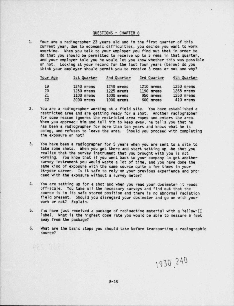
.
QUESTIONS - CHAPTER 8
1. Your are a radiographer 23 years old and in the first quarter of thiscurrent year, due to economic difficulties, you decide you want to workovertime. When you talk to your employer you find out that in order todo that you should be permitted to receive up to 3 rems in that quarter,and your employer told you he would let you know whether this was possibleor not. Looking at your record for the last four years (below) do youthink your employer shou?d permit you to receive 3 rems or not and why?
Your Age 1st Quarter 2nd Quarter 3rd Quarter 4th Quarter
19 1240 mrems 1240 mreins 1210 mrems 1250 mrems20 1250 mrems 1225 mrems 1190 mrems 1265 mrems21 1100 mrems 1000 mrems 950 mrems 1250 mrems22 2000 mrems 1000 mrems 600 mrems 410 mrems
2. You are a radiographer working at a field site. You have established arestricted area and are getting ready for a shot. Another radiographerfor some reason ignores the restricted area ropes and enters the area.When you approacP him and tell him to keep away, he tells you that hehas been a radiographer for more than ten years and knows what he isooing, and refuses to leave the area. Should you proceed with completingthe exposure or not?
3. You have been a radiographer for 5 years when you are sent to a site totake some shots. When you get there and start setting up the shot yourealize that the survey instrument that you brought with you is natworking. You know that if you went back to your company to get anothersurvey instrument you would waste a lot of time, and you nave done thesame kind of exposure with the same source quite a few times in yourSk year career. Is it safe to rely on your previous experience and pro-ceed with the exposure without a survey meter?
4. You are setting up for a shot and when you read your dosimeter it readsoff-scale. You take all the necessary surveys and find out that thesource is in its safe stored position and there is no abnormal radiationfield present. Should you disregard your dosimeter and go on with yourwork or not? Explain.
5. Yau have just received a package of radioactive material with a Yellow-IIlabel. What is the highest dose rate you would be able to measure 6 feetaway from the package?
6. What are the basic steps you should take before transporting a radiographicsource?
\0 ,
8-18

.
.
APPENDIX
Source of Federal Regulations
#For a complete copy of 10 CFR, Chapter 1, which is printed annually, write to:
Superintendent of DocumentsGovernment Printing OfficeWasidngton, D.C. 20402
Attention: Document Order and Distribution Office.
Telephone: (202) 783-3238
Specify that you want 10 CFR Parts 0-199. The cost of this publication was$6.50 for the 1979 Edition.
For a basic looseleaf copy of 10 CFR, Chapter 1, and indefinite supplementalservice, write to the address above, and specify that you want the:
U.S. Nuclear Regulatory Commission Rules and RegulationsTitle 10, Chapter 1, Code of Federal Regulations - Energy
T'.ie cost of this subscription was $47.50 as of October 13, 1977.
For individual copies of specific Parts of Title 10, Chapter 1, Code fo FederalRegulations, write to:
Division of Technical Information & Document ControlDistribution Services BranchU.S. Nuclear Regulatory CommissionWashington, D.C. 20555
- Telephone: (301) 492-7333
Individual copies are furnished free of charge..--
W
4
/
e
i930 24i' -
_.
8-19

.
APPENDIX
NAMES, ADDRESSES, AND TELEPHONE NUMBERS OF REGULATORYAGENCIES IN AGREEMENT STATES
Alabama 205-832-5992 Florida 904-487-1004
Mr. /'brey Godwin, Director Mr. Ulray Clark, AdministratorDivision of Radiological Health Radiological Health ProgramEnvironmental Health Adminis. Health Program OfficeRoom 314, State Office Building Dept. of Health & Rehab. ServiceMontgomery, Alabama 36130 1323 Winewood Blvd.
Tallahassee, Florida 32301Arizona 602-255-4845
Georgia 404-894-5794Mr. Kenneth R. Geiser, Acting Exec. Dir.Arizona Atomic Energy Commission Mr. Charles F. Tedford, Dir.2929 West Indian School Road Radiological Health UnitPhoenix, Arizona 85017 Department of Human Resources
47 Trinity AvenueArkansas 501-661-2307 Atlanta, Georgia 30334
Mr. E. Frank Wilson Idaho 208-384-3335Bureau of Environmental Health ServicesArkansas Department of Health Mr. Robert Funderburg, Superv.4815 West Markham Radiation Control SectionLittle Rock, Arkansas 72201 Idaho Department of Health and Welfare
StatehouseCalifornia 916-445-0931 Boise, Idaho 83720
License Insp.Kansas 913-862-9360
Mr. Joe Ward, Chief - 916-322-2073 Ext-284Radiologic Health SectionDepartment of Health Mr. Gerald W. Allen, Director714 P Street, Rm. 498 Bureau of Radiation ControlSacramento, California 95814 Division of Environment
Dept. of Health & EnvironmentColorado 303-320-8333 Building 740, Forbes Field
Ext. 6246 Topeh, Kansas 66620
Mr. Alvert J. Hazle, Director Kentucky 502-564-3700Radiation & Hazardous Waste Control
Division Mr. Charles M. Hardin, ManagerOffice of Health Protection Radiation Control BranchDepartment of Public Health Bureau for Health Services4210 East lith Avenue Dept. for Human ResourcesDenver, Colorado 80220 275 East Main Street
Frankfort, Kentucky 40601
C))
8-20

.
..
Louisiana 504-925-4518 New Mexico 505-827-5271 Ext-270
Mr. B. Jim Porter Dr. Ted Wolff, ChiefDivision of Radiation Control Radiation Protection SectionNe; ural Resources and Energy Environmental Improvement Div. ,.Lept. of Conservation P.O. Box 968P.O. Box 14690 Crown BuildingBaton Rouge, Louisiana 70808 Santa Fe, New Mexico 87503
Maryland 301-383-2744/2735 New York 518-474-2178
Mr. Robert E. Corcoran, Chief Mr. T. K. DeBoer, DirectorDivision of Radiation Control Technical Development ProgramsDept. of Health and Mental Hygiene New York State Energy Office201 W. Preston Street Agency Building 2Baltimore, Maryland 21201 2 Rockefeller Plaza
Albany, New York 12223Mississioni 601-354-6657/6670
North Carolina 919-733-4283Mr. Eddie S. Fuente, DirectorDivision of Radiological Health Mr. Dayne H. Brown, ChiefState Board af Health Radiation Protection SectionJackson, Mississippi 39205 Division f Facility Service
Box 12200Nebraska 402-471-2160 Raleigh, North Carolina 27605
Mr. Ellis Simmons, Director North Dakota 701-224-2374Division of Radiological HealthState Department of Health Mr. Gene A. Christianson, Dir.301 Centennial Mall South Div. of Environmental EngineeringP.O. Box 95007 Radiological Health ProgramLincoln, Nebraska 68509 State Department of Health
1200 Missouri AvenueNevada 702-885-4750 Bismarck, North Dakota 58501
Al Edmundson Oregon 503-229-5797Radiological HealthConsumer Health Protection Services Marshall Parrott, D.Sc., ManagerRm. 103 Kinkead Bldg., Capitol Complex Radiation Control Service, Div. ofCarson City, Nevada 89710 Health
Department of Health ResourcesNew Hamoshire 603-271-2281 1400 South West Fifth Avenue
Portland, Oregon 97201Mr. John R. Stanton, DirectorRadiation Control AgencyDivision of Public Health ServicesState Department of Health and Welfare
'
State Laboratory BuildingHazen DriveConcord, New Hampsire 03301
1930 243
8-21
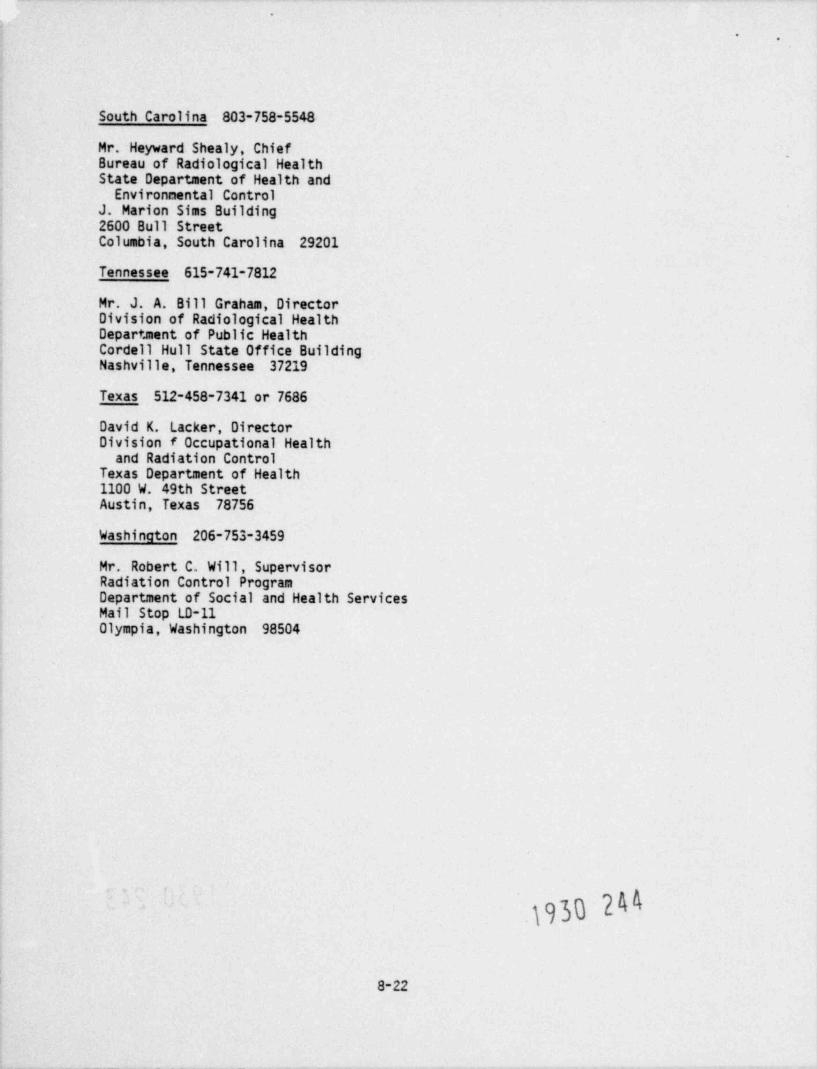
.
*.
South Carolina 803-758-5548
Mr. Heyward Shealy, ChiefBureau of Radiological HealthState Department of Health and
Environmental ControlJ. Marion Sims Building2600 Bull StreetColumbia, South Carolina 29201
Tennessee 615-741-7812
Mr. J. A. Bill Graham, DirectorDivision of Radiological HealthDepartment of Public HealthCordell Hull State Office BuildingNashville, Tennessee 37219
Texas 512-458-7341 or 7686
David K. Lacker, DirectorDivision f Occupational Health
and Radiation ControlTexas Department of Health1100 W. 49th StreetAustin, Texas 78756
Washington 206-753-3459
Mr. Robert C. Will, SupervisorRadiation Control ProgramDepartment of Social and Health ServicesMail Stop LD-11Olympia, Washington 98504
.)0Y
8-22

.
-.
.
ann
9. OPERATING PROCEDURES
#Every employer supplies the individual radiographer with a set of operatingand emergency procedures. Adherence to these procedures is very important.About 75% of all the overexposure accidents can be related to the failure ofthe radiograoher to follow the orocedures. In this chapter, we will discussoperating procedures. Emergency procedures will be discussed in Chapter 10.
1. What Not to Do
A good fraction of the radiographer's work is routine, hard work in whichnothing much happens and where the temptation to take shortcuts may lure aradiographer. When something goes amiss, however, a potential hazard isthere. Since radiation cannot be seen, heard or felt, the radiographer who isnot following his procedures may not know that something is amiss until it istoo late. The procedures are formulated to make sure that both you and thepublic are not exposed to too much radiation. It is little consolation to knowthat you can take 1000 pictures carelessly, with no bad consequences, if youget exposed when you are taking picture number 1001.
2. Operating Procedures: A Nonnegotiable Connitment
The details of operat'ng procedures may vary from company to company to takeinto account the differences in the work performed and the needs of the company.However, in order for the procedures to be approved by the regulating authori-tios, they must incorporate what is in fact the result of many years of combinedexperience in the radiography field.
The adequacy of these procedures and the strict adherence to them constitute abasic and nonnegotiable commitment that the company makes to safe practice,and justifies the right of the company to utilize hazardous material. Theprocedures may appear at times to be unnecessary and time consuming, but oncethe licensee has submitted the procedures to the NRC and they have beenaccepted, the procedures are nonncoctiable. Neither your nor your company ispermitted to " improvise" or " shortcut" the procedures.
3. Your Own Comoany's Operating Procedures
As you are studying this manual you have probably already been working for atleast several months as a radiographer's assistant or as a trainee. You haveprobably already been instructed in your company's operating procedures. Nowis a good time to review your understanding of those procedures by answeringthe questions at the end of this chapter based on your own company's operatingprocedures. After you have answered the questions you should carefully reviewyour answers with someone in your company who is familar with those procedures. '
8e sure to note where your answer is different from the correct operatingprocedure.
.
1930 245.
.
9-1

-,
QUESTIONS - CHAPTER 9
Answer the foilewing questions based on the operating procedures of your ownCompany.
1. You are at yc ur lunch break when another radiographer and friend of yoursapproaches you and asks you if he could use your film badge because hehas to make some exposures and he does not have his with him. Should yoube a good friend and help him out or not and why?
2. You are a trainee radiographer working at a field site under the supervi-sfon of an experienced radiographer. After a few exposures, you noticethat your dosimeter reads about 1/2 scale (approximately 100 mR) and youbring that to the attention of the radiographer. He reassures you that'sno problem and tells you to get back to work. What shculd you do?Explain.
3. Below is a record of your exposure for January and February determined byyour dosimeter and your film badge. Assuming that the quarter started onJanuary 1, then how much of a dose can you receive for the month of Marchif, your company limits your exposure to 1.25 rems per quarter year?
Month Dosimeter Film Badge
January 0.421 rem 0.173 rem
February 0.622 rem 0.660 rem
.
e
.
1930 246
9-2

.
'
- - I.)|7/7i-
-
10. EMERGENCY PROCEDURES
nAn emergency in industrial radiography is any condition that presents a realdanger of uncontrolled radiation exposure to any one. Such a situationtypically requires action by the radiographer to eliminate the danger thatexists. Temporary measures can usually be taken that ensure safety. Onceimmediate safety measures are taken, then the problem can be considered withserie calmness and reason. Your employer is most familiar with the types ofjobs and the equipment with which you will be involved. He has the responsi- .
bility for providing you with the detailed training in handling emergencies.Your employer is required to have procedures for handling emergencies and to-train you in those procedures.
This manual cannot train you in your employer's specific emergency procedures.He must prov1de that training separately. However, we can discuss somegeneral aspects of emergency situations and responses.
10.1 Recoonizina the Emeroency
We shall discuss the role of the radiographer throughout the course of anaccident, from its beginning to its end.
Before any suitable response can be made, the emergency must be recognized.Sometires recognition is easy. For example, if a source is crushed by a pieceof heavy ecuf pment on the job in sight of the radiographer, he knows there isa problem. Similarly, if a radiographer becomes ill or injured and is unableto secure an exposed source, a coworker who is present knows the source isout.
On the other hand, it is quite possible that an emergency may not berecognized. This is especially true if you are routinely engaged in work thatis done frequently according to a more or less fixed routine. In any such-situation, there is a tendency to take procedures for granted and, at times,to cut corners, particularly if the work is done under difficult workingconditions or time is limited. Ninety-nine times out of a 100, no problemresults. The 100th time might lead to an accident. In radiography, the mostnotable example is the failure to perfom radiation surveys as reouf red,especially the survey following completion of an exposure. This practiceseems to be disturbingly common. You must use your survey meter to perceive athreatening situation which may affect your well being. A source may not havereturned to its shielded position. Without a survey meter, you have no way ofknowing this.
10.2 The Immediate Response'
In most emergencies, once the radiographer has recognized the situation, thereis time to make a correct judgment. What should you do if a source isexposed?
.
1930 24710-1
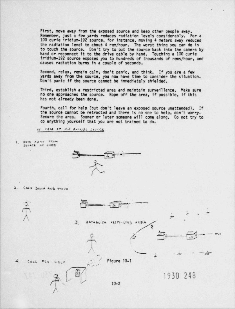
..
First, move away from the exposed source and keep other people away.Remember, just a few yards reduces radiation levels considerably. For a100 curie f ridium-192 source, for instance, moving 4 meters away reducesthe radiation level to about 4 rem / hour. The worst thing you can do isto touch the source. Don't try to put the source back into the camera byhand or reconnect it to the drive cable by hand. Touching a 100 curieiridium-192 source exposes you to hundreds of thousands of rems / hour, andcauses radiation burns in a couple of seconds.
Second, relax, remain calm, don't panic, and think. If you are a fewyards away from the source, you now have time to consider the situation.Don't panic if the source cannot be immediately shielded.
Third, establish a restricted area and maintain surveillance. Make sure
no one approaches the source. Rope off the area, if possible, if thishas not already been done.
Fourth, call for help (but don't leave an exposed source unattended). If
the source cannot be retracted and there is no one to help, don't worry.Secure the area. Sooner or later someone will come along. Do not try todo anything yourself that you are not trained to do.
jd CAta 0! / *] P rossaf 4 : s v e c s,
j, M o v t, a A A */ CASM:,0 *s LC$ A* O *n C Q
h o '
L ;.- M '~~
.!/
1. C aut Sovue AsJb wie
' * - "
.,
-i..
rm -
.
:. e- .
.=*
3, a s7 * a dsw euraicto neah
C 'l A".'- Q----g-~'~~~~~.1/ \
% . ), . - -f---.,
-- #4 c a u. =es aw e. ', , - Figure 10-1
E 1930 248'
s /s. 10-2
\

.
-.
Another situation is one in which the radiographer on a job becomes sick orinjured. Responsibility for action would then fall on the radiographer'sassistant or other coworker, if any. Proper training of the assistant radio-grapher by the employer is necessary for effective action in this case. Theassistant would rapidly have to decide what to do. In the case of a serious ~jinjury to the radiographer, the assistant's immediate concerns would be thesafety of the radiographer and obtaining medical aid as recuired. At the sametime, he would have to see to it that the work area was under proper surveil-lance and, if possible, that the source was secured. If the job were beingcarried out at a location where the assistant could readily contact other per-sonnel for aid, this should be done. However, if the operation were beingconducted at a remote location where aid was not readily available, the assis-tant night be faced with some weighty decisions. His actions would recuireexercise of his own judgment.
10.3 Summonino Assistance
Following the initial response to minimize the danger to yourself and others,._
you should continue to follow the proper emergency procedures. This will fre-quently recuire notification of other persons or support groups. For anyemergency, you must know whom you are supposed to contact. The primaryauthority and responsibility for initial response lies with the leadradiographer on a job.
A common requirement of emergency procedures is that, following the initialresponse, the radiographer should contact the employer's radiation protectionofficer to assist with the problem. Other individuals or groups may also becalled upon as the situation requires.
If the police are contacted, it is important for you to offer as muchinformation and assistance as you can to them. Typically, you will have themost knowledge of the situation and, thus, you will be a key person with whenothers should consult.
10.4 Your Own Comoanv's Emeroency Procedures
You have probably already been trained in your own company's emergencyprocedures. The questions at the end of the c.. apter can 9 ve you some ideaihow well you understand those procedures. Answer the ouestions. Then reviewyour answers carefully with someone else. Did you respond correctly to theseemergency situations?
.
1930 249
10-3

.
OUESTIONS - CHAPTER 10
Answer these questions based on your own company's emergency procedures.
1. After finishing an exposure and taking a cuick survey, you find out thatthe source has not been completely retracted and is somewhere in theguide tube. Circle the sequence of steps that you are going to take.
a. Post high radiation signs.
b. Try to crank the source back,
c. Go call your supervisor from his office.
d. Try to detemine where exactly in the tube the source is.
e. Try to reconnect the cable, if broken, or loosen up the stucksource so that you can retract it.
f. Try to get another radiographer so that you can make sure of whatthe problem is.
g. Move away to a safe distance.
h. Try to get somebody to notify your supervisor and the radiationofficer, but do not leave the immediate area.
)h
10-4

.
'.
f"'"TR 10 !C:!'^;. QGS ;^=
2. You are setting up an exposure "I'a site when you find out that your surveymeter is inoperable. This is a common shot that you have performed many times :#
in your career as a radiographer. Should you go ahead with the shot, based onprevious experience, or not? Why?
3. You are working on a site with your assistant radiographer. In the middle ofa shot, you get dizzy and collapse. Circle the steps that your assistant
should take (there could be more than one):
(a) Leave the area Lamediately and try to get medical help.,
(b) Make you confortable and finish the shot before summoning help.
(c) Stop the exposure, secure the source, and go to get some help.
(d) Stop.the exposure, secure the source, and try to get help immediatelywithout leaving the immediate area.
(e) Take you to the nearest hospital right away.
4. You have just finished an exposure and are trying to retract the sourcewhen you realize that it is stuck. Describe briefly and in order of sequencewhat actions you should take.
5. You are working at a permanent radiographic installation with radiographic cellsequipped with audible high radiation alarms. The alarm of the cell in which youare working goes off with no source besag exposed and you are able to determinethat the alarm is malfunctioning. There is no electronics expert on siteavailable at this time to repair the alarm so you are left with two options!(a) turn the power to the alarm off and continue your work using your surveymeter, and (b) don't take any more shots until the alarm has been repaired.What would you do? Explain.
6. It is early af ternoon, you have just char 3ed your dosimeter and you are settingup for a shot. You take a survey prior to the exposure of the source and yoursurvey meter shows everything is fine, but when you check your dosimeter, itreads off-scale. Would you assume your dostneter is malfunctioning and proceedwith the shot, or would you assume your survey meter is inoperable and try toget another one before completing the shot? Explain.
1930 251
JO -5
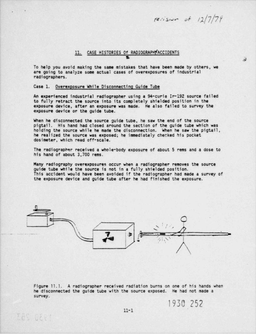
..
/,1||||fft .'iS r. w >f
11. CASE HISTORIES Of RADIOGRAPH / ACCIDENTS* 2
To help you avoid making the same mistakes that have been made by others, weare going to analyze some actual cases of overexposures of industrialradiographers.
Case 1. Overexposure While Disconnecting Guide Tube
An experienced industrial radiographer using a 94-curie Ir-192 source failedto fully retract the source into its completely shielded position in theexposure device, after an exposure was made. He also failed to survey theexposure device or the guide tube.
When he disconnected the source guide tube, he saw the end of the sourcepigtail. His hand had closed around the section of the guide tube which washolding the source while he made the disconnection. When he saw the pigtail,he realized the source was exposed; he immediately checked his pocketdosimeter, which read off-scale.
The radiographer received a whole-body exposure of about 5 rems and a dose tohis hand of about 3,700 rems.
Many radiography overexposures occur when a radiographer removes the sourceguide tube while the source is not in a fully shielded position.This accident would have been avoided if the radiographer had made a survey ofthe exposure device and guide tube after he had finished the exposure.
S _
s yO
A / / Q ! '~s\
'
_ _ _ _ _________. \ 7 '3 ,6~L p /,1* 0;; p
' ~
|
Figure 11.1. A radiographer received radiation burns on one of his hands whenhe disconnected the guide tube with the source exposed. He had not made asurvey.
1930 25211-1
.,

'.
Case 2. Overexposure in a Radiography Cell
After completing an exposure using a 70-curie Co-60 source, a radiographerforgot to retract the source to its safely shielded position. The radiog-rapher entered the shielded radiography cell with a survey meter in hand, butdid not to look at the meter. He remained in the cell and proceeded to set upthe next exposure. When he picked up the survey meter before leaving thecell, he noticed that it road off-scale. He left the cell immediately andread his pocket dosimeter, which was also off-scale.
The exposures were about 10 rems to the eye, 5 rems to the stomach, and 3 remsto the radiographer's hands.
His film badge did not indicate an overexposure because he wore it towards hisside and the beam was highly collimated.
The direct causes for this accident were: (1) the radiographer forgot toretract the source to its safe shielded position at the end of the first expo-sure, and (2) he failed to make a survey when he entered the cell. This manentered the radiation area " blind." Carrying a survey meter offers no protec-tion unless it is used.
* ==:: ss_-====\\. ,
N 11N 11
N ||- \ 11
N _ mm13
s.,Nog ' , so m e g;w
,/\ /s ,,.,"
. /! / yi..,@sfa E.
N \ d-
s sG% y\ .
I"-'-. s .
. ' ' j '* ,2 I- ~ ~,
. . - - s c.
'
- ' '
/
-Figure 11.2. A radiographer did not read his survey meter when he entered anexposure cell with the source exposed.
31-2
1930 2531

..
O
Case 3. Overexoosure Oue to Inadequate Survey
An industrial radiographer accidentally did not fully retract a 75 curieIr-192 source into its completely shielded position after making an exposure.He approached the exposure device with his survey meter, then passed the meter
#close to the device and noted no unusual readings. He proceeded to set up for-the next exposure. When he picked up his meter, he noted that it went off-scale when it passed in front of the device. He returned to the crank andretracted the source with about a quarter turn of the crank. He read hispocket dosimeter and found it read off-scale.
He received a whole-body exposure of about 4 rems.
The radiographer in this case did make a survey, but unfortunately the surveywas inadequate. He failed to include the guide tube and front of the exposuredevice in his survey. In this case, it is not clear whether the radiographerwas not properly trained on how to survey or whether he became careless withjob familiarity.
.
-3
.
1
% } *-
-
.
"
''*D gyp >.
.
Figure 11.3. A survey meter placed behind an exposure device may not detectan exposed source close to the front of the exposure device. The exposuredevice will shield the survey meter from the source.
1930 254' 11-3
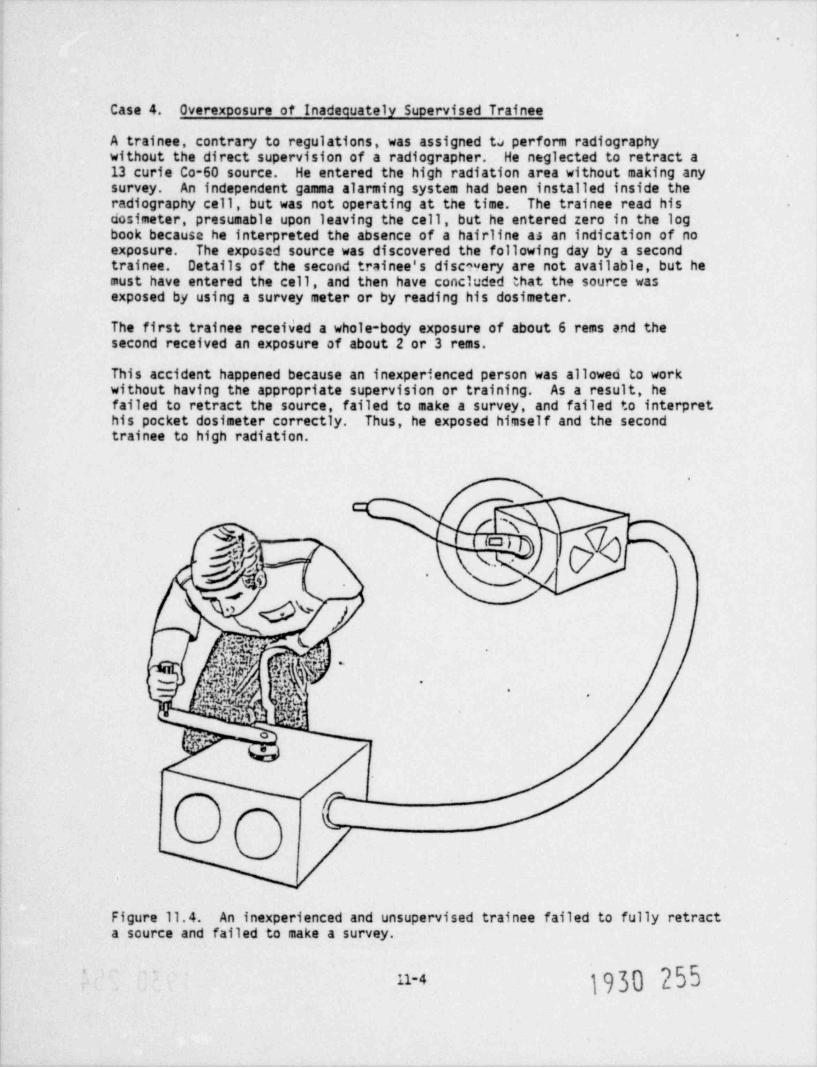
-.
Case 4. Overexoosure of Inadequately Supervised Trainee
A trainee, contrary to regulations, was assigned tu perform radiographywithout the direct supervision of a radiographer. He neglected to retract a13 curie Co-60 source. He entered the high radiation area without making anysurvey. An independent gamma alarming system had been installed inside theradiography cell, but was not operating at the time. The trainee read hisdosimeter, presumable upon leaving the cell, but he entered zero in the logbook because he interpreted the absence of a hairline as an indication of noexposure. The exposed source was discovered the following day by a secondtrainee. Details of the second trainee's discovery are not available, but hemust have entered the cell, and then have concluded that the source wasexposed by using a survey meter or by reading his dosimeter.
The first trainee received a whole-body exposure of about 6 rems and thesecond received an exposure of about 2 or 3 rems.
This accident happened because an inexperienced person was allowed to workwithout having the appropriate supervision or training. As a result, hefailed to retract the source, failed to make a survey, and failed to interprethis pocket dosimeter correctly. Thus, he exposed himself and the secondtrainee to high radiation.
.
)'
|S O |'
pj.,
.
,
o,r7w
L
'
Figure 11.4. An inexperienced and unsupervised trainee failed to fully retracta source and failed to make a survey.
1930 255'
11-4
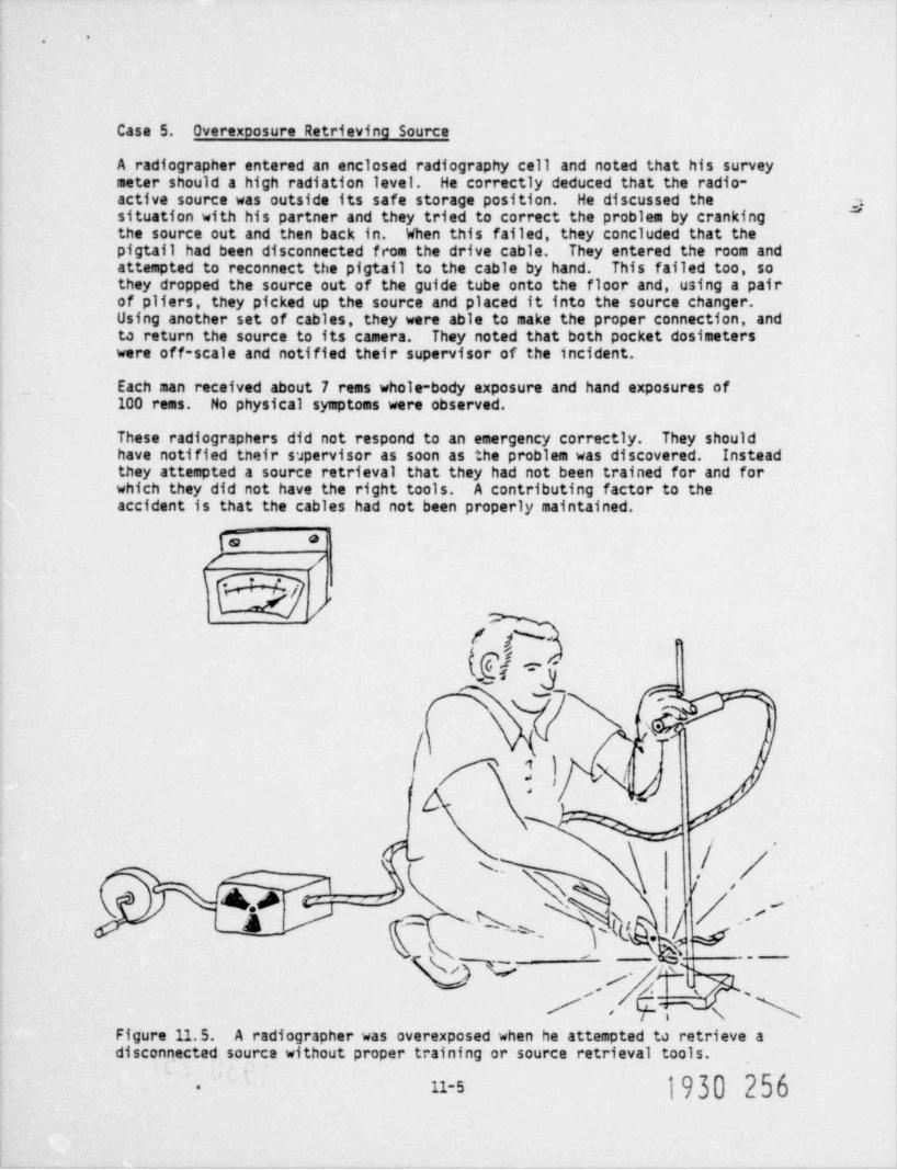
'.
Case 5. Overexposure Retrieving Source
A radiographer entered an enclosed radiography cell and noted that his surveymeter should a high radiation level. He correctly deduced that the radio-active source was outside its safe storage position. He discussed the g,situation with his partner and they tried to correct the problem by crankingthe source out and then back in. When this failed, they concluded that thepigtail had been disconnected from the drive cable. They entered the room andattempted to reconnect the pigtail to the cable by hand. This failed too, sothey dropped the source out of the guide tube onto the floor and, using a pairof pliers, they picked up the source and placed it into the source changer.Using another set of cables, they were able to make the proper connection, andto return the source to its camera. They noted that both pocket dosimeterswere off-scale and notified their supervisor of the incident.
Each man received about 7 rems whole-body exposure and hand exposures of100 rems. No physical symptoms were observed.
These radiographers did not respond to an emergency correctly. They shouldhave notified their supervisor as soon as the problem was discovered. Insteadthey attempted a source retrieval that they had not been trained for and forwhich they did not have the right tools. A contributing factor to theaccident is that the cables had not been properly maintained.
V *
/ /
'S / ,
/ ,
f % ,
(e'n~
Nk[?! N(j
.'
/v s
&<- !j -
q= q % ;te . //1
,// \'
N,
Figure 11.5. A radiographer was overexposed when he attempted to retrieve adisconnected source without proper training or source retrieval tools.
1930 25611-5-

*.
Case 6. Overexposure Due to Ignoring Alarm
At the conclusion of a radiographic exposure using an 80-curie Co-60 source ina permanent radiographic cell, a radiographer noted that the gamma alarm atthe entrance was signaling a high radiation level. The radiographer calledhis supervisor and they both entered the cell with a survey meter. Since thesurvey meter did not indicate any radiation levels, they assumed that thesource was safely stored and that the cell alarm was malfunctioning. They didnot try to verify that the survey meter was operating properly.
The radiographer proceeded to set up the next exposure, although the gammaalarm continued to signal. When he entered the cell again for the next expo-sure, he used a different survey meter, and noted that this meter indicated ahigh radiation level. The radiographer notified his supervisor that thesource was exposed, and the supervisor locked the cell and informed personnelof the exposed source.
The radiographer received about 5 rems whole-body exposure and 15 remsexposure to his hands.
This accident happened because the radiographer and his supervisor failed tobelieve the independent alarming system and failed to verify the properoperation of the first survey meter. Survey meters are generally quitereliable, but they do malfunction from time to time. You should be alert tounusual readings from a survey meter and should know how to check them in thefield for proper operation.
~~h k '
.
.o
k iG'
. {'
hui * A R r.iii' ~"'
/ ,
Y' -,
'( . | & [&@ '/-
3! }tC?M-,,
1 I ,.~. *,*,
- a i 5.
v%y} -
M W E,; g' ,
** / 4.X wtFigure 11.6. An exposure cell alarm sounded, but a survey meter showed noradiation. The radiographstr erroneously chose to believe the survey meter.He should have checked the operation of the survey meter before entering thecell.
} 9N ,11-63
,

'.
Case 7. Overexposure Involving Two Sources
A 44-curie Co-60 source and a 92-curie Ir-192 source were being used to radio-graph the same casting. When the first shift was terminated, the radiographerforgot to retract the Co-60 source into the exposure device. He apparently ,also did not make a survey to ensure the source was properly shielded. Whenthe second shift radiographer came, he entered the area to set up exposureswith the Ir-192 source. He also failed to make a survey and did not find outthat the Co-60 source was still exposed. Presumably he made his exposureswith the IR-192 source without making surveys. When the second shift radiog-rapher attempted to set up an exposure with the Co-60 source, he found outthat it had been left exposed. In his haste to crank in the Co-60 source, hemistakenly cranked out the Ir-192 source. Both sources were now exposed andwhen the radiographer attempted to reduce the persistent radiation levels bycranking the Co-60 source in and out repeatedly, he finally decided that theIr-192 source was exposed.
The first shift radiographer received a whole-body exposure of about 6 or7 rems, and the second shif' radiographer received a whole-body exposure ofabout 25 rems and a hand exposure of about 50 rems.
This accident could have been avoided if the radiographers had made surveysand if the first radiographer had not failed to retract the source. Manage-ment control of this operation must have been exceedingly lax if so manysurveys were omitted. Note also that procedures for using two sources shouldbe developed and followed. With two sources and two sets of controlsconfusion can easily occur over when to use which set of controls.
de9 ' -
o
,% -
J'2. RE RACT s'T '
. -.
#.
THEsoQ QT 7) e.
h bi. 4-
, -..
i MU.' n
Figure 11.7. When two sources are being used it is possible to crank thesecond source out instead of cranking the first source in. Then there are twoexposed sources when there should not be any exposed source. A survey willdetect this mistake.
11-7

_
'.
Case 8. Overexposure After Defeating Safety Alarm
While performing radiography in an enclosed cell, a radiographer decided toprop open the cell doors to allow air circulation in the room as he changedfilms and set up the next exposure. When he did this the first time, heswitched the door alarm to the off position, which also turned off the in-cellradiation alarm. Later the radiographer failed to retract the 166-curie Co-60source being used. He entered the cell without a survey meter and while thecell alarms were off. On top of that, the radiographer was not wearing hisfilm badge or pocket dosimeter. A production coordinator working with theradiographer entered the cell also without wearing a film badge or pocketdosimeter. The radiographer exchanged films, adjusted the source collimator,and left the cell with the production coordinator. When he attempted to crankthe source out to its exposed position, he realized that the source had notbeen retracted on the previous expc.sure and that he and the productioncoordinator had been exposed.
The radiographer received a dose to the portions of his hand that adjusted thesource collimator in excess of 1,250 rems. The production _ coordinatorreceived a dose of 4 rems to his eyes.
Failure to retract the source indicates carelessness. Bypassing the 'sorinterlock and failing to use a survey meter allowed this incident to happen.
A good management review of the cell design should have realized that its lackof ventilation would cause extreme heat buildup that could be alleviated onlyby violating procedures and leaving the door opened. In addition, thisradiogrpher was working under great duress. He had been called in to workovertime while his wife was in the hospital for a serious operation. Whiletheradiographerhadanexcellentrecordofpastperformanceghecouldnotbeexpected to perform well under these circumstances. Management should havebeen more sensitive to the worker's special problems that day.
i
Ek"I~
INTERLOCK !- ~
|, ,0 N. . . .
h((G({dfll{{{{{{{Mfdh~ ' ' ~ '
FFcY/
. .
J p/|' A
'
Il\
\ Ist a
figure 11.8. A gamma alarm was shut off so that the door to an exposure cellcould be left open for ventilation. Thc source was left exposed, and a surveywas not made.
11-8
1930 259

.
Case 9. Overexposure Oue to Radiographer Becoming Ill
A radiographer working alone ended an exposure suddenly when he became veryill. In his haste to secure the device, he failed to fully retract the radio-graphic source to its safe position in the exposure device. In addition, he
#made no survey of the exposure device or the guide tube. The device with theexposed source in the guide tube was placed in a locked van. The radiographerdrove to several locations but did not return the source to W employer'sfacility. The van was parked overnight at his home. The next :orning hepicked up his assistant. The assistant turned on his survey meter and notedthat it read off-scale. They immediately stopped the van and retracted thesource.
The exposure was about 10 rems to the whole body of the radiographer.Radiation levels in an unrestricted area exceeded 6 roentgens per hour. Noone will ever know if any members of the public were overexposed.
This accident could have been prevented had the radiographer fully retractedthe source or had he made a survey. The exposure would have been decreased ifhe had read his pocket dosime+9r and noticed a high reading. Of course, it isdifficult for anyona to perform his job well when he is suddenly taken ill,but at such times accidents are especially likely.
'sa -
- . o / .>- d b
9
* %~.
.i m% ,
\ d) % ft/ 'N.
_ g gA S,m
D\ v ,
\ G >
,
|o
/#o
\
w
Figure 11.9. A radiographer became ill and, in his haste to stop work, failedto retract the source fully, failed to survey, anL failed to read his pocketdosimeter at the end of the day.
1930 260- u-9
I.

-.
=.
Case 10. Overexposure Due to Careless Storage *
In 1962, early in the year, a construction watchman was given a 5-curiecobalt-60 source for safe keeping by a contractor. The watchman did not knowwhat the source was, but he assumed that it was valuable because the con-tractor told him to store it in a safe place, and to make sure no one wentnear it. Since the watchman knew that valuable property should be guardedcarefully, he took the source home with him. Although the source was in alead container, presumably the watchman removed the source frcm the containerout of curiosity to see what was valuable about it.
Sometime between March 21 and April 1, his son found the source and placed itin a front pocket of his trousers. On April 1, his mother found it, andplaced it in a drawer in the kitchen.
On April 17, the watchman's mother in-law came to live with the family and atthat time noted the blackening of the glasses that had been close to thesource in the drawer.
L, 5yv
*
N
\M / p; i
,
"~)'
i,h' , , , ,
\v'
f .:
/ /'
.
>
) N '( \
''f #f(C
Te
[ ,
~
Figure 11.10. Four members of a family died after a source was left in thecare of an untrained person without adequate instructions about storing thesource safely.
This description was obtained directly from people closely associated withthe investigation of this accident. As such, the description corrects someerrors in some previously publisned accounts of the accident.
11-10
1930,261

.
On April 29, the boy died. On July 19, his mother died. Although the causeof their deaths was not suspected at the time, it is evident now that both sonand mother died of acute radiation sickness. It was later estimated that theboy had received a dose in the range between 3,000 and 5,000 rads ** and hismother one in the range between 2,000 and 3,000 rads.
On July 22, 1962, the contractor came to the house, claimed the source, andtook it away. No one suspected the tragedy it had caused. In August, thewatchman's 24 year old daughter became very ill. On August 13, an alertphysician suspected for the first time that the common symptoms of the membersof this family might be due to radiation. On August 18, the little girl died.It was later estimated that she had received between 1,400 and 1,900 rads.
On August 20, the watchman and his mother-in-law were admitted to the hospitalwith what appeared to be radiation exposure symptoms. Since he was away fromthe house a lot, the watchman had been exposed on-and-off, and it is believedthat he had received less exposure than the other members of the family. Hewas discharged from the hospital on September 6, but kept under close medicalobservation. His mother-in-law did not survive, though. She died onOctober 15. It was estimated that she had received between 1500 and 3000 radsover the period of time she was exposed.
This tragedy could have been avoided if the radiography firm had kept a bettercontrol of its sources, so that the missing source would have been noticedimmediately. Radioactive sources must not be left in the care of someone whohas not been trained in the dangers they present and the procedures for takingcare of them.
Case 11: A Detached Source and No Survey.
In doing radiography at a job site, late on a Friday afte. ,on, a radiographerfailed to make a survey at the end of the last exposure. When he finished, heput all the equipment on the back of an open pickup truck and transported itback to the company. Unfortunately, he failed to detect that the source wasnot in its safe shielded position. In fact, at the end of the last exposure,the source had become stuck in the guide tube and when the tube was put in thetruck, the source fell out unnoticed.
The pickup truck was used several times over the weekend by the company'ssecretary and it was parked at several public places. The source was notdiscovered until the following Monday when the device was to be used again. Areenactment of the accident indicated that the radiographer received 93 remsand the secretary received 55 rems. There was no way to tell how many otherpersons received an unnecessary exposure.
We see that the radiographer exposed himself and others to unnecessaryradiation even though explicit instructions in the procedures given to him byhis employer indicated that he should make a survey at the end of each -
exposure.
AAAll the doses were estimated and some of them may be somewhat localizedrather than whole-body doses.
:..
11-11
1930 262

*,
Case 12: Misleading Indicator Lights on Crank-out Device.
In this case, an assistant radiographer was operating an exposure deviceequipped with a light to indicate if the source was in a safe shieldedposition or not.
[ CASE 11:~
THE RADIOGRAPHER FAILED TO MAKE A SU( AT THE END OF AN EXPOSURE.
' \ \j '[ g ..3
I \
% GWMC\I'\,\- /
,, / p-,,,
'
,\'..
\.,
// %'
-- 'Y W.'
% . ~ .'.kN \ p- ']~
g
%~%f '
h'N.h)Ihb;; A cf' )- )..- r eze.:t
'k?m ,/\>
{f" g/g: THE naciocnAPwfR FAstro to MAKE A $URVEy 4 f THE ENO OF AN E YPOSilRE . Tt SosR8
W||'> t. Lit' tXI%5C D iN i/([ L4lt M CF A I)cy vf Tfice**. CVdf /1 LL'EEW6Ut)Adh SL VcGil . I'lCI'L G l~cl''i C Xfi'SiDt
1930 263,
11_1,
.
The assistant cranked the source back, after an exposure, until the greenlight (shielded position) came on. Then he entered the area without making asurvey and proceeded to set up the next exposure.
Shortly afterwards, another radiographer entered the setup area carrying aradiation survey .1eter that indicated a high radiation field. He alerted the Sassistant that the source must be exposed and they both left the areaimmediately. At that time, the assistant checked his 0-200 mR dosimeter andfound that it was off-scale. The assistant received a dose estimated at about10 rems to the whole body.
The assistant radiographer was unnecessarily exposed to high levels ofradiation, although instructions in the procedures given to him by hisemployer explicitly said not to rely on indicating lights on the device but tomake a survey before entering a set up area.
Case 13: Confusion with Two Sources by an Unsupervised Trainee.
A trainee was allowed to perform radiography with two sources and withoutdirect supervision. He mistakenly cranked-out a 35 curie Co-60 source insteadof retracting a 94 curie Ir-192 source. Both cranks were in the same location-and the trainee could not distinguish one from the other.
The supervising radiographer and the trainee entered the cell with bothsources exposed. They were both carrying survey meters but failed to makesurveys. The radiographer set up the new exposure and they both left the cellwithout noting the two exposed sources. When they tried to crank the Co-60source out for the next shot, they realized that they had been overexposed.An investigation showed that the trainee received about 11 rems and theradiographer received 15 rems.
Explicit company instructions, which the radiographer disregarded, said thatno trainee is supposed to perform exposures by himself with no direct supervi-sion. The instructions also said to make a survey when entering i call.
Summary
These cases show clearly that accidents do happen when the training is poorand when the worker does not follow proper procedures. Most accidents can beavoided if proper surveys are made and the worker knows what to do in anemergency situation.
Of course, the physical reason for the accident is always that a source is notin a fully shielded position and that a human being becomes exposed to it.
Why would someone leave a source exposed?
The radiographer may think that he has cranked the source back into itsshielded position. He .hinks so, because he observes that the crank odometerreads zero, or he has counted the number of turns out and the same number ofturns back, or he has met resistance at the crank handle, a sign to him thatthe source is securely in the projector. It is rare that the radiographer
'
11-13
1930 m.,

_
-.
forgets to crank the source (or what he thinks is the source) back towards theprojector. Thinking the source is secure, the radiographer goes on to adjustthe source guide tube or to change the film or whatever--and he is exposed.
The remedy is, of course, a proper radiation survey. It may be argued thatone reason why a proper survey is not always conducted is that surveys are arepetitious and monotonous thing to do.
But if your equipment is well maintained, if you perform proper surveys, andif you learn and follow your company operating and emergency procedures, yourwork can be as safe as any other work.
The job you perform is essential to make the world safe for others.Submarines, airplanes and pipelines would not be as safe without industrialradiography. Please make sure that your work is safe for yourself, too.
.
b11-14,

-.
QUESTIONS - CHAPTER 11
1. What was the reason for most of the overexposures in the cases reviewed?'#2. If you are working in a radiography cell equipped with an audible radia-
tion alarm do you think it is necessary to take the time out for a surveyrather than relying on the very dependable audible alarm?
3. Circle the things that you as a radiographer should never do if a sourceis accidently exposed.
a. Try to shield (i.e., crank, recover ...) tha source.
b. If the source is on the ground, try to get it into its shieldedcont';iner.
c. Rope off the area and go lcok for your supervisor.
d. Call for help but do not leave the area,
e. Ask your trainee for his dosimeter because yours is off-scale.
f. Leave your trainee in the area and go seek help.
4. Read the following hypothetic.al overexposure case and state how the accidentcould have been avoided.
Af ter making an exposure, an industrial radiographer entered theradiographic cell, ea,uipped with high radiation alarm, and proceeded toset up for the next shot. When he got out of the cell, he checked hisdosimeter and found it read off-scale. He then picked up his surveymeter and reentered the cell, taking a complete survey, and found outthat the source had been stuck in the guide tube. It was determinedlater that the cell high radiation alarm was malfunctioning and had notgone off as it was supposed to do when the source was not fully shielded.
5. S tat e, in a few words, what steps should be taken, in your opinion, byemployers to avoid unnecessary overexposures.
1930 266,
.
11-15

f ui5 % ci ||-?/ s*Q'
'~
GLOSSARY
This glossary contains terms used in industrial radiography. If the term hasa special meaning.in industrial radiography that special meaning is given inthis gicssary. Terms underlined in the text of this manual are given in this -;
glossary.
Activity. See Radioactivity.
Acuto Radiation Exposure. Exposure to a radiation field where more than 100 radsof total body dose are received in a short period time.
Acute Radiation Syndrome. The symptoms resulting from a 100 to 200 rems or moretotal body exposure in a short space of time. (Nausea, fatigue, vomiting,diarrhea, loss of body hair).
Agreement State. A state which has signed an agreement with the Nuclear RegulatoryCommission to regulate the use of radioisotopes produced by the fissioningof uranium.
Alpha Particle (Alpha Ray, Alpha Radiation). A small electrically chargedparticle of very high velocity thrown off by many radioactive materials.It is identical with the nucleus of a helium atom, and is made up of twoneutrons and two protons.
ASNT. Abbviation for American Society of Non-Destructive Testing.
Attenuation. The process by which the intensity of the radiation field isreduced when passing through matter.
Atom. A unit of matter that is the smallest unit of an element. An atomconsists of a centrally charged nucleus which is made up of neutronsand protons, and is surrounded by electrons equal in number to thenumber of protons.
Background Radiation. That amount of radioactivty that is emitted from thenaturally occuring radioisotopes from the earth and the cosmic rayswhich constantly bombard the earth from outer space.
BEIR. Abbreviation for Biological Effects of Ionizing Radiation.
Beta Particle (Beta Ray, Beta Radiation). A small electrically charged particleemitted by many radioactive materials. A negatively-charged beta particleis identical with the electron and emerges from many radioactive materialsat high speed, sometimes close to the speed of light.
Bill of ladingByproduct Material. Radioactive material such as Cobalt-60 which is obtained
in the process of usi.ig or producing uranium, thorium or other fissionablematerial.
Cesium-137. An isotope of the element cesium which is radicactive and emitsgamma rays with an energy of .662 MeV. A source used in radiography.Symbol: Cs-137.
1930 267G-1

-.
,
C.F.R. or CFR Abbreviation for Code of Federal Regulations.
Chirper. A dosimeter that can be set to make an audible signal when the receiveddose reaches a certain level.
Cobalt-60. An isotope of the element cobalt which is radioactive and emitsgamma rays of energy 1.17 MeV and 1.33 MeV. A source used in radiography.Symbol: Co-60.
Collimator. A lead, or other heavy metal, radiation shield used with isotopes.It is placed on the end of the guide tube and has only a small portedwindow which will emit a narrow cone of unshielded radiation when the sourceis cranked into position. Use of a collimator will greatly reduce thesize contamination of the area which must be restricted during exposures.
Cosmic radiationCrankout Device. A device used to bring the radiographic source out of its
shielded position.
Curie. The basic ur.!L used to measure the rate at which a radioactive materialthrows off particles or otherwise disintegrates. One curie is equal to 37billion disintegrations per second.
Decay. See Radioactive Decay.
Decay Constant. A constant, characteristic of a radioactive source, thatindicates the decrease of source strength with time.
Depleted Uranium. Uranium having a smaller percentage of Uranium-235 than the0.7% found in natural Uranium. It is obtained from spent (usea) fuelelements or as a byproduct of uranium isotope separation.
Detector. A tool used to measure the presence of radiation.
Dose. The amount of radiation energy absorbed (dose per unit of time.
Dose Equivalent. The absorbed dose mult' plied by the quality factor for theparticular radiation.
Oose Rate. Oose per unit time. Usually means the dose equivalent per unittime. Typical units are rem per hour or mrem per hour.
Dosimeter (Dose Meter). An instrument used to determine the radiation dose aperson has received.
D.O.T. or 00T Abbreviation for Depart of Transportation.
Electron. A very light particle that rotates around the nucleus and carries aunit negative electric charge. It takes about 1,800 electrons to equalthe weight of one proton or neutron.
Electron Volt. A small unit of energy. It is the amount of energy gained byan electron when it is accelerated by a voltage difference of one volt.
1930 268G-2.

, .
Element. A basic substance. There are 92 different kinds of naturally occurringelements including hydrogen, oxygen, lead, and uranium.
Erytherma. Reddening of the skin due to increased local circulation of bloodas a reaction to tissue injury. 3
Exclusive use vehicle
Exponential Decay. A term describing the mathematica~l nature of radioactivedecay phenomena.
Exposure. A measure of the amount of ionizatior; in air that is produced by gamma~rays or x-rays. The unit for exposure is rocr:tgen which is abbreviated as
R or r.
~~
Exposure Device. A device used to expose the radiographic source when takinga picture.
Fallout. Radiation debris, usually from weapon testing, which settles on earthfrom the atemosphere.
Film Badge. A piece of masked photographic film worn by radiation workers as abadge. It is darkened by nuclear radiation, and radiation dose can bemeasured by inspecting the film.
Gamma Alarm. A special alarm that goes off when it detects certain levels ofgamma radiatien.
Gamma Rays ( -Rays or Gamma Radiation). This is the most pnetrating of allradiations. Gamma ray energies are usually between 10 heV and 10 MeV.
Geiger Counter (Geiget-Muller Counter, G-M Counter, G-M Tube). An electricaldevice used to detect the presence of radiation.
General License. A license issued for possession and use of byproduct materialcontained in certain items as well as for ownership of byproduct materials.Usually for small quantities. No specific application is required, butsome regulations must be met.
Guide Tube. A long flexible hollow tube in which the radiographic sourcetravels when it is cranked out of its shielded position in the camera.
Hal f-li fe. The time it takes for half the atoms in a radioactive sample todecay. Half-lives vary in range from a fraction of a second to billionsof years. The half-life of Co-60 is 5.3 years.
Half-Value Layer (Half-Value Thickness). The thickness of a shield that will .
reduce the amount of radiation passing through to one-half its initialvalue. It is different for different materials and different energygamma rays. The half-value thickness of Co-60 gamma rays in lead is1.6 cm.
1930 269G-3,
. ,
High Radiation Area. An area in which the dose rate is at least 100 mremper hour.
I.C.R.P. Abbreviation for the International Commission on Radiological Protec-tion.
Inverse Square Law. A " law" of nature that states how the intensity of radiationdecreases as one moves away from a point source. The law states that theintensity will decrease in tie same proportion as the increase in distancesquared. Moving twice as far from a source decreases the intensity t.v afactor of two squared, or four.
Internal Contamination. Contaminaticn of the body due to radiation source inhaled(dust, gas, etc.) or ingested.
Ion. An atom which has lost one or more of its electrons and is left with apositive electric charge. There are also negative ions which have gainedan extra electron.
Ionization. The proces:, of adding one or more electrons to, or removing oneor more electror.3 from atoms or molecules thereby creating f on:..
Ionization Chamber. A device roughly similar to a geiger counter and used tomeasure radioactivity.
Ionizing Radiation. Any radiation that produces ions in a material. Thistype of radiation is harmful to living things. Alpha rays, beta rays,gamma rays, and x-rays are examples of ionizing radiation. Radiowaves,ultraviolet rays and microwaves are examples of non-ionizing radiation.
Iridium-192. an isotope of the ele nt iridium which is radioactive and emitsgamma rays of energy .3 MeV to .il MeV. A source used in radiography.Symbol: Ir-192.
Isotope. Two nuclei of the same elemtnt which have the same charge but differentmasses are called isotopes. iney contain the same number of protons but adifferent number of neutrons.
kev (kilo electron volts). A unit of energy equal to 1,000 electron volts.
Leak Test. Checking of sealed capsules or sources for tne escape of radioactivematerial.
LSA material.
Licensee. The company or the person authorized to conduct activities under alicense with the Nuclear Regulatory Commission or an Agreement State.
Low Specific Activity Material.
Median Lethal Dose (LD-50/30). The dose that would result in 50% of the peopleexposed to that dose dying within 30 days. This dose is approximately450 rems (450,000 mrems) delivered in a short period of time.
G-4
1930 270

L.
MeV (million electron volts). A unit of energy equal to 1,000,000 electronvolts.
Millirem. A commonly used unit of dose equivalent. One-thousandth of a rem.
Molecule. The smallest unit of a compound. A water molecule consists of twohydrogen atoms combined with one oxygen atom. Hence, the well-knownformula, H 0.
2
Natural Background. Same as background radiation.
Natural Radioactivity. The radioactivity associated with naturally occurringelements such as isotopes of radium and carbon.
NCRP. Abbreviation for National Council on Radiation Protection and Measurement.
Neutron. One of the basic particles of the atomic nucleus (the other is theproton). T M neutron weighs about the same as the proton, but has noelectric charge.
N.R.C. or NRC. Abbreviation for Nuclear Regulatory Commission.
Non exclusive use vehicle. Normal form.
Nucleus. The inner core of the atom. It consists of neutrons and protons tightlylocked together. The prual of nucleus is nuclei.
Operating Procedures. A set of instructions supplied by the individual employeron how to perform radiographic exposures in compliance with regulatoryrequirements.
Penetrating Radiation. Radiation that has the property of easily going throughmatter, usually X-rays or y-rays.
Pig
Pipline Device. A radiographic exposure device especially made for work doneon pipelines.
Placard.
Pocket Chamber. An ionization chamber in the shape of a fountain pen which isused for personnel monitoring.
Pocket Dosimeter. A small sized, air-filled fonization chamber instrument thatmeasures the amount of radiation received by it.
Proton. One of the basic particles of the atomic nucleus (the other is the
neutron). Its charge is as large as that of the electron, but positive.
Quality Factor. A factor that accounts for the fact that different damage canbe caused by different types of radiation.
1930 271G-5
~
m

i .
Rad. A vunit of dose. See Dose.
Radioactivity. The phenomenon whereby atoms disintegrate with emissions ofradiation.
Radiation. The emission of very fast atomic particles or rays by nuclei. Someelements are naturally radioactive while others become radioactive after '
bombardment with neutrons or other particles. The tnree major forms ofradiation are alpha, beta, and gamma.
Radioactive Decay. A natural process by which the strength of a radioactivesource is reduced with time.
Radiat. ion Area. An area where the radiation field is equal or greater than5 mrem per hour or where a person could receive 100 mrem in five consecutivedays.
Radiation Sickness. Any sickness resulting from exposure to radiation. Usuallyrefers to the symptoms associated with the acute radiation syndrome.
Radiograph. A shadow image or an photographic emulsion obtained by the use ofexposing an object to a radioactive source.
Radiographic Cell. A well-shielded room, built especially for radiographicexposures.
Radiographer's Assistant. An individual who has received some classroom trainingand is now being trained to become a radiographer.
Radioisotope. A radioactive isotope of an element.
Restricted Area. An area where the ration field is equal to or greater than2 mR per hour.
Rem. Abbreviation for roentgen-equivalent-man. A dose unit which equals thedose in rads multiplied by the quality factor for the particular radiation.
Roentgen. A unit of exposure.
Scintillation Counter. A device that counts atomic particles by. counting thesmall flashes of light (scintillations) which the particles produce whenthey hit certain crystals.
Sealed Source. Any radioactive material that is encased in a capsule designedto prevent leakage or escape of the material.
Secondary Ion Pair. Same as an ion pair, only this is produced through theinteraction of a primary ion with the atom or molecule and not by theradiation itself.
G-6
3930 2I7-

9 . . t,
Shadow Shield. A shield used to protect personnel against direct line-of-sightradiation.
Shielding. The act of placing material between a radiation source and irradia-tion site for the purpose of reducing radiation.
Source. Any substance that emits radiation. It usually refers to some
radioactive material specially packaged for industrial or scientific use.
Special form.
Specific Activity. Activity per unit weight of the source in question, forexample, curies / gram.
Specific License. A leiense issued to named persons for possession and use(including waste disposal) of byproduct material issued after specificapplication has been made according to approprate regulations.
Survey Meter. A portable instrument which measures dose rate of exposure orradiation intensity.
Survey. As used in this manual, it refers to measurements of levels ofradiation.
Syndrome. The symptoms associated with a disease.
Thulium-170. A radioisotope of the element thulium which emits gamma rays ofenergy 81 key. It has a half-life of about 129 days. A source used inindustrial radiography. Symbol: Th-170.
Thermoluminescent Oosimeter or T.L.D. A radiation censitive material that can beused to measure dose. Serves the same function as a film badge.
Transport Index. Dose rate in mrem / hour at three feet away from any accessibleexterior surface of a package containing radioactive material.
Type A or Type B Package. A special type of package hat meets specificregulations for transporting radioactive materials. Most radiographicsources require Type B packaging.
Unrestricted Area. An area in which the radiation field is less than 2 mrem /hour or in which a person cannot receive more than 100 mrem in one week.
Utilization Log. A written record to keep track of the use of a radiographicsource.
UNSCE Abbreviation for United Nations Scientific Committee on Effects of.
Atomic Radiation.
White I Label.
Yellow II label. +
- 1930 273
G-7

A. 9,
Yellow III label.
X-ray. Radiation similar to light, but more energetic and therefore more'penetrating. 'X-rays can cause damage to living things. They are usually
produced by bombarding a metallic target with fast electrons.
Ytterbium-169. A radioisotope of the element ytterbium which emits gamma raysof energy 19 and 76 key. It has a half-life of 32 days. A source usedin industrial radiography. Symbol: Yb-169.
1930 274,
G-8