Downregulation of high-isoelectric-point extracellular superoxide dismutase mediates alterations in...
14
Downregulation of high-isoelectric-point extracellular superoxide dismutase mediates alterations in the metabolism of reactive oxygen species and developmental disturbances in hybrid aspen Vaibhav Srivastava 1,† , Helga Schinkel 2,† , Johanna Witzell 1 , Magnus Hertzberg 1 , Mikaela Torp 1 , Manoj Kumar Srivastava 1,‡ , Barbara Karpinska 3 , Michael Melzer 4 and Gunnar Wingsle 1,* 1 Umea ˚ Plant Science Centre, Department of Forest Genetics and Plant Physiology, Swedish University of Agricultural Sciences, 901 83 Umea ˚ , Sweden, 2 Fraunhofer Institute for Molecular Biology and Applied Ecology (IME), Forckenbeckstr.6, 52074 Aachen, Germany, 3 Department of Botany, Stockholm University, Lilla Frescativ. 5, SE-10691 Stockholm, Sweden, and 4 Department of Molecular Cell Biology, Institute of Plant Genetics and Crop Plant Research, 06466 Gatersleben, Germany Received 27 June 2006; revised 28 August 2006; accepted 18 September 2006. *For correspondence (fax þ46 90 786 5901; e-mail [email protected]). † Joint first authors. ‡ Present address: Department of Biochemistry, JC Bose Institute of Life Sciences, Bundelkhand University, Jhansi (UP) 284 128, India. Summary Transgenic hybrid aspen (Populus tremula L. · P. tremuloides Michx.) plants expressing a high-isoelectric- point superoxide dismutase (hipI-SOD) gene in antisense orientation were generated to investigate its function. Immunolocalization studies showed the enzyme to be localized extracellularly, in the secondary cell wall of xylem vessels and phloem fibers. The antisense lines of hipI-SOD exhibited a distinct phenotype; growth rate was reduced, stems were thinner and leaves smaller than in wild-type (WT) plants. The abundance of hipI-SOD was reduced in the bark and xylem of plants from these antisense lines. The vascular tissue of transgenic lines became lignified earlier than in WT plants and also showed an increased accumulation of reactive oxygen species (ROS). Xylem fibers and vessels were shorter and thinner in the transgenic lines than in WT plants. The total phenolic content was enhanced in the antisense lines. Furthermore, microarray analysis indicated that several enzymes involved in cell signaling, lignin biosynthesis and stress responses were upregulated in apical vascular tissues of transgenic plants. The upregulation of selected genes involved in lignin biosynthesis was also verified by real-time PCR. The results suggest that, in the transgenic plants, a premature transition into maturation occurs and the process is discussed in terms of the effects of increased accumulation of ROS due to reduced expression of hipI-SOD during development and differentiation. Keywords: extracellular, reactive oxygen species, signaling, lignification, fibers, vessels. Introduction In plants, reactive oxygen species (ROS) are continually produced as byproducts of metabolic pathways localized in different cellular compartments (Foyer and Harbinson, 1994). Various plant oxidases and peroxidases also generate ROS in response to certain environmental conditions (Allan and Fluhr, 1997; Bolwell et al., 1998, 2002). Reactive oxygen species cause oxidative stress in plants, leading to oxidative damage to proteins, DNA and lipids. It has been recently shown that, as well as causing oxidative stress, ROS may also function as signaling molecules in plants and are in- volved in the control and regulation of biological processes such as programmed cell death, hormonal signaling, stress responses and development (Foreman et al., 2003; Karpinski et al., 1999; Kwak et al., 2003; Laloi et al., 2004; Neill et al., 2002; Overmyer et al., 2003). To prevent the harmful effects of ROS generated by cellular processes or in response to external conditions, plants use a number of defense systems against oxidative stress (Bowler et al., 1992; Larson, 1988). Central components of these defense systems are the superoxide dismutase (SOD; super- ª 2006 The Authors 135 Journal compilation ª 2006 Blackwell Publishing Ltd The Plant Journal (2006) 49, 135–148 doi: 10.1111/j.1365-313X.2006.02943.x
-
Upload
independent -
Category
Documents
-
view
0 -
download
0
Transcript of Downregulation of high-isoelectric-point extracellular superoxide dismutase mediates alterations in...

Downregulation of high-isoelectric-point extracellularsuperoxide dismutase mediates alterations in themetabolism of reactive oxygen species and developmentaldisturbances in hybrid aspen
Vaibhav Srivastava1,†, Helga Schinkel2,†, Johanna Witzell1, Magnus Hertzberg1, Mikaela Torp1, Manoj Kumar Srivastava1,‡,
Barbara Karpinska3, Michael Melzer4 and Gunnar Wingsle1,*
1Umea Plant Science Centre, Department of Forest Genetics and Plant Physiology, Swedish University of Agricultural Sciences,
901 83 Umea, Sweden,2Fraunhofer Institute for Molecular Biology and Applied Ecology (IME), Forckenbeckstr.6, 52074 Aachen, Germany,3Department of Botany, Stockholm University, Lilla Frescativ. 5, SE-10691 Stockholm, Sweden, and4Department of Molecular Cell Biology, Institute of Plant Genetics and Crop Plant Research, 06466 Gatersleben, Germany
Received 27 June 2006; revised 28 August 2006; accepted 18 September 2006.
*For correspondence (fax þ46 90 786 5901; e-mail [email protected]).†Joint first authors.‡Present address: Department of Biochemistry, JC Bose Institute of Life Sciences, Bundelkhand University, Jhansi (UP) 284 128, India.
Summary
Transgenic hybrid aspen (Populus tremula L. · P. tremuloides Michx.) plants expressing a high-isoelectric-
point superoxide dismutase (hipI-SOD) gene in antisense orientation were generated to investigate its
function. Immunolocalization studies showed the enzyme to be localized extracellularly, in the secondary cell
wall of xylem vessels and phloem fibers. The antisense lines of hipI-SOD exhibited a distinct phenotype;
growth rate was reduced, stems were thinner and leaves smaller than in wild-type (WT) plants. The abundance
of hipI-SOD was reduced in the bark and xylem of plants from these antisense lines. The vascular tissue of
transgenic lines became lignified earlier than in WT plants and also showed an increased accumulation of
reactive oxygen species (ROS). Xylem fibers and vessels were shorter and thinner in the transgenic lines than
in WT plants. The total phenolic content was enhanced in the antisense lines. Furthermore, microarray analysis
indicated that several enzymes involved in cell signaling, lignin biosynthesis and stress responses were
upregulated in apical vascular tissues of transgenic plants. The upregulation of selected genes involved in
lignin biosynthesis was also verified by real-time PCR. The results suggest that, in the transgenic plants, a
premature transition into maturation occurs and the process is discussed in terms of the effects of increased
accumulation of ROS due to reduced expression of hipI-SOD during development and differentiation.
Keywords: extracellular, reactive oxygen species, signaling, lignification, fibers, vessels.
Introduction
In plants, reactive oxygen species (ROS) are continually
produced as byproducts of metabolic pathways localized in
different cellular compartments (Foyer and Harbinson,
1994). Various plant oxidases and peroxidases also generate
ROS in response to certain environmental conditions (Allan
and Fluhr, 1997; Bolwell et al., 1998, 2002). Reactive oxygen
species cause oxidative stress in plants, leading to oxidative
damage to proteins, DNA and lipids. It has been recently
shown that, as well as causing oxidative stress, ROS may
also function as signaling molecules in plants and are in-
volved in the control and regulation of biological processes
such as programmed cell death, hormonal signaling, stress
responses and development (Foreman et al., 2003; Karpinski
et al., 1999; Kwak et al., 2003; Laloi et al., 2004; Neill et al.,
2002; Overmyer et al., 2003).
To prevent the harmful effects of ROS generated by cellular
processes or in response to external conditions, plants use a
number of defense systems against oxidative stress (Bowler
et al., 1992; Larson, 1988). Central components of these
defense systems are the superoxide dismutase (SOD; super-
ª 2006 The Authors 135Journal compilation ª 2006 Blackwell Publishing Ltd
The Plant Journal (2006) 49, 135–148 doi: 10.1111/j.1365-313X.2006.02943.x

oxide: superoxide oxidoreductase, EC 1.15.1.1) enzymes,
which exists in three different forms (Cu/Zn-SOD, Fe-SOD and
Mn-SOD) with metal co-factors (Fridovich, 1986).
Superoxide dismutases catalyze the dismutation of toxic
superoxide radicals to molecular oxygen (O2) and hydrogen
peroxide (H2O2), thus preventing the oxidation of biological
molecules either by the radicals themselves or by their
derivatives (Liochev and Fridovich, 1994).
An isoform of CuZn-superoxide dismutase (CuZn-SOD)
with an unusually high isoelectric point (>9.5), referred to as
hipI-SOD, was recently discovered in both Scots pine
(Karpinska et al., 2001) and hybrid aspen (Schinkel et al.,
2001). In pine, hipI-SOD has been detected by immunolo-
calization in the plasma membrane of phloem sieve cells, in
the Golgi apparatus of albuminous cells and in secondary
walls and intercellular spaces in the xylem (Karpinska et al.,
2001). In hybrid aspen, hipI-SOD activity has been found in
apical tissues as well as in phloem and xylem (Schinkel
et al., 2001). The extracellular localization of hipI-SOD sug-
gests that it has a specific function, such as the regulation of
ROS production. Other studies have revealed the involve-
ment of NAD(P)H oxidase in the generation of O�2 and of
CuZn-SOD in the generation of H2O2 in vascular tissue
during lignification (Ogawa et al., 1997) and pathogen
defense (Desikan et al., 1996; Kliebenstein et al., 1999).
Karlsson et al. (2005) have discussed the implications of
the expression of hipI-SOD and the presence of H2O2 in the
development of secondary cell walls. H2O2 has been shown
to be involved in the differentiation of secondary cell walls
(Potikha et al., 1999) and in the polymerization of cinnamyl
alcohols in the secondary cell walls of lignifying xylem
vessels (Barcelo, 2005). These studies, together with its
conventional dismutation function, suggest a putative role
for hipI-SOD in regulation of ROS and plant development.
In the present study, we examined the function of hipI-
SOD by generating transgenic hybrid aspen plants expres-
sing the c-DNA of hipI-SOD in an antisense orientation under
the control of the constitutive Cauliflower Mosaic Virus
(CaMV) 35S promoter. Reduction of gene expression using
antisense technology has been identified as a powerful tool
for investigating the function of target genes (Bird et al.,
1991; Kooter and Mol, 1993). The phenotype of the antisense
plants was analyzed and the implications of lowered
expression of hipI-SOD are discussed in terms of increased
accumulation of ROS and concomitant effects on plant
development and maturation.
Results
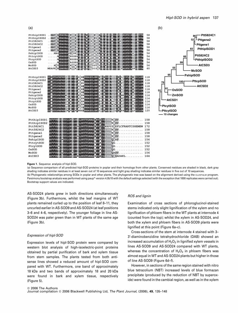
Identification of expressed sequence tags and gene model of
hipI-SOD
Four expressed sequence tags (ESTs; corresponding to two
contigs, POPLAR.5824.C1 and POPLAR.5824.C2) encoding
hipI-SOD in hybrid aspen were identified from the poplar
EST database (PopulusDB; http://www.populus.db.umu.se/).
The two closest gene models for hipI-SOD (estExt_Gene-
wise1_v1.C_LG_XIII1983 and grail3.0065004601) were iden-
tified from the Populus trichocarpa genome database (http://
genome.jgi-psf.org/Poptr1/Poptr1.home.html ). All the predic-
ted protein sequences for hipI-SOD were similar (Figure 1a).
A putative peroxisomal CuZn-SOD, CSD3 (NP_197311) was
found to be the closest homolog of hipI-SOD in Arabidopsis,
sharing 71% identity. HipISOD1 (CAC33847) and hipISOD2
(CAC33846) are 98% identical to each other, whereas
Ptt5824C2 (POPLAR.5824.C2) is 100% identical to hipI-SOD1.
The two predicted gene models (Pttgene1 and Pttgene2)
for hipI-SOD in hybrid aspen share 94% similarity. The
phylogenetic tree shown in Figure 1(b) suggests that all
the predicted hipI-SODs in poplar, CSD3 in Arabidopsis
(AtCSD3) and the ice plant SOD (McSOD) can be grouped
together.
Immunolocalization
Cross-sections of the stem at the fourth internode of wild-
type (WT) hybrid aspen plants were used for immuno-
labeling of hipI-SOD with Alexa 488 (Molecular Probes;
Invitrogen, GmbH Karlsruhe, Germany). Analysis of
fluorescence and transmission images (taken with a Zeiss
LSM 510 META confocal laser scanning microscope)
showed significant antibody signals in the lignified sec-
ondary cell walls of phloem fibers and xylem vessels
(Figure 2).
Generation of hipI-SOD antisense plants
Twenty independently transformed lines (see Experimental
procedures) were regenerated. Southern blot analysis of the
transgenic lines indicated that lines 9 and 24 each had a
single T-DNA insert (data not shown) and were denoted AS-
SOD9 and AS-SOD24, respectively.
Phenotype of hipI-SOD antisense plants
Plants belonging to two lines (AS-SOD9 and AS-SOD24)
exhibited clear phenotypic differences from WT plants
(Figure 3a). Compared with the WT, they were stunted
(Figure 4a), had reduced radial stem growth (Figure 4b) and
fewer and shorter internodes (Figure 4c,d). Plants of line AS-
SOD9 had a sloping stem habit and developed numerous
lateral shoots. The leaves of the antisense plants were
smaller and showed developmental abnormalities com-
pared to the WT (Figure 3b). The leaves of both transgenic
lines were much smaller than leaves of WT plants. Wild-type
leaves first grew longitudinally and then laterally, resulting
in an oval leaf shape. In contrast, the lateral growth of leaves
of AS-SOD9 plants seemed to be inhibited and the leaves of
136 Vaibhav Srivastava et al.
ª 2006 The AuthorsJournal compilation ª 2006 Blackwell Publishing Ltd, The Plant Journal, (2006), 49, 135–148
AS-SOD24 plants grew in both directions simultaneously
(Figure 3b). Furthermore, whilst the leaf margins of WT
plants remained curled up to the position of leaf 8–11, they
uncurled earlier in AS-SOD9 and AS-SOD24 (at leaf positions
3–6 and 4–6, respectively). The younger foliage in line AS-
SOD24 was paler green than in WT plants of the same age
(Figure 3b).
Expression of hipI-SOD
Expression levels of hipI-SOD protein were compared by
western blot analysis of high-isoelectric-point proteins
obtained by partial purification of bark and xylem tissue
from stem samples. The plants tested from both anti-
sense lines showed a reduced amount of hipI-SOD com-
pared with WT. Furthermore, one band of approximately
18 kDa and two bands of approximately 18 and 20 kDa
were found in bark and xylem tissue, respectively
(Figure 5).
ROS and lignin
Examination of cross sections of phloroglucinol-stained
stems indicated only slight lignification of the xylem and no
lignification of phloem fibers in the WT plants at internode 4
(counted from the top); whilst the xylem in AS-SOD24, and
both the xylem and phloem fibers in AS-SOD9 plants were
lignified at this point (Figure 6a–c).
Cross-sections of the stem at internode 4 stained with 3–
3¢-diaminobenzidine tetrahydrochloride (DAB) showed an
increased accumulation of H2O2 in lignified xylem vessels in
lines AS-SOD9 and AS-SOD24 compared with WT plants,
whereas the concentration of H2O2 in phloem fibers was
almost equal in WT and AS-SOD24 plants but higher in those
of line AS-SOD9 (Figure 6d–f).
However, in sections of the same region stained with nitro
blue tetrazolium (NBT) increased levels of blue formazan
precipitate (produced by the reduction of NBT by superox-
ide) were found in the cambial region, as well as in the xylem
68
55
99
66
38
25
59
00197
1625
segnahc 01
1C4285ttP
2C4285ttP
2enegttP
1enegttP
2DOSIpihttP
1DOSIpihttP
3DSCtA
DOScM
DOSIpihsP
DOSpcttP
2DSCtADOSsO
DOSmZ
1DSCtA
DOStyctP
DOStycttP
(a) (b)
Figure 1. Sequence analysis of hipI-SOD.
(a) Sequence comparison of all predicted hipI-SOD proteins in poplar and their homologs from other plants. Conserved residues are shaded in black, dark gray
shading indicates similar residues in at least seven out of 10 sequences and light gray shading indicates similar residues in five out of 10 sequences.
(b) Phylogenetic relationships among SODs in poplar and other plants. The phylogenetic tree was based on the alignment derived using the CLUSTALW program.
Parsimony bootstrap analysis was performed using paup* version 4.0b10 with the default settings selected (with the exception that 1000 replicates were carried out).
Bootstrap support values are indicated.
HipI-SOD in hybrid aspen 137
ª 2006 The AuthorsJournal compilation ª 2006 Blackwell Publishing Ltd, The Plant Journal, (2006), 49, 135–148

vessels of both antisense lines (Figure 6g–i). The superoxide
radical concentration was lower in phloem fibers than in
xylem vessels in all of the plants examined (Figure 6g–i).
Control experiments were performed on WT stem cross-
sections for both DAB and NBT staining. Sections were
incubated with a well-known ROS scavenger (10 mM
reduced glutathione; Karlsson et al., 2005; Larson, 1988),
together with DAB and NBT respectively. A clear reduction
in precipitate of both types of staining was achieved,
indicating the specificity of staining reactions with ROS
(Figure S1).
The total lignin content of mature stems (taken from the
base of the plant) was determined. Both the hipI-SOD
antisense lines exhibited a slight decrease in lignin content
compared with WT plants (Figure 7a). The lignin content
was found to be approximately 22–26% of the cell wall
residue (CWR) on a dry weight basis.
Histological and morphological analysis
Comparison of cross-sections of the tenth internode of WT
and transgenic plants showed histological differences (Fig-
ure 8). The epidermis of plants from both antisense lines
was discontinuous and ruptured. An interesting observation
in antisense plants was the presence of two layers of phloem
fibers. In both antisense lines, the cambium was com-
pressed and disorganized. Furthermore, cells were smaller
in size and rays were fewer and inconsistent.
Downregulation of the hipI-SOD gene in antisense plants
affected the length and diameter of both xylem fibers and
vessels in fully matured internodes. Fibers in the transgenic
lines were approximately 20% shorter and 14% thinner,
while vessels were 17% shorter and 36% thinner than WT
plants (Figure 9a–f).
Analysis of phenolic profiles
The main phenolic compounds found in poplar tissues were
hydroxycinnamic acid derivatives (with UV spectra resem-
bling either that of chlorogenic acid or p-coumaric acid) and
salicylates. Flavonoids were not detected in quantifiable
amounts in stems, although some kaempherol and querce-
tin derivatives were present in the leaves of the same plants
(data not shown). Compared with WT plants, the stems of
transgenic plants generally had higher total concentrations
of phenolic acids and salicylates (Figure 7b,c).
Microarray and real-time RT-PCR analysis
The expression of genes that were upregulated or down-
regulated by a factor of at least 1.8 in the transgenic lines AS-
SOD9 and AS-SOD24 compared with the WT was regarded
as being significantly altered. Genes that fulfilled this
criterion in lines AS-SOD9 and AS-SOD24 are listed in Ta-
ble 1. Some genes involved in lignin biosynthesis (4-coum-
arate:coA ligase, dehydroquinate dehydratase/shikimate
dehydrogenase precursor and basic peroxidase) were up-
regulated in both antisense lines. A number of genes enco-
ding proteins connected with stress responses were also
upregulated, including: the endopeptidase Clp, a drought-
induced protein from sunflower, phosphatase 2C, histone
H1, the protein ERD 15 and several proteins related to
pathogen defense such as phospholipase A2 and a patatin-
like protein. We also found genes involved in signal trans-
duction (such as ankyrins, a putative GTPase, serine/thre-
onine protein kinase, the GTPase Rab 11C and the two
calmodulins) to be upregulated. A ubiquitin-conjugating
enzyme was also upregulated in both antisense lines, and in
line AS-SOD9 a clear increase in mRNA encoding pectate
lyases was observed. All of the genes discussed above had
high scores in BLASTX searches of GenBank, confirming their
assigned functions (Table 1). A few up- and downregulated
Figure 2. Immunolocalization of hipI-SOD with Alexa 488 in cross-sections of
stems of WT hybrid aspen plants. Fluorescence signals showing location of
hipI-SOD in secondary wall forming cells (a), and same section showing
internal structure (b) in a transverse section from internode 4. Ph, phloem
fibers; X, xylem vessels; scale bars represent 20 lm.
138 Vaibhav Srivastava et al.
ª 2006 The AuthorsJournal compilation ª 2006 Blackwell Publishing Ltd, The Plant Journal, (2006), 49, 135–148

sequences were also found in antisense plants that coded
for hypothetical proteins or showed no similarity to any
other sequence in GenBank (data not shown).
To further validate the microarray results, several genes
involved in lignin biosynthesis were selected for real time
RT-PCR. The results showed that the expression of hydroxy-
cinnamate: CoA ligase (4CL), caffeic acid/5-hydroxy ferulic
acid O-methyl transferase (COMT) and hydroxycinnamyl
alcohol dehydrogenase (CAD) was significantly increased in
both antisense lines compared with the WT (Figure 10a–c).
However, no significant differences were found in the
expression level of phenylalanine ammonia lyase (PAL),
caffeoyl-CoA O-methyltransferase 6 (CCoAOMT) and cinna-
mate 4-hydroxylase (C4H) in WT and line AS-SOD9, but
levels were higher in line AS-SOD24 (Figure 10d–f).
Discussion
Expression and localization studies of a hipI-SOD in both
pine and the Zinnia tracheary element (TE) differentiation
system (Karlsson et al., 2005; Karpinska et al., 2001), and
gene expression data from a high-resolution transcript pro-
file from Populus (Schrader et al., 2004), suggest that the
enzyme could be involved in the development and lignifi-
cation of secondary cell walls. Fluorescence analysis
showed specific staining in xylem vessels and phloem
fibers, confirming the localization of hipI-SOD in secondary
wall-forming cells, in agreement with published transcript
profiling data and localization studies (Karlsson et al., 2005;
Karpinska et al., 2001; Schrader et al., 2004).
To elucidate the function of hipI-SOD, we generated
transgenic antisense plants and confirmed that the two lines
showing phenotypic differences from the WT (AS-SOD9 and
AS-SOD24) contained reduced quantities of hipI-SOD in
their bark and xylem tissues. The Western blot analysis
showed bands of different sizes in xylem and phloem tissues
than the expected size of hipI-SOD monomers (16 kDa). The
discrepancy between theoretical and experimental molecu-
lar masses may be due to the presence of two isoforms
differing slightly in molecular mass, or the presence of post-
translational modifications. Tissue-specific differences in
hipI-SOD isozyme pools in hybrid poplar have previously
been reported, and it is hypothesized that there are at least
two isoforms of hipI-SOD (Schinkel et al., 2001). The Popu-
lus genome database (http://genome.jgi-psf.org/Poptr1/
Poptr1.home.html ) also contains two gene models for
hipI-SOD. The adaptive benefits of having a number of hipI-
SOD isoforms are not yet clear, but similar isoforms of other
SODs have been reported (Schinkel et al., 1998; Streller and
Wingsle, 1994; Zhu and Scandalios, 1993), and such redund-
ancy or isovariant dynamics (Meagher et al., 1999) may be
needed to ensure the constant presence of active hipI-SOD
in different tissues.
The dwarfed phenotype of the transgenic plants is appar-
ently due to reductions in both cell division and expansion,
probably caused by a premature transition into maturation,
as indicated by the early onset of lignification. The antisense
lines had an increased number of lateral branches and AS-
SOD9 appeared to have reduced apical dominance. Since
the meristem responsible for producing the leaf blade is
Figure 3. Phenotype of WT and hipI-SOD anti-
sense plants.
(a) Phenotypes of the transgenic lines AS-SOD9
and AS-SOD24 and WT hybrid aspen plants (left
to right). All plants were 13 weeks old.
(b) Leaf development in hybrid aspen plants WT,
line AS-SOD9 and line AS-SOD24. From left to
right leaf numbers 5, 8, 11, 14, 16, 22, are shown,
where leaf 1 is the first leaf longer than 1 cm.
HipI-SOD in hybrid aspen 139
ª 2006 The AuthorsJournal compilation ª 2006 Blackwell Publishing Ltd, The Plant Journal, (2006), 49, 135–148

positioned around the leaf margins (Esau, 1977), the earlier
uncurling of the leaf blade suggests that meristematic
activity had ceased earlier in the transgenic lines.
In the antisense plants, enhanced accumulation of H2O2
and O�2 was found in the vascular tissue of the younger
internodes. In particular, H2O2 was found in the cell walls of
phloem fibers and xylem vessels. Also, in xylem vessels,
elevated O�2 was also localized in the living cells of the
cambium region in the transgenic lines. Lignification of
phloem fibers and xylem vessels was correlated with the
elevated accumulation of H2O2, with earlier initiation in
younger parts of the stem in the transgenic lines compared
with WT plants.
Xylem fibers and vessels were shorter and thinner in
antisense plants than in the WT. As the lignification process
is initiated prematurely whilst vessels and fibers are still
expanding in antisense plants, their cell walls may become
more rigid, thereby limiting the cell expansion. In the
transgenic plants, the cells in the cambium region of the
stem were compressed and looked ‘disorganized’ and cell
division and expansion towards both the phloem and xylem
side were impeded, possibly accounting for the stunted
phenotype. This may be a direct effect of increased oxidative
burst caused by the downregulation of hipI-SOD in these
tissues, thus inhibiting cell division as reported earlier
(Reichheld et al., 1999).
Analysis of the total lignin content of stem samples taken
from the base of fully mature plants gave values in accord-
ance with published data (Baucher et al., 1996). However, a
slight difference was found between transgenic plants and
the WT, indicating that the downregulation of hipI-SOD in
antisense plants affects the initiation of lignification but not
the overall lignin content.
Further indications that the transgenic plants are stressed
are the elevated levels of phenolics observed in their stems.
The phenolics detected in hybrid poplar stems were mainly
hydroxycinnamic acid derivatives and salicylates (salicin,
salicortin and tremulacin), in agreement with published data
on the phenolic chemistry of poplar (Lindroth and Koss,
1996). Accumulation of phenolic acids in transgenic poplar
stems might be activating an antioxidative system to
diminish the oxidative stress (Sakihama et al., 2002), or
may be involved in increased lignification (Robertsen and
Svalheim, 1990). Accumulation of phenolics has also been
described as a defensive or stress response in plants (Dixon
and Paiva, 1995).
Further, the overexpression of genes such as GTPase, IRE
homolog, serine/threonine protein kinase, RAS related pro-
tein Rab 11C and calmodulins (CAM) in transgenic plants is
indicative of the involvement of ROS in plant signaling
(Baxter-Burrell et al., 2002; Lamb and Dixon, 1997). Also,
several genes encoding proteins associated with stress
responses were also found to be upregulated. Meskiene
et al. (2003) have proposed that the mitogen activated
protein kinase (MAPK)-signal pathway is controlled by the
redox-sensitive protein phosphatase 2C (PP2C), which is
induced under oxidative conditions. A similar type of protein
phosphatase 2C was highly expressed in the antisense
plants. The upregulated proteins phospholipase A2 and a
patatin-like protein have been shown to be rapidly induced
Figure 4. Characterization of the stems of WT, AS-SOD9 and AS-SOD24
plants. (a) Growth increment, (b) stem diameter, (c) internode number, (d)
internode length. The error bars represent standard deviations for results
from three (line AS-SOD9) or four samples (WT and line AS-SOD24).
140 Vaibhav Srivastava et al.
ª 2006 The AuthorsJournal compilation ª 2006 Blackwell Publishing Ltd, The Plant Journal, (2006), 49, 135–148

during the hypersensitive response in tobacco (Dhondt
et al., 2000).
The upregulation of the xylem-specific 4CL isoform may
be helping to direct phenylpropanoids to the lignification
pathway in the transgenic plants. An increased expression of
COMT and CAD was observed in the apical tissue of
antisense plants. The expression of basic peroxidase, which
has been described as one of the enzymes responsible for
cell wall lignification in Zinnia elegans (Barcelo and Pomar,
2001), was found to be enhanced in antisense plants. In
Arabidopsis, most of the discussed genes were upregulated
in bolting stems (Ehlting et al., 2005).
Reduced expression of hipI-SOD in the antisense lines
leads to an increase in localized accumulation of O�2 and
H2O2. In the absence of SOD, O�2 has been shown to be
dismutated at a non-enzymatic dismutation rate close to
105M
)1 sec)1 (Torres et al., 2002), and may thus result in an
enhanced production of H2O2 compared with when it is
dismutated in a SOD-dependent manner (Liochev and
Fridovich, 1994). The increased accumulation of ROS seems
to induce both defense responses during pathogen infec-
tions of plants (Kangasjarvi et al., 2005) and signal trans-
duction pathways important for developmental processes.
In addition, the increased H2O2 in the transgenic plants may
also participate directly in the peroxidase-mediated oxida-
tion of monolignols to monolignol radicals in lignin poly-
merization (Ogawa et al., 1997). The localization of hipI-SOD
in phloem fibers and xylem vessels also suggest a role for
the enzyme as a regulator of ROS levels in these cells.
The dwarfed phenotype of the transgenic plants is appar-
ently due to reductions in both cell division and expansion,
probably caused by a premature transition into maturation,
as indicated by the early onset of lignification. Whether this
progression is caused by a change in the concentration of
Figure 5. HipI-SOD expression pattern in hybrid
aspen by Western blot analysis. Western blot of
partially purified protein from bark and xylem
tissue of three individual plants (a–c) from the
WT, AS-SOD9 and AS-SOD24 lines. The size of
the bands on the blot is indicated on the right.
Figure 6. Histochemical analysis of vascular tis-
sue. (a–c) Phloroglucinol staining for lignin in the
vascular tissues of WT (a), AS-SOD9 (b), and AS-
SOD24 (c) plants. (d–f) DAB staining for H2O2 in
vascular tissues of WT (d), AS-SOD9 (e) and AS-
SOD24 (f) plants. (g–i) NBT staining for superox-
ide in vascular tissues of WT (g), AS-SOD9 (h)
and AS-SOD24 (i) plants. Ca, cambium; Epi,
epidermis; P, pith; Ph, phloem; S, sclerenchyma.
Scale bars represent 100 lm.
HipI-SOD in hybrid aspen 141
ª 2006 The AuthorsJournal compilation ª 2006 Blackwell Publishing Ltd, The Plant Journal, (2006), 49, 135–148

ROS acting as signaling molecules or as oxidation equiva-
lents in the production of monolignol radicals, is not clear.
More experimental data are needed to unambiguously
define the function of hipI-SOD.
Experimental procedures
Database search and sequence analysis
To identify the number of ESTs and the most accurate gene modelencoding hipI-SOD in hybrid aspen, amino acid sequences held inthe NCBI database (accession numbers AJ278671 and AJ278670 forhipI-SOD1 and hipI-SOD2, respectively; Schinkel et al., 2001) wereused for Blast (Basic Local Alignment Search Tool; Altschul et al.,1997) searches against the Populus EST database (PopulusDB;http://www.populus.db.umu.se/) and the P. trichocarpa genomedatabase (http://genome.jgi-psf.org/Poptr1/Poptr1.home.html),respectively. Proteins with similarity to hipI-SOD were also identi-fied by performing BLAST analysis (http://www.ncbi.nlm.nih.gov/BLAST/) of the hipI-SOD protein against a translated nucleotidedatabase.
Amino acid sequences of all selected proteins were alignedusing CLUSTALW (http://npsa-pbil.ibcp.fr/cgi-bin/npsa_auto-mat.pl?page¼/NPSA/npsa_clustalw.html) followed by phylogenet-ic analysis using the Paup* program (version 4.0b10; Norberget al., 2005).
Accession numbers
GenBank accession numbers of the sequences analyzed for proteinalignment (Figure 1a) and phylogenetic analysis (Figure 1b) were asfollows: AtCSD1 (NP_172360), AtCSD2 (NP_565666), AtCSD3(NP_197311), McSOD (O49044), OsSOD (P28757), PshipISOD(CAC34448), PtcytSOD (AAD01605), PttcpSOD (CAC33844), Pttcyt-SOD (CAC33845), PtthipISOD1 (CAC33847), PtthipISOD2(CAC33846) and ZmSOD (P11428).
Pttgene1 and Pttgene2 were annotated as estExt_Gene-wise1_v1.C_LG_XIII1983 and grail3.0065004601, respectively, inthe P. trichocarpa genome database, whereas Ptt5824C1 wasannotated as POPLAR.5824.C1 and Ptt5824C2 as POPLAR.5824.C2in the Populus EST database.
Antisense vector construction and plant transformation
The antisense construct used in this study contained the entire hipI-SOD 2 gene (Schinkel et al., 2001), cloned as a SpeI fragment intothe binary vector P 11/2 (provided by Dr B. Reiss, MPI, Koln, Ger-many). The bacterial XL1-blue MRF¢ system was used to amplifyconstructs.
Hybrid aspen (Populus tremula L. · P. tremuloides Michx.) plantswere transformed and regenerated as previously described (Nilssonet al., 1992). A total of 19 independent lines were recovered andmultiplied by cuttings and rooted. Wild-type plants were multipliedin tissue culture in the same way and served as controls.
Plant growth parameters
Plants from the transgenic lines AS-SOD9 and AS-SOD24, and WTplants of the same age, were measured weekly starting 8 weeksafter the plants had been transferred to the greenhouse. Each week,the height and basal diameter of the stem of each plant weremeasured, and the number of internodes and average internodelength were recorded. When the plants had been in the green-house for 13 weeks, the maximal width and length of the blades ofeach of the 20 youngest leaves of plants from all three lines weremeasured.
Protein extraction and Western blot analysis
Three plants from each of the WT and both antisense lines wereselected for protein extraction and Western blot analysis. Tissues
Figure 7. Total lignin content and phenolic pro-
files in stems of WT and transgenic plants.
(a) Total lignin content in WT and transgenic
plants. Values are given as percentage of CWR
and correspond to the mean of three determina-
tions. The vertical bar represents standard devi-
ation.
(b) Total concentration of salicylates (salicin,
salicortin and tremulacin) and
(c) total concentration of phenolic acids (chloro-
genic, p-coumaric and cinnamic acid) in both
young and old internodes of WT, AS-SOD9 and
AS-SOD24 plants. The error bars represent
standard error. Levels of significance were
calculated from ANOVA data (*P < 0.05).
142 Vaibhav Srivastava et al.
ª 2006 The AuthorsJournal compilation ª 2006 Blackwell Publishing Ltd, The Plant Journal, (2006), 49, 135–148

were sampled from bark peeled off internodes 5–25 and by scrapingoff xylem with a sharp blade (Karpinska et al., 2004). Protein wasextracted from 0.4 g of fresh tissue sampled from both bark andxylem tissues. The samples were homogenized in extraction buffer[50 mM 2-amino-2(hydroxymethyl)-1,3-propanediol (TRIS)-HCl, pH8.0, 1 mM EDTA, 1 mM DTT], and centrifuged at 12 000 g for 15 minat 4�C. The supernatant was heated at 70�C for 10 min and centri-fuged again at 12 000 g for 15 min at 4�C. The supernatant waspurified using an anion exchange column (HiQ XL, GE Health-care, Uppsala, Sweden) and proteins in the purified fractionswere precipitated overnight at )20�C by four volumes of chilledacetone.
Protein preparations were mixed with loading buffer to a finalconcentration of 1% SDS (w/v) and 50 mM DTT then incubated at95�C for 5 min prior to loading. Proteins in samples containingapproximately 22 lg of protein were electrophoretically separated
in a 14% SDS-polyacrylamide gel using a MiniProtean II electro-phoresis assembly (Bio-Rad, Hercules, CA, USA) and electroblottedonto nitrocellulose membranes (Hybond�-ECL�, GE Healthcare).Western blot analysis was performed using a specific hipI-SODantibody (Schinkel et al., 2001) as the primary antibody andhorseradish peroxidase-linked anti-rabbit IgG as the secondaryantibody (GE Healthcare). Fluorescence signals were generated byincubation with ECL plus Western blotting detection reagents(GE Healthcare). Pre-stained broad range SDS-PAGE standards(Bio-Rad) were used to estimate the size of the protein bands.
Fixation, substitution and embedding for light microscopy
For the primary fixation of samples, cross-sections of stemapproximately 1 mm thick were incubated overnight at room tem-
Figure 8. Light microscopy of transverse sections of stem. Comparable morphological analysis of WT, AS-SOD9 and AS-SOD24: (a–c) epidermis and periderm; (d–
f) sclerenchyma, phloem and cambium; (g–i) xylem with rays (arrows). Ca, cambium; Epi, epidermis; Ph, phloem; S, sclerenchyma. Scale bars represent 50 lm.
HipI-SOD in hybrid aspen 143
ª 2006 The AuthorsJournal compilation ª 2006 Blackwell Publishing Ltd, The Plant Journal, (2006), 49, 135–148

perature (RT, 22�C) in 50 mM cacodylate buffer, pH 7.2, containing0.5% (v/v) glutaraldehyde and 2.0% (v/v) formaldehyde, followed byone wash with buffer and two washes with distilled water. Samplesfor immunolabeling studies were dehydrated stepwise by increas-ing the concentration of ethanol and concomitantly lowering thetemperature (method of progressive lowering of temperature, PLT)using an automated freeze substitution unit (AFS, Leica, Benzheim,Germany). The steps used were as follows: 30% (v/v), 40% (v/v) and50% (v/v) ethanol for 1 h each at 4�C; 60% (v/v) and 75% (v/v) ethanolfor 1 h each at )15�C; 90% (v/v) ethanol and 2x 100% (v/v) ethanol for1 h at )35�C. The samples were subsequently infiltrated withLowycryl HM20 resin (Plano GmbH, Marburg, Germany) by incu-bating them in the following mixtures: 33% (v/v), 50% (v/v) and 66%(v/v) HM 20 resin in ethanol for 4 h each and then 100% (v/v) HM 20overnight. Samples were transferred into gelatin capsules, incuba-ted for 3 h in fresh resin and polymerized at 35�C for 3 days underindirect UV light.
Samples for ultrastructural and light microscopy analyses weretransferred after primary fixation into a solution of 1% (w/v) OsO4.After 1 h, samples were washed three times with distilled water.Dehydration at 25�C was carried out by increasing the ethanolconcentration in the following steps: 30% (v/v), 50% (v/v), 60% (v/v),75% (v/v), 90% (v/v) and 2x 100% (v/v) ethanol for 1 h. Afteradditional dehydration with propylene oxide for 1 h, the samples
were infiltrated with Spurr’s resin (Plano GmbH, Marburg, Ger-many) as follows: 33% (v/v), 50% (v/v) and 66% (v/v) Spurr’s resin inpropylene oxide for 4 h each and then 100% (v/v) Spurr’s resinovernight. Samples were transferred into embedding molds, incu-bated for 3 h in fresh resin and polymerized at 70�C for 24 h.
Microscopy
Semi-thin sections, 1 lm thick, were cut with a Histo diamond knife,mounted on glass slides and used for fluorescence labeling. Toavoid non-specific antibody binding, sections were blocked at RT for20 min in PBS buffer containing 3% (w/v) bovine serum albuminand 0.1% Tween. Rabbit polyclonal antibody raised against hipI-SOD and Alexa 488 Fluor anti-rabbit IgG (Invitrogen GmbH, Karls-ruhe, Germany) as the secondary antibody were used for immu-nolabeling. The system used to detect Alexa 488 was a ZeissAxiovert 200 microscope (Carl Zeiss, Gottigen, Germany) attachedto a confocal laser scanning system (Zeiss LSM 510 META). Fluor-escence of Alexa 488 was induced by an excitation light at 488 nmproduced by an Ar-laser. A 505–535 nm emission filter and emissionfingerprinting were used to eliminate remaining autofluorescencesignals from other cell components. Control labeling without usingprimary antibody and Alexa 488, respectively, gave no significant
Fib
er le
ngth
(µ
m)
Ves
sel t
otal
leng
th(µ
m)
Ves
sel l
engt
h (µ
m)
Ves
sel d
iam
eter
(µm
)F
iber
dia
met
er (
µm
)
(a) (f)
(b)
(c)
(d)
(e)
Figure 9. Fiber and vessel measurements. (a)
Xylem fiber length, (b) xylem fiber diameter, (c)
xylem vessel total length, (d) xylem vessel
length, (e) xylem vessel diameter, (f) individual
vessel showing measured parameters. All the
measurement values represent means for four
independently generated lines. The vertical bar
represents standard error.
144 Vaibhav Srivastava et al.
ª 2006 The AuthorsJournal compilation ª 2006 Blackwell Publishing Ltd, The Plant Journal, (2006), 49, 135–148

signals (data not shown). Additional images were obtained intransmission mode.
Light microscopical analyses of fresh and fixed stem materialfrom WT and antisense plants were carried out with a CCD camerasystem (Zeiss AxioCam attached to a Zeiss Axiovert 135M micro-scope; Carl Zeiss).
Transverse sections of fresh poplar stem were used for histo-chemical analysis of lignin, H2O2 and superoxide radicals. A LeicaVibratome VT 1000S (Leica Microsystems, Bensheim, Germany)was used to prepare sections with an approximate thickness of150 lm from different internodes (1, 2, 3,…). To detect the presenceof lignin, sections were incubated for 30 min in 1% phloroglucinol inethanol followed by the addition of 10% HCl (Ogawa et al., 1997).H2O2 was analyzed as described by Holm et al. (2003): stem sectionswere incubated in 2 mM DAB dissolved in 50 mM TRIS-HCl buffer pH6.8. To detect superoxide radicals, sections were incubated for 1 h in0.25 mM NBT in 10 mM sodium phosphate, pH 7.8 (Ogawa et al.,1997).
For histological analysis, semi-thin sections of fixed stem mater-ial, approximately 3 lm thick, were prepared using a Leica Ultra-microtome UCT (Leica Microsystems, Bensheim, Germany) and aHisto diamond knife. The sections were mounted on slides andstained for 2 min with 1% (w/v) methylene blue/1% (w/v) Azur II in1% (w/v) aqueous borax at 60�C prior to light microscopicalexamination.
Lignin analysis
The lignin content of three individual plants from each of the WTand AS-SOD9 and AS-SOD24 lines were determined by a modifiedKlason lignin method (Kirk and Obst, 1988). Wood tissue was ballmilled to a fine powder which was then subjected to successiveextraction with water, ethanol, toluene:ethanol (1:1) and acetone.The resulting cell wall residue was used for lignin determination.Five milliliters of 76% H2SO4 was added to 1 g of cell wall residue
Table 1 Results of the microarray experiment comparing gene expression in transgenic lines AS-SOD9 and AS-SOD24 to WT
Acc. no. Annotation Score E24 E9
AI162626 IRE homolog, protein kinase like 50 5.2 2.4AI161963 Chloroplast ribosomal protein S12 205 3 1.1AI164191 Ubiquitin conjugating enzyme-like protein 103 2.9 1.6AI162541 Endopeptidase Clp 128 2.9 1.7AI163594 4-Coumarate:coA ligase 211 2.8 1.4AI163374 Cytoplasmic aconitate hydratase 34 2.6 1.5AI162418 Drought-induced protein (Helianthus)* 93 2.4 1.4AI163572 Dehydroquinate dehydratase/Shikimate dehydrogenase precursor 86 2.2 1.6AI164389 Put. Phospolipase A2 (Dianthus) 157 2.1 1.7AI162596 RAS-related protein RAB11C (Lotus) 271 2.1 1.6AI164638 Put. ankyrin 99 2.1 1.5AI163617 Patatin-like protein 1 (Nicotiana) 82 2 1.1AI162640 Beta 1,2-xylosyltransferase 176 2 1AI164044 Histone H1 60 2.4 2AI162170 Put. beta-hydroxyacyl ACP dehydratase 43 1.9 1.4AI163562 Put. GTPase 195 1.9 1.3AI165218 Kinase-like protein 181 1.8 0.8AI165265 Similarity to protein phosphatase 2C 105 1.8 1.6AI164312 Similarity to ‘tub’ protein gp 108 1.8 1.4AI166132 Put. pectate lyase 179 1.4 2.3AI165989 Put. serine/threonine protein kinase (Mesembryanthemum) 105 1.2 2.3AI162623 Chlorophyll A/B-binding protein precursor (Oryza) 83 0.9 2.2AI163946 Calmodulin-like protein 61 1 2AI162944 Basic peroxidase (Vigna) 228 1.5 2AI163950 Calmodulin-like protein 80 1.2 1.9AI162822 Dehydration-induced protein ERD15 132 1.7 1.9AI162241 Oxygen evolving enhancer protein 1 precursor (Bruguiera) 120 1 1.9AI163014 Ribosomal protein S 15A 64 0.9 0.6AI164188 Ribosomal protein L 29 (Panax) 120 1.2 0.5AI161865 GTP-binding protein (Glycine) 66 0.9 0.5AI165142 GSH-dependent dehydroascorbate reductase 192 1.1 0.5AI165127 NADH-cytochrome b5 reductase 104 0.5 1AI165359 Methyltransferase (Prunus) 97 0.5 1.1AI163311 Cytochrome c oxidase III (Trypanosoma) 37 0.4 0.8
Expression ratio of genes that were up- or downregulated in lines AS-SOD9 and AS-SOD24. Genes that were upregulated ‡1.8-fold ordownregulated ‡1.8-fold (ratios 0.55 and less) compared with the WT are displayed. Acc. no stands for GenBank accession number of the gene onthe array. BLASTX score (score) refers to the similarity to the GenBank annotation given. Only the highest score in BLASTX searches is given exceptfor * which was the second highest score (the first being a hypothetical protein). E9 and E24 show the expression ratios between transgenic lines(AS-SOD9 and AS-SOD24, respectively) and the WT. All annotations refer to Arabidopsis thaliana genes except where another species is given.Put., putative.
HipI-SOD in hybrid aspen 145
ª 2006 The AuthorsJournal compilation ª 2006 Blackwell Publishing Ltd, The Plant Journal, (2006), 49, 135–148

and the mixture was incubated for 1 h at 30�C in a water bath. Thedigestion was terminated by adding 140 ml of water to each sampleand placing the sample in an autoclave for 1 h at 120�C. Whilstmaintaining the temperature of the solution the lignin was filteredthrough a pre-weighed glass fiber filter (Whatman GF/A, Maidstone,UK). The residue was thoroughly washed with hot water tocompletely remove the acid.
Fiber and vessel measurement
Four plants from the WT and each of the antisense lines were cho-sen for fiber and vessel measurement. Trimmed pieces of outerxylem from the mature part of each plant were prepared, and xylempieces were macerated as described by Eriksson et al. (2000). Aftermaceration, the fibers and vessels were separated from each otherby shaking vigorously in water. The length and diameter of 100fibers and 50 vessels per sample were measured using a micro-scope fitted with a camera (Axio vision3, Zeiss, Germany).
Analyses of phenolics
Young and old internodes (taken from the top and bottom of plants,respectively) from plants of lines AS-SOD9, AS-SOD24 and the WTwere analyzed for phenolic glucosides (salicin, salicortin, tremula-
cin) and phenolic acids (p-coumaric acid, cinnamic acid, chlorogenicacid and benzoic acid).
All plant material for the phenolic analyses was air dried atambient temperature, milled to an homogeneous powder andextracted as described by Witzell et al. (2003). Phenolic metabo-lites were analyzed using a Merck Hitachi LaChrom HPLC system(L-7100 pump, L-7200 autosampler, L-7360 column oven and L-7455 diode array detector; Darmstadt, Germany). The oventemperature was set to 30�C. The compounds were separatedon a reversed-phase 100 · 4.6 mm internal diameter, 3 lm particlesize C-18 column (Thermo Quest HyPURITY�, Waltham, MA,USA). The mobile phase consisted of the following complexgradient: 100% A for 0–1 min, declining linearly to 60% A at 6 min,then to 40% at 12 min and 0% A at 28 min, which was maintaineduntil 30 min, before switching to 100% A until 33 min, followed bywashing and equilibration to initial conditions where A is H2O (pHadjusted to 3.0 with H2PO4) and the balance was provided bymethanol (4:6 v/v). The flow rate was 1 ml min)1 throughout andthe injection volume was 15 ll. Individual phenolics were identi-fied by comparing their retention times and UV spectra (200–400 nm) with those of standard compounds (Sigma, St Louis, MO,USA), and their total concentration was calculated. Salicylates(salicin, salicortin and tremulacin) were quantified as salicin(Sigma) equivalents.
Microarray experiments
Poly Aþ RNA was prepared from the apical part (top 3 cm) of aprimary shoot (with leaves attached) of plants of lines AS-SOD9,AS-SOD24 and the WT, using oligo-dT-coated magnetic beads(DYNAL, Oslo, Norway) according to the manufacturer’s recom-mendations. Four hundred nanograms of mRNA from eachsample was labeled using a CyScribe First-Strand cDNA labelingkit (Amersham Pharmacia Biotech, Uppsala, Sweden) and unin-corporated nucleotides were removed using an AutoSeq G-50column (Amersham Pharmacia Biotech). A hybrid aspen cDNAmicroarray (Hertzberg et al., 2001) was hybridized with the labe-led targets using the competitive hybridization technique des-cribed in Schena et al. (1995). Hybridization, washing, scanningand data analysis were performed as described by Hertzberget al. (2001). Annotation of the genes from the array presented inTable 1 was based on the best hit obtained using BLASTX tosearch the NCBI non-redundant protein database.
Quantitative real-time PCR
Genes encoding the following phenylpropanoid biosynthesis en-zymes were chosen for quantitative real-time PCR analysis: phe-nylalanine ammonia lyase (PAL, EC 4.2.1.4), cinnamate 4-hydroxylase (C4H, EC1.14.13.11), hydroxycinnamate: CoA ligase(4CL, EC 6.2.1.12), caffeic acid/5-hydroxy ferulic acid O-methyltransferase (COMT, E.C. 2.1.1.68), caffeoyl-CoA O-methyltransf-erase 6 (CCoAOMT, EC 2.1.1.104) and hydroxycinnamyl alcoholdehydrogenase (CAD; EC 1.1.1.195). The primers were chosen fromthe following Populus tremuloides Michx. cDNA sequences presentin the NCBI database: PtPAL2 (AF480620), PtC4H (U47293),Pt4CL1(AF041049), CAD (AF217957) and the predicted coding regionof the COMT gene (PTOMT1 gene, U13171). The consensus se-quence of the contig POPLAR.912.C2 found in PopulusDB was usedto synthesize the primers for amplification of CCoAOMT cDNA. Thenucleotide sequence was translated to amino acids and was iden-tical with the sequence of CCoAOMT1 (AJ223621) found in poplarby Chen et al. (2000).
Figure 10. Relative transcript abundance in apical tissues of WT and trans-
genic hybrid aspen. Gene expression was analyzed using quantitative RT-
PCR. Results were normalized to the expression of 18S ribosomal RNA. The
vertical bars represent the standard errors of the mean normalized expression
value for three separate biological replicates. Relative expression levels of 4CL
(a), COMT (b), CAD (c), PAL (d), CCoAOMT (e) and C4H (f) in apical shoot of WT
and transgenic plants are shown.
146 Vaibhav Srivastava et al.
ª 2006 The AuthorsJournal compilation ª 2006 Blackwell Publishing Ltd, The Plant Journal, (2006), 49, 135–148

Three plants from each of the WT and AS-SOD9 and AS-SOD24lines were chosen for quantitative real-time PCR of all the genesmentioned above. Total RNA was extracted from the apical part(top 3 cm) of primary shoots using an Aurum total RNA mini kit(Bio-Rad) according to the manufacturer’s instructions. Onemicrogram of RNA was used for cDNA synthesis using an iScript�cDNA Synthesis Kit (Bio-Rad). Quantification was performed asdescribed by Norberg et al. (2005) with an iCycler MyiQ real-timePCR detection system (Bio-Rad) using the Bio-Rad iQ SYBR GreenSupermix. Each sample was loaded in triplicate and the results werenormalized to the expression of 18S ribosomal RNA.
Statistical analyses
All data from the analysis of phenolics were analyzed using ANOVA
(analysis of variance). Differences between samples were consid-ered significant if the probability of the difference arising fromrandom sampling errors was <0.05, expressed as *P < 0.05.
Acknowledgements
We thank Dr B Reiss for the kind donation of vector P11/2 andKjell Olofsson and Eva Mellerowicz for their valuable sugges-tions. This research was funded by grants to SUAS from theSwedish Council for FORMAS, the Swedish Research Council(VR), the Swedish Foundation for Strategic Research (SSF) andby a grant from the Kempe foundation. This work was alsosupported in part by Professor Dr Isabella Prokhorenko by grant436 RUS 17/87/03 and RUS 17/131/05 from the Deutsche Fors-chungsgemeinschaft (DFG).
Supplementary Material
The following supplementary material is available for this articleonline:Figure S1. NBT and DAB staining showing their specificity forsuperoxide radical and H2O respectively.This material is available as part of the online article from http://www.blackwell-synergy.com.
References
Allan, A.C. and Fluhr, R. (1997) Two distinct sources of elicitedreactive oxygen species in tobacco epidermal cells. Plant Cell, 9,1559–1572.
Altschul, S.F., Madden, T.L., Schaffer, A.A., Zhang, J., Zhang, Z.,
Miller, W. and Lipman, D.J. (1997) Gapped BLAST and PSI-BLAST: a new generation of protein database search programs.Nucleic Acid Res. 25, 3389–3402.
Barcelo, A.R. (2005) Xylem parenchyma cells deliver the H2O2
necessary for lignification in differentiating xylem vessels. Planta,220, 747–756.
Barcelo, A.R. and Pomar, F. (2001) Oxidation of cinnamyl alcoholand aldehyde by a basic peroxidase from lignifying Z. eleganshypocotyls. Phytochemistry, 57, 1105–1113.
Baucher, M., Chabbert, B., Pilate, G. et al. (1996) Red xylem andhigher lignin extractability by down-regulating a cinnamyl alco-hol dehydrogenase in poplar (Populus tremula P. alba). PlantPhysiol. 112, 1479–1490.
Baxter-Burrell, A., Yang, Z., Springer, P.S. and Bailey-Serres, J.
(2002) RopGAP4-dependent Rop GTPase rheostat control of Ara-bidopsis oxygen deprivation tolerance. Science, 296, 2026–2028.
Bird, C.R., Ray, J.A., Fletcher, J.D., Boniwell, J.M., Bird, A.S.,
Teulieres, C., Blain, I., Bramley, P.M. and Schuch, W. (1991) Usingantisense RNA to study gene function: inhibition of carotenoidbiosynthesis in transgenic tomatoes. Biotechnology, 9, 635–639.
Bolwell, G.P., Davies, D.R., Gerrish, C., Auh, C.K. and Murphy, T.M.
(1998) Comparative biochemistry of the oxidative burst producedby rose and French bean cells reveals two distinct mechanisms.Plant Physiol. 116, 1379–1385.
Bolwell, G.P., Bindschedler, L.V., Blee, K.A., Butt, V.S., Davies, D.R.,
Gardner, S.L. and Gerrish, C. and Minibayevaet, F. (2002) Theapoplastic oxidative burst in response to biotic stress in plants: athree-component system. J. Exp. Bot. 53, 1367–1376.
Bowler, C., Van Montagu, M. and Inze, D. (1992) Superoxidedismutase and stress tolerance. Annu. Rev. Plant Physiol. PlantMol. Biol. 43, 83–116.
Chen, C.Y., Meyermans, H., Burggraeve, B., De Rycke, R.M., Inoue,
K., De Vleesschauwer, V., Steenackers, M., Van Montagu, M.C.,
Engler, G.J. and Boerjan, W.A. (2000) Cell-specific and conditionalexpression of caffeoyl-coenzyme A-3-O-methyltransferase inpoplar. Plant Physiol. 123, 853–867.
Desikan, R., Hancock, J.T., Coffey, M.J. and Neill, S.J. (1996) Gen-eration of active oxygen in elicited cells of Arabidopsis thaliana ismediated by a NADPH oxidase-like enzyme. FEBS Lett. 382, 213–217.
Dhondt, S., Geoffroy, P., Stelmach, B.A., Legrand, M. and Heitz, T.
(2000) Soluble phospholipase A2 activity is induced before oxyli-pin accumulation in tobacco mosaic virus-infected tobacco leavesand is contributed by patatin-like enzymes. Plant J. 23, 431–440.
Dixon, R.A. and Paiva, N.L. (1995) Stress-induced phenylpropanoidmetabolism. Plant Cell, 7, 1085–1097.
Ehlting, J., Mattheus, N., Aeschliman, D.S. et al. (2005) Globaltranscript profiling of primary stems from Arabidopsis thalianaidentifies candidate genes for missing links in lignin biosynthesisand transcriptional regulators of fiber differentiation. Plant J. 42,618–640.
Eriksson, M.E., Israelsson, M., Olsson, O. and Moritz, T. (2000) In-creased gibberellin biosynthesis in transgenic trees promotesgrowth, biomass production and xylem fiber length. Nat. Bio-technol. 18, 784–788.
Esau, K. (1977) Anatomy of Seed Plants, 2nd edn. New York: JohnWiley & Sons, Inc.
Foreman, J., Demidchik, V., Bothwell, J.H. et al. (2003) Reactiveoxygen species produced by NADPH oxidase regulate plant cellgrowth. Nature, 422, 442–446.
Foyer, C.H. and Harbinson, J.C. (1994) Oxygen metabolism and theregulation of photosynthetic electron transport. In Causes ofPhotooxidative Stress and Amelioration of Defense Systems inPlant (Foyer, C.H. and Mullineaux, P.M., eds). pp. 1–42. Boca Ra-ton, Fla: CRC.
Fridovich, I. (1986) Superoxide dismutases. Adv. Enzymol. Relat.Areas Mol. Biol. 58, 61–97.
Hertzberg, M., Sievertzon, M., Aspeborg, H., Nilsson, P., Sandberg,
G. and Lundeberg, J. (2001) cDNA microarray analysis of smallplant tissue samples using a cDNA tag target amplification pro-tocol. Plant J. 25, 585–592.
Holm, K.B., Andreasen, P.H., Eckloff, R.M.G., Kristensen, B.K. and
Rasmussen, S.K. (2003) Three differentially expressed basic per-oxidases from wound-lignifying Asparagus officinalis. J. Exp.Bot. 54, 2275–2284.
Kangasjarvi, J., Jaspers, P. and Kollist, H. (2005) Signalling andcell death in ozone-exposed plants. Plant Cell Environ. 28, 1021–1036.
Karlsson, M., Melzer, M., Prokhorenko, I., Johansson, T. and
Wingsle, G. (2005) Hydrogen peroxide and expression of
HipI-SOD in hybrid aspen 147
ª 2006 The AuthorsJournal compilation ª 2006 Blackwell Publishing Ltd, The Plant Journal, (2006), 49, 135–148

hipI-superoxide dismutase are associated with the developmentof secondary cell walls in Zinnia elegans. J. Exp. Bot. 56, 2085–2093.
Karpinska, B., Karlsson, M., Schinkel, H., Streller, S., Suss, K.H.,
Melzer, M. and Wingsle, G. (2001) A novel superoxide dismutasewith a high isoelectric point in higher plants; expression, regu-lation and protein localisation. Plant Physiol. 126, 1668–1677.
Karpinska, B., Karlsson, M., Srivastava, M., Stenberg, A., Schrader,
J., Sterky, F., Bhalerao, R. and Wingsle, G. (2004) MYB tran-scription factors are differentially expressed and regulated duringsecondary vascular tissue development in hybrid aspen. PlantMol. Biol. 56, 255–270.
Karpinski, S., Reynolds, H., Karpinska, B., Wingsle, G., Creissen, G.
and Mullineaux, P. (1999) Systemic signaling and acclimation inresponse to excess excitation energy in Arabidopsis. Science,284, 654–657.
Kirk, T.K. and Obst, J.R. (1988) Lignin determination. MethodsEnzymol. 161, 87–101.
Kliebenstein, D.J., Dietrich, R.A., Martin, A.C., Last, R.L. and Dangl,
J.L. (1999) LSD1 regulates salicylic acid induction of copper zincsuperoxide dismutases in Arabidopsis thaliana. Mol. Plant Mi-crobe Interact. 12, 1022–1026.
Kooter, J.M. and Mol, J.N.M. (1993) Trans-activation of geneexpression in plants. Curr. Opin. Biotechnol. 4, 166–171.
Kwak, J.M., Mori, I.C., Pei, Z.M., Leonhardt, N., Torres, M.A., Dangl,
J.L., Bloom, R.E., Bodde, S., Jones, J.G.D. and Schroeder, J.I.
(2003) NADPH oxidase AtrbohD and AtrbohF genes function inROS-dependent ABA signaling in Arabidopsis. EMBO J. 22, 2623–2633.
Laloi, C., Apel, K. and Danon, A. (2004) Reactive oxygen signalling:the latest news. Curr. Opin. Plant Biol. 7, 323–328.
Lamb, C. and Dixon, R.A. (1997) The oxidative burst in plantdisease resistance. Annu. Rev. Plant Physiol. Plant Mol. Biol.48, 251–275.
Larson, R.A. (1988) The antioxidants of higher plants. Phytochem-istry, 24, 969–978.
Lindroth, R.L. and Koss, P.A. (1996) Preservation of Salicaceaeleaves for phytochemical analysis: further assessment. J. Chem.Ecol. 22, 765–771.
Liochev, S. and Fridovich, I. (1994) The role of superoxide anionradicals in the production of hydroxyl radicals: in vitro and in vivo.Free Radic. Biol. Med. 16, 29–33.
Meagher, R.B., McKinney, E.C. and Kandasamy, M.K. (1999)Isovariant dynamics expand and buffer the response of complexsystems: the diverse plant actin gene family. Plant Cell, 11, 995–1005.
Meskiene, I., Baudouin, E., Schweighofer, A., Liwosz, A., Jonak, C.,
Rodriguez, P.L., Jelinek, H. and Hirt, H. (2003) Stress-inducedprotein phosphatase 2C is a negative regulator of a mitogen-activated protein kinase. J. Biol. Chem. 278, 18945–18952.
Neill, S., Desikan, R. and Hancock, J. (2002) Hydrogen peroxidesignalling. Curr. Opin. Plant Biol. 5, 388–395.
Nilsson, O., Alden, T., Sitbon, F., Little, C.H.A., Chalupa, V.,
Sandberg, G. and Olsson, O. (1992) Spatial pattern of cauliflower
mosaic virus 35S promoter-luciferase expression in transgenichybrid aspen trees monitored by enzymatic assay and non-destructive imaging. Transgenic Res. 1, 209–220.
Norberg, M., Holmlund, M. and Nilsson, O. (2005) The blade onpetiole genes act redundantly to control the growth and devel-opment of lateral organs. Development, 132, 2203–2213.
Ogawa, K., Kanematsu, S. and Asada, K. (1997) Generation ofsuperoxide anion and localization of CuZn-superoxide dismutasein the vascular tissues of spinach hypocotyls: their associationwith lignification. Plant Cell Physiol. 38, 1118–1126.
Overmyer, K., Brosche, M. and Kangasjarvi, J. (2003) Reactive oxy-gen species and hormonal control of cell death. Trends Plant Sci.8, 335–342.
Potikha, T.S., Collins, C.C., Johnson, D.I., Delmer, D.P. and Levine,
A. (1999) The involvement of hydrogen peroxide in the differen-tiation of secondary walls in cotton fibers. Plant Physiol. 119, 849–858.
Reichheld, J.P., Vernoux, T., Lardon, F., Van Montagu, M. and
Inze, D. (1999) Specific checkpoints regulate plant cell cycleprogression in response to oxidative stress. Plant J. 17, 647–656.
Robertsen, B. and Svalheim, O. (1990) The nature of lignin-likecompounds in cucumber hypocotyls is induced by a a-1, 4-linkedoligogalacturonides. Physiol. Plant. 79, 512–518.
Sakihama, Y., Cohen, M.F., Grace, S.C. and Yamasaki, H. (2002)Plant phenolic antioxidant and prooxidant activities: phenolics-induced oxidative damage mediated by metals in plants. Toxi-cology, 177, 67–80.
Schena, M., Shalon, D., Davis, R.W. and Brown, P.O. (1995) Quan-titative monitoring of gene-expression patterns with a comple-mentary-DNA microarray. Science, 270, 467–470.
Schinkel, H., Streller, S. and Wingsle, G. (1998) Multiple forms ofextracellular superoxide dismutase in needles, stem tissues andseedlings of Scots pine. J. Exp. Bot. 49, 931–936.
Schinkel, H., Hertzberg, M. and Wingsle, G. (2001) A small family ofnovel CuZn-superoxide dismutases with high isoelectric points inhybrid aspen. Planta, 213, 272–279.
Schrader, J., Nilsson, J., Mellerowicz, E., Berglund, A., Nilsson, P.,
Hertzberg, M. and Sandberg, G. (2004) A high-resolution tran-script profile across the wood-forming meristem of poplar iden-tifies. Potential regulators of cambial stem cell identity. Plant Cell,16, 2278–2292.
Streller, S. and Wingsle, G. (1994) Pinus sylvestris L. needles containextracellular CuZn superoxide dismutase. Planta, 192, 195–201.
Torres, M.A., Dangl, J.L. and Jones, J.D.G. (2002) Arabidopsisgp91phox homologues AtrbohD and AtrbohF are required foraccumulation of reactive oxygen intermediates in the plant def-ense response. Proc. Natl Acad. Sci. USA, 99, 517–522.
Witzell, J., Gref, R. and Nasholm, T. (2003) Plant-part specific andtemporal variation in phenolic compounds of boreal bilberry(Vaccinium myrtillus) plants. Biochem. Syst. Ecol. 31, 115–127.
Zhu, D. and Scandalios, J.G. (1993) Maize mitochondrial manganesesuperoxide dismutases are encoded by a differentially expressedmultigene family. Proc. Natl Acad. Sci. USA, 90, 9310–9314.
148 Vaibhav Srivastava et al.
ª 2006 The AuthorsJournal compilation ª 2006 Blackwell Publishing Ltd, The Plant Journal, (2006), 49, 135–148