DOF-binding sites additively contribute to guard cell-specificity of AtMYB60 promoter
13
RESEARCH ARTICLE Open Access DOF-binding sites additively contribute to guard cell-specificity of AtMYB60 promoter Eleonora Cominelli 1,4* , Massimo Galbiati 1,2 , Alessandra Albertini 1 , Fabio Fornara 3,5 , Lucio Conti 1,2 , George Coupland 3 and Chiara Tonelli 1* Abstract Background: We previously demonstrated that the Arabidopsis thaliana AtMYB60 protein is an R2R3MYB transcription factor required for stomatal opening. AtMYB60 is specifically expressed in guard cells and down- regulated at the transcriptional levels by the phytohormone ABA. Results: To investigate the molecular mechanisms governing AtMYB60 expression, its promoter was dissected through deletion and mutagenesis analyses. By studying different versions of AtMYB60 promoter::GUS reporter fusions in transgenic plants we were able to demonstrate a modular organization for the AtMYB60 promoter. Particularly we defined: a minimal promoter sufficient to confer guard cell-specific activity to the reporter gene; the distinct roles of different DOF-binding sites organised in a cluster in the minimal promoter in determining guard cell-specific expression; the promoter regions responsible for the enhancement of activity in guard cells; a promoter region responsible for the negative transcriptional regulation by ABA. Moreover from the analysis of single and multiple mutants we could rule out the involvement of a group of DOF proteins, known as CDFs, already characterised for their involvement in flowering time, in the regulation of AtMYB60 expression. Conclusions: These findings shed light on the regulation of gene expression in guard cells and provide new promoter modules as useful tools for manipulating gene expression in guard cells, both for physiological studies and future biotechnological applications. Background Land plants uptake carbon dioxide for photosynthesis and lose water vapour by transpiration through stomatal pores, present on the surface of leaves and stems. The opening and closure of the pore is mediated by turgor- driven volume changes of two surrounding guard cells, whose pressure is dynamically adjusted according to environmental and hormonal cues. In response to abiotic stresses, such as drought or high salinity, one of the most rapid responses of plants is the closure of stomata, mediated by the hormone abscisic acid (ABA), to prevent excessive water loss by transpiration (reviewed in [1]). The genetic manipulation of stomatal activity is emer- ging as a promising approach to reduce the water requirement of crops, and to enhance productivity under stress conditions [2]. Proper engineering of stomatal responses requires the use of guard cell-specific promoters, or the identification of guard cell-specific mutants, to avoid undesirable side effects on plant growth and productivity. Several promoters that confer guard cell-specific gene expression or enhanced gene expression in guard cells have been isolated through different methods: functional characterization of single genes [3-9]; large scale gene- or enhancer-trap screens [10-12]. Moreover transcrip- tomic and proteomic studies have identified additional candidates [13-16]. Nevertheless the majority of these promoters are not guard cell-specific, as they drive the expression of reporter genes in other cell types, includ- ing the vascular tissues [6,10,17,18], flower organs [8,9] or starch containing cells [5], significantly reducing the number of true guard cell-specific full size promoters [3,10,14,19,20]. Most importantly, a detailed experimen- tal analysis of guard cell-specific promoters has been performed only in very few cases [11,12,14]. * Correspondence: [email protected]; [email protected] 1 Dipartimento di Scienze Biomolecolari e Biotecnologie, Università degli Studi di Milano, Milano, Italy Full list of author information is available at the end of the article Cominelli et al. BMC Plant Biology 2011, 11:162 http://www.biomedcentral.com/1471-2229/11/162 © 2011 Cominelli et al; licensee BioMed Central Ltd. This is an Open Access article distributed under the terms of the Creative Commons Attribution License (http://creativecommons.org/licenses/by/2.0), which permits unrestricted use, distribution, and reproduction in any medium, provided the original work is properly cited.
-
Upload
independent -
Category
Documents
-
view
1 -
download
0
Transcript of DOF-binding sites additively contribute to guard cell-specificity of AtMYB60 promoter
RESEARCH ARTICLE Open Access
DOF-binding sites additively contribute to guardcell-specificity of AtMYB60 promoterEleonora Cominelli1,4*, Massimo Galbiati1,2, Alessandra Albertini1, Fabio Fornara3,5, Lucio Conti1,2,George Coupland3 and Chiara Tonelli1*
Abstract
Background: We previously demonstrated that the Arabidopsis thaliana AtMYB60 protein is an R2R3MYBtranscription factor required for stomatal opening. AtMYB60 is specifically expressed in guard cells and down-regulated at the transcriptional levels by the phytohormone ABA.
Results: To investigate the molecular mechanisms governing AtMYB60 expression, its promoter was dissectedthrough deletion and mutagenesis analyses. By studying different versions of AtMYB60 promoter::GUS reporterfusions in transgenic plants we were able to demonstrate a modular organization for the AtMYB60 promoter.Particularly we defined: a minimal promoter sufficient to confer guard cell-specific activity to the reporter gene; thedistinct roles of different DOF-binding sites organised in a cluster in the minimal promoter in determining guardcell-specific expression; the promoter regions responsible for the enhancement of activity in guard cells; apromoter region responsible for the negative transcriptional regulation by ABA. Moreover from the analysis ofsingle and multiple mutants we could rule out the involvement of a group of DOF proteins, known as CDFs,already characterised for their involvement in flowering time, in the regulation of AtMYB60 expression.
Conclusions: These findings shed light on the regulation of gene expression in guard cells and provide newpromoter modules as useful tools for manipulating gene expression in guard cells, both for physiological studiesand future biotechnological applications.
BackgroundLand plants uptake carbon dioxide for photosynthesisand lose water vapour by transpiration through stomatalpores, present on the surface of leaves and stems. Theopening and closure of the pore is mediated by turgor-driven volume changes of two surrounding guard cells,whose pressure is dynamically adjusted according toenvironmental and hormonal cues. In response to abioticstresses, such as drought or high salinity, one of the mostrapid responses of plants is the closure of stomata,mediated by the hormone abscisic acid (ABA), to preventexcessive water loss by transpiration (reviewed in [1]).The genetic manipulation of stomatal activity is emer-
ging as a promising approach to reduce the waterrequirement of crops, and to enhance productivityunder stress conditions [2]. Proper engineering of
stomatal responses requires the use of guard cell-specificpromoters, or the identification of guard cell-specificmutants, to avoid undesirable side effects on plantgrowth and productivity.Several promoters that confer guard cell-specific gene
expression or enhanced gene expression in guard cellshave been isolated through different methods: functionalcharacterization of single genes [3-9]; large scale gene-or enhancer-trap screens [10-12]. Moreover transcrip-tomic and proteomic studies have identified additionalcandidates [13-16]. Nevertheless the majority of thesepromoters are not guard cell-specific, as they drive theexpression of reporter genes in other cell types, includ-ing the vascular tissues [6,10,17,18], flower organs [8,9]or starch containing cells [5], significantly reducing thenumber of true guard cell-specific full size promoters[3,10,14,19,20]. Most importantly, a detailed experimen-tal analysis of guard cell-specific promoters has beenperformed only in very few cases [11,12,14].
* Correspondence: [email protected]; [email protected] di Scienze Biomolecolari e Biotecnologie, Università degliStudi di Milano, Milano, ItalyFull list of author information is available at the end of the article
Cominelli et al. BMC Plant Biology 2011, 11:162http://www.biomedcentral.com/1471-2229/11/162
© 2011 Cominelli et al; licensee BioMed Central Ltd. This is an Open Access article distributed under the terms of the CreativeCommons Attribution License (http://creativecommons.org/licenses/by/2.0), which permits unrestricted use, distribution, andreproduction in any medium, provided the original work is properly cited.

A true guard cell-specific promoter is driving expres-sion of the Arabidopsis AtMYB60 (At1g08810) gene[10,19,21,22]. We have previously shown that AtMYB60is expressed in guard cells [10], and the complete 5’ and3’ intergenic genomic regions of this gene, clonedrespectively upstream and downstream to reportergenes, were able to drive specific expression in guardcells [10,19]. Guard cell specificity of the AtMYB60 pro-moter has been also demonstrated by Nagy et al. (2009)and by Meyer et al (2010), who used this promoter tocomplement the mrp5-1 mutant phenotype exclusivelyin guard cells, and to specifically express the AtLMT12protein at high levels in guard cells, respectively.Very little information is available concerning pro-
moter cis-elements regulating guard cell-specificexpression [8,10-12,14,16]. DOF-binding sites havebeen suggested to have a role in such a regulation[8,10-12]. DOF (DNA binding with One Finger) pro-teins are plant specific transcription factors involved inlight, phytohormones and pathogen signalling andresponses as well as seed development (reviewed by[23]). A role for [T/A]AAAG DOF-binding sites inmediating gene expression in guard cells has beenexperimentally defined only for the potato KST1 gene[8]. However, in Arabidopsis the role of DOF-motifs incontrolling guard cell expression is still controversial[10-12]. The study performed on the potato KST1 pro-moter [8] and the bioinformatic analysis performed onseveral guard-cell specific Arabidopsis promoters [10]suggest that the presence of clusters of DOF cis-ele-ments, rather than their absolute number, is importantto confer guard cell-specificity to a promoter region[10]. Yet, the role of DOF-binding sites in drivingguard cell expression in Arabidopsis and the hypoth-esis of cluster organization remains to be experimen-tally investigated.The guard-cell specific AtMYB60 promoter presents
several DOF clusters, making it an ideal model to testthe hypothesis that DOF clusters are important forguard cell-specific expression. Moreover the AtMYB60expression is modulated by different environmental cuessuch as light, dark and drought stress [19], suggestingthe presence of different cis-elements controlling thesetranscriptional responses. In this report we aimed to iso-late the cis-elements responsible for the AtMYB60 guardcell specific expression. We generated Arabidopsis trans-genic lines carrying truncated or mutagenised AtMYB60promoter versions fused to the GUS reporter gene.Using a combination of histochemical and expressionanalysis we were able to identify a minimal promoternecessary and sufficient to drive guard cell specificexpression. Using the same tools, we were also able tomap a region required for ABA-mediated repression.
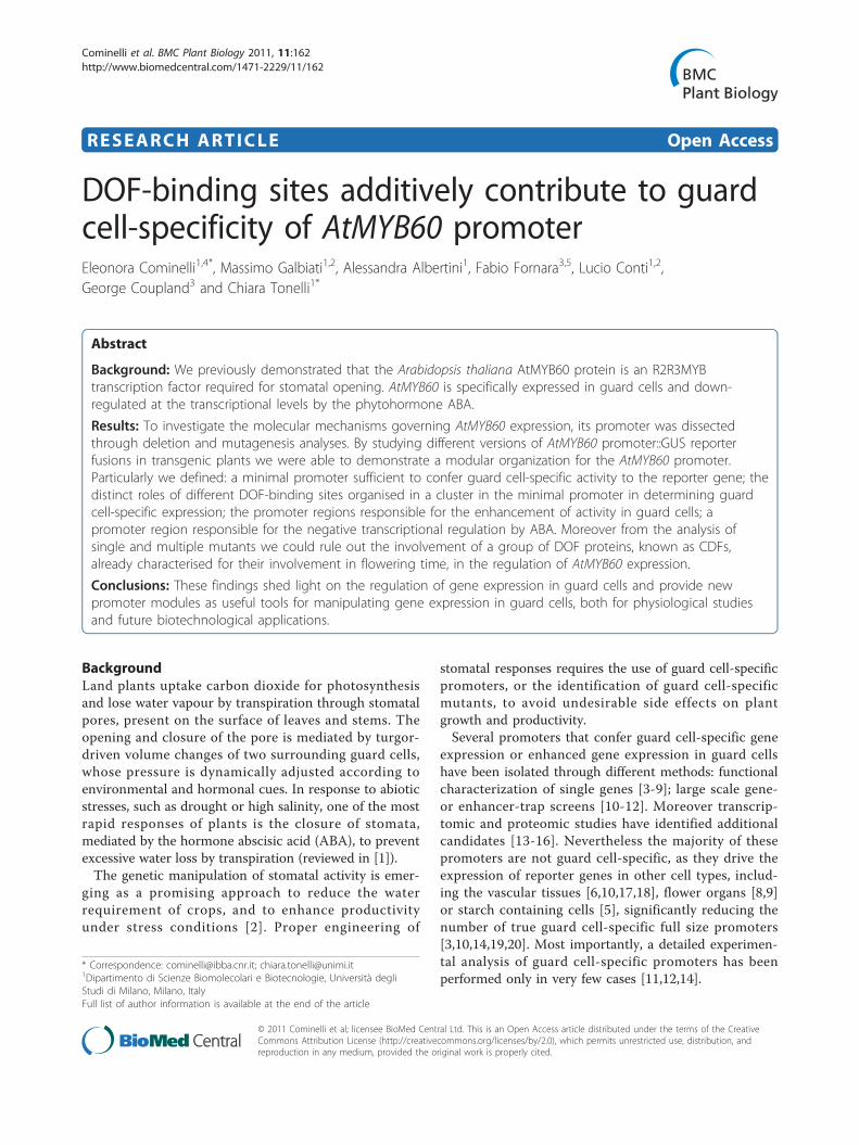
ResultsIn-silico analysis of the AtMYB60 promoterIn a previous study, we demonstrated that the complete5’ and 3’ AtMYB60 intergenic genomic regions - clonedupstream and downstream of the b-glucoronidase (GUS)reporter gene, respectively - could specifically drivestrong GUS activity in stomata of Arabidopsis seedlingsand adult plants [19]. No GUS signals were detected inany other cell type or in tissues devoid of stomata [19].To investigate the possible cis-acting elements that
regulate AtMYB60 expression, we surveyed the genomicregion upstream of the AtMYB60 translational startcodon for the presence of known transcription factorbinding sites using the PLACE software [24]. Our analy-sis produced a significant enrichment in the [A/T]AAAG motifs in the AtMYB60 promoter compared tothe average distribution of [A/T]AAAG oligos in inter-genic regions throughout the Arabidopsis genome (P <0.01) (Figure 1). Interestingly, these [A/T]AAAG motifs,have been shown to be involved in the regulation ofguard cell expression of the potato potassium channelKST1 gene [8]. Also, clusters of [A/T]AAAG motifs,required for the binding of DOF-type transcription fac-tors [25], were over represented in different guard cells-specific promoters [6,10,12]. In particular, Galbiati andcolleagues suggested, as guard cell-specific cis-element,a cluster of at least three [A/T]AAAG motifs located onthe same strand within a region of 100 bp [10]. Usingthe criteria previously described by Galbiati and colla-borators (2008), we found three of these guard cell-spe-cific clusters in the 5’ intergenic region of the AtMYB60gene (Figure 1), suggesting a conserved mechanism forguard cell specific expression.
Identification of the AtMYB60 minimal promoterTo gain more insights into the cis-elements that regulatethe AtMYB60 expression in guard cells, we produced aset of Arabidopsis transgenic lines carrying the complete1,307 bp 5’ intergenic region upstream of the transla-tional start codon fused to the reporter GUS (construct-1,307::GUS, Figure 2A). GUS staining analysis of 15independent T2 lines revealed that this region containsall the cis-acting elements required for expression of thereporter in stomata (Figure 2B), while no GUS signalswere detected in any other cell type or in tissues devoidof stomata (Additional file 1).Next, we made a series of 5’ deletions of the -1,307 bp
genomic region to define the minimum sequence lengthrequired for the expression in guard cells (Figure 2A).These truncated promoters (fused to the GUS gene)were stably transferred to Arabidopsis and 10 to 15independent T2 transgenic lines were analysed in detail.Deletions of the distal part of the 1,307 bp region to
Cominelli et al. BMC Plant Biology 2011, 11:162http://www.biomedcentral.com/1471-2229/11/162
Page 2 of 13
position -619 (construct -619::GUS), -472 (-472::GUS),or -366 (-366::GUS) from the ATG codon, did not alterexpression of the reporter in guard cells located on bothvegetative and floral organs (Figure 2B). Further dele-tions (to position -262) indicated that the 262 bp proxi-mal region was sufficient to drive expression of thereporter in stomata (Figure 2B). However, the removalof the region between -262 bp and -205 bp (construct-205::GUS) completely abolished GUS activity in guardcell (Figure 2B). Transgenic lines carrying the -205::GUSfusion did not show GUS staining in any other cell type,even after prolonged staining (up to 48 h, Figure 2B).This finding suggests that the 57 bp region locatedbetween positions -262 and -205 contains cis-elementsessential for expression in stomatal guard cells. Basedon these results, we defined the -262 bp regionupstream of the ATG codon as the minimal promoterof the AtMYB60 gene.To thoroughly investigate quantitative differences in
GUS expression among lines carrying different deletion:reporter constructs, we determined the relative amountof GUS transcript by quantitative RT-PCR (qRT-PCR).mRNA samples derived from two representative inde-pendent lines (A and B) were analysed for each con-struct (Figure 2C). Lines harbouring the 1,307 bp 5’intergenic region or the -619 deletion fused to the
reporter, did not show any significant differences intheir GUS transcript accumulation. Conversely, deletionsto position -472 and -366 resulted in a two-folddecrease in GUS expression compared to the -1,307::GUS line, while deletion to position -262 resulted in afive-fold decrease (Figure 2C, p < 0.01). These resultsindicate that one or more sequences with function ofenhancer are present in the genomic region between-619 bp and -472 bp and between -472 and -262 fromthe ATG of AtMYB60. In accordance with the resultsobtained from the histochemical analysis, qRT-PCRexperiments did not detect significant GUS transcriptsaccumulation in lines carrying the -205::GUS fusion.
Site-directed mutagenesis of the AtMYB60 minimalpromoterPromoter deletion experiments indicate that theAtMYB60 minimal promoter region (construct -262::GUS) contains all the cis-acting elements required tosustain expression of a reporter gene in guard cells. Thisregion encompasses the [A/T]AAAG cluster proximal tothe ATG codon, which consists of four AAAAG DOF-binding sites (Figures 1 and 3A). In addition, thePLACE software identified in this region a single W-box, corresponding to the binding site of WRKY tran-scription factors [26], located upstream of the [A/T]
CACAAGGACACAAGGACATATGGTATGATGATATGCTTTGTTTCTCTGCTTCTCTTACTAATTTGAAGCTGTTGGATTGATTTGTCTCTTCTTACGTTCCCTTCTTTTTTTTTTCGTTTTCTTTTGTCGTATAGACCAGGCAGGGGCTAGGGCCTAGTGATGGGTATTGGCCCAATACTATTGGGTTATTTGCCTGGTTTATTATTTCGATTTTAGGTTAATTCAATTTTAAGAATACGTAGATTTGTTTGGTTTAGTTTGGTTTGGTTGCACTAAGTTCGGTTTTACATAAATAGAATCTAACACTACTAATTGTTATACGTAAAATACAACAACAATAACAGATTTTTCGTTTCAATTTTCGTTTAAGAGGGTAGACATTTTGGTTTGGTTTGGTTCATTTTTTTTTTCCCTTTCAAATTCACATCCTTCACGTAGATGACAAAATAAAGAAAAACATGAATGAAAGTTGTAACTTGTAAGCATCAACATGGAAATCATATCACAAAGAACACAAATCTAACTAATGGGTCTTTTCACATATTGGTATAATTATAAGTTGTAAGAATATTAGTTAAACAGAGGCAACGAGAGATGCGTGATATATGAAAAGTTGAAAACAAAAGACATGGATCTAAAGAGTCAAGCAAAATGTAATATCTTTTTTTCTTCTAAACTTGAGGATGTCCAAGTTGCAGTGAATGATTCCCTTTAATCATGGAGAAATTCAATGAAATAATTGTGTTTCTTCCCACACTTTATCTTTATTTATTTTCTTACCACAATTACAACTATTATCACAAAAATGTAAGTAACATAGCTTGTGACTCTTCTTCCATTTATGAGTTGATTATCACTATATTTATAAGTAATTACCAACGAATGTTCCAAATTAAGCAAAATATTGTAATCGATACACTATGTATTCATCTACAATATGTTAACGAGCTCCTTTTATGGAAATATTTCGATTGAAAAAACATTTGATGGATCGTTCACTAAATAAATAATCCAGTAACGTTTTCTTAAGGGAGATATACATATTCGTGTGGAGATCAACATATCTTCGTTAATTGACTACGCAAAATAGTTAATGGAAAAGGCAGAGTGACTCGTGAGCTTGGCAGATCCAAAAGAGGTTGTCAAGAAAAAGCAGATTTAAAAGTTCTTCCCTCTTCTTTAAGTCACCCATTAATTTCACATATATGTACATACATGTTGCATTTAACTCATATACATACatattctcacatctataaagagagcataagactcagagagatctagaggaagagagagagagaaagATG
-1307 -1241 -1175 -1109 -1043 -977 -911 -845 -779 -713 -647 -581 -515 -449 -382 -317 -251 -185 -119 -53
Figure 1 Nucleotide sequence of the 5’-region of the AtMYB60 gene. Nucleotides are numbered on the left with the translational start sitedesignated as +1. The ATG is in bold. The 5’ UTR is in lower case letters. The DOF-binding sites are grey boxed, the W-box, considered in thetext, is white boxed. Clusters of DOF-binding sites, as defined by Galbiati and colleagues (2008), are underlined. The CAAGTTG motif described asa putative cis-element for ABA repression ([16]) is dotted underlined.
Cominelli et al. BMC Plant Biology 2011, 11:162http://www.biomedcentral.com/1471-2229/11/162
Page 3 of 13

-120
0 -1
183
-771
-643
-5
94
-547
-147
-1
26
-1,307
-619
-472
-366
-262
-205
-686
-6
99
-860
-671
-541
-352
-210
-1
76
-159
-234
DOF on strand 5’-3’
DOF on strand 3’-5’
W-box considered in the text
5’ UTR
GUS
GUS
GUS
GUS
GUS
GUS
A
B
a b c d
e f g h
i j k l
m n o p
q r s t
u v w x
C
0.0 0.5 1.0 1.5
-205 B
-205 A
-262 B
-262 A
-366 B
-366 A
-472 B
-472 A
-619 B
-619 A
-1307 B
-1307 A
Relative expression Figure 2 Deletion analysis of the AtMYB60 upstream region. A, Schematic diagrams of different deletions of AtMYB60 upstream region fusedto the GUS reporter gene. The positions of the different DOF-binding sites and of the W-box, described in the text, are shown. B, Histochemicalassay for GUS activity in seedlings, rosette leaves and flowers of plants transformed with -1,307::GUS (a-d), -619::GUS (e-h), -472::GUS (i-l), -366::GUS(m-p), -262::GUS (q-t) and -205::GUS (u-x) constructs. The analysis of independent lines harbouring the same construct showed identical patternsof GUS staining. Samples were incubated in the staining solution for 16 hours for all the lines, with the exception of line -205::GUS, for which thestaining was prolonged to 48 hours. Scale bars represent 1 mm. C, Relative expression level of the GUS reporter gene in the different transgeniclines harbouring the -1,307::GUS (-1,307 A and B), -619::GUS (-619 A and B), -472::GUS, -366::GUS (-366 A and B), -262::GUS (-262 A and B) or -205::GUS (-205 A and B) constructs. Two lines for each construct were analysed by Real Time RT-PCR. The transcript amount in the line -1,307 A wasarbitrarily set to 1 (black column) and used to normalize the relative expression levels in each line. The ACTIN2 gene (At3g18780) was used as acontrol.
Cominelli et al. BMC Plant Biology 2011, 11:162http://www.biomedcentral.com/1471-2229/11/162
Page 4 of 13

-147
DOF on strand 5’-3’
DOF on strand 3’-5’ W-box considered in the text
5’ UTR
mutagenised site
mDOF2 GUS
mDOF1 GUS
mW GUS
-126
-210
-1
76
-159
-234
-262 GUS
mDOF(2+3) GUS
mDOF(1+4) GUS
mDOF(1+3) GUS
mDOF3 GUS
mDOF(1+2) GUS
mDOF(2+4) GUS
mDOF(3+4) GUS
mDOF4 GUS
-205 GUS
intermediate
weak
strong
C
no signal
B A
g e f h g
0 20 40 60 80 100
a b c d
Figure 3 Role of DOF-binding sites in the minimal promoter in driving GUS activity in guard cells. A, Schematic diagrams of constructs-262::GUS, -205::GUS and of constructs containing mutagenised version of the minimal promoter in different DOF-binding sites and in the W-boxat position -234. B, Percentage of lines for each construct showing strong (column segment in black), intermediate (in dark grey), weak (lightgrey) or no signal (white). C, A leaf from a line harbouring the -262::GUS construct (a and a particular in e), shown as an example of strong GUSactivity. In the following pictures examples of different lines harbouring the mDOF3::GUS construct showing respectively an intermediate (b andf), a weak (c and g) and no GUS activity (d and h). Scale bars represent 1 mm (a-d) or 0.1 mm (e-h).
Cominelli et al. BMC Plant Biology 2011, 11:162http://www.biomedcentral.com/1471-2229/11/162
Page 5 of 13

AAAG cluster (Figure 3A). To address the functionalsignificance of the individual cis-elements present in theAtMYB60 minimal promoter, we evaluated the effects oftargeted nucleotide substitutions on GUS expression(Figure 3A). Mutated versions of the minimal promoterwere generated by PCR and fused to GUS and at least30 T2 independent transgenic lines for each mutatedpromoter::GUS combination were visually scored andclassified to reflect their relative guard-cell specific GUSstaining. A representative example of each category isprovided in Figure 3C.We initially tested the role of the single W-box cis-
element, by replacing the consensus sequence TTGAC,with the non-functional TTGAA motif [27]. Lines carry-ing the mutated W-box (mW::GUS) showed similarlevels of GUS expression to the wild-type promoter,indicating that W-box does not contribute to mediategene expression in guard cells (Figure 3B). Next, weproduced mutant promoters in which single DOF motifswithin the [A/T]AAAG cluster were converted to theunrelated CGCGA sequence. Inactivation of the mostdistal AAAAG site relative to the ATG (hereinafterreferred to as DOF1) resulted in a dramatic decrease ofGUS expression (mDOF1::GUS construct, Figure 4B).30% of the lines carrying the mDOF1::GUS constructdid not show GUS expression, whereas the remaining70% only showed weak staining, thus indicating a crucialrole for DOF1 in regulating AtMYB60 expression inguard cells (Figure 3B). Mutations of the second, thirdor fourth most proximal AAAAG site (hereinafterreferred to as DOF2, DOF3 and DOF4, respectively),resulted in a reduced GUS expression, although to a les-ser extent than the one in the DOF1 (Figure 4B,mDOF2::GUS, mDOF3::GUS and mDOF4::GUS plants).In particular, none of the 30 mDOF2::GUS transgeniclines displayed strong expression of the reporter, nearly70% showed intermediate expression, 25% showed weakexpression and the remaining 5% did not show any GUSstaining (Figure 3B). A comparable distribution amongstrong, intermediate and weak lines was obtained fromthe analysis of the mDOF3::GUS and mDOF4::GUSplants (Figure 3B).To establish whether DOF-binding sites could exert
additive roles in mediating gene expression in stomatawe produced a second series of promoters, in which twoAAAAG motifs were mutated simultaneously. Mutationsof DOF1 and DOF2 (mDOF(1+2)::GUS), DOF1 andDOF3 (mDOF(1+3)::GUS) or DOF1 and DOF4 (mDOF(1+4)::GUS) completely inactivated the minimal promoter,as GUS expression was abolished in all the mDOF(1+2)::GUS, mDOF(1+3)::GUS and mDOF(1+4)::GUS lines ana-lysed (Figure 3B). Interestingly, the concurrent mutationof DOF2 and DOF3 (mDOF(2+3)::GUS) resulted in astrong, but yet not complete, inactivation of the
promoter activity in guard cells, as 15% of the mDOF(2+3)::GUS lines displayed weak expression of the reporterin stomata. Likewise, concomitant inactivation of eitherDOF2 and DOF4, or DOF3 and DOF4 did not comple-tely eliminate GUS expression in guard cell (Figure 3B).Taken together, these results indicate that the putative[A/T]AAAG DOF-binding sites located in the AtMYB60promoter are necessary to mediate its expression inguard cells.
A single DOF cluster is sufficient to drive low expressionin guard cellOur deletion analysis of the AtMYB60 promoter indi-cates that the 57 bp region between positions -262 and-205 is essential for gene expression in stomatal guardcells (Figure 2). This region contains the DOF1 cis-ele-ment required for guard cell expression as shown bymutagenesis analysis results (Figure 3). To establishwhether this 57 bp region was sufficient to activateexpression in guard cells, we fused one (1x::GUS con-struct), two (2x::GUS) and four tandem copies (4x::GUS)of the 57 bp fragment to the minimal CaMV35S promo-ter [28] upstream of the GUS reporter gene (Figure 4A),effectively reconstructing an artificial DOF cluster con-taining one, two or four copies of the DOF1 element.However, we did not observe GUS activity in any of the30 independent stable transformants produced for eachconstruct, even after prolonged staining (data notshown). These data were confirmed by qRT-PCR analy-sis of independent lines carrying the 4x::GUS fusion(Figure 4B), indicating that the multimerisation of theDOF1 site per se is not sufficient to drive gene expres-sion in guard cell. This might derive from an inap-propriate organization and/or spatial distribution of thedifferent DOF elements in the context of the minimalpromoter. To test this hypothesis we made two 3’ dele-tions of the AtMYB60 minimal promoter: the -148-3’::GUS and -137-3’::GUS constructs containing the firstthree and four DOF-binding sites respectively of themost proximal cluster fused upstream of the minimalCaMV35S promoter (Figure 4B). Our initial histochem-ical analysis did not reveal any GUS positive lines (datanot shown). To substantiate this result we also per-formed a qRT-PCR analysis on fifteen independent linesfor each construct. Interestingly, eight lines out of fif-teen showed a low but significant GUS transcript accu-mulation compared to the full length minimal promoter(Figure 4B). These results suggest that the presence ofthe cluster containing three or four DOF-binding sites issufficient to drive GUS activity in guard cells, eventhough at a very low level. This finding implies thatother cis-elements present downstream of position -137are required for the full functionality of the minimalpromoter.
Cominelli et al. BMC Plant Biology 2011, 11:162http://www.biomedcentral.com/1471-2229/11/162
Page 6 of 13

The guard cell-related CDF1, CDF2, CDF3 and CDF5 DOF-type transcription factors do not regulate AtMYB60expression in stomataTarget mutagenesis experiments of the AtMYB60 pro-moter demonstrated that [A/T]AAAG DNA consensusmotifs are essential cis-acting elements in the regulationof AtMYB60 expression in guard cells. Consequently,their cognate DOF proteins represent the most likelycandidates as trans-acting factors. As the Arabidopsisgenome contains 36 DOF-coding genes [23], candidateDOF transcription factors involved in the regulation ofAtMYB60 expression should fulfil two criteria: theyshould be expressed in guard cells and the loss of theirgene function should abolish or significantly down-regu-late the expression of AtMYB60 in this cell type.The CYCLING DOF FACTOR 1 (CDF1, At5g62430)
gene, involved in the regulation of photoperiodic flower-ing, has been shown to be highly expressed in the vascu-lar tissue and guard cells [29]. We thus investigated theexpression of the AtMYB60 gene in the loss-of-function
cdf1-R allele. As shown in Additional file 2 we did notdetect significant differences in the accumulation ofAtMYB60 transcripts in homozygous cdf1-R plants com-pared with the wild type.It is important to note that in photoperiodic flowering,
CDF1 acts redundantly with three other DOF proteins,namely CDF2 (At5g39660), CDF3 (At3g47500) andCDF5 (At1g69570) [30], belonging to the same phyloge-netic group II [31]. Similarly to CDF1, promoter::GUSanalyses revealed that CDF2, CDF3 and CDF5 arestrongly expressed in guard cells.We thus analysed the expression of AtMYB60 in sin-
gle, double, triple and quadruple cdf mutants to deter-mine the possible role of these additional candidateCDF proteins. As for cdf1-R mutant, the level of expres-sion of AtMYB60 was not significantly reduced in thecdf2-1, cdf3-1 and cdf5-1 single mutants (Additional file2). Likewise, AtMYB60 expression was not altered inany of the double, triple or quadruple mutant combina-tions, indicating that, despite their expression in guard
GUS 4x::GUS
GUS -148-3’::GUS
A
-262 A
4x A
-137-3’ A
-148-3’ A
-262 B
4x B
-137-3’ B
-148-3’ B
B
GUS -137-3’::GUS
-262::GUS GUS -137
-1
48
-262
-205
GUS 2x::GUS
GUS 1x::GUS
DOF on strand 5’-3’
DOF on strand 3’-5’
W-box considered in the text
5’-3’ UTR minimal CaMV35S
0 0.5 1 1.5Relative expression
Figure 4 Oligomerisation of the 57 bp sequence and 3’ deletions of the AtMYB60 minimal promoter. A, Schematic diagrams of theconstructs. In the 4x::GUS construct the fragment of 57 bp between -262 and -205 in tandem array of four copies was fused to the minimalpromoter CaMV 35S (min 35S in the scheme, portion between -46 and +1) upstream of the GUS reporter gene. In the constructs -137-3’::GUSand -148-3’::GUS, 3’ deleted versions of the AtMYB60 minimal promoter were fused to the same portion of the CaMV 35S. (B) Relative expressionlevel of the GUS reporter gene in the different transgenic lines harbouring the constructs. Two lines for each construct were analysed by RealTime RT-PCR. The transcript amount in the line -1307 A was arbitrarily set to 1 (black column) and used to normalize the relative expressionlevels in each line. The ACTIN2 gene was used as a control. The symbols are the same described in Figure 2. The dotted lines indicate theregions deleted in AtMYB60 minimal promoter sequences.
Cominelli et al. BMC Plant Biology 2011, 11:162http://www.biomedcentral.com/1471-2229/11/162
Page 7 of 13
cells, these four CDF proteins are not trans-regulators ofAtMYB60 expression in stomata (Additional file 2).
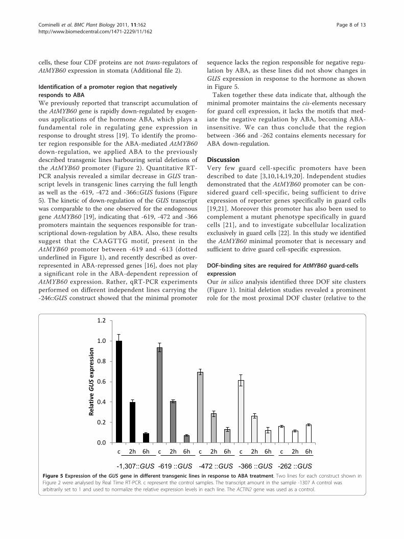
Identification of a promoter region that negativelyresponds to ABAWe previously reported that transcript accumulation ofthe AtMYB60 gene is rapidly down-regulated by exogen-ous applications of the hormone ABA, which plays afundamental role in regulating gene expression inresponse to drought stress [19]. To identify the promo-ter region responsible for the ABA-mediated AtMYB60down-regulation, we applied ABA to the previouslydescribed transgenic lines harbouring serial deletions ofthe AtMYB60 promoter (Figure 2). Quantitative RT-PCR analysis revealed a similar decrease in GUS tran-script levels in transgenic lines carrying the full lengthas well as the -619, -472 and -366::GUS fusions (Figure5). The kinetic of down-regulation of the GUS transcriptwas comparable to the one observed for the endogenousgene AtMYB60 [19], indicating that -619, -472 and -366promoters maintain the sequences responsible for tran-scriptional down-regulation by ABA. Also, these resultssuggest that the CAAGTTG motif, present in theAtMYB60 promoter between -619 and -613 (dottedunderlined in Figure 1), and recently described as over-represented in ABA-repressed genes [16], does not playa significant role in the ABA-dependent repression ofAtMYB60 expression. Rather, qRT-PCR experimentsperformed on different independent lines carrying the-246::GUS construct showed that the minimal promoter
sequence lacks the region responsible for negative regu-lation by ABA, as these lines did not show changes inGUS expression in response to the hormone as shownin Figure 5.Taken together these data indicate that, although the
minimal promoter maintains the cis-elements necessaryfor guard cell expression, it lacks the motifs that med-iate the negative regulation by ABA, becoming ABA-insensitive. We can thus conclude that the regionbetween -366 and -262 contains elements necessary forABA down-regulation.
DiscussionVery few guard cell-specific promoters have beendescribed to date [3,10,14,19,20]. Independent studiesdemonstrated that the AtMYB60 promoter can be con-sidered guard cell-specific, being sufficient to driveexpression of reporter genes specifically in guard cells[19,21]. Moreover this promoter has also been used tocomplement a mutant phenotype specifically in guardcells [21], and to investigate subcellular localizationexclusively in guard cells [22]. In this study we identifiedthe AtMYB60 minimal promoter that is necessary andsufficient to drive guard cell-specific expression.
DOF-binding sites are required for AtMYB60 guard-cellsexpressionOur in silico analysis identified three DOF site clusters(Figure 1). Initial deletion studies revealed a prominentrole for the most proximal DOF cluster (relative to the
-1,307::GUS -619 ::GUS -472 ::GUS -366 ::GUS -262 ::GUS
0.0
0.2
0.4
0.6
0.8
1.0
1.2
c 2h 6h c 2h 6h c 2h 6h c 2h 6h c 2h 6h
Rela
tive GUS
expr
essi
on
Figure 5 Expression of the GUS gene in different transgenic lines in response to ABA treatment. Two lines for each construct shown inFigure 2 were analysed by Real Time RT-PCR. c represent the control samples. The transcript amount in the sample -1307 A control wasarbitrarily set to 1 and used to normalize the relative expression levels in each line. The ACTIN2 gene was used as a control.
Cominelli et al. BMC Plant Biology 2011, 11:162http://www.biomedcentral.com/1471-2229/11/162
Page 8 of 13
ATG start codon). Site-directed mutagenesis showedthat the distal most DOF-binding site (DOF1 at posi-tion -210, Figure 3) plays a major role in driving guardcell expression compared to other DOF motifs of thesame cluster (DOF2 at position -176, DOF3 at -159and DOF4 at -147, Figure 3). These other DOF ele-ments play partially additive roles, as clearly demon-strated by the combined mutagenesis of these sites andDOF1 site which resulted in a drastically reduced GUSactivity (Figure 3). DOF-binding sites are thus keydeterminants in mediating guard cell expression, inaccordance with the DOF cluster hypothesis we pre-viously formulated [10]. A suggestion for a similarinvolvement of DOF cis-elements in Arabidopsisderives from the work of Gardner and colleagues(2009) that identified DOF motifs in a region control-ling guard cell expression. Other authors identified aregion enriched in DOF-binding sites in the guard cell-specific pGC1 promoter, although the mutation of asingle DOF site did not impair promoter activity [14].Interestingly, a DOF cluster organization is present inthe promoter of the grape VvMYB60 gene, a putativeortholog of AtMYB60, indicating a conservation of thecluster structure during the evolution amongAtMYB60 orthologs [32]. The results reported byPlesch and colleagues (2001) on the DOF motif organi-sation in the potato KST1 promoter highlight a moregeneral evolutionary conservation of this module inthe control of guard cell-specific activity of promoters.Although we cannot rule out the possibility that
other unknown transcription factors might interactwith those same cis-elements, DOF factors representlikely candidates as AtMYB60 regulators. The mostparsimonious hypothesis resulting from combining ourresults indicates that DOF proteins act as positive reg-ulators of AtMYB60. The potato StDOF1 protein hasbeen shown to bind in vitro to the guard cell specificpromoter of KST1 [8], while no data are available forany Arabidopsis DOF proteins. Among the ArabidopsisDOF genes, CDF1, CDF2, CDF3, and CDF5 (CDFs) areexpressed in guard cells [29]. However, singles andmultiple cdf mutants show a wild-type pattern ofAtMYB60 expression, ruling out their involvement inAtMYB60 regulation (Additional file 2). The majorityof Arabidopsis DOF genes are expressed in guard cells[33,34] and may thus act redundantly, as alreadydemonstrated among members of this family [30]. Allthese aspects do not facilitate the identification ofobvious candidates as AtMYB60 regulators. We aretrying to identify the DOF genes involved in the regu-lation of AtMYB60 by analysis of its expression inmutants of genes preferentially expressed in the guardcells (http://bbc.botany.utoronto.ca/efp/cgi-bin/efpWeb.cgi[33]).
Multiple cis-elements participate to enhance AtMYB60guard-cells expressionTranscriptional GUS fusions, harbouring different dele-tions of the 5’ intergenic region to position -262 fromthe ATG, conferred GUS activity exclusively in guardcells (Figures 2 and Additional file 1). The activity ofthese promoter regions is in apparent discrepancy withthe detection of AtMYB60 gene expression in seeds, asrevealed by available microarray analysis data [33,34]and in roots, as recently reported [35]. One hypothesisto explain this incongruity could be the presence ofother regulatory regions present outside the complete 5’and 3’ intergenic regions flanking the AtMYB60 codingsequence. Intron sequences, for example, may beinvolved in such a regulation, as previously demon-strated for different plant genes ([36] and referencesherein).While guard-cell specific expression was invariably
maintained by functional AtMYB60 promoter variants,the levels of expression varied considerably. In additionto DOF-binding sites, other cis-elements are required toboost the AtMYB60 expression. Indeed, an artificialDOF1 binding site repeated in single or multiple copiescould not drive guard cell expression (Figure 4A). Theincorporation of the entire proximal DOF cluster (e.g.-137-3’::GUS) resulted in a small but significant guardcell transcriptional activity. Thus, other cis-elementsdownstream of position -137 are required for full activ-ity of the minimal promoter. It is known that cis-ele-ments other than DOF-binding sites are involved in theregulation of guard cell expression. In the case of theguard cell-specific AtPDR3 gene no [A/T]AAAG clusterswere identified in a 1000-bp region upstream of theATG codon, suggesting the presence of other regulatoryunits [10].
Modular organization of the AtMYB60 promoterIn this study we also investigated the regulation of theAtMYB60 promoter activity in response to ABA. ABAtreatments induce global changes in gene expression inArabidopsis [16,37-40]. Transcriptomic analyses revealedextensive regulation of gene expression by ABA also inguard cells [13,14,16]. While cis-elements that positivelyregulate the response to ABA have been functionallycharacterised (for a review, see [41]), those that nega-tively regulate the response to ABA are largelyunknown. A CAA[G/C]TTG motif has been shown tobe over-represented in ABA-repressed gene promotersand thus proposed for such a role [16,39]. TheAtMYB60 promoter contains one CAAGTTG motifbetween -619 and -613 from the ATG, yet our resultsdo not support its proposed role as negative regulator ofABA response. Conversely, a region between positions-366 to -262 contained the entire requirement for the
Cominelli et al. BMC Plant Biology 2011, 11:162http://www.biomedcentral.com/1471-2229/11/162
Page 9 of 13

ABA-mediated repression Figures 5 and 6. It has beenproposed that evolution may have favoured the differen-tiation of mechanisms for ABA down-regulation ratherthan up-regulation, rendering more difficult for anyABA-repression motif to achieve statistical significance[16]. Our data may provide a valuable model system toclarify the mechanism mediating ABA repression.Our data suggests a modular organization for the
AtMYB60 promoter as summarised in Figure 6.Through a serial deletion analysis, we defined theAtMYB60 minimal promoter, sufficient to induce guardcell-specific activity (construct -262::GUS, Figure 2). A57 bp region, located between position -262 and posi-tion -205, is necessary to confer GUS activity in guardcells (Figure 2A). We also identified two regions thatenhance the expression of the GUS gene between -619bp and -472 bp and between -472 and -262 (Figure 2Band 2C). Besides providing pieces of evidence for suchmodular organization, our work indicates that the differ-ent portions of the AtMYB60 promoter may prove use-ful for manipulating gene expression in guard cells, withthe possibility to obtain different level of expression.Moreover, the minimal promoter (whose activity is notinfluenced by ABA) can be used for ABA-independentexpression of target genes in guard cellsInterestingly, both the full length and the minimal
promoters maintain their guard cell-specific activity inheterologous systems, such as the crop species tomatoand tobacco (Francia, personal communication), thusindicating the conservation of this cell-specific regula-tory mechanism among different plant species. More-over, preliminary results suggest that the AtMYB60minimal promoter can be combined with other cis-reg-ulatory modules to produce functional guard cell-spe-cific chimeric promoters (Francia, personalcommunication). As a whole our data demonstratethat both the full length and the minimal AtMYB60promoters provide a valuable tool to manipulate geneexpression specifically in guard cells, both for
physiological studies and downstream biotechnologicalapplications.
ConclusionsOur work provides strong evidence for the involvementof [A/T]AAAG elements in the regulation of theAtMYB60 expression, illustrating their functional clusterorganization. Future work will concentrate on the analy-sis of candidate DOF transcription factors that controlthis mechanism. Finally we identify a region of theAtMYB60 promoter required for the negative regulationby ABA, offering the possibility to discover novel cis-ele-ments for this kind of regulation.
MethodsPlant MaterialAll plant material described was in the Col-0 accession.The cdf1-R line (35S::CDF1-RNAi #23) was kindly pro-vided by Takato Imaizumi [29]. The cdf2-1, cdf3-1 andcdf5-1 null alleles are T-DNA insertion line. Single, dou-ble, triple and quadruple cdf mutants have been pre-viously described [30].
Construction of AtMYB60 promoter::GUS fusions5’-deletions of the 5’ intergenic genomic regionupstream of the AtMYB60 gene were generated by PCRamplification from plasmid p1.3-2.2::GUS, previouslydescribed [19], using different forward primers and asingle reverse primer. Forward and reverse primersincorporated a HindIII and a BamHI, respectively. ThePCR fragments were cloned into the pCR4-TOPO vec-tor (Invitrogen Corporation, Carlsbad, CA), cut withHindIII and BamHI and ligated upstream of the uidAcoding sequence in the pBI101.3 binary vector (Clon-tech, Palo Alto, CA, USA). The resulting plasmids wererenamed -1307::GUS, -619::GUS, -472::GUS, -366::GUS,-262::GUS and -205::GUS (Figure 2).Chimeric promoters containing different 3’-deleted
fragments of the AtMYB60 minimal promoter and 46-
262366472619-1,307
-262-366-472-619
262366472 -262-366-472-619
minimal promoterenhancer enhancer
ABAFigure 6 Modular organization of the AtMYB60 promoter. Different portions of the AtMYB60 promoter defined through deletion analysis areshown. ABA indicates the region responsible for the negative regulation by ABA treatment.
Cominelli et al. BMC Plant Biology 2011, 11:162http://www.biomedcentral.com/1471-2229/11/162
Page 10 of 13
bp CaMV 35S promoter were produced by amplifyingthe sequence of the CaMV 35S promoter from -46 to+1 [28] from plasmid pBI121 (Clontech, Palo Alto, CA,USA), using the forward primer 35SXba containing aXbaI site and the reverse primer 35SBam with a BamHIsite. The PCR product was cloned into the pCR4-TOPOvector and the XbaI-BamHI fragment was cloned intothe pBI101.3 vector (renamed 35Smin-pBI101.3). Theregions from -262 to -137 and from -262 to -148 of theAtMYB60 minimal promoter were amplified by PCRfrom plasmid p1.3-2.2::GUS, using the reverse primersp60R6 and p60R7 incorporating a XbaI site and a singleforward primer p60F3 with the HindIII site. The corre-sponding PCR products were cloned into the pCR4-TOPO vector and the HindIII-XbaI fragments werecloned into the 35Smin-pBI101.3 vector to give the-137-3’::GUS and -148-3’::GUS vectors, respectively (Fig-ure 3).Chimeric promoters containing different copies of the
region between -262 and -205 of the AtMYB60 promo-ter were obtained by synthesising one copy of thissequence, using the forward primer p60F3 with a Hin-dIII site and the reverse primer p60R3 with an XbaIsite. The resulting PCR product was cloned into thepCR4-TOPO vector and the HindIII-XbaI fragment wasligated into the 35Smin-pBI101.3 vector (construct 1x::GUS). A second copy of this region was generated usingthe primers p60F3 and p60R5b, both incorporating aHindIII site; the fragment HindIII-HindIII was clonedinto the construct 1x::GUS, generating the construct 2x::GUS. This plasmid was used as a template to generatetwo other copies of the sequence from -262 to -205using the primers p60F11 and p60R3 incorporating anXbaI site. The fragment XbaI-XbaI was cloned into theplasmid 2x::GUS, to generate the construct 4x::GUS. Allthe oligonucleotide sequences are reported in Table 1.PCR products were sequenced and the correct orienta-tion of the fragment into the final vector was verified byrestriction.
Site-directed mutagenesis analysisBase mutations of the different DOF sites were gener-ated using the megaprimer method [42]. For the muta-genised versions of the AtMYB60 minimal promoterdifferent megaprimers were PCR amplified from plasmidp1.3-2.2::GUS, using as forward primers mp60DOF1F1,mp60DOF2F1, mp60DOF3F2 and mp60DOF4F2 andthe single reverse primer p60R5. The megaprimers weregel purified and used in a second PCR reaction on plas-mid p1.3-2.2::GUS with the primer p60F3. The PCRproducts were cloned into pCR4-TOPO and sequencedbefore cloning into pBI101.3 vector using the restrictionsites HindIII and BamHI to generate the following con-structs: mDOF1::GUS, mDOF2::GUS, mDOF3::GUS,
mDOF4::GUS. To generate multiple mutagenised sitesthe templates for the second PCR amplification wereplasmids already carrying a first mutagenised DOF site.In the case of the preparation of the construct mW::GUS the megaprimer method was not necessary, as thesite to mutagenise is in a position 5’ terminal into theminimal promoter and a single PCR reaction was per-formed with primers mp60WRKYF1 and p60R5, thePCR product was then cloned with the procedurealready described.All the oligonucleotide sequences are reported in
Table 1.
Arabidopsis transformation and growth conditionsWild-type Columbia (Col-0) plants were transformedusing the Agrobacterium tumefaciens strain GV3101 car-rying the constructs described above with the floral dipmethod [43]. Transformed lines were selected on kana-mycin and single-insertion lines were selected forfurther analyses. Analyses of transgenic lines were
Table 1 Sequence of oligonucleotides used in this study
Name Sequence
p60F1 AAGCTTCACAAGGACACAAGGACA
p60 F2b AAGCTTCAAGTTGCAGTGAATGA
p60F8b AAGCTTTAACGAGCTCCTTTTATGG
p60F9 AAGCTTCCATTTATGAGTTGATTATCA
p60F3 AAGCTTCGTGTGGAGATCAACAT
p60F5 AAGCTTGCAGAGTGACTCGTGA
p60R5 TCTCGGATCCTCTAGATCTCTCTG
p60R6 TCTAGAGAAGAACTTTTAAATCTGC
p60R7 TCTAGAAAATCTGCTTTTTCTTGAC
p60R5b AAGCTTCTTTTCCATTAACTATTTTG
p60F11 TCTAGACGTGTGGAGATCAACAT
p60R3 TCTAGACTTTTCCATTAACTATTTTG
35SXba TCTAGACAAGACCCTTCCTC
35SBam GGATCCTCCTCTCCAAATGA
mp60DOF1F1 AGTTAATGGcgcgaGCAGAGTGACTCGTGA
mp60DOF2F1 TGGCAGATCCcgcgaAGGTTGTCAAGAAAA
mp60DOF3F2 TGTCAAGAcgcgaCAGATTTAAAAGTTCTT
mp60DOF4F2 CAAGAAAAAGCAGATTTcgcgaTTCTTC
mp60WRKYF1 AAGCTTCGTGTGGAGATCAACATATCTTCGTTAATTGAaTACGCAAAATA
GUSRTF1 TACGGCAAAGTGTGGGTCAATAATCA
GUSRTR1 CAGGTGTTCGGCGTGGTGTAGAG
ATACT2F TGCTTCTCCATTTGTTTGTTTC
ATACT2R GGCATCAATTCGATCACTCA
qRT-MYB60-F CATGAAGATGGTGATCATGAGG
qRT-MYB60-R TTCCATTTGACCCCCAGTAG
PP2a-F CAGCAACGAATTGTGTTTGG
PP2a-R AAATACGCCCAACGAACAAA
Italic and lower case letters indicate restriction and mutagenised sites,respectively
Cominelli et al. BMC Plant Biology 2011, 11:162http://www.biomedcentral.com/1471-2229/11/162
Page 11 of 13
performed on T2 or on homozygous T3 plants grownunder long-day conditions (16 hr light; 8 hr darkat 100μmol m-2 sec-1) at 22°C in a growth chamber. Seedswere germinated in Petri dishes containing Murashigeand Skoog medium, 1% w/v sucrose and 0.8% w/v agarfor seedling analysis or directly on soil for adult plantorgan analysis. The ABA treatment was performed aspreviously described [19].
GUS activity assays and histochemical stainingFor detection of GUS activity, tissues were fixed for 2 hin 90% (v/v) acetone at -20°C, incubated for 16-48hours, at 37°C, in 0.05% (w/v) X-glucoronic acid, 0.1%(v/v) Triton X-100, and 0.5mM ferrocyanidine in 50mM phosphate buffer (pH 7) and subsequently clearedin 70% (v/v) ethanol. Seedlings and flowers were clearedwith a chloral hydrate:glycerol:water solution (8:1:2, v/v).Samples were examined using a Leica M205 FA stereo-microscope (Leica Microsystems GmbH Wetzlar, Ger-many) and a Zeiss Axiophot D1 microscope (Carl ZeissMicroImaging, LLC Thornwood, New York, USA).Stereomicroscope images were recorded using the LeicaLAS software version 2.8.1. Microscope images wererecorded with an AxioCam MRc5 camera (Zeiss) usingthe AxioVision program (version 5.0).
Quantification of mRNA expressionRNA isolation, reverse transcription, qRT-PCR reactionsand data analysis were performed as previouslydescribed [30]. GUS expression was analysed using pri-mers GUSRT-F1 and GUSRT-R1, ACTIN2 gene (pri-mers ATACT2F, ATACT2R) was used as a referencefor normalization. AtMYB60 expression in different cdfmutants was analysed using primers qRT-MYB60-F andqRT-MYB60-R. PP2A gene, corresponding to At1g13320(primers PP2a-F and PP2a-R) was used as a referencefor normalization [44]. All primer sequences arereported in Table 1.
Additional material
Additional file 1: Analysis of GUS activity in seeds at differentdevelopmental stages in 1,307::GUS line. A: open silique showingsignal only in stomata and not in developing seeds. B: mature-green-stage seed (13 DAP). C: a 24 h imbibed seed. D: embryo isolated from a24 h imbibed seed. The same results were obtained in all transgeniclines described in Figure 2. Scale bars represent 0.1 mm.
Additional file 2: Relative expression of the AtMYB60 gene in thedifferent cdf mutants. cdf1-R is an RNAi line ([29]). The other single andmultiple mutants have been previously described ([30]). The PP2a(At1g13320) gene was used as a control [44].
AcknowledgementsThis work was supported by the Italian “Progetto AGER, bando Viticoltura daVino” (SERRES 2010-2015), by the BIOGESTECA 15083/RCC project, funded by
Regione Lombardia and by the AGRISOST project, funded by FondazioneUmberto Veronesi per il Progresso delle Scienze, Milano, Italy.
Author details1Dipartimento di Scienze Biomolecolari e Biotecnologie, Università degliStudi di Milano, Milano, Italy. 2Fondazione Filarete, Milano, Italy. 3Max PlanckInstitute for Plant Breeding Research, Cologne, Germany. 4Istituto di Biologiae Biotecnologia Agraria, CNR, Milano, Italy. 5Dipartimento di Biologia,Università degli Studi di Milano, Milano, Italy.
Authors’ contributionsEC carried out the construction of promoter-reporter plasmids, planttransformation and drafted the manuscript. EC, AA and LC did transgenicArabidopsis analysis. FF carried out cdf mutant analysis. CT, MG, and GCconceived the study, participated in its design and coordination. MG, LC andFF helped to draft the manuscript. All authors read and approved the finalmanuscript.
Received: 14 September 2011 Accepted: 16 November 2011Published: 16 November 2011
References1. Kim TH, Böhmer M, Hu H, Nishimura N, Schroeder JI: Guard cell signal
transduction network: advances in understanding abscisic acid, CO2, andCa2+ signaling. Annu Rev Plant Biol 2010, 61:13.11-13.31.
2. Schroeder JI, Kwak JM, Allen GJ: Guard cell abscisic acid signalling andengineering drought hardiness in plants. Nature 2001, 410:327-330.
3. Gray JE, Holroyd GH, van der Lee FM, Bahrami AR, Sijmons PC,Woodward FI, Schuch W, Hetherington AM: The HIC signalling pathwaylinks CO2 perception to stomatal development. Nature 2000, 408:713-716.
4. Kopka J, Provart NJ, Muller-Rober B: Potato guard cells respond to dryingsoil by a complex change in the expression of genes related to carbonmetabolism and turgor regulation. Plant J 1997, 11:871-882.
5. Muller-Rober B, La Cognata U, Sonnewald U, Willmitzer L: A truncatedversion of an ADP-glucose pyrophosphorylase promoter from potatospecifies guard cell-selective expression in transgenic plants. Plant Cell1994, 6:601-612.
6. Nakamura RL, McKendree WL, Hirsch RE, Sedbrook JC, Gaber RF,Sussman MR: Expression of an Arabidopsis potassium channel gene inguard cells. Plant Physiol 1995, 109:371-374.
7. Nylander M, Svensson J, Palva ET, Welin BV: Stress-induced accumulationand tissue-specific localization of dehydrins in Arabidopsis thaliana. PlantMol Biol 2001, 45:263-279.
8. Plesch G, Ehrhardt T, Mueller-Roeber B: Involvement of TAAAG elementssuggests a role for Dof transcription factors in guard cell-specific geneexpression. Plant J 2001, 28:455-464.
9. Terryn N, Arias MB, Engler G, Tire C, Villarroel R, Van Montagu M, Inze D:rha1, a gene encoding a small GTP binding protein from Arabidopsis, isexpressed primarily in developing guard cells. Plant Cell 1993,5:1761-1769.
10. Galbiati M, Simoni L, Pavesi G, Cominelli E, Francia P, Vavasseur A, Nelson T,Bevan M, Tonelli C: Gene trap lines identify Arabidopsis genes expressedin stomatal guard cells. Plant J 2008, 53:750-762.
11. Gardner MJ, Baker AJ, Assie JM, Poethig RS, Haseloff JP, Webb AA: GAL4GFP enhancer trap lines for analysis of stomatal guard cell developmentand gene expression. J Exp Bot 2009, 60:213-226.
12. Plesch G, Kamann E, Mueller-Roeber B: Cloning of regulatory sequencesmediating guard-cell-specific gene expression. Gene 2000, 249:83-89.
13. Leonhardt N, Kwak JM, Robert N, Waner D, Leonhardt G, Schroeder JI:Microarray expression analyses of Arabidopsis guard cells and isolationof a recessive abscisic acid hypersensitive protein phosphatase 2Cmutant. Plant Cell 2004, 16:596-615.
14. Yang Y, Costa A, Leonhardt N, Siegel RS, Schroeder JI: Isolation of a strongArabidopsis guard cell promoter and its potential as a research tool.Plant Methods 2008, 4:6.
15. Zhao Z, Zhang W, Stanley BA, Assmann SM: Functional proteomics ofArabidopsis thaliana guard cells uncovers new stomatal signalingpathways. Plant Cell 2008, 20:3210-3226.
16. Wang RS, Pandey S, Li S, Gookin TE, Zhao Z, Albert R, Assmann SM:Common and unique elements of the ABA-regulated transcriptome ofArabidopsis guard cells. BMC Genomics 2011, 12:216.
Cominelli et al. BMC Plant Biology 2011, 11:162http://www.biomedcentral.com/1471-2229/11/162
Page 12 of 13

17. Husebye H, Chadchawan S, Winge P, Thangstad OP, Bones AM: Guard cell-and phloem idioblast-specific expression of thioglucosideglucohydrolase 1 (myrosinase) in Arabidopsis. Plant Physiol 2002,128:1180-1188.
18. Liang YK, Dubos C, Dodd IC, Holroyd GH, Hetherington AM, Campbell MM:AtMYB61, an R2R3-MYB transcription factor controlling stomatalaperture in Arabidopsis thaliana. Curr Biol 2005, 15:1201-1206.
19. Cominelli E, Galbiati M, Vavasseur A, Conti L, Sala T, Vuylsteke M,Leonhardt N, Dellaporta SL, Tonelli C: A guard-cell-specific MYBtranscription factor regulates stomatal movements and plant droughttolerance. Curr Biol 2005, 15:1196-1200.
20. Francia P, Simoni L, Cominelli E, Tonelli C, Galbiati M: Gene trap-basedidentification of a guard cell promoter in Arabidopsis. Plant Signal Behav2008, 3:684-686.
21. Nagy R, Grob H, Weder B, Green P, Klein M, Frelet-Barrand A, Schjoerring JK,Brearley C, Martinoia E: The Arabidopsis ATP-binding cassette proteinAtMRP5/AtABCC5 is a high affinity inositol hexakisphosphate transporterinvolved in guard cell signaling and phytate storage. J Biol Chem 2009,284:33614-33622.
22. Meyer S, Mumm P, Imes D, Endler A, Weder B, Al-Rasheid KA, Geiger D,Marten I, Martinoia E, Hedrich R: AtALMT12 represents an R-type anionchannel required for stomatal movement in Arabidopsis guard cells.Plant J 2010, 63:1054-1062.
23. Yanagisawa S: Dof domain proteins: plant-specific transcription factorsassociated with diverse phenomena unique to plants. Plant Cell Physiol2004, 45:386-391.
24. PLACE. A Database of Plant Cis-acting Regulatory DNA Elements. [http://www.dna.affrc.go.jp/PLACE/].
25. Yanagisawa S, Schmidt RJ: Diversity and similarity among recognitionsequences of Dof transcription factors. Plant J 1999, 17:209-214.
26. Eulgem T, Rushton PJ, Robatzek S, Somssich IE: The WRKY superfamily ofplant transcription factors. Trends Plant Sci 2000, 5:199-206.
27. Yu D, Chen C, Chen Z: Evidence for an important role of WRKY DNAbinding proteins in the regulation of NPR1 gene expression. Plant Cell2001, 13:1527-1540.
28. Velten J, Morey KJ, Cazzonelli CI: Plant viral intergenic DNA sequencerepeats with transcription enhancing activity. Virol J 2005, 2:16.
29. Imaizumi T, Schultz TF, Harmon FG, Ho LA, Kay SA: FKF1 F-box proteinmediates cyclic degradation of a repressor of CONSTANS in Arabidopsis.Science 2005, 309:293-297.
30. Fornara F, Panigrahi KC, Gissot L, Sauerbrunn N, Ruhl M, Jarillo JA,Coupland G: Arabidopsis DOF transcription factors act redundantly toreduce CONSTANS expression and are essential for a photoperiodicflowering response. Dev Cell 2009, 17:75-86.
31. Yanagisawa S: The Dof family of plant transcription factors. Trends PlantSci 2002, 7:555-560.
32. Galbiati M, Matus JT, Francia P, Rusconi F, Canon P, Medina C, Conti L,Cominelli E, Tonelli C, Arce-Johnson P: The grapevine guard cell-relatedVvMYB60 transcription factor is involved in the regulation of stomatalactivity and is differentially expressed in response to ABA and osmoticstress. BMC Plant Biol 2011, 11:142.
33. Winter D, Vinegar B, Nahal H, Ammar R, Wilson GV, Provart NJ: An“Electronic Fluorescent Pictograph” browser for exploring and analyzinglarge-scale biological data sets. PLoS One 2007, 2:e718.
34. Arabidopsis eFP Browser. [http://bbc.botany.utoronto.ca/efp/cgi-bin/efpWeb.cgi].
35. Oh JE, Kwon Y, Kim JH, Noh H, Hong SW, Lee H: A dual role for MYB60 instomatal regulation and root growth of Arabidopsis thaliana underdrought stress. Plant Mol Biol 2011, 77:91-103.
36. Schauer SE, Schluter PM, Baskar R, Gheyselinck J, Bolanos A, Curtis MD,Grossniklaus U: Intronic regulatory elements determine the divergentexpression patterns of AGAMOUS-LIKE6 subfamily members inArabidopsis. Plant J 2009, 59:987-1000.
37. Seki M, Ishida J, Narusaka M, Fujita M, Nanjo T, Umezawa T, Kamiya A,Nakajima M, Enju A, Sakurai T, et al: Monitoring the expression pattern ofaround 7,000 Arabidopsis genes under ABA treatments using a full-length cDNA microarray. Funct Integr Genomics 2002, 2:282-291.
38. Zeller G, Henz SR, Widmer CK, Sachsenberg T, Ratsch G, Weigel D,Laubinger S: Stress-induced changes in the Arabidopsis thalianatranscriptome analyzed using whole-genome tiling arrays. Plant J 2009,58:1068-1082.
39. Choudhury A, Lahiri A: Comparative analysis of abscisic acid-regulatedtranscriptomes in Arabidopsis. Plant Biol 2011, 13:28-35.
40. Matsui A, Ishida J, Morosawa T, Mochizuki Y, Kaminuma E, Endo TA,Okamoto M, Nambara E, Nakajima M, Kawashima M, et al: Arabidopsistranscriptome analysis under drought, cold, high-salinity and ABAtreatment conditions using a tiling array. Plant Cell Physiol 2008,49:1135-1149.
41. Yamaguchi-Shinozaki K, Shinozaki K: Organization of cis-acting regulatoryelements in osmotic- and cold-stress-responsive promoters. Trends PlantSci 2005, 10:88-94.
42. Kammann M, Laufs J, Schell J, Gronenborn B: Rapid insertionalmutagenesis of DNA by polymerase chain reaction (PCR). Nucleic AcidsRes 1989, 17:5404.
43. Clough SJ, Bent AF: Floral dip: a simplified method for Agrobacterium-mediated transformation of Arabidopsis thaliana. Plant J 1998,16:735-743.
44. Hong SM, Bahn SC, Lyu A, Jung HS, Ahn JH: Identification and testing ofsuperior reference genes for a starting pool of transcript normalizationin Arabidopsis. Plant Cell Physiol 2010, 51:1694-1706.
doi:10.1186/1471-2229-11-162Cite this article as: Cominelli et al.: DOF-binding sites additivelycontribute to guard cell-specificity of AtMYB60 promoter. BMC PlantBiology 2011 11:162.
Submit your next manuscript to BioMed Centraland take full advantage of:
• Convenient online submission
• Thorough peer review
• No space constraints or color figure charges
• Immediate publication on acceptance
• Inclusion in PubMed, CAS, Scopus and Google Scholar
• Research which is freely available for redistribution
Submit your manuscript at www.biomedcentral.com/submit
Cominelli et al. BMC Plant Biology 2011, 11:162http://www.biomedcentral.com/1471-2229/11/162
Page 13 of 13