
Doctoral thesis for a doctoral degree at the Graduate School of ...
113
Differences and Similarities in the Impact of Different Types of Stress on Hippocampal Neuroplasticity in Serotonin Transporter Deficient Mice Unterschiede und Gemeinsamkeiten in den Auswirkungen verschiedener Arten von Stress auf die Neuroplastizität im Hippocampus von Mäusen mit fehlendem Serotonin Transporter Doctoral thesis for a doctoral degree at the Graduate School of Life Sciences Julius-Maximilians-Universität Würzburg SFB / TRR 58: Fear, Anxiety, Anxiety Disorders Submitted by Margherita Maria Karabeg, née Lee from Würzburg, Germany Würzburg, 2014
-
Upload
khangminh22 -
Category
Documents
-
view
0 -
download
0
Transcript of Doctoral thesis for a doctoral degree at the Graduate School of ...

Differences and Similarities in the Impact of Different Types of Stress on Hippocampal
Neuroplasticity in Serotonin Transporter Deficient Mice
Unterschiede und Gemeinsamkeiten in den Auswirkungen verschiedener Arten von
Stress auf die Neuroplastizität im Hippocampus von Mäusen mit fehlendem Serotonin
Transporter
Doctoral thesis for a doctoral degree at the Graduate School of Life Sciences
Julius-Maximilians-Universität Würzburg
SFB / TRR 58: Fear, Anxiety, Anxiety Disorders
Submitted by
Margherita Maria Karabeg, née Lee
from Würzburg, Germany
Würzburg, 2014

Submitted on:...............................................................................
Office stamp
Members of the Promotionskomitee :
Chairperson: Prof. Dr. Thomas Dandekar
Supervisor: PD Dr. Angelika Schmitt
(Second): Prof. Dr. Erhardt Wischmeier
(Third): Prof. Dr. Esther Asan
Date of Public Defense: ............................................
Date of Receipt of Certificates: ............................................

Table&of&Contents&
TABLE&OF&CONTENTS& III!
SUMMARY& VI!
ZUSAMMENFASSUNG& IX!
1! INTRODUCTION& 1!
1.1! Adult(Neurogenesis(in(the(Mammalian(Hippocampus( 1!1.1.1! The!Hippocampus! 1!1.1.2! Cell!types!characteristic!for!the!different!stages!of!adult!Neurogenesis! 3!1.1.3! Markers!for!different!stages!of!adult!Neurogenesis! 4!1.1.4! Positive!and!negative!modulators!of!Adult!Neurogenesis! 6!
1.2! The(Serotonergic(System( 7!1.2.1! The!Serotonin!Transporter! 8!1.2.2! The!Serotonin!Transporter!KnockFout!Mouse! 9!1.2.3! The!Serotonergic!System!and!Adult!Neurogenesis! 10!
1.3! Stress( 11!1.3.1! Unpredictable!Chronic!Mild!Stress! 11!1.3.2! HPA!axis!and!Glucocorticoid!signaling! 12!1.3.3! Glucocorticoids!and!aN! 13!
1.4! Learning,(Memory(and(the(plastic(brain( 14!1.4.1! Spatial!learning!and!spatial!learning!tests!for!rodents! 14!1.4.2! Brain!plasticity! 16!
1.5! Aim(of(this(thesis( 19!
2! MATERIAL&AND&METHODS& 20!
2.1! Spatial(Learning(Study( 20!2.1.1! Animals!and!behavioral!testing! 20!2.1.2! Glucocorticoid!analysis! 23!2.1.3! Quantitative!immunohistochemistry! 23!
2.2! Chronic(Mild(Stress(Study( 25!2.2.1! Animals!and!behavioral!testing! 25!2.2.2! Tissue!and!Blood!Collection!and!Determination!of!Serum!Corticosterone!Levels! 28!2.2.3! Quantitative!immunohistochemistry! 29!
3! RESULTS& 33!
3.1! Spatial(Learning(Study( 33!

Table&of&Contents&
iv
3.1.1! Results!of!the!behavioral!study! 33!3.1.2! BM!and!WM!experience!as!well!as!5"HTT!genotype!influence!corticosterone!levels! 33!3.1.3! Quantitative!assessment!of!markers!for!neuronal!activity!and!the!phenomenon!of!adult!neurogenesis!in!the!hippocampus! 35!
3.2! Chronic(Mild(Stress(Study( 40!3.2.1! Results!of!the!behavioral!study! 40!3.2.2! Corticosterone!Levels!in!CMS!treated!mice! 41!3.2.3! Quantitative!assessment!of!markers!for!adult!neurogenesis!in!the!hippocampus!of!CMS!treated!and!behaviourally!tested!mice! 42!3.2.4! Quantitative!assessment!of!markers!for!adult!neurogenesis!in!the!hippocampus!of!CMS!treated!and!behaviorally!nonFtested!mice! 48!
4! DISCUSSION& 51!
4.1! Spatial(Learning(Study( 52!4.1.1! Summary!and!Interpretation!of!the!Behavioral!Results! 52!4.1.2! Corticosterone!levels!in!mice!deficient!for!the!5FHTT,!tested!in!WM!and!BM! 53!4.1.3! Interpretation!of!the!Results!of!the!Quantitative!Immunohistochemistry!Study! 55!
4.2! Chronic(Mild(Stress(Study( 59!4.2.1! Summary!and!Interpretation!of!the!Behavioral!Study! 60!4.2.2! Corticosterone!levels!in!two!different!cohorts!of!CMS!treated!5FHTTFdeficient!mice! 62!4.2.3! Interpretation!of!the!results!of!the!quantitative!assessment!of!markers!for!adult!neurogenesis!in!the!hippocampus!of!CMS!treated!5FHTT!deficient!mice! 63!
4.3! Overall(Discussion( 65!
4.4! Study(Limitations( 66!
4.5! Conclusion( 67!
4.6! Outlook( 68!
5! REFERENCES& 69!
6! ABBREVIATIONS& 87!
7! SUPPLEMENTS& 91!
7.1! Spatial(Learning(Study( 91!
7.2! Chronic(Mild(Stress(Study( 93!
8! LIST&OF&FIGURES& 97!
9! APPENDIX& 99!
9.1! Curriculum(Vitae( 99!
9.2! List(of(Publications( 101!
9.3! Affidavit((Eidesstattliche(Erklärung)( 102!

Table&of&Contents&
v
9.4! Acknowledgements( 103!

Summary&
Stress has been shown to influence neuroplasticity and is suspected to increase the risk for
psychiatric disorders such as major depression and anxiety disorders. Additionally, the short variant of
the human serotonin transporter (5-HTT) length polymorphism (5-HTTLPR) is suggested to increase
the risk for the development of such disorders. While stress as well as serotonergic signaling are not
only discussed to be involved in the development of psychiatric disorders, they are also known to
influence hippocampal adult neurogenesis (aN). Therefore, it has long been suspected that aN is
involved in the etiology of these illnesses. The exact role of aN in this context however, still remains to
be clarified.
In the present doctoral thesis, I am introducing two different studies, which had been carried
out to assess possible changes in neuroplasticity and behavior as a result of 5-HTT genotype by stress
interactions. In both studies, animals of the 5-HTT knock-out (5-HTT-/-) mouse line were used, which
have been found to exhibit increased anxiety- and depression-related behavior, an altered stress
response and decreased aggressive behavior. The aim of the first study, the so-called Spatial Learning
study, had been to evaluate whether mice with altered levels of brain 5-HT as a consequence of lifelong
5-HTT deficiency perform differently in two spatial memory tests, the Morris Water Maze (WM) and
the Barnes Maze (BM) test prospectively differing in aversiveness. Mice of the Spatial Learning study
were of male sex and six months of age, and where subjected to a total of 10 (BM) or 15 (WM) trials.
My particular interest was to elucidate if there are genotype by treatment interactions regarding blood
plasma corticosterone levels and, if neurobiological equivalents in the brain to the found behavioral
differences exist. For this purpose I carried out a quantitative immunohistochemistry study,
investigating stem cell proliferation (via the marker Ki67) and aN (via the immature neuron marker
NeuroD), as well as expression of the two immediate early genes (IEGs) Arc and cFos as a markers for
neuronal activity in the hippocampus. The aim of the second study, the chronic mild stress (CMS)
study had been to evaluate whether the innate divergent depression-like and anxiety-like behavior of
mice with altered levels of brain 5-HT as a consequence of 5-HTT-deficiency is altered any further after
being subjected to a CMS paradigm. Two cohorts of one-year-old female mice had been subjected to a
variety of unpredictable stressors. In order to exclude possible interfering influences of behavioral
testing on corticosterone levels and the outcome of the quantitative immunohistochemistry study the

Summary&
vii
first cohort had been behaviorally tested after CMS while the second one had remained behaviorally
untested. The objective of my part of the study was to find out about possible genotype by treatment
interactions regarding blood plasma corticosterone as well as regarding aN in the hippocampus of the
mice that had been subjected to CMS. For this purpose I performed a quantitative
immunohistochemistry study in order to investigate the phenomenon of adult neurogenesis (via Ki67,
NeuroD and the immature neuron marker DCX).
Both studies led to interesting results. In the CMS study, we could not replicate the increased
innate anxiety- and depression-like behavior in 5-HTT-/- mice known from the literature. However,
with regard to the also well documented reduced locomotor activity, as well as the increased body
weight of 5-HTT-/- mice compared to their 5-HTT+/- and 5-HTT+/+ littermates, we could
demonstrate that CMS leads to increased explorative behavior in the Open Field Test and the
Light/Dark Box primarily in 5-HTT+/- und 5-HTT+/+ mice. The Spatial learning study revealed that
increased stress sensitivity of 5-HTT-/- mice leads to a poorer performance in the WM test in relation
to their 5-HTT+/+ and 5-HTT+/- littermates. As the performance of 5-HTT-/- mice in the less
aversive BM was undistinguishable from both other genotypes, we concluded that the spatial learning
ability of 5-HTT-/- mice is comparable to that of both other genotypes. As far as stress reactivity is
concerned, the experience of a single trial of either the WM or the BM resulted in increased plasma
corticosterone levels, irrespective of the 5-HTT genotype. After several trials 5-HTT-/- mice exhibited
higher corticosterone concentrations compared with both other genotypes in both tests. Blood plasma
corticosterone levels were highest in 5-HTT-/- mice tested in the WM indicating greater aversiveness
of the WM and a greater stress sensitivity of 5-HTT deficient mice. In the CMS study, the
corticosterone assessment of mice of cohort 1, which had undergone behavioral testing before sacrifice,
resulted in significantly elevated corticosterone levels in 5-HTT-/- mice in relation to their 5-HTT+/+
controls. Contrary, corticosterone levels in mice of cohort 1, which had remained behaviorally untested,
were shown to be elevated / increased after CMS experience regardless of the 5-HTT genotype.
Regarding neuroplasticity, the Spatial Learning study revealed higher baseline levels of cFos- and Arc-ir
cells as well as more proliferation (Ki67-ir cells) and higher numbers of neuronal progenitor cells
(NeuroD-ir cells) in 5-HTT-/- compared to 5-HTT+/+ mice. Moreover, in 5-HTT-/- mice we could
demonstrate that learning performance in the WM correlates with the extent of aN. The CMS study, in
which aN (DCX-ir cells), has also been found to be increased in 5-HTT-/- mice compared to their 5-
HTT+/+ littermates, yet only in control animals, did show hampered proliferation (Ki67-ir cells) in the
hippocampus of all 5-HTT genotypes following CMS experience. Interestingly, the number of
immature neurons (DCX-ir cells) was diminished exclusively in 5-HTT-/- mice in response to CMS.

Summary&
viii
From the Spatial Learning study we concluded, that increased IEG expression and aN levels
observed in the hippocampus of 5-HTT deficient mice can be the neurobiological correlate of emotion
circuit dysfunction and heightened anxiety of these mice and that 5-HTT-/- animals per se display a
“stressed” phenotype as a consequence of long-life 5-HTT deficiency. Due to the different age and sex
of the mice in the two studies, they cannot be compared easily. However, although the results of the
CMS study seem to contradict the results of the Spatial Learning study at the first glance, they do
support the conclusion of the Spatial Learning study by demonstrating that although CMS does have an
impact on 5-HTT-/- mice on the neurobiological level (e.g. manifesting in a decrease of DXC-ir cells
following CMS) CMS experience cannot add onto their heightened inborn stress-level and is almost
ineffective regarding further changes of the behavior of 5-HTT-deficient mice. I thus propose, that 5-
HTT-/- mice as a result of lifelong altered 5-HT signaling display a stressed phenotype which
resembles a state of lethargy and is paralleled by baseline heightened IEG expression and aN. It cannot
be altered or increased by CMS, but it becomes most visible in stressful situations such as repeated
spatial learning tests like the WM in which locomotor activity is required.

Zusammenfassung&
Es ist bekannt, dass Stress die Neuroplastizität beeinflusst und es wird vermutet, dass dieser das
Risiko erhöht eine psychische Erkrankung wie Depressionen oder Angststörungen zu entwickeln.
Daneben wurde auch die kurze Variante des menschlichen Serotonintransporter (5-HTT)-Gens mit
einem erhöhten Risiko für psychische Erkrankungen assoziiert. Stress und 5-HT werden jedoch nicht
nur mit psychischen Erkrankungen in Verbindung gebracht, sondern sie sind auch bekannt dafür, dass
sie die hippocampale adulte Neurogenese (aN) beeinflussen. Nicht zuletzt deswegen wird seit langem
vermutet, dass die aN an der Ätiologie dieser Erkrankungen beteiligt ist. Dabei ist jedoch noch unklar,
welche Rolle die aN hierbei spielt.
In der vorliegenden Doktorarbeit stelle ich zwei verschiedene Studien vor, die durchgeführt
wurden um mögliche Veränderungen in der Neuroplastizität und dem Verhalten aufgrund von
Interaktionen zwischen dem 5-HTT Genotyp und Stress zu erforschen. In beiden Studien wurden Tiere
der 5-HTT knock-out (5-HTT-/-) Mauslinie verwendet, die bekannt dafür sind, erhöhtes depressions-
und angstähnliches Verhalten, sowie eine veränderte Stressantwort und verringertes aggressives
Verhalten zu zeigen. Das Ziel der ersten Studie, der sogenannten Spatial Learning-Studie, war es
herauszufinden ob Mäuse, die aufgrund des lebenslangen Fehlens des 5-HTT einen veränderten 5-HT-
Spiegel besitzen, im Morris Water Maze (WM) und dem Barnes Maze (BM), zwei Verhaltenstests, die
das räumliche Gedächtnis überprüfen und sich potenziell in ihrer Aversivität unterscheiden, ein
verändertes Verhalten zeigen. Die Mäuse der Spatial Learing-Studie waren männlich, sechs Monate alt
und waren im Ganzen zehnmal dem BM und fünfzehnmal dem WM unterzogen worden. Mein
spezielles Interesse galt der Untersuchung ob es Interaktionen zwischen Genotyp und Testbedingung
gibt, die sich auf den Kortikosteron-Spiegel sowie auf neurobiologischer Ebene auswirken. Zu diesem
Zweck führte ich eine quantitative immunhistologische Studie durch. In dieser untersuchte ich die
hippocampale aN auf Ebene der Stammzellproliferation (über den Marker Ki67) und auf Ebene von
jungen unreifen Neuronen (über neuronalen Vorläuferzell-Marker NeuroD) sowie die Expression der
beiden Immediate Early Genes (IEGs) Arc und cFos, als Marker für neuronale Aktivität im
Hippocampus. Das Ziel der zweiten Studie, in der Mäuse unterschiedlichen 5-HTT-Genotyps
chronisch mildem Stress (chronic mild stress, CMS) ausgesetzt wurden, war es herauszufinden, ob das
angeborene, verstärkte depressions- und angstähnliche Verhalten von 5-HTT-defizienten Mäusen noch

Zusammenfassung&
x
weiter durch den CMS moduliert wird. Hierfür wurden zwei Kohorten von einjährigen weiblichen
Mäusen unterschiedlichen 5-HTT-Genotyps einer Auswahl an unvorhersehbaren Stressoren ausgesetzt.
Die Mäuse der ersten Kohorte wurden nach dem CMS noch verschiedenen Verhaltenstests unterzogen,
während die Mäuse der zweiten Kohorte nicht auf ihr Verhalten hin getestet wurden. Meine Aufgabe in
dieser Studie zielte darauf, mögliche Interaktionseffekte zwischen dem 5-HTT-Genotyp und der
Behandlung mit CMS in Bezug auf den Kortikosteron-Spiegel im Blutplasma sowie auf die aN im
Hippocampus zu finden. Die aN wurde mithilfe der quantitativen Immunhistochemie und
verschiedenen Markern einzelner aN-Stadien wie z.B. Ki67, NeuroD sowie DCX untersucht.
Beide Studien führten zu interessanten Ergebnissen. Das aus der Literatur bekannte verstärkte
depressions- und angstähnliche Verhalten der 5-HTT-/- Mäuse konnte im Rahmen unserer CMS-
Studie nicht repliziert werden. Jedoch gelang uns bezüglich der bereits von mehreren Studien ebenfalls
dokumentierten reduzierten lokomotiorischen Aktivität, als auch dem erhöhten Gewicht der 5-HTT-/-
- im Vergleich zu den 5-HTT+/+-Mäusen der Nachweis, dass CMS vor allem in 5-HTT+/- und 5-
HTT+/+ Mäusen zu vermehrtem explorativem Verhalten im Open Field Test und in der Light/Dark
Box führt. Die Spatial Learning-Studie zeigte, dass 5-HTT-/- Mäuse im Vergleich zu 5-HTT+/+
Mäusen im der WM-Tests, aber nicht der BM-Tests, eine schlechter Leistung an den Tag legten, dass
aber alle Mäuse unabhängig vom 5-HTT-Gentoyps gut räumlich lernen konnten. Im Hinblick auf die
Stressantwort konnte gezeigt werden, dass ein einzelner Durchgang im WM oder BM zu einem vom 5-
HTT-Genotyp unabhängigen Kortikosteronanstieg im Blutplasma führte. Mehrere Durchgänge
resultierten jedoch in beiden Tests in Genotyp-abhängigen Unterschieden der
Kortikosteronskonzentrationen. Diese waren 5-HTT-/- Mäusen im Vergleich zu 5-HTT+/- und 5-
HTT+/+ Mäusen erhöht. Hierbei zeigten 5-HTT-/- Mäuse nach mehrfacher WM-Erfahrung, noch
tendenziell höhere Kortikosteronkonzentrationen im Blutplasma als die Mäuse nach mehrfacher BM-
Erfahrung, was auf eine höhere Aversivität des WM sowie eine höhere Stresssensitivität der 5-HTT-/-
Mäuse hindeutet. In der CMS-Studie resultierte die Untersuchung von Mäusen der Kohorte 1, die vor
ihrem Tod noch Verhaltenstests unterzogen worden waren, unabhängig von ihrer CMS-Erfahrung in
signifikant erhöhten Kortikosteronkonzentrationen in 5-HTT-/- im Vergleich zu 5-HTT+/- und 5-
HTT+/+ Mäusen. Im Gegensatz dazu waren die Kortikosteron-Werte der Kohorte 2, unabhängig
vom Genotyp, in den CMS-Mäusen im Vergleich zu den Kontrollen siginikant erhöht. Die
Untersuchung neuroplastischer Phänomene ergab im Rahmen der Spatial Learning-Studie in 5-HTT-/-,
verglichen mit 5-HTT+/+-Mäusen, eine erhöhte Anzahl cFos- und Arc-immunreaktiven (ir) Zellen,
eine erhöhte Anzahl neuronaler Vorläuferzellen (NeuroD-ir Zellen) sowie mehr proliferierende Zellen
(Ki67-ir Zellen). Darüberhinaus konnte in 5-HTT-/- Mäusen gezeigt werden, dass die Leistung im

Zusammenfassung&
xi
WM-Test mit der Anzahl neu gebildeter junger Neurone korreliert, was für die funktionelle Relevanz
der aN spricht. Auch in der CMS Studie, war die aN (DCX-ir Zellen) in 5-HTT-/- gegenüber 5-
HTT+/+ Mäusen erhöht, wenn auch nur in Kontrolltieren. Außerdem konnte mit der CMS Studie
gezeigt werden dass Stress, sowohl unabhängig vom Genotyp als auch in Interaktion mit dem 5-HTT-
/- Genotyp, die Proliferation, sowie die Anzahl unreifer Neurone verringert.
Aus den Ergebnissen der Spatial Learning-Studie folgerten wir, dass die Expression von IEGs
und das Ausmaß der aN im Hippocampus von 5-HTT-defizienten Mäusen das neurobiologische
Korrelat von erhöhter Ängstlichkeit sein könnte, da 5-HTT-/- Mäuse aufgrund des ihnen lebenslang
fehlenden 5-HTT von sich aus einen gestressten Phänotyp mitbringen. Aufgrund des unterschiedlichen
Alters und Geschlechts der Mäuse dieser beiden Studien ist es nicht leicht die Ergebnisse dieser beiden
Studien zu vergleichen. Dennoch unterstützen sie die Schlussfolgerung der Spatial Learning-Studie,
selbst wenn die Ergebnisse der CMS Studie denen der Spatial Learning-Studie auf den ersten Blick zu
widersprechen scheinen. Und zwar zeigen die Ergebnisse, dass CMS, obwohl er sich neurobiologisch,
in Form einer Verringerung von DCX-ir Zellen, auf 5-HTT-/- Mäuse auswirkt, nichts in punkto des
angeborenen erhöhten Stress-Levels ausrichten kann und beinahe wirkungslos ist im Hinblick auf das
Verhalten der 5-HTT-/- Mäuse. Aus alledem folgere ich, dass 5-HTT-defiziente Mäuse aufgrund ihrer
lebenslang veränderten 5-HT-Homöostase einen gestressten Phänotyp aufweisen, welcher sich u.a.
durch eine verstärkte Lethargie bemerkbar macht, parallel dazu zeichnen sich die 5-HTT-defizienten
Mäuse durch eine verstärkte Expression von IEGs sowie durch erhöhte aN aus. Die genannte
verstärkte Lethargie kann durch CMS nicht verändert oder verstärkt werden sondern wird in
Situationen mit erhöhtem Stress, z. B. in Verhaltenstests wie dem WM, welche körperliche Aktivität
voraussetzen, besonders leicht erkennbar.

1
1 Introduction&
1.1 Adult(Neurogenesis(in(the(Mammalian(Hippocampus(
Neurogenesis constitutes the process in which neural stem or progenitor cells give rise to new
nerve cells. Adult neurogenesis (aN), the birth of new neurons in the adult brain, was first discovered in
the 1960s by Joseph Altman (Altman 1962; Altman & Das 1965; Altman & Das 1967)and forgotten
soon after, until it was rediscovered in the 1980s and 1990s in various species from songbirds to
primates including humans (Goldman & Nottebohm 1983; Barnea & Nottebohm 1996; P. Eriksson et
al. 1998; Gould, Reeves, et al. 1999b). Substantial production of new neurons in the adult mammalian
brain is restricted to two cortical regions, namely the subventricular zone of the lateral ventricle, and the
dentate gyrus of the hippocampal formation.
1.1.1 The&Hippocampus&
The hippocampal formation is a component of the medial temporal lobe memory system and is
structured in the three characteristic laminae of the archicortex (Andersen et al. 2006). The key
components of the hippocampal formation are the perirhinal cortex, the entorhinal cortex, the
parahippocampal cortex, the subiculum, the cornu ammonis subfields 1-3 (CA 1-3), also called the
hippocampus porper, and the dentate gyrus (DG) (Kandel et al. 2000). In the hippocampus proper
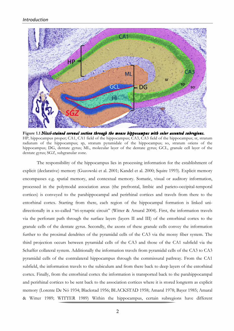
(Figure 1.1) the three laminae are called stratum (str.) oriens, str. pyramidale und str. radiatum, in the
DG (Figure 1.1) they are called hilus, granular cell layer (str. Granulosum; GCL) and molecular layer
(str. Moleculare; ML) (Andersen et al. 2006).
Introduction& &
2
Figure 1.1 Niss l - s ta ined corona l s e c t ion through the mouse h ippocampus wi th co lor a c c en t ed subreg ions . HP, hippocampus proper; CA1, CA1 field of the hippocampus; CA3, CA3 field of the hippocampus; sr, stratum radiatum of the hippocampus; sp, stratum pyramidale of the hippocampus; so, stratum oriens of the hippocampus; DG, dentate gyrus; ML, molecular layer of the dentate gyrus; GCL, granule cell layer of the dentate gyrus; SGZ, subgranular zone.
The responsibility of the hippocampus lies in processing information for the establishment of
explicit (declarative) memory (Guzowski et al. 2001; Kandel et al. 2000; Squire 1993). Explicit memory
encompasses e.g. spatial memory, and contextual memory. Somatic, visual or auditory information,
processed in the polymodal association areas (the prefrontal, limbic and parieto-occipital-temporal
cortices) is conveyed to the parahippocampal and perirhinal cortices and travels from there to the
entorhinal cortex. Starting from there, each region of the hippocampal formation is linked uni-
directionally in a so-called “tri-synaptic circuit” (Witter & Amaral 2004). First, the information travels
via the perforant path through the surface layers (layers II and III) of the entorhinal cortex to the
granule cells of the dentate gyrus. Secondly, the axons of these granule cells convey the information
further to the proximal dendrites of the pyramidal cells of the CA3 via the mossy fiber system. The
third projection occurs between pyramidal cells of the CA3 and those of the CA1 subfield via the
Schaffer collateral system. Additionally the information travels from pyramidal cells of the CA3 to CA3
pyramidal cells of the contralateral hippocampus through the commissural pathway. From the CA1
subfield, the information travels to the subiculum and from there back to deep layers of the entorhinal
cortex. Finally, from the entorhinal cortex the information is transported back to the parahippocampal
and perirhinal cortices to be sent back to the association cortices where it is stored longterm as explicit
memory (Lorente De Nó 1934; Blackstad 1956; BLACKSTAD 1958; Amaral 1978; Bayer 1985; Amaral
& Witter 1989; WITTER 1989) Within the hippocampus, certain subregions have different

Introduction& &
3
connectivities and functions. Moser and Moser suggested that the hippocampus is functionally different
along its dorsoventral (septotemporal) axis (M. B. Moser & E. I. Moser 1998; Fanselow & Dong 2010).
In rodents, spatial memory and episodic memory primarily appear to depend on the dorsal and not the
ventral hippocampus (E. I. Moser 1995; Davachi & Wagner 2002; Broadbent et al. 2004). On the other
hand, the ventral but not dorsal hippocampus is thought to mediate stress responses and therefore to
be involved in emotional behaviour (Henke 1990; Herman et al. 1998; Fanselow & Dong 2010; Segal et
al. 2010).
Figure 1.2 Scheme o f a mouse h ippocampus and sket ches o f corona l bra in s e c t ions . The sketches in the upper right corner display the location of the septal part oft he hippocampus in relation to the corpus callosum (emphasized in light gray), which still connects the right and left hemispheres at this point. In the lower right corner, the temporal hippocampus is displayed in relation to the corpus callosum, which at this point has split up into a left and a right part.
1.1.2 Cell&types&characteristic&for&the&different&stages&of&adult&Neurogenesis&&
Hippocampal adult neurogenesis is restricted to a relatively limited area, the subgranular zone
(SGZ) of the DG (red line in Fig. 1.2). Currently, adult neurogenesis is thought to consist of several
developmental stages (Kempermann et al. 2004; Ming & Song 2005) that are characterized by
morphologically distinct cells (inset in Fig. 1). In the SGZ of the DG two different types of adult neural
stem cells (NSCs) can be found (no. 1 and 2 in Figure 1.2 B). Type 1 cells represent radial glia-like cells,
and type 2 cells are non-radial neural progenitor cells (NPCs). Both are capable of self-replication but
possibly have a reciprocal lineage relationship. They proliferate and their offspring (No. 3 in Figure 1.2
B) differentiate and migrate into the granule cell layer of the DG, and show low proliferative potential.

Introduction& &
4
Later, these cells extend their dendrites toward the molecular layer of the DG, and their axons extend
into the hilus and the CA3 (No. 4-6 in Figure 1.2 B). Finally, the adult newborn neurons become
functionally integrated into the hippocampal network, receiving inputs from the entorhinal cortex and
sending outputs to hippocampal area CA3 and the hilus. However, not all newly formed cells survive,
many of them die within a few days or weeks after generation (Dayer et al. 2003).
Figure 1.3 The h ippocampus , one o f the major s i t e s o f adu l t neurogenes i s . A) Photomicrograph of a Nissl-stained coronal section displaying the mouse hippocampal formation with color accented subregions. The dentate gyrus (DG), which consists of the molecular layer (ML), the granule cell layer (GCL), and the hilus (Hi), as well as the hippocampus proper, which can be subdivided tangentially into the cornu ammonis (CA) sectors CA1, CA2 (not indicated), and CA3 are displayed. The subgranular zone (SGZ), a narrow layer of cells located between the GCL and hilus of the DG, contains adult progenitor cells and is accentuated with red. B) Schematic representation of cells in different adult neurogenesis stages. The generation of new neurons in the DG can be divided into at least 6 stages. Two different types of adult neural stem cells reside at the SGZ of the DG. Cell No. 1 represents type 1 (radial) neural stem cells, and cells No. 2a/b represent non-radial neural stem cells, which possibly have a reciprocal lineage relationship and are capable of self-replication. Cell No. 3 represents migratory progenitor cells, which show little proliferative action. Cells No. 4–6 represent different stages of postmitotic newborn cells fated to finally become mature granule cells. (Image was published in M. M. Lee et al. 2013).
1.1.3 Markers&for&different&stages&of&adult&Neurogenesis&
Typically, adult neurogenesis is detected via immunohistochemistry. This can either be
performed using 5-bromo-2-deoxyuridine (BrdU) antibodies following intraperitoneal (i.p.) BrdU
administration (Gratzner 1982) or using antibodies detecting different endogenous adult neurogenesis
markers without the need for stressful i.p. treatment of the respective animals (Bohlen und Halbach

Introduction& &
5
2011). BrdU can be given either once or a number of times, then incorporating into replicating DNA in
place of thymidine (also called deoxythymidine). Neurons that have incorporated the injected BrdU
during mitosis and which have subsequently differentiated into neurons, can be identified via double-
labeling with antibodies against neuron markers such as NeuN. The use of various antibodies detecting
endogenous adult neurogenesis allows the further division of adult neurogenesis into different
developmental stages, and allows the monitoring of neurogenesis at these different stages in the same
hippocampus using serial sections. This is possible since the various developmental stages correlate
with the expression of different markers (Bohlen und Halbach 2011). For the detection of cell
proliferation at the initial phase of the birth of new neurons (adult neurogenesis) antibodies against the
cell cycle marker Ki67 is commonly used (Kee et al. 2002). It should be mentioned, however, that
antibodies against Ki67 detect mitotic cells regardless of their identity or fate (Gerdes et al. 1991;
Scholzen & Gerdes 2000). Other very popular endogenous adult neurogenesis markers include i)
nestin, an intermediate filament expressed in many, if not all, NPCs, ii) NeuroD, a basic helix-loop-
helix protein and a differentiation factor for neurogenesis in diverse species, iii) the polysialylated
embryonic form of the neural cell adhesion molecule (NCAM), abbreviated to PSA-NCAM which has
been found in cells that seem to be NPCs related to neural stem cells, iv) TUC (TOAD [turned on after
division]/Ulip [UNC-33- like protein]/CRMP [collapsin response-mediator protein])-4 can be used as a
marker for early postmitotic neurons but seems also to be expressed in mitotic cells during
neurogenesis, and v) doublecortin (DCX), a protein that promotes microtubule polymerization, which
is present in migrating neuroblasts and young neurons (for review see: (Bohlen und Halbach 2011). As
DNA labeling by BrdU is potentially toxic, and therefore a confounding factor in some experiments,
the importance of these adult neurogenesis-related antibodies for further investigations of the adult
neurogenesis phenomenon is often underemphasized.

Introduction& &
6
Figure 1.4 Markers used for the de t e c t ion o f c e l l pro l i f e ra t ion and neurogenes i s in rodent h ippocampus t i s sue . At stages 1-5 of aN in the DG, different specific molecules are expressed by the newly formed cells. Stage 1: Proliferation of precursor cells. Stage 2: Differentiation of transiently amplifying cells. Stage 3: Migration of immature neurons into the granular cell layer. Stage 4 (Targeting): Immature and postmitotic extend their dendrites and axons. Stage 5: New granule cells are synaptically integrated into the network of the hippocampal formation. GFAP: Glial fibrillary acidic protein; Pax6: Paired box protein 6; NeuroD: neurogenic differentiation; PSA-NCAM: Polysialylated-neural cell adhesion molecule ; DCX: Doublecortin; Tuc4: TOAD [Turned On After Division]/ Ulip/CRMP 4; Tuj1: Neuron-specific class III β-tubulin; NeuN: neuronal nuclear antigen.
Although, the physiological and behavioral role of adult neurogenesis is still under debate
(Kempermann 2008; Deng et al. 2010; Burghardt et al. 2012; Glasper et al. 2012), nowadays it is well
accepted that it carries on from the late embryonic stage through to old age. The re-discovery of
hippocampal adult neurogenesis in the eighties and nineties by the groups of Goldman and Gould, as
well as the discovery of aN in humans by Eriksson in 1998 has led to an explosion of research in
neuroscience over the past two decades (Goldman & Nottebohm 1983; Barnea & Nottebohm 1996; P.
Eriksson et al. 1998; Gould, Reeves, et al. 1999b).
1.1.4 Positive&and&negative&modulators&of&Adult&Neurogenesis&
In the late 1990s, studies relying on the BrdU integration method, brought about a number of
discoveries regarding the regulation of adult neurogenesis. Among the positive regulators are enriched
environment, voluntary exercise, as well as learning and memory tasks (Kempermann et al. 1997;
Introduction& &
7
Gould, Beylin, et al. 1999a; van Praag et al. 1999; van Praag et al. 2005). In 1997, Kempermann and his
colleagues discovered that mice exposed to an enriched environment, namely a homecage equipped
with a running-wheel, toys and tunnels, displayed significantly more new neurons than mice housed in a
conventional cage (Kempermann et al. 1997). In addition, Henriette van Praag and co-workers
demonstrated that voluntary running alone also resulted in an increase of BrdU-positive cells, indicating
that physical activity can regulate hippocampal neurogenesis (van Praag et al. 1999). Other regulatory
factors are neurotransmitters such as serotonin, which can both enhance the proliferation of NPCs and
the production of new neurons (Gould 1999), gonadal hormones (Spritzer & Galea 2007; Galea et al.
2006; Spritzer et al. 2011), and trophic factors such as brain-derived neurotrophic factor (BDNF)
(Bekinschtein et al. 2011). Moreover, chronic antidepressant treatments markedly stimulate
hippocampal neurogenesis (Malberg et al. 2000; Païzanis et al. 2007). Negative regulators of adult
neurogenesis include binge alcohol exposure in adolescence (S. A. Morris et al. 2010) and oxidative
stress (Taupin 2010). However, the most prominent negative regulators of adult neurogenesis are old
age (Seki & Arai 1995; H. Kuhn et al. 1996; Kempermann et al. 1998; Amrein et al. 2011) and stress
exposure during all ontogenetic phases (Gould et al. 1998; Gould, Beylin, et al. 1999a; Lucassen,
Stumpel, et al. 2010b; Hanson et al. 2011; Mirescu et al. 2004). In addition to a genetic predisposition,
stress can be looked upon as a key vulnerability factor in the development of various neuropsychiatric
disorders. The intense regulation by stress (amongst other factors), strongly implicates adult
neurogenesis in a variety of pathophysiological mechanisms. Therefore, one of the topics under intense
research is the correlation between adult neurogenesis and the pathophysiology of psychiatric disorders
like fear and anxiety (Santarelli et al. 2003; Saxe et al. 2006; Revest et al. 2009), major depression
(Boldrini et al. 2009; Boldrini & Arango 2010; Lucassen, Stumpel, et al. 2010b; Anacker et al. 2011;
Snyder et al. 2011) and schizophrenia (Reif et al. 2006). In a study by Schmitt et al., in which 5-HTT
deficient mice where examined, showed that aN is influenced by the lack of the 5-HTT, even if aN
differences between 5-HTT-/- and +/+ litters could exclusively be revealed in older, and not in young
mice (Schmitt et al. 2007).
1.2 The(Serotonergic(System(
The monoamine Serotonin is derived from the essential amino acid L-Tryptophane. It is
synthesized in two steps. First tryptophane hydroxylase (TPH; in the brain TPH2) metabolizes L-
Tryptophane into 5-Hydrox-L-tryptophan (5-HTP), which is then decarboxylized into Serotonin (or 5-
Hydroxtryptamine; 5-HT) by 5-Hydroxytryptophan decarboxylase (Kandel et al. 2000).

Introduction& &
8
Cell bodies of serotonergic neurons in the mammalian brain are found along the midline of the
brain stem from the midbrain to the medulla oblongata. Here Serotonergic neurons are clustered into
nine nuclei numbered B1-9 on a rostrocaudal axis (Kandel et al. 2000; K. P. Lesch & Waider 2012).
Raphe neurons in the B1-B3 cell groups along the midline of the caudal medulla oblongata send
descending projections to the motor and autonomic systems in the spinal cord. The raphe magnus
nucleus (B4) at the level of the rostral medulla oblon projects to the spinal dorsal horn and is thought
to modulate the perception of pain. The serotonergic groups in the pons and and midbrain (B5-B9)
include pontine, dorsal (B6, B7) and median raphe nuclei (B9, B8, and B5) and project to virtually the
whole of the forebrain. Serotonergic pathways play important regulatory roles in hypothalamic
cardiovascular and thermoregulatotry control and modulate the responsiveness of cortical neurons, and
influence sensory processing, cognition, emotional states, circadian rhythms, food intake, and
reproduction (Kandel et al. 2000; K. P. Lesch & Waider 2012).
1.2.1 The&Serotonin&Transporter&&
Ever since the 1960ies, brain monoamines, and serotonin in particular, have been suspected to
be linked to different psychiatric conditions such as depression, anxiety, antisocial behaviour, and
dependence (BUNNEY 1965; COPPEN 1967; Maas 1975; Heninger et al. 1996). Many studies have
implicated genetic variability in two particular genes, both of which are responsible for the termination
of the serontonergic signal. The first one, the enzyme monoamine oxidase (MAO) which accomplishes
termination through catabolism via oxidative deamination by (Frazer & Hensler 1999). The other and
probably the most important one is the 5-HT Transporter (5-HTT) which is responsible for the re-
uptake of Serotonin into the presynapse (Frazer et al. 1999; Frazer & Hensler 1999). It is one of the
transporter molecules in neurons which support temporal and spatial buffering of neurotransmitter and
neurotransmitter metabolite concentrations and which are responsible for cycling and recycling of
transmitter molecules (Uhl & Johnson 1994). By removing 5-HT from the synaptic cleft the 5-HTT
fine-tunes 5-HT neurotransmission and thus determines the magnitude and duration of postsynaptic
receptor-mediated signaling (Blakely et al. 1994; Uhl & Johnson 1994). The 5-HTT is a Na+/Cl-
dependent transport protein and is made up of 630 amino acids with 12 transmembrane domains. It
was first cloned from rat brain in 1991 by Blakely and colleagues (Blakely et al. 1991). Two years later
Ramamoorthy (Ramamoorthy et al. 1993) and colleagues were able to identify an identical human
placental 5-HTT, and finally in 1994 Lesch et. al isolated human 5-HTT from brain and blood platelets
(K.-P. Lesch et al. 1994). The human 5-HT gene was mapped to human chromosome 17q1 1.2
(Ramamoorthy et al. 1993) and is organized in 14 exons spanning ~ 35kb (K.-P. Lesch et al. 1994). The
5-HTT regulates the concentration of 5-HT in the extracellular space, therefore affecting the receiving

Introduction& &
9
neurons as well as 5-HT turnover in the presynapse. Additionally, it is known to be a principal target
for the most common anti-depressants such as the selective serotonin reuptake inhibitors (SSRIs),
psychostimulants, as well as drugs of abuse including MDMA (“ecstasy”) and cocaine (Wellman et al.
2007; Blakely et al. 1991; Blakely et al. 1994; Uhl & Johnson 1994; K. Lesch 2005; Narayanan et al.
2011; Bengel et al. 1998).
The 5-HTT has long been implicated in a variety of central nervous system (CNS) disorders,
including depression (Tuomisto & Tukiainen 1976; Meltzer et al. 1981; Stanley et al. 1982). Moreover,
altered 5-HTT expression in various types of psychiatric disorders has been well documented and
indicates the importance of 5-HTT expression in maintaining normal brain function (Murphy et al.
2004). Molecular genetic studies in humans have revealed several 5-HTT gene variations which
comprise a repeat length-polymorphism in the transcriptional control region (5-HTT linked
polymorphic region, 5-HTTLPR), resulting in a short (S) and a long (L) allele. The S-allele entails lower
5-HTT mRNA/protein levels and is shown to be associated with personality traits of negative
emotionality including anxiety, depression and aggressiveness (Lewejohann et al. 2010; Gardner et al.
2009; K. Lesch et al. 1996; Holmes 2008; K. Lesch & Mossner 1998; Lanfumey et al. 2008; Canli & K.
P. Lesch 2007; Lowry et al. 2005).
1.2.2 The&Serotonin&Transporter&KnockYout&Mouse&
For a better understanding about how an altered serotonergic system due to different 5-HTT
gene variants influences emotionality and behavior - and since rodents unlike humas do not posess a
length variation in the promotor region - a 5-HTT deficient mouse line was generated by disrupting the
gene via homologous recombination (Bengel et al. 1998).
With the help of this method combined with subsequent crossbreeding, the different genotypes
are generated. Wildtype (5-HTT+/+) mice are genetically unchanged and posess two active 5-HTT
genes. Heterozygous (5-HTT+/-) animals display a reduction of 5-HTT expression by 50% and
homozygous 5-HTT-knockout (5-HTT-/-) mice, in which the 5-HTT gene has been completely
inactivated, cannot produce any 5-HTT molecules (Bengel et al. 1998). The result of the elimination (5-
HTT-/-) or reduction (5-HTT+/-) of the 5-HTT is that less serotonin can be transported back into the
pre-synapse. Thus intracellular and extracellular 5-HT levels are changed from early stages of
development onwards. In more detail, 5-HTT-/- mice show a 5 up to 13-fold increase of 5-HT
concentrations in the extracellular space as evidenced by in vivo microdialysis in different brain regions
including prefrontal cortex, striatum and substantia nigra (Fabre et al. 2000; Shen et al. 2004; Mathews

Introduction& &
10
et al. 2004). In contrast, overall brain tissue levels of 5-HT are significantly (60-80%) reduced (Bengel et
al. 1998).
This lifelong reduced or absent 5-HTT function is associated with a complex series of adaptive
changes at the neurochemical level such as the compensatory increased expression of the organic
cationic transporter 3 in the hippocampus of 5-HTT-/- mice, the expression and function of different
receptors and various neuroplasticity phenomena such as higher spinogenesis in the amygdala of 5-
HTT-/- compared to 5-HTT+/+ mice (Schmitt et al. 2003; Schmitt et al. 2007; Nietzer et al. 2011;
Fabre et al. 2000), for review see (Murphy & K.-P. Lesch 2008). In addition to baseline differential gene
expression of 5-HTT-/- mice, 5-HTT-/- and 5-HTT+/+ animals react differently to acute stress:
While 5-HTT+/+ mice immediately after being exposed to forced swimming for one minute were
found to express genes in the amygdala that are related to neuroplasticity and adaptation to stressors, 5-
HTT-/- express genes more related to chronic stress and pathophysiology (Hohoff et al. 2013).
Furthermore, 5- HTT deficient mice exhibit a changed behavioral phenotype. For instance, when
characterizing 5-HTT-/- mice for anxiety-related behaviors via a battery of behavioral test consisting of
the elevated plus maze, light-dark exploration test, emergence test as well as the open field test, Holmes
and his colleagues found that 5-HTT-/-mice showed increased anxiety-like behavior and inhibited
exploratory locomotion as compared to their +/+ littermates (Holmes et al. 2003). In an earlier study
Holmes had already detected that mice lacking the 5-HTT are less aggressive than 5-HTT+/+ mice in
the resident intruder test (Holmes, Murphy, et al. 2002a). Later this finding was and added onto by
Lewejohann et al., when exposing mice of all three genotypes of the 5-HTT knock-out line to an
enriched environment, which allowed the animals to show a wide variety of spontaneous behavioural
patterns. In addition to the reduced aggressive behavior, he also found reduced locomotion, and
increased socio-positive behaviour in 5-HTT-/- mice compared to their 5-HTT+/+ and 5-HAT+/-
littermates (Lewejohann et al. 2010). Another characteristic of the 5-HTT-/- genotype along with
reduced locomotor activity is late-onset obesity (Üçeyler et al. 2010). Moreover, a number of studies
have already shown that anxiety-and/or depression-related behavior in 5-HTT deficient mice is
exacerbated by stress exposure (Wellman et al. 2007; Heiming et al. 2009; Jansen et al. 2009).
1.2.3 The&Serotonergic&System&and&Adult&Neurogenesis&
Most of the serotonergic neurons, which terminate in the hippocampal formation, origninate
from the median raphe nuclei. In rats it is well established that serotonergic neurons of the median
raphe nucleus predominantly project to the dorsal part of the hippocampal formation rather than in its
ventral part (Vertes et al. 1999). Additionally, within the dorsal (anterior/septal) hippocampal formation

Introduction& &
11
median raphe-neurons terminate within the granule cell layer of the dentate gyrus DG. Within the DG
these projections are closely confined to the SGZ with more representations in the suprapyramidal
(upper) blade than in the infrapyramidal (lower) blade of the DG (Vertes et al. 1999). When taking into
account that Serotonin is a known positive modulator of aN in the hippocampus, it does not come as a
surprise, that Jason S. Snyder and co-workers have found that aN in the granule cell population as a
whole is higher in the DG of dorsal than in the ventral part of the hippocampus (Snyder et al. 2009).
1.3 Stress(
Stress has been defined as a condition where an environmental demand exceeds the natural
regulatory capacity of an organism, particularly in situations that include unpredictability and
uncontrollability (Koolhaas et al. 2011). According to the classical concept by Hans Selye, all living
organisms strive towards a dynamic equilibrium, which is called homeostasis and which is threatened by
certain physical (e.g. noise, bright light, water deprivation, food deprivation) and psychological events
that are known as ‘stressors’ (reviewed by (de Kloet et al. 2005). Selye distinguishes between positive
stressors known as ‘eustress’ (e.g. wedding, birth of a child) and negative stressors known as
‘distress’(Selye 1975). Another distinction is made between the severity (mild and severe) the
predictability (predictable and non-predictable) and the duration of stressors (acute or chronic)
(Koolhaas et al. 2011). Among the more severe stressors, or traumatic experiences are for instance
combat, rape, childhood maltreatment or the sudden death of someone close. Such severe acute
stressors are regarded as significant causal agents in the etiology of a number of neuropsychiatric
disorders such as schizophrenia (Wahlberg et al. 1997; van Os et al. 2010), but predominantly in the
development of anxiety disorders and depression (Caspi et al. 2003; van Praag et al. 2002; Pittenger &
Duman 2008; Saveanu & Nemeroff 2012). Mild chronic stress in humans is usually experienced in a
social context (e.g. in the workplace, in the family, social dominance, sexual arousal etc.). In order to
investigate the impact such chronic mild stressors can have, especially when they are unpredictable,
scientists have established an animal model called the unpredictable chronic mild stress paradigm.
1.3.1 Unpredictable&Chronic&Mild&Stress&
Chronic, low grade stressors or “strains” have long since been suspected to be a causal factor of
depression (Kanner et al. 1981; Willner et al. 1987). In order to investigate this asumption further, a
paradigm, originally discribed by Katz and co-workers and refined by Willner et al., was established, in
which rats were chronically subjected to a variety of unpredictable stressors (Katz et al. 1981; Katz
1982; Katz 1984; Willner et al. 1987; Willner 1997). The requirement for this animal model of course
was to simulate characteristic symptoms of depression. There are two core symptoms for the diagnosis

Introduction& &
12
of major depression, namely depressed mood and anhedonia, defined as the loss of interest or pleasure.
Since however, depressed mood cannot be modelled in animals, but anhedonia can, anhedonia is the
essential symptom to simulate depression realistically (Willner et al. 1992). The chronic mild stress
(CMS) model discribed by Willner et. al (1992), which involves the chronic sequential application, of a
variety of extremely mild stressors to rats, has been found to do so, as during 1-3 weeks exposure to
CMS, rats display a reduction in sensitivity to rewards, which is usually monitored by a decrease in their
consumption of palatable weak sucrose solution (Willner et al. 1992). Most importantly however, the
effect evoked by this paradigm can be reversed by the administration of antidepressant drugs (Willner
et al. 1987; Willner et al. 1992; Willner et al. 1996). Since then the validity oft he CMS model has also
been confirmed for mice (Monleon et al. 1995). Moreover, animal chronic mild stress has been found
to be characterized by endocrine changes similar to those seen in in depressed clinical patients (Heuser
et al. 1994; Checkley 1996). Besides anhedonia, other depressive-like behavior as well as anxiety-like
behavior could be revealed in behavioral tests of CMS-treated mice, the outcome however strongly
depending on the genetic background of the treated mouse-strain (Mineur et al. 2006). Besides effects
that CMS has on behavior, it is also known to decrease the cell proliferation and the survival of new-
born cells in the dentate gyrus (Vollmayr et al. 2007).
1.3.2 HPA&axis&and&Glucocorticoid&signaling&
The experience of a stressful situation has a great impact on the hypothalamic-pituitary–adrenal
(HPA) axis which is thus stimulated. HPA axis activity is governed by the secretion of corticotropin
releasing hormone/factor (CRH/CRF) and vasopressin from the hypothalamus, which in turn activates
the secretion of adrenocorticotrophic hormone (ACTH) from the pituitary to stimulate the secretion of
the glucocorticoids (cortisol in humans and corticosterone in rodents) from the adrenal cortex.
Released glucocorticoids then interact with their receptors in multiple body compartments including
the brain (e.g. hippocampus, hypothalamus) where they serve a vital function in feedback inhibition of
their own secretion. An important brain region involved in the neurocircuitry of stress is the
hippocampus (Squire et al. 1992; Squire 1993; Senba & Ueyama 1997; Broadbent et al. 2004).
Glucocorticoids regulate neuronal survival, neurogenesis and hippocampal volume, as well as the
acquisition of new memories and the emotional appraisal of events (Pariante & Lightman 2008).
HPA hyperactivity is regarded as causally linked to depression and to the modes of action of
antidepressants (Holsboer 2001). Altered feedback inhibition in depressed patients resulting in HPA
hyperactivity was demonstrated by use of the combined dexamethasone (DEX)/ CRH test, a refined
laboratory test for psychiatric disorders. For this test, patients are administered DEX the night prior to

Introduction& &
13
testing and then CRH the following afternoon (Heuser et al. 1994). Measurement of plasma cortisol
and ACTH levels before, during and after CRH administration revealed that depressed patients release
significantly more cortisol and ACTH after DEX and a CRH challenge in comparison with age-
matched controls. This so-called DEX/CRH-test phenomenon supports the assumption that
psychiatric patients are prone to blunted glucocorticoid feedback regulation during the acute illness
episode. Diminished glucocorticoid receptor (GR) expression or function has been postulated as
causative factor (Heuser et al. 1994). Based on these findings, Ridder et al. generated GR-heterozygous
mutant mice (GR+/-), which have a 50% reduction of GR protein levels in the brain (Ridder,
Chourbaji, Hellweg, Urani, Zacher, Schmid, Zink, Hörtnagl, Flor, Henn, Schütz & Gass 2005a). Similar
to depressed patients, these mice display reduced feedback inhibition of the HPA axis and a
pathological DEX/CRH test in which higher levels of circulating glucocorticoids could be measured in
response to the CRH challenge compared to GR- wildtype littermates.
1.3.3 Glucocorticoids&and&aN&
Elevated levels of glucocorticoids, as for instance found in chronically stressed individuals,
stimulate hippocampal glutamate release (Moghaddam et al. 1994) which in turn can diminish stem cell
proliferation in the adult DG (Gould & Tanapat 1999). Thus, it does not come as a surprise that GR+⁄-
mice display significantly less BrdU-positive cells in the DG compared to wildtype mice (Kronenberg et
al. 2009). Behaviorally, GR+⁄- mice show increased learned helplessness, a well-established analogue of
depression-like behaviour in mice (Ridder, Chourbaji, Hellweg, Urani, Zacher, Schmid, Zink, Hörtnagl,
Flor, Henn, Schütz & Gass 2005b). Thomas and co-workers were able to show, in their study published
in 2007, that acute psychosocial stress leads to a reduced number of new neurons in the DG of rats.
Interestingly, stem cell proliferation itself remained unaltered, whereas short- and long-term survival of
the new-born cells was decreased (Thomas et al. 2007). This implicates that acute social stress has a
sustained impact on the integration of new-born cells into the hippocampal neural network by
diminishing their chance of survival. On the other hand, a recent study showed that coping with
intermittent social stress, which is an essential aspect of living in complex social environments,
stimulates adult neurogenesis in adult monkeys (Lyons et al. 2010). On the basis of this finding, one
could speculate that stress coping follows similar mechanisms as living in an enriched environment,
which is known to prompt adult neurogenesis (Kempermann et al. 1997).
HPA axis dysregulation and the adverse effect of increased glucocorticoid concentrations on
adult neurogenesis may be one out of several possible causes of the well documented phenomenon of a
decreased hippocampal volume in patients with mood disorders. Several forms of stress, and especially

Introduction& &
14
repeated periods of stress, are suggested to cause a reduction of hippocampal volume. Among these are
traumatic life events and early life stress such as childhood maltreatment which are often found to be a
key component in the life history of patients with mood disorders such as post-traumatic stress-
disorder and major depression (M. E. Smith 2005; Pittenger & Duman 2008; Teicher et al. 2012).
1.4 Learning,(Memory(and(the(plastic(brain(
In order to form memories, to encode them and store them, the brain acts not as a passive
recorder of experiences, but as a dynamic system that creates and re-writes information. Originally it
was thought that long-term memories are stable and essentially “hardwired”. Nowadays however it is
believed that memories are encoded as dynamic spatio-temporal patterns of synchronized cellular
activity within widespread neural networks and that this dynamic, reverberating activity progressively
results in altered patterns of connectivity among the co-activated neurons. Moreover, we know that the
mechanisms of plasticity in neural circuits that encode and store long-term memories are dynamic and
ongoing throughout the life of a memory (reviewed by (Bruel-Jungerman et al. 2007)). Different types
of learning are required to store different types of information. Implicit (non-declarative) memory
contents for instance require procedural learning, associative learning (important for emotional
responses and the basis for classical conditioning) or non-associative learning which describes the most
simple form of learning found in the behavior of invertebrates (gill-withdrawal reflex of Aplysia) and in
vertebrate reflexes such as fear responses and the eye blink (Bailey & Chen 1983; Sanes & Ison 1983;
Schacter 1987; Graf & Schacter 1985). Explicit (declarative) memory contents like facts and events on
the other hand require contextual and/or spatial learning which take place in the hippocampus (Kandel
et al. 2000).
1.4.1 Spatial&learning&and&spatial&learning&tests&for&rodents&
Spatial learning and memory is a field that was intensely investigated in the 1970ies and 80ies by
researchers like Morris, Nadel, O’Keefe and Barnes (reviewed by (Nadel 1991)). Their research with
rodents led to the cognitive map theory of hippocampal function, a theory about memory for spatial
layouts and the ways in which animals use such a memory system for adaptive behavior in the world
(Nadel 1991).
The two most common spatial learning tests for rodents, the Barnes Maze (BM) (Barnes 1979)
and the Morris Water Maze (WM) (R. Morris 1984) are similar tasks as they both measure the ability of
a mouse to learn and remember the location of a target zone using a configuration of distal visual cues
located around the testing area (Rudy et al. 1987; Harrison et al. 2006) and have proven useful in

Introduction& &
15
detecting hippocampus-dependent cognitive deficits (Pompl et al. 1999). The procedures differ with
regard to the motivation to learn the spatial task and therefore presumably bear different challenges for
the tested mice. In addition, these two tests are suspected to vary in the stress they induce in the tested
animal (Harrison et al. 2009).
The BM takes advantage of the natural preference of rodents to avoid brightly lit open surfaces.
Therefore no additional aversive stimuli are needed (Barnes 1979). The apparatus usually consists of a
brightly lit circular platform that was elevated app. 120 cm above the floor. Twelve holes are arranged
in a clock-face manner close to the edge of the platform. All but one of the holes are closed by short
tubes. The residual hole is connected to the home cage of the tested animal via a wire mesh tunnel. The
home cage is placed directly beneath the center of the platform to ensure that the mouse will not be
able to see or smell the correct hole when it is being placed on the platform. (Barnes 1979; Lewejohann
et al. 2010; Karabeg et al. 2013; Poucet et al. 1991; Bach et al. 1995; Pompl et al. 1999).
The principle of the WM task is based on the animal’s desire to escape from the water (R.
Morris 1984). Typically a rodent is placed into a small pool of water (usually 1 to 1.8 meter in diameter
and 60 centimeters deep), which contains a translucent escape platform hidden a few millimeters below
the water surface. Visual cues, such as colored shapes, are placed around the pool in plain sight of the
animal. A sidewall above the waterline prevents the animal from being distracted, and from climbing
out from the pool. When released, the subject swims around the pool in search of an exit while various
parameters are recorded, including the time spent in each quadrant of the pool, the time taken to reach
the platform (latency), and total distance traveled. Escape from the water reinforces a desire to quickly
find the platform, and on subsequent trials (with the platform in the same position) subjects are able to
locate the platform increasingly rapidly. This improvement in performance is assumed to be a result of
learning and memory.
That the WM is more anxiogenic or stressful, respectively than the BM had been suggested for
a long time, until finally in 2009, Harrison and co-workers were able to confirm this suggestion
impirically (Holmes, Wrenn, et al. 2002b; Pompl et al. 1999; Harrison et al. 2009). In their study, carried
out with mice matched for performance on commonly-used anxiety tasks, they found that WM training
induced greater increases in plasma corticosterone than did BM training, assessed 30 min. after the final
session. They also detected that spatial learning was inversely correlated with corticosterone levels in
the water maze but not the Barnes maze, which led them to the conlusion that performance on the
water maze may be more affected by test-induced stress even within wild-type subjects of the same age
and gender (Harrison et al. 2009).

Introduction& &
16
In this context it is important to mention that hippocampus dependent learning has long since
known to be influenced by stress. While transient mild stress for instance can enhance learning and
memory(Luine et al. 1996), chronic or severe stress has been shown to severely impair hippocampus-
dependent memory in experimental animals (Conrad et al. 1996; Roozendaal et al. 1998; Diamond et al.
1999; R. Sapolsky & Romero 2000; McEwen & R. M. Sapolsky 1995). A similar effect can be reached in
rats via extended or high-dose treatment with glucocorticoids (Bodnoff et al. 1995; Roozendaal et al.
1998). But also humans display specific impairments of hippocampus-dependent explicit memory after
treatment with glucocorticoids (Newcomer et al. 1999; de Quervain et al. 2000) as well as after stress
(Shors 2006).
1.4.2 Brain&plasticity&
Brain plasticity in learning & memory comprises four known mechanisms. The first one is
synaptic strengthening or Long-Term Potentiation (LTP), which occurs during learning (Bruel-
Jungerman et al. 2007). By means of this mechanism, memories are stabilized and stored as
modifications of synaptic strength within the existing neuronal circuits in an activity-dependent way
(Hebb 1949; Bliss & Lomo 1973). The second mechanism, known as Long-Term Depression (LTD) is
a form of activity-dependent long-lasting weakening of synaptic strength and my reflect a mechanism
for forgetting or for weakening unused connections, which has been suggested to also play a role in
play a role in learning (Bruel-Jungerman et al. 2007; Bock & Braun 1999). The third mechanism is
Synaptogenesis and Synapse Remodeling, which is measured by means of changes in the number of
spines or the spine density. It was found for instance in rat hippocampus 24 hrs after training using the
trace eyeblink conditioning paradigm, an associative learning task that requires the hippocampus for
acquisition (Leuner & Gould 2010). The last mechanism is neurogenesis, the birth and growth of new
neurons (see above), which has been shown not only to be influenced by learning and memory
processes in the adult (Gould, Beylin, et al. 1999a) but also to play an important role in spatial learing in
rodents (Clelland et al. 2009). Elizabeth Gould for instance who had trained and tested rats in the
Morris Water Maze, found out that place training in the Water Maze, where the animals had to find the
location of a submerged platform increased the number of BrdU positive cells in the DG (Gould,
Beylin, et al. 1999a). Using adult mice in which hippocampal neurogenesis was ablated, Clelland et. al
found specific impairments in spatial discrimination in a spatial navigation radial arm maze task
(Clelland et al. 2009).
Bliss and Lømo discovered LTP when they examined the after-effects of repetitive stimulation
on the perforant path fibers to the dentate gyrus of the rabbit hippocampi. They found that there was

Introduction& &
17
an enduring increase in synaptic strength at dentate gyrus granule cell synapses (Bliss & Lomo 1973).
This property, which is shared by neurons in many different cortical and subcortical regions is
associated with rapid gene regulation intiated by Immediate Early Genes (IEGs) (Bruel-Jungerman et al.
2007). For instance, learning processes involving patterned synaptic stimulation are followed by rapid
expression of IEGs; (Kubik et al. 2007). These IEGs are also implicated as markers for neuronal
activity as answer to stress (Weinberg et al. 2007).
IEGs may be categorized into two functional classes: (1) regulatory transcription factors which
control the transcription of other “downstream” genes, and (2) effector IEGs, which directly influence
cellular functions (Kubik et al. 2007). One of the effector IEGs is activity-regulated cytoskeleton-
associated protein (Arc; also known as Arg3.1), an indicator of synapse specific modifications during
neuronal plasticity (Link et al. 1995). In granule cells of the hippocampus its expression is strongly
induced by NMDA receptor-dependent synaptic stimuli (Link et al. 1995; Lyford et al. 1995) and Arc
mRNA and protein is localized to dendrites and spines after activity (Lyford et al. 1995; Steward et al.
1998; Rodriguez et al. 2005). Arc not only plays multiple roles in synaptic regulation. For instance, Arc
induction is required for late LTP and memory consolidation (Guzowski et al. 2000; Plath et al. 2006;
Messaoudi et al. 2007). It was also found to underly a negative feedback control mechanism involving
AMPA receptors (AMPAR)(V. R. Rao et al. 2006). First, synaptic activity triggers Arc expression and
induces AMPAR surface delivery, then, Arc facilitates the endocytosis and consequentially the
downregulation of AMPARs through its interaction with endocytic proteins endophilin 3 and dynamin
2 (Chowdhury et al. 2006; Rial Verde et al. 2006). The decreased NMDA/ AMPA ratio closes the loop
by inhibiting excessive Arc transcription. Arc has thus been found to play a pivotal role in LTD
(Waung et al. 2008) and homeostatic plasticity (Shepherd et al. 2006). In summary, the function of Arc
lies in memory consolidation and re-consolidation, which has been demonstrated in behavioral tasks
such as spatial learning, fear conditioning, object recognition and taste aversion (Plath et al. 2006).

Introduction& &
18
Figure 1.5 Scheme o f the main mole cu lar s t eps invo lv ed in synapt i c s t r eng then ing/ LTP Receptor and kinase cascade activation results in synaptic receptor modification and nuclear transcription of immediate early genes. Some encode synaptic proteins, whereas others encode inducible transcription factors or regulatory transcription factors (RTFs) such as cFos, which in turn activates transcription of effector genes, leading to synthesis of the corresponding proteins required for persistent cell modification. LTP = long-term-potentiation. (Image adapted from(Bruel-Jungerman et al. 2007)).
The regulatory transcription factor cFos on the other hand is also called an inducible
transcription factor because its rapid expression is controlled by pre-existing transcription factors such
as cyclic-AMP-response-element-binding protein (CREB) (Herdegen & Leah 1998). It forms dimers
with c-Jun, JunB and JunD but no homodimers (w/ c-fos) and can be part of the AP-1 transcription
complex that binds to regulatory DNA sequences (Chiu et al. 1988; Rauscher et al. 1988). As part of
the AP-1 transcription complex, cFos is responsible for transcription of tyrosine hydroxylase gene
(Herdegen & Leah 1998). As tyrosine hydroxylase is the enzyme responsible for catalyzing the
conversion of the amino acid L-tyrosine to L-3,4-dihydroxyphenylalanine (L-DOPA), cFos can be
regarded as an upstream modulator of the dopaminergic, adrenergic and noradrenergic systems. In
conclusion, cFos can be used as an indicator of recent increases in neuronal excitation and cellular
processes that support neuroplasticity (VanElzakker et al. 2008).

Introduction& &
19
1.5 Aim(of(this(thesis(
In the following chapters, two studies, using animals of the 5-HTT-/- mouse-line will be
introduced.
The aim of the first study, which in the following will be called the “Spatial Learning study”,
was to evaluate whether mice with altered levels of brain 5-HT as a consequence of 5-HTT deficiency
perform differently in two spatial memory tests, the WM and BM, prospectively differing in
aversiveness, and which role stress plays in this context. Additionally, the brains of the tested mice were
to be examined in order to find neuronal correlates of possible behavioral differences. Our co-
operation partner at the Otto Creutzfeldt Center for Cognitive and Behavioral Neuroscience at
University of Münster, Lars Lewejohann and his Master Student Sandra Grauthoff had carried out the
behavioral part of the Spatial Learning study. My part of the study was to examine the corticosterone
levels and the brains of these mice via a quantitative immuno-histochemistry study in which I examined
IEG expression and aN in the brains of these mice.
The purpose of the second study, the “Chronic Mild Stress Study” was to investigate whether
mice with altered levels of brain 5-HT as a consequence of 5-HTT-deficiency display differing
depression-like and anxiety-like behavior after being subjected to a chronic mild stress (CMS) paradigm
and again to see if there are neuronal correlates for possibly different behavior. Sandy Popp of the
behavioral unit of the Division of Molecular Psychiatry, Department of Psychiatry, Psychosomatics and
Psychotherapy, University of Würzburg had carried out the CMS paradigm as well as behavioral testing.
My function in this study was to examine the corticosterone levels and to evaluate aN in a quantitative
immuno-histochemistry study in the brains of the mice that had been subjected to the CMS paradigm.
Additionally, I was interested in working out the influence of a battery of behavioral tests on the effects
of a CMS- and a Control-group of mice.

20
2 Material&and&Methods&
2.1 Spatial(Learning(Study(
2.1.1 Animals&and&behavioral&testing&
Studied mice were bred and behaviorally tested by our co-operation partner by Lars
Lewejohann and his master student Sandra Grauthoff from the Otto Creutzfeldt Center for Cognitive
and Behavioral Neuroscience at University of Münster, Germany. All animals originated from the
internal stock of 5-HTT deficient mice bred at the Department of Behavioural Biology at the
University of Münster. The original breeding stock had been obtained from the Department of
Psychiatry, Psychosomatics and Psychotherapy at the University of Würzburg, Germany, where these
mice had been back-crossed on a C57BL/6 background (Bengel et al. 1998). Genotyping was
accomplished using tissue samples to extract genomic DNA amplified by PCR. Subsequently, 5-HTT
genotypes were identified by gel electrophoresis of DNA- fragments of either 225 bp (5-HTT+/+),
272 bp (5-HTT −/−) or both (5-HTT+/−). The 6 months old male mice of all three genotypes 5-HTT
knockout (-/-), 28 5-HTT heterozygous (+/-), and 28 5-HTT wild-type (+/+) were tested behaviorally
by means of either the Barnes maze (BM; two trials per day) test or by using the Morris water maze
(WM; three trials per day). In the BM, the mice performed two trials per day with a maximum duration
of 300 seconds on five consecutive days. The escape hole remained constant for any given animal over
the first four days of testing. The latency to find the correct hole, total number of errors, number of
stops (zero velocity for 1s) and the path length was recorded by an automated tracking system
(Lewejohann 2004). An error was defined as searching a hole that did not lead to the escape tunnel. At
day five, the escape tunnel was switched to a different, randomly chosen hole as probe trials (trials 9
and 10).

Material&and&Methods& &
21
Figure 2.1 Scheme o f a typ i ca l Barnes Maze s e tup The setup consists of a circular platform with twelve holes, the animals homcage underneath the platform and a tunnel leading to the homecage, which is connected to one of the holes. Visual cues are mounted around the platform, for the mouse to see and a lamp, which brightly lights the area of the platform.

Material&and&Methods& &
22
In the WM mice were tested at three trials per day over five consecutive days. Each trial had a
maximum duration of 60 s and started by gently placing the mouse into the water with its head towards
the pool wall on any of the three quadrants without the platform. If an animal found the platform
within the 60 s, it was left to stay on the platform for 10 s. In cases the animals did not find the
platform they were gently led to the platform by the experimenter (Sandra Grauthoff). Between the
trials, all mice were placed back in their home cages using a spoon-net in order to avoid direct contact
with the experimenter. The inter trial interval on each day was 15 minutes. On the last day, the platform
was placed in a different quadrant and subsequently probe trials (trials 13, 14 and 15) were performed
in order to measure the ability of the mice to generalize the task by re-learning a new position. All trials
were tracked automatically by a digital tracking system assessing path-length, swimming speed, stops,
and latency to escape from the water.
Figure 2.2 Scheme o f a typ i ca l Morr i s Water Maze s e tup The setup consists of a pool filled with water, a hidden platform, which the mouse is supposed to find, visual cues mounted around the pool, fort he mouse to see and a lamp, which brightly lights the area oft he pool.
As the procedures differ with regard to the motivation to learn the spatial task they presumably
bear different challenges for the tested mice. The experimenter was blind to the 5-HTT genotype of the
mice tested in order to avoid any bias. In addition to the BM and WM group, naïve mice (left
undisturbed) with no further testing formed a control group.
Material&and&Methods& &
23
2.1.2 Glucocorticoid&analysis&
In order to further evaluate the effects of the two different learning tests (BM and WM) on
stress physiology, trunk blood of mice from two separate cohorts, altogether consisting of 150 mice (45
5-HTT-/-, 55 5-HTT+/-, 50 5-HTT) was taken. The first cohort was subjected to a single trial and the
second one to three (BM) or four (WM) trials, the last of which was taking place on the second day.
The learning tests were conducted as described above except for the reduced number of trials. 15 min
after the start of the final trial the mice were anaesthetized with isoflurane and decapitated. In the first
hormone cohort, naïve mice were used as an additional group to determine baseline corticosterone
values. As mice were accumulated over time, the glucocorticoid analysis of the two hormone cohorts
were carried out at two different time points. Samples from all groups were taken at the same time of
day. Trunk blood was collected using heparinized capillaries, centrifuged for 5 min at 14800 × g and
plasma was stored at -20°C for later evaluation. Plasma corticosterone concentrations were determined
by enzyme linked immunosorbent assay (EIA, DE4164, Demeditec Diagnostics GmbH, Kiel,
Germany) according to the manufacturer's recommendations. All standards, samples, and controls were
run in duplicate concurrently. The intra- and inter-assay coefficients of variation were 3.3% and 6.0%,
respectively.
2.1.3 Quantitative&immunohistochemistry&
Brain Tissue Sixty hours after the last learning trial all mice of the behavior cohort (BM, WM, CONT) were
deeply anaesthetized with isoflurane and sacrified. Mouse brains were fixed for up to 72 h in 4%
paraformaldehyde (PFA, dissolved in PBS, pH 7.5). Fixed brains were transported to our laboratory in
Würzburg, where they were transferred to 10 and 20% sucrose in PBS. Brains were then frozen in pre-
cooled isopentane and stored at -80°C. There were a total of six experimental groups of mouse brains
5-HTT+/+, C (n = 7); 5-HTT+/+, BM (n = 10); 5-HTT+/+, WM (n = 10); 5-HTT-/-, C (n = 7); 5-
HTT-/-, BM (n = 10); 5-HTT-/-, WM (n = 9). Serial coronal sections were cut at 50 μm on a freezing
microtome. These free-floating sections were collected in a one-in-eight series, placed in 24-well plates
each well filled with 1xTBS.
Immunohis tochemistry In order to process brain slices for immunohistochemistry, they were washed three times for 5
min with 1xTBS and subsequently incubated with 0.6% hydrogen peroxide in TBS for 30 min to inhibit
endogenous peroxidase. Then, after another washing step with 1x TBS, sections transferred to 1.5 ml
tubes filled with 0.01 M citrate buffer with a pH of 8.5 and placed into a waterbath, heated to 80°C for

Material&and&Methods& &
24
35 min for antigen retrieval. After cooling down close to room temperature, the slices were
retransferred to the 24- well plates. After washing with 1x TBS sections were incubated for one hour in
a blocking solution containing normal horse or normal goat serum respectively [5% normal serum;
0.25% Triton-X-100; 2% bovine serum albumin (BSA) in 1xTBS, pH 7.5]. Thereafter, sections were
incubated in blocking buffer containing either the polyclonal anti-Ki67 antibody produced in rabbit
(1:3000; VP-K451; Vector Laboratories; Burlingame, CA 94010 U.S.A.) the NeuroD [1:2000; made in
goat; (N-19); sc-1084], Arc [1:2000; made in rabbit; (H-300); sc-15325] or c-Fos antibodies [1:8000;
made in rabbit; (4); sc-52] (all three of them Santa Cruz Biotechnology, Santa Cruz, CA, USA) for 48 h
at 4 °C. After 48 hours, sections were washed again three times for 5 min in 1x TBS and incubated for
1.5 h in biotinylated goat anti-rabbit or horse anti-goat secondary antibodies, respectively [Vector
Laboratories; diluted 1:1000, in a solution containing 2% normal horse or normal goat serum; 0,25%
Triton-X-100; 2% BSA, in TBS, pH 7,5]. Sections were then washed and processed with avidin-
biotinylated horseradish peroxidase complex in 1x TBS (Elite Kit; Vector Laboratories) for 1 h at room
temperature. After another washing step the binding sites of the antibodies were visualized using 3,3'-
Diaminobenzidine (DAB Substrate Kit, Roche Diagnostics, Mannheim, Germany). The reaction was
stopped by transferring the slices into 1xTBS. Subsequently sections were mounted on slides, were left
to dry at least overnight and were coverslipped with VitroClud (R. Langenbrinck; Emmendingen,
Germany).
Quanti f i cat ion o f immuno- labe led ce l l s and s tat i s t i c s An Olympus BX51 microscope (Olympus, Hamburg, Germany) coupled to the Neurolucida
imaging system (Microbrightfield, Inc., Willsiton, VT, USA) was used to acquire representative images
from the examined specimens and to quantify cFos and Arc immunoreactive (ir) cells found in the
GCL as well as Ki67 and NeuroD immunostained cells detected in the SGZ. Labelled cells were
counted in both hippocampi in every eighth 50 μm thick section, recording the upper and lower layer
of the SGZ/GCL separately. This resulted in an analysis of an average of 8 sections per mouse brain.
Experimenters blind regarding the treatment and 5-HTT genotype of each brain did all the assessments.
Immunopositive cells were counted at 20 x 1600 magnification using the serial section manager
function of the Neurolucida software (Microbrightfield, Inc., Willsiton, VT, USA).
For quantitative evaluation a fictive coronal separation plane along the septotemporal axis of
the hippocampus was used to divide the data obtained from septal and temporal sections. The first
section that showed the corpus callosum disconnected inside the two hemispheres was declared as the
first section of the temporal part of the hippocampus. All sections before that point were considered to

Material&and&Methods& &
25
hold the septal part of the hippocampus (between interaural 1.26 and 1.34 mm; Franklin & Paxinos,
1997).
Some sections were lost in the process of cutting or during free-floating immunohistochemistry,
which resulted in an irregular number of sections. Therefore, those brains that had less than three
sections within the septal or temporal part of the hippocampus were excluded from the evaluation of
the septal or temporal hippocampus, respectively. After this adjustment, the mean number of
immunopositive cells in the upper and/or lower layer of the SGZ/GCL was calculated per septal or
temporal hippocampus per section for each brain.
Two-way-ANOVA was used to analyze possible gene x environment interactions and effects
that occur between the two independent variables, treatment and 5-HTT genotype, followed by post-
hoc t-tests with Bonferroni correction for multiple testing. Statistical evaluation was performed via
SPSS (IBM Deutschland GmbH, Ehningen) and significance was defined as P ≤ 0.05. P-values of ≤
0.01 were considered to be highly significant and 0.05 < p < 0.1 was defined as a trend. Data are
reported as mean values ± standard error of the mean (S.E.M). Spearman's rank correlation coeffcient
was used to assess correlation between learning performance, represented by the area under the
(learning-) curve (AuC) and numbers of immunoreactive cells in the DG.
2.2 Chronic(Mild(Stress(Study(
2.2.1 Animals&and&behavioral&testing&
The 5-HTT deficient mice used, for behavioral experiments two seperate cohorts of mice were
generated. The first cohort (cohort 1; generated in 2008/ 2011) consisted of 36 female littermate
wildtype (5-HTT+/+; n=10), heterozygous (5-HTT+/+; n=12), and homozygous 5-HTT knockout
(5-HTT-/-; n=14) mice. Since there were only 31 individual left to be examined histologically after
behavioral testing, the cohort was restocked by additional 5-HTT+/- (n=2) and 5-HTT+/+ (n=3)
mice in 2011. The second cohort (cohort 2; generated in 2011) consisted of 57 female littermate mice
(5-HTT+/+; n=19, 5-HTT+/-; n=19 and 5-HTT-/-; n=19). Genotyping was accomplished as
described in the materials and methods part of the Learning & memory study. Test animals were
maintained in sibling groups until they underwent CMS procedures and behavioral testing at an age of
12 ± 0,5 months. Animals were transferred to the behavioral lab one week prior to the initiation of
experimental manipulations. Mice were housed individually (in polysulfone type II standard cages with
woodchip bedding and nesting material) under a non-reversed 12-hour light/dark cycle (lights on at
07:00 and lights off at 19:00) in a temperature and humidity controlled environment (21.5 ± 1°C and 55

Material&and&Methods& &
26
± 5%, respectively). Food and water were provided ad libitum. Animals were randomly assigned to
undergo chronic mild stress (CMS) for 25 consecutive days prior to behavioral testing or to be reared
under standard facility conditions (control group) throughout the entire experimental period. During
the course of behavioral analysis, mice were between 8 and 10 months of age. Experiments were
designed in such a way that the number of animals used and their suffering was minimized. All animal
protocols have been reviewed and approved by the review board of the Government of Lower
Franconia and the University of Würzburg and conducted according to the Directive of the European
Communities Council of 24 November 1986 (86/609/EEC).
CMS and behavioral t es t ing carr i ed out by Sandy Popp Half of the animals of cohort 1 (5 5-HTT+/+, 6 5-HTT+/- and 7 5-HTT-/- mice) and half the
anmials of cohort 2 (11 5-HTT+/+, 9 5-HTT+/- and 10 5-HTT-/- mice) were chronically exposed to
unpredictable mild stressors for 25 consecutive days. The CMS procedure followed a fixed weekly
schedule and consisted of a variety of commonly used stressors, including one period of overnight
illumination (continuous light for 36h), one period of food deprivation for 15h, one period of water
deprivation for 15h, two periods of tilted cage (30°) for 3h, two periods of exposure to an empty bottle
for 3h, and four periods of restraint stress (confinement to a restricted space of 8x8x11cm within the
home cage) for 1h. The other half of the cohorts 1 (5 5-HTT+/+, 6 5-HTT+/- and 7 5-HTT-/- mice)
and 2 (8 5-HTT+/+, 10 5-HTT+/- and 9 5-HTT-/- mice) served as control groups for subsequent
examination. Throughout the time course of CMS, non-stressed control animals remained undisturbed
except for standard handling procedures (i.e. weighing and cage change).
Sucrose consumption was measured the week prior to the chronic mild stress paradigm
(baseline consumption) and during the last week of CMS. In a two-bottle choice paradigm, animals
were allowed to drink 2.5% sucrose solution and tap water for four/six consecutive days. The intake of
water and sucrose was measured every 24h by weighing the bottles and expressed as mean
consumption in four days. Liquid intake was also assessed relative to body weight (mg/g). Bottles
containing sucrose solution were renewed every 48h to prevent bacteria growth.

Material&and&Methods& &
27
Figure 2.3 Timel ine d i sp lay ing o f the t r ea tments app l i ed to mic e o f cohor t 2 in choro log i ca l order . This image displays the timeline for CMS mice. The same timeline, yet without CMS was applied to control mice. Sucrose consumption was measured during the first seven days before CMS and during the last five days of CMS. Mice were sacrificed 24 hours after the last stressor. CMS= Chronic Mild Stress.
Behavioral testing was only applied to mice of cohort 1 and was performed immediately after
the completion of the CMS paradigm, beginning with less stressful and ending with more stressful
assays, including:
The barrier test (BT), in which an empty type III Macrolon cage, divided in two halves by a plastic
hurdle is used to test the exploratory drive oft he animal (Richter et al. 2011).
The elevated plus-maze (EPM), in which an apparatus comprised two sets of opposing arms (30 x 5
cm) extending from a central platform (5 x 5 cm) are enclosed by 15 cm high, opaque walls, is used to
evaluate anxiety-like behavior in rodents. (Pellow et al. 1985).
The open field test (OF) in which a well illuminated area, several times larger than the home cage, is
used to test anxiety in rodents (Hall 1934; Walsh & Cummins 1976).
The light/dark (LD) transition test, in which an apparatus consisting of a dark chamber and a
brightly illuminated chamber is used to evaluate bright-space anxiety in mice by means of the number
of entries of the mouse into the bright chamber and the duration of time spent there (Takao &
Miyakawa 2006).
Stress-induced hyperthermia (SIH), the measurement of changes of the core body temperature in
response to a stressful event (Olivier et al. 2003).
Fear conditioning (FC), a behavioral paradigm, used to test how well an animal learns to
predict aversive events (reviewed by (Maren 2001)) and
The forced swim test (FST), a test used to measure behavioral despair, or depression-like behavior in
mice (Porsolt et al. 1977). Behavioral testing was performed during the animals' inactive light phase
(between 08:00 and 16:00).

Material&and&Methods& &
28
Figure 2.4 Timel ine d i sp lay ing o f the t r ea tments app l i ed to mic e o f cohor t 1 in chrono log i ca l order . This image displays the timeline for CMS mice. The same timeline, yet without CMS, was applied to control mice. Sucrose consumption was measured during the first seven days before CMS and during the last five days of CMS. Mice were sacrificed 24 hours after the measurement of stress-induced hyperthermia. CMS= Chronic Mild Stress, EPM = Elevated Plus Maze, OF= Open Field Test; LDB= Light/Dark Box.
2.2.2 Tissue&and&Blood&Collection&and&Determination&of&Serum&Corticosterone&Levels&
Animals of the six groups of cohort 1 (5-HTT+/+/CONT, n=5; 5-HTT+/-/CONT, n=6; 5-
HTT-/-/CONT, n=7; 5-HTT+/+/CMS, n=5; 5-HTT+/-/CMS, n=6; 5-HTT-/-/CMS, n=7) and
cohort 2 (5-HTT+/+/CONT, n=8; 5-HTT+/-/CONT, n=10; 5-HTT-/-/CONT, n=9; 5-
HTT+/+/CMS, n=11; 5-HTT+/-/CMS, n=9; 5-HTT-/-/CMS, n=10) were deeply anesthetized with
Forene® (Abbott, Baar, Switzerland) and sacrificed by cervical dislocation 24hrs after the last
behavioral test (cohort 1) or after the last exposure to a stressor (cohort 2). Additionally, trunk blood
was collected in order to examine serum corticosterone (CORT) levels in CMS and control animals of
both cohorts.
The brains were dissected and fixed in 4% freshly prepared paraformaldehyde (pH 7.4) at 4°C
for 72h. Following fixation, brains were washed in 0,01M phosphate-buffered saline (1x PBS) and
successively immersed overnight in 10 and 20% sucrose in 1x PBS. The tissue was then snap-frozen in
isopentane (cooled down with dry ice) and stored until use at -80°C.
The trunk blood was centrifuged at 4000 rpm for 10 minutes at 4°C, and plasma was then
frozen at –20°C/ -80°C for later CORT level analysis via radioimmunoassay (ICN, Orangeburg, NY;
sensitivity 0.017 ng/ml; the intra-assay coefficient was 6.1%), carried out by our co-operation partners
in Maastricht.

Material&and&Methods& &
29
2.2.3 Quantitative&immunohistochemistry&
Brain Tissue The brains of cohort 1 were cut into coronal 20 μm sections on a freezing microtome (Microm
HM500 O; Thermo Fisher Scientific,). Sections were serially mounted on SuperFrost® Plus (Menzel,
Braunschweig, Germany) microscopic slides to obtain 8 series from each brain.
The brains of cohort 2 were cut into 50µm serial coronal sections on the same freezing
microtome. These free-floating sections were collected in a one-in-eight series, placed in 24-well plates
each well filled with 1xTBS.
Immunohis tochemistry On the serially mounted frozen brain sections of cohort 1, the number of Ki-67-, NeuroD and
doublecortin (DCX)-positive cells in the hippocampus was examined via immunohistochemistry
Sections of one series were used to conduct a staining for Ki-67 (1:1000; VP-K451; Vector
Laboratories; Burlingame, CA 94010 U.S.A.), an endogenous marker of proliferation that labels cells in
all active phases of the cell cycle, (Scholzen & Gerdes 2000). Anti-DCX [ 1:100; made in goat;(C-18);
sc-8066; Santa Cruz Biotechnology, Santa Cruz, CA, USA], a well-accepted marker of immature
neurons, was used to examine neurogenesis in sections of another series (Brown et al. 2003; M. S. Rao
& Shetty 2004)). NeuroD (goat anti-NeuroD, 1:200; Santa Cruz Inc., Heidelberg, Germany), a basic
helix-loop-helix protein and transcription factor, which is expressed in late phases of neuronal
determination and is crucial for the proliferation of neurons in the DG and for postnatal
differentiation, (J. Lee et al. 1995; Tamimi et al. 1996; Miyata et al. 1999; Liu et al. 2000) was used to
examine neurogenesis in sections of a third series. Prior to staining, tissue sections were allowed to
thaw and dry for 20 min.
For Ki-67 and NeuroD, staining assays followed the same protocol as follows: antigen retrieval
was performed by placing the slides in 0,01 M citrate buffer (pH 6) and heating them in microwave
(800W) for 15 min. after onset of boiling. After cooling and rinsing, endogenous peroxidases were
quenched for 30 min in 0.6% H2O2 in TBS. After rinsing, nonspecific staining was blocked with
blocking-serum I (1x TBS/ 0.25% of Triton-X/ 2% bovine serum albumin/ 5% normal serum as
appropriate) for 60-90min and subsequently incubated in primary antibody diluted in blocking-serum
overnight at 4°C. After incubation and subsequent washing, sections were incubated in biotinylated
goat anti-rabbit or horse anti-goat secondary antibodies, respectively [Vector Laboratories; diluted
1:400, in a solution containing 2% normal horse or normal goat serum; 0,25% Triton-X-100; 2% BSA,
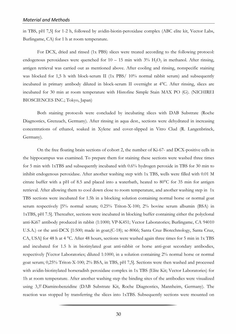
Material&and&Methods& &
30
in TBS, pH 7,5] for 1-2 h, followed by avidin-biotin-peroxidase complex (ABC elite kit, Vector Labs,
Burlingame, CA) for 1 h at room temperature.
For DCX, dried and rinsed (1x PBS) slices were treated according to the following protocol:
endogenous peroxidases were quenched for 10 – 15 min with 3% H2O2 in methanol. After rinsing,
antigen retrieval was carried out as mentioned above. After cooling and rinsing, nonspecific staining
was blocked for 1,5 h with block-serum II (1x PBS/ 10% normal rabbit serum) and subsequently
incubated in primary antibody diluted in block-serum II overnight at 4°C. After rinsing, slices are
incubated for 30 min at room temperature with Histofine Simple Stain MAX PO (G). (NICHIREI
BIOSCIENCES INC.; Tokyo, Japan)
Both staining protocols were concluded by incubating slices with DAB Substrate (Roche
Diagnostics, Grenzach, Germany). After rinsing in aqua dest., sections were dehydrated in increasing
concentrations of ethanol, soaked in Xylene and cover-slipped in Vitro Clud (R. Langenbrinck,
Germany).
On the free floating brain sections of cohort 2, the number of Ki-67- and DCX-positive cells in
the hippocampus was examined. To prepare them for staining these sections were washed three times
for 5 min with 1xTBS and subsequently incubated with 0.6% hydrogen peroxide in TBS for 30 min to
inhibit endogenous peroxidase. After another washing step with 1x TBS, wells were filled with 0.01 M
citrate buffer with a pH of 8.5 and placed into a waterbath, heated to 80°C for 35 min for antigen
retrieval. After allowing them to cool down close to room temperature, and another washing step in 1x
TBS sections were incubated for 1.5h in a blocking solution containing normal horse or normal goat
serum respectively [5% normal serum; 0.25% Triton-X-100; 2% bovine serum albumin (BSA) in
1xTBS, pH 7.5]. Thereafter, sections were incubated in blocking buffer containing either the polyclonal
anti-Ki67 antibody produced in rabbit (1:1000; VP-K451; Vector Laboratories; Burlingame, CA 94010
U.S.A.) or the anti-DCX [1:500; made in goat;(C-18); sc-8066; Santa Cruz Biotechnology, Santa Cruz,
CA, USA] for 48 h at 4 °C. After 48 hours, sections were washed again three times for 5 min in 1x TBS
and incubated for 1.5 h in biotinylated goat anti-rabbit or horse anti-goat secondary antibodies,
respectively [Vector Laboratories; diluted 1:1000, in a solution containing 2% normal horse or normal
goat serum; 0,25% Triton-X-100; 2% BSA, in TBS, pH 7,5]. Sections were then washed and processed
with avidin-biotinylated horseradish peroxidase complex in 1x TBS (Elite Kit; Vector Laboratories) for
1h at room temperature. After another washing step the binding sites of the antibodies were visualized
using 3,3'-Diaminobenzidine (DAB Substrate Kit, Roche Diagnostics, Mannheim, Germany). The
reaction was stopped by transferring the slices into 1xTBS. Subsequently sections were mounted on

Material&and&Methods& &
31
slides, were left to dry at least overnight and were coverslipped with VitroClud (R. Langenbrinck;
Emmendingen, Germany).
Quali tat ive and quant i tat ive evaluat ion o f immuno- labe led ce l l s Cell counting regarding cohort 1 was done using the 10x objective of a Olympus BX 51
(Olympus, Hamburg, Germany) microscope equipped with a motorized stage, video camera system and
Neurolucida morphometry software (MBF Biosciences, Williston, VT). Numbers of proliferating cells
(Ki67) and neural progenitor cells (NeuroD) and immature neurons (DCX), within the molecular layer
(ML), granule cell layer (GCL), subgranular zone (SGZ) and Hilus (HL), throughout the rostrocaudal
extent of the DG were recorded. The SGZ was defined as a two-nucleus thick region from the inner
margin of the dentate GCL (Heine et al., 2004). All cell counts were made on coded slides by an
investigator blind to genotype and treatment group. Every section displaying the hippocampal area in
each of the three stained series was used for each animal and the total number of stained cells was
recorded for both hemispheres separately. Resulting numbers were multiplied by eight to obtain the
estimated total number of Ki-67-/NeuroD-/DCX-positive cells per SGZ, GCL, HL and ML of the
DG. Additionally, every section displaying the hippocampus in the Nissl-stained series was used to
measure the areas of HI, SGZ, GCL and ML of each DG. Measuring also was carried out using
Neurolucida. From these values the volume of each DG as well as the mean number of the labeled cells
per DG, counted in all sections per series was calculated for each mouse. Photomicrographs were
recorded at 10-20x using the Neurolucida System. Regarding cohort 2, cell counting was carried out,
using the same Olympus BX 51 (Olympus, Hamburg, Germany) microscope used with cohort 1.
Numbers of proliferating cells (Ki67) and immature neurons (DCX), within the molecular layer (ML),
granule cell layer (GCL), subgranular zone (SGZ) and Hilus (HL), in every 6th section throughout the
entire rostrocaudal extent of the of the DG (300 µm apart) were recorded using the StereoInvestigator
system (Microbrightfield Inc., Williston, VT, USA). In each animal, immunopositive cells were counted
within counting frames (each measuring 150 x 150 µm) in every 15th section using the 40x objective
lens. The numbers and densities of frames were determined by entering the parameter grid size (150
x150 µm) in the optical fractionator component of the StereoInvestigator system.
Stat is t i c s Two-way-ANOVA was used to analyze possible gene x environment interactions and effects
that occur between the two independent variables, CMS-treatment and the 5-HTT genotype, followed
by post-hoc t-tests with Bonferroni correction for multiple testing. Statistical evaluation was performed
via Graph Pad Prism (GraphPad Software, Inc.; La Jolla, CA, USA) and significance was defined as P ≤

Material&and&Methods& &
32
0.05. P-values of ≤ 0.01 were considered to be highly significant and 0.05 < p < 0.1 was defined as a
trend. Data are reported as mean values ± standard error of the mean (S.E.M).

33
3 Results&
3.1 Spatial(Learning(Study(
3.1.1 Results&of&the&behavioral&study&&
Our co-operation partners at the University of Münster carried out a study in order to evaluate
if spatial memory is affected by the 5HTT genotype in mice per se and if there is an interaction with the
aversiveness of different testing conditions (Karabeg et al. 2013). The aim of the study had been to
evaluate whether mice with altered levels of brain 5-HT as a consequence of 5-HTT deficiency perform
differently in two spatial memory tests, the WM and BM, prospectively differing in aversiveness. The
results of the study revealed that 5-HTT-/- mice need more time to find the hidden platform compared
to 5-HTT+/- and +/+ mice in the WM, but that this delay is primarily caused by an increased number
of stops which have been identified as floating periods. Moreover, this poor WM performance
improves in the course of the 12 WM trials to WT level (Figure 7.1 in Supplements). In contrast to the
WM results, BM performance did not differ regarding escape latency, stops as well as errors between
mice of all three 5-HTT genotypes, neither in the first BM trials nor in the final ones (Figure 7.2 in
Supplements).
In the light of these behavioral testing results, I was eager to find out about possible gene x
treatment interactions regarding blood plasma corticosterone levels and, if neurobiological equivalents
to the found performance differences exist. For this purpose, I, together with two undergraduate
students (Sina Kollert and Magdalena Weidner), investigated the phenomenon of adult neurogenesis
and IEG expression as a marker for neuronal activity in the hippocampus in addition to blood
corticosterone levels as a marker for stress reactivity of these mice.
3.1.2 BM&and&WM&experience&as&well&as&5DHTT&genotype&influence&corticosterone&levels&
Statistical analysis of the plasma corticosterone levels from mice of the first hormone cohort
that had lived under control conditions (basal level) or experienced one BM or one WM trial, revealed
no significant genotype effect (2-way ANOVA, F[2,89]=1.14; p=0.33). However, a highly significant
treatment effect was found (2-way ANOVA, F[2,89]=137.40, p<0.0001, Figure 3.1 A). Bonferroni
corrected post hoc t-tests detected that a single trial of the BM as well as a single trial of the WM

Results& &
34
elevates corticosterone titers significantly compared to basal levels, regardless of the genotype (CONT
vs. WM: t=23.2; p<0.001; CONT vs. BM t=18.54; p<0.001). Additionally, mean corticosterone
concentrations were significantly higher for mice tested in the WM compared to mice tested in the BM
(t-test: t=4.49; p<0.05). Although there was no significant gene by treatment interaction, it seems that
5-HTT-/- mice exhibit higher corticosterone levels in the WM compared to the BM (Uncorrected t-
tests: t=1.94, p=0.07). This effect was neither found in 5-HTT+/+ mice nor in 5-HTT+/- mice.
Corticosterone measured after a single trial on the second testing day (Figure 3.1 B) yielded a significant
effect of genotype (ANOVA, F[2,46]=9.92, p<0.001). Bonferroni corrected post- hoc t-tests revealed
significantly elevated corticosterone levels in 5-HTT-/- mice compared to 5-HTT+/+ mice (t-test,
t=4.47, p<0.001) as well as significantly elevated corticosterone levels in 5-HTT+/- compared to
HTT+/+ mice (t-test, t=2.83, p<0.05).
Despite the fact that there was no significant gene by treatment interaction, uncorrected t-tests
revealed significantly higher corticosterone levels in 5-HTT-/- mice versus 5-HTT+/+ mice in the BM
(t-test, t=4.68, p<0.001) as well as the WM (t- test, t=4.46, p<0.001). 5-HTT+/- mice generally
exhibited intermediate corticosterone levels. An uncorrected t-test showed that 5-HTT+/- had
significantly higher levels than 5- HTT+/+ mice in the BM (t-test, t=3.55, p<0.01) but not in the WM.
Noteworthy, 5-HTT-/- mice tested in the WM tended to show higher corticosterone stress hormone
levels compared with mice of the same 5-HTT genotype tested in the BM (uncorrected t-test, t=1.88,
p=0.09). Such a trend was found neither for the 5-HTT+/- nor for the 5-HTT+/+ mice.
Figure 3.1: Corticosterone stress hormone concentration. (A) Trunk blood plasma samples taken 15 min after the mice either experienced a single trial in the barnes maze (BM) or in the water maze (WM) on day 1 of the testing procedures. ANOVA revealed a significant treatment effect. Post hoc analysis using Bonferroni corrected t-tests revealed highly significant differences for all genotypes between control and both tests as well as significant differences between BM and WM. An uncorrected t-test revealed that 5-HTT-/- mice tested in the WM exhibited higher values than 5-HTT-/- mice tested in the BM at trend-level (indicated by a broken line). (B) Trunk blood samples taken from mice 15 min

Results& &
35
after the first trial on the second day in the respective learning tests. ANOVA revealed a highly significant effect of genotype. Uncorrected t-tests (indicated by broken lines) revealed highly significant differences between 5-HTT-/- and 5-HTT+/+ mice in BM and WM as well as between 5-HTT+/- and 5-HTT+/+ mice exclusively in the BM. As in (A), 5-HTT-/- mice tested in the WM exhibited higher values than 5-HTT-/- mice tested in the BM at trend-level. Bars represent the mean concentration, whiskers express + SEM. #=p<0.1; *=p<0.05; **=p<0.01; ***=p<0.001.
3.1.3 Quantitative&assessment&of&markers&for&neuronal&activity&and&the&phenomenon&of&
adult&neurogenesis&in&the&hippocampus&
The quantitative immunohistochemistry study was carried out, in order to see if there are
neurobiological correlates for the above mentioned greater stress sensitivity and the resulting
performance differences of 5-HTT-/- mice, compared to their 5-HTT+/- and +/+ littermates, in the
more stressful WM.
5-HTT genotype has an inf luence on the number o f c e l l s express ing the two di f f erent IEGs cFos and Arc
We examined the brains of 5-HTT+/+, 5-HTT+/+ and 5-HTT-/- mice, which had been
subjected either to the control condition (undisturbed in home cage) or to one of the two spatial
learning tests (BM or WM). Quantitative immunohistochemistry exclusively revealed 5-HTT genotype
effects irrespective of the treatment. Cell counts for cFos and Arc were found to be higher in 5-HTT-
/- compared to 5-HTT+/+ mice in any of the examined hippocampus subregions (Fig. 3.2). Statistical
analysis detected that the number of cFos-ir cells in 5-HTT-/- mice was significantly higher compared
to 5-HTT+/+ mice in the upper blade of the GCL (F[1,47]=11.845; p=0.001) as well as in the total
GCL (F[1,47]=8.498; p=0.005). These effects were discovered exclusively in the septal hippocampus,
whereas no significant effects regarding cFos cell counts could be found in the temporal part of the
hippocampus (Fig. 3.2 B).
Arc-ir cell counts were also found to be significantly higher in the 5-HTT-/- compared to 5-
HTT+/+ mice in the upper blade of the GCL and in the total GCL. Here, the effect was found in both
the septal (upper blade: F[1,45]=4.648; p=0.036; total GCL: F[1, 45]=4.723; p=0.035) and the temporal
hippocampus (upper blade: F[1,35]=4.672; p=0.038; total GCL: F[1, 35]=4.684; p=0.037) (Fig. 3.2 D).

Results& &
36
Figure 3.2 Incr eased number o f c e l l s expres s ing the immedia t e ear ly g enes cFos and Arc in the granule c e l l layer o f the h ippocampus o f 5-HTT-/- compared to 5-HTT+/+ mice . Quantitative immunohistochemistry study using the ABC method and DAB as substrate of the peroxidase exclusively revealed significant differences of the number of cFos and Arc-immunoreactive (ir) cells between mice of the two 5-HTT genotypes investigated. Different treatments such as the experience of BM, WM or none of both experiences (in the control group, in which mice were left undisturbed in their home cage) do not seem to have a significant impact on the expression of these two immediate early genes. (A) Representative image of the dentate gyrus of the hippocampus after cFos immunohistochemistry. Most of the cFos-ir cell nuclei (dark brown) are located in the granule cell layer. (a) Higher magnification of cFos-ir cell nuclei in the GCL as indicated by arrows. (C) Quantitative evaluation of cFos-ir cells. Septal and temporal hippocampus as well as suprapyramidal and infrapyramidal blade of the GCL were analyzed separately. (B) Representative image of the dentate gyrus after Arc immunohistochemistry. Most of the Arc-ir cells are located in the GCL. (b) Higher magnification of Arc-ir cells bodies (indicated by black arrows) exhibiting also stained processes (indicated by grey arrow heads). (D) Quantitative evaluation of Arc-ir cells. Septal and temporal hippocampus as well as suprapyramidal and infrapyramidal blade of the GCL were analyzed separately. Two-way ANOVA, data is expressed as arithmetic mean of the number of ir-cells per section +SEM; #=p<0.1; *=p<0.05; ***=p≤ 0.005. GCL, granule cell layer; SGZ, subgranular zone; Hi, hilus; CONT, control mice; BM, barnes maze tested mice; WM, Morris water maze tested mice. Scale bar in A represents 200 µm for A and B, Scale bar in a represent 60 µm in a and b.
Adult neurogenes i s i s in f luenced by 5-HTT genotype as wel l as spat ia l l earning tes ts The number of cells positive for the proliferation marker Ki-67 is significantly increased in 5-
HTT-/- mice compared to 5-HTT+/+ animals (Fig. 3.3 B). These 5-HTT genotype effects could be
revealed in the upper blade of the SGZ (F[1, 45]=4.123; p=0.048) of the septal hippocampus as well as

Results& &
37
in the lower blade of the SGZ (F[1, 41]=4.166; p=0.048) and the total (upper & lower) SGZ (F[1,
41]=4.578; p=0.038) of the temporal hippocampus. There was however no main effect of treatment.
Yet, a significant 5-HTT genotype x treatment interaction of Ki67-ir cells in the total SGZ of the septal
hippocampus could be found (Fig. 3.3). In 5-HTT+/+ mice, the number of Ki67-ir cells was
significantly increased in mice subjected to the WM compared to untreated (control) animals (t=-2.61;
p=0.021), whereas the number of Ki67-positive cells in 5-HTT+/+ BM and control mice as well as in
5-HTT-/- mice of all treatment groups did not differ. Moreover, a trend towards higher amounts of
Ki67-ir cells in 5-HTT-/- compared to 5-HTT+/+ could be found in the naïve control groups (t=-
1.99; p=0.084), whereas in the BM and WM groups this 5-HTT genotype effect couldn`t be revealed.
The number of cells expressing NeuroD, a marker for immature neurons, was significantly
increased in 5-HTT-/- compared to 5-HTT+/+ mice in the upper blade of the SGZ (F[1, 40]=4.181;
p=0.048) as well as in the total SGZ (F[1, 40]=4.263; p=0.045) in the temporal part of the
hippocampus (Fig. 3.3 D).

Results& &
38
Figure 3.3 Incr eased number o f c e l l s expres s ing the two adu l t neurogenes i s marker Ki67 and NeuroD in the granule c e l l layer o f the h ippocampus o f 5 -HTT-/- compared to 5-HTT+/+ mice and gene by env i ronment in t e rac t ion o f the number o f Ki67-pos i t i v e pro l i f e ra t ing c e l l s in the to ta l SGZ o f the s ep ta l h ippocampus . Quantitative immunohistochemistry study using the ABC method and DAB as substrate of the peroxidase exclusively revealed significant differences of the number of Ki67 and NeuroD-immunoreactive (ir) cells between mice of the two 5-HTT genotypes (5-HTT-/- and 5-HTT+/+) investigated. Different treatments such as the experience of BM, WM or none of both experiences (in the control group, in which mice were left undisturbed in their home cage) do not seem to have a significant impact on the expression of these adult neurogenesis markers. In total SGZ of 5-HTT+/+ mice after WM treatment, the number of Ki67-ir cells was significantly increased vs. naïve 5-HTT+/+ mice. Additionally, a trend towards higher amounts of Ki67-ir cells in naïve 5-HTT-/- compared to naïve 5-HTT+/+ was found in total SGZ. (A) Representative image of the dentate gyrus of the hippocampus after Ki67 immunohistochemistry. Most of the Ki67-ir cell nuclei (dark brown) are located in the subgranular zone (SGZ). (a) Higher magnification of Ki67-ir cell nuclei in the SGZ as indicated by arrows. (C) Quantitative evaluation of Ki67-ir cells. Septal and temporal hippocampus as well as suprapyramidal and infrapyramidal blade of the GCL were analyzed separately. (B) Representative image of the dentate gyrus after NeuroD immunohistochemistry. Most of the NeuroD-ir cell nuclei are located in the SGZ. (b) Higher magnification of NeurD-ir cell nuclei in the SGZ as indicated by arrows. (D) Quantitative evaluation of NeuroD-ir cells. Septal and temporal hippocampus as well as suprapyramidal and infrapyramidal blade of the GCL were analyzed separately. Two-way ANOVA, data represent arithmetic means of the number of ir-cells per section + SEM; #=p<0.1; *=p<0.05. GCL, granule cell layer; SGZ, subgranular zone; Hi, hilus; CONT, control mice; BM, barnes maze tested mice; WM, Morris water maze tested mice. Scale bar in A represents 200 µm for A and B, Scale bar in a represent 60 µm in a and b.
Learning per formance o f 5-HTT-/- mice in the WM is corre lated with the extent o f adul t neurogenes is
Applying the Spearman`s test the only linear dependence between the escape latency (area
under the learning curve; AuC) and the number of new-born immature neurons immunoreactive for
NeuroD could be found in the temporal hippocampus of 5-HTT-/- mice tested in the WM (Fig. 3.4).
In more detail, we discovered a significant negative correlation of learning performance in the WM and
the number of NeuroD-ir cells in both the upper blade (Spearman’s r (rs) =-0.72, p=0.036,) and the
whole blade (rs=-0.69, p=0.043) of the SGZ. Moreover, a trend towards a negative correlation could
also be found in the lower blade of the SGZ (rs=-0.63, p=0.076).

Results& &
39
Figure 3.4 In 5-HTT-/- mice sub j e c t ed to the WM a low number o f NeuroD pos i t i v e c e l l s i s co rr e la t ed wi th a poor per formance in the Morr i s Water Maze . Applying the Spearman`s test in the quantitative immunohistochemistry study using the ABC method and DAB as substrate of the peroxidase revealed a linear dependence between the learning performance (decreasing learning performance is expressed as increasing area under the learning curve; AuC) and the number of NeuroD-immunoreactive (ir) cells could be found in the temporal hippocampus of 5-HTT-/- WM mice. A significant correlation between AuC and the number of NeuroD-ir (Data represent arithmetic mean per section) cells in both, the upper blade of the subgranular zone (SGZ) as well as the total SGZ. A trend towards a negative correlation was found in the lower blade of the SGZ.

Results& &
40
3.2 Chronic(Mild(Stress(Study(
3.2.1 Results&of&the&behavioral&study&&
The purpose of the CMS-behavioral tests carried out by Sandy Popp was to evaluate whether
mice with altered levels of brain 5-HT as a consequence of 5-HTT-deficiency display altered
depression-like and anxiety-like behavior after being subjected to a CMS paradigm.
The results of the study revealed that 5-HTT-/- mice (of both, cohort 1 and 2) have
significantly higher body weights than 5-HTT+/- and 5-HTT-/- mice (Fig. 7.3 A in Supplements;
diagrams display results of cohort 1). However, general liquid intake during the baseline sucrose
consumption test did not differ between the three genotypes (Fig. 7.3 B in Supplements; diagrams
display results of cohort 1), suggesting that increased body weights in 5-HTT-/- mice are not due to
altered ingestive behavior (Due to technical problems with the drinking bottles during the test with
mice of cohort 2, no reliable data for fluid consumption is available). After exposure to three weeks of
CMS, sucrose consumption (Fig. 7.4 C in Supplements) and general liquid intake (Fig. 7.4 D in
Supplements) tended to decrease in CMS mice as compared to CONT, indicating that CMS induced
anhedonia-like behavior. However, the anhedonic effect was most pronounced in stressed 5-HTT+/+
mice compared to their respective controls. In the FST, no differences between groups or genotypes
were observed regarding the latency to start floating (Fig. 7.4 A in Supplements) and the cumulative
time spent floating (Fig. 7.4 B in Supplements), reflecting that neither CMS nor 5-HTT genotype
induced behavioral despair. 5-HTT-/- mice displayed pronounced hypoactivity in several exploration-
based tests. In the EPM and OF, 5-HTT-/- mice traveled significantly shorter distances (Fig. 7.5 A and
C in Supplements) and made less vertical rears (Fig. 7.5 B and D in Supplements) as compared to 5-
HTT+/- and 5-HTT+/+ mice. Moreover, 5-HTT-/- mice showed significantly enhanced latencies to
enter the dark compartment of the LDB (Fig. 7.5 E in Supplements), again arguing for reduced
exploratory activity. In addition, stressed mice displayed enhanced exploratory activity and/or curiosity
when compared to controls, as reflected by an increased number of vertical rears in the OF (Fig. 7.5 D
in Supplements) and LDB (Fig. 7.5 F in Supplements) and longer center distance in the OF (Fig. 7.6 D
in Supplements). Furthermore, stressed 5-HTT+/- and – to a lesser extent – 5-HTT+/+ mice made
more transitions between the dark and lit compartment of the LDB (Fig. 7.6 F in Supplements),
indicating that CMS increased activity/curiosity in 5-HTT+/- and 5-HTT+/+ mice, whereas CMS had
little to no effect on exploratory activity in 5-HTT-/- mice. No differences between groups or
genotypes were found for the time spent on the open arms of the EPM (Fig. 7.6 A in Supplements),
the time spent in the center of the OF (Fig. 7.6 C in Supplements) and the time spent in the lit

Results& &
41
compartment of the LDB (Fig. 7.6 E in Supplements), suggesting that neither CMS nor 5-HTT
genotype affected classical measures of anxiety-like behavior.
Building up on these results, the purpose of my part of the study was to find out about possible
gene x treatment interactions regarding blood plasma corticosterone as well as regarding aN in the
hippocampus in the mice that had been subjected to CMS. For this, I investigated the phenomenon of
adult neurogenesis in mice of cohorts 1 (behaviorally tested) and 2 (behaviorally non-tested; together
with the two undergraduate students Stephanie Pinzel and Sahab Arinrad) in addition to blood
corticosterone levels as a marker for stress reactivity of these mice.
3.2.2 Corticosterone&Levels&in&CMS&treated&mice&&
Plasma corticosterone was measured in mice that experienced CMS and behavioral testing
(cohort 1) and in mice that experienced CMS without behavioral testing (cohort 2). Data were analyzed
via 3-way ANOVA with cohort, treatment and genotype as between-subject factors. ANOVA revealed
a significant cohort x treatment interaction, F[1,83]=17.18; p<0.001. Follow-up analysis revealed a
significant genotype effect for plasma corticosterone levels in mice of cohort 1 (figure 3.5 A), which
had been behaviorally tested after either living under control conditions or experiencing CMS (2-way
ANOVA, F[2,30]=4.22; p=0.02). Bonferroni corrected post hoc t-tests detected that 5-HTT-/- mice
had significantly higher corticosterone levels compared to 5-HTT+/+, regardless of the treatment
group (5-HTT+/+ vs. 5-HTT-/-: t=2.732; p<0.05). By contrast, statistical analysis of plasma
corticosterone levels in mice of cohort 2 (figure 3.5 B), which had either lived under control conditions
or experienced CMS, revealed a significant treatment effect (2-way ANOVA, F[1,53]=67.12;
p<0.0001), reflecting that mice which had experienced CMS prior to sacrifice had greatly elevated
CORT levels compared to mice that had lived under control conditions. A direct comparison of cohort
1 and cohort 2 revealed a significant cohort by treatment effect (2-way ANOVA, F[1,91]=20.17;
p<0.0001). Bonferroni corrected post-hoc t-tests revealed significantly elevated corticosterone levels in
behaviorally tested compared to behaviorally non-tested CONT mice (t-test, t=6.793, p<0.001) as well
as significantly elevated corticosterone levels in behaviorally non-tested CMS mice versus CMS mice (t-
test, t=6.893, p<0.001; figure 3.5 C). These findings indicate that the behavioral testing battery, which
was performed in order from less to more aversive experiments, masked the effect of CMS by
increasing plasma CORT in control animals.

Results& &
42
Figure 3.5 Cort i co s t e rone l ev e l s in CMS trea t ed and behav iora l l y t e s t ed and non - t e s t ed mice (A) Trunk blood plasma samples of mice that had lived either under control conditions (CONT) or experienced CMS, taken 24hrs after the last behavioral test (cohort 1). ANOVA revealed a significant genotype effect. Post hoc analysis using Bonferroni corrected t-tests revealed a significant difference between 5-HTT-/- mice and 5-HTT+/+ mice. (B) Trunk blood plasma samples of mice that had lived either under control conditions (CONT) or experienced CMS, taken 24hrs after the last exposure to a stressor (cohort 2). ANOVA revealed an extremely significant treatment effect, which states that CMS mice exhibited higher values than CONT mice. (C) Comparison of cohorts 1 and 2. ANOVA revealed an extremely significant cohort by treatment. Bars represent the mean concentration, whiskers express + SEM. *=p<0.05; ***=p<0.001.
3.2.3 Quantitative&assessment&of&markers&for&adult&neurogenesis&in&the&hippocampus&of&
CMS&treated&and&behaviourally&tested&mice&
In order to see if CMS treatment influences different stages of aN, immunohistochemical
stainings were carried out with antibodies against proliferation marker Ki67, immature neuron marker
DCX and neuronal progenitor marker NeuroD. For quantitative analysis, Ki67, NeuroD- and DCX-ir
cells were counted in the suprapyramidal and infrapyramidal blade of the GCL and SGZ. Addtionally
Ki67-ir cells were counted in the ML, and the hilus. As there were only few cells counted in the ML and
no significant effects arose from statistical analyses and above all, it is highly unlikely for aN to take
place in this hippocampal region, the ML data will not be shown.

Results& &
43
Figure 3.6 Photomicrographs o f the denta t e gyrus o f the h ippocampus d i sp lay ing Ki67- i r , NeuroD-ir and DCX-ir c e l l s . Immunhistochemistry via the ABC method with DAB as a substrate for the peroxidase was applied using Ki67 as a marker for proliferation, NeuroD as a marker for neuronal progenitor cells and DCX as a marker of immature neurons. (A) Representative image of the dentate gyrus with Ki67-ir (immunoreactive) cells. Most of the Ki67-ir cell nuclei (dark brown) are located in the subgranular zone (SGZ). (a) Higher magnification of Ki67-ir cell nuclei in the SGZ as indicated by black arrows. (B) Representative image of the dentate gyrus with NeuroD-ir cells. Most of the NeuroD-ir cell nuclei are located in the SGZ. (b) Higher magnification of NeurD-ir cell nuclei in the SGZ as indicated by black arrows. . (C) Representative image of the dentate gyrus with DCX-ir cells. Most of the DCX-ir cell bodies are located in the SGZ. (c) Higher magnification of a DCX-ir cell (white arrowhead) with dendritic extension (gray arrowhead) in the SGZ. GCL, granular cell layer; SGZ, subgranular zone; Hi, hilus. Scale bar in C represents 200 µm for A,B and C, Scale bar in c represent 60 µm in a, b and c.
5-HTT genotype as wel l as CMS have an inf luence on the number o f pro l i f erat ing ce l l s In mice of cohort 1, which had been behaviorally tested, statistical analysis of Ki67-ir cell
counts in the suprapyramidal blade of the SGZ, revealed a significant treatment x genotype interaction
(2-way ANOVA: F(2,30)=3.548; p=0.0414). Bonferroni corrected post hoc t-tests showed that CMS
treatment increases Ki67-ir cells in 5-HTT+/+ mice (t=2.987; p<0.05; figure 3.7 A). After CMS 5-
HTT+/+ mice have significantly more Ki67-ir cells than 5-HTT+/-(t=2.563; p<0.05; figure 3.7 A) and

Results& &
44
5-HTT-/- mice (t=2.752; p<0.05; figure 3.7 A). In the infrapyramidal SGZ 2-way ANOVA also
resulted in a treatment x genotype interaction (F(2,30)=4.195; p=0.0247). Bonferroni corrected post
hoc t-tests again discovered that CMS increases Ki67-ir cells significantly in 5-HTT+/+ mice (t=2.856;
p<0.05; figure 3.7 B). Additionaly, after CMS 5-HTT+/+ mice have significantly more Ki67-ir cells in
the infrapyramidal blade of the SGZ than 5-HTT-/- mice (t=2.932; p<0.05; figure 3.7 B). These effects
are confirmed when pooling the data of supra- and the infrapyramidal blades (2-way ANOVA of total
SGZ: Treatment x genotype interaction; F(2,30)=4.003; p=0.0288; Bonferroni corrected post hoc t-
tests: CMS/ 5-HTT+/+ vs. 5-HTT+/-, t=2.511; p<0.05; CMS/ 5-HTT+/+ vs. 5-HTT-/-, t=2.941;
p<0.05; 5-HTT+/+/ CONT vs. CMS, t=3.076; p<0.05; figure 3.7 C). Statistical analysis of data from
all regions of the hippocampus (shown as total DG: ML, GCL, SGZ and hilus) pooled together also
detected a treatment x genotype interaction (2-way ANOVA of total DG: F(2,30)=5.157; p=0.0119). In
the total DG Bonferroni corrected post hoc t-tests showed that exposure to CMS results in a highly
significant increase of Ki67-ir cells of 5-HTT+/+ mice (t=3.293; p<0.01; figure 3.7 D) and that after
CMS 5-HTT+/+ mice have significantly more Ki67-ir cells than 5-HTT-/- mice (t=2.964; p<0.05;
figure 3.7 D).
Figure 3.7 Cohor t 1 : CMS interac t s w i th g eno type , a f f e c t ing pro l i f e ra t ion SGZ and to ta l DG Quantitative immunohistochemistry using Ki67 as a marker for proliferation and the ABC method with DAB as substrate of the peroxidase exclusively revealed significant CMS-treatment by 5-HTT-genotype interaction effects regarding the number of Ki67-immunoreactive (ir) cells. In the supra- and infrapyramidal blades of the SGZ as well as the total SGZ and the total DG (ML, SGZ, GCL & Hilus), CMS treatment increases Ki67-ir cells in 5-HTT+/+ mice compared to untreated 5-HTT-mice and after CMS 5-HTT+/+ mice have significantly
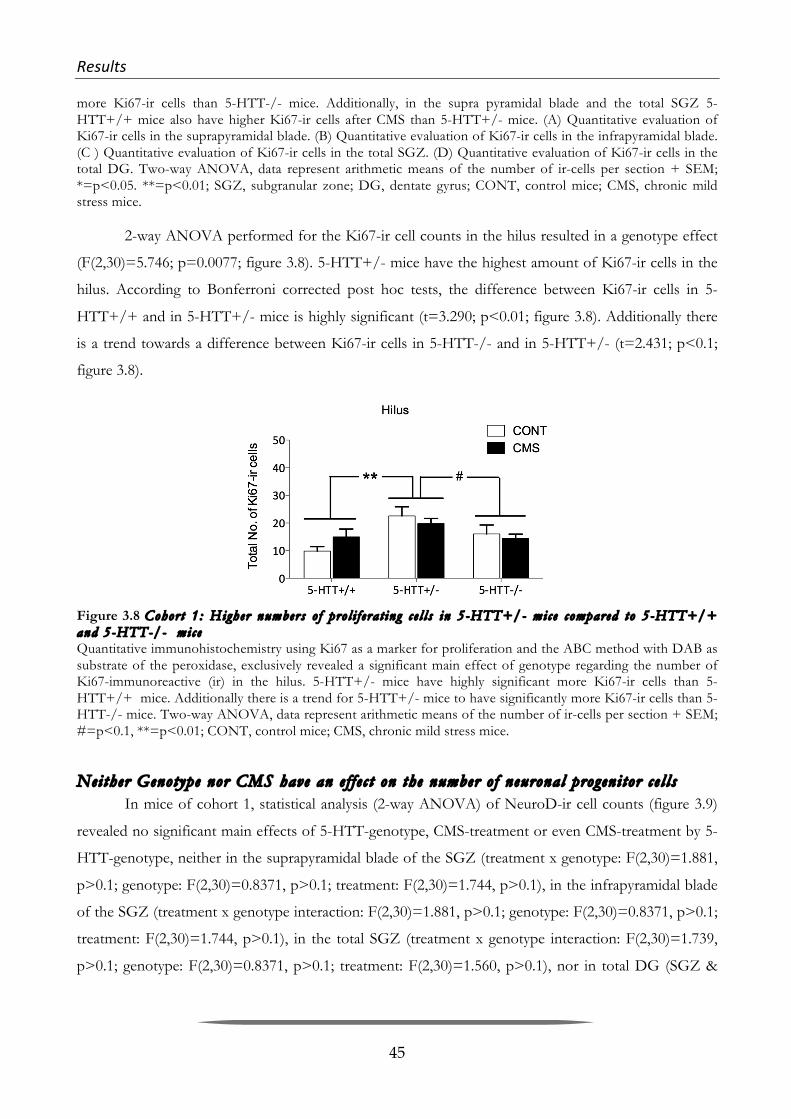
Results& &
45
more Ki67-ir cells than 5-HTT-/- mice. Additionally, in the supra pyramidal blade and the total SGZ 5-HTT+/+ mice also have higher Ki67-ir cells after CMS than 5-HTT+/- mice. (A) Quantitative evaluation of Ki67-ir cells in the suprapyramidal blade. (B) Quantitative evaluation of Ki67-ir cells in the infrapyramidal blade. (C ) Quantitative evaluation of Ki67-ir cells in the total SGZ. (D) Quantitative evaluation of Ki67-ir cells in the total DG. Two-way ANOVA, data represent arithmetic means of the number of ir-cells per section + SEM; *=p<0.05. **=p<0.01; SGZ, subgranular zone; DG, dentate gyrus; CONT, control mice; CMS, chronic mild stress mice.
2-way ANOVA performed for the Ki67-ir cell counts in the hilus resulted in a genotype effect
(F(2,30)=5.746; p=0.0077; figure 3.8). 5-HTT+/- mice have the highest amount of Ki67-ir cells in the
hilus. According to Bonferroni corrected post hoc tests, the difference between Ki67-ir cells in 5-
HTT+/+ and in 5-HTT+/- mice is highly significant (t=3.290; p<0.01; figure 3.8). Additionally there
is a trend towards a difference between Ki67-ir cells in 5-HTT-/- and in 5-HTT+/- (t=2.431; p<0.1;
figure 3.8).
Figure 3.8 Cohor t 1 : Higher numbers o f pro l i f e ra t ing c e l l s in 5-HTT+/- mice compared to 5-HTT+/+ and 5-HTT-/- mice Quantitative immunohistochemistry using Ki67 as a marker for proliferation and the ABC method with DAB as substrate of the peroxidase, exclusively revealed a significant main effect of genotype regarding the number of Ki67-immunoreactive (ir) in the hilus. 5-HTT+/- mice have highly significant more Ki67-ir cells than 5-HTT+/+ mice. Additionally there is a trend for 5-HTT+/- mice to have significantly more Ki67-ir cells than 5-HTT-/- mice. Two-way ANOVA, data represent arithmetic means of the number of ir-cells per section + SEM; #=p<0.1, **=p<0.01; CONT, control mice; CMS, chronic mild stress mice.
Neither Genotype nor CMS have an e f f e c t on the number o f neuronal progeni tor ce l l s In mice of cohort 1, statistical analysis (2-way ANOVA) of NeuroD-ir cell counts (figure 3.9)
revealed no significant main effects of 5-HTT-genotype, CMS-treatment or even CMS-treatment by 5-
HTT-genotype, neither in the suprapyramidal blade of the SGZ (treatment x genotype: F(2,30)=1.881,
p>0.1; genotype: F(2,30)=0.8371, p>0.1; treatment: F(2,30)=1.744, p>0.1), in the infrapyramidal blade
of the SGZ (treatment x genotype interaction: F(2,30)=1.881, p>0.1; genotype: F(2,30)=0.8371, p>0.1;
treatment: F(2,30)=1.744, p>0.1), in the total SGZ (treatment x genotype interaction: F(2,30)=1.739,
p>0.1; genotype: F(2,30)=0.8371, p>0.1; treatment: F(2,30)=1.560, p>0.1), nor in total DG (SGZ &
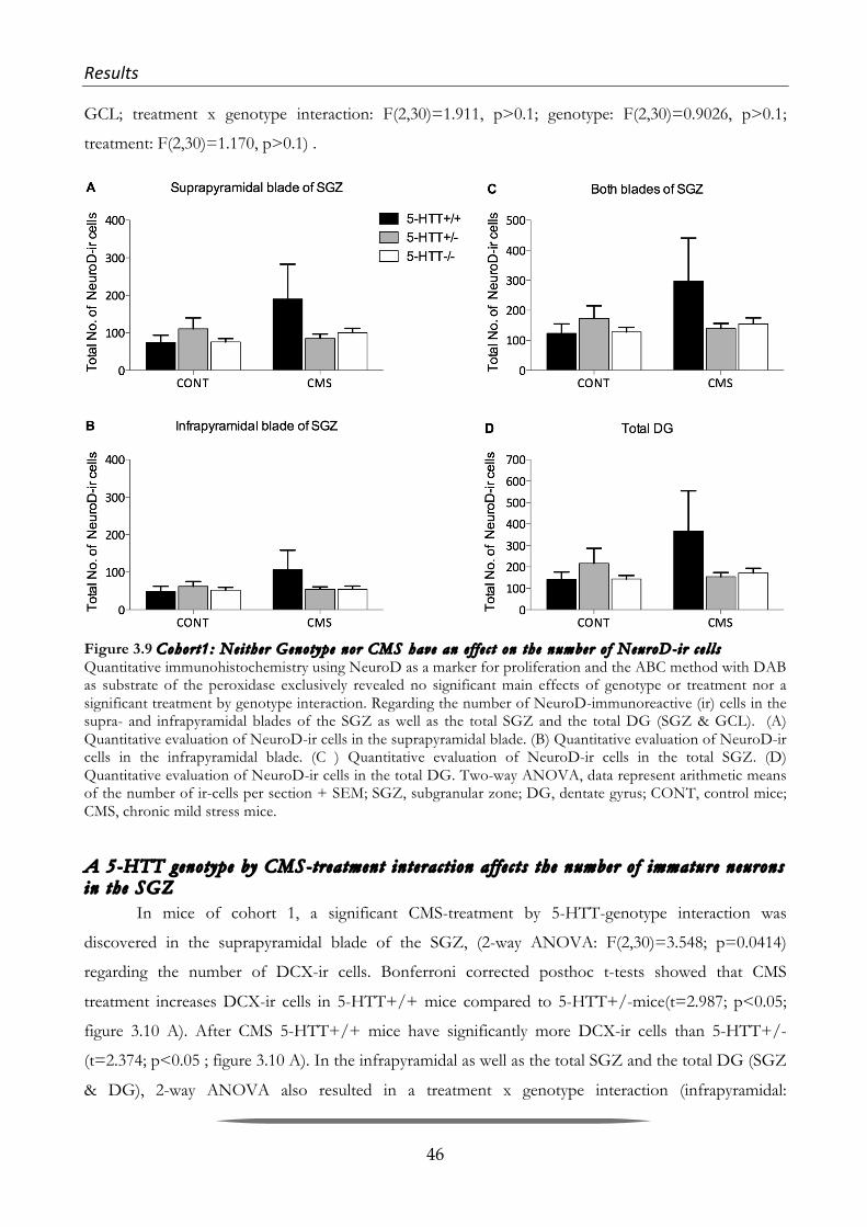
Results& &
46
GCL; treatment x genotype interaction: F(2,30)=1.911, p>0.1; genotype: F(2,30)=0.9026, p>0.1;
treatment: F(2,30)=1.170, p>0.1) .
Figure 3.9 Cohor t1 : Nei ther Genotype nor CMS have an e f f e c t on the number o f NeuroD-ir c e l l s Quantitative immunohistochemistry using NeuroD as a marker for proliferation and the ABC method with DAB as substrate of the peroxidase exclusively revealed no significant main effects of genotype or treatment nor a significant treatment by genotype interaction. Regarding the number of NeuroD-immunoreactive (ir) cells in the supra- and infrapyramidal blades of the SGZ as well as the total SGZ and the total DG (SGZ & GCL). (A) Quantitative evaluation of NeuroD-ir cells in the suprapyramidal blade. (B) Quantitative evaluation of NeuroD-ir cells in the infrapyramidal blade. (C ) Quantitative evaluation of NeuroD-ir cells in the total SGZ. (D) Quantitative evaluation of NeuroD-ir cells in the total DG. Two-way ANOVA, data represent arithmetic means of the number of ir-cells per section + SEM; SGZ, subgranular zone; DG, dentate gyrus; CONT, control mice; CMS, chronic mild stress mice.
A 5-HTT genotype by CMS-treatment interac t ion af f e c t s the number o f immature neurons in the SGZ
In mice of cohort 1, a significant CMS-treatment by 5-HTT-genotype interaction was
discovered in the suprapyramidal blade of the SGZ, (2-way ANOVA: F(2,30)=3.548; p=0.0414)
regarding the number of DCX-ir cells. Bonferroni corrected posthoc t-tests showed that CMS
treatment increases DCX-ir cells in 5-HTT+/+ mice compared to 5-HTT+/-mice(t=2.987; p<0.05;
figure 3.10 A). After CMS 5-HTT+/+ mice have significantly more DCX-ir cells than 5-HTT+/-
(t=2.374; p<0.05 ; figure 3.10 A). In the infrapyramidal as well as the total SGZ and the total DG (SGZ
& DG), 2-way ANOVA also resulted in a treatment x genotype interaction (infrapyramidal:

Results& &
47
F(2,30)=3.423; p=0.0485; total SGZ: F(2,30)=3.423; p=0.0485; total DG: F(2,30)=3.258; p=0.054),
however in neither of the regions, bonferroni corrected post hoc t-tests revealed significant differences
between the genotypes.
Figure 3.10 Cohor t 1 : CMS incr ease s the number o f immature neurons in the SGZ o f 5 -HTT+/+ mice compared to 5-HTT-/- mice . Quantitative immunohistochemistry using DCX as a marker for proliferation and the ABC method with DAB as substrate of the peroxidase exclusively revealed significant CMS-treatment by 5-HTT-genotype interaction effects regarding the number of DCX-immunoreactive (ir) cells in the supra- and infrapyramidal blades of the SGZ as well as the total SGZ and the total DG (SGZ & GCL). Only in the suprapyramidal blade Bonferroni corrected t-tests revealed that 5-HTT+/+ mice have significantly more DCX-ir cells than 5-HTT+/- mice. (A) Quantitative evaluation of DCX-ir cells in the suprapyramidal blade. (B) Quantitative evaluation of DCX-ir cells in the infrapyramidal blade. (C) Quantitative evaluation of DCX-ir cells in the total SGZ. (D) Quantitative evaluation of DCX-ir cells in the total DG. Two-way ANOVA, data represent arithmetic means of the number of ir-cells per section + SEM; *=p<0.05.; SGZ, subgranular zone; DG, dentate gyrus; CONT, control mice; CMS, chronic mild stress mice.
Results& &
48
3.2.4 Quantitative&assessment&of&markers&for&adult&neurogenesis&in&the&hippocampus&of&
CMS&treated&and&behaviorally&nonYtested&mice&
In order to reappraise if the effects we saw regarding the proliferation and aN in the
hippocampi of the mice of cohort 1 where a result of CMS treatment, or rather the consequence of the
behavioral testing battery, we had generated another cohort of mice. These mice of what we named
cohort 2, had received the same treatment as mice of cohort 1 except that they were sacrificed one day
after the last stressor, without being subjected to behavioral testing of an sort. With the brains thus
received we again carried out immunohistochemical stainings with antibodies against proliferation
marker Ki67, and immature neuron marker. For quantitative analysis, Ki67 DCX-ir cells were counted
in the suprapyramidal and infrapyramidal blade of the SGZ.
CMS decreases the number o f pro l i f erat ing ce l l s in mice that had not undergone behavioral t es t ing
In mice of cohort 2, statistical analysis of Ki67-ir cell counts with 2-way ANOVA in the
suprapyramidal and the infrapyramidal blade of the SGZ as well as in the total SGZ, revealed
significant treatment effects, which demonstrate that CMS decreases the number of Ki-67-ir cells
(suprapyramidal: F(1,49)=10.69; figure 3.11 A; p=0.0020; infrapyramidal: F(1,49)=4.495; p=0.0391;
figure 3.11 B ; total SGZ: F(1,49)=8.363; p=0.0057; figure 3.11 C).

Results& &
49
Figure 3.11 Cohor t 2 : CMS decr ease s pro l i f e ra t ion in the SGZ o f behav iora l l y non- t e s t ed mice Quantitative immunohistochemistry using Ki67 as a marker for proliferation and the ABC method with DAB as substrate of the peroxidase exclusively revealed significant CMS-treatment effect regarding the number of Ki67-immunoreactive (ir) cells. In the supra- and infrapyramidal blades of the SGZ as well as the total SGZ, CMS treatment decreases Ki67-ir cells in all mice regardless of genotype. (A) Quantitative evaluation of Ki67-ir cells in the suprapyramidal blade. (B) Quantitative evaluation of Ki67-ir cells in the infrapyramidal blade. (C) Quantitative evaluation of Ki67-ir cells in the total SGZ. Two-way ANOVA, data represent arithmetic means of the number of ir-cells per section + SEM; *=p<0.05. **=p<0.01; SGZ, subgranular zone; CONT, control mice; CMS, chronic mild stress mice.
Undisturbed 5-HTT-/- mice d isp lay the highest numbers o f immature neurons and CMS-treatment decreases the number o f immature neurons in 5-HTT-/- mice
In mice of cohort 2, a highly significant CMS-treatment by 5-HTT-genotype interaction was
detected in the suprapyramidal blade of the SGZ (2-way ANOVA: F(2,47)=5.906; p=0.0052) regarding
the number of DCX-ir cells. Bonferroni corrected posthoc t-tests showed that undisturbed (CONT) 5-
HTT-/- mice have significantly higher numbers of DCX-ir cells compared to CONT 5-HTT+/+ mice
(t=3.849; p<0.001; figure 3.12 A). Addtionally CMS decreases the number of DCX-ir cells in 5-HTT-/-
mice compared to CONT 5-HTT-/- mice (t=4.700; p<0.001; figure 3.12 A). In the infrapyramidal
blade, 2-way ANOVA only resulted in a trend towards a treatment effect, with CMS treated mice
displaying lower numbers of DCX-ir cells thatn CONT animals (F(1,47)=3.308; p=0.0753;). Finally,
when analyzing both blades of the SGZ together again a highly significant CMS-treatment by 5-HTT-
genotype interaction was detected (2-way ANOVA: F(2,47)=7.839; p<0.01). Again, bonferroni
corrected posthoc t-tests showed that undisturbed (CONT) 5-HTT-/- mice have significantly higher

Results& &
50
numbers of DCX-ir cells compared to CONT 5-HTT+/+ mice (t=3.369; p<0.01; figure 3.12 C) and
that CMS decreases the number of DCX-ir cells in 5-HTT-/- mice compared to CONT 5-HTT-/-
mice (t=4.258; p<0.001 ; figure 3.12 C).
Figure 3.12 Cohor t 2 : CMS interac t s w i th 5-HTT-/- geno type and decr eases the number o f immature neurons in the SGZ. Quantitative immunohistochemistry using DCX as a marker for proliferation and the ABC method with DAB as substrate of the peroxidase revealed significant CMS-treatment by 5-HTT-genotype interaction effects regarding the number of DCX-immunoreactive (ir) cells in the suprapyramidal blade oft he SGZ as well as the total SGZ. Bonferroni corrected t-tests revealed that 5-HTT-/- mice have significantly more DCX-ir cells than 5-HTT+/+ and that CMS decreases the number of DCX-ir cells in 5-HTT-/- mice. (A) Quantitative evaluation of DCX-ir cells in the suprapyramidal blade. (B) Quantitative evaluation of DCX-ir cells in the infrapyramidal blade. (C ) Quantitative evaluation of DCX-ir cells in the total SGZ. Two-way ANOVA, data represent arithmetic means of the number of ir-cells per section + SEM; #=p<0.1; **=p<0.01; ***=p<0.001; SGZ, subgranular zone; CONT, control mice; CMS, chronic mild stress mice.
In summary, the results of the examination of cohort 2 clearly revealed a CMS treatment effect
regarding proliferation, with CMS decreasing the number of Ki67-ir cells in general. Additionally, CMS
treatment interacts with 5-HTT-genotype to decrease immature neurons in 5-HTT-/- mice. This stands
contrary to the results of the examination of proliferation and aN in cohort 1. Here a gene x treatment
effect regarding the number of Ki-67-ir cells had been revealed, with CMS increasing proliferation
exclusively in 5-HTT+/+ mice. Additionally, a CMS treatment effect on the number of immature
neurons had been found with CMS in general increasing the number of DCX-ir cells.

Discussion& &
51
4 Discussion&
In both studies introduced in this doctoral thesis, animals of the 5-HTT-/- mouse line were
used. Mice in the Spatial Learning study were of male sex and six months of age, while mice in the CMS
study where one year old females. In the Spatial Learning study, mice experienced WM-stress, which
could be classified as a predictable stressor, as the mice where subjected to a total of 10 (BM) or 15
(WM) trials. Contrary to this, mice of the CMS study where subjected to a number of unpredictable
stressors. As far as behavior is concerned, both studies showed that 5-HTT-/- mice display hypo-
locomotion. While predictable WM-stress decreases locomotor activity from trial to trial, during the
same day, in 6 months old male 5-HTT-/- mice, unpredictable CMS does not further decrease
locomotor activity displayed in OF, EPM or LDB in 1-year old female 5-HTT-/-. On the other hand
there is significant increase of rearing behavior in the LDB and a trend towards an increase of rearings
in the OF in response to CMS independently of the genotype. Concerning Neuroplasticity, the Spatial
Learning study revealed higher amounts of cFos- and Arc-ir cells in 5-HTT-/- compared to 5-
HTT+/+ mice at baseline. Regarding aN, 5-HTT-/- exhibited more proliferation (Ki67-ir cells) and
neuronal progenitor cells (NeuroD-ir cells) than 5-HTT+/+. Additionally, a genotype by treatment
interaction showed that WM and not BM treatment leads to an increase of proliferation in 5-HTT+/+
mice. Moreover, a positive correlation between the number of NeuroD-ir cells in 5-HTT-/- mice
subjected to the WM and their overall performance (AuC) could be found revealing that mice with
lower numbers of NeuroD-ir cells performed worse in the WM. Within the scope of the CMS study,
investigation of the brains of mice of cohort 2, which had only experienced CMS and had not been
behaviorally tested, revealed that non-stressed 5-HTT-/- exhibited significantly more immature
neurons than their non-stressed 5-HTT+/+ littermates. On the other hand, a decrease in proliferation
in response to stress, as well as a decrease in the number of immature neurons in 5-HTT-/- mice as a
response to stress was detected. This was in accordance with result of the assessment of the
corticosterone levels, which showed a clear increase in corticosterone levels in response to the CMS
treatment. Examination of the brains of mice of cohort 1 had not resulted in a treatment effect, neither
had the examination of their corticosterone levels, which shows clearly that the effect of the CMS
paradigm had been washed out by the behavioral testing battery.

Discussion& &
52
4.1 Spatial(Learning(Study(
The aim of this study was to evaluate whether mice with altered levels of brain 5-HT as a
consequence of lifelong 5-HTT deficiency perform differently in two spatial memory tests, the WM
and the BM, prospectively differing in aversiveness. Moreover, we asked whether neurobiological
equivalents to possible learning differences exist. For this purpose, we investigated the phenomenon of
adult neurogenesis and IEG expression as a marker for neuronal activity in the hippocampus in
addition to blood corticosterone levels as a marker for stress reactivity of these mice.
4.1.1 Summary&and&Interpretation&of&the&Behavioral&Results&
rom numerous studies, it is known that the lifelong reduced or absent 5-HTT function is
associated with multiple changes at the behavioral level compared to 5-HTT+/+ littermates (for review
see: (Murphy & K.-P. Lesch 2008)). As far as the behavioral phenotype of 5-HTT mutant mice is
concerned, it has been found to primarily include increased anxiety– and depression-related behavior,
reduced aggression, but also deficits in fear extinction recall (Holmes et al. 2003; Wellman et al. 2007;
Murphy & K.-P. Lesch 2008; Narayanan et al. 2011; Heiming et al. 2013). However, so far only little
was known about the influence of the 5-HTT on cognitive functions. Thus, the behavioral study by
Lars Lewejohann and his Master Student Sandra Grauthoff provides first insights into spatial learning
capacities of mice deficient for the 5-HTT using two different learning tests: the WM and the BM
(Karabeg et al. 2013). They found, that 5-HTT null mutant mice required more time to find the hidden
platform in the WM, compared to 5-HTT+/- and 5-HTT+/+ mice. But as the overall swimming
distance of the 5-HTT-/- mice was not altered their more frequent floating behavior (stops) led to
reduced overall swimming speed levels, which finally resulted in this delayed learning performance. Yet,
this interrupted movement of 5-HTT null mutant mice is unique to the WM test, as in the BM test 5-
HTT-/- mice did not stop walking more frequently than 5-HTT+/- and 5-HTT+/+ mice. On the
other hand, 5-HTT-/- mice advanced in the course of the 12 WM test trials to 5-HTT+/+ levels, so
that in the last trials mice of all three 5-HTT genotypes showed similar learning performance. It was
concluded, that these results point towards a comparable spatial learning capacity of 5-HTT-/-, 5-
HTT+/- and 5-HTT+/+ mice, but that there are some time-dependent performance differences in the
WM, but not in the BM.
Lars Lewejohann suggested that the observed floating behavior observed in 5-HTT-/- mice
might resemble the “immobility time” known from the forced swim test, which is discussed to be a
behavioral correlate of despair and thus indicates depression-like behavior. As a consequence, floating
in the WM could reflect a state of despair indicating that 5-HTT-/- mice gave up active coping with the

Discussion& &
53
challenging situation earlier than both other genotypes (Karabeg et al. 2013). Initially, 5-HTT-/- mice
did not differ from 5-HTT+/- and 5-HTT+/+ mice regarding the extent of floating, instead their
number of floatings increased in the course of three subsequently performed trials per day during the
first three days of the WM training session. This was found to resemble the results of two different
studies performed by the groups of Wellman (Wellman et al. 2007) and Carroll (Carroll et al. 2007) who
showed that 5-HTT-/- mice exhibit significantly increased depression-related behavior compared to 5-
HTT+/- and 5-HTT+/+ mice exclusively in response to repeated forced swimming, but not to a
single exposure to the forced swim test. However, as the exaggerated floating behavior returned to
baseline level during the first trial of the following days (day 2 and 3), and the fact that this increased
immobility of 5-HTT-/- mice was unique to the more aversive WM test gives the idea that 5-HTT-/-
mice are more sensitive to the aversive situation and show more despair during the second and third
WM trial than their 5-HTT+/- and 5-HTT+/+ littermates instead of actively coping with the stressful
situation.
4.1.2 Corticosterone&levels&in&mice&deficient&for&the&5YHTT,&tested&in&WM&and&BM&
In contrast to the WM the BM does not utilize the fear of drowning as a strongly aversive
stimulus, and was already shown to result in lower blood plasma stress hormone levels (Harrison et al.
2009). On top of that, spatial learning was shown to be inversely correlated with corticosterone levels in
the WM, but not in the BM, suggesting that performance in the WM may be more prone to test-
induced stress (Harrison et al. 2009). The aversiveness of the WM is further corroborated by the fact
that WM induces hypothermia. For instance, Streijger et. al have reported, that a 2 min swim in the
WM decreases the core body temperature by 4.5°C (Streijger et al. 2009) and five swims of 45 sec with
a very short inter trial interval are reported to lead to a rectal temperature drop of 9°C (Iivonen et al.
2003). Moreover, it is well-known from other behavioral tasks that high levels of stress can influence
the animal's performance (Hölscher 1999). The significantly increased corticosterone levels detected in
WM-tested compared to BM-tested mice 15 min after the start of one single trial, are in accordance
with the study of Harrison and co-workers (Harrison et al. 2009), interestingly however, this was not
found in mice after the first trial on the second testing day. Although there was no significant gene by
treatment interaction WM-tested 5-HTT deficient mice tended to exhibit higher corticosterone
concentrations compared to mice of the same 5-HTT genotype after BM experience in both stress
hormone cohorts. Moreover, repeatedly experiencing any of the two spatial learning tests resulted in 5-
HTT genotype differences with highest plasma corticosterone levels in 5-HTT-/- mice, while 5-
HTT+/+ mice seemed to habituate regarding their stress response. These 5-HTT genotype differences
are in accordance with former studies analyzing glucocorticoid and ACTH levels in mice of different 5-

Discussion& &
54
HTT genotypes in response to stressors such as handling and injection (Q. Li et al. 1999; Jiang et al.
2009). Furthermore, an exaggerated adrenalin/epinephrine release into plasma could be shown as a
reaction to 15 min of immobilization (Armando et al. 2003), whereas tyrosine hydroxylase mRNA
expression after this type of acute stress in 5-HTT+/+ and 5-HTT-/- mice was not changed. In line
with most previous studies, the 5-HTT-/-, 5-HTT+/- and 5-HTT+/+ mice used in the present study
(Karabeg et al. 2013) exhibit similar basal glucocorticoid levels (Tjurmina 2002; Tjurmina et al. 2004;
Jansen et al. 2009; Bartolomucci et al. 2010). For this reason, it is proposed that the observed 5-HTT
genotype differences after repeated spatial learning test performance can be regarded as a result of a
gene by stress experience interaction and that it points towards an altered stress sensitivity of 5-HTT
deficient mice. This increased stress sensitivity of the tested 5-HTT deficient animals in combination
with the assumed different levels of aversiveness of BM and WM might explain inferior learning
performance in the WM as an altered responsiveness of 5-HTT-/- mice towards a seemingly
inescapable aversive WM environment without actually affecting spatial learning abilities per se.
Comparable to the results of 5-HTT deficient mice or rats, human individuals carrying the S-
allele of the 5-HTT gene exhibit a greater reactivity to stress. This relation is supported by human
imaging studies, which have found that carriers of the S-allele display greater amygdala activation in
response to fearful stimuli (Hariri et al. 2002), that is dependent on 5-HTT availability (Rhodes et al.
2007). Besides that, stressful life events were shown to correlate with the severity and number of
episodes of major depression in the individuals carrying lower expressing 5-HTT genes (Caspi et al.
2003; Zalsman et al. 2006). Additionally, cognitive deficits in memory and decision-making have been
found to be present early in the course of major depressive disorder (Trivedi & Greer 2014). On the
other hand, it has also been reported that carriers of the L-allele, who had recovered from depression
were more vulnerable later on to an increase in depressive symptoms than those carrying the S-allele
(Moreno et al. 2002). Moreover, Roiser had demonstrated, that L-allele carriers showed inferior
cognitive performance than S-allele carriers in a visual planning task as well as in an attentional test that
measures the ability to withhold an intentional motor response based on the emotional valence of
words (Roiser et al. 2006; Roiser et al. 2007).

Discussion& &
55
4.1.3 Interpretation&of&the&Results&of&the&Quantitative&Immunohistochemistry&Study&
Increased number o f IEG-posi t ive ce l l s in the hippocampus o f 5-HTT-/- mice compared to 5-HTT+/+ l i t t ermates point to higher base l ine hippocampal ac t iv i ty l eve l s in mice with l i f e long 5-HTT def i c i ency
Immunocytochemistry as well as in mRNA-studies IEG protein/mRNA expression had been
shown to peak within the first hours (protein) or even minutes (mRNA expression) after different
learning tests or stress exposure and then return to baseline after a maximum of 24 hrs (Link et al.
1995; Bertaina-Anglade et al. 2000; Wallace et al. 1998; Guzowski et al. 2001). Therefore we were aware
that the chosen time-point was not ideal for IEG expression analysis related to effects of the exposure
to the different test apparatuses. However, as the survival time of 60 hrs after 5 days of learning test
exposure seems to be perfect for the analysis of early neurogenesis stages we decided to quantify the
number of cFos- and Arc-positive cells in the DG of our mice (behavior-cohort) sacrificed 60 hrs after
the last spatial learning test. This was done, as we aimed at testing the hypothesis suggested by (Nietzer
et al. 2011) that 5-HTT-/- brains display a “stressed” morphological phenotype and hoped to find
increased numbers of IEG-expressing cells in the hippocampus of 5-HTT-/- compared to 5-HTT+/+
mice.
As expected, we did not detect differences between the number of Arc positive cells in mice of
the different treatment groups (CONT, BM, WM), but overall 5-HTT genotype effects with
significantly higher number of Arc-ir cells in the DG of 5-HTT-/- mice compared to 5-HTT+/+ mice
(Karabeg et al. 2013). In contrast to our results, significantly reduced Arc mRNA expression was
detected in the whole hippocampus of 5-HTT deficient rats, irrespective of hippocampal subregions
and different layers (Molteni et al. 2009). Additionally, Eriksson et al. found that Flinders sensitive line
(FSL) rats display emotional memory impairments accompanied by reduced Arc mRNA expression
specifically in brain regions implicated in cognitive processing such as the dentate gyrus (T. M. Eriksson
et al. 2012). These results, however, do not necessarily contradict our results, as our study analyzed the
total number of Arc-ir cells and exclusively focused on the granule cell layer of the DG (Karabeg et al.
2013). The fact that both, the Molteni study using 5-HTT deficient rats and our study using 5-HTT
deficient mice revealed 5-HTT genotype dependent alterations in baseline Arc expression may be
explained by a compensatory mechanism in response to the lifelong altered extracellular and
intracellular 5-HT-levels, which could be the neurobiological correlate of heightened anxiety. This
notion is supported by the fact that serotonergic signaling and Arc thus have opposing effects on
AMPA type glutamate receptor (AMPAR)-trafficking. While serotonergic signaling enhances trans-
synaptic signaling efficiency via the insertion of AMPARs into synapses at postsynaptic neurons (K. P.

Discussion& &
56
Lesch & Waider 2012), Arc expression is reciprocally regulated by activity and facilitates the
endocytosis of certain AMPARs resulting in reduced AMPARs surface expression (Chowdhury et al.
2006). The latter possibly resulting in an increase of the threshold for LTP followed by altered learning
and memory processes. From the results of our quantitative immunohistochemistry study we cannot
tell if Arc is overexpressed in single cells of the DG in 5-HTT-/- compared to 5-HTT+/+ mice. Yet,
one could speculate that having more cells expressing Arc at baseline coincides with the overexpression
in single cells.
Regarding cFos, significantly increased numbers of cFos-ir cells were found in the septal
hippocampus of 5-HTT-/- compared to 5-HTT+/+ mice (Karabeg et al. 2013). From a number of
older studies it is known that cFos is upregulated in the DG in response to various acute stressors
including immobilization, noxious stimulation and hyper osmotic stress (Senba et al. 1994; Senba &
Ueyama 1997) as well as in response to chronic defeat (Matsuda et al. 1996). Interestingly, chronic
restraint stress has also been found to cause a significant increase in 5-HT (Adell et al. 1988). This
gives reason to assume that 5-HTT-/- mice, which have higher basal extracellular 5-HT levels and thus
resemble the mentioned chronically stressed mice, are closer to a “stress threshold” than 5-HTT+/+
mice. Beyond that, it is known that chronic restraint stress causes atrophy of apical dendrites in CA3
pyramidal neurons of the hippocampus (Watanabe, Gould, Cameron, et al. 1992b; Watanabe, Gould &
McEwen 1992a; Watanabe, Gould, Daniels, et al. 1992c; Magarinos & McEwen 1995; Magariños &
McEwen 1995; Sunanda et al. 1995) and that this atrophy is correlated with impaired spatial memory
performance. Interestingly, the atrophy as well as the impairment could be reversed by lowering
extracellular 5-HT pharmacologically in the septal hippocampus (Luine et al. 1994; Conrad et al. 1996).
Finally, the increased baseline expression of the two IEGs Arc and cFos shown in this study
may have promoted the pronounced stress sensitivity of 5-HTT-/- mice. Thus, elevated Arc and cFos
protein levels could represent a “stressed” brain phenotype in consequence of lifelong 5-HTT
deficiency, thereby presenting the neurobiological correlate of increased stress sensitivity and
heightened anxiety of 5-HTT-/- animals.
Increased adul t neurogenes i s in the hippocampus o f 5-HTT-/- mice compared to 5-HTT+/+ l i t t ermates indicates a compensatory mechanism for l i f e long increased extrace l lu lar 5-HT leve l s
5-HTT genotype effects regarding proliferation (Ki67) and neurogenesis (NeuroD) comprise 5-
HTT-/- mice displaying more immunoreactive cells than 5-HTT+/+ animals. Schmitt et al. have
already achieved similar results in 2007, albeit only in aged (14 months old), and not in young adult
mice. These results are quite feasible when keeping in mind that, first, the lack of 5-HT clearance in

Discussion& &
57
these mice results in a persistent 5 to 13-fold increase of 5-HT concentrations in the extracellular space
(Shen et al. 2004; Fabre et al. 2000), and second, that 5-HT is a positive modulator of stem cell
proliferation and neurogenesis in the adult hippocampus (Brezun & Daszuta 2000; Banasr et al. 2006;
Benninghoff et al. 2010). In a recent study, immunoreactivity for the neurogenesis marker doublecortin
was discovered to be increased in the dentate gyrus of 5-HTT-/- rats in comparison with 5-HTT+/+
rats (Schipper et al. 2011). By feeding 5-HTT-/- animals a special diet, Schipper et al. were able to
normalize both, increased anxiety-like behavior of 5-HTT-/- rats and altered neurogenesis level. This
led them conclude, that the behavioral effect of their special diet was probably conveyed via
normalizing neurogenesis (Schipper et al. 2011). Thus, increased stem cell proliferation and
neurogenesis in 5-HTT-/- animals can be discussed as neurobiological correlates to the increased
anxiety-like behavior of 5-HTT-/- mice and rats.
Beyond the overall increased aN levels in 5-HTT deficient compared to 5-HTT+/+ mice, our
statistical tests revealed a significant gene by environment interaction of Ki67-ir cells in the SGZ of
total DG. The number of Ki67-ir cells was shown to be different between naïve and WM-tested 5-
HTT+/+ mice, with those tested in the WM displaying more proliferating cells in the SGZ than naïve
animals. Moreover, naïve 5-HTT-/- mice already exhibited a trend towards higher numbers of
proliferating cells compared to 5-HTT+/+ mice, whereas there was neither a significant difference
between the two genotypes after BM or WM nor a significant effect of treatment within 5-HTT-/-
mice (Karabeg et al. 2013). Considering that stress decreases aN (Gould & Tanapat 1999; Ehninger &
Kempermann 2006; Lucassen, Meerlo, et al. 2010a; M. M. Lee et al. 2013), and that 5-HT increases aN
(Gould 1999), these lacking effects might be the result of the antagonistic effects of increased 5-HT-
levels on the one hand, and the increased stress sensitivity of 5-HTT-/- mice on the other hand. There
were two studies dealing with the impact of WM-testing on hippocampal aN in the rat. Interestingly,
they resulted in contradictory outcomes. The first one, probably the most well known study to convey
the enhancing effects of spatial learning on aN, was carried out by Gould and colleagues in 1999
(Gould, Beylin, et al. 1999a). In their experiment rats were exposed to 4 WM trials per day for 4
consecutive days with a maximum trial duration of 60 s. The number of proliferating cells was
increased in rats that had undergone hippocampus-dependent spatial learning in the WM compared to
all control groups (including a swim-stress group) (Gould, Beylin, et al. 1999a). In the second study,
Namestkova and co-workers (Namestkova et al. 2005) subjected rats to the same procedure, but for 15
consecutive days. This long-lasting treatment caused a significant decrease in progenitor cell
proliferation in the granule cell layer of the hippocampus compared to controls (Namestkova et al.
2005). In our study mice were subjected to WM training on 5 consecutive days and thus resembling the

Discussion& &
58
Gould et al. study more than the Namestkova et al. study. Therefore, the detected increase of
proliferating cells in WM trained 5-HTT+/+ mice found in our study may be regarded as a result of
spatial learning.
The finding that there was no significant difference between the differently treated 5-HTT-/-
mice, including the fact that there was already a trend towards higher numbers of proliferating cells in
naïve 5-HTT-/- vs. 5-HTT+/+ mice suggests that this embodies a ceiling effect, that 5-HTT-/- mice
are closer to a “stress threshold” than 5-HTT+/+ mice. This notion is substantiated by the fact that a
comparable ceiling effect was already described by Nietzer and co-workers, who found higher
spinogenesis in the amygdala in naïve 5-HTT-/- vs. 5-HTT+/+ mice (Nietzer et al. 2011). As stress-
dependent effects were not detected in 5-HTT-/- animals, the authors concluded that these mice per se
display a “stressed” morphological phenotype as a consequence of long-life 5-HTT deficiency (Nietzer
et al. 2011). We suggest that such ceiling effects act as a part of a compensatory mechanism, which
probably sensitizes or desensitizes for future additional stress experience. Additionally, it may explain
why spatial learning over a moderate time in the WM – normally a positive modulator of progenitor cell
proliferation (Gould, Beylin, et al. 1999a) – had no effect on proliferation in the SGZ of 5-HTT
deficient mice.
Regarding the number of NeuroD-ir cells, a correlation between the number of NeuroD-ir cells
in 5-HTT-/- mice subjected to the WM and their overall performance (Area under the learning curve;
AuC) could be found revealing that mice with lower numbers of NeuroD-ir cells performed worse in
the WM (Karabeg et al. 2013). The modulation of aN by WM test experience and the detected
correlation between the number of newborn neurons and the learning performances point to an
important role of this neuroplasticity phenomenon in the handling of stress and in learning and
memory processes. This discovery is consistent with a study of human hippocampal neurogenesis by
Coras and colleagues showing that patients with low numbers of proliferating progenitor cells and new-
born neurons in vivo and in vitro showed a severe learning and memory impairments (Coras et al.
2010).
Different hippocampal subreg ions with di f f erent connec t iv i t i e s and funct ion Moser and Moser suggested that the hippocampus is functionally different along its
septotemporal axis (M. B. Moser & E. I. Moser 1998). In rodents, spatial and episodic memory appears
to depend on septal but not temporal hippocampus (E. I. Moser 1995; Davachi & Wagner 2002;
Broadbent et al. 2004). On the other hand, the temporal but not the septal hippocampus is thought to
mediate stress responses and thus emotional behavior (Henke 1990; Herman et al. 1998; Fanselow &

Discussion& &
59
Dong 2010; Segal et al. 2010). In rats it is well established that serotonergic neurons of the median
raphe nucleus primarily project to the septal part of the hippocampal formation rather than in its
temporal part (Vertes et al. 1999). Additionally, within the septal hippocampal formation median raphe
neurons terminate throughout the rostrocaudal extent of the DG. Within the DG these projections are
closely confined to the SGZ with more representations in the suprapyramidal blade than in the
infrapyramidal blade of the DG (Vertes et al. 1999). Considering this, it is not surprising, that most
significant 5-HTT genotype effects regarding cell activation (Arc/ cFos) or neurogenesis (Ki67/
NeuroD) were found in the suprapyramidal blade of the DG. However, only environmental factors like
spatial experience, transient forebrain ischemia, or kainic acid administration, have been found to cause
region specific effects regarding IEG-expression, aN and or even morphological changes (Lawston et
al. 2000; Choi et al. 2003; Choi et al. 2007; Chawla et al. 2005; Leuner & Gould 2010). So far, this is the
first study showing region-specific genotype differences in aN and the number of IEG expressing cells.
In summary, our BM and WM results point to comparable spatial learning capabilities of mice
with different 5-HTT genotypes, but the performance differences of 5-HTT deficient mice in the WM
task support a role for the 5-HTT in the hippocampally-mediated interaction between stress and spatial
learning performance. Moreover, we suggest that increased IEG expression and aN levels observed in
the hippocampus of 5-HTT deficient mice can be the neurobiological correlate of emotion circuit
dysfunction and heightened anxiety as 5-HTT-/- animals per se display a “stressed” phenotype as a
consequence of long-life 5-HTT deficiency. The experience of BM and WM tests results in either
altered or even absent neuroadaptive increases in IEG and aN marker expression in 5-HTT-/- animals
reducing adequate compensation processes and resulting in exaggerated behavioral responses e.g. in the
more aversive WM.
4.2 Chronic(Mild(Stress(Study(
The aim of this study was to evaluate whether mice with altered levels of brain 5-HT as a
consequence of lifelong 5-HTT deficiency respond with a further increase of depression-like and
anxiety-like behavior after being subjected to a chronic mild stress (CMS) paradigm. As CMS is a well
established animal model for depression, we were looking for possible 5-HTT-genotype by CMS-
treatment interactions regarding aN in the hippocampus in addition to blood corticosterone levels as a
marker for stress reactivity of these mice in two different cohorts of mice. The first cohort (cohort 1)
had been subjected to an extensive testing battery after CMS and cohort 2 had remained behaviorally
untested.

Discussion& &
60
4.2.1 Summary&and&Interpretation&of&the&Behavioral&Study&
One-year-o ld f emale 5-HTT def i c i ent mice have a s igni f i cant ly higher body weight compared to the ir 5-HTT+/+ and 5-HTT+/- l i t t ermates
Weight measurements taken by Sandy Popp as part of the present study, revealed that the
tested one-year-old 5-HTT-/- mice had a significantly higher body weight than 5-HTT+/+ and 5-
HTT+/- mice, which is in accordance with earlier findings by Üceyler et al. and Holmes et al. (Üçeyler
et al. 2010; Holmes, Yang, et al. 2002c). Üceyler and co-workers, who concluded from the results of
their study in 2010 that lifelong low brain serotonin level due to the 5-HTT-/- genotype leads to
reduced physical activity and low BDNF, resulting in late-onset obesity. However, they also found that
although a lack of the 5-HTT is a genetic vulnerability factor for obesity, female gender is protective
(Üçeyler et al. 2010). The result of the present study contradicts the Üceyler study and suggests that
female gender does not protect form becoming obese. However, our study is in line with an earlier
study by Sookoian and colleagues, who examined adolescent human subjects and had found that the S
allele of the SLC6A4 promoter variant was a risk factor for overweight, independently of gender, as
well as with the study by Holmes et al. (Sookoian et al. 2007; Holmes, Yang, et al. 2002c).
Motor ac t iv i ty i s decreased by 5-HTT def i c i ency and increased by CMS exper ience Earlier studies have examined behavioral alterations in 5-HTT−/− mice, reporting, overall
hypo-locomotion (Kalueff et al. 2007; Kalueff et al. 2006). Therefore it does not come as a surprise,
that the locomotor profiles of 5-HTT-/- mice, be it the distance travelled in the Elevated Plus Maze
(EPM) or the Open Field (OF), the number of rears in the EPM or the OF, or the latency to change
compartments in the Light/Dark Box (LDB), strikingly demonstrates their lack of physical activity
compared to their 5-HTT+/- and 5-HTT+/+ littermates. Curiously, significantly higher numbers of
rears in the lit compartment of the LDB and a trend towards a higher number of rears in the OF
displayed by mice subjected to CMS, seems to demonstrate a contrary effect of CMS on motor activity,
independently of the genotype. Increased motor activity by CMS has already been shown in earlier
studies, albeit mostly regarding the distance travelled in the OF (Harris 1997; GRONLI et al. 2005).
Grønli and co-workers for instance revealed that rats tended to exhibit higher activity in the center
squares of the OF after prior exposure to CMS. They suggest that this may represent a model of the
psychomotor agitation observed in some depressed humans(GRONLI et al. 2005). Now, one might ask
why this treatment effect only appears in the form of increased rearings and not in an increased
distance travelled in the EPM and the OF as was the case in the other mentioned studies (Harris 1997;
GRONLI et al. 2005). The explanation is actually quite simple, when taking a closer look at the graphs,
depicting the distance travelled in the EPM and the OF, which reveal that the distance travelled is

Discussion& &
61
slightly increased, yet not enough to be significant. As the time the mouse spends making rearings
cannot be spent travelling a horizontal distance, the increase in the distance travelled does not become
significant.
Depress ion-re lated behavior : Neither genotype nor CMS af f e c t behavior a f t er one exposure o f forced swim and only CMS has an e f f e c t on sucrose pre f erence
The forced swim test is used frequently to test behavioral despair in rodents (Porsolt 1979). In
the present study neither a genotype x treatment effect, nor main-effects for genotype or treatment
were found regarding the depression-related immobility time in the forced swim test. Prior studies have
found increased immobility in 5-HTT-/- compared to their 5-HTT+/+ littermates, (Wellman et al.
2007; Carroll et al. 2007; Holmes, Yang, et al. 2002c; Lira et al. 2003). However, often this outcome was
only true if the mice had experienced repeated exposure to the FST (Carroll et al. 2007; Wellman et al.
2007). Additionally, whether or not the mouse reacts with increased immobility, seems to depend
strongly on the background strain of the animal (Holmes, Yang, et al. 2002c; Holmes et al. 2003). In the
study by Holmes and co-workers for instance, 5-HTT-/- mice on the 29S6/SvEvTac background
exhibited increased immobility in the forced swim test compared to their 5-HTT+/+ littermates, while
5-HTT-/- mice on the C57BL/6J background, showed equal levels of immobility (Holmes, Yang, et al.
2002c). Like Holmes, the present study used 5-HTT deficient mice backcrossed to the C57BL/6J
strain, exposing them only once to the FST and like Holmes, we found no significant effect of
genotype on immobility time for mice in the FST. We however suspect that, according to the findings
of Wellman and Carroll, if our mice had been exposed to the FST repeatedly, we would have found
depression-related behavior. CMS on the other hand is well known to evoke increased immobility times
in the forced swim test, even when mice were exposed only a single time (Solberg et al. 1999;
Tannenbaum et al. 2002; Strekalova et al. 2004). This inconsistency of our result with the literature
might again be explained by another factor masking the CMS effect. This factor might be found in the
fact that the forced swim test was one of the last two behavioral tests in a large battery of tests
including fear conditioning (data not shown), taking place app. three weeks after the end of the CMS
paradigm. In contrast, in the other studies, mice were either exposed to the FST no later than one week
after the stress procedure had ended (Tannenbaum et al. 2002; Strekalova et al. 2004) or there were no
other stressful experiences in-between the stress procedure and the FST (Solberg et al. 1999).
Sofar nobody has assessed anhedonia after CMS in the 5-HTT deficient mouse strain.
However, Allan Kalueff and his colleagues reported in 2006, that unhandled 5-HTT-/-, 5-HTT+/+
and 5-HTT+/- on the genetic background of C57BL/6J mice, evaluated in the sucrose preference test,
showed similar sucrose preference, indicating an unaltered hedonic state (Kalueff et al. 2006). In
Discussion& &
62
accordance with these findings, only CMS and not genotype seems to have an influence on the sucrose
preference of the mice in our study, as demonstrated by the trend towards a treatment effect regarding
sucrose consumption after CMS.
Anxiety-re lated behavior i s e l evated by 5-HTT def i c i ency as wel l as by CMS in combinat ion with 5-HTT def i c i ency
Notably, 5-HTT-/- mice travelled a significantly smaller distance on the open arm of the EPM
than mice of the other two genotypes, and thus displayed a general suppression of exploratory behavior
in the test. At the same time, this behavior in 5-HTT-/- mice is not affected by prior CMS experience.
A similar outcome of the EPM test has already been achieved by Carroll and co-workers (Carroll et al.
2007), who had tested postnatally stressed and non-stressed mice of all three genotypes of the 5-HTT-
/- strain. They had proposed that such a profile could result from either a profound anxiety-like
phenotype that reduces exploratory behavior per se or a non-specific reduction in locomotor behavior
(Carroll et al. 2007). In the OF however CMS treated mice displayed enhanced exploratory activity
when compared to controls as reflected by longer center distances. This result overlaps with the result
by Gronli et al, who had placed rats into an open field and had found that the rats tended to have
higher activity in the center squares in the open field. They argued that this may represent a model for
the psychomotor agitation observed in some depressed patients (GRONLI et al. 2005). Additionally,
CMS-treated 5-HTT-/- mice made fewer transitions between the lit and the dark compartment of the
Light/Dark Box than CMS treated mice of the two other genotypes. This again may be a consequence
of their overall lack of activity due to their higher body weight.
4.2.2 Corticosterone&levels&in&two&different&cohorts&of&CMS&treated&5YHTTYdeficient&mice&
The corticosterone assessment of mice of cohort 1, which had undergone an extensive
behavioral testing battery before sacrifice, resulted in significantly elevated corticosterone levels in 5-
HTT-/- mice relative to their 5-HTT+/+ littermates. Contrary, corticosterone levels in mice of cohort
2, which had remained behaviorally untested, were CMS-dependently elevated, regardless of genotype.
Different from our results attained by examining the mice of cohort 1, a study by Li et al. had shown
that 5-HTT -/- mice have significantly reduced basal plasma corticosterone levels compared to their
wild type and heterozygous littermates, while Tjurmina and co-workers had found that there is no
significant difference in the corticosterone levels of unstressed 5-HTT-/- compared to their 5-
HTT+/+ littermates (Q. Li et al. 1999; Tjurmina 2002; Tjurmina et al. 2004). Our results of cohort 2
however are in accordance with the discovery of Tjurmina and co-workers that plasma levels of
unstressed mice of the 5-HTT-/- strain do not differ between genotypes and that corticosterone levels
increased in all genotypes, after stress (Tjurmina 2002; Tjurmina et al. 2004). Our findings from cohort

Discussion& &
63
2 together with the fact that there was no sign of a stress effect in the corticosterone levels of cohort 1,
we concluded that it is very unlikely that the actual stress treatment, but rather the extensive testing
battery caused the effect. This idea is reinforced by the outcome of a direct comparison of the two
cohorts presenting a cohort (behavioral testing) by treatment effect. This effect states that there is a
significant difference regarding corticosterone levels between behaviorally tested and non-tested
CONT mice and confirms the grave effect that the behavioral battery had on these mice, masking the
underlying stress effect. On the other hand, the genotype effect found in cohort 1, which is missing in
cohort 2 as well as the findings by Tjurmina et al. may actually be a response to the behavioral testing
battery, which in this context simply appears to be a continuation CMS paradigm or even a more severe
chronic unpredictable stress-paradigm, since the behavioral battery alone is enough to elevate the
corticosteron level to a certain maximum that cannot be overrun by CMS experience alone or CMS
experience in combination with behavioral testing.
4.2.3 Interpretation&of&the&results&of&the&quantitative&assessment&of&markers&for&adult&
neurogenesis&in&the&hippocampus&of&CMS&treated&5YHTT&deficient&mice&
In mice of cohort 2, which have not been subjected to behavioral tests, we have found that
CMS decreases the number of Ki67-ir cells, regardless of the genotype. First of all, this discovery
complements the fact that CMS increased the corticosterone levels in the same mice and clearly shows
that the CMS paradigm affects proliferation in a negative way. Moreover, these results reflect exactly
what is expected when considering previous findings by other work groups, stating that CMS decreases
proliferation due to the hampering effect of corticosterone(Alonso et al. 2004; Heine et al. 2004; Wong
& Herbert 2006). Regarding the number of immature neurons in the suprapyramidal blade and in both
blades of the SGZ (total SGZ) we could show, that CMS and 5-HTT-genotype have interacting effects.
Among undisturbed control mice, 5-HTT-/- mice have significantly higher numbers of DCX-ir cells
compared to their undisturbed 5-HTT+/+ littermates. This matches what has been found by Schipper
et al. in 5-HTT-/- rats, which had significantly higher amounts of DCX-ir cells in the DG than their 5-
HTT+/+ littermates (Schipper et al. 2011). Since 5-HTT-/- mice have lifelong elevated levels of 5-HT,
this finding again is in accordance with earlier findings, stating, that increased levels of 5-HT increase
aN (Banasr et al. 2004; Brezun & Daszuta 2000; Benninghoff et al. 2010; Schmitt et al. 2007). Yet, we
also discovered that only 5-HTT-/- mice and not their 5-HTT+/+ and 5-HTT+/- littermates, react to
CMS by generating a significantly lower number of immature neurons in the suprapyramidal blade and
the total SGZ compared to undisturbed 5-HTT-/- animals. Which suggests that 5-HTT+/+ and 5-
HTT+/- mice remain unaffected by CMS in these regions. This gene x treatment interaction is contrary
to our earlier findings regarding proliferation as well as to the broad opinion that both, proliferation

Discussion& &
64
and survival are negatively affected by corticosterone (Schoenfeld & Gould 2012). Moreover, it
suggests that 5-HT and corticosterone, which have clearly been shown to have opposing effects,
accumulate in this case, resulting in a significant decrease of immature neurons. It is therefore quite
likely that there are other factors in the background, driving the 5-HTT-genotype by CMS-treatment
interaction at hand. The most likely candidate for such a factor is brain-derived neurotrophic factor
(BDNF), a key player in neuronal plasticity, which is implicated in the etiology and treatment of
depression (Duman 1998; Karege et al. 2005; M. Kuhn et al. 2014). BDNF in the dentate gyrus of the
hippocampus is reported to be decreased by stress (Duman 1998; M. A. Smith et al. 1995) as well as by
the 5-HTT-genotype (Molteni et al. 2010). While Molteni et al. were able to show that chronic
treatment of rats with the SSRI fluoxetine increases BDNF gene expression, they could also show that
BDNF expression is reduced in 5-HTT-/- rats and concluded that this reduction is not a consequence
of adult impairment of the transporter, but rather due to the long-lasting impact of impaired 5-HTT
function during development. Above all that increased adult neurogenesesis following treatment with
antidepressants is accounted to BDNF and impaired BDNF signaling has been shown to result in
impaired neurogenesis (Y. Li et al. 2008; Sairanen et al. 2005). From the results of their study Sairanen
and his co-workers conclude that BDNF signaling is required for the long-term survival of newborn
neurons in mouse hippocampus (Sairanen et al. 2005). Altogether, this could be an explanation why we
see the CMS-treatment by 5-HTT-genotype interaction in DCX-ir cells but not in Ki67-ir cells.
In mice of cohort 1, which have been subjected to behavioral tests subsequent to the 30 days of
the CMS paradigm, we have found 5-HTT-genotype by CMS-treatment effects regarding proliferating
cells throughout the SGZ, which suggest that CMS in these mice only has an effect on proliferation in
5-HTT+/+ mice and not in 5-HTT-/- or 5-HTT+/- mice. To be more specific, the “add-on” of CMS
to the behavioral battery seems to increase the number of Ki-67 positive cells in 5-HTT+/+ mice,
while it remains the same in the two other genotypes. Only in the hilus a significant genotype effect was
found, here proliferation is highest in 5-HTT+/- mice. At the same time, we find an increased number
of DCX positive cells in CMS treated 5-HTT+/+ mice compared to 5-HTT+/- mice albeit only
significant in the suprapyramidal blade of the SGZ. Statistically, there is neither a genotype effect nor a
CMS-treatment effect regarding the number of neuronal progenitor cells. When looking at the graphs
showing the number of Neuro-D positive cells however, one can clearly discover a similar pattern as in
the graphs displaying the number of DCX positive cells. Again CMS treated and behaviorally tested 5-
HTT+/+ mice seemingly display a higher extent of aN compared to mice of the other groups. Thus it
seems that CMS in addition to the effects of behavioral testing evokes a 5-HTT-genotype by CMS-
treatment interaction which solely appears in 5-HTT+/+ mice, manifesting itself in a significant

Discussion& &
65
increase of proliferation, and immature neurons. In other words, regarding aN, the other two genotypes
remain unaffected by the CMS treatment in addition to the behavioral testing battery. Above that,
stress is known to be accompanied by elevated HPA activity, which results in the release of
glucocorticoids into the blood. Mice of cohort 1, however show a contrary reaction to CMS, with 5-
HTT+/+ mice reacting with an increase of aN to CMS. Additionally, none of the mice of cohort 1
display increased corticosterone levels in response to CMS. In general, glucocorticoids appear to inhibit
adult neurogenesis in the dentate gyrus, as exogenous administration of corticosterone to rodents were
shown to produce a decrease in the number of proliferating cells and surviving new granule neurons
(Cameron & Gould 1994; Wong & Herbert 2006; Brummelte & Galea 2010). Hence it is not surprising
that mice which do not show a corticosterone reaction to CMS, do not display a decrease in aN.
The question now is, why there was no significant physiological stress reaction found after CMS
in mice of cohort 1. The answer springs to mind when considering the outcome of the corticosterone
assessment in cohort 2. Mice of this cohort were treated in the same way as those of cohort 1, with the
one difference, that they hadn’t been behaviorally tested after CMS. These mice of cohort 2 indeed
show a significant CMS effect, with CMS increasing corticosterone levels in all animals regardless of
genotype. All together this leads to the assumption, that the reason for this surprising outcome might
be an effect of the extensive behavioral testing battery. Another possibility might be, that the behavioral
testing experience in CMS treated 5-HTT+/+ animals has the same effect as does physical exercise on
otherwise untreated rodents (van Praag et al. 1999; Kronenberg et al. 2006). This thought is quite
feasible when considering that 5-HTT+/+ mice have been shown to display significantly higher
locomotor activity than 5-HTT-/- mice in all of the behavioral tests.
The outcome of the quantitative immunohistochemistry study shows that the CMS procedure,
which has been proven successful by the sucrose preference test as well as the measurements of the
corticosterone levels, but didn’t influence behavior in 5-HTT-/- mice, does have an impact on these
mice on the molecular level. Moreover, as we primarily learned by the study using the brains of cohort
2, 5-HTT genotype and CMS influence the number of Ki67- and DCX-ir cells differently, namely, CMS
alone negatively influences proliferation (Ki67), while CMS interacts with the 5-HTT-genotype
regarding survival and differentiation (DCX).
4.3 Overall(Discussion(
The results of the Spatial Learning study and the CMS study are not easily compared since the
mice used were one-year old females in the CMS study and six months old males in the Spatial
Learning study. Moreover, the two stressors applied had completely different characteristics. While

Discussion& &
66
WM-Stress is a more severe, immediate and predictable stressor, linked to locomotor activity, CMS is
mild, unpredictable but long lasting, without the demand of locomotor activity. Additionally, the WM
in itself is a behavioral test, which just so happens to be stressful and has an immediate impact on the
outcome of the test. CMS on the other hand is a stress paradigm and its impact on animal behavior
needs to be tested in separate tests much like the WM or the BM test. Therefore it is not surprising that
CMS has a different effect on proliferation than short-term WM- or BM-stress, with CMS decreasing
proliferation (cohort 2) and WM- or BM-stress having only a small effect. The missing genotype effect
regarding proliferating cells in the CMS study on the other hand, might be a consequence of the
differing age of the mice tested in the two studies and of the fact that we had a purely male cohort in
the Spatial Learning study and a purely female cohort in the CMS study. However, there are also
similarities in the outcomes of the two studies. Upon comparing the results of the corticosterone level
assessment of the Spatial Learning study and those the CMS study, we find that among those mice that
had experienced more severe stress (Spatial learning: blood sample taken on day 2; CMS: blood sample
taken after behavioral testing in cohort 1) 5-HTT-/- mice displayed significantly higher corticosterone
levels than their 5-HTT+/+ littermates. This genotype effect is not found among mice which have
experienced moderate stress (Spatial learning: blood sample taken on day 1; CMS: blood sample taken
after CMS in cohort 2). Additionally, preliminary data (not shown) from a more thorough evaluation of
the number of DCX-ir cells in the hippocampus of cohort 2 from the CMS study points towards more
significant effects in the suprapyramidal blade of the temporal hippocampus, which would be in line
with the results regarding aN in the Spatial Learning study, and indicates that both forms of stress
(WM-/BM-stress and CMS) primarily influence neuronal networks that, are involved in the processing
of stress and emotion (Henke 1990; Herman et al. 1998; Fanselow & Dong 2010; Segal et al. 2010;
Vertes et al. 1999).
4.4 Study(Limitations(
It should be noted that the present study has some limitations. First of all, since we set out to
investigate aN as well as IEG expression in the Spatial Learning study, we had to compromise on the
time point of sacrifice. We thus had met the perfect time-point in order to see proliferation and
migration in response to WM- and BM-treatment, but were too late for IEG expression in reaction to
the treatment. However, one must consider that in order to have the best of both worlds, aN and IEG-
expression, double the amount of mice would have been necessary. Another limitation was the group
size in cohort 1 of the CMS study, as the number of usable individuals for the immunohistochemistry
study unfortunately had shrunk during behavioral testing. The final group size could only be reached by
adding individuals to the cohort at a later time point. As far as the assessment of aN is considered one

Discussion& &
67
must keep in mind, that Ki67 can never tell the whole story, as it a marker for all proliferating cells and
not only potential new neurons.
4.5 Conclusion(
Taken together, the present data suggest that 5-HTT-/- mice react to WM stress and not CMS
stress with altered behavior. While stress experienced in the WM leads to a poorer performance of 5-
HTT-/- mice in this spatial learning test in relation to their 5-HTT+/+ littermates, CMS does not lead
to altered depression- or anxiety-like behavior in 5-HTT-/- mice. Moreover, the results of the less
stressful BM test illustrated that 5-HTT-/- mice are equally good spatial learners as their 5-HTT+/-
and 5-HTT+/+ littermates. In neuroplasticity, the Spatial Learning study revealed higher amounts of
cFos- and Arc-ir cells at baseline as well as more proliferation (Ki67-ir cells) and neuronal progenitor
cells (NeuroD-ir cells) in 5-HTT-/- compared to 5-HTT+/+. The CMS study, in which aN (DCX-ir
cells), has also been found to be increased in 5-HTT-/- mice compared to their 5-HTT+/+ littermates,
yet only in control animals, did show decreases in proliferation, as well as in immature neurons in 5-
HTT-/- mice in response to stress. From the Spatial Learning study we concluded, that increased IEG
expression and aN levels observed in the hippocampus of 5-HTT deficient mice can be the
neurobiological correlate of emotion circuit dysfunction and heightened anxiety as 5-HTT-/- animals
per se display a “stressed” phenotype as a consequence of long-life 5-HTT deficiency. Although the
results of the CMS study seem contrary at the first glance, they do support this conclusion, by
demonstrating that although CMS does have an impact on 5-HTT-/- mice on the neurobiological level,
manifesting in a decrease of DXC-ir cells in response to CMS, CMS experience cannot add onto their
heightened inborn stress-level and is simply ineffective regarding the behavior of 5-HTT-deficient
mice. Looking at the results of the behavioral tests once more, we find that 5-HTT-/- mice are prone
to late onset obesity and that they display decreased locomotor activity. As expected, we found elevated
anxiety-like behavior in 5-HTT-deficient mice, which however, is mostly seen in tests that require
locomotor activity and may very well be a result of their lacking agility. Hence, this behavior may only
be mistaken for anxiety-like behavior. Although, the 5-HTT-/- mouse line is commonly regarded as a
model for anxiety our results rather suggest that 5-HTT-/- mice as a result of lifelong altered 5-HT
signaling are in a constant state of lethargy or fatigue, which incidentally is a well known symptom in
human depressed patients and often comes along with reduced interest and guilt (Baldwin &
Papakostas 2006). I thus conclude, that 5-HTT-/- mice as a result of lifelong altered 5-HT signaling are
in a constant state of lethargy, which is paralleled by baseline heightened IEG expression and aN. It
cannot be altered or increased by CMS, but it becomes most visible in stressful situations such as
behavioral tests like the WM in which locomotor activity is required.

Discussion& &
68
4.6 Outlook(
The current doctoral thesis did not include all the behavioral data from the CMS study.
Probably the most important behavioral test that has so far not been evaluated is the fear conditioning
with mice of cohort 1. This may shed some light on the so far rather ambiguous results regarding
anxiety-like behavior, which may just have been hypo-locomotion. On the neurobiological level one
might consider to look at BDNF expression in combination with markers for aN after subjecting mice
of the 5-HTT-/- strain to a long-lasting and hence more stressful version of the WM, which has been
shown to reduce proliferating cells in rats. This may provide insight, if BDNF is really the factor in the
background, driving the 5-HTT-genotype by stress interaction which is capable of overriding the
promoting effect of increased 5-HT on aN shown in the present work.

References& &
69
5 References& Adell, A. et al., 1988. Chronic Stress Increases Serotonin and Noradrenaline in Rat Brain and Sensitizes
Their Responses to a Further Acute Stress. Journal Of Neurochemistry, 50(6), pp.1678–1681.
Alonso, R. et al., 2004. Blockade of CRF(1) or V(1b) receptors reverses stress-induced suppression of neurogenesis in a mouse model of depression. Molecular Psychiatry, 9(3), pp.278–86– 224.
Altman, J., 1962. Are New Neurons Formed in Brains of Adult Mammals. Science (New York, NY), 135(3509), pp.1127–1128.
Altman, J. & Das, G.D., 1965. Post-natal origin of microneurones in the rat brain. Nature, 207(5000), pp.953–956.
Altman, J. & Das, G.D., 1967. Postnatal Neurogenesis in Guinea-Pig. Nature, 214(5093), pp.1098–1101.
Amaral, D.G., 1978. A Golgi study of cell types in the hilar region of the hippocampus in the rat. The Journal of Comparative Neurology, 182(4 Pt 2), pp.851–914.
Amaral, D.G. & Witter, M., 1989. The three-dimensional organization of the hippocampal formation: A review of anatomical data. Neuroscience, 31(3), pp.571–591. Available at: http://www.sciencedirect.com/science/article/pii/0306452289904247.
Amrein, I., Isler, K. & Lipp, H.-P., 2011. Comparing adult hippocampal neurogenesis in mammalian species and orders: influence of chronological age and life history stage. The European journal of neuroscience, 34(6), pp.978–987.
Anacker, C. et al., 2011. Antidepressants increase human hippocampal neurogenesis by activating the glucocorticoid receptor. Molecular Psychiatry, 16(7), pp.738–750.
Andersen, P. et al., 2006. The hippocampus book, Oxford University Press.
Armando, I. et al., 2003. The Serotonin Transporter is Required for Stress-Evoked Increases in Adrenal Catecholamine Synthesis and Angiotensin II AT2 Receptor Expression. Neuroendocrinology, 78(4), pp.217–225.
Bach, M.E. et al., 1995. Impairment of spatial but not contextual memory in CaMKII mutant mice with a selective loss of hippocampal ltp in the range of the θ frequency. Cell, 81(6), pp.905–915.
Bailey, C.H. & Chen, M., 1983. Morphological basis of long-term habituation and sensitization in Aplysia. Science (New York, NY), 220(4592), pp.91–93.
Baldwin, D.S. & Papakostas, G.I., 2006. Symptoms of fatigue and sleepiness in major depressive disorder. The Journal of clinical psychiatry, 67 Suppl 6, pp.9–15.
Banasr, M. et al., 2006. Agomelatine, a new antidepressant, induces regional changes in hippocampal neurogenesis. Biological Psychiatry, 59(11), pp.1087–1096.

References& &
70
Banasr, M. et al., 2004. Serotonin-induced increases in adult cell proliferation and neurogenesis are mediated through different and common 5-HT receptor subtypes in the dentate gyrus and the subventricular zone. Neuropsychopharmacology, 29(3), pp.450–460.
Barnea, A. & Nottebohm, F., 1996. Recruitment and replacement of hippocampal neurons in young and adult chickadees: An addition to the theory of hippocampallearning. Proceedings of the National Academy of Sciences, 93, pp.714–718.
Barnes, C.A., 1979. Memory deficits associated with senescence: a neurophysiological and behavioral study in the rat. Journal of comparative and physiological psychology, 93(1), pp.74–104.
Bartolomucci, A. et al., 2010. Increased vulnerability to psychosocial stress in heterozygous serotonin transporter knockout mice. Disease models & mechanisms, 3(7-8), pp.459–470.
Bayer, S.A., 1985. The rat nervous system: forebrain and midbrain. New York, Academic Press, vol.1, pp.335–352.
Bekinschtein, P. et al., 2011. Effects of environmental enrichment and voluntary exercise on neurogenesis, learning and memory, and pattern separation: BDNF as a critical variable? Seminars in cell & developmental biology, 22(5), pp.536–542.
Bengel, D. et al., 1998. Altered brain serotonin homeostasis and locomotor insensitivity to 3, 4-methylenedioxymethamphetamine (“Ecstasy”) in serotonin transporter-deficient mice. Molecular Pharmacology, 53(4), pp.649–655.
Benninghoff, J. et al., 2010. Serotonin depletion hampers survival and proliferation in neurospheres derived from adult neural stem cells. Neuropsychopharmacology, 35(4), pp.893–903.
Bertaina-Anglade, V., Tramu, G. & Destrade, C., 2000. Differential learning-stage dependent patterns of c-Fos protein expression in brain regions during the acquisition and memory consolidation of an operant task in mice. The European journal of neuroscience, 12(10), pp.3803–3812.
BLACKSTAD, T.W., 1958. On the termination of some afferents to the hippocampus and fascia dentata; an experimental study in the rat. Acta anatomica, 35(3), pp.202–214.
Blackstad, T.W., 1956. Commissural connections of the hippocampal region in the rat, with special reference to their mode of termination. The Journal of Comparative Neurology, 105(3), pp.417–537.
Blakely, R.D. et al., 1991. Cloning and expression of a functional serotonin transporter from rat brain. Nature, 354(6348), pp.66–70.
Blakely, R.D., De Felice, L.J. & Hartzell, H.C., 1994. Molecular physiology of norepinephrine and serotonin transporters. Journal of Experimental ….
Bliss, T.V. & Lomo, T., 1973. Long-lasting potentiation of synaptic transmission in the dentate area of the anaesthetized rabbit following stimulation of the perforant path. The Journal of physiology, 232(2), pp.331–356.
Bock, J. & Braun, K., 1999. Blockade of N-methyl-D-aspartate receptor activation suppresses learning-induced synaptic elimination. Proceedings Of The National Academy Of Sciences Of The United States Of America, 96(5), pp.2485–2490.
Bodnoff, S.R. et al., 1995. Enduring effects of chronic corticosterone treatment on spatial learning,
References& &
71
synaptic plasticity, and hippocampal neuropathology in young and mid-aged rats. The Journal of neuroscience : the official journal of the Society for Neuroscience, 15(1 Pt 1), pp.61–69.
Bohlen und Halbach, von, O., 2011. Immunohistological markers for proliferative events, gliogenesis, and neurogenesis within the adult hippocampus. Cell and Tissue Research, 345(1), pp.1–19.
Boldrini, M. & Arango, V., 2010. Antidepressants, age, and neuroprogenitors. Neuropsychopharmacology, 35(1), pp.351–352.
Boldrini, M., Underwood, M. & Hen, R.E., 2009. Antidepressants increase neural progenitor cells in the human hippocampus. Neuropsychopharmacology, 34, pp.2376–2389.
Brezun, J.M. & Daszuta, A., 2000. Serotonin may stimulate granule cell proliferation in the adult hippocampus, as observed in rats grafted with foetal raphe neurons. The European journal of neuroscience, 12(1), pp.391–396.
Broadbent, N., Squire, L. & Clark, R., 2004. Spatial memory, recognition memory, and the hippocampus. Proceedings Of The National Academy Of Sciences Of The United States Of America, 101(40), pp.14515–14520.
Brown, J.P. et al., 2003. Transient expression of doublecortin during adult neurogenesis. The Journal of Comparative Neurology, 467(1), pp.1–10.
Bruel-Jungerman, E., Davis, S. & Laroche, S., 2007. Brain Plasticity Mechanisms and Memory: A Party of Four. The Neuroscientist, 13(5), pp.492–505.
Brummelte, S. & Galea, L.A.M., 2010. Chronic high Corticosterone reduces Neurogenesis in the Dentate Gryus of Adult Male and Female Rats. Neuroscience, 168(3), pp.680–690.
BUNNEY, W.E., 1965. Norepinephrine in Depressive Reactions. Archives of General Psychiatry, 13(6), pp.483–494.
Burghardt, N.S. et al., 2012. Adult-born hippocampal neurons promote cognitive flexibility in mice. Hippocampus, 22(9), pp.1795–1808.
Cameron, H.A. & Gould, E., 1994. Adult neurogenesis is regulated by adrenal steroids in the dentate gyrus. Neuroscience, 61(2), pp.203–209.
Canli, T. & Lesch, K.P., 2007. Long story short: the serotonin transporter in emotion regulation and social cognition. Nature Neuroscience, 10(9), pp.1103–1109.
Carroll, J.C. et al., 2007. Effects of mild early life stress on abnormal emotion-related behaviors in 5-HTT knockout mice. Behavior genetics, 37(1), pp.214–222.
Caspi, A. et al., 2003. Influence of life stress on depression: Moderation by a polymorphism in the 5-HTT gene. Science (New York, NY), 301(5631), pp.386–389.
Chawla, M.K. et al., 2005. Sparse, environmentally selective expression of Arc RNA in the upper blade of the rodent fascia dentata by brief spatial experience. Hippocampus, 15(5), pp.579–586.
Checkley, S., 1996. The neuroendocrinology of depression and chronic stress. British medical bulletin.
Chiu, R. et al., 1988. The c-Fos protein interacts with c-Jun/AP-1 to stimulate transcription of AP-1

References& &
72
responsive genes. Cell, 54(4), pp.541–552.
Choi, Y.-S. et al., 2003. Regional differences in enhanced neurogenesis in the dentate gyrus of adult rats after transient forebrain ischemia. Molecules and cells, 16(2), pp.232–238.
Choi, Y.-S., Cho, K.-O. & Kim, S.Y., 2007. Asymmetry in enhanced neurogenesis in the rostral dentate gyrus following kainic acid-induced status epilepticus in adult rats. Archives of pharmacal research, 30(5), pp.646–652.
Chowdhury, S. et al., 2006. Arc/Arg3. 1 interacts with the endocytic machinery to regulate AMPA receptor trafficking. Neuron, 52(3), pp.445–459.
Clelland, C.D. et al., 2009. A Functional Role for Adult Hippocampal Neurogenesis in Spatial Pattern Separation. Science (New York, NY), 325(5937), pp.210–213.
Conrad, C.D. et al., 1996. Chronic stress impairs rat spatial memory on the Y maze, and this effect is blocked by tianeptine pretreatment. Behavioral neuroscience, 110(6), pp.1321–1334.
COPPEN, A., 1967. The Biochemistry of Affective Disorders. The British Journal of Psychiatry, 113(504), pp.1237–1264.
Coras, R. et al., 2010. Low proliferation and differentiation capacities of adult hippocampal stem cells correlate with memory dysfunction in humans. Brain : a journal of neurology, 133(11), pp.3359–3372.
Davachi, L. & Wagner, A.D., 2002. Hippocampal contributions to episodic encoding: insights from relational and item-based learning. Journal of neurophysiology, 88(2), pp.982–990.
Dayer, A.G. et al., 2003. Short-term and long-term survival of new neurons in the rat dentate gyrus. The Journal of Comparative Neurology, 460(4), pp.563–572.
de Kloet, E.R., Joëls, M. & Holsboer, F., 2005. Stress and the brain: from adaptation to disease. Nature Reviews Neuroscience, 6(6), pp.463–475.
de Quervain, D.J. et al., 2000. Acute cortisone administration impairs retrieval of long-term declarative memory in humans. Nature Neuroscience, 3(4), pp.313–314.
Deng, W., Aimone, J.B. & Gage, F.H., 2010. New neurons and new memories: how does adult hippocampal neurogenesis affect learning and memory? Nature Reviews Neuroscience, 11(5), pp.339–350.
Diamond, D.M. et al., 1999. Exposing rats to a predator impairs spatial working memory in the radial arm water maze. Hippocampus, 9(5), pp.542–552.
Duman, R.S., 1998. Novel therapeutic approaches beyond the serotonin receptor. Biological Psychiatry, 44(5), pp.324–335.
Ehninger, D. & Kempermann, G., 2006. Paradoxical effects of learning the Morris water maze on adult hippocampal neurogenesis in mice may be explained by a combination of stress and physical activity. Genes, brain, and behavior, 5(1), pp.29–39.
Eriksson, P. et al., 1998. Neurogenesis in the adult human hippocampus. Nature Medicine, 4(11), pp.1313–1317.

References& &
73
Eriksson, T.M. et al., 2012. Emotional memory impairments in a genetic rat model of depression: involvement of 5-HT/MEK/Arc signaling in restoration. Molecular Psychiatry, 17(2), pp.173–184.
Fabre, V. et al., 2000. Altered expression and functions of serotonin 5-HT1A and 5-HT1B receptors in knock-out mice lacking the 5-HT transporter. The European journal of neuroscience, 12(7), pp.2299–2310.
Fanselow, M.S. & Dong, H.-W., 2010. Are the Dorsal and Ventral Hippocampus Functionally Distinct Structures? Neuron, 65(1), pp.7–19.
Frazer, A. & Hensler, J.G., 1999. Serotonin 6 ed., Philadelphia: Lippincott-Raven. Available at: http://www.ncbi.nlm.nih.gov/books/NBK28150/.
Frazer, A., Gerhardt, G.A. & Daws, L.C., 1999. New views of biogenic amine transporter function: implications for neuropsychopharmacology. The International Journal of Neuropsychopharmacology, 2(4), pp.305–320.
Galea, L.A.M. et al., 2006. Gonadal hormone modulation of hippocampal neurogenesis in the adult. Hippocampus, 16(3), pp.225–232.
Gardner, K.L. et al., 2009. Adverse early life experience and social stress during adulthood interact to increase serotonin transporter mRNA expression. Brain Research, 1305, pp.47–63.
Gerdes, J. et al., 1991. Immunobiochemical and Molecular Biologic Characterization of the Cell Prolifartion-Associated Nuclear Antigen that is Defined by the Monoclonal Antibody Ki-67. American Journal Of Pathology, 138(4), pp.867–873.
Glasper, E.R., Schoenfeld, T.J. & Gould, E., 2012. Adult neurogenesis: optimizing hippocampal function to suit the environment. Behavioural Brain Research, 227(2), pp.380–383.
Goldman, S.A. & Nottebohm, F., 1983. Neuronal Production, Migration, and Differentiation in a Vocal Control Nucleus of the Adult Female Canary Brain. Proceedings Of The National Academy Of Sciences Of The United States Of America-Biological Sciences, 80(8), pp.2390–2394.
Gould, E., 1999. Serotonin and Hippocampal Neurogenesis. Neuropsychopharmacology, 21(2), pp.46S–51S.
Gould, E. & Tanapat, P., 1999. Stress and hippocampal neurogenesis. Biological Psychiatry, 46(11), pp.1472–1479.
Gould, E. et al., 1998. Proliferation of granule cell precursors in the dentate gyrus of adult monkeys is diminished by stress. Proceedings Of The National Academy Of Sciences Of The United States Of America, 95(6), pp.3168–3171.
Gould, E., Beylin, A., et al., 1999a. Learning enhances adult neurogenesis in the hippocampal formation. Nature Neuroscience, 2(3), pp.260–265.
Gould, E., Reeves, A.J., et al., 1999b. Hippocampal neurogenesis in adult Old World primates. Proceedings Of The National Academy Of Sciences Of The United States Of America, 96(9), pp.5263–5267.
Graf, P. & Schacter, D.L., 1985. Implicit and explicit memory for new associations in normal and amnesic subjects. Journal of experimental psychology. Learning, memory, and cognition, 11(3), pp.501–518.
Gratzner, H.G., 1982. Monoclonal antibody to 5-bromo- and 5-iododeoxyuridine: A new reagent for

References& &
74
detection of DNA replication. Science (New York, NY), 218(4571), pp.474–475.
GRONLI, J. et al., 2005. Effects of chronic mild stress on sexual behavior, locomotor activity and consumption of sucrose and saccharine solutions. Physiology & Behavior, 84(4), pp.571–577.
Guzowski, J.F. et al., 2001. Experience-dependent gene expression in the rat hippocampus after spatial learning: a comparison of the immediate-early genesArc, c-fos, and zif268. The Journal of neuroscience : the official journal of the Society for Neuroscience, 21(14), pp.5089–5098.
Guzowski, J.F. et al., 2000. Inhibition of activity-dependent arc protein expression in the rat hippocampus impairs the maintenance of long-term potentiation and the consolidation of long-term memory. The Journal of neuroscience : the official journal of the Society for Neuroscience, 20(11), pp.3993–4001.
Hall, C.S., 1934. Emotional behavior in the rat. I. Defecation and urination as measures of individual differences in emotionality. Journal of Comparative Psychology, 18(3), pp.385–403.
Hanson, N.D., Owens, M.J. & Nemeroff, C.B., 2011. Depression, antidepressants, and neurogenesis: a critical reappraisal. Neuropsychopharmacology, 36(13), pp.2589–2602.
Hariri, A.R. et al., 2002. Serotonin transporter genetic variation and the response of the human amygdala. Science (New York, NY), 297(5580), pp.400–403.
Harris, R., 1997. Failure to Change Exploration or Saccharin Preference In Rats Exposed to Chronic Mild Stress. Physiology & Behavior, 63(1), pp.91–100.
Harrison, F.E. et al., 2006. Spatial and nonspatial escape strategies in the Barnes maze. Learning & Memory, 13(6), pp.809–819.
Harrison, F.E., Hosseini, A.H. & McDonald, M.P., 2009. Endogenous anxiety and stress responses in water maze and Barnes maze spatial memory tasks. Behavioural Brain Research, 198(1), pp.247–251.
Hebb, D.O., 1949. The Organization of Behavior: A Neurophysiologcal Theory, New York: John Wiley & Sons.
Heiming, R.S. et al., 2009. Living in a dangerous world: the shaping of behavioral profile by early environment and 5-HTT genotype. Frontiers in Behavioral Neuroscience, 3, p.26.
Heiming, R.S. et al., 2013. To attack, or not to attack? The role of serotonin transporter genotype in the display of maternal aggression. Behavioural Brain Research, 242, pp.135–141.
Heine, V.M. et al., 2004. Prominent decline of newborn cell proliferation, differentiation, and apoptosis in the aging dentate gyrus, in absence of an age-related hypothalamus-pituitary-adrenal axis activation. Neurobiology Of Aging, 25(3), pp.361–375.
Heninger, G.R., Delgado, P.L. & Charney, D.S., 1996. The revised monoamine theory of depression: a modulatory role for monoamines, based on new findings from monoamine depletion experiments in humans. Pharmacopsychiatry, 29(01), pp.2–11.
Henke, P., 1990. Hippocampal Pathway to the Amygdala and Stress-Ulcer Development. Brain research bulletin, 25(5), pp.691–695.
Herdegen, T. & Leah, J.D., 1998. Inducible and constitutive transcription factors in the mammalian nervous system: control of gene expression by Jun, Fos and Krox, and CREB/ATF proteins. Brain

References& &
75
Research Reviews, 28(3), pp.370–490.
Herman, J., Dolgas, C. & Carlson, S., 1998. Ventral subiculum regulates hypothalamo-pituitary-adrenocortical and behavioural responses to cognitive stressors. Neuroscience, 86(2), pp.449–459.
Heuser, I., Yassouridis, A. & Holsboer, F., 1994. The combined dexamethasone/CRH test: a refined laboratory test for psychiatric disorders. Journal of psychiatric research, 28(4), pp.341–356.
Hohoff, C. et al., 2013. Effect of acute stressor and serotonin transporter genotype on amygdala first wave transcriptome in mice. PLoS One, 8(3), p.e58880.
Holmes, A., 2008. Genetic variation in cortico-amygdala serotonin function and risk for stress-related disease. Neuroscience & Biobehavioral Reviews, 32(7), pp.1293–1314.
Holmes, A., Murphy, D.L. & Crawley, J.N., 2003. Abnormal behavioral phenotypes of serotonin transporter knockout mice: parallels with human anxiety and depression. Biological Psychiatry, 54(10), pp.953–959.
Holmes, A., Murphy, D.L. & Crawley, J.N., 2002a. Reduced aggression in mice lacking the serotonin transporter. Psychopharmacology, 161(2), pp.160–167.
Holmes, A., Wrenn, C.C., et al., 2002b. Behavioral profiles of inbred strains on novel olfactory, spatial and emotional tests for reference memory in mice. Genes, brain, and behavior, 1(1), pp.55–69.
Holmes, A., Yang, R.J., et al., 2002c. Evaluation of antidepressant-related behavioral responses in mice lacking the serotonin transporter. Neuropsychopharmacology, 27(6), pp.914–923.
Holsboer, F., 2001. Stress, hypercortisolism and corticosteroid receptors in depression: implications for therapy. Journal of affective disorders, 62(1-2), pp.77–91.
Hölscher, C., 1999. Stress impairs performance in spatial water maze learning tasks. Behavioural Brain Research, 100(1-2), pp.225–235.
Iivonen, H. et al., 2003. Hypothermia in mice tested in Morris water maze. Behavioural Brain Research, 141(2), pp.207–213.
Jansen, F. et al., 2009. Modulation of behavioural profile and stress response by 5-HTT genotype and social experience in adulthood. Behavioural Brain Research, pp.1–9.
Jiang, X. et al., 2009. Impaired hypothalamic-pituitary-adrenal axis and its feedback regulation in serotonin transporter knockout mice. Psychoneuroendocrinology, 34(3), pp.317–331.
Kalueff, A.V. et al., 2007. Hypolocomotion, anxiety and serotonin syndrome-like behavior contribute to the complex phenotype of serotonin transporter knockout mice. Genes, brain, and behavior, 6(4), pp.389–400.
Kalueff, A.V., Gallagher, P.S. & Murphy, D.L., 2006. Are serotonin transporter knockout mice “depressed?”: hypoactivity but no anhedonia. Neuroreport, 17(12), pp.1347–1351.
Kandel, E.R., Kupfermann, I. & Iversen, S., 2000. Learning and Memory. Principles of neural science New York: McGraw Hill, pp.1227–1246.
Kanner, A.D. et al., 1981. Comparison of two modes of stress measurement: daily hassles and uplifts

References& &
76
versus major life events. Journal of behavioral medicine, 4(1), pp.1–39.
Karabeg, M.M. et al., 2013. 5-HTT deficiency affects neuroplasticity and increases stress sensitivity resulting in altered spatial learning performance in the Morris water maze but not in the Barnes maze. PLoS One, 8(10), p.e78238.
Karege, F. et al., 2005. Neurotrophin levels in postmortem brains of suicide victims and the effects of antemortem diagnosis and psychotropic drugs. Brain research. Molecular brain research, 136(1-2), pp.29–37.
Katz, R.J., 1982. Animal model of depression: pharmacological sensitivity of a hedonic deficit. Pharmacology Biochemistry And Behavior, 16(6), pp.965–968.
Katz, R.J., 1984. Effects of zometapine, a structurally novel antidepressant, in an animal model of depression. Pharmacology Biochemistry And Behavior, 21(4), pp.487–490.
Katz, R.J., Roth, K.A. & Carroll, B.J., 1981. Acute and chronic stress effects on open field activity in the rat: implications for a model of depression. Neuroscience & Biobehavioral Reviews, 5(2), pp.247–251.
Kee, N. et al., 2002. The utility of Ki-67 and BrdU as proliferative markers of adult neurogenesis. Journal Of Neuroscience Methods, 115(1), pp.97–105.
Kempermann, G., 2008. NeuroMolecular Medicine, Volume 10, Number 2 - SpringerLink. Neuromolecular medicine.
Kempermann, G. et al., 2004. Milestones of neuronal development in the adult hippocampus. Trends In Neurosciences, 27(8), pp.447–452.
Kempermann, G., Kuhn, H. & Gage, F.H., 1998. Experience-induced neurogenesis in the senescent dentate gyrus. The Journal of neuroscience : the official journal of the Society for Neuroscience, 18(9), pp.3206–3212.
Kempermann, G., Kuhn, H. & Gage, F.H., 1997. More hippocampal neurons in adult mice living in an enriched environment. Nature, 386(6624), pp.493–495.
Koolhaas, J.M. et al., 2011. Stress revisited: A critical evaluation of the stress concept. Neuroscience & Biobehavioral Reviews, 35(5), pp.1291–1301.
Kronenberg, G. et al., 2006. Physical exercise prevents age-related decline in precursor cell activity in the mouse dentate gyrus. Neurobiology Of Aging, 27(10), pp.1505–1513.
Kronenberg, G. et al., 2009. Reduced hippocampal neurogenesis in the GR(+/-) genetic mouse model of depression. European Archives Of Psychiatry And Clinical Neuroscience, 259(8), pp.499–504.
Kubik, S., Miyashita, T. & Guzowski, J.F., 2007. Using immediate-early genes to map hippocampal subregional functions. Learning & Memory, 14(11), pp.758–770.
Kuhn, H., DickinsonAnson, H. & Gage, F.H., 1996. Neurogenesis in the dentate gyrus of the adult rat: Age-related decrease of neuronal progenitor proliferation. The Journal of neuroscience : the official journal of the Society for Neuroscience, 16(6), pp.2027–2033.
Kuhn, M., Popovic, A. & Pezawas, L., 2014. Neuroplasticity and memory formation in major

References& &
77
depressive disorder: An imaging genetics perspective on serotonin and BDNF. Restorative neurology and neuroscience, 32(1), pp.25–49.
Lanfumey, L. et al., 2008. Corticosteroid-serotonin interactions in the neurobiological mechanisms of stress-related disorders. Neuroscience & Biobehavioral Reviews, 32(6), pp.1174–1184.
Lawston, J. et al., 2000. Changes in hippocampal morphology following chronic treatment with the synthetic cannabinoid WIN 55,212-2. Brain Research, 877(2), pp.407–410.
Lee, J. et al., 1995. Conversion of Xenopus Ectoderm into Neurons by NeuroD, a Basic Helix-Loop-Helix Protein. Science (New York, NY), 268(5212), pp.836–844.
Lee, M.M., Reif, A. & Schmitt, A.G., 2013. Major depression: a role for hippocampal neurogenesis? Current topics in behavioral neurosciences, 14(Chapter 226), pp.153–179.
Lesch, K., 2005. Alcohol dependence and gene x environment interaction in emotion regulation: Is serotonin the link? European Journal Of Pharmacology, 526(1-3), pp.113–124.
Lesch, K. & Mossner, R., 1998. Genetically driven variation in serotonin uptake: Is there a link to affective spectrum, neurodevelopmental, and neurodegenerative disorders? Biological Psychiatry, 44(3), pp.179–192.
Lesch, K. et al., 1996. Association of anxiety-related traits with a polymorphism in the serotonin transporter gene regulatory region. Science (New York, NY), 274(5292), pp.1527–1531.
Lesch, K.-P. et al., 1994. Organization of the human serotonin transporter gene. Journal of neural transmission (Vienna, Austria : 1996), 95(2), pp.157–162.
Lesch, K.P. & Waider, J., 2012. Serotonin in the Modulation of Neural Plasticity and Networks: Implications for Neurodevelopmental Disorders. Neuron, 76(1), pp.175–191.
Leuner, B. & Gould, E., 2010. Structural Plasticity and Hippocampal Function. Annual Review of Psychology, 61(1), pp.111–140.
Lewejohann, L., 2004. Digital image processing in behavioral sciences.
Lewejohann, L. et al., 2010. Social status and day-to-day behaviour of male serotonin transporter knockout mice. Behavioural Brain Research, 211(2), pp.220–228.
Li, Q. et al., 1999. Reduction of 5-hydroxytryptamine (5-HT) 1A-mediated temperature and neuroendocrine responses and 5-HT1A binding sites in 5-HT transporter knockout mice. Journal of Pharmacology and Experimental Therapeutics, 291(3), pp.999–1007.
Li, Y. et al., 2008. TrkB regulates hippocampal neurogenesis and governs sensitivity to antidepressive treatment. Neuron, 59(3), pp.399–412.
Link, W. et al., 1995. Somatodendritic expression of an immediate early gene is regulated by synaptic activity. Proceedings Of The National Academy Of Sciences Of The United States Of America, 92(12), pp.5734–5738.
Lira, A. et al., 2003. Altered depression-related behaviors and functional changes in the dorsal raphe nucleus of serotonin transporter-deficient mice. Biological Psychiatry, 54(10), pp.960–971.

References& &
78
Liu, M. et al., 2000. Loss of BETA2/NeuroD leads to malformation of the dentate gyrus and epilepsy. Proceedings Of The National Academy Of Sciences Of The United States Of America, 97(2), pp.865–870.
Lorente De Nó, R., 1934. Studies on the structure of the cerebral cortex. II. Continuation of the study of the ammonic system. Journal für Psychologie und Neurologie.
Lowry, C.A. et al., 2005. Modulation of anxiety circuits by serotonergic systems. Stress (Amsterdam, Netherlands), 8(4), pp.233–246.
Lucassen, P.J., Meerlo, P., et al., 2010a. Regulation of adult neurogenesis by stress, sleep disruption, exercise and inflammation: Implications for depression and antidepressant action. European Neuropsychopharmacology, 20(1), pp.1–17.
Lucassen, P.J., Stumpel, M.W., et al., 2010b. Decreased numbers of progenitor cells but no response to antidepressant drugs in the hippocampus of elderly depressed patients. Neuropharmacology, 58(6), pp.940–949.
Luine, V. et al., 1994. Repeated stress causes reversible impairments of spatial memory performance. Brain Research, 639(1), pp.167–170.
Luine, V. et al., 1996. Restraint stress reversibly enhances spatial memory performance. Physiology & Behavior, 59(1), pp.27–32.
Lyford, G.L. et al., 1995. Arc, a growth factor and activity-regulated gene, encodes a novel cytoskeleton-associated protein that is enriched in neuronal dendrites. Neuron, 14(2), pp.433–445.
Lyons, L. et al., 2010. Fluoxetine reverses the memory impairment and reduction in proliferation and survival of hippocampal cells caused by methotrexate chemotherapy. Psychopharmacology, 215(1), pp.105–115.
Maas, J.W., 1975. Biogenic Amines and Depression. Archives of General Psychiatry, 32(11), pp.1357–1361.
Magarinos, A.M. & McEwen, B.S., 1995. Stress-induced atrophy of apical dendrites of hippocampal CA3c neurons: comparison of stressors. Neuroscience, 69(1), pp.83–88.
Magariños, A.M. & McEwen, B.S., 1995. Stress-induced atrophy of apical dendrites of hippocampal CA3c neurons: involvement of glucocorticoid secretion and excitatory amino acid receptors. Neuroscience, 69(1), pp.89–98.
Malberg, J.E. et al., 2000. Chronic antidepressant treatment increases neurogenesis in adult rat hippocampus. The Journal of neuroscience : the official journal of the Society for Neuroscience, 20(24), pp.9104–9110.
Maren, S., 2001. Neurobiology of Pavlovian fear conditioning. Annual Review Of Neuroscience, 24, pp.897–931.
Mathews, T.A. et al., 2004. Gene dose-dependent alterations in extraneuronal serotonin but not dopamine in mice with reduced serotonin transporter expression. Journal Of Neuroscience Methods, 140(1-2), pp.169–181.
Matsuda, S. et al., 1996. Persistent c-fos expression in the brains of mice with chronic social stress. Neuroscience Research, 26(2), pp.157–170.
References& &
79
McEwen, B.S. & Sapolsky, R.M., 1995. Stress and cognitive function. Current Opinion In Neurobiology, 5(2), pp.205–216.
Meltzer, H.Y. et al., 1981. Serotonin uptake in blood platelets of psychiatric patients. Archives of General Psychiatry, 38(12), pp.1322–1326.
Messaoudi, E. et al., 2007. Sustained Arc/Arg3.1 Synthesis Controls Long-Term Potentiation Consolidation through Regulation of Local Actin Polymerization in the Dentate Gyrus In Vivo. The Journal of neuroscience : the official journal of the Society for Neuroscience, 27(39), pp.10445–10455.
Mineur, Y.S., Belzung, C. & Crusio, W.E., 2006. Effects of unpredictable chronic mild stress on anxiety and depression-like behavior in mice. Behavioural Brain Research, 175(1), pp.43–50.
Ming, G.-L. & Song, H., 2005. Adult neurogenesis in the mammalian central nervous system. Annual Review Of Neuroscience, 28, pp.223–250.
Mirescu, C., Peters, J.D. & Gould, E., 2004. Early life experience alters response of adult neurogenesis to stress. Nature Neuroscience, 7(8), pp.841–846.
Miyata, T., Maeda, T. & Lee, J.E., 1999. NeuroD is required for differentiation of the granule cells in the cerebellum and hippocampus. Genes & Development, 13(13), pp.1647–1652.
Moghaddam, B. et al., 1994. Glucocorticoids mediate the stress-induced extracellular accumulation of glutamate. Brain Research, 655(1-2), pp.251–254.
Molteni, R. et al., 2009. Altered expression and modulation of activity-regulated cytoskeletal associated protein (Arc) in serotonin transporter knockout rats. European Neuropsychopharmacology, 19(12), pp.898–904.
Molteni, R. et al., 2010. Reduced function of the serotonin transporter is associated with decreased expression of BDNF in rodents as well as in humans. Neurobiology Of Disease, 37(3), pp.747–755.
Monleon, S. et al., 1995. Attenuation of sucrose consumption in mice by chronic mild stress and its restoration by imipramine. Psychopharmacology, 117(4), pp.453–457.
Moreno, F.A. et al., 2002. Association between a serotonin transporter promoter region polymorphism and mood response during tryptophan depletion. Molecular Psychiatry, 7(2), pp.213–216.
Morris, R., 1984. Developments of a water-maze procedure for studying spatial learning in the rat. Journal Of Neuroscience Methods, 11(1), pp.47–60.
Morris, S.A. et al., 2010. Alcohol inhibition of neurogenesis: a mechanism of hippocampal neurodegeneration in an adolescent alcohol abuse model. Hippocampus, 20(5), pp.596–607.
Moser, E.I., 1995. Learning-related changes in hippocampal field potentials. Behavioural Brain Research, 71(1-2), pp.11–18.
Moser, M.B. & Moser, E.I., 1998. Functional differentiation in the hippocampus. Hippocampus, 8(6), pp.608–619.
Murphy, D.L. & Lesch, K.-P., 2008. Targeting the murine serotonin transporter: insights into human neurobiology. Nature Reviews Neuroscience, 9(2), pp.85–96.

References& &
80
Murphy, D.L. et al., 2004. Serotonin transporter: gene, genetic disorders, and pharmacogenetics. Molecular interventions, 4(2), pp.109–123.
Nadel, L., 1991. The hippocampus and space revisited. Hippocampus, 1(3), pp.221–229.
Namestkova, K., Simonova, Z. & Sykova, E., 2005. Decreased proliferation in the adult rat hippocampus after exposure to the Morris water maze and its reversal by fluoxetine. Behavioural Brain Research, 163(1), pp.26–32.
Narayanan, V. et al., 2011. Social defeat: impact on fear extinction and amygdala-prefrontal cortical theta synchrony in 5-HTT deficient mice. PLoS One, 6(7), p.e22600.
Newcomer, J.W. et al., 1999. Decreased memory performance in healthy humans induced by stress-level cortisol treatment. Archives of General Psychiatry, 56(6), pp.527–533.
Nietzer, S.L. et al., 2011. Serotonin transporter knockout and repeated social defeat stress: impact on neuronal morphology and plasticity in limbic brain areas. Behavioural Brain Research, 220(1), pp.42–54.
Olivier, B. et al., 2003. Stress-induced hyperthermia and anxiety: pharmacological validation. European Journal Of Pharmacology, 463(1-3), pp.117–132.
Païzanis, E., Hamon, M. & Lanfumey, L., 2007. Hippocampal Neurogenesis, Depressive Disorders, and Antidepressant Therapy. Neural Plasticity, 2007(3), pp.1–7.
Pariante, C.M. & Lightman, S.L., 2008. The HPA axis in major depression: classical theories and new developments. Trends In Neurosciences, 31(9), pp.464–468.
Pellow, S. et al., 1985. Validation of open:closed arm entries in an elevated plus-maze as a measure of anxiety in the rat. Journal Of Neuroscience Methods, 14(3), pp.149–167.
Pittenger, C. & Duman, R.S., 2008. Stress, depression, and neuroplasticity: a convergence of mechanisms. Neuropsychopharmacology, 33(1), pp.88–109.
Plath, N. et al., 2006. Arc/Arg3. 1 is essential for the consolidation of synaptic plasticity and memories. Neuron, 52(3), pp.437–444.
Pompl, P.N. et al., 1999. Adaptation of the circular platform spatial memory task for mice: use in detecting cognitive impairment in the APP(SW) transgenic mouse model for Alzheimer's disease. Journal Of Neuroscience Methods, 87(1), pp.87–95.
Porsolt, R.D., 1979. Animal model of depression. Biomedicine / [publiée pour l'A.A.I.C.I.G.], 30(3), pp.139–140.
Porsolt, R.D., Bertin, A. & Jalfre, M., 1977. Behavioral despair in mice: a primary screening test for antidepressants. Archives internationales de pharmacodynamie et de thérapie, 229(2), pp.327–336.
Poucet, B., Herrmann, T. & Buhot, M.C., 1991. Effects of short-lasting inactivations of the ventral hippocampus and medial septum on long-term and short-term acquisition of spatial information in rats. Behavioural Brain Research, 44(1), pp.53–65.
Ramamoorthy, S. et al., 1993. Antidepressant- and cocaine-sensitive human serotonin transporter: molecular cloning, expression, and chromosomal localization. Proceedings of the ….

References& &
81
Rao, M.S. & Shetty, A.K., 2004. Efficacy of doublecortin as a marker to analyse the absolute number and dendritic growth of newly generated neurons in the adult dentate gyrus. The European journal of neuroscience, 19(2), pp.234–246.
Rao, V.R. et al., 2006. AMPA receptors regulate transcription of the plasticity-related immediate-early gene Arc. Nature Neuroscience, 9(7), pp.887–895.
Rauscher, F.J. et al., 1988. Common DNA binding site for Fos protein complexes and transcription factor AP-1. Cell, 52(3), pp.471–480.
Reif, A. et al., 2006. Neural stem cell proliferation is decreased in schizophrenia, but not in depression. Molecular Psychiatry, 11(5), pp.514–522.
Revest, J.-M. et al., 2009. Adult hippocampal neurogenesis is involved in anxiety-related behaviors. Molecular Psychiatry, 14(10), pp.959–967.
Rhodes, R.A. et al., 2007. Human 5-HT transporter availability predicts amygdala reactivity in vivo. The Journal of neuroscience : the official journal of the Society for Neuroscience, 27(34), pp.9233–9237.
Rial Verde, E.M. et al., 2006. Increased Expression of the Immediate-Early Gene Arc/Arg3.1 Reduces AMPA Receptor-Mediated Synaptic Transmission. Neuron, 52(3), pp.461–474.
Richter, S.H. et al., 2011. Effect of population heterogenization on the reproducibility of mouse behavior: a multi-laboratory study. PLoS One, 6(1), p.e16461.
Ridder, S., Chourbaji, S., Hellweg, R., Urani, A., Zacher, C., Schmid, W., Zink, M., Hörtnagl, H., Flor, H., Henn, F.A., Schütz, G. & Gass, P., 2005a. Mice with genetically altered glucocorticoid receptor expression show altered sensitivity for stress-induced depressive reactions. The Journal of neuroscience : the official journal of the Society for Neuroscience, 25(26), pp.6243–6250.
Ridder, S., Chourbaji, S., Hellweg, R., Urani, A., Zacher, C., Schmid, W., Zink, M., Hörtnagl, H., Flor, H., Henn, F.A., Schütz, G. & Gass, P., 2005b. Mice with genetically altered glucocorticoid receptor expression show altered sensitivity for stress-induced depressive reactions.
Rodriguez, J.J. et al., 2005. Long‐term potentiation in the rat dentate gyrus is associated with enhanced Arc/Arg3. 1 protein expression in spines, dendrites and glia. The European journal of neuroscience, 21(9), pp.2384–2396.
Roiser, J.P. et al., 2006. The effect of polymorphism at the serotonin transporter gene on decision-making, memory and executive function in ecstasy users and controls. Psychopharmacology, 188(2), pp.213–227.
Roiser, J.P. et al., 2007. The effects of acute tryptophan depletion and serotonin transporter polymorphism on emotional processing in memory and attention. The international journal of neuropsychopharmacology / official scientific journal of the Collegium Internationale Neuropsychopharmacologicum (CINP), 10(4), pp.449–461.
Roozendaal, B., de Quervain, D.J.F. & McGaugh, J.L., 1998. Stress and glucocorticoids impair retrieval of long-term spatial memory. Nature, 394(6695), pp.787–790.
Rudy, J.W., Stadler-Morris, S. & Albert, P., 1987. Ontogeny of spatial navigation behaviors in the rat: Dissociation of "proximal"- and “distal-”cue-based behaviors. Behavioral neuroscience, 101(1), pp.62–

References& &
82
73.
Sairanen, M. et al., 2005. Brain-derived neurotrophic factor and antidepressant drugs have different but coordinated effects on neuronal turnover, proliferation, and survival in the adult dentate gyrus. The Journal of neuroscience : the official journal of the Society for Neuroscience, 25(5), pp.1089–1094.
Sanes, J.N. & Ison, J.R., 1983. Habituation and sensitization of components of the human eyeblink reflex. Behavioral neuroscience, 97(5), pp.833–836.
Santarelli, L. et al., 2003. Requirement of hippocampal neurogenesis for the behavioral effects of antidepressants. Science (New York, NY), 301(5634), pp.805–809.
Sapolsky, R. & Romero, L., 2000. How Do Glucocorticoids Influence Stress Responses? Integrating Permissive, Suppressive, Stimulatory, and Preparative Actions. Endocrine reviews.
Saveanu, R.V. & Nemeroff, C.B., 2012. Etiology of depression: genetic and environmental factors. The Psychiatric clinics of North America, 35(1), pp.51–71.
Saxe, M.D. et al., 2006. Ablation of hippocampal neurogenesis impairs contextual fear conditioning and synaptic plasticity in the dentate gyrus. Proceedings Of The National Academy Of Sciences Of The United States Of America, 103(46), pp.17501–17506.
Schacter, D.L., 1987. Implicit expressions of memory in organic amnesia: learning of new facts and associations. Human neurobiology, 6(2), pp.107–118.
Schipper, P., Kiliaan, A.J. & Homberg, J.R., 2011. A mixed polyunsaturated fatty acid diet normalizes hippocampal neurogenesis and reduces anxiety in serotonin transporter knockout rats. Behavioural pharmacology, 22(4), pp.324–334.
Schmitt, A. et al., 2007. Adult neurogenesis in serotonin transporter deficient mice. Journal of Neural Transmission, 114(9), pp.1107–1119.
Schmitt, A. et al., 2003. Organic cation transporter capable of transporting serotonin is up-regulated in serotonin transporter-deficient mice. Journal Of Neuroscience Research, 71(5), pp.701–709.
Schoenfeld, T.J. & Gould, E., 2012. Stress, stress hormones, and adult neurogenesis. Experimental Neurology, 233(1), pp.12–21.
Scholzen, T. & Gerdes, J., 2000. The Ki-67 protein: from the known and the unknown. Journal of cellular physiology, 182(3), pp.311–322.
Segal, M., Richter-Levin, G. & Maggio, N., 2010. Stress-induced dynamic routing of hippocampal connectivity: A hypothesis. Hippocampus, 20(12), pp.1332–1338.
Seki, T. & Arai, Y., 1995. Age-related production of new granule cells in the adult dentate gyrus. Neuroreport, 6(18), pp.2479–2482.
Selye, H., 1975. Confusion and controversy in the stress field. Journal of human stress, 1(2), pp.37–44.
Senba, E. & Ueyama, T., 1997. Stress-induced expression of immediate early genes in the brain and peripheral organs of the rat. Neuroscience Research, 29(3), pp.183–207.
Senba, E. et al., 1994. Differential expression of fos family and jun family mRNAs in the rat

References& &
83
hypothalamo-pituitary-adrenal axis after immobilization stress. Molecular Brain Research, 24(1-4), pp.283–294.
Shen, H.-W. et al., 2004. Regional Differences in Extracellular Dopamine and Serotonin Assessed by In Vivo Microdialysis in Mice Lacking Dopamine and/or Serotonin Transporters. Neuropsychopharmacology, 29(10), pp.1790–1799.
Shepherd, J.D. et al., 2006. ScienceDirect.com - Neuron - Arc/Arg3.1 Mediates Homeostatic Synaptic Scaling of AMPA Receptors. Neuron.
Shors, T.J., 2006. Stressful experience and learning across the lifespan. Annual Review of Psychology, 57, pp.55–85.
Smith, M.A. et al., 1995. Stress and glucocorticoids affect the expression of brain-derived neurotrophic factor and neurotrophin-3 mRNAs in the hippocampus. The Journal of neuroscience : the official journal of the Society for Neuroscience, 15(3 Pt 1), pp.1768–1777.
Smith, M.E., 2005. Bilateral hippocampal volume reduction in adults with post-traumatic stress disorder: a meta-analysis of structural MRI studies. Hippocampus, 15(6), pp.798–807.
Snyder, J.S. et al., 2011. Adult hippocampal neurogenesis buffers stress responses and depressive behaviour. Nature, pp.1–5.
Snyder, J.S. et al., 2009. Adult-born hippocampal neurons are more numerous, faster maturing, and more involved in behavior in rats than in mice. The Journal of neuroscience : the official journal of the Society for Neuroscience, 29(46), pp.14484–14495.
Solberg, L.C., Horton, T.H. & Turek, F.W., 1999. Circadian rhythms and depression: effects of exercise in an animal model. The American journal of physiology, 276(1 Pt 2), pp.R152–61.
Sookoian, S. et al., 2007. Short allele of serotonin transporter gene promoter is a risk factor for obesity in adolescents. Obesity (Silver Spring, Md.), 15(2), pp.271–276.
Spritzer, M.D. & Galea, L.A.M., 2007. Testosterone and dihydrotestosterone, but not estradiol, enhance survival of new hippocampal neurons in adult male rats. Developmental neurobiology, 67(10), pp.1321–1333.
Spritzer, M.D. et al., 2011. Testosterone and social isolation influence adult neurogenesis in the dentate gyrus of male rats. Neuroscience, 195, pp.180–190.
Squire, L.R., 1993. The hippocampus and spatial memory. Trends In Neurosciences, 16(2), pp.56–57.
Squire, L.R. et al., 1992. Activation of the hippocampus in normal humans: a functional anatomical study of memory. Proceedings of the National Academy of Sciences, 89(5), pp.1837–1841.
Stanley, M., Virgilio, J. & Gershon, S., 1982. Tritiated imipramine binding sites are decreased in the frontal cortex of suicides. Science (New York, NY), 216(4552), pp.1337–1339.
Steward, O. et al., 1998. Synaptic activation causes the mRNA for the IEG Arc to localize selectively near activated postsynaptic sites on dendrites. Neuron, 21(4), pp.741–751.
Streijger, F. et al., 2009. Mice lacking brain-type creatine kinase activity show defective thermoregulation. Physiology & Behavior, 97(1), pp.76–86.

References& &
84
Strekalova, T. et al., 2004. Stress-induced anhedonia in mice is associated with deficits in forced swimming and exploration. Neuropsychopharmacology, 29(11), pp.2007–2017.
Sunanda, Rao, M.S. & Raju, T.R., 1995. Effect of chronic restraint stress on dendritic spines and excrescences of hippocampal CA3 pyramidal neurons--a quantitative study. Brain Research, 694(1-2), pp.312–317.
Takao, K. & Miyakawa, T., 2006. Light/dark transition test for mice. Journal of visualized experiments : JoVE, (1), pp.104–e104.
Tamimi, R. et al., 1996. The NEUROD gene maps to human chromosome 2q32 and mouse chromosome 2. Genomics, 34(3), pp.418–421.
Tannenbaum, B. et al., 2002. Neurochemical and behavioral alterations elicited by a chronic intermittent stressor regimen: implications for allostatic load. Brain Research, 953(1-2), pp.82–92.
Taupin, P., 2010. A dual activity of ROS and oxidative stress on adult neurogenesis and Alzheimer's disease. Central nervous system agents in medicinal chemistry, 10(1), pp.16–21.
Teicher, M.H., Anderson, C.M. & Polcari, A., 2012. Childhood maltreatment is associated with reduced volume in the hippocampal subfields CA3, dentate gyrus, and subiculum. Proceedings of the National Academy of Sciences, 109(9), pp.E563–72.
Thomas, R.M., Hotsenpiller, G. & Peterson, D.A., 2007. Acute psychosocial stress reduces cell survival in adult hippocampal neurogenesis without altering proliferation. The Journal of neuroscience : the official journal of the Society for Neuroscience, 27(11), pp.2734–2743.
Tjurmina, O.A., 2002. Exaggerated Adrenomedullary Response to Immobilization in Mice with Targeted Disruption of the Serotonin Transporter Gene. Endocrinology, 143(12), pp.4520–4526.
Tjurmina, O.A. et al., 2004. Life-long serotonin reuptake deficiency results in complex alterations in adrenomedullary responses to stress. Annals of the New York Academy of Sciences, 1018, pp.99–104.
Trivedi, M.H. & Greer, T.L., 2014. Cognitive dysfunction in unipolar depression: Implications for treatment. Journal of affective disorders, 152-154, pp.19–27.
Tuomisto, J. & Tukiainen, E., 1976. Decreased uptake of 5-hydroxytryptamine in blood platelets from depressed patients. Nature, 262(5569), pp.596–598.
Uhl, G.R. & Johnson, P.S., 1994. Neurotransmitter transporters: three important gene families for neuronal function. The Journal of experimental biology, 196, pp.229–236.
Üçeyler, N. et al., 2010. Lack of the serotonin transporter in mice reduces locomotor activity and leads to gender-dependent late onset obesity. International Journal of Obesity, 34(4), pp.701–711. Available at: http://www.nature.com/doifinder/10.1038/ijo.2009.289.
van Os, J., Kenis, G. & Rutten, B.P.F., 2010. The environment and schizophrenia. Nature, 468(7321), pp.203–212.
van Praag, H. et al., 2005. Exercise enhances learning and hippocampal neurogenesis in aged mice. The Journal of neuroscience : the official journal of the Society for Neuroscience, 25(38), pp.8680–8685.
van Praag, H. et al., 2002. Functional neurogenesis in the adult hippocampus. Nature, 415(6875),

References& &
85
pp.1030–1034.
van Praag, H., Christie, B.R. & Sejnowski, T., 1999. Running enhances neurogenesis, learning, and long-term potentiation in mice. Proceedings of the National Academy of Sciences.
VanElzakker, M. et al., 2008. Environmental novelty is associated with a selective increase in Fos expression in the output elements of the hippocampal formation and the perirhinal cortex. Learning & Memory, 15(12), pp.899–908.
Vertes, R.P., Fortin, W.J. & Crane, A.M., 1999. Projections of the median raphe nucleus in the rat. The Journal of Comparative Neurology, 407(4), pp.555–582.
Vollmayr, B., Mahlstedt, M.M. & Henn, F.A., 2007. Neurogenesis and depression: what animal models tell us about the link. European Archives Of Psychiatry And Clinical Neuroscience, 257(5), pp.300–303.
Wahlberg, K.E. et al., 1997. Gene-environment interaction in vulnerability to schizophrenia: findings from the Finnish Adoptive Family Study of Schizophrenia. The American journal of psychiatry, 154(3), pp.355–362.
Wallace, C.S. et al., 1998. Differential intracellular sorting of immediate early gene mRNAs depends on signals in the mRNA sequence. The Journal of neuroscience : the official journal of the Society for Neuroscience, 18(1), pp.26–35.
Walsh, R.N. & Cummins, R.A., 1976. The Open-Field Test: a critical review. Psychological bulletin, 83(3), pp.482–504.
Watanabe, Y., Gould, E. & McEwen, B.S., 1992a. Stress induces atrophy of apical dendrites of hippocampal CA3 pyramidal neurons. Brain Research, 588(2), pp.341–345.
Watanabe, Y., Gould, E., Cameron, H.A., et al., 1992b. Phenytoin prevents stress- and corticosterone-induced atrophy of CA3 pyramidal neurons. Hippocampus, 2(4), pp.431–435.
Watanabe, Y., Gould, E., Daniels, D.C., et al., 1992c. Tianeptine attenuates stress-induced morphological changes in the hippocampus. European Journal Of Pharmacology, 222(1), pp.157–162.
Waung, M.W. et al., 2008. ScienceDirect.com - Neuron - Rapid Translation of Arc/Arg3.1 Selectively Mediates mGluR-Dependent LTD through Persistent Increases in AMPAR Endocytosis Rate. Neuron.
Weinberg, M.S., Girotti, M. & Spencer, R.L., 2007. Restraint-induced fra-2 and c-fos expression in the rat forebrain: Relationship to stress duration. Neuroscience, 150(2), pp.478–486.
Wellman, C.L. et al., 2007. Impaired stress-coping and fear extinction and abnormal corticolimbic morphology in serotonin transporter knock-out mice. The Journal of neuroscience : the official journal of the Society for Neuroscience, 27(3), pp.684–691.
Willner, P., 1997. Validity, reliability and utility of the chronic mild stress model of depression: a 10-year review and evaluation. Psychopharmacology, 134(4), pp.319–329.
Willner, P. et al., 1996. Decreased hedonic responsiveness following chronic mild stress is not secondary to loss of body weight. Physiology & Behavior, 60(1), pp.129–134.
Willner, P. et al., 1987. Reduction of sucrose preference by chronic unpredictable mild stress, and its

References& &
86
restoration by a tricyclic antidepressant. Psychopharmacology.
Willner, P., Muscat, R. & Papp, M., 1992. Chronic mild stress-induced anhedonia: a realistic animal model of depression. Neuroscience & Biobehavioral Reviews, 16(4), pp.525–534.
WITTER, M.P., 1989. Connectivity of the rat hippocampus. Neurology and neurobiology, 52, pp.53–69.
Witter, M.P. & Amaral, D.G., 2004. Hippocampal Formation. In The Rat Nervous System. Elsevier, pp. 635–704.
Wong, E.Y.H. & Herbert, J., 2006. Raised circulating corticosterone inhibits neuronal differentiation of progenitor cells in the adult hippocampus. Neuroscience, 137(1), pp.83–92.
Zalsman, G. et al., 2006. Association of a triallelic serotonin transporter gene promoter region (5-HTTLPR) polymorphism with stressful life events and severity of depression. The American journal of psychiatry, 163(9), pp.1588–1593.

87
6 Abbreviations&
5-HT Serotonin (or 5-Hydroxtryptamine; 5-HT)
5-HTP 5-Hydrox-L-tryptophan (5-HTP),
5-HTT+/+ 5-HTT Wildtype
5-HTT+/- 5-HTT Heterozygous
5-HTT-/- 5-HTT-knockout
A
aN Adult neurogenesis
ACTH adrenocorticotrophic hormone
AMPA α-Amino-3-hydroxy-5-methyl-4-isoxazolepropionic acid
AMPAR AMPA receptor
ANOVA analysis of variance
AUC Area under the learning curve
B
BM Barnes maze
BrdU 5-bromo-2-deoxyuridine
BSA bovine serum albumin
BT Barrier test
C
CA Cornu ammonis subfields
CMS Chronic mild stress
CORT Corticosterone
CREB Cyclic-AMP-response-element-binding protein
CRF Corticotropin releasing factor
CRH Corticotropin releasing hormone
CRMP collapsin response-mediator protein

Abbreviations& &
88
D
DAB Diaminobenzidine
DCX Doublecortin
DG Dentate gyrus
E
EPM Elevated plus-maze
F
FC Fear conditioning
FST The forced swim test
G
GCL Granular cell layer
GR Glucocorticoid receptor
GR+/- GR-heterozygous mutant mice
H
HPA Hypothalamic-pituitary–adrenal
hrs Hours
I
i.p. Intraperitoneal
IEG Immediate Early Gene
ir Immunoreactive
L
L-allele Long allele
L-DOPA L-tyrosine to L-3,4-dihydroxyphenylalanine
LD Light/dark box
LTD Long-Term Depression
LTP Long-Term Potentiation

Abbreviations& &
89
M
MAO Monoamine oxidase
min Minutes
ML Molecular layer
ml Milliliters
MDMA 3,4-Methylendioxy-N-methylamphetamin
µm Mikrometers
N
NMDA N-methyl-D-aspartate
NMDAR NMDA receptor
NPC Neural progenitor cells
NSC Neural stem cells
O
OF Open field test
P
PBS Phosphate buffered saline
PSA-NCAM Polysialylated embryonic form of the neural cell adhesion
molecule abbreviated to PSA-NCAM
S
s Seconds
S-allele Short allele
SGZ Subgranular zone
SIH Stress-induced hyperthermia
str. Stratum
SSRI Selective Serotonin reuptake inhibitor
T
TBS Tris-buffered saline
TOAD turned on after division
TPH Tryptophane hydroxylase
Abbreviations& &
90
TUC TOAD/ Ulip /CRMP -4
U
Ulip UNC-33- like protein
W
WM Morris water maze

91
7 Supplements&
7.1 Spatial(Learning(Study(
Figure 7.1 Learning performance in the Morris water maze. Mice of all three genotypes (5-HTT+/+, 5-HTT+/-, 5-HTT-/-) were tested three times per day for 4 consecutive days (acquisition phase; trials 1 - 12) in a circular pool with a hidden platform 1 cm below the water surface. On the fifth day, the position of the platform was switched in order to evaluate re-learning (porbe trials; trials 13-15). (A) Learning curve for the latency to find the platform. RM-ANOVA revealed a significant effect of trial (indicating learning performance) and a significant genotype effect. (B) The area under the learning curve (AuC) was calculated for each individual for statistical comparison of learning in the acquisition phase. ANOVA revealed a significant effect of genotype. Post hoc analysis using Bonferroni corrected t-tests revealed significant differences between 5-HTT-/- and both other genotypes. (C) Curve depicting the number of stops for each trial. RM-ANOVA revealed a significant effect of trial and genotype. (D) The AuC for the number of stops revealed a significant effect of genotype. Post hoc analysis using Bonferroni corrected t-tests revealed significant differences between 5-HTT-/- and both other genotypes. Data in all figures represent means + SEM. ***=p<0.001.

Supplements& &
92
Figure 7.2 Learning performance in the Barnes maze task. Mice of all three genotypes (5-HTT+/+, 5-HTT+/-, 5-HTT-/-) were tested twice per day on 4 consecutive days (acquisition phase; trials 1 - 8) on a circular platform with 12 holes of which one led back to the home cage. On the fifth day, the position of the correct hole was switched in order to evaluate re-learning (probe trials; trials 9 - 10). (A) Learning curve for the latency to enter the right hole. RM-ANOVA revealed a significant effect of trial (indicating learning performance) but no genotype effect. (B) The area under the learning curve (AuC) was calculated for each individual for statistical comparison of learning in the acquisition phase. ANOVA revealed no significant effects of genotype. (C) Curve depicting the number of stops during each trial. (D) Learning curve of the number of errors. For stops (C) and errors (D) RM-ANOVA revealed a significant effect of trial indicating learning performance but no genotype effect. Data in all figures represent means + SEM.

Supplements& &
93
7.2 Chronic(Mild(Stress(Study(
Figure 7.3 Body Weight and Sucrose Pre f e r ence Tes t in CMS trea t ed and behav iora l l y t e s t ed 5-HTT de f i c i en t mic e ( cohor t 1) Mice of all three genotypes (5-HTT+/+, 5-HTT+/-, 5-HTT-/-), half of them treated in the Chronic Mild Stress (CMS) paradigm, the other half left undisturbed in their home cages (CONT) were tested for sucrose consumption and were later on subjected to an extensive behavioral testing battery (cohort 1). Body Weight was assessed twice a week throughout the CMS treatment period and the behavioral testing phase. Graph A displays the mean across all body weight measurements. Graph B shows the overall fluid consumption (sucrose solution + water) before CMS treatment. Two-way ANOVA revealed significant genotype effects regarding body weight. Data in all figures represent means + SEM. **=p<0.01.

Supplements& &
94
Figure 7.4 Depress ion - l ike behav ior and anhedonia in CMS trea t ed 5-HTT de f i c i en t mic e Mice of all three genotypes (5-HTT+/+, 5-HTT+/-, 5-HTT-/-), half of them treated in the Chronic Mild Stress (CMS) paradigm, the other half left unhandled in their homcages and served as the control group (CONT) were tested for sucrose consumption and were lateron subjected to an extensive behavioral testing battery (cohort 1). Graph A and B display the outcome of the forced swim test, regarding the latency to start floating (A) and the time spent floating (B). Graph C and D show the result of the sucrose preference during the last week of CMS treatment, focusing on the consumption of sucrose flavored water (C) and overall fluid consumption (D) over four consecutive days. Two-way ANOVA revealed a highly significant genotype x treatment effect in the overall fluid consumption. Data in all figures represent means + SEM. **=p<0.01.

Supplements& &
95
Figure 7.5 Locomotor a c t iv i t y o f CMS trea t ed 5-HTT de f i c i en t mic e Mice of all three genotypes (5-HTT+/+, 5-HTT+/-, 5-HTT-/-), half of them treated in the Chronic Mild Stress (CMS) paradigm, the other half left unhandled in their homcages and served as the control group (CONT) were tested for sucrose consumption and were lateron subjected to an extensive behavioral testing battery. Graph A and B show the outcome of the Elevated Plus Maze test, regarding the distance travelled (A) and the number of rears (B). Graph C and D show the result of the Open Field test, regarding the distance travelled (C) and the number of rears (D). Graph E and F show the result of the Light/Dark Box test, regarding the latency to enter the dark compartment (E) and the number of rears in the lit compartment (F). Two-way ANOVA mostly revealed significant genotype effects (A-E). Treatment effects were found in the number of rears in the Open Field test, as well as in the lit compartement of the Light/Dark Box. Data in all figures represent means + SEM. #=p<0.1; *=p<0.05; **=p<0.01; ***=p<0.001.

Supplements& &
96
Figure 7.6 Anxie ty - l ike behav ior in CMS trea t ed 5-HTT de f i c i en t mic e Mice of all three genotypes (5-HTT+/+, 5-HTT+/-, 5-HTT-/-), half of them treated in the Chronic Mild Stress (CMS) paradigm, the other half left unhandled in their homcages and served as the control group (CONT) were tested for sucrose consumption and were lateron subjected to an extensive behavioral testing battery. Graph A and B show the outcome of the Elevated Plus Maze (EPM) test, regarding the time spent in the open arm (A) and the distance travelled in the open arm (B). Graph C and D show the result of the Open Field test, regarding the time spent in the center of the open field (C) and the distance travelled in the center (D). Graph E and F show the result of the Light/Dark Box test, concerning the time spent in the lit compartment (E) and the number of transitions between lit and dark compartment (F). Two-way ANOVA revealed a significant genotype effect in the distance travelled in the open arm of the EPM and significant genotype x treatment interaction effects regarding the number of transitions in the Light/Dark Box test. Data in all figures represent means + SEM. #=p<0.1; *=p<0.05; **=p<0.01; ***=p<0.001.

List&of&Figures& &
97
8 List&of&Figures&
FIGURE 1.1 NISSL-STAINED CORONAL SECTION THROUGH THE MOUSE HIPPOCAMPUS WITH COLOR ACCENTED SUBREGIONS. ............................................................................................................................... 2
FIGURE 1.2 SCHEME OF A MOUSE HIPPOCAMPUS AND SKETCHES OF CORONAL BRAIN SECTIONS. .............. 3
FIGURE 1.3 THE HIPPOCAMPUS, ONE OF THE MAJOR SITES OF ADULT NEUROGENESIS. ............................... 4
FIGURE 1.4 MARKERS USED FOR THE DETECTION OF CELL PROLIFERATION AND NEUROGENESIS IN RODENT HIPPOCAMPUS TISSUE. ..................................................................................................................... 6
FIGURE 1.5 SCHEME OF THE MAIN MOLECULAR STEPS INVOLVED IN SYNAPTIC STRENGTHENING/ LTP ......................................................................................................................................................................... 18
FIGURE 2.1 SCHEME OF A TYPICAL BARNES MAZE SETUP ............................................................................. 21
FIGURE 2.2 SCHEME OF A TYPICAL MORRIS WATER MAZE SETUP ................................................................ 22
FIGURE 2.3 TIMELINE DISPLAYING OF THE TREATMENTS APPLIED TO MICE OF COHORT 2 IN
CHOROLOGICAL ORDER. ............................................................................................................................... 27
FIGURE 2.4 TIMELINE DISPLAYING OF THE TREATMENTS APPLIED TO MICE OF COHORT 1 IN CHRONOLOGICAL ORDER. ............................................................................................................................ 28
FIGURE 3.1: CORTICOSTERONE STRESS HORMONE CONCENTRATION. .......................................................... 34
FIGURE 3.2 INCREASED NUMBER OF CELLS EXPRESSING THE IMMEDIATE EARLY GENES CFOS AND ARC IN THE GRANULE CELL LAYER OF THE HIPPOCAMPUS OF 5-HTT-/- COMPARED TO 5-HTT+/+ MICE. ............................................................................................................................................................... 36
FIGURE 3.3 INCREASED NUMBER OF CELLS EXPRESSING THE TWO ADULT NEUROGENESIS MARKER KI67
AND NEUROD IN THE GRANULE CELL LAYER OF THE HIPPOCAMPUS OF 5-HTT-/- COMPARED TO 5-HTT+/+ MICE AND GENE BY ENVIRONMENT INTERACTION OF THE NUMBER OF KI67-POSITIVE PROLIFERATING CELLS IN THE TOTAL SGZ OF THE SEPTAL HIPPOCAMPUS. ....................................... 38
FIGURE 3.4 IN 5-HTT-/- MICE SUBJECTED TO THE WM A LOW NUMBER OF NEUROD POSITIVE CELLS IS
CORRELATED WITH A POOR PERFORMANCE IN THE MORRIS WATER MAZE. ...................................... 39
FIGURE 3.5 CORTICOSTERONE LEVELS IN CMS TREATED AND BEHAVIORALLY TESTED AND NON-TESTED MICE ................................................................................................................................................. 42
FIGURE 3.6 PHOTOMICROGRAPHS OF THE DENTATE GYRUS OF THE HIPPOCAMPUS DISPLAYING KI67-IR, NEUROD-IR AND DCX-IR CELLS. .............................................................................................................. 43

List&of&Figures& &
98
FIGURE 3.7 COHORT 1: CMS INTERACTS WITH GENOTYPE, AFFECTING PROLIFERATION SGZ AND TOTAL DG .................................................................................................................................................................. 44
FIGURE 3.8 COHORT 1: HIGHER NUMBERS OF PROLIFERATING CELLS IN 5-HTT+/- MICE COMPARED TO 5-HTT+/+ AND 5-HTT-/- MICE ............................................................................................................... 45
FIGURE 3.9 COHORT1: NEITHER GENOTYPE NOR CMS HAVE AN EFFECT ON THE NUMBER OF NEUROD-IR CELLS ......................................................................................................................................................... 46
FIGURE 3.10 COHORT 1: CMS INCREASES THE NUMBER OF IMMATURE NEURONS IN THE SGZ OF 5-HTT+/+ MICE COMPARED TO 5-HTT-/- MICE. ....................................................................................... 47
FIGURE 3.11 COHORT 2: CMS DECREASES PROLIFERATION IN THE SGZ OF BEHAVIORALLY NON-TESTED MICE ................................................................................................................................................. 49
FIGURE 3.12 COHORT 2: CMS INTERACTS WITH 5-HTT-/- GENOTYPE AND DECREASES THE NUMBER OF IMMATURE NEURONS IN THE SGZ. ............................................................................................................ 50
FIGURE 7.1 LEARNING PERFORMANCE IN THE MORRIS WATER MAZE. ........................................................ 91
FIGURE 7.2 LEARNING PERFORMANCE IN THE BARNES MAZE TASK. ............................................................ 92
FIGURE 7.3 BODY WEIGHT AND SUCROSE PREFERENCE TEST IN CMS TREATED AND BEHAVIORALLY TESTED 5-HTT DEFICIENT MICE (COHORT 1) ........................................................................................... 93
FIGURE 7.4 DEPRESSION-LIKE BEHAVIOR AND ANHEDONIA IN CMS TREATED 5-HTT DEFICIENT MICE ......................................................................................................................................................................... 94
FIGURE 7.5 LOCOMOTOR ACTIVITY OF CMS TREATED 5-HTT DEFICIENT MICE ......................................... 95
FIGURE 7.6 ANXIETY-LIKE BEHAVIOR IN CMS TREATED 5-HTT DEFICIENT MICE ................................... 96

Appendix& &
101
9.2 List(of(Publications(
Karabeg, M. M., Grauthoff, S., Kollert, S. Y., Weidner, M., Heiming, R. S., Jansen, F., et al. (2013). 5-
HTT deficiency affects neuroplasticity and increases stress sensitivity resulting in altered spatial
learning performance in the Morris water maze but not in the Barnes maze. PLoS One, 8(10),
e78238. doi:10.1371/journal.pone.0078238
Lee, M. M., Reif, A., & Schmitt, A. G. (2013). Major depression: a role for hippocampal neurogenesis?
Current Topics in Behavioral Neurosciences, 14(Chapter 226), 153–179.
doi:10.1007/7854_2012_226
Appendix& &
102
9.3 Affidavit((Eidesstattliche(Erklärung)(
Affidavit I hereby confirm that my thesis entitled "Differences and Similarities in the Impact of Different Types of Stress on Hippocampal Neuroplasticity in Serotonin Transporter Deficient Mice" is the result of my own work. I did not receive any help or support from commercial consultants. All sources and / or materials applied are listed and specified in the thesis. Furthermore, I confirm that this thesis has not yet been submitted as part of another examination process neither in identical nor in similar form.
Place, Date Signature
Eidesstattliche Erklärung Hiermit erkläre ich an Eides statt, die Dissertation "Unterschiede und Gemeinsamkeiten in den Auswirkungen von verschiedenen Arten von Stress auf die Neuroplastizität im Hippocampus von Mäusen mit fehlendem Serotonin Transporter" eigenständig, d.h. insbesondere selbständig und ohne Hilfe eines kommerziellen Promotionsberaters, angefertigt und keine anderen als die von mir angegebenen Quellen und Hilfsmittel verwendet zu haben. Ich erkläre außerdem, dass die Dissertation weder in gleicher noch in ähnlicher Form bereits in einem anderen Prüfungsverfahren vorgelegen hat.
Ort, Datum Unterschrift

Appendix& &
103
9.4 Acknowledgements(
Ich danke...
PD Dr. Angelika Schmitt für die kompetente Betreuung, die jahrelange freundschaftliche
Zusammenarbeit, die Korrektur diverser Publikation, insbesondere dieser Arbeit, und natürlich für den
Spaß den wir auf unseren Kongressreisen hatten. Besonders Amsterdam und Tours werde ich nie
vergessen!
Prof. Dr. Klaus–Peter Lesch für die Finanzierung meiner Arbeit als Mitglied des SFB / TRR 58.
Prof. Dr. Jürgen Deckert, der mir als Leiter der Klinik das Anfertigen meiner Dissertation in den
Laboren des Instituts ermöglichte.
PD Dr. Lars Lewejohann für die Zusammenarbeit an unserem Spatial Learning Paper und die
Hilfestellung dabei, das Verhalten von Mäusen besser zu verstehen.
Sandy Popp für die gute Zusammenarbeit am CMS Projekt, dafür, dass Sie mich sicher im Umgang mit
Statistik gemacht hat und für die netten und lehrreichen Gespräche die ich mit ihr führen durfte.
Prof. Dr. Esther Asan und Prof. Dr. Wischmeier für die Betreuung meiner Arbeit als Mitglied des
Promotionskomitees.
Sören Merker für die schöne und lustige gemeinsame Zeit in unserem Histo-Büro. Mit Dir würde ich
jederzeit wieder ein Büro teilen.
Dr. Sarah Nietzer, meiner Vorgängerin, für die nette und kompetente Einweisung am Neurolucida
System.
Dr. Maria Bonn für die immer wieder lustige Reisebegleitung auf diversen Kongressen und den Spaß
den wir auch außerhalb der Arbeit hatten.
Meiner Mutter, die mir trotz schwerer Krankheit immer wieder den Rücken gestärkt hat und nie
aufgehört hat an mich zu glauben.
Dr. Reinhard Braul für die stets schnelle und unkomplizierte Hilfe, wann immer etwas mit dem
Neurolucida System war.

Appendix& &
104
Marion Winnig für die methodische Unterstützung bei allem was mit Histologie zu tun hat.
Sina Kollert und Magdalena Weindner für ihre tatkräftig Unterstützung bei der Auszählung der Zellen
im Spatial Learning Projekt, sowie Eva Gerten, Philip Albers, Stephanie Pinzel und Sahab Arinrad für
die Hilfe bei der Datenerhebung im CMS Projekt.
Julia Geissler, Karla Schraut, Andrea Meier und Sarah Sich für den wissenschaftlichen Austausch aber
vor allem für ihre Freundschaft, ihr allzeit offenes Ohr, die lustigen Gespräche und Kongressreisen, die
Fahrten nach Frankfurt ins English Theatre, und natürlich ihren phänomenalen Einsatz als
Brautjungfern auf meiner Hochzeit. Ihr Mädels seid einfach spitze!
Allen Leuten im Labor denen ich jetzt nicht namentlich gedankt habe, für das tolle Arbeitsklima. Es hat
wirklich Spaß gemacht mit Euch.
Robert Karabeg, den besten Ehemann der Welt, der immer Verständnis für mich und meine Arbeit
hatte, und mich jederzeit emotional, moralisch, technisch und kulinarisch unterstützt hat.