Diving and Hyperbaric Medicine - European Underwater and ...
68
Print Post Approved PP 331758/0015 ISSN 1833 3516 ABN 29 299 823 713 Volume 42 No. 1 March 2012 The Journal of the South Pacific Underwater Medicine Society (Incorporated in Victoria) A0020660B and the European Underwater and Baromedical Society Diving and Hyperbaric Medicine Perfluorocarbon emulsion for severe DCS Direct effect of Co 2 on apnea-induced haemoglobin increase how consistent are doctors in assessing ‘fitness to dive’? The health of recreational dive masters and instructors Risk factors for rapid ascent and buoyancy problems Scuba diver’s pulmonary oedema can be fatal ultrasound under pressure
-
Upload
khangminh22 -
Category
Documents
-
view
0 -
download
0
Transcript of Diving and Hyperbaric Medicine - European Underwater and ...
Print Post ApprovedPP 331758/0015
ISSN 1833 3516ABN 29 299 823 713
Volume 42 No. 1 March 2012
The Journal of the South Pacifi c Underwater Medicine Society (Incorporated in Victoria) A0020660Band the European Underwater and Baromedical Society
Diving and Hyperbaric Medicine
Perfl uorocarbon emulsion for severe DCS
Direct effect of Co2 on apnea-induced haemoglobin increase
how consistent are doctors in assessing ‘fi tness to dive’?
The health of recreational dive masters and instructors
Risk factors for rapid ascent and buoyancy problems
Scuba diver’s pulmonary oedema can be fatal
ultrasound under pressure
SOUTH PACIFIC UNDERWATERMEDICINE SOCIETY
OFFICE HOLDERSPresident
MikeBennett <[email protected]>PastPresident
ChrisAcott <[email protected]>Secretary
KarenRichardson <[email protected]>Treasurer
JanLehm <[email protected]>Education Officer
DavidSmart <[email protected]>Public Officer
AndrewFock <[email protected]>Chairman ANZHMG
DavidSmart <[email protected]>Committee Members
GlenHawkins <[email protected]>PeterSmith <[email protected]>GuyWilliams <[email protected]>
ADMINISTRATIONMembership
SteveGoble <[email protected]>Editorial Assistant
NickyMcNeish <[email protected]>
MEMBERSHIPFor further informationonSPUMSand tocompleteamembershipapplication,gototheSociety’swebsite: <www.spums.org.au>
TheofficialaddressforSPUMSis: c/oAustralianandNewZealandCollegeofAnaesthetists, 630StKildaRoad,Melbourne, Victoria3004,Australia
EUROPEAN UNDERWATER ANDBAROMEDICAL SOCIETY
DivingandHyperbaricMedicineVolume42No.1March2012
PURPOSES OF THE SOCIETIESTo promote and facilitate the study of all aspects of underwater and hyperbaric medicine
To provide information on underwater and hyperbaric medicineTo publish a journal and to convene members of each Society annually at a scientific conference
OFFICE HOLDERSPresident PeterGermonpré <[email protected]>Vice President CostantinoBalestra <[email protected]>Immediate Past President AlfBrubakk <[email protected]>Past President NoemiBitterman <[email protected]>Honorary Secretary JoergSchmutz <[email protected]>Member at Large 2011 DrFionaSharp <[email protected]>Member at Large 2010 J-MPontier <[email protected]>Member at Large 2009 AndreasMøllerløkken <[email protected]>Liason Officer PhilBryson <[email protected]>
ADMINISTRATIONHonorary Treasurer & Membership Secretary PatriciaWooding <[email protected]> 16BurselmAvenue, Hainault,Ilford Essex,IG63EH,UnitedKingdom Phone & Fax:+44-(0)20-85001778
MEMBERSHIPForfurtherinformationonEUBSandtocompleteamembershipapplicationgototheSociety’swebsite:<www.eubs.org>
Editor-in-Chief: MichaelDavis <[email protected]>c/-HyperbaricMedicineUnitChristchurchHospital,PrivateBag4710Christchurch,NewZealandPhone:+64(0)33640045or(0)3-329-6857Fax:+64(0)3364-0817or(0)3-329-6810
European Editor:PeterMüller <[email protected]>
Editorial Board:CostantinoBalestra,BelgiumMichaelBennett,AustraliaAlfBrubakk,NorwayPeterGermonpré,BelgiumJaneHeyworth,AustraliaJacekKot,PolandSimonMitchell,NewZealandNealPollock,USAMartinSayer,UnitedKingdomDavidSmart,Australia
DIVING and HYPERBARIC MEDICINE<www.dhmjournal.com>
Diving and Hyperbaric Medicine is published jointly by the South Pacific Underwater Medicine Societyand the European Underwater and Baromedical Society
Submissions to the Journal should be sent to: <[email protected]>
Diving and Hyperbaric Medicine Volume 42 No. 1 March 2012 1
The Editor’s offeringUndertaking research is a challenge, and never more so than in primary health care. Whilst general practitioners may see opportunities for clinical investigation amongst the range of pathologies they deal with in their everyday practice and have potential opportunities to contribute to epidemiological studies, there are major barriers to turning these into a project that can be seen through successfully. This is yet more so in a narrow field such as diving medicine. Amongst the barriers they face are lack of time (research for most must be done in their own time), minimal resources, both financial and professional, limited training in research methodology and the ever present conflict with needing to earn a living for their staff and themselves. You have to be either totally dedicated or mad, perhaps a little of both. One way around the obstacles is to enlist outside help from ‘experts’, both as mentors and active participants in a project. Such arrangements may be formal (e.g., supervising a project for a post-graduate qualification) or an informal collegiate relationship.
A few years ago, Mike Bennett and I presented a session at a Hyperbaric Technicians and Nurses Association Annual Scientific Meeting on how to set about a research project. As part of it, Mike discussed how to do a literature search and what sorts of research might be achievable (Table 1), whilst I discussed the components that make up a research project (Table 2). These apply, in general, to all research. Few people appreciate how many preparatory steps must be taken before actually doing the research, and a common reason for failure or for a less than satisfactory outcome is lack of sufficient attention to these preliminaries.
In this issue, we have two good examples of research in a primary health setting. Greg van der Hulst started his project (towards a distance-learning Postgraduate Diploma in Medical Science – Diving and Hyperbaric Medicine from the University of Auckland) whilst he was a junior resident in emergency medicine at Whangarei Hospital, completing it subsequently whilst in a busy general practice in Northland, New Zealand.1 In the process, he enlisted the help of David Doolette, a physiologist at the US Naval Experimental Diving Unit, Panama City, and whose methodology he employed, and Peter Buzzacott, who at the time was a doctoral candidate at the School of Sports Science, the University of Western Australia. Whilst Chris Sames holds a small part-time appointment at the Slark Hyperbaric Unit, he is predominantly employed as a general practitioner (GP) in the Naval Health Unit in Auckland, and his project was conducted in his own time.2
Other examples of general practitioners publishing independent research in the pages of Diving and Hyperbaric Medicine within the past few years are Cathy Meehan, a GP in Cairns (who enlisted Mike Bennett’s help) and Douglas Walker with Project Stickybeak (now incorporated into the DAN Dive Fatality Reporting Project).3,4 We encourage GPs
to pursue diving medicine topics of interest to them; there are plenty of people within our two societies keen to help.
Table 1What types or classes of projects are achievable?
Magnitude – how big is the problem?Therapy or intervention – what works?Diagnosis – what is the best way to tell if someone has...?Equipment – does this ‘thingy’ do what it should?Quality – what works within our system, and why?Cost – how much does it cost to achieve what we can do in our system?Teaching – how effective is the instruction process?
Table 2Elements of a research project
1. Asking a question2. Doing a literature search3. Understanding the literature4. Making a plan5. Finding somewhere to do it6. Finding people to provide advice and help7. Finding people/animals/stuff to do it on8. Finding/costing equipment and materials9. Writing a proposal10. Obtaining ethical approval11. Getting the money12. Doing the work13. Analysing the data14. Presenting the results15. Keeping everyone “sweet as”
References
1 van der Hulst GA, Buzzacott PL. Diver Health Survey score and probability of decompression sickness among occupational dive guides and instructors. Diving Hyperb Med. 2012;42:18-23.
2 Sames C, Gorman D, Mitchell S. Postal survey of fitness -to- dive opinions of diving doctors and general practitioners. Diving Hyperb Med. 2012;42:24-9.
3 Meehan CA, Bennett MH. Medical assessment of fitness to dive – comparing a questionnaire and a medical interview-based approach. Diving Hyperb Med. 2010;40:119-24.
4 Walker D, Lippmann J, Lawrence CL, Fock A, Wodak T, Jamieson S. Provisional report on diving-related fatalities in Australian waters 2005. Diving Hyperb Med. 2010;40:131-50.
Michael Davis
The front page photo of Cairns professional musician and diver Kirtley Leigh was taken by Bob Halstead, well known to many members for his entertaining writings in the diving magazines. In 2008, Bob was inducted into the International Scuba Diving Hall of Fame.
Diving and Hyperbaric Medicine Volume 42 No. 1 March 20122
The Presidents’ pages
The
website is at <www.eubs.org>
Members are encouraged to log in and to keep their personal details up to date
Peter GermonpréPresident, EUBS
Dear friends,
In 2002, during the course of the Cooperation in Science and Technology (COST) B14 Action, we had the opportunity to develop and start a multicentre research protocol on the treatment of idiopathic sudden sensorineural hearing loss (ISSHL), more commonly called ‘sudden deafness’. The COST Action was a European Commission-sponsored consorted action, and the funds allowed us not to run the trial itself but to coordinate this and other hyperbaric oxygen therapy (HBOT) evidence-based and quality-related issues (for a full overview of the COST Action, visit the website <www.oxynet.org> or the European Committee for Hyperbaric Medicine website: <www.echm.org>).
Only three hyperbaric centres (out of five involved) actually enrolled patients in this study, which had a very ambitious protocol. Current practice for treating ISSHL consists of high-dose cortisone, a treatment that, together with the spontaneous recovery rate within the first 10 days, results in return of useful hearing in about 70% of cases. The remaining patients have a poor likelihood of recovery, and that was precisely our target group. Retrospective studies had indicated that in this subgroup of patients, HBOT could result in further improvement in about 40–50%.
The study was probably too ambitious. We recognised that ISSHL has multiple causes, from vascular to viral to auto-immune to trauma, and that what is considered ‘sudden deafness’ may only manifest itself as a minor tonal audiogram change. We wanted to standardise our study population as much as possible by maintaining strict inclusion criteria. Then, patients were randomised to a 10-day course of ‘HBOT’ or ‘no HBOT’. Providing sham hyperbaric treatments was technically and logistically not possible in four out of the five centres, and, furthermore, it was considered that sham compression would result in possible side effects.
By 2007, it had become evident that less than 1% of cases labelled as ISSHL were eligible for the study, making the general applicability of the results questionable. Most patients were presenting too late to be enrolled, but many were excluded by their ENT surgeon on the basis of a subjective feeling that the patient ‘should get all chances possible’. Over the course of almost nine years, about 100 patients will be analysable, a task which will be undertaken now. The results will be heavily criticised, no doubt – a pity, because many were awaiting them anxiously.
A Sydney group, driven by the current SPUMS President, Mike Bennett is now engaged in a similar study, with sham compression and inclusion criteria that are much wider; more importantly, they have a unique cooperation with the ENT surgeons from the region, making inclusion of patients possibly much easier.
In the meantime, the Hyperbaric Oxygen Committee of the Underwater and Hyperbaric Medical Society (UHMS) has officially recognised ISSHL as an indication for HBOT. I am not quite sure whether to be happy or sad at this news. On the one hand, many patients will now probably be able to benefit from this treatment and add to the already substantial database of retrospective studies. On the other hand, I can already see the difficulties in convincing ENT surgeons to participate in randomised prospective trials on ISSHL: ‘Has it not been recognised as an indication? Is not the UHMS one of the major players in the field of HBOT and its evidence base?’ I fear the good intentions of the UHMS Committee may make our task – to prove that HBOT can contribute significantly to the treatment of ISSHL – more difficult than before.
As you read this message, it is time to send your abstracts and register for our Annual Scientific Meeting. This year, the location is Belgrade (Serbia), and it will be preceded by an ECHM Consensus Conference. The location and organisation look excellent, and the registration fees and accomodation prices are as low as we have not seen in years – so there is no excuse not to attend (www.eubs2012.org).
Plans for the 2013 ASM are well underway too: a joint EUBS–SPUMS Meeting, halfway between our continents in the Indian Ocean (Reunion Island). The South African Underwater and Hyperbaric Medical Association is keen to join as well, so we are talking about a tri-continent meeting on diving and hyperbaric medicine - and all are willing to make this a big success !

Diving and Hyperbaric Medicine Volume 42 No. 1 March 2012 3
Michael BennettPresident, SPUMS
Another year has passed in the life of SPUMS, and that life continues to be full of interest. Committee work seems to involve a lot of heads down burrowing through the detail, so it is a great pleasure to step back and try to give you all an overview of how things are going.
You will all be aware that this year we will hold our 41st ASM at the Madang Resort just outside the town of Madang on the north coast of Papua New Guinea. As I write, I am happy to say the recent political crisis seems substantially settled and all is looking good for our arrangements. This is our second visit to Madang, and those who were there in 2001 will remember it very fondly (hard to believe it was that long ago!). Cathy Meehan has done a great job getting it all together and the programme is looking full of interest to our members. The theme is “What lies beneath: the pleasures and perils of our diving environment”. Cathy has organised two world-class speakers in Associate Professor Jamie Seymour (AKA ‘the jelly dude’) and Richard Fitzpatrick (AKA ‘the shark guy’), both from James Cook University in Cairns. I have seen some of their presentations in other forums, and I can thoroughly recommend them to you. The shark wrestling videos are particularly engaging! We will also be continuing our popular diving and hyperbaric update workshops. All details are on the SPUMS website <www.SPUMS.org.au> along with the links to register and book accommodation and flights to suit your purposes. I look forward to seeing many of you there. On the subject of ASMs, Cathy has also agreed to head up our new ‘future meetings’ sub-committee. This is a group constituted at our last AGM, and given the task of seeking out interesting destinations for the Society, along with individual members who would be willing to convene those meetings. At present the sub-committee consists of Cathy, Janine Gregson and Sue Paton, but if you are willing to assist with your time or even simply to put an idea forward, you will be welcomed with open arms. Please contact Cathy for more detail. (NOTE: membership of this sub-committee does not indicate you are willing to convene a meeting!)
For the immediate future, we are planning a joint meeting with the EUBS and SAUHMA (South African Underwater and Hyperbaric Medical Association) in Réunion in 2013 (date to be determined). Our secretary, Karen Richardson has put her hand up to convene this meeting for us, so watch the website and this journal for more information on what is sure to be a true watershed meeting for all three societies.
The great and continuing project that is joint ‘ownership’ of the Journal with the EUBS continues. The meeting in 2013 will be a great opportunity for members of both societies to get together and discuss all those things that are of common interest to us. The Journal continues to go from strength to
strength and must count as SPUMS’ greatest achievement of the last few years – largely due to the continuing efforts of our evergreen editor. More strength to him! The successful listing on Medline is a dispassionate recognition of just how far we have come. An agreement to continue joint ownership of this Journal is accepted in principle, and the editorial contract to cover 2013 onwards is now being prepared.
On a less rosy note, the Committee (and in particular our Education Officer, David Smart) has been doing battle on several bureaucratic fronts. Of most direct interest to SPUMS members is the growing practice throughout most of Australia for dive training agencies to drop the requirement for a medical examination prior to dive training. Such a medical remains a firm recommendation from this society and we are vitally interested in hearing any comments from our members – and particularly any experiences you have had of direct consequences from this change in policy.
We are also fighting hard on two other fronts. Firstly, David has formulated a very lengthy reply to proposed changes to the Work Health and Safety Diving Regulations and their wide implications for the safety of occupational divers in Australia – these, along with the proposed abandonment of local Standards in the area are likely to greatly impact the future of professional diving in our region. Secondly, both David and I are currently embroiled in the continuing evaluation by the Medicare Services Advisory Committee of hyperbaric oxygen indications. At the time of writing, we are waiting to see a draft report from the Committee on the continuing support for non-diabetic wounds and soft-tissue radiation injuries. Watch this space…
So it is all go here, as ‘Punter’ (former Australian cricket captain, Ricky Ponting) scores his first ‘ton’ for two years and our new captain (‘Pup’ Clarke) has knocked up his first triple ton. It is good to be alive in a Sydney summer. All the best to all of you for the New Year and I look forward to seeing many of you in Madang. If not there, then perhaps in 2013?
The
website is at<www.spums.org.au>
Members are encouraged to log in and to keep their personal details up to date
Diving and Hyperbaric Medicine Volume 42 No. 1 March 20124
Original articlesEffect of hypercapnia on spleen-related haemoglobin increase during apneaMatt X Richardson, Harald K Engan, Angelica Lodin-Sundström and Erika Schagatay
Abstract(Richardson MX, Engan HK, Lodin-Sundström A, Schagatay E. Effect of hypercapnia on spleen-related haemoglobin increase during apnea. Diving Hyperb Med. 2012;42(1):4-9.)Background: Splenic contraction associated with apnea causes increased haemoglobin concentration and haematocrit (Hct), an effect that may promote prolonged breath-holding. Hypoxia has been shown to augment this effect, but hypercapnic influences have not been investigated previously.Methods: Eight non-divers performed three series of apneas on separate days after inspiration of oxygen with different carbon dioxide (CO
2) levels. Each series consisted of three apneas 2 minutes apart: one with pre-breathing of 5% CO
2 in
oxygen (O2, ‘Hypercapnia’); one with pre-breathing of 100% O
2 (‘Normocapnia’); and one with hyperventilation of 100%
O2 (‘Hypocapnia’). The apnea durations were repeated identically in all trials, determined from the maximum duration
attained in the CO2 trial. A fourth trial, breathing 5 % CO
2 in O
2 for the same duration as these apneas was also performed
(‘Eupneic hypercapnia’). In three subjects, spleen size was measured using ultrasonic imaging.Results: Haemoglobin concentration increased by 4% after apneas in the ‘Hypercapnia’ trial (P = 0.002) and by 3% in the ‘Normocapnia’
trial (P = 0.011), while the ‘Hypocapnia’ and ‘Eupneic hypercapnia’ trials showed no changes. The ‘easy’
phase of apnea, i.e., the period without involuntary breathing movements, was longest in the ‘Hypocapnia’ trial and shortest in the ‘Hypercapnia’ trial. A decrease in spleen size was evident in the hypercapnic trial, whereas in the hypocapnia trial spleen size increased, while only minor changes occurred in the other trials. No differences were observed between trials in the cardiovascular diving response.Conclusion: There appears to be a dose-response effect of CO
2 on triggering splenic contraction during apnea in the
absence of hypoxia.
Key wordsBreath-hold diving, carbon dioxide, hypercapnia, haematology, respiration, physiology
Introduction
Apneic diving is associated with several physiological adjustments in order to maintain brain and heart function during interrupted gas exchange with the environment, the best described of which is the cardiovascular ‘diving response’ consisting of bradycardia and peripheral vasoconstriction.1 The human diving response has been found to be oxygen-conserving, likely owing to both the reliance of non-perfused areas on anaerobic metabolism, and to the bradycardia, limiting the oxygen demand of the myocardium.2,3 The diving response is initiated by apnea and may be modified by face immersion and possibly by chemoreceptor input.4,5
Recent work suggests that splenic contraction may also be a protective response which serves to increase body gas storage capacity by elevating circulating red cell mass.6,7 Increases in haemoglobin concentration (Hb) and haematocrit (Hct) have been demonstrated during both single and repeated apneas performed within short intervals.7–9 The increases in Hb and Hct are related to contraction of the spleen, an effect that is maximised after three to five apneas and reversed within 8–9 minutes after cessation of the series of
apneas.6,7,10 These changes may increase oxygen-carrying capacity and carbon dioxide (CO
2 ) buffering during apnea
and have been shown to prolong breath-hold time across a series of apneas.7
The correlation between changes in Hb and Hct and splenic contraction is strong, and it is estimated that approximately 60% of the change in these parameters during apnea can be directly attributed to the emptying of the spleen’s stored contents.7,11 This response does not appear to be affected by face immersion, which makes it different to the cardiovascular diving response, which is fortified by face immersion.12,13 It has been shown that the magnitude of the spleen-related Hb increase is augmented by hypoxia,14 but there may be other apnea-related components that cause some contraction even in the absence of hypoxia. Of these, hypercapnia is a strong candidate as it is a largely unavoidable consequence of cessation of breathing.14 In a recent study, we found that apnea or hypoxic breathing resulted in different levels of splenic contraction despite similar levels of arterial oxygen saturation (S
aO
2), with the
response to apnea being twice that of hypoxia breathing.15 One explanation for this could be the high partial pressure of carbon dioxide (P
aCO
2) arising from apnea, but other apnea-
Diving and Hyperbaric Medicine Volume 42 No. 1 March 2012 5
induced mechanisms could also be involved. It remains to be tested whether P
aCO
2 has a separate initiation or modifying
effect on splenic contraction.
Previous research shows that reaching a threshold level of CO
2 initiates both the ‘struggle phase’, defined as the onset
of involuntary breathing movements, and the end point of apnea, at least in novice apnea subjects.16 Therefore, hyperventilation can prolong apneic duration by reducing the CO
2 content of the tissues and blood, so that the breaking
point of apnea is reached later, which is beneficial for the diver when sufficient O
2 exists. However, if CO
2 has a role in
inducing spleen contraction, hyperventilation could prevent the development of this apnea-prolonging response. In order to reveal the separate role of the P
aCO
2 stimulus we examined
changes in haematological parameters and splenic volume during apneas conducted at varying P
aCO
2 levels without
the influence of hypoxia.
Methods
SUBJECTS
Four male and four female subjects of mean (SD) age 28 (7) years, weight 78 (19) kg and height 176 (11) cm volunteered for the study. Mean vital capacity for the subjects was 5.0 (1.0) L. Subjects signed a consent form after being informed of the experimental protocol, which was in accordance with the Declaration of Helsinki and had been approved by the regional human research ethics board at Umeå University, Sweden. All were non-smokers although one subject used snuff. Subjects were involved in physical exercise for an average of 2.9 (2.7) h per week for general fitness. Subjects had only limited lifetime experience in breath-holding, with no current activity.
EXPERIMENTAL PROCEDURE
The subjects completed four experimental trials spaced by at least 24 hours. Each trial consisted of three apneas spaced by 2 minutes of rest. Hypoxia was eliminated by O
2
breathing and apnea times held constant in all tests allowing the capnic influence to vary independently. In order to reveal any effect of hypercapnia without apnea, a fourth test using eupneic hypercapnia was included. The individual apneic times produced in the hypercapnia trial were repeated in the following trials, which were performed in a randomised order. The four trials were thus:• Three maximal apneas after first breathing 100% O
2 for
90 s and then 5% CO2 in O
2 for 30 s (‘Hypercapnia’);
• Three fixed duration apneas after breathing 100% O2 at
a normal rate for 120 s (‘Normocapnia’);• Three fixed duration apneas after first breathing 100% O
2
at a normal rate for 90 s and then 30 s hyperventilation on O
2 (‘Hypocapnia’);
• Breathing of 100% O2 at a normal rate for 90 s, breathing
5% CO2 in O
2 for 30 s and subsequently for a similar
period as the apneas in the other trials (‘Eupneic hypercapnia’).
Subjects were unaware of which gas was being inspired at which time and during which trial.
Subjects reported to the laboratory fasted and without caffeine for at least 2 hours prior to testing. Vital capacity was measured via a spirometer (Compact II, Vitalograph, Buckingham, England) and an intravenous catheter was placed in the antecubital region using sterile technique.
Subjects lay prone for the duration of the trials, beginning with a 20-minute period of prone horizontal rest. A nose clip was placed prior to the first 2-minute countdown and remained in place until 2 minutes after the final apnea. Subjects were administered a normal-fitting mask for breathing the gas mixtures with a flow rate of approximately 10 L min-1 during the 2-min countdown periods. At the end of the countdown, the subject was instructed to exhale fully, followed by a deep but not maximal inspiration and begin the apnea. In previous studies, recordings of inspiratory volume after this instruction have documented lung filling to approximately 85% of vital capacity with low inter- and intra-individual variance.16 Subjects were instructed to avoid hyperventilation, with the exception of the final 30 s of the countdowns in the ‘Hypocapnia’ trial. Upon completion of the apnea, subjects expired fully into the mask and then resumed normal breathing. In the ‘Hypercapnia’ trial, apneas were conducted to maximum duration without time cues. In the three time-limited trials, subjects terminated apneas after a 5 s countdown.
Blood samples (2 ml) were taken via the intravenous catheter 2 min before the first apnea, immediately after the first and third apneas and 10 min after the third apnea. Waste samples of 1–2 ml preceded each blood sample and the catheter was rinsed with 2 ml saline following each sample. The total volume of blood (including waste volume) removed from each subject was approximately 15 ml, and the injected saline was approximately 12 ml. Blood samples were analysed for Hb via an automated blood analysis unit (Micros 60 Analyzer, ABX Diagnostics, Montpellier, France).
From 2 min prior to each apnea until 2 min post-apnea, the following parameters were measured continuously: arterial haemoglobin saturation (SaO
2) and heart rate (HR) via a
finger pulse oximeter (Biox 3700e, Ohmeda, Madison, WI, USA), mean arterial pressure (MAP) via continuous finger plethysmography (Finapres 2300, Ohmeda, Madison, WI, USA), skin blood flow (SkBF) via laser-Doppler (Periflux System 5000, Perimed, Järfälla, Sweden) on the thumb, and breathing movements via a laboratory-developed pneumatic chest bellows. Breath-by-breath CO
2 was measured before
and after each apnea via a Normocap Oxy™ gas analyser, (Datex-Ohmeda, Helsinki, Finland). Data were stored via a BioPac MH100A CE multi-channel data acquisition system
Diving and Hyperbaric Medicine Volume 42 No. 1 March 20126
(BioPac Systems Inc., Goleta, CA, USA). The continuous monitoring of the cardiovascular parameters was done to detect the diving response and for safety.
SaO2 values from the 30 s after each apnea were analysed to
determine if any desaturation occurred as a result of apnea, and compared to both control and end-apneic SaO
2 values.
Expired CO2 percentages from the first breath following each
apnea (and prior to gas mixture inhalation) were compared between trials. Apneas were divided into an ‘easy’ phase (prior to the onset of involuntary breathing movements) and a ‘struggle phase’ (with involuntary breathing movements), and durations compared between trials.17
SPLEEN MEASUREMENTS
Three subjects had triaxial measurement of spleen size using ultrasonic imaging (Mindray DP-6600, Shenzhen Mindray Bio-Medical Electronics Compan Ltd, Shenzhen, China) simultaneously with all blood-sampling occasions, for all four trials. Measurements of the maximal diameters of spleen length (L), width (W) and thickness (T) were used to calculate spleen volume according to the Pilström equation:10
L (WT-T2)/3
Splenic volumes after the first and third apnea were compared to the pre-apnea volume and with the 10-minute post-apneic measurement. The ultrasonic imaging technique was not available during the initiation of the study, and so only three subjects were measured.
STATISTICAL ANALYSIS
The Hb values obtained 2 min before the first apnea were used as the control. Mean percentage changes from control were used to compare changes within each trial, and pooled mean changes from apneas were used to compare between trials. Subjects served as their own controls, and effects were expressed as percentage changes from control. All variables were log-transformed before analysis to reduce non-uniformity of error. Excel™ templates were used for the calculations, purpose-designed for analyses using physiological data.18 Comparisons were done using Student’s t-tests with a level for acceptance of significant changes set at P < 0.05. A Bonferroni correction was then applied for multiple comparisons and significance was accepted at the respective calculated level from the correction.
Results are reported as mean (SD) for point values, and as mean (90% confidence intervals, CI) for comparisons. One subject’s blood values were lost for the normocapnic trial due to catheter failure, so this subject’s data were not included in the blood analyses for this trial. The ‘missing’ subject was included in the analysis of the remaining trials because the loss of one subject is compensated by a reduction of the degrees of freedom in the calculations. As spleen
measurements were obtained from only three subjects, these data are reported without statistical analysis.
Results
DURATION OF APNEA
All subjects successfully repeated the following apnea times (SD) in all trials: 216 (68) s for apnea 1, 222 (80) s for apnea 2, and 245 (55) s for apnea 3. There was a trend (P = 0.07) towards prolonged apneic duration from the first to the third apnea. The ‘easy’ phase of the apneas was shortest in the ‘Hypercapnic’ trial at 90 (27) s, followed by the ‘Normocapnic’
trial at 103 (47) s and longest in the
‘Hypocapnic’ trial at 132 (43) s. The ‘easy’ phase was significantly longer in the ‘Hypocapnic’ trial than in the ‘Hypercapnic’ trial (P = 0.012). There were no significant differences in the ‘struggle phase’ duration: ‘Hypercapnic’ trial 134 (38) s; ‘Hypocapnic’ trial 118 (44) s and the ‘Normocapnic’
trial 112 (48) s.
ARTERIAL HAEMOGLOBIN SATURATION
Mean control values for SaO2 were above 98% for all trials,
and SaO2 did not change from control levels during any of
the apneas, or during post-apneic periods. There were no differences between trials.
HAEMOGLOBIN CONCENTRATION
Baseline values of Hb before the apneas were the same for all conditions. After the first apnea, Hb had increased in the
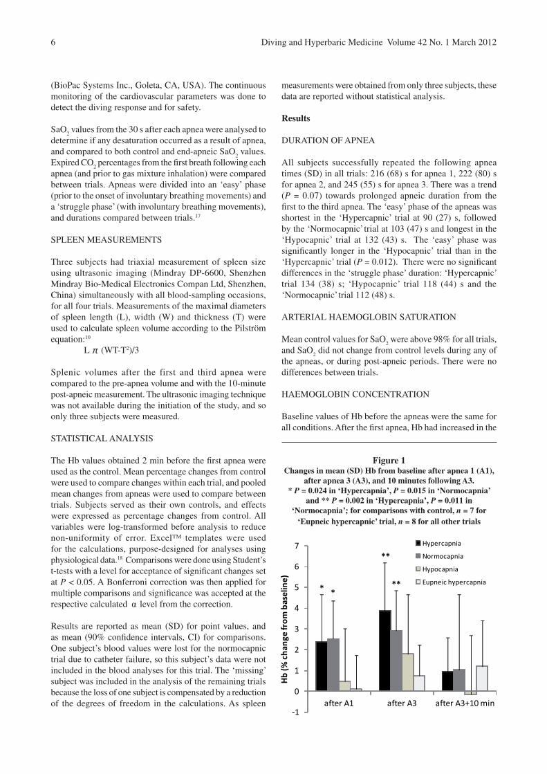
Figure 1Changes in mean (SD) Hb from baseline after apnea 1 (A1),
after apnea 3 (A3), and 10 minutes following A3.* P = 0.024 in ‘Hypercapnia’, P = 0.015 in ‘Normocapnia’
and ** P = 0.002 in ‘Hypercapnia’, P = 0.011 in ‘Normocapnia’; for comparisons with control, n = 7 for
‘Eupneic hypercapnic’ trial, n = 8 for all other trials
α
π
-1
0
1
2
3
4
5
6
7
after A1 after A3 after A3+10 min
Hb
(% c
hang
e fr
om b
asel
ine)
Hypercapnia
Normocapnia
Hypocapnia
Eupneic hypercapnia* *
**
**
Diving and Hyperbaric Medicine Volume 42 No. 1 March 2012 7
‘Hypercapnic’ (P = 0.024) and ‘Normocapnic’ (P = 0.015) trials, while the ‘Hypocapnic’ and ‘Eupneic hypercapnic’ trials were similar to control values (Figure 1). After the final apnea, Hb had further increased in the ‘Hypercapnic’ trial, to 4% above baseline (P = 0.002), and by 3 % in the ‘Normocapnic’ trial (P = 0.011), while the ‘Hypocapnic’ and ‘Eupneic hypercapnic’ trials showed no significant changes. Ten minutes after apneas, Hb values were not different from control values for any of the trials, nor were they different among trials. A comparison of the magnitude of change from baseline revealed no significant difference between trials.
SPLENIC VOLUME
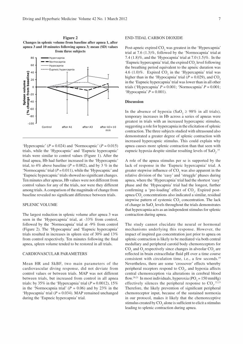
The largest reduction in splenic volume after apnea 3 was seen in the ‘Hypercapnia’ trial, at -33% from control, followed by the ‘Normocapnia’ trial at -9% from control (Figure 2). The ‘Hypocapnia’ and ‘Eupneic hypercapnia’ trials resulted in increases in spleen size of 30% and 13% from control respectively. Ten minutes following the final apnea, spleen volume tended to be restored in all trials.
CARDIOVASCULAR PARAMETERS
Mean HR and SkBF, two main parameters of the cardiovascular diving response, did not deviate from control values or between trials. MAP was not different between trials, but increased from control in all apnea trials: by 35% in the ‘Hypercapnia’ trial (P = 0.0012), 15% in the ‘Normocapnia trial’ (P = 0.06) and by 23% in the ‘Hypocapnia’ trial (P = 0.034). MAP remained unchanged during the ‘Eupneic hypercapnia’ trial.
END-TIDAL CARBON DIOXIDE
Post-apneic expired CO2 was greatest in the ‘Hypercapnia’
trial at 7.6 (1.3)%, followed by the ‘Normocapnia’ trial at
7.4 (1.8)%, and the ‘Hypocapnia’ trial at 7.0 (1.5)%. In the ‘Eupneic hypercapnia’ trial, the expired CO
2 level following
the breathing period equivalent to the apneic duration was 4.6 (1.0)%. Expired CO
2 in the ‘Hypercapnia’ trial was
higher than in the ‘Hypocapnia’ trial (P = 0.029), and CO2
in the ‘Eupneic hypercapnia’ trial was lower than in all other trials (‘Hypercapnia’ P = 0.001; ‘Normocapnia’ P = 0.001; ‘Hypocapnia’ P = 0.001).
Discussion
In the absence of hypoxia (SaO2 ≥ 98% in all trials),
temporary increases in Hb across a series of apneas were greatest in trials with an increased hypercapnic stimulus, suggesting a role for hypercapnia in the elicitation of splenic contraction. The three subjects studied with ultrasound also demonstrated a greater degree of splenic contraction with increased hypercapnic stimulus. This could explain why apnea causes more splenic contraction than that seen with eupneic hypoxia despite similar resulting levels of SaO
2.15
A role of the apnea stimulus per se is supported by the lack of response in the ‘Eupneic hypercapnia’ trial. A greater stepwise influence of CO
2 was also apparent in the
relative division of the ‘easy’ and ‘struggle’ phases during apnea, where the ‘Hypercapnia’ trial had the shortest ‘easy’ phase and the ‘Hypocapnia’ trial had the longest, further confirming a ‘pre-loading’ effect of CO
2. Expired post-
apnea CO2 concentrations also indicated a similar, residual
stepwise pattern of systemic CO2 concentration. The lack
of change in SaO2 levels throughout the trials demonstrates
that hypercapnia acts as an independent stimulus for splenic contraction during apnea.
The study cannot elucidate the neural or hormonal mechanisms underlying this response. However, the impact of inspired gas concentration just prior to apnea on splenic contraction is likely to be mediated via both central medullary and peripheral carotid body chemoreceptors for CO
2 and O
2 respectively since changes in alveolar CO
2 are
reflected in brain extracellular fluid pH over a time course consistent with circulation time, i.e., a few seconds.19 Nevertheless, there are some ‘crossover’ effects whereby peripheral receptors respond to CO
2, and hypoxia affects
central chemoreception via alterations in cerebral blood flow.20,21 In most individuals, hyperoxia (PO
2 = 150 mmHg)
effectively silences the peripheral response to CO2.22,23
Therefore, the likely prevention of significant peripheral chemoreceptor input, because of the sustained normoxia in our protocol, makes it likely that the chemoreceptive stimulus created by CO
2 alone is sufficient to elicit a stimulus
leading to splenic contraction during apnea.
Figure 2Changes in splenic volume from baseline after apnea 1, after apnea 3 and 10 minutes following apnea 3; mean (SD) values
from three subjects
-100
-80
-60
-40
-20
0
20
40
60
80
Control after A1 after A3 after A3+10 min
Sp
leen
vo
lum
e(%
ch
an
ge f
rom
base
lin
e)
Hypercapnia
Normocapnia
Hypocapnia
Eupneic hypercapnia
Diving and Hyperbaric Medicine Volume 42 No. 1 March 20128
Although the mechanisms leading to splenic contraction are, as yet, only partially defined, they almost certainly involve sympathetic innervation. The splenic nerve is mainly adrenergic in composition, and has been shown to respond powerfully to sympathetic discharge and related adrenergic output.24–26 Hoka and associates also noted marked changes in blood volume following hypercapnia in spleen-intact dogs, whereas this response was considerably decreased in splenectomised dogs.27 Similar sympathetic activity on the part of the splenic nerve in humans is likely. Bradycardia, a main component of the cardiovascular diving response, was not significant in any trial nor different between trials, suggesting that variations in CO
2 levels do not affect this
response. This also illustrates the independent elicitation of the splenic response, in accordance with previous findings.28
Both hypoxia and hypercapnia develop upon cessation of breathing, and splenic contraction-induced blood boosting may counteract, to some degree, these effects. Breath-hold divers would likely benefit from a strong splenic contraction, as the increase in circulating Hb would result in increased oxygen storage capacity, increased CO
2 buffering capacity
and a speedier recovery from hypoxia between apneas, especially when these haematological effects remain across several minutes between dives, whereas the cardiovascular diving response reverses rapidly.28 Based on the observations in this study, an increased capnic stimulus during apnea may elicit a stronger or earlier spleen response and subsequent Hb increase than apnea preceded by hyperventilation.
A direction for further research could be to focus on whether there is a true dose-response relationship between arterial CO
2 content and the splenic contraction response, as
appears possible from this study. It would also be of interest to compare the individual splenic responses to elevated CO
2 concentration of competition divers who employ
hyperventilation during ‘warm-up’ and divers without ‘warm-up’ practices before competition.29
Conclusions
The enhanced spleen-induced increase in Hb during normoxic hypercapnia suggests a role of hypercapnia as a trigger for splenic contraction during apnea. A separate role of the apnea stimulus is suggested by the lack of response in the ‘Eupneic hypercapnia’ trial.
Acknowledgements
We thank our subjects for participating in these studies, and the temporary co-worker Robert de Bruijn for valuable help during experiments. The study complies with Swedish laws and ethical standards and was financed by the Swedish National Centre for Research in Sports (CIF), and Mid Sweden University.
References
1 Gooden BA. Mechanisms of the human diving response. Integr Physiol Behav Sci. 1994;29;6-16.
2 Andersson J, Schagatay E. Arterial oxygen desaturation during apneas in humans. Undersea Hyperb Med. 1998;25:21-5.
3 Schagatay E, Andersson J. Diving response and apneic time in humans. Undersea Hyperb Med. 1998;25:13-9.
4 Schuitema K, Holm B. The role of different facial areas in eliciting human diving bradycardia. Acta Physiol Scand. 1988;132:119-20.
5 Lin YC, Shida KK, Hong SK. Effects of hypercapnia, hypoxia, and rebreathing on heart rate response during apnea. J Appl Physiol. 1983;54:166-71.
6 Hurford WE, Hong SK, Park YS, Ahn DW, Shiraki K, Mohri M, et al. Splenic contraction during breath-hold diving in the Korean Ama. J Appl Physiol. 1990;69:932-6.
7 Schagatay E, Andersson JPA, Hallén M, Pålsson B. Selected contribution: role of spleen emptying in prolonging apneas in humans. J Appl Physiol. 2000;90:1623-9.
8 Espersen K, Frandsen H, Lorentzen T, Kanstrup I-L, Christensen NJ. The human spleen as an erythrocyte reservoir in diving-related interventions. J Appl Physiol. 2002;92:2071-9.
9 Bakovic D, Eterovic D, Saratlija-Novakovic Z, Palada I, Valic Z, Bilopavlovic N, et al. Effect of human splenic contraction on variation in circulating blood cell counts. Clin Exp Pharmacol Physiol. 2005;32:944-51.
10 Schagatay E, Haughey H, Reimers J. Speed of spleen volume changes evoked by serial apneas. Eur J Appl Physiol. 2005;93:447-52.
11 Richardson MX, Lodin A, Reimers J, Schagatay E. Short-term effects of normobaric hypoxia on the human spleen. Eur J Appl Physiol. 2007;104:395-9.
12 Schagatay E, Andersson J, Nielsen B. Hematological response and diving response during apnea and apnea with face immersion. Eur J Appl Physiol. 2007;101:125-32.
13 Andersson JP, Liner MH, Runow E, Schagatay EK. Diving response and arterial oxygen saturation during apnea and exercise in breath-hold divers. J Appl Physiol. 2002;93:882-6.
14 Richardson MX, de Bruijn R, Schagatay E. Hypoxia augments apnea-induced increase in haemoglobin concentration and hematocrit. Eur J Appl Physiol. 2009;105:63-8.
15 Lodin-Sundström A, Schagatay E. Spleen contraction during 20 min normobaric hypoxia and 2 min apnea in humans. Aviat Space Environ Med. 2010;81:545-9.
16 Schagatay E. The human diving response: effects of temperature and training (dissertation). Lund, Sweden: Lund University; 1996.
17 Moore TO, Hong SK. Physiological and conventional breath-hold breaking points. J Appl Physiol. 1974;37:291-6.
18 Hopkins W. A new view of statistics. Available from: Internet Society for Sport Science http://www.sportsci.org/resource/stats/
19 Ahmad HR, Loeschcke HH. Fast bicarbonate-chloride exchange between plasma and brain extracellular fluid at maintained PCO
2. Pflugers Arch. 1982;395:300-5.
20 Poulin MJ, Liang PJ, Robbins PA. Dynamics of the cerebral blood flow response to step changes in end-tidal PCO
2 and
PO2 in humans. J Appl Physiol. 1996;81:1084-95.
21 Vovk A, Cunningham DA, Kowalchuk JM, Paterson DH, Duffin J. Cerebral blood flow responses to changes in oxygen
Diving and Hyperbaric Medicine Volume 42 No. 1 March 2012 9
and carbon dioxide in humans. Can J Physiol Pharmacol. 2002;80:819-27.
22 Lloyd BB, Jukes MG, Cunningham DJ. The relation between alveolar oxygen pressure and the respiratory response to carbon dioxide in man. Q J Exp Physiol Cogn Med Sci. 1958;43:214-27.
23 Mohan R, Duffin J. The effect of hypoxia on the ventilatory response to carbon dioxide in man. Respir Physiol. 1997;108:101-15.
24 Ayers AB, Davies BN, Withrington PG. Responses of the isolated, perfused human spleen to sympathetic nerve stimulation, catecholamines and polypeptides. Br J Pharmacol. 1972;44:17-30.
25 Davies BN, Powis DA, Withrington PG. The differential effect of cooling on the responses of splenic capsular and vascular smooth muscle to nerve stimulation and noradrenaline. Pflugers Archiv. 1978;377:87-94.
26 Herman NL, Kostreva DR, Kampine JP. Splenic afferents and some of their reflex responses. Am J Physiol. Regul Integr Comp Physiol. 1982;242:R247-54.
27 Hoka S, Arimura H, Bosnjak ZJ, Kampine JP. Regional venous outflow, blood volume, and sympathetic nerve activity during hypercapnia and hypoxic hypercapnia. Can J Physiol Pharmacol. 1970;70:1032-9.
28 Schagatay E, Andersson JP, Nielsen B. Hematologial response and diving response during apnea and apnea with face immersion. Eur J Appl Physiol. 2007;101:125-32.
29 Schagatay E. Predicting performance in competitive apnea diving. Part II: dynamic apnea. Diving Hyperb Med. 2010;40:11-22.
Submitted: 08 April 2011Accepted: 18 October 2011
Matt X Richardson, PhD, Harald K Engan, MSc, Angelica Lodin-Sundström, MSc, Erika Schagatay, PhD,Environmental Physiology Group, Department of Engineering and Sustainable Development, Mid Sweden University, Östersund, Sweden.Professor Schagatay also works at the National Winter Sports Research Centre, Östersund, Sweden.
Address for correspondence:Harald K EnganEnvironmental Physiology Group, Mid Sweden UniversityDepartment of Engineering and Sustainable DevelopmentAkademigatan 1, SE 831 25Östersund, Sweden.Phone: +47-(0)73-558954Fax: +46-(0)63-165700E-mail: <[email protected]>
Diving and Hyperbaric Medicine Volume 42 No. 1 March 201210
The effect of intravenous perfluorocarbon emulsions on whole-body oxygenation after severe decompression sicknessCameron R Smith, J Travis Parsons, Jiepei Zhu and Bruce D Spiess
Introduction
Breathing compressed air increases the amount of nitrogen (N
2) dissolved in body fluids.1–3 Factors such as ambient
pressure and time at depth are the primary determinants of the amount of N
2 absorbed.1–4 As ambient pressure
decreases, dissolved gas tensions in tissue can exceed ambient pressure. This supersaturated state may lead to the formation and growth of gas bubbles, resulting in venous gas emboli (VGE) and possible arterial gas emboli (AGE).4,5 It is believed that these bubbles within the vasculature and tissues are the root cause of decompression sickness (DCS).4,5 There are likely multiple pathophysiological mechanisms at play in DCS, including impairment of microcirculation by inert gas bubbles, increased blood viscosity, endothelial damage and complement activation.6–10 The physicochemical discontinuity of the gas-blood interface can also denature proteins promoting the release of fatty acids from cell membranes leading to the formation of fat emboli.4,5 When bubbles obstruct capillaries or venules, ischaemia ensues followed by reperfusion-induced oxidative tissue damage.11
Perfluorocarbon emulsions (PFCs) are emulsions of fluorinated hydrocarbons within phospholipid micro-particle micelles.12 PFCs have been developed in medicine as intravenous oxygen (O
2) therapeutics.12 However,
compared to how whole blood carries the majority of its O2,
the transport of O2 by PFCs is fundamentally different. O
2
carried by PFCs is not bound, as with haemoglobin, rather it is dissolved in the PFC. Pure perfluorocarbons can dissolve up to 600 ml L-1 O
2,13 whereas plasma can only dissolve
0.031 ml L-1 and whole blood at 150 gm L-1 haemoglobin can contain up to 210 ml L-1 O
2.12 The O
2 dissolved in PFCs is
all available to tissue, whereas that bound by haemoglobin is restricted (arterial pO
2 would need to drop below 40 mmHg
for greater than 25% of bound O2 to be released).14
Microcirculatory changes such as oedema, vasospasm, white cell activation and vessel plugging result in decreased erythrocyte delivery of O
2 to watershed tissue beds, yet
plasma flow may continue without red cells.15 PFCs, due to their extremely small particle size (~0.1–0.4 µm), can be delivered in this trickle-flow of plasma.12,16,17 Plasma O
2 delivery by PFCs is enough to keep tissue alive, as seen
with Fluosol DA-20%, a PFC which reduced myocardial infarction and garnered FDA approval.18,19
PFCs are also effective in treating DCS, AGE and VGE.20–26 Using a swine saturation dive model with direct ascent to the surface, it was found that administration of intravenous (IV) PFCs and 100% O
2 post-decompression decreased mortality,
the incidence of DCS and the number of neurological events compared to animals administered just 100% O
2 or
room air.21 Also, PFC and 1 hour of 100% O2 given at the
onset of DCS significantly decreased mortality observed 24 hours post-dive compared to animals treated with saline and 100% O
2 in a swine model of rapid decompression.27
AbstractSmith CR, Parsons JT, Zhu J, Spiess BD. The effect of intravenous perfluorocarbon emulsions on whole-body oxygenation after severe decompression sickness. Diving Hyperb Med. 2012;42(1):10-17.)Introduction: Decompression sickness (DCS) results from a decrease in ambient pressure leading to supersaturation of tissues with inert gas and bubble formation. Perfluorocarbons (PFCs) are able to dissolve vast amounts of non-polar gases. Intravenous (IV) PFC emulsions reduce both morbidity and mortality associated with DCS, but the mechanism of this protective effect has not yet been demonstrated.Methods: Juvenile Dorper-cross sheep (n = 31) were anaesthetised and instrumented for physiological monitoring, IV fluid administration and blood sampling. Animals were compressed in air in a hyperbaric chamber to 608 kPa for 30 minutes and then rapidly decompressed. Upon decompression, animals were randomly assigned to receive 6 mL kg-1 of PFC or saline over 10 minutes beginning immediately after chamber exit. Arterial and mixed venous bloods were drawn at 5, 10, 15, 30, 60 and 90 minutes to examine pH, partial pressures of oxygen and carbon dioxide, oxygen saturation and electrolytes.Results: Compared to saline, PFC administration increased arterial oxygen content (16.33 ± 0.28 vs. 14.68 ± 0.26 ml dL-1, P < 0.0001), mixed venous oxygen content (12.56 ± 0.28 vs. 11.62 ± 0.26 ml dL-1, P = 0.0167), oxygen delivery (14.83 ± 0.28 vs. 13.39 ± 0.26 ml min-1 kg-1, P = 0.0003) and tissue oxygen consumption (3.30 ± 0.15 vs. 2.78 ± 0.13 ml min-1 kg-1, P = 0.0149) but did not increase the extraction ratio (0.22 ± 0.012 vs. 0.21 ± 0.011, P = 0.5343).Conclusions: It is likely that the improved oxygenation explains, at least in part, the previously-observed therapeutic effects of PFCs in DCS.
Key wordsBlood substitutes, perfluorocarbons, decompression sickness, treatment, oxygen, oxygen consumption
Diving and Hyperbaric Medicine Volume 42 No. 1 March 2012 11
Similarly it was found that IV PFCs improve outcomes after massive VGE, cerebral AGE, and coronary AGE.23,24,28 IV PFCs have also been shown to increase N
2 washout after
VGE.26 PFC administration is of benefit in the treatment of decompression illnesses, but the mechanism of this benefit has not been elucidated. Is it the PFCs’ ability to increase N
2 washout and remove bubbles obstructing circulation,
a product of improving O2 supply and metabolic state
of tissue, or some combination of these? The research described here was designed to investigate the effect of IV PFCs administered acutely after surfacing on whole-body oxygenation in an ovine model of severe DCS.
Materials and methods
All experiments were performed in accordance with the National Institutes of Health Guide for the care and use of laboratory animals, and were approved by the Department of Defense and the Virginia Commonwealth University Institutional Animal Care and Use Committees. Juvenile Dorper-cross sheep of either sex (Robinson Services, Inc., Mocksville, NC) weighing 18.5 ± 2.6 kg were housed in United States Department of Agriculture and Association for Assessment and Accreditation of Laboratory Animal Care International approved facilities in social flocks with free access to food and water on a 12-hour light/dark cycle. Sheep were allowed a minimum of three days for acclimatisation and veterinary inspection prior to use in any experiment.
PREPARATION AND INSTRUMENTATION
Prior to the experiment, sheep were muzzled for a period of 48 hours in order to prevent access to food but to provide free access to water while remaining with their flock to limit animal stress. Sheep were sedated with ketamine/xylazine (20.0/2.0 mg kg-1 IM) and placed supine on the surgical table. Animals were intubated with a 9.0 mm internal diameter cuffed endotracheal tube (Hudson RCI, Temecula, CA) and ventilated with 50/50 N
2/O
2 using a Siemens 900C ventilator
(Siemens Corp., New York, NY) with a tidal volume of approximately 10 ml kg-1 and a rate of approximately 15 breaths per minute adjusted to maintain arterial pCO
2 at 40
± 5 mmHg. An orogastric tube fashioned from TYGON® R-3603 tubing (Satin-Gobain Performance Plastics Corp., Akron, OH) was advanced into the rumen to allow for fluid drainage and to allow gas accumulated in the gut during the air dive to vent upon decompression. A MAC® 2-port introducer sheath (Arrow International Inc., Reading, PA) was placed in the right external jugular vein to allow for the administration of fluids and anaesthetic cocktail.
Once IV access was secured, administration of ‘triple drip’ anaesthetic cocktail (ketamine/xylazine/guaifenesin 2.0/0.1/50.0 mg ml-1 in 5% dextrose) was begun immediately at 1.0–2.0 ml kg-1 hr-1 titrated to maintain a surgical plane of anaesthesia using a Harvard Apparatus PHD 2000 syringe pump (Harvard Apparatus, Holliston, MA). The left
femoral artery was cannulated with an 18-gauge femoral arterial catheter (Arrow International Inc., Reading, PA) for monitoring of arterial pressure (AP) and arterial blood sampling. The right femoral vein was cannulated with a 4-French double-lumen catheter (Arrow International Inc., Reading, PA) for the anaesthetic administration while in the hyperbaric chamber and for study drug administration after exiting the chamber. The left femoral vein was cannulated for the placement of a 7.5 Fr CCOmbo® continuous cardiac output (CCO) pulmonary artery catheter (Edwards Lifesciences, Irvine, CA) to allow for CCO, central venous (CVP) and pulmonary arterial pressure (PAP) monitoring and mixed venous blood sampling. Respiratory gases were monitored continuously using an MGA 1100 respiratory mass spectrometer (Perkin-Elmer, Norwalk, CT).
Following surgical manipulations, all animals were allowed to stabilise for 30 minutes. After stabilisation, animals were weaned off the ventilator until capable of spontaneously breathing prior to being placed inside the hyperbaric chamber. Normal saline was administered intravenously at a rate of approximately 1 ml min-1 throughout the surgical procedure in order to ensure proper hydration of all animals.
SHEEP DRY-DIVE PROCEDURES
Once weaned from the ventilator, monitoring equipment was disconnected and sheep (n = 31) were placed into a Reimers Systems model #17-48-100 Research Hyperbaric Chamber (Reimers Systems, Inc., Springfield, VA). During the dry dive all animals breathed room air and general anaesthesia was maintained using a continuous infusion of ‘triple drip’ as described above. Sheep were subjected to the following dive profile. The chamber was pressurised at a rate of 101.3 kPa min-1 to a pressure of 203 kPa. From 203 kPa the chamber was pressurised at a rate of 203 kPa min-1 to a pressure of 608 kPa. The pressure of 608 kPa was maintained for 27 minutes, after which sheep were immediately decompressed to ambient pressure at a rate of 203 kPa min-1.
POST-DECOMPRESSION MONITORING
Upon complete decompression (time = 0) all animals were quickly removed from the hyperbaric chamber and monitoring equipment was reconnected, as was the ventilator with settings and breathing gas unchanged from pre-compression/decompression settings. Animals were randomised using a computer-generated block randomisation sequence such that for each eight animals, four were assigned to receive IV infusion of 6.0 ml kg-1 PFC (n = 15, 60% w/v tert-butyl perfluorocyclohexane) and four were assigned to receive saline control (n = 16) as an infusion over 10 minutes. All animals were monitored for 90 minutes after decompression, during which time both arterial and mixed venous blood samples were drawn and analysed using a Radiometer OSM 3 Hemoximeter and a Radiometer ABL
Diving and Hyperbaric Medicine Volume 42 No. 1 March 201212
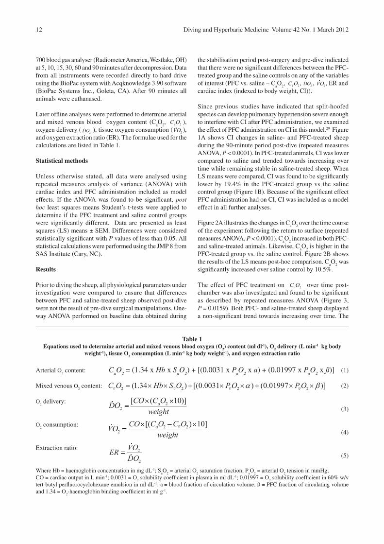
700 blood gas analyser (Radiometer America, Westlake, OH) at 5, 10, 15, 30, 60 and 90 minutes after decompression. Data from all instruments were recorded directly to hard drive using the BioPac system with Acqknowledge 3.90 software (BioPac Systems Inc., Goleta, CA). After 90 minutes all animals were euthanased.
Later offline analyses were performed to determine arterial and mixed venous blood oxygen content (C
aO
2, ),
oxygen delivery ( ), tissue oxygen consumption ( ), and oxygen extraction ratio (ER). The formulae used for the calculations are listed in Table 1.
Statistical methods
Unless otherwise stated, all data were analysed using repeated measures analysis of variance (ANOVA) with cardiac index and PFC administration included as model effects. If the ANOVA was found to be significant, post hoc least squares means Student’s t-tests were applied to determine if the PFC treatment and saline control groups were significantly different. Data are presented as least squares (LS) means ± SEM. Differences were considered statistically significant with P values of less than 0.05. All statistical calculations were performed using the JMP 8 from SAS Institute (Cary, NC).
Results
Prior to diving the sheep, all physiological parameters under investigation were compared to ensure that differences between PFC and saline-treated sheep observed post-dive were not the result of pre-dive surgical manipulations. One-way ANOVA performed on baseline data obtained during
the stabilisation period post-surgery and pre-dive indicated that there were no significant differences between the PFC-treated group and the saline controls on any of the variables of interest (PFC vs. saline – C
aO
2, , , , ER and
cardiac index (indexed to body weight, CI)).
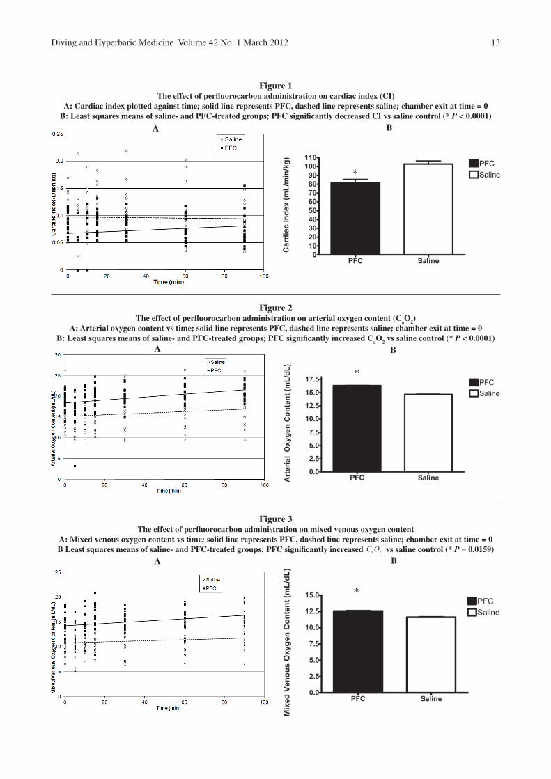
Since previous studies have indicated that split-hoofed species can develop pulmonary hypertension severe enough to interfere with CI after PFC administration, we examined the effect of PFC administration on CI in this model.29 Figure 1A shows CI changes in saline- and PFC-treated sheep during the 90-minute period post-dive (repeated measures ANOVA, P < 0.0001). In PFC-treated animals, CI was lower compared to saline and trended towards increasing over time while remaining stable in saline-treated sheep. When LS means were compared, CI was found to be significantly lower by 19.4% in the PFC-treated group vs the saline control group (Figure 1B). Because of the significant effect PFC administration had on CI, CI was included as a model effect in all further analyses.
Figure 2A illustrates the changes in CaO
2 over the time course
of the experiment following the return to surface (repeated measures ANOVA, P < 0.0001). C
aO
2 increased in both PFC-
and saline-treated animals. Likewise, CaO
2 is higher in the
PFC-treated group vs. the saline control. Figure 2B shows the results of the LS means post-hoc comparison. C
aO
2 was
significantly increased over saline control by 10.5%.
The effect of PFC treatment on over time post-chamber was also investigated and found to be significant as described by repeated measures ANOVA (Figure 3, P = 0.0159). Both PFC- and saline-treated sheep displayed a non-significant trend towards increasing over time. The
Arterial O2 content: C
aO
2 = (1.34 x Hb x S
aO
2) + [(0.0031 x P
aO
2 x a) + (0.01997 x P
aO
2 x ß)] (1)
Mixed venous O2 content: (2)
O2 delivery:
(3)
O2 consumption:
(4)
Extraction ratio: (5)
Where Hb = haemoglobin concentration in mg dL-1; SaO
2 = arterial O
2 saturation fraction; P
aO
2 = arterial O
2 tension in mmHg;
CO = cardiac output in L min-1; 0.0031 = O2 solubility coefficient in plasma in ml dL-1; 0.01997 = O
2 solubility coefficient in 60% w/v
tert-butyl perfluorocyclohexane emulsion in ml dL-1; a = blood fraction of circulation volume; ß = PFC fraction of circulating volume and 1.34 = O
2-haemoglobin binding coefficient in ml g-1.
22
[ ( 10)]aCO C ODOweight
2 22
[( ) 10]a vCO C O C OVOweight
2
2
VOERDO
Table 1Equations used to determine arterial and mixed venous blood oxygen (O2) content (ml dl-1), O2 delivery (L min-1 kg body
weight-1), tissue O2 consumption (L min-1 kg body weight-1), and oxygen extraction ratio
2 2 2 2(1.34 ) [(0.0031 ) (0.01997 )]v v v vC O Hb S O P O P O
2vC O
2DO 2VO 2vC O
2vC O 2DO 2VO
Diving and Hyperbaric Medicine Volume 42 No. 1 March 2012 13
Figure 2The effect of perfluorocarbon administration on arterial oxygen content (CaO2)
A: Arterial oxygen content vs time; solid line represents PFC, dashed line represents saline; chamber exit at time = 0B: Least squares means of saline- and PFC-treated groups; PFC significantly increased CaO2 vs saline control (* P < 0.0001)
Figure 3The effect of perfluorocarbon administration on mixed venous oxygen content
A: Mixed venous oxygen content vs time; solid line represents PFC, dashed line represents saline; chamber exit at time = 0B Least squares means of saline- and PFC-treated groups; PFC significantly increased 2vC O vs saline control (* P = 0.0159)
Figure 1The effect of perfluorocarbon administration on cardiac index (CI)
A: Cardiac index plotted against time; solid line represents PFC, dashed line represents saline; chamber exit at time = 0B: Least squares means of saline- and PFC-treated groups; PFC significantly decreased CI vs saline control (* P < 0.0001)
A
A
A
B
B
B
*
*
*
Diving and Hyperbaric Medicine Volume 42 No. 1 March 201214
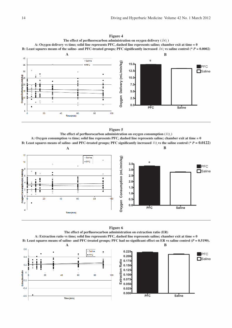
Figure 4The effect of perfluorocarbon administration on oxygen delivery ( 2DO )
A: Oxygen delivery vs time; solid line represents PFC, dashed line represents saline; chamber exit at time = 0B: Least squares means of the saline- and PFC-treated groups; PFC significantly increased 2DO vs saline control (* P = 0.0002)
Figure 5The effect of perfluorocarbon administration on oxygen consumption ( 2VO )
A: Oxygen consumption vs time; solid line represents PFC, dashed line represents saline; chamber exit at time = 0B: Least squares means of saline- and PFC-treated groups; PFC significantly increased 2VO vs the saline control (* P = 0.0122)
Figure 6The effect of perfluorocarbon administration on extraction ratio (ER)
A: Extraction ratio vs time; solid line represents PFC, dashed line represents saline; chamber exit at time = 0B: Least squares means of saline- and PFC-treated groups; PFC had no significant effect on ER vs saline control (P = 0.5190).
A
A
A
B
B
B
*
*
Diving and Hyperbaric Medicine Volume 42 No. 1 March 2012 15
results of the LS means comparison are shown in Figure 3B. was found to be significantly higher in PFC animals vs saline control by 6.7%.
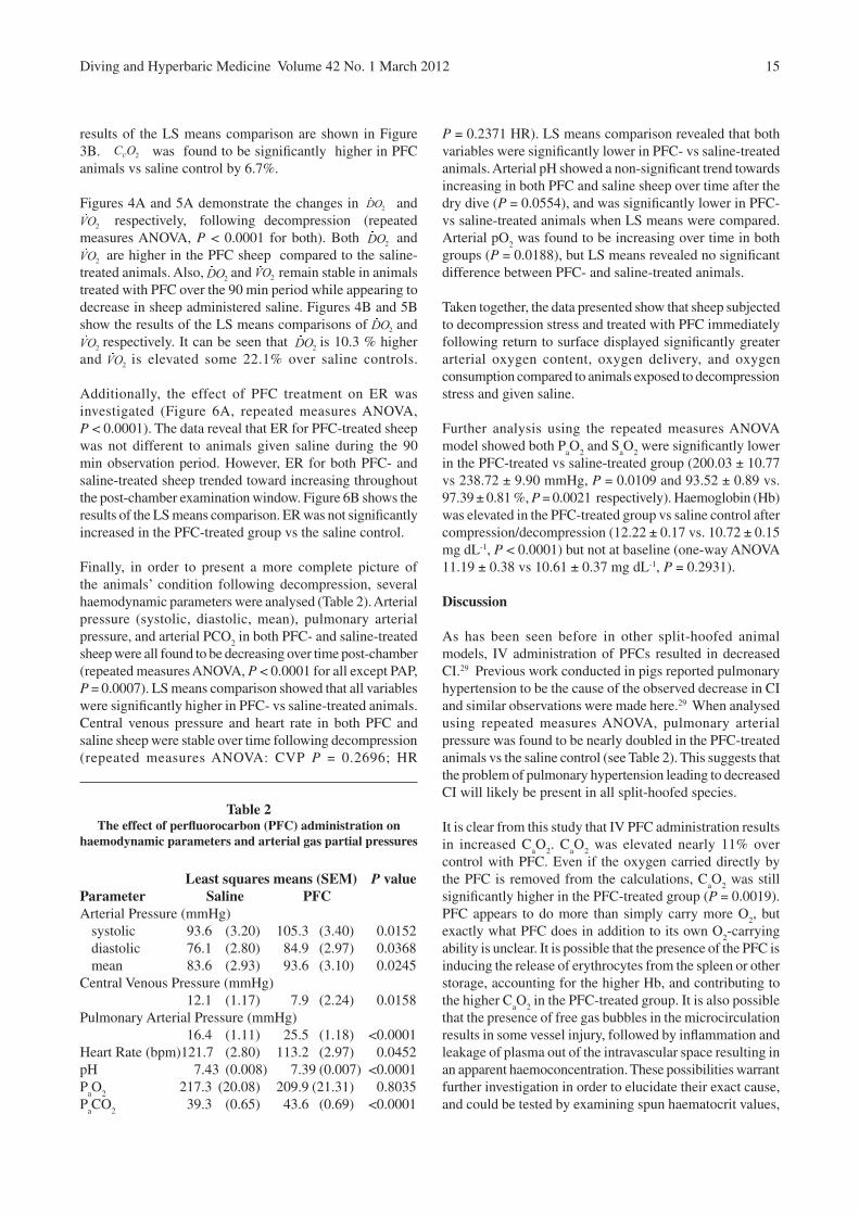
Figures 4A and 5A demonstrate the changes in and respectively, following decompression (repeated measures ANOVA, P < 0.0001 for both). Both and are higher in the PFC sheep compared to the saline-treated animals. Also, and remain stable in animals treated with PFC over the 90 min period while appearing to decrease in sheep administered saline. Figures 4B and 5B show the results of the LS means comparisons of and respectively. It can be seen that is 10.3 % higher and is elevated some 22.1% over saline controls.
Additionally, the effect of PFC treatment on ER was investigated (Figure 6A, repeated measures ANOVA,P < 0.0001). The data reveal that ER for PFC-treated sheep was not different to animals given saline during the 90 min observation period. However, ER for both PFC- and saline-treated sheep trended toward increasing throughout the post-chamber examination window. Figure 6B shows the results of the LS means comparison. ER was not significantly increased in the PFC-treated group vs the saline control.
Finally, in order to present a more complete picture of the animals’ condition following decompression, several haemodynamic parameters were analysed (Table 2). Arterial pressure (systolic, diastolic, mean), pulmonary arterial pressure, and arterial PCO
2 in both PFC- and saline-treated
sheep were all found to be decreasing over time post-chamber (repeated measures ANOVA, P < 0.0001 for all except PAP, P = 0.0007). LS means comparison showed that all variables were significantly higher in PFC- vs saline-treated animals. Central venous pressure and heart rate in both PFC and saline sheep were stable over time following decompression (repeated measures ANOVA: CVP P = 0.2696; HR
P = 0.2371 HR). LS means comparison revealed that both variables were significantly lower in PFC- vs saline-treated animals. Arterial pH showed a non-significant trend towards increasing in both PFC and saline sheep over time after the dry dive (P = 0.0554), and was significantly lower in PFC- vs saline-treated animals when LS means were compared. Arterial pO
2 was found to be increasing over time in both
groups (P = 0.0188), but LS means revealed no significant difference between PFC- and saline-treated animals.
Taken together, the data presented show that sheep subjected to decompression stress and treated with PFC immediately following return to surface displayed significantly greater arterial oxygen content, oxygen delivery, and oxygen consumption compared to animals exposed to decompression stress and given saline.
Further analysis using the repeated measures ANOVA model showed both P
aO
2 and S
aO
2 were significantly lower
in the PFC-treated vs saline-treated group (200.03 ± 10.77 vs 238.72 ± 9.90 mmHg, P = 0.0109 and 93.52 ± 0.89 vs. 97.39 ± 0.81 %, P = 0.0021 respectively). Haemoglobin (Hb) was elevated in the PFC-treated group vs saline control after compression/decompression (12.22 ± 0.17 vs. 10.72 ± 0.15 mg dL-1, P < 0.0001) but not at baseline (one-way ANOVA 11.19 ± 0.38 vs 10.61 ± 0.37 mg dL-1, P = 0.2931).
Discussion
As has been seen before in other split-hoofed animal models, IV administration of PFCs resulted in decreased CI.29 Previous work conducted in pigs reported pulmonary hypertension to be the cause of the observed decrease in CI and similar observations were made here.29 When analysed using repeated measures ANOVA, pulmonary arterial pressure was found to be nearly doubled in the PFC-treated animals vs the saline control (see Table 2). This suggests that the problem of pulmonary hypertension leading to decreased CI will likely be present in all split-hoofed species.
It is clear from this study that IV PFC administration results in increased C
aO
2. C
aO
2 was elevated nearly 11% over
control with PFC. Even if the oxygen carried directly by the PFC is removed from the calculations, C
aO
2 was still
significantly higher in the PFC-treated group (P = 0.0019). PFC appears to do more than simply carry more O
2, but
exactly what PFC does in addition to its own O2-carrying
ability is unclear. It is possible that the presence of the PFC is inducing the release of erythrocytes from the spleen or other storage, accounting for the higher Hb, and contributing to the higher C
aO
2 in the PFC-treated group. It is also possible
that the presence of free gas bubbles in the microcirculation results in some vessel injury, followed by inflammation and leakage of plasma out of the intravascular space resulting in an apparent haemoconcentration. These possibilities warrant further investigation in order to elucidate their exact cause, and could be tested by examining spun haematocrit values,
Table 2The effect of perfluorocarbon (PFC) administration on
haemodynamic parameters and arterial gas partial pressures
Least squares means (SEM) P valueParameter Saline PFCArterial Pressure (mmHg)
systolic 93.6 (3.20) 105.3 (3.40) 0.0152diastolic 76.1 (2.80) 84.9 (2.97) 0.0368mean 83.6 (2.93) 93.6 (3.10) 0.0245
Central Venous Pressure (mmHg) 12.1 (1.17) 7.9 (2.24) 0.0158Pulmonary Arterial Pressure (mmHg) 16.4 (1.11) 25.5 (1.18) <0.0001Heart Rate (bpm) 121.7 (2.80) 113.2 (2.97) 0.0452pH 7.43 (0.008) 7.39 (0.007) <0.0001P
aO
2 217.3 (20.08) 209.9 (21.31) 0.8035
PaCO
2 39.3 (0.65) 43.6 (0.69) <0.0001
2DO
2VO
2DO
2VO
2VO 2DO
2DO
2VO
2VO 2DO
2vC O
Diving and Hyperbaric Medicine Volume 42 No. 1 March 201216
plasma protein content and/or by conducting tagged RBC concentration studies.
The observation that PFC administration results in increases in both and of 10% and 22%, respectively, demonstrates that the PFC was able to not only increase the amount of O
2 present in the blood, but to improve tissue
access to that O2. This suggests that the mechanism whereby
IV PFC improves tissue oxygenation is not simply a result of its ability to carry greater quantities of O
2, but that it
facilitates O2 delivery to cells. This may take the form of the
PFC extravasating in capillary beds, taking dissolved oxygen with it. Alternatively, the PFC emulsion particles, being approximately 1/100th–1/1000th the size of an erythrocyte, may be able to pass through blood vessels where red cell flow has been blocked by bubbles, but a trickle flow of plasma remains.21,25 In this case the small amount of O
2 carried in the
PFC may be sufficient to keep viable tissues that otherwise might succumb to hypoxic injury.
More interestingly, PFC particles may act as a bridge, facilitating the movement of O
2 from erythrocytes into
tissues. This possibility has very intriguing implications. As shown above, the amount of O
2 actually dissolved in
PFC is relatively small. Haemoglobin binding O2 remains
the dominant mechanism for O2 transport. Once in capillary
beds, the greatest impediment to the offloading of O2 from
haemoglobin is the plasma.30 O2 is very insoluble in plasma,
and much more soluble in PFC. Therefore, PFC could act as a transport vessel for O
2, ferrying it from erythrocytes to
tissues, a mechanism somewhat akin to facilitated diffusion across cell membranes. These possible mechanisms should be explored further in future studies.
Conclusion
These results demonstrate that improved tissue oxygenation at a whole-body level is likely responsible for at least a portion of the beneficial effects offered by the IV administration of PFC emulsions after decompression sickness.
Acknowledgements
The authors thank Drs Kevin Ward, R Wayne Barbee, and Penny S Reynolds for their insight and suggestions during the experimental design and data analysis.
Conflict of interest
Travis Parsons is an investor owning 90 shares of stock in Oxygen Biotherapeutics, Inc., less than 0.0001% of public shares available.
Bruce Spiess is an investor owning 10,000 shares of stock in Oxygen Biotherapeutics Inc., less than 0.01% of public shares available.
References
1 Guyton AC, Hall JE. Physiology of deep-sea diving and other hyperbaric conditions. In: Guyton AC, Hall AC, editors. Textbook of medical physiology, 10th ed. Philadelphia, PA: Saunders; 2000. p. 504-9.
2 Moon RE. Treatment of diving emergencies. Crit Care Clin. 1999;15:429-56.
3 Replogle WH, Sanders SD, Keeton JE, Phillips DM. Scuba diving injuries. Am Fam Physician. 1988;37:135-42.
4 DeGorordo A, Vallejo-Manzur F, Chanin K, Varon J. Diving emergencies. Resuscitation. 2003;59:171-80.
5 Dutka AJ, Francis TJ. Pathophysiology of decompression sickness. In: Bove AA, Davis JC, editors. Bove and Davis’ diving medicine, 3rd ed. Philadelphia: Saunders; 1997. p. 159-75.
6 Dutka AJ, Kochanek PM, Hallenbeck JM. Influence of granulocytopenia on canine cerebral ischemia induced by air embolism. Stroke. 1989;20:390-5.
7 Hallenbeck JM, Bove AA, Elliot DH. The bubble as a non-mechanical trigger in decompression sickness. In: Ackles KN, editor. Proceedings of a symposium on blood bubble interactions. Report # 73-CP-960. Downsview, Ontario: Defence and Civil Institute for Environmental Medicine; 1973.
8 Hallenbeck JM, Bove AA, Moquin RB, Elliott DH. Accelerated coagulation of whole blood and cell-free plasma by bubbling in vitro. Aerosp Med. 1973;44:712-4.
9 Hills BA, James PB. Microbubble damage to the blood-brain barrier: Relevance to decompression sickness. Undersea Biomed Res. 1991;18:111-6.
10 Vane JR, Anggard EE, Botting RM. Regulatory functions of the vascular endothelium. N Engl J Med. 1990;323:27-36.
11 Elliot DH, Moon RE. Manifestations of decompression disorders. In: Bennett PB, Elliott DH, editors. The physiology and medicine of diving, 4th ed. Philadelphia: Saunders; 1993. P 613.
12 Stowell CP, Levin J, Spiess BD, Winslow RM. Progress in the development of rbc substitutes. Transfusion. 2001;41: 87-99.
13 O’Brien RN, Langlais AJ, Seufert WD. Diffusion coefficients of respiratory gases in a perfluorocarbon liquid. Science. 1982;217:153-5.
14 Leow MK. Configuration of the hemoglobin oxygen dissociation curve demystified: A basic mathematical proof for medical and biological sciences undergraduates. Adv Physiol Educ. 2007;31:198-201.
15 Theilen H, Schrock H, Kuschinsky W. Gross persistence of capillary plasma perfusion after middle cerebral artery occlusion in the rat brain. J Cereb Blood Flow Metab. 1994;14:1055-61.
16 Biro GP. Perfluorocarbon-based red blood cell substitutes. Transfus Med Rev. 1993;7:84-95.
17 Riess JG. Perfluorocarbon-based oxygen delivery. Artif Cells Blood Substit Immobil Biotechnol. 2006;34:567-80.
18 Kerins DM. Role of the perfluorocarbon fluosol-da in coronary angioplasty. Am J Med Sci. 1994;307:218-21.
19 Kent KM, Cleman MW, Cowley MJ, Forman MB, Jaffe CC, Kaplan M, et al. Reduction of myocardial ischemia during percutaneous transluminal coronary angioplasty with oxygenated fluosol. Am J Cardiol. 1990;66:279-84.
20 Dainer H, Nelson J, Brass K, Montcalm-Smith E, Mahon R.
2DO 2VO
Diving and Hyperbaric Medicine Volume 42 No. 1 March 2012 17
Short oxygen prebreathing and intravenous perfluorocarbon emulsion reduces morbidity and mortality in a swine saturation model of decompression sickness. J Appl Physiol. 2007;102:1099-104.
21 Dromsky DM, Spiess BD, Fahlman A. Treatment of decompression sickness in swine with intravenous perfluorocarbon emulsion. Aviat Space Environ Med. 2004;75:301-5.
22 Spiess BD. Perfluorocarbon emulsions: one approach to intravenous artificial respiratory gas transport. Int Anesthesiol Clin. 1995;33:103-13.
23 Spiess BD, Braverman B, Woronowicz AW, Ivankovich AD. Protection from cerebral air emboli with perfluorocarbons in rabbits. Stroke. 1986;17:1146-9.
24 Spiess BD, McCarthy R, Piotrowski D, Ivankovich AD. Protection from venous air embolism with fluorocarbon emulsion fc-43. J Surg Res. 1986;41:439-44.
25 Spiess BD, McCarthy RJ, Tuman KJ, Woronowicz AW, Tool KA, Ivankovich AD. Treatment of decompression sickness with a perfluorocarbon emulsion (fc-43). Undersea Biomed Res. 1988;15:31-7.
26 Zhu J, Hullett JB, Somera L, Barbee RW, Ward KR, Berger BE, et al. Intravenous perfluorocarbon emulsion increases nitrogen washout after venous gas emboli in rabbits. Undersea Hyperb Med. 2007;34:7-20.
27 Mahon RT, Watanabe TT, Wilson MC, Auker CR. Intravenous perfluorocarbon after onset of decompression sickness decreases mortality in 20-kg swine. Aviat Space Environ Med. 2010;81:555-9.
28 Spiess BD, McCarthy RJ, Tuman KJ, Ivankovich AD. Protection from coronary air embolism by a perfluorocarbon emulsion (fc-43). J Cardiothorac Anesth. 1987;1:210-5.
29 Spiess BD, Zhu J, Pierce B, Weis R, Berger BE, Reses J, et al. Effects of perfluorocarbon infusion in an anesthetized swine decompression model. J Surg Res. 2009;153:83-94.
30 Popel AS. A finite-element model of oxygen diffusion in the pulmonary capillaries. J Appl Physiol. 1997;82:1717-8.
Submitted: 18 August 2011Accepted: 12 January 2012
Cameron R Smith, PhD1,3,5, J Travis Parsons, PhD4,5, Jiepei Zhu, MD, PhD1,5, and Bruce D Spiess, MD1,2,5
Departments of Anesthesiology1, Emergency Medicine2,
Physiology3, Neurosurgery4, and the Virginia Commonwealth University Reanimation Engineering Shock Center (VCURES)5, Virginia Commonwealth University Medical Center, Richmond, Virginia, USA.
Address for correspondence:Cameron R Smith, PhDPO Box 980695RichmondVirginia 23298-0695, USAPhone: +01-(0)804-827-2205Fax: +01-(0)804-828-6413E-mail: <[email protected]>
This work was supported by a grant from the United States Office of Naval Research (ONR) (N000140210399)
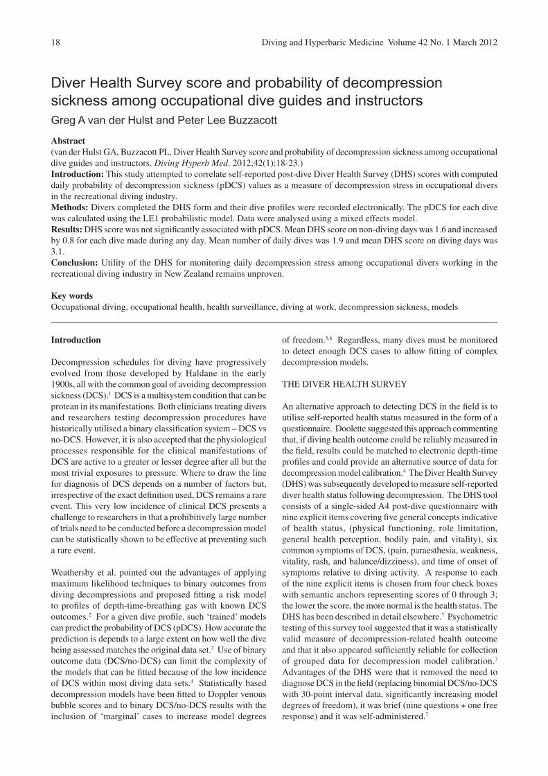
Diving and Hyperbaric Medicine Volume 42 No. 1 March 201218
Diver Health Survey score and probability of decompression sickness among occupational dive guides and instructorsGreg A van der Hulst and Peter Lee Buzzacott
Abstract(van der Hulst GA, Buzzacott PL. Diver Health Survey score and probability of decompression sickness among occupational dive guides and instructors. Diving Hyperb Med. 2012;42(1):18-23.)Introduction: This study attempted to correlate self-reported post-dive Diver Health Survey (DHS) scores with computed daily probability of decompression sickness (pDCS) values as a measure of decompression stress in occupational divers in the recreational diving industry.Methods: Divers completed the DHS form and their dive profiles were recorded electronically. The pDCS for each dive was calculated using the LE1 probabilistic model. Data were analysed using a mixed effects model.Results: DHS score was not significantly associated with pDCS. Mean DHS score on non-diving days was 1.6 and increased by 0.8 for each dive made during any day. Mean number of daily dives was 1.9 and mean DHS score on diving days was 3.1.Conclusion: Utility of the DHS for monitoring daily decompression stress among occupational divers working in the recreational diving industry in New Zealand remains unproven.
Key wordsOccupational diving, occupational health, health surveillance, diving at work, decompression sickness, models
Introduction
Decompression schedules for diving have progressively evolved from those developed by Haldane in the early 1900s, all with the common goal of avoiding decompression sickness (DCS).1 DCS is a multisystem condition that can be protean in its manifestations. Both clinicians treating divers and researchers testing decompression procedures have historically utilised a binary classification system – DCS vs no-DCS. However, it is also accepted that the physiological processes responsible for the clinical manifestations of DCS are active to a greater or lesser degree after all but the most trivial exposures to pressure. Where to draw the line for diagnosis of DCS depends on a number of factors but, irrespective of the exact definition used, DCS remains a rare event. This very low incidence of clinical DCS presents a challenge to researchers in that a prohibitively large number of trials need to be conducted before a decompression model can be statistically shown to be effective at preventing such a rare event.
Weathersby et al. pointed out the advantages of applying maximum likelihood techniques to binary outcomes from diving decompressions and proposed fitting a risk model to profiles of depth-time-breathing gas with known DCS outcomes.2 For a given dive profile, such ‘trained’ models can predict the probability of DCS (pDCS). How accurate the prediction is depends to a large extent on how well the dive being assessed matches the original data set.3 Use of binary outcome data (DCS/no-DCS) can limit the complexity of the models that can be fitted because of the low incidence of DCS within most diving data sets.4 Statistically based decompression models have been fitted to Doppler venous bubble scores and to binary DCS/no-DCS results with the inclusion of ‘marginal’ cases to increase model degrees
of freedom.5,6 Regardless, many dives must be monitored to detect enough DCS cases to allow fitting of complex decompression models.
THE DIVER HEALTH SURVEY
An alternative approach to detecting DCS in the field is to utilise self-reported health status measured in the form of a questionnaire. Doolette suggested this approach commenting that, if diving health outcome could be reliably measured in the field, results could be matched to electronic depth-time profiles and could provide an alternative source of data for decompression model calibration.4 The Diver Health Survey (DHS) was subsequently developed to measure self-reported diver health status following decompression. The DHS tool consists of a single-sided A4 post-dive questionnaire with nine explicit items covering five general concepts indicative of health status, (physical functioning, role limitation, general health perception, bodily pain, and vitality), six common symptoms of DCS, (pain, paraesthesia, weakness, vitality, rash, and balance/dizziness), and time of onset of symptoms relative to diving activity. A response to each of the nine explicit items is chosen from four check boxes with semantic anchors representing scores of 0 through 3; the lower the score, the more normal is the health status. The DHS has been described in detail elsewhere.7 Psychometric testing of this survey tool suggested that it was a statistically valid measure of decompression-related health outcome and that it also appeared sufficiently reliable for collection of grouped data for decompression model calibration.7 Advantages of the DHS were that it removed the need to diagnose DCS in the field (replacing binomial DCS/no-DCS with 30-point interval data, significantly increasing model degrees of freedom), it was brief (nine questions + one free response) and it was self-administered.7
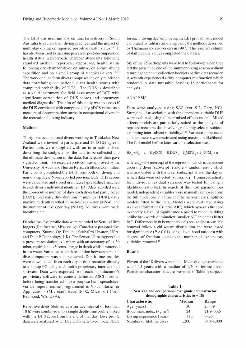
Diving and Hyperbaric Medicine Volume 42 No. 1 March 2012 19
The DHS was used initially on tuna farm divers in South Australia to review their diving practices and the impact of multi-day diving on reported post-dive health status.8,9 It has also been used to measure perceived post-decompression health status in hyperbaric chamber attendants following standard medical hyperbaric exposures, health status following dry chamber dives on nitrox, on a cave diving expedition and on a small group of technical divers.10–13 The work on tuna farm divers comprises the only published data correlating occupational diver health scores with computed probability of DCS. The DHS is described as a valid instrument for field assessment of DCS with significant correlation of DHS scores and concurrent medical diagnosis.7 The aim of this study was to assess if the DHS correlated with computed daily pDCS values as a measure of decompression stress in occupational divers in the recreational diving industry.
Methods
Thirty-one occupational divers working in Tutukaka, New Zealand were invited to participate and 25 (81%) agreed. Participants were supplied with an information sheet describing the study’s aims, the data to be collected and the ultimate destination of the data. Participants then gave signed consent. The research protocol was approved by the University of Auckland Human Research Ethics Committee. Participants completed the DHS form both on diving and non-diving days. None reported previous DCS. DHS scores were calculated and stored in an Excel spreadsheet matched to each diver’s individual identifier (ID). Also recorded were the consecutive number of days each diver had participated (DAY), total daily dive duration in minutes (DUR), daily maximum depth reached in metres’ sea water (MSW) and the number of dives per day (NUM). All dives were made breathing air.
Depth-time dive profile data were recorded by Sensus Ultra loggers (Reefnet inc, Mississauga, Canada) or personal dive computers (Suunto Oy, Finland; ScubaPro Uwatec, USA; and DeltaP Technology, UK). The Sensus Ultra loggers had a pressure resolution to 1 mbar, with an accuracy of +/-30 mbar, equivalent to 30 cms change in depth whilst immersed in sea water. Variation in depth resolution between personal dive computers was not measured. Depth-time profiles were downloaded from each depth-time recorder directly to a laptop PC using each unit’s proprietary interface and software. Data were exported from each manufacturer’s proprietary software in comma-delimited ASCII format, before being transferred into a purpose-built spreadsheet via an import routine programmed in Visual Basic for Applications (Microsoft Excel 2002, Microsoft Corp, Redmond, WA, USA).
Repetitive dives (defined as a surface interval of less than 18 h) were combined into a single depth-time profile linked with the DHS score from the end of that day. Dive profile data were analysed by Dr David Doolette to compute pDCS
for each ‘diving day’ employing the LE1 probabilistic model calibrated to military air diving using the methods described by Thalmann and co-workers in 1997.6 The resultant column of daily pDCS values completed the dataset.
Six of the 25 participants were lost to follow-up when they left the area at the end of the summer diving season without returning their data collection booklets or dive data recorder. A seventh experienced a dive computer malfunction which rendered its data unusable, leaving 18 participants for analysis.
ANALYSIS
Data were analysed using SAS (ver. 9.2, Cary, NC). Strengths of association with the dependent variable DHS were evaluated using a linear mixed effects model. Mixed effects models are particularly suited to the analysis of repeated measures data involving randomly selected subjects exhibiting inter-subject variability.14,15 Variance components and parameters were estimated using maximum likelihood. The full model before later variable selection was:
HSij = ß
0i + e
i + ß
1pDCS
ij + ß
2DUR
ij + ß
3MSW
ij + ß
4NUM
ij + e
i
where ß0 = the intercept of the regression which is dependent
upon the diver (subscript i) and e = random error, which was associated with the diver (subscript i) and the day on which data were collected (subscript j). Homoscedasticity for individual residual variance was tested for using a likelihood ratio test. In search of the most parsimonious model, independent variables were manually removed from the full model one at a time and the increasingly simplified models fitted to the data. Models were evaluated using Akaike Information Criteria (AIC), which bypasses the need to specify a level of significance a priori to model building unlike backwards elimination; smaller AIC indicates better fit.15 Differences in fit between models pre- and post-variable removal follow a chi-square distribution and were tested for significance (P < 0.01) using a likelihood ratio test with degrees of freedom equal to the number of explanatory variables removed.16
Results
Eleven of the 18 divers were male. Mean diving experience was 11.5 years with a median of 1,200 lifetime dives. Participant characteristics are presented in Table 1; subjects
Characteristic Median RangeAge (years) 30 23–39Body mass index (kg m-2) 24 21.9–33.5Diving experience (years) 11.5 6–26Number of lifetime dives 1,200 340–5,000
Table 1New Zealand occupational dive guide and instructor
demographic characteristics (n = 18)
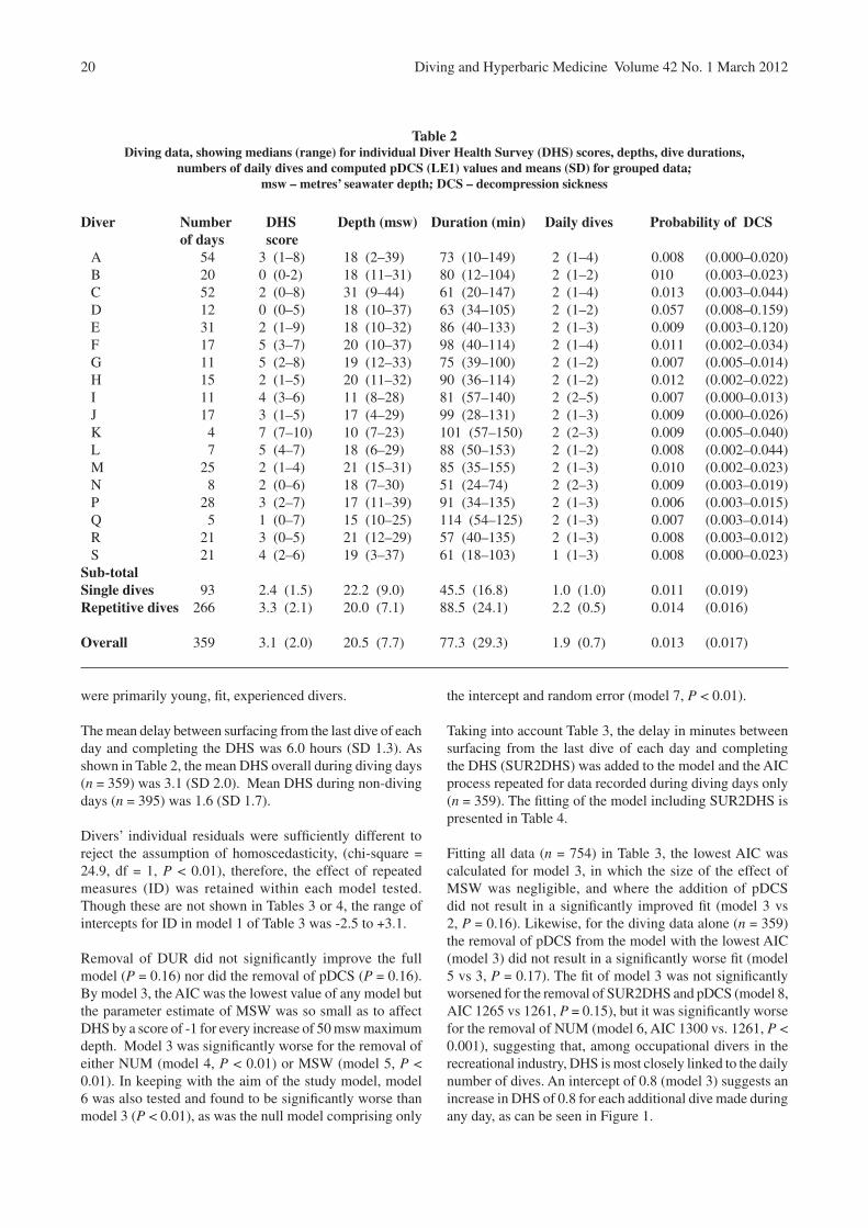
Diving and Hyperbaric Medicine Volume 42 No. 1 March 201220
were primarily young, fit, experienced divers.
The mean delay between surfacing from the last dive of each day and completing the DHS was 6.0 hours (SD 1.3). As shown in Table 2, the mean DHS overall during diving days (n = 359) was 3.1 (SD 2.0). Mean DHS during non-diving days (n = 395) was 1.6 (SD 1.7).
Divers’ individual residuals were sufficiently different to reject the assumption of homoscedasticity, (chi-square = 24.9, df = 1, P < 0.01), therefore, the effect of repeated measures (ID) was retained within each model tested. Though these are not shown in Tables 3 or 4, the range of intercepts for ID in model 1 of Table 3 was -2.5 to +3.1.
Removal of DUR did not significantly improve the full model (P = 0.16) nor did the removal of pDCS (P = 0.16). By model 3, the AIC was the lowest value of any model but the parameter estimate of MSW was so small as to affect DHS by a score of -1 for every increase of 50 msw maximum depth. Model 3 was significantly worse for the removal of either NUM (model 4, P < 0.01) or MSW (model 5, P < 0.01). In keeping with the aim of the study model, model 6 was also tested and found to be significantly worse than model 3 (P < 0.01), as was the null model comprising only
the intercept and random error (model 7, P < 0.01).
Taking into account Table 3, the delay in minutes between surfacing from the last dive of each day and completing the DHS (SUR2DHS) was added to the model and the AIC process repeated for data recorded during diving days only (n = 359). The fitting of the model including SUR2DHS is presented in Table 4.
Fitting all data (n = 754) in Table 3, the lowest AIC was calculated for model 3, in which the size of the effect of MSW was negligible, and where the addition of pDCS did not result in a significantly improved fit (model 3 vs 2, P = 0.16). Likewise, for the diving data alone (n = 359) the removal of pDCS from the model with the lowest AIC (model 3) did not result in a significantly worse fit (model 5 vs 3, P = 0.17). The fit of model 3 was not significantly worsened for the removal of SUR2DHS and pDCS (model 8, AIC 1265 vs 1261, P = 0.15), but it was significantly worse for the removal of NUM (model 6, AIC 1300 vs. 1261, P < 0.001), suggesting that, among occupational divers in the recreational industry, DHS is most closely linked to the daily number of dives. An intercept of 0.8 (model 3) suggests an increase in DHS of 0.8 for each additional dive made during any day, as can be seen in Figure 1.
Diver Number DHS Depth (msw) Duration (min) Daily dives Probability of DCS of days score
A 54 3 (1–8) 18 (2–39) 73 (10–149) 2 (1–4) 0.008 (0.000–0.020) B 20 0 (0-2) 18 (11–31) 80 (12–104) 2 (1–2) 010 (0.003–0.023)C 52 2 (0–8) 31 (9–44) 61 (20–147) 2 (1–4) 0.013 (0.003–0.044)D 12 0 (0–5) 18 (10–37) 63 (34–105) 2 (1–2) 0.057 (0.008–0.159)E 31 2 (1–9) 18 (10–32) 86 (40–133) 2 (1–3) 0.009 (0.003–0.120)F 17 5 (3–7) 20 (10–37) 98 (40–114) 2 (1–4) 0.011 (0.002–0.034)G 11 5 (2–8) 19 (12–33) 75 (39–100) 2 (1–2) 0.007 (0.005–0.014)H 15 2 (1–5) 20 (11–32) 90 (36–114) 2 (1–2) 0.012 (0.002–0.022)I 11 4 (3–6) 11 (8–28) 81 (57–140) 2 (2–5) 0.007 (0.000–0.013)J 17 3 (1–5) 17 (4–29) 99 (28–131) 2 (1–3) 0.009 (0.000–0.026)K 4 7 (7–10) 10 (7–23) 101 (57–150) 2 (2–3) 0.009 (0.005–0.040)L 7 5 (4–7) 18 (6–29) 88 (50–153) 2 (1–2) 0.008 (0.002–0.044)M 25 2 (1–4) 21 (15–31) 85 (35–155) 2 (1–3) 0.010 (0.002–0.023)N 8 2 (0–6) 18 (7–30) 51 (24–74) 2 (2–3) 0.009 (0.003–0.019)P 28 3 (2–7) 17 (11–39) 91 (34–135) 2 (1–3) 0.006 (0.003–0.015)Q 5 1 (0–7) 15 (10–25) 114 (54–125) 2 (1–3) 0.007 (0.003–0.014)R 21 3 (0–5) 21 (12–29) 57 (40–135) 2 (1–3) 0.008 (0.003–0.012)S 21 4 (2–6) 19 (3–37) 61 (18–103) 1 (1–3) 0.008 (0.000–0.023)
Sub-totalSingle dives 93 2.4 (1.5) 22.2 (9.0) 45.5 (16.8) 1.0 (1.0) 0.011 (0.019)Repetitive dives 266 3.3 (2.1) 20.0 (7.1) 88.5 (24.1) 2.2 (0.5) 0.014 (0.016)
Overall 359 3.1 (2.0) 20.5 (7.7) 77.3 (29.3) 1.9 (0.7) 0.013 (0.017)
Table 2Diving data, showing medians (range) for individual Diver Health Survey (DHS) scores, depths, dive durations,
numbers of daily dives and computed pDCS (LE1) values and means (SD) for grouped data;msw – metres’ seawater depth; DCS – decompression sickness
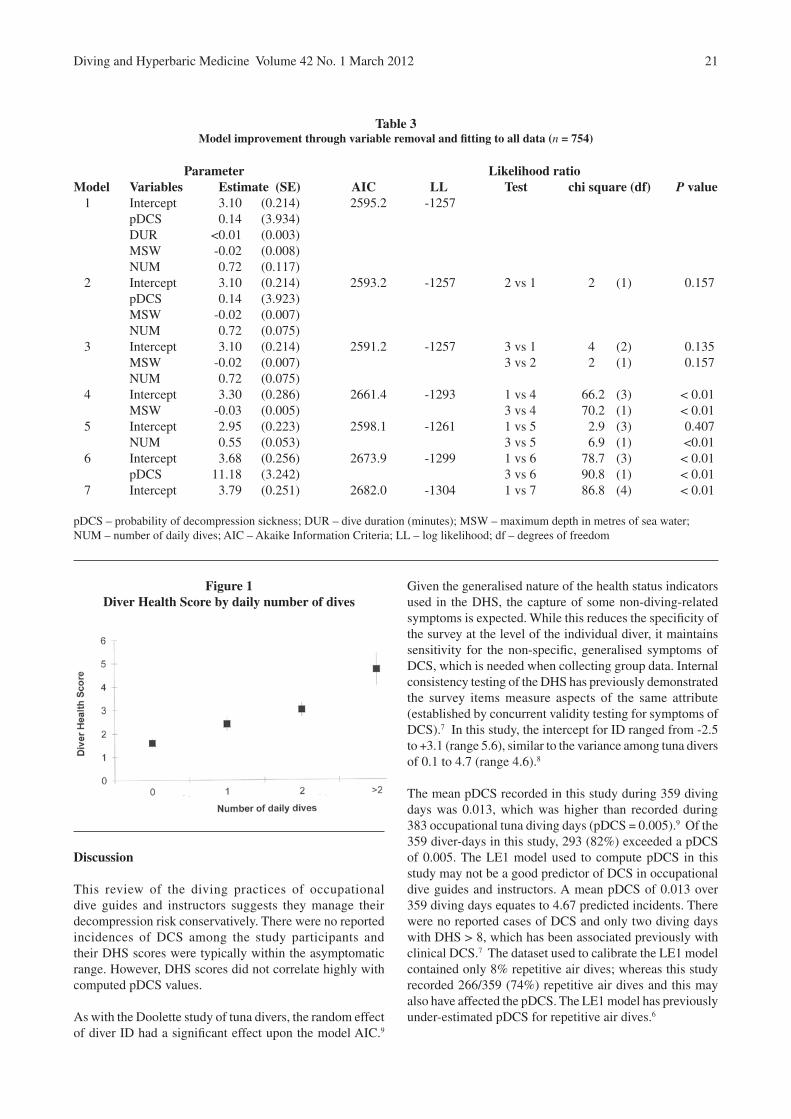
Diving and Hyperbaric Medicine Volume 42 No. 1 March 2012 21
Discussion
This review of the diving practices of occupational dive guides and instructors suggests they manage their decompression risk conservatively. There were no reported incidences of DCS among the study participants and their DHS scores were typically within the asymptomatic range. However, DHS scores did not correlate highly with computed pDCS values.
As with the Doolette study of tuna divers, the random effect of diver ID had a significant effect upon the model AIC.9
Given the generalised nature of the health status indicators used in the DHS, the capture of some non-diving-related symptoms is expected. While this reduces the specificity of the survey at the level of the individual diver, it maintains sensitivity for the non-specific, generalised symptoms of DCS, which is needed when collecting group data. Internal consistency testing of the DHS has previously demonstrated the survey items measure aspects of the same attribute (established by concurrent validity testing for symptoms of DCS).7 In this study, the intercept for ID ranged from -2.5 to +3.1 (range 5.6), similar to the variance among tuna divers of 0.1 to 4.7 (range 4.6).8
The mean pDCS recorded in this study during 359 diving days was 0.013, which was higher than recorded during 383 occupational tuna diving days (pDCS = 0.005).9 Of the 359 diver-days in this study, 293 (82%) exceeded a pDCS of 0.005. The LE1 model used to compute pDCS in this study may not be a good predictor of DCS in occupational dive guides and instructors. A mean pDCS of 0.013 over 359 diving days equates to 4.67 predicted incidents. There were no reported cases of DCS and only two diving days with DHS > 8, which has been associated previously with clinical DCS.7 The dataset used to calibrate the LE1 model contained only 8% repetitive air dives; whereas this study recorded 266/359 (74%) repetitive air dives and this may also have affected the pDCS. The LE1 model has previously under-estimated pDCS for repetitive air dives.6
Parameter Likelihood ratioModel Variables Estimate (SE) AIC LL Test chi square (df) P value
1 Intercept 3.10 (0.214) 2595.2 -1257 pDCS 0.14 (3.934) DUR <0.01 (0.003) MSW -0.02 (0.008) NUM 0.72 (0.117)2 Intercept 3.10 (0.214) 2593.2 -1257 2 vs 1 2 (1) 0.157 pDCS 0.14 (3.923) MSW -0.02 (0.007) NUM 0.72 (0.075)3 Intercept 3.10 (0.214) 2591.2 -1257 3 vs 1 4 (2) 0.135 MSW -0.02 (0.007) 3 vs 2 2 (1) 0.157 NUM 0.72 (0.075)4 Intercept 3.30 (0.286) 2661.4 -1293 1 vs 4 66.2 (3) < 0.01 MSW -0.03 (0.005) 3 vs 4 70.2 (1) < 0.015 Intercept 2.95 (0.223) 2598.1 -1261 1 vs 5 2.9 (3) 0.407 NUM 0.55 (0.053) 3 vs 5 6.9 (1) <0.016 Intercept 3.68 (0.256) 2673.9 -1299 1 vs 6 78.7 (3) < 0.01 pDCS 11.18 (3.242) 3 vs 6 90.8 (1) < 0.017 Intercept 3.79 (0.251) 2682.0 -1304 1 vs 7 86.8 (4) < 0.01
pDCS – probability of decompression sickness; DUR – dive duration (minutes); MSW – maximum depth in metres of sea water;NUM – number of daily dives; AIC – Akaike Information Criteria; LL – log likelihood; df – degrees of freedom
Table 3Model improvement through variable removal and fitting to all data (n = 754)
Figure 1Diver Health Score by daily number of dives
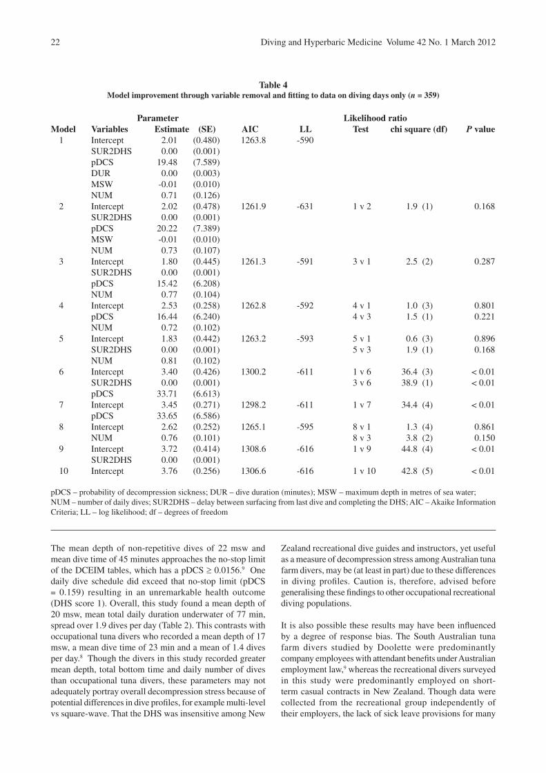
Diving and Hyperbaric Medicine Volume 42 No. 1 March 201222
The mean depth of non-repetitive dives of 22 msw and mean dive time of 45 minutes approaches the no-stop limit of the DCEIM tables, which has a pDCS ≥ 0.0156.9 One daily dive schedule did exceed that no-stop limit (pDCS = 0.159) resulting in an unremarkable health outcome (DHS score 1). Overall, this study found a mean depth of 20 msw, mean total daily duration underwater of 77 min, spread over 1.9 dives per day (Table 2). This contrasts with occupational tuna divers who recorded a mean depth of 17 msw, a mean dive time of 23 min and a mean of 1.4 dives per day.8 Though the divers in this study recorded greater mean depth, total bottom time and daily number of dives than occupational tuna divers, these parameters may not adequately portray overall decompression stress because of potential differences in dive profiles, for example multi-level vs square-wave. That the DHS was insensitive among New
Zealand recreational dive guides and instructors, yet useful as a measure of decompression stress among Australian tuna farm divers, may be (at least in part) due to these differences in diving profiles. Caution is, therefore, advised before generalising these findings to other occupational recreational diving populations.
It is also possible these results may have been influenced by a degree of response bias. The South Australian tuna farm divers studied by Doolette were predominantly company employees with attendant benefits under Australian employment law,9 whereas the recreational divers surveyed in this study were predominantly employed on short-term casual contracts in New Zealand. Though data were collected from the recreational group independently of their employers, the lack of sick leave provisions for many
Parameter Likelihood ratioModel Variables Estimate (SE) AIC LL Test chi square (df) P value
1 Intercept 2.01 (0.480) 1263.8 -590 SUR2DHS 0.00 (0.001) pDCS 19.48 (7.589) DUR 0.00 (0.003) MSW -0.01 (0.010) NUM 0.71 (0.126)2 Intercept 2.02 (0.478) 1261.9 -631 1 v 2 1.9 (1) 0.168 SUR2DHS 0.00 (0.001) pDCS 20.22 (7.389) MSW -0.01 (0.010) NUM 0.73 (0.107)3 Intercept 1.80 (0.445) 1261.3 -591 3 v 1 2.5 (2) 0.287 SUR2DHS 0.00 (0.001) pDCS 15.42 (6.208) NUM 0.77 (0.104)4 Intercept 2.53 (0.258) 1262.8 -592 4 v 1 1.0 (3) 0.801 pDCS 16.44 (6.240) 4 v 3 1.5 (1) 0.221 NUM 0.72 (0.102)5 Intercept 1.83 (0.442) 1263.2 -593 5 v 1 0.6 (3) 0.896 SUR2DHS 0.00 (0.001) 5 v 3 1.9 (1) 0.168 NUM 0.81 (0.102)6 Intercept 3.40 (0.426) 1300.2 -611 1 v 6 36.4 (3) < 0.01 SUR2DHS 0.00 (0.001) 3 v 6 38.9 (1) < 0.01 pDCS 33.71 (6.613)7 Intercept 3.45 (0.271) 1298.2 -611 1 v 7 34.4 (4) < 0.01 pDCS 33.65 (6.586)8 Intercept 2.62 (0.252) 1265.1 -595 8 v 1 1.3 (4) 0.861 NUM 0.76 (0.101) 8 v 3 3.8 (2) 0.1509 Intercept 3.72 (0.414) 1308.6 -616 1 v 9 44.8 (4) < 0.01 SUR2DHS 0.00 (0.001)10 Intercept 3.76 (0.256) 1306.6 -616 1 v 10 42.8 (5) < 0.01
pDCS – probability of decompression sickness; DUR – dive duration (minutes); MSW – maximum depth in metres of sea water;NUM – number of daily dives; SUR2DHS – delay between surfacing from last dive and completing the DHS; AIC – Akaike Information Criteria; LL – log likelihood; df – degrees of freedom
Table 4Model improvement through variable removal and fitting to data on diving days only (n = 359)
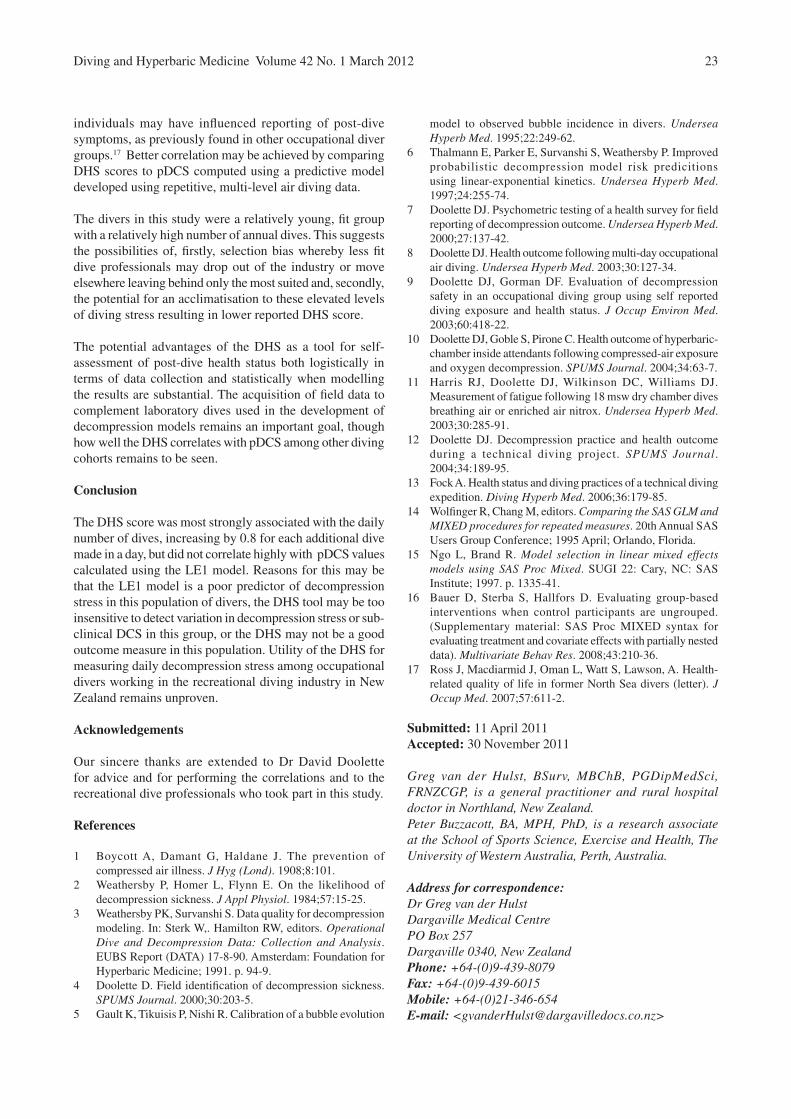
Diving and Hyperbaric Medicine Volume 42 No. 1 March 2012 23
individuals may have influenced reporting of post-dive symptoms, as previously found in other occupational diver groups.17 Better correlation may be achieved by comparing DHS scores to pDCS computed using a predictive model developed using repetitive, multi-level air diving data.
The divers in this study were a relatively young, fit group with a relatively high number of annual dives. This suggests the possibilities of, firstly, selection bias whereby less fit dive professionals may drop out of the industry or move elsewhere leaving behind only the most suited and, secondly, the potential for an acclimatisation to these elevated levels of diving stress resulting in lower reported DHS score.
The potential advantages of the DHS as a tool for self-assessment of post-dive health status both logistically in terms of data collection and statistically when modelling the results are substantial. The acquisition of field data to complement laboratory dives used in the development of decompression models remains an important goal, though how well the DHS correlates with pDCS among other diving cohorts remains to be seen.
Conclusion
The DHS score was most strongly associated with the daily number of dives, increasing by 0.8 for each additional dive made in a day, but did not correlate highly with pDCS values calculated using the LE1 model. Reasons for this may be that the LE1 model is a poor predictor of decompression stress in this population of divers, the DHS tool may be too insensitive to detect variation in decompression stress or sub-clinical DCS in this group, or the DHS may not be a good outcome measure in this population. Utility of the DHS for measuring daily decompression stress among occupational divers working in the recreational diving industry in New Zealand remains unproven.
Acknowledgements
Our sincere thanks are extended to Dr David Doolette for advice and for performing the correlations and to the recreational dive professionals who took part in this study.
References
1 Boycott A, Damant G, Haldane J. The prevention of compressed air illness. J Hyg (Lond). 1908;8:101.
2 Weathersby P, Homer L, Flynn E. On the likelihood of decompression sickness. J Appl Physiol. 1984;57:15-25.
3 Weathersby PK, Survanshi S. Data quality for decompression modeling. In: Sterk W,. Hamilton RW, editors. Operational Dive and Decompression Data: Collection and Analysis. EUBS Report (DATA) 17-8-90. Amsterdam: Foundation for Hyperbaric Medicine; 1991. p. 94-9.
4 Doolette D. Field identification of decompression sickness. SPUMS Journal. 2000;30:203-5.
5 Gault K, Tikuisis P, Nishi R. Calibration of a bubble evolution
model to observed bubble incidence in divers. Undersea Hyperb Med. 1995;22:249-62.
6 Thalmann E, Parker E, Survanshi S, Weathersby P. Improved probabilistic decompression model risk predicitions using linear-exponential kinetics. Undersea Hyperb Med. 1997;24:255-74.
7 Doolette DJ. Psychometric testing of a health survey for field reporting of decompression outcome. Undersea Hyperb Med. 2000;27:137-42.
8 Doolette DJ. Health outcome following multi-day occupational air diving. Undersea Hyperb Med. 2003;30:127-34.
9 Doolette DJ, Gorman DF. Evaluation of decompression safety in an occupational diving group using self reported diving exposure and health status. J Occup Environ Med. 2003;60:418-22.
10 Doolette DJ, Goble S, Pirone C. Health outcome of hyperbaric-chamber inside attendants following compressed-air exposure and oxygen decompression. SPUMS Journal. 2004;34:63-7.
11 Harris RJ, Doolette DJ, Wilkinson DC, Williams DJ. Measurement of fatigue following 18 msw dry chamber dives breathing air or enriched air nitrox. Undersea Hyperb Med. 2003;30:285-91.
12 Doolette DJ. Decompression practice and health outcome during a technical diving project. SPUMS Journal. 2004;34:189-95.
13 Fock A. Health status and diving practices of a technical diving expedition. Diving Hyperb Med. 2006;36:179-85.
14 Wolfinger R, Chang M, editors. Comparing the SAS GLM and MIXED procedures for repeated measures. 20th Annual SAS Users Group Conference; 1995 April; Orlando, Florida.
15 Ngo L, Brand R. Model selection in linear mixed effects models using SAS Proc Mixed. SUGI 22: Cary, NC: SAS Institute; 1997. p. 1335-41.
16 Bauer D, Sterba S, Hallfors D. Evaluating group-based interventions when control participants are ungrouped. (Supplementary material: SAS Proc MIXED syntax for evaluating treatment and covariate effects with partially nested data). Multivariate Behav Res. 2008;43:210-36.
17 Ross J, Macdiarmid J, Oman L, Watt S, Lawson, A. Health-related quality of life in former North Sea divers (letter). J Occup Med. 2007;57:611-2.
Submitted: 11 April 2011Accepted: 30 November 2011
Greg van der Hulst, BSurv, MBChB, PGDipMedSci, FRNZCGP, is a general practitioner and rural hospital doctor in Northland, New Zealand.Peter Buzzacott, BA, MPH, PhD, is a research associate at the School of Sports Science, Exercise and Health, The University of Western Australia, Perth, Australia.
Address for correspondence:Dr Greg van der HulstDargaville Medical CentrePO Box 257Dargaville 0340, New ZealandPhone: +64-(0)9-439-8079Fax: +64-(0)9-439-6015Mobile: +64-(0)21-346-654E-mail: <[email protected]>
Diving and Hyperbaric Medicine Volume 42 No. 1 March 201224
Postal survey of fitness-to-dive opinions of diving doctors and general practitionersChris Sames, Des Gorman and Simon Mitchell
Abstract(Sames C, Gorman D, Mitchell S. Postal survey of fitness-to-dive opinions of diving doctors and general practitioners. Diving Hyperb Med. 2012;42(1):24-29.)Aim: To determine the consensus and concordance with published standards and expert opinion among New Zealand’s designated diving doctors (DDDs) and general practitioners (GPs) regarding medical fitness-to-dive.Methods: A postal survey canvassed doctors’ opinions regarding fitness to dive of 20 ‘real-life’ applicants with potentially relevant medical conditions. In 17 cases, a ‘desired response’ was identified as expert opinion and the relevant published Standard concurred; the remaining three cases were excluded from analysis. Consensus was measured between the groups of doctors, and concordance measured against the ‘desired response’. The performance of the DDDs was also correlated with both the number of diver medical assessments conducted annually and time since completing a diving medicine course.Results: Seventy-seven of 98 DDDs (79%) and 75 of 200 GPs (38%) responded to the questionnaire. The mean concordance was 60% and 50% for DDDs and GPs respectively. Consensus between DDDs and GPs was generally high, but was low between these groups and the ‘desired response’. DDDs’ concordance was negatively correlated (r = -0.3) with time since undertaking a diving medicine course, but was positively correlated (r = 0.2) with their annual number of dive medical assessments. Both groups were more likely to differ from the ‘desired response’ by considering an ‘unfit’ diver as ‘fit’ than the converse.Conclusions: There is poor concordance between doctors assessing fitness to dive and both published recommendations and expert opinion when there is a relevant medical condition. This supports the current New Zealand practice of centralised audit of occupational diver medical fitness prior to certification.
Key wordsFitness to dive, medical examinations, compressed-gas divers, scuba divers, recreational divers, occupational divers
Introduction
In New Zealand (NZ), the estimated compressed-gas diver fatality rate was 5.8 deaths per 100,000 divers per year during 1996–2000,1 or a mean death rate of 6 per year from 1980–2006.1,2 This figure represents only about 5% of drowning fatalities and suggests that diving is a relatively safe occupation or pastime. However, of the 40 diver deaths in NZ from 2000–2006, 12 should have been disqualified from diving on medical grounds and, although the relationship between the medical condition and the accident was often unclear, these pre-existing medical conditions were considered by the coroner to be either causative or contributory to their deaths.2
Recreational divers in NZ are required to undergo a medical examination conducted by a medical practitioner prior to concluding training. There is no requirement for the examining doctor to have undergone training in diving medicine, and there is no ongoing health surveillance for these divers. In contrast, occupational divers undergo a five-yearly medical examination conducted by a ‘designated diving doctor’ (DDD) who has undertaken post-graduate training in diving medicine recognised by the South Pacific Underwater Medicine Society (SPUMS). In intervening years, the divers complete an annual health questionnaire. Both the medical examination documentation and the annual health questionnaires are independently reviewed
by an expert medical panel. This system has been shown to be reliable, but controversy periodically arises about the justification for expert and independent review of the medical documentation.3
One reason for such a review is the potential for inconsistency in decision making, even between doctors trained in diving medicine. A previous study of doctors in Queensland, Australia, who had training in diving medicine, showed a low level of consensus in regard to the impact of certain medical conditions on ‘fitness’ to dive.4 Similar problems were found in a review of the process used to certify civil pilots fit to fly in NZ.5,6
The present study re-examined this issue in NZ; the aim was to determine consensus and concordance with expert opinion among NZ DDDs and general practitioners (GPs) regarding fitness for diving (both occupational and recreational), to consequently see if there is an ongoing need for independent review or arbitration of occupational diving medical evaluations and to identify possible improvements to recreational diving medical evaluations.
Method
A questionnaire describing 20 compressed-gas diving candidates who had a medical condition that could affect diving fitness was mailed, along with a reply-paid envelope,
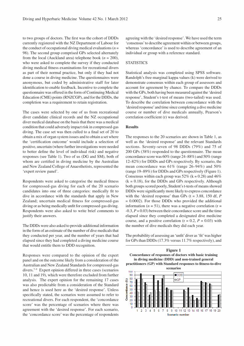
Diving and Hyperbaric Medicine Volume 42 No. 1 March 2012 25
to two groups of doctors. The first was the cohort of DDDs currently registered with the NZ Department of Labour for the conduct of occupational diving medical evaluations (n = 98). The second group comprised GPs selected alternately from the local (Auckland area) telephone book (n = 200), who were asked to complete the survey if they conducted diving medical fitness examinations for recreational divers as part of their normal practice, but only if they had not done a course in diving medicine. The questionnaires were anonymous, but coded by administrative staff for later identification to enable feedback. Incentive to complete the questionnaire was offered in the form of Continuing Medical Education (CME) points (RNZCGP), and for the DDDs, the completion was a requirement to retain registration.
The cases were selected by one of us from recreational diver candidate clinical records and the NZ occupational diver medical database on the basis that there was a medical condition that could adversely impact risk in compressed-gas diving. The case set was then culled to a final set of 20 to obtain a mix of organ system issues and to obtain a set where the ‘certification outcome’ would include a selection of positive, uncertain (where further investigations were needed to better define the level of individual risk) and negative responses (see Table 1). Two of us (DG and SM), both of whom are certified in diving medicine by the Australian and New Zealand College of Anaesthetists, represented the ‘expert review panel’.
Respondents were asked to categorise the medical fitness for compressed-gas diving for each of the 20 scenario candidates into one of three categories: medically fit to dive in accordance with the standards that apply in New Zealand; uncertain medical fitness for compressed-gas diving or as being medically unfit for compressed-gas diving. Respondents were also asked to write brief comments to justify their answers.
The DDDs were also asked to provide additional information in the form of an estimate of the number of dive medicals that they conducted per year, and the number of years that had elapsed since they had completed a diving medicine course that would entitle them to DDD recognition.
Responses were compared to the opinion of the expert panel and on the outcome likely from a consideration of the Australian and New Zealand Standards for compressed-gas divers.7–9 Expert opinion differed in three cases (scenarios 10, 11 and 19), which were therefore excluded from further analysis. The expert opinion for the remaining 17 cases was also predictable from a consideration of the Standard and hence is used here as the ‘desired response’. Unless specifically stated, the scenarios were assumed to refer to recreational divers. For each respondent, the ‘concordance score’ was the percentage of scenarios where there was agreement with the ‘desired response’. For each scenario, the ‘concordance score’ was the percentage of respondents
agreeing with the ‘desired response’. We have used the term ‘consensus’ to describe agreement within or between groups, whereas ‘concordance’ is used to describe agreement of an individual or group with a reference standard.
STATISTICS
Statistical analysis was completed using SPSS software. Randolph’s free-marginal kappa values (k) were derived to demonstrate consensus within each group of assessors and account for agreement by chance. To compare the DDDs with the GPs, both having been measured against the ‘desired response’, Student’s t-test of means (two-tailed) was used. To describe the correlation between concordance with the ‘desired response’ and time since completing a dive medicine course or number of dive medicals annually, Pearson’s correlation coefficient (r) was derived.
Results
The responses to the 20 scenarios are shown in Table 1, as well as the ‘desired response’ and the relevant Standards sections. Seventy-seven of 98 DDDs (79%) and 75 of 200 GPs (38%) responded to the questionnaire. The mean concordance score was 60% (range 24–88%) and 50% (range 12–82%) for DDDs and GPs respectively. By scenario, the mean concordance was 61% (range 26–94%) and 50% (range 19–89%) for DDDs and GPs respectively (Figure 1). Consensus within each group was 52% (k = 0.28) and 46% (k = 0.18), for the DDDs and GPs respectively. Although both groups scored poorly, Student’s t-tests of means showed DDDs were significantly more likely to express concordance with the ‘desired response’ than GPs (t = 3.88, 150 df, P = 0.0002). For those DDDs who provided the additional information (n = 51), there was a negative correlation (r = -0.3, P = 0.03) between their concordance score and the time elapsed since they completed a designated dive medicine course, and a positive correlation (r = 0.2, P = 0.03) with the number of dive medicals they did each year.
The probability of assessing an ‘unfit’ diver as ‘fit’ was higher for GPs than DDDs (17.3% versus 11.7% respectively), and
Figure 1Concordance of responses of doctors with basic training
in diving medicine (DDD) and non-trained general practitioners (GP) with Standard responses to fitness-to-dive
scenarios
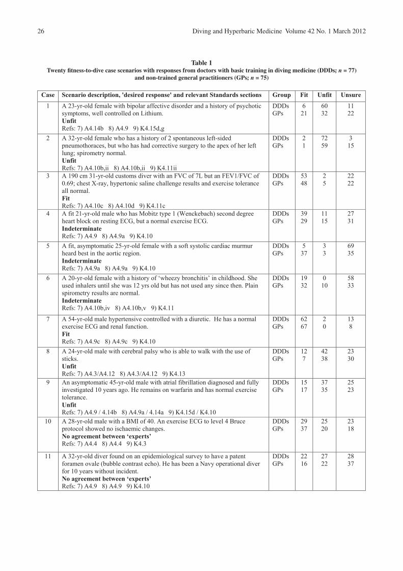
Diving and Hyperbaric Medicine Volume 42 No. 1 March 201226
Table 1Twenty fitness-to-dive case scenarios with responses from doctors with basic training in diving medicine (DDDs; n = 77)
and non-trained general practitioners (GPs; n = 75)
Case Scenario description, 'desired response' and relevant Standards sections Group Fit Unfit Unsure
1 A 23-yr-old female with bipolar affective disorder and a history of psychotic symptoms, well controlled on Lithium. UnfitRefs: 7) A4.14b 8) A4.9 9) K4.15d,g
DDDsGPs
6 21
60 32
11 22
2 A 32-yr-old female who has a history of 2 spontaneous left-sided pneumothoraces, but who has had corrective surgery to the apex of her left lung; spirometry normal. UnfitRefs: 7) A4.10b,ii 8) A4.10b,ii 9) K4.11ii
DDDsGPs
2 1
72 59
3 15
3 A 190 cm 31-yr-old customs diver with an FVC of 7L but an FEV1/FVC of 0.69; chest X-ray, hypertonic saline challenge results and exercise tolerance all normal. FitRefs: 7) A4.10c 8) A4.10d 9) K4.11c
DDDsGPs
53 48
2 5
22 22
4 A fit 21-yr-old male who has Mobitz type 1 (Wenckebach) second degree heart block on resting ECG, but a normal exercise ECG. IndeterminateRefs: 7) A4.9 8) A4.9a 9) K4.10
DDDsGPs
39 29
11 15
27 31
5 A fit, asymptomatic 25-yr-old female with a soft systolic cardiac murmur heard best in the aortic region. IndeterminateRefs: 7) A4.9a 8) A4.9a 9) K4.10
DDDs GPs
5 37
3 3
69 35
6 A 20-yr-old female with a history of ‘wheezy bronchitis’ in childhood. She used inhalers until she was 12 yrs old but has not used any since then. Plain spirometry results are normal. IndeterminateRefs: 7) A4.10b,iv 8) A4.10b,v 9) K4.11
DDDsGPs
19 32
0 10
58 33
7 A 54-yr-old male hypertensive controlled with a diuretic. He has a normal exercise ECG and renal function. FitRefs: 7) A4.9c 8) A4.9c 9) K4.10
DDDs GPs
62 67
2 0
13 8
8 A 24-yr-old male with cerebral palsy who is able to walk with the use of sticks. UnfitRefs: 7) A4.3/A4.12 8) A4.3/A4.12 9) K4.13
DDDsGPs
12 7
42 38
23 30
9 An asymptomatic 45-yr-old male with atrial fibrillation diagnosed and fully investigated 10 years ago. He remains on warfarin and has normal exercise tolerance. UnfitRefs: 7) A4.9 / 4.14b 8) A4.9a / 4.14a 9) K4.15d / K4.10
DDDsGPs
15 17
37 35
25 23
10 A 28-yr-old male with a BMI of 40. An exercise ECG to level 4 Bruce protocol showed no ischaemic changes. No agreement between ‘experts’Refs: 7) A4.4 8) A4.4 9) K4.3
DDDsGPs
29 37
25 20
23 18
11 A 32-yr-old diver found on an epidemiological survey to have a patent foramen ovale (bubble contrast echo). He has been a Navy operational diver for 10 years without incident. No agreement between ‘experts’Refs: 7) A4.9 8) A4.9 9) K4.10
DDDsGPs
22 16
27 22
28 37
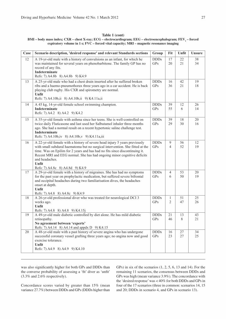
Diving and Hyperbaric Medicine Volume 42 No. 1 March 2012 27
Table 1 (cont)BMI – body mass index; CXR – chest X-ray; ECG – electrocardiogram; EEG – electroencephalogram; FEV1 – forced
expiratory volume in 1 s; FVC – forced vital capacity; MRI – magnetic resonance imaging
was also significantly higher for both GPs and DDDs than the converse probability of assessing a ‘fit’ diver as ‘unfit’ (3.3% and 2.6% respectively).
Concordance scores varied by greater than 15% (mean variance 27.7%) between DDDs and GPs (DDDs higher than
GPs) in six of the scenarios (1, 2, 5, 6, 13 and 14). For the remaining 11 scenarios, the consensus between DDDs and GPs was high (mean variance 3.9%). The concordance with the ‘desired response’ was < 40% for both DDDs and GPs in four of the 17 scenarios (three in common: scenarios 14, 15 and 20; DDDs in scenario 4, and GPs in scenario 13).
Case Scenario description, 'desired response' and relevant Standards sections Group Fit Unfit Unsure
12 A 19-yr-old male with a history of convulsions as an infant, for which he was maintained for several years on phenobarbitone. The family GP has no record of any fits. IndeterminateRefs: 7) A4.8b 8) A4.8b 9) K4.9
DDDsGPs
17 20
22 21
38 34
13 A 25-yr-old male who had a chest drain inserted after he suffered broken ribs and a haemo-pneumothorax three years ago in a car accident. He is back playing club rugby. His CXR and spirometry are normal. UnfitRefs: 7) A4.10b,ii 8) A4.10b,ii 9) K4.11a,ii
DDDsGPs
16 36
42 21
19 18
14 A 45 kg, 14-yr-old female school swimming champion. IndeterminateRefs: 7) A4.2 8) A4.2 9) K4.2
DDDsGPs
39 55
12 6
26 14
15 A 35-yr-old female with asthma since her teens. She is well-controlled on twice daily Fluticasone and last used her Salbutamol inhaler three months ago. She had a normal result on a recent hypertonic saline challenge test. IndeterminateRefs: 7) A4.10b,iv 8) A4.10b,v 9) K4.11a,iii
DDDsGPs
39 29
18 30
20 16
16 A 22-yr-old female with a history of severe head injury 5 years previously with small subdural haematoma but no surgical intervention. She fitted at the time. Was on Epilim for 2 years and has had no fits since discontinuing it. Recent MRI and EEG normal. She has had ongoing minor cognitive deficits and headaches. UnfitRefs: 7) A4.8c 8) A4.8d 9) K4.9
DDDsGPs
9 4
56 52
12 19
17 A 29-yr-old female with a history of migraines. She has had no symptoms for the past year on prophylactic medication, but suffered severe bifrontal and occipital headaches during two familiarisation dives, the headaches onset at depth. UnfitRefs: 7) A4.8 8) A4.8c 9) K4.9
DDDsGPs
4 6
53 50
20 19
18 A 26-yr-old professional diver who was treated for neurological DCI 3 weeks ago. UnfitRefs: 7) A4.8 8) A4.8 9) K4.15j
DDDsGPs
1 2
51 47
25 26
19 A 49-yr-old male diabetic controlled by diet alone. He has mild diabetic retinopathy. No agreement between ‘experts’Refs: 7) A4.14 8) A4.14 and appdx D 9) K4.15
DDDsGPs
21 46
13 8
43 21
20 A 48-yr-old male with a past history of severe angina who has undergone successful coronary vessel grafting three years ago; no angina now and good exercise tolerance. UnfitRefs: 7) A4.9 8) A4.9 9) K4.10
DDDs GPs
16 23
27 27
34 25
Diving and Hyperbaric Medicine Volume 42 No. 1 March 201228
Discussion
The scenarios used in this survey were selected to include important respiratory, cardiovascular and neurological health issues for divers. Many of our ‘real-life’ cases were similar to those used in the Queensland study, some of which were fictitious and some real, emphasising that these are the kind of medical conditions that arise relatively commonly in assessing would-be divers.4 They were also selected to present a challenge to the assessing doctors as compared to more straightforward cases, which represent the great majority of assessments. It follows that the current survey does not represent the outcome likely from a random selection of cases in which a much higher concordance would be expected.
The overall 38% response rate for surveyed GPs is likely to mask a much higher response rate for those GPs who fulfilled the inclusion criteria (those who conduct recreational diving medical fitness examinations but have not completed a diving medicine course) as many GPs do not undertake diving fitness assessments.
The published standards for fitness to dive are conservative, and if strictly applied they may result in divers being inappropriately denied medical clearance for diving.7–9 However, the finding that both DDDs and GPs were more likely to assess an unfit or indeterminate diver as fit, rather than the converse, suggests either disagreement with, or a lack of familiarity with the published standards, as the bias in the latter is in the opposite direction.
There was a wide range of opinions and a low mean concordance with the ‘desired response’ for both DDDs and GPs. This, together with the negative correlation between concordance score and time since completing a designated diving medicine course, suggests potential benefit could arise from periodic refreshers and/or regular formative assessments of DDDs and GPs. It also suggests that the most reliable method of assessing someone’s medical fitness for occupational diving involves an expert in diving medicine and/or a risk evaluation conducted by a specifically trained doctor who has ready access to expert advice. The problem with either of these ‘solutions’ is that there are very few diving medicine experts and hence access would be limited. The central audit facility for employed divers that exists in New Zealand is a workable solution to this problem and is clearly independent and less vulnerable to diver-advocacy bias. It is noteworthy that many divers who might otherwise have been disqualified, have been able to continue a career in diving, with specified constraints, due to the intervention of this facility.
For recreational divers, there is evidence both supporting
and refuting the utility of a medical examination prior to training.10–12 In the face of this controversy, most countries have now adopted a self-declaration health questionnaire
for recreational scuba diving candidates in line with the ISO standards.13 However, for occupational divers, there remains a widespread reliance on annual medical examinations conducted by doctors analogous to our DDDs. Our study suggests that in the absence of independent review, there is a strong possibility that candidates with significant medical conditions who undergo such an examination will receive a determination of fitness different to that which an expert would deliver or that expected by consideration of the relevant Standard. To the extent that we derived a ‘desired response’, this study suggests that independent review by such experts is a valuable adjunct to the process of occupational diver evaluation.
LIMITATIONS OF THE STUDY
The respondents, both DDDs and GPs, were asked only to assess the diving candidates’ fitness to dive on the basis of the brief vignette. There was no specification regarding fitness for occupational versus recreational diving. Therefore, it is possible that some of the respondents, especially the GPs, may have applied a more liberal ‘informed risk acceptor’ approach in their decision making. It should be noted, however, that there are very few differences between the published standards for occupational and recreational diving.
Conclusions
This study supports the need for better, iterative and formative diving medical education for DDDs, and the desirability of diving medical education for any GP who wishes to conduct recreational dive medicals. The overall low concordance of both DDDs and GPs with published recommendations and expert opinion is mitigated for DDDs performing occupational diving medicals in the New Zealand setting by the existence of a central, independent and expert audit authority.
Conflict of interest
Drs Des Gorman and Chris Sames are members of the Department of Labour Diving Medical Directorate, which is responsible to the Department of Labour for the certification of the medical fitness of occupational divers in New Zealand.
References
1 Davis M, Warner M, Ward B. Snorkelling and scuba diving deaths in New Zealand, 1980-2000. SPUMS Journal. 2002;32:70-80.
2 McClelland A. Diving-related deaths in New Zealand 2000-2006. Diving Hyperb Med. 2007;37:174-88.
3 Sames C, Gorman D, Mitchell SJ, Gamble G. Utility of regular medical examinations of occupational divers. Internal Medicine Journal. 2009;39:763-6.
4 Simpson G, Roomes D. Scuba diving medical examinations
Diving and Hyperbaric Medicine Volume 42 No. 1 March 2012 29
in practice: a postal survey. MJA. 1999;171:595-8.5 Gorman DF, Scott PJ. The process of determining fitness to fly
aeroplanes in New Zealand: A review of current practice and recommended changes. Wellington: Civil Aviation Authority of New Zealand; 2001.
6 Gorman DF, Scott PJ. The process of determining fitness to fly aeroplanes in New Zealand: A follow up audit report of current practice and recommended changes. Wellington: Civil Aviation Authority of New Zealand; 2003.
7 Training and certification of recreational divers – Part 1: minimum entry-level SCUBA diving (AS 4005.1 – 2000), 2nd ed. Sydney: Standards Australia; 2000.
8 Guidelines on medical risk assessment for recreational diving. Melbourne: South Pacific Underwater Medicine Society; 2011. Available at: http://www.SPUMS.org.au.
9 Occupational diving operations – Part 1: standard operational practice (AS/NZS 2299.1 – 2007), 2nd ed. Sydney/Wellington: Standards Australia/Standards New Zealand; 2007.
10 Meehan CA, Bennett MH. Medical assessment of fitness to dive – comparing a questionnaire and a medical interview-based approach. Diving Hyperb Med. 2010;40:119-24.
11 Glen S, White S, Douglas J. Medical supervision of sport diving in Scotland: reassessing the need for routine medical examinations. Br J Sports Med. 2000;34:375-8.
12 Glen S. Three year follow up of a self certification system for assessment of fitness to dive in Scotland. Br J Sports Med. 2004;38:754-7.
13 ISO 24801-2:2007. Recreational diving services – Safety related minimum requirements for the training of recreational scuba divers – Part 2: Level 2 – Autonomous diver. Geneva, Switzerland: International Organization for Standardization; 2007.
Submitted: 01 June 2011Accepted: 03 January 2012
Chris Sames, BSc, MBChB, MMedSc, is Hyperbaric Medical Officer, Naval Health Unit, Royal New Zealand Navy, Devonport, Auckland.Des Gorman, MD, PhD, is Professor of Medicine, University of Auckland, Executive Chairman of Health Workforce New Zealand and Director of Diving and Hyperbaric Medical Services.Simon Mitchell, PhD, FANZCA is Associate Professor, Head of the Department of Anaesthesiology, University of Auckland and Consultant Anaesthetist, Dept of Anaesthesiology, Auckland District Health Board.
Address for correspondence:Dr Chris SamesSlark Hyperbaric Unit, Naval Health Unit91 Calliope RoadPrivate Bag 32901Devonport, Auckland 1309 New ZealandPhone: +64-(0)9-445-5922Fax: +64-(0)9-445-5941E-mail: <[email protected]>
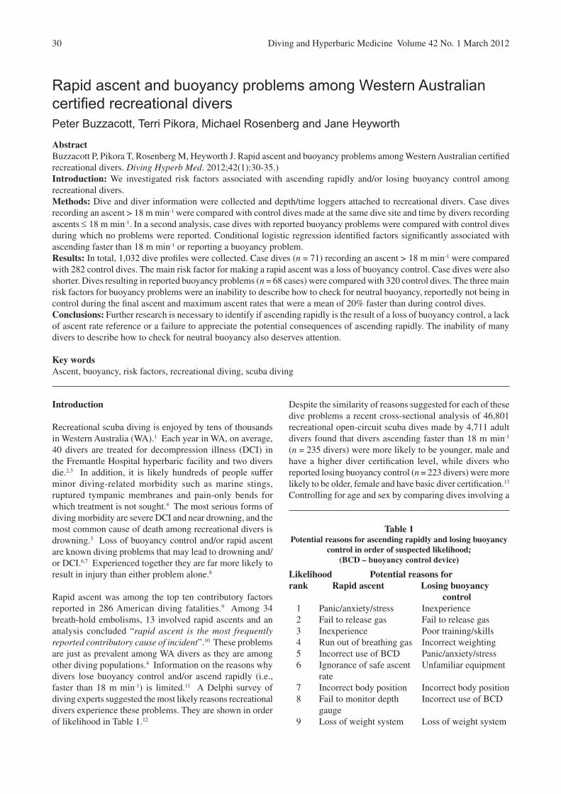
Diving and Hyperbaric Medicine Volume 42 No. 1 March 201230
Rapid ascent and buoyancy problems among Western Australian certified recreational diversPeter Buzzacott, Terri Pikora, Michael Rosenberg and Jane Heyworth
Abstract Buzzacott P, Pikora T, Rosenberg M, Heyworth J. Rapid ascent and buoyancy problems among Western Australian certified recreational divers. Diving Hyperb Med. 2012;42(1):30-35.)Introduction: We investigated risk factors associated with ascending rapidly and/or losing buoyancy control among recreational divers.Methods: Dive and diver information were collected and depth/time loggers attached to recreational divers. Case dives recording an ascent > 18 m min-1 were compared with control dives made at the same dive site and time by divers recording ascents ≤ 18 m min-1. In a second analysis, case dives with reported buoyancy problems were compared with control dives during which no problems were reported. Conditional logistic regression identified factors significantly associated with ascending faster than 18 m min-1 or reporting a buoyancy problem.Results: In total, 1,032 dive profiles were collected. Case dives (n = 71) recording an ascent > 18 m min-1 were compared with 282 control dives. The main risk factor for making a rapid ascent was a loss of buoyancy control. Case dives were also shorter. Dives resulting in reported buoyancy problems (n = 68 cases) were compared with 320 control dives. The three main risk factors for buoyancy problems were an inability to describe how to check for neutral buoyancy, reportedly not being in control during the final ascent and maximum ascent rates that were a mean of 20% faster than during control dives.Conclusions: Further research is necessary to identify if ascending rapidly is the result of a loss of buoyancy control, a lack of ascent rate reference or a failure to appreciate the potential consequences of ascending rapidly. The inability of many divers to describe how to check for neutral buoyancy also deserves attention.
Key wordsAscent, buoyancy, risk factors, recreational diving, scuba diving
Introduction
Recreational scuba diving is enjoyed by tens of thousands in Western Australia (WA).1 Each year in WA, on average, 40 divers are treated for decompression illness (DCI) in the Fremantle Hospital hyperbaric facility and two divers die.2,3 In addition, it is likely hundreds of people suffer minor diving-related morbidity such as marine stings, ruptured tympanic membranes and pain-only bends for which treatment is not sought.4 The most serious forms of diving morbidity are severe DCI and near drowning, and the most common cause of death among recreational divers is drowning.5 Loss of buoyancy control and/or rapid ascent are known diving problems that may lead to drowning and/or DCI.6,7 Experienced together they are far more likely to result in injury than either problem alone.8
Rapid ascent was among the top ten contributory factors reported in 286 American diving fatalities.9 Among 34 breath-hold embolisms, 13 involved rapid ascents and an analysis concluded “rapid ascent is the most frequently reported contributory cause of incident”.10 These problems are just as prevalent among WA divers as they are among other diving populations.4 Information on the reasons why divers lose buoyancy control and/or ascend rapidly (i.e., faster than 18 m min-1) is limited.11 A Delphi survey of diving experts suggested the most likely reasons recreational divers experience these problems. They are shown in order of likelihood in Table 1.12
Despite the similarity of reasons suggested for each of these dive problems a recent cross-sectional analysis of 46,801 recreational open-circuit scuba dives made by 4,711 adult divers found that divers ascending faster than 18 m min-1
(n = 235 divers) were more likely to be younger, male and have a higher diver certification level, while divers who reported losing buoyancy control (n = 223 divers) were more likely to be older, female and have basic diver certification.13 Controlling for age and sex by comparing dives involving a
Likelihood Potential reasons forrank Rapid ascent Losing buoyancy control
1 Panic/anxiety/stress Inexperience2 Fail to release gas Fail to release gas3 Inexperience Poor training/skills4 Run out of breathing gas Incorrect weighting5 Incorrect use of BCD Panic/anxiety/stress6 Ignorance of safe ascent Unfamiliar equipment rate7 Incorrect body position Incorrect body position8 Fail to monitor depth Incorrect use of BCD gauge 9 Loss of weight system Loss of weight system
Table 1Potential reasons for ascending rapidly and losing buoyancy
control in order of suspected likelihood;(BCD – buoyancy control device)
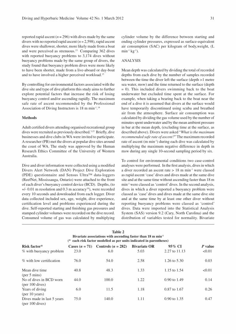
Diving and Hyperbaric Medicine Volume 42 No. 1 March 2012 31
reported rapid ascent (n = 296) with dives made by the same divers with no reported rapid ascent (n = 2,598), rapid ascent dives were shallower, shorter, more likely made from a boat and were perceived as strenuous.13 Comparing 362 dives with reported buoyancy problems to 3,174 dives without buoyancy problems made by the same group of divers, the study found that buoyancy problem dives were more likely to have been shorter, made from a live-aboard or day-boat and to have involved a higher perceived workload.13
By controlling for environmental factors associated with the dive site and type of dive platform this study aims to further explore potential factors that increase the risk of losing buoyancy control and/or ascending rapidly. The maximum safe rate of ascent recommended by the Professional Association of Diving Instructors is 18 m min-1.14
Methods
Adult certified divers attending organised recreational group dives were recruited as previously described.3,15 Briefly, dive businesses and dive clubs in WA were invited to participate. A researcher (PB) met the divers at popular dive sites around the coast of WA. The study was approved by the Human Research Ethics Committee of the University of Western Australia.
Dive and diver information were collected using a modified Divers Alert Network (DAN) Project Dive Exploration (PDE) questionnaire and Sensus Ultra™ data-loggers (ReefNet, Mississauga, Ontario) were attached to the front of each diver’s buoyancy control device (BCD). Depths, (to +/- 0.01 m resolution and 0.3 m accuracy16), were recorded every 10 seconds and downloaded from each logger. Diver data collected included sex, age, weight, dive experience, certification level and problems experienced during the dive. Self-reported starting and finishing gas pressures and stamped cylinder volumes were recorded on the dive record. Consumed volume of gas was calculated by multiplying
cylinder volume by the difference between starting and ending cylinder pressures, expressed as surface-equivalent air consumption (SAC) per kilogram of body,weight, (L min-1 kg-1).
ANALYSIS
Mean depth was calculated by dividing the total of recorded depths from each dive by the number of samples recorded between the time the diver left the surface (depth >1 metre sea water, msw) and the time returned to the surface (depth = 0). This included divers swimming back to the boat underwater but excluded time spent at the surface. For example, when taking a bearing back to the boat near the end of a dive it is assumed that divers at the surface would have temporarily discontinued using scuba and breathed air from the atmosphere. Surface air consumption was calculated by dividing the gas volume used by the number of minutes spent underwater and by the mean ambient pressure in bar at the mean depth, (excluding time at the surface, as described above). Divers were asked “What is the maximum recommended safe rate of ascent?” The maximum recorded rate of ascent (m min-1) during each dive was calculated by multiplying the maximum negative difference in depth in msw during any single 10-second sampling period by six.
To control for environmental conditions two case-control analyses were performed. In the first analysis, dives in which a diver recorded an ascent rate > 18 m min-1 were classed as rapid ascent ‘case’ dives and dives made at the same dive site and at the same time without ascending faster than 18 m min-1 were classed as ‘control’ dives. In the second analysis, dives in which a diver reported a buoyancy problem were classed as ‘case’ dives and dives made at the same dive site and at the same time by at least one other diver without reporting buoyancy problems were classed as ‘control’ dives. Data were imported into the Statistical Analysis System (SAS) version 9.2 (Cary, North Carolina) and the distribution of variables tested for normality. Bivariate
Risk factor* Cases (n = 71) Controls (n = 282) Bivariate OR 95% CI P value% with buoyancy problem 23.0 6.0 5.03 2.27 to 11.13 <0.01
% with low certification 76.0 54.0 2.58 1.26 to 5.30 0.03 Mean dive time 40.8 48.3 1.33 1.15 to 1.54 <0.01(per 5 mins) No of dives in BCD worn 44.0 100.0 1.22 0.90 to 1.49 0.14(per 100 dives) Years of diving 6.0 11.5 1.18 0.87 to 1.67 0.26(per 10 years) Dives made in last 5 years 75.0 140.0 1.11 0.90 to 1.35 0.47(per 100 dives)
Table 2Bivariate associations with ascending faster than 18 m min-1
(* each risk factor modelled as per units indicated in parentheses)
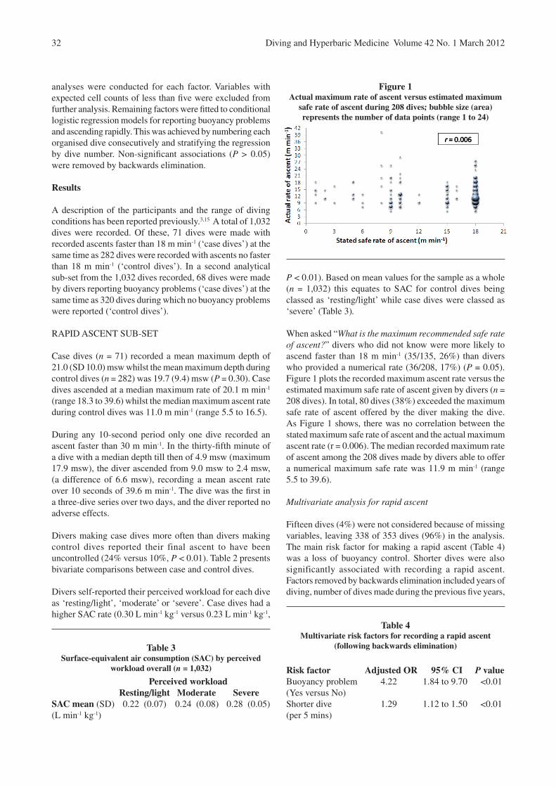
Diving and Hyperbaric Medicine Volume 42 No. 1 March 201232
analyses were conducted for each factor. Variables with expected cell counts of less than five were excluded from further analysis. Remaining factors were fitted to conditional logistic regression models for reporting buoyancy problems and ascending rapidly. This was achieved by numbering each organised dive consecutively and stratifying the regression by dive number. Non-significant associations (P > 0.05) were removed by backwards elimination.
Results
A description of the participants and the range of diving conditions has been reported previously.3,15 A total of 1,032 dives were recorded. Of these, 71 dives were made with recorded ascents faster than 18 m min-1 (‘case dives’) at the same time as 282 dives were recorded with ascents no faster than 18 m min-1 (‘control dives’). In a second analytical sub-set from the 1,032 dives recorded, 68 dives were made by divers reporting buoyancy problems (‘case dives’) at the same time as 320 dives during which no buoyancy problems were reported (‘control dives’).
RAPID ASCENT SUB-SET
Case dives (n = 71) recorded a mean maximum depth of 21.0 (SD 10.0) msw whilst the mean maximum depth during control dives (n = 282) was 19.7 (9.4) msw (P = 0.30). Case dives ascended at a median maximum rate of 20.1 m min-1 (range 18.3 to 39.6) whilst the median maximum ascent rate during control dives was 11.0 m min-1 (range 5.5 to 16.5).
During any 10-second period only one dive recorded an ascent faster than 30 m min-1. In the thirty-fifth minute of a dive with a median depth till then of 4.9 msw (maximum 17.9 msw), the diver ascended from 9.0 msw to 2.4 msw, (a difference of 6.6 msw), recording a mean ascent rate over 10 seconds of 39.6 m min-1. The dive was the first in a three-dive series over two days, and the diver reported no adverse effects.
Divers making case dives more often than divers making control dives reported their final ascent to have been uncontrolled (24% versus 10%, P < 0.01). Table 2 presents bivariate comparisons between case and control dives.
Divers self-reported their perceived workload for each dive as ‘resting/light’, ‘moderate’ or ‘severe’. Case dives had a higher SAC rate (0.30 L min-1 kg-1 versus 0.23 L min-1 kg-1,
P < 0.01). Based on mean values for the sample as a whole (n = 1,032) this equates to SAC for control dives being classed as ‘resting/light’ while case dives were classed as ‘severe’ (Table 3).
When asked “What is the maximum recommended safe rate of ascent?” divers who did not know were more likely to ascend faster than 18 m min-1 (35/135, 26%) than divers who provided a numerical rate (36/208, 17%) (P = 0.05). Figure 1 plots the recorded maximum ascent rate versus the estimated maximum safe rate of ascent given by divers (n = 208 dives). In total, 80 dives (38%) exceeded the maximum safe rate of ascent offered by the diver making the dive. As Figure 1 shows, there was no correlation between the stated maximum safe rate of ascent and the actual maximum ascent rate (r = 0.006). The median recorded maximum rate of ascent among the 208 dives made by divers able to offer a numerical maximum safe rate was 11.9 m min-1 (range 5.5 to 39.6).
Multivariate analysis for rapid ascent
Fifteen dives (4%) were not considered because of missing variables, leaving 338 of 353 dives (96%) in the analysis. The main risk factor for making a rapid ascent (Table 4) was a loss of buoyancy control. Shorter dives were also significantly associated with recording a rapid ascent. Factors removed by backwards elimination included years of diving, number of dives made during the previous five years,
Perceived workload Resting/light Moderate SevereSAC mean (SD) 0.22 (0.07) 0.24 (0.08) 0.28 (0.05)(L min-1 kg-1)
Table 3Surface-equivalent air consumption (SAC) by perceived
workload overall (n = 1,032)
Figure 1Actual maximum rate of ascent versus estimated maximum
safe rate of ascent during 208 dives; bubble size (area) represents the number of data points (range 1 to 24)
Risk factor Adjusted OR 95% CI P valueBuoyancy problem 4.22 1.84 to 9.70 <0.01(Yes versus No) Shorter dive 1.29 1.12 to 1.50 <0.01(per 5 mins)
Table 4Multivariate risk factors for recording a rapid ascent
(following backwards elimination)
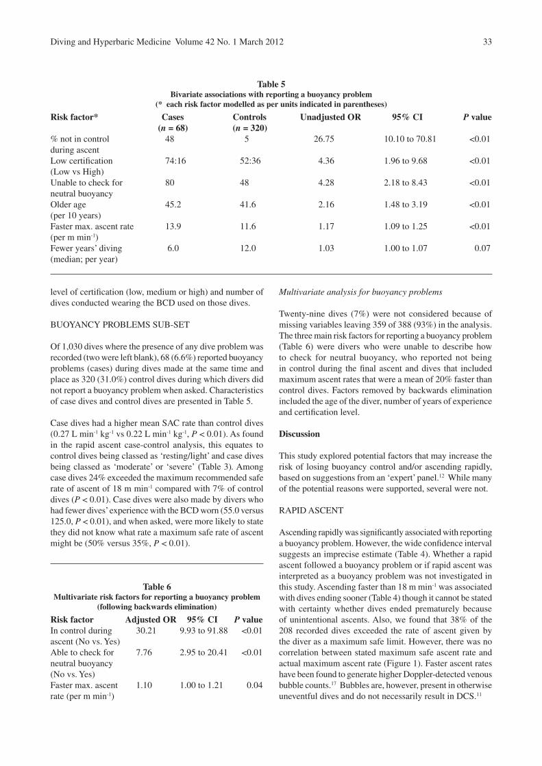
Diving and Hyperbaric Medicine Volume 42 No. 1 March 2012 33
level of certification (low, medium or high) and number of dives conducted wearing the BCD used on those dives.
BUOYANCY PROBLEMS SUB-SET
Of 1,030 dives where the presence of any dive problem was recorded (two were left blank), 68 (6.6%) reported buoyancy problems (cases) during dives made at the same time and place as 320 (31.0%) control dives during which divers did not report a buoyancy problem when asked. Characteristics of case dives and control dives are presented in Table 5.
Case dives had a higher mean SAC rate than control dives (0.27 L min-1 kg-1 vs 0.22 L min-1 kg-1, P < 0.01). As found in the rapid ascent case-control analysis, this equates to control dives being classed as ‘resting/light’ and case dives being classed as ‘moderate’ or ‘severe’ (Table 3). Among case dives 24% exceeded the maximum recommended safe rate of ascent of 18 m min-1 compared with 7% of control dives (P < 0.01). Case dives were also made by divers who had fewer dives’ experience with the BCD worn (55.0 versus 125.0, P < 0.01), and when asked, were more likely to state they did not know what rate a maximum safe rate of ascent might be (50% versus 35%, P < 0.01).
Multivariate analysis for buoyancy problems
Twenty-nine dives (7%) were not considered because of missing variables leaving 359 of 388 (93%) in the analysis. The three main risk factors for reporting a buoyancy problem (Table 6) were divers who were unable to describe how to check for neutral buoyancy, who reported not being in control during the final ascent and dives that included maximum ascent rates that were a mean of 20% faster than control dives. Factors removed by backwards elimination included the age of the diver, number of years of experience and certification level.
Discussion
This study explored potential factors that may increase the risk of losing buoyancy control and/or ascending rapidly, based on suggestions from an ‘expert’ panel.12 While many of the potential reasons were supported, several were not.
RAPID ASCENT
Ascending rapidly was significantly associated with reporting a buoyancy problem. However, the wide confidence interval suggests an imprecise estimate (Table 4). Whether a rapid ascent followed a buoyancy problem or if rapid ascent was interpreted as a buoyancy problem was not investigated in this study. Ascending faster than 18 m min-1 was associated with dives ending sooner (Table 4) though it cannot be stated with certainty whether dives ended prematurely because of unintentional ascents. Also, we found that 38% of the 208 recorded dives exceeded the rate of ascent given by the diver as a maximum safe limit. However, there was no correlation between stated maximum safe ascent rate and actual maximum ascent rate (Figure 1). Faster ascent rates have been found to generate higher Doppler-detected venous bubble counts.17 Bubbles are, however, present in otherwise uneventful dives and do not necessarily result in DCS.11
Risk factor* Cases Controls Unadjusted OR 95% CI P value (n = 68) (n = 320)% not in control 48 5 26.75 10.10 to 70.81 <0.01during ascentLow certification 74:16 52:36 4.36 1.96 to 9.68 <0.01(Low vs High)Unable to check for 80 48 4.28 2.18 to 8.43 <0.01neutral buoyancyOlder age 45.2 41.6 2.16 1.48 to 3.19 <0.01(per 10 years)Faster max. ascent rate 13.9 11.6 1.17 1.09 to 1.25 <0.01(per m min-1)Fewer years’ diving 6.0 12.0 1.03 1.00 to 1.07 0.07(median; per year)
Table 5Bivariate associations with reporting a buoyancy problem
(* each risk factor modelled as per units indicated in parentheses)
Risk factor Adjusted OR 95% CI P valueIn control during 30.21 9.93 to 91.88 <0.01ascent (No vs. Yes) Able to check for 7.76 2.95 to 20.41 <0.01neutral buoyancy (No vs. Yes)Faster max. ascent 1.10 1.00 to 1.21 0.04rate (per m min-1)
Table 6Multivariate risk factors for reporting a buoyancy problem
(following backwards elimination)
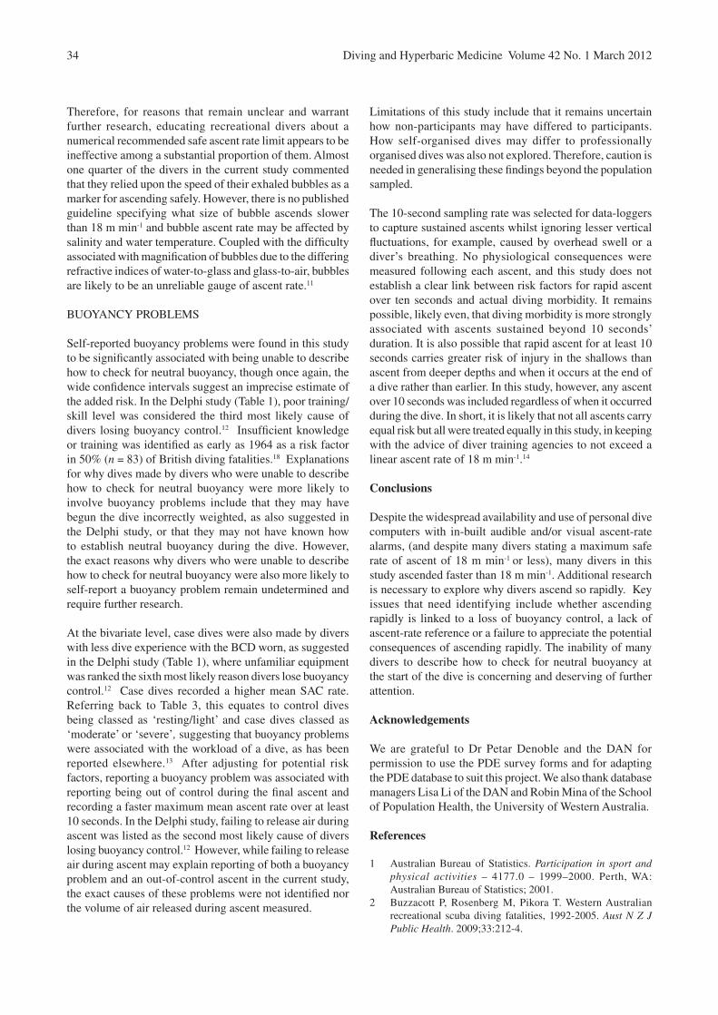
Diving and Hyperbaric Medicine Volume 42 No. 1 March 201234
Therefore, for reasons that remain unclear and warrant further research, educating recreational divers about a numerical recommended safe ascent rate limit appears to be ineffective among a substantial proportion of them. Almost one quarter of the divers in the current study commented that they relied upon the speed of their exhaled bubbles as a marker for ascending safely. However, there is no published guideline specifying what size of bubble ascends slower than 18 m min-1 and bubble ascent rate may be affected by salinity and water temperature. Coupled with the difficulty associated with magnification of bubbles due to the differing refractive indices of water-to-glass and glass-to-air, bubbles are likely to be an unreliable gauge of ascent rate.11
BUOYANCY PROBLEMS
Self-reported buoyancy problems were found in this study to be significantly associated with being unable to describe how to check for neutral buoyancy, though once again, the wide confidence intervals suggest an imprecise estimate of the added risk. In the Delphi study (Table 1), poor training/skill level was considered the third most likely cause of divers losing buoyancy control.12 Insufficient knowledge or training was identified as early as 1964 as a risk factor in 50% (n = 83) of British diving fatalities.18 Explanations for why dives made by divers who were unable to describe how to check for neutral buoyancy were more likely to involve buoyancy problems include that they may have begun the dive incorrectly weighted, as also suggested in the Delphi study, or that they may not have known how to establish neutral buoyancy during the dive. However, the exact reasons why divers who were unable to describe how to check for neutral buoyancy were also more likely to self-report a buoyancy problem remain undetermined and require further research.
At the bivariate level, case dives were also made by divers with less dive experience with the BCD worn, as suggested in the Delphi study (Table 1), where unfamiliar equipment was ranked the sixth most likely reason divers lose buoyancy control.12 Case dives recorded a higher mean SAC rate. Referring back to Table 3, this equates to control dives being classed as ‘resting/light’ and case dives classed as ‘moderate’ or ‘severe’, suggesting that buoyancy problems were associated with the workload of a dive, as has been reported elsewhere.13 After adjusting for potential risk factors, reporting a buoyancy problem was associated with reporting being out of control during the final ascent and recording a faster maximum mean ascent rate over at least 10 seconds. In the Delphi study, failing to release air during ascent was listed as the second most likely cause of divers losing buoyancy control.12 However, while failing to release air during ascent may explain reporting of both a buoyancy problem and an out-of-control ascent in the current study, the exact causes of these problems were not identified nor the volume of air released during ascent measured.
Limitations of this study include that it remains uncertain how non-participants may have differed to participants. How self-organised dives may differ to professionally organised dives was also not explored. Therefore, caution is needed in generalising these findings beyond the population sampled.
The 10-second sampling rate was selected for data-loggers to capture sustained ascents whilst ignoring lesser vertical fluctuations, for example, caused by overhead swell or a diver’s breathing. No physiological consequences were measured following each ascent, and this study does not establish a clear link between risk factors for rapid ascent over ten seconds and actual diving morbidity. It remains possible, likely even, that diving morbidity is more strongly associated with ascents sustained beyond 10 seconds’ duration. It is also possible that rapid ascent for at least 10 seconds carries greater risk of injury in the shallows than ascent from deeper depths and when it occurs at the end of a dive rather than earlier. In this study, however, any ascent over 10 seconds was included regardless of when it occurred during the dive. In short, it is likely that not all ascents carry equal risk but all were treated equally in this study, in keeping with the advice of diver training agencies to not exceed a linear ascent rate of 18 m min-1.14
Conclusions
Despite the widespread availability and use of personal dive computers with in-built audible and/or visual ascent-rate alarms, (and despite many divers stating a maximum safe rate of ascent of 18 m min-1 or less), many divers in this study ascended faster than 18 m min-1. Additional research is necessary to explore why divers ascend so rapidly. Key issues that need identifying include whether ascending rapidly is linked to a loss of buoyancy control, a lack of ascent-rate reference or a failure to appreciate the potential consequences of ascending rapidly. The inability of many divers to describe how to check for neutral buoyancy at the start of the dive is concerning and deserving of further attention.
Acknowledgements
We are grateful to Dr Petar Denoble and the DAN for permission to use the PDE survey forms and for adapting the PDE database to suit this project. We also thank database managers Lisa Li of the DAN and Robin Mina of the School of Population Health, the University of Western Australia.
References
1 Australian Bureau of Statistics. Participation in sport and physical activities – 4177.0 – 1999–2000. Perth, WA: Australian Bureau of Statistics; 2001.
2 Buzzacott P, Rosenberg M, Pikora T. Western Australian recreational scuba diving fatalities, 1992-2005. Aust N Z J Public Health. 2009;33:212-4.
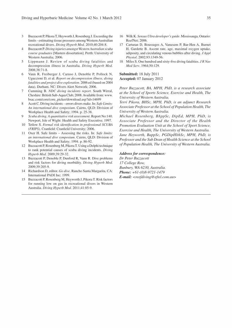
Diving and Hyperbaric Medicine Volume 42 No. 1 March 2012 35
3 Buzzacott P, Pikora T, Heyworth J, Rosenberg J. Exceeding the limits - estimating tissue pressures among Western Australian recreational divers. Diving Hyperb Med. 2010;40:204-8.
4 Buzzacott P. Diving injuries amongst Western Australian scuba course graduates [Masters dissertation]. Perth: University of Western Australia; 2006.
5 Lippmann J. Review of scuba diving fatalities and decompression illness in Australia. Diving Hyperb Med. 2008;38:71-8.
6 Vann R, Freiberger J, Caruso J, Denoble P, Pollock N, Uguccioni D, et al. Report on decompression illness, diving fatalities and project dive exploration. 2006 ed (based on 2004 data). Durham, NC: Divers Alert Network; 2006.
7 Cumming B. NDC diving incidents report. South Wirral, Cheshire: British Sub-Aqua Club; 2008. Available from: www.bsac.com/core/core_picker/download.asp?id=14699
8 Acott C. Diving incidents – errors divers make. In: Safe Limits: An international dive symposium. Cairns, QLD: Division of Workplace Health and Safety; 1994. p. 25-38.
9 Scuba diving. A quantitative risk assessment. Report No:140. Newport, Isle of Wight: Health and Safety Executive; 1997.
10 Tetlow S. Formal risk identification in professional SCUBA (FRIPS). Cranfield: Cranfield University; 2006.
11 Oxer H. Safe limits – Assessing the risks. In: Safe limits: an international dive symposium. Cairns, QLD: Division of Workplace Health and Safety; 1994. p. 86-92.
12 Buzzacott P, Rosenberg M, Pikora T. Using a Delphi technique to rank potential causes of scuba diving incidents. Diving Hyperb Med. 2009;39:29-32.
13 Buzzacott P, Denoble P, Dunford R, Vann R. Dive problems and risk factors for diving morbidity. Diving Hyperb Med. 2009;39:205-9.
14 Richardson D, editor. Go dive. Rancho Santa Margarita, CA: International PADI Inc; 1999.
15 Buzzacott P, Rosenberg M, Heyworth J, Pikora T. Risk factors for running low on gas in recreational divers in Western Australia. Diving Hyperb Med. 2011;41:85-9.
16 Wilk K. Sensus Ultra developer’s guide. Mississauga, Ontario: ReefNet; 2006.
17 Carturan D, Boussuges A, Vanuxem P, Bar-Hen A, Burnet H, Gardette B. Ascent rate, age, maximal oxygen uptake, adiposity, and circulating venous bubbles after diving. J Appl Physiol. 2002;93:1349-56.
18 Miles S. One hundred and sixty-five diving fatalities. J R Nav Med Serv. 1964;50:129.
Submitted: 18 July 2011Accepted: 07 January 2012
Peter Buzzacott, BA, MPH, PhD, is a research associate at the School of Sports Science, Exercise and Health, The University of Western Australia.Terri Pikora, BHSc, MPH, PhD, is an adjunct Research Associate Professor at the School of Population Health, The University of Western Australia.Michael Rosenberg, BAppSc, DipEd, MPH, PhD, is Associate Professor and the Director of the Health Promotion Evaluation Unit at the School of Sport Science, Exercise and Health, The University of Western Australia.Jane Heyworth, BappSc, PGDipHlthSc, MPH, PhD, is Professor and the Sub Dean of Health Science at the School of Population Health, The University of Western Australia.
Address for correspondence:Dr Peter Buzzacott17 College Row,Bunbury, WA 6230, Australia.Phone: +61-(0)8-9721-1479E-mail: <[email protected]>
Diving and Hyperbaric Medicine Volume 42 No. 1 March 201236
Introduction
Rapidly advancing technology has enabled ultrasound machines to become more affordable and compact, and to provide higher-quality imaging. Ultrasound provides a safe and effective, dynamic and repeatable form of imaging that can be performed at the patient bedside, and is free from the harmful effects of ionising radiation. The combination of these factors has led to ultrasound becoming increasingly popular across nearly every speciality of medicine.
Point-of-care ultrasound is defined as ultrasound performed and interpreted at the bedside and has led to the concept of the ‘ultrasound stethoscope’.1 Ultrasound education for non-imaging specialties is now relatively advanced, with guidelines established by many specialty colleges.2 It is now being included in the syllabus for many speciality registrar training schemes and is being considered for inclusion in undergraduate training in many centres in the United States, the United Kingdom and Australia.3 Some American medical schools are even beginning to provide their students with hand-held ultrasound machines for use during clinical rotations.4
A formal role for the use of point-of-care ultrasound in the field of hyperbaric medicine has yet to be clearly established; however, we see many possibilities for both clinical and research purposes. Within hyperbaric chambers, ultrasound transducers have been passed through access ports to study physiological parameters.5–7 To our knowledge, ultrasound scanning with a machine inside the chamber has not been reported.
Potential applications of ultrasound in hyperbaric medicine
Ready and immediate access to an ultrasound machine within a recompression chamber could benefit patients in a number of ways.
PNEUMOTHORAX DETECTION
The role of ultrasound in the detection of pneumothoracies is well established in emergency medicine.8 Divers with cerebral arterial gas embolism (CAGE) have pulmonary barotrauma by definition and may have an increased risk of developing a pneumothorax. If this occurs during hyperbaric treatment and remains undetected during ascent, the consequences are potentially catastrophic. Routine treatment of CAGE involves keeping the patient supine. For pneumothorax detection, a supine chest X-ray has a sensitivity ranging from 28% to 75%, whereas lung ultrasound has a sensitivity ranging from 86% to 98% even with minimal training.9,10 The absence of the lung sliding sign, comet tail artefacts and the presence of a contact point confirms the diagnosis. The study can be successfully completed within 2–3 minutes.11
The clinical challenge of pneumothorax detection relies on identifying increased resonance to percussion and reduced breath sounds on the affected side. Early detection inside a noisy chamber can be very difficult and the decision to needle the chest without convincing evidence of pneumothorax is often difficult. The ability to image at depth with in-chamber ultrasound would allow detection of supine pneumothoracies before compression, and, if one developed at depth, would allow thoracocentesis to be performed when indicated. It would also allow clinicians to entertain other diagnoses when pneumothorax had been excluded as a cause for deterioration at depth.
CRITICAL CARE PATIENTS
Critical care patients inside the chamber pose unique problems to the hyperbaric physician. Some hyperbaric facilities run daily hyperbaric oxygen treatments for intensive care patients. In-chamber ultrasound provides a useful tool for a wide range of critical care applications. Pulmonary ultrasonography has been shown to be more
Review articleUltrasound in diving and hyperbaric medicineIan C Gawthrope
Abstract
(Gawthorpe IC. Ultrasound in diving and hyperbaric medicine. Diving and Hyperb Med. 2012;42(1):36-39.)Ultrasound is a safe and effective imaging modality, the use of which is increasing exponentially in many areas of clinical medicine. In this article, we present what is, to our knowledge, the first in-chamber use of an ultrasound machine. We discuss the challenges this presented and how they were addressed, and explore the possible clinical applications that in-chamber ultrasound may deliver in hyperbaric medicine.
Key wordsUltrasound, hyperbaric medicine, equipment, review article
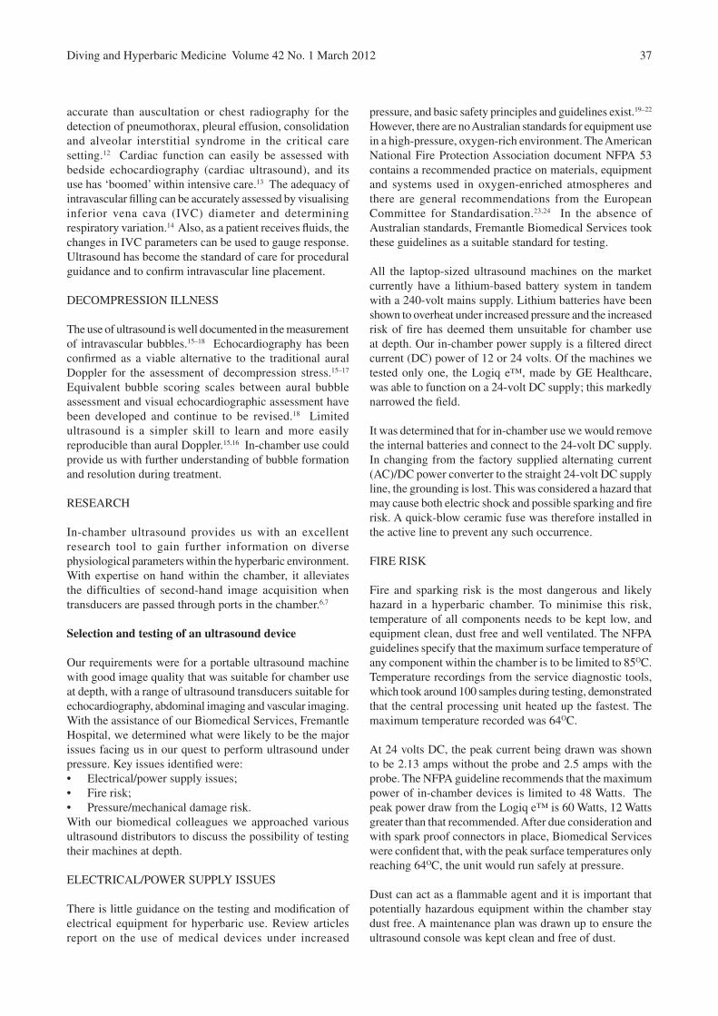
Diving and Hyperbaric Medicine Volume 42 No. 1 March 2012 37
accurate than auscultation or chest radiography for the detection of pneumothorax, pleural effusion, consolidation and alveolar interstitial syndrome in the critical care setting.12 Cardiac function can easily be assessed with bedside echocardiography (cardiac ultrasound), and its use has ‘boomed’ within intensive care.13 The adequacy of intravascular filling can be accurately assessed by visualising inferior vena cava (IVC) diameter and determining respiratory variation.14 Also, as a patient receives fluids, the changes in IVC parameters can be used to gauge response. Ultrasound has become the standard of care for procedural guidance and to confirm intravascular line placement.
DECOMPRESSION ILLNESS
The use of ultrasound is well documented in the measurement of intravascular bubbles.15–18 Echocardiography has been confirmed as a viable alternative to the traditional aural Doppler for the assessment of decompression stress.15–17 Equivalent bubble scoring scales between aural bubble assessment and visual echocardiographic assessment have been developed and continue to be revised.18 Limited ultrasound is a simpler skill to learn and more easily reproducible than aural Doppler.15,16 In-chamber use could provide us with further understanding of bubble formation and resolution during treatment.
RESEARCH
In-chamber ultrasound provides us with an excellent research tool to gain further information on diverse physiological parameters within the hyperbaric environment. With expertise on hand within the chamber, it alleviates the difficulties of second-hand image acquisition when transducers are passed through ports in the chamber.6,7
Selection and testing of an ultrasound device
Our requirements were for a portable ultrasound machine with good image quality that was suitable for chamber use at depth, with a range of ultrasound transducers suitable for echocardiography, abdominal imaging and vascular imaging. With the assistance of our Biomedical Services, Fremantle Hospital, we determined what were likely to be the major issues facing us in our quest to perform ultrasound under pressure. Key issues identified were:• Electrical/power supply issues;• Fire risk;• Pressure/mechanical damage risk.With our biomedical colleagues we approached various ultrasound distributors to discuss the possibility of testing their machines at depth.
ELECTRICAL/POWER SUPPLY ISSUES
There is little guidance on the testing and modification of electrical equipment for hyperbaric use. Review articles report on the use of medical devices under increased
pressure, and basic safety principles and guidelines exist.19–22 However, there are no Australian standards for equipment use in a high-pressure, oxygen-rich environment. The American National Fire Protection Association document NFPA 53 contains a recommended practice on materials, equipment and systems used in oxygen-enriched atmospheres and there are general recommendations from the European Committee for Standardisation.23,24 In the absence of Australian standards, Fremantle Biomedical Services took these guidelines as a suitable standard for testing.
All the laptop-sized ultrasound machines on the market currently have a lithium-based battery system in tandem with a 240-volt mains supply. Lithium batteries have been shown to overheat under increased pressure and the increased risk of fire has deemed them unsuitable for chamber use at depth. Our in-chamber power supply is a filtered direct current (DC) power of 12 or 24 volts. Of the machines we tested only one, the Logiq e™, made by GE Healthcare, was able to function on a 24-volt DC supply; this markedly narrowed the field.
It was determined that for in-chamber use we would remove the internal batteries and connect to the 24-volt DC supply. In changing from the factory supplied alternating current (AC)/DC power converter to the straight 24-volt DC supply line, the grounding is lost. This was considered a hazard that may cause both electric shock and possible sparking and fire risk. A quick-blow ceramic fuse was therefore installed in the active line to prevent any such occurrence.
FIRE RISK
Fire and sparking risk is the most dangerous and likely hazard in a hyperbaric chamber. To minimise this risk, temperature of all components needs to be kept low, and equipment clean, dust free and well ventilated. The NFPA guidelines specify that the maximum surface temperature of any component within the chamber is to be limited to 85OC. Temperature recordings from the service diagnostic tools, which took around 100 samples during testing, demonstrated that the central processing unit heated up the fastest. The maximum temperature recorded was 64OC.
At 24 volts DC, the peak current being drawn was shown to be 2.13 amps without the probe and 2.5 amps with the probe. The NFPA guideline recommends that the maximum power of in-chamber devices is limited to 48 Watts. The peak power draw from the Logiq e™ is 60 Watts, 12 Watts greater than that recommended. After due consideration and with spark proof connectors in place, Biomedical Services were confident that, with the peak surface temperatures only reaching 64OC, the unit would run safely at pressure.
Dust can act as a flammable agent and it is important that potentially hazardous equipment within the chamber stay dust free. A maintenance plan was drawn up to ensure the ultrasound console was kept clean and free of dust.
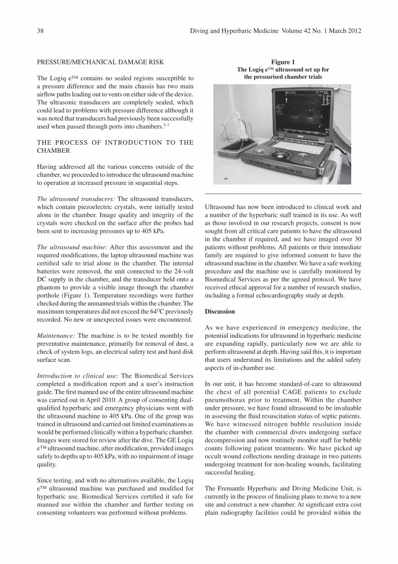
Diving and Hyperbaric Medicine Volume 42 No. 1 March 201238
PRESSURE/MECHANICAL DAMAGE RISK
The Logiq e™ contains no sealed regions susceptible to a pressure difference and the main chassis has two main airflow paths leading out to vents on either side of the device. The ultrasonic transducers are completely sealed, which could lead to problems with pressure difference although it was noted that transducers had previously been successfully used when passed through ports into chambers.5–7
THE PROCESS OF INTRODUCTION TO THE CHAMBER
Having addressed all the various concerns outside of the chamber, we proceeded to introduce the ultrasound machine to operation at increased pressure in sequential steps.
The ultrasound transducers: The ultrasound transducers, which contain piezoelectric crystals, were initially tested alone in the chamber. Image quality and integrity of the crystals were checked on the surface after the probes had been sent to increasing pressures up to 405 kPa.
The ultrasound machine: After this assessment and the required modifications, the laptop ultrasound machine was certified safe to trial alone in the chamber. The internal batteries were removed, the unit connected to the 24-volt DC supply in the chamber, and the transducer held onto a phantom to provide a visible image through the chamber porthole (Figure 1). Temperature recordings were further checked during the unmanned trials within the chamber. The maximum temperatures did not exceed the 64OC previously recorded. No new or unexpected issues were encountered.
Maintenance: The machine is to be tested monthly for preventative maintenance, primarily for removal of dust, a check of system logs, an electrical safety test and hard disk surface scan.
Introduction to clinical use: The Biomedical Services completed a modification report and a user’s instruction guide. The first manned use of the entire ultrasound machine was carried out in April 2010. A group of consenting dual-qualified hyperbaric and emergency physicians went with the ultrasound machine to 405 kPa. One of the group was trained in ultrasound and carried out limited examinations as would be performed clinically within a hyperbaric chamber. Images were stored for review after the dive. The GE Logiq e™ ultrasound machine, after modification, provided images safely to depths up to 405 kPa, with no impairment of image quality.
Since testing, and with no alternatives available, the Logiq e™ ultrasound machine was purchased and modified for hyperbaric use. Biomedical Services certified it safe for manned use within the chamber and further testing on consenting volunteers was performed without problems.
Ultrasound has now been introduced to clinical work and a number of the hyperbaric staff trained in its use. As well as those involved in our research projects, consent is now sought from all critical care patients to have the ultrasound in the chamber if required, and we have imaged over 30 patients without problems. All patients or their immediate family are required to give informed consent to have the ultrasound machine in the chamber. We have a safe working procedure and the machine use is carefully monitored by Biomedical Services as per the agreed protocol. We have received ethical approval for a number of research studies, including a formal echocardiography study at depth.
Discussion
As we have experienced in emergency medicine, the potential indications for ultrasound in hyperbaric medicine are expanding rapidly, particularly now we are able to perform ultrasound at depth. Having said this, it is important that users understand its limitations and the added safety aspects of in-chamber use.
In our unit, it has become standard-of-care to ultrasound the chest of all potential CAGE patients to exclude pneumothorax prior to treatment. Within the chamber under pressure, we have found ultrasound to be invaluable in assessing the fluid resuscitation status of septic patients. We have witnessed nitrogen bubble resolution inside the chamber with commercial divers undergoing surface decompression and now routinely monitor staff for bubble counts following patient treatments. We have picked up occult wound collections needing drainage in two patients undergoing treatment for non-healing wounds, facilitating successful healing.
The Fremantle Hyperbaric and Diving Medicine Unit, is currently in the process of finalising plans to move to a new site and construct a new chamber. At significant extra cost plain radiography facilities could be provided within the
Figure 1The Logiq e™ ultrasound set up for
the pressurised chamber trials
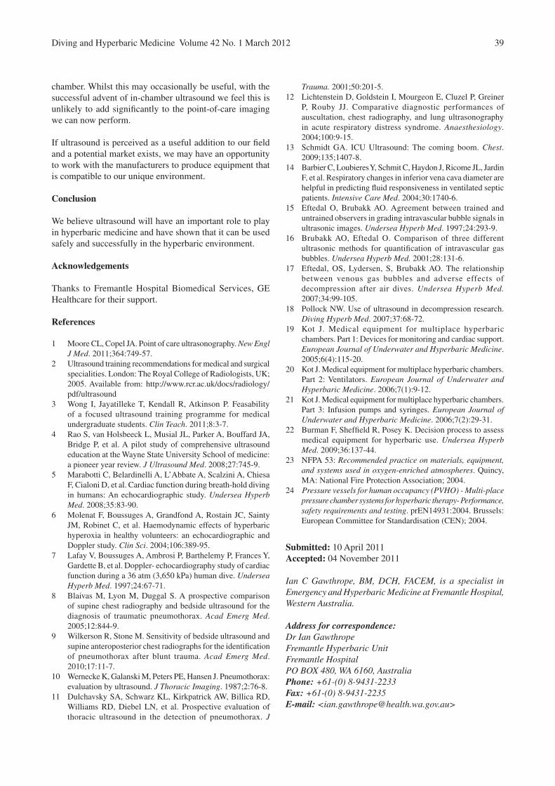
Diving and Hyperbaric Medicine Volume 42 No. 1 March 2012 39
chamber. Whilst this may occasionally be useful, with the successful advent of in-chamber ultrasound we feel this is unlikely to add significantly to the point-of-care imaging we can now perform.
If ultrasound is perceived as a useful addition to our field and a potential market exists, we may have an opportunity to work with the manufacturers to produce equipment that is compatible to our unique environment.
Conclusion
We believe ultrasound will have an important role to play in hyperbaric medicine and have shown that it can be used safely and successfully in the hyperbaric environment.
Acknowledgements
Thanks to Fremantle Hospital Biomedical Services, GE Healthcare for their support.
References
1 Moore CL, Copel JA. Point of care ultrasonography. New Engl J Med. 2011;364:749-57.
2 Ultrasound training recommendations for medical and surgical specialities. London: The Royal College of Radiologists, UK; 2005. Available from: http://www.rcr.ac.uk/docs/radiology/pdf/ultrasound
3 Wong I, Jayatilleke T, Kendall R, Atkinson P. Feasability of a focused ultrasound training programme for medical undergraduate students. Clin Teach. 2011;8:3-7.
4 Rao S, van Holsbeeck L, Musial JL, Parker A, Bouffard JA, Bridge P, et al. A pilot study of comprehensive ultrasound education at the Wayne State University School of medicine: a pioneer year review. J Ultrasound Med. 2008;27:745-9.
5 Marabotti C, Belardinelli A, L’Abbate A, Scalzini A, Chiesa F, Cialoni D, et al. Cardiac function during breath-hold diving in humans: An echocardiographic study. Undersea Hyperb Med. 2008;35:83-90.
6 Molenat F, Boussuges A, Grandfond A, Rostain JC, Sainty JM, Robinet C, et al. Haemodynamic effects of hyperbaric hyperoxia in healthy volunteers: an echocardiographic and Doppler study. Clin Sci. 2004;106:389-95.
7 Lafay V, Boussuges A, Ambrosi P, Barthelemy P, Frances Y, Gardette B, et al. Doppler- echocardiography study of cardiac function during a 36 atm (3,650 kPa) human dive. Undersea Hyperb Med. 1997;24:67-71.
8 Blaivas M, Lyon M, Duggal S. A prospective comparison of supine chest radiography and bedside ultrasound for the diagnosis of traumatic pneumothorax. Acad Emerg Med. 2005;12:844-9.
9 Wilkerson R, Stone M. Sensitivity of bedside ultrasound and supine anteroposterior chest radiographs for the identification of pneumothorax after blunt trauma. Acad Emerg Med. 2010;17:11-7.
10 Wernecke K, Galanski M, Peters PE, Hansen J. Pneumothorax: evaluation by ultrasound. J Thoracic Imaging. 1987;2:76-8.
11 Dulchavsky SA, Schwarz KL, Kirkpatrick AW, Billica RD, Williams RD, Diebel LN, et al. Prospective evaluation of thoracic ultrasound in the detection of pneumothorax. J
Trauma. 2001;50:201-5.12 Lichtenstein D, Goldstein I, Mourgeon E, Cluzel P, Greiner
P, Rouby JJ. Comparative diagnostic performances of auscultation, chest radiography, and lung ultrasonography in acute respiratory distress syndrome. Anaesthesiology. 2004;100:9-15.
13 Schmidt GA. ICU Ultrasound: The coming boom. Chest. 2009;135;1407-8.
14 Barbier C, Loubieres Y, Schmit C, Haydon J, Ricome JL, Jardin F, et al. Respiratory changes in inferior vena cava diameter are helpful in predicting fluid responsiveness in ventilated septic patients. Intensive Care Med. 2004;30:1740-6.
15 Eftedal O, Brubakk AO. Agreement between trained and untrained observers in grading intravascular bubble signals in ultrasonic images. Undersea Hyperb Med. 1997;24:293-9.
16 Brubakk AO, Eftedal O. Comparison of three different ultrasonic methods for quantification of intravascular gas bubbles. Undersea Hyperb Med. 2001;28:131-6.
17 Eftedal, OS, Lydersen, S, Brubakk AO. The relationship between venous gas bubbles and adverse effects of decompression after air dives. Undersea Hyperb Med. 2007;34:99-105.
18 Pollock NW. Use of ultrasound in decompression research. Diving Hyperb Med. 2007;37:68-72.
19 Kot J. Medical equipment for multiplace hyperbaric chambers. Part 1: Devices for monitoring and cardiac support. European Journal of Underwater and Hyperbaric Medicine. 2005;6(4):115-20.
20 Kot J. Medical equipment for multiplace hyperbaric chambers. Part 2: Ventilators. European Journal of Underwater and Hyperbaric Medicine. 2006;7(1):9-12.
21 Kot J. Medical equipment for multiplace hyperbaric chambers. Part 3: Infusion pumps and syringes. European Journal of Underwater and Hyperbaric Medicine. 2006;7(2):29-31.
22 Burman F, Sheffield R, Posey K. Decision process to assess medical equipment for hyperbaric use. Undersea Hyperb Med. 2009;36:137-44.
23 NFPA 53: Recommended practice on materials, equipment, and systems used in oxygen-enriched atmospheres. Quincy, MA: National Fire Protection Association; 2004.
24 Pressure vessels for human occupancy (PVHO) - Multi-place pressure chamber systems for hyperbaric therapy- Performance, safety requirements and testing. prEN14931:2004. Brussels: European Committee for Standardisation (CEN); 2004.
Submitted: 10 April 2011Accepted: 04 November 2011
Ian C Gawthrope, BM, DCH, FACEM, is a specialist in Emergency and Hyperbaric Medicine at Fremantle Hospital, Western Australia.
Address for correspondence:Dr Ian GawthropeFremantle Hyperbaric UnitFremantle HospitalPO BOX 480, WA 6160, AustraliaPhone: +61-(0) 8-9431-2233Fax: +61-(0) 8-9431-2235E-mail: <[email protected]>
Diving and Hyperbaric Medicine Volume 42 No. 1 March 201240
Case reportScuba divers’ pulmonary oedema: recurrences and fatalitiesCarl Edmonds, John Lippmann, Sarah Lockley and Darren Wolfers
Abstract
(Edmonds C, Lippmann J, Lockley S, Wolfers D. Scuba divers’ pulmonary oedema: recurrences and fatalities. Diving Hyperb Med. 2012;42(1):40-44.)Scuba divers’ pulmonary oedema (SDPE) is an increasingly recognised disorder in divers. We report three fatal cases of SDPE, demonstrating its potentially serious nature even in the absence of underlying cardiac disease demonstrable clinically or at autopsy. This, together with the frequency of recurrences, has implications on assessing fitness for subsequent diving, snorkelling and swimming. The differential diagnosis of this disorder is also considered, as is its possible inducement by salt water aspiration and its relationship to drowning.
Key wordsScuba diving, pulmonary oedema, salt water aspiration, deaths, case reports
Introduction
Scuba divers’ pulmonary oedema (SDPE) was first recorded in 1981.1 Comprehensive reviews have been prepared since by various authors.2–6 In these reviews, the physiological bases of the disorder have been canvassed; it is one type of immersion pulmonary oedema (IPE). SDPE presents with scuba divers developing fast shallow respirations, dyspnoea, fatigue, cough and white or sometimes blood-stained frothy expectoration. The signs include hypoxia and the auscultatory evidence of pulmonary oedema. Investigations reveal impaired spirometry and reduced lung compliance, hypoxaemia and characteristic radiological (plain chest X-ray or CT scan) abnormalities.
SDPE is said to be more frequent in older divers and those with cardiovascular pathology.1–6 It tends to recur in at least 30% of cases.5 Exertion during the dive is often not excessive and frequently the condition becomes more evident during ascent or surface swimming. Spontaneous resolution is often prompt after leaving the water. Only one death has been reported in the traditional medical literature and this was based on significant pre-existing cardiac pathology.5 The latter is characteristic of some of these SDPE cases and is one aetiological feature that may be amenable to correction.
These three case histories illustrate the difficulty in predicting the development of non-cardiac based SDPE, the significance of recurrences and the possibility of death from this disorder. They have implications regarding appropriate advice that is given to affected divers.
Case history 1
Incident 1: A 51-year-old female nurse had no significant past medical history other than a mild allergic diathesis
in early life, presenting with eczema and hay fever. She was an experienced scuba diver, logging over 900 dives without incident and possessing open-water and deep-diving qualifications. She was considered a conservative but enthusiastic club diver. The day before the incident she had completed two non-decompression, computer-assisted dives in an area well known to her. The first was to 24 metres’ sea water (msw) for 50 minutes, followed by a surface interval of three hours; the second to 7 msw for 10 minutes, aborted due to currents and poor visibility. That afternoon and night she consumed 70 grams of alcohol, together with other fluids.
The following day, she felt well, although a little fatigued. At 0800 h she commenced a dive profile that she had undertaken on other occasions without difficulty. This involved a 30-metre surface swim, fully equipped but finning on her back and with the regulator out. The conditions were described as perfect, and the current was considered “moderate at the worst”. Although she reported that she did not experience any aspiration, she did state that the wash from a boat splashed over her head once, causing her to cough and swallow some sea water. Later, during this four-minute swim, she became dyspnoeic. Her companion observed that it was a “tough swim” and that her lips appeared cyanotic and her breathing rate was rapid during the minute she spent resting on the marker buoy. In subsequent interrogations, she denied any salt water aspiration, chest discomfort, palpitations or syncopal sensation at that time.
Because they thought there could be less current at depth, they commenced the dive but only reached about 12 msw in one to two minutes. They aborted the dive after three minutes, due to her progressive dyspnoea and feeling fatigued. They ascended slowly, over about five minutes, before surfacing near the shore. She was then assisted in walking and removing her equipment.
Diving and Hyperbaric Medicine Volume 42 No. 1 March 2012 41
Her coughing was frequent with expectoration initially whitish but becoming pink and frothy and she was aware of fluid rattling in her chest. She was dyspnoeic and cyanotic, with a grey appearance. She improved somewhat over the next quarter of an hour and was then able to walk unassisted. Ambulance paramedics administered high-concentration oxygen, until the medevac helicopter arrived. In telephone discussion with the DAN diving emergency service (DES), the clinician heard her wheezing and noted her complaints of dyspnoea and a “rattling” in her chest. She was transferred to the metropolitan hospital, breathing oxygen administered via a simple face mask.
Her vital signs on admission at 1045 h were not grossly abnormal, with a heart rate of 100 beats per minute and a respiratory rate of 24 breaths per minute, but she still had a persistent, non-productive cough with wheezing and crepitations at both lung bases. She was continued on oxygen and bronchodilators were administered. The chest X-ray showed minor linear basal densities, more on the right, consistent with interstitial oedema. All other investigations (ECG, lung function, electrolytes, biochemistry, liver function, oxygen saturation) were normal. The respiratory difficulty settled by 1500 hours and she was discharged the following day, for later review. Then, her lung function tests showed improvements of 18% in forced vital capacity, increasing to 26% following administration of a bronchodilator. The original impairment was considered to be consistent with increased airway reactivity associated with lung damage. A mild neutrophil leucocytosis was similarly explained. There were no other symptoms or signs suggestive of decompression sickness or pulmonary barotrauma and the dive profile was not indicative of these disorders.
A month later, a specialist cardiologist consultation included clinical assessment, ECG, stress testing and transthoracic echocardiograms, without any abnormality being detected. He concluded that the episode of pulmonary oedema was non-cardiogenic and that the patient had normal cardiac function. Repeat lung function testing at the same laboratory showed normal lung values and an asthma provocation test was negative. There was an improvement in lung volumes compared to the previous tests.
Her enthusiasm to return to diving and to re-establish her DAN diving insurance for future overseas diving trips led to consultations with at least six diving medical specialists. The diagnoses were divided between SDPE and the salt water aspiration syndrome (SWAS), and advice varied from unfitness for any diving (snorkel or scuba) to approval for unrestricted diving. She considered the conflicting advice available and also attempted her own research on this subject, and then resumed diving.
Incident 2: Almost a year later, now with another 54 logged dives, and with no further medical history apart from the
incident above, she died whilst diving. She was participating in a night dive from shore. There was a moderate wind and the surface was choppy. Surface water temperature was about 22OC reducing to 19OC at depth and was described as comfortable. She was wearing a semi-dry suit.
The victim was with three others, in two buddy pairs. They swam on the surface for about 30 metres before descending and working along the sloping bottom to a maximum depth of 18 msw. For most of the dive the victim appeared to be fine and responded affirmatively to the buddy’s regular ‘OK?’ signals. However, after about 25 minutes, at a depth of 14 msw, she signalled that she was ‘not OK’. They decided to return and they swam underwater up the slope and towards the shore. Each time the buddy enquired if she was OK she responded in the negative. On reaching a depth of 7 msw, the buddy held her hand and they slowly ascended and surfaced in a sheltered area, with a dive time of 37 minutes.
At the surface, she vomited a brown, lumpy liquid. She was trying to cough and had an audible wheeze. She stated faintly that she could not breathe and she continued to vomit. Her BCD was inflated and she rolled over onto her back as the buddy towed her towards the shore. The buddy could hear her wheezing and struggling to breathe. She was still conscious and complained that she could not breathe, but tried to kick her legs to assist the buddy towing her. The buddy towed her approximately 100 metres to thigh-deep water beside rocks. She was assisted onto the rocks. It was believed that she did not inhale any water during the rescue.
She then became unconscious and apnoeic, and her buddy commenced basic life support. This produced regurgitation of stomach contents and some bloody sputum. Others assisted until the paramedics arrived about 15 minutes later. They implemented advanced life support but she failed to respond.
At autopsy the lungs were oedematous, weighing over 1.4 kg, and did not appear unduly hyperexpanded. There was no pathological evidence to indicate other causes of death, including previous or recent cardiovascular disease. The heart weighed 310 g. Toxicology was negative. The pathological diagnosis of acute pulmonary oedema was made.
Case history 2
This 45-year-old woman was apparently healthy and had become certified as an Open Water Diver one week earlier, having completed four open-water training dives. She was then participating in an Advanced Open Water course and had completed three uneventful dives on the previous day to a maximum depth of 7 msw, with a surface interval of 8 hours between the last two dives.
On the day, the water was calm and clear with visibility of 10–15 msw, and the dive was at slack water. The victim was
Diving and Hyperbaric Medicine Volume 42 No. 1 March 201242
with a group of six students, accompanied by an instructor and a divemaster. They descended to a depth of 26 msw and knelt on the sea bed while answering questions on a slate. The duration of the dive was 16 minutes and she had completed other narcosis tasks. The victim then gave a low-on-air hand signal. The instructor noted that her contents gauge read 120 bar and gave her his ‘octopus’ regulator to breathe on briefly while he breathed on her demand valve, to check that it was ‘OK’; it appeared to be functioning normally. She then took back her own regulator. However, a short time later, she again signalled she was low on air before starting to ascend. The instructor indicated to the others to remain on the sea bed with the divemaster and caught hold of the victim by her buoyancy compensator. They then ascended together while using his buoyancy to control their ascent rate. Soon after departure he noticed she seemed to be having some difficulty with her breathing, taking rapid, short, shallow breaths. However, she refused the offer of his secondary regulator. She then ceased to respond to his signals. The ascent was described as controlled and at a rate of around 15 msw per minute. On surfacing, the instructor asked if she was ‘OK’ to which she replied “No, I don’t feel good” before rolling onto her side, unconscious. Shortly afterwards, white froth began to flow from her mouth.
The instructor then towed the victim some 30 metres to shore, intermittently providing rescue breaths, despite the continued flow of frothy sputum. Another diver assisted the victim onto the shore where she was assessed as unconscious and apnoeic. Basic life support was commenced and was complicated by vomitus, water, bile and froth obstructing her airway. After about ten minutes, another diver arrived with an automated external defibrillator which indicated that no shock be given. At this time, the victim had fixed, dilated pupils.
Paramedics arrived soon after and commenced advanced life support. A shockable cardiac rhythm was briefly created although subsequent defibrillation failed to restore sinus rhythm. There was continued difficulty ventilating the victim as the airway appeared to be obstructed by fluid.
An equipment check on the beach showed the remaining air at 90 bar. Examination of her equipment by the police diving branch subsequently showed no abnormality in equipment or gas, except for the hose to her primary regulator. This was kinked (longstanding) and this kink may have restricted the air flow. However, a subsequent test dive with the equipment failed to elicit this restriction, despite using various activities, positions and depths up to 29 msw.
The victim had passed a fit-to-dive medical but had omitted to mention that she had taken dexamphetamine (25–30 mg daily) for adult onset attention deficit hyperactivity disorder and also suffered from migraine, though rarely. She may have discontinued this medication before diving as no drugs were detected by toxicology at autopsy.
Autopsy X-ray two days after death showed generalized air distribution throughout the body and all the vascular system. This was attributed to post-mortem decompression artifact possibly aggravated by the resuscitative attempts. She was slightly overweight (height 176 cm; weight 84 kg; BMI 27). The heart weighed 360 g and was normal with minor degrees of atheroma and up to 20% narrowing of the coronary arteries. No evidence of infarction or fibrosis was seen, but there was fine patchy replacement fibrosis in the heart on histology, which is not explained. The right and left lungs weighed 915 g and 740 g respectively and were well-expanded and the parenchyma showed extensive pulmonary oedema but no congestion. There were gastric contents in the upper airways.
The pathological diagnosis of acute pulmonary oedema was made. As the symptoms commenced and progressed at maximum depth and as there was no preceding ascent, both decompression sickness and pulmonary barotrauma diagnoses were dismissed.
Case history 3
Another death was mentioned as an unreferenced addition in a previous review of SDPE.3 This case probably originated from a DAN report of a fatality in 1996.7 This was followed up with the original source and the following information was elicited.
A 51-year-old experienced, female diver undertook an uneventful, short, shallow dive with her husband. On surfacing she became dyspnoeic. She was towed with her buoyancy compensator inflated and allegedly with her head above water. She was then brought on board the diving boat where she lost consciousness and died despite resuscitation efforts. Autopsy revealed no evidence of pulmonary barotrauma, air embolism or decompression sickness. The lungs were extremely oedematous and frothy pink fluid filled the airways. There was some evidence of arteriosclerosis – the left anterior descending coronary artery had a stenosis of over 50% – but the coronaries were still patent. There was no evidence of previous or recent cardiac disease.
The pathological diagnosis of acute pulmonary oedema was made.
Discussion
Pons et al described SDPE as a rare event in healthy individuals.8 The actual incidence is unknown, but it is likely to be under-diagnosed.3–6,8 Deaths from SDPE are probably under-reported because the disease is not a high profile one (even amongst diving clinicians) and pathological findings are similar to those of drowning.9,10 The latter diagnosis is often the default one for those who die in the ocean and have heavy, fluid-filled lungs. Differentiating drowning from SDPE pathology is a complex and questionable procedure,
Diving and Hyperbaric Medicine Volume 42 No. 1 March 2012 43
not achieved at most autopsies. Also, a diver incapacitated by acute pulmonary oedema is then susceptible to superimposed water aspiration, with drowning obliterating the original pathology. The identification/distribution of diatoms is unlikely to be of value, as both can occur with immersion deaths. There is no single pathognomonic discriminator. It is possible that emphysema aquosum may be more typical of drowning pathology, but its aetiology is presumed to be associated with bronchoconstriction and this occurs also with SDPE.
Recurrences of SDPE have been reported in up to 30% of cases. This is likely to be a considerable underestimate of the actual risk, as treating clinicians usually do not perform long-term reviews on successfully treated cases. Also, contact may not be possible with this itinerant group and some divers affected by SDPE may avoid the risk of a recurrence by avoiding exposure to the cause – scuba diving or snorkelling. Recurrences may occur in both surface swimming and diving activities; the real recurrence rate is unknown.
The one death from SDPE that has been reported in the traditional medical literature was associated with significant cardiac pathology – in a diver with hypertension, hyperlipidaemia and arteriopathy and who sustained a cardiac arrest whilst swimming back to shore. He died 72 hours later from cerebral oedema.5 He had suffered a SDPE episode that had been well documented, eight months previously. The problems of cardiac-based SDPE have already been canvassed and warnings given regarding the risk of subsequent immersion and diving.6
Other causes of pulmonary oedema that may occur with scuba diving should be considered in the differential diagnosis of SDPE. These include existing cardiac disease and diving- or immersion-induced diseases, e.g., salt water aspiration and the drowning syndromes, gas-induced pulmonary toxicity, dysbaric lung disease and pulmonary decompression sickness. Certain marine envenomations, especially the Irukandji syndrome, cold urticaria, asthma and other medical disorders may produce or simulate pulmonary oedema and be aggravated by the diving environment and equipment.11
Most differential diagnoses to explain the initial incident in Case 1 had been excluded by the dive profile or by subsequent medical assessments and investigations. The remaining differential diagnosis is what has been termed the salt water aspiration syndrome (SWAS), which is described in detail elsewhere.11 Distinguishing between SDPE and SWAS is a difficult diagnostic conundrum. It is possible that sea water aspiration may precede or even induce the development of SDPE in some cases (as may be so in Case 1) by damaging pulmonary capillaries and then exposing them to the increased negative inspiratory pressures experienced with scuba diving, snorkelling and immersion.
SWAS has many clinical features similar to SDPE.12,13 The dyspnoea, cough and expectoration are common to both, as are reduced lung volumes, arterial hypoxia and rapidly changing radiological signs in the lungs. The clinical manifestations of SWAS, such as fever and rigors, nausea, headache, muscular pain and mild leucocytosis are probably due to the combination of the lung pathology of aspiration and associated cold exposure, in the original series. The main differentiation, clinically, is that SWAS tends to develop soon after the dive whereas SDPE develops during the immersion, and is aggravated with the ascent.
Cases of both SDPE and SWAS have a rapid improvement with oxygen supplementation, and so the initial rescue from the water and conventional diver first aid treatments are applicable to both.
Subsequent management of the SDPE cases is hampered by the relatively few case histories documented. The medical advice to be given to victims of SDPE, even those without cardiac pathology, should probably be based on the high risk of recurrences, the possibility of death and our failure to clarify what environmental conditions, apart from immersion, precipitate the event.
Conclusions
SDPE is a serious illness amongst scuba divers. It tends to recur, even without known predisposing factors (other than age and immersion). Cardiac pathology may be influential in some cases and salt water aspiration in others. However, it is potentially lethal even in those without pre-existing clinical or demonstrable cardiac disease and without significant cardiac pathology, as detected at autopsy.
We present, for the first time to our knowledge, evidence of fatal consequences of SDPE without any significant demonstrable cardiovascular pathology.
Advice against further immersion (e.g., snorkelling, scuba diving) exposure in those victims who survive the first episode, is probably warranted. The illness and fatality rates are not known, but are probably underestimated in the diving medical literature.
Acknowledgements
We thank the Divers Alert Network, DAN Asia-Pacific and DAN America and Drs Stephen Wills and Chris Lawrence, forensic pathologists.
References
1 Wilmshurst P, Nuri M, Crowther A, Betts I, Webb-Peploe MM. Forearm vascular response in subjects who develop recurrent pulmonary oedema when scuba diving: a new syndrome. Br Heart J. 1981;45:349.
Diving and Hyperbaric Medicine Volume 42 No. 1 March 201244
2 Lundgren CE, Miller JN. The lung at depth. Lung biology in health and disease. Vol 132. New York: Marcel Dekker Inc; 1999.
3 Slade JB, Hattori T, Ray CS, Bove AA, Cianci P. Pulmonary oedema associated with scuba diving. Chest. 2001;120:1686-94.
4 Koehle MS, Lepawsky M, McKenzie DC. Pulmonary oedema of immersion. Sports Med. 2005;35:183-90.
5 Cochard G, Arvieux J, Lacour J-M, Madouas G, Mongredien H, Arvieux CC. Pulmonary edema in scuba divers. Recurrence and fatal outcome. Undersea Hyperb Med. 2005;32:39-44.
6 Edmonds C. Scuba divers’ pulmonary oedema – a review. Diving Hyperb Med. 2009;39:226-31.
7 Diver Alert Network. Report on decompression illness and diving fatalities. 1998 Edition. Durham, NC: Divers Alert Network; 2000. Record No: 3696. p. 101.
8 Pons M, Blickenstorfer D, Oechslin E, Hold G, Greminger P. Pulmonary oedema in healthy persons during scuba diving and swimming. Eur Respir. 1995;8:762-7.
9 Christe A, Aghayev E, Jackowski C, Thali MJ, Vock P. Drowning – post–mortem imaging findings by computed tomography. Eur Radiol. 2008;18:283-90.
10 Lorin de la Grandmaison G, Paraire F. Place of pathology in the forensic diagnosis of drowning. Ann Pathol. 2003; 23:400-7.
11 Edmonds C, Lowry C, Pennefather J, Walker R. Diving and subaquatic medicine. 4th ed. London: Arnold; 2002.
12 Coulange M, Rossi P, Gargne O, Gole Y, Bessereau J, Regnard J, et al. Pulmonary oedema in healthy SCUBA divers: new physiopathological pathways. Clin Physiol Funct Imaging.
2010;30:181-6.13 Edmonds C. A salt water aspiration syndrome. Mil Med.
1970;135:779-85.
Submitted: 13 November 2011Accepted: 11 January 2012
Carl Edmonds, MB, BS, MRCP(Lond), DPM, MRCPsych, FRANZCP, FRACP, DipDHM, FAFOM, is a consultant in diving medicine, Sydney, Australia.John Lippmann, BSc, Dip Ed, MAppSc, is Executive Director, Divers Alert Network (DAN) Asia-Pacific.Sarah Lockley, BMedSc, BMed(Hons), FRACGP, is a Royal Australian Naval Reserve Medical Officer, MLC Medical Centre, Sydney.Darren Wolfers, LM, MBBS(Hons), FANZCA, Cert DHM (ANZCA) is a consultant in the Department of Diving and Hyperbaric Medicine, Prince of Wales Hospital, Randwick, Australia.
Address for correspondence:Carl Edmonds11/69-74 North SteyneManly, NSW 2095AustraliaPhone: +61-(0)2-9976-5556E-mail: <[email protected]>
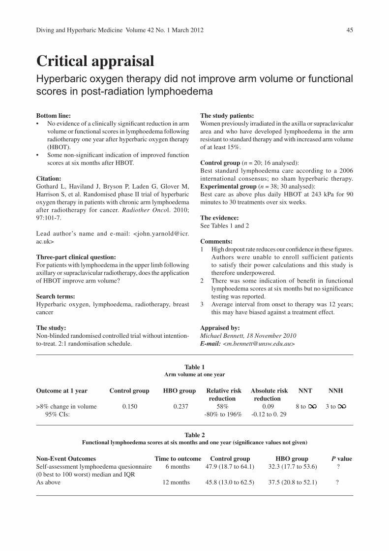
Diving and Hyperbaric Medicine Volume 42 No. 1 March 2012 45
Critical appraisal
Bottom line:• No evidence of a clinically significant reduction in arm
volume or functional scores in lymphoedema following radiotherapy one year after hyperbaric oxygen therapy (HBOT).
• Some non-significant indication of improved function scores at six months after HBOT.
Citation:Gothard L, Haviland J, Bryson P, Laden G, Glover M, Harrison S, et al. Randomised phase II trial of hyperbaric oxygen therapy in patients with chronic arm lymphoedema after radiotherapy for cancer. Radiother Oncol. 2010; 97:101-7. Lead author’s name and e-mail: <[email protected]>
Three-part clinical question: For patients with lymphoedema in the upper limb following axillary or supraclavicular radiotherapy, does the application of HBOT improve arm volume?
Search terms:Hyperbaric oxygen, lymphoedema, radiotherapy, breast cancer
The study:Non-blinded randomised controlled trial without intention-to-treat. 2:1 randomisation schedule.
The study patients:Women previously irradiated in the axilla or supraclavicalur area and who have developed lymphoedema in the arm resistant to standard therapy and with increased arm volume of at least 15%. Control group (n = 20; 16 analysed):Best standard lymphoedema care according to a 2006 international consensus; no sham hyperbaric therapy. Experimental group (n = 38; 30 analysed):Best care as above plus daily HBOT at 243 kPa for 90 minutes to 30 treatments over six weeks.
The evidence:See Tables 1 and 2
Comments:1 High dropout rate reduces our confidence in these figures.
Authors were unable to enroll sufficient patients to satisfy their power calculations and this study is therefore underpowered.
2 There was some indication of benefit in functional lymphoedema scores at six months but no significance testing was reported.
3 Average interval from onset to therapy was 12 years; this may have biased against a treatment effect.
Appraised by:Michael Bennett, 18 November 2010E-mail: <[email protected]>
Hyperbaric oxygen therapy did not improve arm volume or functional scores in post-radiation lymphoedema
Outcome at 1 year Control group HBO group Relative risk Absolute risk NNT NNH reduction reduction>8% change in volume 0.150 0.237 58% 0.09 8 to 3 to
95% CIs: -80% to 196% -0.12 to 0. 29
Table 1Arm volume at one year
Table 2Functional lymphoedema scores at six months and one year (significance values not given)
Non-Event Outcomes Time to outcome Control group HBO group P valueSelf-assessment lymphoedema quesionnaire 6 months 47.9 (18.7 to 64.1) 32.3 (17.7 to 53.6) ?(0 best to 100 worst) median and IQRAs above 12 months 45.8 (13.0 to 62.5) 37.5 (20.8 to 52.1) ?
Diving and Hyperbaric Medicine Volume 42 No. 1 March 201246
Continuing professional development
Hyperbaric oxygen therapy for delayed post-radiation injuryErik JansenAccreditation statement
Intended audienceThe intended audience consists of all physicians subscribing to Diving and Hyperbaric Medicine (DHM), including anaesthetists and other specialists who are members of the Australia and New Zealand College of Anaesthetists (ANZCA) Diving and Hyperbaric Medicine Special Interest Group (DHM SIG). However, all subscribers to DHM may apply to their respective CPD programme coordinator or specialty college for approval of participation. This activity, published in association with DHM, is accredited by the ANZCA Continuing Professional Development Programme for members of the ANZCA DHM SIG under Learning Projects: Category 2/Level 2: 2 credits per hour. ANZCA Fellows may only claim for this provided they submit their answers to the CPD coordinator.
ObjectivesThe questions are designed to affirm the takers’ knowledge of the topics covered, and participants should be able to evaluate the appropriateness of the clinical information as it applies to the provision of patient care.
Faculty disclosureAuthors of these activities are required to disclose activities and relationships that, if known to others, might be viewed as a conflict of interest. Any such author disclosures will be published with each relevant CPD activity.
Do I have to pay?All activities are free to subscribers.
Background readingPractitioners are referred to the following background references and reading:Frequency of radiated patients:1 <http://www.cancer.gov/cancertopics/factsheet/Therapy/
radiation>Intensity-modulated radiotherapy:2 Staffurth J. Radiotherapy Development Board: a review of the
clinical evidence for intensity-modulated radiotherapy. Clin Oncol (R Coll Radiol). 2010;22:643-57.
Radiation injury:3 Stone HB, Coleman CN, Anscher MS, McBride WH. Effects
of radiation on normal tissue: consequences and mechanisms. Lancet Oncol. 2003;4:529-36. [Review]
Hyperbaric oxygen for the treatment of radiation injury:
4 Feldmeier JJ, Hampson NB. A systematic review of the literature reporting the application of hyperbaric oxygen prevention and treatment of delayed radiation injuries: an evidence based approach. Undersea Hyperb Med. 2003;30:327-30.
5 Clarke RE, Tenorio LMC, Hussey JR, Toklu AS, Cone DL, Hinoiosa JG, et al. Hyperbaric oxygen treatment of chronic refractory radiation proctitis: a randomized and controlled double-blind crossover trial with long-term follow-up. Int J Radiat Oncol Biol Phys. 2008;72:134-43.
6 Craighead P, Shea-Budgell MA, Nation J, Esmail R, Evans AW, Parliament M, et al. Hyperbaric oxygen therapy for late radiation tissue injury in gynecologic malignancies. Curr Oncol. 2011;18:220-7.
7 Sidik S, Hardjodisastro D, Setiabudy R, Gondowiardjo S. Does hyperbaric oxygen administration decrease side effects and improve quality of life after pelvic radiation? Acta Med Indones. 2007;39:169-73.
Cancer and hyperbaric oxygen:8 Granowitz EV, Tonomura N, Benson RM, Katz DM, Band V,
Makari-Judson GP, et al. Hyperbaric oxygen inhibits benign and malignant human mammary epithelial cell proliferation. Anticancer Res. 2005;25(6B):3833-42.
Cell production in relation to hyperbaric oxygenation:9 Thom SR, Bhopale VM, Velazquez OC, Goldstein LJ, Thom
LH, Buerk DG.Stem cell mobilization by hyperbaric oxygen. Am J Physiol Heart Circ Physiol. 2006;290:H1378-86.
How to answer the questionsPlease answer all responses (A to E or F) as True or False.
Answers should be posted by e–mail to the nominated CPD co–ordinator.For EUBS members for this CPD issue this will be Dr Erik Jansen, e-mail: <[email protected]>.For ANZCA DHM SIG members and SPUMS members, this will be Dr David Cooper, e-mail: <[email protected]>.
On submission of your answers, you will receive a set of correct answers with a brief explanation of why each response is correct or incorrect.
Successfully undertaking the activity will require a correct response rate of 80% or more. Each task will expire within 24 months of its publication to ensure that additional, more recent data has not superceded the activity.
Key wordsMOPS (maintenance of professional standards), radiotherapy, hyperbaric oxygen therapy, hyperbaric medicine, bone necrosis, soft-tissue radionecrosis
CME ACTIVITY 2011/1
Diving and Hyperbaric Medicine Volume 42 No. 1 March 2012 47
Question 1. Radiation therapy of malignant tissue has the following characteristics:
A. In industrialised countries, about half of all cancer patients receive some type of radiation therapy sometime during the course of their treatment.B. Delayed radiation injury is estimated to affect 5–15% of long-term survivors.C. The risk of getting delayed radiation injury is limited to the first year after radiation therapy.D. The acute reaction to radiation is always a strong indicator for development of delayed radiation injury.E. Only the malignant tissue will react with delayed radiation injury.F. Intensity-modulated radiotherapy ( IMRT )increases delayed radiation injuries.
Question 2. The radiation injury:A. Vascular injury is not an effect of radiation injury.B. Cellular depletion and tissue fibrosis are important components of delayed radiation injury.C. Hypoxia may occur due to delayed radiation injury.D. Reliable assays are available to accurately identify patients who are at high risk of developing delayed radiation injury.E. The delayed radiation injury may be precipitated by further injury or trauma.F. Delayed radiation injury resolves over time without further treatment.
Question 3. Osteoradionecrosis, delayed radiation injury of the jaw:
A. In the treatment of osteoradionecrosis of the jaw, it is essential to includes surgical extirpation of necrotic bone.B. When surgery is required the majority of hyperbaric oxygen treatments should be given after the surgical intervention.C. The risk of osteoradionecrosis is increased due to impaired saliva production and the increased frequency of dental caries.D. Osteoradionecrosis of the jaw does not affect the nutrition of the patient.E. It is generally agreed that hyperbaric oxygen treatment is always indicated as a prophylaxis before extractions and other oral surgical procedures in radiation patients.
Question 4. Radiation injury of abdominal organs:A. It is possible to find a recent randomised controlled study which demonstrates the effect of hyperbaric oxygen treatment on radiation proctitis.B. Common symptoms of delayed radiation injury of the pelvic region include increased stool frequency, urgency, spotting of blood and partial incontinence.C. Hyperbaric oxygen administration decreases delayed radiation injury effect and improve quality of life after pelvic radiation.D. Hyperbaric oxygen treatment is indicated in the treatment of radiation cystitis when conventional treatment of instillation of alum or formalin is not effective.E. If all studies on radiation injury of abdominal organs are pooled, 87% of the patients have a successful or partly successful result of hyperbaric oxygen treatment.
Question 5. Hyperbaric oxygen and cell proliferation:A. Hyperbaric oxygen enhances cancer growth or recurrence in humans.B. Hyperbaric oxygen inhibits in vitro growth of human mammary transplanted tumor.C. Hyperbaric environment increases the number of stem cells in humans.D. Hyperbaric oxygen environment increases the number of stem cells in humans.E. Hyperbaric oxygen increases angiogenesis by release of vascular endothelial growth factor.
The database of randomised controlled trials in hyperbaric medicine maintained byMichael Bennett and his colleagues at the Prince of Wales Hospital
Diving and Hyperbaric Medicine Unit, Sydney is at: <www.hboevidence.com>
Professor Bennett advises that this site is being reconstructed and brought up to date currently. For the past two years, the Prince of Wales team have been heavily involved in the installation of their new three-compartment rectangular chamber, and the complete rebuild of the department, and apologise that this website has not been in their focus during this time.
Diving and Hyperbaric Medicine Volume 42 No. 1 March 201248
Book reviewMastering rebreathers, 2nd editionJeff Bozanic
Softcover, full colour, 704 pagesISBN: 9781930536579Best Publishing Company, Flagstaff AZ, 2010Available from: <www.bestpub.com>Price: USD54.99
There is little doubt that one of the most significant developments in recreational diving over the last 15 years has been ‘technical diving’ methods. These diving methods, previously the province of military and some occupational diving groups, extend both the depths and durations of dives. They have opened up an entirely new world of exploration and underwater experience, and the appeal is self-apparent.
No single technique epitomises technical diving like the use of a rebreather. These devices recycle exhaled gas through a carbon dioxide (CO
2) absorbent, and maintain safe oxygen
levels in the recycled gas by various means, depending on type. This minimises consumption of expensive inert gases such as helium, which is used during deep diving to minimise nitrogen narcosis. To all intents and purposes (depending on the type of rebreather), gas consumption may be limited to as little as the oxygen that the diver metabolises, giving extremely long underwater durations from a small gas supply. Unlike open-circuit scuba, this duration is unaffected by depth. This makes rebreathers the ultimate deep-diving exploration tool.
However, rebreathers are complex and they have many failure modes. Perhaps not surprisingly, their use appears to be significantly more hazardous than use of open-circuit scuba. There is probably no single answer to mitigating this risk, but one strategy believed by many (including this writer) to be crucial is high standards of training and education for rebreather divers.
Enter Mastering rebreathers (2nd edition). Jeff Bozanic is a well-known and respected member of the diving community who published the first edition of this book in 2002. The range of rebreather technologies and models has moved on considerably in the 10 years since then, and it was appropriate that the publication be updated or risk becoming irrelevant. Bozanic has a PhD in education and is a very experienced rebreather diver; an ideal combination of skills for an undertaking of this nature. Most importantly, he is an experienced instructor on multiple models of rebreather, and thus has considerable insight into those areas of relevant knowledge that are difficult to impart to students. This is
reflected in the style of the book, which is fundamentally a textbook for the novice rebreather diver.
Though substantial at 700 pages, virtually half of it is given to appendices. It is organised into 14 chapters and the aforementioned appendices. The first seven chapters could be described as providing background information. These are entitled ‘Introduction to rebreathers’, ‘History of rebreathers’, ‘Types of rebreathers’, ‘Diving physics’, ‘Physiology’, ‘Theory’ and ‘Rebreather design’. Chapters 8–10 detail the approach to an actual rebreather dive and are entitled ‘Preparing for the dive’, ‘Diving techniques’ and ‘Post-dive procedures’. Chapter 12, which covers ‘Emergency procedures’ belongs in this group also. The chaptered section is rounded out by Chapter 11 ‘Long-term maintenance’, and Chapters 13 and 14 entitled ‘Travel’ and ‘Where do you go from here?’ respectively. The second half of the book consists of 20 appendices which, other than one covering ‘Dive tables’, a glossary and an index, are given over to aspects of the procedures (including checklists and maintenance schedules in some cases) for using a wide range of different brands of rebreather. Some of these sections are quite comprehensive and others less so because the material reflects the content that the respective manufacturers were inclined to provide. There is a wealth of information in these appendices, and those who enjoy possessing knowledge about a variety of rebreathers they do not own will find them fascinating.
The first thing that should be made clear is that this is not a manual for advanced rebreather diving. It does not cover deep diving, mixed-gas diving, decompression diving, and diving in special environments (such as caves). These are subjects that Bozanic intends to address in a second volume whose release is apparently not far off. This is not a criticism. Indeed, this reviewer applauds the stated aim of the book which is to “discuss introductory rebreather principles and introduce readers to rebreathers, basic physiology and physics, and their use in recreational environments”. Nor do I mean that experienced rebreather divers will not find the book useful. As an experienced and well-informed rebreather diver, I still found the material highly interesting and a great resource, especially as a repository for the numerous slightly arcane rebreather-related equations that I do not carry around in my head.
The book is well written in a clear didactic style and will serve rebreather novices extremely well. It is presented for the most part in both Imperial and metric units, so it has utility in this regard beyond the USA. Indeed, I would have no hesitation in recommending it as a textbook for any entry-level rebreather course as a supplement to unit-specific and training-agency material. Each chapter ends with a series of multi-choice and occasional short answer questions designed to test understanding of key concepts, further increasing utility as a training textbook. In general, I found these questions to be well thought out and pitched at the right level for a new rebreather diver.
Diving and Hyperbaric Medicine Volume 42 No. 1 March 2012 49
Many hyperbaric facilities in the region have collected data for their own clinical indicators (CIs) for some time. With an increasing interest in comparing CIs in the field of hyperbaric medicine, a plan for the commencement of a set of Australian and New Zealand CIs was undertaken following the 2008 Hyperbaric Technicians and Nurses Association (HTNA) Conference. The DDHM undertook this challenge on behalf of the HTNA. When formulating the CIs the DDHM reviewed and considered indicators that would be broadly applicable across different units and methods of hyperbaric delivery. The aim has been to develop a set of CIs that all units in the region collect, share and publish.
An e-mail poll was undertaken, feedback from six units was received and the three most favoured CIs were agreed upon for implementation for 2009. The three initial 2009 CIs were:• Clinical in-chamber hypoglycaemic event in patient
with diabetes;• Decompression illness in chamber attendant;• Unplanned insertion of tympanostomy tube(s) or
performance of myringotomy in patients scheduled for routine hyperbaric oxygen therapy (HBOT).
Following the same process a further CI was introduced in 2010:• Oxygen toxic seizure. In 2012 collection of a fifth indicator has commenced:• Failure to arrive for a planned HBOT session.
In 2010, the Special Interest Group – Diving and Hyperbaric Medicine of the ANZ College of Anaesthetists gave an undertaking to have the CIs formally accepted by the Australian Council on Healthcare Standards. We are currently awaiting progress with this.
Thus, there are currently five CIs collected by Australian and New Zealand hyperbaric units. It is envisaged that CIs will be reviewed after two years; if a CI has not been triggered by any facility contributing data we would consider no longer collecting those data. It is envisaged that CIs will continue to be introduced in the future. CIs are collected in six-month periods, January to June and July to December. The contributing units’ CI results are published annually in the HTNA conference booklet.
Further information relating to CIs and contributing to these can be found on the HTNA forum, the ANZHMG chat line: <[email protected]> or by contacting the DDHM at POWH directly. We strongly encourage and are happy to assist any unit with the collection of indicators.
Key wordsClinical audit, hyperbaric oxygen therapy, hyperbaric facilities, safety, occupational health
Clinical indicators in hyperbaric medicineJoanne James, Jan Lehm and Michael BennettDepartment of Diving and Hyperbaric Medicine, Prince of Wales Hospital (DDHM POWH), Sydney, Australia
It was a pleasure to read what is essentially rebreather training material and not finding myself constantly wincing over inaccuracies or unsubstantiated claims, as is frequently the case when I read some technical training agency material. Not surprisingly, Bozanic is at his best when writing about areas of experience-based expertise. This applies, in particular, to those chapters about the rebreathers themselves, and the practical aspects of diving them. The travel chapter is innovative and full of useful tips to keep the diver out of trouble, especially during air travel.
It is not all perfect, however. I found the physiology chapter a little lightweight. This might reflect my own personal belief in the importance of this subject, but I think there are one or two important omissions. For example, the discussion of the causes of CO
2 toxicity during rebreather diving focuses
entirely on hypercapnia due to absorbent breakthrough and does not mention the fact that hypoventilation and CO
2
retention are common in diving, and particularly rebreather diving when work of breathing increases. I also found that the index was not particularly thorough.
These are minor criticisms of what is, for the most part, an excellent book for the purposes for which it was written. I would strongly recommend it to those toying with the idea of buying a rebreather, or those who have taken the plunge and are learning to use their unit. It would not be out of place on the shelf of any rebreather diver. Finally, diving physicians who are not rebreather divers but who want a thorough account of relevant information could not find a better resource.
Associate Professor Simon MitchellHead of Department of AnaesthesiologyUniversity of AucklandPO Box 92019, Auckland, New Zealand
Key wordsRebreathers/closed circuit, rebreathers/semi-closed circuit, training, textbook, book reviews
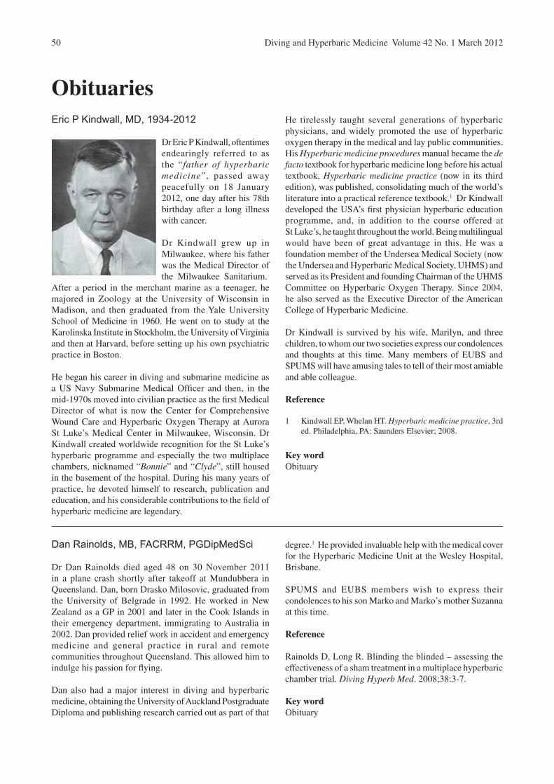
Diving and Hyperbaric Medicine Volume 42 No. 1 March 201250
After a period in the merchant marine as a teenager, he majored in Zoology at the University of Wisconsin in Madison, and then graduated from the Yale University School of Medicine in 1960. He went on to study at the Karolinska Institute in Stockholm, the University of Virginia and then at Harvard, before setting up his own psychiatric practice in Boston.
He began his career in diving and submarine medicine as a US Navy Submarine Medical Officer and then, in the mid-1970s moved into civilian practice as the first Medical Director of what is now the Center for Comprehensive Wound Care and Hyperbaric Oxygen Therapy at Aurora St Luke’s Medical Center in Milwaukee, Wisconsin. Dr Kindwall created worldwide recognition for the St Luke’s hyperbaric programme and especially the two multiplace chambers, nicknamed “Bonnie” and “Clyde”, still housed in the basement of the hospital. During his many years of practice, he devoted himself to research, publication and education, and his considerable contributions to the field of hyperbaric medicine are legendary.
He tirelessly taught several generations of hyperbaric physicians, and widely promoted the use of hyperbaric oxygen therapy in the medical and lay public communities. His Hyperbaric medicine procedures manual became the de facto textbook for hyperbaric medicine long before his actual textbook, Hyperbaric medicine practice (now in its third edition), was published, consolidating much of the world’s literature into a practical reference textbook.1 Dr Kindwall developed the USA’s first physician hyperbaric education programme, and, in addition to the course offered atSt Luke’s, he taught throughout the world. Being multilingual would have been of great advantage in this. He was a foundation member of the Undersea Medical Society (now the Undersea and Hyperbaric Medical Society, UHMS) and served as its President and founding Chairman of the UHMS Committee on Hyperbaric Oxygen Therapy. Since 2004, he also served as the Executive Director of the American College of Hyperbaric Medicine.
Dr Kindwall is survived by his wife, Marilyn, and three children, to whom our two societies express our condolences and thoughts at this time. Many members of EUBS and SPUMS will have amusing tales to tell of their most amiable and able colleague.
Reference
1 Kindwall EP, Whelan HT. Hyperbaric medicine practice, 3rd ed. Philadelphia, PA: Saunders Elsevier; 2008.
Key wordObituary
Obituaries
Dr Eric P Kindwall, oftentimes endearingly referred to as the “father of hyperbaric medicine”, passed away peacefully on 18 January 2012, one day after his 78th birthday after a long illness with cancer.
Dr Kindwall grew up in Milwaukee, where his father was the Medical Director of the Milwaukee Sanitarium.
Eric P Kindwall, MD, 1934-2012
Dan Rainolds, MB, FACRRM, PGDipMedSci
Dr Dan Rainolds died aged 48 on 30 November 2011 in a plane crash shortly after takeoff at Mundubbera in Queensland. Dan, born Drasko Milosovic, graduated from the University of Belgrade in 1992. He worked in New Zealand as a GP in 2001 and later in the Cook Islands in their emergency department, immigrating to Australia in 2002. Dan provided relief work in accident and emergency medicine and general practice in rural and remote communities throughout Queensland. This allowed him to indulge his passion for flying.
Dan also had a major interest in diving and hyperbaric medicine, obtaining the University of Auckland Postgraduate Diploma and publishing research carried out as part of that
degree.1 He provided invaluable help with the medical cover for the Hyperbaric Medicine Unit at the Wesley Hospital, Brisbane.
SPUMS and EUBS members wish to express their condolences to his son Marko and Marko’s mother Suzanna at this time.
Reference
Rainolds D, Long R. Blinding the blinded – assessing the effectiveness of a sham treatment in a multiplace hyperbaric chamber trial. Diving Hyperb Med. 2008;38:3-7.
Key wordObituary
Diving and Hyperbaric Medicine Volume 42 No. 1 March 2012 51
South Pacific Underwater Medicine Society Diploma of Diving and Hyperbaric Medicine
SPUMS notices and news
Requirements for candidates (updated October 2008)
In order for the Diploma of Diving and Hyperbaric Medicine to be awarded by the Society, the candidate must comply with the following conditions:
1 The candidate must be medically qualified, and be a current financial member of the Society.
2 The candidate must supply evidence of satisfactory completion of an examined twoweek fulltime course in Diving and Hyperbaric Medicine at an approved facility. The list of approved facilities providing twoweek courses may be found on the SPUMS website.
3 The candidate must have completed the equivalent (as determined by the Education Officer) of at least six months’ fulltime clinical training in an approved Hyperbaric Medicine Unit.
4 The candidate must submit a written proposal for research in a relevant area of underwater or hyperbaric medicine, in a standard format, for approval before commencing their research project.
5 The candidate must produce, to the satisfaction of the Academic Board, a written report on the approved research project, in the form of a scientific paper suitable for publication. Accompanying this written report should be a request to be considered for the SPUMS Diploma and supporting documentation for 1–4 above.
6 In the absence of documentation otherwise, it will be assumed that the paper is submitted for publication in Diving and Hyperbaric Medicine. As such, the structure of the paper needs to broadly comply with the ‘Instructions to Authors’ – full version, published in Diving and Hyperbaric Medicine 2010; 40(2):110-2.
7 The paper may be submitted to journals other than Diving and Hyperbaric Medicine; however, even if published in another journal, the completed paper must be submitted to the Education Officer for assessment as a diploma paper. If the paper has been accepted for publication or published in another journal, then evidence of this should be provided.
8 The diploma paper will be assessed, and changes may be requested, before it is regarded to be of the standard required for award of the Diploma. Once completed to the reviewers’ satisfaction, papers not already submitted to, or accepted by other journals should be forwarded to the Editor of Diving and Hyperbaric Medicine for consideration. At this point the Diploma will be awarded, provided all other requirements are satisfied. Diploma projects submitted to Diving and Hyperbaric Medicine for consideration of publication will be subject to the Journal’s own peer review process.
Additional information – prospective approval of projects is required
The candidate must contact the Education Officer in writing (email is acceptable) to advise of their intended candidacy, and to discuss the proposed subject matter of their research. A written research proposal must be submitted before commencing the research project.
All research reports must clearly test a hypothesis. Original basic or clinical research is acceptable. Case series reports may be acceptable if thoroughly documented, subject to quantitative analysis, and the subject is extensively researched and discussed in detail. Reports of a single case are insufficient. Review articles may be acceptable if the world literature is thoroughly analysed and discussed, and the subject has not recently been similarly reviewed. Previously published material will not be considered.
It is expected that all research will be conducted in accordance with the joint NHMRC/AVCC statement and guidelines on research practice (available at: <http://www.health.gov.au/nhmrc/research/general/nhmrcavc.htm>) or the equivalent requirement of the country in which the research is conducted. All research involving humans or animals must be accompanied by documented evidence of approval by an appropriate research ethics committee. It is expected that the research project and the written report will be primarily the work of the candidate, and that the candidate is the first author, where there are more than one.
The SPUMS Diploma will not be awarded until all requirements are completed. The individual components do not necessarily need to be completed in the order outlined above. However, it is mandatory that the research project is approved prior to commencing research.
The Academic Board reserves the right to modify any of these requirements from time to time. As of October 2011, the SPUMS Academic Board consists of:Associate Professor David Smart, Education Officer Associate Professor Simon MitchellAssociate Professor (retired) Mike Davis.
All enquiries and applications should be sent to the Education Officer:Associate Professor David SmartGPO Box 463, Hobart, Tasmania 7001Email: <[email protected]>
Key wordsQualifications, underwater medicine, hyperbaric oxygen, research, medical society
Diving and Hyperbaric Medicine Volume 42 No. 1 March 201252
Minutes of the SPUMS Executive Committee Meeting 22 and 25 May 2011 at Hilton Guam Resort and Spa, Tumon Bay, Guam, USA
Opened: 1515 h
Present:M Bennett, S Lockley, J Lehm, D Smart, P Smith and C Meehan (invited guest as ASM 2012 Convenor)
Apologies:G Williams, C Acott, M Davis and G Hawkins
1. Minutes of previous meeting:Minutes accepted for Executive Committee Meeting. Minutes to be forwarded to Executive Committee not present for acceptance. Proposed M Bennett, seconded J Lehm, carried.
2. Matters arising from previous minutes:2.1 Cost-effectiveness of printing DHM in Europe. Quote from Elsevier discussed. Currently no cost benefit identified. President of EUBS continues to investigate.(Action: M Bennett, M Davis, Ongoing) 2.2 Discussed current issue with dive medical assessments for people with diabetes. No training courses yet available for diabetic divers in Australia and NZ. (Action: M Bennett will continue to keep the Committee posted on new developments particularly in Queensland where some interest has been expressed in setting up a course. Ongoing)2.3 ISO standards further discussed. Queensland OH&S independently regulates dive medicals, with reference to the ASNZ Diving Standards. Written submission to Workplace Health and Safety (Qld) by M Bennett. This was sent through outlining the new SPUMS dive medical guidelines. Advice received that they are appreciative of the ongoing input of SPUMS. Proposal to Australian Standards to accept an adapted ISO standard. Formal decision to be made as to whether or not the ISO is accepted or an adapted version is accepted. If the proposal is accepted, the Australian Standards will fund it. (Action: D Smart and M Bennett to update Committee on developments. Ongoing)2.4 Epilepsy position paper is being drafted currently. (Action: M Bennett will update the Committee on any developments. Ongoing)
3. Annual Scientific Meetings:3.1 ASM 2011
3.1.1 S Lockley provided update. Based on preliminary budget, a small profit is expected. Registrant numbers – 55 full registrants, nine accompanying adults and three accompanying children.
3.1.2 The final budget will be forwarded out to the Committee when complete as well as the post-ASM report.3.1.3 ASM Convenor proposed using Cvent as an event builder and website provider. Proposed: S Lockley, seconded: M Bennett, unanimously agreed.
3.2 ASM 2012C Meehan was invited to provide an update for the ASM 2012. Quotations have been received through a travel agent and some estimated prices were provided. The venue will be Madang Resort, Madang PNG. Two guest speakers (Associate Professors Jamie Seymour and Richard Fitzpatrick) have been approached and have tentatively agreed. The resort is located on a peninsula and well protected by good security. Flights ex-Sydney, Cairns and Brisbane are available to Madang. Estimated costs were outlined from preliminary quotations and are comparable with previous meetings.3.3 ASM 2013Combined meeting with EUBS was discussed but not likely to go ahead. Some proposed destinations were discussed including the Maldives, Reunion and the Seychelles. As yet, there is no volunteer to convene this meeting. (Action: M Bennett to investigate further the possibility of Reunion as a destination)3.4 ASM 2014Berjaya Tioman was proposed as a venue. There is no volunteer to convene this meeting.
4. Journal matters:4.1 Congratulations extended to M Davis regarding the Medline indexation of the DHM.4.2 Further discussion that DHM is a separate entity sponsored by the two societies - the SPUMS and the EUBS. The DHM Editor has total control over the DHM budget. The Committee approves the budget and the DHM Editor manages the journal finances for the financial year.[Correction of fact: At this time, the SPUMS Treasurer continues to exercise control of the finances in consultation with the SPUMS Committee, the Editor and the EUBS Committee.]4.3 Discussed increasing the Editorial Board by two people (currently comprising seven members).4.4 DHM Journal report should be provided to the Committee and the Editor should attend the SPUMS Committee meeting on invitation.4.5 Request Editor’s Handbook. Proposed: D Smart, seconded: M Bennett, carried. (Action: M Bennett to contact P Germonpré to generate letter to formally request)4.6 Legal opinion outlining the legal position of the DHM/Publisher/the SPUMS and the EUBS. (Action: M Bennett to follow up)
Diving and Hyperbaric Medicine Volume 42 No. 1 March 2012 53
5. Website matters5.1 Discussed removal of the earthquake warning from website. Action: G Hawkins to remove warning.5.2 It was recommended that a hot key be provided to allow members to access their registration details easily. (Action: G Hawkins to further investigate)5.4 Proposal made that members pay the following year’s membership from 01 November the preceding year and annually from that date. Proposed: G Hawkins (via correspondence), seconded J Lehm. Accepted by all present.5.5 It was further discussed that an e-mail reminder be sent out on 01 November for membership and a discount be added to the registrations received before 01 January the following year.[Correction: before 01 February]
6. Education Officer’s report6.1 ANZHMG:
6.1.2 Two executive members on MSAC. Fund HBOT in process.6.1.3 AGM at HTNA in Sydney, September 2011.
6.2 SPUMS Diploma to be awarded to Cathy Meehan.6.3 Discussed proposal for an internationally recognised curriculum for a diving and hyperbaric medicine course at Stellenbosch University. (Action: D Smart is involved with aligning courses, gap analysis and accreditation).6.4 Discussed OHS legislation federally. This is to be presented by D Smart during the ASM 2011.
Meeting was temporarily closed at 1735 h and resumed on Wednesday 25 May at 1400 h.
7. Secretary’s report7.1 Assets were discussed. S Lockley to consolidate the SPUMS and DHM assets list with new Secretary. (Action: Secretary to contact Committee Members to confirm current assets held)7.2 Membership list as at 29 April 2011 provided (as extracted by Webmaster).7.3 Discussed current SPUMS address. Return to sender item received by member who contacted Secretary after attempting to post a proxy vote form through. No other issues with mailing address raised by other members of the Committee. Continue using current mailing address.
8. Treasurer’s report8.1 Discussed fee structure for 2012–2013. Suggested to notify members by e-mail around 01 November each year and then offer a substantial discount for payments within three months. It is proposed to set the new full membership fee to AUD175, if paid on-line before 31 January and AUD200 if paid later. Paper renewals are an extra AUD10 and shipping DHM to an address outside New Zealand/Australia will also add an extra AUD10.8.2 Proposal was made to increase the list of people authorised to operate the various SPUMS accounts. This
was proposed by J Lehm, seconded by S Lockley and unanimously accepted by the Committee. It is proposed to add Dr Peter Smith, the Public Officer, Dr Andrew Fock and the new Secretary (Dr Karen Richardson, refer AGM Minutes, Para 9) to become signatories to the accounts with St George Bank as well as authorised users of Business Banking On-Line. Furthermore, it was proposed and accepted by the Executive Committee unanimously, that the new Secretary (Dr Karen Richardson) will take over the SPUMS Visa card from the current Secretary, Dr Sarah Lockley. Members finishing their term on the Executive Committee will be removed from the list of authorised people (including Dr Sarah Lockley). The list of people now authorised to operate the various accounts is therefore:Secretary, Dr Karen Richardson (as elected AGM, refer AGM Minutes, Para 9)Treasurer, Dr Jan LehmPublic Officer, Dr Andrew FockCommittee Member, Dr Guy WilliamsCommittee Member, Peter Smith
9. Other business9.1 E-mail correspondence received by G Williams (not in attendance).
9.1.1 Raised concerns with current membership numbers compared with six years ago and declining membership. Point was raised in discussion that some members of EUBS now receive the journal without also becoming members of SPUMS, as was previously the case. Hence, some of these perceived financial losses due to declining membership are offset by financial contributions by EUBS toward DHM costs.9.1.2 Support expressed for DHM being offered in an electronic format to those who would prefer this and that this might save on printing and distribution costs. This is to be explored further as per 2.1 above.9.1.3 Expressed concern that the ASM numbers have declined over the past and this should be investigated. It was noted, based on recent figures, that ASM registrant numbers (where registrant does not include speakers, administrator or convenor) were 55 for 2011, with 12 accompanying guests; 61 for 2010, with 27 accompanying guests; and 56 for 2009, with 37 accompanying guests, therefore actual number of registrants over recent years has been relatively steady, with variability in the accompanying guest numbers. Data from further back were not available although could be sought.9.1.4 Concern was raised that it has been some time since ASM Convenors have sought accreditation from the RACGP for CME points. This point was addressed by both D Smart and S Lockley, who had both independently contacted the RACGP to apply for CME points. Unlike ANZCA and ACEM,
Diving and Hyperbaric Medicine Volume 42 No. 1 March 201254
Annual General Meeting of the Australian and New Zealand Hyperbaric Medicine Group, 15 September 2011 at the Crowne Plaza Hotel, Coogee Beach, Sydney
Opened: 0900 h
1. AttendanceD Smart, D Wilkinson, M Bennett, S Mitchell, S Szekely, I Millar, N Banham, C Meehan, G Hawkins, J Lehm, J Orton, R McKay, D Matley, K Thistlethwaite, D Blake, F Sharp, M Walker, M Davis. Guest: F Linde
2. ApologiesM Hodgson, H Oxer, B Webb, B Wong, A Fock
3. Office bearersAs the MSAC review is approaching a denouement, D Smart has offered to continue in his role as Chair to see this process through. D Wilkinson is agreeable to continuing in the role of Secretary role. Unanimous support for this proposal.
4. Minutes of 2010 Annual General MeetingAccepted
5. Business arisingDiscussed under current agenda items
6. Chairmans report:6.1 Overview: The main areas of activity for ANZHMG have been:
1. Hyperbaric medicine funding in Australia – MSAC2. Australian Federal Government Occupational Work, Health and Safety Regulations3. Diving medical training4. Australian Standard for Compressed Air and Hyperbaric Facilities
6.2 Medicare Services Advisory CommitteeD Smart and M Bennett have been intimately involved with this process. A draft report has been prepared from scratch by MSAC, despite the detailed submission by ANZHMG, AHHA, ASA, and SPUMS. The draft report is assessing the evidence base and cost effectiveness of hyperbaric oxygen treatment for refractory problem wounds (non-diabetic) and soft-tissue radiation injury. This has been a huge effort for ANZHMG members over 10 years and is still yet to be resolved.6.3 Federal Work, Health and Safety RegulationsSPUMS through ANZHMG has provided significant input to these regulations. There is serious risk that they will lead to adverse outcomes for divers as a result of major inadequacies in the legislation. Despite a draft code of practice being released with extra detail, there are fundamental flaws in the legislation which could lead to untrained divers working as occupational divers.
the RACGP now has a very involved process for course and education accreditation. The college also requires payment of a fee of around AUD400 for the submission to be processed. Both these previous ASM Convenors highlighted the complexity of this process as the reason for not completing the accreditation process for RACGP and SPUMS members. By comparison, the application to ANZCA and ACEM for ASM accreditation was very simple and involved e-mail correspondence including the full academic programme outlining the details of the presentations and workshops. Action: S Lockley has volunteered to assist the Convenor ASM 2012 to review this process again and consider a submission.
9.2 M Bennett has suggested that Key Performance Indicators (KPI) be set for Committee Members. These should include: attendance at Committee Meetings (minimum four of six), attention to action items within the timeframe determined at the meeting, and 90% of minutes to be generated within one month after the meeting. Note that no timeframes have been set for action items on these minutes and that KPI can only apply to future meetings.9.3 Last update of ASM Convenor’s Manual was done by D Smart in 2009. Action: S Lockley to update ASM Convenor’s Manual and forward copy to ASM Convenor 20129.4 Discussed appointing two committee members as Assistant Secretary and Assistant Treasurer. It was proposed that G Williams and P Smith (until the ballot election) be appointed into these positions. This will have to be further discussed with these members directly. Proposed: M Bennett, seconded S Lockley, carried.9.5 The formation of an ASM sub-committee to assist with planning future meetings, including sites and potential convenors, was proposed by M Bennett, seconded by D Smart, carried unanimously.
10. CorrespondenceNil.
11. Next meetingA date in November 2011 to be confirmed.
Closed: 1500 h
Key wordsMeetings, medical society
Diving and Hyperbaric Medicine Volume 42 No. 1 March 2012 55
6.4 Australian Standards for professional diversThe main area of activity at present for Australian Standards is a revision of the 2815 series. Given the Federal Government’s ‘dumbing-down’ of workplace legislation, 2815 has not been referenced and the ADAS qualification appears to have been abandoned. Unfortunately, in the proposed Federal Government legislation, the AS2299 series is being referred to for construction diving – another serious omission. Given that the legislation has a higher status than the Standards, lobbying is taking place to have the Standards appropriately referenced in the legislation.6.5 Australian Standard for Compressed Air and Hyperbaric FacilitiesThe HTNA has commenced a review of the AS4774 series of Australian Standards. Support from the ANZHMG is important to ensure that standards are maintained for the delivery of service, safety and staff quality and that of hyperbaric care in Australia. There will be liaison meeting relating to this with HTNA. In light of the ongoing saga with MSAC, it is critically important that very high standards are maintained in comprehensive facilities across Australia.6.6 Research
6.6.1 HOLLT trial: This is proceeding steadily and a recent interim analysis of data indicated that the difference between the two groups is greater than originally predicted and numbers have been able to be reduced from 250 to 120. Hopefully the trial will be completed towards the end of 2012.6.6.2 HORTIS: R Clarke has announced that the HORTIS trial for radiation cystitis is closed due to insufficient recruitment numbers.
6.7. ANZ list of indications for hyperbaric treatmentThis is due to be reviewed again this year having been published in Diving and Hyperbaric Medicine Journal in 2009.6.8. Support the HTNA ConferenceI again encourage all members to provided contributions to the conference. It is a unique event and it needs to be supported and I congratulate the Prince of Wales Hospital Team in running this year’s conference. I also congratulate the Prince of Wales Team on their new facility.6.9 Courses in diving and hyperbaric medicine
6.9.1 The Royal Adelaide Hyperbaric Medicine Course continues. N Banham also ran a short course for recreational medicals in Fremantle last year. The remainder of courses in Australia were Prince of Wales (PoW) Course in Diving and Hyperbaric Medicine and also the Royal Australian Navy Course at the HMAS Penguin.6.9.2 Due to pressures from MSAC and the Federal Work, Health and Safety Regulations, no further progress has been made by the ANZHMG Chair on the assessment and accreditation of the courses in my role as SPUMS Education Officer.
6.10 Welcome to J Orton: Dr Orton has been appointed as Director of the Townsville Hospital Hyperbaric
Facility and we welcome his attendance and input to this meeting.
7. MSAC report and Federal Government funding issuesThe HBO indications of soft-tissue radiation injury and non-diabetic wounds currently operate under a temporary ministerial approval; however, they come for review before MSAC soon. This is the last application under the old MSAC process and MSAC are keen to wrap this up.
8. Hyperbaric problem wound studyThere are six years of data now, with 440 patients enrolled (355 HBO). Very impressive results with overall 80% healed or substantially healed at 12 months. Perhaps mirroring our individual experiences, the data shows 45–50% healing at the end of HBOT with continuing clinical improvement after cessation of HBOT to reach 80% at 12 months. These results are to be published. Whilst this is not an RCT, it is nevertheless an impressive, prospective cohort. MSAC have to accept this as valid evidence, particularly when you consider the poor evidence for other wound healing strategies that continue to benefit from public funding under Medicare’s indifference to patient outcomes.
9. HORTISThe radiation cystitis arm of HORTIS has been closed. M Bennett reminded the meeting of the value of quality research and noted that the HORTIS radiation proctitis study has been important in the MSAC process.
10. ANZHMG/SIG list of approved indications for HBOTThe current list was examined and the process explained. There were no new indications suggested for consideration. M Bennett suggested that a workshop might be held to comprehensively deal with this issue, allowing a restating of principles, examination of the evidence and a review of the list of indications for HBOT. It was suggested it might be part of the next HTNA ASM. The current list will continue for the time being.
11. Introductory course in hyperbaric medicineM Bennett reported that the next course to be run from the PoW Hospital will be from 20 February to 02 March, 2012. Fourteen candidates have already booked. It will be held within the new Department of Diving and Hyperbaric Medicine with access to new electronic facilities. The video system within the new chamber allows for recording of real-time events and another goal will be to develop simulation exercises that can be run under pressure. It was emphasised that the course is owned by the ANZHMG although it is run from the PoW Hospital. The frequent use of invited lecturers reflects this stance. All were encouraged to support the successful continuation of ‘our’ course. It was suggested that there was a need for a new
Diving and Hyperbaric Medicine Volume 42 No. 1 March 201256
course that went beyond the introductory level and that could be suitable for higher level practitioners. Further discussion required.
12.Australian Standards reportI Millar updated the meeting on the status of several Standards:AS4005.1 – this Standard will probably lapse and will be replaced by the relevant International Standard which does not demand a face-to-face medical examination. It is understood that of the training agencies, SSI will use a questionnaire and PADI will leave it to the discretion of the member.AS/NZS2299.1 – despite its age, this Standard is valid and will probably continue. It is a core reference.AS4774.2 – Standards Australia will fund the review of this Standard. The HTNA are pivotally involved. Dr Davis lamented the failure of the NZ authorities to embrace AS4774.2 in the same way they recognised AS/NZS2299.1. It was thought that perhaps a letter alerting the NZ authorities to the impending review might stimulate interest. P Atkinson as the HTNA rep should be consulted. (Action: I Millar to organise/negotiate a letter alerting New Zealand to the impending review of AS4774.2)
13. Diving and Hyperbaric Medicine JournalThe editor presented his report.
14. Minimum data set/registry developmentsNot progressed; remove item
15. Hyperbaric medicine clinical indicatorsClinical indicator data are published in the HTNA meeting proceedings. Data have not been submitted by every hyperbaric facility. It was reminded that the value of this project makes it worth persevering with at this early point. Some discussion about the definition of the CIs.(Action: J Lehm to circulate full definitions of the clinical indicators on the chatline)
16. Clinical trials for discussionScheduled elsewhere in this meeting.
17. HTNA issuesNil
18. Other BusinessNil
19. Next meetingA time and place to be determined
David WilkinsonHon Secretary ANZHMG
Key wordsMeetings, medical society
Education Officer’s report
SPUMS Diploma on Diving and Hyperbaric Medicine
Since the ASM 2010, two candidates have been awarded their SPUMS Diploma. On behalf of the Academic Board of SPUMS, I offer congratulations to Dr Neil Banham, whose project was entitled: Oxygen toxicity seizures: a hyperbaric unit’s 20 year experience, and also to Dr Clinton Gibbs, whose project was entitled: Sea legs: sharpened Romberg test after 3 days on a live-aboard dive boat. Both projects have been published in Diving and Hyperbaric Medicine.
David Smart
Key wordsQualifications, underwater medicine, hyperbaric oxygen, research, medical society
UHM Fellows of the Undersea and Hyperbaric Medical Society (UHMS)
Recently the UHMS established a new recognition, that of UHM Fellow, for members who have made outstanding contributions to diving and hyperbaric medicine. Amongst the first to be recognised as deserving of this appellation are several SPUMS and EUBS members – Michael Bennett, Alf Brubakk, David Elliott and Simon Mitchell. The inaugural UHM Fellows will receive formal recognition at the 2012 UHMS Annual Scientific Meeting.
Congratulations to these individuals for their well-deserved recognition by our sister USA-based organisation.
Diving and Hyperbaric Medicine Volume 42 No. 1 March 2012 57
South Pacific Underwater Medicine Society41st Annual Scientific Meeting 2012
Dates: 20–27 May 2012Venue: Madang Resort, Madang, Papua New Guinea
Theme:What lies beneath: the pleasures and perils of our diving environment
Keynote speakers:Associate Professor Jamie Seymour, James Cook University, Queensland,
“the jelly dude”Richard Fitzpatrick, James Cook University, Queensland
“the shark guy”
Call for abstracts, conference information and registration forms
Abstracts:Abstracts for presentation should be submitted before 31 March 2012 as a Word file of up to 250 words (excluding
references – four only) and with one figure. Please forward to: <[email protected]>
Intending speakers are reminded that it is SPUMS policy that, wherever possible, their presentationshould be submitted for consideration of publication in Diving and Hyperbaric Medicine.
Papers should preferably reflect the themes of the conference. However, all free papers relevant to diving and hyperbaric medicine will be considered.
If you wish to present a paper please contact:SPUMS ASM 2012 Convenor
Dr Cathy MeehanE-mail: <[email protected]>
Mobile: +61-(0)4-1778-3653
Bookings should be made via the Cvent portal which can be accessed on the SPUMS website:
<www.spums.org.au>
Packages must be booked before 31st March to ensure current prices.
Registrations not done via the website will incur a handling fee.
Diving and Hyperbaric Medicine Volume 42 No. 1 March 201258
The SPUMS Annual General Meeting 2012 Notice of meeting
The AGM will be held at Madang Resort, Papua New Guinea, at 1700 h, Thursday 24 May 2012
Agenda
1. Apologies
2. Minutes of the previous meetingMinutes of the previous meeting will be posted on the notice board at the Madang Resort and were published in Diving and Hyperbaric Medicine. (Minutes of the Annual General Meeting of SPUMS held at Hilton Guam Resort and Spa, Guam, at 1630 h, Friday 27 May 2011. Diving Hyperb Med. 2011;41:170-175.)
3. Matters arising from the minutes
4. Annual reportsPresident’s reportSecretary’s reportEducations Officer’s reportAnnual financial statement and Treasurer’s reportJournal Editor’s report
5. Subscription fees for 2012Treasurer
6. Election of office bearersTreasurerCommittee member
7. Appointment of the Auditor 2012Treasurer
8. Business of which notice has been givenNominations for office bearers and expressions of interest for the Treasurer and the Committee Member positions, are to be forwarded to the Secretary by 10 May 2012.No notices have been received at this stage for other business. Any notice for other business must be received in writing to the Secretary by 31 April 2012.
Diver Medical Technician training at the Royal Adelaide Hospital
The Diver Medical Technician’s Course conducted by the Royal Adelaide Hospital (RAH) Hyperbaric Medicine Unit is 25 years old. The course was started by the original head of the unit, Des Gorman, in 1986, but has been run for many years by Chris Acott and his colleagues. It is a three-week course comprising an occupational first-aid course, a clinical attachment in the RAH (simulation and direct patient contact) and a lecture programme. The course has evolved over the 25 years, being responsive to feedback and the requirements of the commercial and recreational diving industries.
The course’s aim is train professional divers to become ‘first responders’ in the management of a diving or any accident in an isolated environment (for example, a diving platform or oil rig). It is one of the few courses recognised by the International Marine Contractors Association (IMCA). The course has proven to be very popular and now has a waiting list of over a year. More than 500 divers have been trained as ‘diving paramedics’ or diving medical technicians (DMTs) so far.
Although the course originally developed for the commercial diving industry, it has also proved popular among diving instructors in the recreational diving industry. All commercial saturation divers have to be trained as DMTs, and IMCA requires that all DMTs, whether divers or not, do a refresher course every three years. Therefore, RAH also offers a two-week refresher (for more information, see the information on page 62 of this issue).
DIVING HISTORICAL SOCIETY
AUSTRALIA, SE ASIA
P O Box 347, Dingley Village, Victoria, 3172, AustraliaE-mail: <[email protected]>Website: <www.classicdiver.org>
Diving and Hyperbaric Medicine Volume 42 No. 1 March 2012 59
ANZCA Certificate in Diving and Hyperbaric MedicineEligible candidates are invited to present for the examination for the Certificate in Diving and Hyperbaric Medicine of the Australian and New Zealand College of Anaesthetists.
Eligibility criteria are:1 Fellowship of a Specialist College in Australia or New
Zealand. This includes all specialties, and the Royal Australian College of General Practitioners.
2 Completion of training courses in Diving Medicine and in Hyperbaric Medicine of at least four weeks’ total duration. For example, one of: a ANZHMG course at Prince of Wales Hospital
Sydney, and Royal Adelaide Hospital or HMAS Penguin diving medical officers course OR
b Auckland University Diploma in Diving and Hyperbaric Medicine.
3 EITHER:a Completion of the Diploma of the South Pacific
Underwater Medicine Society, including six months’ full-time equivalent experience in a hyperbaric unit and successful completion of a thesis or research project approved by the Assessor, SPUMS
b and Completion of a further 12 months’ full-time equivalent clinical experience in a hospital-based hyperbaric unit which is approved for training in Diving and Hyperbaric Medicine by the ANZCA.
OR:
c Completion of 18 months’ full-time equivalent experience in a hospital-based hyperbaric unit which is approved for training in Diving and Hyperbaric Medicine by the ANZCA
d and Completion of a formal project in accordance with ANZCA Professional Document TE11 “Formal Project Guidelines”. The formal project must be constructed around a topic which is relevant to the practice of Diving and Hyperbaric Medicine, and must be approved by the ANZCA Assessor prior to commencement.
4 Completion of a workbook documenting the details of clinical exposure attained during the training period.
5 Candidates who do not hold an Australian or New Zealand specialist qualification in Anaesthesia, Intensive Care or Emergency Medicine are required to demonstrate airway skills competency as specified by ANZCA in the document “Airway skills requirement for training in Diving and Hyperbaric Medicine”.
All details are available on the ANZCA website at:<www.anzca.edu.au/edutraining/DHM/index.htm>
Dr Suzy Szekely, FANZCAChair, ANZCA/ASA Special Interest Group in Diving and Hyperbaric MedicineE-mail: <[email protected]>
Commercial advertising is now welcomed within the pages of Diving and Hyperbaric Medicine. Companies and organisations within the diving, hyperbaric medicine and wound-care communities who might wish to advertise their equipment and services are welcome. The advertising policy of the parent societies – EUBS and SPUMS – appears on the journal website: <www.dhmjournal.com>.
Details of advertising rates and formatting requirements for publication may be obtained on request to this office at:E-mail: <[email protected]>Fax: +64-(0)3-329-6810
Advertising in Diving and Hyperbaric Medicine
TheDiving and Hyperbaric Medicine
journal website is at<www.dhmjournal.com>
Readers are encouraged to visit
Diving and Hyperbaric Medicine Volume 42 No. 1 March 201260
Executive Committee (as of January 2011)PRESIDENTDr Peter GermonpréCentre for Hyperbaric Oxygen TherapyMilitary Hospital BrusselsB-1120 Brussels, BelgiumPhone: +32-(0)2-264-4868Fax: +32-(0)2-264-4861E-mail: <[email protected]>
VICE PRESIDENTProf Costantino BalestraEnvironmental & OccupationalPhysiology LaboratoryHaute Ecole Paul Henri Spaak91, Av. C. Schaller B-1160 Auderghem, BelgiumPhone & Fax: +32-(0)2-663-0076E-mail: <[email protected]>
IMMEDIATE PAST PRESIDENTProf Alf O BrubakkNTNU, Dept. Circulation & ImagingN-7089 Trondheim, NorwayPhone: +47-(0)73-598904Fax: +47-(0)73-597940E-mail: <[email protected]>
PAST PRESIDENTDr Noemi BittermanTechnion, Israel Institute of TechnologyTechnion CityHaifa 32000, IsraelPhone: +972-(0)4-829-4909Fax: +972-(0)4-824-6631E-mail: <[email protected]>
HONORARY SECRETARYDr Joerg SchmutzFoundation for Hyperbaric MedicineKleinhuningerstrasse 177CH-4057 Basel, SwitzerlandPhone: +41-(0)61-631-3013Fax: +41-(0)61-631-3006E-mail: <[email protected]>
MEMBER AT LARGE 2011Dr Fiona SharpFremantle Hospital, Alma StreetFreemantle, WA 6160, AustraliaPhone: +61-(0)8-9431-2233E-mail: <[email protected]>
MEMBER AT LARGE 2010Dr Jean-Michel PontierDepartment of Underwater MedicineFrench Navy Diving School BP 311F-83800 Toulon cedex 09, FrancePhone: +33-(0)494-114568Fax: +33-(0)494-114810E-mail: <[email protected]>
MEMBER AT LARGE 2009Dr Andreas MøllerløkkenNTNU, Dept. Circulation & ImagingN-7089 Trondheim, NorwayPhone: +47-(0)73-598907Fax: +47-(0)73-598613E-mail: <[email protected]>
LIAISON OFFICERDr Phil BrysonMedical Director of Diving ServicesAbermed LtdUnit 15, 4 Abercrombie CourtArnhall Business Park, WesthillAberdeen, AB32 6FE, ScotlandPhone.: +44-(0)1224-788800E-mail: <[email protected]>
HONORARY TREASURER & MEMBERSHIP SECRETARYMs Patricia Wooding16 Burselm AvenueHainault, IlfordEssex, IG6 3EH, United KingdomPhone & Fax: +44-(0)20-85001778E-mail: <[email protected]>
EUROPEAN EDITOR, DIVING AND HYPERBARIC MEDICINE JOURNALDr Peter HJ MüllerOP Manager, University HospitalHebelstrasse 2, CH-4031 Basel, SwitzerlandPhone: +41-(0)61-3287760E-mail: <[email protected]>
Diving and Hyperbaric Medicine Volume 42 No. 1 March 2012 61
38th EUBS Annual Scientific Meeting 2012Second Announcement and Call for Abstracts
Dates: 11–16 September 2012Venue: Sava Centre, Belgrade, Serbia
11–12 September: ECHM Consensus ConferenceOrganisation of a clinical hyperbaric therapy centre and related health management issues
12–15 September: EUBS Annual Conference12 September: EUBS Workshop16 September: DAN Divers Day
Hosts: The Centre for Hyperbaric Medicine and the University of Belgrade School of Medicine
For the first time the EUBS Annual Meeting will be preceded by an ECHM Consensus Conference on the organisation of a clinical hyperbaric therapy centre and related health management issues. This offers the unique opportunity to
participate in two highly significant events for the European hyperbaric community.
Chairman of the Organising Committee: Miodrag ZaricExecutive Secretary of the Organising Committee: Alessandro Marroni
Conference main topics:Pressure physiology and medicineDiving physiology and medicine
Basic research in hyperbaric medicineNew frontiers of HBOT
Hyperbaric oxygenation fundamentalsCost-benefit in HBOT
Nursing in hyperbaric medicine practice
EUBS Workshop:What is the point of research in hyperbaric medicine – if there is a point, how can we do it better?
Call for AbstractsAbstracts for oral and poster presentations should be submitted electronically on the website <www.EUBS2012.org>.
Preliminary timetable:March 2012: Opening for registration
15 May: Deadline for submission of abstracts15 June: Notification of accepted abstracts
Language: The official language of the conference will be English.
Contact details:Centre for Hyperbaric Medicine
Mackov kamen 24a11040 Belgrade, Serbia
Phone: +381-(0)11-3670-158Fax: +381-(0)11-2650-823
E-mail: <[email protected]> or <[email protected]>
Website: <www.EUBS2012.org>
Diving and Hyperbaric Medicine Volume 42 No. 1 March 201262
Royal Adelaide HospitalHyperbaric Medicine Unit Courses 2012
Medical Officers CourseDecember 2012Unit 1 3–7 DecemberUnit 2 10–14 December
Diving Medical Technician (DMT) – Full CourseMay 2012Unit 1 21–25 MayUnit 2 28 May–1 June (lecture week)Unit 3 4–8 June
July/August 2012Unit 1 30 July–3 AugustUnit 2 6–10 August (lecture week)Unit 3 13–17 August
A refresher course was held in January and there will not be a further one in 2012.
For further information, please contact:E-mail: <[email protected] >Phone: +61-(0)8-8222-5116Fax: +61-(0)8-8232-4207
Royal Australian Navy Medical Officers’ Underwater Medicine Course 2012
Dates: 29 October – 09 NovemberVenue: HMAS PENGUIN, Sydney
The MOUM course seeks to provide the medical practitioner with an understanding of the range of potential medical problems faced by divers. Considerable emphasis is placed on the contra-indications to diving and the diving medical, together with the pathophysiology, diagnosis and management of the more common diving-related illnesses. The course includes scenario-based simulation focusing on management of diving emergencies and workshops covering the key components of the diving medical.
Cost: TBA (including accommodation at HMAS PENGUIN or without accommodation)
For information and application forms contact:Rajeev Karekar, for Officer in Charge,Submarine and Underwater Medicine UnitHMAS PENGUINMiddle Head Rd, MosmanNSW 2088, AustraliaPhone: +61-(0)2-9647 5572Fax: +61-(0)2-9960 4435E-mail: <[email protected]>
Asian Hyperbaric & Diving Medical Association
8th Annual Scientific Meeting 2012
Dates: 26–28 July 2012Venue: Phuket, Thailand
Guest SpeakersProfessor Alf Brubakk and Assoc. Professor David Smart
Post-conference course Medical support of commercial diving
(equivalent to EDTC Level IIa)
Dates: 29–31 July 2012Faculty: Professors Alf Brubakk and David Elliott
For all enquiries visit: <www.ahdma.org>
Hyperbaric Technicians and Nurses Association
20th Annual Scientific Meeting 2012
Dates: 23–25 August 2012 Venue: Chateau on the Park Christchurch
Guest SpeakersRichard Moon Cathy Hammond,with others to be advised
For information and registration go to: Website: <www.htna.com.au>or E-mail: <[email protected]>
4th International Arthur-Bornstein Workshop on medical aspects of hyperbaric tunnelling
“Diggin’ ever deeper – worldwide”Focus on China
Dates: 9–11 March 2012Venue: The Museum of Work, Hamburg
Programme to follow on: <www.gtuem.de>
Diving and Hyperbaric Medicine Volume 42 No. 1 March 2012 63
British Hyperbaric AssociationAnnual Scientific Meeting 2012
Dates: 09–11 November 2012
Venue: Sheraton Skyline Hotel, Heathrow Airport, UK.
This is a Joint BHA meeting with the Association of Aviation Medical ExaminersMeeting theme: Medicine in extreme environments
More details will be available soon.Website: <http://www.hyperbaric.org.uk>
Inter-university Diploma in Diving and Hyperbaric Medicine, France
For further information go to:<http://www.medsubhyp.org> or<http://medecine.univ-lille2.fr/format/diu/hyperbar.htm>
German Society for Diving and Hyperbaric Medicine (GTUeM)
An overview of basic and refresher courses in diving and hyperbaric medicine, accredited by the German Society for Diving and Hyperbaric Medicine (GTUeM) according to EDTC/ECHM curricula, can be found on the website:<http://www.gtuem.org/212/Kurse_/_Termine/Kurse.html>
Undersea and Hyperbaric Medical Society45th Annual Scientific Meeting
Preliminary announcement
Dates: 20–23 June 2012Venue: JW Marriott Desert Ridge Resort, Phoenix AZ
Contact: <www.uhms.org>
Hyperbaric Oxygen, Karolinska
Welcome to <http://www.hyperbaricoxygen.se/>. This site, supported by the Karolinska University Hospital, Stockholm, Sweden, offers publications and free, high-quality video lectures from leading authorities and principal investigators in the field of hyperbaric medicine.
You need to register to obtain a password via e-mail. Once registered, watch the lectures on-line, or download them to your iPhone or computer for later viewing.
We offer video lectures from:• The 5th Karolinska PG course in clinical hyperbaric
oxygen therapy, 07 May 2009• The European Committee for Hyperbaric Medicine
‘Oxygen and infection’ Conference, 08–09 May 2009• The 17th International Congress on Hyperbaric
Medicine, Cape Town, 17–18 March 2011
Also available is the 2011 Stockholm County Council report: Treatment with hyperbaric oxygen (HBO) at the Karolinska University Hospital
For further information contact:Folke Lind, MD, PhD,E-mail: <[email protected]>Website: Editor <www.hyperbaricoxygen.se>
Scott Haldane Foundation
The Scott Haldane Foundation is dedicated to education in diving medicine, and has organised more than 100 courses over the past few years, both in the Netherlands and abroad. Below is an overview of courses planned for 2012. More information can be found at: <www.scotthaldane.nl>.
The new basic course (Part I plus Part II) fully complies with the current EDTC/ECHM curriculum for Level I (Diving Medical Examiner), and the different advanced courses offer a modular way to achieve Level IIa competence according to the EDTC/ECHM guidelines.
Course details for 2012
14, 20 and 21 April: Basic Course Part II (Amsterdam, NL)08–15 May: Basic Course Part II (Dahab, Egypt)22 September: Refresher Course Diving Medical Examiner, (Amsterdam, NL)09–17 November: Basic Course Part I (Maldives)16–24 November: 20th In-depth Course – Diving Medicine (topics to be confirmed) (Maldives)23 November–01 December: 20th In-depth Course – Diving Medicine (topics to be confirmed) (Maldives)
For further information: <www.scotthaldane.nl>
3rd Ostrava’s day of Hyperbaric MedicineInternational Workshop on Diving Medicine
Dates: 21–22 June 2012Venue: The Hotel Hukvaldy, Hukvaldy, Czech Republic
For more information go to:<www.hbova.cz> or <www.cshlm.cz>
Hosts: Centre of Hyperbaric Medicine, Municipal Hospital of Ostrava, Czech RepublicE-mail: <[email protected]>
Diving and Hyperbaric Medicine Volume 42 No. 1 March 201264
Instructions to authors(Short version, updated December 2011)Diving and Hyperbaric Medicine welcomes contributions (including letters to the Editor) on all aspects of diving and hyperbaric medicine. Manuscripts must be offered exclusively to Diving and Hyperbaric Medicine, unless clearly authenticated copyright exemption accompanies the manuscript. All manuscripts will be subject to peer review. Accepted contributions will also be subject to editing. An accompanying letter signed by all authors should be sent.Contributions should be sent to:The Editor, Diving and Hyperbaric Medicine,C/o Hyperbaric Medicine Unit, Christchurch Hospital,Private Bag 4710, Christchurch, New Zealand.Email: <[email protected]>
Requirements for manuscriptsDocuments should be submitted electronically. The preferred format is Microsoft® Office Word or rich text format (RTF). Paper submissions will not be accepted. All articles should include a title page, giving the title of the paper and the full names and qualifications of the authors, and the positions they held when doing the work being reported. Identify one author as correspondent, with their full postal address, telephone and fax numbers, and email address supplied. The text should generally be subdivided into the following sections: a structured Abstract of no more than 250 words, Introduction, Methods, Results, Discussion, Conclusion(s), Acknowledgements and References. Acknowledgements should be brief. Legends for tables and figures should appear at the end of the text file after the references. Conflicts of interest and funding sources should be identified.
The text should be 1.5 lines spaced, using both upper and lower case. Headings should conform to the current format in Diving and Hyperbaric Medicine. All pages should be numbered. Underlining should not be used. SI units are to be used (mmHg is acceptable for blood pressure measurements; bar for cylinder pressures); normal ranges should be shown. Abbreviations may be used after being shown in brackets after the complete expression, e.g., decompression illness (DCI) can thereafter be referred to as DCI.
Preferred length for Original Articles is up to 3,000 words. Inclusion of more than five authors requires justification, as does that of more than 30 references. Case Reports should not exceed 1,500 words, and a maximum of 15 references. Abstracts are required for all articles. Letters to the Editor should not exceed 500 words and a maximum of five references. Legends for figures and tables should generally be shorter than 40 words in length.
Illustrations, figures and tables must NOT be embedded in the wordprocessor document, only their position indicated, and each should be submitted as a separate file.Tables should be presented either with tab-separated columns (preferred) or in table format. No gridlines, borders
or shading are to be used.Illustrations and figures should be submitted in TIFF, high resolution JPG or BMP format. If figures are created in Excel, submit the complete Excel file. Large files (> 10 Mb) should be submitted on disk.Photographs should be glossy, blackandwhite or colour. Colour printing is available only when it is essential and will be at the authors’ expense. Indicate magnification for photomicrographs.
ReferencesThe Journal reference style is based closely on the the International Committee of Medical Journal Editors (ICMJE) Uniform Requirements for Manuscripts. Examples are given in detail at:<http://www.nlm.nih.gov/bsd/uniform_requirements.html> (last updated August 2009). References must appear in the text as superscript numbers at the end of the sentence after the full stop.1,2 Numbered them in order of quoting. Use Index Medicus abbreviations for journal names:<http://www.nlm.nih.gov/tsd/serials/lji.html>).Examples of the exact format for a standard paper and a book are given below:1 Freeman P, Edmonds C. Inner ear barotrauma. Arch
Otolaryngol. 1972;95:55663.2 Hunter SE, Farmer JC. Ear and sinus problems in diving. In:
Bove AA, editor. Bove and Davis’ diving medicine, 4th ed. Philadelphia: Saunders; 2003. p. 43159.
Accuracy of references is the responsibility of the authors.
Manuscripts not complying with the above requirements will be returned to the author(s) before being considered for publication.
ConsentStudies on human subjects must comply with the Helsinki Declaration of 1975 (revised 2000) and those using animals must comply with health and medical research council guidelines or their national equivalent. A statement affirming ethics committee (institutional review board) approval should be included in the text. A copy of that approval (and consent forms) should be available if requested.
CopyrightAuthors must agree to accept the standard conditions of publication. These grant Diving and Hyperbaric Medicine a nonexclusive licence to publish the article in printed form in Diving and Hyperbaric Medicine and in other media, including electronic form. Also granting the right to sublicence third parties to exercise all or any of these rights. Diving and Hyperbaric Medicine agrees that in publishing the article(s) and exercising this nonexclusive publishing sublicence, the author(s) will always be acknowledged as the copyright owner(s) of the article.
Full instructions to authors (revised July 2011) may be found on the DHM Journal, EUBS and SPUMS websites and should be consulted before submission.
DivingandHyperbaricMedicineVolume42No.1March2012
DIVING INCIDENT MONITORING STUDY (DIMS)DIMSisanongoingstudyofdivingincidents.Anincidentisanyerrororoccurrencewhichcould,ordid,reducethesafetymarginforadiveronaparticulardive.Pleasereportanonymouslyanyincidentoccurringinyourdiveparty.Mostincidentscausenoharmbutreportingthemwillgivevaluableinformationaboutwhichincidentsarecommon
andwhichtendtoleadtodiverinjury.Usingthisinformationtoalterdiverbehaviourwillmakedivingsafer.
The Diving Incident Report Form can be downloaded from, or an on-line formaccessed at the DAN AP website:<www.danasiapacific.org>
DAN Asia-Pacific DIVE ACCIDENT REPORTING PROJECTThisprojectisanongoinginvestigationseekingtodocumentalltypesandseveritiesofdivingrelatedaccidents.
Information,allofwhichistreatedasbeingconfidentialinregardtoidentifyingdetails,isutilisedinreportsonfatalandnonfatalcases.
Suchreportscanbeusedbyinterestedpeopleororganisationstoincreasedivingsafetythroughbetterawarenessofcriticalfactors.
Informationmaybesent(inconfidenceunlessotherwiseagreed)to:DANResearch
DiversAlertNetworkAsiaPacificPOBox384,AshburtonVIC3147,AustraliaEnquiriesto:<[email protected]>
DIVER EMERGENCY SERVICES PhoNE NuMbERS
DISCLAIMERAll opinions expressed in this publication are given in good faith and in all cases represent the views of the writer
and are not necessarily representative of the policies or views of SPUMS or EUBS or the editor and publisher.
The DES numbers (except uK) are generously supported by DAN
AUSTRALIA1800088200 (in Australia, toll-free)
+61882129242 (International)
NEW ZEALAND08004DES-111 (in New Zealand, toll-free)
+6494458454 (International)
SOUTH-EAST ASIA+852-3611-7326 (China)+10-4500-9113 (Korea)
+81-3-3812-4999 (Japan)
SOUTHERN AFRICA 0800-020111 (in South Africa, toll-free)
+27-10-209-8112 (international, call collect)
EUROPE+39-06-4211-8685 (24-hour hotline)
UNITED KINGDOM+44-07740-251-635
USA+1-919-684-9111
Printed by Snap Printing, 166 Burwood Road, Hawthorn, Victoria 3122, <[email protected]>
CONTENTS
Editorials
SPuMS notices & news
Diving and hyperbaric Medicine is indexed on MEDLINE, SciSearch® and Embase/Scopus
Diving and hyperbaric Medicine Volume 42 No. 1 March 2012
EubS notices & news
original articles
Case report
obituaries
Review article
Critical appraisal
book review
Continuing professional development 62 Courses and meetings
64 Instructions to authors(short version)
1 The Editor’s offering 2 The President’s pages
4 Effect of hypercapnia on spleen-related haemoglobin increase during apneaMatt X Richardson, Harald K Engan, Angelica Lodin-Sundström and Erika Schagatay
10 The effect of intravenous perfluorocarbon emulsions on whole-body oxygenation after severe decompression sicknessCameron R Smith, J Travis Parsons, Jiepei Zhu and Bruce D Spiess
18 Diver health Survey score and probability of decompression sickness among occupational dive guides and instructorsGreg A van der Hulst and Peter Lee Buzzacott
24 Postal survey of fitness-to-dive opinions of diving doctors and general practitionersChris Sames, Des Gorman and Simon Mitchell
30 Rapid ascent and buoyancy problems among Western Australian certified recreational diversPeter Buzzacott, Terri Pikora, Michael Rosenberg and Jane Heyworth
36 ultrasound in diving and hyperbaric medicineIan C Gawthrope
40 Scuba diver’s pulmonary oedema: recurrences and fatalitiesCarl Edmonds, John Lippmann, Sarah Lockley and Darren Wolfers
45 hyperbaric oxygen therapy did not improve arm volume or functional scores in post-radiation lymphoedemaMichael Bennett
48 Mastering rebreathers, 2nd editionSimon Mitchell
50 Eric P Kindwall50 Dan Rainolds
51 Diploma of Diving and hyperbaric Medicine
52 Minutes of the SPuMS Executive Committee Meeting 22 and 25 May 2011 at hilton Guam Resort and Spa, Tumon bay, Guam
54 Annual General Meeting of the Australian and New Zealand hyperbaric Medicine Group, 15 September 2011, Crowne Plaza hotel, Coogee beach, Sydney
57 SPuMS 41st Annual Scientific Meeting 2012
58 SPuMS Annual General Meeting 2012
59 ANZCA Certificate in Diving and hyperbaric Medicine
61 38th EubS Annual Scientific Meeting 2012Second Announcement and Call for Abstracts
46 hyperbaric oxygen therapy for delayed post-radiation injuryErik Jansen
49 Clinical indicators in hyperbaric medicine
Joanne James, Jan Lehm and Michael Bennett