Diving and Hyperbaric Medicine - EUBS
74
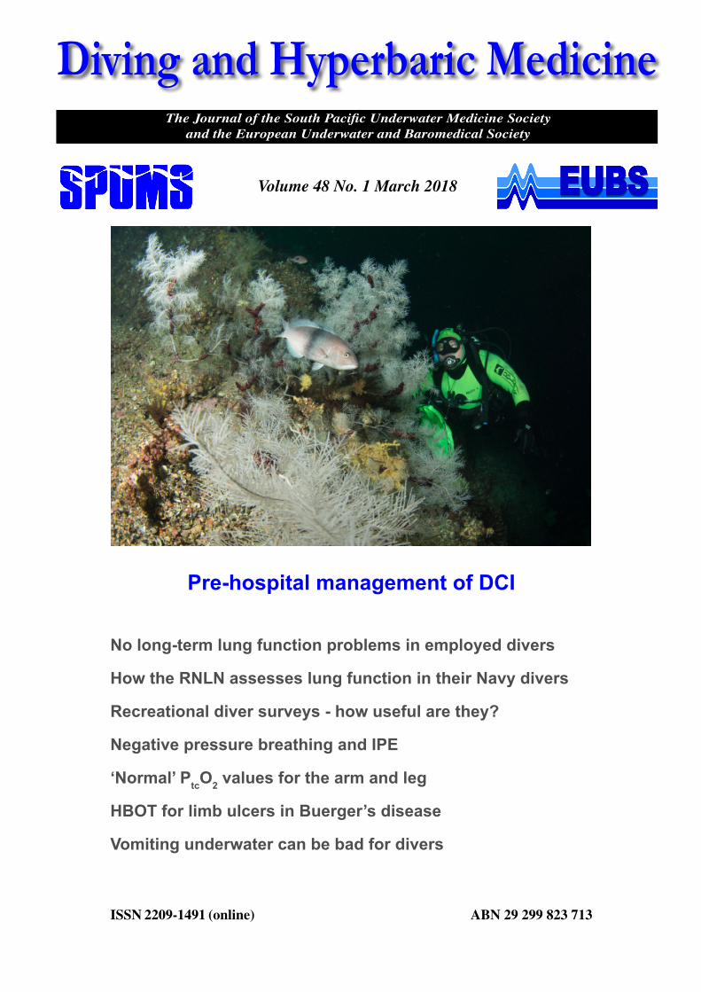
Volume 48 No. 1 March 2018 Diving and Hyperbaric Medicine The Journal of the South Pacific Underwater Medicine Society and the European Underwater and Baromedical Society ISSN 2209-1491 (online) Pre-hospital management of DCI No long-term lung function problems in employed divers How the RNLN assesses lung function in their Navy divers Recreational diver surveys - how useful are they? Negative pressure breathing and IPE ‘Normal’ P tc O 2 values for the arm and leg HBOT for limb ulcers in Buerger’s disease Vomiting underwater can be bad for divers ABN 29 299 823 713
-
Upload
khangminh22 -
Category
Documents
-
view
6 -
download
0
Transcript of Diving and Hyperbaric Medicine - EUBS
Volume 48 No. 1 March 2018
Diving and Hyperbaric MedicineThe Journal of the South Pacific Underwater Medicine Society
and the European Underwater and Baromedical Society
ISSN 2209-1491 (online)
Pre-hospital management of DCI
No long-term lung function problems in employed divers
How the RNLN assesses lung function in their Navy divers
Recreational diver surveys - how useful are they?
Negative pressure breathing and IPE
‘Normal’ PtcO2 values for the arm and leg
HBOT for limb ulcers in Buerger’s disease
Vomiting underwater can be bad for divers
ABN 29 299 823 713
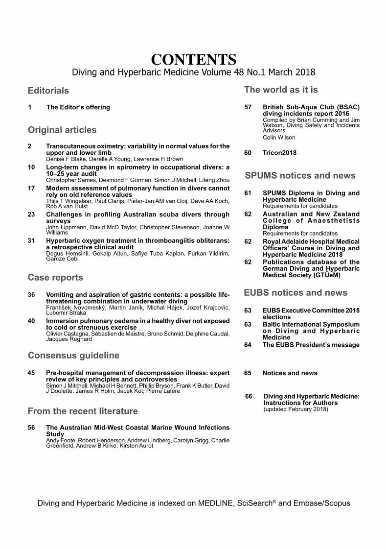
CONTENTS
EUBS notices and news
Diving and Hyperbaric Medicine is indexed on MEDLINE, SciSearch® and Embase/Scopus
Diving and Hyperbaric Medicine Volume 48 No.1 March 2018
Original articles
SPUMS notices and news
65 Notices and news
2 Transcutaneous oximetry: variability in normal values for the upper and lower limbDenise F Blake, Derelle A Young, Lawrence H Brown
10 Long-term changes in spirometry in occupational divers: a 10–25 year auditChristopher Sames, Desmond F Gorman, Simon J Mitchell, Lifeng Zhou
17 Modern assessment of pulmonary function in divers cannot rely on old reference valuesThijs T Wingelaar, Paul Clarijs, Pieter-Jan AM van Ooij, Dave AA Koch, Rob A van Hulst
23 Challenges in profiling Australian scuba divers through surveysJohn Lippmann, David McD Taylor, Christopher Stevenson, Joanne W Williams
31 Hyperbaric oxygen treatment in thromboangiitis obliterans: a retrospective clinical auditDogus Hemsinli, Gokalp Altun, Safiye Tuba Kaplan, Furkan Yildirim, Gamze Cebi
63 EUBS Executive Committee 2018 elections
63 Baltic International Symposium on Diving and Hyperbaric Medicine
64 The EUBS President’s message
61 SPUMS Diploma in Diving and Hyperbaric MedicineRequirements for candidates
62 Australian and New Zealand Col lege of Anaesthet is ts DiplomaRequirements for candidates
62 Royal Adelaide Hospital Medical Officers’ Course in Diving and Hyperbaric Medicine 2018
62 Publications database of the German Diving and Hyperbaric Medical Society (GTÜeM)
36 Vomiting and aspiration of gastric contents: a possible life-threatening combination in underwater divingFrantišek Novomeský, Martin Janík, Michal Hájek, Jozef Krajcovic, Lubomír Straka
40 Immersion pulmonary oedema in a healthy diver not exposed to cold or strenuous exerciseOlivier Castagna, Sébastien de Maistre, Bruno Schmid, Delphine Caudal, Jacques Regnard
Consensus guideline
The world as it is
57 British Sub-Aqua Club (BSAC) diving incidents report 2016Compiled by Brian Cumming and Jim Watson, Diving Safety and Incidents AdvisorsColin Wilson
Case reports
45 Pre-hospital management of decompression illness: expert review of key principles and controversiesSimon J Mitchell, Michael H Bennett, Phillip Bryson, Frank K Butler, David J Doolette, James R Holm, Jacek Kot, Pierre Lafère
60 Tricon2018
Editorials1 The Editor’s offering
From the recent literature56 The Australian Mid-West Coastal Marine Wound Infections
StudyAndy Foote, Robert Henderson, Andrew Lindberg, Carolyn Grigg, Charlie Greenfield, Andrew B Kirke, Kirsten Auret
66 Diving and Hyperbaric Medicine: Instructions for Authors(updated February 2018)
PURPOSES OF THE SOCIETIESTo promote and facilitate the study of all aspects of underwater and hyperbaric medicine
To provide information on underwater and hyperbaric medicineTo publish a journal and to convene members of each Society annually at a scientific conference
SOUTH PACIFIC UNDERWATERMEDICINE SOCIETY
OFFICE HOLDERSPresident
David Smart <[email protected]>Past President
Michael Bennett <[email protected]>Secretary
Douglas Falconer <[email protected]>Treasurer
Sarah Lockley <[email protected]>Education Officer
David Wilkinson <[email protected]>Chairman ANZHMG
Neil Banham <[email protected]> Committee Members
Jen Coleman Tamara Ford Ian Gawthrope Cathy Meehan Peter Smith
WebmasterJoel Hissink <[email protected]>
ADMINISTRATIONMembership
Steve Goble <[email protected]>MEMBERSHIP
For further information on SPUMS and to register to become a member, go to the Society’s website: <www.spums.org.au> The official address for SPUMS is: c/o Australian and New Zealand College of Anaesthetists, 630 St Kilda Road, Melbourne, Victoria 3004, AustraliaSPUMS is incorporated in Victoria A0020660B
EUROPEAN UNDERWATER ANDBAROMEDICAL SOCIETY
OFFICE HOLDERSPresident Jacek Kot <[email protected]>Vice President Ole Hyldegaard <[email protected]>Immediate Past President Costantino Balestra <[email protected]>Past President Peter Germonpré <[email protected]>Honorary Secretary Peter Germonpré <[email protected]>Member-at-Large 2017 Rodrigue Pignel <[email protected]>Member-at-Large 2016 Bengusu Oroglu <[email protected]> Member-at-Large 2015 Karin Hasmiller <[email protected]>Liaison Officer Phil Bryson <[email protected]>
ADMINISTRATIONHonorary Treasurer and Membership Secretary Kathleen Pye <[email protected]>
MEMBERSHIPFor further information on EUBS and to complete a membership application, go to the Society’s website: <www.eubs.org>The official address for EUBS is: Kathleen Pye Chantrey, Hillside Road Stromness, Orkney KW16 3HR, United KingdomEUBS is a UK Registered Charity No. 264970
EditorMichael Davis <[email protected]>
European (Deputy) EditorLesley Blogg <[email protected]>
Associate Editor Simon Mitchell <[email protected]>Editorial Assistant
Nicky Telles <[email protected]>
Submissions: <https://www.manuscriptmanager.net/dhm>
Back issues to 2017Steve Goble <[email protected]>
Editorial BoardMichael Bennett, AustraliaDavid Doolette, USAChristopher Edge, United KingdomIngrid Eftedal, NorwayPeter Germonpré, BelgiumJacek Kot, PolandSimon Mitchell, New ZealandClaus-Martin Muth, GermanyNeal Pollock, CanadaMonica Rocco, ItalyMartin Sayer, United KingdomErika Schagatay, SwedenDavid Smart, AustraliaRobert van Hulst, The Netherlands
Diving and Hyperbaric Medicine is published jointly by the South Pacific Underwater Medicine Society and the European Underwater and Baromedical Society (ISSN 1833-3516, ABN 29 299 823 713)
DIVING AND HYPERBARIC MEDICINE<www.dhmjournal.com>

Diving and Hyperbaric Medicine Volume 48 No. 1 March 2018 11
The Editor’s offeringWelcome to this, the first electronic issue of Diving and Hyperbaric Medicine (DHM). We would appreciate your feedback so that we can develop DHM as the very best in presenting research in this specialised field of medicine and physiology.
This is not the only major change for DHM in 2018. I am delighted to say that Professor Simon Mitchell has been apointed as its next Editor, taking over from me later this year. DHM will be in excellent hands and will have a bright future under his guidance.
A few years ago, a study on ‘normal’ transcutaneous oxygen partial pressures (P
tcO
2) was thwarted by a simple technical
error – the wrong membranes on the O2 electrodes. The paper
was retracted. The study has now been repeated and the main conclusion is that there is no such thing as a single ‘normal’ P
tcO
2 value for either the upper or lower limb.1
Hyperbaric physicians often battle against the unwillingness of many physicians and surgeons to recognise the potential value of hyperbaric oxygen treatment (HBOT) in selected patients. I was strongly reminded of this in a recent email from a paraplegic scientist friend of mine. He wrote:“In the last five years minor abrasions on my toes have developed a habit of getting deeper and then not healing. I have started to use HBOT quite seriously to promote healing, with very successful results so far. Two vascular surgeons stated that a slow-healing wound on my left foot meant that the toe would have to be amputated in 2015, but HBOT in 2016 healed it completely. The problems of paraplegic foot circulation after 50 years in a wheelchair are very similar to diabetes, but I am not diabetic. As I am sure you know, the NHS [UK] does not believe in the effectiveness of HBOT, so it is quite a battle to self-manage my care programme.”
With this in mind, it is interesting to see a report from Turkey on HBOT in thromboangiitis obliterans (TAO) demonstrating a clear medium-term benefit in these patients.2 There is much potential for HBOT in non-diabetic, non-healing wounds if only the opportunities for clinical research were more readily supported by the profession at large.
Queensland Recreational Diving, Recreational Technical Diving and Snorkelling 2018 Code of Practice
In 2017, the Queensland Government undertook a review of its 2011 Code of Practice because of an increase in snorkelling and diving fatalities in late 2016. In February 2018, the Queensland Minister for Education and Minister for Industrial Relations approved a revised Code of Practice. A summary of the key changes, courtesy of Bradley Bick, Office of Industrial Relations, Queensland, is as follows:• Requiring operators to provide automated external
defibrillators as part of their operations (for example,
either on a vessel or at the dive site); • Introduction of control measures for at risk snorkellers
which includes a requirement for:• operators to be able to obtain medical declarations from at-risk snorkellers; • operators to have a system in place for easy visual identification of at-risk snorkellers; • for at-risk snorkellers to wear and/or use a flotation device; and • requirement for at-risk snorkellers to swim in a buddy pair.
• Consistent safety messages required for all recreational snorkellers and at-risk snorkellers – this to ensure snorkellers are given consistent messages about the risks of snorkelling and the required safety measures;
• Reducing the minimum age for participation in entry level certificate diving, subject to additional safeguards, to align Queensland with international standards of dive training agencies and other Australian jurisdictions;
• Requiring operators to teach resort divers to inflate and deflate their buoyancy control device, and for them to practice doing it themselves, rather than just explaining how the device can be used;
• Minor changes to supervision requirements for resort divers that will prevent large groups swimming in single file, and to be consistent with existing requirements that instructors always be positioned to make physical contact with any diver; and
• Simplifying the requirements for recreational technical diving, and stating that this type of diving be undertaken in accordance with training agency standards, which are updated more regularly than the code of practice.
The link to the Code is: <https://www.worksafe.qld.gov.au/__data/assets/pdf_file/0006/58191/recreational-diving-recreational-technical-diving-snorkelling-cop-2011.pdf>.
References
1 Blake DF, Young DA, Brown LH. Transcutaneous oximetry: variability in normal values for the upper and lower limb. Diving Hyperb Med. 2018;48:3–10. doi:10.28920/dhm48.1.3-10.
2 Hemsinli D, Altun G, Tuba Kaplan S, Yildirim F, Cebi G. Hyperbaric oxygen therapy in thromboangiitis obliterans: Does it make a significant contribution? Diving Hyperb Med. 2018;48:32–6. doi:10.28920/dhm48.1.32-36.
Mike Davis
Key wordsHyperbaric oxygen therapy; Chronic wounds; Transcutaneous oximetry; Diving industry; Safety; General interest
A deep-water, remote, temperate-water dive site – southern Fiordland, South Island, New Zealand; the nearest recompression chamber is hundreds of kilometres away across a high mountain range.Photo courtesy of Quentin Bennett, 2016
Diving and Hyperbaric Medicine Volume 48 No. 1 March 20182
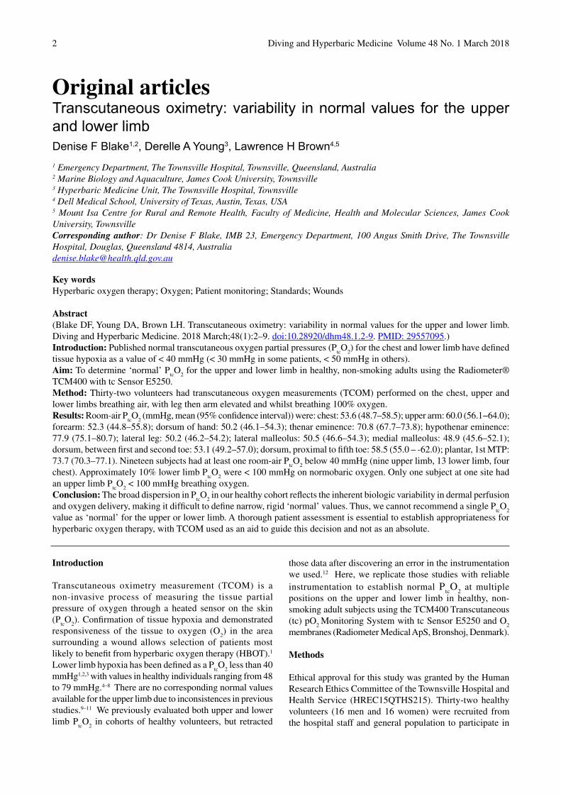
Original articlesTranscutaneous oximetry: variability in normal values for the upper and lower limbDenise F Blake1,2, Derelle A Young3, Lawrence H Brown4,5
1 Emergency Department, The Townsville Hospital, Townsville, Queensland, Australia2 Marine Biology and Aquaculture, James Cook University, Townsville3 Hyperbaric Medicine Unit, The Townsville Hospital, Townsville4 Dell Medical School, University of Texas, Austin, Texas, USA5 Mount Isa Centre for Rural and Remote Health, Faculty of Medicine, Health and Molecular Sciences, James Cook University, TownsvilleCorresponding author: Dr Denise F Blake, IMB 23, Emergency Department, 100 Angus Smith Drive, The Townsville Hospital, Douglas, Queensland 4814, [email protected]
Key wordsHyperbaric oxygen therapy; Oxygen; Patient monitoring; Standards; Wounds
Abstract(Blake DF, Young DA, Brown LH. Transcutaneous oximetry: variability in normal values for the upper and lower limb. Diving and Hyperbaric Medicine. 2018 March;48(1):2–9. doi:10.28920/dhm48.1.2-9. PMID: 29557095.)Introduction: Published normal transcutaneous oxygen partial pressures (P
tcO
2) for the chest and lower limb have defined
tissue hypoxia as a value of < 40 mmHg (< 30 mmHg in some patients, < 50 mmHg in others).Aim: To determine ‘normal’ P
tcO
2 for the upper and lower limb in healthy, non-smoking adults using the Radiometer®
TCM400 with tc Sensor E5250.Method: Thirty-two volunteers had transcutaneous oxygen measurements (TCOM) performed on the chest, upper and lower limbs breathing air, with leg then arm elevated and whilst breathing 100% oxygen.Results: Room-air P
tcO
2 (mmHg, mean (95% confidence interval)) were: chest: 53.6 (48.7–58.5); upper arm: 60.0 (56.1−64.0);
forearm: 52.3 (44.8–55.8); dorsum of hand: 50.2 (46.1–54.3); thenar eminence: 70.8 (67.7–73.8); hypothenar eminence: 77.9 (75.1–80.7); lateral leg: 50.2 (46.2–54.2); lateral malleolus: 50.5 (46.6–54.3); medial malleolus: 48.9 (45.6–52.1); dorsum, between first and second toe: 53.1 (49.2–57.0); dorsum, proximal to fifth toe: 58.5 (55.0 – -62.0); plantar, 1st MTP: 73.7 (70.3–77.1). Nineteen subjects had at least one room-air P
tcO
2 below 40 mmHg (nine upper limb, 13 lower limb, four
chest). Approximately 10% lower limb PtcO
2 were < 100 mmHg on normobaric oxygen. Only one subject at one site had
an upper limb PtcO
2 < 100 mmHg breathing oxygen.
Conclusion: The broad dispersion in PtcO
2 in our healthy cohort reflects the inherent biologic variability in dermal perfusion
and oxygen delivery, making it difficult to define narrow, rigid ‘normal’ values. Thus, we cannot recommend a single PtcO
2
value as ‘normal’ for the upper or lower limb. A thorough patient assessment is essential to establish appropriateness for hyperbaric oxygen therapy, with TCOM used as an aid to guide this decision and not as an absolute.
Introduction
Transcutaneous oximetry measurement (TCOM) is a non-invasive process of measuring the tissue partial pressure of oxygen through a heated sensor on the skin (P
tcO
2). Confirmation of tissue hypoxia and demonstrated
responsiveness of the tissue to oxygen (O2) in the area
surrounding a wound allows selection of patients most likely to benefit from hyperbaric oxygen therapy (HBOT).1 Lower limb hypoxia has been defined as a P
tcO
2 less than 40
mmHg1,2,3 with values in healthy individuals ranging from 48 to 79 mmHg.4–8 There are no corresponding normal values available for the upper limb due to inconsistences in previous studies.9–11 We previously evaluated both upper and lower limb P
tcO
2 in cohorts of healthy volunteers, but retracted
those data after discovering an error in the instrumentation we used.12 Here, we replicate those studies with reliable instrumentation to establish normal P
tcO
2 at multiple
positions on the upper and lower limb in healthy, non-smoking adult subjects using the TCM400 Transcutaneous (tc) pO
2 Monitoring System with tc Sensor E5250 and O
2
membranes (Radiometer Medical ApS, Bronshoj, Denmark).
Methods
Ethical approval for this study was granted by the Human Research Ethics Committee of the Townsville Hospital and Health Service (HREC15QTHS215). Thirty-two healthy volunteers (16 men and 16 women) were recruited from the hospital staff and general population to participate in
Diving and Hyperbaric Medicine Volume 48 No. 1 March 2018 3
this study. Exclusion criteria included subjects younger than 18 years old; current or former smokers; known cardiovascular disease including treated or untreated hypertension; significant respiratory disease or any other significant medical condition. Subjects missing a limb, or with significant scarring or a skin condition on a limb, were also excluded. As subjects were required to have a plastic hood placed over their head to receive O
2 for part of the study,
severe claustrophobia was a further exclusion criterion.
All participants were given a study information sheet and informed consent was obtained. Subjects refrained from consuming food or caffeine or performing heavy exercise for two hours prior to participating in the study. Subjects lay supine on a hospital bed with their head slightly raised on one pillow and were offered a blanket for comfort and to limit any vasoconstrictive effects of being cold. The room temperature was maintained between 22.0 and 22.5°C.
Basic demographic data were collected including height and weight. O
2 saturation and blood pressure were measured
on both arms. Upper and lower limb pulses were recorded bilaterally. Toe pressures were measured on the randomized limb. Ankle brachial index (ABI) and toe brachial index (TBI) were calculated. Any abnormalities in the baseline observations would have led to exclusion from the study.
Participants were randomized to have 12 sensors placed on their right or left side (chest, arm and leg). The sensor sites were prepared by shaving hair if necessary, wiping clean, rubbing with an alcohol swab and drying with gauze. The chest sensor was placed at the second intercostal space in the mid-clavicular line. For the upper limb, sensors were placed: mid-way between the highest bony point on the shoulder and the olecranon process on the lateral aspect of the upper arm; 5 cm distal to the brachial crease on the lateral aspect of the lower arm; on the thenar and hypothenar eminences and centrally on the dorsum of the hand between the third and fourth metacarpal bones away from any obvious veins. For the lower limb, sensors were positioned: 10 cm distal to the lateral femoral epicondyle; 5 cm proximal to both the lateral and medial malleoli; on the dorsum of the foot attempting to avoid large superficial vessels, one between the first and second metatarsal heads and the second proximal to the fifth metatarsal phalangeal (MTP) joint and on the plantar aspect of the foot proximal to the first metatarsal phalangeal joint. The leads were secured in place with tape to prevent pull on the sensors. Subjects were requested to keep talking to a minimum during the study.
All TCOM assessments were performed by the same technician (DY) using the TCM400 P
tcO
2 Monitoring
System. The TCM400 has six tc E5250 sensors and can record P
tcO
2 data from all sensors simultaneously. Two
machines were used, alternating between the upper and lower limb. The electrode temperatures were pre-set to 44°C and atmospheric and zero point electrode calibrations were
performed as per the manufacturer’s recommendations. A humidity correction factor was calculated from the room temperature, saturated water vapour pressure and relative humidity and input into the machine according to the TCM400 operator’s manual.13 The TCM400 displays P
tcO
2
values in mmHg units.
We used the TCOM protocol described by Sheffield which is historically used in hyperbaric medicine to identify tissue hypoxia and responsiveness to hyperoxia.14,15 Initial normobaric room air readings from all sensors were recorded after a minimum 20-minute equilibration period, allowing all sensors to stabilize.4 The leg was then elevated 45 degrees above its resting level and placed on a foam wedge, with sensor readings recorded after five minutes. The elevation process was then repeated for the arm. The arm or leg were returned to the horizontal position for a minimum five-minute period allowing all sensor readings to re-stabilize, and another set of readings were recorded to ensure P
tcO
2 had
returned to baseline. The subjects then breathed 100% O2
for 10 minutes via a clear plastic hood with a soft neck seal, with sensor readings recorded at the end of the 10-minute period, once stabilized. At the conclusion of the session, all sites were inspected for thermal injury.
ANALYSIS
All collected data were de-identified and entered into a pre-formatted Excel worksheet. These data were subsequently exported into Stata Statistical Software: Release 11 (StataCorp LP, College Station, TX, USA) for analysis.
The primary outcome of this study was a determination of the normal range of P
tcO
2 when measured at various places
on the upper and lower limbs of healthy volunteer subjects. Based on previous reports of mean normal P
tcO
2 readings
ranging from 58 to 65 mmHg (upper limb)11,16 and 48 to 79 mmHg (lower limb)2, 4–7 with a standard deviation (SD) of approximately 10 mmHg, our sample size of 32 subjects was intended to allow us to estimate mean P
tcO
2 readings
with a 95% confidence interval (95% CI) of ± 3.5 mmHg. Having 16 male and 16 female subjects also provided 80% power (with α = 0.05) to detect a 10 mmHg difference in mean P
tcO
2 of males versus females using Student’s t-test.
Descriptive statistics are reported for PtcO
2 at each of
the 12 sensor sites. The Shapiro-Wilk test was used to evaluate normality of the data distributions. For normally distributed data, mean, 95% CI, and/or standard deviation and range are reported. For non-parametric data, median, inter-quartile range and approximate 95% CI for the median are reported. Demographic characteristics of male and female subjects were compared using Fisher’s Exact Test (FET) or Student’s t-test as appropriate. Differences between mean P
tcO
2 for males and females were compared
using Student’s t-test when data were normally distributed, Wilcoxon Rank Sum test for non-parametric data, and FET
Diving and Hyperbaric Medicine Volume 48 No. 1 March 20184
for frequency data. Correlations between baseline perfusion measures of systolic blood pressure (SBP), diastolic blood pressure (DBP), oxygen saturation (SpO
2) and toe SBP in
the randomized limb and the observed room air and on-O2
PtcO
2 at each sensor site were evaluated using Pearson’s
correlation coefficient with Bonferroni correction for multiple comparisons.
Results
Demographic and baseline perfusion measures for the 32 subjects are shown in Table 1. Subjects ranged in age from 26 to 80 years. Baseline perfusion measures were clinically unremarkable in all subjects. The only statistically significant difference between female and male subjects was systolic blood pressure, but this difference was clinically irrelevant. There were no statistically significant correlations between baseline measures of perfusion and any of the P
tcO
2
measurements (data not shown*).
ROOM-AIR PtcO
2
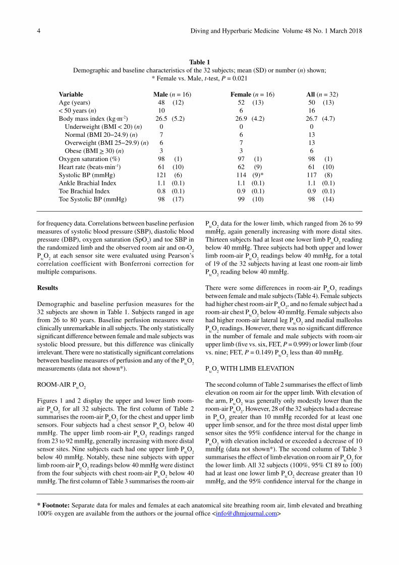
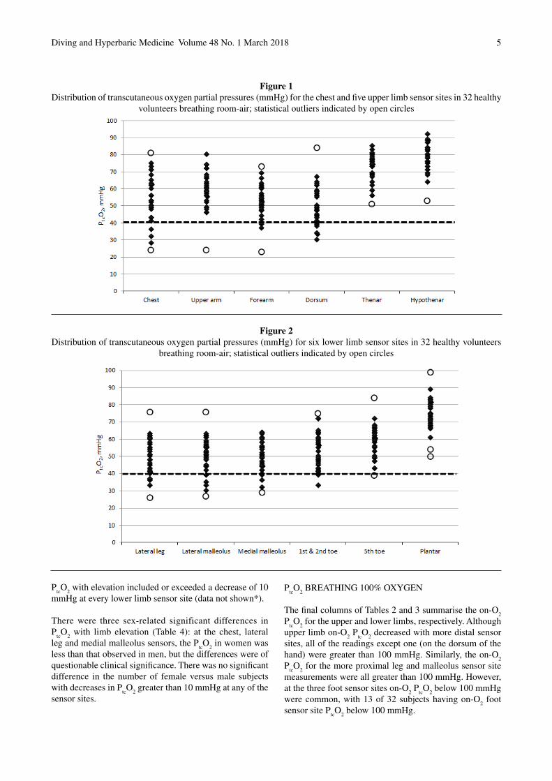
Figures 1 and 2 display the upper and lower limb room-air P
tcO
2 for all 32 subjects. The first column of Table 2
summarises the room-air PtcO
2 for the chest and upper limb
sensors. Four subjects had a chest sensor PtcO
2 below 40
mmHg. The upper limb room-air PtcO
2 readings ranged
from 23 to 92 mmHg, generally increasing with more distal sensor sites. Nine subjects each had one upper limb P
tcO
2
below 40 mmHg. Notably, these nine subjects with upper limb room-air P
tcO
2 readings below 40 mmHg were distinct
from the four subjects with chest room-air PtcO
2 below 40
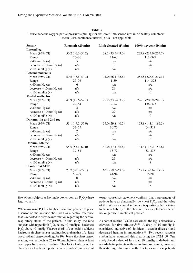
mmHg. The first column of Table 3 summarises the room-air
PtcO
2 data for the lower limb, which ranged from 26 to 99
mmHg, again generally increasing with more distal sites. Thirteen subjects had at least one lower limb P
tcO
2 reading
below 40 mmHg. Three subjects had both upper and lower limb room-air P
tcO
2 readings below 40 mmHg, for a total
of 19 of the 32 subjects having at least one room-air limb P
tcO
2 reading below 40 mmHg.
There were some differences in room-air PtcO
2 readings
between female and male subjects (Table 4). Female subjects had higher chest room-air P
tcO
2, and no female subject had a
room-air chest PtcO
2 below 40 mmHg. Female subjects also
had higher room-air lateral leg PtcO
2 and medial malleolus
PtcO
2 readings. However, there was no significant difference
in the number of female and male subjects with room-air upper limb (five vs. six, FET, P = 0.999) or lower limb (four vs. nine; FET, P = 0.149) P
tcO
2 less than 40 mmHg.
PtcO
2 WITH LIMB ELEVATION
The second column of Table 2 summarises the effect of limb elevation on room air for the upper limb. With elevation of the arm, P
tcO
2 was generally only modestly lower than the
room-air PtcO
2. However, 28 of the 32 subjects had a decrease
in PtcO
2 greater than 10 mmHg recorded for at least one
upper limb sensor, and for the three most distal upper limb sensor sites the 95% confidence interval for the change in P
tcO
2 with elevation included or exceeded a decrease of 10
mmHg (data not shown*). The second column of Table 3 summarises the effect of limb elevation on room air P
tcO
2 for
the lower limb. All 32 subjects (100%, 95% CI 89 to 100) had at least one lower limb P
tcO
2 decrease greater than 10
mmHg, and the 95% confidence interval for the change in
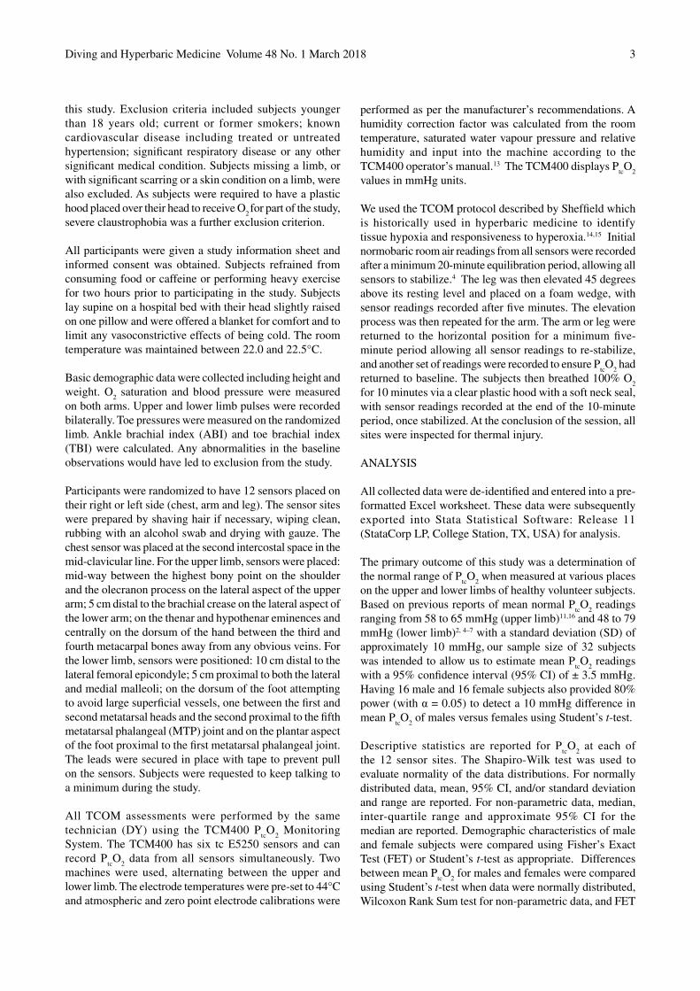
Table 1Demographic and baseline characteristics of the 32 subjects; mean (SD) or number (n) shown;
* Female vs. Male, t-test, P = 0.021
Variable Male (n = 16) Female (n = 16) All (n = 32)Age (years) 48 (12) 52 (13) 50 (13)< 50 years (n) 10 6 16Body mass index (kg∙m-2) 26.5 (5.2) 26.9 (4.2) 26.7 (4.7)
Underweight (BMI < 20) (n) 0 0 0Normal (BMI 20−24.9) (n) 7 6 13Overweight (BMI 25−29.9) (n) 6 7 13Obese (BMI > 30) (n) 3 3 6
Oxygen saturation (%) 98 (1) 97 (1) 98 (1)Heart rate (beats∙min-1) 61 (10) 62 (9) 61 (10)Systolic BP (mmHg) 121 (6) 114 (9)* 117 (8)Ankle Brachial Index 1.1 (0.1) 1.1 (0.1) 1.1 (0.1)Toe Brachial Index 0.8 (0.1) 0.9 (0.1) 0.9 (0.1)Toe Systolic BP (mmHg) 98 (17) 99 (10) 98 (14)
* Footnote: Separate data for males and females at each anatomical site breathing room air, limb elevated and breathing 100% oxygen are available from the authors or the journal office <[email protected]>
Diving and Hyperbaric Medicine Volume 48 No. 1 March 2018 5
Figure 1Distribution of transcutaneous oxygen partial pressures (mmHg) for the chest and five upper limb sensor sites in 32 healthy
volunteers breathing room-air; statistical outliers indicated by open circles
Figure 2Distribution of transcutaneous oxygen partial pressures (mmHg) for six lower limb sensor sites in 32 healthy volunteers
breathing room-air; statistical outliers indicated by open circles
PtcO
2 with elevation included or exceeded a decrease of 10
mmHg at every lower limb sensor site (data not shown*).
There were three sex-related significant differences in P
tcO
2 with limb elevation (Table 4): at the chest, lateral
leg and medial malleolus sensors, the PtcO
2 in women was
less than that observed in men, but the differences were of questionable clinical significance. There was no significant difference in the number of female versus male subjects with decreases in P
tcO
2 greater than 10 mmHg at any of the
sensor sites.
PtcO
2 BREATHING 100% OXYGEN
The final columns of Tables 2 and 3 summarise the on-O2
PtcO
2 for the upper and lower limbs, respectively. Although
upper limb on-O2 P
tcO
2 decreased with more distal sensor
sites, all of the readings except one (on the dorsum of the hand) were greater than 100 mmHg. Similarly, the on-O
2
PtcO
2 for the more proximal leg and malleolus sensor site
measurements were all greater than 100 mmHg. However, at the three foot sensor sites on-O
2 P
tcO
2 below 100 mmHg
were common, with 13 of 32 subjects having on-O2 foot
sensor site PtcO
2 below 100 mmHg.

Diving and Hyperbaric Medicine Volume 48 No. 1 March 20186
Sensor Room air (20 min) Limb elevated (5 min) 100% oxygen (10 min)Chest
Mean (95% CI) 53.6 (48.7–58.5) 54.2 (49.0–59.4) 397.1 (380.1–414.1)Range 24–81 12–74 308–500< 40 mmHg (n) 4 n/a n/adecrease > 10 mmHg (n) n/a 2 n/a< 100 mmHg (n) n/a n/a 0
Upper armMean (95% CI) 60.0 (56.1–64.0) 59.2 (55.2–63.3) 421.1 (408.1–434.1)Range 24–80 28–79 335–486< 40 mmHg (n) 1 n/a n/adecrease > 10 mmHg (n) n/a 3 n/a< 100 mmHg (n) n/a n/a 0
ForearmMean (95% CI) 52.3 (48.8–55.8) 47.2 (42.7–51.7) 310.1 (282.4–337.8)Range 23–73 17–66 150–469< 40 mmHg (n) 3 n/a n/adecrease > 10 mmHg (n) n/a 7 n/a< 100 mmHg (n) n/a n/a 0
Dorsum handMean (95% CI) 50.2 (46.1–54.3) 33.8 (27.6–39.9) 278.4 (249.7–307.2)Range 30–84 1–74 89–440< 40 mmHg (n) 5 n/a n/adecrease > 10 mmHg (n) n/a 21 n/a< 100 mmHg (n) n/a n/a 1
Thenar eminenceMean (95% CI) 70.8 (67.7–73.8) 58.5 (54.3–62.8) 229.4 (211.1–247.6)Range 51–85 28–77 101–314< 40 mmHg (n) 0 n/a n/adecrease > 10 mmHg (n) n/a 17 n/a< 100 mmHg (n) n/a n/a 0
Hypothenar eminenceMean or [median]† 77.9 (75.1–80.7) [71.0 (63.0–73.5)] 212.4 (195.5–229.3)Range 53–92 36–81 124–308< 40 mmHg (n) 0 n/a n/adecrease > 10 mmHg (n) n/a 11 n/a< 100 mmHg (n) n/a n/a 0
Table 2Transcutaneous oxygen partial pressures (mmHg) for chest and five upper limb sensor sites in 32 healthy volunteers;mean (95% confidence interval); † [median (approx. 95% CI)] for non-normally distributed data; n/a – not applicable
Female subjects had significantly lower on-O2 P
tcO
2 for
the sensor placed at the fifth toe on the dorsum of the foot(Table 4), but there was no significant difference in the number of female and male subjects with on-O
2 P
tcO
2 less
than 100 mmHg at this site (six vs two; FET, P = 0.220) or any other sensor site.
Discussion
Clinical practice guidelines for PtcO
2 have been developed
to assist the clinician in selecting appropriate patients for HBOT.1 A thorough clinical history and exam remains essential, with P
tcO
2 results integrated as one variable in the
workup. Using the reference value of 40 mmHg to define
hypoxia for all locations on the lower limb would result in 16% of readings on the lateral leg and 31% of the malleoli values in our healthy subjects being classified as hypoxic. The most distal sensor sites on the palm and on the plantar aspect of the foot were the only sites where 100% of our healthy subjects had values above 40 mmHg. Overall, more than half of our ‘healthy’ subjects recorded a room air P
tcO
2
below 40 mmHg for at least one limb sensor site.
PtcO
2 measurements of 30–40 mmHg have been considered
to fall within a grey zone for classification of hypoxia with the value of 50 mmHg used in patients with other factors such as diabetes and renal failure.1 Using a more conservative reference value of 30 mmHg would still classify
Diving and Hyperbaric Medicine Volume 48 No. 1 March 2018 7
five of our subjects as having hypoxic room air PtcO
2 (three
leg; two arm).
When assessing PtcO
2, it has been common practice to place
a sensor on the anterior chest wall as a central reference that is reported to provide information regarding the cardio-respiratory status of the patient. In this study, the nine subjects with upper limb P
tcO
2 below 40 mmHg all had chest
PtcO
2 above 40 mmHg. Yet, two-thirds of our healthy subjects
had room air chest sensor readings lower than that of at least one arm/hand sensor reading; for 10 subjects the chest sensor reading was as much as 25 to 30 mmHg lower than at least one upper limb sensor reading. This lack of utility of the chest sensor has been reported in other studies17 and a recent
expert consensus statement confirms that a percentage of patients have an abnormally low chest P
tcO
2, and the value
of this site as a central reference is questionable.2 Owing to the unreliability of the chest sensor as a reference site we no longer use it in clinical practice.
As part of routine TCOM assessment the leg is historically elevated for five minutes.18–20 A drop of 10 mmHg is considered indicative of significant vascular disease21 and decreased healing in amputations.22 Two recent vascular studies have examined this area using the TCM400. One study found a drop of less than 10 mmHg in diabetic and non-diabetic patients with severe limb ischaemia; however, their starting values were in the low teens and these patients
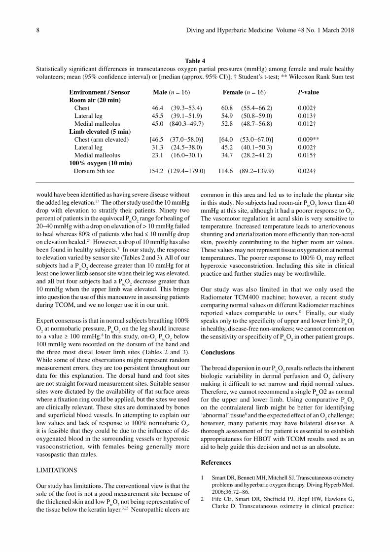
Sensor Room air (20 min) Limb elevated (5 min) 100% oxygen (10 min)Lateral leg
Mean (95% CI) 50.2 (46.2–54.2) 38.2 (33.5–43.0) 239.9 (214.0–265.7)Range 26–76 11-63 111–397< 40 mmHg (n) 5 n/a n/adecrease > 10 mmHg (n) n/a 19 n/a< 100 mmHg (n) n/a n/a 0
Lateral malleolusMean (95% CI) 50.5 (46.6–54.3) 31.0 (26.4–35.6) 252.8 (226.5–279.1)Range 27–76 1-59 114–375< 40 mmHg (n) 6 n/a n/adecrease > 10 mmHg (n) n/a 29 n/a< 100 mmHg (n) n/a n/a 0
Medial malleolusMean (95% CI) 48.9 (45.6–52.1) 28.9 (23.9–33.9) 226.3 (205.9–246.7)Range 29–64 2-54 138–373< 40 mmHg (n) 4 n/a n/adecrease > 10 mmHg (n) n/a 29 n/a< 100 mmHg (n) n/a n/a 0
Dorsum, 1st and 2nd toeMean (95% CI) 53.1 (49.2–57.0) 35.0 (29.8–40.2) 163.8 (141.1–186.5)Range 33–75 10-72 64–317< 40 mmHg (n) 2 n/a n/adecrease > 10 mmHg (n) n/a 28 n/a< 100 mmHg (n) n/a n/a 5
Dorsum, 5th toeMean (95% CI) 58.5 (55.1–62.0) 42.0 (37.4–46.6) 134.4 (116.2–152.6)Range 39–84 13-72 53–238< 40 mmHg (n) 1 n/a n/adecrease > 10 mmHg (n) n/a 29 n/a< 100 mmHg (n) n/a n/a 8
Plantar, 1st MTPMean (95% CI) 73.7 (70.3–77.1) 63.2 (59.3–67.0) 165.4 (143.6–187.2)Range 50–99 41-94 67–280< 40 mmHg (n) 0 n/a n/adecrease > 10 mmHg (n) n/a 15 n/a< 100 mmHg (n) n/a n/a 6
Table 3Transcutaneous oxygen partial pressures (mmHg) for six lower limb sensor sites in 32 healthy volunteers;
mean (95% confidence interval) ; n/a – not applicable
Diving and Hyperbaric Medicine Volume 48 No. 1 March 20188
would have been identified as having severe disease without the added leg elevation.23 The other study used the 10 mmHg drop with elevation to stratify their patients. Ninety two percent of patients in the equivocal P
tcO
2 range for healing of
20–40 mmHg with a drop on elevation of > 10 mmHg failed to heal whereas 80% of patients who had ≤ 10 mmHg drop on elevation healed.24 However, a drop of 10 mmHg has also been found in healthy subjects.7 In our study, the response to elevation varied by sensor site (Tables 2 and 3). All of our subjects had a P
tcO
2 decrease greater than 10 mmHg for at
least one lower limb sensor site when their leg was elevated, and all but four subjects had a P
tcO
2 decrease greater than
10 mmHg when the upper limb was elevated. This brings into question the use of this manoeuvre in assessing patients during TCOM, and we no longer use it in our unit.
Expert consensus is that in normal subjects breathing 100% O
2 at normobaric pressure, P
tcO
2 on the leg should increase
to a value ≥ 100 mmHg.8 In this study, on-O2 P
tcO
2 below
100 mmHg were recorded on the dorsum of the hand and the three most distal lower limb sites (Tables 2 and 3). While some of these observations might represent random measurement errors, they are too persistent throughout our data for this explanation. The dorsal hand and foot sites are not straight forward measurement sites. Suitable sensor sites were dictated by the availability of flat surface areas where a fixation ring could be applied, but the sites we used are clinically relevant. These sites are dominated by bones and superficial blood vessels. In attempting to explain our low values and lack of response to 100% normobaric O
2,
it is feasible that they could be due to the influence of de-oxygenated blood in the surrounding vessels or hyperoxic vasoconstriction, with females being generally more vasospastic than males.
LIMITATIONS
Our study has limitations. The conventional view is that the sole of the foot is not a good measurement site because of the thickened skin and low P
tcO
2 not being representative of
the tissue below the keratin layer.3,25 Neuropathic ulcers are
common in this area and led us to include the plantar site in this study. No subjects had room-air P
tcO
2 lower than 40
mmHg at this site, although it had a poorer response to O2.
The vasomotor regulation in acral skin is very sensitive to temperature. Increased temperature leads to arteriovenous shunting and arterialization more efficiently than non-acral skin, possibly contributing to the higher room air values. These values may not represent tissue oxygenation at normal temperatures. The poorer response to 100% O
2 may reflect
hyperoxic vasoconstriction. Including this site in clinical practice and further studies may be worthwhile.
Our study was also limited in that we only used the Radiometer TCM400 machine; however, a recent study comparing normal values on different Radiometer machines reported values comparable to ours.8 Finally, our study speaks only to the specificity of upper and lower limb P
tcO
2
in healthy, disease-free non-smokers; we cannot comment on the sensitivity or specificity of P
tcO
2 in other patient groups.
Conclusions
The broad dispersion in our PtcO
2 results reflects the inherent
biologic variability in dermal perfusion and O2 delivery
making it difficult to set narrow and rigid normal values. Therefore, we cannot recommend a single P
tcO2 as normal
for the upper and lower limb. Using comparative PtcO
2
on the contralateral limb might be better for identifying ‘abnormal’ tissue8 and the expected effect of an O
2 challenge;
however, many patients may have bilateral disease. A thorough assessment of the patient is essential to establish appropriateness for HBOT with TCOM results used as an aid to help guide this decision and not as an absolute.
References
1 Smart DR, Bennett MH, Mitchell SJ. Transcutaneous oximetry problems and hyperbaric oxygen therapy. Diving Hyperb Med. 2006;36:72−86.
2 Fife CE, Smart DR, Sheffield PJ, Hopf HW, Hawkins G, Clarke D. Transcutaneous oximetry in clinical practice:
Environment / Sensor Male (n = 16) Female (n = 16) P-valueRoom air (20 min)
Chest 46.4 (39.3−53.4) 60.8 (55.4−66.2) 0.002†Lateral leg 45.5 (39.1−51.9) 54.9 (50.8−59.0) 0.013†Medial malleolus 45.0 (840.3−49.7) 52.8 (48.7−56.8) 0.012†
Limb elevated (5 min)Chest (arm elevated) [46.5 (37.0−58.0)] [64.0 (53.0−67.0)] 0.009**Lateral leg 31.3 (24.5−38.0) 45.2 (40.1−50.3) 0.002†Medial malleolus 23.1 (16.0−30.1) 34.7 (28.2−41.2) 0.015†
100% oxygen (10 min)Dorsum 5th toe 154.2 (129.4−179.0) 114.6 (89.2−139.9) 0.024†
Table 4Statistically significant differences in transcutaneous oxygen partial pressures (mmHg) among female and male healthy volunteers; mean (95% confidence interval) or [median (approx. 95% CI)]; † Student’s t-test; ** Wilcoxon Rank Sum test

Diving and Hyperbaric Medicine Volume 48 No. 1 March 2018 9
Consensus statements from an expert panel based on evidence. Undersea Hyperb Med. 2009;36:1−11.
3 Rich K. Transcutaneous oxygen measurements: Implications for nursing. J Vasc Nurs. 2001;19:55-9.
4 Dowd GS, Linge K, Bentley G. Measurement of transcutaneous oxygen pressure in normal and ischemic skin. J Bone Joint Surg. 1983;65B:79−83.
5 Dowd GS, Linge K, Bentley G. The effect of age and sex of normal volunteers upon the transcutaneous oxygen tension in the lower limb. Clin Phys Physiol Meas. 1983;4:65−8.
6 Hauser CJ, Shoemaker WC. Use of a transcutaneous PO2
regional perfusion index to quantify tissue perfusion in peripheral vascular disease. Ann Surg. 1983;197:337−43.
7 Dooley J, King G, Slade B. Establishment of reference pressure of transcutaneous oxygen for the comparative evaluation of problem wounds. Undersea Hyperb Med. 1997;24:235−44.
8 Trinks TP, Blake DF, Young DA, Thistlethwaite K, Vangaveti VN. Transcutaneous oximetry measurements of the leg: comparing different measuring equipment and establishing values in healthy young adults. Diving Hyperb Med. 2017;47:82−7.
9 Silverstein JL, Steen VD, Medsger TA, Falanga V. Cutaneous hypoxia in patients with systemic sclerosis (scleroderma). Arch Dermatol. 1988;124:1379−82.
10 Daviet J-C, Dudognon P, Preux PM, Rebeyrotte I, Lacroix P, Munoz M, Salle JY. Reliability of transcutaneous oxygen tension measurement on the back of the hand and complex regional pain syndrome after stroke. Arch Phys Med Rehabil. 2004;85:1102−5.
11 Babilas P, Lamby P, Prantl L, Schreml S, Jung EM, Liebsch G, et al. Transcutaneous pO
2 imaging during tourniquet-
induced forearm ischemia using planar optical oxygen sensors. Skin Res Technol. 2008;14:304−11. doi:10.1111/j.1600-0846.2008.00295.x
12 Blake DF, Young DA, Brown LH. Retraction of two papers investigating transcutaneous oxygen tensions in healthy volunteers. Diving Hyperb Med. 2016;46:54.
13 Radiometer Medical ApS. TCM400 transcutaneous pO2
monitoring system. Operator’s manual. Bronshoj, Denmark: Radiometer; 2005.
14 Sheffield PJ. Measuring tissue oxygen tension: a review. Undersea Hyperb Med. 1998;25:179−88.
15 Shah JB, Ram DM, Fredrick E, Otto H, Sheffield PJ. Determination of ideal PtcO
2 measurement time in evaluation
of hypoxic wound patients. Undersea Hyperb Med. 2008;35:41−51.
16 Manabe S, Tabuchi N, Toyama M, Yoshizaki T, Kato M, Wu H, et al. Oxygen pressure measurement during grip exercise reveals exercise intolerance after radial artery
harvest. Ann Thorac Surg. 2004;77:2066−70. doi:10.1016/j.athoracsur.2003.10.052.
17 de Meijer VE, van’t Sant HP, Spronk S, Kusters FJ, den Hoed PT. Reference value of transcutaneous oxygen measurement in diabetic patients compared with nondiabetic patients. J Vasc Surg. 2008;48:382−8. doi:10.1016/j.jvs.2008.03.010.
18 Sheffield PJ. Clinical application of transcutaneous pO
2 in hyperbaric oxygen treatment. Blood Gas News.
1998;7(2):10−13.19 Rooke TW. Transcutaneous pO
2 in noninvasive vascular
medicine. Blood Gas News. 1998;7(2):21–3.20 Radiometer Medical ApS. The tcpO
2 handbook. Bronshoj,
Denmark: Radiometer; 2004.21 Hauser CJ, Appel P, Shoemaker WC. Pathophysiologic
classification of peripheral vascular disease by positional changes in regional transcutaneous oxygen tension. Surgery. 1983;95:689−93.
22 Bacharach JM, Rooke TW, Osmundson PJ, Glovicki P. Predictive value of transcutaneous oxygen pressure and amputation success by use of supine and elevations measurements. J Vasc Surg. 1992;15:558−63.
23 Biotteau E, Mahe G, Rousseau P, Leftheriotis G, Abraham P. Transcutaneous oxygen pressure measurements in diabetic and non-diabetic patients clinically suspected of severe limb ischemia: a matched-pair retrospective analysis. Int Angiol. 2009;28:479−83.
24 Rugangsetakit C, Chinsakchai K, Mahawongkajit P, Wongwanit C, Mutirangura P. Transcutaneous oxygen tension: a useful predictor of ulcer healing in critical limb ischemia. J Wound Care. 2010;19:202−6.
25 Clarke D. Transcutaneous monitoring of pO2 in hyperbaric
medicine. Columbia, South Carolina: Richland Memorial Hospital Hyperbaric Medicine Unit; 1997.
Acknowledgements
The authors gratefully acknowledge the financial assistance received from the Townsville Hospital and Health Service Study, Education and Research Trust Account (SERTA) grant, and thank our subjects for their participation.
Conflict of interest: nil
Submitted: 24 October 2017; revised 22 December 2017Accepted: 27 January 2018
Copyright: This article is the copyright of the authors who grant Diving and Hyperbaric Medicine a non-exclusive licence to publish the article in electronic and other forms.
Diving and Hyperbaric Medicine Volume 48 No. 1 March 201810
Long-term changes in spirometry in occupational divers: a 10–25 year auditChristopher Sames1, Desmond F Gorman2, Simon J Mitchell3, Lifeng Zhou4
1 Slark Hyperbaric Unit, Waitemata District Health Board, Auckland, New Zealand2 Dept of Medicine, University of Auckland, Auckland3 Dept of Anaesthesiology, University of Auckland4 Waitemata and Auckland District Health Boards, AucklandCorresponding author: Dr Chris Sames, Clinical Director, Slark Hyperbaric Unit, PO Box 32051 Devonport, Auckland 0744, New [email protected]
Key wordsLung function; Fitness to dive; Surveillance; Occupational diving; Medicals – diving
Abstract(Sames C, Gorman D, Mitchell S, Zhou L. Long-term changes in spirometry in occupational divers: a 10–25 year audit. Diving and Hyperbaric Medicine. 2018 March;48(1):10–16. doi:10.28920/dhm48.1.10-16. PMID: 29557096.)Aim: To determine whether long-term engagement in occupational diving causes significant changes in spirometric measurements.Method: All divers with adequate spirometric records spanning at least 10 years were identified from the New Zealand occupational diver database. Changes in lung function over time were compared with normative values derived using published prediction equations. Any significant changes were tested for correlation with age, duration of occupational diving, gender, smoking history and body mass index (BMI).Results: Spirometry data spanning periods of 10 to 25 years were analysed for 232 divers. Forced vital capacity (FVC) and forced expiratory volume in one second (FEV
1) declined with increasing duration of diving, but slightly less than predicted
with increasing age, while peak expiratory flow (PEF) declined more than expected for age in longer-term divers. The changes in PEF were statistically significant, and correlated with duration of diving exposure, initial age and final BMI. Nevertheless, the changes were small and probably clinically insignificant.Conclusion: We compared changes in spirometric parameters over long periods of occupational diving with normative data and found no clinically significant differences that could be attributed to diving. We found no justification for routine spirometry in asymptomatic divers.
Introduction
The lung function of professional divers is important to the performance of their role. The question of whether diving causes lung function deterioration in the long-term has been investigated previously, and changes such as blunted respiratory response to carbon dioxide,1 airway inflammation, airway hyper-reactivity2,3 and reduced diffusion capacity for carbon monoxide4–7 have been reported. Various pathophysiological theories have been advanced to account for these changes including repeated exposures to the pulmonary effects of inert gas microemboli, and to hyperoxia leading to pulmonary oxygen toxicity.2,3
However, a 1994 consensus recognised that the various published investigations of changes in lung function among occupational divers were of limited quality and often produced conflicting results. The consensus included a plea for further research, particularly longitudinal studies, to further characterise any correlation with diving and any long-term impact on health.8 Since then, studies have continued to produce inconsistent results based on small sample sizes and variable methods.9–18 The ongoing
limitations of research in this area are evident from two recently published literature reviews.9,19
The first, comparing relevant papers over 30 years to 2014 found fourteen such studies,9 seven of which followed divers for an average of five years or less,10–16 and only one for longer than 10 years.17 Seven studies involved fewer than 50 divers. Prospective studies used appropriately matched control groups, while the retrospective studies used different normative datasets for comparison with the divers. Only three longitudinal studies reported changes as percentages of the reference values.11,16,18
The second is the most recent and comprehensive review of both short and long-term effects of diving on lung function.19 This included commentary on all published longitudinal studies (including recreational divers) and a large 30-year study of Dutch naval divers20 over a 70-year period to 2017. It emphasised that although past studies have provided disparate results, most agree that lung function changes are of minimal clinical significance. The exception is for the small number of individuals who may be adversely affected in the long-term, but are likely to be identifiable based on

Diving and Hyperbaric Medicine Volume 48 No. 1 March 2018 11
their particular diving history or exposure and physiological predisposition to lung function impairment.
Using a large database containing serial spirometry measurements on occupational divers over periods ranging from 10 to 25 years, we sought evidence for any deterioration in lung function that was disproportionate to changes predicted by age-adjusted normative values. The null hypothesis was that there would be no difference between age-adjusted predicted values for spirometric indices and the values obtained from long-term occupational divers.
Method
Ethical approval for this study was granted by the Waitemata District Health Board Human Ethics Committee (reference number RM 13630).
The New Zealand national occupational divers’ database was searched for all divers registered for 10 years or longer, whether currently registered or not. The identified divers’ medical records were searched for spirometric data. Inclusion in this study required the diver to have two adequate spirometry records, including at least forced vital capacity (FVC) and forced expiratory volume in one second (FEV
1), but preferably also peak expiratory
flow (PEF), separated by at least 10 years. For each diver the most recent and the earliest suitable recordings were selected. De-identified demographic data were collated for stratification and comparison. Changes in FVC, FEV
1, FEV
1/
FVC ratio and PEF between the first and most recent suitable recordings were calculated and expressed as medians for the entire cohort combined, and with subjects stratified into groups with 10–15 years and > 15 years diving activity between observations. In parallel, two algorithms (see Appendix), the Global Lung Function Initiative (GLI-2012) and the third National Health and Nutrition Examination Survey (NHANES III), were used to calculate the age–related changes in these parameters expected for each subject’s gender, height and age at the first measurement, and subsequent period of observation. These changes were also expressed as medians for the entire cohort combined, and with subjects stratified into groups with 10–15 years and > 15 years diving activity between the first and final (here termed the ‘second’) observations.
The primary outcome of this study was a comparison of the changes in spirometric indices over the period of observation to those predicted on the basis of ageing alone, in order to deduce any independent effect of occupational diving. Predicted values and z-values for FVC, FEV
1 and the FEV
1/
FVC ratio were generated using software downloaded from the GLI website.21 Similarly, the predicted values for the same parameters, as well as those for PEF, derived using the NHANES III equations, were extracted from published data for the appropriate ethnic group, gender, height and age.22 Correlations were also sought between changes in
lung function and age of the diver, smoking status, gender and body mass index (BMI).
Statistical analysis was performed using SAS® v9.4 software (SAS Institute Inc., Cary, North Carolina, USA). Frequency and proportion (%) were used for describing categorical variables, such as gender, smoking status and type of diving. Median with minimum and maximum were used for describing the continuous variables including age and BMI as they did not follow normal distribution. Duration of diving experience was categorical in some comparison analyses and continuous in the regression models. Median, and its distribution-free 95% confidence intervals, was used to present the study outcomes including observed values, predicted values, percent predicted values and z values of FVC, FEV
1, FEV1/FVC and PEF. Spearman correlation
was used for simple correlation analysis. Robust regression models (an alternative to least squares regression when data are contaminated with outliers, or for detecting influential observations) and analysis of co-variance with general linear models, were used in multiple regression analyses. A P-value of < 0.05 was considered to be statistically significant. To account for outliers and avoid the possibility of missing important information, type 1 error was not adjusted for multiple comparisons.
Results
The entry criteria were satisfied by 232 divers. The mean interval between recordings was 13.6 years. The group was stratified into those with 10–15 years (n = 159, mean = 11.6 y), and those with greater than 15 years (n = 73, mean 18.1 y) between spirometric recordings. Demographic characteristics, including breakdown into the various occupational diving categories, are represented in Table 1. Of note, the commonest type of diving was ‘scientific’, comprising over one third of the group. The group was predominantly male and exclusively so for the more experienced group. It should be noted that the group comprised divers using a variety of breathing apparatus, including scuba (open and closed-circuit), surface-supplied gas and saturation systems. Non-smokers (never smoked) comprised three quarters of the group, while the vast majority of the remainder were ex-smokers. The entry criteria dictated that this was a relatively old group of divers, with an average age of 48 y at the time of the second assessment. There was a small mean increase in BMI (1.6 kg∙m-2) over the assessment period.
Initial FVC measurements among our divers were not significantly different from the age-adjusted norms. Comparisons of subsequent observed and predicted changes in spirometric indices over the period of observation are presented in Table 2. These data showed a reduction in FVC and FEV
1 with increasing duration of diving career,
but this was less than predicted on the basis of increasing age by either prediction method. Similarly, the FEV
1/FVC
Diving and Hyperbaric Medicine Volume 48 No. 1 March 201812
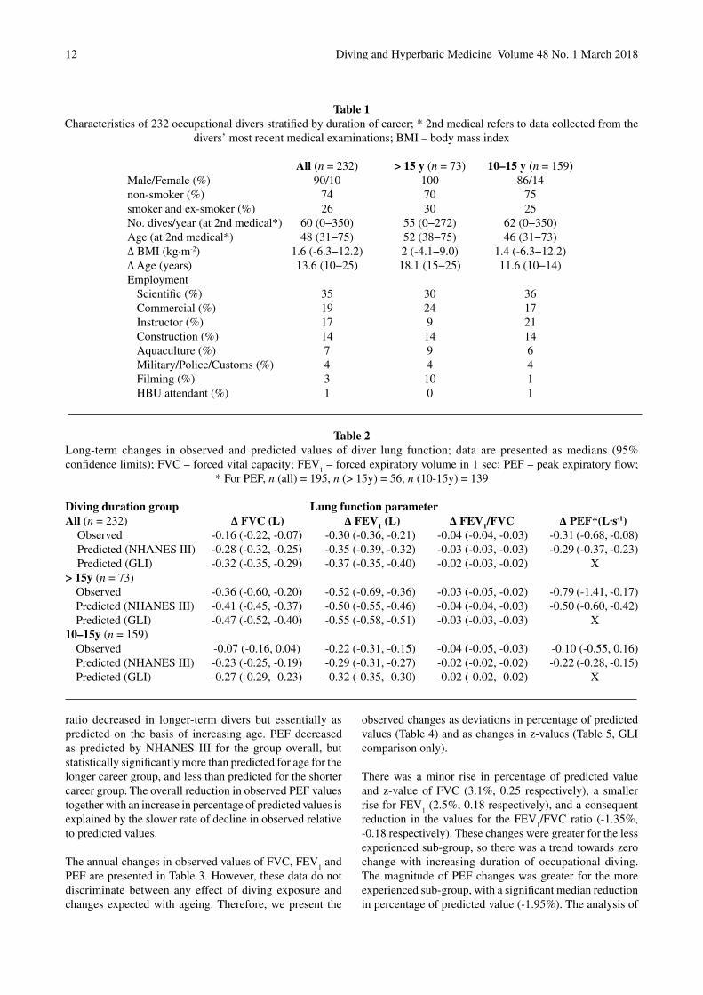
ratio decreased in longer-term divers but essentially as predicted on the basis of increasing age. PEF decreased as predicted by NHANES III for the group overall, but statistically significantly more than predicted for age for the longer career group, and less than predicted for the shorter career group. The overall reduction in observed PEF values together with an increase in percentage of predicted values is explained by the slower rate of decline in observed relative to predicted values.
The annual changes in observed values of FVC, FEV1 and
PEF are presented in Table 3. However, these data do not discriminate between any effect of diving exposure and changes expected with ageing. Therefore, we present the
observed changes as deviations in percentage of predicted values (Table 4) and as changes in z-values (Table 5, GLI comparison only).
There was a minor rise in percentage of predicted value and z-value of FVC (3.1%, 0.25 respectively), a smaller rise for FEV
1 (2.5%, 0.18 respectively), and a consequent
reduction in the values for the FEV1/FVC ratio (-1.35%,
-0.18 respectively). These changes were greater for the less experienced sub-group, so there was a trend towards zero change with increasing duration of occupational diving. The magnitude of PEF changes was greater for the more experienced sub-group, with a significant median reduction in percentage of predicted value (-1.95%). The analysis of
Table 1Characteristics of 232 occupational divers stratified by duration of career; * 2nd medical refers to data collected from the
divers’ most recent medical examinations; BMI – body mass index
Table 2Long-term changes in observed and predicted values of diver lung function; data are presented as medians (95% confidence limits); FVC – forced vital capacity; FEV
1 – forced expiratory volume in 1 sec; PEF – peak expiratory flow;
* For PEF, n (all) = 195, n (> 15y) = 56, n (10-15y) = 139
All (n = 232) > 15 y (n = 73) 10–15 y (n = 159)Male/Female (%) 90/10 100 86/14non-smoker (%) 74 70 75smoker and ex-smoker (%) 26 30 25No. dives/year (at 2nd medical*) 60 (0−350) 55 (0−272) 62 (0−350)Age (at 2nd medical*) 48 (31−75) 52 (38−75) 46 (31−73)∆ BMI (kg∙m-2) 1.6 (-6.3−12.2) 2 (-4.1−9.0) 1.4 (-6.3−12.2)∆ Age (years) 13.6 (10−25) 18.1 (15−25) 11.6 (10−14)Employment
Scientific (%) 35 30 36Commercial (%) 19 24 17Instructor (%) 17 9 21Construction (%) 14 14 14Aquaculture (%) 7 9 6Military/Police/Customs (%) 4 4 4Filming (%) 3 10 1HBU attendant (%) 1 0 1
Diving duration group Lung function parameterAll (n = 232) ∆ FVC (L) ∆ FEV1 (L) ∆ FEV1/FVC ∆ PEF*(L∙s-1)
Observed -0.16 (-0.22, -0.07) -0.30 (-0.36, -0.21) -0.04 (-0.04, -0.03) -0.31 (-0.68, -0.08)Predicted (NHANES III) -0.28 (-0.32, -0.25) -0.35 (-0.39, -0.32) -0.03 (-0.03, -0.03) -0.29 (-0.37, -0.23)Predicted (GLI) -0.32 (-0.35, -0.29) -0.37 (-0.35, -0.40) -0.02 (-0.03, -0.02) X
> 15y (n = 73)Observed -0.36 (-0.60, -0.20) -0.52 (-0.69, -0.36) -0.03 (-0.05, -0.02) -0.79 (-1.41, -0.17)Predicted (NHANES III) -0.41 (-0.45, -0.37) -0.50 (-0.55, -0.46) -0.04 (-0.04, -0.03) -0.50 (-0.60, -0.42)Predicted (GLI) -0.47 (-0.52, -0.40) -0.55 (-0.58, -0.51) -0.03 (-0.03, -0.03) X
10–15y (n = 159)Observed -0.07 (-0.16, 0.04) -0.22 (-0.31, -0.15) -0.04 (-0.05, -0.03) -0.10 (-0.55, 0.16)Predicted (NHANES III) -0.23 (-0.25, -0.19) -0.29 (-0.31, -0.27) -0.02 (-0.02, -0.02) -0.22 (-0.28, -0.15)Predicted (GLI) -0.27 (-0.29, -0.23) -0.32 (-0.35, -0.30) -0.02 (-0.02, -0.02) X
Diving and Hyperbaric Medicine Volume 48 No. 1 March 2018 13
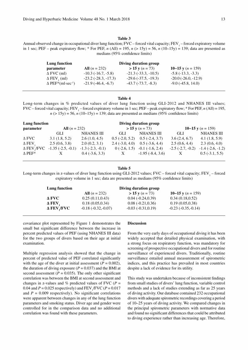
Table 3Annual observed change in occupational diver lung function; FVC – forced vital capacity; FEV
1 – forced expiratory volume
in 1 sec; PEF – peak expiratory flow; * For PEF, n (All) = 195, n (> 15y) = 56, n (10–15y) = 139; data are presented as medians (95% confidence limits)
Table 4Long-term changes in % predicted values of diver lung function using GLI-2012 and NHANES III values;FVC – forced vital capacity; FEV
1 – forced expiratory volume in 1 sec; PEF – peak expiratory flow; * For PEF, n (All) = 195,
n (> 15y) = 56, n (10–15y) = 139; data are presented as medians (95% confidence limits)
Table 5Long-term changes in z-values of diver lung function using GLI-2012 values; FVC – forced vital capacity; FEV
1 – forced
expiratory volume in 1 sec; data are presented as medians (95% confidence limits)
Lung function Diving duration groupparameter All (n = 232) > 15 y (n = 73) 10−15 y (n = 159)∆ FVC (ml) -10.3 (-16.7, -5.8) -21.3 (-33.3, -10.5) -5.8 (-13.3, -3.3)∆ FEV
1 (ml) -23.2 (-28.3, -17.3) -29.6 (-37.5, -19.3) -20.0 (-26.0, -12.9)
∆ PEF*(ml∙sec-1) -21.9 (-46.4, -6.7) -43.7 (-73.7, -8.3) -9.0 (-45.8, 14.0)
Lung function Diving duration groupparameter All (n = 232) > 15 y (n = 73) 10−15 y (n = 159) GLI NHANES III GLI NHANES III GLI NHANES III∆ FVC 3.1 (1.8, 5.2) 2.6 (1.0, 4.5) 0.5 (-2.0, 5.2) 0.5 (-2.4, 3.7) 3.6 (2.4, 6.7) 4.1 (1.8, 5.9)∆ FEV
1 2.5 (0.6, 3.8) 2.0 (0.2, 3.1) 2.4 (-3.0, 4.0) 0.5 (-3.6, 4.4) 2.5 (0.6, 4.4) 2.3 (0.6, 4.0)
∆ FEV1/FVC -1.35 (-2.5, -0.1) -1.3 (-2.3, -0.1) 0 (-2.6, 1.5) -0.1 (-1.6, 2.4) -2.5 (-2.7, -0.2) -1.4 (-2.6, -1.2)
∆ PEF* X 0.4 (-3.6, 3.3) X -1.95 (-8.4, 3.6) X 0.5 (-3.1, 5.5)
Lung function Diving duration group All (n = 232) > 15 y (n = 73) 10−15 y (n = 159)∆ FVC 0.25 (0.11,0.43) 0.04 (-0.24,0.39) 0.34 (0.18,0.52)∆ FEV1 0.18 (0.05,0.34) 0.08 (-0.21,0.36) 0.19 (0.05,0.38)∆ FEV1/FVC -0.18 (-0.32,-0.07) -0.03 (-0.31,0.19) -0.23 (-0.35,-0.14)
covariance plot represented by Figure 1 demonstrates the small but significant difference between the increase in percent predicted values of PEF (using NHANES III data) for the two groups of divers based on their age at initial examination.
Multiple regression analysis showed that the change in percent of predicted value of PEF correlated significantly with the age of the diver at initial assessment (P = 0.002), the duration of diving exposure (P = 0.037) and the BMI at second assessment (P = 0.035). The only other significant correlation was between the BMI at second assessment and changes in z-values and % predicted values of FVC (P = 0.04 and P = 0.025 respectively) and FEV
1/FVC (P = 0.017
and P = 0.009 respectively). No significant correlations were apparent between changes in any of the lung function parameters and smoking status. Diver age and gender were controlled for in the comparison data and no additional correlation was found with these parameters.
Discussion
From the very early days of occupational diving it has been widely accepted that detailed physical examination, with a strong focus on respiratory function, was mandatory for screening of prospective occupational divers and for routine surveillance of experienced divers. Traditionally, routine surveillance entailed annual measurement of spirometric indices, and this practice has prevailed in most countries despite a lack of evidence for its utility.
This study was undertaken because of inconsistent findings from small studies of divers’ lung function, variable control methods and a lack of studies extending as far as 25 years of diving activity. Our database contained 232 occupational divers with adequate spirometric recordings covering a period of 10–25 years of diving activity. We compared changes in the principal spirometric parameters with normative data and found no significant differences that could be attributed to diving experience rather than increasing age. Therefore,

Diving and Hyperbaric Medicine Volume 48 No. 1 March 201814
the main finding of this study relevant to working divers is that while small changes in some spirometric parameters may reach statistical significance, there is no evidence of change attributable to diving that is likely to be of clinical significance in the long-term. Prospective occupational divers, and those who remain in the industry for many years, should be encouraged by this further evidence of the relative lack of harm to the respiratory system from diving.
These results were confluent with our previous study of a cohort of 336 divers over a mean period of 5.6 years, except that in that study we found small but statistically significant reductions in the percent of predicted values for FEV
1 and
PEF using NHANES III normative data.22 The current study found no significant change for FEV
1 and a small
but statistically significant rise in percent of predicted PEF. Our results also support those of a prospective study of 37 Norwegian professional divers who showed no correlation between diving exposure (total number of dives) and FVC and FEV
1 over 12 years.17
These findings also cast doubt on the utility of routine annual spirometric measurement in all occupational divers. There seems little point in conducting serial investigation for
changing spirometry over a diving career in the absence of convincing evidence that such change is a feature of long-term diving. However, we recognise that spirometry would be indicated if some other aspect of a diver’s medical history, likely to be detected on their annual health questionnaire, implied that significant change was plausible (such as a significant respiratory illness in the preceding year).
LIMITATIONS
First, and most significantly, we used years of occupational diving as a surrogate for diving exposure. Clearly, this is a blunt measure of exposure, but we had no access to more precise data. The ideal would have been to record number of dives with times, depths and gases used, but we lacked such records over the long period of observation involved. Even the number of dives per year was not consistently recorded, and most likely inaccurate. Such detail may only be available in the setting of a prospective study. With this limitation acknowledged, it nevertheless seems implausible that divers would maintain medical fitness certification for occupational diving over a prolonged period in the absence of moderate diving activity.
Figure 1Relationship between age at the earliest spirometry test, diving exposure and long-term changes in % predicted values of
divers’ peak expiratory flow (PEF) based on NHANES III prediction equations
Diving and Hyperbaric Medicine Volume 48 No. 1 March 2018 15
Secondly, we cannot exclude some degree of selection bias, where divers may have quit with less than 10 years’ experience due to deteriorating lung function. We know from a previous study that there is an attrition rate of nearly 80% over a five-year period for New Zealand occupational divers, suggesting the possibility of a significant ‘healthy worker effect’. However, we think this is unlikely to have influenced our findings in relation to spirometric changes. The collective qualitative experience of the medical authors among our group is that occupational diver attrition due to deterioration in lung health in the absence of a discrete accident (such as pulmonary barotrauma) or a non-diving medical explanation is virtually unheard of. For example, in the current study, no diver was found to have clinically significant lung function deterioration. From our previous study of those remaining in the job for a mean of 5.6 years, only two out of 336 divers were found to have abnormal spirometry, but after further investigation neither was considered unfit for diving.23
Thirdly, we restricted this study to what we considered to be the principal spirometric parameters, namely FVC, FEV
1,
FEV1/FVC ratio and PEF, to avoid erosion of the sample
size, since the other parameters were far less consistently recorded, especially in the older clinical records. We do not believe this detracts from our findings, but it does make this study a less than complete survey of lung function.
Fourthly, as with most retrospective studies, the quality of spirometric data was beyond our control, and likely to have varied widely.
Finally, we chose to compare our data with the NHANES III and GLI normative data because these sets of prediction equations are widely accepted internationally, despite the fact that neither set is based on data drawn from the New Zealand population. An argument against using such ‘normal’ population data is that, with a cohort comprising only divers, we are not dealing with a ‘normal’ population, so they would more appropriately be compared with a control group of similar fitness engaged in equally strenuous activity. Previous studies have used such occupations as submariners,15 policemen24 and non-diving offshore workers25 for comparison. However, any error introduced because of our selection of comparative data is not likely to be significant, and we have previously demonstrated close alignment of the NHANES III data with data from NZ divers.18
Conclusions
The small changes in lung function found in divers with a 10–25 year occupational diving history are generally confluent with predictions based on ageing, and not likely to be clinically significant. There appears to be no justification for routine spirometry in asymptomatic divers.
Appendix
Brief background to the derivation of spirometric reference values used in this study.
THIRD NATIONAL HEALTH AND NUTRITION EXAMINATION SURVEY (NHANES III)
The NHANES III prediction equations are based on data collected from a random sample of the population across the USA between 1988 and 1994. The initial total of 20,627 subjects from three ethnic groups (Caucasian, Afro-American and Mexican-American) was reduced to 7,429 after exclusions to comply with the criteria that all subjects were asymptomatic, life-long non-smokers and could provide at least two acceptable spirometric manoeuvres. Subjects were between 8 and 80 years old. The analysis and resulting prediction equations were published in 1999.22 The equations are race/ethnic group and gender specific, with age and height as independent variables. The equations are polynomials of the form:
Lung function parameter =b
0 + b
1xAge + b
2xAge2 + b
3xHeight2
where b0 is the intercept and b
1, b
2 and b
3 are coefficients
that vary for each lung function parameter with race/ethnic group and gender. There are also different sets of coefficient values for males under 20 and females under 18 years old. This set of equations gained considerable global popularity, and has recently been the reference dataset most commonly used throughout New Zealand.26
GLOBAL LUNG FUNCTION INITIATIVE
A new set of lung function reference value prediction equations was developed and published in 2012 by the Global Lung Function Initiative (GLI-2012).21 This large collaborative study resolved many of the inherent problems with the existing collection of published prediction equations for spirometric reference values. Specifically, problems with existing datasets included small population/ethnic group sample numbers, out-moded methodologies and discontinuity between age groups. Through collaboration with researchers from 70 centres in 26 countries across five continents, and including data from earlier significant studies, such as NHANES III, continuous equations suitable for ages from three to 95 years based on data from 74,187 healthy non-smokers were derived.21 The equation is in the form of a linear regression expression using age and height as independent variables, with coefficients dependent on lung function parameter, gender and ethnic group. The four ethnic groupings specified are Caucasian (providing most of the data), Afro-American and North and South East Asian. Another set of equations, based on an average of the others, can be used for ‘other’ ethnic groupings. The authors consider the development of this dataset complete for the Caucasian group, but ongoing for possible future modification when further data has been collected from
Diving and Hyperbaric Medicine Volume 48 No. 1 March 201816
the other ethnic groups. Calculations for individuals or large groups have been facilitated by the GLI-2012 group’s provision of the required software on their website.21 This dataset has been endorsed by the major respiratory and thoracic societies from Europe, America, Asia, Australia and New Zealand.
References
1 Earing CMN, McKeon DJ, Kubis H. Divers revisited: The ventilator response to carbon dioxide in experienced scuba divers. Respir Med. 2014;108:758−65. doi:10.1016/j.rmed.2014.02.010.
2 Thorsen E, Segedal K, Kambestad B, Gulsvic A. Divers’ lung function: small airways disease? Br J Ind Med. 1990;47(8):519−23.
3 Thom SR, Milovanova TN, Bogush M, Bhopale VM, Yang M, Bushmann K, et al. Microparticle production, neutrophil activation, and intravascular bubbles bubbles following open-water SCUBA diving. J Appl Physiol. 2012;112:1268−78. doi:10.1152/japplphysiol.01305.2011.
4 Cotes JE, Davey IS, Reed JW, Rooks M. Respiratory effects of a single saturation dive to 300m. Br J Ind Med. 1987;44:76−82.
5 Dujic Z, Eterovic D, Denoble P, Krstacic G, Tocilj J, Gosovic S. Effect of a single air dive on pulmonary diffusing capacity in professional divers. J Appl Physiol. 1993;74:55−61.
6 Thorsen E, Segedal K, Kambestad BK. Mechanisms of reduced pulmonary function after a saturation dive. Eur Respir J. 1994;7:4−10.
7 Thorsen E, Risberg J, Segedal K, Hope S. Effects of venous gas microemboli on pulmonary gas transfer function. Undersea Hyperb Med. 1995;22:347−53.
8 Hope A, Lund T, Elliott DH, Halsey MJ, Wiig H. Long-term health effects of diving. An international consensus conference. Godøysund 6–10 June 1993. Bergen: John Greig forlag A/S;1994:387−91. Cited in: Brubakk AO, Neuman TS, editors, Bennett and Elliott’s physiology and medicine of diving, 5th ed. Edinburgh: Saunders; 2003. p. 656.
9 Pougnet R, Pougnet L, Lucas D, Uguen M, Henckes A, Dewitte J, et al. Longitudinal change in professional divers’ lung function: literature review. Int Marit Health. 2014;65:223−9. doi:10.5603/IMH.2014.0042.
10 Watt SJ. Effect of commercial diving on ventilatory function. Br J Ind Med. 1985;42:59−62.
11 Thorsen E, Segadal K, Kambestad BK, Gulsvik A. Pulmonary function one and four years after a deep saturation dive. Scand J Work Environ Health. 1993;19:115−20.
12 Skogstad M, Thorsen E, Haldorsen T. Lung function over the first 3 years of a professional diving career. Occup Environ Med. 2000;57:390−5.
13 Fitzpatrick DT, Conkin J. Improved pulmonary function in working divers breathing nitrox at shallow depths. Aviat Space Environ Med. 2003;74:763−7.
14 Lucas D, Loddé B, Choucroun P, Jegaden D, Mialon P, Sarni D, et al. Five year study of changes in the lung function of a cohort of 31 professional divers. Med Marit. 2005;5:17−28.
15 Tetzlaff K, Theysohn J, Stahl C, Schlegel S, Koch A, Muth CM. Decline of FEV1 in scuba divers. Chest. 2006;130:238−43.
16 Chong SJ, Tan TW, Lim JY. Changes in lung function in Republic of Singapore Navy divers. Diving Hyperb Med. 2008;38:68−70.
17 Skogstad M, Skare O. Pulmonary function among professional divers over 12 years and the effect of total number of dives. Aviat Space Environ Med. 2008;79:883−7. doi:10.3357/ASEM.2333.2008.
18 Sames C, Gorman DF, Mitchell SJ, Gamble G. The long-term effects of compressed gas diving on lung function in New Zealand occupational divers: a retrospective analysis. Diving Hyperb Med. 2009;39:133−7.
19 Tetzlaff K, Thomas PS. Short- and long-term effects of diving on pulmonary function. Eur Respir Rev. 2017;26:160097. doi: 10.1183/16000617.0097-2016.
20 Voortman M, van Ooij PJAM, van Hulst RA, Zanen P. Pulmonary function in professional navy divers and changes during their professional careers. Undersea Hyperb Med. 2016;43:649–57.
21 Quanjer PH, Stanojevic S, Cole TJ, Baur X, Hall GL, Culver BH, et al, and the ERS Global Lung Function Initiative. Multi-ethnic reference values for spirometry for the 3-95-yr age range: the global lung function 2012 equations. Eur Respir J. 2012;40:1324-43. doi: 10.1183/09031936.00080312. [cited 2017 December 26]. Available from: http://www.ers-education.org/guidelines/global-lung-function-initiative/tools.aspx.
22 Hankinson JL, Odencrantz JR, Fedan KB. Spirometric reference values from a sample of the general US population. Am J Respir Crit Care Med. 1999;159:179–87.
23 Sames C, Gorman D, Mitchell SJ, Gamble G. Utility of regular medical examinations of occupational divers. Intern Med J. 2009;39:763–70.
24 Skogstad M, Thorsen E, Haldorsen T, Kjuus H. Lung function over six years among professional divers. Occup Environ Med. 2002;59:629–33.
25 Macdiarmid JI, Ross JAS, Taylor CL, Watt SJ, Adie W, Osman LM, et al. Co-ordinated investigation into the possible long-term health effects of diving at work. Examination of the long-term health impact of diving: the ELTHI Study. University of Aberdeen, Scotland; Research Report 230, HSE Books; 2004.
26 Marsh S, Aldington S, Williams M, Weatherall M, Robiony-Rogers D, et al. Pulmonary function testing in New Zealand: the use and importance of reference ranges. Respirology. 2007;12:367–74.
Acknowledgements
We thank Mr Murray Polson (CEO, Erudite Software Ltd) for developing and maintaining the electronic database for New Zealand occupational divers, and retrieving much of the data required for this study.
Funding sources: nil
Conflicts of interest: nil
Submitted: 19 October 2017; revised 26 December 2017Accepted: 11 January 2018
Copyright: This article is the copyright of the authors who grant Diving and Hyperbaric Medicine a non-exclusive licence to publish the article in electronic and other forms.
Diving and Hyperbaric Medicine Volume 48 No. 1 March 2018 17
Modern assessment of pulmonary function in divers cannot rely on old reference valuesThijs T Wingelaar1,2, Paul Clarijs1, Pieter-Jan AM van Ooij1, Dave AA Koch1, Rob A van Hulst2
1 Royal Netherlands Navy Diving Medical Centre, Den Helder, The Netherlands 2 Department of Anesthesiology, Academic Medical Centre, University of Amsterdam, The NetherlandsCorresponding author: Thijs T Wingelaar, Royal Netherlands Navy Diving Medical Centre, Rijkszee en marinehaven, 1780 CA Den Helder, The Netherlands [email protected]
Key wordsFitness to dive; Flowchart; Global lung initiative; Lung function; Standards; Military diving
Abstract(Wingelaar TT, Clarijs P, van Ooij PJAM, Koch DAA, van Hulst RA. Modern assessment of pulmonary function in divers cannot rely on old reference values. Diving and Hyperbaric Medicine. 2018 March;48(1):17–22. doi:10.28920/dhm48.1.17-22. PMID: 29557097.)Introduction: Pulmonary function testing (PFT) is an important part of dive medical examinations. Depending on the standard used to assess fitness to dive, different reference sets and fixed cut-off points are used. Reference values are part of an ongoing debate regarding the validity and accuracy related to different age groups, sex and ethnic backgrounds. The Global Lung Initiative (GLI) has provided an all-age reference set which corrects for sex and ethnicity (GLI-2012); this has had substantial impact on pulmonary medicine.Method: We present an algorithm that can be used to standardise analysis of PFT in divers using the GLI-2012 reference set. Differences in the analysis of PFT between the ECSC/ERS-1993 and the GLI-2012 reference values are illustrated by means of three case reports.Conclusion: Using a valid database of reference values increases accuracy and might prevent additional medical investigations and/or incorrect assessment of fitness to dive. Although our algorithm needs further evaluation to ensure its validity, the preliminary results are promising. Whatever algorithm is used, we urge dive medical physicians to consider using valid reference sets when analysing PFT for assessment of fitness to dive.
Introduction
Diving requires substantial adaptation in human physiology. Without these necessary changes, immersion and/or breathing hyperbaric mixtures may lead to significant and potentially life-threatening injuries. Especially existing pathology in the areas of ENT and cardiopulmonary fitness may cause severe problems when an individual is exposed to hyperbaric conditions; the latter being the second most common cause of lethal diving accidents, after faulty procedures and panic.1,2
Although occupational safety laws and medical assessment of fitness to dive are nowadays common, this was not always the case. For example, after numerous injuries and deaths when building the pillars of the Brooklyn Bridge, the first legislation to protect employees from occupational health damage was established in 1909.3 Today, many (inter-)national recommendations exist regarding safe diving procedures and fitness to dive.4–8
Assessment of occupational divers, such as commercial or military divers, should include pulmonary function testing (PFT). The results of PFT are compared to a reference set, such as those of the European Community for Steel and Coal (ECSC), the European Respiratory Society
(ERS), the National Health and Nutrition Examination Survey (NHANES) or the American Thoracic Society (ATS).9,10 Although reference tables can focus on sports, commercial or military diving, many standards have similar recommendations regarding cardiopulmonary fitness. Small differences exist between the main fitness-to-dive standards, most, but not all of which specify a fixed cut-off point for parameters such as forced vital capacity (FVC), forced expiratory volume in one second (FEV
1) and the
FEV1/FVC ratio (Table 1).
There are other standards available than those presented in Table 1 and there are even more reference sets.11 Despite revisions over time, the various reference tables have remained a topic of discussion in pulmonary medicine. For example, many lacked accuracy for ethnic groups other than Caucasians, African and Mexican Americans, and some were unable to properly correct for age and sex.12 In 2008 the Global Lung Initiative (GLI) was established by the ERS and ATS to develop all-age reference equations with correction for sex and ethnic background. In 2012 the first results were published and were rapidly endorsed worldwide in pulmonary medicine and other fields of practice.13,14
Compared to any of the previous reference sets, one of the most important changes is the definition of a ‘normal’

Diving and Hyperbaric Medicine Volume 48 No. 1 March 201818
value. Previously, an expected value was reported as a fixed number, whereas in the GLI-2012 it is described as the Z-score from the mean. This means there is a normal distribution of expected values instead of a single expected value. This approach allows easy comparison with the reference group and more accurate assessment of pulmonary function.15 For example, a Z-score of ± 1.64 indicates a value is ± 1.64 standard deviations (SD) from the mean and, therefore, outside the 90% confidence interval (CI), excluding 5% at the extremes of the normal distribution, whilst a Z-score of ± 1.96 places a value outside the 95% CI, excludes 2.5% at each end of a normal distribution. These values are translated into the upper and lower limits of normal (ULN and LLN, respectively). A ULN-95 indicates that a value is above the Z-score of 1.64 and a LLN-2.5 puts a value below the Z-score of -1.96.
The cut-off point of any test influences the predictive value. If a test is too stringent, many individuals will be flagged positive while there are no significant problems (false positive). On the other end of the spectrum, a cut-off value that is too low may fail to identify persons with pathology (false negative). Depending on the setting in which a medical test is utilized, the cut-off point must be chosen wisely. Preferably, a diagnostic test has a high negative predictive value to ensure that no cases are incorrectly regarded as pathologic. However, a test used for screening purposes has, preferably, a high positive predictive value to ensure that no cases are missed. Although the lower limit for fitness to dive has yet to be determined, it stands to reason that a pulmonary function within the 90% CI in a healthy individual without clinical signs of pulmonary disease can be regarded as normal. This is similar to pulmonary screening in the Netherlands, both in general practice and in hospital analysis by a pulmonary specialist.
In this paper we present three people who were assessed for fitness to dive at the Royal Netherlands Navy (RNLN) Diving Medical Centre. We compare the ECSC/ERS-1993 reference set with the GLI-2012 reference set. Although medical ethical approval was not required, our methods for handling data and privacy are in line with the Declaration of
Helsinki and national laws. Additionally, changes in policy regarding fitness to dive are in agreement with the Surgeon General of the Ministry of Defence. To provide the proper context for the cases, we first describe the algorithm used to evaluate these cases.
Royal Netherlands Navy algorithm to interpret PFT in divers
The RNLN Diving Medical Centre has developed an algorithm in cooperation with pulmonary specialists from the Military Hospital (Figure 1). With regard to PFT, we feel that an individual should be considered fit to dive when the main spirometric parameters (FVC, FEV
1, FEV
1/FVC)
are within the 90% CI (Z-score of ± 1.64) and there are no signs of pathology in the history or physical examination. If a person’s PFT is outside the 95% CI (Z-score ± 1.96), we refer the person to a pulmonary specialist and review their fitness to dive case-by-case afterwards. Note that we refrain from analysing FEF
25-75, since these values have no impact
on clinical decision-making.16
When FVC, FEV1 or FEV
1/FVC are outside the 90% CI,
but within the 95% CI (i.e., a Z-score between ± 1.64 and ± 1.96), further assessment is required, even if there are no signs and symptoms of pathology. For instance, a lower than normal FVC could be the result of anatomical anomalies, such as bullae or blebs. Ventilatory dead-space can be assessed using body plethysmography or high-resolution computed tomography (HR-CT). A decreased FEV
1 or FEV
1/
FVC might be the result of bronchoconstriction, which is a risk factor for intrapulmonary air-trapping and can lead to pneumothorax or arterial gas embolism.
The FEV1 or FEV
1/FVC could be decreased for several
weeks after a pulmonary stressor, albeit a common viral infection or exposure to non-specific agents (such as dust, which is a relevant factor in deployed military forces). In case of a low FEV
1 or FEV
1/FVC (LLN 2.5), that person is
(temporarily) unfit to dive and PFT is repeated after at least six weeks. Additional testing (next paragraph) is delayed until the PFT is normalised. When the PFT has normalised, we regard the decrease as temporary and non-significant.
Bronchospasm should be investigated using bronchial challenge or exercise tolerance testing.17 Note that the Dutch guidelines regarding fitness to dive in occupational divers recommend routine screening for bronchospasm. A methacholine challenge test is performed at the initial dive medical assessment, subsequent assessments do not require further testing for bronchial hyper-reactivity unless indicated. This can be different in other nations or when screening recreational divers. It stands to reason that any history of bronchospasm should prompt further investigation in all divers. The goal of bronchial challenge testing is to evaluate hyperreactivity. A subject is exposed to an increasing dose of an irritable substance, such as histamine or methacholine, and performs several flow-volume curves
Table 1Overview of pulmonary function test (PFT) criteria from different standards; FVC – forced vital capacity;FEV
1 – Forced expiratory volume in 1 sec; FEF
25-75 – Forced
expiratory flow at 25–75% of the pulmonary volume;PEF – Peak expiratory flow
Standard Recommended PFT Suggested lower limitEDTC4 FVC, FEV
1, FEV
1/FVC Not specified
ADC5 FVC, FEV1, FEF
25 –75 > 75%
BTS6/MA17 FVC, FEV1, PEF > 80%
FEV1/FVC > 70%
ADivP-18 Not specified, other than Not specified PFT should be performed
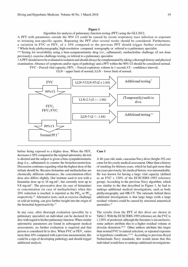
Diving and Hyperbaric Medicine Volume 48 No. 1 March 2018 19
before being exposed to a higher dose. When the FEV1
decreases ≥ 20% compared to the original spirometry, the test is aborted and the subject is given a beta-sympathomimetic drug (i.e., salbutamol) to counter the bronchoconstriction. Discussion continues regarding what the highest dose of the irritant should be. Because histamine and methacholine are chemically different substances, the concentration-effect dose also differs slightly. Our institute used to test with a histamine dose up to 16 mg∙ml-1, but currently tests up to 9.8 mg∙ml-1. The provocative dose (in case of histamine) or concentration (in case of methacholine) when this 20% reduction is reached, is reported as the PD
20 or PC
20
respectively.18 Alternative tests, such as exercise challenge or cold air testing, can give further insight into the origin of the bronchial hyperreactivity.17
In any case, after thorough evaluation (possibly by a pulmonary specialist) an individual can be declared fit to dive with regard to his/her pulmonary function. When similar spirometric parameters are found in subsequent medical assessments, no further evaluation is required and that person is considered fit to dive. When FVC or FEV
1 varies
more than 10% compared with a previous measurement, this could be a sign of developing pathology and should trigger additional analysis.
Case 1
A 46-year-old, male, caucasian Navy diver (height 192 cm) came for his yearly medical assessment. Other than a history of smoking for thirteen years, which he had quit more than ten years previously, his medical history was unremarkable. He was known for having a large vital capacity (defined as an FVC ≥ 120% of the ECSC/ERS-1993 reference group). According to the previous Navy algorithm, which was similar to the that described in Figure 1, he had to undergo additional medical investigations, such as body plethysmography and HR-CT. The rationale behind these additional investigations is that large lungs (with a large residual volume) could be caused by structural anatomical anomalies.19
The results from the PFT of this diver are shown inTable 2. With the ECSC/ERS-1993 reference set, the FVC is≥ 120% of predicted; although the literature is inconclusive, some authors attribute this to a higher residual volume or alveolar distention.20,21 Other authors attribute this larger than normal FVC to natural selection, or repeated exposure to hyperbaric conditions.22−24 According to previous Royal Netherlands Navy standards, this would mean that this individual would have to undergo additional investigations.
Figure 1Algorithm for analysis of pulmonary function testing (PFT) using the GLI-2012.
A PFT with parameters outside the 90% CI could be caused by recent respiratory tract infection or exposure to irritating non-specific agents. Repeating the PFT after several weeks should be considered. Furthermore, a variation in FVC or FEV
1 of ≥ 10% compared to the previous PFT should trigger further evaluation.
* Whole-body plethysmography, high-resolution computed tomography or referral to a pulmonary specialist.** Testing for reversibility using a beta-sympatomimetic drug (i.e., salbutamol), methacholine challenge (if not done previously), exercise challenge testing, or referral to a pulmonary specialist.† A PFT should never be evaluated in isolation and should always be complemented by taking a thorough history and physical examination. Absence of symptoms and/or signs of pathology and a PFT within the 90% CI should be considered normal.
FVC – Forced vital capacity; FEV1 – Forced expiratory volume in 1 second; CI – confidence interval;
ULN – upper limit of normal; LLN – lower limit of normal.
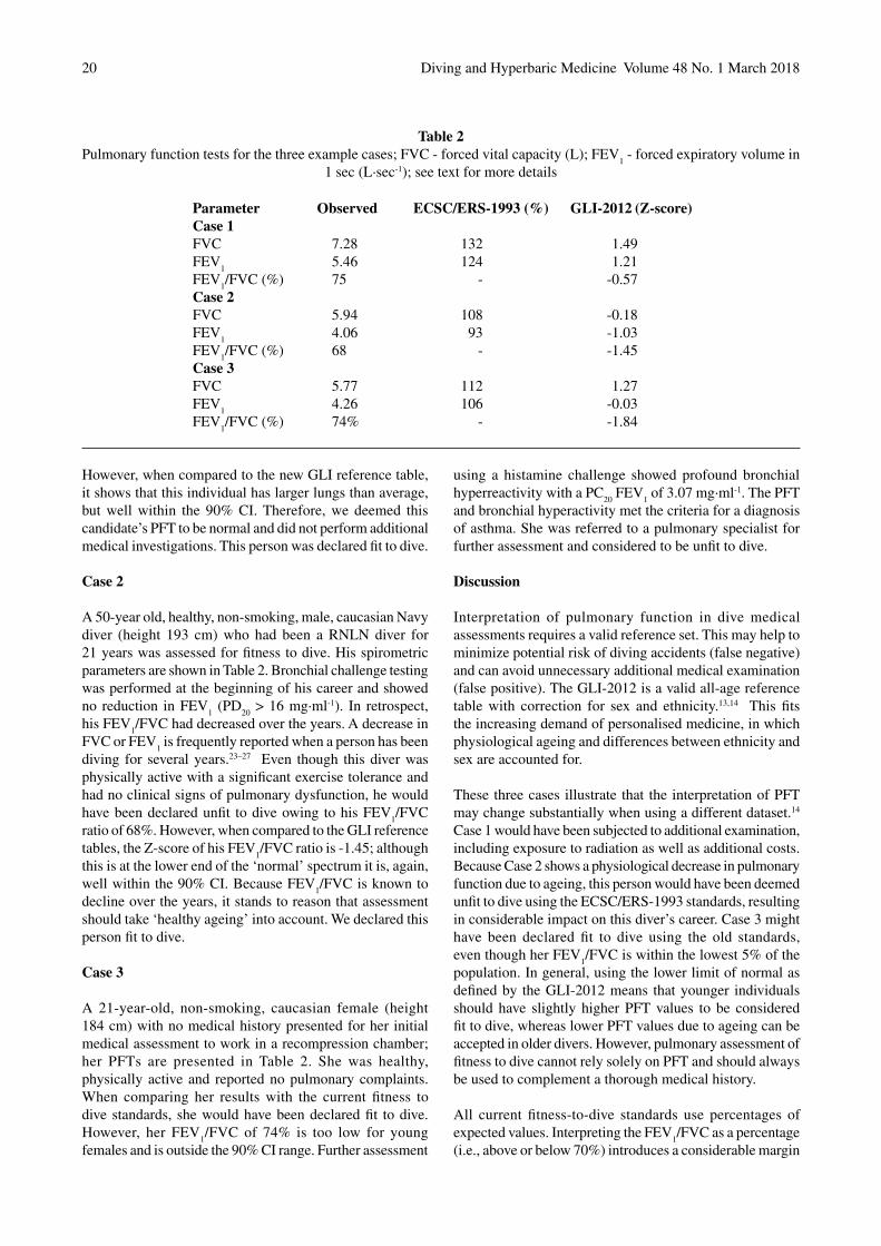
Diving and Hyperbaric Medicine Volume 48 No. 1 March 201820
However, when compared to the new GLI reference table, it shows that this individual has larger lungs than average, but well within the 90% CI. Therefore, we deemed this candidate’s PFT to be normal and did not perform additional medical investigations. This person was declared fit to dive.
Case 2
A 50-year old, healthy, non-smoking, male, caucasian Navy diver (height 193 cm) who had been a RNLN diver for 21 years was assessed for fitness to dive. His spirometric parameters are shown in Table 2. Bronchial challenge testing was performed at the beginning of his career and showed no reduction in FEV
1 (PD
20 > 16 mg∙ml-1). In retrospect,
his FEV1/FVC had decreased over the years. A decrease in
FVC or FEV1 is frequently reported when a person has been
diving for several years.23–27 Even though this diver was physically active with a significant exercise tolerance and had no clinical signs of pulmonary dysfunction, he would have been declared unfit to dive owing to his FEV
1/FVC
ratio of 68%. However, when compared to the GLI reference tables, the Z-score of his FEV
1/FVC ratio is -1.45; although
this is at the lower end of the ‘normal’ spectrum it is, again, well within the 90% CI. Because FEV
1/FVC is known to
decline over the years, it stands to reason that assessment should take ‘healthy ageing’ into account. We declared this person fit to dive.
Case 3
A 21-year-old, non-smoking, caucasian female (height184 cm) with no medical history presented for her initial medical assessment to work in a recompression chamber; her PFTs are presented in Table 2. She was healthy, physically active and reported no pulmonary complaints. When comparing her results with the current fitness to dive standards, she would have been declared fit to dive. However, her FEV
1/FVC of 74% is too low for young
females and is outside the 90% CI range. Further assessment
using a histamine challenge showed profound bronchial hyperreactivity with a PC
20 FEV
1 of 3.07 mg∙ml-1. The PFT
and bronchial hyperactivity met the criteria for a diagnosis of asthma. She was referred to a pulmonary specialist for further assessment and considered to be unfit to dive.
Discussion
Interpretation of pulmonary function in dive medical assessments requires a valid reference set. This may help to minimize potential risk of diving accidents (false negative) and can avoid unnecessary additional medical examination (false positive). The GLI-2012 is a valid all-age reference table with correction for sex and ethnicity.13,14 This fits the increasing demand of personalised medicine, in which physiological ageing and differences between ethnicity and sex are accounted for.
These three cases illustrate that the interpretation of PFT may change substantially when using a different dataset.14 Case 1 would have been subjected to additional examination, including exposure to radiation as well as additional costs. Because Case 2 shows a physiological decrease in pulmonary function due to ageing, this person would have been deemed unfit to dive using the ECSC/ERS-1993 standards, resulting in considerable impact on this diver’s career. Case 3 might have been declared fit to dive using the old standards, even though her FEV
1/FVC is within the lowest 5% of the
population. In general, using the lower limit of normal as defined by the GLI-2012 means that younger individuals should have slightly higher PFT values to be considered fit to dive, whereas lower PFT values due to ageing can be accepted in older divers. However, pulmonary assessment of fitness to dive cannot rely solely on PFT and should always be used to complement a thorough medical history.
All current fitness-to-dive standards use percentages of expected values. Interpreting the FEV
1/FVC as a percentage
(i.e., above or below 70%) introduces a considerable margin
Parameter Observed ECSC/ERS-1993 (%) GLI-2012 (Z-score)Case 1FVC 7.28 132 1.49FEV
1 5.46 124 1.21
FEV1/FVC (%) 75 - -0.57
Case 2FVC 5.94 108 -0.18FEV
1 4.06 93 -1.03
FEV1/FVC (%) 68 - -1.45
Case 3FVC 5.77 112 1.27FEV
1 4.26 106 -0.03
FEV1/FVC (%) 74% - -1.84
Table 2Pulmonary function tests for the three example cases; FVC - forced vital capacity (L); FEV
1 - forced expiratory volume in
1 sec (L∙sec-1); see text for more details

Diving and Hyperbaric Medicine Volume 48 No. 1 March 2018 21
of error.13 In young individuals, an FEV1/FVC of 75% could
be a sign of obstructive lung disease, whereas an FEV1/FVC
of 65% in elderly persons is physiological. Even though healthy ageing includes a decline of lung function, there is probably a lower limit at which diving is considered safe; however, this lower limit has not yet been determined.
Interpretation of a Z-score is slightly more abstract than a percentage relative to the ‘normal value’. It requires an extensive dataset, which might be difficult to implement in the software of older PFT devices. To help the clinician with assessment of lung function using the GLI-2012 dataset, several software solutions are freely available online (www.lungfunction.org). Currently the GLI-2012 reference set includes FVC, FEV
1, FEV
1/FVC and FEF
25–75; however,
additional values are expected to be added in the coming years. Moreover, reference values for TL
CO have recently
been published.28
Even though a detailed history, physical examination and PFT can generate important information regarding fitness to dive, it will not necessarily prevent pulmonary barotrauma. In a study of barotrauma in a large cohort of healthy subjects participating in submarine ascent training,29 there were 10 pulmonary barotrauma cases, one fatal, in 115,090 ascents. Either ascent training is safe or fitness-to-dive assessment has minimised the potential risk of injury. Conversely, abnormal PFTs have not been shown to be predictive of pulmonary barotrauma, although sufficient evidence is probably impossible to generate since many divers will be disqualified for diving when an abnormal PFT is found.
An important remaining question is: which PFT values can be accepted when determining a person’s fitness to dive. Our centre has used the algorithm described here to assess occupational divers since 2015. During this period we have assessed more than 900 divers, submariners and hyperbaric technicians. Our intention is to evaluate and publish the data on the safety and cost-effectiveness of these assessments. Although our algorithm may require some modification, the preliminary results are promising. We have deemed several individuals fit to dive using the GLI-2012 standards, whereas these persons would have been declared unfit using the ECSC/ERS-1993 standards. Conversely, we have referred a few individuals to a pulmonary specialist for analysis and have identified pathology, such as airtrapping found on HR-CT, which we would not have identified using the ECSC/ERS-1993 standards and current fitness-to-dive standards.
PFT is part of both the initial and subsequent annual medical assessment of occupational divers in the Netherlands by regulation. However, its contribution to dive safety has been questioned.30 Occasionally an annual PFT in physical fit and healthy divers brings additional information to light which could not have been gained by a thorough history. What frequency, and which examinations, should be performed to optimize dive safety remain to be determined.
Conclusion
Irrespective of the chosen algorithm and lower limit, every physician who is faced with evaluating pulmonary function should be using the most appropriate reference tables available for their population. In the field of pulmonary medicine, the introduction of the GLI-2012 standard has made a considerable positive impact on the assessment of pulmonary function testing. The GLI-2012 is an all-age reference table with correction for sex and ethnicity. We recommend that, in the development of fitness to dive standards, the concepts of upper and lower limits of normal should be adopted, and the use of fixed cut-off points for parameters such as FVC, FEV
1 and FVC/FEV
1 should be
avoided.
References
1 Lippmann J, Baddeley A, Vann R, Walker D. An analysis of the causes of compressed-gas diving fatalities in Australia from 1972–2005. Undersea Hyperb Med. 2013;40:49−61.
2 Vann R, Lang M. Recreational diving fatalities. Undersea Hyperb Med. 2011;38:257−60.
3 Neuman T, Thom SR. Physiology and medicine of hyperbaric oxygen therapy. Philadelphia, PA: Saunders; 2008.
4 Wendling JED, Nome T. Medical assessment of working divers. Fitness to dive standards of European Diving Technology Committee. 1st ed. Biele-Biene: Hyperbaric Editions; 2004. [cited 2017/12/21]. Available from: http://www.edtc.org/EDTC-Fitnesstodivestandard-2003.pdf.
5 International consensus standards for commercial diving and underwater operations. Houston, TX: Association of Diving Contractors International Inc. 6.2 ed; 2016. [cited 2017/12/21]. Available from: http://www.adc-int.org/files/C11634_ADC%20Consensus%20Standard.pdf.
6 British Thoracic Society guidelines on respiratory aspects of fitness for diving. Thorax. 2003;58:3−13.
7 The medical examination and assessment of commercial divers (revision 4). Health and Safety Executive; 2015. [cited 2017 December 21]. Available from: https://www.theunderwatercentre.com/wp-content/uploads/2013/02/HSE-Medical-Requirements.pdf.
8 ADivP-1(B): Allied guide to diving operations. NATO Standardization Agency; 2010. [cited 2017 December 21]. Available from: http://nso.nato.int/nso/.
9 Quanjer PH, Tammeling GJ, Cotes JE, Pedersen OF, Peslin R, Yernault JC. Lung volumes and forced ventilatory flows. Report Working Party Standardization of Lung Function Tests, European Community for Steel and Coal. Official Statement of the European Respiratory Society. Eur Respir J. 1993;16:5−40.
10 American Thoracic Society. Standardization of spirometry, 1994 update. Am J Respir Crit Care Med. 1995;152:1107−36.
11 Pellegrino R, Viegi G, Brusasco V, Crapo RO, Burgos F, Casaburi R, et al. Interpretative strategies for lung function tests. Eur Respir J. 2005;26:948−68. doi: 10.1183/09031936.05.00035205.
12 Rossiter CE, Weill H. Ethnic differences in lung function: evidence for proportional differences. Int J Epidemiol. 1974;3:55−61.
13 Quanjer PH, Stanojevic S, Cole TJ, Baur X, Hall GL, Culver BH, et al. Multi-ethnic reference values for spirometry for the
Diving and Hyperbaric Medicine Volume 48 No. 1 March 201822
3-95-yr age range: the global lung function 2012 equations. Eur Respir J. 2012;40:1324−43. doi: 10.1183/09031936.00080312.
14 Stanojevic S, Stocks J, Bountziouka V, Aurora P, Kirkby J, Bourke S, et al. The impact of switching to the new global lung function initiative equations on spirometry results in the UK CF registry. J Cyst Fibros. 2014;13:319−27. doi: 10.1016/j.jcf.2013.11.006.
15 Quanjer PH, Brazzale DJ, Boros PW, Pretto JJ. Implications of adopting the Global Lungs Initiative 2012 all-age reference equations for spirometry. Eur Respir J. 2013;42:1046−54. doi: 10.1183/09031936.00195512.
16 Quanjer PH, Weiner DJ, Pretto JJ, Brazzale DJ, Boros PW. Measurement of FEF25-75% and FEF75% does not contribute to clinical decision making. Eur Respir J. 2014;43:1051−8. doi: 10.1183/09031936.00128113.
17 Fuentes C, Contreras S, Padilla O, Castro-Rodriguez JA, Moya A, Caussade S. Exercise challenge test: is a 15% fall in FEV1 sufficient for diagnosis? J Asthma. 2011;48:729−35. doi: 10.3109/02770903.2011.
18 Coates AL, Wanger J, Donald DW, Culver BH, Carlsen KH, et al. ERS technical standard on bronchial challenge testing: general considerations and performance of methacholine challenge tests. Eur Respir J. 2017;49(5). pii:1601526. doi: 10.1183/13993003.01526-2016.
19 Voortman M, Ooij P, Hulst RAV, Zanen P. Pulmonary function changes in Navy divers during their professional careers. Eur Respir J. 2016;43:649−57.
20 Crosbie WA, Reed JW, Clarke MC. Functional characteristics of the large lungs found in commercial divers. J Appl Physiol Respir Environ Exerc Physiol. 1979;46:639−45.
21 Calder IM, Sweetnham K, Chan KK, Williams MM. Relation of alveolar size to forced vital capacity in professional divers. Br J Ind Med. 1987;44:467−9.
22 Crosbie WA, Clarke MB, Cox RA, McIver NK, Anderson IK, Evans HA, et al. Physical characteristics and ventilatory function of 404 commercial divers working in the North Sea. Br J Ind Med. 1977;34:19−25.
23 Skogstad M, Thorsen E, Haldorsen T, Kjuus H. Lung function over six years among professional divers. J Occup Environ Med. 2002;59:629−33.
24 Adir Y, Shupak A, Laor A, Weiler-Ravell D. Large lungs in divers: natural selection or a training effect? Chest. 2005;128:224−8.
25 Watt SJ. Effect of commercial diving on ventilatory function. Br J Ind Med. 1985;42:59−62.
26 Thorsen E, Segadal K, Kambestad B, Gulsvik A. Divers’ lung function: small airways disease? Br J Ind Med. 1990;47:519−23.
27 Skogstad M, Thorsen E, Haldorsen T. Lung function over the first three years of a professional diving career. Int J Occup Environ Med. 2000;57:390−5.
28 Stanojevic S, Graham BL, Cooper BG, Thompson BR, Carter KW, Francis RW, et al. Official ERS technical standards: Global Lung Function Initiative reference values for the carbon monoxide transfer factor for Caucasians. Eur Respir J. 2017; 50(3). pii:1700010. doi:10.1183/13993003.00010-2017.
29 Benton PJ, Francis TJ, Pethybridge RJ. Spirometric indices and the risk of pulmonary barotrauma in submarine escape training. Undersea Hyperb Med. 1999;26:213−7.
30 Gorman D, Sames C, Mitchell S. Routine occupational dive medical examinations. Diving Hyperb Med. 2009;39:109−10.
Acknowledgements: nil
Funding and conflicts of interest: All authors declare that they have no conflicts of interest or commercial affiliations.
Submitted: 25 October 2017; revised 18 December 2017Acccepted: 22 December 2017
Copyright: This article is the copyright of the authors who grant Diving and Hyperbaric Medicine a non-exclusive licence to publish the article in electronic and other forms.
Diving and Hyperbaric Medicine Volume 48 No. 1 March 2018 23
Challenges in profiling Australian scuba divers through surveysJohn Lippmann1,4, David McD Taylor2,3, Christopher Stevenson4, Joanne W Williams4
1 Divers Alert Network Asia-Pacific, Ashburton, Victoria, Australia2 Department of Emergency Medical Research, Austin Hospital, Melbourne, Victoria 3 Department of Medicine, Melbourne University, Victoria4 School of Health and Social Development, Deakin University, MelbourneCorresponding author: John Lippmann, Divers Alert Network Asia Pacific, PO Box 384 Ashburton, VIC 3147, [email protected]
Key wordsSurvey; Fitness to dive; Health surveillance; Cardiovascular; Scuba divers; Recreational divers
Abstract(Lippmann J, McD Taylor D, Stevenson C, Williams J. Challenges in profiling Australian scuba divers through surveys. Diving and Hyperbaric Medicine. 2018 March;48(1):23–30. doi:10.28920/dhm48.1.23-30. PMID: 29557098.)Introduction: This study aimed to compare the results from three Australian scuba diver surveys. As the surveys differed in recruitment methods, the expectation was that respondents would differ in some important characteristics.Methodology: Anonymous, online, cross-sectional surveys of the demographics, health, diving practices and outcomes were distributed to: (1) Divers Alert Network Asia-Pacific (DAN AP) members; (2) Professional Association of Diving Instructors (PADI) Asia-Pacific members; and (3) divers who had received any PADI non-leadership certification within the previous four years. Only data from divers resident in Australia were analysed.Results: A total of 2,275 responses were received from current Australian residents, comprising 1,119 of 4,235 (26.4%) DAN members; 350 of 2,600 (13.5%) PADI members; and 806 of 37,000 (2.2%) PADI divers. DAN and PADI members had similar diving careers (medians 14 and 15 years, respectively). PADI members had undertaken more dives (median 800) than DAN members (330) and PADI divers (28). A total of 692 respondents reported suffering from diabetes or a cardiovascular, respiratory, neurological or psychological condition and included 34% of the DAN members and 28% of each of the PADI cohorts. Eighty-four divers had been treated for decompression illness (approximately 5% of DAN and PADI member groups and 1% of the PADI divers). Eighty-seven of 1,156 (7.5%) PADI respondents reported a perceived life-threatening incident while diving.Conclusions: Despite low response rates, this study indicates clear differences in the characteristics of the divers in the three cohorts. Therefore, a survey of a single cohort may represent that diving population alone and the findings may be misleading. This bias needs to be clearly understood and any survey findings interpreted accordingly.
Introduction
Historically, participants in recreational scuba diving in Australia were mainly young, fit males who were experienced breath-hold divers.1 More recently, broader subsets of the population (age, gender and aquatic skills) have been attracted to the sport. In addition, some of the earlier divers who have remained active have aged with an associated risk of co-existing disease and subsequent morbidity and mortality during diving.2–7 It is important to have an understanding of the demographics, activities and health of current divers to better cater for their needs. For example, if a substantial older diver demographic is identified, targeted diving health campaigns could be offered, better pre-dive screening tools created and implemented and potential justification provided for the increased availability of defibrillators on dive boats. In general, the availability of such data enables the appropriate planning for incident mitigation and management strategies as well as safety initiatives. It also can serve to inform parts of the diving community about the level and type of activity of certain diving cohorts. However, there are few useful and publicly
available data on Australian scuba divers since most relevant data are captured and held internally by the diver certification agencies for commercial purposes.
The aim of this study was to compare demographic, health and diving activity data of respondents to three Australian scuba diver surveys. As the survey samples were recruited from different cohorts of divers (i.e., insured divers, certified dive professionals and other, generally more-recently certified divers) the expectation was that respondents would differ in important characteristics. Confirmation of such differences would highlight that a survey of a single diver group may not be representative of the general diving population. Therefore, diver surveys must ensure that respondents are drawn from divers from a variety of different affiliations covering a spectrum of age, experience and diving activity.
Methodology
Two similar anonymous, online, cross-sectional surveys for distribution to scuba divers were created using an

Diving and Hyperbaric Medicine Volume 48 No. 1 March 201824
online survey development system (Survey Monkey). Ethics approval was received from the Human Research Ethics Committee of Deakin University, Victoria, Australia (HEAG-H 100_2015). As there were no pre-existing questions on which validity and reliability had been tested, we developed our own questionnaire. The questionnaire was trialled for face validity on a small group of potential respondents and revised prior to its use.
DAN MEMBERS SURVEY
The Divers Alert Network Asia-Pacific (DAN AP) is a non-profit, membership-based association with a mission to improve recreational diving safety. Among other benefits, it provides its members with access to diving injury insurance. In December 2014, an invitation to participate in a DAN AP survey (DAN-S)8 was emailed to all current DAN AP members over 18-years-old (9,927) with a recorded email address at that time (99% of members). A reminder was sent in March 2015 and the survey was closed in April 2015. No a priori sample size calculation was undertaken as all DAN AP members were invited to participate. The survey sought details about the respondents’ age, gender, height and weight (from which body mass index (BMI) was calculated), perceived fitness, any significant medical conditions and diving history and activity. Diving data included the years of diving, number of dives, dives per year, frequency of diving and the types of dives undertaken (deeper, technical, decompression and repetitive). Data were also collected on personal experiences with decompression illness (DCI).
PADI SURVEY
The Professional Association of Diving Instructors (PADI) is the world’s largest diver certification agency and PADI Asia-Pacific is responsible for the vast majority of diver certifications in Australia. A survey was distributed by PADI Asia-Pacific in September 2015 and a reminder sent in December 2015 to two cohorts of its certified divers: (1) current PADI members (i.e., divers with a PADI divemaster or instructor qualification, or higher, PADI-M; 2,600 divers) recorded as living in Australia; and (2) divers with an Australian address who had received any non-leadership certification from PADI within the previous four years (PADI-D; 37,000 divers).
The survey distributed to the PADI cohorts included almost identical questions to the DAN-S. However, it also contained a screening question to identify those who had already responded to the DAN survey, as well as some questions about life-threatening diving incidents that had been experienced. The questions about life-threatening incidents were added in order to gather information for additional research, subsequent to the DAN-S. The authors were not provided with data on the proportion of each of the PADI groups for which email addresses were known. The PADI surveys were closed in February 2016. For all three surveys,
only respondents with an Australian residential address were included in the analysis of the surveys.
STATISTICAL ANALYSIS
Statistical analysis was conducted using SPSS Version 22 (IBM, Armonk, NY; 2013). Estimates were presented as means or proportions with 95% confidence intervals. Chi-square tests were used to compare categorical variables such as health conditions, diving certifications and demographic characteristics of the participants in the three surveys. ANOVA was used to compare age and BMI across the three surveys.
The level of significance used throughout was 0.05. A priori sample size calculations were performed using the National Statistical Service online calculator.9 The calculated sample sizes required were 335 (CL = 95, proportion = 0.5, CI = 0.05) for PADI-M, and 381 for PADI-D (CL = 95, proportion = 0.5, CI = 0.05). Prevalence rates were calculated based on an exact binomial test in the R statistical package.10
Results
DEMOGRAPHICS
A total of 9,927 DAN AP members were emailed and details of the full cohort of respondents are reported elsewhere.8 Of the 4,235 of these invitees recorded as Australian residents, there were 1,119 (26.4%) respondents. Information was available on the age and gender of all DAN AP members so it was possible to determine the age and gender of non-respondents. Three-hundred-and-seventy-five of 2,600 (14.5%) and 868 of 37,000 (2.3%) of the PADI-M and PADI-D invitees responded, respectively. Demographic data on invitees and respondents are shown in Table 1.
Of the 37,000 invited divers who had received a (non-leadership) PADI certification in the previous four years, approximately 14,000 opened the invitation email and 868 responded (2.3%) to the survey. Of the 2,600 PADI members who were invited to participate, 1,458 (56%) opened the invitation and of these 375 (25.7%) responded. Overall, 329 DAN-S, 25 PADI-M and 62 PADI-D respondents were excluded as they no longer lived in Australia. Twenty-nine PADI divers (two PADI-M and 27 PADI-D divers) had previously completed the DAN-S so were excluded from the PADI data. Thus, the following results are based on 1,119 DAN members (DM), 350 PADI members (PM) and 806 PADI divers (PD), a total of 2,275 Australian-based divers. Respondents to the DAN-S were on average significantly younger than non-respondents (mean ages 50 and 54 years respectively; P < 0.001).
Table 2 describes the demographic characteristics of respondents from the three cohorts. These differed significantly in mean age and gender mix. There was a
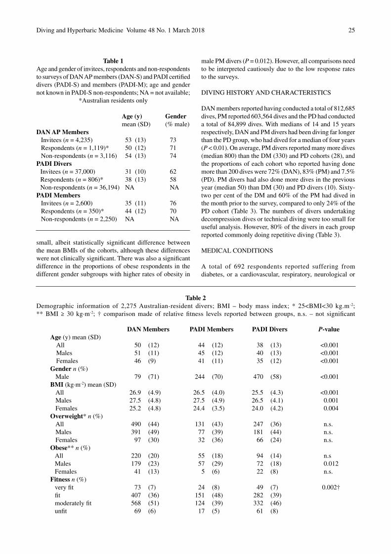
Diving and Hyperbaric Medicine Volume 48 No. 1 March 2018 25
Table 1Age and gender of invitees, respondents and non-respondents to surveys of DAN AP members (DAN-S) and PADI certified divers (PADI-S) and members (PADI-M); age and gender not known in PADI-S non-respondents; NA = not available;
*Australian residents only
Table 2Demographic information of 2,275 Australian-resident divers; BMI – body mass index; * 25<BMI<30 kg.m-2;** BMI ≥ 30 kg∙m-2; † comparison made of relative fitness levels reported between groups, n.s. – not significant
Age (y) Gender mean (SD) (% male)DAN AP Members
Invitees (n = 4,235) 53 (13) 73 Respondents (n = 1,119)* 50 (12) 71 Non-respondents (n = 3,116) 54 (13) 74
PADI DiversInvitees (n = 37,000) 31 (10) 62 Respondents (n = 806)* 38 (13) 58 Non-respondents (n = 36,194) NA NA
PADI MembersInvitees (n = 2,600) 35 (11) 76 Respondents (n = 350)* 44 (12) 70 Non-respondents (n = 2,250) NA NA
DAN Members PADI Members PADI Divers P-valueAge (y) mean (SD)
All 50 (12) 44 (12) 38 (13) <0.001Males 51 (11) 45 (12) 40 (13) <0.001 Females 46 (9) 41 (11) 35 (12) <0.001
Gender n (%) Male 79 (71) 244 (70) 470 (58) <0.001
BMI (kg∙m-2) mean (SD) All 26.9 (4.9) 26.5 (4.0) 25.5 (4.3) <0.001Males 27.5 (4.8) 27.5 (4.9) 26.5 (4.1) 0.001 Females 25.2 (4.8) 24.4 (3.5) 24.0 (4.2) 0.004
Overweight* n (%) All 490 (44) 131 (43) 247 (36) n.s.Males 391 (49) 77 (39) 181 (44) n.s. Females 97 (30) 32 (36) 66 (24) n.s.
Obese** n (%) All 220 (20) 55 (18) 94 (14) n.sMales 179 (23) 57 (29) 72 (18) 0.012 Females 41 (13) 5 (6) 22 (8) n.s.
Fitness n (%) very fit 73 (7) 24 (8) 49 (7) 0.002† fit 407 (36) 151 (48) 282 (39) moderately fit 568 (51) 124 (39) 332 (46) unfit 69 (6) 17 (5) 61 (8)
small, albeit statistically significant difference between the mean BMIs of the cohorts, although these differences were not clinically significant. There was also a significant difference in the proportions of obese respondents in the different gender subgroups with higher rates of obesity in
male PM divers (P = 0.012). However, all comparisons need to be interpreted cautiously due to the low response rates to the surveys.
DIVING HISTORY AND CHARACTERISTICS
DAN members reported having conducted a total of 812,685 dives, PM reported 603,564 dives and the PD had conducted a total of 84,899 dives. With medians of 14 and 15 years respectively, DAN and PM divers had been diving far longer than the PD group, who had dived for a median of four years (P < 0.01). On average, PM divers reported many more dives (median 800) than the DM (330) and PD cohorts (28), and the proportions of each cohort who reported having done more than 200 dives were 72% (DAN), 83% (PM) and 7.5% (PD). PM divers had also done more dives in the previous year (median 50) than DM (30) and PD divers (10). Sixty-two per cent of the DM and 60% of the PM had dived in the month prior to the survey, compared to only 24% of the PD cohort (Table 3). The numbers of divers undertaking decompression dives or technical diving were too small for useful analysis. However, 80% of the divers in each group reported commonly doing repetitive diving (Table 3).
MEDICAL CONDITIONS
A total of 692 respondents reported suffering from diabetes, or a cardiovascular, respiratory, neurological or

Diving and Hyperbaric Medicine Volume 48 No. 1 March 201826
Table 3Diving history and characteristics of combined survey participants; OW − open water; OW+ − post-basic certification other than others listed; Tech − technical diver; DM − divemaster; Inst − instructor; Comm − commercial diver; * hold
commercial qualifications but still dive recreationally
Group DAN members PADI members PADI divers P−valueYears diving, med (range) 14 (1−60) 15 (1−47) 4 (1−45) < 0.01Qualifications n (%)
OW only 34 (4) NA 299 (37) < 0.01OW+ 357 (39) NA 483 (60) Tech 126 (14) NA 17 (2) DM 203 (22) 143 (41) NA Inst 160 (17) 178 (51) NA Comm* 35 (4) 28 (8) 6 (<1)Total dives, med (range) 330 (4−16,000) 800 (15−20,000) 28 (4−10000) < 0.01 Dives past year, med (range) 30 (0−500) 50 (2−1000) 10 (0−200) < 0.01
Time since last dive, months (%)<1 673 (62) 208 (60) 194 (24) < 0.011 to < 6 322 (30) 82 (24) 267 (33) 6 to <12 72 (7) 37 (11) 270 (34) ≥12 19 (2) 19 (5) 71 (9)
Dives > 30 m deep (%; med (IQR)) 10 (3−25) 10 (5−25) 1 (0−10) Repetitive dives (%; med (IQR)) 80 (40−95) 80 (50−95) 80 (38−100)
Figure 1Comparative proportions of medical conditions in the general community11 and the diving survey cohorts
psychological condition. These included 376 (34%), 97 (28%) and 222 (28%) of the DM, PM and PD cohorts, respectively. Three respondents reported multiple conditions. With the exception of cardiac conditions (P = 0.099), the cohorts differed significantly in the proportions with the other medical conditions (P < 0.001 for all except neurological conditions P = 0.033). The most obvious difference was the higher incidence of hypertension in the
older cohorts. The numbers reporting inner ear injuries (39 DM, 15 PM and 25 PD respectively) are unexpectedly high. Figure 1 compares the proportions in the Australian adult population with particular medical conditions, as reported in Australian Bureau of Statistics National Health Survey 2014–2015,11 to those in our survey respondents.
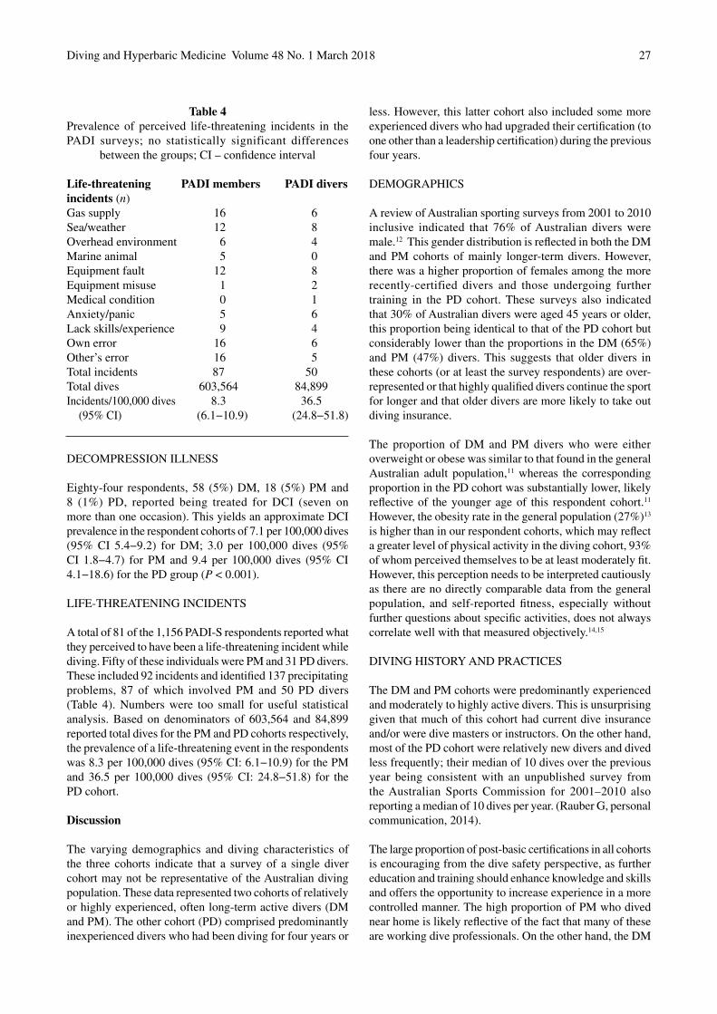
Diving and Hyperbaric Medicine Volume 48 No. 1 March 2018 27
DECOMPRESSION ILLNESS
Eighty-four respondents, 58 (5%) DM, 18 (5%) PM and 8 (1%) PD, reported being treated for DCI (seven on more than one occasion). This yields an approximate DCI prevalence in the respondent cohorts of 7.1 per 100,000 dives(95% CI 5.4−9.2) for DM; 3.0 per 100,000 dives (95% CI 1.8−4.7) for PM and 9.4 per 100,000 dives (95% CI 4.1−18.6) for the PD group (P < 0.001).
LIFE-THREATENING INCIDENTS
A total of 81 of the 1,156 PADI-S respondents reported what they perceived to have been a life-threatening incident while diving. Fifty of these individuals were PM and 31 PD divers. These included 92 incidents and identified 137 precipitating problems, 87 of which involved PM and 50 PD divers(Table 4). Numbers were too small for useful statistical analysis. Based on denominators of 603,564 and 84,899 reported total dives for the PM and PD cohorts respectively, the prevalence of a life-threatening event in the respondents was 8.3 per 100,000 dives (95% CI: 6.1−10.9) for the PM and 36.5 per 100,000 dives (95% CI: 24.8−51.8) for the PD cohort.
Discussion
The varying demographics and diving characteristics of the three cohorts indicate that a survey of a single diver cohort may not be representative of the Australian diving population. These data represented two cohorts of relatively or highly experienced, often long-term active divers (DM and PM). The other cohort (PD) comprised predominantly inexperienced divers who had been diving for four years or
less. However, this latter cohort also included some more experienced divers who had upgraded their certification (to one other than a leadership certification) during the previous four years.
DEMOGRAPHICS
A review of Australian sporting surveys from 2001 to 2010 inclusive indicated that 76% of Australian divers were male.12 This gender distribution is reflected in both the DM and PM cohorts of mainly longer-term divers. However, there was a higher proportion of females among the more recently-certified divers and those undergoing further training in the PD cohort. These surveys also indicated that 30% of Australian divers were aged 45 years or older, this proportion being identical to that of the PD cohort but considerably lower than the proportions in the DM (65%) and PM (47%) divers. This suggests that older divers in these cohorts (or at least the survey respondents) are over-represented or that highly qualified divers continue the sport for longer and that older divers are more likely to take out diving insurance.
The proportion of DM and PM divers who were either overweight or obese was similar to that found in the general Australian adult population,11 whereas the corresponding proportion in the PD cohort was substantially lower, likely reflective of the younger age of this respondent cohort.11 However, the obesity rate in the general population (27%)13 is higher than in our respondent cohorts, which may reflect a greater level of physical activity in the diving cohort, 93% of whom perceived themselves to be at least moderately fit. However, this perception needs to be interpreted cautiously as there are no directly comparable data from the general population, and self-reported fitness, especially without further questions about specific activities, does not always correlate well with that measured objectively.14,15
DIVING HISTORY AND PRACTICES
The DM and PM cohorts were predominantly experienced and moderately to highly active divers. This is unsurprising given that much of this cohort had current dive insurance and/or were dive masters or instructors. On the other hand, most of the PD cohort were relatively new divers and dived less frequently; their median of 10 dives over the previous year being consistent with an unpublished survey from the Australian Sports Commission for 2001–2010 also reporting a median of 10 dives per year. (Rauber G, personal communication, 2014).
The large proportion of post-basic certifications in all cohorts is encouraging from the dive safety perspective, as further education and training should enhance knowledge and skills and offers the opportunity to increase experience in a more controlled manner. The high proportion of PM who dived near home is likely reflective of the fact that many of these are working dive professionals. On the other hand, the DM
Table 4Prevalence of perceived life-threatening incidents in the PADI surveys; no statistically significant differences
between the groups; CI – confidence interval
Life-threatening PADI members PADI diversincidents (n) Gas supply 16 6 Sea/weather 12 8 Overhead environment 6 4 Marine animal 5 0 Equipment fault 12 8 Equipment misuse 1 2 Medical condition 0 1 Anxiety/panic 5 6 Lack skills/experience 9 4 Own error 16 6 Other’s error 16 5Total incidents 87 50 Total dives 603,564 84,899 Incidents/100,000 dives 8.3 36.5
(95% CI) (6.1−10.9) (24.8−51.8)
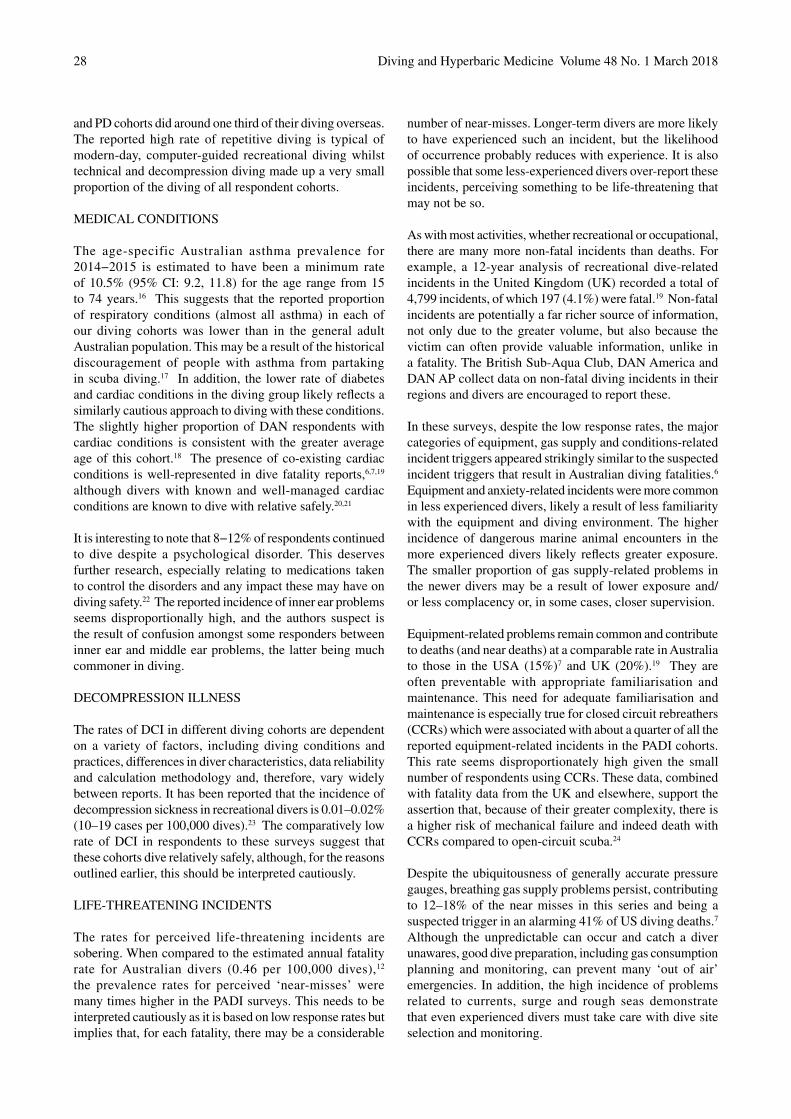
Diving and Hyperbaric Medicine Volume 48 No. 1 March 201828
and PD cohorts did around one third of their diving overseas. The reported high rate of repetitive diving is typical of modern-day, computer-guided recreational diving whilst technical and decompression diving made up a very small proportion of the diving of all respondent cohorts.
MEDICAL CONDITIONS
The age-specific Australian asthma prevalence for 2014−2015 is estimated to have been a minimum rate of 10.5% (95% CI: 9.2, 11.8) for the age range from 15 to 74 years.16 This suggests that the reported proportion of respiratory conditions (almost all asthma) in each of our diving cohorts was lower than in the general adult Australian population. This may be a result of the historical discouragement of people with asthma from partaking in scuba diving.17 In addition, the lower rate of diabetes and cardiac conditions in the diving group likely reflects a similarly cautious approach to diving with these conditions. The slightly higher proportion of DAN respondents with cardiac conditions is consistent with the greater average age of this cohort.18 The presence of co-existing cardiac conditions is well-represented in dive fatality reports,6,7,19 although divers with known and well-managed cardiac conditions are known to dive with relative safely.20,21
It is interesting to note that 8−12% of respondents continued to dive despite a psychological disorder. This deserves further research, especially relating to medications taken to control the disorders and any impact these may have on diving safety.22 The reported incidence of inner ear problems seems disproportionally high, and the authors suspect is the result of confusion amongst some responders between inner ear and middle ear problems, the latter being much commoner in diving.
DECOMPRESSION ILLNESS
The rates of DCI in different diving cohorts are dependent on a variety of factors, including diving conditions and practices, differences in diver characteristics, data reliability and calculation methodology and, therefore, vary widely between reports. It has been reported that the incidence of decompression sickness in recreational divers is 0.01–0.02% (10–19 cases per 100,000 dives).23 The comparatively low rate of DCI in respondents to these surveys suggest that these cohorts dive relatively safely, although, for the reasons outlined earlier, this should be interpreted cautiously.
LIFE-THREATENING INCIDENTS
The rates for perceived life-threatening incidents are sobering. When compared to the estimated annual fatality rate for Australian divers (0.46 per 100,000 dives),12 the prevalence rates for perceived ‘near-misses’ were many times higher in the PADI surveys. This needs to be interpreted cautiously as it is based on low response rates but implies that, for each fatality, there may be a considerable
number of near-misses. Longer-term divers are more likely to have experienced such an incident, but the likelihood of occurrence probably reduces with experience. It is also possible that some less-experienced divers over-report these incidents, perceiving something to be life-threatening that may not be so.
As with most activities, whether recreational or occupational, there are many more non-fatal incidents than deaths. For example, a 12-year analysis of recreational dive-related incidents in the United Kingdom (UK) recorded a total of 4,799 incidents, of which 197 (4.1%) were fatal.19 Non-fatal incidents are potentially a far richer source of information, not only due to the greater volume, but also because the victim can often provide valuable information, unlike in a fatality. The British Sub-Aqua Club, DAN America and DAN AP collect data on non-fatal diving incidents in their regions and divers are encouraged to report these.
In these surveys, despite the low response rates, the major categories of equipment, gas supply and conditions-related incident triggers appeared strikingly similar to the suspected incident triggers that result in Australian diving fatalities.6 Equipment and anxiety-related incidents were more common in less experienced divers, likely a result of less familiarity with the equipment and diving environment. The higher incidence of dangerous marine animal encounters in the more experienced divers likely reflects greater exposure. The smaller proportion of gas supply-related problems in the newer divers may be a result of lower exposure and/or less complacency or, in some cases, closer supervision.
Equipment-related problems remain common and contribute to deaths (and near deaths) at a comparable rate in Australia to those in the USA (15%)7 and UK (20%).19 They are often preventable with appropriate familiarisation and maintenance. This need for adequate familiarisation and maintenance is especially true for closed circuit rebreathers (CCRs) which were associated with about a quarter of all the reported equipment-related incidents in the PADI cohorts. This rate seems disproportionately high given the small number of respondents using CCRs. These data, combined with fatality data from the UK and elsewhere, support the assertion that, because of their greater complexity, there is a higher risk of mechanical failure and indeed death with CCRs compared to open-circuit scuba.24
Despite the ubiquitousness of generally accurate pressure gauges, breathing gas supply problems persist, contributing to 12–18% of the near misses in this series and being a suspected trigger in an alarming 41% of US diving deaths.7 Although the unpredictable can occur and catch a diver unawares, good dive preparation, including gas consumption planning and monitoring, can prevent many ‘out of air’ emergencies. In addition, the high incidence of problems related to currents, surge and rough seas demonstrate that even experienced divers must take care with dive site selection and monitoring.
Diving and Hyperbaric Medicine Volume 48 No. 1 March 2018 29
LIMITATIONS
There are a variety of limitations to this study, the major one being the low and differing response rates. This non-response creates the potential for the data not to accurately reflect each group. However, large numbers within the groups and the substantial differences between them suggest that, while selection bias may have affected the results, it is unlikely that it was so large that it was responsible for all the observed group differences.
In addition, DAN members are likely older than the general diving population and respondents to the DAN-S were younger than non-respondents, possibly introducing some selection bias. Although there were no details of non-respondents to the PADI surveys, it appears that they were likely older than non-respondents and there was a higher response rate from females, potential sources of selection bias. The very low response rate from the PADI divers would likely introduce further bias towards more experienced divers or enthusiastic divers who engage with the sport.
Whilst many Australian divers will fall into one of these population groups, they will not be representative of the entire Australian diving population. Although it would have been useful to examine age-specific combined data, this was not possible as the ages of the PADI populations were not available. The nature of some of the more historical questions, such as the number and characteristics of dives undertaken, may have introduced a recall bias and this may be more likely in the longer-term divers. Finally, self-reporting on medical conditions, diving activities and events may have been another source of reporting bias.
Conclusions
Although limited by low and differing response rates and potential response bias, this study indicates that there are differences in the health-related conditions and diving experiences of the respondents in the three survey groups. Therefore, a survey of any single diver group may not be representative of the general recreational diving population and the findings from such surveys may be misleading. This bias needs to be clearly understood and any survey findings interpreted accordingly. Despite their limitations, these data provide an insight into the varying demographics, diving practices and outcomes of these groups of active Australian divers and can provide a background for further research in accident mitigation, other safety initiatives and industry planning.
References
1 Byron T. History of spearfishing and scuba diving in Australia – the first 80 years 1917 to 1997. Sydney: Tom Byron Publications; 1998.
2 Mozaffarian D, Benjamin EJ, Go AS, Arnett DK, Blaha MJ, Cushman M, et al. Heart disease and stroke
statistics-2015 update: a report from the American Heart Association. Circulation. 2015; 131:e29–e322. doi: 10.1161/CIR.0000000000000152. [cited 2015 December 21]. Available from: http://circ.ahajournals.org/content/131/4/e29.
3 Berenji Ardestani S, Buzzacott P, Eftedal I. The aging diver: endothelial biochemistry and its potential implications for cardiovascular health. Diving Hyperb Med. 2015;45:235–9.
4 Pougnet R, Constanzo L, Lodde B, Henckers A, Dherbecourt L, Lucas D, et al. Cardiovascular risk factors and cardiovascular assessment in professional divers. Int Marit Health. 2012;63:164–9.
5 Jousilahti P, Vartiainen E, Tuomilehto J, Uuska P. Sex, age, cardiovascular risk factors and coronary heart disease: a prospective follow-up study of 14,786 middle-aged men and women in Finland. Circulation. 1999;99:1165–72.
6 Lippmann J, Baddeley A, Vann R, Walker D. An analysis of the causes of compressed gas diving fatalities in Australia from 1972–2005. Undersea Hyperb Med. 2013;40:49–61.
7 Denoble PJ, Caruso JL, Dear Gde L, Pieper CF, Vann RD. Common causes of open-circuit recreational diving fatalities. Undersea Hyperb Med. 2008;35:393–406.
8 Lippmann J, McD Taylor D, Stevenson C, Mitchell S. The demographics and diving behaviour of DAN Asia-Pacific members with and without pre-existing medical conditions. Diving Hyperb Med. 2016;46:200–6.
9 National Statistical Service. Sample size calculator. [cited 2015 August 16]. Available from: http://www.nss.gov.au/nss/home.NSF/pages/Sample+size+calculator.
10 Ihaka R, Gentleman R. R: A language for data analysis and graphics. J Comp Graph Stat. 1996;5:299–314.
11 Australian Bureau of Statistics. 4364.0.55.001- National Health Survey: First results 2014–15. 2015. [cited 2017 February 04]. Available from: http://www.abs.gov.au/ausstats/[email protected]/Lookup/by%20Subject/4364.0.55.001~2014-15~Main%20Features~About%20the%20National%20Health%20Survey~3.
12 Lippmann J, Stevenson C, McD Taylor D, Williams J. Estimating the risk of a scuba diving fatality in Australia. Diving Hyperb Med. 2016;46:241–7.
13 Australian Institute of Health and Welfare (AIHW) 2015. Cardiovascular disease, diabetes and chronic kidney disease - Australian facts: risk factors. Cardiovascular, diabetes and chronic kidney disease series no. 4. Cat. no. CDK 004. Canberra: AIHW. [cited 2017 January 25]. Available from: http://www.aihw.gov.au/publication-detail/?id=60129550538.
14 Obling KH, Hansen A-LS, Overgaard K, Normann K, Sandbaek A, Maindal HT. Association between self-reported and objectively measured physical fitness level in a middle-aged population in primary care. Prev Med Rep. 2015;2:462–6. doi: 10.1016/j.pmedr.2015.05.010.
15 Knapik JJ, Jones BH, Reynolds KL, Staab JS. Validity of self-assessed physical fitness. Am J Prev Med. 1992;8:367–72.
16 Australian Institute of Health and Welfare (AIHW). Asthma and other chronic respiratory conditions. [cited 2017 November 20]. Available from: https://www.aihw.gov.au/reports/asthma-other-chronic-respiratory-conditions/asthma/data.
17 Victorian Asthma Foundation. Policy of the Victorian Asthma Foundation. SPUMS J. 1987;17:133–4.
18 Australian Institute of Health and Welfare (AIHW). How many Australian have cardiovascular disease. 2011–2012. [cited 2016 December 13]. Available from: http://www.aihw.gov.au/cardiovascular-disease/prevalence.
19 Cumming B, Peddie C, Watson. A review of the nature of
Diving and Hyperbaric Medicine Volume 48 No. 1 March 201830
diving in the United Kingdom and of diving fatalities (1998-2009). In: Vann RD, Lang MA, editors. Recreational diving fatalities. Proceedings of the Divers Alert Network 2010 April 8–10 Workshop. Durham, NC: Divers Alert Network; 2011. p. 99–117
20 Lippmann J, McD Taylor D, Stevenson C, Williams J, Mitchell S. Diving with pre-existing health conditions. Diving Hyperb Med. 2017;47:180–90.
21 St Leger Dowse M, Waterman MK, Penny CE, Smerdon GR. Does self-certification reflect the cardiac health of UK sport divers? Diving Hyperb Med. 2015;45:184–9.
22 Querido AL. Diving and antidepressants. Diving Hyperb Med. 2017; 47:253–6. doi:10.28920/dhm47.4.253–256.
23 Vann RD, Butler FK, Mitchell SJ, Moon RE. Decompression illness. Lancet. 2010;377:153-64. doi: 10.1016/S0140-6736(10)61085–9.
24 Fock AW. Analysis of recreational closed-circuit rebreather deaths 1998–2010. Diving Hyperb Med. 2013;43:78–85.
Acknowledgments
The authors wish to acknowledge the assistance of PADI Asia-Pacific enabling access to members and divers, and to Adam Lippmann and Scott Jamieson for assisting with data collection.
Conflicts of interest and funding
John Lippmann is Chairman of DAN Asia-Pacific which sells dive injury insurance.
Submitted: 09 August 2017; revised 28 November and18 December 2017Accepted: 05 January 2018
Copyright: This article is the copyright of the author who grants Diving and Hyperbaric Medicine a non-exclusive licence to publish the article electronically and in other forms.
WorldWide emergency evacuation + travel assistance Benefits + dive injury treatment coverage + 24/7 medical assistance
+ suBscription to “alert diver” magazine
Wherever I am divingI take DAN with me
To find out more or to join DAN nationals/residents of the asia-pacific visit www.danap.org
european nationals/residents visit www.daneurope.org
Diving and Hyperbaric Medicine Volume 48 No. 1 March 2018 31
Hyperbaric oxygen treatment in thromboangiitis obliterans: a retrospective clinical auditDogus Hemsinli1, Gokalp Altun2, Safiye Tuba Kaplan3, Furkan Yildirim4, Gamze Cebi5
1 Cardiovascular Surgery Department, Health Sciences University, Kanuni Research and Training Hospital, Trabzon, Turkey2 Cardiovascular Surgery Department, Karadeniz Technical University, Faculty of Medicine, Trabzon3 Internal Medicine Department, Health Sciences University, Kanuni Research and Training Hospital, Trabzon4 Underwater and Hyperbaric Medicine Department, Health Sciences University, Antalya Research and Training Hospital, Antalya, Turkey5 Underwater and Hyperbaric Medicine Department, Health Sciences University, Kanuni Research and Training Hospital, TrabzonCorresponding author: Dogus Hemsinli, Cardiovascular Surgery Department, Health Sciences University, Kanuni Research and Training Hospital, Kasustu Mahallesi, Topal Osman Sokak, No:7, 61290 Yomra, Trabzon, [email protected]
Key wordsBuerger’s disease; Chronic wounds; Pain; Outcome
Abstract(Hemsinli D, Altun G, Tuba Kaplan S, Yildirim F, Cebi G. Hyperbaric oxygen therapy in thromboangiitis obliterans: a retrospective clinical audit. Diving and Hyperbaric Medicine. 2018 March;48(1):31–35. doi:10.28920/dhm48.1.31-35. PMID: 29557099.)Introduction: Wounds refractory to standard treatment in patients with thromboangiitis obliterans (TAO, Buerger’s disease) are associated with amputation, other morbidity and mortality. The purpose of this study was to investigate the effect of hyperbaric oxygen treatment (HBOT) in patients with TAO.Materials and methods: Ninety-seven patients with TAO with ischaemic wounds treated between January 2007 and July 2016 were included in this dual-centre, non-randomised, retrospective study. Patients receiving HBOT in addition to conventional treatment were enrolled in an HBOT group (n = 47) and those receiving conventional treatment alone in a non-HBOT group (n = 50). All patients were Rutherford grade III at the time of enrolment.Results: Significant improvement in the major amputation rate was observed in the HBOT group 10 months after starting treatment (2/47 vs. 13/50, P = 0.007). Numbers of patients progressing to Rutherford grade I (27/47 vs. 17/50, P = 0.035), numbers of patients healing completely (21 vs. 11, P = 0.031 and pain scores (visual analogue scale; 1, range 0−8 vs. 6, range 0−9, P < 0.001) were also significantly improved in the HBOT group.Conclusion: The addition of HBOT to conventional treatment in TAO patients with non-healing ischaemic wounds and severe extremity pain, conferred significant benefits in terms of wound healing and rest pain control. Multi-centre, prospective, randomized studies with blinded outcome analysis are now needed to elicit more reliable results.
Introduction
Thromboangiitis obliterans (TAO, Buerger’s disease) is a non-atherosclerotic, segmental, inflammatory disease of uncertain etiology affecting the small and medium-sized vessels in the extremities. The prevalence of the disease among all patients with peripheral arterial disease ranges from as low as 0.5−5.6% in Western Europe to as high as 45−63% in India, 16−66% in Korea and Japan and 80% amongst Jews of Ashkenazi ancestry living in Israel. A powerful association exists between smoking and the inflammatory process involved in the onset and progression of the disease.1,2
The pathological process that begins with hypercellular and inflammatory thrombus formation in TAO concludes with occlusion in the distal vascular bed and tissue hypoxia. Clinical symptoms begin with claudication, followed by severe rest pain and ischaemic wounds (IWs) caused by tissue necrosis as the disease progresses.1,2 Severe rest pain
and IWs in TAO cause social problems and workforce losses and thus have an adverse impact on daily life. Standard treatments include revascularization techniques and agents such as acetylsalicylic acid, pentoxifylline, clopidogrel, cilostazol and intravenous iloprost infusion. IWs, many of which do not heal with standard treatment methods, also provoke secondary diseases, such as infection and organ dysfunction. Non-healing wounds have a high risk of leading to amputation, which is in turn associated with other morbidity, mortality and increased treatment costs.1–5
Surgical revascularization in patients with TAO is generally not possible due to distal and diffuse segmental occlusion in the extremity arteries. In addition, the benefits of bypass surgery are questionable due to high graft failure rates. However, bypass surgery may be considered in the presence of a suitable distal vessel bed in patients with severe ischaemic findings.1,2,6,7 Surgical revascularization is reported to have been possible in only 21 out of 216 patients and that patency levels were not promising.1 Studies have
Diving and Hyperbaric Medicine Volume 48 No. 1 March 201832
emphasized that smoking cessation is the most successful method of treating TAO, and that all other methods are palliative.1,2,6,7
Several studies have shown that hyperbaric oxygen treatment (HBOT) significantly accelerates healing in IWs, increases oxygen flux in the wound area and reduces the tendency to necrosis in the extremity.8,9 However, almost all the literature consists of studies involving diabetic or atherosclerotic patients.8–10 In a series of 36 patients with TAO and IWs,11 we concluded that the patients’ clinical condition improved significantly with HBOT and that it was easier for them to perform their daily activities. Both pain and wound area were significantly better (P < 0.001 for both.11 These findings constituted useful evidence for the use of HBOT in the treatment of IWs in TAO. However, the small number of patients and lack of a control group limit the value of these clinical results. As a result, we performed a larger dual-centre retrospective clinical audit. The main outcome criterion was improvement in the major amputation rate at the tenth month after initiation of treatment. Secondary outcomes were improvement in Rutherford grade, healing of IWs and pain scores at 10 months.
Materials and methods
A dual-centre, non-randomised, comparative, retrospective study was performed with the approval of the local ethics committee and in line with the Declaration of Helsinki. The archive records of patients treated and monitored with a diagnosis of TAO at the Karadeniz Technical University Medical Faculty and Health Sciences University Kanuni Training and Research Hospital, Turkey between January 2007 and July 2016 were reviewed. Data were obtained from the archive records, clinical follow-ups and telephone interviews with physicians. One-hundred-thirteen patients diagnosed with TAO on the basis of clinical and radiological findings and with IWs of the extremities were identified. Sixteen patients were excluded: one with osteomyelitis based on magnetic resonance imaging; four whose records were missing; two unable to receive HBOT owing to claustrophobia; five with chronic obstructive lung disease and four with an ejection fraction < 35%. Following exclusions, all patients included for analysis in this study commenced as Rutherford grade III prior to intervention.
The Rutherford classification is widely used in cardiovascular surgery departments to evaluate the severity of peripheral vascular diseases. In this classification, grade 0 is used to define asymptomatic patients, grade I for patients with claudication (mild, moderate, severe), grade II for patients with ischaemic rest pain, and grade III for patients with ulcers, gangrene or tissue loss.12
Based on these criteria, data from 97 patients with severe ischaemic rest pain and infected ischaemic ulcers in the extremities were analyzed. The HBOT group (n = 47) which included the 36 patients from the previous study
consisted of patients receiving HBOT in addition to standard treatment methods. The non-HBOT group (n = 50) consisted of patients not receiving HBOT. Reasons for not receiving HBOT included non-availability of a hyperbaric physician and the fact that the hyperbaric medicine unit did not open until 2010.
HBOT was administered in a multiplace chamber (Hiperbot Model 101, 2005, Turkey) allowing 12 patients to be treated simultaneously. The chamber was pressurized with medical air to 240 kPa (2.37 ATA) over 15 minutes (min), then patients received three sessions of 100% oxygen by mask for 30 min, each session separated by a 5-min air break and decompression was over 10 min. HBOT was administered five days a week for the duration of hospital stay. All patients were accompanied by a member of the medical staff during HBOT. After discharge from hospital, patients received HBOT only when clinically indicated.
All patients received standard medical treatment consisting of acetylsalicylic acid, pentoxifylline, clopidogrel and cilostazol. Patients without an ejection fraction < 40% and/or New York Heart Association (NYHA) heart failure class 3–4 were started on intravenous iloprost at0.5 ng∙kg-1∙min-1 (Ilomedin, Bayer-Schering AG, Germany) for six hours per day for 21 days. On the first day, the dose was increased by 0.5 ng∙kg-1∙min-1 every half hour to a maximum of2 ng∙kg-1∙min-1. If any side effects appeared, the dose was reduced back to the preceding one. Empiric antibiotic therapy was started after culturing the wound and was revised according to the culture results. When necessary, aggressive debridement or amputation was performed on the extremity containing the wound, followed by wound care and dressings. The dressings were changed at frequent intervals and wounds were protected from uncontrolled mechanical pressure.
Patients’ clinical status and severity of peripheral vascular disease were evaluated using Rutherford’s criteria at admission, and 10 months after commencing treatment, either HBOT or the initial treatment of non-HBOT patients.12 Vascular lesions were classified according to Graziani’s morphological classification (data not reported here).13 At 10 months, cases were classified as complete healing (no infection in the wound, no necrotic tissue, adequate granulation tissue formation and completion of epithelialization) or incomplete healing (infection in the wound or presence of necrotic tissue or inadequate granulation tissue or incomplete epithelialization). The location and size of IWs were recorded at admission, at discharge, and during outpatient visits in case of incomplete healing at discharge. The areas of IWs were obtained from patient records, and was calculated by multiplying the longest and widest dimensions.
Severity of ischaemic extremity pains was evaluated using a visual analogue scale (VAS) ranging from 0 (no pain) to 10 (worst imaginable pain). Pain-free walking
Diving and Hyperbaric Medicine Volume 48 No. 1 March 2018 33
distances were measured only in patients who had not undergone major amputation and with a Rutherford grade < II. Demographic variables such as smoking status and duration, pain characteristics, previous surgical interventions (sympathectomy, peripheral revascularization procedures, minor/major amputation), endovascular therapy, medical treatments received for TAO, complications and mortality were recorded during treatment and follow-up.
STATISTICAL ANALYSIS
The Statistical Package for the Social Sciences (SPSS) 23.0 software was used for data analysis. Normality of distribution was examined using the one-sample Kolmogorov-Smirnov test. Results were expressed as mean (standard deviation) for normal data (age), median (range) for non-normal data (duration of follow-up, IW area, VAS, duration of hospitalization, pain-free walking distance), and median values for Rutherford classifications, and as number for categoric data (gender, smoking status, thrombophlebitis migrans, surgical interventions, complete healing, and Rutherford class). Comparison of numerical variables between two independent groups was performed using the Mann Whitney U test since normal distribution was not established. The chi square test was used to analyze differences between ratios of categoric variables in independent groups. P < 0.05 was regarded as statistically significant.
Results
Ninety-seven patients, presenting with IW and Rutherford grade III diagnosed with TAO were included in the final analysis (Table 1). Briefly, both groups were similar in terms of age, severity of disease, and comorbidities such as
smoking status and previous surgical procedures. There were no statistically significant differences in terms of medical treatments or smoking cessation between the HBOT and non-HBOT groups. No procedure-related complications occurred in any patient.
Patients in the HBOT group were followed up for a median of 30 (range 10–48) months and those in the non-HBOT group for 23 (range 10–48) months (P = 0.071). The 10-month follow-up data are summarised in Table 2. During this period, patients in the HBOT group received a median of 34 sessions (range 10–62). The incidence of minor amputation (amputation leaving sufficient functional foot to permit the patient to walk without prosthesis) was similar in both groups (19 vs 30, P = 0.084). However, the number of major amputations (patients with Syme’s amputation, above or below knee amputation) was significantly lower in the HBOT group (2 vs. 13, P = 0.007). Significantly more patients in the HBOT group were completely healed at 10 months (21 vs. 11, P = 0.031) and VAS scores were lower in the HBOT group. Median post-treatment Rutherford grade was 1 (range 1–3) in the HBOT group, and 2 (range 1–3) in the non-HBOT group (P = 0.043); more patients in the HBOT group improved to grade I. IW area did not differ significantly, and duration of hospitalization was similar in the two groups.
Discussion
This study shows that the addition of HBOT to conventional medical treatment of TAO reduced the number of major amputations, improved the Rutherford grade, the rate of healing of IWs and VAS scores at 10 months from the initiation of treatment for both groups.
Table 1Baseline characteristics of the patient population (number or median and range); HBOT − hyperbaric oxygen treatment; VAS − visual analogue scale; 25 patients in the HBOT group
and 17 patients in the non-HBOT group
Table 2Outcomes at 10 months (number or median (range); HBOT − hyperbaric oxygen treatment; VAS − visual analogue scale; * P = 0.007; † P = 0.031; ‡ 0.0043; § P < 0.001; ** owing
to incomplete healing and amputation
HBOT Non-HBOT (n = 47) (n = 50)Females/Males 1/46 1/49Age (years) 50 (32–68) 45 (37–75)Smoker 44 48Thrombophlebitis migrans 7 9Previous sympathectomy 20 23Previous surgical revascularization 5 6Previous endovascular therapy 4 4Upper extremity involvement 5 7Previous minor amputation 12 15Previous major amputation 8 6IW area (cm2) 21 (5–70) 15 (2–45)VAS score 8 (5–9) 8 (5–9)Rutherford grade III All All
HBOT Non-HBOT (n = 47) (n = 50)Major amputation * 2 13
during hospitalization 1 6during follow-up 1 7
Complete healing † 21 11Rutherford grade ‡
I 27 17II 5 10III 15 23
Wound area (cm2) 12 (0–60) 11 (0–45)VAS score § 1 (0–8) 6 (0–9)Pain-free walking (m) ** 200 (40–500) 200 (100–200)Hospitalization (days) 60 (30–120) 60 (60–120)Mortality at 10 months 1 1

Diving and Hyperbaric Medicine Volume 48 No. 1 March 201834
The healing of IWs represents the most critical stage in terms of preventing amputation.9,14,15 Therefore, adjuvant therapeutic options such as HBOT, capable of contributing to wound healing and preventing amputation in TAO patients with IWs, are important. A literature review revealed no previous studies on HBOT in the treatment of TAO patients with IWs apart from our own.11 One study reported an 11% risk of major amputation (above the knee, below the knee or hand amputation) at five years, 21% at 10 years and 23% and 20 years in TAO patients treated using conventional methods.16 In our study, there were 13 major amputations in 50 patients (26%) in the non-HBOT group. We attribute the higher level of amputation in the non-HBOT group to all the patients being Rutherford grade III at entry, in contrast to the previous study.
HBOT has been shown to act both locally and systemically. Locally, it increases several growth factors, such as vascular endothelial growth factor (VEGF) and nitric oxide (NO) involved in angiogenesis in ischaemic tissue,17 and enhances diffusion gradients for oxygen into the wound. In addition, HBOT reduces capillary pressure and transcapillary fluid transfer and increases extravascular fluid absorption, thus reducing lower extremity oedema. Systemically, it stimulates the release of bone marrow progenitor stem cells,18 decreases circulating inflammatory cytokines and increases fibroblastic activity, collagen production and the efficacy of antibiotics.
Despite the limited studies of the efficacy of the different methods used to treat TAO, some studies have elicited promising results. Applying vascular endothelial growth factor improved wound healing in four out of six patients with IWs and increased collateral vascularization around the injection site in five of the seven subjects.17 Autologous bone marrow transplantation in seven TAO patients with ischaemic extremities, combined with four sessions of HBOT two days before transplant, then one day, two and four weeks after transplant, resulted in significant improvement in pain and walking distances compared to the pre-treatment period.3
The improvement in VAS values was more marked than in our previous single-cohort study of 36 patients in whom the mean VAS score was 7.1 (SD 1.7) before HBOT compared to 2.2 (3.0) after treatment (P = 0.0001).11 We attribute this to patients newly included in the study having lower pain scores after treatment compared to those from the previous study. One patient who died and two who underwent major amputation in the HBOT group, and one patient who died and 13 who underwent major amputation in the non-HBOT group had continued to smoke. This demonstrates the importance of smoking cessation as part of the overall management of these patients.
LIMITATIONS
The principal limitations of this study are that it is non-randomised, retrospective and the patient numbers are
limited. Also, only a short (10 months) follow up was undertaken. However, the fact that no consensus has been achieved concerning treatment protocols for TAO and that the disease is relatively rare make it difficult to perform large, prospective, randomized studies of these patients.
Conclusions
The addition of HBOT to standard treatment methods in patients with TAO with non-healing IWs and severe extremity pain appears to provide significant benefits in terms of the rate of major amputation, healing of IWs and control of rest pain. Multi-centre, prospective randomized studies with blinded outcome analysis are now needed to elicit more reliable results.
References
1 Sayin A, Bozkurt AK, Tüzün H, Vural FS, Erdog G, Ozer M. Surgical treatment of Buerger’s disease: experience with 216 patients. Cardiovasc Surg. 1993;1:377–80. PubMed PMID: 8076063.
2 Olin JW, Shih A. Thromboangiitis obliterans (Buerger’s disease). N Engl J Med. 2006;18:18−24. PubMed PMID: 16344615.
3 Saito S, Nishikawa K, Obata H, Goto F. Autologous bone marrow transplantation and hyperbaric oxygen therapy for patients with thromboangiitis obliterans. Angiology. 2007;58:429−34. doi: 10.1177/0003319706292015. PubMed PMID: 17652224.
4 Boulton AJ, Vileikyte L, Ragnarson Tennvall G, Apelqvist J. The global burden of diabetic foot disease. Lancet. 2005;366:1719−24. doi:10.1016/S0140-6736(05)67698-2. PubMed PMID:16291066.
5 Ragnarson Tennvall G, Apelqvist J. Health economic consequences of diabetic foot lesions. Clin Infect Dis. 2004;39:132−9. doi: 10.1086/383275. PMID: 15306992.
6 Highlander P, Southerland CC, VonHerbulis E, Gonzalez A. Buerger disease (thromboangiitis obliterans): a clinical diagnosis. Adv Skin Wound Care. 2011;24:15−7. doi: 10.1097/01.ASW.0000392923.43. PMID: 21173586.
7 Ohta T, Ishioashi H, Hosaka M, Suqimoto I. Clinical and social consequences of Buerger Disease. J Vasc Surg. 2004;39:176−80. doi: 10.1016/j.jvs.2003.08.006. PMID: 14718836.
8 Andre-Levigne D, Modarressi A, Pignel R, Bochaton-Piallat ML, Pittet-Cuenod B. Hyperbaric oxygen therapy promotes wound repair in ischaemic and hyperglycemic conditions, increasing tissue perfusion and collagen deposition. Wound Repair Regen. 2016;24:954−65. doi: 10.1111/wrr.12480. PMID: 27684570.
9 Heyboer M, Grant WD, Byrne J, Pons P, Morgan M, Iqbal B, et al. Hyperbaric oxygen for the treatment of non healing arterial insufficiency ulcers. Wound Repair Regen. 2014;22:351−5. doi: 10.1111/wrr.12176.
10 Chen SJ, Yu CT, Cheng YL, Yu SY, Lo HC. Effects of hyperbaric oxygen therapy on circulating interleukin-8, nitricoxide, and insulin-like growth factors in patients with type 2 diabetes mellitus. Clin Biochem. 2007;40:30−6. doi: 10.1016/j.clinbiochem.2006.07.007. PMID: 16996047.
11 Hemsinli D, Kaplan ST, Kaplan S, Yildirim F. Hyperbaric oxygen therapy in the treatment of Fontaine stage IV
Diving and Hyperbaric Medicine Volume 48 No. 1 March 2018 35
thromboangiitis obliterans. Int J Low Extrem Wounds. 2016;15:366−70. doi: 10.1177/1534734616666866. PMID: 27647524.
12 Rutherford RB, Baker JD, Ernst C, Johnston KW, Porter JM, Ahn S, et al. Recommended standards for reports dealing with lower extremity ischemia: revised version. J Vasc Surg. 1997;26:517−38. Erratum in: 2001;33:805. doi: 10.1016/S0741-5214(97)70045-4. PMID: 9308598.
13 Graziani L, Silvestro A, Bertone V, Manara E, Andreini R, Sigala A, et al. Vascular involvement in diabetic subjects with ischaemic foot ulcer: a new morphologic categorization of disease severity. Eur J Vasc Endovasc Surg. 2007;33:453−60. doi: 10.1016/j.ejvs.2006.11.022. PMID: 17196848.
14 Kaya A, Aydin F, Altay T, Karapinar L, Ozturk H, Karakuzu C. Can major amputation rates be decreased in diabetic foot ulcers with hyperbaric oxygen therapy? Int Orthop. 2009;33:441−6. doi: 10.1007/s00264-008-0623-y. PMID: 18654777.
15 Fife CE, Buyukcakir C, Otto G, Sheffield P, Love T, Warriner R. Factors influencing the outcome of lower extremity diabetic ulcers treated with hyperbaric oxygen therapy. Wound Repair Regen. 2007;15:322−31. doi: 10.1111/j.1524-475X.2007.00234.x. PMID: 17537119.
16 Cooper LT, Tse TS, Mikhail MA, McBane RD, Stanson AW, Ballman KV. Long-term survival and amputation risk in thromboangiitis obliterans (Buerger’s disease). J Am Coll Cardiol. 2004;44:2410−11. doi: 10.1016/j.jacc.2004.09.029. PMID: 15607407.
17 Boykin JV, Baylis C. Hyperbaric oxygen therapy mediates increased nitric oxide production associated with wound healing: a preliminary study. Adv Skin Wound Care. 2007;20:382−8. doi: 10.1097/01.ASW.0000280.198.81130.d5. PMID: 17620739.
18 Milovanova TN, Bhopale VM, Sorokina EM, Moore JS, Hunt TK, Hauer-Jensen M, et al. Hyperbaric oxygen stimulates vasculogenic stem cell growth and differentiation in vivo. J Appl Physiol. 2009;106:711−28. doi: 10.1152/japplphysiol.91054.2008. PMID: 19023021.
19 Kim HJ, Jang SY, Park JI, Byun J, Kim DI, Do YS, et al. Vascular endothelial growth factor induced angiogenic gene therapy in patients with peripheral arterydisease. Exp Mol Med. 2004;36:336−44. doi: 10.1038/emm.2004.44. PMID: 15365252.
Funding and conflicts of interest: nil
Submitted: 16 July 2017; revised 17 October 2017 and 17 January 2018Accepted: 28 January 2018
Copyright: This article is the copyright of the author who grants Diving and Hyperbaric Medicine a non-exclusive licence to publish the article in electronic and other forms.
The database of randomised controlled trials in diving and hyperbaric medicine maintained by Michael Bennett and his colleagues at the Prince of Wales Hospital
Diving and Hyperbaric Medicine Unit, Sydney is at: <http://hboevidence.unsw.wikispaces.net/>
Assistance from interested physicians in preparing critical appraisals (CATs) is welcomed, indeed needed, as there is a considerable backlog.
Guidance on completing a CAT is provided.Contact Professor Michael Bennett: <[email protected]>
Diving and Hyperbaric Medicine Volume 48 No. 1 March 201836
Case reportsVomiting and aspiration of gastric contents: a possible life-threatening combination in underwater divingFrantišek Novomeský1, Martin Janík1, Michal Hájek2, Jozef Krajčovič1, Lubomír Straka1
1 Department of Forensic Medicine and Medicolegal Expertises, Jessenius Faculty of Medicine, Comenius University, University Hospital, Martin, Slovak Republic2 Hyperbaric Medicine Center, Municipal Hospital, Ostrava, Czech Republic Chair of Biomedical Sciences, Medical Faculty, University of Ostrava, Czech RepublicCorresponding author: Professor František Novomeský, Institute of Forensic Medicine and Medicolegal Expertises, Jessenius Faculty of Medicine, Comenius University, University Hospital, 036 59 Martin, Slovak [email protected]
Key wordsPathology; Gastrointestinal tract; Diving incidents; Diving deaths; Autopsy
Abstract
(František Novomeský, Martin Janík, Michal Hájek, Jozef Krajčovič, Lubomír Straka. Vomiting and aspiration of gastric contents: a possible life-threatening combination in underwater diving. Diving and Hyperbaric Medicine. 2018 March;48(1):36–39. doi: 10.28920/dhm48.1.36-39. PMID: 29557100.)Vomiting and aspiration of gastric contents into the airways and lungs is a common and well-known clinicopathological entity. This phenomenon might also occur in underwater diving, where it can lead to life-threatening or fatal situations. This article presents two incidents involving diving-related vomiting with associated aspiration of gastric contents. One case, a 39-year-old commercial diver using a full-face diving mask, was fatal and the other was a 33-year-old, female recreational scuba diver in whom underwater vomiting was complicated by pulmonary aspiration of a solid foreign body (a peanut) into the right lower lobe bronchus. The peanut was successfully removed and, following bronchoscopic pulmonary lavage, the patient made an uneventful recovery. The causes and consequences of nausea and vomiting within the underwater environment are discussed and possible interpretative problems are highlighted.
Introduction
Vomiting with possible aspiration of gastric contents is a well-known clinicopathological phenomenon. Sequelae associated with aspiration include pulmonary obstruction, chemical pneumonitis, secondary infection of airways or lung parenchyma and possible death.1,2 Morbidity following aspiration is enhanced with an increased volume of aspirate, more acidic pH, high particle content and bacterial contamination. Aspiration is most likely to occur in subjects with a decreased level of consciousness. Multiple physiological, pharmacological and pathological conditions are associated with aspiration. In divers, vomiting and aspiration of gastric contents can have a somewhat different pathophysiological background and frequently leads to a fatal outcome.3,4 This article presents two diving incidents involving vomiting with associated pulmonary aspiration of gastric contents.
Case 1
A 39-year-old, healthy, trained, male commercial diver descended with a partner to a depth of 42 metres in a
hydroelectric power station to perform a planned task. The diving equipment included a surface-supplied breathing apparatus, full-face diving mask (FFM) with a built-in oronasal mask (Figure 1), a ceramic microphone for bilateral hard-wire communication, protective plastic helmet with a light, fixed over the FFM, and a drysuit with attached hood.
The descent of both divers was slow, without equalization problems, and the breathing of both divers was deep and regular. After 10-minutes bottom time, the victim suddenly reported to the surface that he had stomach problems and his breathing became irregular, noisy and after a few seconds stopped. The buddy swam to the victim almost immediately, found him unconscious with free-flow of the FFM regulator. The rescue of the victim to the surface took approximately seven minutes. The oronasal mask of the FFM and the mouth cavity of the victim were almost completely filled with reddish gastric contents containing particles of a semi-digested meal. He was declared dead at the scene. Further investigation revealed that the deceased has consumed barbecued meat with a mixed vegetable salad several hours prior to the dive.
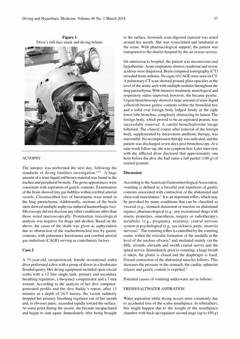
Diving and Hyperbaric Medicine Volume 48 No. 1 March 2018 37
AUTOPSY
The autopsy was performed the next day, following the standards of diving fatalities investigation.3,5,6 A huge amount of a semi-liquid red/brown material was found in the trachea and peripheral bronchi. The gross appearances were consistent with aspiration of gastric contents. Examination of the brain showed tiny gas bubbles within cerebral arterial vessels. Circumscribed foci of barotrauma were noted in the lung parenchyma. Additionally, sections of the brain stem showed multiple asphyxia-induced haemorrhagic foci. Microscopy did not disclose any other conditions other than those noted macroscopically. Postmortem toxicological analysis was negative for drugs and alcohol. Based on the above, the cause of the death was given as asphyxiation due to obstruction of the tracheobronchial tree by gastric contents, with pulmonary barotrauma and cerebral arterial gas embolism (CAGE) serving as contributory factors.
Case 2
A 33-year-old, inexperienced, female recreational scuba diver performed a dive with a group of divers in a freshwater flooded quarry. Her diving equipment included open-circuit scuba with a 12 litre single tank, primary and secondary breathing regulators, a buoyancy compensator and a 7 mm wetsuit. According to the analysis of her dive computer-generated profile and the dive buddy’s report, after 13 minutes at a depth of 24.5 metres, the victim suddenly dropped her primary breathing regulator out of her mouth and, in obvious panic, ascended rapidly toward the surface. At some point during the ascent, she became incapacitated and began to sink again. Immediately after being brought
to the surface, brownish semi-digested material was noted around her mouth. She was resuscitated and intubated at the scene. With pharmacological support, the patient was transported to the district hospital by the air rescue service.
On admission to hospital, the patient was unconscious and hypothermic. Acute respiratory distress syndrome and severe acidosis were diagnosed. Brain computed tomography (CT) revealed brain oedema. No signs of CAGE were seen on CT. A pulmonary CT scan showed ground-glass opacities at the level of the aortic arch with multiple nodules throughout the lung parenchyma. With intensive treatment, neurological and respiratory status improved; however, she became pyretic. Urgent bronchoscopy showed a large amount of semi-liquid yellowish-brown gastric contents within the bronchial tree and a solid oval foreign body lodged firmly in the right lower lobe bronchus, completely obstructing its lumen. The foreign body, which proved to be an aspirated peanut, was successfully removed. A careful bronchoalveolar lavage followed. The clinical course after removal of the foreign body, supplemented by intravenous antibiotic therapy, was uneventful. No recompression therapy was indicated, and the patient was discharged seven days post bronchoscopy. At a nine-week follow-up, she was symptom free. Later interview with the afflicted diver disclosed that approximately one hour before the dive she had eaten a full packet (100 g) of roasted peanuts.
Discussion
According to the American Gastroenterological Association, vomiting is defined as a forceful oral expulsion of gastric contents associated with contraction of the abdominal and chest wall musculature.2 It is an important reflex, which may be provoked by many conditions that can be classified as visceral (e.g., stomach distension or traction on abdominal organs), pharmacological (e.g., any recreational drugs with emetic properties, anaesthesia, surgery or radiotherapy), metabolic (e.g., pregnancy, uraemia), central nervous system or psychological (e.g., sea sickness, panic, anorexia nervosa).7 The vomiting reflex is controlled by the vomiting centre within the reticular formation of the medulla at the level of the nucleus olivaris,8 and mediated mainly via the fifth, seventh, eleventh and twelth cranial nerves and the spinal nerves. Immediately prior to vomiting, a large breath is taken, the glottis is closed and the diaphragm is fixed. Forced contraction of the abdominal muscles follows. This increases the pressure in the stomach, the cardiac sphincter relaxes and gastric content is expelled.8
Potential causes of vomiting underwater are as follows:
FRESH/SALTWATER ASPIRATION
Water aspiration while diving occurs most commonly due to accidental loss of the scuba mouthpiece. In rebreathers, this might happen due to the weight of the mouthpiece chamber with back-up regulator second stage (up to 450 g)
Figure 1Diver’s full-face mask and diving helmet
Diving and Hyperbaric Medicine Volume 48 No. 1 March 201838
leading to tiring of the jaw muscles, or loss of consciousness underwater.9 Leakage of water into the second stage via an incompetent exhalation valve of the scuba regulator may also cause vomiting.10 A frequent cause of water aspiration, mostly seen in recreational diving, is the voluntary removal of the scuba regulator out of diver’s mouth, both underwater (gas switching or buddy breathing), or whilst swimming on the surface. Aspiration of even a small amount of fresh/salt water to the upper airways evokes a cough reflex, when the jerky movements of the diaphragm may trigger vomiting. Coughing underwater, in the novice diver in particular, might lead to panic and possible drowning.
SEA SICKNESS
Sea sickness is another plausible stimulus for vomiting during diving. Swimming on the surface in choppy seas as well as disorientation and tumbling underwater from heavy swells may lead to severe in-water sea sickness.
ERUCTATION
Eructation is the reflux of small quantities of acidic fluid from the stomach to the mouth.2 As the diver moves, being compressed by a tight-fitting wetsuit and breathing apparatus harness, particularly the abdominal belt, the risk of involuntary eructation trigging vomiting and aspiration of gastric contents increases.11,12 Irritation of the laryngeal mucosa by acidic gastric contents also may provoke severe coughing. Even small amounts of gastric contents stuck on the exhaust valve may lead to water leakage or obstruction of the regulator‘s lever system leading to a possible free-flow. All these unpredictable situations may lead to panic and an uncontrolled ascent and possible drowning.11,13
AEROPHAGIA
Some air is unavoidably swallowed while eating, drinking, or swallowing saliva in normobaric conditions. 1,14 While underwater, a diver physiologically swallows saliva together with small gulps of air at elevated pressure, equivalent to the depth of dive. When the diver ascends to the surface, the gas in the stomach expands, and typically escapes from the stomach cavity freely. However, some risk of sudden eructation and vomiting remains. Aerophagia is more pronounced when using a scuba regulator, as the mouthpiece leads to increased salivation and aerophagia. This may lead to abdominal discomfort and nausea which may also induce vomiting with aspiration of gastric contents.14
Another source of increased aerophagia in diving is the use of FFM or diving helmet with internal oronasal mask. The advantages of a FFM/diving helmet in operational diving are many and well known.11,15 However, ear clearing in some types of FFMs is more complicated than in an ordinary dive mask. The divers using FFM perform Valsalva or other manoeuvres more frequently, while the frequency
of swallowing gas also increases. A more voluminous aerophagia in FFM/diving helmet divers appears also when the underwater communication system is used.11 Some talkative divers are at risk of not only premature consumption of breathing gas if the FFM is with open-circuit scuba, but also of increased salivation. If vomiting occurs when using an FFM or diving helmet, the oronasal mask could trap the vomitus in immediate proximity to the mouth making aspiration more likely, as in the first case.
ALCOHOL AND MEALS
The consumption of alcohol around the time of diving activities is common in recreational diving.16 Excess consumption of alcoholic beverages leads to an increased risk of vomiting. In addition, alcohol-related hangover the day after excessive alcohol consumption places divers at significant risk of gastrointestinal symptoms including nausea and vomiting. In regard to food, it is advisable to avoid meals which may produce excess gas in the stomach or bowels in the process of digestion (freshly baked bread, beans, peas, etc.) before a dive. It is also suggested that divers not eat meals which are generally difficult to digest such as cellulose-containing vegetables or barbecued or smoked meats. As the process of digestion does not stop underwater, digestive gases are being produced almost continuously. If in abundance, the intestinal gas might induce abdominal discomfort, nausea, and even vomiting.14 Eating a packet of peanuts immediately before the dive, leading to regurgitation, aspiration and panic in an inexperienced diver was the likely scenario in the second case.
MEDICATIONS
Nausea and vomiting underwater may result from seasickness medication, non-steroidal anti-inflammatory drugs, aspirin, antihypertensives, diuretics, and oral contraceptives.2,12
POSITION
Nausea induced by positional changes and dysorientation may occur in diving (e.g., ascent vertigo). Vomiting might be provoked in divers who frequently change their swimming position from prone to head-down. In this situation, the gastric contents are being mixed and pushed to the gastric fundus, inducing an unpleasant sensation. A diver with an incompetent gastroesophageal sphincter might suffer from gastroesophageal reflux inducing vomiting.2,7,8
INNER EAR LABYRINTHINE STIMULATION
Nausea and vomiting may also be induced by ear problems while diving. The most frequent is alternobaric vertigo, provoked by asymetric pressure changes in the middle ear cavities, leading to rotational nystagmus, typically during ascent.17 Rupture of an eardrum may lead to caloric-induced nystagmus and vertigo.18 Less frequent causes of vomiting
Diving and Hyperbaric Medicine Volume 48 No. 1 March 2018 39
underwater are acute perilymphatic fistula and inner ear
decompression sickness.19,20
In an early report of scuba diving deaths, four out of 34 divers had pathological evidence of aspiration of gastric contents and debris.3 When sudden vomiting hits the recreational diver, it is not necessarily a catastrophic event. A well-trained diver can remove the mouthpiece from the mouth, expel the bolus of vomit to the surrounding water and continue normal breathing. If vomit passes through the primary regulator, it is strongly advised to switch to the secondary regulator. In contrast, the FFM or diving helmet with internal oronasal mask may become a risky piece of equipment for a vomiting diver. The oronasal mask fits rather snugly over the nose and mouth, and the whole FFM is firmly fixed on the diver’s head by the ‘spider’ head straps. The diver in case one was also equipped with a protective helmet and, thus, was unable to remove such complicated equipment from his face and head by a simple manoeuvre and then restore the FFM back again after the episode of vomiting. The oronasal mask in the diving helmet is also fixed firmly, and it is difficult to remove in the event of vomiting.15
Finally, to reduce the risk of vomiting underwater, we would like to suggest several precautions. Divers should avoid hard-to-digest foods and carbonated beverages before any dive. Other precautionary principles include strict avoidance of alcohol, drugs or emetogenic medications, and to avoid aerophagia as much as possible. Recreational divers should be cautioned to stay well-hydrated and rested, without any digestive or ear problems and dive within the limits of their training. Divers should not enter the water if severely affected by sea sickness.
Conclusions
In the case of a fatal diving accident, vomiting with asphyxiation has to be taken into account as the possible cause of death. Thus, while examining the diving equipment of the deceased, the diving regulator or oronasal mask (FFM/diving helmet) should be carefully inspected. The passive vomiting which happens either following removal of the diver‘s body from the water or after bringing the victim from depth (expansion of gas in the stomach) should not represent a diagnostic problem for a qualified medical examiner.
References
1 Barrett KE, Barman SM, Boitano S, Brooks H, editors. Ganong’s review of medical physiology, 23rd ed. New York: McGraw-Hill Medical; 2010.
2 Quigley EM, Hasler WL, Parkman HP. AGA technical review on nausea and vomiting. Gastroenterology. 2001;120:263–86.
3 Goldhahn RT Jr. Scuba diving deaths: a review and approach for the pathologist. In: Wecht CH, series editor. Legal Medicine Annual 1976; 1977. p. 109–32.
4 Petri NM, Stipancevic H, Sutlovis D, Gojanovic MD. Death of a scuba diver caused by vomiting and panic: a case report.
Am J Forensic Med Pathol. 2011;32:186–9.5 Edmonds C. Investigation of diving fatalities. In: Edmonds
C, Bennett M, Lippmann J, Mitchell SJ, editors. Diving and subaquatic medicine, 5th ed. Boca Raton: CRC Press; 2016. p. 583–600.
6 Edmonds C, Caruso J. Diving fatality investigations: recent changes. Diving Hyperb Med. 2014;2:91–6.
7 Lee M, Feldman M. Nausea, vomiting and anorexia. In: Stein JH, editor. Internal medicine. 4th ed. St. Louis: Mosby - Year Book Inc; 1994. p. 408–12.
8 Becker DE. Nausea, vomiting, and hiccups: a review of mechanisms and treatment. Anesth Prog. 2010;57:150–7.
9 Haynes P. Increasing the probability of surviving loss of consciousness underwater when using a rebreather. Diving Hyperb Med. 2016;46:253–9.
10 Edmonds C. Why divers die: the facts and figures. In: Edmonds C, Lowry C, Pennefather J, Walker R, editors. Diving and subaquatic medicine, 4th ed. Boca Raton: CRC Press; 2005. p. 473–89.
11 Barsky SM, Neuman TS. Investigating recreational and commercial diving accidents. Ventura: Hammerhead Press; 2003.
12 Diversalertnetwork.org [internet]. North Carolina: Divers Alert Network; c2017. [cited 2017 October 18]. Available from: https://www.diversalertnetwork.org/medical/articles/Gastrointestinal_Issues.
13 Bove AA, Davis J. Bove and Davis’ diving medicine, 4th ed. Philadelphia: Elsevier Inc.; 2003.
14 Lundgren CE, Ornhagen H. Nausea and abdominal discomfort – possible relation to aerophagia during diving: an epidemiologic study. Undersea Biomed Res. 1975;2:155–60.
15 Gošović S, Gošović G. Commercial and naval deep diving manual. Zagreb: Laurana d.o.o., 2010.
16 St Leger Dowse M, Cridge C, Shaw S, Smerdon G. Alcohol and UK recreational divers: consumption and attitudes. Diving Hyperb Med. 2012;42:201–7.
17 Uzun C, Yagiz R, Tas A, Adali MK, Inan N, Koten M, Karasalihoglu AR. Alternobaric vertigo in sport SCUBA divers and the risk factors. J Laryngol Otol. 2003;117:854–60.
18 Kossowski M, Coulet O, Florentin JL. Incidence des vertiges en plongée sous-marine. Rev Laryngol Otol Rhinol. 1997;5:301–5. French.
19 Pullen FW 2nd. Perilymphatic fistula induced by barotrauma. Am J Otol. 1992;13:270–2.
20 Morvan JB, Gempp E, Rivière D, Louge P, Vallee N, Verdalle P. Perilymphatic fistula after underwater diving: a series of 11 cases. Diving Hyperb Med. 2016;46:72–5.
Acknowledgements
The authors would like to thank the patient from Case 2 for giving her consent to report her experience. Information on Case 1 is in the public domain. We gratefully acknowledge the referees for their valuable comments and suggestions which greatly improved this paper.
Conflicts of interest and funding: nil
Submitted: 06 June 2017; revised 25 July, 19 October and 21 November 2017Accepted: 27 December 2017
Copyright: This article is the copyright of the author who grants Diving and Hyperbaric Medicine a non-exclusive licence to publish the article in electronic and other forms.
Diving and Hyperbaric Medicine Volume 48 No. 1 March 201840
Immersion pulmonary oedema in a healthy diver not exposed to cold or strenuous exerciseOlivier Castagna1,2, Sébastien de Maistre3, Bruno Schmid1, Delphine Caudal4, Jacques Regnard5
1 Underwater Research Team (ERRSO), Military biomedical research institute (IRBA), Toulon, France2 Université de Toulon, LAMHESS (EA 6312), Toulon3 Department of hyperbaric medicine, HIA St Anne military hospital, Toulon4 Department of radiology, HIA St Anne military hospital, Toulon5 University Hospital, EA3920 University Bourgogne Franche-Comté, Besançon, FranceCorresponding author: Olivier Castagna, MD, Ph.D. Underwater research team – ERRSO, Military biomedical research institute – IRBA, BP 600, 83800 TOULON, Cedex 9 [email protected]
Key wordsRebreathers/closed circuit; Pulmonary function; Negative pressure breathing; Case reports
Abstract
(Castagna O, de Maistre S, Schmid B, Caudal D, Regnard J. Immersion pulmonary oedema in a healthy diver not exposed to cold or strenuous exercise. Diving and Hyperbaric Medicine. 2018 March;48(1):40–44. doi: 10.28920/dhm48.1.40-44. PMID: 29557101.)In healthy divers, the occurrence of immersion pulmonary oedema (IPE) is commonly caused by contributory factors including strenuous exercise, cold water and negative-pressure breathing. Contrary to this established paradigm, this case reports on a 26-year-old, well-trained combat swimmer who succumbed to acute IPE during static immersion in temperate (21°C) water, while using a front-mounted counterlung rebreather. The incident occurred during repeated depth-controlled ascent practice at the French military diving school. It was discovered that the diver had attempted to stop any gas leakage into the system by over-tightening the automatic diluent valve (ADV) (25th notch of 27) during the dive, thus causing a high resistance to inspiratory flow. The ventilatory constraints imposed by this ADV setting were assessed as a 3.2 Joules∙L-1 inspiratory work of breathing and -5 kPa (-50 mbar) transpulmonary pressure. This report confirms the key role of negative pressure breathing in the development of interstitial pulmonary oedema. Such a breathing pattern can cause a lowering of thoracic, airway and interstitial lung pressure, leading to high capillary pressure during each inspiration. Repetition of the diving drills resulted in an accumulation of interstitial lung water extravasation, causing pathological decompensation and proven symptoms.
Introduction
Immersion pulmonary oedema (IPE) is reported during sustained swimming, for example during triathlon competitions, and also during dives that involve exercise, particularly in cold water.1,2 For example, we have observed that thirty minutes of moderate scuba exercise could lead to extravascular lung water accumulation, which correlated with increases in inferior vena cava diameter, systolic pulmonary artery pressure and an increased right/left ventricle ratio.2 We have also observed the key role of negative-pressure breathing (using back-mounted counterlung rebreather equipment) as a contributory factor to IPE.3,4 Contrary to this well documented paradigm, a young, well-trained military diver developed severe IPE during static immersion, while using a front-mounted counterlung rebreather in temperate (21°C) shallow water (maximum depth 7 metres’ sea water (msw).
Case report
A physically fit, well-trained, 26-year-old male (height 1.89 m, weight 85 kg, and 4.02 L∙min-1 maximum oxygen uptake) undergoing combat swimmer training was diving using a front-mounted counterlung closed circuit rebreather (FROGS, Aqualung™, Carros, France) to practice depth-controlled ascent at sea. The drill entailed a return from7 msw to surface within 10 min, without releasing any gas in the water (no bubbles). This trial had to be repeated five times within one hour. At the end of the fourth attempt, the subject experienced breathing discomfort and coughing, but persisted to the end of that repetition. As soon as the counterlung bag was refilled and he resumed the prone position, the pulmonary symptoms vanished. At the beginning of the fifth repeat, he started to cough uncontrollably and experienced pronounced dyspnea. On admission to the medical department of the diving school, he presented with dyspnoea, was coughing frothy sputum, had chest tightness and bilateral rales. Pulse oximetry
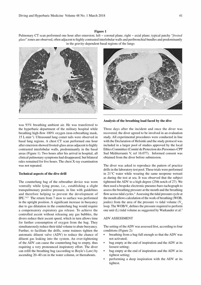
Diving and Hyperbaric Medicine Volume 48 No. 1 March 2018 41
was 93% breathing ambient air. He was transferred to the hyperbaric department of the military hospital while breathing high-flow 100% oxygen (non-rebreathing mask, 15 L∙min-1). Ultrasound lung comet tails were observed in basal lung regions. A chest CT scan performed one hour after emersion showed frosted-glass areas adjacent to highly contrasted interlobular walls, predominantly in the basal areas (Figure 1). Two hours after his arrival in hospital, all clinical pulmonary symptoms had disappeared, but bilateral rales remained for five hours. The chest X-ray examination was not repeated.
Technical aspects of the dive drill
The counterlung bag of the rebreather device was worn ventrally while lying prone, i.e., establishing a slight transpulmonary positive pressure, in line with guidelines and therefore helping to prevent the development of IPE.3,4,5 The return from 7 msw to surface was performed in the upright position. A significant increase in buoyancy due to gas dilatation in the counterlung bag would require a compensatory expiratory gas release. To achieve the controlled ascent without releasing any gas bubbles, the divers reduce their ascent speed, which in turn allows time for further consumption of oxygen from the bag. They simultaneously reduce their tidal volume to abate buoyancy. Further, to facilitate the drills, some trainees tighten the automatic diluent valve (ADV) to reduce the chance of diluent gas leaking into the system. An over-tightening of the ADV can cause the counterlung bag to empty, thus requiring a very pronounced inspiratory effort. The diver can refill the breathing bag (according to Boyle’s Law) by ascending 20–40 cm in the water column, or thereabouts.
Analysis of the breathing load faced by the dive
Three days after the incident and once the diver was recovered, the diver agreed to be involved in an evaluation study. All experimental procedures were conducted in line with the Declaration of Helsinki and the study protocol was included in a larger pool of studies approved by the local Ethics Committee (Comité de Protection des Personnes-CPP Sud Méditerranée V, ref 16.077). Informed consent was obtained from the diver before submission.
The diver was asked to reproduce the pattern of practice drills in the laboratory test pool. These trials were performed in 21°C water while wearing the same neoprene wetsuit as during the test at sea. It was observed that the subject tightened the ADV to a high degree (25th notch of 27). We then used a bespoke electronic pneumo-baro-tachograph to assess the breathing pressure at the mouth and the breathing flow across tidal cycles.4 Assessing the tidal pressure cycle at the mouth allows calculation of the work of breathing (WOB, joules) from the area of the pressure vs tidal volume (V
t)
loop. The WOB/Vt defines the pressure required to perform
one unit (L) tidal volume as suggested by Warkander et al.5
ADV ASSESSMENT
The setting of the ADV was assessed first, according to four conditions (Figure 2):• breathing from a bag full enough so that the ADV was
not activated;• bag empty at the end of inspiration and the ADV at its
loosest setting;• bag empty at the end of inspiration and the ADV at its
tightest setting;• performing a deep inspiration with the ADV at its
tightest.
Figure 1Pulmonary CT scan performed one hour after emersion; left − coronal plane, right − axial plane; typical patchy “frosted glass” zones are observed, often adjacent to highly contrasted interlobular walls and peribronchial bundles and predominantly
in the gravity-dependent basal regions of the lungs
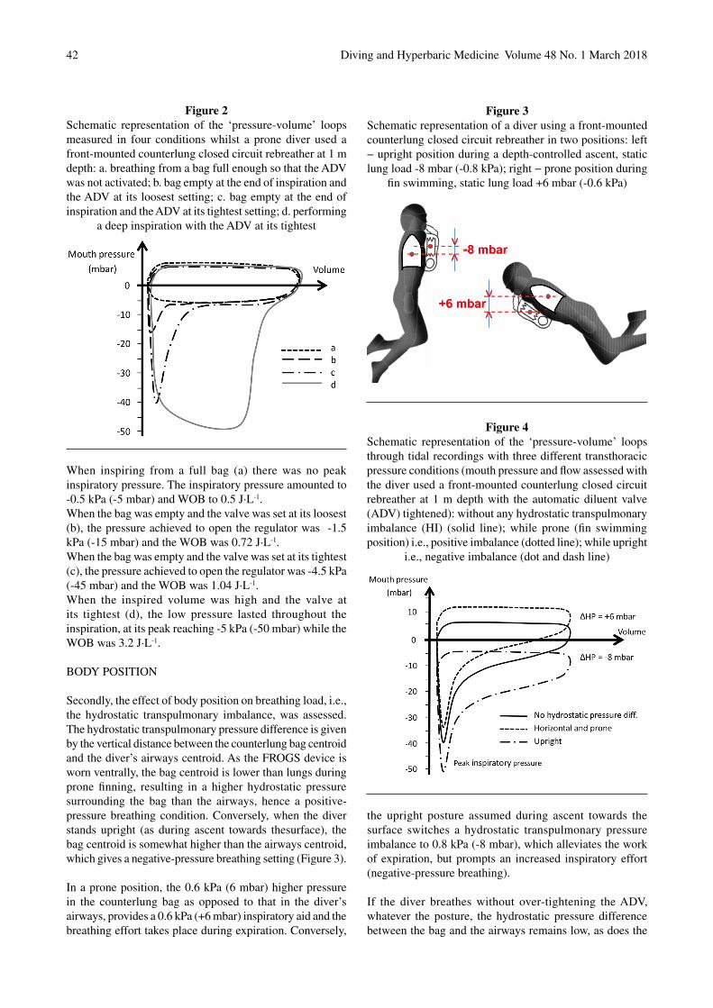
Diving and Hyperbaric Medicine Volume 48 No. 1 March 201842
When inspiring from a full bag (a) there was no peak inspiratory pressure. The inspiratory pressure amounted to -0.5 kPa (-5 mbar) and WOB to 0.5 J∙L-1.When the bag was empty and the valve was set at its loosest (b), the pressure achieved to open the regulator was -1.5 kPa (-15 mbar) and the WOB was 0.72 J∙L-1.When the bag was empty and the valve was set at its tightest (c), the pressure achieved to open the regulator was -4.5 kPa (-45 mbar) and the WOB was 1.04 J∙L-1.When the inspired volume was high and the valve at its tightest (d), the low pressure lasted throughout the inspiration, at its peak reaching -5 kPa (-50 mbar) while the WOB was 3.2 J∙L-1.
BODY POSITION
Secondly, the effect of body position on breathing load, i.e., the hydrostatic transpulmonary imbalance, was assessed. The hydrostatic transpulmonary pressure difference is given by the vertical distance between the counterlung bag centroid and the diver’s airways centroid. As the FROGS device is worn ventrally, the bag centroid is lower than lungs during prone finning, resulting in a higher hydrostatic pressure surrounding the bag than the airways, hence a positive-pressure breathing condition. Conversely, when the diver stands upright (as during ascent towards thesurface), the bag centroid is somewhat higher than the airways centroid, which gives a negative-pressure breathing setting (Figure 3). In a prone position, the 0.6 kPa (6 mbar) higher pressure in the counterlung bag as opposed to that in the diver’s airways, provides a 0.6 kPa (+6 mbar) inspiratory aid and the breathing effort takes place during expiration. Conversely,
the upright posture assumed during ascent towards the surface switches a hydrostatic transpulmonary pressure imbalance to 0.8 kPa (-8 mbar), which alleviates the work of expiration, but prompts an increased inspiratory effort (negative-pressure breathing).
If the diver breathes without over-tightening the ADV, whatever the posture, the hydrostatic pressure difference between the bag and the airways remains low, as does the
Figure 2Schematic representation of the ‘pressure-volume’ loops measured in four conditions whilst a prone diver used a front-mounted counterlung closed circuit rebreather at 1 m depth: a. breathing from a bag full enough so that the ADV was not activated; b. bag empty at the end of inspiration and the ADV at its loosest setting; c. bag empty at the end of inspiration and the ADV at its tightest setting; d. performing
a deep inspiration with the ADV at its tightest
Figure 3Schematic representation of a diver using a front-mounted counterlung closed circuit rebreather in two positions: left − upright position during a depth-controlled ascent, static lung load -8 mbar (-0.8 kPa); right − prone position during
fin swimming, static lung load +6 mbar (-0.6 kPa)
Figure 4Schematic representation of the ‘pressure-volume’ loops through tidal recordings with three different transthoracic pressure conditions (mouth pressure and flow assessed with the diver used a front-mounted counterlung closed circuit rebreather at 1 m depth with the automatic diluent valve (ADV) tightened): without any hydrostatic transpulmonary imbalance (HI) (solid line); while prone (fin swimming position) i.e., positive imbalance (dotted line); while upright
i.e., negative imbalance (dot and dash line)
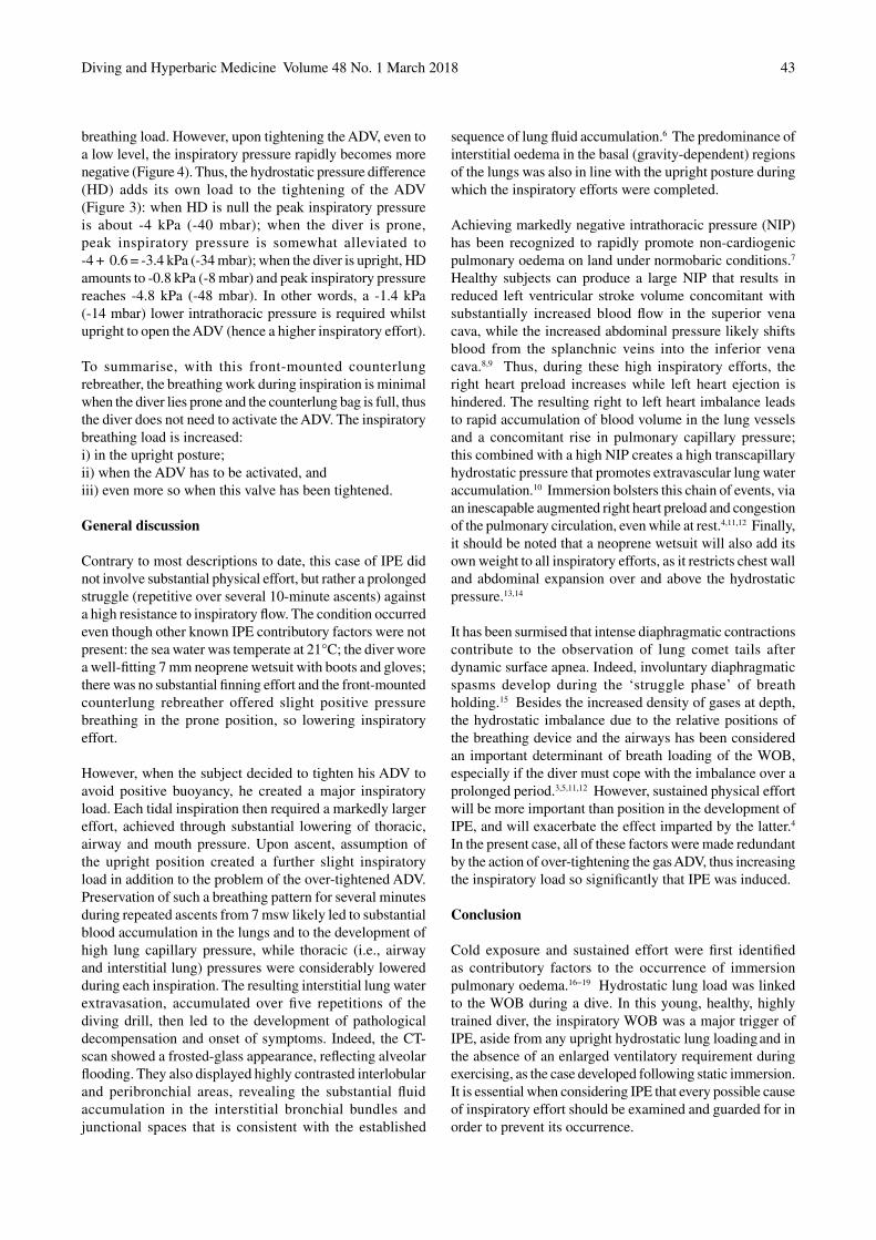
Diving and Hyperbaric Medicine Volume 48 No. 1 March 2018 43
breathing load. However, upon tightening the ADV, even to a low level, the inspiratory pressure rapidly becomes more negative (Figure 4). Thus, the hydrostatic pressure difference (HD) adds its own load to the tightening of the ADV(Figure 3): when HD is null the peak inspiratory pressure is about -4 kPa (-40 mbar); when the diver is prone, peak inspiratory pressure is somewhat alleviated to-4 + 0.6 = -3.4 kPa (-34 mbar); when the diver is upright, HD amounts to -0.8 kPa (-8 mbar) and peak inspiratory pressure reaches -4.8 kPa (-48 mbar). In other words, a -1.4 kPa(-14 mbar) lower intrathoracic pressure is required whilst upright to open the ADV (hence a higher inspiratory effort).
To summarise, with this front-mounted counterlung rebreather, the breathing work during inspiration is minimal when the diver lies prone and the counterlung bag is full, thus the diver does not need to activate the ADV. The inspiratory breathing load is increased:i) in the upright posture;ii) when the ADV has to be activated, andiii) even more so when this valve has been tightened.
General discussion
Contrary to most descriptions to date, this case of IPE did not involve substantial physical effort, but rather a prolonged struggle (repetitive over several 10-minute ascents) against a high resistance to inspiratory flow. The condition occurred even though other known IPE contributory factors were not present: the sea water was temperate at 21°C; the diver wore a well-fitting 7 mm neoprene wetsuit with boots and gloves; there was no substantial finning effort and the front-mounted counterlung rebreather offered slight positive pressure breathing in the prone position, so lowering inspiratory effort.
However, when the subject decided to tighten his ADV to avoid positive buoyancy, he created a major inspiratory load. Each tidal inspiration then required a markedly larger effort, achieved through substantial lowering of thoracic, airway and mouth pressure. Upon ascent, assumption of the upright position created a further slight inspiratory load in addition to the problem of the over-tightened ADV. Preservation of such a breathing pattern for several minutes during repeated ascents from 7 msw likely led to substantial blood accumulation in the lungs and to the development of high lung capillary pressure, while thoracic (i.e., airway and interstitial lung) pressures were considerably lowered during each inspiration. The resulting interstitial lung water extravasation, accumulated over five repetitions of the diving drill, then led to the development of pathological decompensation and onset of symptoms. Indeed, the CT-scan showed a frosted-glass appearance, reflecting alveolar flooding. They also displayed highly contrasted interlobular and peribronchial areas, revealing the substantial fluid accumulation in the interstitial bronchial bundles and junctional spaces that is consistent with the established
sequence of lung fluid accumulation.6 The predominance of interstitial oedema in the basal (gravity-dependent) regions of the lungs was also in line with the upright posture during which the inspiratory efforts were completed.
Achieving markedly negative intrathoracic pressure (NIP) has been recognized to rapidly promote non-cardiogenic pulmonary oedema on land under normobaric conditions.7 Healthy subjects can produce a large NIP that results in reduced left ventricular stroke volume concomitant with substantially increased blood flow in the superior vena cava, while the increased abdominal pressure likely shifts blood from the splanchnic veins into the inferior vena cava.8,9 Thus, during these high inspiratory efforts, the right heart preload increases while left heart ejection is hindered. The resulting right to left heart imbalance leads to rapid accumulation of blood volume in the lung vessels and a concomitant rise in pulmonary capillary pressure; this combined with a high NIP creates a high transcapillary hydrostatic pressure that promotes extravascular lung water accumulation.10 Immersion bolsters this chain of events, via an inescapable augmented right heart preload and congestion of the pulmonary circulation, even while at rest.4,11,12 Finally, it should be noted that a neoprene wetsuit will also add its own weight to all inspiratory efforts, as it restricts chest wall and abdominal expansion over and above the hydrostatic pressure.13,14
It has been surmised that intense diaphragmatic contractions contribute to the observation of lung comet tails after dynamic surface apnea. Indeed, involuntary diaphragmatic spasms develop during the ‘struggle phase’ of breath holding.15 Besides the increased density of gases at depth, the hydrostatic imbalance due to the relative positions of the breathing device and the airways has been considered an important determinant of breath loading of the WOB, especially if the diver must cope with the imbalance over a prolonged period.3,5,11,12 However, sustained physical effort will be more important than position in the development of IPE, and will exacerbate the effect imparted by the latter.4 In the present case, all of these factors were made redundant by the action of over-tightening the gas ADV, thus increasing the inspiratory load so significantly that IPE was induced.
Conclusion
Cold exposure and sustained effort were first identified as contributory factors to the occurrence of immersion pulmonary oedema.16−19 Hydrostatic lung load was linked to the WOB during a dive. In this young, healthy, highly trained diver, the inspiratory WOB was a major trigger of IPE, aside from any upright hydrostatic lung loading and in the absence of an enlarged ventilatory requirement during exercising, as the case developed following static immersion. It is essential when considering IPE that every possible cause of inspiratory effort should be examined and guarded for in order to prevent its occurrence.
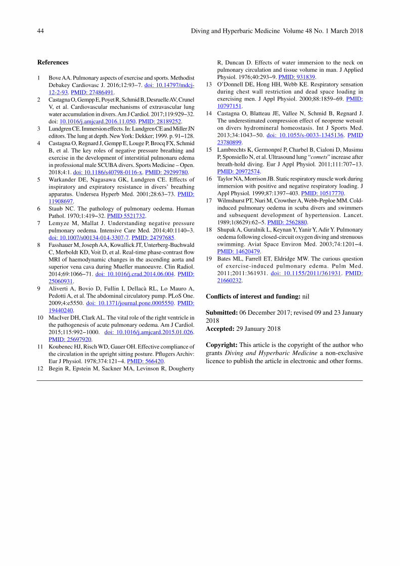
Diving and Hyperbaric Medicine Volume 48 No. 1 March 201844
References
1 Bove AA. Pulmonary aspects of exercise and sports. Methodist Debakey Cardiovasc J. 2016;12:93−7. doi: 10.14797/mdcj-12-2-93. PMID: 27486491.
2 Castagna O, Gempp E, Poyet R, Schmid B, Desruelle AV, Crunel V, et al. Cardiovascular mechanisms of extravascular lung water accumulation in divers. Am J Cardiol. 2017;119:929−32. doi: 10.1016/j.amjcard.2016.11.050. PMID: 28189252.
3 Lundgren CE. Immersion effects. In: Lundgren CE and Miller JN editors. The lung at depth. New York: Dekker; 1999. p. 91−128.
4 Castagna O, Regnard J, Gempp E, Louge P, Brocq FX, Schmid B, et al. The key roles of negative pressure breathing and exercise in the development of interstitial pulmonaru edema in professional male SCUBA divers. Sports Medicine – Open. 2018;4:1. doi: 10.1186/s40798-0116-x. PMID: 29299780.
5 Warkander DE, Nagasawa GK, Lundgren CE. Effects of inspiratory and expiratory resistance in divers’ breathing apparatus. Undersea Hyperb Med. 2001;28:63−73. PMID: 11908697.
6 Staub NC. The pathology of pulmonary oedema. Human Pathol. 1970;1:419−32. PMID 5521732.
7 Lemyze M, Mallat J. Understanding negative pressure pulmonary oedema. Intensive Care Med. 2014;40:1140−3. doi: 10.1007/s00134-014-3307-7. PMID: 24797685.
8 Fasshauer M, Joseph AA, Kowallick JT, Unterberg-Buchwald C, Merboldt KD, Voit D, et al. Real-time phase-contrast flow MRI of haemodynamic changes in the ascending aorta and superior vena cava during Mueller manoeuvre. Clin Radiol. 2014;69:1066−71. doi: 10.1016/j.crad.2014.06.004. PMID: 25060931.
9 Aliverti A, Bovio D, Fullin I, Dellacà RL, Lo Mauro A, Pedotti A, et al. The abdominal circulatory pump. PLoS One. 2009;4:e5550. doi: 10.1371/journal.pone.0005550. PMID: 19440240.
10 MacIver DH, Clark AL. The vital role of the right ventricle in the pathogenesis of acute pulmonary oedema. Am J Cardiol. 2015;115:992−1000. doi: 10.1016/j.amjcard.2015.01.026. PMID: 25697920.
11 Koubenec HJ, Risch WD, Gauer OH. Effective compliance of the circulation in the upright sitting posture. Pflugers Archiv: Eur J Physiol. 1978;374:121−4. PMID: 566420.
12 Begin R, Epstein M, Sackner MA, Levinson R, Dougherty
R, Duncan D. Effects of water immersion to the neck on pulmonary circulation and tissue volume in man. J Applied Physiol. 1976;40:293−9. PMID: 931839.
13 O’Donnell DE, Hong HH, Webb KE. Respiratory sensation during chest wall restriction and dead space loading in exercising men. J Appl Physiol. 2000;88:1859−69. PMID: 10797151.
14 Castagna O, Blatteau JE, Vallee N, Schmid B, Regnard J. The underestimated compression effect of neoprene wetsuit on divers hydromineral homeostasis. Int J Sports Med. 2013;34:1043−50. doi: 10.1055/s-0033-1345136. PMID 23780899.
15 Lambrechts K, Germonpré P, Charbel B, Cialoni D, Musimu P, Sponsiello N, et al. Ultrasound lung “comets” increase after breath-hold diving. Eur J Appl Physiol. 2011;111:707−13. PMID: 20972574.
16 Taylor NA, Morrison JB. Static respiratory muscle work during immersion with positive and negative respiratory loading. J Appl Physiol. 1999;87:1397−403. PMID: 10517770.
17 Wilmshurst PT, Nuri M, Crowther A, Webb-Peploe MM. Cold-induced pulmonary oedema in scuba divers and swimmers and subsequent development of hypertension. Lancet. 1989;1(8629):62−5. PMID: 2562880.
18 Shupak A, Guralnik L, Keynan Y, Yanir Y, Adir Y. Pulmonary oedema following closed-circuit oxygen diving and strenuous swimming. Aviat Space Environ Med. 2003;74:1201−4. PMID: 14620479.
19 Bates ML, Farrell ET, Eldridge MW. The curious question of exercise-induced pulmonary edema. Pulm Med. 2011;2011:361931. doi: 10.1155/2011/361931. PMID: 21660232.
Conflicts of interest and funding: nil
Submitted: 06 December 2017; revised 09 and 23 January 2018Accepted: 29 January 2018
Copyright: This article is the copyright of the author who grants Diving and Hyperbaric Medicine a non-exclusive licence to publish the article in electronic and other forms.

Diving and Hyperbaric Medicine Volume 48 No. 1 March 2018 45
Consensus guidelinePre-hospital management of decompression illness: expert review of key principles and controversiesSimon J Mitchell1, Michael H Bennett2, Phillip Bryson3, Frank K Butler4, David J Doolette5, James R Holm6, Jacek Kot7, Pierre Lafère8
1 Department of Anaesthesiology, University of Auckland, Auckland, New Zealand2 University of New South Wales, Sydney, Australia3 International SOS, Aberdeen, United Kingdom4 Joint Trauma System, Defence Center of Excellence for Trauma, San Antonio, USA5 University of Auckland, Auckland6 Virginia Mason Medical Centre, Seattle, USA.7 National Centre for Hyperbaric Medicine, Institute of Maritime and Tropical Medicine in Gdynia, Medical University of Gdansk, Poland8 ORPHY Laboratory EA4324, Université de Bretagne Occidentale, Brest, FranceCorresponding author: Professor Simon J Mitchell, Department of Anaesthesiology, School of Medicine, University of Auckland, Private Bag 92019, Auckland, New [email protected]
Key wordsDecompression sickness; Arterial gas embolism; Recompression; Remote locations; First aid; In-water recompression (IWR); Transport
Abstract
(Mitchell SJ, Bennett MH, Bryson P, Butler FK, Doolette DJ, Holm JR, Kot J, Lafère P. Pre-hospital management of decompression illness: expert review of key principles and controversies. Diving and Hyperbaric Medicine. 2018 March;48(1):45–55. doi: 10.28920/dhm48.1.45-55. PMID: 29557102.)Guidelines for the pre-hospital management of decompression illness (DCI) had not been formally revised since the 2004 Divers Alert Network/Undersea and Hyperbaric Medical Society workshop held in Sydney, entitled “Management of mild or marginal decompression illness in remote locations”. A contemporary review was initiated by the Diver’s Alert Network and undertaken by a multinational committee with members from Australasia, the USA and Europe. The process began with literature reviews by designated committee members on: the diagnosis of DCI; first aid strategies for DCI; remote triage of possible DCI victims by diving medicine experts; evacuation of DCI victims; effect of delay to recompression in DCI; pitfalls in management when DCI victims present at hospitals without diving medicine expertise and in-water recompression. This was followed by presentation of those reviews at a dedicated workshop at the 2017 UHMS Annual Meeting, discussion by registrants at that workshop and finally several committee meetings to formulate statements addressing points considered of prime importance to the management of DCI in the field. The committee placed particular emphasis on resolving controversies around the definition of “mild DCI” arising over 12 years of practical application of the 2004 workshop’s findings, and on the controversial issue of in-water recompression. The guideline statements are promulgated in this paper. The full workshop proceedings are in preparation for publication.
Introduction
Decompression illness (DCI) is a collective term which embraces decompression sickness (DCS) and arterial gas embolism (AGE);1 two dysbaric pathologies in which bubbles are presumed to be the primary vectors of injury. In the former, bubbles form in tissues and/or venous blood from dissolved inert gas absorbed during the dive and, in the latter, bubbles are introduced into the arterial circulation by pulmonary barotrauma. These pathologies are described in detail elsewhere.1 In practice, while DCS is more commonly seen than AGE, some manifestations are potentially common
to both and management is generally the same for both. Therefore, the collective term ‘DCI’ is used here except where there is a need to refer to either pathology specifically.
DCI may present with a wide range of symptoms of variable specificity and severity.1 Some presentations are mild and unlikely to result in long-term harm even without medical management, whereas some are potentially disabling or even life threatening and require therapeutic intervention. After the reported success of recompression in 1909,2 it became a quasi-standard of care for DCI. Between 1939 and 1965, treatment tables utilizing oxygen (O
2) breathing

Diving and Hyperbaric Medicine Volume 48 No. 1 March 201846
were developed,3−5 and recompression with hyperbaric O
2 has similarly become a standard of care. In the early
2000s, as dive travel to remote locations gathered pace, the perception that recompression was necessary for all cases of DCI irrespective of severity became troublesome. Increasing numbers of seemingly mild DCI cases were occurring in remote locations where evacuations for treatment were logistically difficult, very expensive and potentially hazardous.
These challenges motivated consideration of whether some DCI cases might not require evacuation and could be managed without recompression. A workshop entitled Management of mild or marginal decompression illness in remote locations workshop (henceforth referred to as the ‘2004 workshop’) was conducted as a two-day pre-course to the 2004 Undersea and Hyperbaric Medical Society Annual Scientific Meeting in Sydney.6 A series of presentations on various aspects of pre-hospital management of DCI were given by recognised experts. Commentary during discussion sessions was invited from all attendees, but final decisions on the consensus statements were taken among a group of 25 ‘discussants’ who were all experienced diving physicians from a broad range of nations. The most significant outcome of the 2004 workshop was a consensus that DCI presentations conforming to a definition of ‘mild’ could be adequately managed without recompression. The symptoms and signs included in the mild category were musculoskeletal pain, rash, constitutional symptoms and some cutaneous sensory changes. These manifestations were further characterised by explanatory footnotes, as were other criteria required for the mild definition to be applied.
The 2004 workshop definition of mild DCI has been widely applied in making decisions not to recompress, usually in situations where recompression would be difficult to access. It is fascinating to reflect on how this paradigm considered radical in 2004 has subsequently come to be viewed as routine practice. Indeed, aspects of the definition of mild are now considered by many as being too restrictive. In particular, the 2004 workshop consensus stipulated that in order for a case to be considered mild there must be a neurological examination by a doctor to exclude non-obvious but significant neurological signs. Such an examination may not be readily available in remote locations.
Other recent attempts to review the necessity for a neurological examination in designating mild DCI were made at diving medicine conferences in 2013 and 2016. A number of commentators suggested it was already relatively common practice for diving medicine physicians remotely triaging injured divers to waive the need for a neurological examination in designating a case as mild if, based on evaluation of the available information, they were comfortable that significant neurological manifestations were very unlikely. This approach appeared popular but no process for codifying or quantifying the participants’ views was achieved and no proceedings were ever published. If
nothing else, the recurrence of this subject on multiple diving medical society agendas suggests that it justifies attention.
Another controversial issue of high relevance to pre-hospital management of DCI is that of in-water recompression (IWR). The primary indication for IWR is to rapidly initiate treatment for DCI when a recompression chamber is not readily available. However, during IWR it is not possible to provide other medical care, the patient is exposed to environmental stresses, and a convulsion due to central nervous system (CNS) oxygen toxicity could result in drowning. As a result, IWR schedules are typically shallower and shorter than standard treatment tables used in recompression chambers. It is difficult to evaluate the benefits of IWR versus its recognized risks.
There are compelling reasons to consider IWR when evaluating the pre-hospital management of DCI. First, IWR is happening. IWR has and continues to be actively promoted by prominent diving physicianss for use by diving fishermen operating in locations remote from recompression chamber facilities.1,7,8 Second, recreational diving is increasingly taking place in remote locations without ready access to recompression chamber facilities. Third, with the increase in so-called technical diving there are more diving operations with the requisite equipment and skill mix that might be considered appropriate for conduct of IWR.9 There is no documentation of how frequently technical divers are using IWR, but one technical diving training organization has begun conducting training specifically in IWR methods.10 It is the existence of technical divers in the modern diving milieu that perhaps most strongly justifies a revision of the medical community’s perspectives on IWR. Finally, divers suffering neurological DCI are often left with residual neurological problems despite evacuation for recompression.11,12 There is a widely held belief that early recompression may be associated with better outcomes in such cases and IWR offers an obvious opportunity for this.
The present initiative was instigated by the Diver’s Alert Network (DAN) who seek clarity from the medical community on the above controversies, within the framework of a broader review of guidelines for pre-hospital management of DCI. The process employed in generating these guidelines is presented below. This is followed by the consensus statements derived from that process. The discussion section provides contextualisation and justification of some potentially controversial statements.
Methods
Representatives of DAN America and DAN Europe jointly approached one of the authors (SJM) to chair a committee of experts tasked with reviewing guidelines for pre-hospital management of DCI. The following criteria were applied to committee membership:• No committee members would be employees of DAN,
have relevant conflicts of interest, or receive any

Diving and Hyperbaric Medicine Volume 48 No. 1 March 2018 47
remuneration for participation;• With one exception (see below) all committee members
would be highly experienced diving medicine physicians actively involved in treating divers with DCI;
• One committee member would be a non-physician diver;• The committee membership would be drawn from
various regions to provide a global perspective.Potential committee members were identified in joint discussions between the chair and DAN representatives and the final composition is reflected in the authorship. The diver representative (author DJD) is an active technical diver and a decompression physiologist. Members were drawn from Australasia (two), the USA (three) and Europe (three).
The approach to deriving the consensus statements was similar to the one adopted for the 2004 workshop. The committee’s deliberations were based around a pre-course workshop held prior to the 2017 Undersea and Hyperbaric Medical Society Annual Scientific Meeting. Topics of relevance to pre-hospital management of DCI were identified as follows:• Presentations of DCI and diagnostic pearls;• First-aid strategies for DCI and the evidence
underpinning them;• Common pitfalls when divers present to hospitals or
doctors without expertise in diving medicine;• Remote triage of the possible DCI case by diving
medicine experts;• Transportation of a diver with DCI and the effect of
increasing delay to recompression on outcome;• The controversial issue of in water recompression.
Each topic was allocated to a committee member who presented it at the pre-course and produced a manuscript for the proceedings. Where appropriate, the presenter was also required to generate a series of draft consensus statements related to their topic for subsequent discussion by the committee. To be clear, it was not the intent to comprehensively embrace all facets of the topic areas (and therefore all aspects of pre-hospital management of DCI) within the consensus statements. That level of detail will be contained in the published proceedings. The consensus statements presented here are intended to address principles that were considered deserving of emphasis. Some are simply re-statements of widely accepted and non-controversial principles, whilst others address more controversial issues.
Each presentation was followed by 30 minutes for questions and commentary involving any of the 55 registrants who wished to contribute. The chair kept notes of this commentary. On each of the following two days the committee met in private for four hours to discuss, modify and finalise the draft consensus statements proposed for the relevant subject areas. We prospectively determined that any statement upon which we could not agree unanimously would be subjected to a majority-rules vote, and that the need for a vote and its result would be reported.
Consideration was given to applying a formal classification of evidence to our consensus statements. However, it was decided that any system chosen would be difficult to apply to an area of practice that is poorly informed by directly relevant human data, frequently based on indirectly evidence, such as inferences from animal data, and influenced heavily by observational studies and anecdote. It was determined that simply describing any relevant evidence would be the best option under the circumstances.
Results
The committee’s consensus on important matters are presented in Table 1. All were accepted unanimously.
Discussion
These statements represent practice recommendations issued by a committee of experts after a review process comprised sequentially of a literature review, presentation and discussion at a conference event convened for the purpose followed by two half-day committee meetings. Many of the statements constitute endorsement of previously established and widely accepted practice. The statements in relation to defining mild DCI draw heavily on the findings of the 2004 workshop. However, there are several that represent important modifications. These include: the addition of subcutaneous swelling (lymphatic DCI) to the mild category; the softening of the requirement for a neurological examination by a doctor before classifying a case of DCI as mild; and the conditional recognition of in-water recompression (IWR) as a legitimate option in the management of DCI. These are modifications to previously held positions that merit further discussion.
SUBCUTANEOUS SWELLING (LYMPHATIC DCI) AS A MILD MANIFESTATION
There are several reasons why lymphatic DCI was added to the definition of mild DCI established by the 2004 workshop. First, lymphatic DCI can occur as an isolated manifestation in divers who remain otherwise well. Second, there is no clear association between lymphatic symptoms and concurrent appearance of other more serious manifestations. Third, the value of recompression for lymphatic manifestations is unknown, but certainly not obvious. Recompression often seems to make little difference to the presence of the swelling which typically resolves spontaneously over 24–48 hours. Finally, there appear to be no long-term consequences of lymphatic swelling in DCI.
REMOTE CLASSIFICATION OF DCI AS MILD WITHOUT A NEUROLOGICAL EXAMINATION
The matter of whether a neurological examination by a doctor should be mandatory prior to diagnosing mild DCI (as defined in the preceding statements) was a key issue for the present committee to resolve after several recent consensus
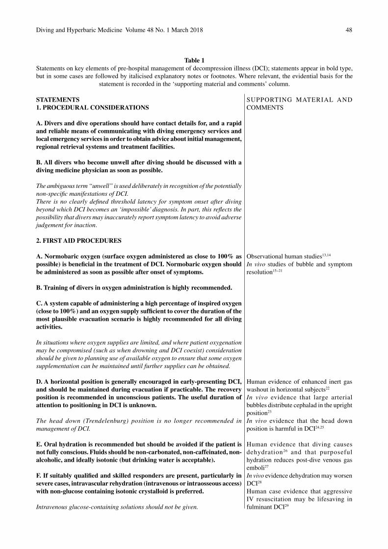
Diving and Hyperbaric Medicine Volume 48 No. 1 March 2018 48
STATEMENTS1. PROCEDURAL CONSIDERATIONS
A. Divers and dive operations should have contact details for, and a rapid and reliable means of communicating with diving emergency services and local emergency services in order to obtain advice about initial management, regional retrieval systems and treatment facilities.
B. All divers who become unwell after diving should be discussed with a diving medicine physician as soon as possible.
The ambiguous term “unwell” is used deliberately in recognition of the potentially non-specific manifestations of DCI.There is no clearly defined threshold latency for symptom onset after diving beyond which DCI becomes an ‘impossible’ diagnosis. In part, this reflects the possibility that divers may inaccurately report symptom latency to avoid adverse judgement for inaction.
2. FIRST AID PROCEDURES
A. Normobaric oxygen (surface oxygen administered as close to 100% as possible) is beneficial in the treatment of DCI. Normobaric oxygen should be administered as soon as possible after onset of symptoms.
B. Training of divers in oxygen administration is highly recommended.
C. A system capable of administering a high percentage of inspired oxygen (close to 100%) and an oxygen supply sufficient to cover the duration of the most plausible evacuation scenario is highly recommended for all diving activities.
In situations where oxygen supplies are limited, and where patient oxygenation may be compromised (such as when drowning and DCI coexist) consideration should be given to planning use of available oxygen to ensure that some oxygen supplementation can be maintained until further supplies can be obtained.
D. A horizontal position is generally encouraged in early-presenting DCI, and should be maintained during evacuation if practicable. The recovery position is recommended in unconscious patients. The useful duration of attention to positioning in DCI is unknown.
The head down (Trendelenburg) position is no longer recommended in management of DCI.
E. Oral hydration is recommended but should be avoided if the patient is not fully conscious. Fluids should be non-carbonated, non-caffeinated, non-alcoholic, and ideally isotonic (but drinking water is acceptable).
F. If suitably qualified and skilled responders are present, particularly in severe cases, intravascular rehydration (intravenous or intraosseous access) with non-glucose containing isotonic crystalloid is preferred.
Intravenous glucose-containing solutions should not be given.
SUPPORTING MATERIAL AND COMMENTS
Observational human studies13,14
In vivo studies of bubble and symptom resolution15−21
Human evidence of enhanced inert gas washout in horizontal subjects22
In vivo evidence that large arterial bubbles distribute cephalad in the upright position23
In vivo evidence that the head down position is harmful in DCI24,25
Human evidence that diving causes dehydration26 and that purposeful hydration reduces post-dive venous gas emboli27
In vivo evidence dehydration may worsen DCI28
Human case evidence that aggressive IV resuscitation may be lifesaving in fulminant DCI29
Table 1Statements on key elements of pre-hospital management of decompression illness (DCI); statements appear in bold type, but in some cases are followed by italicised explanatory notes or footnotes. Where relevant, the evidential basis for the
statement is recorded in the ‘supporting material and comments’ column.
Diving and Hyperbaric Medicine Volume 48 No. 1 March 201849
G. Treatment with a non-steroidal anti-inflammatory drug (NSAID) is appropriate if there are no contraindications.
H. Other agents such as corticosteroids, pentoxyphylline, aspirin, lidocaine and nicergoline have been utilized by suitably qualified responders in early management of DCI but there is insufficient evidence to support or refute their application.
I. Divers should be kept thermally comfortable (warm but not hyperthermic). Hyperthermia should be avoided especially in cases with severe neurological signs and symptoms. For example, avoid exposure to the sun, unnecessary activity, or excess clothing.
3. TRIAGE BY TELEMEDICINE
A. The principle goals of triage are to:evaluate the likelihood that reported symptoms are DCI, another diving disorder, or a non-diving disorder;
advise on patient management and the need for evacuation to a specialist diving medical service for assessment and possible recompression treatment.
Triage in this context refers to consultation via telephone or some other means of communication with a diving medicine expert who is not present at the accident site.
B. With respect to DCI, ‘mild’ symptoms and signs are: limb pain (footnotes 1, 2); constitutional symptoms such as fatigue; some cutaneous sensory changes (3); rash; subcutaneous swelling (‘lymphatic DCI’) where these manifestations are static or remitting (4, 5) and significant (6) neurological dysfunction is excluded to the satisfaction of a diving medicine physician (7).
1. Severity of pain has little prognostic significance, but severity of pain may influence management decisions independent of the classification of pain as a ‘mild’ symptom.
2. Classical girdle pain syndromes are suggestive of spinal involvement and do not fall under the classification of “limb pain”.
3. The intent of “some cutaneous sensory changes” is to embrace subjective cutaneous sensory phenomena such as ‘tingling’ present in patchy or non-dermatomal distributions suggestive of non-spinal, non-specific, and benign processes. Subjective sensory changes in certain characteristic patterns such as in both feet, may predict evolution of spinal symptoms and should not be considered as ‘mild’.
4. If symptoms are qualitatively mild but are progressive, then the diver must be continuously monitored to detect any appearance of symptoms not considered mild. The ‘mild’ status cannot be considered final until symptoms are static or remitting.
5. The possibility of the delayed development of new symptoms means the ‘mild’ designation must be repeatedly reviewed over at least 24 hours following diving or the most recent decompression, the latter applying if there has been an ascent to altitude. Untreated mild symptoms and signs due to DCI are unlikely to progress after 24 hours from completion of diving.
Human RCT showing improved tempo of recovery in DCI using a NSAID as an adjuvant to hyperbaric oxygen30
Human evidence that warm subjects eliminate inert gas more quickly22,31
Mild hyperthermia worsens neurological injury in vivo32,33 and in humans34
Conclusion of the 2004 workshop6 with two changes:
1. Subcutaneous swelling added to the definition of mild DCI by the present committee (see discussion);
2. Criteria for exclusion of significant neurological dysfunction rephrased by the present committee (see discussion).
Diving and Hyperbaric Medicine Volume 48 No. 1 March 2018 50
6. “Significant” in this setting is intended to imply a problem that has the potential to leave the diver with functionally important sequelae.
7. Exclusion of significant neurological signs is most reliably achieved by a neurological examination performed by a doctor. However, such examination may not be available, and there are plausible scenarios in which a global appraisal of other facts of the case renders significant neurological injury extremely unlikely. In such scenarios it can be appropriate for a diving medicine physician to manage a case as ‘mild’ in the absence of a neurological examination.
C. Recompression therapy is the gold standard therapy for DCI. However, some divers with symptoms or signs meeting the above definition of mild DCI may be managed without recompression therapy.
The phrase ‘some divers’ is used intentionally. Divers with mild DCI will often be offered recompression if it is readily available because this will speed recovery.
A decision to invoke this guideline can only be made by a diving medicine physician on a case-by-case basis (see Guideline 1B). It is not to be used to formulate management policy for all divers with apparently mild DCI.
D. Divers diagnosed with mild DCI who do not undergo recompression should be treated in accordance with guideline 2 A-I for a duration at the discretion of the advising diving medicine physician. These divers should be monitored regularly for 24 hours to exclude development of new symptoms falling outside the mild definition.
4. EFFECT OF DELAY TO RECOMPRESSION
A. The best outcomes after recompression (especially in cases with more severe symptoms) are likely to be obtained by immediate recompression. The latter will only be possible if on-site recompression is available.
B. For cases suffering mild symptoms, a delay prior to recompression is unlikely to be associated with any worsening of long-term outcome.
C. In more serious presentations recompression should be obtained as soon as safely possible. There is limited evidence that delays longer than six hours result in slower or less complete recovery.
5. TRANSPORTATION OF A DCI PATIENT
A. Arrangements for transport of a diver with DCI should be agreed between the first responders, the triaging diving medicine physician, the receiving physician and the retrieval team before the evacuation begins.
B. If air evacuation is used, the aircraft should either be pressurized to one atmosphere or remain at a low-altitude where possible.
Low altitude in this context is preferably less than 150 m above pick-up location. The risk of greater altitude exposures should be balanced against the risk of deterioration if not retrieved and should be made in consultation with a diving medicine physician.
C. Some divers with mild symptoms or signs (defined above) after diving may be evacuated by commercial airliner to obtain treatment after a surface interval of at least 24 hours, and this is unlikely to be associated with worsening of outcome.
Conclusion of the 2004 workshop6
Conclusion of the 2004 workshop6
Observational human evidence that good outcomes are obtained with very early recompression35−37
Conclusion of the 2004 workshop6
Observational human evidence for an inflection in risk of less complete recovery with recompression latencies longer than six hours11,12,38
Conclusion of the 2004 workshop6

Diving and Hyperbaric Medicine Volume 48 No. 1 March 201851
Most favourable experience with commercial airliner evacuations comes from short haul flights of between one and two hours duration. There is much less experience with longer flights.
Provision of oxygen in as high an inspired percentage as possible is optimal practice for such evacuations. In addition, the risk of such evacuation will be reduced by pre-flight oxygen breathing.
6. IN-WATER RECOMPRESSION (IWR)
A. Recompression and hyperbaric oxygen administered in a recompression chamber is acknowledged as the gold standard of care for DCI. However, in locations without ready access to a suitable hyperbaric chamber facility, and if symptoms are significant or progressing, in-water recompression using oxygen is an option. This is only appropriate where groups of divers (including the ‘patient’) have prior relevant training (see below) that imparts an understanding of related risks and facilitates a collective acceptance of responsibility for the decision to proceed.
B. IWR should not be conducted if there is hearing loss, vertigo, vomiting, altered level of consciousness, shock, respiratory distress or a degree of physical incapacitation that makes return underwater unsafe.
C. The team, which at a minimum includes the patient, a dive buddy who will accompany the patient throughout the in-water recompression, and a surface supervisor, must all be trained, certified and practiced in decompression procedures using 100% oxygen underwater.
D. The team must be suitably equipped for IWR using oxygen including: adequate thermal protection; an adequate oxygen supply and a means of supplying 100% oxygen (or close to it) for the duration of the anticipated protocol (both in-water and surface phases); a means of maintaining stable depth; a method of communication (e.g., a slate). A full-face mask or mouthpiece retaining device is strongly recommended.
E. IWR should be accomplished with the patient breathing 100% oxygen, and at a maximum depth of 9 msw (30 fsw), according to a recognized protocol. The use of breathing gases other than oxygen for IWR is not recommended.
Recognised protocols include the “Clipperton protocol”, “Australian method”, and the oxygen IWR method of the US Navy.
F. IWR may not result in complete resolution of DCI, and signs or symptoms may recur. Any injured diver completing an IWR procedure should be discussed with or reviewed by a diving medicine physician at the earliest possible opportunity.
Observational human evidence that very early recompression results in good outcomes,36,39−46 or better outcomes compared to longer delays37
Observational human evidence for the efficacy of mouthpiece retaining devices in preventing drowning after loss of consciousness underwater47
Published regimens for IWR,8,48−50 with some observational human evidence of efficacy8,49
initiatives failed to publish a conclusion. The committee considered four related options.
1. Retain the requirement for a competent neurological examination prior to remote classification of a DCI case as mild.There was historical support for this option. One paper in the 2004 workshop proceedings6 refers to datasets demonstrating the frequent co-existence of mild symptoms
and more serious neurological manifestations in divers with DCI.51 These data did not identify what proportion of such cases would have required a neurological examination to detect the serious neurological component (as opposed to detection by symptom history alone). However, the author cited anecdote from several authorities who, during comprehensive evaluation of divers, found neurological problems that were not reported in the referral history. These observations culminated in his conclusion:
Diving and Hyperbaric Medicine Volume 48 No. 1 March 201852
“Until a person with any decompression manifestation has been competently examined neurologically, there can be no confident prediction that they have only mild manifestations at that stage and do not need an urgent recompression”.51
This resonated strongly with the 2004 workshop discussants who were already grappling with the prospect of adopting a new and liberal approach to the management of DCI patients. The requirement for a neurological examination before that liberal approach could be invoked appealed as a safety net that would minimise the risk of inappropriate patient management decisions. The present committee retained an open mind on this option. However, practice recommendations that are increasingly ignored or modified in real world application (see Introduction) deserve scrutiny and possibly revision. The committee ultimately settled on a more nuanced approach (see option four below).
2. Eliminate the requirement for a neurological examination from the definition of mild DCI.Based on the 12-year experience with increasingly liberal application of the 2004 workshop findings, some diving medicine physicians have suggested removing any requirement for a neurological examination in defining mild DCI. However, the committee did not agree that wholesale rejection of the exam was wise, and as alluded to above, adopted a more nuanced approach.
3. Retain the neurological examination requirement, but widen the group who can administer it.It has been suggested that divers themselves could learn to administer a neurological examination thus widening the pool of available examiners in remote locations. Indeed, some diver training agencies already teach a ‘five-minute neuro’ screening examination to divers. However, it is unlikely that such examinations would be sufficiently sensitive in the present context, or that their findings could be defended in the face of critical scrutiny. It is difficult to teach effective neurological examination even to medical students, despite the fact they are knowledgeable and intelligent, taught by experts and have many opportunities to see patients with real neurological signs.52,53 The notion that effective neurological examination could be taught by diving instructors (who themselves have never seen an abnormal neurological sign) to diver students with no opportunities to see real signs or practice on patients must be considered with scepticism. At the very least, it seems debateable whether a remote diving medicine expert trying to decide whether to evacuate a sick diver could rely upon neurological examination findings recorded by another diver. The committee saw no harm in divers attempting neurological examinations and offering their findings to a remote diving medicine physician. However, we considered it impractical to formally codify a role for non-medical neurological examiners in best-practice recommendations.
4. Reword the relevant statement to allow a remote diving medicine physician more discretion over how neurological
function is assessed. The committee resolved to deal with this issue by changing the original wording in the 2004 workshop consensus from “…and associated objective neurological dysfunction has been excluded by medical examination” to “…and significant neurological dysfunction has been excluded to the satisfaction of a diving medicine physician.”
Although similar, there are some subtle but important changes in meaning. First, the emphasis has been shifted from detecting any objective neurological dysfunction to detecting “significant” neurological dysfunction. This reflects a view that neurological manifestations likely to result in disability can most often be detected by a blunter instrument than meticulous examination and will often be obvious to the diver (or an unskilled observer) and so reported as a symptom.
Secondly, the explicit reference to “medical examination” has been dropped in favour of a less-directive reference to “the satisfaction of a diving medicine physician”. A neurological examination by a doctor will still be part of achieving “satisfaction” in many (perhaps most) scenarios, but the revised wording leaves open the possibility that it might not. In the latter scenario, a remote diving medicine physician may feel they have excluded significant neurological dysfunction ‘to their satisfaction’ based on an appraisal of all the facts of the case and their own experience. It would be extremely difficult to codify a protocol for making such decisions because the circumstances under which they might be made are so varied.
Finally, the term “diving medicine physician” has been employed explicitly to imply that decisions invoking the definition of mild DCI in management decisions should be made by a physician with training and experience in the management of DCI, especially if the definition is to be applied in the absence of a neurological examination. Paramedics or inexperienced diving doctors should escalate such decisions to the most senior and experienced diving medicine physician accessible. Such practitioners are best positioned to filter the case information and apply their experience to interpreting the type of diving, the nature of the symptoms, the tempo of symptom onset, the time since diving and other relevant facts in deciding whether a neurological examination is necessary.
IN-WATER RECOMPRESSION
Prominent publications providing guidelines on treatment of DCI generally avoid the topic of IWR,54,55 or are discouraging.11 One contemporary textbook does provide supportive commentary and suggests an approach for implementation.1 Whatever the opinion of the wider diving medicine community, IWR has for some time been practiced by groups of sea harvesters (with support of interested medical groups), and by technical divers.7,8,48,49,56−60
Diving and Hyperbaric Medicine Volume 48 No. 1 March 2018 53
The principle argument in support of IWR is in scenarios where there is no realistic possibility of accessing recompression in a hyperbaric chamber, or to achieve recompression much more quickly than would be possible by evacuation to a hyperbaric facility. Unfortunately, there is little evidence for an outcome advantage for very early recompression because most attempts to quantify outcomes in DCI cases stratified by both severity and latency to recompression involve latencies much longer than can be achieved with IWR. One small study in military divers suggested recompression within one hour was associated with a better outcome than longer latencies.37 Also, there is considerable anecdote supporting good results with early recompression in military and commercial diving scenarios where hyperbaric chambers are immediately available. One member of the committee (DJD) synthesised data from multiple reports of US Navy test dive programmes where divers developing DCI were almost invariably recompressed rapidly. The data were broadly indicative of rapid and complete recovery in the vast majority of cases.35,36,38−46,61 This contrasted with a large series of recreational divers with much longer median recompression latency who required greater numbers of recompression treatments and exhibited a substantially higher incidence of residual symptoms on completion of hyperbaric treatment.62 This analysis will be reported in more detail in the 2017 workshop proceedings.
The principle argument against IWR is its perceived hazards. Arguably the most significant is an oxygen toxic seizure. The inspired oxygen partial pressure (PO
2) threshold
below which seizures never occur irrespective of exposure duration has not been defined but it is lower than usually recommended for IWR (typically 192 kPa [1.9 atm abs]); breathing 100% oxygen at 9 msw or 30 fsw).8,48−50 Whilst we are not aware of any reports of an oxygen toxicity event during IWR, seizures have certainly occurred in oxygen exposures of equivalent magnitude.63−65
In this regard, there is an obvious trade-off between increasing pressure to achieve bubble volume reduction and the safety of the inspired PO
2. The committee does not
support IWR at pressures greater than 192 kPa (1.9 atm abs). Greater safety could be achieved by limiting oxygen breathing to lower pressures where convulsions are rarer, but whereas there is some evidence for the efficacy of treatment of DCI at pressures near 1.9 atm,5 the extent to which lower pressures might compromise the efficacy of the intervention is unknown.
Mitigating the risk of adverse consequences of a seizure centres on protecting the airway. This can be achieved (though is not guaranteed) by the use of a full-face mask or a mouthpiece retaining strap.47 Other key risk management strategies include tethering the diver to a decompression stage throughout the recompression so they cannot sink in the event of loss of consciousness, and ensuring the diver is accompanied at all times so they can be rescued immediately to the surface if a seizure occurs.
Evaluation of contemporary real-world practice trends and of the potential benefits and risks of IWR led the committee to issue a related series of essentially positive statements with conditional references to the use of oxygen, the prior training of all participants (including the victim), maximum pressure, contraindications and equipment requirements. There are other aspects of this complex topic, such as patient selection, which will be further elaborated in the 2017 pre-course proceedings.
Conclusion
These guidelines for early management of DCI represent the consensus of a committee of experts. Many of the recommendations draw heavily on the collective experience of that expert group rather than on objective evidence. In much the same way as experience in application of the 2004 workshop guidelines has provided impetus and direction for aspects of this review, future experience with the present guidelines or the emergence of new experimental evidence may determine that these recommendations be reviewed and changed.
References
1 Edmonds C, Bennett MH, Lippmann J, Mitchell SJ, editors. Diving and subaquatic medicine, 5th ed. Boca Raton, FL: Taylor and Francis; 2015.
2 Keays FL. Compressed air illness, with a report of 3692 cases. Dept Med Pub Cornell Univ Med Coll. 1909;2:1–55.
3 Yarbrough OD Behnke AR. The treatment of compressed air illness utilizing oxygen. J Industr Hyg Toxicol. 1939;21:213-8.
4 Van der Aue OE, White WA Jr, Hayter R, Brinton ES, Kellar RJ, Behnke AR. Physiological factors underlying the prevention and treatment of decompression sickness. Research Report NEDU TR 1–45. Washington DC: Navy Experimental Diving Unit; 1945. Available from: http://archive.rubicon-foundation.org/3791. [cited 2017 December 06].
5 Goodman MW, Workman RD. Minimal-recompression, oxygen-breathing approach to treatment of decompression sickness in divers and aviators. Research Report NEDU TR 5–65. Washington DC: Navy Experimental Diving Unit; 1965. Available from: http://archive.rubicon-foundation.org/3342. [cited 2017 December 06].
6 Mitchell SJ, Doolette DJ, Wachholz C, Vann RD, editors. Management of mild or marginal decompression illness in remote locations. 2004 Workshop Proceedings. Washington, DC: Undersea and Hyperbaric Medical Society; 2005. Available from: http://archive.rubicon-foundation.org/5523. [cited 2017 December 06].
7 Edmonds C. Underwater oxygen treatment of DCS. In: Moon RE, Sheffield PJ, editors. Treatment of decompression illness. Proceedings of the 45th Undersea and Hyperbaric Medical Society Workshop. Kensington, MD: Undersea and Hyperbaric Medical Society; 1996. p. 255–66. Available from: http://archive.rubicon-foundation.org/7999. [cited 2017 December 06].
8 Blatteau JE, Pontier JM, Buzzacott P, Lambrechts K, Nguyen VM, Cavenel P, Ruffez J. Prevention and treatment of decompression sickness using training and in-water recompression among fisherman divers in Vietnam. Inj Prev. 2016;22:25–32. doi: 10.1136/injuryprev-2014-041464. PMID: 25991710.
Diving and Hyperbaric Medicine Volume 48 No. 1 March 201854
9 Mitchell SJ, Doolette DJ. Recreational technical diving part 1: an introduction to technical diving methods and activities. Diving Hyperb Med. 2013;43:86–93. PMID: 23813462.
10 Dituri J, Sadler R. Emergency management of stricken divers in remote locations. Lake City, FL: International Association of Nitrox and Technical Divers; 2015.
11 Moon RE, Gorman DF. Treatment of decompression disorders. In: Brubakk AO, Neuman,TS, editors. Bennett and Elliott’s physiology and medicine of diving, 5th ed. Edinburgh: Saunders; 2003.
12 Blatteau JE, Gempp E, Simon O, Coulange M, Delafosse B, Souday V et al. Prognostic factors of spinal cord decompression sickness in recreational diving: retrospective and multicentric analysis of 279 cases. Neurocrit Care. 2011;15:120–7. doi: 10.1007/s12028-010-9370-1. PMID: 20734244.
13 Longphre JM, Denoble PJ, Moon RE, Vann RD, Freiberger JJ. First aid normobaric oxygen for the treatment of recreational diving injuries. Undersea Hyperb Med. 2007;34:43–9. PMID: 17393938.
14 Krause KM, Pilmanis AA. The effectiveness of ground level oxygen treatment for altitude decompression sickness in human research subjects. Aviat Space Environ Med. 2000;71:115–8. PMID: 10685583.
15 Ber t P. La press ion barométr ique . Recherches de physiologie expérimentale. Paris: Masson; 1878. Available from: https://ia800303.us.archive.org/22/items/lapressionbarom00bertgoog/lapressionbarom00bertgoog.pdf. [cited 2017 Dec 06]. French.
16 Hyldegaard O, Madsen J. Influence of heliox, oxygen, and N
2O-O
2 breathing on N
2 bubbles in adipose tissue. Undersea
Biomed Res. 1989;16:185–93. PMID: 2741253.17 Hyldegaard O, Moller M, Madsen J. Effect of He-O2, O2, and
N2O-O2 breathing on injected bubbles in spinal white matter. Undersea Biomed Res. 1991;18:361–71. PMID: 1746064.
18 Hyldegaard O, Moller M, Madsen J. Protective effect of oxygen and heliox breathing during development of spinal decompression sickness. Undersea Hyperb Med. 1994;21:115–28. PMID: 8061554.
19 Hyldegaard O, Kerem D, Melamed Y. Effect of combined recompression and air, oxygen, or heliox breathing on air bubbles in rat tissues. J Appl Physiol. 2001;90:1639–47. doi: 10.1152/jappl.2001.90.5.1639. PMID: 11299250.
20 Gennser M, Loveman G, Seddon F, Thacker J, Blogg SL. Oxygen and carbogen breathing following simulated submarine escape. Undersea Hyperb Med. 2014;41:387–92. PMID: 25558547.
21 Loveman GAM, Seddon FM, Jurd KM, Thacker JC, Fisher AS. First aid oxygen treatment for decompression illness in the goat after simulated submarine escape. Aerosp Med Hum Perform. 2015;86:1020–7. doi: 10.3357/AMHP.4306.2015. PMID: 26630048.
22 Balldin U. Effects of ambient temperature and body position on tissue nitrogen elimination in man. Aerospace Med. 1973;44:365–70. PMID: 4694843.
23 Van Allen CM, Hrdina LS, Clark J. Air embolism from the pulmonary vein. Arch Surg. 1929;19:567–99.
24 Dutka AJ, Polychronidis J, Mink RB, Hallenbeck JM. Head-down position after air embolism impairs recovery of brain function as measured by the somatosensory evoked response in canines. Undersea Biomed Res. 1990;17(Suppl):64. Available from: http://archive.rubicon-foundation.org/7056. [cited 2018 February 18].
25 Polychronidis JE, Dutka AJ, Mink RB, Hallenbeck JM. Head down position after cerebral air embolism: effects on intracranial
pressure, pressure volume index and blood-brain barrier. Undersea Biomed Res. 1990;17(Suppl):99. Available from: http://archive.rubicon-foundation.org/7155. [cited 2018 Feb 18].
26 Williams ST, Prior FGR, Bryson P. Hematocrit change in tropical scuba divers. Wilderness Environ Med. 2007;18;48–53. PMID: 17447715.
27 Gempp E, Blatteau J-E, Pontier J-M, Balestra C, Louge P. Preventative effect of pre-dive hydration on bubble formation in divers. Br J Sports Med. 2009;43:224–8. doi: 10.1136/bjsm.2007.043240. PMID: 18308884.
28 Fahlmann A, Dromsky DM. Dehydration effects on the risk of severe decompression sickness in a swine model. Aviat Space Environ Med. 2006;77:102–6. PMID: 16491576.
29 Trytko B, Mitchell SJ. Extreme survival: a deep technical diving accident. SPUMS Journal. 2005;35,23–7.
30 Bennett MH, Mitchell SJ, Dominguez A. Adjunctive treatment of decompression illness with a non-steroidal anti-inflammatory drug reduces compression requirement. Undersea Hyperb Med. 2003;30:195–205. PMID: 14620099.
31 Pendergast DR, Senf CJ, Fletcher MC, Lundgren CEG. Effects of ambient temperature on nitrogen uptake and elimination in humans. Undersea Hyperb Med. 2015;42:85–94. PMID: 26094308.
32 Minamisawa H, Smith ML, Siesjo BK. The effect of mild hyperthermia and hypothermia on brain damage following 5, 10, and 15 minutes of forebrain ischemia. Ann Neurol. 1990;28:26–33. doi:10.1002/ana.410280107. PMID: 2375631.
33 Wass CT, Lanier WL, Hofer RE, Scheithauer BW, Andrews AG. Temperature changes of > or = 1 degree C alter functional neurologic outcome and histopathology in a canine model of complete cerebral ischemia. Anesthesiology. 1995;83:325–35. PMID: 7631955.
34 Greer DM, Funk SE, Reaven NL, Ouzounelli M, Uman GC. Impact of fever on outcome in patients with stroke and neurologic injury: a comprehensive meta-analysis. Stroke. 2008;39:3029–35. doi: 10.1161/STROKEAHA.108.521583. PMID: 18723420.
35 Green JW, Tichenor J, Curley MD. Treatment of type I decompression sickness using the U.S. Navy treatment algorithm. Undersea Biomed Res. 1989;16:465–70. PMID: 2603243.
36 Thalmann ED. Principles of U.S. Navy recompression treatments for decompression sickness. In: Moon RE, Sheffield PJ, editors. Treatment of decompression illness. Proceedings of the 45th Undersea and Hyperbaric Medical Society Workshop. Kensington MD: Undersea and Hyperbaric Medical Society; 1996. p. 75–95. Available from: http://archive.rubicon-foundation.org/7999. [cited 2017 Dec 06].
37 Blatteau JE, Gempp E, Constantin P, Louge P. Risk factors and clinical outcome in military divers with neurological decompression sickness: influence of time to recompression. Diving Hyperb Med. 2011;41:129–34.
38 Rivera JC. Decompression sickness among divers: an analysis of cases. Research Report NEDU TR 1-63. Washington DC: Navy Experimental Diving Unit; 1963. Available from: http://archive.rubicon-foundation.org/3858. [cited 2017 Dec 06].
39 Survanshi SS, Thalmann ED, Parker EC, Gummin DD, Isakov AP, Homer LD. Dry decompression procedure using oxygen for naval special warfare. Technical Report 97-03. Bethesda MD: Naval Medical Research Institute; 1997.
40 Survanshi SS, Parker EC, Gummin DD, Flynn ET, Toner CB, Temple DJ et al. Human decompression trial with 1.3 ATA oxygen in helium. Technical Report 98-09. Bethesda MD: Naval Medical Research Institute; 1998.
41 Thalmann ED, Kelleher PC, Survanshi SS, Parker EC,
Diving and Hyperbaric Medicine Volume 48 No. 1 March 2018 55
Weathersby PK. Statistically based decompression tables XI: manned validation of the LE probabilistic decompression model for air and nitrogen-oxygen diving. Joint Technical Report NEDU 1-99 and NMRC 99-01. Panama City FL: Navy Experimental Diving Unit and Naval Medical Research Center; 1999. Available from: http://archive.rubicon-foundation.org/3412. [cited 2017 Dec 27].
42 Hamilton RW, Thalmann ED, Flynn ET, Temple DJ. No-stop 60 fsw wet and dry dives using air, heliox, and oxygen-nitrogen mixtures. Data report on projects 88-06 and 88-06A. Technical Report 2002-002. Silver Spring MD: Naval Medical Research Center; 2002.
43 Hamilton RW, Thalmann ED, Temple DJ. Surface decompression diving. Data report on protocol 90-02. Technical Report 2003-001. Silver Spring MD: Naval Medical Research Center; 2003. Available from: http://archive.rubicon-foundation.org/4980. [cited 2017 Dec 27].
44 Gerth WA, Ruterbusch VL, Long ET. The influence of thermal exposure on diver susceptibility to decompression sickness. Technical Report NEDU TR 06-07. Panama City FL: Navy Experimental Diving Unit; 2007. Available from: http://archive.rubicon-foundation.org/5063. [cited 2017 Dec 27].
45 Doolette DJ, Gerth WA, Gault KA. Risk of central nervous system decompression sickness in air diving to no-stop limits. Technical Report NEDU TR 09-03. Panama City FL: Navy Experimental Diving Unit; 2009. Available from: http://archive.rubicon-foundation.org/9965. [cited 2017 Dec 27].
46 Doolette DJ, Gerth WA, Gault KA. Redistribution of decompression stop time from shallow to deep stops increases incidence of decompression sickness in air decompression dives. Technical Report NEDU TR 11-06. Panama City FL: Navy Experimental Diving Unit; 2011. Available from: http://archive.rubicon-foundation.org/10269. [cited 2017 Dec 27].
47 Gempp E, Louge P, Blatteau J-E, Hugon M. Descriptive epidemiology of 153 diving injuries with rebreathers among French military divers from 1979–2009. Mil Med. 2011;176;446–450. PMID: 21539168.
48 Blatteau J-E, Jean F, Pontier J-M, Blanche E, Bompar J-M, Meaudre E, Etienne J-M. [Decompression sickness accident management in remote areas. Use of immediate in-water recompression therapy. Review and elaboration of a new protocol targeted for a mission at Clipperton Atoll.] Ann Françaises d’Anesthésie et de Réanim. 2006;25:874–83. doi:10.1016/j.annfar.2006.04.007. PMID: 16860525. French.
49 Edmonds C. Pearls from the deep. A study of Australian pearl diving 1988-1991. SPUMS Journal. 2006;26(Supp):26–30.
50 Naval Sea Systems Command. U.S. Navy diving manual. Revision 7. Arlington (VA): Naval Sea Systems Command; 2016. Available from: http://www.navsea.navy.mil/Portals/103/Documents/SUPSALV/Diving/US%20DIVING%20MANUAL_REV7.pdf?ver=2017-01-11-102354-393. [cited 2017 Dec 27].
51 Elliott DH. The natural history of untreated decompression illness. In: Mitchell SJ, Doolette DJ, Wachholz C, Vann RD, editors. Management of mild or marginal decompression illness in remote locations 2004 Workshop Proceedings. Washington DC: Undersea and Hyperbaric Medical Society; 2005. p. 30–48.
52 Kamel H, Dhaliwal G, Navi BB, Pease AR, Shah M, Dhand A, Johnston SC, Josephson SA. A randomized trial of hypothesis-driven vs screening neurological examination. Neurology. 2011;77:1395–400. doi: 10.1212/WNL.0b013e3182315249. PMID: 21900631. PMCID:PMC3182756.
53 Wiles CM. Introducing neurological examination for medical undergraduates – how I do it. Pract Neurol. 2013;13:49–50.
doi: 10.1136/practneurol-2012-000282. PMID: 23315462.54 Vann RD, Butler FK, Mitchell SJ, Moon RE. Decompression
illness. Lancet. 2011;377:153–64. doi: 10.1016/S0140-6736(10)61085-9. PMID:21215883.
55 Bennett MH, Mitchell SJ. Hyperbaric and diving medicine. In: Longo DL, Fauci AS, Kasper DL, Hauser SL, Jameson JL, Loscalzo J, editors. Harrison’s principles of internal medicine. 19th ed. New York: McGraw-Hill; 2015. Chapter 477e1-8.
56 Farm FP Jr, Hayashi EM, Beckman EL. Diving and decompression sickness treatment practices among Hawaii’s diving fishermen. Sea Grant Technical Paper UNIHISEAGRANT-TP-86-01. Honolulu, HI: University of Hawaii Sea Grant College Program; 1986. Available from: http://nsgl.gso.uri.edu/hawau/hawaut86001.pdf. [cited: 2018 Feb 18].
57 Wong RM. Pearl diving from Broome. SPUMS Journal. 1996;26(Suppl):15–25.
58 Pyle R, Youngblood D. In water recompression as an emergency field treatment of decompression illness. SPUMS Journal. 1997;27:154–69.
59 Gold D, Geater A, Aiyarak S, Juengprasert W, Chuchaisangrat B, Samakkaran A. The indigenous fishermen divers of Thailand: in-water recompression. Int Marit Health. 1999;50:39–48. PMID: 10970270.
60 Westin AA, Asvall J, Idrovo G, Denoble P, Brubakk AO. Diving behaviour and decompression sickness among Galapagos underwater harvesters. Undersea Hyperb Med. 2005;32:175–84. PMID: 16119309.
61 Bayne CG. Acute decompression sickness: 50 cases. J Am Coll Emerg Phys. 1978;7:351–4. PMID: 45670.
62 Haas RM, Hannam JA, Sames C, Schmidt R, Tyson A, Francombe M, et al. Decompression illness in divers treated in Auckland, New Zealand, 1996–2012. Diving Hyperb Med. 2014;44:20–5. PMID: 24687481.
63 Yarbrough OD, Welham W, Brinton ES, Behnke AR. Symptoms of oxygen poisoning and limits of tolerance at rest and at work. Technical Report NEDU TR 1-47. Washington DC: Navy Experimental Diving Unit; 1947. Available from: http://archive.rubicon-foundation.org/3316. [cited 2017 Dec 27].
64 Butler FK Jr, Thalmann ED. CNS (central nervous system) oxygen toxicity in closed circuit scuba divers. Technical Report NEDU TR 11-84. Panama City FL: Navy Experimental Diving Unit; 1984. Available from: http://archive.rubicon-foundation.org/3378. [cited 2017 Dec 27].
65 Donald KW. Oxygen and the diver. 2nd ed. Welshpool: The SPA; 1992.
Acknowledgements
The committee thank Stacey Rupert and the Undersea and Hyperbaric Medical Society administration staff for logistic help in organising the workshop.
Funding and conflicts of interest: The Divers Alert Network part-funded Committee members to attend the 2017 Undersea and Hyperbaric Medicine Society Annual Scientific Meeting in order to attend the workshop and subsequent committee meetings.
Submitted: 07 November 2017, revised 02 January 2018Accepted: 07 January 2018
Copyright: This article is the copyright of the authors who grant Diving and Hyperbaric Medicine a non-exclusive licence to publish the article in electronic and other forms.

Diving and Hyperbaric Medicine Volume 48 No. 1 March 201856
The Australian Mid-West Coastal Marine Wound Infections StudyAndy Foote, Robert Henderson, Andrew Lindberg, Carolyn Grigg, Charlie Greenfield, Andrew B Kirke and Kirsten Auret
Abstract
Background and objective: Marine organism wound infections are common in coastal regions of Western Australia. Local treatment guidelines are based on studies from elsewhere. The objective of this article was to identify the causative organisms in marine wounds sustained in the subtropical and tropical coastal waters of the Indian Ocean, Gascoyne region (north-west), Western Australia.Method: This was a prospective study. A single wound swab was taken from 28 consenting patients who presented with a suppurating marine wound to the emergency departments of Carnarvon and Exmouth hospitals.Results: The wounds of 27 out of 28 patients returned a positive culture. The two most common organisms were Staphylococcus aureus (n = 18/28; 64.3%) and Vibrio species (n = 9/28; 32.1%). The culture was polymicrobial in 11 patients (39.3%).Discussion: S. aureus or Vibrio species were frequently seen in marine wounds, and infections were often polymicrobial. Our results suggest that flucloxacillin (or clindamycin) and doxycycline (or ciprofloxacin) would be a reasonable combination for empirical oral therapy in adults.
Abstract reprinted with kind permission of The Royal Australian College of General Practitioners from The Australian mid-west coastal marine wound infections study. Foote A, Henderson R, Lindberg A, Grigg C, Greenfield C, Kirke AB, Auret K. Australian Family Physician. 2017;46:923−27. [cited 2018 January 27]. Available at:www.racgp.org.au/afp/2017/december/marine-infections-study.
Key wordsInjuries; Marine animals; Bacteriology; Treatment; Reprinted from
From the recent literature
TheDiving and Hyperbaric Medicine Journal
website is at
<www.dhmjournal.com>
A new site has been launched for 2018 to coincide with DHM becoming an electronic publication. Various components are still being built – your patience is appreciated.
The latest issues, embargoed for one year, are available to society members only. Please respect that these are restricted access and to distribute any of their contents within one year of publication is a breach of copyright.
Older issues (from March 2017 and currently back to 2013), articles for immediate release into the public domain, information about submitting to the Journal, profiles of the Editorial Board, contents lists and the
Abstracts of the most recent (embargoed) issues are to be found on the site in the public domain.This will be expanded progressively as resources allow.
Your membership ensures the publication of DHM - thank you for your support of SPUMS and EUBS.
Diving and Hyperbaric Medicine Volume 48 No. 1 March 2018 57
The world as it is
For over 25 years BSAC have been collecting and reporting yearly on diving incidents. The recent reports are easily available, though earlier ones (before 2012) require you to contact BSAC.1,2 The quality of information collected has improved over these years, with the annual report presented at the BSAC annual conference and a written, more detailed report subsequently being made available. Although these data are mainly from club members’ reports, other sources are also used.3,4 Analysis of these reports has allowed BSAC to identify common errors and mistakes leading to changes in training to reduce these errors and monitor the effects of these changes. The reports have been summarised in this journal since 2006 with the earlier reports detailing the data collecting methods, which remain unchanged.3,4
The 2016 report, as before, covers primarily the United Kingdom (UK) though there are a few overseas incidents reported by BSAC divers. There had been a decline in reports over the previous 10 years but this has now stabilised with 228 incidents reported for 2016. Is this due to less diving, less reporting or less incidents? The authors of the report believe it unlikely to be due to a decline in reporting. Whilst there were fewer reports on ascent problems, both decompression illness (DCI) incidents and fatalities increased in 2016; DCI incidents almost doubling from the 2015 report to 75 incidents. Not surprisingly the ascent has been identified as being a potentially dangerous phase of the dive. The effort channelled into reducing ascent incidents since its peak in 2006 of 99 has proven successful with the lowest number of reports (31) this year.
The number of reported incidents by month follows a sinusoidal form with the lowest in the northern hemispheres winter months of December and January; however, the previous spring time (April to July) rise has been absent for the past three years, suggesting better care by divers as they return to their diving at the beginning of the season. The involvement of the Coastguard and Search and Rescue (SAR) helicopters has increased from the previous year. The Royal National Lifeboat Institute (RNLI) involvement was unchanged, mainly supporting divers with disabled boats, searching for missing divers and recovery of divers with DCI.
Fatalities
The number of reported diving fatalities (11) in 2016 is slightly higher than in 2015, though lower than the last 10-year average of 15.2. BSAC members only accounted for five of these, below the previous decade’s average of six.
Unlike in reports of diving fatalities in other publications,5 the BSAC reports sometimes have lacked the quality and depth of information for comparison. This may make it difficult to ascertain a root cause, though in most cases an appropriate educated assessment is made. Also made clear is that multiple causes may be at play when disaster happens. In 2016, there were no cases involving rebreather divers. Broadly the causes of these fatalities are similar to previous years with the analysis of the facts showing:• Ten of the fatalities were aged between 40 and 67 years
(average 53 years), higher than previous years and this continues to rise. In one case the age was unknown.
• Seven cases involved the casualty falling unconscious underwater with rescue, therefore, being a greater problem.
• There were no confirmed cases suffering a ‘non-diving’-related medical incident (e.g., heart attack) whilst in the water, though strong indications suggest there actually may have been eight of these incidents with insufficient information on two incidents to assess fully.
• Three cases involved divers diving in a group of three.• Only one case involved a rapid ascent.• Seven cases involved a separation of some kind but only
in one of these were they diving in a group of more than two divers.
• One case was diving solo on open circuit; insufficient information is available to understand the cause.
From the fatalities section:
CASE 1
A group of divers had chartered a yacht for a week’s holiday but were not engaged in any diving activities during that time. Whilst sailing back to port they passed a wreck site and noticed two hardboats on the site. The boats were approximately two hundred metres away when the yacht heard a ‘Mayday’ from one of the dive boats saying they had an unconscious diver aboard. The Coastguard scrambled a lifeboat and helicopter which arrived on the scene shortly afterwards. One of the yacht’s crew was a doctor and offered assistance to the dive boat who replied there was already a doctor aboard from the other dive boat. The yacht dropped sail and approximately ten minutes later came alongside the dive boat. The yacht’s doctor, accompanied by another crew member who was a nurse, went aboard the dive boat. The doctor from the second dive boat was already administering CPR [cardio-pulmonary resuscitation] and the yacht’s doctor and nurse assisted while surfacing divers were picked up
British Sub-Aqua Club (BSAC) diving incidents report 2016Compiled by Brian Cumming and Jim Watson, Diving Safety and Incidents Advisors<https://www.bsac.com/document/bsac-diving-incident-report-2016/>Summary of the 2016 report prepared by Colin Wilson
Diving and Hyperbaric Medicine Volume 48 No. 1 March 201858
by the two dive boats. Approximately forty minutes after the ‘Mayday’ had been issued the doctors reached a joint decision that resuscitation had not been possible and the diver was pronounced deceased. Back aboard the yacht the doctor and nurse reported that they had not been offered a working oxygen kit on the dive boat and the first aid kit was inadequate. They also reported that the casualty had been found by two divers who were not his buddies. He had been seen falling through the water without his regulator in and the two divers had sent him to the surface from a reported depth of 20 m by dropping his weight belt. They had followed him up and subsequently assisted in giving CPR. The two divers, as a result of their fast ascent, were suspected of having DCI and transferred to the lifeboat. When they arrived back in port the divers were taken by ambulance to a recompression chamber for treatment and later discharged.
CASE 2
A group of divers carried out a hardboat dive on a wreck. One of the divers was a qualified diver but, not having dived for some years, had starting his training again from the beginning and this was his seventh open water dive. He was buddied with an instructor who thought the diver appeared nervous and out of breath whilst kitting up. Once they were ready the instructor gave the diver time to calm down by letting another pair of divers enter the water first. When they entered the water and reached the shot line the diver appeared out of breath so they waited for a minute or two before descending. The dive boat had come alongside to check on the pair but both gave the ‘OK’ signal and descended. The descent was slow to a maximum depth of 22 m and the diver stirred up the silt as he hit the bottom. The shot had been pulled off the wreck so the instructor indicated he would pick it up and place it back on the wreck. The shot was pulled from his hand and turning to look for the diver the instructor saw he was 3 m above him. He gave the diver the ‘OK’ signal which the diver returned and descended to the instructor. The dive plan was for the diver to lead but he headed off in the wrong direction away from the wreck. The instructor tugged on his fins, the diver turned around and the instructor pointed to the wreck to show him the way. When he looked back at the diver he was not there. The instructor carried out a 360 degree search but could not see the diver. A dive knife landed in front of the instructor which he assumed was the diver’s. He picked it up to return it later, deployed his DSMB [deployable surface marker buoy] and ascended to regroup at the surface as had been agreed in separation procedure given on the dive brief. The instructor surfaced and could not see the diver but then saw a group of three, one of whom appeared to be unconscious so he gave the distress signal to the boat and moved to the group to find it was the missing diver who was unconscious. No more than 5 min into the dive the boat skipper had seen a diver surface on his own in an inverted position. He shouted at him to inflate his BCD [buoyancy compensation device] and then noticed he was unresponsive and sinking. The skipper manoeuvred
the boat to try and recover the diver onto the boat lift but was unable to do so as the diver continued to sink. He made an emergency call to the Coastguard requesting helicopter and lifeboat assistance. Two other divers had descended the shot line on their dive and found the diver lying on his back, at the bottom of the shot, with his mask on and regulator in, but he was unresponsive. They carried out a controlled buoyant lift on the diver but the rate of ascent from 10 m was fairly quick. The diver was given rescue breaths on the surface, recovered aboard the boat and CPR commenced. The two divers who had carried out the buoyant lift had headaches and were clearly in shock so were made to lie down and given oxygen and nitrox32, which was alternated between the pair. CPR continued and two lifeboats arrived shortly followed by a helicopter which airlifted the diver to hospital but he did not recover. One of the lifeboats took the [divers], who had recovered the [victim] ashore where they were taken to a hyperbaric chamber but later discharged without the need for treatment.
CASE 3 (OVERSEAS INCIDENT)
A diver and his buddy carried out two dives on the last day of a week’s holiday on a liveaboard. The first dive was to a maximum depth of 12 m for a dive time of 62 min and after a 3 hour 52 min surface interval they carried out an uneventful dive to a wreck at 30 m. The divers ascended together on the boat’s anchor line, which was attached to the wreck and, with no mandatory decompression stops, they carried out a 3 min safety stop at 5 m. The anchor line was crowded with around ten other divers carrying out stops and the pair lost sight of each other as they floated just off the anchor line. The buddy could not find the diver and assumed he had swum with several other divers as they returned to the stern ladder of the liveaboard to exit the water. The buddy surfaced with a dive time of 33 min to a maximum depth of 30 m and exited the water but did not find the diver or his equipment aboard the boat as expected. The missing diver was spotted by the crew of one of the liveaboard’s support inflatables approximately 30 m from the boat. He was unconscious and was recovered to the liveaboard where CPR was started and oxygen applied whilst the boat returned to port. The diver was transferred by ambulance to hospital and assessed by a neurologist and a hyperbaric doctor by which time he was conscious and able to answer questions although unable to remember anything about the incident. Following a CT scan the examining neurologist suspected a stroke. There was no evidence of DCI and recompression treatment was not given due to the risk of the diver’s condition worsening. During the night the diver’s condition deteriorated and he passed away later the next afternoon.
Decompression incidents (DCI)
There were 75 decompression incident reports with some involving more than one casualty. Causal factors were virtually identical to previous reports and are as follows;• 36% were within the limits of tables or computers;
Diving and Hyperbaric Medicine Volume 48 No. 1 March 2018 59
• 22% involved repetitive diving;• 22% involved diving to depths greater than 30 m;• 19% involved rapid ascents;• 2% involved missed decompression stops.Some cases involved more that one of these factors. It is noted that there are more DCI cases arising from dives reported to be within decompression limits with divers warned to be alert to DCI symptoms arising from any dive. A number of the “diver injury/illness” reports (32), though less in 2016, are probably also DCI.
From the DCI section:
CASE 4
A diver using nitrox28 had completed a boat dive on a wreck to a maximum depth of 30 m for dive duration of 42 min including a 5 min decompression stop at 6 m. After a surface interval of 3 hours 30 min, the diver using nitrox31 and her buddy using nitrox32 carried out a second wreck dive to a maximum depth of 32 m for dive duration of 39 min including a 3 min safety stop at 4 m. Ten minutes after surfacing the diver had acute back pain followed by numbness and “tingling” from the waist down and she [felt] dizzy. She was immediately put on oxygen and a local recompression chamber contacted. The dive boat returned to port where a waiting ambulance took the diver to the chamber where she was diagnosed with spinal DCI and given recompression treatment. The diver was re-assessed the following morning and had no apparent after effects.
CASE 5
An instructor was carrying out shore based training dives with a group of six divers on a hot and sunny Sunday. The first dive was to a maximum depth of 10 m which included mask clearing at 6 m, an exploratory dive to 10 m, ascent to 6 m and alternate source ascents from 6 m to the surface with a dive duration of 35 min including a safety stop of 4 min at 5 m. The surface interval was 120 min during which the instructor’s ‘O’ ring blew out twice whilst he changed cylinders. He decided to change his regulators from an A clamp to a DIN fitting which also meant changing his drysuit inflator valve. One of the trainees in the group tore a wrist seal and just as the group were about to enter the water for the second dive another seal was torn necessitating a change in drysuit. Because of the stressful situation the instructor decided to carry out an exploratory depth-progression dive with no training skills to a maximum depth of 16 m. Twenty-five min into the dive one of the trainees started to have a panic attack and her buddy ascended with her. The instructor signalled to the remaining four divers to ascend. The panicked diver did not have a fast ascent, which was supported by her buddy’s computer readout and they completed a 5 min safety stop at 6 m before surfacing with a dive time of 30 min to a maximum depth of 16 m. The divers left the site but driving home the instructor had “pins and needles” on the inside of his right arm which he
put down to travelling with his hand in the same position. As a precaution the instructor self-administered oxygen for an hour which resolved the pins and needles. The instructor went to work on the Monday feeling tired which he put down to exertion. The trainee who had the panic attack contacted the instructor on the Tuesday evening complaining of extreme tiredness and he advised her to contact a hyperbaric chamber for advice. The trainee was diagnosed with DCI and had three sessions of recompression treatment. When the instructor woke up on the Thursday morning he had “pins and needles” in his feet and hands. He contacted the hyperbaric chamber, was diagnosed with DCI and [also] underwent three recompression treatments.
The summary section in this report also provides valuable information on immersion pulmonary oedema; this was following a lecture by Dr Peter Wilmshurst at the 2014 BSAC conference. The intention is to increase awareness of this potentially fatal condition with an explanation of what is known about its causation and symptoms and signs.
Previous reports indicated that the overall incidence in recent years of DCI was falling; this year shows a change to this and also to the number of fatalities. Hopefully subsequent years will show this to be an aberration. As in the past, these reports, in which common failures are demonstrated, help to direct future education and learning, including better understanding for medical practitioners. Thanks again go to Brian Cumming and his team at BSAC for collating this report. We gratefully acknowledge the courage and generosity of those who report on their experiences; the least we can do is to use this information to avoid similar problems.
References
1 BSAC diving incident report archive 1980 to 2011. [cited 2017 December 20]. Available from: BSAC at [email protected]
2 BSAC annual diving incident report 2012 to 2016. [cited 2017 Dec 20]. Available from: https://www.bsac.com/safety/diving-incidents/annual-diving-incident-report/.
3 Wilson CM. British Sub-Aqua Club (BSAC) diving incident report 2006. Diving Hyperb Med. 2007;37:85–6.
4 Wilson CM. British Sub-Aqua Club (BSAC) diving incident report 2007. Diving Hyperb Med. 2008;38:165–6.
5 Lippman J, Lawrence CL, Wodack T, Fock A, Jamieson S, Walker D, Harris R. Provisional report on diving-related fatalities in Australian waters 2010. Diving Hyperb Med. 2015;45:154–75.
Colin M Wilson is Medical Director of the West Scotland Centre for Diving and Hyperbaric Medicine, Scottish Association for Marine Science, Oban, [email protected]
Key wordsRecreational diving; Diving deaths; Decompression illness; Abstracts; Case reports
Diving and Hyperbaric Medicine Volume 48 No. 1 March 201860
Second Tricontinental Scientific Conference onDiving and Hyperbaric Medicine
http://www.tricon2018.orgDates: 23−29 September 2018Venue: Durban, South Africa
After a successful first edition in 2013 on Reunion Island, our next Tricontinental Scientific Conference will take place in the coastal city of Durban, KwaZulu Natal, South Africa. Tricon2018 replaces the usual EUBS and SPUMS meetings for 2018.
We are once again organising a full week with scientific days interspersed between diving workshops and social events. The academic programme will include oral and poster presentations, workshops, discussion sessions and special topic conferences.
Plan your Abstract and submission now for the ZetterstrÖm or Musimu Award.
A joint organising committee from EUBS, SPUMS, SAUHMA and the Scott Haldane Foundation will work together with local Durban Hyperbaric Centre staff and a South Africa Event Management Bureau to make sure everything runs smoothly.
An excellent social calendar is planned, including opportunities to dive the nearby Aliwal Shoal, visit wildlife game parks, take in a local rugby match and explore Zulu culture. A combination of easy access, friendly people, rich culture, nature at its most spectacular and affordable prices makes this an opportunity not to be missed. The weather in September is ideal with temperatures in the low 20s on land and in the sea and little chance of rain. Why not plan an extra week before or after the conference to travel the area and experience more of South Africa's amazing diversity, hospitality and wildlife.
Bring your family too – there are lots of child-friendly activities nearby!
The dedicated website: <http://www.tricon2018.org> is now ready and accessible - registration, abstract submissionand accommodation booking are now open..
Early bird registration ends 31 March

Diving and Hyperbaric Medicine Volume 48 No. 1 March 2018 61
SPUMS Diploma in Diving and Hyperbaric Medicine
Requirements for candidates (May 2014)
In order for the Diploma of Diving and Hyperbaric Medicine to be awarded by the Society, the candidate must comply with the following conditions:1 (S)he must be medically qualified, and remain a current
financial member of the Society at least until they have completed all requirements of the Diploma.
2 (S)he must supply evidence of satisfactory completion of an examined two -week full- time course in diving and hyperbaric medicine at an approved facility. The list of such approved facilities may be found on the SPUMS website.
3 (S)he must have completed the equivalent (as determined by the Education Officer) of at least six months’ full- time clinical training in an approved Hyperbaric Medicine Unit.
4 (S)he must submit a written proposal for research in a relevant area of underwater or hyperbaric medicine, in a standard format, for approval before commencing the research project.
5 (S)he must produce, to the satisfaction of the Academic Board, a written report on the approved research project, in the form of a scientific paper suitable for publication. Accompanying this report should be a request to be considered for the SPUMS Diploma and supporting documentation for 1–4 above.
In the absence of other documentation, it will be assumed that the paper is to be submitted for publication in Diving and Hyperbaric Medicine. As such, the structure of the paper needs to broadly comply with the ‘Instructions to authors’ available on the SPUMS website <www.spums.org.au> or at <www.dhmjournal.com>.
The paper may be submitted to journals other than Diving and Hyperbaric Medicine; however, even if published in another journal, the completed paper must be submitted to the Education Officer (EO) for assessment as a diploma paper. If the paper has been accepted for publication or published in another journal, then evidence of this should be provided.
The diploma paper will be assessed, and changes may be requested, before it is regarded to be of the standard required for award of the Diploma. Once completed to the reviewers’ satisfaction, papers not already submitted to, or accepted by, other journals should be forwarded to the Editor of Diving and Hyperbaric Medicine for consideration. At this point the Diploma will be awarded, provided all other requirements are satisfied. Diploma projects submitted to Diving and Hyperbaric Medicine for consideration of publication will be subject to the Journal’s own peer review process.
Additional information – prospective approval of projects is required
The candidate must contact the EO in writing (or e mail) to advise of their intended candidacy and to discuss the proposed topic of their research. A written research proposal must be submitted before commencement of the research project.
All research reports must clearly test a hypothesis. Original basic and clinical research are acceptable. Case series reports may be acceptable if thoroughly documented, subject to quantitative analysis and if the subject is extensively researched in detail. Reports of a single case are insufficient. Review articles may be acceptable if the world literature is thoroughly analysed and
discussed and the subject has not recently been similarly reviewed. Previously published material will not be considered. It is expected that the research project and the written report will be primarily the work of the candidate, and that the candidate is the first author where there are more than one.
It is expected that all research will be conducted in accordance with the joint NHMRC/AVCC statement and guidelines on research practice, available at: <www.nhmrc.gov.au/_files_nhmrc/publications/attachments/r39.pdf>, or the equivalent requirement of the country in which the research is conducted. All research involving humans, including case series, or animals must be accompanied by documentary evidence of approval by an appropriate research ethics committee. Human studies must comply with the Declaration of Helsinki (1975, revised 2013). Clinical trials commenced after 2011 must have been registered at a recognised trial registry site such as the Australia and New Zealand Clinical Trials Registry <http://www.anzctr.org.au/> and details of the registration provided in the accompanying letter. Studies using animals must comply with National Health and Medical Research Council Guidelines or their equivalent in the country in which the work was conducted.
The SPUMS Diploma will not be awarded until all requirements are completed. The individual components do not necessarily need to be completed in the order outlined above. However, it is mandatory that the research proposal is approved prior to commencing research.
Projects will be deemed to have lapsed if:• the project is inactive for a period of three years, or• the candidate fails to renew SPUMS Membership in any year
after their Diploma project is registered (but not completed).
For unforeseen delays where the project will exceed three years, candidates must explain to the EO by email why they wish their diploma project to remain active, and a three-year extension may be approved. If there are extenuating circumstances why a candidate is unable to maintain financial membership, then these must be advised by email to the EO for consideration by the SPUMS Executive. If a project has lapsed, and the candidate wishes to continue with their DipDHM, then they must submit a new application as per these guidelines.
The Academic Board reserves the right to modify any of these requirements from time to time. As of January 2016, the SPUMS Academic Board consists of:
Dr David Wilkinson, Education Officer, Adelaide;Professor Simon Mitchell, Auckand;Dr Denise Blake, Townsville.
All enquiries and applications should be addressed to:David [email protected]
Key wordsQualifications; Underwater medicine; Hyperbaric oxygen; Research; Medical society
Diving and Hyperbaric Medicine Volume 48 No. 1 March 201862
Notices and news
All SPUMS society information and news is to be found mainly on thesociety website: <www.spums.org.au>
Australian and New Zealand College of Anaesthetists news
The Diploma of Advanced Diving and Hyperbaric Medicine was launched in July 2017. Those interested in applying for training are directed to the Australian and New Zealand College of Anaesthetists website: <http://www.anzca.edu.au/training/diving-and-hyperbaric-medicine>. The curriculum and handbook for training can be found there, as well as documents for units wishing to apply for accreditation.
The process for application for transition credits for those transitioning from the Certificate in DHM to the Diploma closed in August 2017. Those who did not apply within that timeframe can apply for recognition of prior learning. All queries should be directed to <[email protected]>.
Suzy SzekelyChair, ANZCA Diving and Hyperbaric Medicine Special Interest [email protected]
Publications database of the German Diving and Hyperbaric Medical Society (GTUeM)
EUBS and SPUMS members are able to access the German Society’s very large database of publications in diving and hyperbaric medicine. This will enhance anyone’s literature search. EUBS members have had this access for many years and it is now available for SPUMS members. SPUMS members should log onto the SPUMS website with their user name and password, click on “Resources” then on “GTÜeM database” in the pull-down menu. This opens a new window; click on the link provided and enter the user name and password listed on the page that appears, which will then access the database.
Much of the SPUMS Journal and all of Diving and Hyperbaric Medicine up till late 2016 is now on this database as individual, searchable and downloadable articles.
SPUMS Facebook page
Remember to ‘like’ SPUMS at:<http://www.facebook.com/pages/SPUMS-South-Pacific-Underwater-Medicine-Society/221855494509119>
The
website is at<www.spums.org.au>
Members are encouraged to log in
Royal Adelaide Hospital Medical Officers’ Course in Diving and Hyperbaric Medicine
2018Dates: 05–16 NovemberVenue: The Royal Adelaide Hospital, AdelaideCost: AUD2,500.00 (inclusive of GST)
Course Conveners: David Wilkinson and Suzy SzekelyInvited faculty includes: Professors Michael Bennett andSimon Mitchell
The course content includes:• Physics and physiology of diving• Recreational fitness-to-dive• Occupational fitness-to-dive• Decompression illness and non-dysbaric injuries• Medical management and return to diving• Technical and professional diving• Marine envenomation• Introduction to hyperbaric medicine
Contact for information:Ms Lorna Mirabelli, Course AdministratorPhone: +61-(0)8-8222-5116E-mail: <[email protected]>
DIVING HISTORICAL SOCIETY
AUSTRALIA, SE ASIA
P O Box 347, Dingley Village Victoria, 3172, AustraliaE-mail: <[email protected]>Website: <www.classicdiver.org>
Diving and Hyperbaric Medicine Volume 48 No. 1 March 2018 63
Notices and news
EUBS notices and news and all other society information is now to be found mainly on thesociety's website: <www.eubs.org>
The
website is at <www.eubs.org>
Members are encouraged to log in and keep their personal details up to date
EUBS 2018 Annual Scientific Meeting
In 2018, the EUBS ASM will be held in Durban, South Africa. The second Tricontinental Meeting on Diving and Hyperbaric Medicine (TRICON2018) will be held 23−29 September, co-organised with SPUMS and SAUHMA. There will be no other EUBS meeting in 2018. This will be a once-in-a-long time occasion to meet people you mostly only hear or read about, and in a most exciting environment. Besides enjoying a full academic programme, you will have the opportunity to go diving with sharks on Aliwal Shoal, or drive among elephants, rhinoceros, giraffe and buffalo in Hluhluwe National Park.
For full information go to <http://www.tricon2018.org> (please remember to type the “http://” otherwise you may receive a security warning when using certain web browsers); or by visiting the EUBS website. Early registration for TRICON2018 ends on 31 March. Please submit an abstract; your input is an important part of the success of our meetings.
EUBS Executive Committee 2018 elections
Every year, a new Executive Committee member needs to be elected – elections start well before our next General Assembly. Candidates will be presented by the Executive Committee by June and voting will be, as usual, by internet ballot, starting on 30 June. If you wish to contribute to our Society, please submit a short CV to our secretary <[email protected]>.
This year, we will need a new Member-at-Large and a new Vice-President, as Prof Dr Ole Hyldegaard will be taking over the presidency of EUBS in September. If you do not feel up to presenting yourself, why not nominate someone else? Suggestions are welcome at the same e-mail address.
Baltic International Symposium on Diving and Hyperbaric Medicine
Dates: 31 May−02 June 2018Venue: Hotel Nadmorski, Gdynia, Poland
This regional professional meeting celebrates the 20th anniversary of the creation of the National Centre for Hyperbaric Medicine in Poland. The conference aims to exchange knowledge between scientists and clinical practitioners on both diving and hyperbaric medicine, based on a series of invited lectures by leading experts in their fields. There are also two pre-symposium workshops planned: one on detection of bubbles after diving and the other on intensive care in the hyperbaric chamber.
Register now to receive a discount and to reserve your place in your chosen workshop.For more information visit: <http://www.BISDHM.events>
German Society for Diving andHyperbaric Medicine (GTÜeM)
An overview of basic and refresher courses in diving and hyperbaric medicine, accredited by GTÜeM according to EDTC/ECHM curricula, can be found on the website:<http://www.gtuem.org/212/Kurse_/_Termine/Kurse.html>
The Science of Diving
Support EUBS by buying the PHYPODE book “The science of diving”. Written for anyone with an interest in the latest research in diving physiology and pathology. The royalties from this book are being donated to the EUBS.Need more reason to buy? We don’t think so!Available from: Morebooks <https://www.morebooks.de/store/gb/book/the-science-of-diving/isbn/978-3-659-66233-1>
A new “EUBS History” section has been added under the Menu item “The Society”. There is still some information missing in the list of EUBS Meetings, Presidents and Members-at-Large. Please dig into your memories and help us complete this list! By popular demand, EUBS Members can now also download the complete Abstract Book of previous EUBS Meetings from the Members’ Area.
Diving and Hyperbaric Medicine Volume 48 No. 1 March 2018 64
Scott Haldane Foundation
Dedicated to education in diving m e d i c i n e , t h e S c o t t H a l d a n e Foundation has organized more than 250 courses over the past 20 years, increasingly targeting an international audience with courses worldwide.
Decompression illness − optimising outcomes
Date: Saturday 14 April 2018Venue: Hull Royal Infirmary, East Riding Medical Education Centre, Hull, UKRegistration Fee: £25
For detailed information: <http://www.dcistudyday.com>Email: <[email protected]>
The courses Medical Examiner of Diver (part I and II) and SHF in-depth courses, as modules of the level 2d Diving Medicine Physician course, fully comply with the ECHM/EDTC curriculum for Level 1 and 2d respectively and are accredited by the European College of Baromedicine (ECB).
SHF Course Calendar 20187, 13 & 14 April: Medical Examiner of Divers part 2, Amsterdam, NL12–19 May: Medical Examiner of Divers part 2, Bonaire15−16 June: In-depth course, Lungs in the abyss (level 2d), Driebergen, NL10−17 November: Medical Examiner of Divers part 1, tbd17−24 November: 26th In depth course, Diving Medicine (level 2d), venue tbd24 Nov−1 December: 26th In depth course, Diving Medicine (level 2d), venue tbdTbd: HBOT and decompression (level 2d), venue tbdTbd: Refresher course, Organization Diving medical, NL
On request: Internship different types of diving (DMP), NLOn request: Internship HBOT (DMP certification), NL/Belgium
The course calendar will be supplemented regularly.For the latest information: <www.scotthaldane.org>
The EUBS President’s messageJacek Kot, President, EUBS
This is the first President’s message distributed fully electronically. When thinking about the New Year, I remember that once upon a time a friend of mine claimed that my previous year’s wishes for “all the best” did not work out too well, so next time he preferred to receive some electronics or cash instead! Keeping this in mind, I would like to send you just minimal wishes for 2018: to have at least one success at work, to send at least one submission to a scientific journal and to attend at least one conference on diving and hyperbaric medicine. This creates an excellent plan for the whole year; especially as our journal, Diving and Hyperbaric Medicine (DHM), with an Impact Factor of 1.2 for 2016 and a 5-year IF of 0.955 awaits your submissions and the next TriContinental Conference of three societies, EUBS, SPUMS and SAUHMA, in Durban is ready to accept your application. Anything more than that plan should give you cause to be surprisingly satisfied!
For sure, 2018 will be full of changes for our Society, some transparent to our members and some not so obvious. First is the Journal switching to fully electronic format. It has been a long process with lots of discussion and with much work for the Journal office. Whilst I cannot confirm yet that this will be a success without any pitfalls, I can confirm that it was an inevitable undertaking given the costs of producing a quality journal and the way that medical publishing is moving. In fact, nowadays most scientific readers get their articles in electronic format not always knowing the content of the full journal. The fact that some users still prefer to have a paper version is secondary. With an electronic version you can do whatever you want to do – you might even re-write it calligraphically on paper!
Secondly is the change of our Presidency. In Durban during TriCon2018, Professor Ole Hyldegaard will take over from me. The important question now is who will be elected as the next Vice-President (to become the next President after three years)? Also, we need a new Member-at-Large. Here your opinion is of the greatest importance as it will influence the whole Society. Please, consider yourself as the possible next President. Why not? Maybe you feel that you could do the job better or at least differently? If you do not feel
up to presenting yourself, why not nominate someone else? I am sure that there are many excellent candidates within the ranks of our Society. Suggestions are welcome to our Secretary at [email protected].
Third is a change of the Journal Editor. Professor Simon Mitchell from New Zealand will replace Mike Davis later in the year. Because of their personal appointments, this is a long-expected change. This replacement is of great importance for the Journal but not necessarily for contributing authors. Both have fine academic backgrounds, both are excellent professionals, and both are native English speakers, something we see as important in our Editor.
In conclusion, a lot of changes are planned to keep our Society constantly developing. They will all be reported to you as they occur. For the time being, enjoy the current issue of the Journal and go through your plan for 2018: a submission to DHM and attendance at TRICON2018.
Key wordsMedical society; General interest
Diving and Hyperbaric Medicine Volume 48 No. 1 March 201865
Undersea and Hyperbaric Medical SocietyAnnual Scientific Meeting 2018
Dates: 28–30 JuneVenue: Disney's Coronado Springs Resort Lake Buena Vista, Florida
Pre-course: 27 June Topic: How to prepare for accreditationProgramme Chair: Tom Workman
Call for abstracts: closes 02 AprilRegistration: <https://www.uhms.org/asm-new.html>For further information: <[email protected]>
Hyperbaric Oxygen, Karolinska
Welcome to: <http://www.hyperbaricoxygen.se/>This site, supported by the Karolinska University Hospital, Stockholm, Sweden, offers publications and high -quality lectures from leading investigators in hyperbaric medicine. Please register to obtain a password via e- mail. Once registered, watch on line, or download to your iPhone, iPad or computer for later viewing.For further information contact:E- mail: <[email protected]>
Copyright
All articles in Diving and Hyperbaric Medicine are published under licence from the authors. Copyright to these articles remains with these authors. Any distribution, apart from for limited educational purposes, is in breach of copyright.
Advertising in Diving and Hyperbaric Medicine
Companies and organisations within the diving, hyperbaric medicine and wound-care communities wishing to advertise their goods and services in Diving and Hyperbaric Medicine are welcome. The advertising policy of the parent societies appears on the journal website: <www.dhmjournal.com>
Details of advertising rates and formatting requirements are available on request from:E-mail: <[email protected]>
DAN Europe
DAN Europe has a fresh, multilingual selection of recent news, articles and events featuring DAN and its staff.
Go to the website: <http://www.daneurope.org/web/guest/>
Royal Australian Navy Medical Officers’ Underwater Medicine Course 2018
Dates: October (tbc)Venue: HMAS Penguin, Sydney
The MOUM course seeks to provide the medical practitioner with an understanding of the range of potential medical problems faced by divers. Emphasis is placed on the contra indications to diving and the diving medical assessment, together with the pathophysiology, diagnosis and management of common diving- related illnesses. The course includes scenario -based simulation focusing on the management of diving emergencies and workshops covering the key components of the diving medical.
Cost: AUD1,355 without accommodation (tbc with accommodation and meals at HMAS Penguin)
For information and application forms contact:Rajeev Karekar, for Officer in Charge,Submarine and Underwater Medicine UnitHMAS PenguinMiddle Head Rd, MosmanNSW 2088, AustraliaPhone: +61- (0)2- 9647-5572Fax: +61 -(0)2 -9647-5117E -mail: <[email protected]>
British Hyperbaric AssociationAnnual Scientific Meeting 2018
Dates: 08–09 NovemberVenue: Central London (tba)
This year’s ASM will be hosted by London Diving Chamber <http://www.londondivingchamber.co.uk/>
Further information in next issue, or visit:<http://www.ukhyperbaric.com/> for latest details.
20th International Congress on Hyperbaric Medicine 2020
Dates: 13−16 September 2020Venue: Rio de Janeiro, BrazilFor preliminary information contact:Dr Mariza D’Agostino DiasEmail: <[email protected]>
Diving and Hyperbaric Medicine Volume 48 No. 1 March 201866
Diving and Hyperbaric Medicine (DHM) is the combined journal of the South Pacific Underwater Medicine Society (SPUMS) and the European Underwater and Baromedical Society (EUBS). It seeks to publish papers of high quality on all aspects of diving and hyperbaric medicine of interest to diving medical professionals, physicians of all specialties, members of the diving and hyperbaric industries, and divers. Manuscripts must be offered exclusively to Diving and Hyperbaric Medicine, unless clearly authenticated copyright exemption accompanies the manuscript. All manuscripts will be subject to peer review. Accepted contributions will also be subject to editing.
Address: The Editor, Diving and Hyperbaric Medicine, P O Box 35, Tai Tapu, Canterbury 7645, New ZealandEmail: [email protected]: +64-(0)3-329-6857Mobile: +64-(0)27-433-2218European Editor: [email protected] Assistant: [email protected]: [email protected]
Contributions should be submitted electronically by following the link:<http://www.manuscriptmanager.net/dhm>There is on-screen help on the platform to assist authors as they assemble their submission. In order to submit, the corresponding author needs to create an ‘account’ with a user name and password (keep a record of these for subsequent use). The process of uploading the files related to the submission is simple and well described in the on-screen help, provided the instructions are followed carefully. The submitting author must remain the same throughout the peer review process.
Types of articles
DHM welcomes contributions of the following types:
Original Articles, Technical Reports and Case Series: up to 3,000 words is preferred, and no more than 30 references (excluded from word count). Longer articles will be considered. These articles should be subdivided into the following sections: an Abstract (subdivided into Introduction, Methods, Results and Conclusions) of no more than 250 words (excluded from word count), Introduction, Methods, Results, Discussion, Conclusions, References, Acknowledgements, Funding sources and any Conflicts of interest. Legends / captions for illustrations, figures and tables should be placed at the end of the text file.
Review Articles: up to 5,000 words is preferred and a maximum of 50 references (excluded from word count); include an informative Abstract of no more than 300 words
(excluded from word count); structure of the article and abstract is at the author(s)’ discretion.
Case reports, Short communications, Work in progress reports, etc: maximum 1,500 words, and 20 references (excluded from word count); include an informative Abstract (structure at author’s discretion) of no more than 200 words (excluded from word count).
Educational articles, Commentaries, Consensus reports,, etc for occasional sections may vary in format and length, but should generally be a maximum of 2,000 words and 15 references (excluded from word count); include an informative Abstract of no more than 200 words (excluded from word count).
Letters to the Editor: maximum 600 words, plus one figure or table and five references.
Formatting of manuscripts
All submissions must comply with the following requirements. Non-compliant manuscripts will be suspended whilst the authors correct their submission. Guidance on structure for the different types of articles is given above.
Title page: Irrespective of article type, it must have a Title Page which lists the title of the paper, all authors’ names in full and their affiliations and provide full contact details for the first (and corresponding, if different) author(s).
Key words: The title page must also list a maximum of seven key words best describing the paper. These should be chosen from the list on the journal website DHM Key words 2018 or on the Manuscript Manager website. New key words, complementary with the US National Library of Medicine NML MeSH, <http://www.nlm.nih.gov/mesh/> may be used but are at the discretion of the Editor. Do not use key words that already appear in the title of your article.
Text format: The preferred format is Microsoft Office Word or rich text format (RTF), with 1.5 line spacing, using both upper and lower case throughout. The preferred font is Times New Roman, font size 11 or 12.
Section Headings should conform to the current format in DHM:Section heading (for Introduction, Methods, etc)SUBSECTION HEADING 1Subsection heading 2
Numbering. All pages must be numbered, but no other text should appear in the header and footer space of the document. Lines must be numbered continuously, not page-
Diving and Hyperbaric Medicine: Instructions for Authors(updated February 2018)
Diving and Hyperbaric Medicine Volume 48 No. 1 March 2018 67
by-page, throughout the manuscript to facilitate the review process. Do not use underlining. No running title is required.
English spelling will be in accordance with the Concise Oxford Dictionary, 11th edition revised (or later). Oxford: Oxford University Press; 2006.
Measurements will be in SI units (mmHg are acceptable for blood pressure measurements) and normal ranges should be included where appropriate. Authors are referred to the online BIPM brochure, International Bureau of Weights and Measures (2006), The International System of Units (SI), 8th ed, available as a pdf at <https://www.bipm.org/en/publications/si-brochure/>. Atmospheric and gas partial pressures and blood gas values should be presented in kPa (atmospheres absolute [atm abs]/bar/mmHg may be provided in parenthesis on the first occasion). The ambient pressure should always be given in absolute (a) not gauge (g) values unless there is a particular reason to use gauge pressure and the distinction is made clear. Water depths should be presented in metres of sea (or fresh) water (msw or mfw). Cylinder pressures may be presented as ‘bar’.
Abbreviations may be used once they have been shown in parenthesis after the complete expression. For example, decompression illness (DCI) can thereafter be referred to as DCI. This applies separately to the abstract and main text. Use generally accepted abbreviations that readers are likely to be familiar with rather than neologisms of your own invention.
References should be numbered consecutively in the order in which they are first mentioned in the text, tables or figures where they should appear as superscript numbers, either following the statement referenced1 or at the end of the sentence, after the full stop.1,2 Do not use references in the Abstract. References appearing in tables or figures or their legends should continue the sequence of reference numbering in the main text of the article in accordance with the position of first citing the table/figure in the text. Use MEDLINE abbreviations for journal names. Journals not indexed in MEDLINE should have the journal name written in full.
The Journal reference style is based exactly on that of the International Committee of Medical Journal Editors (ICMJE) Recommendations for the Conduct, Reporting, Editing and Publication of Scholarly Work in Medical Journals:Sample References (updated December 2017) <https://www.nlm.nih.gov/bsd/uniform_requirements.html>. Examples of the formats for different types of references (journal articles, books, monographs, electronic material, etc.) are given in detail on this website. Authors MUST consult this in preparing their reference list.
An example of a journal reference in the ICMJE format is:
Wilson CM, Sayer MDJ. Transportation of divers with decompression illness on the west coast of Scotland. Diving Hyperb Med. 2011 June;41(2):64-69.
If a journal uses continuous pagination throughout a volume (as many do) then the month and issue number should be omitted and the pagination reduced. Therefore, the shortened ICMJE version used in DHM is:
Wilson CM, Sayer MDJ. Transportation of divers with decompression illness on the west coast of Scotland. Diving Hyperb Med. 2011;41:64-9.
If an article has a unique identifier for the citation (e.g., PubMed PMID, PubMed Central PMCID or DOI number) then this must be included at the end of the reference. The format and order for this is:doi: number. PubMed PMID: number. PubMed Central PMCID: number.
An example book reference is:
Kindwall EP, Whelan HT, editors. Hyperbaric medicine practice, 3rd ed. Flagstaff, AZ: Best Publishing Company; 2008.
Examples of many other types of references are to be found on the National Library of Medicine site (see earlier link)
When citing workshop/conference proceedings or technical reports, authors are requested to investigate their availability on-line, and provide an on-line source for the reference if available. The date that the reference was cited (year/month/day) from the source should be noted. For example:
Goodman MW, Workman RD. Minimal-recompression, oxygen-breathing approach to treatment of decompression sickness in divers and aviators. Research Report NEDU TR 5-65. Washington DC: Navy Experimental Diving Unit; 1965. Available from: <http://archive.rubicon-foundation.org/3342>. [cited 2018 January 02].
Many of the proceedings and technical report documents commonly cited in diving and hyperbaric medical manuscripts can be found on The Rubicon Research Repository website <http://archive.rubicon-foundation.org/xmlui/>.
Additional notes regarding referencing in DHM are:
• If using EndNote to prepare references, use the NLM template, downloadable from <http://endnote.com/downloads/style/national-library-medicine-nlm>. Once accepted, the final version of the submitted text should have all EndNote field codes removed (see EndNote website for advice on how to do this).
• Verifying the accuracy of references against the original documents is the responsibility of authors.

Diving and Hyperbaric Medicine Volume 48 No. 1 March 201868
• Personal communications should appear as such in the text and not be included in the reference list (e.g., Smith AN, personal communication, year).
• Abstracts from meeting proceedings should not be used as references unless absolutely essential, as these are generally not peer-reviewed material.
Tables must not be embedded in the main manuscript document. They are to be uploaded as separate Word documents (one document per table) in Manuscript Manager (use the ‘tables’ category when asked to select a description of the document being uploaded). Name the document with the first author’s name and table number as appropriate. Also label each Table at the top of the page with the first author name and the Table number.
Tables should be presented either as tab-spaced normal text or using MS Word table format, with tab-separated columns auto-formatted to fit content. No grid lines, borders or shading should be used. Please avoid complicated, large tables whenever possible. Very large tables (full page or more) may not be incorporated into the final article but, rather, displayed in the journal website as additional material at the Editor’s discretion.
The table legend should not to be included in the table but, rather, appear in a ‘legends’ section at the end of the manuscript document. Legends should generally contain fewer than 40 words and should be thorough enough to be understood independently of the main text. Define all acronyms used in the table in the legend.
The table must be mentioned within the text of the article, e.g., “Differences in rates of decompression illness were not significant (Table 1)”, etc. The approximate positions of tables and figures should also be identified in the manuscript text.
Figures (including photos, graphs, diagrams, illustrations and radiographs) must not be embedded in the main manuscript document. They are to be uploaded as separate electronic files in high resolution TIFF or JPEG format in Manuscript Manager. Name the document with the first author’s name and figure number as appropriate. The Figures should be uploaded in their numbered order, which results in them being compiled in the review document in the correct order.
The figure legend should not to be included in the figure but, rather, appear in a ‘legends’ section at the end of the manuscript document. Legends should generally contain fewer than 40 words and should be thorough enough to be understood independently of the main text. Magnifications of photomicrographs must be given in the legend. Consider the positioning of labels on diagnostic material carefully as this can greatly influence the size of reproduction that can be achieved in the published article.
Graphs may be submitted either in colour or grey-scale, with no unnecessary shading, grid lines or box lines. Both markers and lines should be unique to facilitate easy discrimination of the data being presented. Special attention should be given to ensuring that font sizes within a diagram are sufficiently large to be legible should the diagram be sized for single-column presentation. The preferred font in diagrams and graphs is Times New Roman. Any definition of the symbols in a graph should appear within the white space of the figure or be included in the legend.
Any graphs or histograms created in Excel should be sent within their original Excel file, including the data table(s) from which they were produced. This allows the journal office to edit figures for maximum legibility when printed. Upload the spreadsheet to Manuscript Manager with the other manuscript documents and select the designator ‘other’ and the option ‘hide from reviewers’ so that the spreadsheet is not incorporated in the review document.
Any photograph or radiograph of a patient must be de-identified. Patient details must be removed and photographs made unrecognizable. Colour photos are acceptable.
If any figures, images or tables are reproduced from previous publications, it is the author(s) responsibility to obtain the necessary permissions. This permission should be acknowledged in the figure legend using the format “Reproduced with permission of …..” or any format specified by the copyright holder granting permission.
Other manuscript requirements and guidelines
DHM follows as much as possible the Recommendations for the conduct, reporting, editing and publication of scholarly work in medical journals. International Committee of Medical Journal Editors; December 2015.Available from: <http://www.icmje.org/recommendations/>. Authors are strongly encouraged to read this and other documents on the ICMJE website in preparing their submission. Authors should also consult guidelines for specific types of study (e.g., the CONSORT guidelines for the reporting of randomized controlled trials); see <http://www.equator-network.org/>.
Trial design, analysis and presentation: before preparing their manuscript, authors must read the summary advice on the journal website on the reporting of trial design, sample size calculation, statistical methods and results:<http://www.dhmjournal.com/index.php/Trial design analysis and presentation>.
Consent and ethical approval: studies on human subjects must comply with the Helsinki Declaration of 1975, revised October 2013 (see <http://www.dhmjournal.com/index.php/instructions-to-authors> for a copy).
Diving and Hyperbaric Medicine Volume 48 No. 1 March 2018 69
Studies using animals must comply with National Health and Medical Research Council Guidelines or their equivalent in the country in which the work was conducted. It is insufficient to refer to previous publications for details of animal welfare and procedural care. The Physiological Society provides detailed advice regarding animal experimentation and its reporting in research publications and this link is provided with their kind permission: http://physoc.onlinelibrary.wiley.com/hub/animal-experiments.html
A statement affirming Ethics Committee (Institutional Review Board) approval (and the approval number) should be included in the text at the beginning of the methods section. A copy of that approval should be uploaded with the submission. Similarly, a statement affirming the securing of written informed consent from subjects should be included in the methods where this was part of the methodology.
Clinical trials commenced after 2011 must have been registered at a recognised trial registry site such as the Australia and New Zealand Clinical Trials Registry http://www.anzctr.org.au/ or EudraCT in Europe https://eudract.ema.europa.eu/. Details of the registration must be provided in the accompanying MSF, and should also be mentioned in the methods section.
For individual case reports, evidence of informed patient consent to anonymous publication of their clinical details or images etc. must be provided. Case series, where only limited anonymous summary data are reported do not require patient consent, but must have been assessed by an ethics committee and, if indicated, have ethics approval. Consult your local ethics committee if you are unsure.
Authorship: authors must have contributed significantly to the study (see guideline to authorship at:<http://www.dhmjournal.com/files/Guideline_to_authorship_in_DHM_Journal_PDF.pdf>).Inclusion of more than six authors in any one manuscript requires strong justification. Other contributors may be listed in the Acknowledgements section.
Mandatory Submission Form (MSF): a fully completed MSF must be signed by the first author (and the corresponding author, if different) and must be uploaded with other manuscript documents in Manuscript Manager with all submissions, irrespective of type. Authors should be listed with the principal author first. Authors should be listed on the MSF in the order intended for the published paper. The form requires the full postal address, phone number and e-mail address supplied for the first author; if the corresponding author is not the first author, then full contact details for both are required. The MSF is available for download on the DHM website.<http://www.dhmjournal.com/index.php/instructions-to-authors>
Conflict of Interest Form: all conflicts of interest by any author must be reported in summary in the Mandatory Submission Form. If your paper is accepted and any conflicts have been listed here, then more detailed information will be required using the ICMJE form available on the ICMJE website at: <http://www.icmje.org/coi_disclosure.pdf>. A form for each author for whom a potential conflict was listed must be submitted before final acceptance can be given.
All potential conflicts of interest, financial or otherwise (e.g., consultancies, equity interests, patent-licensing arrangements, lack of access to data, or lack of control of the decision to publish) by any author must be declared. DHM reserves the right to seek further clarification as necessary. All conflicts or a declaration of no conflicts will appear at the end of the published article. Failure to report potential conflicts of interest prior to peer review may result in publication delays or rejection of the manuscript.
Authors should consult the WAME website <http://www.wame.org/about/conflict-of-interest-in-peer-reviewed-medical> if they need further clarification.
Peer review and publication process: all submitted manuscripts will be subject to open peer review usually by a member of the Editorial Board and/or external reviewers. Reviewer comments will be provided to authors with any recommendations for improvement before acceptance for publication, or if the article is rejected. DHM believes that a transparent review process is indicated in such a small specialty; reviewers are often able to identify the origin of manuscripts. Therefore, in the interests of fairness, the authors are generally provided the names of their reviewers. The review process typically takes about six to eight weeks but can be longer. If additional reviews are needed, this will prolong the process. Papers are generally scheduled for publication in order of final acceptance. The Editor retains the right to delay or expedite publication in the interests of the Journal.
If the submission requires revision and resubmission before it can be accepted for publication (and the majority of papers do), then the revised files must be submitted by logging on again at <http://www.manuscriptmanager.net/dhm> with the same user name and password created for the original submission. Then the article can be resubmitted by clicking the resubmit link, NOT the new submission link. Do NOT create a new account.
Proofs of articles to be published will be sent to corresponding authors in PDF format by e-mail close to the time of publication. You will require Adobe Reader to access this, which may be downloaded from <https://get.adobe.com/reader/>. Authors are expected to read the proofs very carefully and inform the editorial office within the time specified of any corrections they require. Corrections should be listed in an e-mail sent to the journal address
Diving and Hyperbaric Medicine Volume 48 No. 1 March 201870
[email protected], or annotated electronically within the pdf file and returned to the same address. It is expected that the corresponding author will have obtained the approval of all authors for this final version.
English as a second language: adequate English usage and grammar are prerequisites for acceptance of a paper. However, some editorial assistance may be provided to authors for whom English is not their native language. English language services can be accessed through the European Association of Science Editors (EASE) website <http://www.ease.org.uk/>. Alternatively, the journal office may be able to put you in touch with a commercial scientific ghost writer.
Copyright: manuscripts must be offered exclusively to Diving and Hyperbaric Medicine, unless clearly authenticated copyright exemption accompanies the manuscript. Authors must agree to accept the standard conditions of publication. These grant DHM a non-exclusive license to publish the article in digital (electronic) form in Diving and Hyperbaric Medicine; also granting the right to sublicence third parties to exercise all or any of these rights. Diving and Hyperbaric Medicine agrees that in publishing the article(s) and exercising this non-exclusive publishing sub-license, the author(s) will always be acknowledged as the copyright owner(s) of the article.
One-year embargo: Articles are embargoed for one year from the date of publication, after which they will be free to access on the DHM website. If authors wish their article to be free to access immediately upon publication, then a fee (determined by the publishers, EUBS and SPUMS) will be charged for its release.
Following publication, two PDF copies of articles will be forwarded to the corresponding author. One of these, watermarked “restricted use” may be placed on the author’s institutional website during the one-year embargo following publication. Thereafter, the non-watermarked pdf may be used ad lib.
These Instructions for Authors are available as a pdf file on the DHM website at:<http://www.dhmjournal.com/index.php/instructions-for-authors> and on the web platform <http://www.manuscriptmanager.net/dhm>. They are also available on the EUBS and SPUMS websites.
Summary of files to be uploaded in Manuscript Manager when submitting an article:
• Mandatory submission form;• Ethics approval letter where relevant, and/or signed
patient consent;• Manuscript document;• Tables where relevant (each table as a separate Word
document);• Figures where relevant, uploaded in the order in which
they should appear in the manuscript (each Figure as a separate high resolution TIFF or JPEG file);
• Excel spreadsheet with data and graphs if graphs have been generated in Excel;
• Submission letter; authors can use this to communicate any particular considerations or issues they wish the editor to be aware of in relation to their manuscript. The letter should state that the paper is being submitted exclusively to DHM. This can be placed in the Accompanying Letter window during the submission process.
Documents on DHM website <http://www.dhmjournal.com/index.php/author-instructions>
The following pdf files are available on the DHM website to assist authors in preparing their submission:
• Instructions for authors
• DHM Key words 2018
• DHM Mandatory Submission Form 2018 (downloadable)
• Trial design analysis and presentation
• Conflict of interest statement
• English as a second language
• Guideline to authorship in DHM 2015
• Helsinki Declaration revised 2013
• Is ethics approval needed?
DIVER EMERGENCY SERVICES PHONE NUMBERS
The DES numbers (except UK) are generously supported by DAN
AUSTRALIA1800-088200 (in Australia, toll-free)
+61-8-8212-9242 (International)
NEW ZEALAND0800-4DES-111 (in New Zealand, toll-free)
+64-9-445-8454 (International)
ASIA+81-3-3812-4999 (Japan)
EUROPE+39-6-4211-8685 (24-hour hotline)
UNITED KINGDOM+44-7740-251-635
SOUTHERN AFRICA 0800-020111 (in South Africa, toll-free)
+27-828-106010 (International, call collect)
USA+1-919-684-9111
DAN ASIA-PACIFIC DIVE ACCIDENT REPORTING PROJECT
This project is an ongoing investigation seeking to document all types and severities of diving-related incidents. All information is treated confidentially with regard to identifying details when utilised in reports on fatal and non-fatal cases. Such reports may be used by interested parties to increase diving safety through better awareness of critical factors.
Information may be sent (in confidence unless otherwise agreed) to:
DAN ResearchDivers Alert Network Asia Pacific
PO Box 384, Ashburton VIC 3147, AustraliaEnquiries to e-mail: <[email protected]>
DAN Asia-Pacific NON-FATAL DIVING INCIDENTS REPORTING (NFDIR)
NFDIR is an ongoing study of diving incidents, formerly known as the Diving Incident Monitoring Study (DIMS). An incident is any error or occurrence which could, or did, reduce the safety margin for a diver on a particular dive. Please report anonymously any incident occurring in your dive party. Most incidents cause no harm but reporting them will give valuable information about which incidents are common and which tend to lead to diver injury. Using this information to
alter diver behaviour will make diving safer.
The NFDIR reporting form can be accessed on line at the DAN AP website:<www.danasiapacific.org/main/accident/nfdir.php>
DISCLAIMER
All opinions expressed in this publication are given in good faith and in all cases represent the views of the authors and are not necessarily representative of the policies or views of the SPUMS, EUBS or the Editor and Board.