Analysis of the Petunia TM6 MADS box gene reveals functional divergence within the DEF/AP3 lineage
Upload
khangminh22Category
view
0download
0
ORIGINAL ARTICLE
Distribution of indole-3-acetic acid in Petunia hybrida shoot tipcuttings and relationship between auxin transport, carbohydratemetabolism and adventitious root formation
Amir H. Ahkami • Michael Melzer • Mohammad R. Ghaffari •
Stephan Pollmann • Majid Ghorbani Javid • Fahimeh Shahinnia •
Mohammad R. Hajirezaei • Uwe Druege
Received: 5 December 2012 / Accepted: 28 May 2013 / Published online: 14 June 2013
� The Author(s) 2013. This article is published with open access at Springerlink.com
Abstract To determine the contribution of polar auxin
transport (PAT) to auxin accumulation and to adventitious
root (AR) formation in the stem base of Petunia hybrida
shoot tip cuttings, the level of indole-3-acetic acid (IAA)
was monitored in non-treated cuttings and cuttings treated
with the auxin transport blocker naphthylphthalamic acid
(NPA) and was complemented with precise anatomical
studies. The temporal course of carbohydrates, amino
acids and activities of controlling enzymes was also
investigated. Analysis of initial spatial IAA distribution in
the cuttings revealed that approximately 40 and 10 % of
the total IAA pool was present in the leaves and the stem
base as rooting zone, respectively. A negative correlation
existed between leaf size and IAA concentration. After
excision of cuttings, IAA showed an early increase in the
stem base with two peaks at 2 and 24 h post excision and,
thereafter, a decline to low levels. This was mirrored by
the expression pattern of the auxin-responsive GH3 gene.
NPA treatment completely suppressed the 24-h peak of
IAA and severely inhibited root formation. It also reduced
activities of cell wall and vacuolar invertases in the early
phase of AR formation and inhibited the rise of activities
of glucose-6-phosphate dehydrogenase and phosphofruc-
tokinase during later stages. We propose a model in
which spontaneous AR formation in Petunia cuttings is
dependent on PAT and on the resulting 24-h peak of IAA
in the rooting zone, where it induces early cellular events
and also stimulates sink establishment. Subsequent root
development stimulates glycolysis and the pentose phos-
phate pathway.
Keywords Polar auxin transport (PAT) � IAA � GH3 �Sink establishment � Petunia � Root development
M. R. Hajirezaei and U. Druege have contributed equally to the work.
Electronic supplementary material The online version of thisarticle (doi:10.1007/s00425-013-1907-z) contains supplementarymaterial, which is available to authorized users.
A. H. Ahkami
Institute of Biological Chemistry (IBC), Washington State
University, Pullman, WA 99164-6340, USA
M. Melzer � M. R. Ghaffari � M. R. Hajirezaei (&)
Leibniz Institute of Plant Genetics and Crop Plant Research
(IPK), Corrensstr. 3, Gatersleben, 06466 Seeland, Germany
e-mail: [email protected]
S. Pollmann
Parque Cientıfico y Tecnologico de la U.P.M, Centro de
Biotecnologıa y Genomica de Plantas U.P.M.-I.N.I.A, Campus
de Montegancedo, Pozuelo de Alarcon, 28223 Madrid, Spain
M. Ghorbani Javid
Department of Agronomy and Plant Breeding Sciences,
College of Abureihan, University of Tehran, Tehran, Iran
F. Shahinnia
Australian Centre for Plant Functional Genomics,
University of Adelaide, Waite Campus, Hartley Grove Urrbrae,
Adelaide 5064, Australia
U. Druege (&)
Leibniz Institute of Vegetable and Ornamental Crops
Großbeeren/Erfurt e.V. (IGZ), Kuehnhaeuser Str. 101,
99090 Erfurt, Germany
e-mail: [email protected]
123
Planta (2013) 238:499–517
DOI 10.1007/s00425-013-1907-z

Abbreviations
AR Adventitious root
AUX Auxin permease
CDK Cyclin-dependent kinase
FW Fresh weight
GC–MS/MS Gas chromatography–tandem mass
spectrometry
GH3 Gretchen Hagen 3
Glc6PDH Glucose-6-phosphate dehydrogenase
Hpe Hours post excision
IAA Indole-3-acetic acid
IAAasp Indole-3-acetylaspartic acid
LAX Like AUX
MDR Multidrug resistance
PGP P-glycoprotein
NPA Naphthylphthalamic acid
PAT Polar auxin transport
PFK Phosphofructokinase
PIN Pin-formed
PRP Proline-rich protein
TIBA Triiodobenzoic acid
Introduction
When a cutting is removed from a plant under appropriate
conditions, it may produce a new root system and finally an
entire individual with a balanced root-to-shoot ratio (Lovell
and White 1987). These events involve various anatomical,
physiological and molecular changes associated with
wound responses in addition to those involved in root
formation itself. Adventitious rooting can be considered as
an interesting process of post-embryonic organogenesis as
it describes the development of new root tissues in loca-
tions other than the primary root system (Blakesley et al.
1991). In addition, adventitious root (AR) formation in
leafy stem cuttings is a crucial physiological process for the
propagation of many ornamental plant species. Despite
intensive control of environmental factors in the modern
propagation industry, high economic losses still occur as a
result of insufficient rooting, which is in direct contrast
with the increasing demand for fast and synchronous
rooting to meet horticultural standards (Druege 2009).
The formation of ARs is a complex process. It involves
successive developmental phases requiring different hor-
monal signals and other endogenous factors, in which
auxin plays a pivotal role (Kevers et al. 1997; De Klerk
et al. 1999), and is affected by environmental factors, such
as wounding or light (Sorin et al. 2005). Auxins, of which
indole-3-acetic acid (IAA) constitutes the most important
endogenous physiologically active fraction (Kerr and
Bennett 2007), have been shown to be effective inducers of
AR initiation (Ludwig-Muller 2009). Since their discovery,
they have been frequently used in horticultural practice to
stimulate rooting and particularly in plant species that show
only weak AR formation without external stimuli (Hart-
mann et al. 2011).
Considering auxin homeostasis in the rooting zone, it
has been concluded from observations on different plant
species that high concentrations of free auxin are needed
during the induction phase of adventitious rooting, whereas
during later stages, high auxin levels obviously have an
inhibitory action on differentiation and outgrowth of root
primordia (De Klerk et al. 1999). One way to reduce active
auxin is degradation via oxidative decarboxylation, which
is probably associated with one of the important functions
of higher peroxidase activity repeatedly observed after the
induction phase (Kevers et al. 1997; Tonon et al. 2001).
Another way to reduce the level of active auxin is by
conjugation to sugars and amino acids (Woodward and
Bartel 2005), which may be indicated by an increased
levels of IAA conjugates such as indole-3-acetylaspartic
acid (IAAasp) during later stages of AR formation
(Nordstrom and Eliasson 1991; Garcia Gomez et al. 1994).
In this respect, auxin-inducible GH3 (Gretchen Hagen 3)
genes can play an important role in the control of free
auxin levels because specific GH3s can catalyse conjuga-
tion of amino acids to IAA (Staswick et al. 2005; Wang
et al. 2007). Because of the presence of auxin-responsive
elements in the GH3 s’ promoter region, the expression of
GH3 genes can be used to monitor auxin activity (Hagen
et al. 1991; Wang et al. 2007).
Spontaneous AR formation, which does not rely on
auxin application, is observed in leafy stem cuttings of
many plant species in response to excision from the donor
plant. Basipetal auxin transport is assumed to contribute to
this phenomenon (Blakesley 1994; De Klerk et al. 1999).
This conception is mainly based on the following obser-
vations. Firstly, monitoring of endogenous auxin, particu-
larly of IAA, revealed a transient increase in the rooting
zone (Blakesley et al. 1991; Blazkova et al. 1997; Tonon
et al. 2001). Secondly, labelled auxin applied to the apex of
cuttings was transported to the stem base (Baadsmand and
Andersen 1984; Guerrero et al. 1999). Finally, removal of
potential source organs of auxin or application of blockers
of polar auxin transport (PAT), such as naphthylphthalamic
acid (NPA) or triiodobenzoic acid (TIBA), decreased AR
formation (Liu and Reid 1992; Garrido et al. 2002). For
example, decapitation and treatment of pea stem cuttings
with NPA led to the reduction in IAA levels in cutting
bases during the first days after excision, which was
associated with lower numbers and shorter lengths of ARs
(Nordstrom and Eliasson 1991; Koukourikou-Petridou and
Bangerth 1997). Similarly, application of TIBA to avocado
500 Planta (2013) 238:499–517
123
cuttings inhibited the differentiation of root primordia and
reduced the percentage of rooted cuttings, while the IAA
level in the basal stem was only slightly reduced (Garcia
Gomez et al. 1994). These studies did not demonstrate a
significant increase of IAA in the stem base of non-treated
control cuttings, although they did produce a high number
of roots. Because a transient increase in the level of IA-
Aasp was detected in the basal part of untreated cuttings,
the authors speculated that the initial IAA level could be
sufficient to induce ARs or that a steady but non-detected
release of IAA from IAAasp possibly contributed to AR
formation (Nordstrom and Eliasson 1991; Garcia Gomez
et al. 1994). However, Blakesley et al. (1991) detected a
sharp peak of IAA in hypocotyls of Phaseolus aureus
already within the first 10-h post excision. Thus, the first
samplings of pea and avocado at 24 h and 3 days post
excision, respectively (Nordstrom and Eliasson 1991; Ko-
ukourikou-Petridou and Bangerth 1997; Garcia Gomez
et al. 1994), may have missed the transient IAA peak.
Overall, there are only a few studies that combine modi-
fications of auxin transport with early and frequent analysis
of the auxin level in the rooting zone and with precise
anatomical investigation.
The role of auxin transport and accumulation in the
rooting zone is particularly unclear in relation to the
response of carbohydrate metabolism, frequently observed
during AR formation in cuttings (Ahkami et al. 2009;
Druege 2009). Interrelationships between auxin and car-
bohydrate metabolism during adventitious rooting have
been investigated by the application of auxins such as
a-naphthalene acetic acid and indole-3-butyric acid and
monitoring of carbohydrate levels, carbon translocation
and activities of some enzymes in the rooting zone. It has
been found that auxin application stimulated mobilization
of carbohydrates in the upper shoot, increased the trans-
location of assimilates and increased sugar availability at
the site of root primordia development (Altman and
Wareing 1975; Haissig 1986; Husen and Pal 2007;
Agullo-Anton et al. 2011). Haissig (1974) observed a
stimulation of activity of glycerin-aldehyde-3-phosphate
dehydrogenase together with enhanced root primordium
initiation in the rooting zone of bean hypocotyl cuttings
after IAA treatment and suggested that carbohydrate uti-
lization is also subject to auxin. Considering the response
of carbohydrate and protein levels in the rooting zone of
Tectonia grandis cuttings, Husen and Pal (2007) proposed
that auxin contributes to the release of energy and
mobilization of proteins, which are necessary for cell
division and differentiation. Furthermore, there is
increasing evidence that auxin homeostasis and auxin
response of root development can be modulated by sugar
signalling (Mishra et al. 2009). However, the complex
interactions between auxin, overall primary metabolism
and cell division during AR formation are far from being
elucidated.
Petunia hybrida is an ornamental plant of high eco-
nomic importance in global horticulture. Over the past two
decades, the genus Petunia has served as an excellent
model system for uncovering the molecular, biochemical
and physiological bases of several plant processes
(Underwood et al. 2009). Recently, we established P.
hybrida as a model species to study molecular and physi-
ological bottle-necks in adventitious rooting of leafy stem
cuttings. Focusing on the response of primary metabolism
in the rooting zone in relation to anatomical stages, we
defined three metabolic phases of AR formation: (1) the
sink establishment phase, characterized by apoplastic
unloading of sucrose as reflected by induced expression
and high activity of cell wall invertase, (2) the recovery
phase, characterized by replenishment of resources and (3)
the maintenance phase, in which a steady state is main-
tained via symplastic unloading of sucrose (Ahkami et al.
2009). Ahkami et al. (2009) also observed a fast and
transient increase in jasmonic acid in the rooting zone and
jasmonates has been shown to stimulate auxin biosynthesis,
modulate PAT and interact with auxin signalling (Sun et al.
2009; Hoffmann et al. 2011). However, the role of auxin in
AR formation of P. hybrida cuttings is still poorly under-
stood. This particularly applies to the contribution of auxin
transport and to the interrelationship between auxin and the
metabolic response.
The objectives of the present study were as follows: (1)
to determine the initial spatial distribution of IAA as an
important physiologically active auxin fraction in P. hyb-
rida cuttings, (2) to elucidate the relationship between
auxin transport, temporal distribution of auxin in the
rooting zone and spontaneous development of ARs in P.
hybrida in response to excision from donor plants and (3)
to investigate the possible impact of auxin transport on the
primary metabolic responses during AR formation. The
spatial and temporal distribution of IAA was analysed in
the leaves and stem base of cuttings in non-treated and
NPA-treated cuttings using gas chromatography–tandem
mass spectroscopy (GC–MS/MS). These experiments were
complemented with time course analysis of GH3 expres-
sion, of metabolites, of activities of key enzymes and of
anatomical changes during AR formation stages.
Materials and methods
Plant material, growth conditions, NPA treatment
and sampling
Leafy stem cuttings of Petunia hybrida cv. Mitchell (ori-
ginal seeds provided from Marcel Buchers lab, ETH
Planta (2013) 238:499–517 501
123

Zurich, Switzerland) were used for all experiments. Shoot
tip cuttings (Fig. 1a) were excised from donor plants and
were planted for rooting in plastic trays containing the
chemically inert substrate perlite (‘Perligran A’, particle
size 0–6 mm, Knauf Perlite GmbH, Dortmund, Germany).
Growth conditions were as described in Ahkami et al.
(2009). Foliar spraying of NPA (Duchefa, Haarlem, The
Netherlands) was conducted immediately after excision of
cuttings to inhibit basal auxin transport in the cuttings.
NPA was dissolved in 1 ml 1 N NaOH according to the
manufacturer’s protocol and diluted with distilled water to
a final stock concentration of 100 lM. Appropriate vol-
umes of stock solution were added to distilled water to
obtain the different concentrations required. Five different
concentrations of NPA, including 10, 25, 50, 80 and
100 lM, were tested. Consequently, the concentration of
50 lM NPA was used in the following experiments as it
caused severe inhibition of rooting without causing wilting
of the cuttings.
For analysis of the spatial distribution of IAA, samples
of different plant tissues (complete leaves of five different
positions, four transversal sections of lowest leaf and three
stem sections, as illustrated in Fig. 1a) were immediately
transferred into pre-weighed Eppendorf tubes, and the
tubes were then re-weighed, immersed in liquid nitrogen
and stored at -80 �C. For analysis of metabolites, enzyme
activities and RNA extraction in the rooting zone, stem
bases (0.5 cm) were abscised from non-treated and NPA-
treated cuttings [no treatment within 0 h post excision
(hpe)] at 11 time points (0, 2, 4, 6, 12, 24, 48, 72, 96, 144,
192 hpe) during rooting before any roots emerged and then
sampled in the same way.
Anatomical investigations and rating of rooting
response
For histochemical examination of stem cuttings, 1-mm
thick cross-sections were fixed overnight at 4 �C in 50 mM
cacodylate buffer, pH 7.2, containing 2 % (v/v) glutaral-
dehyde and 2 % (v/v) formaldehyde, followed by one wash
with buffer and two washes with distilled water. For sec-
ondary fixation, samples were transferred to a solution of
1 % (w/v) OsO4. After 1 h, samples were washed three
times with distilled water. Dehydration at 21 �C was per-
formed stepwise by increasing the concentration of ethanol
(%, v/v) as follows: 30, 40, 50, 60, 75, 90 % and
2 9 100 % ethanol, for 1 h each. Samples were infiltrated
with Lowicryl, LR White (Plano GmbH, Wetzlar, Ger-
many) as follows: (%, v/v) 25 % overnight, 50 % and 75 %
resin in 100 % ethanol for 4 h each, and then 100 % resin
overnight. Samples were transferred into BEEM capsules,
incubated there for 3 h in fresh resin, and polymerized at
60 �C for 48 h. Semi-thin sections (thickness 3 lm) were
mounted on slides and stained for 2 min with 1 % (w/v)
methylene blue and 1 % (w/v) Azur II in 1 % (w/v)
aqueous borax at 60 �C before light microscopic exami-
nation, using a Zeiss Axiovert 135 (Jena, Germany) with an
attached Zeiss Axiocam. After a rooting period of 14 days
post excision, roots were counted, and then root length was
determined as described in detail by Agullo-Anton et al.
(2011).
0
20
40
60
0
2
4
6
8
IAA
(pm
ol g
-1 F
W)
Leaf position
(b)
(c)
IAA
(pm
ol le
af-1
)
1
10
100
1000
Leaf FW (g)
IAA
(pm
ol g
-1 F
W) y = 3.955x
-0.8142
R2
= 0.7547
L1 L2 L3 L4 L6
0.01 0.1 1
a
abab
b b
abab
a
bb
(d)
(a)
Fig. 1 a Different sections of an excised leafy cutting of P. hybrida
collected for IAA analysis. b IAA concentrations and c IAA contents
in leaves (L) of different positions. d Relationship between leaf fresh
mass and IAA concentration. SA shoot apex including smallest
enclosing leaves; SM medium shoot position (1.5–2.5 cm from the
basal end); SB stem base (0–0.5 cm). Mean and SE from five
individual cuttings, columns which do not share a common letter are
significantly different (P B 0.05, Kruskal–Wallis test, n = 5). log
log-1 plot of IAA concentrations versus leaf mass of 20 individual
leaves (L1–L4) from five individual cuttings
502 Planta (2013) 238:499–517
123

RNA isolation, northern blot analysis
and Real-time PCR
Total RNA was extracted from P. hybrida cutting bases as
described by Logemann et al. (1987). A Northern blot
analysis was carried out as described by Ahkami et al.
(2009). Four time points (0, 24, 72 and 144 hpe) were
additionally analysed with Real-Time PCR. DNA was
removed from RNA-extracts with RQ1 DNase (Promega,
Madison, WI, USA), and first-strand cDNA was reverse
transcribed using M-MLV RT RNase H reverse transcrip-
tase (Promega) according to the manufacturer’s protocol.
The relative cDNA abundance was detected by the i-cycler
iQ (Bio-Rad, Hercules, CA, USA) using iQ SYBR Green
SuperMix (Bio-Rad). The mRNA levels were determined
by relative quantification using petunia actin mRNA
sequence as a reference related to the 0 h control. The
Petunia GH3 gene (CV296522) was amplified from Petu-
nia cDNA derived from the total RNA with the following
specific primer pair (GH3 for: 50-CACCGGCCCTTCA
GTTCATC-30; GH3 rev: 50-CAGCAAGGCCACCAGGA
GTC-30), resulting in a fragment of 507 bp.
Analysis of IAA, metabolites and enzyme activity
Extraction, clean-up and analysis of IAA were carried out
according to a modified protocol as described by Muller
et al. (2002). For extraction, 1 ml methanol containing
10 pmol (2H)2-IAA was added to a frozen sample together
with five stainless steel balls (3 mm diameter) in a tube.
The tube was immediately exposed to 60 �C for 20 min
and the content subsequently disrupted for 20 min using a
vibrating-ball micromill (Retsch MM301, Haan, Germany)
at a vibration frequency of 30 s-1. After vortexing and
incubation for 15 min at room temperature, the extract was
centrifuged for 10 min at 14,000g, and the supernatant was
collected. The residue was resuspended in 300 ll metha-
nol, incubated and then centrifuged as described above.
Pooled supernatants were reduced to dryness in a vacuum
centrifuge (Savant SPD 111 V, Fisher Scientific, Schwerte,
Germany) at 40 �C for 30 min at 320 mbar and thereafter
at 200 mbar. The dried sample was dissolved in 50 ll
methanol by vortexing and subsequent ultrasonic treatment
for 5 min. After a short centrifugation, 200 ll diethyl ether
was added, and the closed tube was vortexed and then
exposed to ultrasonic treatment, followed by centrifuga-
tion, as described above. An aminopropyl solid phase
extraction column (Chromabond NH2 shorty 10 mg,
Macherey–Nagel GmbH, Duren, Germany) was equili-
brated with 200 ll diethyl ether prior to application of the
dissolved sample. The empty tube was flushed with 100 ll
diethyl ether, which was also applied on the column. The
column was washed twice with 200 ll diethyl ether, three
times with 200 ll of a mixture of chloroform/2-propanol
(2:1, v/v), three times with 200 ll chloroform and finally
with 100 ll diethyl ether. The IAA fraction was eluted with
three flushes of 200 ll diethyl ether containing 4 % acetic
acid. Combined eluates were reduced to dryness in a
stream of nitrogen at room temperature, redissolved in
20 ll methanol, methylated with 200 ll ethereal diazo-
methane, taken to dryness again and dissolved in 10 ll
ethyl acetate.
Separation and mass fragment analysis were conducted
using a Varian Saturn 2200 ion-trap mass spectrometer
connected to a CP-3800 gas chromatograph (Agilent, Santa
Clara, CA, USA) fitted with a CombiPal autoinjector (CTC
Analytics AG, Zwingen, Switzerland).
The GC settings were as follows: splitless injection
(1 ll) with 1-min pressure pulse at 24 psi; splitter opening
1:100 after 1 min; columns: Phe-Sil retention gap
10 m 9 0.32 mm ID, ZB-50 50 % phenyl–50 % dim-
ethylpolysiloxane 30 m 9 0.25 mm ID, 0.25 lm film
thickness, Phenomenex; carrier gas: He, 1 ml min-1,
constant flow; temperature program: 1 min isothermally at
60 �C, followed by a linear ramp at a rate of 40 �C min-1
to 150 �C, isothermally for 6 min at 150 �C, followed by a
linear ramp of 20 �C min-1 to 250 �C; transfer line tem-
perature 230 �C.
The MS settings were as follows: CI-MRM mode;
positive ion detection; reactant gas methanol; temperatures
of manifold and ion trap 60 and 200 �C, respectively; axial
modulation 4 V; scan time 0.4 s scan-1; multiplier offset
300 V; emission current 50 lA; maximum ionization time
2 ms; maximum reaction time 128 ms; waveform: reso-
nant. Settings for endogenous IAA were chosen as follows
(m/z): parent ion = 190 (M ? H)?, diagnostic product
ion = 130, excitation amplitude 0.5 V. A second channel
analysing the isotopically labelled standard (2H)2-IAA used
the parent ion (m/z) = 192 (M ? H)? and the diagnostic
daughter ion (m/z) = 132. The amount of endogenous
compound was calculated from the signal ratio of the
unlabelled over the corresponding stable isotope-contain-
ing mass fragments. Recovery of the isotopically labelled
standard was close to 50 %.
Carbohydrates were extracted in hot 80 % aqueous
ethanol and glucose, fructose, and sucrose were analysed
via enzymatic assay in microplates according to Hajirezaei
et al. (2000). Amino acids were analysed in the same
extracts after derivatization with 6-aminoquinolyl-N-hy-
droxysuccinimidyl carbamate by ultra-high performance
liquid chromatography using fluorescence detection (exci-
tation at 300 nm and emission at 400 nm) according to
Zurbriggen et al. (2009). Extraction and determination of
enzyme activities were conducted according to the method
of Zrenner et al. (1995) with minor modifications, as
described previously (Ahkami et al. 2009).
Planta (2013) 238:499–517 503
123

Statistics
Because variances of data sets of IAA concentrations were
not homogeneous among different groups according to the
Hartley–Cochran–Bartlett test (P B 0.05), differences
between groups were tested using the Mann–Whitney
U test for comparisons between two groups, and using the
Kruskal–Wallis test for comparisons between several
groups (P B 0.05). Measurements of metabolites and
enzyme activities were compared using the t test
(P B 0.05).
Results
Spatial distribution of IAA in P. hybrida cuttings
To elucidate the initial spatial distribution of IAA in the
shoot tip cuttings and to assess the IAA pools possibly
contributing to subsequent AR formation, first we analysed
the IAA levels in different parts of cuttings at the time of
excision from the donor plants. The different sections of an
excised leafy petunia cutting are illustrated in Fig. 1a.
Upon comparing leaves of different ages, IAA concentra-
tions were highest in youngest leaves near the apex and
decreased with lower position down to the fourth-eldest
leaf (L4), the IAA level of which was similar to that of the
lowest leaf (L6) close to the stem base (Fig. 1b). The L5
leaf was not analysed because its developmental stage was
close to that of the L6 leaf. When focusing on the younger
leaves, which showed the strongest variability in leaf size
(L1–L4), we observed a strong inverse correlation between
leaf weight and IAA levels. Thus, IAA concentrations
followed a log log-1 plot versus leaf fresh mass, which
explained 76 % of the variation in IAA concentration
(Fig. 1d). Inverse to the gradient in IAA concentration
(Fig. 1b), and strongly determined by leaf weight, the
absolute pool size (content) of IAA among leaves was,
however, highest in the lowest leaf (Fig. 1c). Transversal
sections of L6 revealed significantly higher IAA concen-
trations in the petiole than in the attached leaf base
(Fig. 2a). Variation of IAA level within the leaf lamina was
not significant; the tip showed higher mean levels, but there
was strong variation among individual samples. Stem tis-
sues revealed highest IAA concentrations; levels were even
higher than those found in younger leaves (Fig. 2b).
However, there was no gradient of IAA level between the
three stem sections. Calculating the absolute size of IAA
pools in the different parts of the cuttings revealed that at
the time of excision, about 40 % of IAA was located in the
leaves, with 12 % in the eldest leaf and approximately
Per
cent
age
of t
otal
IAA
po
ol p
er c
uttin
g
0
5
10
15
20
25
0
40
80
120
IAA
(pm
ol g
-1F
W)
(a)
(b)
aab
b
SA SM SB
n.s.
ab
tip centre base petiole
Shoot position
Section of leaf 6
(c)
US50 %
11 %
SB
12 %
L6
27%
12%
11%
50%
US
L1,L2,L3,L4,L5
SB
L6
Fig. 2 a IAA concentrations in transversal sections of L6 leaf. b IAA
concentrations in different stem positions. c Total IAA pool sizes in
different cutting parts of P. hybrida. L leaves of different positions, as
shown in Fig. 1a. US complete upper stem above the stem base (SB);
tip, leaf section 0–1.5 cm; centre, leaf section [1.5 cm and B3 cm;
base, leaf section [3 cm. In a and b, mean and SE from five
individual cuttings. Columns which do not share a common letter are
significantly different (P B 0.05, Kruskal–Wallis test, n = 5). Dith-
ered lines indicate the mean IAA level in the complete lamina and
shoot. IAA contents in c were calculated from IAA concentrations as
illustrated in Fig. 1b and Fig. 2b (mean value of the three stem
sections) and as estimated for L5 (14 pmol g-1 FW as mean value of
L4 and L6) using the following fresh masses (mg): L1 (42), L2 (79),
L3 (174), L4 (239), L5 (390), L6 (473), SB (65), US (290)
504 Planta (2013) 238:499–517
123

60 % in the stem tissue, with 11 % in the zone of sub-
sequent root regeneration (Fig. 2c).
Influence of NPA on AR formation in P. hybrida
cuttings
The auxin inhibitor NPA was employed to inhibit PAT and
to evaluate the extent to which formation of ARs depends
on basipetal transport from the upper shoot. Control cut-
tings showed intensive root formation. After 14 days, 96 %
of cuttings were rooted and showed [50 roots per cutting
(Table 1). Spraying of cuttings with NPA severely inhib-
ited AR formation (Fig. 3). Thus, only 21 % of NPA-
treated cuttings rooted until 14 days post excision, at very
low intensity, as reflected by the low number of roots
(Table 1). The overall rooting response as reflected by the
total root length produced per planted cutting was reduced
by the NPA treatment to 2 % of the value of the controls.
The strong inhibition of AR formation in the cuttings in
response to blocking of PAT was further highlighted by the
anatomical studies. Control cuttings showed root meri-
stemoids (new meristematic cells of the developing root
meristems) at 72 hpe (Fig. 4e, f), globular root meristems
at 144 hpe (Fig. 4i, j) and dome-shaped root primordia at
192 hpe (Fig. 4m, n). In NPA-treated cuttings, formation of
new meristematic cells was not observed until 72 hpe
(Fig. 4g, h) and was only hardly detectable at 144 hpe
(Fig. 4k, l). In the same cuttings, few globular meristems
appeared until 192 hpe when no dome-shaped primordia
were observed (Fig. 4o, p).
Temporal course of IAA during AR formation
in response to NPA treatment
We then investigated the response of the IAA level in the
rooting zone to the excision of cuttings and its dependency
on PAT. Cuttings were planted, and the temporal course of
IAA levels in the stem base of a non-treated control and of
NPA-treated cuttings was monitored. Considering that
oldest leaves contain the highest initial amount of IAA
among the leaves (Fig. 1c) and according to the previous
studies identifying fully developed leaves of carnation
shoot tip cuttings as an important IAA source for
subsequent AR formation (Garrido et al. 2002), we also
monitored the IAA levels in the L6 leaves. Because those
showed some variation of IAA levels within transversal
sections (Fig. 2a), leaf discs (7 mm diameter), equally
distributed between the tip, middle and base position of the
leaf lamina, were included in each sample.
IAA levels in L6 leaves during the course of AR for-
mation are illustrated in Fig. 5a. Control cuttings main-
tained initial IAA concentrations until 48 hpe, followed by
a continuous rise until the end of the experiment. As a
result, IAA concentrations at 96 and 192 hpe were signif-
icantly higher by 88 and 208 % compared with the initial
level at the time of planting (P B 0.05, Mann–Whitney
U test, n = 5). After spraying with NPA, IAA levels fol-
lowed a trend similar to that observed for the control until
144 hpe but with some fluctuation (Fig. 5a). In contrast to
the control, NPA-treated cuttings did not accumulate IAA
in L6 leaves until the end of the experiment.
Table 1 Influence of NPA treatment (50 lM) on subsequent rooting of P. hybrida determined at day 14 post excision
Percentage of rooted cuttings (%) No. of roots per rooted cutting Length per root (cm) Total root length per planted cutting (cm)
Control 95.8 ± 4.2 53.2 ± 3.3 1.4 ± 0.06 47.7 ± 7.8
NPA 20.8 ± 4.2* 8.0 ± 2.9* 0.6 ± 0.04* 1.0 ± 0.4*
Mean values ± SE of four replications (each consisting of six cuttings)
* Significant effect of NPA application (p B 0.05, t test or Mann–Whitney U test in case of total root length)
Fig. 3 Picture of overall rooting of representative control (left) and
NPA-treated (50 lM) cuttings of Petunia hybrida ‘Mitchell’ at
14 dpe. The background was digitally blackened
Planta (2013) 238:499–517 505
123

With regard to the stem base (Fig. 5b), IAA concen-
trations in control cuttings significantly increased during
the first 2 hpe. This was followed by a transient decrease
until 6 hpe and a subsequent recovery to a maximum at
24 hpe. From 2 until 24 hpe, the stem base of control
cuttings contained significantly higher IAA levels com-
pared with those at the time of excision (P B 0.05,
Mann–Whitney U test, n = 5); at 24 hpe the initial IAA
level was exceeded by ?240 pmol g-1 fresh weight (FW)
(?216 %). IAA concentrations decreased thereafter
(Fig. 5b), and from 96 hpe onwards remained below the
initial level measured at the time of planting (P B 0.05,
Mann–Whitney U test, n = 5). IAA in the stem base of
NPA-treated cuttings exhibited the same increase until
2 hpe, as was observed for the control (Fig. 5b), but
thereafter showed a sharp decrease to initial levels. In
response to the NPA treatment, the IAA peak that was
observed in the control cuttings was completely sup-
pressed from 4 until 24 hpe (IAA levels at 4, 6, 12,
24 hpe no different from IAA level at 0 hpe, P B 0.05,
Mann–Whitney U test, n = 5). NPA-treated cuttings at 12
and 24 hpe showed significantly lower IAA levels in the
stem base than in the control cuttings (Fig. 5b). After
72 hpe, NPA-treated cuttings showed the same low levels
of IAA in the stem base as those observed for the control
(Fig. 5b).
Fig. 4 Light microscopy of stem base of P. hybrida cuttings during
rooting under non-treated and NPA-treated conditions. In all micro-
graphs, semi-thin cross-sections from 1 to 4 mm above the excision
site are shown. a, b 24 hpe control cuttings. c, d 24 hpe NPA-treated
cuttings. e, f 72 hpe control cuttings. g, h 72 hpe NPA-treated
cuttings. i, j 144 hpe control cuttings. k, l 144 hpe NPA-treated
cuttings. m, n 192 hpe control cuttings. o, p 192 hpe NPA-treated
cuttings. Ca cambium, Co cortex, M meristem of AR, Md meristem-
oid of AR, Pi pith, RP primordia of AR, Vt vascular tissue, hpe hours
post excision
506 Planta (2013) 238:499–517
123
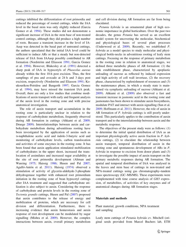
Transcript accumulation of auxin-responsive GH3 gene
during AR formation
To determine whether the time course of IAA in the stem
base of control cuttings is reflected by the expression of an
auxin-responsive gene, the transcript levels of the Petunia
GH3 gene were monitored by Northern blot analysis. With
a first increase to a maximum at 4 hpe, a decline thereafter,
and a second increase beginning at 24 hpe (Fig. 5c), the
GH3 expression showed a corresponding pattern to the
IAA levels in the rooting zone (Fig. 5b). The induction of
Petunia GH3 at 24 hpe and high expression at later stages
(72 hpe, 144 hpe) were confirmed by quantitative Real-
Time PCR (Online Resource 1).
Influence of NPA on enzyme activities in the stem base
To characterize further the effect of auxin transport on
metabolic activity in primary metabolism during AR for-
mation, the activities in the stem base of key enzymes
involved in sucrose metabolism, the pentose phosphate
pathway and glycolysis were analysed in response to
NPA treatment. Results are illustrated in Fig. 6. In the
0 2 4 6 12 24 48 72 96 144 192
Hours Post Excision (hpe)
GH3
RNA
0
10
20
30
40
0
100
200
300
400
Hours Post Excision (hpe)
(a)
(b)
(c)
IAA
(pm
ol g
-1 F
W)
0 2 4 6 12 24 48 72 96 144 192
**
Fig. 5 Temporal course of IAA concentrations in the lowest leaf
(L6, a) and the basal stem (0.5 cm, b) of cuttings of P. hybrida during
rooting under non-treated (solid line) and NPA-treated (dashed line)
conditions (n = 5, each sample from two individual cuttings).
Asterisks indicate a significant effect of the NPA treatment at the
specified time after excision of cuttings (Mann–Whitney U test,
P B 0.05). c Transcript accumulation of auxin-responsive GH3 gene
in the basal stem of P. hybrida during rooting under standard (non-
treated) conditions. Northern blot analysis was performed with 20 g
RNA per sample, and separation on a 1.5 % (w/v) formaldehyde
agarose gel. After transfer of RNA to a nitrocellulose membrane, it
was hybridized to radioactively labelled cDNA fragments of the
corresponding gene. The below picture shows the loading control of
rRNA, the ratio of intensity of 28S RNA to 18S RNA (for total RNA)
is 1:1
200
400
600 cell wall invertase (a)
** *
*
prot
ein)
0
150
200cytosolic invertase (b)
*
**
*
*
* * *
ty (
nmol
min
-1g-
1p
2000
3000
0
50
100
vacuolar invertase (c)
* *
Enz
yme
activ
i 0
1000
150
Glc6PDH (d)
* *
* * * * * * * * **
0
50
100
150 phosphofructokinase (e)
***
* * *
0
50
100
** * *
Hours Post Excision (hpe)
0 2 4 6 12 24 48 72 96 144 192
Fig. 6 Alterations in the enzyme activities involved in sucrose
degradation, glycolysis and pentose phosphate pathways in the basal
stem of P. hybrida during rooting under non-treated (solid line) and
NPA-treated (dashed line) conditions. a Cell wall invertase, b cyto-
solic invertase, c vacuolar invertase, d glucose-6-phosphate dehydro-
genase, e phosphofructokinase. Each value is represented by the mean
of five independent replicates ± SE. Asterisks indicate a significant
effect of the NPA treatment at the specified time after excision of
cuttings (t test, P B 0.05)
Planta (2013) 238:499–517 507
123

non-treated cuttings, the activity of cell wall invertase
increased soon after excision, peaking at 6 hpe, before
decreasing to approximately its initial activity until
192 hpe. In contrast, cell wall invertase activity in NPA-
treated cuttings remained at an initial low level until 6 hpe,
and thereafter, increased up to twofold to levels similar to
those of the control cuttings (Fig. 6a). The activity of
cytosolic invertase showed a similar trend in both non-
treated and NPA-treated cuttings, and decreased continu-
ously during AR formation (Fig. 6b). The activity of vac-
uolar invertase increased up to two-and-half-fold in the
stem base of non-treated cuttings and decreased until
192 hpe to reach approximately 30 % of the initial value.
However, activity of vacuolar invertase in the stem base of
NPA-treated cuttings remained unchanged until 96 hpe and
decreased thereafter (Fig. 6c). Furthermore, in the stem
base of non-treated cuttings, the activity of glucose-6-
phosphate dehydrogenase (Glc6PDH) fluctuated in the first
days of post excision and increased in the later course of AR
formation (Fig. 6d). In NPA-treated cuttings, the activity of
the same enzyme followed a similar trend; it was, however,
less steep, with slightly higher activity than the control
cuttings until 48 hpe, and vice versa after 96 hpe. The
activity of phosphofructokinase (PFK) was similar and did
not change significantly in the stem base of both non-treated
and NPA-treated cuttings up to 72 hpe. Thereafter, PFK
activity increased significantly in the stem base of non-
treated cuttings up to 192 hpe, while it remained unchanged
in the stem base of NPA-treated cuttings (Fig. 6e).
Influence of NPA on sugar concentrations
in the stem base
The concentrations of soluble sugars were measured during
rooting in the stem base of non-treated and NPA-treated
cuttings to evaluate whether auxin transport changes the
distribution of the sugars. In the stem base of non-treated
cuttings, a continuous increase of glucose, fructose and
sucrose was observed after 24 hpe, reaching 10–15-fold
higher levels at 192 hpe relative to the initial values
(Fig. 7a–c). In the stem base of NPA-treated cuttings, the
increase in concentrations of soluble sugars was even more
pronounced. At later time points, levels were three times
higher for glucose and fructose, and 1.5-fold higher for
sucrose compared with the levels measured in the stem
base of non-treated cuttings (Fig. 7a–c).
Effect of NPA on soluble amino acids in the stem base
To determine whether auxin transport changes the com-
position of soluble amino acids, which are necessary for
protein synthesis, during AR formation, the concentrations
of total and individual amino acids were determined in the
stem base of both non-treated and NPA-treated cuttings.
The concentration of total amino acids showed a similar
trend in both non-treated and NPA-treated cuttings, and
increased continuously after 24 hpe. However, the levels
reached at 144 and 192 hpe were about 30–40 % lower in
the stem base of NPA-treated cuttings compared with the
controls (Fig. 8a). The concentrations of the most abundant
amino acids in the stem base, including glutamate, gluta-
mine, aspartate, asparagine and proline, were compared.
Glutamate concentrations showed a trend similar to the
total amino acids; at 144 and 192 hpe, concentrations
reached a twofold higher level in non-treated cuttings
compared with NPA-treated cuttings (Fig. 8b). Similar but
even more pronounced differences were found for gluta-
mine concentrations at 144 and 192 hpe (Fig. 8c). Fur-
thermore, a threefold increase of glutamine concentration
until 2 hpe was observed only in non-treated cuttings; it
was absent in NPA-treated cuttings (Fig. 8c). The con-
centrations of aspartate, even though on a lower level,
followed a similar trend and showed the same response
to NPA treatment as glutamate (Fig. 8d). In the stem base
of non-treated cuttings, the concentration of asparagine
0
20
40
60
0
5
10
15
20
0
20
40
60
glucose
sucrose
fructose
Hours Post Excision (hpe)
(a)
(b)
(c)
0 2 4 6 12 24 48 72 96 144 192
Sug
ar c
onte
nt (
µmol
g-1
FW
) * * **
** * *
* * * *
**
* *
* **
* **
*
Fig. 7 Concentrations of soluble sugars in the basal stem of P.
hybrida during rooting under non-treated (solid line) and NPA-treated
(dashed line) conditions. a Glucose, b fructose, c sucrose. Each value
is represented by the mean of five independent replicates ± SE.
Asterisks indicate a significant effect of the NPA treatment at the
specified time after excision of cuttings (t test, P B 0.05)
508 Planta (2013) 238:499–517
123

increased until 2 hpe and fluctuated thereafter during AR
formation to decrease to a lower level than the initial value
at 192 hpe. In contrast, the asparagine concentration in the
stem base of NPA-treated cuttings decreased continuously
up to 6 hpe but thereafter showed a transient increase to
levels that were higher than those measured in non-treated
cuttings (Fig. 8e). The concentration of proline increased
constantly after 48 hpe, up to 200-fold at 192 hpe, com-
pared with the initial values, in a similar manner for both
non-treated and NPA-treated cuttings (Fig. 8f).
Discussion
In the present study, the initial spatial distribution of IAA
in P. hybrida shoot tip cuttings at the time of excision and
the relationship between subsequent homeostasis of IAA in
the rooting zone, PAT and AR formation were investi-
gated. In addition, the interrelations between auxin and
metabolic response in the rooting zone were elucidated.
Spatial distribution of IAA in P. hybrida shoot tip
cuttings before excision
We used GC–MS/MS to analyse the initial distribution of
IAA in cuttings of P. hybrida at the time of excision.
Concentrations found in the L3 leaves (Fig. 1b) and in the
shoot apex (Fig. 2b) correspond to gas chromatography–
mass spectrometry data published for similar tissues of the
same cultivar (Tobena-Santamaria et al. 2002). The strong
negative relationship established for young Petunia leaves
between leaf fresh mass and IAA concentration (Fig. 1d) is
in accordance with corresponding relationships found for
Arabidopsis, where IAA levels were high in those leaves
that showed high rates of cell division but strongly
decreased when leaf expansion was initiated (Ljung et al.
2001). Ljung et al. (2001) also reported that for young
leaves of Nicotiana that were in transition from division to
elongation growth, high IAA levels were restricted to the
base and middle of the blade as zones of intense cell
division. We did not observe such a gradient, and the IAA
levels were generally low in the lamina of Petunia L6
leaves (Fig. 2a), which had about 50 % of the maximum
leaf length that we observed in old plants. This supports the
assumption that cell division has almost completely ceased
in L6 leaves. However, the higher IAA level in the petiole
compared with the adjacent base (Fig. 2a) reflects the same
accumulation pattern of IAA as observed in Arabidopsis
and Nicotiana (Ljung et al. 2001; Muller et al. 2002),
which indicates that active auxin transport occurred
between the leaf and the stem of the Petunia cutting at the
time of excision.
The similar IAA levels measured for the different stem
positions of P. hybrida cuttings at the time of excision
(Fig. 2b) are in accordance with data obtained for young
shoots of other plant species (Nordstrom and Eliasson
12000
total amino acid (a)
**
0
4000
8000
3000
glutamate (b)
(c)
* *
* **
16000
1000
2000
3000
glutamine
* ** *
400
800
1200
1600 glutamine
ent (
nmol
g-1
FW
)
** * *
** *
0
400
800
1200aspartate (d)
Am
ino
acid
con
te *
* * *
500
1000
0asparagine (e)
*
*
*
0proline
4000
6000 (f)
* * *
* * *
Hours Post Excision (hpe)
0
2000
0 2 4 6 12 24 48 72 96 144 192
* * * * * *
*
Fig. 8 Concentrations of amino acids in the basal stem of P. hybrida
during rooting under non-treated (solid line) and NPA-treated (dashed
line) conditions. a Total amino acids, b glutamate, c glutamine,
d aspartate, e asparagine, f proline. Each value is represented by the
mean of five independent replicates ± SE. Asterisks indicate a
significant effect of the NPA treatment at the specified time after
excision of cuttings (t test, P B 0.05)
Planta (2013) 238:499–517 509
123

1991; Kojima et al. 2002; Jager et al. 2007). It has been
shown that auxin can be produced in, and exported from,
diverse parts of the young shoot such as the apex, young
leaves, and developing and also expanding leaves (Ljung
et al. 2001; Garrido et al. 2002; Woodward and Bartel
2005; Jager et al. 2007). Thus, the same IAA levels for the
lower and upper shoot positions may indicate a current
auxin influx not only from the apex but also from the lower
leaves of Petunia cuttings.
The influence of PAT on auxin homeostasis and AR
formation in P. hybrida
According to the current model, auxin travels through a
combination of two processes: (1) rapid (5–10 cm h-1)
non-directional transport that occurs in the vasculature and
(2) slower PAT (5–10 mm h-1) (Friml and Palme 2002;
Kerr and Bennett 2007). It has been further shown that
conjunctions exist between these two routes. IAA from
leaves can be loaded into the phloem, transported by this
route (Morris and Kadir 1972; Borkovec et al. 1994) and
then further transferred into the extravascular PAT path-
way mainly at the shoot apex (Cambridge and Morris
1996). Specific influx [(auxin permease (AUX), like AUX
(LAX)], specific efflux [pin-formed (PIN)], and multidrug
resistance/P-glycoprotein [MDR/PGP] auxin carriers are
involved, and their turnover, cycling and trafficking con-
tribute to the asymmetric and dynamic nature of PAT
(Morris et al. 2004; Kerr and Bennett 2007). The synthetic
auxin transport inhibitor NPA is supposed to disrupt auxin
efflux from the cell, even though the mode of action is still
a matter of debate (Morris et al. 2004). It is thought that
NPA acts via binding to a putative NPA-binding receptor
protein, which is assumed to be located on the cytoplasmic
face of the plasma membrane. It has been reported that
inhibitors of PAT prevent the traffic of PIN1 and other
rapidly cycled proteins to and from the plasma membrane
in Arabidopsis root cells (Geldner et al. 2001). NPA has
also been shown to interact with members of the MDR/
PGB family in Arabidopsis.
The role of PAT in AR formation of cuttings has been
studied intensively in carnations. Guerrero et al. (1999)
measured the transport of labelled IAA through 1-cm long
stem sections of freshly excised carnation cuttings. First and
most of the labelled IAA passed the 1-cm section after a
transport period of 1–2 h and 8 h, respectively, while
application of NPA to the stem sections inhibited basipetal
transport by 62–91 %, depending on the particular test
conditions. Garrido et al. (2002) showed also that labelled
IAA applied to mature leaves of cuttings was transported to
the stem base and that excision of complete leaves, but not of
the apex and of youngest leaves, inhibited rooting, while
detached leaves could be partially substituted by auxin
application to the stem. Application of NPA to the cuttings
also resulted in inhibition of rooting in other plant species
(Nordstrom and Eliasson 1991; Liu and Reid 1992; Ko-
ukourikou-Petridou and Bangerth 1997). In accordance to
these studies, NPA severely inhibited and delayed the
development of root primordia in P. hybrida (Figs. 3, 4) and
decreased the overall intensity of rooting determined at
14 days post excision by 98 % (Table 1). A parallel analysis
of IAA level highlighted the relationships between PAT,
auxin accumulation in the rooting zone and different
developmental phases in Petunia cuttings. The significant
increase of IAA in the stem base of control cuttings between
2 and 24 hpe (Fig. 5b), which was followed by the formation
of first meristematic cells at 72 hpe (Fig. 4e, f), contrasted
with the elimination of the 24-h peak of IAA (Fig. 5b) and
subsequent severe inhibition of adventitious rooting in NPA-
treated cuttings (Figs. 3, 4; Table 1). This provides evidence
for the essential contribution of PAT and the early accu-
mulation of IAA in the rooting zone to spontaneous AR
formation in Petunia cuttings in response to excision.
The kinetic of IAA levels in the rooting zone of control
cuttings indicates two overlapping peaks, with maxima at 2
and 24 hpe, reflecting a strong regulation of auxin
homeostasis soon after excision of Petunia. Similar fast
changes of IAA levels have also been detected in the
rooting zone of some other plant species (Blakesley et al.
1991). The observation that the 24-h peak of IAA was
completely prevented by application of NPA provides
evidence that this peak was the outcome of PAT. However,
the first IAA maximum at 2 hpe was not prevented by
NPA. We applied the NPA immediately after, but not
before, excision of cuttings because we did not want to
manipulate the initial auxin homeostasis in the cuttings
before severance. Considering also the initial distribution
of IAA pools within the cutting (Fig. 2c) and the speed of
PAT, as discussed above, the missing effect of NPA until
2 hpe may reflect a lag phase between NPA application and
its action on the cutting. NPA action may also have been
underestimated by the IAA analyses of the complete stem
(further discussed below). Nevertheless, the first IAA peak
may also reflect transport in the phloem, local synthesis or
mobilization from conjugates.
Wounding is an intrinsic process when cuttings are
excised from donor plants, and we have already detected a
strong increase of the wound-responsive hormone jasmonic
acid in the stem base of Petunia cuttings during the first
30 min after excision (Ahkami et al. 2009). There are
indications in the literature that wounding of plant tissues
(Sztein et al. 2002) and jasmonates (Grsic et al. 1999; Sun
et al. 2009) can stimulate IAA biosynthesis in plant tissues.
According to the inhibition of auxin efflux, it has been
observed that auxins accumulate in cells treated with
blockers and that application of NPA to stems stimulates
510 Planta (2013) 238:499–517
123

auxin accumulation in the tissues above the NPA-treated
region (Morris et al. 2004). Regarding the initial size of the
IAA pool and the particular accumulation of IAA in the
petiole of L6 leaves (Figs. 1c, 2a), we monitored the IAA
level in L6 leaves as potential source organs for auxin
export. As illustrated in Fig. 5a, IAA did not accumulate
significantly in L6 leaves of NPA-treated cuttings com-
pared with the non-treated ones. This provides no sup-
portive indication of a predominant role of the L6 leaf as
source organ for providing the auxin accumulation in the
rooting zone. However, the observed oscillation of IAA
levels in the NPA-treated cuttings may reflect a feedback
inhibition of IAA biosynthesis in response to trapping of
IAA, which has been found in expanding leaves of Ara-
bidopsis (Ljung et al. 2001). Distinct blocking of auxin
transport in separate parts of Petunia cuttings should be
done in future studies, to elucidate these relationships.
Interestingly, the IAA level showed a continuous increase
after 72 hpe only in L6 leaves of non-treated cuttings; at
192 hpe, it reached a level significantly higher than the
initial level (Fig. 5a). Considering that roots were formed
in the non-treated cuttings during this period and regarding
the possibility of de novo IAA biosynthesis in roots, which
has been shown for Arabidopsis (Ljung et al. 2001), root-
sourced auxin may have contributed to this increase.
Auxin action and AR formation
We analysed the Petunia GH3 gene expression in the stem
base of non-treated control cuttings as an early marker of
auxin activity (Hagen and Guilfoyle 1985) to obtain
information on the involvement of auxin action at certain
developmental stages of AR formation. The time course of
GH3 gene expression (Fig. 5c) followed the trend of the
IAA level (Fig. 5b), while the strong increase of transcript
level at 4 hpe indicated a lag phase of ca. 2 h behind the
peaking of IAA. Considering the dose-dependent induction
of the GH3 promoter by active auxin, as shown in trans-
genic tobacco (Hagen et al. 1991; Li et al. 1999), these
observations support the view that early auxin activity is
causally involved in initiating the primary events of ARF in
Petunia. Regarding also the fact that several GH3 genes in
Arabidopsis encode IAA-amido synthetases, which are
important for maintaining auxin homeostasis by conjugat-
ing excess IAA to amino acids (Staswick et al. 2005; Wang
et al. 2007), GH3 expression may also contribute to auxin
conjugation in Petunia cuttings to reduce the level of active
auxin and thus to avoid inhibitory influences during later
processes of AR formation (De Klerk et al. 1999). How-
ever, elucidation of these processes was not in the focus of
the present study and requires further investigations
involving quantification of IAA conjugates and GH3
monitoring in response to NPA treatment.
Despite their broad use, the physiological mechanisms
underlying the AR-induction by auxins are still far from
being understood (Ludwig-Muller 2009). According to a
recent model, plant phospholipase 2 is involved in auxin
signal transduction during AR formation, and a large
family of auxin-induced gene products (so-called ‘Aux/
IAA’ proteins), auxin-responsive transcription factors
(auxin response factors) and ARGONAUTE1 are involved
in auxin-induced gene expression controlling the cellular
events such as changes in cell cycle and cytoskeleton,
tissue reorganization and cell wall modification (Ludwig-
Muller 2009). Specific GH3 genes may have particular
functions in AR formation, which go beyond their role in
auxin homeostasis, as supported by studies with Arabid-
opsis seedlings (Sorin et al. 2005). Recently, Gutierrez
et al. (2012) showed that three auxin-inducible GH3 genes
are required for positive regulation of light-induced AR
formation in Arabidopsis. They act by mediating the
upstream interaction of three auxin response factors and
downstream modulation of jasmonic acid homeostasis
(Gutierrez et al. 2012).
Auxin might affect the root formation by acting directly
in the cambium cells that initiate root primordia or indi-
rectly through its engagement in the overall metabolism
(Altman and Wareing 1975). Regarding the role of auxin in
cell division and elongation, a complex interaction between
auxin, cytokinin, cyclins and cyclin-dependent kinases
(CDKs) governs the plant cell cycle (Hartig and Beck
2006; Komaki and Sugimoto 2012) Auxin treatment
stimulated expression of cyclins including mitotic B1
cyclins in Arabidopsis (Ferreira et al. 1994; Richard et al.
2002), and expression of cyclin genes and of CDKs has
already been related to auxin-induced AR formation
(Lindroth et al. 2001; Neves et al. 2006). When Petunia
cuttings experienced the same rooting conditions as applied
to the controls in the present study, the transcript of a
Petunia CycB1 encoding a mitotic B1 cyclin accumulated
in the stem base at 48 hpe (Ahkami et al. 2009). Based on
the observation of IAA accumulation until 24 hpe
(Fig. 5b), this may have contributed to the induction of
Petunia CycB1 and thereby stimulated the initiation of cell
division. Furthermore, other genes such as SCARECROW-
like genes may be involved; expression has already been
shown to be stimulated in rooting-competent cuttings of
Pinus radiata and Castanea sativa by auxin treatment
before activation of cell division (Sanchez et al. 2007).
Involvement of PAT and auxin homeostasis
in the response of primary metabolism during
AR formation
Plant meristems are utilization sinks, in which cell division
activity governs sink strength (Hartig and Beck 2006).
Planta (2013) 238:499–517 511
123

Monitoring of enzyme activities in the stem base of P.
hybrida cuttings during AR formation revealed that some
enzymes, such as cell wall and vacuolar invertases, as well
as PFK, were modulated at specific root developmental
stages and that these modulations were subjected to NPA
treatment (Fig. 6). While the activity of invertases showed
a maximum during the first hours of post excision in non-
treated cuttings, the early increases of cell wall invertase
(Fig. 6a) and of vacuolar invertase (Fig. 6c) were pre-
vented by NPA application, indicating that PAT contrib-
uted to the increase in activities of these sucrose degrading
enzymes. These results reflect a specific relationship
between basipetal auxin transport and extracellular and
vacuolar invertases, and suggest that auxin may regulate
the activity of enzymes in the sink establishment phase of
AR formation in Petunia cuttings. Importantly, one of the
proposed functions of cell wall invertase is to act as a linker
between hormonal responses and primary metabolism
(Roitsch and Gonzalez 2004). Even though there is indi-
cation in literature that vacuolar and cell wall invertases
can be stimulated by auxin (Morris and Arthur 1984, Lee
et al. 1997), the underlying mechanisms are far from being
understood. The dominant mechanism that determines
steady-state level of invertases is a highly responsive
transcriptional regulation; however, invertases are further
controlled at post-translational level and via protein traf-
ficking and transcript turnover (Roitsch and Gonzalez
2004; Albacate et al. 2011). Yun et al. (2002) could
counteract a rapid repression of vacuolar invertase in
excised etiolated hypocotyl segments of mung bean by
incubation with IAA, while the auxin treatment increased
enzyme activity and the level and half-life of transcripts of
the vacuolar invertase gene VR-AI1. Responses to inhibi-
tors of polymerase II and of protein biosynthesis further
supported the view that IAA enhanced transcript stability.
Proels et al. (2003), who described the full genomic
sequence and expression pattern of the LIN5 gene coding
for an extracellular invertase in tomato, provided evidence
that auxin stimulated expression of this gene while auxin
responsiveness was mediated via a 1.6-kb Lin5 promoter
fragment.
The temporal courses of IAA and invertase activities in
the stem base reveal that NPA treatment-reduced activities
of cell wall (4 hpe, Fig. 6a) and of vacuolar invertases
(2 hpe, Fig. 6c) before a significant reduction in IAA level
were detected (12 hpe, Fig. 5b). PAT in stems occurs in the
xylem parenchyma cells, the vascular cambium and its
partially differentiated derivative (Galweiler et al. 1998;
Morris et al. 2004). Thus, IAA might have responded
earlier to the NPA treatment in particular tissues and cell
compartments, and thereby influenced invertase activity
before a significant change of IAA level was detected in the
complete stem base. Future studies of spatial and cellular
distribution of auxin activity by use of auxin-responsive
promoters in response to NPA treatment may elucidate
these relationships.
Comparing enzyme activities with measured sugar lev-
els, it has to be considered that activities of invertases were
determined for certain cell compartments (apoplast, cyto-
sol, vacuole), whereas sugar levels reflect the mean situa-
tion of the whole stem base of the cuttings. Furthermore,
sugar levels may be additionally influenced by fluxes or
metabolic events which are not limited by those enzymes
analysed in this study. Considering the period between 4
and 12 hpe, NPA treatment decreased the activities of cell
wall and vacuolar invertases but slightly enhanced the
levels of glucose and fructose. Apart from the influence of
cell compartments, this may indicate that NPA also
inhibited utilisation of hexoses. As already discussed
above, the expression of mitotic cyclin genes is typical for
cells shortly before they divide (Ferreira et al. 1994), and
an enhanced expression of a Petunia CycB1 gene was
detected in P. hybrida at 48 hpe when rooting conditions
were the same as applied to the control cuttings in the
present study (Ahkami et al. 2009). Considering, that rep-
lication of DNA, RNA and protein synthesis occurs before
the mitosis phase of the cell cycle (Hartig and Beck 2006),
there is a need of carbon skeletons before cell division
starts. In conclusion, the slightly higher sugar levels in
NPA-treated cuttings compared to the controls between 4
and 12 hpe may reflect a higher ratio between hexose
consumption due to auxin-stimulated cell division on the
one side and sucrose delivery and hexose release on the
other side. However, the data of Glc6PDH and of PFK do
not provide indication that these enzymes were primarily
bottlenecks of glucose utilization during this early phase.
Although subsequent root formation was delayed using the
blocker of PAT (Fig. 4), soluble sugars, particularly, hex-
oses accumulated more strongly in the stem base of NPA-
treated cuttings beginning 48 h after excision compared
with the controls (Fig. 7). From a first point of view, these
results seem to contradict findings that auxin application to
the stem base of cuttings of other plant species enhances
the sugar level in the rooting zone (Altman and Wareing
1975; Husen and Pal 2007; Agullo-Anton et al. 2011).
However, it has to be taken into account that in the present
study, the role of ‘‘natural auxin level’’ in response to
excision of cuttings was investigated. In the studies men-
tioned above, applied auxin probably enhanced auxin
concentration in the rooting zone to much higher levels so
that the balance between the responses at sink establish-
ment level (modifying carbohydrate influx) versus root
development level (modifying carbohydrate utilization)
might have been different in our study. Based on our
observations, we conclude that the elevated sugar accu-
mulation in NPA-treated Petunia cuttings during the later
512 Planta (2013) 238:499–517
123

stages of AR formation is not directly affected by the lower
auxin level until 24 hpe. This phenomenon is rather the
outcome of the lower utilization of carbohydrates due to
the strongly inhibited root formation. This conclusion is
strongly supported by our finding that during the period
after 96 hpe, the NPA-treated cuttings demonstrated sig-
nificantly lower activities of PFK and of Glc6PDH than the
control cuttings.
Amino acids are needed during AR formation because
they provide essential components for protein synthesis
Auxin accumulation(peak at 24 hpe)S
ho
ot b
asip
etal
auxi
ntr
ansp
ort
& lo
cal a
uxi
nm
etab
olis
m
SinkEstablishment
Root meristemoid formation
Stimulation of sucrose degrading enzymes:release of glucose and fructose
Sucrose
Hexose-P
Hexoses
Glycolysis
Pent
ose
Phos
phat
epa
thw
ay
CW Invertase
apoplast
cytosol
Induction of PFK& Glc6PDH
Root emergence (192 hpe)
Rec
over
y P
hase
24 –
72 h
peM
aint
enan
ce P
hase
72 –
192
hpe
Excision
Induction of auxinresponsive GH3
Sin
k E
stab
lishm
ent
Pha
se (
4 –
24 h
pe)
Induction of cell division
Induction of CycB1
Auxin signaltransduction
?
?
Development of root meristems & primordia
Fig. 9 A postulated model of
relationship between PAT,
auxin accumulation, primary
metabolism and AR formation
in Petunia cuttings in response
to excision from the donor
plant. Red solid arrows indicate
processes that are dependent on
PAT and are correlated with the
resulting auxin peak at 24 hpe
in the rooting zone. Red dashed
arrows indicate additional
processes which are
hypothetically depending on
PAT as supported by data of the
present study and of Ahkami
et al. (2009). Thin black arrows
with question mark show
possible interrelations between
GH3 induction, auxin
metabolism and cellular events
of AR formation. Further details
regarding different stages and
phases of AR formation in
petunia cuttings are described in
Ahkami et al. (2009). PAT polar
auxin transport, CW invertase
cell wall invertase, PFK
phosphofructokinase, Glc6PDH
glucose-6-phosphate
dehydrogenase, hpe hours post
excision
Planta (2013) 238:499–517 513
123

required for cell division, elongation and function. In
accordance with findings of earlier studies (Ahkami et al.
2009), total amino acids, glutamate, glutamine and aspar-
tate accumulated in the rooting zone of control cuttings in
this study as well. The highest levels were observed during
the period following 96 hpe (Fig. 8), when root meristems
and primordia were formed (Fig. 3). Accumulation of those
amino acids was significantly lower in NPA-treated cut-
tings (Fig. 8b–d). Interestingly, inhibition of AR formation
was associated with increased sugar levels but with
decreased amino acid levels, even though both can be
considered as important resources for root development
(Haissig 1986). This discrepancy suggests that the intensity
of influx and biosynthesis of amino acids in the rooting
zone is directly related to differentiation and growth of
ARs (Fig. 3), whereas the sugar level in the rooting zone,
which is also highly dependent on the strength of the car-
bon source in the cutting (Rapaka et al. 2005), reflects the
balance between influx and utilization of carbon. In this
context, Petunia cuttings reveal high carbon assimilation,
comparable to that of intact donor plants even at irradiance
lower than that used in the present study (Klopotek et al.
2012).
Interestingly, NPA treatment led to inhibition of the
early increase of glutamine and asparagine in the rooting
zone in the sink establishment phase (Fig. 8c, e). These two
compounds belong to the group of amino acids transported
in the phloem (Urquhart and Joy 1982; Lohaus and Mo-
ellers 2000), and asparagine is suggested to be the main
nitrogen transport compound in the initiating roots of cut-
tings (Suzuki and Kohno 1983). Considering on the one
hand, the possible roles of PAT and early auxin accumu-
lation (Fig. 4b) in driving sucrose utilization via stimula-
tion of cell wall and vacuolar invertase (Fig. 6a, c), and on
the other hand the mechanism of co-transport of sucrose
and amino acids in the phloem (Lalonde et al. 2004), the
lower levels of both amino acids in the stem base of NPA-
treated cuttings may be the result of decreased apoplastic
unloading of sucrose.
A strong accumulation of proline was found at later
stages of AR formation, while similar levels were moni-
tored in both non-treated and NPA-treated cuttings
(Fig. 8f). These results do not support a role of PAT or of
auxin homeostasis in proline accumulation during AR
formation in Petunia cuttings. This is in contrast with the
observations that auxin application increased the levels of
proline in mung bean hypocotyls (Kuraishi et al. 1967) and
transcripts of proline-rich proteins (PRPs) involved in lat-
eral root formation in Arabidopsis and carrot (Ebener et al.
1993; Neuteboom et al. 1999; Bernhardt and Tierney
2000). However, it has been suggested that auxin acts as a
regulator of PRP genes in a particular time- and concen-
tration-dependent manner (Thomas et al. 2003).
Conclusive model
Overall, our study clearly shows that spontaneous AR
formation in leafy stem cuttings of P. hybrida is dependent
on PAT and on resulting auxin homeostasis in the rooting
zone. A respective model of the relationship between PAT,
auxin, primary metabolism and AR formation is postulated
and illustrated in Fig. 9. After excision of cuttings, PAT
enables the accumulation of free auxin in the rooting zone,
where it (1) contributes to the establishment of the new
sink via stimulation of cell wall and vacuolar invertases
and (2) induces the first cell divisions of the new root
meristems. After PAT-dependent induction of root meris-
tems and sink establishment, the decrease of auxin levels
allows for subsequent root development, which in turn
stimulates the entry of glucose into glycolysis and the
pentose phosphate pathway as reflected by the NPA-
inhibited activation of PFK and G6PDH. Stimulated gly-
colysis and pentose pathway generate the energy and car-
bon skeletons to be used for synthesis of amino acids,
proteins and nucleic acids. The present data further support
the assumption that accumulation of free auxin contributes
to induction of GH3 gene while previous results (Ahkami
et al. 2009) suggest involvement of Petunia CycB1 gene in
induction of cell division. GH3 may stimulate auxin con-
jugation thereby reducing the level of active auxin after
24 hpe and may be further involved in initiation of cellular
events. Activity of sucrose degrading enzymes provides the
hexoses needed for supply of energy and of carbon to cell
proliferation, differentiation and growth. Considering the
effects of sugars, particularly of hexoses on gene expres-
sion (Rolland et al. 2006), released hexoses may be further
involved in the control of cell cycle (Hartig and Beck 2006)
and modification of auxin homeostasis (Sairanen et al.
2012) and signal transduction (Mishra et al. 2009), while
the steady-state levels of individual sugars reflect the bal-
ance between the influx and degradation of sucrose and the
utilization of hexoses via glycolysis and pentose phosphate
pathway.
Acknowledgments We thank Sabine Czekalla, IGZ, for her skilful
technical assistance. We thank Klaus Fricke, IGZ, for his great job in
establishing the GC–MS/MS analysis and Petra Duchting, Ruhr
University Bochum, Germany for her support. This work was funded
by the Pakt fur Forschung und Innovation of the Leibniz-Gemein-
schaft, Germany, and supported by the states of Saxony-Anhalt and
Brandenburg, the Free State of Thuringia and the Federal Republic of
Germany.
514 Planta (2013) 238:499–517
123

Open Access This article is distributed under the terms of the
Creative Commons Attribution License which permits any use, dis-
tribution, and reproduction in any medium, provided the original
author(s) and the source are credited.
References
Agullo-Anton MA, Sanchez-Bravo J, Acosta M, Druege U (2011)
Auxins or sugars: what makes the difference in the adventitious
rooting of stored carnation cuttings? J Plant Growth Regul
30:100–113
Ahkami AH, Lischewski S, Haensch KT, Porfirova S, Hofmann J,
Rolletschek H, Melzer M, Franken P, Hause B, Druege U,
Hajirezaei MR (2009) Molecular physiology of adventitious root
formation in Petunia hybrida cuttings: involvement of wound
response and primary metabolism. New Phytol 181:613–625
Albacate A, Grosskinsky DK, Roitsch TH (2011) Trick and treat: a
review on the function and regulation of plant invertases in the
abiotic stress response. Phyton 50:181–204
Altman A, Wareing PF (1975) Effect of IAA on sugar accumulation
and basipetal transport of C14-labeled assimilates in relation to
root formation in Phaseolus vulgaris cuttings. Physiol Plant
33:32–38
Baadsmand S, Andersen AS (1984) Transport and accumulation of
indole-3-acetic acid in pea cuttings under two levels of
irradiance. Physiol Plant 61:107–113
Bernhardt C, Tierney ML (2000) Expression of AtPRP3, a proline-
rich structural cell wall protein from Arabidopsis, is regulated by
cell-type-specific developmental pathways involved in root hair
formation. Plant Physiol 122:705–714
Blakesley D (1994) Auxin metabolism and adventitious root initia-
tion. In: Davis TD, Haissig BE (eds) Biology of adventitious root
formation. Plenum Press, New York, pp 143–153
Blakesley D, Weston GD, Hall JF (1991) The role of endogenous
auxin in root initiation. Part I: evidence from studies on auxin
application and analysis of endogenous levels. Plant Growth
Regul 10:341–353
Blazkova A, Sotta B, Tranvan H, Maldiney R, Bonnet M, Einhorn J,
Kerhoas L, Miginiac E (1997) Auxin metabolism and rooting in
young and mature clones of Sequoia sempervirens. Physiol Plant
99:73–80
Borkovec V, Didehvar F, Baker DA (1994) The biosynthesis and
translocation of C14 IAA in Ricinus communis. Plant Growth
Regul 15:137–141
Cambridge AP, Morris DA (1996) Transfer of exogenous auxin from
the phloem to the polar auxin transport pathway in pea (Pisum
sativum L.). Planta 199:583–588
De Klerk GJ, van der Krieken W, de Jong J (1999) Review the
formation of adventitious roots: new concepts, new possibilities.
In Vitro Cell Develop Biol Plant 35:189–199
Druege U (2009) Involvement of carbohydrates in survival and
adventitious root formation of cuttings within the scope of global
horticulture. In: Niemi K, Scagel C (eds) Adventitious root
formation of forest trees and horticultural plants—from genes to
applications. Research Signpost, Kerala, pp 187–208
Ebener W, Fowler TJ, Suzuki H, Shaver J, Tierney ML (1993)
Expression of Dcprp1 is linked to carrot storage root-formation
and is induced by wounding and auxin treatment. Plant Physiol
101:259–265
Ferreira PC, Hemerly AS, Engler JD, van Montagu M, Engler G, Inze
D (1994) Developmental expression of the Arabidopsis cyclin
gene cyc1At. Plant Cell 6:1763–1774
Friml J, Palme K (2002) Polar auxin transport—old questions and
new concepts? Plant Mol Biol 49:273–284
Galweiler L, Guan C, Muller A, Wisman E, Mendgen K, Yephremov
A, Palme K (1998) Regulation of polar auxin transport by
AtPIN1 in Arabidopsis vascular tissue. Science 282:2226–2230
Garcia Gomez ML, Sanchez Romero C, Barcelo Munoz A, Heredia
A, Pliego Alfaro F (1994) Levels of endogenous indole-3-acetic
acid and indole-3-acetyl-aspartic acid during adventitious root-
ing in avocado microcuttings. J Exp Bot 45:865–870
Garrido G, Guerrero JR, Cano EA, Acosta M, Sanchez-Bravo J
(2002) Origin and basipetal transport of the IAA responsible for
rooting of carnation cuttings. Physiol Plant 114:303–312
Geldner N, Friml J, Stierhof YD, Jurgens G, Palme K (2001) Auxin
transport inhibitors block PIN1 cycling and vesicle trafficking.
Nature 413:425–428
Grsic S, Kirchheim B, Pieper K, Fritsch M, Hilgenberg W, Ludwig-
Muller J (1999) Induction of auxin biosynthetic enzymes by
jasmonic acid and in clubroot diseased Chinese cabbage plants.
Physiol Plant 105:521–531
Guerrero JR, Garrido G, Acosta M, Sanchez-Bravo J (1999) Influence
of 2,3,5-triiodobenzoic acid and 1-N-naphthylphthalamic acid on
indoleacetic acid transport in carnation cuttings: relationship
with rooting. Plant Growth Regul 18:183–190
Gutierrez L, Mongelard G, Flokova K, Pacurar DI, Novak O,
Staswick P, Kowalczyk M, Pacurar M, Demailly H, Geiss G,
Bellini C (2012) Auxin controls Arabidopsis adventitious root
initiation by regulating jasmonic acid homeostasis. Plant Cell
24:2515–2527
Hagen G, Guilfoyle TJ (1985) Rapid induction of selective transcrip-
tion by auxins. Mol Cell Biol 5:1197–1203
Hagen G, Martin G, Li Y, Guilfoyle TJ (1991) Auxin-induced
expression of the soybean Gh3 promoter in transgenic tobacco
plants. Plant Mol Biol 17:567–579
Haissig BE (1974) Metabolism during adventitious root primordium
initiation and development. New Zeal J For Sci 4:324–337
Haissig BE (1986) Metabolic processes in adventitious rooting of
cuttings. In: Jackson MB (ed) New root formation in plants and
cuttings. Martinus Nijhoff Publishers, Dordrecht, pp 141–189
Hajirezaei MR, Takahata Y, Trethewey RN, Willmitzer L, Sonnewald
U (2000) Impact of elevated cytosolic and apoplastic invertase
activity on carbon metabolism during potato tuber development.
J Exp Bot 51:439–445
Hartig B, Beck E (2006) Crosstalk between auxin, cytokinins, and
sugars in the plant cell cycle. Plant Biol 8:389–396
Hartmann HT, Kester DE, Davies FT Jr., Geneve RL (2011)
Hartmann and Kester’s plant propagation—principles and prac-
tices, 8th edn. (Prentice Hall). Upper Saddle River, New Jersey
Hoffmann M, Hentrich M, Pollmann S (2011) Auxin–oxylipin crosstalk:
relationship of antagonists. J Integrative Plant Biol 53:429–445
Husen A, Pal M (2007) Metabolic changes during adventitious root
primordium development in Tectona grandis Linn. f. (teak)
cuttings as affected by age of donor plants and auxin (IBA and
NAA) treatment. New Forest 33:309–323
Jager CE, Symons GM, Glancy NE, Reid JB, Ross JJ (2007) Evidence
that the mature leaves contribute auxin to the immature tissues of
pea (Pisum sativum L.). Planta 226:361–368
Kerr ID, Bennett MJ (2007) New insight into the biochemical
mechanisms regulating auxin transport in plants. Biochem J
401:613–622
Kevers C, Hausman JF, Faivre-Rampant O, Evers D, Gaspar T (1997)
Hormonal control of adventitious rooting: progress and ques-
tions. Angew Bot 71:71–79
Klopotek Y, George E, Druege U, Klaering HP (2012) Carbon
assimilation of petunia cuttings in a non-disturbed rooting
environment: response to environmental key factors and adven-
titious root formation. Sci Hortic 145:118–126
Planta (2013) 238:499–517 515
123
Kojima K, Ohtake E, Yu Z (2002) Distribution and transport of IAA
in tomato plants. Plant Growth Regul 37:249–254
Komaki S, Sugimoto K (2012) Control of plant cell cycle by
developmental and environmental cues. Plant Cell Physiol
53:953–964
Koukourikou-Petridou MA, Bangerth F (1997) Effect of changing the
endogenous concentration of auxins and cytokinins and the
production of ethylene in pea stem cuttings on adventitious root
formation. Plant Growth Regul 22:101–108
Kuraishi S, Uematsu S, Yamaki T (1967) Auxin-induced incorporation
of proline in mung bean hypocotyls. Plant Cell Physiol 8:527
Lalonde S, Wipf D, Frommer WB (2004) Transport mechanisms for
organic forms of carbon and nitrogen between source and sink.
Annu Rev Plant Biol 55:341–372
Lee TH, Sugiyama A, OfosuAnim J, Takeno K, Ohno H, Yamaki S
(1997) Activation of sucrose-metabolizing enzymes and stimu-
lation of sucrose uptake by auxin and sucrose in eggplant
(Solanum melongena L.). J Plant Physiol 150:297–301
Li Y, Wu YH, Hagen G, Guilfoyle T (1999) Expression of the auxin-
inducible GH3 promoter GUS fusion gene as a useful molecular
marker for auxin physiology. Plant Cell Physiol 40:675–682
Lindroth AM, Kvarnheden A, von Arnold S (2001) Isolation of a
PSTAIRE CDC2 cDNA from Pinus contorta and its expression
during adventitious root development. Plant Physiol Biochem
39:107–114
Liu HJ, Reid DM (1992) Auxin and ethylene-stimulated adventitious
rooting in relation to tissue sensitivity to auxin and ethylene
production in sunflower hypocotyls. J Exp Bot 43:1191–1198
Ljung K, Bhalerao RP, Sandberg G (2001) Sites and homeostatic
control of auxin biosynthesis in Arabidopsis during vegetative
growth. Plant J 28:465–474
Logemann J, Schell J, Willmitzer L (1987) Improved method for the
isolation of RNA from plant-tissues. Anal Biochem 163:16–20
Lohaus G, Moellers C (2000) Phloem transport of amino acids in two
Brassica napus L. genotypes and one B. carinata genotype in
relation to their seed protein content. Planta 211:833–840
Lovell PH, White J (1987) Anatomical changes during adventitious
root formation. In: Jackson MB (ed) New root formation in
plants and cuttings. Martinus Nijhoff Publishers, Dordrecht,
pp 111–140
Ludwig-Muller J (2009) Molecular basis for the role of auxins in
adventitious rooting. In: Niemi K, Scagel C (eds) Adventitious
root formation of forest trees and horticultural plants—from
genes to applications. Research Signpost, Kerala, pp 1–29
Mishra BS, Singh M, Aggrawal P, Laxmi A (2009) Glucose and auxin
signaling interaction in controlling Arabidopsis thaliana seed-
lings root growth and development. PLoS One 4(2):e4502
Morris DA, Arthur ED (1984) Invertase and auxin-induced elongation
in internodal aegments of Phaseolus vulgaris. Phytochemistry
23:2163–2167
Morris DA, Kadir G (1972) Pathways of auxin transport in the intact
pea seedling (Pisum sativum L.). Planta 107:171–182
Morris DV, Friml J, Zazımalova E (2004) The transport of auxin. In:
Davies PJ (ed) Plant hormones. Kluwer Academic Publishers,
Dordrecht The Netherlands, pp 437–470
Muller A, Duchting P, Weiler W (2002) A multiplex GC-MS/MS
technique for the sensitive and quantitative single-run analysis of
acidic phytohormones and related compounds, and its applica-
tion to Arabidopsis thaliana. Planta 216:55–56
Neuteboom LW, Ng JMY, Kuyper M, Clijdesdale OR, Hooykaas PJJ,
van der Zaal BJ (1999) Isolation and characterization of cDNA
clones corresponding with mRNAs that accumulate during
auxin-induced lateral root formation. Plant Mol Biol 39:273–287
Neves C, Hand P, Amancio S (2006) Patterns of B-type cyclin gene
expression during adventitious rooting of micropropagated cork
oak. Plant Cell Tiss Org Cult 86:367–374
Nordstrom AC, Eliasson L (1991) Levels of endogenous indole-3-
acetic acid and indole-3-acetylaspartic acid during adventitious
root formation in pea cuttings. Physiol Plant 82:599–605
Proels RK, Hause B, Berger S, Roitsch T (2003) Novel mode of
hormone induction of tandem invertase genes in floral tissues.
Plant Mol Biol 52:191–201
Rapaka VK, Bessler B, Schreiner M, Druege U (2005) Interplay
between initial carbohydrate availability, current photosynthesis
and adventitious root formation in Pelargonium cuttings. Plant
Sci 168:1547–1560
Richard C, Lescot M, Inze D, De Veylder L (2002) Effect of auxin,
cytokinin, and sucrose on cell cycle gene expression in
Arabidopsis thaliana cell suspension cultures. Plant Cell Tiss
Org 69:167–176
Roitsch T, Gonzalez MC (2004) Function and regulation of plant
invertases: sweet sensations. Trends Plant Sci 9:606–613
Rolland F, Baena-Gonzales E, Sheen J (2006) Sugar sensing and
signaling in plants: conserved and novel mechanisms. Annu Rev
Plant Biol 57:675–709
Sairanen I, Novak Pencik A, Ikeda Y, Jones B, Sandberg G, Ljung K
(2012) Soluble carbohydrates regulate auxin biosynthesis via PIF
proteins in Arabidopsis. Plant Cell 24:4907–4916
Sanchez C, Vielba JM, Ferro E, Covelo G, Sole A, Abarca D, de Mier
BS, Dıaz-Sala C (2007) Two SCARECROW-LIKE genes are
induced in response to exogenous auxin in rooting-competent
cuttings of distantly related forest species. Tree Physiol
27:1459–1470
Sorin C, Bussell JD, Camus I, Ljung K, Kowalczyk M, Geiss G,
McKhann H, Garcion C, Vaucheret H, Sandberg G, Bellini C
(2005) Auxin and light control of adventitious rooting in
Arabidopsis require ARGONAUTE1. Plant Cell 17:1343–1359
Staswick PE, Serban B, Rowe M, Tiryaki I, Maldonado MT,
Maldonado MC, Suza W (2005) Characterization of an Arabid-
opsis enzyme family that conjugates amino acids to indole-3-
acetic acid. Plant Cell 17:616–627
Sun JQ, Xu YX, Ye SQ, Jiang HL, Chen Q, Liu F, Zhou WK, Chen R,
Li XG, Tietz O, Wu XY, Cohen JD, Palme K, Li CY (2009)
Arabidopsis ASA1 is important for jasmonate-mediated regula-
tion of auxin biosynthesis and transport during lateral root
formation. Plant Cell 21:1495–1511
Suzuki T, Kohno K (1983) Changes in nitrogen levels and free amino-
acids in rooting cuttings of mulberry (Morus alba). Physiol Plant
59:455–460
Sztein AE, Ilic N, Cohen JD, Cooke TJ (2002) Indole-3-acetic acid
biosynthesis in isolated axes from germinating bean seeds: the
effect of wounding on the biosynthetic pathway. Plant Growth
Regul 36:201–207
Thomas P, Lee MM, Schiefelbein J (2003) Molecular identification of
proline-rich protein genes induced during root formation in
grape (Vitis vinifera L.) stem cuttings. Plant Cell Environ
26:1497–1504
Tobena-Santamaria R, Bliek M, Ljung K, Sandberg G, Mol JN, Souer
E, Koes R (2002) FLOOZY of petunia is a flavin mono-
oxygenase-like protein required for the specification of leaf and
flower architecture. Genes Dev 16:753–763
Tonon G, Kevers C, Gaspar T (2001) Changes in polyamines, auxins
and peroxidase activity during in vitro rooting of Fraxinus
angustifolia shoots: an auxin-independent rooting model. Tree
Physiol 21:655–663
Underwood BA, Jones ML, Clark DG (2009) Petunia biotechnology.
In: Gerats T, Strommer J (eds) Petunia: evolutionary, develop-
mental and physiological genetics, 2nd edn. Springer Life
Sciences
Urquhart AA, Joy KW (1982) Transport, metabolism, and redistri-
bution of xylem-borne amino acids in developing pea shoots.
Plant Physiol 69:1226–1232
516 Planta (2013) 238:499–517
123

Wang D, Pei K, Fu Y, Sun Z, Li S, Liu H, Tang K, Han B, Tao Y
(2007) Genome-wide analysis of the auxin response factors
(ARF) gene family in rice (Oryza sativa). Gene 394:13–24
Woodward AW, Bartel B (2005) Auxin: regulation, action, and
interaction. Ann Bot 95:707–735
Yun HS, Yoon IS, Kang BG (2002) Rapid repression of vacuolar
invertase in mung bean hypocotyl segments and regulation by
sucrose, auxin and light. Plant Growth Regul 38:181–189
Zrenner R, Salanoubat M, Willmitzer L, Sonnewald U (1995)
Evidence for the crucial role of sucrose synthase for sink
strength using transgenic potato plants (Solanum tuberosum L.).
Plant J 7:97–107
Zurbriggen MD, Carrillo N, Tognetti V, Melzer M, Peisker M, Hause
B, Hajirezaei MR (2009) Chloroplast-generated reactive oxygen
species contribute decisively to localized cell death during a
plant-microorganism nonhost interaction. Plant J 60:962–973
Planta (2013) 238:499–517 517
123
Copyright © 2022 FDOKUMEN


![Synthesis and evaluation of new antitumor 3-aminomethyl-4,11-dihydroxynaphtho[2,3-f]indole-5,10-diones](https://static.fdokumen.com/doc/165x107/6325800a852a7313b70e8646/synthesis-and-evaluation-of-new-antitumor-3-aminomethyl-411-dihydroxynaphtho23-findole-510-diones.jpg)