Molecular Analysis of the anthocyanin2 Gene of Petunia and Its Role in the Evolution of Flower Color
13
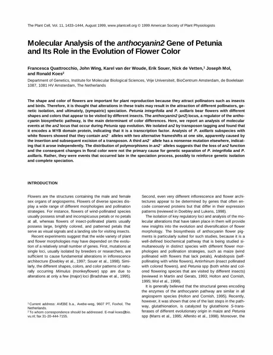
The Plant Cell, Vol. 11, 1433–1444, August 1999, www.plantcell.org © 1999 American Society of Plant Physiologists Molecular Analysis of the anthocyanin2 Gene of Petunia and Its Role in the Evolution of Flower Color Francesca Quattrocchio, John Wing, Karel van der Woude, Erik Souer, Nick de Vetten, 1 Joseph Mol, and Ronald Koes 2 Department of Genetics, Institute for Molecular Biological Sciences, Vrije Universiteit, BioCentrum Amsterdam, de Boelelaan 1087, 1081 HV Amsterdam, The Netherlands The shape and color of flowers are important for plant reproduction because they attract pollinators such as insects and birds. Therefore, it is thought that alterations in these traits may result in the attraction of different pollinators, ge- netic isolation, and ultimately, (sympatric) speciation. Petunia integrifolia and P. axillaris bear flowers with different shapes and colors that appear to be visited by different insects. The anthocyanin2 (an2) locus, a regulator of the antho- cyanin biosynthetic pathway, is the main determinant of color differences. Here, we report an analysis of molecular events at the an2 locus that occur during Petunia spp evolution. We isolated an2 by transposon tagging and found that it encodes a MYB domain protein, indicating that it is a transcription factor. Analysis of P. axillaris subspecies with white flowers showed that they contain an2 2 alleles with two alternative frameshifts at one site, apparently caused by the insertion and subsequent excision of a transposon. A third an2 2 allele has a nonsense mutation elsewhere, indicat- ing that it arose independently. The distribution of polymorphisms in an2 2 alleles suggests that the loss of an2 function and the consequent changes in floral color were not the primary cause for genetic separation of P. integrifolia and P. axillaris. Rather, they were events that occurred late in the speciation process, possibly to reinforce genetic isolation and complete speciation. INTRODUCTION Flowers are the structures containing the male and female sex organs of angiosperms. Flowers of diverse species dis- play a wide range of different morphologies and pollination strategies. For instance, flowers of wind-pollinated species usually possess small and inconspicuous petals or no petals at all, whereas flowers of insect-pollinated plants usually possess large, brightly colored, and patterned petals that serve as visual signals and a landing site for visiting insects. Recent experiments suggest that the wide variety of plant and flower morphologies may have depended on the evolu- tion of a relatively small number of genes. First, mutations at single loci, usually isolated by breeders or researchers, are sufficient to cause fundamental alterations in inflorescence architecture (Doebley et al., 1997; Souer et al., 1998). Simi- larly, the different shapes, colors, and color patterns of natu- rally occurring Mimulus (monkeyflower) spp are due to alterations at only a few (major) loci (Bradshaw et al., 1995). Second, even very different inflorescence and flower archi- tectures appear to be determined by genes that often en- code conserved proteins but that differ in their expression patterns (reviewed in Doebley and Lukens, 1998). The isolation of key regulatory loci and analysis of the mo- lecular alterations that have taken place in them will provide new insights into the evolution and diversification of flower morphology. The biosynthesis of anthocyanin flower pig- ments is particularly suited for such studies, because it is a well-defined biochemical pathway that is being studied si- multaneously in distinct species with different flower mor- phologies and pollination strategies, such as maize (wind pollinated with flowers that lack petals), Arabidopsis (self- pollinating with white flowers), Antirrhinum (insect pollinated with colored flowers), and Petunia spp (both white and col- ored flowering species that are visited by different insects) (reviewed in Martin and Gerats, 1993; Holton and Cornish, 1995; Mol et al., 1998). It is generally believed that the structural genes encoding the enzymes of the anthocyanin pathway are similar in all angiosperm species (Holton and Cornish, 1995). Recently, however, it was shown that one of the last steps in the path- way, glutathionation, is catalyzed by glutathione S-trans- ferases of different evolutionary origin in maize and Petunia spp (Marrs et al., 1995; Alfenito et al., 1998). Moreover, the 1 Current address: AVEBE b.a., Avebe-weg, 9607 PT, Foxhol, The Netherlands. 2 To whom correspondence should be addressed. E-mail koes@bio. vu.nl; fax 31-20-444-7155.
-
Upload
independent -
Category
Documents
-
view
2 -
download
0
Transcript of Molecular Analysis of the anthocyanin2 Gene of Petunia and Its Role in the Evolution of Flower Color
The Plant Cell, Vol. 11, 1433–1444, August 1999, www.plantcell.org © 1999 American Society of Plant Physiologists
Molecular Analysis of the
anthocyanin2
Gene of Petuniaand Its Role in the Evolution of Flower Color
Francesca Quattrocchio, John Wing, Karel van der Woude, Erik Souer, Nick de Vetten,
1
Joseph Mol,and Ronald Koes
2
Department of Genetics, Institute for Molecular Biological Sciences, Vrije Universiteit, BioCentrum Amsterdam, de Boelelaan 1087, 1081 HV Amsterdam, The Netherlands
The shape and color of flowers are important for plant reproduction because they attract pollinators such as insectsand birds. Therefore, it is thought that alterations in these traits may result in the attraction of different pollinators, ge-
netic isolation, and ultimately, (sympatric) speciation.
Petunia integrifolia
and
P. axillaris
bear flowers with differentshapes and colors that appear to be visited by different insects. The
anthocyanin2
(
an2
) locus, a regulator of the antho-cyanin biosynthetic pathway, is the main determinant of color differences. Here, we report an analysis of molecularevents at the
an2
locus that occur during
Petunia
spp evolution. We isolated
an2
by transposon tagging and found thatit encodes a MYB domain protein, indicating that it is a transcription factor. Analysis of
P. axillaris
subspecies with
white flowers showed that they contain
an2
2
alleles with two alternative frameshifts at one site, apparently caused by
the insertion and subsequent excision of a transposon. A third
an2
2
allele has a nonsense mutation elsewhere, indicat-ing that it arose independently. The distribution of polymorphisms in
an2
2
alleles suggests that the loss of
an2
functionand the consequent changes in floral color were not the primary cause for genetic separation of
P. integrifolia
and
P.axillaris.
Rather, they were events that occurred late in the speciation process, possibly to reinforce genetic isolationand complete speciation.
INTRODUCTION
Flowers are the structures containing the male and femalesex organs of angiosperms. Flowers of diverse species dis-play a wide range of different morphologies and pollinationstrategies. For instance, flowers of wind-pollinated speciesusually possess small and inconspicuous petals or no petalsat all, whereas flowers of insect-pollinated plants usuallypossess large, brightly colored, and patterned petals thatserve as visual signals and a landing site for visiting insects.
Recent experiments suggest that the wide variety of plantand flower morphologies may have depended on the evolu-tion of a relatively small number of genes. First, mutations atsingle loci, usually isolated by breeders or researchers, aresufficient to cause fundamental alterations in inflorescencearchitecture (Doebley et al., 1997; Souer et al., 1998). Simi-larly, the different shapes, colors, and color patterns of natu-rally occurring
Mimulus
(monkeyflower) spp are due toalterations at only a few (major) loci (Bradshaw et al., 1995).
Second, even very different inflorescence and flower archi-tectures appear to be determined by genes that often en-code conserved proteins but that differ in their expressionpatterns (reviewed in Doebley and Lukens, 1998).
The isolation of key regulatory loci and analysis of the mo-lecular alterations that have taken place in them will providenew insights into the evolution and diversification of flowermorphology. The biosynthesis of anthocyanin flower pig-ments is particularly suited for such studies, because it is awell-defined biochemical pathway that is being studied si-multaneously in distinct species with different flower mor-phologies and pollination strategies, such as maize (windpollinated with flowers that lack petals), Arabidopsis (self-pollinating with white flowers), Antirrhinum (insect pollinatedwith colored flowers), and
Petunia
spp (both white and col-ored flowering species that are visited by different insects)(reviewed in Martin and Gerats, 1993; Holton and Cornish,1995; Mol et al., 1998).
It is generally believed that the structural genes encodingthe enzymes of the anthocyanin pathway are similar in allangiosperm species (Holton and Cornish, 1995). Recently,however, it was shown that one of the last steps in the path-way, glutathionation, is catalyzed by glutathione
S
-trans-ferases of different evolutionary origin in maize and
Petunia
spp (Marrs et al., 1995; Alfenito et al., 1998). Moreover, the
1
Current address: AVEBE b.a., Avebe-weg, 9607 PT, Foxhol, TheNetherlands.
2
To whom correspondence should be addressed. E-mail [email protected]; fax 31-20-444-7155.

1434 The Plant Cell
expression of the structural genes appears to be regulateddifferently in distinct species (reviewed in Mol et al., 1998;Weisshaar and Jenkins, 1998). Mutational analyses showedthat in maize, transcription of the entire set of structural an-thocyanin biosynthesis genes is controlled as a single unitby two families of regulatory genes. The so-called
c1
/
pl
andthe
r
/
b
gene families comprise multiple paralogous genesthat encode functionally similar proteins that include a MYBdomain and a basic helix-loop-helix (bHLH) domain, respec-tively, and that have distinct expression patterns (Ludwigand Wessler, 1990; Cone et al., 1993).
In Antirrhinum and
Petunia
spp flowers, however, the path-way appears to be regulated in at least two distinct units(Martin et al., 1991; Quattrocchio et al., 1993). Mutations atthe
anthocyanin1
(
an1
),
an2
,
an4
, and
an11
loci of
P. hybrida
cause inactivation of structural genes acting late in thepathway in specific parts of the flower (Beld et al., 1989;Quattrocchio et al., 1993; Huits et al., 1994a). The sameholds true for mutations at
delila
(
del
) and
rosea
of Antirrhi-num (Martin et al., 1991). Remarkably, the early structuralgenes remain transcriptionally active in these
Petunia
sppand Antirrhinum mutants, suggesting that they are activatedby another set of regulatory genes (Moyano et al., 1996;Quattrocchio et al., 1998).
These findings can be explained in at least two ways: (1)the regulators of flower pigmentation in Antirrhinum and
Pe-tunia
spp are not related to
c1
and
r
from maize; or (2)
c1
-and
r
-related genes do control flower pigmentation, andregulatory differences are due to divergent evolution of thetarget structural genes. Although several regulatory antho-cyanin genes have now been isolated from Antirrhinum and
Petunia
spp, the data are too incomplete to exclude either ofthese two possibilities.
Molecular analysis of the
an11
locus of
Petunia
sppshowed that it encodes a WD-40 repeat protein that is highlyconserved in nature, even in animals and yeast that cannotsynthesize anthocyanins (de Vetten et al., 1997). However,no information is available on the function of these
an11
ho-mologs in their cognate hosts. The
an1
locus includes agene that encodes a bHLH protein but this gene cannot beconsidered to be an ortholog of the maize
r
gene (C. Speltand R. Koes, unpublished data), because another petuniagene,
jaf13
, has greater similarity to
r
(Quattrocchio etal., 1998). Although
jaf13
can replace the function of
r
insome functional assays, it cannot complement
r
2
mutants(Quattrocchio et al., 1998). Similarly, the
del
gene of Anti-rrhinum, encoding a bHLH protein with homology to R(Goodrich et al., 1992), failed to complement
r
2
mutants(Mooney et al., 1995). Therefore, it is not clear whether
jaf13
,
del
, and
r
are truly orthologous. However, our knowledge ofthe factors that control anthocyanin biosynthesis is far fromcomplete because, for instance, information on the productsencoded by
an2
and
an4
of
Petunia
spp and
rosea
of Anti-rrhinum is lacking. These loci are particularly interesting be-cause they appear to have played a role in the diversificationof flower color and patterns in nature.
P. integrifolia
and
P. axillaris
subspecies occur naturally inSouth America in partially overlapping areas. Yet, both spe-cies remain genetically separated, apparently because theirflowers are visited by different insects (Wijsman, 1982, 1983).
P. integrifolia
flowers have a purple corolla with a short widetube and are pollinated by bees (Figure 1A), whereas
P. axil-laris
flowers have a white corolla with a long narrow tube(Figure 1B), which is typical of moth-pollinated flowers(Wijsman, 1982). Manual cross-pollination, however, readilyproduces fertile progeny. In fact, the garden petunia (
P. hy-brida
) is thought to be derived from such interspecificcrosses (Wijsman, 1983; Sink, 1984; Koes et al., 1987).
Genetic analyses showed that differences at five loci,
an2
,
an4
,
hydroxylation-at-five
(
hf1
),
flavonols
(
fl
), and
pollen
(
po
),are responsible for the different floral organ colors of varioussubspecies of
P. integrifolia
(genotype
An2
1
An4
1
Hf1
1
fl
2
Po
1
)and
P. axillaris
(genotype
an2
2
an4
2
hf1-1fl
2
po
2
or
an2
2
an4
2
hf1-1Fl
1
po
2
) (Wijsman, 1983). The
an4
2
and
po
2
markers of
P. axillaris
are responsible for the yellow anthers,but because these markers do not affect coloration of thecorolla, they are not discussed here (cf. van Tunen et al.,1991; Quattrocchio et al., 1993). The markers
an2
2
,
hf1-1
,and
Fl
1
contribute in different degrees to the white color ofthe
P. axillaris
flower corolla. The
an2
2
genotype strongly re-duces but does not completely abolish the transcription ofstructural anthocyanin genes and coloration, when com-pared with
An2
1
. On their own, neither
Fl
1
nor the partially
Figure 1. Flower Phenotypes of Petunia spp.
(A) Flowers of P. integrifolia subsp violacea line S9.(B) Flowers of P. axillaris subsp axillaris line S1.(C) P. hybrida flower harboring the unstable an2-W82 allele.(D) P. hybrida flower harboring a revertant allele derived from an2-W82.

Flower Color Evolution in Petunia 1435
active hf1-1 allele contributes strongly to the white flowercolor. Fl1 makes the flower more bluish (copigmentation)without reducing the accumulation of delphinidin-type an-thocyanins. Mutation of Hf1 results in the formation of red-dish cyanidin-type anthocyanins instead of delphinidin-typeanthocyanins. Only the combination of Fl1 and hf1-1 (orhf12) reduces the synthesis of anthocyanins; for unknownreasons, flavonol synthesis competes with cyanidin synthe-sis and not so much with delphinidin synthesis (Wiering andDe Vlaming, 1984). During the evolution of petal color, therole of the an2 locus appears to have been more prominentthan the combination of hf1 and Fl1. This is because (1) P.axillaris accessions can be either Fl1 or fl2 (Wijsman, 1983),indicating that this difference arose late, after the geneticseparation from P. integrifolia; (2) An21fl2hf1-1 flowers canstill accumulate a considerable amount of anthocyanins(see, e.g., Figures 6H and 6I in Quattrocchio et al., 1998),whereas (3) the effect of an an22 allele in a Hf11Fl1 back-ground is considerably stronger (see Figures 6J and 6K inQuattrocchio et al., 1998).
To gain further insight into the molecular mechanisms thatcontrol flower pigmentation and into the evolution of thosemechanisms, we conducted a molecular analysis of the an2locus. We isolated an2 by transposon tagging and showedthat it encodes a MYB domain protein. A comparison of thean2 alleles in Petunia spp showed that the lack of an2 func-tion in P. axillaris flowers results from two independent loss-of-function mutations, one of which was apparently inducedby the insertion and excision of a transposable element.Analysis of an2 polymorphisms indicated that mutations inan2 occurred relatively late in the speciation process, sug-gesting that in this instance, floral color change was not theprimary speciation event but rather a mechanism to rein-force genetic separation.
RESULTS
Molecular Analysis of the an2 Locus
P. hybrida line W82 in the Amsterdam collection harbors anunstable an2 allele, herein named an2-W82, that is presum-ably identical to the an2-n allele described earlier (Cornu,1977). The flowers of an2-W82/an2-W82 plants have pale-colored corolla limbs with sectors and spots of a different in-tensity (Figure 1C). Among progeny obtained by self-pollina-tion of an2-W82 homozygotes, we identified four plants withfull-colored flowers (Figure 1D) that originated from indepen-dent sporogenic reversion events.
Because most unstable alleles in P. hybrida contain inser-tions of the 284-bp transposable element dTph1 (vanHouwelingen et al., 1998), we anticipated the presence ofdTph1 in the an2-W82 allele. To clone a fragment of the an2locus, we isolated dTph1 flanking sequences that were
present in two an2-W82 homozygous plants, but not in a ho-mozygote for the revertant allele An21-R1, by a combinationof inverse polymerase chain reactions (PCRs) and differen-tial screening of cloned amplification products (Souer et al.,1995). One of these dTph1 flanking sequences, jaf41, hy-bridized with a 2.2-kb fragment in an2-W82 homozygousplants, whereas a 1.9-kb fragment was detected in plantsharboring four independently derived excision alleles (Figure2A). This result showed that jaf41 contains part of the an2locus. We subsequently isolated the complete locus andcDNA clones by hybridizing jaf41 with corresponding librar-ies that were made from the An21 P. hybrida line V26.
The sequence of an an2 cDNA revealed a large open
Figure 2. Molecular Analysis of an2.
(A) DNA gel blot of P. hybrida plants harboring the mutable an2-W82allele (W82) or four independently derived An21 revertant alleles (R1to R4) hybridized with the dTph1 flanking sequence jaf41. The sizeof the hybridizing fragments (in kilobases) is indicated at right.(B) Diagram showing the structure of the an2 gene (top) and cDNA(bottom). Exon sequences are shown as a block; the protein codingsequences are blocks that are twice as high. The region encodingthe MYB domain is shown in black, and the two repeats (R2 and R3)that make up this domain are indicated with arrows. The triangleshows the position of the dTph1 insertion in the an2-W82 allele.(C) Sequence around the ATG translation start in wild-type An21,unstable an2-W82, and revertant An21-Rev1 alleles. The ATG at thestart of the an2 ORF is marked in boldface. The arrows denote the8-bp target site duplicated by the dTph1 insertion (triangle).

1436 The Plant Cell
reading frame (ORF) of 765 bp (Figure 2B) that is precededby a small (69 bp) ORF. In an2-W82, the dTph1 insertion du-plicated 8 bp, including the ATG start codon of the largeORF (Figure 2C), without lowering the an2 mRNA level (datanot shown). This suggests that the mutant phenotype iscaused by reduced translation initiation of the large ORFand implies that this ORF encodes AN2. The finding thatnaturally occurring an22 alleles contain mutations that dis-rupt the 765-bp ORF lends further support to this conclusion(see below).
Database searches revealed that the AN2 protein is simi-lar to a range of so-called MYB domain proteins from ani-mals and plants. Parsimony analysis showed that, in general,AN2 is more related to plant MYB proteins than to the ani-mal c-MYB, the prototype of this superfamily (Figure 3A).The protein with the highest similarity found in the data-bases is that encoded by the myb75 gene of Arabidopsis, agene whose function is not known (Kranz et al., 1998). Be-cause the majority of genes in this Arabidopsis family havebeen sequenced, this suggests that myb75 may be the Ara-bidopsis homolog of an2. However, AN2 does not displaysignificantly greater similarity to other MYB domain proteinsimplicated in flavonoid biosynthesis, such as MYB305 andMYB340 of Antirrhinum (Moyano et al., 1996), C1 and Pfrom maize (Paz-Ares et al., 1987; Grotewold et al., 1991), orMYBPH3 from P. hybrida (Solano et al., 1995), than it doesto MYB domain proteins involved in other processes (Figure3B). In all cases, the similarity was restricted to the two re-peats that make up the MYB domain, whereas no similari-ties were detectable in the C-terminal half of these proteins(Figure 3B). Comparison of the genomic and cDNA se-quences showed that the an2 gene is split by two introns
(Figure 2B) in positions that are conserved in other myb-related genes.
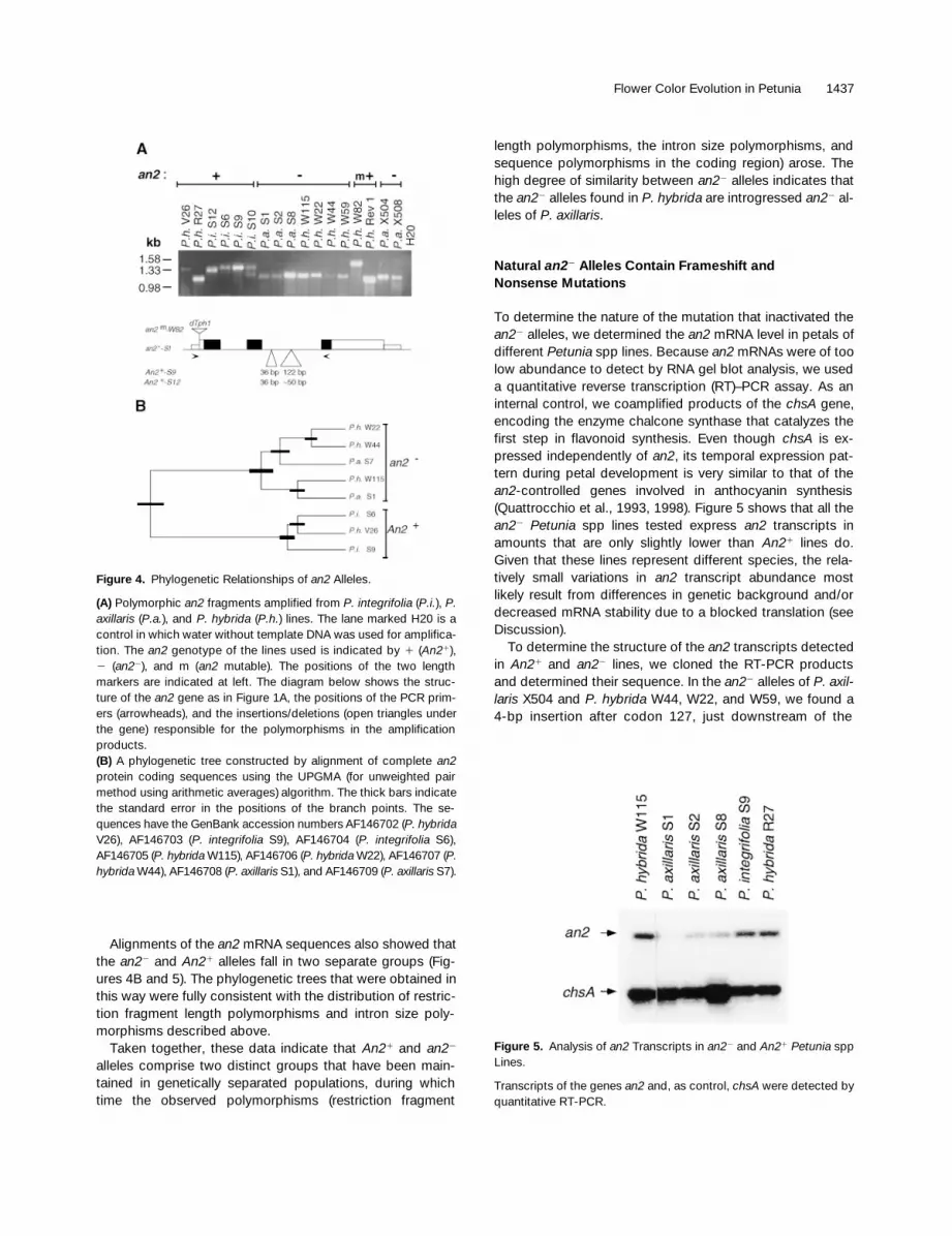
Polymorphisms among an2 Alleles
To investigate the evolutionary relationships between thevarious An21 and an22 alleles of different Petunia spp, weconducted DNA gel blot analyses. These experimentsshowed that P. axillaris subsp and an22 P. hybrida lines con-tain genomic an2 fragments with nearly identical restrictionmaps, whereas the P. integrifolia subsp and An21 P. hybridalines harbored different polymorphic fragments (data notshown). We also detected polymorphisms by using PCRamplification of an an2 fragment that spans both introns(Figure 4A). In an22 lines of P. axillaris or P. hybrida, this am-plification generated a 1.2-kb fragment, whereas the samefragment in An21 lines of P. integrifolia and P. hybrida variedin size between 1.2 and 1.4 kb (Figure 4A). Further mappingwith different primer combinations showed that the poly-morphism resides in the second an2 intron (data not shown).To determine the nature of the polymorphism, we PCR am-plified and sequenced the second intron of the an2 allele ofP. axillaris S1 and P. integrifolia S9 and S12. Comparison ofthese sequences showed that the polymorphism resultedfrom two insertions or deletions—one of 36 bp and a secondof variable length (z50 bp in P. integrifolia S12 and 122 bpin P. integrifolia S9) (Figure 4A). A closer examination ofthese insertions/deletions and surrounding region did notreveal specific features (e.g., repeated sequences) thatmight account for a mechanism by which the polymorphismwas established.
Figure 3. an2 Encodes a MYB Domain Protein.
(A) A neighbor-joining phylogenic tree of AN2 and selected MYB domain proteins from other species. The numbers in the branches indicate thepercentage of bootstrap support after 500 replicates. For construction of the tree, we used only the MYB domains; for c-MYB, we also excludedrepeat 1 of its MYB domain, because this repeat is not found in the plant proteins.(B) Alignment of the MYB domain of AN2 and proteins controlling anthocyanin (C1) and phlobaphene (P) synthesis in maize, phenylpropanoidmetabolism (MYB305) in Antirrhinum and Petunia spp (MYBPH3), cell shape in Antirrhinum (MIXTA) and Petunia spp (MYBPH1), trichome devel-opment in Arabidopsis (GL1), and hematopoiesis in humans (c-MYB) (reviewed in Martin and Paz-Ares, 1997), and MYB75 of Arabidopsis, a pro-tein with unknown function. Residues on a black background denote sequence similarity between AN2 and one or more of the other proteins.
Flower Color Evolution in Petunia 1437
Alignments of the an2 mRNA sequences also showed thatthe an22 and An21 alleles fall in two separate groups (Fig-ures 4B and 5). The phylogenetic trees that were obtained inthis way were fully consistent with the distribution of restric-tion fragment length polymorphisms and intron size poly-morphisms described above.
Taken together, these data indicate that An21 and an22
alleles comprise two distinct groups that have been main-tained in genetically separated populations, during whichtime the observed polymorphisms (restriction fragment
length polymorphisms, the intron size polymorphisms, andsequence polymorphisms in the coding region) arose. Thehigh degree of similarity between an22 alleles indicates thatthe an22 alleles found in P. hybrida are introgressed an22 al-leles of P. axillaris.
Natural an22 Alleles Contain Frameshift andNonsense Mutations
To determine the nature of the mutation that inactivated thean22 alleles, we determined the an2 mRNA level in petals ofdifferent Petunia spp lines. Because an2 mRNAs were of toolow abundance to detect by RNA gel blot analysis, we useda quantitative reverse transcription (RT)–PCR assay. As aninternal control, we coamplified products of the chsA gene,encoding the enzyme chalcone synthase that catalyzes thefirst step in flavonoid synthesis. Even though chsA is ex-pressed independently of an2, its temporal expression pat-tern during petal development is very similar to that of thean2-controlled genes involved in anthocyanin synthesis(Quattrocchio et al., 1993, 1998). Figure 5 shows that all thean22 Petunia spp lines tested express an2 transcripts inamounts that are only slightly lower than An21 lines do.Given that these lines represent different species, the rela-tively small variations in an2 transcript abundance mostlikely result from differences in genetic background and/ordecreased mRNA stability due to a blocked translation (seeDiscussion).
To determine the structure of the an2 transcripts detectedin An21 and an22 lines, we cloned the RT-PCR productsand determined their sequence. In the an22 alleles of P. axil-laris X504 and P. hybrida W44, W22, and W59, we found a4-bp insertion after codon 127, just downstream of the
Figure 4. Phylogenetic Relationships of an2 Alleles.
(A) Polymorphic an2 fragments amplified from P. integrifolia (P.i.), P.axillaris (P.a.), and P. hybrida (P.h.) lines. The lane marked H20 is acontrol in which water without template DNA was used for amplifica-tion. The an2 genotype of the lines used is indicated by 1 (An21),2 (an22), and m (an2 mutable). The positions of the two lengthmarkers are indicated at left. The diagram below shows the struc-ture of the an2 gene as in Figure 1A, the positions of the PCR prim-ers (arrowheads), and the insertions/deletions (open triangles underthe gene) responsible for the polymorphisms in the amplificationproducts.(B) A phylogenetic tree constructed by alignment of complete an2protein coding sequences using the UPGMA (for unweighted pairmethod using arithmetic averages) algorithm. The thick bars indicatethe standard error in the positions of the branch points. The se-quences have the GenBank accession numbers AF146702 (P. hybridaV26), AF146703 (P. integrifolia S9), AF146704 (P. integrifolia S6),AF146705 (P. hybrida W115), AF146706 (P. hybrida W22), AF146707 (P.hybrida W44), AF146708 (P. axillaris S1), and AF146709 (P. axillaris S7).
Figure 5. Analysis of an2 Transcripts in an22 and An21 Petunia sppLines.
Transcripts of the genes an2 and, as control, chsA were detected byquantitative RT-PCR.

1438 The Plant Cell
conserved MYB domain. This insertion results in a readingframe shift and a premature stop codon, thereby preventingthe translation of the nonconserved C-terminal half of theAN2 protein (Figures 6A and 6B). In the an22 alleles in P. ax-illaris S1 and X508 and P. hybrida W115, we found a 1-bpdeletion in codon 127, which also prevents translation of theC-terminal half of AN2 (Figure 6B). Partial sequencing of thean22 allele of P. axillaris S8 showed that this allele containedthe same 1-bp deletion.
Partial sequencing of the an2-S7 mRNA showed that thereading frame was intact at codon 127. We subsequentlydetermined the complete mRNA sequence. This showedthat an2-S7 contains a nonsense mutation farther down-stream at codon 196 (Figure 6B).
The Frameshift and Nonsense Mutations Are Responsible for an2 Inactivation
We considered the possibility that the frameshift and non-sense mutations arose in an2 alleles that had been alreadyinactivated by other mutations. It is very unlikely that muta-tions upstream of the frameshift had inactivated an2 prior tothe frameshift, because the truncated protein encoded byan2-W22 has the same sequence as the corresponding re-gion in that encoded by An21-V26 (Figure 6A). However, wecould not exclude that inactivation had occurred down-stream from the frameshift.
To identify evolutionary old mutations that might have inac-tivated the an22 alleles before the frameshift and nonsense
Figure 6. Analysis of the Mutations That Inactivate an22 Alleles.
(A) Alignment of translated An21 and an22 alleles. To produce the translations of the an22 alleles, the frameshift or nonsense mutations were ig-nored. The positions of these frameshift and nonsense mutations in an22 alleles are indicated by a dot on a red background. Polymorphismsspecific for An21 and an22 alleles are indicated in purple and green, respectively. Other polymorphisms are highlighted in black. Numbering of theamino acid residues is indicated above the sequences (dots indicate every tenth residue). For species abbreviations, see the legend to Figure 4A.(B) DNA and deduced protein sequences around the frameshift and nonsense mutations in an22 alleles. Codons 127 and 196, at which frame-shift and nonsense mutations were found, are in red. The broken lines denote the sequences in between these two regions. Stop codons thatterminate the AN2 protein coding sequence are boxed.(C) Structure and activity of An21, an22, and hybrid alleles. The maps denote the mRNA structure of the different alleles. Black regions denotethe MYB domain, and gray regions denote other translated sequences. White regions indicate mRNA sequences that are not translated due to thepresence of a frameshift (an2-W44) or a nonsense mutation (an2-S7) at the site marked by a red asterisk. The green and purple bars indicate theposition of polymorphisms, as given in (A). The activity of these alleles, when expressed from the 35S promoter, was measured after micro-projectile delivery into an2-W115 petal cells by using a dfrA–luc reporter gene. Activities are given as the mean 6SE of five independent bom-bardment assays and are expressed in arbitrary units.
Flower Color Evolution in Petunia 1439
mutation arose, we searched for sequence polymorphismsthat are consistently present in (nearly) all an22 alleles.Therefore, we aligned the translated AN2 sequences whileignoring the frameshift and nonsense mutations in the an22
alleles (Figure 6A). This revealed some sequence variationsfor single alleles that are scattered throughout the protein(Figure 6A). This distribution pattern indicates that these mu-tations arose relatively late in evolution, after the frameshiftand nonsense mutations, and therefore were not responsi-ble for gene inactivation in nature. Because the an22 allelesare no longer under selection pressure, one might expectthat more such mutations will accumulate at a high rate.
At 13 positions, all clustered in the C terminus, the pro-teins of the An21 and an22 alleles differed consistently (Fig-ure 6A), indicating that these variations occurred soon afterthe genetic separation of both groups of alleles. To examinewhether these early alterations resulted in an inactive AN2protein, we used a conserved ClaI restriction site to replacethe last 65 codons of the wild-type An21-V26 cDNA withthose from an22-W44 and fused the cDNAs to the constitu-tive 35S promoter of the cauliflower mosaic virus. To assaythe activity of the various an2 constructs, we used a com-plementation assay in which they were delivered by particlebombardment into an22 petal cells. To measure AN2 activ-ity, we assayed the expression of a codelivered reportergene (luc, encoding luciferase) driven by the an2-responsivepromoter of the dfrA gene, which encodes dihydroflavonol4-reductase, a “late” enzyme in the anthocyanin pathway.To normalize the dfrA-driven LUC activity for variations intransformation efficiency, we measured the activity of acodelivered reporter gene consisting of the b-glucuronidasecoding sequence driven by the 35S promoter (35S–gus).Figure 6C shows that an2-W115, containing a 21-bp frame-shift at codon 127, is a null allele because it fails to inducethe dfrA–luc reporter gene. The hybrid allele An21-V26/W44,however, activated the dfrA promoter at least as efficientlyas the wild-type An21-V26 allele. This indicates that thepolymorphisms near the former stop codon of an2-W44 orits progenitor allele did not inactivate the protein. This ap-parent high tolerance for sequence variations cannot betaken as evidence that this part of the AN2 protein is withoutfunction, because truncation of the C-terminal domain, as inan2-S7, results in complete loss of AN2 activity (Figure 6C).
Taken together, these data strongly suggest that inactiva-tion of an2 in nature was caused by the frameshift and non-sense mutations, and not by the sequence variationsobserved in the C terminus of the protein.
DISCUSSION
To elucidate the molecular mechanisms that control flowerpigmentation patterns, we studied a set of regulatory genesthat control the tissue-specific transcription of the structuralgenes encoding enzymes of the anthocyanin pathway. Early
experiments indicated that transcription of these structuralgenes in P. hybrida, Arabidopsis, and tobacco could be acti-vated by combined expression of the c1 and r genes frommaize (Lloyd et al., 1992; Quattrocchio et al., 1998). How-ever, this does not necessarily mean that r- and c1-homolo-gous genes control pigmentation of the flower in vivo. Infact, the finding that an11 and an1 encode, respectively, aWD-40 protein (de Vetten et al., 1997) and a bHLH protein(C. Spelt and R. Koes, unpublished data) that had not beenpreviously identified in other species indicated a different di-rection. Here, we report the isolation of an2, a third regulatorof the anthocyanin pathway in Petunia spp, which acts inconcert with an1 and an11 to activate transcription of struc-tural anthocyanin genes in the petal limb.
an2 Encodes a MYB Domain Protein
The homology of AN2 to various MYB domain proteins sug-gests that AN2 is a transcription factor that activates a sub-set of structural anthocyanin genes. Such an activity wouldbe consistent with the results of transient expression as-says, which show that AN2 can activate promoter activity ofthe dfr gene (Figure 6C and Quattrocchio et al., 1998).Whether this activity involves binding of AN2 to the pro-moter of the structural genes or operates via an intermediateregulatory gene is currently under investigation.
Strikingly, AN2 does not display significantly more similar-ity to MYB305 and MYB340, two regulators of early struc-tural genes in Antirrhinum (Sablowski et al., 1994; Moyano etal., 1996), or C1 and Pl from maize. This finding, togetherwith the observation that an2, myb305/myb340, and c1 con-trol different sets of target structural genes in their respec-tive hosts (reviewed in Mol et al., 1998), may at first sightsuggest that these genes are not homologs. However,subsequent expression assays and complementation ex-periments showed that AN2 can replace C1 and vice versa,indicating that c1 and an2 are homologous genes(Quattrocchio et al., 1998). The different sets of target an-thocyanin biosynthesis genes controlled by an2 and c1 intheir cognate hosts therefore appear to be due to divergentevolution of the target genes rather than of the regulatoryproteins (Koes et al., 1994; Quattrocchio et al., 1998).
For C1, it was shown that its specificity resides in theDNA binding MYB domain (Sainz et al., 1997; Williams andGrotewold, 1997) and that this domain interacts with bHLHproteins encoded by the r gene family (Goff et al., 1992).However, also in this more limited domain, the similarity be-tween AN2 and C1 does not appear to be significantlyhigher than it is between functionally unrelated MYB domainproteins (Figure 3), even though AN2 is the only MYB proteinknown that can complement mutations in c1 and interactwith maize R proteins or the petunia homolog JAF13 in tran-sient expression assays (Quattrocchio et al., 1998) and in ayeast two-hybrid assay (A. Kroon and R. Koes, unpublisheddata). The C-terminal half of AN2 functions in yeast as a
1440 The Plant Cell
transcriptional activation domain (A. Kroon and R. Koes, un-published data), similar to the C-terminal domain of C1 (Goffet al., 1992). This suggests that the similarity between C1and AN2 is higher than can be detected by sequence align-ments or, in other words, that a significant number of alter-ations are tolerated—especially in the C-terminal domain—without loss of activity or specificity. This may account forthe relatively high sequence divergence in the (former) C-ter-minal domains of an22 and An21 alleles (Figure 6A). Never-theless, this domain appears essential for activity, becausethe truncated protein encoded by an2-S7 is inactive (Figure 6C).
In P. hybrida, mutations in either of the regulators an1 oran11 leads to a complete loss of expression of their targetanthocyanin genes in all pigmented tissues and, conse-quently, to completely white flowers (Quattrocchio et al.,1993). The function of an2, on the other hand, seems highlyredundant, because in an22 mutants, pigmentation ofseeds, the flower stem, anthers, and the corolla tube is unal-tered, whereas only pigmentation of the corolla limb is re-duced. In an22 corolla limbs, some residual expression oftarget genes remains detectable (Quattrocchio et al., 1993);as a consequence, the corolla limb is patchy and pale col-ored rather than completely white. In a P. axillaris back-ground, however, the formation of the pale petal color isprevented by the hf1-1 and Fl1 alleles, which in combinationalso reduce anthocyanin formation (Wiering and De Vlaming,1984). In transient expression assays, we could not detectactivity of the an22 alleles, even when they were expressedfrom the strong 35S promoter, indicating that they are nullalleles. Therefore, it is possible that the residual expressionof structural genes in an22 corolla limbs is controlled by an-other paralogous locus. We recently isolated a candidateAN2 paralog by yeast two-hybrid screens (A. Kroon and R.Koes, unpublished data), a finding that may help to solvethis issue.
The Role of Transposons in the Generation ofan22 Alleles
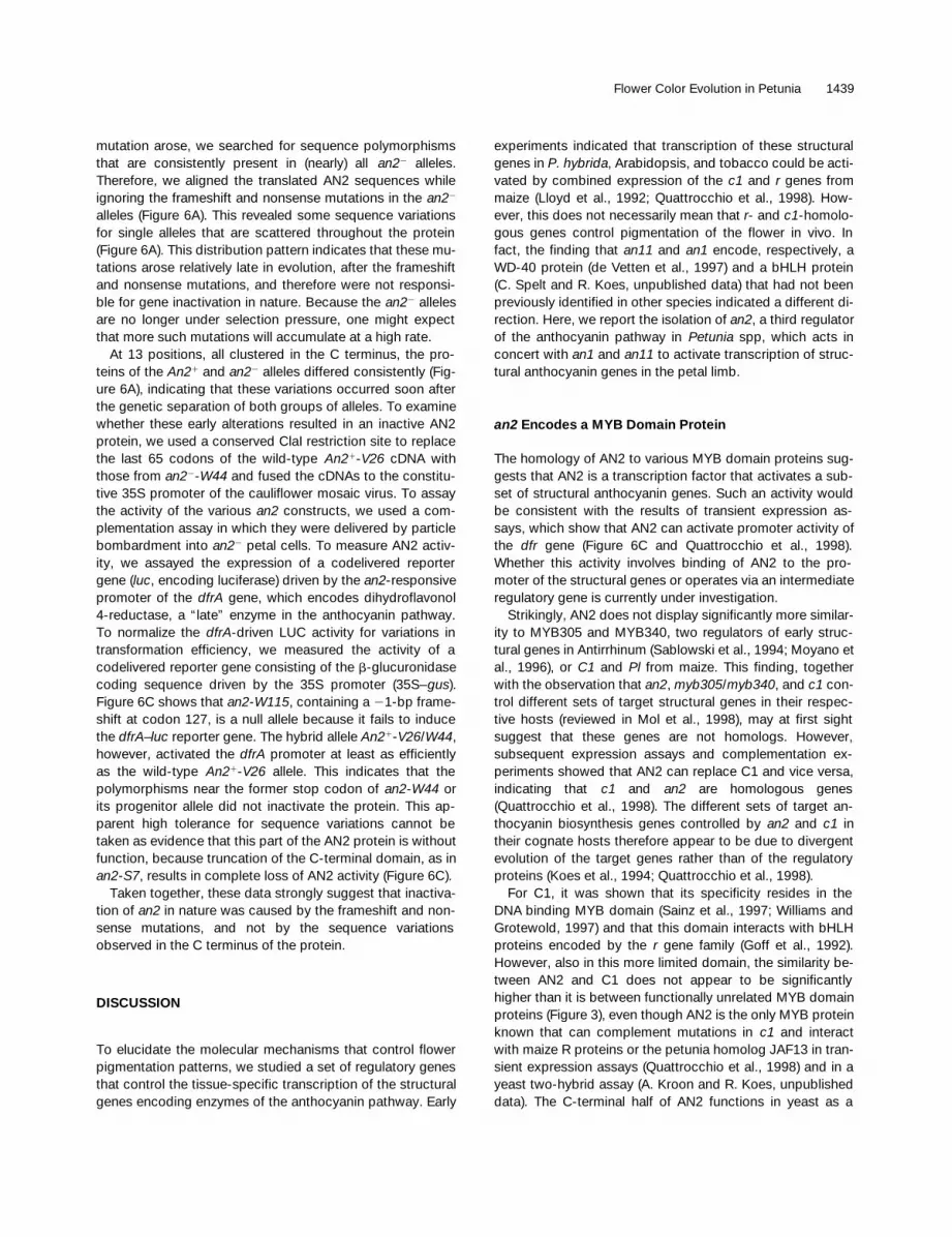
In natural isolates of P. axillaris, we found three differentan22 alleles harboring either a nonsense mutation or a 14-or a 21-bp frameshift mutation. Our data strongly suggestthat these were the mutations that inactivated an2 in nature.First, an22 Petunia spp lines still express an2 transcripts, in-dicating that cis-acting elements in the promoters of thecorresponding an2 alleles are still working properly. Theslightly lower abundance of an2 transcripts in an22 linescompared with An21 lines may be due to the different ge-netic backgrounds of the Petunia spp lines and to reducedstability of an2 transcripts because of premature terminationof translation. The latter phenomenon was also observed fortranscripts with frameshifts of an11 (de Vetten et al., 1997),an1 (C Spelt, F. Quattrocchio, J. Mol, and R. Koes, manu-script in preparation), and alf (Souer et al., 1998) and was re-ported by others as well (van Hoof and Green, 1996).
Because the 14- and the 21-bp frameshifts found inmost an22 alleles are located on the same site, it is unlikelythat they were generated by completely independentevents. Closer inspection of the 4-bp insertion sequenceshows that it has precisely the structure predicted for the in-sertion and subsequent excision of a transposon, stronglysuggesting that the 14-bp and 21-bp an22 alleles weregenerated by two independent excisions of the same trans-poson (Figure 7). The petunia transposons dTph1 (Gerats etal., 1990), dTph2 (van Houwelingen et al., 1998), dTph3(Kroon et al., 1994), dTph4 (Renckens et al., 1996; Alfenito etal., 1998), and dTph5 (C. Spelt and R. Koes, unpublisheddata) all belong to the Activator superfamily and generate an8-bp target site duplication upon insertion. After excision, afootprint is left behind that consists of remains of the targetsite duplication (>7 bp of each target site duplication; cf.Coen et al., 1986; Kunze et al., 1997) often separated by oneor more inverted nucleotides of the target site duplication.Figure 7A shows that the 21- and 14-bp frameshift muta-tions can be explained, consistent with existing models forfootprint formation, by two independent excisions of thesame Activator-like transposon. However, in our analyses offootprints produced by dTph1 elements, we never found therelatively large deletions in the target site duplication in-ferred in Figure 7A, suggesting that these may be rareevents (van Houwelingen et al., 1999).
More recently, two families of petunia transposons—Ps1(Snowden and Napoli, 1998) and dTph6 (C. Spelt and R.Koes, unpublished data)—were discovered that belong tothe so-called CACTA family. Ps1 and dTph6 also belong to theso-called CACTA family. These transposons generate a 3-bp
Figure 7. Model Showing How the Insertion and Excision of aTransposable Element May Have Created an22 Alleles with Two Dif-ferent Frameshifts (21 and 14 bp) at One Site.
(A) Model based on the insertion and excision of an Activator-like el-ement, such as dTph1.(B) Model based on the insertion and excision of a CACTA element,such as dTph6.In (A) and (B), the nucleotides that are duplicated by the transposoninsertion are underlined; nucleotides that have been lost during sub-sequent transposon excision and break–repair are shown as dots.
Flower Color Evolution in Petunia 1441
target site duplication upon insertion and produce footprintsthat consist of remains of the target site duplication (<3 bp),often separated by small inversions of the target site dupli-cation without missing nucleotides at the symmetry axis (cf.Coen et al., 1986; Kunze et al., 1997). Figure 7B shows thatthe 21- and 14-bp frameshift an22 alleles are fully consis-tent with the insertion and excision of a CACTA transposon.
All of the above-mentioned transposons were identified inP. hybrida initially. However, DNA gel blot experimentsshowed that all P. axillaris and P. integrifolia accessions an-alyzed contained numerous copies of dTph1 (Gerats et al.,1990; Huits et al., 1994b), Ps1 (Snowden and Napoli, 1998),and dTph6 (F. Quattrocchio, C. Spelt, and R. Koes, unpub-lished data).
Taken together, these data suggest that in nature, the 21-and 14-bp frameshift in the an22 alleles of P. axillaris weremost likely generated by a transposable element, presum-ably a member of the CACTA family, although the involve-ment of an Activator-like element cannot be excluded.
The Evolution of Flower Color in Petunia
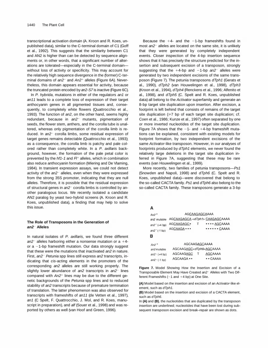
The genetic separation of populations followed by mutation,selection, and genetic drift is thought to be the basis for thecreation and divergence of species (Coyne, 1992). Tradition-ally, it is believed that this requires physical isolation, for in-stance, by geographical factors (allopatric speciation).However, speciation can in principle also occur between in-dividuals that grow side by side (sympatric speciation). Asflower shape and color attract specific pollinating animals,alteration of one or more of these traits may cause the at-traction of different pollinators and genetic isolation (Vickery,1992). For example, Mimulus spp mutations at only a fewloci are sufficient to alter floral traits, such as shape, color,color pattern, and nectar yield, and switch from bee to hum-mingbird pollination (Bradshaw et al., 1995). Although this isconsistent with a sympatric speciation scenario, it remainsto be determined if Mimulus spp arose sympatrically. Oneproblem with such sympatric speciation scenarios is to envi-sion how the first mutation, for example, a new color, be-comes fixed in the population, because it might initially beat a disadvantage as long as the flower retains its originalshape.
The sequences of the an2 alleles provide a record of theirhistory from which one can deduce how different flower col-ors arose in the genus Petunia in relation to speciationevents. Figure 8 provides a model that summarizes and ex-plains our findings. First, the presence of mutated an22 al-leles in P. axillaris subsp indicates that the common ancestorof P. integrifolia and P. axillaris was colored flowering (An21)and that the white (an22) P. axillaris flowers arose by subse-quent loss of an2 function. This is in contrast to the evolu-tion of kernel pigmentation in maize, which was establishedby gain-of-function mutations that altered the expressionpattern of the regulatory c1 and r genes of its progenitor Te-
osinte (Hanson et al., 1996). Second, we postulate that theancestral petunia population split for unknown reasons (e.g.,a geographical barrier or alterations in flower shape) into twogenetically isolated groups that provided the foundation fortoday’s P. integrifolia and P. axillaris subsp.
The old polymorphisms in An21 and an22 alleles are notspread randomly over the gene but rather are clustered inregions in which sequence alterations are relatively easilytolerated without loss of function, such as the intron and the39 end of the coding sequence (Figure 4). This indicates thatthe an2 gene was after the genetic separation, at least forsome time, still under selection pressure in both Petunia spppopulations. This implies that no inactivating mutations hadoccurred and that the flowers were still colored (An21) atthis stage of speciation. In the group that founded P. axil-laris, at least two independent an22 alleles arose through thegeneration of a nonsense mutation and the insertion and ex-cision of a transposon, respectively. Presumably, unknowncircumstances (e.g., the insects visiting the flowers) favoredwhite corolla limbs in this population, which explains whythe an22 alleles became fixed and both the An21 progenitorand An21 revertant alleles (in which transposon excisiongenerated an active An21 allele) seem to have disappeared.In this respect, it is interesting that insects (bees) are able toassociate color with other characteristics such as scent andreward (Srinivasan et al., 1998).
Because in P. hybrida, other anthocyanin genes, such asan1 (a regulatory gene) and an3 (encoding the enzyme fla-vanone 3b-hydroxylase), are more susceptible to transpo-son insertions than is an2 (van Houwelingen et al., 1998), it
Figure 8. Model Summarizing Events at the an2 Locus during Petu-nia spp Evolution.
The rectangles describe molecular alterations at the an2 locus. Theresulting flower phenotypes are indicated by the cartoons. The crossindicates the possible extinction of an allele.

1442 The Plant Cell
is likely that the an22 mutants were only a few out of manywhite-flowering petunia mutants that arose in nature. We as-sume that only the an2 alleles became fixed, because they onlyreduce anthocyanin synthesis in the petal limb, without ad-ditional effects on flavonoid accumulation. Mutation of anyof the other known anthocyanin genes also reduces the syn-thesis of flavonoids in other tissues, such as the flower tubeand the seed coat, and/or reduces the synthesis of other fla-vonoid compounds, such as flavonols (Quattrocchio et al.,1993; Huits et al., 1994a; van Houwelingen et al., 1998),which is apparently disadvantageous in a natural habitat.
Taken together, our data indicate that the flower colorchange caused by an22 mutations occurred after the ge-netic separation of both Petunia groups. Therefore, it seemsthat the an22 mutation(s) was not the primary cause of spe-ciation but rather a reinforcement mechanism (Coyne, 1992)that helped to complete the genetic separation.
METHODS
Plant Materials
The inbred lines of Petunia axillaris subsp axillaris (S1 and S2) andsubsp parodii (S7 and S8) and P. integrifolia subsp inflata (S6), subspviolacea (S9 and S10), and subsp integrifolia (S12) have been de-scribed previously (Wijsman, 1982). The P. axillaris accessions X504and X508 (our numbering) were obtained around 1980 from the Bo-tanical Garden of Liebig University (Giessen, Germany) and fromR.N. Bowman (Goldsmith Seeds, Gilroy, CA), respectively. The an2-W82 allele was kept in the Amsterdam collection in the P. hybrida lineW82 and is presumably identical to an2-n (Cornu, 1977).
Nucleic Acid Analysis
DNA extraction, DNA gel blot analysis, polymerase chain reaction(PCR) amplification, and sequence analyses were performed as pre-viously described (Souer et al., 1995; de Vetten et al., 1997). Theflanking sequence of the dTph1 element in an2-W82 (jaf41) was iden-tified by established procedures (Souer et al., 1995; de Vetten et al.,1997) and used to screen a petal cDNA library and a genomic libraryof P. hybrida V26. an2 cDNAs from other lines were isolated by re-verse transcription (RT)–PCR, and crucial regions were sequenced inat least two independently amplified products and in many cases bysequencing the corresponding region in PCR products amplifiedfrom genomic DNA. Analyses of DNA sequences were performed byusing the program Geneworks 2.3 (Intelligenetics, Mountain View,CA). Maximum parsimony analysis was performed with the programPAUP 3.1.1 (Smithsonian Institution, Washington, DC).
Quantification of mRNAs by RT-PCR analysis was performed asdescribed previously (Quattrocchio et al., 1998).
Transient Expression Assays
an2 cDNA fragments were ligated between the cauliflower mosaic vi-rus 35S promoter and nopaline synthase polyadenylation signal and
delivered into an2-W115 mutant petal limbs by particle bombard-ment. AN2 activity was determined by measuring the induction of adfrA–luc reporter gene and normalization for transformation effi-ciency using a 35S–b-glucuronidase gene, as described previously(de Vetten et al., 1997; Quattrocchio et al., 1998).
ACKNOWLEDGMENTS
We are grateful to Mark Rausher for discussions and review of themanuscript, Dik Roelofs for help with the parsimony analyses, andJane Olsson for help in improving the manuscript. This work wassupported by the Netherlands Organisation for Chemical research(S.O.N.), with financial aid from the Netherlands Organisation for theAdvancement of Research (N.W.O.), and by the European UnionBIOTECH program.
Received January 14, 1998; accepted May 19, 1999.
REFERENCES
Alfenito, M.R., Souer, E., Goodman, C.D., Buell, R., Mol, J., Koes,R., and Walbot, V. (1998). Functional complementation of antho-caynin sequestration in the vacuole by widely divergent glu-tathione S-transferases. Plant Cell 10, 1135–1149.
Beld, M., Martin, C., Huits, H., Stuitje, A.R., and Gerats, A.G.M.(1989). Flavonoid synthesis in Petunia hybrida: Partial character-ization of dihydroflavonol 4-reductase genes. Plant Mol. Biol. 13,491–502.
Bradshaw, H.D., Jr., Wilbert, S.M., Otto, K.G., and Schemske,D.W. (1995). Genetic mapping of floral traits associated withreproductive isolation in monkey flowers (Mimulus). Nature 376,762–765.
Coen, E.S., Carpenter, R., and Martin, C. (1986). Transposableelements generate novel spatial patterns of gene expression inAntirrhinum majus. Cell 47, 285–296.
Cone, K.C., Cocciolone, S.M., Burr, F.A., and Burr, B. (1993).Maize anthocyanin regulatory gene pl is a duplicate of c1 thatfunctions in the plant. Plant Cell 5, 1795–1805.
Cornu, A. (1977). Systemes instables induits chez le petunia. Mut.Res. 42, 235–248.
Coyne, J.A. (1992). Genetics and speciation. Nature 355, 511–515.
de Vetten, N., Quattrocchio, F., Mol, J., and Koes, R. (1997). Thean11 locus controlling flower pigmentation in petunia encodes anovel WD-repeat protein conserved in yeast, plants and animals.Genes Dev. 11, 1422–1434.
Doebley, J., and Lukens, L. (1998). Transcriptional regulators andthe evolution of plant form. Plant Cell 10, 1075–1082.
Doebley, J., Stec, A., and Hubbard, L. (1997). The evolution of api-cal dominance in maize. Nature 386, 485–488.
Gerats, A.G.M., Huits, H., Vrijlandt, E., Maraña, C., Souer, E., andBeld, M. (1990). Molecular characterization of a nonautonomoustransposable element (dTph1) of petunia. Plant Cell 2, 1121–1128.
Flower Color Evolution in Petunia 1443
Goff, S.A., Cone, K.C., and Chandler, V.L. (1992). Functional analy-sis of the transcription activator encoded by the maize B-gene:Evidence for a direct functional interaction between two classesof regulatory proteins. Genes Dev. 6, 864–875.
Goodrich, J., Carpenter, R., and Coen, E.S. (1992). A commongene regulates pigmentation pattern in diverse plant species. Cell68, 955–964.
Grotewold, E., Athma, P., and Peterson, T. (1991). Alternativelyspliced products of the maize P gene encode proteins withhomology to the DNA binding domain of myb-like transcriptionfactors. Proc. Natl. Acad. Sci. USA 88, 4587–4591.
Hanson, M.A., Gaut, B.S., Stec, A.O., Fuerstenberg, S.I., Goodman,M.M., Coe, E.H., and Doebley, J.F. (1996). Evolution of anthocy-anin biosynthesis in maize kernels: The role of regulatory andenzymatic loci. Genetics 143, 1395–1407.
Holton, T.A., and Cornish, E.C. (1995). Genetics and biochemistryof anthocyanin biosynthesis. Plant Cell 7, 1071–1083.
Huits, H.S.M., Gerats, A.G.M., Kreike, M.M., Mol, J.N.M., andKoes, R.E. (1994a). Genetic control of dihydroflavonol 4-reduc-tase gene expression in Petunia hybrida. Plant J. 6, 295–310.
Huits, H.S.M., Koes, R.E., Wijsman, H.J.W., and Gerats, A.G.M.(1994b). Genetic characterization of Act1 the activator of a non-autonomous transposable element from Petunia hybrida. Theor.Appl. Genet. 91, 110–117.
Koes, R.E., Spelt, C.E., Mol, J.N.M., and Gerats, A.G.M. (1987).The chalcone synthase multigene family of Petunia hybrida (V30):Sequence homology, chromosomal localization and evolutionaryaspects. Plant Mol. Biol. 10, 375–385.
Koes, R.E., Quattrocchio, F., and Mol, J.N.M. (1994). The fla-vonoid biosynthetic pathway in plants: Function and evolution.Bioessays 16, 123–132.
Kranz, H.D., Denekamp, M., Greco, R., Jin, H., Leyva, A., Meisner,R.C., Petroni, K., Urzainqui, A.B., Bevan, M., Martin, C.,Smeekens, S., Tonelli, C., Paz-Ares, J., and Weisshaar, B.(1998). Towards functional characterisation of the members of theR2R3-MYB gene family from Arabidopsis thaliana. Plant J. 16,263–276.
Kroon, J., Souer, E., de Graaff, A., Xue, Y., Mol, J., and Koes, R.(1994). Cloning and structural analysis of the anthocyanin pig-mentation locus Rt of Petunia hybrida: Characterization of inser-tion sequences in two mutant alleles. Plant J. 5, 69–80.
Kunze, R., Saedler, H., and Loennig, W.-E. (1997). Plant transpos-able elements. Adv. Bot. Res. 27, 331–469.
Lloyd, A.M., Walbot, V., and Davis, R.W. (1992). Arabidopsis andNicotiana anthocyanin production activated by maize regulators Rand C1. Science 258, 1773–1775.
Ludwig, S.R., and Wessler, S.R. (1990). Maize R gene family: Tis-sue specific helix-loop-helix proteins. Cell 62, 849–851.
Marrs, K.A., Alfenito, M.R., Lloyd, A.M., and Walbot, V. (1995). Aglutathione S-transferase involved in vacuolar transfer encodedby the maize gene Bronze-2. Nature 375, 397–400.
Martin, C., and Gerats, T. (1993). The control of pigment biosynthe-sis genes during petal development. Plant Cell 5, 1253–1264.
Martin, C., and Paz-Ares, J. (1997). MYB transcription factors inplants. Trends Genet. 13, 67–73.
Martin, C., Prescott, A., Mackay, S., Bartlett, J., and Vrijlandt, E.(1991). Control of anthocyanin biosynthesis in flowers of Antirrhi-num majus. Plant J. 1, 37–49.
Mol, J., Grotewold, E., and Koes, R. (1998). How genes paint flow-ers and seeds. Trends Plant Sci. 3, 212–217.
Mooney, M., Desnos, T., Harrison, K., Jones, J., Carpenter, R.,and Coen, E. (1995). Altered regulation of tomato and tobaccopigmentation genes caused by the delila gene of Antirrhinum.Plant J. 7, 333–339.
Moyano, E., Martínez-Garcia, J.F., and Martin, C. (1996). Appar-ent redundancy in myb gene function provides gearing for thecontrol of flavonoid biosynthesis in Antirrhinum flowers. Plant Cell8, 1519–1532.
Paz-Ares, J., Ghosal, D., Wienand, U., Peterson, P.A., andSaedler, H. (1987). The regulatory C1 locus of Zea mays encodesa protein with homology to myb proto-oncogene products andwith structural similarities to transcriptional activators. EMBO J. 6,3553–3558.
Quattrocchio, F., Wing, J.F., Leppen, H.T.C., Mol, J.N.M., andKoes, R.E. (1993). Regulatory genes controlling anthocyanin pig-mentation are functionally conserved among plant species andhave distinct sets of target genes. Plant Cell 5, 1497–1512.
Quattrocchio, F., Wing, J.F., van der Woude, K., Mol, J.N.M., andKoes, R. (1998). Analysis of bHLH and MYB-domain proteins:Species-specific regulatory differences are caused by divergentevolution of target anthocyanin genes. Plant J. 13, 475–488.
Renckens, S., De Greve, H., Beltrán-Herrera, J., Toong, L.T.,Deboeck, F., De Rycke, R., Van Montagu, M., and Hernalsteens,J.P. (1996). Insertion mutagenesis and study of transposable ele-ments using a new unstable virescent seedling allele for isolationof haploid petunia lines. Plant J. 10, 533–544.
Sablowski, R.W.M., Moyano, E., Culianez-Macia, F.A., Schuch,W., Martin, C., and Bevan, M. (1994). A flower specific myb geneactivates transcription of phenylpropanoid biosynthetic genes.EMBO J. 13, 128–137.
Sainz, M., Grotewold, E., and Chandler, V.L. (1997). Evidence fordirect activation of an anthocyanin promoter by the maize C1 pro-tein and comparison of DNA binding by related Myb domain pro-teins. Plant Cell 9, 611–625.
Snowden, K.C., and Napoli, C.A. (1998). PsI: A novel Spm-liketransposable element from Petunia hybrida. Plant J. 14, 43–54.
Solano, R., Nieto, C., Avila, J., Cañas, L., Diaz, I., and Paz-Ares,J. (1995). Dual DNA binding specificity of a petal epidermis-spe-cific MYB transcription factor (Myb.Ph3) from Petunia hybrida.EMBO J. 14, 1773–1784.
Souer, E., Quattrocchio, F., de Vetten, N., Mol, J.N.M., and Koes,R.E. (1995). A general method to isolate genes tagged by a highcopy number transposable element. Plant J. 7, 677–685.
Souer, E., van der Krol, A.R., Kloos, D., Spelt, C., Bliek, M., Mol,J., and Koes, R. (1998). Genetic control of branching pattern andfloral identity during Petunia inflorescence development. Develop-ment 125, 733–742.

1444 The Plant Cell
Srinivasan, M.V., Zhang, S.W., and Zhu, H. (1998). Honeybees linksights to smells. Nature 396, 637–638.
van Hoof, A., and Green, P.J. (1996). Premature nonsense codonsdecrease the stability of phytohemagglutinin mRNA in a position-dependent manner. Plant J. 10, 415–424.
van Houwelingen, A., Souer, E., Spelt, C., Kloos, D., Mol, J., andKoes, R. (1998). Analysis of flower pigmentation mutants gener-ated by random transposon mutagenesis in Petunia hybrida. PlantJ. 13, 39–50.
van Houwelingen, A., Souer, E., Mol, J., and Koes, R.E. (1999).Epigenetic interactions among three dTph1 transposons in twohomologous chromosomes activate a new excision-repair mech-anism in petunia. Plant Cell 11, 1319–1336.
van Tunen, A.J., Mur, L.A., Recourt, K., Gerats, A.G.M., and Mol,J.N.M. (1991). Regulation and manipulation of flavonoid geneexpression in anthers of petunia: The molecular basis of the pomutation. Plant Cell 3, 39–48.
Vickery, R.K., Jr. (1992). Pollinator preferences for yellow, orangeand red flowers of Mimulus verbenaceus and M. cardinalis. GreatBasin Naturalist 52, 145–148.
Weisshaar, B., and Jenkins, G.I. (1998). Phenylpropanoid biosyn-thesis and its regulation. Curr. Opin. Plant Biol. 1, 251–257.
Wijsman, H.J.W. (1982). On the interrelationships of certain speciesof petunia. I. Taxonomic notes on the parental species of Petuniahybrida. Acta Bot. Neerl. 31, 477–490.
Wijsman, H.J.W. (1983). On the interrelationships of certain speciesof petunia. II. Experimental data: Crosses between different taxa.Acta Bot. Neerl. 32, 97–107.
Williams, C.E., and Grotewold, E. (1997). Differences betweenplant and animal Myb domains are fundamental for DNA bindingactivity and chimeric Myb domains have novel DNA-binding spec-ificities. J. Biol. Chem. 272, 563–571.

DOI 10.1105/tpc.11.8.1433 1999;11;1433-1444Plant Cell
Ronald KoesFrancesca Quattrocchio, John Wing, Karel van der Woude, Erik Souer, Nick de Vetten, Joseph Mol and
Color Gene of Petunia and Its Role in the Evolution of Floweranthocyanin2Molecular Analysis of the
This information is current as of August 13, 2015
References http://www.plantcell.org/content/11/8/1433.full.html#ref-list-1
This article cites 48 articles, 18 of which can be accessed free at:
Permissions https://www.copyright.com/ccc/openurl.do?sid=pd_hw1532298X&issn=1532298X&WT.mc_id=pd_hw1532298X
eTOCs http://www.plantcell.org/cgi/alerts/ctmain
Sign up for eTOCs at:
CiteTrack Alerts http://www.plantcell.org/cgi/alerts/ctmain
Sign up for CiteTrack Alerts at:
Subscription Information http://www.aspb.org/publications/subscriptions.cfm
is available at:Plant Physiology and The Plant CellSubscription Information for
ADVANCING THE SCIENCE OF PLANT BIOLOGY © American Society of Plant Biologists