Distinct Stromal Cell Factor Combinations Can Separately Control Hematopoietic Stem Cell Survival,...
12
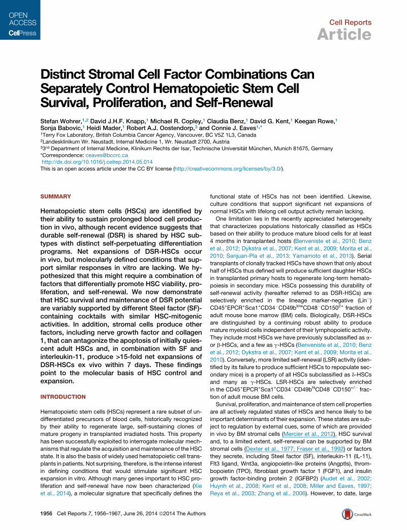
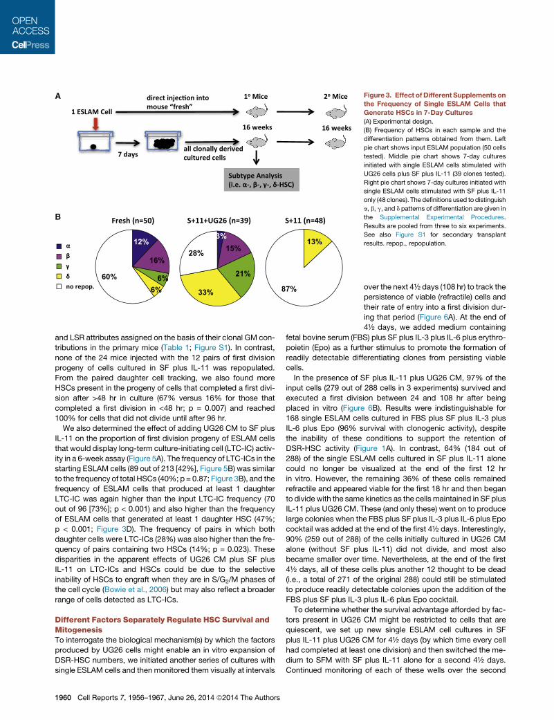
Cell Reports Article Distinct Stromal Cell Factor Combinations Can Separately Control Hematopoietic Stem Cell Survival, Proliferation, and Self-Renewal Stefan Wohrer, 1,2 David J.H.F. Knapp, 1 Michael R. Copley, 1 Claudia Benz, 1 David G. Kent, 1 Keegan Rowe, 1 Sonja Babovic, 1 Heidi Mader, 1 Robert A.J. Oostendorp, 3 and Connie J. Eaves 1, * 1 Terry Fox Laboratory, British Columbia Cancer Agency, Vancouver, BC V5Z 1L3, Canada 2 Landesklinikum Wr. Neustadt, Internal Medicine 1, Wr. Neustadt 2700, Austria 3 3 rd Department of Internal Medicine, Klinikum Rechts der Isar, Technische Universita ¨ t Mu ¨ nchen, Munich 81675, Germany *Correspondence: [email protected] http://dx.doi.org/10.1016/j.celrep.2014.05.014 This is an open access article under the CC BY license (http://creativecommons.org/licenses/by/3.0/). SUMMARY Hematopoietic stem cells (HSCs) are identified by their ability to sustain prolonged blood cell produc- tion in vivo, although recent evidence suggests that durable self-renewal (DSR) is shared by HSC sub- types with distinct self-perpetuating differentiation programs. Net expansions of DSR-HSCs occur in vivo, but molecularly defined conditions that sup- port similar responses in vitro are lacking. We hy- pothesized that this might require a combination of factors that differentially promote HSC viability, pro- liferation, and self-renewal. We now demonstrate that HSC survival and maintenance of DSR potential are variably supported by different Steel factor (SF)- containing cocktails with similar HSC-mitogenic activities. In addition, stromal cells produce other factors, including nerve growth factor and collagen 1, that can antagonize the apoptosis of initially quies- cent adult HSCs and, in combination with SF and interleukin-11, produce >15-fold net expansions of DSR-HSCs ex vivo within 7 days. These findings point to the molecular basis of HSC control and expansion. INTRODUCTION Hematopoietic stem cells (HSCs) represent a rare subset of un- differentiated precursors of blood cells, historically recognized by their ability to regenerate large, self-sustaining clones of mature progeny in transplanted irradiated hosts. This property has been successfully exploited to interrogate molecular mech- anisms that regulate the acquisition and maintenance of the HSC state. It is also the basis of widely used hematopoietic cell trans- plants in patients. Not surprising, therefore, is the intense interest in defining conditions that would stimulate significant HSC expansion in vitro. Although many genes important to HSC pro- liferation and self-renewal have now been characterized (Xie et al., 2014), a molecular signature that specifically defines the functional state of HSCs has not been identified. Likewise, culture conditions that support significant net expansions of normal HSCs with lifelong cell output activity remain lacking. One limitation lies in the recently appreciated heterogeneity that characterizes populations historically classified as HSCs based on their ability to produce mature blood cells for at least 4 months in transplanted hosts (Benveniste et al., 2010; Benz et al., 2012; Dykstra et al., 2007; Kent et al., 2009; Morita et al., 2010; Sanjuan-Pla et al., 2013; Yamamoto et al., 2013). Serial transplants of clonally tracked HSCs have shown that only about half of HSCs thus defined will produce sufficient daughter HSCs in transplanted primary hosts to regenerate long-term hemato- poiesis in secondary mice. HSCs possessing this durability of self-renewal activity (hereafter referred to as DSR-HSCs) are selectively enriched in the lineage marker-negative (Lin ) CD45 + EPCR + Sca1 + CD34 CD49b low CD48 CD150 2+ fraction of adult mouse bone marrow (BM) cells. Biologically, DSR-HSCs are distinguished by a continuing robust ability to produce mature myeloid cells independent of their lymphopoietic activity. They include most HSCs we have previously subclassified as a- or b-HSCs, and a few as g-HSCs (Benveniste et al., 2010; Benz et al., 2012; Dykstra et al., 2007; Kent et al., 2009; Morita et al., 2010). Conversely, more limited self-renewal (LSR) activity (iden- tified by its failure to produce sufficient HSCs to repopulate sec- ondary mice) is a property of all HSCs subclassified as d-HSCs and many as g-HSCs. LSR-HSCs are selectively enriched in the CD45 + EPCR + Sca1 + CD34 CD49b hi CD48 CD150 +/ frac- tion of adult mouse BM cells. Survival, proliferation, and maintenance of stem cell properties are all actively regulated states of HSCs and hence likely to be important determinants of their expansion. These states are sub- ject to regulation by external cues, some of which are provided in vivo by BM stromal cells (Mercier et al., 2012). HSC survival and, to a limited extent, self-renewal can be supported by BM stromal cells (Dexter et al., 1977; Fraser et al., 1992) or factors they secrete, including Steel factor (SF), interleukin-11 (IL-11), Flt3 ligand, Wnt3a, angiopoietin-like proteins (Angptls), throm- bopoietin (TPO), fibroblast growth factor 1 (FGF1), and insulin growth factor-binding protein 2 (IGFBP2) (Audet et al., 2002; Huynh et al., 2008; Kent et al., 2008; Miller and Eaves, 1997; Reya et al., 2003; Zhang et al., 2006). However, to date, large 1956 Cell Reports 7, 1956–1967, June 26, 2014 ª2014 The Authors
Transcript of Distinct Stromal Cell Factor Combinations Can Separately Control Hematopoietic Stem Cell Survival,...
Cell Reports
Article
Distinct Stromal Cell Factor Combinations CanSeparately Control Hematopoietic Stem CellSurvival, Proliferation, and Self-RenewalStefan Wohrer,1,2 David J.H.F. Knapp,1 Michael R. Copley,1 Claudia Benz,1 David G. Kent,1 Keegan Rowe,1
Sonja Babovic,1 Heidi Mader,1 Robert A.J. Oostendorp,3 and Connie J. Eaves1,*1Terry Fox Laboratory, British Columbia Cancer Agency, Vancouver, BC V5Z 1L3, Canada2Landesklinikum Wr. Neustadt, Internal Medicine 1, Wr. Neustadt 2700, Austria33rd Department of Internal Medicine, Klinikum Rechts der Isar, Technische Universitat Munchen, Munich 81675, Germany
*Correspondence: [email protected]://dx.doi.org/10.1016/j.celrep.2014.05.014
This is an open access article under the CC BY license (http://creativecommons.org/licenses/by/3.0/).
SUMMARY
Hematopoietic stem cells (HSCs) are identified bytheir ability to sustain prolonged blood cell produc-tion in vivo, although recent evidence suggests thatdurable self-renewal (DSR) is shared by HSC sub-types with distinct self-perpetuating differentiationprograms. Net expansions of DSR-HSCs occurin vivo, but molecularly defined conditions that sup-port similar responses in vitro are lacking. We hy-pothesized that this might require a combination offactors that differentially promote HSC viability, pro-liferation, and self-renewal. We now demonstratethat HSC survival and maintenance of DSR potentialare variably supported by different Steel factor (SF)-containing cocktails with similar HSC-mitogenicactivities. In addition, stromal cells produce otherfactors, including nerve growth factor and collagen1, that can antagonize the apoptosis of initially quies-cent adult HSCs and, in combination with SF andinterleukin-11, produce >15-fold net expansions ofDSR-HSCs ex vivo within 7 days. These findingspoint to the molecular basis of HSC control andexpansion.
INTRODUCTION
Hematopoietic stem cells (HSCs) represent a rare subset of un-
differentiated precursors of blood cells, historically recognized
by their ability to regenerate large, self-sustaining clones of
mature progeny in transplanted irradiated hosts. This property
has been successfully exploited to interrogate molecular mech-
anisms that regulate the acquisition andmaintenance of the HSC
state. It is also the basis of widely used hematopoietic cell trans-
plants in patients. Not surprising, therefore, is the intense interest
in defining conditions that would stimulate significant HSC
expansion in vitro. Although many genes important to HSC pro-
liferation and self-renewal have now been characterized (Xie
et al., 2014), a molecular signature that specifically defines the
1956 Cell Reports 7, 1956–1967, June 26, 2014 ª2014 The Authors
functional state of HSCs has not been identified. Likewise,
culture conditions that support significant net expansions of
normal HSCs with lifelong cell output activity remain lacking.
One limitation lies in the recently appreciated heterogeneity
that characterizes populations historically classified as HSCs
based on their ability to produce mature blood cells for at least
4 months in transplanted hosts (Benveniste et al., 2010; Benz
et al., 2012; Dykstra et al., 2007; Kent et al., 2009; Morita et al.,
2010; Sanjuan-Pla et al., 2013; Yamamoto et al., 2013). Serial
transplants of clonally tracked HSCs have shown that only about
half of HSCs thus defined will produce sufficient daughter HSCs
in transplanted primary hosts to regenerate long-term hemato-
poiesis in secondary mice. HSCs possessing this durability of
self-renewal activity (hereafter referred to as DSR-HSCs) are
selectively enriched in the lineage marker-negative (Lin�)CD45+EPCR+Sca1+CD34�CD49blowCD48�CD1502+ fraction of
adult mouse bone marrow (BM) cells. Biologically, DSR-HSCs
are distinguished by a continuing robust ability to produce
mature myeloid cells independent of their lymphopoietic activity.
They include most HSCs we have previously subclassified as a-
or b-HSCs, and a few as g-HSCs (Benveniste et al., 2010; Benz
et al., 2012; Dykstra et al., 2007; Kent et al., 2009; Morita et al.,
2010). Conversely, more limited self-renewal (LSR) activity (iden-
tified by its failure to produce sufficient HSCs to repopulate sec-
ondary mice) is a property of all HSCs subclassified as d-HSCs
and many as g-HSCs. LSR-HSCs are selectively enriched
in the CD45+EPCR+Sca1+CD34�CD49bhiCD48�CD150+/� frac-
tion of adult mouse BM cells.
Survival, proliferation, andmaintenance of stem cell properties
are all actively regulated states of HSCs and hence likely to be
important determinants of their expansion. These states are sub-
ject to regulation by external cues, some of which are provided
in vivo by BM stromal cells (Mercier et al., 2012). HSC survival
and, to a limited extent, self-renewal can be supported by BM
stromal cells (Dexter et al., 1977; Fraser et al., 1992) or factors
they secrete, including Steel factor (SF), interleukin-11 (IL-11),
Flt3 ligand, Wnt3a, angiopoietin-like proteins (Angptls), throm-
bopoietin (TPO), fibroblast growth factor 1 (FGF1), and insulin
growth factor-binding protein 2 (IGFBP2) (Audet et al., 2002;
Huynh et al., 2008; Kent et al., 2008; Miller and Eaves, 1997;
Reya et al., 2003; Zhang et al., 2006). However, to date, large
net expansions of DSR-HSCs ex vivo have not been achieved
using defined factors, and the relative roles of different factors
in promoting DSR-HSC viability, proliferation, and self-renewal
are not understood.
To elucidate mechanisms by which stromal cells regulate key
functions of HSCs, we chose the urogenital ridge-derived UG26-
1B6 (UG26) cell line as a source of additional external cues
because it had been found to be exceptionally potent in support-
ing HSCs in a contact-independent fashion (Oostendorp et al.,
2002, 2005). As targets, we used CD45+EPCR+CD48�CD150+
(ESLAM) adult mouse cells (�40% pure HSCs; Kent et al.,
2009). Our results identify nerve growth factor (NGF) and
collagen 1 (Col 1) as additives that can optimize DSR-HSC sur-
vival in a defined serum-free medium (SFM) and also synergize
with the mitogenic and self-renewal-promoting activity of SF
and IL-11 to achieve an unprecedented expansion of total
HSCs while maintaining input DSR-HSC numbers.
RESULTS
Stromal Cell-Derived Factors Enhance the SF PlusIL-11-Stimulated Expansion of DSR-HSCsTo first compare the DSR-HSC-stimulating activity of various
additives reported to support adult mouse BM HSC expansion
in vitro, we set up test cultures with 30 ESLAM cells each and
then 7 days later, performed limiting dilution transplant assays
to determine the numbers of DSR-HSCs, as well as the total
HSCs present (Figure 1A). HSCs were defined as cells whose
progeny constituted >1% of all the nucleated peripheral blood
(PB) cells present 16–24 weeks posttransplant. DSR-HSCs
were defined as the subset of HSCs that generated >1% of all
the circulating granulocyte and monocyte (GM) cells present
16–24 weeks posttransplant (and LSR-HSCs as all the other
HSCs). Single-cell transplants indicated that the 30 input ESLAM
cells contained, on average, 12 total HSCs of which 8 were DSR-
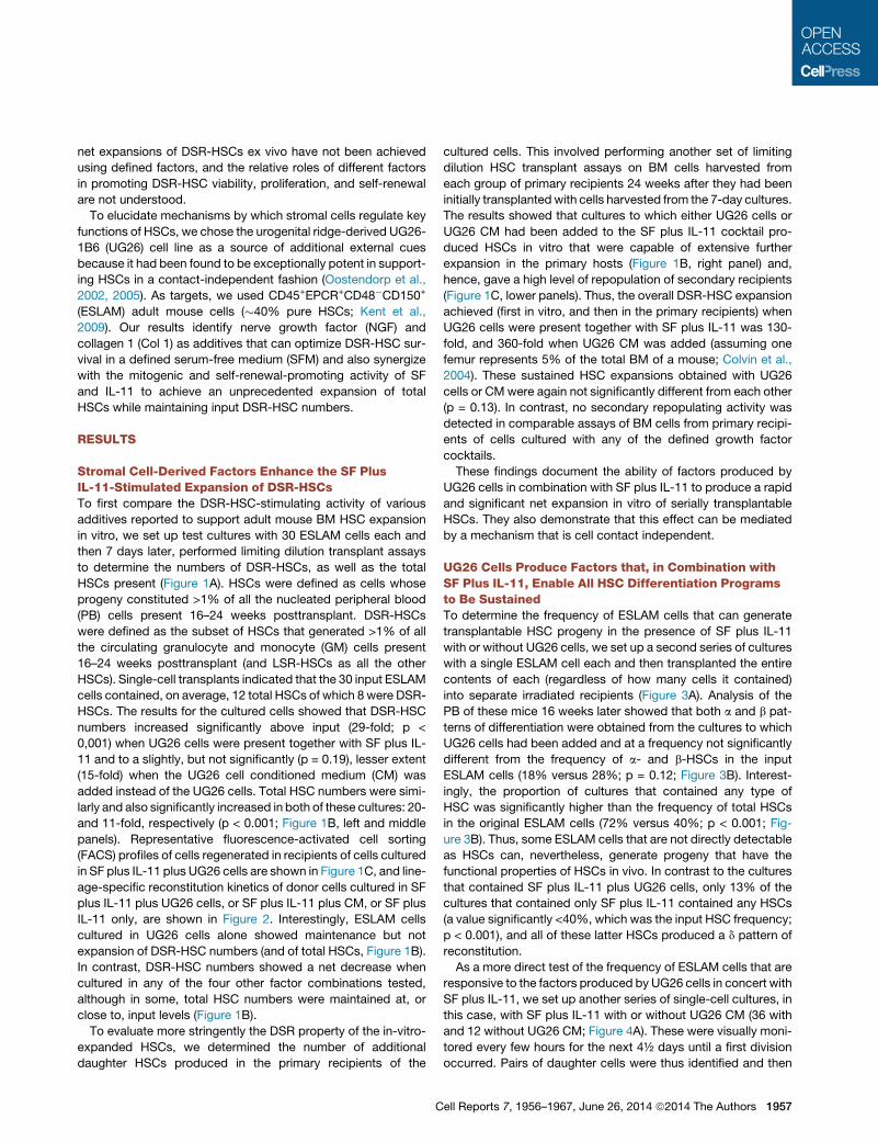
HSCs. The results for the cultured cells showed that DSR-HSC
numbers increased significantly above input (29-fold; p <
0,001) when UG26 cells were present together with SF plus IL-
11 and to a slightly, but not significantly (p = 0.19), lesser extent
(15-fold) when the UG26 cell conditioned medium (CM) was
added instead of the UG26 cells. Total HSC numbers were simi-
larly and also significantly increased in both of these cultures: 20-
and 11-fold, respectively (p < 0.001; Figure 1B, left and middle
panels). Representative fluorescence-activated cell sorting
(FACS) profiles of cells regenerated in recipients of cells cultured
in SF plus IL-11 plus UG26 cells are shown in Figure 1C, and line-
age-specific reconstitution kinetics of donor cells cultured in SF
plus IL-11 plus UG26 cells, or SF plus IL-11 plus CM, or SF plus
IL-11 only, are shown in Figure 2. Interestingly, ESLAM cells
cultured in UG26 cells alone showed maintenance but not
expansion of DSR-HSC numbers (and of total HSCs, Figure 1B).
In contrast, DSR-HSC numbers showed a net decrease when
cultured in any of the four other factor combinations tested,
although in some, total HSC numbers were maintained at, or
close to, input levels (Figure 1B).
To evaluate more stringently the DSR property of the in-vitro-
expanded HSCs, we determined the number of additional
daughter HSCs produced in the primary recipients of the
C
cultured cells. This involved performing another set of limiting
dilution HSC transplant assays on BM cells harvested from
each group of primary recipients 24 weeks after they had been
initially transplanted with cells harvested from the 7-day cultures.
The results showed that cultures to which either UG26 cells or
UG26 CM had been added to the SF plus IL-11 cocktail pro-
duced HSCs in vitro that were capable of extensive further
expansion in the primary hosts (Figure 1B, right panel) and,
hence, gave a high level of repopulation of secondary recipients
(Figure 1C, lower panels). Thus, the overall DSR-HSC expansion
achieved (first in vitro, and then in the primary recipients) when
UG26 cells were present together with SF plus IL-11 was 130-
fold, and 360-fold when UG26 CM was added (assuming one
femur represents 5% of the total BM of a mouse; Colvin et al.,
2004). These sustained HSC expansions obtained with UG26
cells or CMwere again not significantly different from each other
(p = 0.13). In contrast, no secondary repopulating activity was
detected in comparable assays of BM cells from primary recipi-
ents of cells cultured with any of the defined growth factor
cocktails.
These findings document the ability of factors produced by
UG26 cells in combination with SF plus IL-11 to produce a rapid
and significant net expansion in vitro of serially transplantable
HSCs. They also demonstrate that this effect can be mediated
by a mechanism that is cell contact independent.
UG26 Cells Produce Factors that, in Combination withSF Plus IL-11, Enable All HSC Differentiation Programsto Be SustainedTo determine the frequency of ESLAM cells that can generate
transplantable HSC progeny in the presence of SF plus IL-11
with or without UG26 cells, we set up a second series of cultures
with a single ESLAM cell each and then transplanted the entire
contents of each (regardless of how many cells it contained)
into separate irradiated recipients (Figure 3A). Analysis of the
PB of these mice 16 weeks later showed that both a and b pat-
terns of differentiation were obtained from the cultures to which
UG26 cells had been added and at a frequency not significantly
different from the frequency of a- and b-HSCs in the input
ESLAM cells (18% versus 28%; p = 0.12; Figure 3B). Interest-
ingly, the proportion of cultures that contained any type of
HSC was significantly higher than the frequency of total HSCs
in the original ESLAM cells (72% versus 40%; p < 0.001; Fig-
ure 3B). Thus, some ESLAM cells that are not directly detectable
as HSCs can, nevertheless, generate progeny that have the
functional properties of HSCs in vivo. In contrast to the cultures
that contained SF plus IL-11 plus UG26 cells, only 13% of the
cultures that contained only SF plus IL-11 contained any HSCs
(a value significantly <40%, which was the input HSC frequency;
p < 0.001), and all of these latter HSCs produced a d pattern of
reconstitution.
As a more direct test of the frequency of ESLAM cells that are
responsive to the factors produced by UG26 cells in concert with
SF plus IL-11, we set up another series of single-cell cultures, in
this case, with SF plus IL-11 with or without UG26 CM (36 with
and 12 without UG26 CM; Figure 4A). These were visually moni-
tored every few hours for the next 4½ days until a first division
occurred. Pairs of daughter cells were thus identified and then
ell Reports 7, 1956–1967, June 26, 2014 ª2014 The Authors 1957
A
B
C
Figure 1. HSC Numbers Produced in 7-Day Cultures of ESLAM Cells Containing Different Supplements
(A) Experimental design.
(B) Results of 16-week limiting dilution transplant assays used to determine the outputs of HSCs and DSR-HSCs (left 2 panels) and the cumulative DSR-HSC
expansion obtained first in vitro and then in primary (1�) recipients (right panel) (12 mice/condition/experiment, 3–5 experiments/condition, mean and SEM for
each condition). Dotted lines show the total and DSR-HSCs estimated to be present in the 30 input ESLAM cells. Holm-corrected pairwise significance values are
shown (*p = 0.05; **p = 0.01; ***p < 0.001). Where a limiting dilution was not reached, the bar indicates the minimal HSC value detectable with an upward arrow.
Supplements were as follows: F+S+3+6+E (15% FBS plus 50 ng/ml SF plus 10 ng/ml IL-3 plus 10 ng/ml IL-6 plus 3 U/ml Epo); S+11 (100 ng/ml SF plus 20 ng/ml
plus IL-11); S+T+A+I+F+H (10 ng/ml SF plus 20 ng/ml TPO plus 100 ng/ml Angptl3 plus 500 ng/ml IGFBP2 plus 10 ng/ml FGF1 plus 10 mg/ml heparin [H]); S+W
(30 ng/ml SF plus 100 ng/ml Wnt3a); UG26 cells; CM (50% UG26 CM); and S+11+CM (SF plus IL-11 plus CM).
(C) Representative FACSprofiles of PBcells obtained 16weeks after transplanting primary and secondarymicewith cells harvested fromcultures containingUG26
cells plus SF plus IL-11, described in (B). The followingmarkerswere used to investigate donor chimerism: Ly6g/Mac1 (GMcells), CD19 (B cells), andCD5 (T cells).
1958 Cell Reports 7, 1956–1967, June 26, 2014 ª2014 The Authors
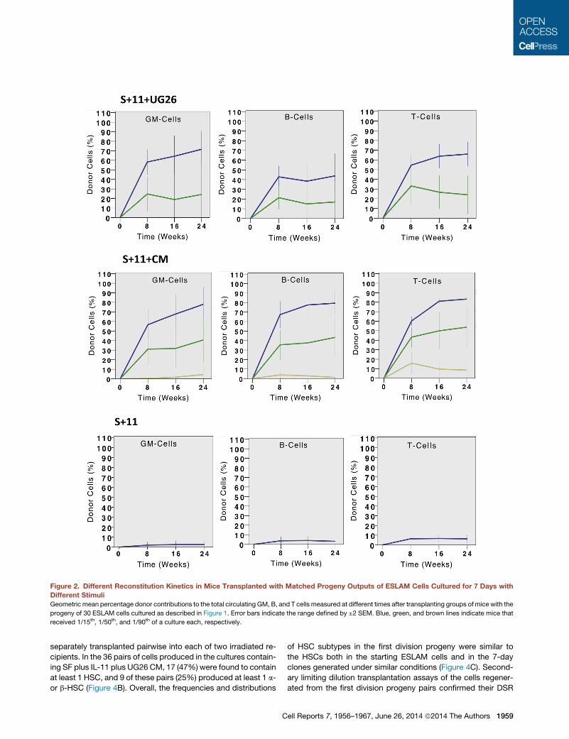
Figure 2. Different Reconstitution Kinetics in Mice Transplanted with Matched Progeny Outputs of ESLAM Cells Cultured for 7 Days with
Different Stimuli
Geometric mean percentage donor contributions to the total circulating GM, B, and T cells measured at different times after transplanting groups of mice with the
progeny of 30 ESLAM cells cultured as described in Figure 1. Error bars indicate the range defined by ±2 SEM. Blue, green, and brown lines indicate mice that
received 1/15th, 1/50th, and 1/90th of a culture each, respectively.
separately transplanted pairwise into each of two irradiated re-
cipients. In the 36 pairs of cells produced in the cultures contain-
ing SF plus IL-11 plus UG26 CM, 17 (47%) were found to contain
at least 1 HSC, and 9 of these pairs (25%) produced at least 1 a-
or b-HSC (Figure 4B). Overall, the frequencies and distributions
C
of HSC subtypes in the first division progeny were similar to
the HSCs both in the starting ESLAM cells and in the 7-day
clones generated under similar conditions (Figure 4C). Second-
ary limiting dilution transplantation assays of the cells regener-
ated from the first division progeny pairs confirmed their DSR
ell Reports 7, 1956–1967, June 26, 2014 ª2014 The Authors 1959
A
B
Figure 3. Effect of Different Supplements on
the Frequency of Single ESLAM Cells that
Generate HSCs in 7-Day Cultures
(A) Experimental design.
(B) Frequency of HSCs in each sample and the
differentiation patterns obtained from them. Left
pie chart shows input ESLAM population (50 cells
tested). Middle pie chart shows 7-day cultures
initiated with single ESLAM cells stimulated with
UG26 cells plus SF plus IL-11 (39 clones tested).
Right pie chart shows 7-day cultures initiated with
single ESLAM cells stimulated with SF plus IL-11
only (48 clones). The definitions used to distinguish
a, b, g, and d patterns of differentiation are given in
the Supplemental Experimental Procedures.
Results are pooled from three to six experiments.
See also Figure S1 for secondary transplant
results. repop., repopulation.
and LSR attributes assigned on the basis of their clonal GM con-
tributions in the primary mice (Table 1; Figure S1). In contrast,
none of the 24 mice injected with the 12 pairs of first division
progeny of cells cultured in SF plus IL-11 was repopulated.
From the paired daughter cell tracking, we also found more
HSCs present in the progeny of cells that completed a first divi-
sion after >48 hr in culture (67% versus 16% for those that
completed a first division in <48 hr; p = 0.007) and reached
100% for cells that did not divide until after 96 hr.
We also determined the effect of adding UG26 CM to SF plus
IL-11 on the proportion of first division progeny of ESLAM cells
that would display long-term culture-initiating cell (LTC-IC) activ-
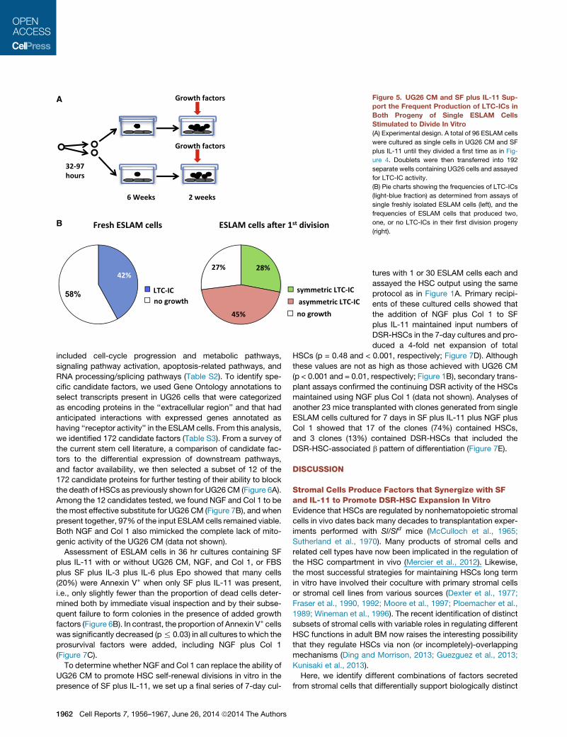
ity in a 6-week assay (Figure 5A). The frequency of LTC-ICs in the
starting ESLAM cells (89 out of 213 [42%], Figure 5B) was similar
to the frequency of total HSCs (40%; p = 0.87; Figure 3B), and the
frequency of ESLAM cells that produced at least 1 daughter
LTC-IC was again higher than the input LTC-IC frequency (70
out of 96 [73%]; p < 0.001) and also higher than the frequency
of ESLAM cells that generated at least 1 daughter HSC (47%;
p < 0.001; Figure 3D). The frequency of pairs in which both
daughter cells were LTC-ICs (28%) was also higher than the fre-
quency of pairs containing two HSCs (14%; p = 0.023). These
disparities in the apparent effects of UG26 CM plus SF plus
IL-11 on LTC-ICs and HSCs could be due to the selective
inability of HSCs to engraft when they are in S/G2/M phases of
the cell cycle (Bowie et al., 2006) but may also reflect a broader
range of cells detected as LTC-ICs.
Different Factors Separately Regulate HSC Survival andMitogenesisTo interrogate the biological mechanism(s) by which the factors
produced by UG26 cells might enable an in vitro expansion of
DSR-HSC numbers, we initiated another series of cultures with
single ESLAM cells and then monitored them visually at intervals
1960 Cell Reports 7, 1956–1967, June 26, 2014 ª2014 The Authors
over the next 4½ days (108 hr) to track the
persistence of viable (refractile) cells and
their rate of entry into a first division dur-
ing that period (Figure 6A). At the end of
4½ days, we added medium containing
fetal bovine serum (FBS) plus SF plus IL-3 plus IL-6 plus erythro-
poietin (Epo) as a further stimulus to promote the formation of
readily detectable differentiating clones from persisting viable
cells.
In the presence of SF plus IL-11 plus UG26 CM, 97% of the
input cells (279 out of 288 cells in 3 experiments) survived and
executed a first division between 24 and 108 hr after being
placed in vitro (Figure 6B). Results were indistinguishable for
168 single ESLAM cells cultured in FBS plus SF plus IL-3 plus
IL-6 plus Epo (96% survival with clonogenic activity), despite
the inability of these conditions to support the retention of
DSR-HSC activity (Figure 1A). In contrast, 64% (184 out of
288) of the single ESLAM cells cultured in SF plus IL-11 alone
could no longer be visualized at the end of the first 12 hr
in vitro. However, the remaining 36% of these cells remained
refractile and appeared viable for the first 18 hr and then began
to divide with the same kinetics as the cells maintained in SF plus
IL-11 plus UG26 CM. These (and only these) went on to produce
large colonies when the FBS plus SF plus IL-3 plus IL-6 plus Epo
cocktail was added at the end of the first 4½ days. Interestingly,
90% (259 out of 288) of the cells initially cultured in UG26 CM
alone (without SF plus IL-11) did not divide, and most also
became smaller over time. Nevertheless, at the end of the first
4½ days, all of these cells plus another 12 thought to be dead
(i.e., a total of 271 of the original 288) could still be stimulated
to produce readily detectable colonies upon the addition of the
FBS plus SF plus IL-3 plus IL-6 plus Epo cocktail.
To determine whether the survival advantage afforded by fac-
tors present in UG26 CM might be restricted to cells that are
quiescent, we set up new single ESLAM cell cultures in SF
plus IL-11 plus UG26 CM for 4½ days (by which time every cell
had completed at least one division) and then switched the me-
dium to SFM with SF plus IL-11 alone for a second 4½ days.
Continued monitoring of each of these wells over the second
A
B
C
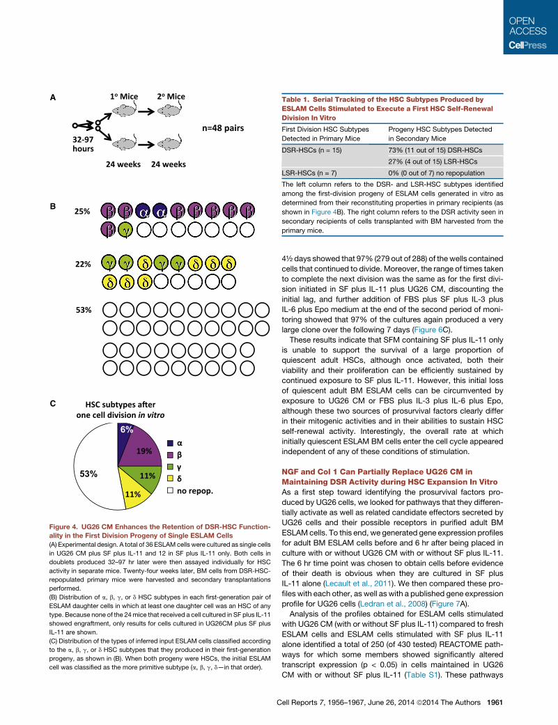
Figure 4. UG26 CM Enhances the Retention of DSR-HSC Function-
ality in the First Division Progeny of Single ESLAM Cells
(A) Experimental design. A total of 36 ESLAM cells were cultured as single cells
in UG26 CM plus SF plus IL-11 and 12 in SF plus IL-11 only. Both cells in
doublets produced 32–97 hr later were then assayed individually for HSC
activity in separate mice. Twenty-four weeks later, BM cells from DSR-HSC-
repopulated primary mice were harvested and secondary transplantations
performed.
(B) Distribution of a, b, g, or d HSC subtypes in each first-generation pair of
ESLAM daughter cells in which at least one daughter cell was an HSC of any
type. Because none of the 24mice that received a cell cultured in SF plus IL-11
showed engraftment, only results for cells cultured in UG26CM plus SF plus
IL-11 are shown.
(C) Distribution of the types of inferred input ESLAM cells classified according
to the a, b, g, or d HSC subtypes that they produced in their first-generation
progeny, as shown in (B). When both progeny were HSCs, the initial ESLAM
cell was classified as the more primitive subtype (a, b, g, d—in that order).
Table 1. Serial Tracking of the HSC Subtypes Produced by
ESLAM Cells Stimulated to Execute a First HSC Self-Renewal
Division In Vitro
First Division HSC Subtypes
Detected in Primary Mice
Progeny HSC Subtypes Detected
in Secondary Mice
DSR-HSCs (n = 15) 73% (11 out of 15) DSR-HSCs
27% (4 out of 15) LSR-HSCs
LSR-HSCs (n = 7) 0% (0 out of 7) no repopulation
The left column refers to the DSR- and LSR-HSC subtypes identified
among the first-division progeny of ESLAM cells generated in vitro as
determined from their reconstituting properties in primary recipients (as
shown in Figure 4B). The right column refers to the DSR activity seen in
secondary recipients of cells transplanted with BM harvested from the
primary mice.
C
4½ days showed that 97% (279 out of 288) of the wells contained
cells that continued to divide. Moreover, the range of times taken
to complete the next division was the same as for the first divi-
sion initiated in SF plus IL-11 plus UG26 CM, discounting the
initial lag, and further addition of FBS plus SF plus IL-3 plus
IL-6 plus Epo medium at the end of the second period of moni-
toring showed that 97% of the cultures again produced a very
large clone over the following 7 days (Figure 6C).
These results indicate that SFM containing SF plus IL-11 only
is unable to support the survival of a large proportion of
quiescent adult HSCs, although once activated, both their
viability and their proliferation can be efficiently sustained by
continued exposure to SF plus IL-11. However, this initial loss
of quiescent adult BM ESLAM cells can be circumvented by
exposure to UG26 CM or FBS plus IL-3 plus IL-6 plus Epo,
although these two sources of prosurvival factors clearly differ
in their mitogenic activities and in their abilities to sustain HSC
self-renewal activity. Interestingly, the overall rate at which
initially quiescent ESLAM BM cells enter the cell cycle appeared
independent of any of these conditions of stimulation.
NGF and Col 1 Can Partially Replace UG26 CM inMaintaining DSR Activity during HSC Expansion In VitroAs a first step toward identifying the prosurvival factors pro-
duced by UG26 cells, we looked for pathways that they differen-
tially activate as well as related candidate effectors secreted by
UG26 cells and their possible receptors in purified adult BM
ESLAMcells. To this end, we generated gene expression profiles
for adult BM ESLAM cells before and 6 hr after being placed in
culture with or without UG26 CM with or without SF plus IL-11.
The 6 hr time point was chosen to obtain cells before evidence
of their death is obvious when they are cultured in SF plus
IL-11 alone (Lecault et al., 2011). We then compared these pro-
files with each other, as well as with a published gene expression
profile for UG26 cells (Ledran et al., 2008) (Figure 7A).
Analysis of the profiles obtained for ESLAM cells stimulated
with UG26 CM (with or without SF plus IL-11) compared to fresh
ESLAM cells and ESLAM cells stimulated with SF plus IL-11
alone identified a total of 250 (of 430 tested) REACTOME path-
ways for which some members showed significantly altered
transcript expression (p < 0.05) in cells maintained in UG26
CM with or without SF plus IL-11 (Table S1). These pathways
ell Reports 7, 1956–1967, June 26, 2014 ª2014 The Authors 1961
A
B
Figure 5. UG26 CM and SF plus IL-11 Sup-
port the Frequent Production of LTC-ICs in
Both Progeny of Single ESLAM Cells
Stimulated to Divide In Vitro
(A) Experimental design. A total of 96 ESLAM cells
were cultured as single cells in UG26 CM and SF
plus IL-11 until they divided a first time as in Fig-
ure 4. Doublets were then transferred into 192
separate wells containing UG26 cells and assayed
for LTC-IC activity.
(B) Pie charts showing the frequencies of LTC-ICs
(light-blue fraction) as determined from assays of
single freshly isolated ESLAM cells (left), and the
frequencies of ESLAM cells that produced two,
one, or no LTC-ICs in their first division progeny
(right).
included cell-cycle progression and metabolic pathways,
signaling pathway activation, apoptosis-related pathways, and
RNA processing/splicing pathways (Table S2). To identify spe-
cific candidate factors, we used Gene Ontology annotations to
select transcripts present in UG26 cells that were categorized
as encoding proteins in the ‘‘extracellular region’’ and that had
anticipated interactions with expressed genes annotated as
having ‘‘receptor activity’’ in the ESLAM cells. From this analysis,
we identified 172 candidate factors (Table S3). From a survey of
the current stem cell literature, a comparison of candidate fac-
tors to the differential expression of downstream pathways,
and factor availability, we then selected a subset of 12 of the
172 candidate proteins for further testing of their ability to block
the death of HSCs as previously shown for UG26 CM (Figure 6A).
Among the 12 candidates tested, we found NGF and Col 1 to be
the most effective substitute for UG26 CM (Figure 7B), and when
present together, 97% of the input ESLAM cells remained viable.
Both NGF and Col 1 also mimicked the complete lack of mito-
genic activity of the UG26 CM (data not shown).
Assessment of ESLAM cells in 36 hr cultures containing SF
plus IL-11 with or without UG26 CM, NGF, and Col 1, or FBS
plus SF plus IL-3 plus IL-6 plus Epo showed that many cells
(20%) were Annexin V+ when only SF plus IL-11 was present,
i.e., only slightly fewer than the proportion of dead cells deter-
mined both by immediate visual inspection and by their subse-
quent failure to form colonies in the presence of added growth
factors (Figure 6B). In contrast, the proportion of Annexin V+ cells
was significantly decreased (p% 0.03) in all cultures to which the
prosurvival factors were added, including NGF plus Col 1
(Figure 7C).
To determine whether NGF and Col 1 can replace the ability of
UG26 CM to promote HSC self-renewal divisions in vitro in the
presence of SF plus IL-11, we set up a final series of 7-day cul-
1962 Cell Reports 7, 1956–1967, June 26, 2014 ª2014 The Authors
tures with 1 or 30 ESLAM cells each and
assayed the HSC output using the same
protocol as in Figure 1A. Primary recipi-
ents of these cultured cells showed that
the addition of NGF plus Col 1 to SF
plus IL-11 maintained input numbers of
DSR-HSCs in the 7-day cultures and pro-
duced a 4-fold net expansion of total
HSCs (p = 0.48 and < 0.001, respectively; Figure 7D). Although
these values are not as high as those achieved with UG26 CM
(p < 0.001 and = 0.01, respectively; Figure 1B), secondary trans-
plant assays confirmed the continuing DSR activity of the HSCs
maintained using NGF plus Col 1 (data not shown). Analyses of
another 23 mice transplanted with clones generated from single
ESLAM cells cultured for 7 days in SF plus IL-11 plus NGF plus
Col 1 showed that 17 of the clones (74%) contained HSCs,
and 3 clones (13%) contained DSR-HSCs that included the
DSR-HSC-associated b pattern of differentiation (Figure 7E).
DISCUSSION
Stromal Cells Produce Factors that Synergize with SFand IL-11 to Promote DSR-HSC Expansion In VitroEvidence that HSCs are regulated by nonhematopoietic stromal
cells in vivo dates back many decades to transplantation exper-
iments performed with Sl/Sld mice (McCulloch et al., 1965;
Sutherland et al., 1970). Many products of stromal cells and
related cell types have now been implicated in the regulation of
the HSC compartment in vivo (Mercier et al., 2012). Likewise,
the most successful strategies for maintaining HSCs long term
in vitro have involved their coculture with primary stromal cells
or stromal cell lines from various sources (Dexter et al., 1977;
Fraser et al., 1990, 1992; Moore et al., 1997; Ploemacher et al.,
1989; Wineman et al., 1996). The recent identification of distinct
subsets of stromal cells with variable roles in regulating different
HSC functions in adult BM now raises the interesting possibility
that they regulate HSCs via non (or incompletely)-overlapping
mechanisms (Ding and Morrison, 2013; Guezguez et al., 2013;
Kunisaki et al., 2013).
Here, we identify different combinations of factors secreted
from stromal cells that differentially support biologically distinct
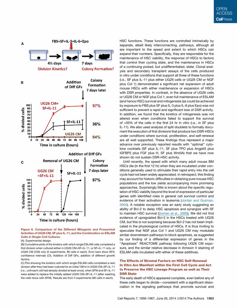
Figure 6. Comparison of the Different Mitogenic and ProsurvivalActivities of UG26CM, SF plus IL-11, and theCombination on ESLAM
Cells in Single-Cell Cultures
(A). Experimental design.
(B) Cumulative plots of the kinetics with which single ESLAM cells completed a
first division when cultured either in UG26 CM+SF+IL-11, or SF+IL-11 only, or
UG26 CM (CM) only (5 experiments, 96 cells in each). Error bars show 95%
confidence intervals (CI). Addition of Diff GFs, addition of different growth
factors.
(C) Plot showing the kinetics with which single ESLAM cells completed a next
division after they had been cultured for an initial 108 hr in UG26 CM+SF+IL-11
(i.e., until each cell had already divided at least once), when SFM and SF+IL-11
were added to replace the initially added UG26 CM+SF+IL-11 (after washing
the cells twice with SFM). Results are from 3 experiments (96 cells in each).
C
HSC functions. These functions are controlled intrinsically by
separate, albeit likely interconnecting, pathways, although all
are important to the speed and extent to which HSCs can
expand their numbers. Specifically, they are responsible for the
maintenance of HSC viability, the response of HSCs to factors
that control their cycling state, and the maintenance in HSCs
of a continuing poised, but undifferentiated, state. Clonal anal-
ysis and secondary transplant assays of the cells produced
in vitro under conditions that support all three of these functions
(i.e., SF plus IL-11 plus either UG26 cells or UG26 CM or NGF
plus Col 1) demonstrated a significant net expansion of adult
mouse HSCs with either maintenance or expansion of HSCs
with DSR properties. In contrast, in the absence of UG26 cells
or UG26 CM or NGF plus Col 1, even full maintenance of ESLAM
(and hence HSC) survival andmitogenesis (as could be achieved
by exposure to FBS plus SF plus IL-3 plus IL-6 plus Epo) was not
sufficient to prevent a rapid and significant loss of DSR activity.
In addition, we found that the kinetics of mitogenesis was not
altered even when conditions failed to support the survival
of >50% of the cells in the first 24 hr in vitro (i.e., in SF plus
IL-11). We also used analysis of split doublets to formally docu-
ment the execution of first divisions that produce twoDSR-HSCs
under conditions where survival, proliferation, and self-renewal
are all well supported. These findings thus represent a major
advance over previously reported results with ‘‘optimal’’ cyto-
kine cocktails (SF plus IL-11, SF plus TPO plus Angptl3 plus
IGFBP2 plus FGF plus H, SF plus Wnt3A) that we have now
shown do not sustain DSR-HSC activity.
Until recently, the speed with which many adult mouse BM
HSCs die (in the first 12 hr) when they are incubated under con-
ditions generally used to stimulate their rapid entry into the cell
cycle had not been widely appreciated. In retrospect, this finding
may account for historic difficulties in obtaining puremouse HSC
populations and the low yields accompanying more promising
approaches. Surprisingly little is known about the specific regu-
lation of HSC viability beyond the level of expression of particular
genes with identified roles in general cell survival control and
evidence of their activation in leukemia (Jordan and Guzman,
2004). A notable exception was an early study suggesting an
ability of Bcl-2 to delay HSC apoptosis and synergize with SF
to maintain HSC survival (Domen et al., 2000). We did not find
evidence of upregulated Bcl-2 in the HSCs treated with UG26
CM, but this is not surprising because Bcl-2 has not been impli-
cated in the physiological control of HSCs. It is thus inviting to
speculate that NGF plus Col 1 and UG26 CM may modulate
similar downstream pathways to block apoptosis, as suggested
by our finding of a differential expression of genes in the
‘‘Apoptosis’’ REACTOME pathway following UG26 CM expo-
sure, and the similar relative decrease in Annexin V staining of
ESLAM cells incubated with either of these additives.
The Effects of Stromal Factors on HSC Self-RenewalIn Vitro Are Manifest within the First Cell Cycle and Actto Preserve the HSC Lineage Program as well as TheirDSR StateThe early death of HSCs appeared complete, even before any of
these cells began to divide—consistent with a significant disso-
ciation in the signaling pathways that promote survival and
ell Reports 7, 1956–1967, June 26, 2014 ª2014 The Authors 1963
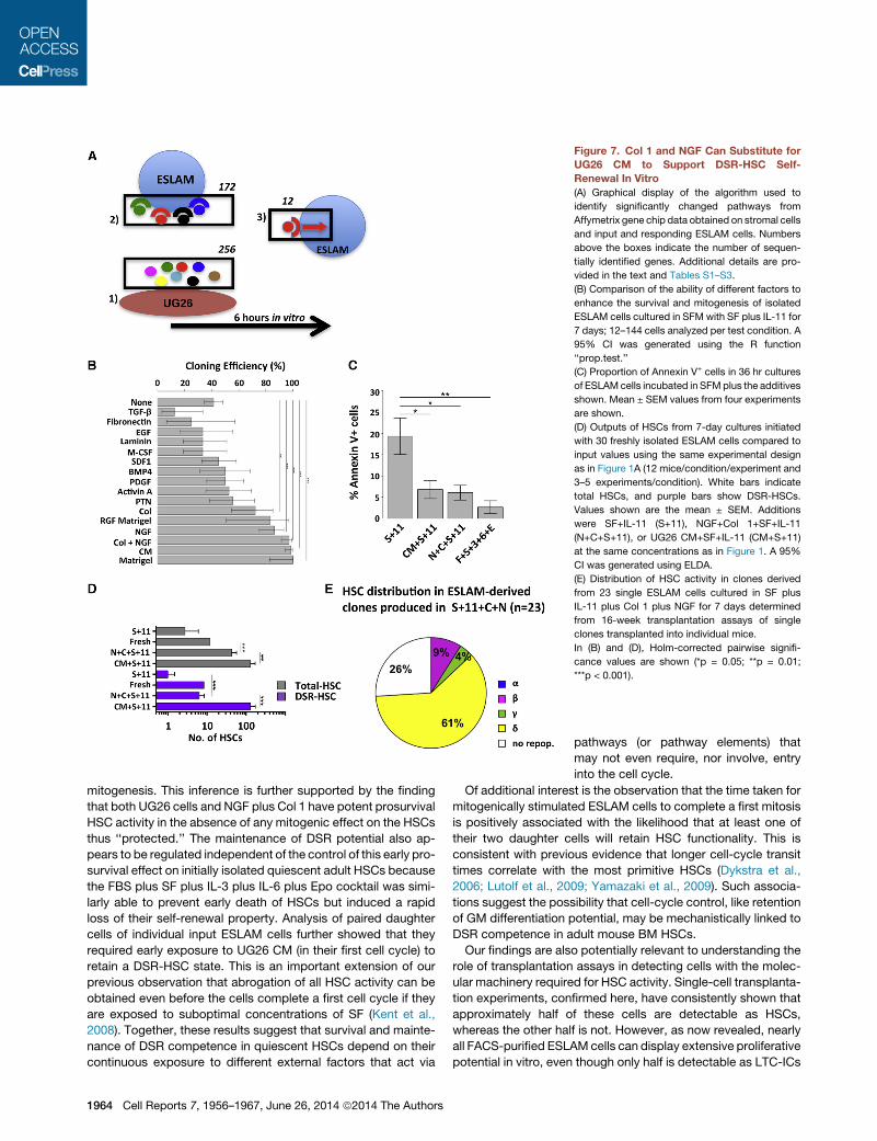
Figure 7. Col 1 and NGF Can Substitute for
UG26 CM to Support DSR-HSC Self-
Renewal In Vitro
(A) Graphical display of the algorithm used to
identify significantly changed pathways from
Affymetrix gene chip data obtained on stromal cells
and input and responding ESLAM cells. Numbers
above the boxes indicate the number of sequen-
tially identified genes. Additional details are pro-
vided in the text and Tables S1–S3.
(B) Comparison of the ability of different factors to
enhance the survival and mitogenesis of isolated
ESLAM cells cultured in SFM with SF plus IL-11 for
7 days; 12–144 cells analyzed per test condition. A
95% CI was generated using the R function
‘‘prop.test.’’
(C) Proportion of Annexin V+ cells in 36 hr cultures
of ESLAMcells incubated in SFMplus the additives
shown. Mean ± SEM values from four experiments
are shown.
(D) Outputs of HSCs from 7-day cultures initiated
with 30 freshly isolated ESLAM cells compared to
input values using the same experimental design
as in Figure 1A (12 mice/condition/experiment and
3–5 experiments/condition). White bars indicate
total HSCs, and purple bars show DSR-HSCs.
Values shown are the mean ± SEM. Additions
were SF+IL-11 (S+11), NGF+Col 1+SF+IL-11
(N+C+S+11), or UG26 CM+SF+IL-11 (CM+S+11)
at the same concentrations as in Figure 1. A 95%
CI was generated using ELDA.
(E) Distribution of HSC activity in clones derived
from 23 single ESLAM cells cultured in SF plus
IL-11 plus Col 1 plus NGF for 7 days determined
from 16-week transplantation assays of single
clones transplanted into individual mice.
In (B) and (D), Holm-corrected pairwise signifi-
cance values are shown (*p = 0.05; **p = 0.01;
***p < 0.001).
mitogenesis. This inference is further supported by the finding
that both UG26 cells and NGF plus Col 1 have potent prosurvival
HSC activity in the absence of any mitogenic effect on the HSCs
thus ‘‘protected.’’ The maintenance of DSR potential also ap-
pears to be regulated independent of the control of this early pro-
survival effect on initially isolated quiescent adult HSCs because
the FBS plus SF plus IL-3 plus IL-6 plus Epo cocktail was simi-
larly able to prevent early death of HSCs but induced a rapid
loss of their self-renewal property. Analysis of paired daughter
cells of individual input ESLAM cells further showed that they
required early exposure to UG26 CM (in their first cell cycle) to
retain a DSR-HSC state. This is an important extension of our
previous observation that abrogation of all HSC activity can be
obtained even before the cells complete a first cell cycle if they
are exposed to suboptimal concentrations of SF (Kent et al.,
2008). Together, these results suggest that survival and mainte-
nance of DSR competence in quiescent HSCs depend on their
continuous exposure to different external factors that act via
1964 Cell Reports 7, 1956–1967, June 26, 2014 ª2014 The Authors
pathways (or pathway elements) that
may not even require, nor involve, entry
into the cell cycle.
Of additional interest is the observation that the time taken for
mitogenically stimulated ESLAM cells to complete a first mitosis
is positively associated with the likelihood that at least one of
their two daughter cells will retain HSC functionality. This is
consistent with previous evidence that longer cell-cycle transit
times correlate with the most primitive HSCs (Dykstra et al.,
2006; Lutolf et al., 2009; Yamazaki et al., 2009). Such associa-
tions suggest the possibility that cell-cycle control, like retention
of GM differentiation potential, may be mechanistically linked to
DSR competence in adult mouse BM HSCs.
Our findings are also potentially relevant to understanding the
role of transplantation assays in detecting cells with the molec-
ular machinery required for HSC activity. Single-cell transplanta-
tion experiments, confirmed here, have consistently shown that
approximately half of these cells are detectable as HSCs,
whereas the other half is not. However, as now revealed, nearly
all FACS-purified ESLAMcells can display extensive proliferative
potential in vitro, even though only half is detectable as LTC-ICs
in a 6- to 7-week assay (Kent et al., 2009). Moreover, the fre-
quency of ESLAM cells that can respond to SF plus IL-11 in
the presence of UG26 CM in vitro by generating progeny HSCs
that are functional in vivo is significantly higher than the 40% of
freshly isolated ESLAM cells that are directly detectable in vivo
as HSCs. Taken together, this raises the possibility that most
adult BM ESLAM cells have not irreversibly lost the molecular
status of HSCs.
Overall, our results suggest that several core HSC behavioral
programs can be functionally uncoupled, allowing their differen-
tial and combinatorial activation by an array of external factors.
This differential program activation could result from activation
of multiple independent signaling pathways (a combinatorial
switchmechanism), by different levels of activation in a few com-
mon pathways (a cellular rheostat-like mechanism), or by some
combination of these two. In either case, additional downstream
molecular interactions are likely. The various genes and path-
ways previously implicated in HSC maintenance/expansion are
consistent with a combinatorial mechanism operating to control
HSCs (Reya et al., 2003). Similarly, the fact that low concentra-
tions of SF provide HSC survival benefits, whereas maintenance
of repopulation potential requires high levels of SF, supports the
existence of mechanisms that depend on different signaling
thresholds in these cells (Kent et al., 2008).
Implications for Future Improvement of HSC ExpansionProtocolsOur findings reemphasize the deficiency of 4- to 6-month PB
repopulation endpoints that do not specifically measure the
output of donor-derived GM cells in order to distinguish between
HSCs that have retained or lost DSR activity, as recently high-
lighted by others (Yamamoto et al., 2013). They also demon-
strate that mouse DSR-HSC self-renewal divisions can be
achieved under defined conditions in vitro in the absence of
any other cells, but to achieve this response, multiple extrinsic
factors are required. Interestingly, at least some of these factors,
exemplified by Col 1 (Hu et al., 2011) and NGF (Garcıa et al.,
2004), may be ubiquitously prevalent extracellular matrix com-
ponents of the interstitial space within hematopoietic tissue.
Recent studies have highlighted a differential expression on
the surface of HSCs and their closely related downstream deriv-
atives of several integrins, which are receptors for such proteins
(Benveniste et al., 2010; Notta et al., 2011; Wagers and Weiss-
man, 2006). Thus, factors that activate these receptors may
constitute an additional strategy for enhancing HSC expansion,
as suggested by others (Celebi et al., 2011; Kurth et al., 2011;
Umemoto et al., 2012). The ability of a combination of defined
soluble proteins to promote HSC expansion in vitro refutes the
hypothesis that cell contact is required to mediate such
responses and should facilitate future interrogation of the mech-
anisms involved in themaintenance of the DSR state when HSCs
are stimulated to proliferate.
EXPERIMENTAL PROCEDURES
Mice
C57Bl/6J (B6)-Ly5.1 or C57Bl/6J (B6)-Ly5.2mice and congenic B6-W41/W41-
Ly5.1 and B6-W41/W41-Ly5.2 (W41-5.1 andW41-5.2, respectively) mice were
C
bred and maintained in our animal resource center in microisolator cages and
provided with continuous sterile food, water, and bedding. Procedures for
isolating ESLAM cells from adult mouse BM and performing and assessing
transplants were as previously described (Benz et al., 2012; Kent et al.,
2009) and carried out with approval from the University of British Columbia
Animal Care Committee. For further details, see Supplemental Experimental
Procedures.
UG26 Cells and CM
UG26 stromal cells were cultured as previously described (Oostendorp et al.,
2002) and CM obtained from confluent UG26 cells X-irradiated with 30 Gy and
then incubated for 3 days with SFM after removal of the UG26 culture medium
and rinsing the cultures several times with PBS. The CM was then filtered
through a 40 mm cell strainer (Becton Dickinson) and stored frozen at �20�C.
ESLAM Cell Cultures
ESLAM cells were deposited into the round-bottomed wells of 96-well plates
using the single-cell deposition unit of the sorter, each well having been pre-
loaded with 100 ml of SFM (Iscove’s medium with 10 mg/ml BSA, 10 mg/ml
insulin, and 200 mg/ml transferrin, 40 mg/ml low-density lipoproteins,
100 U/ml penicillin, 100 mg/ml streptomycin [STEMCELL Technologies]) and
10�4 M b-mercaptoethanol (Sigma-Aldrich). The presence of single cells
was then confirmed by visual inspection. A second 100 ml of medium was
added for cultures initiated with 30 ESLAM cells. The following additives
were used as indicated: mouse SF and IL-3, and human Epo and Col 1 pur-
chased from STEMCELL Technologies; human IL-11 and macrophage colony
stimulating factor (M-CSF) obtained as gifts from Genetics Institute; mouse
Wnt3a, Angptl3, and IGFBP2, and human NGF, pleiotrophin, bone morphoge-
netic protein 4 (BMP4), Activin A, transforming growth factor b (TGF-b), and
platelet-derived growth factor (PDGF) BB purchased from R&D Systems;
mouse TPO obtained as a gift from Genentech; human FGF-1 purchased
from Invitrogen; heparin, fibronectin, human epidermal growth factor, and
mouse laminin purchased from Sigma-Aldrich; human IL-6 obtained as a gift
from Cangene; reduced growth factor (RGF) Matrigel purchased from BD;
and SDF-1 obtained as a gift from Dr. I. Clark Lewis (University of British
Columbia).
For in vivo and LTC-IC assessment of the first division progeny of single
ESLAM cells, cultures were examined microscopically every 4 hr starting
32 hr after initiation of the culture. Thereafter, the entire volume in each well
found to have produced two cells in the previous 4 hr was distributed into three
or more wells to obtain both daughters in different wells so they could then be
transplanted separately into two different mice or used to initiate two separate
LTCs. If only one cell was recovered, the remaining cell was discarded. To
track the kinetics of cell division and viability, cultures were monitored starting
12 hr after initiation and thereafter as indicated. The first appearance of two
refractile cells was used to indicate completion of a first division. Following
the addition of 15% FBS, 50 ng/ml SF, 10 ng/ml IL-3, 10 ng/ml IL-6, and
3 U/ml Epo to each well after 4½ days of incubation, evidence of viability
was inferred from the detection of a clone of seven or more refractile cells.
To measure apoptotic cells, cells were harvested after 36 hr, washed, resus-
pended in Binding Buffer, stained with Annexin V eFluor 450 and FITC-conju-
gated anti-CD45 (all from eBioscience), and analyzed on a BD LSR Fortessa.
LTC-IC Assays
Visually confirmed single cells were added onto irradiated UG26 feeder cells in
flat-bottomed wells containing 200 ml of MyeloCult (STEMCELL Technologies)
supplemented with 10�6 M hydrocortisone (Sigma-Aldrich) in 96-well plates
and cultures maintained with weekly half-medium changes (Woehrer et al.,
2013). After 6 weeks, fresh SFM containing FBS plus SF plus IL-3 plus IL-6
plus Epo was added to the wells, and those containing clusters of >25
nonadherent cells 12 days later were counted as positive.
Transcriptome Analyses
Between 2,000 (cultured cells) and 6,000 (freshly isolated adult BM) ESLAM
cells were collected, and mRNA was extracted with RNeasy (QIAGEN).
mRNA from three independent experiments was pooled, reverse transcribed,
and amplified with Nanokit following the manufacturer’s instructions (Agilent
ell Reports 7, 1956–1967, June 26, 2014 ª2014 The Authors 1965
Technologies). cRNA was hybridized onto two gene chips (GeneChip
Mouse Gene 1.0 ST Array; Affymetrix) per condition (GSE57220, http://www.
ncbi.nlm.nih.gov/geo/query/acc.cgi?acc=GSE57220). Data analysis was per-
formed as detailed in Supplemental Experimental Procedures.
Statistical Analysis
GraphPad Prism version 5 or R (http://www.R-project.org/) was used to
perform basic statistical analyses, including calculation of mean ± SEM values
and to perform Student’s t tests. ELDA: Extreme Limiting Dilution Analysis
(http://bioinf.wehi.edu.au/software/elda/) or the ‘‘elda’’ function in the R pack-
age, ‘‘statmod,’’ was used to perform the limiting dilution analyses and to eval-
uate the significance of differences obtained using different culture conditions.
ACCESSION NUMBERS
TheGEO accession number for the transcriptome data reported in this paper is
GSE57220.
SUPPLEMENTAL INFORMATION
Supplemental Information includes Supplemental Experimental Procedures,
one figure, and three tables and can be found with this article online at
http://dx.doi.org/10.1016/j.celrep.2014.05.014.
AUTHOR CONTRIBUTIONS
S.W., D.J.H.F.K., and C.J.E. conceived and oversaw the design and execution
of the experiments. S.W., D.J.H.F.K., M.R.C., C.B., D.G.K., S.B., K.R., and
H.M. collected the data. S.W., D.J.H.F.K., M.R.C., C.B., D.G.K., and S.B.
contributed to the interpretation of the data. S.W., D.J.H.F.K., and C.J.E. wrote
the manuscript, and all authors approved it.
ACKNOWLEDGMENTS
We thank the staff of the Flow Cytometry Facility of the Terry Fox Laboratory
and the Animal ResourceCentre of the BCCancer Agency. This work was sup-
ported by grants from the National Cancer Institute of Canada (Toronto, ON),
with funds from the Terry Fox Run, and the Canadian Institutes of Health
Research (CIHR, Toronto, ON). S.W. received an Erwin Schroedinger Fellow-
ship (J2684) as well as an FWF grant (P25134) from the Austrian Science Fund
(Vienna). D.J.H.F.K. received studentships from CIHR including a Vanier Stu-
dentship. C.B. received a fellowship from the Deutsche Forschungsgemein-
schaft (Bonn). M.R.C. and D.G.K. received studentships from the Michael
Smith Foundation for Health Research and CIHR. S.B. received studentships
from the University of British Columbia and CIHR.
Received: January 12, 2014
Revised: February 2, 2014
Accepted: May 6, 2014
Published: June 5, 2014
REFERENCES
Audet, J., Miller, C.L., Eaves, C.J., and Piret, J.M. (2002). Common and distinct
features of cytokine effects on hematopoietic stem and progenitor cells
revealed by dose-response surface analysis. Biotechnol. Bioeng. 80, 393–404.
Benveniste, P., Frelin, C., Janmohamed, S., Barbara, M., Herrington, R.,
Hyam, D., and Iscove, N.N. (2010). Intermediate-term hematopoietic stem
cells with extended but time-limited reconstitution potential. Cell Stem Cell
6, 48–58.
Benz, C., Copley, M.R., Kent, D.G., Wohrer, S., Cortes, A., Aghaeepour, N.,
Ma, E., Mader, H., Rowe, K., Day, C., et al. (2012). Hematopoietic stem cell
subtypes expand differentially during development and display distinct lym-
phopoietic programs. Cell Stem Cell 10, 273–283.
Bowie, M.B., McKnight, K.D., Kent, D.G., McCaffrey, L., Hoodless, P.A., and
Eaves, C.J. (2006). Hematopoietic stem cells proliferate until after birth and
1966 Cell Reports 7, 1956–1967, June 26, 2014 ª2014 The Authors
show a reversible phase-specific engraftment defect. J. Clin. Invest. 116,
2808–2816.
Celebi, B., Mantovani, D., and Pineault, N. (2011). Effects of extracellular
matrix proteins on the growth of haematopoietic progenitor cells. Biomed.
Mater. 6, 055011.
Colvin, G.A., Lambert, J.F., Abedi, M., Hsieh, C.C., Carlson, J.E., Stewart,
F.M., and Quesenberry, P.J. (2004). Murine marrow cellularity and the concept
of stem cell competition: geographic and quantitative determinants in stem
cell biology. Leukemia 18, 575–583.
Dexter, T.M., Allen, T.D., and Lajtha, L.G. (1977). Conditions controlling the
proliferation of haemopoietic stem cells in vitro. J. Cell. Physiol. 91, 335–344.
Ding, L., and Morrison, S.J. (2013). Haematopoietic stem cells and early
lymphoid progenitors occupy distinct bone marrow niches. Nature 495,
231–235.
Domen, J., Cheshier, S.H., and Weissman, I.L. (2000). The role of apoptosis in
the regulation of hematopoietic stem cells: overexpression of Bcl-2 increases
both their number and repopulation potential. J. Exp. Med. 191, 253–264.
Dykstra, B., Ramunas, J., Kent, D., McCaffrey, L., Szumsky, E., Kelly, L., Farn,
K., Blaylock, A., Eaves, C., and Jervis, E. (2006). High-resolution video moni-
toring of hematopoietic stem cells cultured in single-cell arrays identifies
new features of self-renewal. Proc. Natl. Acad. Sci. USA 103, 8185–8190.
Dykstra, B., Kent, D., Bowie, M., McCaffrey, L., Hamilton, M., Lyons, K., Lee,
S.J., Brinkman, R., and Eaves, C. (2007). Long-term propagation of distinct
hematopoietic differentiation programs in vivo. Cell Stem Cell 1, 218–229.
Fraser, C.C., Eaves, C.J., Szilvassy, S.J., and Humphries, R.K. (1990). Expan-
sion in vitro of retrovirally marked totipotent hematopoietic stem cells. Blood
76, 1071–1076.
Fraser, C.C., Szilvassy, S.J., Eaves, C.J., and Humphries, R.K. (1992).
Proliferation of totipotent hematopoietic stem cells in vitro with retention of
long-term competitive in vivo reconstituting ability. Proc. Natl. Acad. Sci.
USA 89, 1968–1972.
Garcıa, R., Aguiar, J., Alberti, E., de la Cuetara, K., and Pavon, N. (2004). Bone
marrow stromal cells produce nerve growth factor and glial cell line-derived
neurotrophic factors. Biochem. Biophys. Res. Commun. 316, 753–754.
Guezguez, B., Campbell, C.J., Boyd, A.L., Karanu, F., Casado, F.L., Di Cresce,
C., Collins, T.J., Shapovalova, Z., Xenocostas, A., and Bhatia, M. (2013).
Regional localization within the bone marrow influences the functional capac-
ity of human HSCs. Cell Stem Cell 13, 175–189.
Hu, G., Xu, J.J., Deng, Z.H., Feng, J., and Jin, Y. (2011). Supernatant of bone
marrow mesenchymal stromal cells induces peripheral blood mononuclear
cells possessing mesenchymal features. Int. J. Biol. Sci. 7, 364–375.
Huynh, H., Iizuka, S., Kaba, M., Kirak, O., Zheng, J., Lodish, H.F., and Zhang,
C.C. (2008). Insulin-like growth factor-binding protein 2 secreted by a tumori-
genic cell line supports ex vivo expansion of mouse hematopoietic stem cells.
Stem Cells 26, 1628–1635.
Jordan, C.T., and Guzman, M.L. (2004). Mechanisms controlling pathogenesis
and survival of leukemic stem cells. Oncogene 23, 7178–7187.
Kent, D.G., Dykstra, B.J., Cheyne, J., Ma, E., and Eaves, C.J. (2008). Steel
factor coordinately regulates the molecular signature and biologic function
of hematopoietic stem cells. Blood 112, 560–567.
Kent, D.G., Copley, M.R., Benz, C., Wohrer, S., Dykstra, B.J., Ma, E., Cheyne,
J., Zhao, Y., Bowie, M.B., Zhao, Y., et al. (2009). Prospective isolation and
molecular characterization of hematopoietic stem cells with durable self-
renewal potential. Blood 113, 6342–6350.
Kunisaki, Y., Bruns, I., Scheiermann, C., Ahmed, J., Pinho, S., Zhang, D.,
Mizoguchi, T.,Wei, Q., Lucas, D., Ito, K., et al. (2013). Arteriolar nichesmaintain
haematopoietic stem cell quiescence. Nature 502, 637–643.
Kurth, I., Franke, K., Pompe, T., Bornhauser, M., and Werner, C. (2011).
Extracellular matrix functionalized microcavities to control hematopoietic
stem and progenitor cell fate. Macromol. Biosci. 11, 739–747.
Lecault, V., Vaninsberghe, M., Sekulovic, S., Knapp, D.J., Wohrer, S., Bowden,
W., Viel, F., McLaughlin, T., Jarandehei, A., Miller, M., et al. (2011).
High-throughput analysis of single hematopoietic stem cell proliferation in
microfluidic cell culture arrays. Nat. Methods 8, 581–586.
Ledran, M.H., Krassowska, A., Armstrong, L., Dimmick, I., Renstrom, J., Lang,
R., Yung, S., Santibanez-Coref, M., Dzierzak, E., Stojkovic, M., et al. (2008).
Efficient hematopoietic differentiation of human embryonic stem cells on stro-
mal cells derived from hematopoietic niches. Cell Stem Cell 3, 85–98.
Lutolf, M.P., Doyonnas, R., Havenstrite, K., Koleckar, K., and Blau, H.M.
(2009). Perturbation of single hematopoietic stem cell fates in artificial niches.
Integr. Biol. (Camb) 1, 59–69.
McCulloch, E.A., Siminovitch, L., Till, J.E., Russell, E.S., and Bernstein, S.E.
(1965). The cellular basis of the genetically determined hemopoietic defect in
anemic mice of genotype Sl-Sld. Blood 26, 399–410.
Mercier, F.E., Ragu, C., and Scadden, D.T. (2012). The bone marrow at the
crossroads of blood and immunity. Nat. Rev. Immunol. 12, 49–60.
Miller, C.L., and Eaves, C.J. (1997). Expansion in vitro of adult murine hemato-
poietic stem cells with transplantable lympho-myeloid reconstituting ability.
Proc. Natl. Acad. Sci. USA 94, 13648–13653.
Moore, K.A., Ema, H., and Lemischka, I.R. (1997). In vitro maintenance of high-
ly purified, transplantable hematopoietic stem cells. Blood 89, 4337–4347.
Morita, Y., Ema, H., and Nakauchi, H. (2010). Heterogeneity and hierarchy
within the most primitive hematopoietic stem cell compartment. J. Exp.
Med. 207, 1173–1182.
Notta, F., Doulatov, S., Laurenti, E., Poeppl, A., Jurisica, I., and Dick, J.E.
(2011). Isolation of single human hematopoietic stem cells capable of long-
term multilineage engraftment. Science 333, 218–221.
Oostendorp, R.A., Harvey, K.N., Kusadasi, N., de Bruijn, M.F., Saris, C., Ploe-
macher, R.E., Medvinsky, A.L., and Dzierzak, E.A. (2002). Stromal cell lines
from mouse aorta-gonads-mesonephros subregions are potent supporters
of hematopoietic stem cell activity. Blood 99, 1183–1189.
Oostendorp, R.A., Robin, C., Steinhoff, C., Marz, S., Brauer, R., Nuber, U.A.,
Dzierzak, E.A., and Peschel, C. (2005). Long-term maintenance of hematopoi-
etic stem cells does not require contact with embryo-derived stromal cells in
cocultures. Stem Cells 23, 842–851.
Ploemacher, R.E., van der Sluijs, J.P., Voerman, J.S., and Brons, N.H. (1989).
An in vitro limiting-dilution assay of long-term repopulating hematopoietic
stem cells in the mouse. Blood 74, 2755–2763.
C
Reya, T., Duncan, A.W., Ailles, L., Domen, J., Scherer, D.C., Willert, K., Hintz,
L., Nusse, R., and Weissman, I.L. (2003). A role for Wnt signalling in self-
renewal of haematopoietic stem cells. Nature 423, 409–414.
Sanjuan-Pla, A., Macaulay, I.C., Jensen, C.T., Woll, P.S., Luis, T.C., Mead, A.,
Moore, S., Carella, C., Matsuoka, S., Bouriez Jones, T., et al. (2013). Platelet-
biased stem cells reside at the apex of the haematopoietic stem-cell hierarchy.
Nature 502, 232–236.
Sutherland, D.J., Till, J.E., and McCulloch, E.A. (1970). A kinetic study of the
genetic control of hemopoietic progenitor cells assayed in culture and in vivo.
J. Cell. Physiol. 75, 267–274.
Umemoto, T., Yamato, M., Ishihara, J., Shiratsuchi, Y., Utsumi, M., Morita, Y.,
Tsukui, H., Terasawa, M., Shibata, T., Nishida, K., et al. (2012). Integrin-avb3
regulates thrombopoietin-mediated maintenance of hematopoietic stem cells.
Blood 119, 83–94.
Wagers, A.J., and Weissman, I.L. (2006). Differential expression of alpha2
integrin separates long-term and short-term reconstituting Lin-/loThy1.1(lo)
c-kit+ Sca-1+ hematopoietic stem cells. Stem Cells 24, 1087–1094.
Wineman, J., Moore, K., Lemischka, I., and Muller-Sieburg, C. (1996). Func-
tional heterogeneity of the hematopoietic microenvironment: rare stromal
elements maintain long-term repopulating stem cells. Blood 87, 4082–4090.
Woehrer, S., Miller, C.L., and Eaves, C.J. (2013). Long-term culture-initiating
cell assay for mouse cells. Methods Mol. Biol. 946, 257–266.
Xie, H., Xu, J., Hsu, J.H., Nguyen, M., Fujiwara, Y., Peng, C., and Orkin, S.H.
(2014). Polycomb repressive complex 2 regulates normal hematopoietic
stem cell function in a developmental-stage-specific manner. Cell Stem Cell
14, 68–80.
Yamamoto, R., Morita, Y., Ooehara, J., Hamanaka, S., Onodera, M., Rudolph,
K.L., Ema, H., and Nakauchi, H. (2013). Clonal analysis unveils self-renewing
lineage-restricted progenitors generated directly from hematopoietic stem
cells. Cell 154, 1112–1126.
Yamazaki, S., Iwama, A., Takayanagi, S., Eto, K., Ema, H., and Nakauchi, H.
(2009). TGF-beta as a candidate bone marrow niche signal to induce hemato-
poietic stem cell hibernation. Blood 113, 1250–1256.
Zhang, C.C., Kaba, M., Ge, G., Xie, K., Tong, W., Hug, C., and Lodish, H.F.
(2006). Angiopoietin-like proteins stimulate ex vivo expansion of hematopoiet-
ic stem cells. Nat. Med. 12, 240–245.
ell Reports 7, 1956–1967, June 26, 2014 ª2014 The Authors 1967