
Distinct Expression Profiles of Three Melatonin Receptors during Early Development and Metamorphosis...
11
Int. J. Mol. Sci. 2014, 15, 20789-20799; doi:10.3390/ijms151120789 International Journal of Molecular Sciences ISSN 1422-0067 www.mdpi.com/journal/ijms Article Distinct Expression Profiles of Three Melatonin Receptors during Early Development and Metamorphosis in the Flatfish Solea senegalensis Olivier Lan-Chow-Wing 1,† , Francesca Confente 1 , Patricia Herrera-Pérez 1,5 , Esther Isorna 2 , Olvido Chereguini 3 , Maria del Carmen Rendón 1 , Jack Falcón 4 and José A. Muñoz-Cueto 1,5, * 1 Department of Biology, Faculty of Marine and Environmental Sciences, University of Cádiz, Marine Campus of International Excellence (CEIMAR), Agrifood Campus of International Excellence (ceiA3), E-11510 Puerto Real, Spain; E-Mails: [email protected] (F.C.); [email protected] (P.H.-P.); [email protected] (M.C.R.) 2 Department of Physiology (Animal Physiology II), Faculty of Biology, Complutense University of Madrid, E-28040 Madrid, Spain; E-Mail: [email protected] 3 IEO, Spanish Institute of Oceanography, Santander Oceanographic Centre, Promontorio de San Martí n, s/n, P.O. Box 240, E-39080 Santander, Spain; E-Mail: [email protected] 4 AragóLaboratory—UMR7628 (CNRS and UPMC) and GDR2821 (CNRS/Ifremer), F-66651 Banyuls/Mer, France; E-Mail: [email protected] 5 INMAR-CACYTMAR Research Institutes, Puerto Real University Campus, E-11510 Puerto Real, Spain † Present address: Department of Fish Physiology and Biotechnology, Institute of Aquaculture of Torre de la Sal, Spanish National Research Council (CSIC), Torre de la Sal, Ribera de Cabanes, E-12595 Castellón, Spain; E-Mail: [email protected]. * Author to whom correspondence should be addressed; E-Mail: [email protected]; Tel.: +34-956-016023; Fax: +34-956-016019. External Editor: Rudiger Hardeland Received: 29 July 2014; in revised form: 31 October 2014 / Accepted: 3 November 2014 / Published: 13 November 2014 Abstract: Melatonin actions are mediated through G protein-coupled transmembrane receptors. Recently, mt1, mt2, and mel1c melatonin receptors were cloned in the Senegalese sole. Here, their day-night and developmental expressions were analyzed by quantitative PCR. These results revealed distinct expression patterns of each receptor OPEN ACCESS
-
Upload
independent -
Category
Documents
-
view
0 -
download
0
Transcript of Distinct Expression Profiles of Three Melatonin Receptors during Early Development and Metamorphosis...

Int. J. Mol. Sci. 2014, 15, 20789-20799; doi:10.3390/ijms151120789
International Journal of
Molecular Sciences ISSN 1422-0067
www.mdpi.com/journal/ijms
Article
Distinct Expression Profiles of Three Melatonin Receptors
during Early Development and Metamorphosis in the Flatfish
Solea senegalensis
Olivier Lan-Chow-Wing 1,†, Francesca Confente 1, Patricia Herrera-Pérez 1,5, Esther Isorna 2,
Olvido Chereguini 3, Maria del Carmen Rendón 1, Jack Falcón 4 and José A. Muñoz-Cueto 1,5,*
1 Department of Biology, Faculty of Marine and Environmental Sciences,
University of Cádiz, Marine Campus of International Excellence (CEIMAR),
Agrifood Campus of International Excellence (ceiA3), E-11510 Puerto Real, Spain;
E-Mails: [email protected] (F.C.); [email protected] (P.H.-P.);
[email protected] (M.C.R.) 2 Department of Physiology (Animal Physiology II), Faculty of Biology,
Complutense University of Madrid, E-28040 Madrid, Spain; E-Mail: [email protected] 3 IEO, Spanish Institute of Oceanography, Santander Oceanographic Centre, Promontorio de San Martín,
s/n, P.O. Box 240, E-39080 Santander, Spain; E-Mail: [email protected] 4 Aragó Laboratory—UMR7628 (CNRS and UPMC) and GDR2821 (CNRS/Ifremer),
F-66651 Banyuls/Mer, France; E-Mail: [email protected] 5 INMAR-CACYTMAR Research Institutes, Puerto Real University Campus,
E-11510 Puerto Real, Spain
† Present address: Department of Fish Physiology and Biotechnology,
Institute of Aquaculture of Torre de la Sal, Spanish National Research Council (CSIC),
Torre de la Sal, Ribera de Cabanes, E-12595 Castellón, Spain; E-Mail: [email protected].
* Author to whom correspondence should be addressed; E-Mail: [email protected];
Tel.: +34-956-016023; Fax: +34-956-016019.
External Editor: Rudiger Hardeland
Received: 29 July 2014; in revised form: 31 October 2014 / Accepted: 3 November 2014 /
Published: 13 November 2014
Abstract: Melatonin actions are mediated through G protein-coupled transmembrane
receptors. Recently, mt1, mt2, and mel1c melatonin receptors were cloned in the
Senegalese sole. Here, their day-night and developmental expressions were analyzed by
quantitative PCR. These results revealed distinct expression patterns of each receptor
OPEN ACCESS

Int. J. Mol. Sci. 2014, 15 20790
through development. mel1c transcripts were more abundant in unfertilized ovulated
oocytes and declined during embryonic development. mt1 and mt2 expression was higher
at the earliest stages (2–6 days post-fertilization), decreasing before (mt2) or during (mt1)
metamorphosis. Only mt1 and mel1c expression exhibited day-night variations, with higher
nocturnal mRNA levels. These results suggest different roles and transcriptional regulation
of these melatonin receptors during flatfish development and metamorphosis.
Keywords: melatonin receptors; development; metamorphosis; flatfish
1. Introduction
In fish, as in other non-mammalian vertebrates, the pineal organ transduces the time of the day and
of the year into a rhythmic melatonin secretion, which is modulated by environmental light and
temperature [1,2]. This hormone has been involved in many circadian and circannual rhythmic
processes such as reproduction and feeding [2]. Melatonin actions are mediated through
seven-transmembrane domain G protein-coupled receptors [3]. Three high-affinity melatonin receptor
subtypes have been cloned in fish: mt1, mt3, and mel1c [4]. These receptors seem to be widely
distributed through the brain but also in several peripheral tissues such as liver, gills, and skin [4,5].
Melatonin and the pineal gland seem to play an important role in the early development of fish [6,7].
Indeed, the pineal gland develops before the retina, both structurally and functionally: morphologically
identified photoreceptor cells, photopigment molecules, pinealofugal innervation and melatonin
biosynthesis all start in the pineal organ, well before the retina [8–10]. It has been proposed
that melatonin mediates phototransduction during hatching [6,9]. In anurans, the hormone has been
implicated in metamorphosis, acting as a transducer of environmental information that modulates
the timing of this event [11]. It has been suggested that the hormone antagonizes the
hypothalamus-pituitary-thyroid axis, because thyroid hormones and melatonin are negatively
correlated [11] and ocular melatonin binding sites decrease during amphibian metamorphosis [12].
However, data on the exact role melatonin plays during development remain anecdotal [6,13],
and absolutely no information is available in metamorphic fish species. For this reason, and because
we are accumulating data on the melatonin-synthesizing process in the Senegalese sole,
Solea senegalensis [14–17], we decided to investigate the developmental pattern of the melatonin
receptors that we have previously cloned in this nocturnal flatfish [18].
2. Results
The day-night expression of the three melatonin receptors was examined in developing sole by
quantitative real time PCR, using β-actin for normalization. Expression of β-actin did not exhibit
significant day-night differences at the ontogenetic stages analyzed (p values from 0.054 to 0.92),
revealing that it represented an adequate housekeeping gene for this study.

Int. J. Mol. Sci. 2014, 15 20791
2.1. mt1 Expression
Both developmental and day-night variations in mt1 expression were revealed by two-way ANOVA
analysis of the data obtained. Transcript levels were higher at night; also, a significant interaction
between developmental stage and hour of sampling was found (Table 1). Thus, a Kruskal–Wallis
one-way analysis of variance was performed, revealing that significant developmental changes in mt1
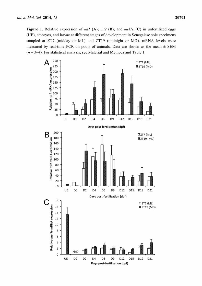
mRNA levels were evident mainly at night (Figure 1A, Table 1). mt1 transcripts were very low in
unfertilized eggs and augmented significantly in 0 dpf embryos. Its expression increased again
between 0 and 6 dpf (9-fold), remained elevated from 6 to 15 dpf (early metamorphosis), decreasing
thereafter to 19 dpf (metamorphic climax) and 21 dpf (late metamorphosis). Although mt1 relative
expression was elevated at night in relation to day-time levels on almost all sampling days, differences
were only significant at 6 and 12 dpf (Figure 1A, Table 1).
Table 1. Significance levels of statistical analysis for each melatonin receptor.
mt1
TWO-WAY ANOVA KRUSKAL WALLIS
Developmental stage Hour of sampling Interaction H = 60.4; p < 0.0001
F = 10.83; p < 0.001 F = 23.6; p < 0.001 F = 3.15; p < 0.01
BONFERRONI TEST
Developmental stage UE 0 dpf 2 dpf 4 dpf 6 dpf 9 dpf 12 dpf 15 dpf 19 dpf 21 dpf
Hour of sampling MD ML MD ML MD ML MD ML MD ML MD ML MD ML MD ML MD ML MD
Comparison a abc
d
ab ab abc
d
abc
d
cde abc
d
e abc
d
bcd
e
abcd e abc
d
de abc abc
d
ab abc
mt2
TWO-WAY ANOVA KRUSKAL WALLIS
Developmental stage Hour of sampling Interaction H = 57.7; p < 0.0001
F = 10.67; p < 0.001 F = 0.10; p = 0.756 F = 2.29; p < 0.05
BONFERRONI TEST
Developmental stage UE 0 dpf 2 dpf 4 dpf 6 dpf 9 dpf 12 dpf 15 dpf 19 dpf 21 dpf
Hour of sampling MD ML MD ML MD ML MD ML MD ML MD ML MD ML MD ML MD ML MD
Comparison a abc a abc
def
g
fg def
g
cdef
g
g bcd
efg
efg abcd
efg
abc
def
abc
def
abc
de
abc
de
abc
d
abc
def
abc
d
abc
de
mel1c
TWO-WAY ANOVA
Developmental stage Hour of sampling Interaction
F = 6.54; p < 0.001 F = 16.87; p < 0.001 F = 0.44; p = 0.8729
NEWMAN-KEULS TEST
Developmental stage UE 0 dpf 2 dpf 4 dpf 6 dpf 9 dpf 12 dpf 15 dpf 19 dpf 21 dpf
Comparison a N/D bc bc b bc c c b b
Hour of sampling ML MD
Comparison a b
There are no statistical differences among groups that share common letters. UE: unfertilized eggs; ML midday or ZT7;
MD midnight or ZT19; N/D: non detected, mRNA levels below the limits of detection of the technique.
Int. J. Mol. Sci. 2014, 15 20792
Figure 1. Relative expression of mt1 (A); mt2 (B); and mel1c (C) in unfertilized eggs
(UE), embryos, and larvae at different stages of development in Senegalese sole specimens
sampled at ZT7 (midday or ML) and ZT19 (midnight or MD). mRNA levels were
measured by real-time PCR on pools of animals. Data are shown as the mean ± SEM
(n = 3–4). For statistical analysis, see Material and Methods and Table 1.

Int. J. Mol. Sci. 2014, 15 20793
2.2. mt2 Expression
mt2 expression varied during development, without significant day-night changes (two-way
ANOVA, Figure 1B, Table 1). There was a significant interaction between the hour of sampling and
developmental stage, and differences among experimental groups were also significant as revealed by
a Kruskal–Wallis one-way analysis of variance (Table 1). mt2 transcripts were found at low levels in
unfertilized eggs and at 0 dpf embryos. Diurnal mt2 mRNA levels increased 10-fold from 0 to 6 dpf
and then decreased 10-fold between 6 to 15 dpf, remaining low until 21 dpf (Figure 1B). Nocturnal
mt2 expression exhibited a significant 500-fold rise between 0 to 2 dpf, followed by a progressive
reduction (4.5-fold) from 2 dpf until the end of metamorphosis (Figure 1B).
2.3. mel1c Expression
mel1c transcript levels were abundant in unfertilized eggs and decreased considerably after
fertilization, being below the limits of detection at 0 dpf (Figure 1C). Significant developmental and
day-night fluctuations were detected, with higher transcript levels at night than during the daytime
(Table 1). No interaction was found between the two factors (Table 1). mel1c expression doubled from
2 to 6 dpf, then decreased 2-fold to 15 dpf, only to increase again to 21 dpf (Figure 1C, Table 1).
3. Discussion
We reported here, for the first time in fish, distinct day-night and developmental expression patterns
of the genes encoding for three different melatonin receptor subtypes (mt1, mt2, and mel1c).
In the sole, the three melatonin receptor mRNAs were detected in unfertilized ovulated oocytes. mel1c
transcripts were abundant in unfertilized eggs, and decreased at early stages of development, whereas the
transcription of the other two receptor genes, mt1 and mt2, was low in unfertilized eggs and increased
after fertilization. Similar results were reported in the Japanese quail, Coturnix japonica [19,20].
This expression reflects the presence of mRNA from maternal origins, and it has been proposed that
such receptors could mediate some protective effects of melatonin from free radicals formed during
intensive embryonic metabolism [19,20]. Interestingly, a very marked nocturnal spawning rhythm was
reported in sole, with spawning beginning after dusk and the acrophase occurring around 23 h [21].
Whether these mel1c receptors are mediating the synchronizing effects of melatonin on sole spawning
remains to be investigated.
At hatching (i.e., 2 dpf) a strong increase in mt2 transcript levels was initiated, whereas the
elevation in mt1 mRNA levels occurred from 0 dpf but becomes more conspicuous at 4 dpf, coinciding
with the opening of the mouth and beginning of external feeding (Figure 2). This is concomitant with
the onset of pineal (2 dpf) and retinal (3 dpf) photoreception, elevation of pineal aanat2 (2 dpf) and
retinal aanat1a (4 dpf) mRNA levels [16,17,22], and, probably, a rise in circulating melatonin.
Recently, it has been demonstrated that lighting conditions during incubation affect hatching rhythms
and subsequent larval development (growth, mouth opening, and appearance of pectoral fins) in the
Senegal sole [23]. Expression of melatonin receptors (particularly mt2), is up-regulated in Danio rerio
embryos, where Mt2 might play a major role in mediating melatonin-accelerated development at these
early stages [6]. Indeed, melatonin stimulated cell proliferation in zebrafish, and melatonin-treated

Int. J. Mol. Sci. 2014, 15 20794
embryos developed faster and hatched earlier, the maximal effects being obtained when melatonin
receptors were widely expressed [6]. In another flatfish, the halibut (Hippoglossus hippoglossus),
hatching seems to be modulated by photoperiod acting through the pineal gland [9]. The putative role
of mt1 and mt2 receptors in the mediation of photoperiod effects reported on sole feeding and
hatching [23,24] rhythms requires further characterization.
An interesting finding of this study is that different day-night and developmental patterns of
expression exist for the three melatonin receptor subtypes. mt1 mRNA abundance exhibited day-night
variation, with higher nocturnal expression. In agreement with these results, melatonin-synthesizing
enzymes aanat1a and aanat2 also exhibited higher transcript levels at MD than at ML from 2 dpf and
at most developmental stages analyzed [16,17]. The lack of midday/midnight difference in mt2 mRNA
abundance may indicate either an absence of daily rhythm or a phase difference between the mt1 and
mt2 mRNA rhythms. Otherwise, mt2 daily rhythms of expression may be masked by phase differences
in transcript levels between different expression sites.
Figure 2. Developmental expression profiles of the three melatonin receptor subtypes
(mt1, mt2, mel1c) and the pineal-specific enzyme arylalkylamine N-acetyltransferase
(aanat2) [16], together with thyroid hormone levels (T4, grey columns) [25] in sole. Note
that the melatoninergic system is up-regulated at hatching and the onset of external
feeding; also note the inverse profile between mt1, mt2, and aanat2 expression and thyroid
hormone levels during sole development and metamorphosis.
In flatfish (as in anurans), thyroid hormones drive morphological, molecular, and physiological
changes that occur during metamorphosis [26]. In addition, the thyroid system (hormone levels,
receptor expression, deiodinase expression and activity) is up-regulated during sole metamorphosis [25].
We now provide evidence that the melatonin system is concomitantly down-regulated, as summarized
in Figure 2. This includes the melatonin-synthesizing enzymes aanat1 and aanat2 [16,17], and the
melatonin receptors mt1 and mt2 (this study). Earlier studies indicated that melatonin is involved in
amphibian metamorphosis, acting as an antagonist of thyroid hormones [11]; similar antagonizing

Int. J. Mol. Sci. 2014, 15 20795
effects were also reported in some non-metamorphic fish [27], but such antagonism remains
unexplored in flatfish.
The mel1c receptor subtype was cloned for the first time from isolated melanophores of
Xenopus [28] and seems to mediate melatonin effects on pigmentation [29]. It is interesting to note that
photoperiod and light spectrum affect early growth and development in sole, including eye
pigmentation [23]. In the adult sole, mel1c expression was seen in the retina, brain, pituitary, muscle,
and skin [18]. The skin of the sea bass Dicentrarchus labrax [4] and goldfish scales [30] also express
mel1c. Poikilotherms, including fish, exhibit circadian rhythms in color change, with nocturnal
blanching related to pigment melanophore aggregation induced by melatonin [31]. As in other flatfish,
pigmentation abnormalities have been observed during sole metamorphosis, which appears to be
influenced by light intensity and background color of tanks during larval rearing [32]. Interestingly,
mel1c expression exhibited day-night differences during sole development, with higher nocturnal
levels, as reported for aanat1a and aanat2 in developing sole [16,17]. Moreover, regulation of mel1c
expression during development appears to differ compared to mt1 and mt2 because its expression
increased throughout metamorphosis. In the course of this event, the right side of the body (ocular side)
becomes more pigmented and the left side (blind side) loses coloration and becomes almost entirely
white. Thus, mel1c receptors expressed during development could be mediating early effects of
photoperiod and/or melatonin on sole pigmentation.
4. Materials and Methods
4.1. Animals and Sampling
Senegalese sole fertilized eggs were obtained in May (0 days post-fertilization or 0 dpf) from
“IFAPA El Toruño” (Junta de Andalucía, Puerto de Santa María, Spain) and maintained in the
“Laboratorio de Cultivos Marinos” (University of Cádiz) as previously described [16,25]. Eggs were
incubated under a natural photoperiodic environment (sunrise 07:31 h, sunset 21:22 h, GMT + 2,
illumination of 300-500 lux on water surface), at 19 ± 1 °C of temperature and 39 ppt of salinity. Animals
were sampled at nine stages of development: before hatching (0 dpf), before the beginnings of
metamorphosis (2, 4, 6, 9 dpf), and during metamorphosis (pre-, early-, middle-, and late-metamorphosis;
12, 15 19 and 21 dpf, respectively). At each developmental stage, pools of whole animals were
obtained at ZT7 (midday, 14:30 h GMT + 2) and ZT19 (midnight, 02:30 h GMT + 2), which exhibited
day-night differences in the expression of melatonin-synthesizing enzymes [16,17]. The number of
individuals per pool was 20–30 at 0–6 dpf, 10–20 at 9–15 dpf, and 5–10 at 19–21 dpf, respectively.
The larvae’s metamorphic process was followed daily under a photomicroscope. Senegalese sole
ovulated unfertilized eggs were obtained from the IEO (MICINN, Centro Oceanográfico de Santander,
Santander, Spain) and used to detect the presence of melatonin receptor mRNA of maternal origin.
Samples were frozen in liquid nitrogen and stored at −80 °C until used. All animal experiments were
approved by the Institutional Animal Care and Use Committee at the University of Cádiz and were
conducted in accordance with international standards.
Int. J. Mol. Sci. 2014, 15 20796
4.2. Quantitative Real-Time PCR Analysis
Total RNA was extracted from larval pools using “EUROzol” (EuroClone, Siziano, Italy) according
to the manufacturer’s instructions. Total RNA (1 μg) was reverse-transcribed and genomic DNA
removed (QuantiTect® Reverse Transcription Kit, Qiagen, Hilden, Germany). Real-time gene
expression analysis was performed in a Chromo 4™ Four-Color Real-Time System (Bio-Rad,
Alcobendas, Spain), using β-actin for normalization (GeneBank accession number DQ485686). PCR
reactions were developed in duplicate in a 25 μL volume using cDNA generated from 1 μg of RNA,
iTaq™ SYBR® Green Supermix with ROX (Bio-Rad), and specific primers (0.4 μM, Table 2)
designed from the cloned mt1, mt2, and mel1c sequences of the sole [18]. All calibration curves
exhibited slopes close to −3.32 and efficiencies around 100%. The conditions of the PCR reactions
were similar for the four genes analyzed (38 cycles): 3 min at 95 °C, 30 s at 95 °C, 45 s at 60 °C, and
45 s at 72 °C. PCR products were run in agarose gels, sequenced, and melting curves were analyzed
for each sample to confirm that only a single sequence was amplified. Negative controls included
replacement of cDNA by water and the use of non retro-transcribed total RNA. The ΔΔCt method [33]
was used to determine the relative mRNA expression.
Table 2. Sequences of the primers used.
Primer Name Sequence (5'-3')
ssmt1F GCGGAAAGGAATAAATGAGGC
ssmt1R GGAGTTGGTGCGTCACAGTG
ssmt2F GCGTCAACGAAGAGCGAAAT
ssmt2R GCCCGAAACTGGCCATAAAT
ssmel1cF ACTTCAACAGCTGCCTCAACG
ssmel1cR AGCAAACGTGGGATGCAAAG
ssβactinF GGATCTGCATGCCAACACTG
ssβactinR TCTGCATCCTGTCAGCAATG
4.3. Statistical Analysis
Day-night statistical differences in housekeeping gene expression in different developmental stages
were determined by Paired T-test. Developmental and day-night statistical differences among groups
were analyzed using two-way ANOVA. When significant interaction between the hour of the day and
developmental stage was found (i.e., for mt1 and mt2), differences among groups were determined
using non-parametric Kruskal–Wallis analysis followed by Bonferroni test because of the absence of
homogeneous variance even after data transformation. When no significant interaction between factors
existed (i.e., for mel1c), each factor was analyzed separately by one-way ANOVA followed by the
Newman–Keuls test. Statistical tests were made using the Statgraphics plus (version 5.1) software
(Manugistics, Rockville, MD, USA, 2000).
5. Conclusions
Taken together, all these data strengthen the idea that melatonin, acting through Mt1, Mt2, and/or
Mel1c receptors, could play an important role in setting physiological and behavioral rhythms during

Int. J. Mol. Sci. 2014, 15 20797
sole gametogenesis, development, and metamorphosis. The presence of high mel1c transcript levels in
mature unfertilized eggs suggests that this receptor could be mediating melatonin effects in ovulated
oocytes. Moreover, we have demonstrated that these receptors exhibited distinct developmental
expression profiles, suggesting that they are subjected to different transcriptional regulatory mechanisms.
The fact that mt1 and mt2 receptor expression decline during metamorphosis, when thyroid hormone
levels rise, reinforces the interest of analyzing the functional antagonism between melatoninergic and
thyroid hormone system in the regulation of this event. Further studies are being directed to elucidate
the melatonin effects on relevant processes such as cell proliferation, hatching, metamorphosis,
locomotor activity, and feeding at early development stages of the Senegalese sole, and to determine
which melatonin receptors appear involved in these putative actions.
Acknowledgments
The authors thank J. Pedro Cañavate from IFAPA “El Toruño” (Junta de Andalucía,
Puerto de Santa María) for the generous gift of sole embryos. We also thank all staff from the
“Planta de Cultivos Marinos” (University of Cádiz) for the maintaining of animals used in
developmental studies. This work was supported by grants from MINECO (AGL2007-66507-C02-01
and AGL2010-22139-C03-03) to JAM-C.
Author Contributions
Maria del Carmen Rendón, Jack Falcón and José A. Muñoz-Cueto designed the study.
Olivier Lan-Chow-Wing, Francesca Confente, Patricia Herrera-Pérez, Esther Isorna and
Olvido Chereguini performed the animal collection and sampling. Olivier Lan-Chow-Wing,
Francesca Confente and Patricia Herrera-Pérez managed the literature searches. Olivier Lan-Chow-Wing,
Francesca Confente and Esther Isorna perfomed the RNA, cDNA and quantitative Real-Time PCR
analysis. Olivier Lan-Chow-Wing, Francesca Confente and Esther Isorna undertook the statistical
analysis. Olivier Lan-Chow-Wing and Francesca Confente wrote the first draft of the manuscript.
All researchers contributed to and have approved the final manuscript.
Conflicts of Interest
The authors declare no conflict of interest.
References
1. Reiter, R.J. The melatonin rhythm. Experientia 1993, 49, 654–664.
2. Falcón, J.; Migaud, H.; Muñoz-Cueto, J.A.; Carrillo, M. Current knowledge on the melatonin
system in teleost fish. Gen. Comp. Endocrinol. 2010, 165, 469–482.
3. Reppert, S.M.; Weave, D.R.; Godson, C. Melatonin receptors step into the light: cloning and
classification of subtypes. Trends Pharmacol. Sci. 1996, 17, 100–102.
4. Sauzet, S.; Besseau, L.; Herrera-Perez, P.; Covès, D.; Chatain, B.; Peyric, E.; Boeuf, G.;
Muñoz-Cueto, J.A.; Falcón, J. Cloning and retinal expression of melatonin receptors in the
European sea bass, Dicentrarchus labrax. Gen. Comp. Endocrinol. 2008, 157, 186–195.

Int. J. Mol. Sci. 2014, 15 20798
5. Herrera-Pérez, P.; Rendón, M.C.; Besseau, L.; Sauzet, S.; Falcón, J.; Muñoz-Cueto, J.A.
Melatonin receptors in the brain of the European sea bass: An in situ hybridization and
autoradiographic study. J. Comp. Neurol. 2010, 518, 3495–3511.
6. Danilova, N.; Krupnik, V.E.; Sugden, D.; Zhdanova, I.V. Melatonin stimulates cell proliferation
in zebrafish embryo and accelerates its development. FASEB J. 2004, 18, 751–753.
7. Ziv, L.; Gothilf, Y. Circadian time-keeping during early stages of development. Proc. Natl. Acad.
Sci. USA 2006, 103, 4146–4151.
8. Wilson, S.W.; Easter, S.S., Jr. Stereotyped pathway selection by growth cones of early epiphysial
neurons in the embryonic zebrafish. Development 1991, 112, 723–746.
9. Forsell, J.; Holmqvist, B.; Helvik, J.V.; Ekström, P. Role of the pineal organ in the photoregulated
hatching of the Atlantic halibut. Int. J. Dev. Biol. 1997, 41, 591–595.
10. Vuilleumier, R.; Besseau, L.; Boeuf, G.; Piparelli, A.; Gothilf, Y.; Gehring, W.G.; Klein, D.C.;
Falcón, J. Starting the zebrafish pineal circadian clock with a single photic transition.
Endocrinology 2006, 147, 2273–2279.
11. Wright, M.L. Melatonin, diel rhythms, and metamorphosis in anuran amphibians. Gen. Comp.
Endocrinol. 2002, 126, 251–254.
12. Isorna, E.; Guijarro, A.I.; Delgado, M.J.; López-Patiño, M.A.; de Pedro, N.; Alonso-Gómez, A.L.
Ontogeny of central melatonin receptors in tadpoles of the anuran Rana perezi: modulation of
dopamine release. J. Comp. Physiol. A 2005, 191, 1099–1105.
13. Shi, Q.; Ando, H.; Coon, S.L.; Sato, S.; Ban, M.; Urano, A. Embryonic and post-embryonic
expression of arylalkylamine N-acetyltransferase and melatonin receptor genes in the eye and
brain of chum salmon (Oncorhynchus keta). Gen. Comp. Endocrinol. 2004, 136, 311–321.
14. Bayarri, M.J.; Muñoz-Cueto, J.A.; López-Olmeda, J.F.; Vera, L.M.; Rol de Lama, M.A.;
Madrid, J.A.; Sánchez-Vázquez, F.J. Daily locomotor activity and melatonin rhythms in Senegal
sole (Solea senegalensis). Physiol. Behav. 2004, 81, 577–583.
15. Vera, L.M.; Oliveira, C.; López-Olmeda, J.F.; Ramos, J.; Mañanos, E.; Madrid, J.A.;
Sánchez-Vázquez, F.J. Seasonal and daily plasma melatonin rhythms and reproduction in Senegal
sole kept under natural photoperiod and natural or controlled water temperature. J. Pineal Res.
2007, 43, 50–55.
16. Isorna, E.; El M’Rabet, A.; Confente, F.; Falcón, J.; Muñoz-Cueto, J.A. Cloning and expression of
arylalkylamine N-acetyltranferase-2 during early development and metamorphosis in the sole
Solea senegalensis. Gen. Comp. Endocrinol. 2009, 161, 97–102.
17. Isorna, E.; Aliaga-Guerrero, M.; El M’Rabet, A.; Servili, A.; Falcón, J.; Muñoz-Cueto, J.A.
Identification of two arylalkylamine N-acetyltranferase 1 genes with different developmental
expression profiles in the flatfish Solea senegalensis. J. Pineal Res. 2011, 51, 434–444.
18. Confente, F.; Rendón, M.C.; Besseau, L.; Falcón, J.; Muñoz-Cueto, J.A. Melatonin receptors in a
pleuronectiform species, Solea senegalensis: Cloning, tissue expression, day-night and seasonal
variations. Gen. Comp. Endocrinol. 2010, 167, 202–214.
19. Obłap, R.; Olszanska, B. Expression of melatonin receptor transcripts (mel-1a, mel-1b and mel-1c)
in Japanese quail oocytes and eggs. Zygote 2001, 9, 237–244.
Int. J. Mol. Sci. 2014, 15 20799
20. Olszanska, B.; Majewski, P.; Lewczuk, B.; Stepinska, U. Melatonin and its synthesizing enzymes
(arylalkylamine N-acetyltransferase-like and hydroxyindole-O-methyltransferase) in avian eggs
and early embryos. J. Pineal Res. 2007, 42, 310–318.
21. Oliveira, C.; Dinis, M.T.; Soares, F.; Cabrita, E.; Pousão-Ferreira, P.; Sánchez-Vázquez, F.J.
Lunar and daily spawning rhythms of Senegal sole Solea senegalensis. J. Fish. Biol. 2009, 75,
61–74.
22. El M’Rabet, A.; Confente, F.; Ouarour, A.; Muñoz-Cueto, J.A. Ontogenia del órgano pineal del
lenguado, Solea senegalensis. In Avances en Endocrinología Comparada; Muñoz-Cueto, J.A.,
Mancera, J.M., Martínez, G., Eds.; Cádiz: Servicio de Publicaciones de la Universidad de Cádiz,
Cádiz, Spain, 2008, Volume 4, pp. 167–170.
23. Blanco-Vives, B.; Aliaga-Guerrero, M.; Cañavate, J.P.; Muñoz-Cueto, J.A.; Sánchez-Vázquez, F.J.
Does lighting manipulation during incubation affect hatching rhythms and early development
of sole? Chronobiol. Int. 2011, 28, 300–306.
24. Cañavate, J.P.; Zerolo, R.; Fernández-Díaz, C. Feeding and development of Senegal sole
(Solea senegalensis) larvae reared in different photoperiods. Aquaculture 2006, 258, 368–377.
25. Isorna, E.; Obregon, M.J.; Calvo, R.M.; Vázquez, R.; Pendón, C.; Falcón, J.; Muñoz-Cueto, J.A.
Iodothyronine deiodinases and thyroid hormone receptors regulation during flatfish
(Solea senegalensis) metamorphosis. J. Exp. Zool. B 2009, 312, 231–246.
26. Power, D.M.; Llewellyn, L.; Faustino, M.; Nowell, M.A.; Bjornsson, B.T.; Einarsdottir, I.E.;
Canario, A.V.M.; Sweeney, G.E. Thyroid hormones in growth and development of fish.
Comp. Biochem. Physiol. C 2001, 130, 447–459.
27. Kulczykowska, E.; Sokolowska, E.; Takvam, B.; Stefansson, S.; Ebbesson, L. Influence of
exogenous thyroxine on plasma melatonin in juvenile Atlantic salmon (Salmo salar).
Comp. Biochem. Physiol. B 2004, 137, 43–47.
28. Ebisawa, T.; Karnes, S.; Lerner, M.R.; Reppert, S.M. Expression cloning of a high-affinity
melatonin receptor from Xenopus dermal melanophores. Proc. Natl. Acad. Sci. USA 1994, 91,
6133–6137.
29. Vanecek, J. Cellular mechanisms of melatonin action. Physiol. Rev. 1998, 78, 687–721.
30. Ikegami, T.; Azuma, K.; Nakamura, M.; Suzuki, N.; Hattori, A.; Ando, H. Diurnal expressions
of four subtypes of melatonin receptor genes in the optic tectum and retina of goldfish.
Comp. Biochem. Physiol. A 2009, 152, 219–224.
31. Aspengren, S.; Skold, H.N.; Quiroga, G.; Martensson, L.; Wallin, M. Noradrenaline- and
melatonin-mediated regulation of pigment aggregation in fish melanophores. Pigment. Cell. Res.
2003, 16, 59–64.
32. Dinis, M.T.; Ribeiro, L.; Soares, F.; Sarasquete, C. A review on the cultivation potential of
Solea senegalensis in Spain and in Portugal. Aquaculture 1999, 176, 27–38.
33. Livak, K.J.; Schmittgen, T.D. Analysis of relative gene expression data using real-time
quantitative PCR and the 2-[Delta][Delta]CT method. Methods 2001, 25, 402–408.
© 2014 by the authors; licensee MDPI, Basel, Switzerland. This article is an open access article
distributed under the terms and conditions of the Creative Commons Attribution license
(http://creativecommons.org/licenses/by/4.0/).