Molecular identification of genes involved in testicular steroid synthesis and characterization of...
10
Molecular identification of genes involved in testicular steroid synthesis and characterization of the response to gonadotropic stimulation in the Senegalese sole (Solea senegalensis) testis Rubén Marín-Juez a , Barbara Castellana a , Manuel Manchado b , Josep V. Planas a,⇑ a Departament de Fisiologia, Facultat de Biologia, Universitat de Barcelona and Institut de Biomedicina de la Universitat de Barcelona (IBUB), 08028 Barcelona, Spain b IFAPA Centro El Toruño, Consejería de Agricultura y Pesca, Junta de Andalucía, Apartado 16, 11500 El Puerto de Santa María, Cádiz, Spain article info Article history: Available online 16 February 2011 Keywords: Teleost Testis ESTs hCG Spermatogenesis Microarray abstract In male teleosts, testicular steroids are essential hormones for the regulation of spermatogenesis and their production is regulated by pituitary gonadotropins. In the Senegalese sole (Solea senegalensis), an econom- ically important flatfish with semi-cystic and asynchronous spermatogenesis, the gonadotropic regulation of spermatogenesis, particularly regarding the production and regulation of testicular steroids, are not well understood. For this reason, we first cloned and characterized the response of several key genes for the pro- duction and action of testicular steroids to the in vivo administration of human chorionic gonadotropin (hCG) and, second, we investigated the transcriptomic effects of hCG in the Senegalese sole testis. We suc- ceded in cloning the full-length cDNAs for Steroidogenic Acute Regulatory protein (StAR), 3b-hydroxyster- oid dehydrogenase (3b-HSD), 17b-HSD and 20b-HSD and a partial cDNA for the nuclear progesterone receptor. In this study we also identified a transcript encoding a protein with homology to StAR, which we named StAR-like, that could represent a new member of the StAR-related lipid transfer (START) family. All the cloned genes were expressed in the testis and their expression levels were significantly increased by the in vivo administration of hCG. The plasma levels of testosterone and 11-ketotestosterone also increased in response to hCG administration, likely as a result of the induction of the expression of steroidogenic enzymes by hCG. Furthermore, gene expression analysis by microarray identified 90 differentially expressed genes in the testis in response to hCG administration, including genes potentially involved in ste- roidogenesis, progression of spermatogenesis and germ cell maturation and cytoskeletal organization. Our results have identified for the first time a number of key genes involved in the regulation of steroid produc- tion and spermatogenesis in the Senegalese sole testis that are under gonadotropic control. Ó 2011 Elsevier Inc. All rights reserved. 1. Introduction In teleosts, as in mammals, steroids play critical roles in sexual development and throughout the reproductive cycle. In male tele- osts, steroid hormones have important and distinct roles regulating spermatogenesis [37]. Particularly, the major testicular androgens testosterone (T) and 11-ketotestosterone (11-KT), and the proges- tin 17a,20b-dihydroxy-4-pregnen-3-one (17,20b-P) are known to control spermatogenesis by exerting feedback effects on the pitui- tary and the brain but also by their direct influence in the gonads regulating germ cell development and maturation [27,29,38]. Tes- ticular androgens, and specifically 11-KT, have been shown to sup- port the whole spermatogenic process in the testis or to regulate specific steps such as germ cell proliferation and maturation whereas progestins promote testicular hydration and spermiation [26,30,32] and even meiosis [27]. The production of testicular steroids in teleosts is primarily regu- lated by the pituitary gonadotropins follicle stimulating hormone (FSH) and luteinizing hormone (LH) [34]. Although most of the infor- mation is derived from studies in salmonids primarily due to the scarcity of homologous FSH or specific assays for measuring its levels in the blood in other species, FSH is believed to play a major role in the production of testicular steroids (mostly 11-KT) in earlier stages of spermatogenesis [34]. Therefore, FSH is considered to be the phys- iological regulator of spermatogenesis in fish. LH, on the other hand, is known to stimulate the production of 11-KT and 17,20b-P during spawning and is considered the major gonadotropin regulating sperm maturation and spermiation [34]. Direct and indirect evi- dence in different teleosts indicate that both FSH and LH target the steroid-producing Leydig cells, shown to have FSH and LH receptors [15,31,33], and stimulate their steroidogenic activity. However, although cholesterol carriers (i.e. StAR) and steroidogenic enzymes, essential elements of the steroidogenic Leydig cells, have been iden- tified by molecular cloning in a number of teleost species, their reg- ulation by pituitary gonadotropins is poorly documented. 0016-6480/$ - see front matter Ó 2011 Elsevier Inc. All rights reserved. doi:10.1016/j.ygcen.2011.02.003 ⇑ Corresponding author. Fax: +34 934110358. E-mail address: [email protected] (J.V. Planas). General and Comparative Endocrinology 172 (2011) 130–139 Contents lists available at ScienceDirect General and Comparative Endocrinology journal homepage: www.elsevier.com/locate/ygcen
-
Upload
juntadeandalucia -
Category
Documents
-
view
0 -
download
0
Transcript of Molecular identification of genes involved in testicular steroid synthesis and characterization of...

General and Comparative Endocrinology 172 (2011) 130–139
Contents lists available at ScienceDirect
General and Comparative Endocrinology
journal homepage: www.elsevier .com/locate /ygcen
Molecular identification of genes involved in testicular steroid synthesisand characterization of the response to gonadotropic stimulationin the Senegalese sole (Solea senegalensis) testis
Rubén Marín-Juez a, Barbara Castellana a, Manuel Manchado b, Josep V. Planas a,⇑a Departament de Fisiologia, Facultat de Biologia, Universitat de Barcelona and Institut de Biomedicina de la Universitat de Barcelona (IBUB), 08028 Barcelona, Spainb IFAPA Centro El Toruño, Consejería de Agricultura y Pesca, Junta de Andalucía, Apartado 16, 11500 El Puerto de Santa María, Cádiz, Spain
a r t i c l e i n f o a b s t r a c t
Article history:Available online 16 February 2011
Keywords:TeleostTestisESTshCGSpermatogenesisMicroarray
0016-6480/$ - see front matter � 2011 Elsevier Inc. Adoi:10.1016/j.ygcen.2011.02.003
⇑ Corresponding author. Fax: +34 934110358.E-mail address: [email protected] (J.V. Planas).
In male teleosts, testicular steroids are essential hormones for the regulation of spermatogenesis and theirproduction is regulated by pituitary gonadotropins. In the Senegalese sole (Solea senegalensis), an econom-ically important flatfish with semi-cystic and asynchronous spermatogenesis, the gonadotropic regulationof spermatogenesis, particularly regarding the production and regulation of testicular steroids, are not wellunderstood. For this reason, we first cloned and characterized the response of several key genes for the pro-duction and action of testicular steroids to the in vivo administration of human chorionic gonadotropin(hCG) and, second, we investigated the transcriptomic effects of hCG in the Senegalese sole testis. We suc-ceded in cloning the full-length cDNAs for Steroidogenic Acute Regulatory protein (StAR), 3b-hydroxyster-oid dehydrogenase (3b-HSD), 17b-HSD and 20b-HSD and a partial cDNA for the nuclear progesteronereceptor. In this study we also identified a transcript encoding a protein with homology to StAR, whichwe named StAR-like, that could represent a new member of the StAR-related lipid transfer (START) family.All the cloned genes were expressed in the testis and their expression levels were significantly increased bythe in vivo administration of hCG. The plasma levels of testosterone and 11-ketotestosterone also increasedin response to hCG administration, likely as a result of the induction of the expression of steroidogenicenzymes by hCG. Furthermore, gene expression analysis by microarray identified 90 differentiallyexpressed genes in the testis in response to hCG administration, including genes potentially involved in ste-roidogenesis, progression of spermatogenesis and germ cell maturation and cytoskeletal organization. Ourresults have identified for the first time a number of key genes involved in the regulation of steroid produc-tion and spermatogenesis in the Senegalese sole testis that are under gonadotropic control.
� 2011 Elsevier Inc. All rights reserved.
1. Introduction
In teleosts, as in mammals, steroids play critical roles in sexualdevelopment and throughout the reproductive cycle. In male tele-osts, steroid hormones have important and distinct roles regulatingspermatogenesis [37]. Particularly, the major testicular androgenstestosterone (T) and 11-ketotestosterone (11-KT), and the proges-tin 17a,20b-dihydroxy-4-pregnen-3-one (17,20b-P) are known tocontrol spermatogenesis by exerting feedback effects on the pitui-tary and the brain but also by their direct influence in the gonadsregulating germ cell development and maturation [27,29,38]. Tes-ticular androgens, and specifically 11-KT, have been shown to sup-port the whole spermatogenic process in the testis or to regulatespecific steps such as germ cell proliferation and maturationwhereas progestins promote testicular hydration and spermiation[26,30,32] and even meiosis [27].
ll rights reserved.
The production of testicular steroids in teleosts is primarily regu-lated by the pituitary gonadotropins follicle stimulating hormone(FSH) and luteinizing hormone (LH) [34]. Although most of the infor-mation is derived from studies in salmonids primarily due to thescarcity of homologous FSH or specific assays for measuring its levelsin the blood in other species, FSH is believed to play a major role inthe production of testicular steroids (mostly 11-KT) in earlier stagesof spermatogenesis [34]. Therefore, FSH is considered to be the phys-iological regulator of spermatogenesis in fish. LH, on the other hand,is known to stimulate the production of 11-KT and 17,20b-P duringspawning and is considered the major gonadotropin regulatingsperm maturation and spermiation [34]. Direct and indirect evi-dence in different teleosts indicate that both FSH and LH target thesteroid-producing Leydig cells, shown to have FSH and LH receptors[15,31,33], and stimulate their steroidogenic activity. However,although cholesterol carriers (i.e. StAR) and steroidogenic enzymes,essential elements of the steroidogenic Leydig cells, have been iden-tified by molecular cloning in a number of teleost species, their reg-ulation by pituitary gonadotropins is poorly documented.
R. Marín-Juez et al. / General and Comparative Endocrinology 172 (2011) 130–139 131
The Senegalese sole (Solea senegalensis) has recently become oneof the main target species for the aquaculture industry in southernEurope. In contrast to other teleosts, the Senegalese sole follows asemi-cystic pattern of spermatogenesis with small reniform testes(gonadosomatic index lower than 0.15% throughout the testicularcycle) [17]. This species spawns primarily in spring (between Febru-ary and May) [3] and, although spermatids are present in the testisthroughout the year, spermatogenesis is stimulated during the win-ter, increasing the number of spermatogonia and spermatocytes[8,16,17]. During this period, plasma levels of T and 11-KT are ele-vated [8,17] as well as the expression levels of the FSH b subunit inthe pituitary [8], changes that are accompanied by increases in theexpression levels of FSH and LH receptors in the testis [10]. Duringspawning in spring, androgen levels decrease [8,16] and peakexpression levels of the LH b subunit in the pituitary [8] and of tes-ticular LH receptors occur [10]. Overall, these findings suggest thatspermatogenesis and spermiation in the Senegalese sole may becontrolled by pituitary gonadotropins through their stimulation oftesticular steroid production. However, the specific effects of gonad-otropic stimulation on testicular function have not been examinedto date in this teleost species. Thus, the aim of this study was, first,to investigate the effects of the in vivo administration of human cho-rionic gonadotropin (hCG) on the expression of cloned key genes in-volved in testicular steroid production and action and, second, onthe transcriptome in the Senegalese sole testis.
2. Materials and methods
2.1. Animals and experimental treatment
Adult Senegalese sole (S. senegalensis) (N = 18), with an averageweight of 60.4 ± 2.2 g (mean ± SEM) and gonadosomatic index(GSI) of 0.048 ± 0.005, were obtained from the IFAPA ‘‘El Toruño’’aquaculture facilities (El Puerto de Santa María, Cádiz, Spain) in Au-gust, 2009. Fish were anesthetized with 2-phenoxyethanol (0.05%;Sigma, Alcobendas, Spain), sacrificed by spinal section and tissues(muscle, brain, gill, trunk kidney, head kidney, heart, skin, stomach,anterior and posterior intestine, ovary, testis, and spleen) were dis-sected, immediately frozen in liquid nitrogen and stored at �80 �Cfor RNA extraction. Remaining fish were divided into two groupsof eight individuals each, one group was given an intraperitonealinjection of saline (control group) and the other group was givenan intraperitoneal injection of human chorionic gonadotropin(hCG; 5 IU/g body weight; Calbiochem, San Diego, USA). Fish weresacrificed 24 h after the injection by spinal section. Testes were dis-sected and immediately frozen in liquid nitrogen. Blood was drawnfrom the caudal vein, centrifuged at 1500 rpm and the plasma wasimmediately frozen in liquid nitrogen. Tissues and plasma sampleswere stored at �80 �C until analysis.
2.2. Measurement of androgen levels in plasma
The levels of 11-KT and T in plasma were quantified directlyusing a commercial Enzyme-Linked ImmunoSorbent Assay (ELISA)and radioimmunoassay (RIA), respectively, following the manufac-turer’s indications (Cayman Chemical Co., Ann Arbour, MI andSchering-CIS, Madrid, Spain).
2.3. Cloning of StAR, StAR-like, 3-HSD, 17-HSD, 20-HSD and PR fromthe Senegalese sole
Expressed sequence tags (ESTs) with homology to steroidogenicacute regulatory protein (StAR), 3b-hydroxysteroid dehydrogenase(3b-HSD), 17b-hydroxysteroid dehydrogenase (17b-HSD), 20b-hydroxysteroid dehydrogenase (20b-HSD) and the nuclear proges-
terone receptor (PR) were identified by searching the Senegalesesole EST collection previously generated by single-pass 30 sequenc-ing of pooled and normalized cDNA libraries from different larvalstages and adult tissues [9]. cDNA clones (in pBK-CMV PhagemidVector, Stratagene, Santa Clara; CA) tentatively corresponding toSenegalese sole StAR, 3b-HSD, 17b-HSD, 20b-HSD and PR containedinserts of 1.2, 0.5, 2, 1.2, and 1.2 Kb and were sequenced at leasttwo times from each side with T7 and T3 vector primers usingthe BigDye v3.1 sequencing kit (Applied Biosystems, Foster City,CA). Full-length sequences of putative StAR and 20b-HSD cDNAswere obtained by overlapping 30 and 50 sequences. To obtain thefull-length sequence of the 17b-HSD cDNA, internal primers weredesigned (Supplementary Table 1) in order to overlap the differentsets of partial sequences. Primers were designed using the Genam-ics Expression software (www.genamics.com). The cDNA clonescorresponding to Senegalese sole 3b-HSD and PR were partialand, in both cases, the missing sequence was at the 50 end. In orderto obtain their full-length sequence, rapid amplification of cDNAends (RACE) was performed with the 50 RACE System kit (Invitro-gen, Prat del Llobregat, Spain), using gene specific primers (Supple-mentary Table 1) and pooled mRNA from Senegalese sole testes,following the manufacturer’s indications. This method was onlysuccessful in obtaining additional 50 sequences for the 3b-HSDcDNA.
Sequence analysis revealed a lower homology than expectedbetween the full-length Senegalese sole StAR clone sequence iden-tified here (62–66%; Table 2) and other known teleost StAR se-quences. This led us to hypothesize that it could correspond to adifferent form of StAR and therefore we labeled it ‘‘StAR-like’’. Inorder to clone StAR in the Senegalese sole, degenerate primerswere designed based on highly conserved regions in teleost StARsequences (Supplementary Table 1). RT-PCR was performed using1 ll of cDNA from testes from hCG-injected sole in a total volumeof 100 ll containing 1X PCR buffer, 1.5 mM Mg2+, 10 mM dNTPs,0.5 lM of each forward and reverse primers and 2.5 IU of recombi-nant Taq DNA Polymerase (Invitrogen) under the followingconditions: 3 min at 94 �C followed by 35 cycles of 94 �C for 45 s,52–55 �C for 30 s and 72 �C for 1 min. PCR products were isolatedfrom the agarose gel using Geneclean Spin Kit (MP Biomedicals,Solon, Ohio) and ligated into the pGEM-T Easy vector (PromegaCorp. Madison, WI). The cDNA was sequenced at least two timesfrom each side with specific T7 and SP6 primers using the BigDyev3.1 sequencing kit and the obtained sequences showed highhomology to other teleost StAR sequences (higher than 90%). Inorder to obtain the full-length cDNA encoding Senegalese soleStAR, 50and 30 RACE were performed and PCR products sequencedat least two times from each side as described above. The resultingsequence showed high homology to other known teleost StAR se-quences (87–96%) and corresponded to the Senegalese sole StARfull-length cDNA.
2.4. Sequence alignments and molecular phylogenetic analysis
Multiple sequence alignments of the deduced amino acid se-quence from the Senegalese sole cDNAs identified in this studywith other teleost sequences were performed using CLC SequenceViewer 6.4 (CLC Bio A/S, Aarhus, Denmark, www.clcbio.com).Protein sequences were predicted using the ExPASy ProteomicsTools server of the Swiss Institute of Bioinformatics (SIB; http://www.expasy.org; [18]). Identification of protein domains as wellas their probability of import into the mitochondria was performedusing SMART 6 [22] and Mitoprot II (http://ihg2.helmholtz-muen-chen.de/ihg/mitoprot.html; [14]), respectively. Conservation ofcritical amino acid residues was analyzed by multiple alignmentswith deduced amino acid sequences from other teleost speciesusing the Clustal W algorithm in CLC Sequence Viewer.

132 R. Marín-Juez et al. / General and Comparative Endocrinology 172 (2011) 130–139
Phylogenetic relationships were inferred based on the deducedamino acid sequence for StAR, StAR-like, 3b-HSD, 17b-HSD, 20b-HSD and PR from the Senegalese sole, using the UPGMA method[39] in CLC Sequence Viewer 6.4. One thousand iterations wereused to generate a bootstrap consensus tree for each phylogeneticanalysis.
2.5. Gene expression analysis by RT-PCR
In order to describe the tissue distribution of the expression ofthe cloned genes, total RNA from individual Senegalese sole tissueswas extracted using the RNeasy Maxikit (Qiagen, Barcelona, Spain)and treated with RQ1 RNase-free DNase (Promega) as indicated bythe manufacturer to remove any contaminating genomic DNA.First-strand cDNA was synthesized from 2 lg DNase I-treated totalRNA by using 0.25 lg oligo(dT)15, 0.25 lg random primers, 10 mMdNTPs and 200 IU SuperScript III RNase H� Reverse Transcriptase(RT; Invitrogen), for 5 min. at 65 �C, 1 h at 55 �C and 15 min at70 �C. Conventional PCR was performed with 1 ll of the RT reac-tion in a total volume of 100 ll containing 1X PCR buffer,1.5 mM Mg2+, 10 mM dNTPs, 0.5 lM of each forward and reverseprimers and 2.5 IU of recombinant Taq DNA Polymerase (Invitro-gen) using a MJ Research PT-200 thermocycler. Specific primerswere designed as indicated above according to the nucleotide se-quences of each gene (Supplementary Table 1) The PCR protocolwas 94 �C for 3 min followed by 35 cycles of 94 �C for 45 s, 55–57 �C for 30 s and 72 �C for 1 min, and a final step of 72 �C for10 min. PCR products were resolved in a 1.5% agarose gel andstained with ethidium bromide for visualization. Parallel PCRswere carried out with specific primers for the ribosomal 18S fornormalization (Supplementary Table 1).
2.6. Quantitative Real-Time PCR (qPCR)
Total RNA from testes of saline (control) and hCG-treated fishwas extracted and reverse-transcribed as previously described.qPCR reactions (20 ll final volume) contained 10 ll SYBR GreenERqPCR SuperMix (Invitrogen), 0.5 lM of each forward and reverseprimers, 3 ll RNase/DNase-free water and 5 ll of template DNA(cDNA at a 1:15 dilution or plasmid DNA). Reactions were run ina MyiQ Single-Color Real-Time PCR Detection System (Bio-Rad,Prat de Llobregat, Spain) under the following conditions: 50 �Cfor 2 min, 95 �C for 8.5 min, followed by 40 cycles of 95 �C for
Table 1Percent homology of the deduced amino acid and nucleotide sequences of the cloned Sen
3b-HSD 17b-HSD 20b-HS
African catfish – – 81 (71)Atlantic cod – – 91 (78)Atlantic croaker – – –Atlantic salmon – 91 (79) 81 (70)Ayu – – 68 (–)Black bass – – –Brook trout – – –Burton’s mouthbreeder – – –Channel catfish 84 (67) – –Japanese eel – – 78 (68)Medaka 88 (77) – 89 (81)Nile tilapia 87 (77) 63 (-) 87 (81)Rainbow trout 86 (76) – 68 (–)Rainbow smelt 84 (74) – –Sablefish – – 92 (86)Seabream – – –Zebrafish 81 (67) 88 (77) 81 (–)
The values within parentheses represent nucleotide sequence homology. (-) Sequence n* Homology with the StAR sequence.** Partial sequence.
15 s and 55–57 �C for 30 s, and a final melting step (95 �C for1 min, 55 �C for 1 min and 81 cycles of 55 �C + 0.5 �C cycle each10 s) to identify the presence of primer dimers and to analyzethe specificity of the reaction. As described above, primers weredesigned using the Genamics Expression software. All reactionswere run in triplicate and the fluorescence was measured at theend of every extension cycle and used to calculate the thresholdcycle value (Ct) for each sample. The Ct values were used to calcu-late copy numbers following the absolute quantitation method[47]. Standard plots were constructed for each target gene usingspecific primers (Supplementary Table 1) and serial dilutions(10�4 to 10�9) of plasmid DNA corresponding to cloned qPCRproducts into the pGEM-T Easy vector (Promega Corp. Madison,WI). The cloned circular plasmids were quantified using a QubitTM
fluorometer (Invitrogen, Prat del Llobregat, Spain). The Ct valueswere plotted against the logarithm of their initial template copynumbers. Each standard curve was generated by a linear regressionof the plotted points. All the standard curves exhibited correlationcoefficients higher than 0.99, and efficiencies were greater than99%. For each gene, the copy number, calculated according to themethod published by Whelan et al. [47], of samples from thehCG-injected group was expressed relative to that of the saline-injected group (experimental controls) as fold change.
2.7. Microarray analysis
Total RNA from testes from saline-injected and hCG-injectedSenegalese sole was extracted using the RNeasy extraction kit (Qia-gen) and treated with DNAse following the manufacturer’s instruc-tions. Quantitative and qualitative analysis of total RNA wasperformed using the Agilent 2100 bioanalyzer. RNA samples fromeach group were pooled and amplified, labeled and hybridized toa custom-made oligonucleotide (60-mer probes) microarray con-taining 5087 Senegalese sole Unigene sequences, that was devel-oped and validated as previously described [9,40]. In brief,pooled testicular RNAs from each group were amplified and theresulting cRNAs labeled with Cy3 and Cy5, respectively, mixed inequal amounts and hybridized to the microarray for 17 h at60 �C. Each hybridization was performed in duplicate. The aquiredraw fluorescence intensity data was processed using the Polyphe-musTM software (Oryzon Genomics, Barcelona, Spain), which in-cludes spatial data compensation, non-significant expressed datafiltering, and data normalization. Data normalization was carried
egalese sole genes with those from other teleost species.
D StAR StAR-like* Progesterone receptor**
89 (76) 64 –90 (79) 62 –95 (89) 64 –– – –– – –96 (90) 64 –93 (81) 62 –– – 95 (86)– – –91 (77) 65 93 (80)95 (84) 62 95 (86)– – –94 (81) 63 –– – –– – –96 (91) 62 –87 (75) 66 93 (79)
ot available.

R. Marín-Juez et al. / General and Comparative Endocrinology 172 (2011) 130–139 133
out by an improved version of the nonlinear Q-splines normaliza-tion method [48], enhanced with robust regression techniques.Normalized and log-transformed data were used to calculate thefold change (FC) values. Differential expression was assessed withthe Polyphemus software. The p-values (considered significant at<0.01) were calculated based on the absolute value of the regular-ized t-statistic [48], which uses a Bayesian framework to derive thealgorithm, using internal replicated controls to assess the mini-mum technical variability of the process. Cut-offs for significantchanges was always greater than the inherent experimental varia-tion as assessed by the FC of internal controls and/or self-to-selfhybridizations. To estimate the rate of false-positive expression, aself-to-self hybridization was carried out, in which two aliquotsof pooled total RNA from control testes were used to produceeither Cy3 or Cy5 labeled cRNA. Fluorescent intensity data were ac-quired, processed and analyzed as previously described [9,40]. Themicroarray data have been deposited in the NCBI Gene ExpressionOmnibus (GEO) repository (http://www.ncbi.nlm.nih.gov/geo/)and are accessible through GEO Series accession numberGSE25733.
2.8. Statistical analysis
Statistical analyses were performed using SPSS11 (SPSS, Chi-cago, IL). Statistical differences between saline and hCG-treatedfish were analyzed by Mann–Whitney non-parametric tests andwere considered to be significant at p 6 0.05.
3. Results
3.1. Cloning and sequence analysis of StAR, StAR-like, 3b-HSD,17b-HSD, 20b-HSD and PR cDNAs from the Senegalese sole
By searching a Senegalese sole EST collection, we identified fiveclones with sequence similarity to teleost 3b-HSD, 17b-HSD, 20b-HSD, progesterone receptor and StAR, respectively. Further se-
Table 2Differentially expressed genes representative of major functional categories under-going changes in the Senegalese sole testis in response to in vivo treatment withhuman chorionic gonadotropin.
GenBankaccession
Swiss-prot hit FC
Steroidogenesis and steroid actionFF291770 StAR-like +4.39FF287782 Progesterone receptor +1.69
Transcriptional and translational regulatorsFF290985 Putative homeodomain transcription factor 1 +2.00FF283210 Eukaryotic translation initiation factor4A +1.76FF284048 JunB proto-oncogen -1.85FF285974 Small nuclear RNA auxiliary factor 1 -1.84FF290864 General transcription factor IIIA -1.66
Spermatogenesis and germ cell proliferationFF284682 Gonadal soma-derived growth factor +1.85FF286031 Protein tyrosine phosphatase type IVA, member
2+1.68
FF282991 Zpc domain containing protein 5 �2.53
Apoptosis inhibitionFF287928 Survivin 2 �1.77FF290789 Inmediate early response 3 �1.77
Cytoskeleton and cell adhesionFF281951 Super cysteine rich protein (fragment) +1.64FF285305 Claudin-like protein �1.93FF283806 Myosin light chain (mlc-4) �1.78FF287418 Muscle cofilin 2 �1.69
Data shown as fold change (FC) with respect to the control group.
quence characterization yielded the full-length cDNA sequence ofSenegalese sole 17b-HSD (ss17b-HSD) and 20b-HSD (ss20b-HSD).The 1970 bp ss17b-HSD cDNA contained a 61 bp 50 untranslatedregion (UTR), a 960 bp open reading frame (ORF) and a 949 bp30UTR, encoding a predicted protein of 319 amino acids. The1072 bp ss20b-HSD cDNA contained a 20 bp 50UTR, an 828 bpORF and a 224 bp 30UTR, encoding a predicted protein of 275 ami-no acids. The full-length Senegalese sole 3b-HSD (ss3b-HSD) cDNAwas obtained by combining complete sequencing of the partialclone with 50 RACE, resulting in a 1335 bp sequence that containeda 23 bp 50UTR, a 1122 bp ORF and a 190 bp 30UTR, encoding a pre-dicted protein of 373 amino acids. In the case of the Senegalesesole PR (ssPR) cDNA, only a partial sequence was obtained thatcontained 435 bp of the ORF and a 657 bp 30UTR, encoding a pre-dicted partial C-terminal fragment of 144 amino acids. Comparisonof the deduced amino acid sequences of ss3b-HSD, ss17b-HSD,ss20b-HSD and ssPR with other available sequences from teleostfish evidenced a high degree of conservation among teleosts (Table1; Supplementary Figs. 1–4;) with the highest homology with me-daka (88%), Atlantic salmon (91%), sablefish (92%) and Burton’smouthbreeder or medaka (both 95%), respectively, (Table 1). Phy-logenetic analyses of vertebrate 3b-HSD (Supplementary Fig. 1B),ss17b-HSD (Supplementary Fig. 2B) and 20b-HSD proteins (Supple-mentary Fig. 3B) clearly showed that the Senegalese sole sequencesgroup with those of other teleosts, supporting the notion that theyare homologous to those of other piscine species and, therefore,well conserved in this taxa. Phylogenetic analysis of ssPR wasomitted because only a partial amino acid sequence could be de-duced (144 amino acids of a protein likely containing approxi-mately 700 amino acids).
The EST clone with homology to StAR, when fully sequenced,corresponded to a 1227 bp full-length cDNA that contained a14 bp 50UTR, an 852 bp ORF and a 361 bp 30UTR, encoding a pre-dicted protein of 283 amino acids. When compared with other tel-eost StAR protein sequences, this sequence showed only a 62–66%homology, which was lower than expected given the high conser-vation of StAR among teleosts (higher than 90% homology) (Table1; Fig. 1A). Homology searches in fish genomic sequence databases(http://www.ensembl.org) using this sequence as a querry identi-fied homologous sequences in the genome databases for Danio re-rio (73% homology at the protein level; ENSDARP00000055742),Gasterosteus aculeatus (84%; ENSGACP00000003019), Oryziaslatipes (79%; ENSORLP00000011263), Takifugu rubripes (81%;ENSTRUP00000033953) and Tetraodon nigroviridis (80%; ENST-NIP00000000275) (Fig. 1B). Interestingly, no homologous se-quences were identified in genomic sequence databases of othervertebrate groups: amphibian (Xenopus tropicalis), reptiles (Anoliscarolinensis), avian (Gallus gallus) and mammalian (Homo sapiens).Furthermore, phylogenetic analysis of this sequence placed it out-side the vertebrate StAR cluster but with a higher phylogeneticrelationship to it than to the MLN64 cluster (Fig. 2). Homologousprotein sequences obtained from fish genomic sequence databases(D. rerio, G. aculeatus, T. rubripes and T. nigroviridis) clustered to-gether with the Senegalese sole sequence. In view of this evidence,we postulate that the obtained full-length cDNA sequence from theSenegalese sole may correspond to a different form of StAR andhave consequently named it StAR-like (ssStAR-like). In order tocompare it to StAR, we cloned the Senegalese sole StAR (ssStAR)by degenerate primer RT-PCR followed by 30 and 50 RACE. Theresulting full-length ssStAR cDNA sequence was 1481 bp long, con-taining a 127 bp 50UTR, an 861 bp ORF and a 493 bp 30UTR bp andencoded a predicted protein of 286 amino acids. ssStAR showed ahigh degree of homology with all known teleost StAR sequences(between 87% and 96%) (Table 1; Fig. 1A) and, in accordance withthe high conservation of this gene, ssStAR was clearly positionedwithin the phylogenetic tree in the cluster containing teleost StAR

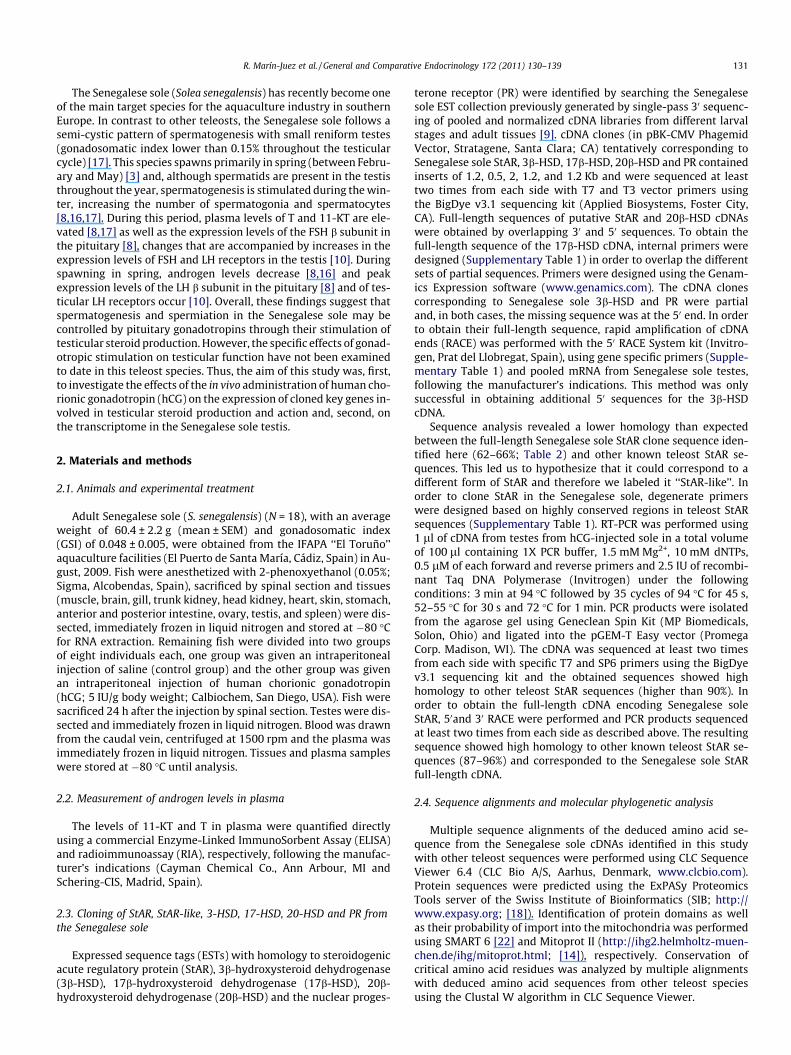
Fig. 1. Senegalese sole StAR and StAR-like sequence analysis. (A) Multiple alignments of the predicted Senegalese sole StAR and StAR-like proteins with other teleost StARproteins. Identical residues identified using ClustalW are indicated as dots. Grey boxes: Putative amino acid residues directly contributing to the hydrophobic tunnel structure[42]. Box: Conserved phosphorylation motifs for protein kinase A [44]. Underlined: Conserved residues important for the steroidogenic function of StAR in mammals [45].Dashed box: Predicted N-terminal mitochondrial targeted residues. (B) Multiple alignment of the predicted Senegalese sole StAR-like protein with other teleost StAR-likeproteins deduced from genome sequence databases. Danio rerio: ENSDARP00000055742; Gasterosteus aculeatus: ENSGACP00000003019; Oryzias latipes: ENS-ORLP00000011263; Takifugu rubripes: ENSTRUP00000033953; Tetraodon nigroviridis: ENSTNIP00000000275.
134 R. Marín-Juez et al. / General and Comparative Endocrinology 172 (2011) 130–139
sequences (Fig. 2). Furthermore, in contrast to ssStAR-like, ssStARprotein contains the two well-conserved consensus protein kinaseA phosphorylation motifs present in all vertebrate StARs (Fig. 1A).Interestingly, the first 25 amino acids of the N-termini of ssStARand ssStAR-like have characteristics of a mitochondria import
motif, as predicted in silico with MitoProtII [14] (0.97 for ssSTARand 0.95 for ssStAR-like). The resulting full-length sequences wereentered in GenBank with accession numbers HQ392856 for ssStAR,EU921450 for ssStAR-like, FJ786643 for ss3b-HSD, EU921451 forss17b-HSD and EU918603 for ss20b-HSD. The ssPR partial

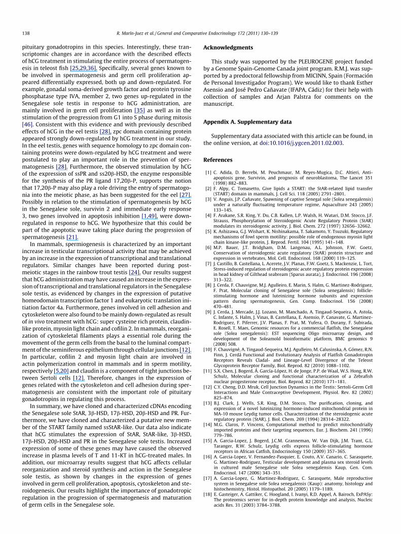
Fig. 2. Unrooted phylogenetic tree of StAR and StAR-like amino acid sequences. The tree was created by UPGMA method using ClustalW multiple alignment and bootstrapped1000 times. The scale for the given branch length indicates 0.600 amino acid substitutions per site. Accession numbers were retrieved from public databases- zebrafishMLN64: AF258786; brook trout MLN64: AF284379; human MLN64: CAA56489; Senegalese sole StAR like: EU921450; chicken: NP_990017; African clawed frog:NP_001167502; sea bream: ABV25959; black bass: AAZ92554; Senegalese sole: HQ392856; Atlantic croaker: ABG34343; medaka: NP_001098380; rainbow trout:NP_001117674; brook trout: Q9DE06; Japanese eel: BAC66210; Atlantic cod: AAP44111; zebrafish: NP_571738; African catfish: ACT36707; pig: NP_998920; cattle: Q28918;human: NP_000340; rat: NP_113746; mouse: NP_035615; Danio rerio: ENSDARP00000055742; Gasterosteus aculeatus: ENSGACP00000003019; Oryzias latipes: ENS-ORLP00000011263; Takifugu rubripes: ENSTRUP00000033953; Tetraodon nigroviridis: ENSTNIP00000000275.
R. Marín-Juez et al. / General and Comparative Endocrinology 172 (2011) 130–139 135
sequence was entered in GenBank with accession numberHQ392857.
3.2. Tissue expression of ssStAR, ssStAR-like, ss3b-HSD, ss17b-HSD,ss20b-HSD and ssPR
The expression pattern of the cloned genes was investigated inSenegalese sole tissues by non-quantitative RT-PCR (Fig. 3). Of thesix genes examined, ssPR, ss20b-HSD and ss17b-HSD wereexpressed in almost all tissues, with the exception of the skin. Onthe other hand, ss3b-HSD, ssStAR and ssStAR-like showed a morerestricted pattern of expression. Of note were the differences be-tween the two forms of StAR, with ssStAR transcripts present onlyin trunk and head kidney, testis and spleen and with ssStAR-liketranscripts present only in gill, heart, ovary, testis and spleen.Importantly, all six genes were expressed in the testis, althoughat different levels. Comparison of the basal expression levels ofthe cloned genes in the adult Senegalese sole testis by qPCR re-vealed that ssPR was the gene most highly expressed, followedby ssStAR-like, ss3b-HSD, ss20b-HSD, ss17b-HSD and ssStAR asthat with the lowest level of expression (Supplementary Fig. 5).
B G TK HK H L Sk St AI PI O TM Sp
PR
20 -HSD
17 -HSD
3 -HSD
StAR-like
StAR
18S
Fig. 3. Tissue expression pattern of the PR, 20b-HSD, 17b-HSD, 3b-HSD, StAR-likeand StAR in adult Senegalese sole. Gene expression was analyzed by RT-PCR usingspecific primers for the cloned genes and for 18S, used as a normalization control.The tissues shown are: muscle (M), brain (B), gill (G), trunk kidney (TK), headkidney (HK), heart (H), liver (L), skin (Sk), stomach (St), anterior intestine (AI),posterior intestine (PI), ovary (O), testis (T) and spleen (Sp) (B).
3.3. Regulation of the expression of StAR, StAR-like, 3b-HSD, 17b-HSD,20b-HSD and PR in the Senegalese sole testis by hCG administrationin vivo
The gonadotropic regulation of the expression of ssStAR,ssStAR-like, ss3b-HSD, ss17b-HSD, ss20b-HSD and ssPR in theSenegalese sole testis was examined in vivo by administeringhCG for lack of available homologous gonadotropins. At 24 h afterhCG treatment, a statistically significant stimulation (p < 0.05) ofssStAR, ssStAR-like, ss3b-HSD, ss17b-HSD, ss20b-HSD and ssPRexpression levels was detected in the Senegalese sole testis(Fig. 4). qPCR analyses evidenced inductions by the hCG treatmentbetween 5.4 and 23.2-fold over the saline-injected controls forssStAR-like, ss3b-HSD, ss17b-HSD and ssPR (Fig. 4A–E). Interest-ingly, ssStAR was strongly stimulated in response to the treatmentwith hCG, showing approximately a 160 fold increase over controls(Fig. 4F).
3.4. Effects of in vivo hCG administration on plasma androgen levels
Plasma levels of T and 11-KT were increased in response tothe in vivo administration of hCG. Plasma T levels in the hCG-in-jected group were approximately 5-fold higher than those in thesaline-injected group (1.6 ± 0.5 ng/ml and 0.3 ± 0.9 ng/ml, respec-tively) (Fig. 5A). The increase of 11-KT levels in plasma in thehCG-treated group was stronger than that of T, with hCG causingapproximately a 38-fold increase over the controls (103.3 ±52.1 ng/ml and 2.7 ± 0.5 ng/ml, respectively) (Fig. 5B). For bothandrogens, the variation within the hCG-injected group was lar-ger than within the saline-injected group, similar to what wasobserved at the level of expression of the cloned testicular genes(Fig. 4).
3.5. Transcriptomic effects of in vivo hCG administration on theSenegalese sole testis
Changes in gene expression in response to hCG treatment in theSenegalese sole testis were assessed by microarray analysis using aspecific oligonucleotide platform previously validated for thisspecies [9,40]. Our analysis resulted in the identification of 90differentially expressed genes (46 up-regulated and 44 down-regulated) in the Senegalese sole testis in response to hCG

05
10152025
hCG
*
02468
10
hCG
*
SalineSaline
0
5
10
15
20
hCG
*
Saline0
10
20
30
40
hCG
*
Saline
012345678
hCG
*
Saline0
50100150200250300
hCG
*
Saline
PR
(Fol
d ch
ange
)
20-H
SD(F
old
chan
ge)
3-H
SD
(Fol
d ch
ange
)
StAR
-like
(F
old
chan
ge)
StAR
(F
old
chan
ge)
A B
C D
E F
17-H
SD
(Fol
d ch
ange
)
Fig. 4. Regulation of the PR, 20b-HSD, 17b-HSD, 3b-HSD, StAR-like and StAR expression in the Senegalese sole testis by hCG treatment in vivo. The expression of the PR, 20b-HSD, 17b-HSD, 3b-HSD, StAR-like and StAR was analyzed by qPCR and quantified by the absolute quantitation method using cloned PR, 20b-HSD, 17b-HSD, 3b-HSD, StAR-likeand StAR cDNA fragments as standard. Data are expressed relative to control, saline-injected fish (mean ± S.E.). Asterisks (⁄) indicate significant differences (p 6 0.05).
136 R. Marín-Juez et al. / General and Comparative Endocrinology 172 (2011) 130–139
treatment. Some of the up-regulated transcripts (Table 2;Supplementary Table 2) corresponded to genes involved in steroido-genesis (e.g. StAR-like and PR), spermatogenesis and primordialgerm cell proliferation (e.g. gonadal soma-derived growth factorand protein tyrosine phosphatase type IVA, member 2) and tran-scriptional and translational regulation (e.g. putative homeodomaintranscription factor 1 and eukaryotic translation initiation factor 4a).Other up-regulated genes included ATPase Na+/K + transportingalpha 1a, betaine-homocysteine methyltransferase, ubiquitin-activating enzyme E1, transferrin receptor 1b, hemogoblin alphaembryonic-1, glycine amidinotransferase and super cysteine richprotein (Supplementary Table 2). On the other hand, hCG stimula-tion resulted in the down-regulation (Table 2; Supplementary Table3) of genes involved in apoptosis inhibition (e.g. survivin 2 andimmediate early response 3), sperm binding (e.g. zona pellucida gly-coprotein), transcriptional regulation (e.g. jun B proto-oncogene,small nuclear RNA auxiliary factor1 and general transcription factorIIIA) and cytoskeleton (e.g. claudin-like protein, myosine light chainand muscle cofilin 2). Furthermore, genes such as NAD(P)H dehydro-genase, inositol(myo)-1(or 4)-monophosphatase 1, histone H2A,zygote arrest, metallothionein-2, ferritin middle subunit andpolyglutamine binding protein1-like were also down-regulated(Supplementary Table 2).
In order to validate the microarray results, qPCR analyses wereconducted for three up-regulated genes (ATPase Na+/K + transport-ing alpha 1a, eukaryotic translation initiation factor 4A and proteintyrosine phosphatase type IVA, member 2) and two down-regu-lated genes (zpc domain and claudin-like). Additionally, up-regula-tion of StAR-like and PR expression by microarray analysis (Table2; Supplementary Table 2) was validated by our results on theexpression of these two genes in testes from hCG-treated fish byqPCR (Fig. 4). As shown in Table 3, the expression of all seven genesfollowed the same pattern whether evaluated by microarray orqPCR.
4. Discussion
In the present study, we have cloned full-length cDNAs from theSenegalese sole that encode proteins with high sequence homologyto StAR, 3b-HSD, 17b-HSD and 20b-HSD from other teleost fish andmammals. Phylogenetic analyses of these four proteins clearlyindicate that they correspond to the Senegalese sole homologs ofStAR, 3b-HSD, 17b-HSD and 20b-HSD. In addition, we identified apartial cDNA for the Senegalese sole PR with high homology tothe nuclear receptor for 17,20b-P described in the eel and in otherteleost species [11,41].
In this study we also have cloned a full-length cDNA encoding aprotein with a 60% sequence homology to StAR that we havenamed ssStAR-like. As with ssStAR, the deduced ssStAR-likeprotein has a conserved C-terminal START domain containinghydrophobic residues that contribute to forming a hydrophobictunnel structure [42] and residues considered to be relevant forthe steroidogenic function of StAR in mammals [45], all well con-served in teleost StARs [19] (Fig. 1A). Furthermore, similar tossStAR, the N-terminus of ssStAR-like has a domain composed ofbasic and hydrophobic amino acids that is characteristic of mito-chondrial targeting sequences [13,23], suggesting that ssStAR-likemay be imported into the mitochondria. However, in contrast tossStAR, ssStAR-like lacks the two conserved PKA phosphorylationmotifs but, surprisingly, conserves Ser196 (Ser195 in mammalianStAR). Mutation of this residue to a non-phosphorylatable alanineresidue reduced the steroidogenic activity of mammalian StARmore than 50%, confirming this as the only functionally significantphosphorylation site [4]. At the present time, we do not know thefunctional role of Ser196 in ssStAR-like but it is possible that itsphosphorylation state may be modulated by kinases other thanPKA. Overall, these observations suggest that ssStAR-like couldhave a role in cholesterol transport into the mitochondria, as hasbeen described for StAR, and may be involved in testicular steroi-

Table 3Real-time qPCR validation of differential expressed genes in the Senegalese sole testisin response to in vivo treatment with human chorionic gonadotropin.
Gene name Microarray qPCR
StAR-like +4.39 +5.39ATPase, NA+/K + transporting 1a polypeptide +3.80 +6.76Eukaryotic translation initiation factor4A +1.76 +2.70Progesterone receptor +1.69 +13.51Protein tyrosine phosphatase type IVA, member 2 +1.68 +2.80Zpc domain containing protein 5 �2.53 �7.37Claudin-like protein �1.93 �1.13
Data shown as fold change with respect to the control group.
0.0
0.5
1.0
1.5
2.0
2.5
Saline hCG
0
20
40
60
80
100
120
140
160
180
Saline hCG
*
A
B
11-K
etot
esto
ster
one
(ng/
ml)
Test
oste
rone
(ng/
ml)
Fig. 5. Effects of in vivo hCG administration on androgen plasma levels in maleSenegalese sole. (A) Testosterone and (B) 11-Ketotestosterone levels in plasma wereanalyzed by RIA and ELISA respectively. N = 4 for saline and N = 6 for hCG injection.Data are shown as mean ± S.E. Asterisks (⁄) indicate significant differences (p 6 0.05).
R. Marín-Juez et al. / General and Comparative Endocrinology 172 (2011) 130–139 137
dogenesis as it is highly expressed in the testis. Recently, Chau-vigné et al. [10] showed by in situ hybridization that StAR is ex-pressed in Leydig cells of the Senegalese sole testis; however,since the probe used was generated based on the same EST clonewith homology to StAR that has been renamed here to ssStAR-like,we suggest that ssStAR-like transcripts may be present in Leydigcells. On the other hand, several pieces of evidence support the rea-soning that ssStAR-like may be a different form of StAR and that itcould represent a new member of the STARD1/StAR group or sub-family of intracellular cholesterol carriers [2]. Firstly, phylogeneticanalyses placed the ssStAR-like protein, as well as other teleostStAR-like proteins identified in silico, between the vertebrate StARtree and the related MLN64 tree. A sequence comparison of thessStAR-like protein with all 15 known members of the START fam-ily in humans only revealed homologies higher than 50% with
MLN64 (55%) and StAR (64%) (data not shown). Secondly, as statedabove, ssStAR contains a mitochondrial targeting motif which inthe large family of START proteins is found only in the STARD1/StAR subfamily. Thirdly, ssStAR-like has a different tissue expres-sion pattern than ssStAR, with transcripts present in gill and heartbut not in head or trunk kidney, contrary to ssStAR. ssStAR wasexpressed in trunk kidney, head kidney, and testis in agreementwith previous studies where StAR was shown to be mainly ex-pressed in steroidogenic tissues [6,7,19,23]. Therefore, ssStAR-likeand ssStAR have in similar a rather restricted and gene-specificpattern of expression, with both transcripts present in the testisalbeit more abundantly for the former than the later.
In teleosts, like in other vertebrates, the progression of sper-matogenesis and the production of testicular steroids are underthe control of pituitary gonadotropins [34,37]. In male Senegalesesole, the expression levels of FSH and LH in the pituitary appearto be temporally related to elevated androgen plasma levels andincreased spermatozoa production, respectively, [8]. For this rea-son, it has been postulated that in the Senegalese sole, like in otherteleosts, FSH may be involved in the regulation of spermatogenesiswhereas LH may be involved in the regulation of sperm maturationand spermiation [8,34]. Recently, FSH and LH receptors have beenidentified in the Senegalese sole testis, with FSH receptors ex-pressed in Sertoli cells and LH receptors expressed in Leydig cellsand in spermatids [10]. The presence of LH receptors in steroido-genic Leydig cells is consistent with the hypothesis that gonadotro-pin action through the LH receptors may result in the stimulationof the production of testicular steroids, as shown in several teleostspecies [34]. However, to date there is no data on the biological ef-fects of gonadotropins on testicular function in the Senegalese sole.Due to the lack of homologous Senegalese sole gonadotropins, wehave studied the effects of hCG administration in males of this spe-cies. hCG is an analog of LH with known steroidogenic activity inteleosts and recently shown to be able to interact with Senegalesesole FSH and LH receptors heterologously expressed in Xenopus oo-cytes [10]. In this study, we show that hCG treatment in vivo signif-icantly increased the expression of ssStAR, ssStAR-like, ss3b-HSD,ss17b-HSD, ss20b-HSD and ssPR in the Senegalese sole testis andthe plasma levels of T and 11-KT. Our data clearly support thehypothesis that testicular androgen production in the Senegalesesole may be stimulated by pituitary gonadotropins through thestimulation of the expression of genes involved in mitochondrialcholesterol transport (ssStAR and ssStAR-like) and androgensynthesis (ss3b-HSD and ss17b-HSD). Interestingly, ssStAR-likeexpression in the testis increased as a result of hCG in vivo admin-istration, suggesting that ssStAR-like, shown to be expressed inLeydig cells [10], may contribute to the gonadotropin-stimulatedandrogen production in the Senegalese sole testis. However, addi-tional studies are needed to understand the function of ssStAR-likeand its implication in steroidogenesis. The reported presence ofssStAR-like transcripts [10] and 3b-HSD activity [17] in Leydig cellsfrom the Senegalese sole testis, coupled with the stimulation of theexpression of steroidogenic genes and of androgen plasma levelsby hCG administration, support the idea that the cellular targetfor hCG in the Senegalese sole testis may be the Leydig cell.Although we did not measure 17,20b-P in plasma, the increase inss20b-HSD and ssPR expression by hCG administration would sug-gest that pituitary gonadotropins could also be stimulating the tes-ticular production of the maturation-inducing steroid and itsreceptor. This, in turn, would argue for a possible role of 17,20b-P in sperm maturation and hydration and acquisition of spermmotility, as has been shown in other teleost species [27,30,43].
By performing transcriptome analyses using a Senegalese soleoligonucleotide microarray [9], we have identified 90 genes thatshowed differential expression in the testis in response to hCGtreatment and that, therefore, could be potential target genes for
138 R. Marín-Juez et al. / General and Comparative Endocrinology 172 (2011) 130–139
pituitary gonadotropins in this species. Interestingly, these tran-scriptomic changes are in accordance with the described effectsof hCG treatment in stimulating the entire process of spermatogen-esis in teleost fish [25,29,36]. Specifically, several genes known tobe involved in spermatogenesis and germ cell proliferation ap-peared differentially expressed, both up and down-regulated. Forexample, gonadal soma-derived growth factor and protein tyrosinephosphatase type IVA, member 2, two genes up-regulated in theSenegalese sole testis in response to hCG administration, aremainly involved in germ cell proliferation [35] as well as in thestimulation of the progression from G1 into S phase during mitosis[46]. Consistent with this evidence and with previously describedeffects of hCG in the eel testis [28], zpc domain containing proteinappeared strongly down-regulated by hCG treatment in our study.In the eel testis, genes with sequence homology to zpc domain con-taining proteins were down-regulated by hCG treatment and werepostulated to play an important role in the prevention of sper-matogenesis [28]. Furthermore, the observed stimulation by hCGof the expression of ssPR and ss20b-HSD, the enzyme responsiblefor the synthesis of the PR ligand 17,20b-P, supports the notionthat 17,20b-P may also play a role driving the entry of spermatogo-nia into the meiotic phase, as has been suggested for the eel [27].Possibly in relation to the stimulation of spermatogenesis by hCGin the Senegalese sole, survivin 2 and immediate early response3, two genes involved in apoptosis inhibition [1,49], were down-regulated in response to hCG. We hypothesize that this could bepart of the apoptotic wave taking place during the progression ofspermatogenesis [21].
In mammals, spermiogenesis is characterized by an importantincrease in testicular transcriptional activity that may be achievedby an increase in the expression of transcriptional and translationalregulators. Similar changes have been reported during post-meiotic stages in the rainbow trout testis [24]. Our results suggestthat hCG administration may have caused an increase in the expres-sion of transcriptional and translational regulators in the Senegalesesole testis, as evidenced by changes in the expression of putativehomeodomain transcription factor 1 and eukaryotic translation ini-tiation factor 4a. Furthermore, genes involved in cell adhesion andcytoskeleton were also found to be mainly down-regulated as resultof in vivo treatment with hCG: super cysteine rich protein, claudin-like protein, myosin light chain and cofilin 2. In mammals, reorgani-zation of cytoskeletal filaments plays a essential role during themovement of the germ cells from the basal to the luminal compart-ment of the seminiferous epithelium through cellular junctions [12].In particular, cofilin 2 and myosin light chain are involved inactin polymerization control in mammals and in sperm motility,respectively [5,20] and claudin is a component of tight junctions be-tween Sertoli cells [12]. Therefore, changes in the expression ofgenes related with the cytoskeleton and cell adhesion during sper-matogenesis are consistent with the important role of pituitarygonadotropins in regulating this process.
In summary, we have cloned and characterized cDNAs encodingthe Senegalese sole StAR, 3b-HSD, 17b-HSD, 20b-HSD and PR. Fur-thermore, we have cloned and characterized a putative new mem-ber of the START family named ssStAR-like. Our data also indicatethat hCG stimulates the expression of StAR, StAR-like, 3b-HSD,17b-HSD, 20b-HSD and PR in the Senegalese sole testis. Increasedexpression of some of these genes may have caused the observedincrease in plasma levels of T and 11-KT in hCG-treated males. Inaddition, our microarray results suggest that hCG affects cellularreorganization and steroid synthesis and action in the Senegalesesole testis, as shown by changes in the expression of genesinvolved in germ cell proliferation, apoptosis, cytoskeleton and ste-roidogenesis. Our results highlight the importance of gonadotropicregulation in the progression of spermatogenesis and maturationof germ cells in the Senegalese sole.
Acknowledgments
This study was supported by the PLEUROGENE project fundedby a Genome Spain-Genome Canada joint program. R.M.J. was sup-ported by a predoctoral fellowship from MICINN, Spain (Formaciónde Personal Investigador Program). We would like to thank EstherAsensio and José Pedro Cañavate (IFAPA, Cádiz) for their help withcollection of samples and Arjan Palstra for comments on themanuscript.
Appendix A. Supplementary data
Supplementary data associated with this article can be found, inthe online version, at doi:10.1016/j.ygcen.2011.02.003.
References
[1] C. Adida, D. Berrebi, M. Peuchmaur, M. Reyes-Mugica, D.C. Altieri, Anti-apoptosis gene, Survivin, and prognosis of neuroblastoma, The Lancet 351(1998) 882–883.
[2] F. Alpy, C. Tomasetto, Give lipids a START: the StAR-related lipid transfer(START) domain in mammals, J. Cell Sci. 118 (2005) 2791–2801.
[3] V. Anguis, J.P. Cañavate, Spawning of captive Senegal sole (Solea senegalensis)under a naturally fluctuating temperature regime, Aquaculture 243 (2005)133–145.
[4] F. Arakane, S.R. King, Y. Du, C.B. Kallen, L.P. Walsh, H. Watari, D.M. Stocco, J.F.Strauss, Phosphorylation of Steroidogenic Acute Regulatory Protein (StAR)modulates its steroidogenic activity, J. Biol. Chem. 272 (1997) 32656–32662.
[5] K. Ashizawa, G.J. Wishart, K. Nishinakama, T. Sakamoto, Y. Tsuzuki, Regulatorymechanisms of fowl sperm motility: possible role of endogenous myosin lightchain kinase-like protein, J. Reprod. Fertil. 104 (1995) 141–148.
[6] M.P. Bauer, J.T. Bridgham, D.M. Langenau, A.L. Johnson, F.W. Goetz,Conservation of steroidogenic acute regulatory (StAR) protein structure andexpression in vertebrates, Mol. Cell. Endocrinol. 168 (2000) 119–125.
[7] J. Castillo, B. Castellana, L. Acerete, J.V. Planas, F.W. Goetz, S. Mackenzie, L. Tort,Stress-induced regulation of steroidogenic acute regulatory protein expressionin head kidney of Gilthead seabream (Sparus aurata), J. Endocrinol. 196 (2008)313–322.
[8] J. Cerda, F. Chauvigne, M.J. Agulleiro, E. Marin, S. Halm, G. Martinez-Rodriguez,F. Prat, Molecular cloning of Senegalese sole (Solea senegalensis) follicle-stimulating hormone and luteinizing hormone subunits and expressionpattern during spermatogenesis, Gen. Comp. Endocrinol. 156 (2008)470–481.
[9] J. Cerda, J. Mercade, J.J. Lozano, M. Manchado, A. Tingaud-Sequeira, A. Astola,C. Infante, S. Halm, J. Vinas, B. Castellana, E. Asensio, P. Canavate, G. Martinez-Rodriguez, F. Piferrer, J.V. Planas, F. Prat, M. Yufera, O. Durany, F. Subirada,E. Rosell, T. Maes, Genomic resources for a commercial flatfish, the Senegalesesole (Solea senegalensis): EST sequencing Oligo microarray design, anddevelopment of the Soleamold bioinformatic platform, BMC genomics 9(2008) 508.
[10] F. Chauvigné, A. Tingaud-Sequeira, M.J. Agulleiro, M. Calusinska, A. Gómez, R.N.Finn, J. Cerdá Functional and Evolutionary Analysis of Flatfish GonadotropinReceptors Reveals Cladal- and Lineage-Level Divergence of the TeleostGlycoprotein Receptor Family, Biol. Reprod. 82 (2010) 1088–1102.
[11] S.X. Chen, J. Bogerd, Á. García-López, H. de Jonge, P.P. de Waal, W.S. Hong, R.W.Schulz, Molecular cloning and functional characterization of a Zebrafishnuclear progesterone receptor, Biol. Reprod. 82 (2010) 171–181.
[12] C.Y. Cheng, D.D. Mruk, Cell Junction Dynamics in the Testis: Sertoli-Germ CellInteractions and Male Contraceptive Development, Physiol. Rev. 82 (2002)825–874.
[13] B.J. Clark, J. Wells, S.R. King, D.M. Stocco, The purification, cloning, andexpression of a novel luteinizing hormone-induced mitochondrial protein inMA-10 mouse Leydig tumor cells. Characterization of the steroidogenic acuteregulatory protein (StAR), J. Biol. Chem. 269 (1994) 28314–28322.
[14] M.G. Claros, P. Vincens, Computational method to predict mitochondriallyimported proteins and their targeting sequences, Eur. J. Biochem. 241 (1996)779–786.
[15] A. Garcia-Lopez, J. Bogerd, J.C.M. Granneman, W. Van Dijk, J.M. Trant, G.L.Taranger, R.W. Schulz, Leydig cells express follicle-stimulating hormonereceptors in African Catfish, Endocrinology 150 (2009) 357–365.
[16] A. Garcia-Lopez, V. Fernandez-Pasquier, E. Couto, A.V. Canario, C. Sarasquete,G. Martinez-Rodriguez, Testicular development and plasma sex steroid levelsin cultured male Senegalese sole Solea senegalensis Kaup, Gen. Com.Endocrinol. 147 (2006) 343–351.
[17] A. Garcia-Lopez, G. Martinez-Rodriguez, C. Sarasquete, Male reproductivesystem in Senegalese sole Solea senegalensis (Kaup): anatomy, histology andhistochemistry, Histol. Histopathol. 20 (2005) 1179–1189.
[18] E. Gasteiger, A. Gattiker, C. Hoogland, I. Ivanyi, R.D. Appel, A. Bairoch, ExPASy:The proteomics server for in-depth protein knowledge and analysis, Nucleicacids Res. 31 (2003) 3784–3788.

R. Marín-Juez et al. / General and Comparative Endocrinology 172 (2011) 130–139 139
[19] F.W. Goetz, B. Norberg, L.A.R. McCauley, D.B. Iliev, Characterization of the cod(Gadus morhua) steroidogenic acute regulatory protein (StAR) sheds light onStAR gene structure in fish, Comp. Biochem. Physiol Part B: Biochem. Mol. Biol.137 (2004) 351–362.
[20] T.Y. Huang, C. DerMardirossian, G.M. Bokoch, Cofilin phosphatases andregulation of actin dynamics, Curr. Opin. Cell Biol. 18 (2006) 26–31.
[21] J. Kwon, K. Mochida, Y.-L. Wang, S. Sekiguchi, T. Sankai, S. Aoki, A. Ogura, Y.Yoshikawa, K. Wada, Ubiquitin c-terminal hydrolase L-1 is essential for theearly apoptotic wave of germinal cells and for sperm quality control duringspermatogenesis, Biol. Reprod. 73 (2005) 29–35.
[22] I. Letunic, T. Doerks, P. Bork, S.M.A. RT, SMART 6: recent updates and newdevelopments, Nucleic Acids Res. 37 (Suppl. 1) (2008) D229–D232.
[23] Y.-Y. Li, K. Inoue, Y. Takei, Steroidogenic acute regulatory protein in eels: cDNAcloning and effects of ACTH and seawater transfer on its mRNA expression,Zool. Sci. 20 (2003) 211–219.
[24] D. Mazurais, J. Montfort, C. Delalande, F.L. Gac, Transcriptional analysis oftestis maturation using trout cDNA macroarrays, Gen. Comp. Endocrinol. 142(2005) 143–154.
[25] C. Miura, T. Miura, N. Kudo, M. Yamashita, K. Yamauchi, CDNA cloning of astage-specific gene expressed during HCG-induced spermatogenesis in theJapanese eel, Develop. Growth. Differ. 41 (1999) 463–471.
[26] C. Miura, T. Miura, M. Yamashita, K. Yamauchi, Y. Nagahama, Hormonalinduction of all stages of spermatogenesis in germ-somatic cell coculture fromimmature Japanese eel testis, Develop. Growth. Differ. 38 (1996) 257–262.
[27] T. Miura, M. Higuchi, Y. Ozaki, T. Ohta, C. Miura, Progestin is an essential factorfor the initiation of the meiosis in spermatogenetic cells of the eel, Proc. Natl.Acad. Sci. USA 103 (2006) 7333–7338.
[28] T. Miura, N. Kudo, C. Miura, K. Yamauchi, Y. Nagahama, Two testicular cDNAclones suppressed by gonadotropin stimulation exhibit ZP2- and ZP3-likestructures in Japanese eel, Mol. Reprod. Develop. 51 (1998) 235–242.
[29] T. Miura, K. Yamauchi, H. Takahashi, Y. Nagahama, Hormonal induction of allstages of spermatogenesis in vitro in the male Japanese eel (Anguilla japonica),Proc. Natl. Acad. Sci. USA 88 (1991) 5774–5778.
[30] T. Miura, K. Yamauchi, H. Takahashi, Y. Nagahama, The role of hormones in theacquisition of sperm motility in salmonid fish, J. Exp. Zool. 261 (1992) 359–363.
[31] S. Miwa, L. Yan, P. Swanson, Localization of two gonadotropin receptors in thesalmon gonad by in vitro ligand autoradiography, Biol. Reprod. 50 (1994) 629–642.
[32] Y. Nagahama, Endocrine regulation of gametogenesis in fish, Int. J. Develop.Biol. 38 (1994) 217–229.
[33] T. Ohta, H. Miyake, C. Miura, H. Kamei, K. Aida, T. Miura, Follicle-StimulatingHormone Induces Spermatogenesis Mediated by Androgen Production inJapanese Eel, Anguilla japonica, Biol. Reprod. 77 (2007) 970–977.
[34] J.V. Planas, P. Swanson, Physiological function of Gonadotropins in fish, in: M.J.Rocha, A. Arukwe, B.G. Kapoor (Eds.), Fish Reproduction, Science Publishers,USA, 2007, pp. 37–66.
[35] E. Sawatari, S. Shikina, T. Takeuchi, G. Yoshizaki, A novel transforming growthfactor-[beta] superfamily member expressed in gonadal somatic cellsenhances primordial germ cell and spermatogonial proliferation in rainbowtrout (Oncorhynchus mykiss), Dev. Biol. 301 (2007) 266–275.
[36] R. Schiavone, L. Zilli, S. Vilella, C. Fauvel, Human chorionic gonadotropininduces spermatogenesis and spermiation in 1-year-old European sea bass(Dicentrarchus labrax): Assessment of sperm quality, Aquaculture 255 (2006)522–531.
[37] R.W. Schulz, L.R. de França, J.-J. Lareyre, F. LeGac, H. Chiarini-Garcia, R.H.Nobrega, T. Miura, Spermatogenesis in fish, Gen. Comp. Endocrinol. 165 (2010)390–411.
[38] R.W. Schulz, H.J.T. Goos, Puberty in male fish: concepts and recentdevelopments with special reference to the African catfish (Clariasgariepinus), Aquaculture 177 (1999) 5–12.
[39] P.H.A. Sneath, R.R. Sokal, Numerical taxonomy: the principles and practice ofnumerical classification, San Francisco: Freeman (1972).
[40] A. Tingaud-Sequeira, F. Chauvigne, J. Lozano, M. Agulleiro, E. Asensio, J. Cerda,New insights into molecular pathways associated with flatfish ovariandevelopment and atresia revealed by transcriptional analysis, BMC Genomics10 (2009) 434.
[41] T. Todo, T. Ikeuchi, T. Kobayashi, H. Kajiura-Kobayashi, K. Suzuki, M.Yoshikuni, K. Yamauchi, Y. Nagahama, Characterization of a testicular17alpha, 20beta-dihydroxy-4-pregnen-3-one (a spermiation-inducingsteroid in fish) receptor from a teleost, Japanese eel (Anguilla japonica),FEBS letters 465 (2000) 12–17.
[42] Y. Tsujishita, J.H. Hurley, Structure and lipid transport mechanism of a StAR-related domain, Nat. Struct. Mol. Biol. 7 (2000) 408–414.
[43] H. Ueda, A. Kambegawa, Y. Nagahama, Involvement of gonadotrophin andsteroid hormones in spermiation in the amago salmon, Oncorhynchusrhodurus, and goldfish, Carassius auratus, Gen. Comp. Endocrinol. 59 (1985)24–30.
[44] X. Wang, Z. Liu, S. Eimerl, R. Timberg, A.M. Weiss, J. Orly, D.M. Stocco,Effect of truncated forms of the steroidogenic acute regulatory protein onintramitochondrial cholesterol transfer, Endocrinology 139 (1998) 3903–3912.
[45] H. Watari, F. Arakane, C. Moog-Lutz, C.B. Kallen, C. Tomasetto, G.L. Gerton, M.-C. Rio, M.E. Baker, J.F. Strauss, MLN64 contains a domain with homology to thesteroidogenic acute regulatory protein (StAR) that stimulates steroidogenesis,Proc. Natl. Acad. Sci. USA 94 (1997) 8462–8467.
[46] S.R. Werner, P.A. Lee, M.W. DeCamp, D.N. Crowell, S.K. Randall, P.L. Crowell,Enhanced cell cycle progression and down regulation of p21Cip1/Waf1 by PRLtyrosine phosphatases, Cancer Lett. 202 (2003) 201–211.
[47] J.A. Whelan, N.B. Russell, M.A. Whelan, A method for the absolutequantification of cDNA using real-time PCR, J. Immunol. Methods 278 (2003)261–269.
[48] C. Workman, L.J. Jensen, H. Jarmer, R. Berka, L. Gautier, H.B. Nielser, H.H. Saxild,C. Nielsen, S. Brunak, S. Knudsen, A new non-linear normalization method forreducing variability in DNA microarray experiments, Genome Biol. 3 (2002)96–99.
[49] M.X. Wu, Z. Ao, K.V. Prasad, S. nbsp, R. Wu, S.F. Schlossman, IEX-1L, anApoptosis Inhibitor Involved in NF-{kappa}B-Mediated Cell Survival, Science281 (1998) 998–1001.