Disconnection as a mechanism for cognitive dysfunction in multiple sclerosis
11
Disconnection as a mechanism for cognitive dysfunction in multiple sclerosis R. A. Dineen, 1 J. Vilisaar, 2 J. Hlinka, 1 C. M. Bradshaw, 3 P. S. Morgan, 1 C. S. Constantinescu 2 and D.P. Auer 1 1 Department of Academic Radiology, 2 Department of Clinical Neurology and 3 Department of Psychiatry, University of Nottingham, Nottingham, UK Correspondence to: Dr R. A. Dineen, BMBS, MRCP, FRCR, Department of Academic Radiology, University of Nottingham, Queen’s Medical Centre, Derby Road, Nottingham NG7 2UH, UK E-mail: [email protected] Disconnection of cognitively important processing regions by injury to the interconnecting white matter pro- vides a potential mechanism for cognitive dysfunction in multiple sclerosis. The contribution of tract-specific white matter injury to dysfunction in different cognitive domains in patients with multiple sclerosis has not pre- viously been studied.We apply tract-based spatial statistics (TBSS) to diffusion tensor imaging (DTI) in a cohort of multiple sclerosis patients to identify loci where reduced white matter tract fractional anisotropy (FA) pre- dicts impaired performance in cognitive testing. Thirty-seven multiple sclerosis patients in remission (median age 43.5 years; Expanded Disability Status Scale range 1.5^6.5; 35 relapsing remitting, two secondary-progres- sive) underwent 3 T MRI including high-resolution DTI. Multiple sclerosis patients underwent formal testing of performance in multiple cognitive domains. Normalized cognitive scores were used for voxel-wise statistical analysis using TBSS, while treating age as a covariate of no interest. Permutation-based inference on cluster size (t ` 2, P _0.05 corrected) was used to correct for multiple comparisons. Statistical mapping revealed dif- ferential patterns of FA reduction for tests of sustained attention, working memory and processing speed, visual working memory and verbal learning and recall. FA was not associated with frontal lobe function or visuospatial perception. Cognitively relevant tract localizations only partially overlapped with areas of high FLAIR lesion probability, confirming the contribution of normal-appearing white matter abnormality to cognitive dysfunc- tion. Of note, tract localizations showing significant associations with cognitive impairment were found to inter- connect cortical regions thought to be involved in processing in these cognitive domains, or involve possible compensatory processing pathways.This suggests that TBSS reveals functionally relevant tract injury underlying cognitive dysfunction in patients with multiple sclerosis. Keywords: multiple sclerosis; cognitive impairment; magnetic resonance imaging; diffusion tensor imaging; disconnection Abbreviations: BA = Brodmann area; CIS = clinically isolated syndrome; COWAT = Controlled Oral Word Association Test; DTI = diffusion tensor imaging; FA = fractional anisotropy; MRS = magnetic resonance spectroscopy; MTI = magnetization transfer imaging; NAWM = normal-appearing white matter; PASAT = Paced Auditory Serial Addition Test; ROI = region of interest; TBSS = tract-based spatial statistics. Received April 6, 2008. Revised September 4, 2008. Accepted October 1, 2008 Introduction Cognitive dysfunction occurs in 43–65% of patients with multiple sclerosis and most commonly manifests as disturbances in recent memory, sustained attention, verbal fluency, conceptual reasoning and visuospatial perception (Rao et al., 1991). The pattern of cognitive decline is not uniform, and disease duration and physical disability correlate only weakly with cognitive impairment (Ron et al., 1991). The mechanism underlying cognitive impair- ment in multiple sclerosis has not been fully elucidated. In recent years there has been renewed interest in the role of disconnection in the aetiology of higher brain dysfunc- tion (Catani and ffytche, 2005). In this context disconnec- tion may occur between cognitively important cortical or subcortical regions, either at the level of interconnecting white matter, or in cortical relays within association cortex. Quantitative imaging techniques, such as diffusion tensor imaging (DTI), have allowed study of the substrates under- pinning models of connectivity, and the disconnection paradigm has broadened to include neurodevelopmental doi:10.1093/brain/awn275 Brain (2008) Page 1 of 11 ß The Author (2008). Published by Oxford University Press on behalf of the Guarantors of Brain. All rights reserved. For Permissions, please email: [email protected] Brain Advance Access published October 25, 2008 by guest on May 9, 2014 http://brain.oxfordjournals.org/ Downloaded from
-
Upload
independent -
Category
Documents
-
view
1 -
download
0
Transcript of Disconnection as a mechanism for cognitive dysfunction in multiple sclerosis
Disconnection as a mechanism for cognitivedysfunction in multiple sclerosisR. A. Dineen,1 J.Vilisaar,2 J. Hlinka,1 C. M. Bradshaw,3 P. S. Morgan,1 C. S. Constantinescu2 and D. P. Auer1
1Department of Academic Radiology, 2Department of Clinical Neurology and 3Department of Psychiatry, University ofNottingham, Nottingham, UK
Correspondence to: Dr R. A. Dineen, BMBS, MRCP, FRCR, Department of Academic Radiology, University of Nottingham,Queen’s Medical Centre, Derby Road, Nottingham NG7 2UH,UKE-mail: [email protected]
Disconnection of cognitively important processing regions by injury to the interconnecting white matter pro-vides a potential mechanism for cognitive dysfunction in multiple sclerosis. The contribution of tract-specificwhite matter injury to dysfunction in different cognitive domains in patients with multiple sclerosis has not pre-viously been studied.We apply tract-based spatial statistics (TBSS) to diffusion tensor imaging (DTI) in a cohortof multiple sclerosis patients to identify loci where reduced white matter tract fractional anisotropy (FA) pre-dicts impaired performance in cognitive testing. Thirty-seven multiple sclerosis patients in remission (medianage 43.5 years; Expanded Disability Status Scale range 1.5^6.5; 35 relapsing remitting, two secondary-progres-sive) underwent 3T MRI including high-resolution DTI. Multiple sclerosis patients underwent formal testing ofperformance in multiple cognitive domains. Normalized cognitive scores were used for voxel-wise statisticalanalysis using TBSS, while treating age as a covariate of no interest. Permutation-based inference on clustersize (t` 2, P_0.05 corrected) was used to correct for multiple comparisons. Statistical mapping revealed dif-ferential patterns of FA reduction for tests of sustained attention, workingmemory and processing speed, visualworkingmemory and verbal learning and recall. FAwas not associated with frontal lobe function or visuospatialperception. Cognitively relevant tract localizations only partially overlapped with areas of high FLAIR lesionprobability, confirming the contribution of normal-appearing white matter abnormality to cognitive dysfunc-tion.Of note, tract localizations showing significant associations with cognitive impairment were found to inter-connect cortical regions thought to be involved in processing in these cognitive domains, or involve possiblecompensatory processing pathways.This suggests that TBSS reveals functionally relevant tract injury underlyingcognitive dysfunction in patients with multiple sclerosis.
Keywords: multiple sclerosis; cognitive impairment; magnetic resonance imaging; diffusion tensor imaging; disconnection
Abbreviations: BA=Brodmann area; CIS=clinically isolated syndrome; COWAT=Controlled Oral Word AssociationTest;DTI=diffusion tensor imaging; FA= fractional anisotropy; MRS=magnetic resonance spectroscopy; MTI=magnetizationtransfer imaging; NAWM=normal-appearing white matter; PASAT=Paced Auditory Serial AdditionTest; ROI=region ofinterest; TBSS= tract-based spatial statistics.
Received April 6, 2008. Revised September 4, 2008. Accepted October 1, 2008
IntroductionCognitive dysfunction occurs in 43–65% of patients withmultiple sclerosis and most commonly manifests asdisturbances in recent memory, sustained attention, verbalfluency, conceptual reasoning and visuospatial perception(Rao et al., 1991). The pattern of cognitive decline is notuniform, and disease duration and physical disabilitycorrelate only weakly with cognitive impairment (Ronet al., 1991). The mechanism underlying cognitive impair-ment in multiple sclerosis has not been fully elucidated.
In recent years there has been renewed interest in the roleof disconnection in the aetiology of higher brain dysfunc-tion (Catani and ffytche, 2005). In this context disconnec-tion may occur between cognitively important cortical orsubcortical regions, either at the level of interconnectingwhite matter, or in cortical relays within association cortex.Quantitative imaging techniques, such as diffusion tensorimaging (DTI), have allowed study of the substrates under-pinning models of connectivity, and the disconnectionparadigm has broadened to include neurodevelopmental
doi:10.1093/brain/awn275 Brain (2008) Page 1 of 11
� The Author (2008). Published by Oxford University Press on behalf of the Guarantors of Brain. All rights reserved. For Permissions, please email: [email protected]
Brain Advance Access published October 25, 2008 by guest on M
ay 9, 2014http://brain.oxfordjournals.org/
Dow
nloaded from
and neurodegenerative disorders (Catani, 2006).Given the multifocal white matter pathology that occurs
in multiple sclerosis, it is conceivable that cognitive impair-ment in multiple sclerosis is, at least in part, caused bydisconnection. A disconnectionist model for cognitive imp-airment in multiple sclerosis could explain the involvementof multiple cognitive domains as a series of disconnectionsyndromes affecting different cognitive networks. Further-more, the disconnection model has the appeal that it doesnot exclude other mechanisms shown to contribute tocognitive impairment in multiple sclerosis, such as greymatter pathology (Amato et al., 2004; Morgen et al., 2006).
The relationship between white matter injury andcognitive performance in multiple sclerosis has been studiedpreviously. Frontal lesion volume has been shown to affectperformance in tests of frontal lobe function (Arnett et al.,1994; Foong et al., 1997; Rovaris et al., 1998), and frontaland parietal lesion burden has been shown to correlate withperformance on tests of complex attention and verbalworking memory (Sperling et al., 2001). Additionally, it hasbeen shown that injury to white matter which is not detec-table by conventional MRI [within the so-called normal-appearing white matter (NAWM)] but which can bedetected by techniques such as DTI, magnetization transferimaging (MTI) and magnetic resonance spectroscopycontributes to cognitive dysfunction in multiple sclerosispatients (Rovaris et al., 1998; Pan et al., 2001; Rovaris et al.,2002; Cox et al., 2004;).
Studies attempting to ‘map’ cognitive dysfunction towhite matter injury using a voxel-wise statistical analysis arelimited. Charil et al. (2003) studied a data set of 452multiple sclerosis patients and used statistical parametricmapping to investigate the relationship between lesionlocation and cognitive performance. The use of conven-tional imaging in that study precluded assessment of thecontribution of ultrastructural tissue injury within NAWMto cognition. In addition the mental component of theExpanded Disability Severity Score (EDSS) was used, whichis a subjective, non-specific measure of cognitive function.Despite these limitations a correlation between cognitivedysfunction and lesions at the grey–white matter junctionof the associative, limbic and prefrontal cortices was iden-tified. Ranjeva et al. (2005) studied patients with clinicallyisolated syndromes, finding that poorer performance duringthe Paced Auditory Serial Addition Test (PASAT), a test ofprocessing speed and working memory was associated withMTR-defined abnormalities in the splenium of the corpuscallosum, the right superior longitudinal fasciculus, the leftBrodmann area (BA) 40 and right BA4.
DTI provides a three-dimensional representation of themagnitude of diffusion encapsulating its directional depen-dence. When applied to the brain, DTI is a powerful non-invasive technique for exploring cerebral ultrastructure.Fractional anisotropy (FA), a parameter derived from DTIdata, characterizes the shape of the diffusion tensor ellip-soid within each voxel and hence provides a surrogate
quantification of ultrastuctural fibre organization (Basserand Pierpaoli, 1996). DTI examination of multiple sclerosispatients has revealed reduced FA in plaques, adjacent toplaques and to varying degrees in NAWM (Werring et al.,1999; Guo et al., 2002; Hasan et al., 2005; Kealey et al.,2005). Furthermore, Lin et al. (2007) have shown thatreduced FA in the pyramidal tract NAWM correlates withpyramidal tract lesion burden. This was interpreted as sug-gesting that Wallerian degeneration secondary to macro-scopic multiple sclerosis lesions leads to spatially remote FAreductions in regions connected by white matter tracts.
Tract-based approaches to co-registration have thepotential to improve anatomical specificity when comparedto conventional co-registration and lesion morphometrictechniques. Tract-based spatial statistics (TBSS) (Smithet al., 2006), part of FSL (Smith et al., 2004), is a recentlydescribed technique allowing voxel-wise analysis of multi-subject diffusion tensor data. In TBSS the study group FAdata are projected onto an alignment-invariant tractrepresentation, or ‘mean FA tract skeleton’, followed bythe application of voxel-wise cross-subject statistics
We hypothesize that multiple sclerosis-related cognitivedysfunction results from a series of domain-specific discon-nection phenomena. As such, disruption to critical whitematter tracts will lead to reduced functional connectivitybetween cortico-cortical and cortico-subcortical cognitiveprocessing regions, resulting in impairment to specificcognitive domains. To test this hypothesis, we use TBSS forvoxel-wise statistical mapping of white matter tract injury(as quantified by FA) in a cohort of multiple sclerosispatients compared to control subjects, and then apply theTBSS technique to map white matter tract disruption whichpredicts performance during testing of multiple cognitivedomains in a cohort of patients with multiple sclerosis.
MethodsSubjectsThe study was approved by the National Health Service LocalResearch Ethics Committee for Nottingham and the University ofNottingham Research Ethics Committee. Forty-one patients withdefinite multiple sclerosis between the ages of 31 and 56 years(median 44.3) and 27 healthy controls were prospectively recruitedfrom a local database of relapsing remitting multiple sclerosis(RRMS) patients. At the time of assessment and scanning threepatients were found to have progressed to secondary progressive(SPMS) disease. Multiple sclerosis patients had not experiencedrelapse or required steroid treatment for at least 2 months prior toinclusion, and none were on immunomodulatory therapy. Parti-cipants did not have any other significant neurological, medical,psychiatric or cognitive disorder. All multiple sclerosis patientsunderwent neurological assessment using the EDSS at the time ofparticipation in the study.
Neuropsychological test protocolAll multiple sclerosis patients underwent a detailed neuropsychia-tric test battery based on the Minimal Assessment of Cognitive
Page 2 of 11 Brain (2008) R. A. Dineen et al.
by guest on May 9, 2014
http://brain.oxfordjournals.org/D
ownloaded from
Function in Multiple Sclerosis battery (Benedict et al., 2002). Thetests and versions performed were as follows: ‘Controlled Oral
Word Association Test (COWAT)’, a test of phonetic fluency(Benton et al., 1983); letters F, A and S were used. ‘Benton VisualRetention Test (BVRT)’, a test of visual perception, visual memory
and visuoconstructive ability (Benton, 1974); Form C, adminis-tration A was used and the ‘total correct’ sub-score was used forthe statistical analysis. PASAT, a test of sustained attention,working memory and processing speed; Rao’s adaptation of the
Gronwall administration (Gronwall, 1977; Rao et al., 1991), formA, 3 s stimulus was used. ‘Judgement of Line Orientation (JLO)’, atest of visual perception and spatial processing (Benton et al.,1978); Form V was used. ‘Delis-Kaplan Executive Function Score
Sorting Test (DST)’, a test of executive function (Delis et al.,2001); Card sets 1 and 2 (standard form) were used and the‘confirmed correct sorts’ (DST-CS) and ‘Free sorting description
score’ (DST–DS) sub-scores were used for the statistical analysis.‘California Verbal Learning Test version II (CVLT-II)’, a test ofverbal learning and memory (Delis et al., 2000); the standard formwas used, and the sub-score for ‘Short delay free recall’ (CVLT-II
sdf), a measure of verbal learning and recall was used for thestatistical analysis.
The protocol used varied from the full Minimal Assessment of
Cognitive Function in Multiple Sclerosis battery in that theSymbol Digit Modality Test was not performed, and the BVRTwas used in place of the Brief Visuospatial Memory Test. Inaddition the National Adult Reading Test (NART) was performed
as an estimate of premorbid intelligence. All tests were admin-istered by a single researcher (R.A.D.) during an interview on theday of the MRI scan.
Neuropsychological test scores were expressed as z-scoresderived from published normative data. For the DST andCVLT-II, the normative data used were obtained from theaccompanying test manuals (Delis et al., 2000, 2001). Sources of
normative data for the PASAT (Rao et al., 1991) and BVRT(Youngjohn et al., 1993) are detailed in the references. Full,appropriate normative data were not available for the JLO. Raw
JLO scores were corrected for age and sex according to theadjustments recommended by Benton et al. (1978). NART scorewas converted to the Wechsler Adult Intelligence Scale – RevisedFull-Scale IQ (WAIS-R FSIQ) using the formula of Nelson and
Willison (1991). This was then used to give a predicted score for
the COWAT using the formula of Crawford et al. (1992);
deviation from the predicted score was expressed as a z-score.
MRI protocol and image analysisMRI scanning was performed on a Philips Achieva (Philips,
Eindhoven, NL) at 3 T. All patients underwent axial DTI (Single-shot diffusion weighted EPI, b = 1000 mm2/s, 15 directions,
TE = 56 ms, TR = 9700 ms, 2� 2� 2.5 mm3 voxel size interpolatedto 1� 1� 2.5 mm3 voxels, 45 interleaved slices with no gap, four
averages), axial FLAIR (TR = 11 000 ms, TE = 125 ms, TI = 2800 ms,Matrix 256� 256, slice thickness 2.5 mm/0mm gap, FOV = 256)
and sagittal MPRAGE (TR = 7.5, TE = 2.2, Flip = 8, Matrix256� 256, voxel size = 0.8� 0.8� 0.8 mm, TFE factor = 236,
FOV = 205). Intravenous gadolinium-chelate contrast material
was not administered.Post-processing of diffusion tensor data was performed using
the FSL version 3.3 (Smith et al., 2004). Following eddy
current correction using the FMRIB’s Diffusion Toolbox (FDT),non-brain voxels were extracted using the Brain Extraction Tool
(Smith, 2002) with a brain extraction factor of 0.3. FA maps weregenerated using FDT. Individual FA maps were visually inspected
for the presence of significant residual motion or other artefacts,
which led to the exclusion of four of the multiple sclerosis patientsand two control subjects from the TBSS analysis. The TBSS
registration and tract skeletonization process was performed asdescribed previously (Fig. 1A and B), using a lower threshold of
FA of 0.2 to prevent inclusion of non-skeleton voxels (Smith et al.,2006). The skeletonized, fully non-linearly aligned FA data sets
obtained using the TBSS process were then used for voxel-wisecross-subject statistical analysis.
T2-hyperintense lesion volume measurement was performed onthe FLAIR images using the locally developed NeuRoi software
package (Dr C. Tench, Department of Clinical Neurology,University of Nottingham, UK). This provides a semi-automated
method for defining T2-hyperintense lesions using Sobel edgedetection and non-maximum suppression. Mean lesion probability
distribution images were created by first registering the brain-extracted FLAIR images to MNI152 space, and then applying the
transformation matrix for each individual FLAIR image to thecorresponding masked binarized lesion map. The co-registered
lesion maps were averaged using FSLmaths tools to create a lesion
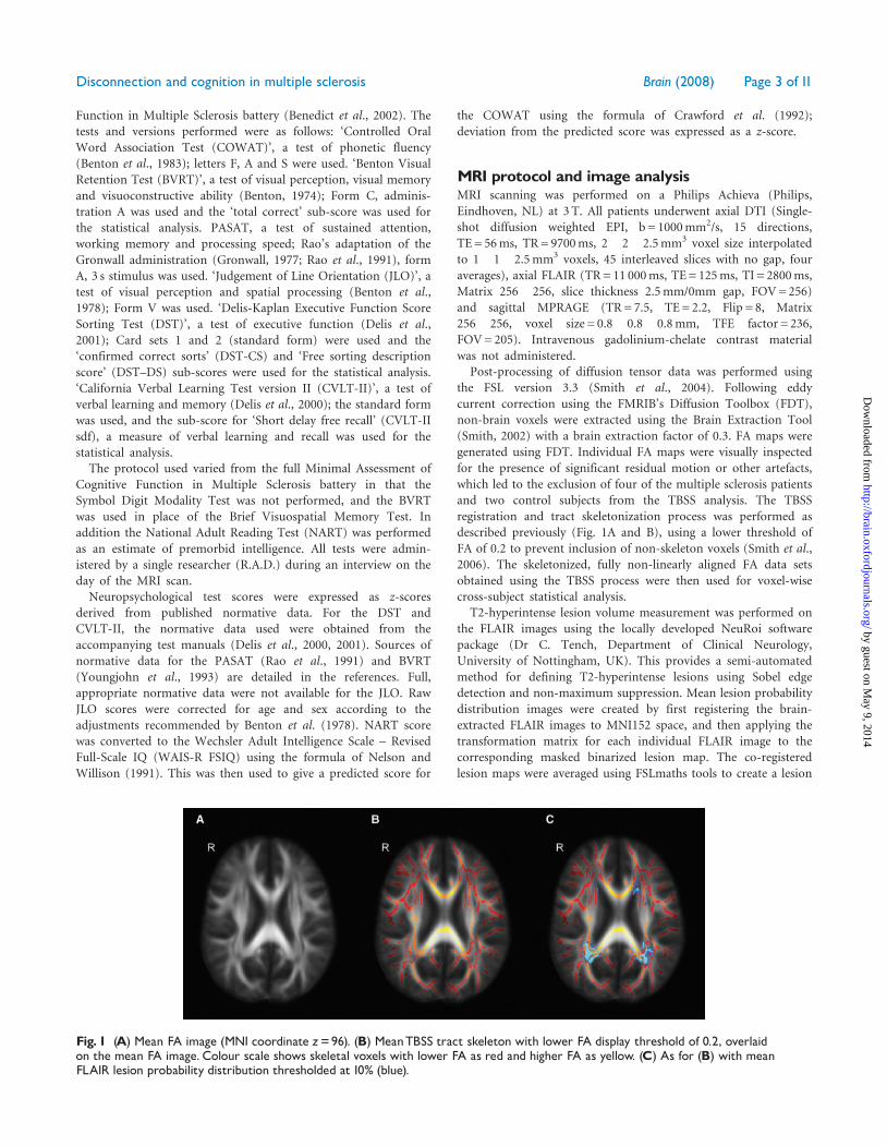
Fig. 1 (A) Mean FA image (MNI coordinate z=96). (B) MeanTBSS tract skeleton with lower FA display threshold of 0.2, overlaidon the mean FA image. Colour scale shows skeletal voxels with lower FA as red and higher FA as yellow. (C) As for (B) with meanFLAIR lesion probability distribution thresholded at 10% (blue).
Disconnection and cognition in multiple sclerosis Brain (2008) Page 3 of 11
by guest on May 9, 2014
http://brain.oxfordjournals.org/D
ownloaded from
probability distribution which was thresholded at 10% (i.e. voxelswhich contain macroscopic ‘lesion’ in 10% of subjects) (Fig. 1C).
Volumetric analysis of the MPRAGE images was performedusing JIM 3.0 software (Xinapse Medical Systems, ThorpeWaterville, UK). Brain extraction and segmentation was per-formed using the semi-automated Brain Finder tool whichprovided measurements of total intracranial volume, parenchymalbrain volume, CSF volume and parenchymal brain fraction.
Statistical analysisPrior to the voxel-wise analysis, group comparison was madebetween the whole-brain TBSS skeleton mean FA for multiplesclerosis patients and controls. Normality of distribution of thedata was assessed by the Shapiro–Wilk test, following which groupcomparison was performed by ANCOVA, treating age andparenchymal brain fraction as covariates.
The voxel-wise statistical analysis employed is based on a non-parametric approach utilizing permutation test theory with astandard general linear model design matrix. This approach hasthe advantage that it allows inference on the statistical maps whenthe null distribution is not known and provides an easilyimplementable solution to the multiple testing problem (Nicholsand Holmes, 2007). The permutation testing was performed usingthe program Randomise, part of FSL, which uses Monte Carlopermutation testing where n = 5000, to generate the randompermutations. Group comparison between multiple sclerosispatients and controls was performed for the conditions ‘voxel FAfor controls 4 voxel FA for multiple sclerosis patients’, and theconverse statement ‘voxel FA for multiple sclerosis patients4voxelFA for controls’. The output was thresholded at cluster level and thuscorrected for multiple comparisons using the null distribution of themaximum (across image) cluster size (t42, P50.05).
For the multiple sclerosis patients, inter-subject voxel-wisecorrelation was performed between skeletal voxel FA and theneuropsychological test z-scores, while treating age as a covariateof no interest. As premorbid IQ was felt to be a potentialconfounding variable, the analysis was repeated treating WAIS-RFSIQ as a covariate of no interest. Furthermore, the relationshipbetween cognitive performance and overall burden of disease (asmeasured by EDSS) was explored by repeating the TBSS analysisof normalized PASAT z-scores while treating EDSS score as acovariate of no interest. Again the output was thresholded atcluster level and thus corrected for multiple comparisons using thenull distribution of the maximum (across image) cluster size(t42, P50.05). Significant skeleton voxels were overlaid onto themean FA template to allow anatomical localization by visualinspection. Reference was made to an MRI-based atlas of whitematter tracts to describe affected locations (Mori et al., 2005).
As a test of the validity of the tract localizations demonstratedby TBSS and to visually assess for outlier effects, region of interest(ROI) analysis was performed. Two ROIs were defined on theTBSS skeleton, one at a site where significant correlations withPASAT z-scores were seen (posterior left cingulate) and one at asite where no significant correlations with PASAT z-score wereseen (left parahippocampal white matter). The MNI152 spacecoordinates of the boundaries of the left posterior cingulate ROIwere x = 98 to 104, y = 86–105, z = 104–111 and of the leftparahippocampal white matter were x = 113–120, y = 95–114,z = 39–59. The ROI was applied to each individual MS subject’s FAskeleton and the mean FA and standard deviation was calculated for
each ROI using FSLstats. The mean ROI FA values at each site wereused separately as variables in a partial correlation with PASATz-score, while controlling for age and EDSS.
ResultsFor four of the 41 multiple sclerosis subjects the DTI datawere degraded by motion artefact. Hence 37 multiplesclerosis subjects had usable DTI data and cognitive data.DTI data were degraded from 2 of 27 control subjects andhence 25 control data sets were finally included. Thecharacteristics of the subjects included in the TBSS analysisare shown in Table 1.
Neuropsychological testingThe multiple sclerosis patient group cognitive profile isshown in Table 2. Overall the group had a mild degree ofcognitive impairment, with performance in all tests fallingwithin 1 SD of the normative group mean. A significantdifference was found between multiple sclerosis patientsand the normative data for performance in the COWATand PASAT. The poorest performance was seen in thePASAT, COWAT and BVRT, where the group means were 0.6,0.5 and 0.4 SD below the normative data mean, respectively.Performance in the JLO is not expressed as z-scores, but thegroup mean (from raw scores corrected for age and sex)showed no group impairment with the group medianequalling the maximum score possible. Premorbid IQ wasestimated using the WAIS–R FSIQ scores derived fromNART. Scores ranged from 81 to 123 with a median of 102.7.
One multiple sclerosis subject had acquired alexiawithout agraphia. This individual was able to completethe neuropsychological testing with the exception of theDST and NART, both of which require intact readingability. Hence this subject was excluded from analyseswhere WAIS-R FSIQ (derived form the NART score) wasused and from the analysis of DST and COWAT z-scores.
Table 1 Characteristics of the included participants
MultipleSclerosispatients (n=37)
Healthycontrols (n=25)
Sex (M:F ratio) 1:2.4 1:2Median age (years) 43.5 (range 31.1^56.3,
IQR 35.3^49.7)36.4 (range 28.2^55.3,IQR 31.5^43.3)
Ethnic Group Caucasian=36,Afro-Caribbean=1
Caucasian=25
Hand dominance R=34, L=3 R=21, L=4Type of MS RRMS=35, SPMS=2 NAMedian multiplesclerosisduration (years)
10.5 (range 3.3^28.0,IQR 6.3^14.8)
NA
Median EDSS 3 (range 1.5^6.5,IQR 2.4 to 4)
NA
MedianT2- lesionvolume (ml)
6.3 (range 0.1^30.3,IQR 2.8^12.3)
NA
NA=Not Applicable.
Page 4 of 11 Brain (2008) R. A. Dineen et al.
by guest on May 9, 2014
http://brain.oxfordjournals.org/D
ownloaded from
TBSS analysis
Comparison between multiple sclerosis patients and controls
A small but significant difference in the whole-brain TBSSskeleton mean FA was demonstrated between patients andcontrols by ANCOVA (0.427 versus 0.445, F = 5.7,P = 0.020), while treating age and parenchymal brainfraction as covariates. Neither age nor atrophy exerted a
covariate effect which is in line with suppression of CSFpartial volume effects due to the imposed FA threshold inthe skeletonization process.
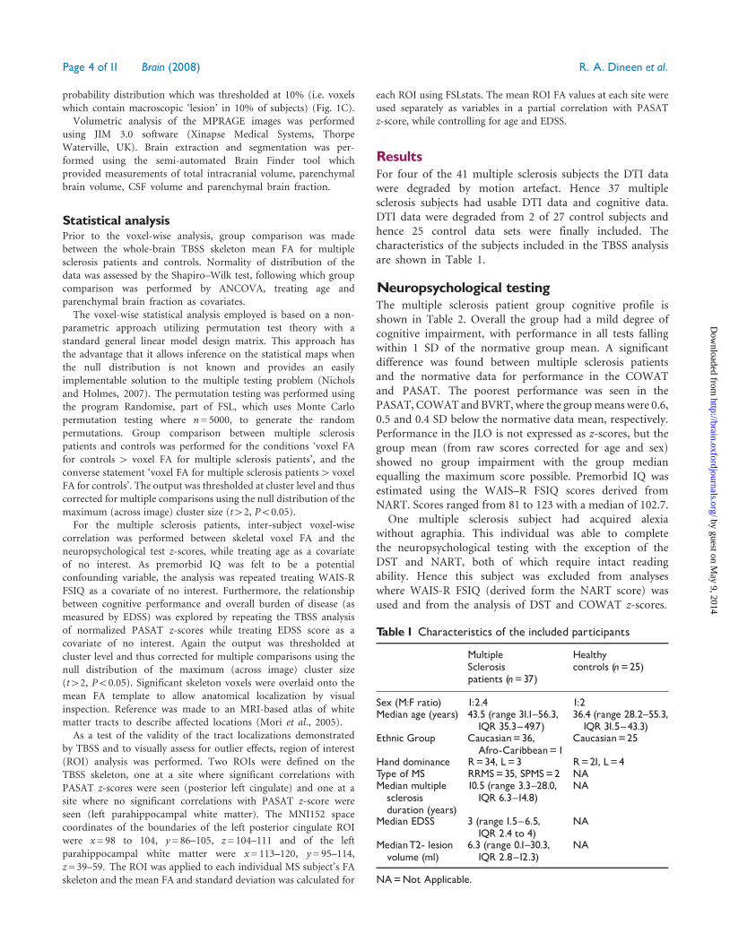
In the voxel-based group comparison, multiple areaswere identified where reduction in skeleton FA in multiplesclerosis patients versus controls reached significance,including the corpus callosum, the inferior longitudinalfasciculi and inferior fronto-occipital fasciculi bilaterally,the bodies and tails of the fornices bilaterally, the posteriorcorona radiata bilaterally and in the left cerebral peduncle(Fig. 2). To some extent these overlapped with the distribu-tion of macroscopic lesions but tract FA reductions were alsoseen where probability of macroscopic lesion was510%. Noareas were identified where reduction in skeleton FA in thecontrol versus multiple sclerosis patients reached significance.
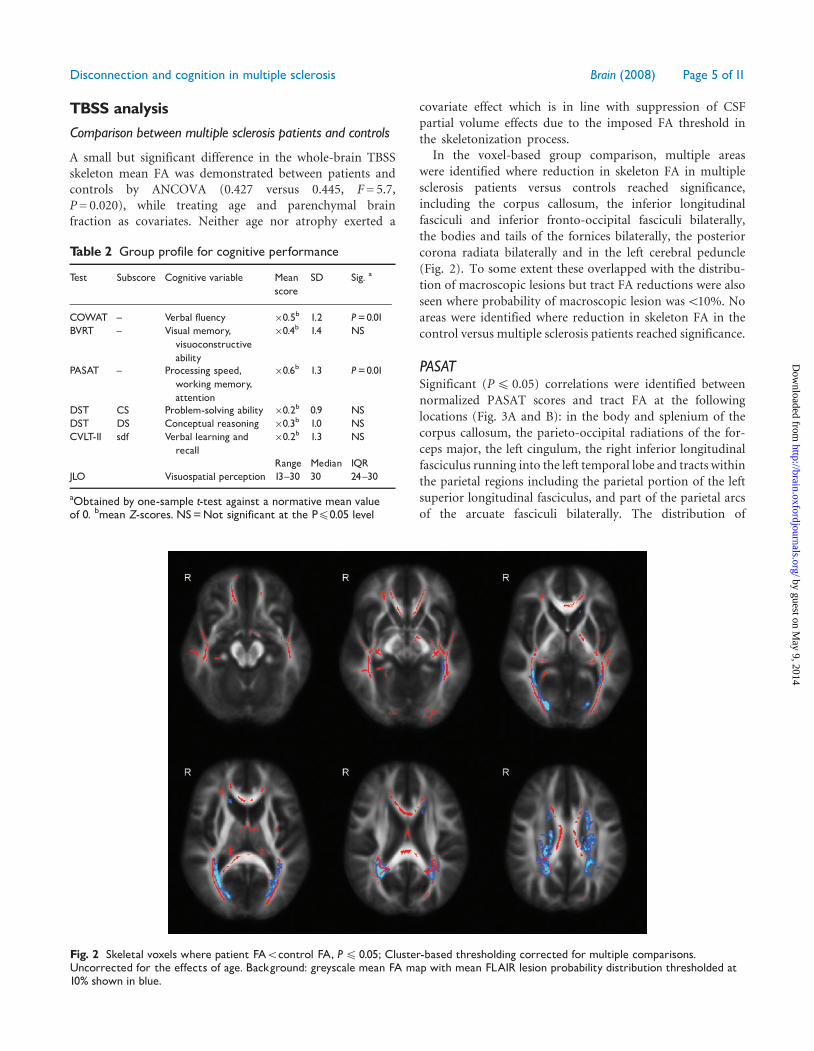
PASATSignificant (P4 0.05) correlations were identified betweennormalized PASAT scores and tract FA at the followinglocations (Fig. 3A and B): in the body and splenium of thecorpus callosum, the parieto-occipital radiations of the for-ceps major, the left cingulum, the right inferior longitudinalfasciculus running into the left temporal lobe and tracts withinthe parietal regions including the parietal portion of the leftsuperior longitudinal fasciculus, and part of the parietal arcsof the arcuate fasciculi bilaterally. The distribution of
Fig. 2 Skeletal voxels where patient FA5control FA, P4 0.05; Cluster-based thresholding corrected for multiple comparisons.Uncorrected for the effects of age. Background: greyscale mean FA map with mean FLAIR lesion probability distribution thresholded at10% shown in blue.
Table 2 Group profile for cognitive performance
Test Subscore Cognitive variable Meanscore
SD Sig. a
COWAT ^ Verbal fluency �0.5b 1.2 P=0.01BVRT ^ Visual memory,
visuoconstructiveability
�0.4b 1.4 NS
PASAT ^ Processing speed,working memory,attention
�0.6b 1.3 P=0.01
DST CS Problem-solving ability �0.2b 0.9 NSDST DS Conceptual reasoning �0.3b 1.0 NSCVLT-II sdf Verbal learning and
recall�0.2b 1.3 NS
Range Median IQRJLO Visuospatial perception 13^30 30 24^30
aObtained by one-sample t-test against a normative mean valueof 0. bmean Z-scores. NS=Not significant at the P40.05 level
Disconnection and cognition in multiple sclerosis Brain (2008) Page 5 of 11
by guest on May 9, 2014
http://brain.oxfordjournals.org/D
ownloaded from
correlations was similar when premorbid IQ estimate(WAIS-R FSIQ) was treated as a covariate of no interest.
To further explore the relationship of confoundingvariables the TBSS analysis was repeated for the PASATz-scores whilst controlling for overall multiple sclerosisseverity as measured by the EDSS. The same outputthreshold and significance level were applied as previously.The distribution overlapped with that for PASAT whenEDSS was not controlled for, but limited to the left side ofthe body and splenium of the corpus callosum radiatinginto the left parieto-occipital region, the left cingulum andthe parietal arc of the left arcuate fasciculus.
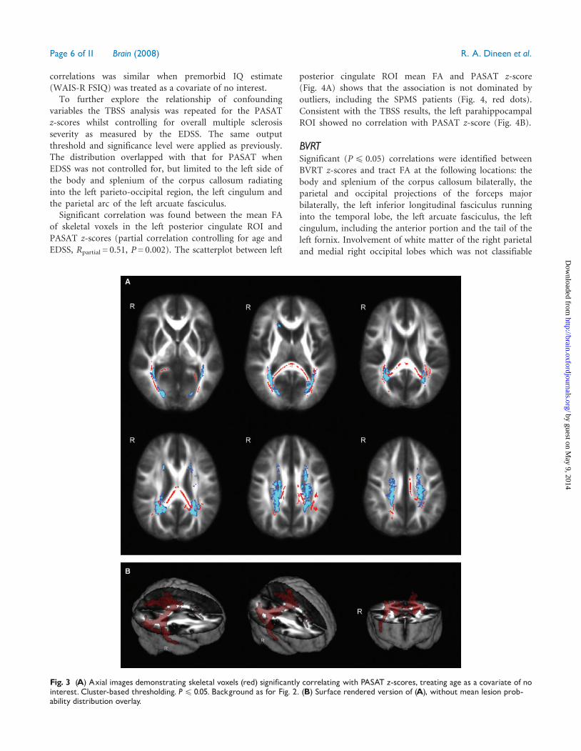
Significant correlation was found between the mean FAof skeletal voxels in the left posterior cingulate ROI andPASAT z-scores (partial correlation controlling for age andEDSS, Rpartial = 0.51, P = 0.002). The scatterplot between left
posterior cingulate ROI mean FA and PASAT z-score(Fig. 4A) shows that the association is not dominated byoutliers, including the SPMS patients (Fig. 4, red dots).Consistent with the TBSS results, the left parahippocampalROI showed no correlation with PASAT z-score (Fig. 4B).
BVRTSignificant (P4 0.05) correlations were identified betweenBVRT z-scores and tract FA at the following locations: thebody and splenium of the corpus callosum bilaterally, theparietal and occipital projections of the forceps majorbilaterally, the left inferior longitudinal fasciculus runninginto the temporal lobe, the left arcuate fasciculus, the leftcingulum, including the anterior portion and the tail of theleft fornix. Involvement of white matter of the right parietaland medial right occipital lobes which was not classifiable
Fig. 3 (A) Axial images demonstrating skeletal voxels (red) significantly correlating with PASAT z-scores, treating age as a covariate of nointerest. Cluster-based thresholding. P4 0.05. Background as for Fig. 2. (B) Surface rendered version of (A), without mean lesion prob-ability distribution overlay.
Page 6 of 11 Brain (2008) R. A. Dineen et al.
by guest on May 9, 2014
http://brain.oxfordjournals.org/D
ownloaded from
as named tracts was also seen. When premorbid IQ wascontrolled for, a very similar distribution was seen,with stronger effects in the right temporal white mattertracts (Fig. 5).
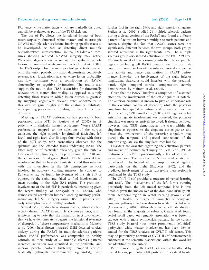
CVLT-IISignificant (P4 0.05) correlations were identified betweenCVLT-II sdf z-scores and tract FA at the followinglocations: the body and splenium of the corpus callosum,the parietal and occipital projections of the forceps majorbilaterally, the parietal portion of the left superior long-itudinal fasciculus, the left inferior longitudinal fasciculusand arcuate fasciculus, the left posterior cingulum and theleft fornix running posteriorly from the left mesial temporallobe. The distribution of correlations was similar whenpremorbid IQ was controlled for, with the exception thatthe correlations in the body of the corpus callosum and leftcingulate white matter were not significant at the P4 0.05threshold (Fig. 6).
Other cognitive testsNo significant (P4 0.05) correlation was identified betweentract FA and adjusted COWAT, JLO, DST CS or DST DSscores using permutation-based inference (cluster size t42).
DiscussionUsing tract-based FA regression analysis we were able tomap the anatomical pattern of white matter tract involve-ment in multiple sclerosis brains where ultrastructural fibreintegrity predicts impaired performance in specific cogni-tive domains. Differential, although partially overlapping,patterns of tract FA reduction were demonstrated for testsof sustained attention, working memory and processingspeed, visual working memory and verbal learning andrecall. The partial dissociation of cognitively relevant whitematter involvement demonstrated by TBSS from areas ofhigh lesion probability confirms that NAWM abnormalitymakes an important contribution to cognitive dysfunctionin multiple sclerosis (Zivadinov et al., 2001; Rovariset al., 2002).
TBSS provides a technique for co-registering likely whitematter tracts to allow multi-subject voxel-wise statisticalanalysis. During co-registration TBSS attempts to deforman individual’s natural white matter tract ‘skeleton’ (or atleast, the voxels most likely to lie at the centre of thenatural skeleton) to a common skeleton. This allows tissueinjury (as measured by FA) occurring in white matter tractsto be assigned to a ‘tract’ location. The individual tractskeletons are constructed regardless of localized reductions in
Fig. 4 (A) Location of the left posterior cingulate ROI (red) drawn on the mean tract skeleton (blue, thresholded at tract voxel FA5 0.2),with scatterplot demonstrating a relationship between ROI mean FA and PASAT z-score (red dotsçSPMS patients). (B) Location ofthe left parahippocampal white matter ROI, with corresponding scatterplot showing no relationship with PASAT z-scores.
Disconnection and cognition in multiple sclerosis Brain (2008) Page 7 of 11
by guest on May 9, 2014
http://brain.oxfordjournals.org/D
ownloaded from
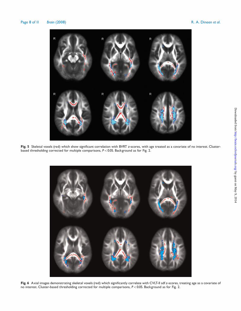
Fig. 6 Axial images demonstrating skeletal voxels (red) which significantly correlate with CVLT-II sdf z-scores, treating age as a covariate ofno interest. Cluster-based thresholding corrected for multiple comparisons, P50.05. Background as for Fig. 2.
Fig. 5 Skeletal voxels (red) which show significant correlation with BVRT z-scores, with age treated as a covariate of no interest. Cluster-based thresholding corrected for multiple comparisons, P50.05. Background as for Fig. 2.
Page 8 of 11 Brain (2008) R. A. Dineen et al.
by guest on May 9, 2014
http://brain.oxfordjournals.org/D
ownloaded from
FA; hence, white matter tracts which are markedly disruptedcan still be evaluated as part of the TBSS skeleton.
The use of FA allows the functional impact of bothmacroscopically abnormal white matter and microscopicNAWM multiple sclerosis lesions involving specific tracts tobe investigated. As well as detecting direct multiplesclerosis-related ultrastructural injury, DTI-derived mea-sures showing reduced NAWM integrity may reflectWallerian degeneration secondary to spatially remotelesions in connected white matter tracts (Lin et al., 2007).The TBSS output for the neuropsychological tests overlaidonto the lesion probablitilty maps demonstrate cognitivelyrelevant tract localizations at sites where lesion probabilitywas low, consistent with a contribution of NAWMabnormality to cognitive dysfunction. The results alsosupport the notion that TBSS is sensitive for functionallyrelevant white matter abnormality, as opposed to simplydetecting those tracts in which lesion burden is greatest.By mapping cognitively relevant tract abnormality inthis way, we gain insights into the anatomical substratesunderpinning performance in testing of different functionaldomains.
Mapping of PASAT performance has previously beenperformed using MTI by Ranjeva et al. (2005) in 18patients with clinically isolated syndromes. Worse PASATperformance mapped to the splenium of the corpuscallosum, the right superior longitudinal fasciculus, leftBA40 and right BA4. Our findings thus overlap with thoseof the MTI study, with correlations observed for thesplenium and the left-sided tracts underlying BA40. Thelatter may be of particular relevance, given the putativelocation of the phonological loop involving the BA40 andthe left inferior frontal gyrus (BA44). The left parietal tractinvolvement that we have demonstrated could thus interferewith the interaction in the left hemispheric networkinvolved in auditory working memory. In contrast toRanjeva et al., we found involvement of the left SLF asopposed to the right, and failed to find involvement oftracts running to the right BA4 region. The prominentinvolvement of the left SLF is particularly interesting giventhe recent findings of Karlsgodt et al. (2008), whodemonstrated correlation between working memory perfor-mance and left SLF integrity using TBSS in patients withearly schizophrenia and healthy controls.
Several fMRI studies have shown compensatory corticalactivity during PASAT in multiple sclerosis patients, and itis interesting to note that the pattern of tract involvementthat we have demonstrated suggests the functional relevanceof disruption of these compensatory mechanisms. Maineroet al. (2004) have shown increased fMRI-detected corticalactivity during the PASAT in multiple sclerosis patientswhose PASAT performance was comparable to healthycontrols. In their study of 22 multiple sclerosis patientsincreased activation was identified in the prefrontal andinferior parietal cortices bilaterally, temporal corticesbilaterally (although predominantly right-sided), with
further foci in the right SMA and right anterior cingulate.Staffen et al. (2002) studied 21 multiple sclerosis patientsduring a visual version of the PASAT and found a differentpattern of activation between multiple sclerosis patients andcontrols, despite the fact that PASAT scores were notsignificantly different between the two groups. Both groupsshowed activations in the right frontal area. The multiplesclerosis group also showed activation in the left BA39 area.The involvement of tracts running into the inferior parietalregions (including left BA39) demonstrated by our datacould thus result in loss of the inferior parietal compensa-tory activity and hence deterioration in PASAT perfor-mance. Likewise, the involvement of the right inferiorlongitudinal fasciculus could interfere with the predomi-nantly right temporal cortical compensatory activitydemonstrated by Mainero et al. (2004).
Given that the PASAT involves a component of sustainedattention, the involvement of the left cingulum is notable.The anterior cingulum is known to play an important rolein the executive control of attention, while the posteriorcingulum has spatial attention and orienting functions(Hirono et al., 1998; Small et al., 2003). Although some leftanterior cingulate involvement was observed, the posteriorcingulate was more extensively involved. It should be noted,however, that TBSS demonstrates the tract within thecingulum as opposed to the cingulate cortex per se, andhence the involvement of the posterior cingulum maydisrupt the temporal and parietal connections of theanterior cingulate via the cingulate tract bundle.
Less data are available regarding the activation patternsand impact of localized tract injury on BVRT and CVLT-IIperformance. BVRT is predominantly a test of short-termvisual memory. The hypothetical ‘visuospatial scratchpad’is believed to be located in the temporoparietal regions,particularly on the right (Baddeley, 1992), and thepredicted involvement of tracts subserving these regions isconfirmed by the TBSS study.
The CVLT-II sdf provides a measure of verbal learningand recall. The involvement of the left fornix runningposteriorly from the left mesial temporal lobe is thusnotable, given the known role of the dominant (usually left)mesial temporal region in verbal learning (Powell et al.,2005). In health, the degree of symmetry of perisylvianlanguage pathways has been shown to relate to verbal recall(Catani et al., 2007). Although extreme left lateralizationwas found in the majority of subjects, Catani et al. foundverbal recall based on semantic association was better insubjects with a more symmetrical pattern. In the currentTBSS study bilateral (but more prominently left-sided)perisylvian white matter involvement has been demon-strated for the TBSS analysis of CVLT-II sdf scores. Thismay be particularly relevant as CVLT-II sdf performance isenhanced if the semantic associations within the word listare identified by the subject.
Performance during the CVLT is known to be affected byfrontal lesions, particularly left posterior dorsolateral frontal
Disconnection and cognition in multiple sclerosis Brain (2008) Page 9 of 11
by guest on May 9, 2014
http://brain.oxfordjournals.org/D
ownloaded from
region and the posterior medial frontal region, due toimpairment of organizational structure to the new learning(Alexander et al., 2003). We failed to demonstrate directinvolvement of the frontal lobe tracts (with the exception ofthe anterior corpus callosum). However, involvement of theleft arcuate fasciculus and superior longitudinal fasciculuscould impair temporo-frontal and parieto-frontal connec-tivity, respectively.
The two tests of frontal lobe function, the DST andCOWAT, failed to show any significant TBSS correlations.The reason for this is unclear, but may reflect the morevariable localization of function within the frontal lobes.This relative dispersion of function would limit the abilityof the TBSS technique to localize relevant tract injury. Wealso failed to demonstrate the expected parietal localizationof tract injury for the JLO, a test of visuospatial perception.The negative findings for both the JLO and the DST arelikely to reflect the low spread of data in a small sampleresulting in insufficient statistical power to detectrelationships.
The distribution of statistically significant tract-based FAreductions in multiple sclerosis patients compared tocontrols showed only partial overlap with the visible T2lesions. There was a double dissociation with large areas ofhigh T2 lesion probability but no significant tract FAreduction in the centrum semiovale, and several tracts withreduced FA but 510% lesion probability, such as fornices,corpus callosum and fronto-temporal white matter. Afurther notable result from the comparison with healthycontrols is that many of the tracts showing associationswith cognitive performance did not co-localize withbetween-group statistical tract abnormality. For example,the TBSS output for PASAT showed correlations with theleft cingulate white matter tract and the left superiorlongitudinal fasciculus which were not identified as beingsignificantly different during the comparison of multiplesclerosis patients and controls.
An important consideration which is as yet unresolved isthe degree to which normal inter-subject variations in tractFA influence cognitive performance. The assumptionunderlying the proposed hypothesis is that the TBSSlocalizations demonstrated relate to pathology, but physio-logical variation may account in part for the findings. In anattempt to partially control for inherent variability werepeated the TBSS analysis of cognitive performancecontrolling for premorbid IQ, and found a similar patternof tract localizations for the cognitive tests. However,domain-specific differences in white matter organization aremore difficult to control for. For example, the heterogeneityin lateralization of perisylvian language networks recentlydemonstrated by (Catani et al., 2007) has been shown torelate to verbal memory performance. This inherentstructural variability in health has the potential to confoundthe results of the TBSS study, but could also explain thefact that significant tract localizations were identified atsites where no difference between the patient and control
groups were found. Thus the localizations demonstratedcannot necessarily be assumed to be the result of disease-specific alterations to white matter structure.
In summary, the application of TBSS to map cognitivelyrelevant tract disorganization has been demonstrated in acohort of multiple sclerosis patients, and thus providesinsights into the anatomical substrates underlying func-tional connectivity. Many of the localizations of whitematter injury can be shown to be consistent with theknown functional anatomy of processing within the specificcognitive domain studied, and in the case of PASATperformance, the localization of injury could result indisconnection of compensatory mechanisms previouslyshown to occur in multiple sclerosis patients in fMRIstudies.
AcknowledgementsThe authors gratefully acknowledge technical assistancefrom Dr Dylan Shen and Ms Maryam Abaei for assistancewith image post-processing and the TBSS analysis, and DrChris Tench providing the NeuRoi software and technicaladvice for the FLAIR lesion volume measurements.
ReferencesAlexander MP, Stuss DT, Fansabedian N. California Verbal Learning Test:
performance by patients with focal frontal and non-frontal lesions. Brain
2003; 126: 1493–503.
Amato MP, Bartolozzi ML, Zipoli V, Portaccio E, Mortilla M, Guidi L,
et al. Neocortical volume decrease in relapsing-remitting MS patients
with mild cognitive impairment. Neurology 2004; 63: 89–93.
Arnett PA, Rao SM, Bernardin L, Grafman J, Yetkin FZ, Lobeck L.
Relationship between frontal lobe lesions and Wisconsin Card Sorting
Test performance in patients with multiple sclerosis. Neurology 1994;
44: 420–5.
Baddeley A. Working memory. Science 1992; 255: 556–9.
Basser PJ, Pierpaoli C. Microstructural and physiological features of tissues
elucidated by quantitative-diffusion-tensor MRI. J Magn Reson B 1996;
111: 209–19.
Benedict RH, Fischer JS, Archibald CJ, Arnett PA, Beatty WW, Bobholz J,
et al. Minimal neuropsychological assessment of MS patients: a
consensus approach. Clin Neuropsychol 2002; 16: 381–97.
Benton AL. The revised visual retention test. 4th edn. New York:
Psychological Corporation; 1974.
Benton AL, Hamsher KD, Sivan AB. Multilingual aphasia examination.
3rd edn. Iowa: AJA Associates; 1983.
Benton AL, Varney NR, Hamsher KD. Visuospatial judgment. A clinical
test. Arch Neurol 1978; 35: 364–7.
Catani M. Diffusion tensor magnetic resonance imaging tractography in
cognitive disorders. Curr Opin Neurol 2006; 19: 599–606.
Catani M, Allin MP, Husain M, Pugliese L, Mesulam MM, Murray RM,
et al. Symmetries in human brain language pathways correlate with
verbal recall. Proc Natl Acad Sci USA 2007; 104: 17163–8.
Catani M, ffytche DH. The rises and falls of disconnection syndromes.
Brain 2005; 128: 2224–39.
Charil A, Zijdenbos AP, Taylor J, Boelman C, Worsley KJ, Evans AC, et al.
Statistical mapping analysis of lesion location and neurological disability
in multiple sclerosis: application to 452 patient data sets. Neuroimage
2003; 19: 532–44.
Cox D, Pelletier D, Genain C, Majumdar S, Lu Y, Nelson S, et al. The
unique impact of changes in normal appearing brain tissue on cognitive
Page 10 of 11 Brain (2008) R. A. Dineen et al.
by guest on May 9, 2014
http://brain.oxfordjournals.org/D
ownloaded from
dysfunction in secondary progressive multiple sclerosis patients. Mult
Scler 2004; 10: 626–9.
Crawford JR, Moore JW, Cameron IM. Verbal fluency: a NART-based
equation for the estimation of premorbid performance. Br J Clin
Psychol 1992; 31: 327–9.
Delis DC, Kaplan E, Kramer JH. Delis Kaplan executive function system
examiner’s manual. San Antonio: The Psychological Corporation; 2001.
Delis DC, Kramer JH, Kaplan E, Ober BA. California verbal learning test
manual. 2nd edn. San Antonio: The Psychological Corporation; 2000.
Foong J, Rozewicz L, Quaghebeur G, Davie CA, Kartsounis LD,
Thompson AJ, et al. Executive function in multiple sclerosis. The role
of frontal lobe pathology. Brain 1997; 120: 15–26.
Gronwall DMA. Paced auditory serial-addition task: a measure of recovery
from concussion. Percept Mot skills 1977; 44: 367–73.
Guo AC, MacFall JR, Provenzale JM. Multiple sclerosis: diffusion tensor
MR imaging for evaluation of normal-appearing white matter.
Radiology 2002; 222: 729–36.
Hasan KM, Gupta RK, Santos RM, Wolinsky JS, Narayana PA. Diffusion
tensor fractional anisotropy of the normal-appearing seven segments of
the corpus callosum in healthy adults and relapsing-remitting multiple
sclerosis patients. J Magn Reson Imaging 2005; 21: 735–43.
Hirono N, Mori E, Ishii K, Ikejiri Y, Imamura T, Shimomura T, et al.
Hypofunction in the posterior cingulate gyrus correlates with disor-
ientation for time and place in Alzheimer’s disease. J Neurol Neurosurg
Psychiatry 1998; 64: 552–4.
Karlsgodt KH, van Erp TGM, Poldrack RA, Bearden CE, Nuechterlein KH,
Cannon TD. Diffusion tensor imaging of the superior longitudinal
fasciculus and working memory in recent-onset schizophrenia. Biol
Psychiatry 2008; 63: 512–8.
Kealey SM, Kim Y, Whiting WL, Madden DJ, Provenzale JM.
Determination of multiple sclerosis plaque size with diffusion-tensor
MR Imaging: comparison study with healthy volunteers. Radiology
2005; 236: 615–20.
Lin F, Yu C, Jiang T, Li K, Chan P. Diffusion tensor tractography-based
group mapping of the pyramidal tract in relapsing-remitting multiple
sclerosis patients. AJNR Am J Neuroradiol 2007; 28: 278–82.
Mainero C, Caramia F, Pozzilli C, Pisani A, Pestalozza I, Borriello G, et al.
fMRI evidence of brain reorganization during attention and memory
tasks in multiple sclerosis. Neuroimage 2004; 21: 858–67.
Morgen K, Sammer G, Courtney SM, Wolters T, Melchior H, Blecker CR,
et al. Evidence for a direct association between cortical atrophy and
cognitive impairment in relapsing-remitting MS. NeuroImage 2006; 30:
891.
Mori S, Wakana S, Nagae-Poetscher LM, van Zijl PCM. MRI atlas of
human white matter. Amsterdam, The Netherlands: Elsevier B. V.; 2005.
Nelson HE, Willison J. National adult reading test manual (2nd ed.).
Windsor, UK: NFER-Nelson; 1991.
Nichols TE, Holmes AP. Non-parametric procedures. In: Friston KJ,
Ashburner JT, Kiebel SJ, Nichols TE, Penny WD, editors. Statistical
parametric mapping – the analysis of functional brain images. London,
UK: Academic Press; 2007. p. 253–72.
Pan JW, Krupp LB, Elkins LE, Coyle PK. Cognitive dysfunction lateralizes
with NAA in multiple sclerosis. Appl Neuropsychology 2001; 8: 155–60.
Powell HWR, Koepp MJ, Symms MR, Boulby PA, Salek-Haddadi A,
Thompson PJ, et al. Material-specific lateralization of memory encoding
in the medial temporal lobe: blocked versus event-related design.
NeuroImage 2005; 27: 231.
Ranjeva JP, Audoin B, Au Duong MV, Ibarrola D, Confort-Gouny S,
Malikova I, et al. Local tissue damage assessed with statistical mapping
analysis of brain magnetization transfer ratio: relationship with
functional status of patients in the earliest stage of multiple sclerosis.
AJNR Am Journal Neuroradiol 2005; 26: 119–27.
Rao SM, Leo GJ, Bernardin L, Unverzagt F. Cognitive dysfunction in
multiple sclerosis. I. Frequency, patterns, and prediction. Neurology
1991; 41: 685–91.
Ron MA, Callanan MM, Warrington EK. Cognitive abnormalities in
multiple sclerosis: a psychometric and MRI study. Psychol Med 1991;
21: 59–68.
Rovaris M, Filippi M, Falautano M, Minicucci L, Rocca MA, Martinelli V,
et al. Relation between MR abnormalities and patterns of cognitive
impairment in multiple sclerosis. Neurology 1998; 50: 1601–8.
Rovaris M, Iannucci G, Falautano M, Possa F, Martinelli V, Comi G, et al.
Cognitive dysfunction in patients with mildly disabling relapsing-
remitting multiple sclerosis: an exploratory study with diffusion tensor
MR imaging. J Neurol Sci 2002; 195: 103–9.
Small DM, Gitelman DR, Gregory MD, Nobre AC, Parrish TB,
Mesulam MM. The posterior cingulate and medial prefrontal cortex
mediate the anticipatory allocation of spatial attention. NeuroImage
2003; 18: 633.
Smith SM. Fast robust automated brain extraction. Hum Brain Mapp
2002; 17: 143–55.
Smith SM, Jenkinson M, Johansen-Berg H, Rueckert D, Nichols TE,
Mackay CE, et al. Tract-based spatial statistics: voxelwise analysis of
multi-subject diffusion data. Neuroimage 2006; 31: 1487–505.
Smith SM, Jenkinson M, Woolrich MW, Beckmann CF, Behrens TE,
Johansen-Berg H, et al. Advances in functional and structural MR image
analysis and implementation as FSL. Neuroimage 2004; 23 (Suppl 1):
S208–19.
Sperling RA, Guttmann CR, Hohol MJ, Warfield SK, Jakab M, Parente M,
et al. Regional magnetic resonance imaging lesion burden and cognitive
function in multiple sclerosis: a longitudinal study. Arch Neurol 2001;
58: 115–21.
Staffen W, Mair A, Zauner H, Unterrainer J, Niederhofer H, Kutzelnigg A,
et al. Cognitive function and fMRI in patients with multiple sclerosis:
evidence for compensatory cortical activation during an attention task.
Brain 2002; 125: 1275–82.
Steffens DC, McQuoid DR, Welsh-Bohmer KA, Krishnan KR. Left orbital
frontal cortex volume and performance on the Benton visual retention
test in older depressives and controls. Neuropsychopharmacology 2003;
28: 2179–83.
Werring DJ, Clark CA, Barker GJ, Thompson AJ, Miller DH. Diffusion
tensor imaging of lesions and normal-appearing white matter in
multiple sclerosis. Neurology 1999; 52: 1626–32.
Youngjohn JR, Larrabee GJ, Crook TH III. New adult age- and education-
correction norms for the Benton Visual Retention Test. Clin
Neuropsychol 1993; 7: 155–160.
Zivadinov R, Masi RD, Nasuelli D, Bragadin LM, Ukmar M, Pozzi-
Mucelli RS, et al. MRI techniques and cognitive impairment in the
early phase of relapsing-remitting multiple sclerosis. Neuroradiology
2001; 43: 272.
Disconnection and cognition in multiple sclerosis Brain (2008) Page 11 of 11
by guest on May 9, 2014
http://brain.oxfordjournals.org/D
ownloaded from