Diffuse Lung Disease in Young Children: Application of a Novel Classification Scheme
9
Diffuse Lung Disease in Young Children Application of a Novel Classification Scheme Gail H. Deutsch 1 *, Lisa R. Young 2 *, Robin R. Deterding 3 , Leland L. Fan 4 , Sharon D. Dell 5 , Judy A. Bean 6 , Alan S. Brody 7 , Lawrence M. Nogee 8 , Bruce C. Trapnell 9 , Claire Langston 10 , and the Pathology Cooperative Group: Eric A. Albright 11 , Frederic B. Askin 12 , Peter Baker 11 , Pauline M. Chou 13 , Carlyne M. Cool 14 , Susan C. Coventry 15 , Ernest Cutz 16 , Mary M. Davis 17 , Megan K. Dishop 10 , Csaba Galambos 18 , Kathleen Patterson 19 , William D. Travis 20 , Susan E. Wert 9 , and Frances V. White 21 ; on behalf of the ChILD Research Co-operative † Divisions of 1 Pathology, 2 Pulmonary Medicine, 6 Epidemiology, and 9 Pulmonary Biology, and 7 Department of Radiology, Cincinnati Children’s Hospital Medical Center, Cincinnati, Ohio; 3 Department of Pediatrics, Children’s Hospital, Denver, Colorado; Departments of 4 Pediatrics and 10 Pathology, Texas Children’s Hospital, Houston, Texas; Divisions of 5 Respiratory Medicine and 16 Pathology, The Hospital for Sick Children, Toronto, Ontario, Canada; Departments of 8 Pediatrics and 12 Pathology, Johns Hopkins University School of Medicine, Baltimore, Maryland; 11 Department of Anatomic Pathology, Children’s Hospital, Columbus, Ohio; 13 Department of Pathology, Children’s Memorial Hospital, Chicago, Illinois; 14 Department of Pathology, University of Colorado Health Science Center, Denver, Colorado; 15 Department of Pathology, Kosair Children’s Hospital, Louisville, Kentucky; 17 Division of Pediatric Pathology, James Whitcomb Riley Hospital for Children, Indianapolis, Indiana; 18 Department of Pathology, Children’s Hospital of Pittsburgh, Pittsburgh, Pennsylvania; 19 Department of Laboratories, Children’s Hospital and Regional Medical Center, Seattle, Washington; 20 Department of Pathology, Memorial Sloan-Kettering Cancer Center, New York, New York; and 21 Division of Surgical Pathology, Washington University School of Medicine, St. Louis, Missouri Rationale: Considerable confusion exists regarding nomenclature, classification, and management of pediatric diffuse lung diseases due to the relative rarity and differences in the spectrum of disease between adults and young children. Objectives: A multidisciplinary working group was formed to: (1) apply consensus terminology and diagnostic criteria for disorders presenting with diffuse lung disease in infancy; and (2) describe the distribution of disease entities, clinical features, and outcome in young children who currently undergo lung biopsy in North America. Methods: Eleven centers provided pathologic material, clinical data, and imaging from all children less than 2 years of age who underwent lung biopsy for diffuse lung disease from 1999 to 2004. Measurements and Main Results: Multidisciplinary review categorized 88% of 187 cases. Disorders more prevalent in infancy, including primary developmental and lung growth abnormalities, neuroendo- crine cell hyperplasia of infancy, and surfactant-dysfunction disorders, constituted the majority of cases (60%). Lung growth disorders were often unsuspected clinically and under-recognized histologically. Cases with known surfactant mutations had characteristic pathologic features. Age at biopsy and clinical presentation varied among categories. Pulmonary hypertension, presence of a primary develop- mental abnormality, or ABCA3 mutation was associated with high mortality, while no deaths occurred in cases of pulmonary interstitial glycogenosis, or neuroendocrine cell hyperplasia of infancy. Conclusions: This retrospective cohort study identifies a diverse spec- trum of lung disorders, largely unique to young children. Application of a classification scheme grouped clinically distinct patients with variable age of biopsy and mortality. Standardized terminology and classifica- tion will enhance accurate description and diagnosis of these disorders. Keywords: infant; pulmonary; interstitial lung disease; surfactant; neuroendocrine hyperplasia Diffuse lung disease in children, which includes interstitial lung disease (ILD), comprises a heterogeneous group of uncommon disorders characterized by impaired gas exchange and diffuse infiltrates by imaging (1). To date, description and management of pediatric ILD has largely been based on case reports and small series, complicating accurate diagnosis, assessment of prevalence, and definitive therapy for these disorders. In contrast, significant progress has been made in generating a standardized classifica- tion of adult idiopathic interstitial pneumonias by integrating international clinical, radiographic, and pathologic expertise (2). While there is some overlap in the histologic patterns of ILD between adults and children, direct application of the adult classification scheme to pediatric ILD is limited by differing frequency and spectrum of disease, clinical manifestations, and outcome. Specifically, usual interstitial pneumonia (UIP), the pathologic correlate of idiopathic pulmonary fibrosis/crypto- genic fibrosing alveolitis (IPF/CFA), is the most common ILD in adults; nonetheless, the defining histologic feature of UIP, fibroblastic foci, has yet to be described in any pediatric case (3, 4). Furthermore, children given the diagnosis of IPF/CFA have a clinical course that differs from that of adults, as pediatric cases are characterized by nonprogressive symptoms and relative AT A GLANCE COMMENTARY Scientific Knowledge on the Subject There are only limited studies of diffuse lung disease in infants. Progress has been hindered by the rarity of these disorders and the use of adult terminology and classifica- tion systems that do not adequately address pediatric or genetic entities. What This Study Adds to the Field This multicenter study of diffuse lung disease in infants employs a new classification system emphasizing infant disorders. Its use in a large cohort of infants with lung biopsy provides new information on disease frequency, clinical settings, and outcome. (Received in original form March 9, 2007; accepted in final form September 20, 2007) *These authors contributed equally to this work. Supported in part by the Rare Lung Diseases Consortium, National Institutes of Health RR019498 (to B.C.T.). †Members of the ChILD Research Co-operative who participated in the current study are listed in APPENDIX 2. Correspondence and requests for reprints should be addressed to Claire Langston, M.D., Department of Pathology, Texas Children’s Hospital, 6621 Fannin Street, Houston, TX 77030. E-mail: [email protected] Am J Respir Crit Care Med Vol 176. pp 1120–1128, 2007 Originally Published in Press as DOI: 10.1164/rccm.200703-393OC on September 20, 2007 Internet address: www.atsjournals.org
-
Upload
independent -
Category
Documents
-
view
2 -
download
0
Transcript of Diffuse Lung Disease in Young Children: Application of a Novel Classification Scheme
Diffuse Lung Disease in Young ChildrenApplication of a Novel Classification Scheme
Gail H. Deutsch1*, Lisa R. Young2*, Robin R. Deterding3, Leland L. Fan4, Sharon D. Dell5, Judy A. Bean6,Alan S. Brody7, Lawrence M. Nogee8, Bruce C. Trapnell9, Claire Langston10, and the Pathology CooperativeGroup: Eric A. Albright11, Frederic B. Askin12, Peter Baker11, Pauline M. Chou13, Carlyne M. Cool14,Susan C. Coventry15, Ernest Cutz16, Mary M. Davis17, Megan K. Dishop10, Csaba Galambos18,Kathleen Patterson19, William D. Travis20, Susan E. Wert9, and Frances V. White21; on behalf of the ChILDResearch Co-operative†
Divisions of 1Pathology, 2Pulmonary Medicine, 6Epidemiology, and 9Pulmonary Biology, and 7Department of Radiology, Cincinnati Children’s
Hospital Medical Center, Cincinnati, Ohio; 3Department of Pediatrics, Children’s Hospital, Denver, Colorado; Departments of 4Pediatrics and10Pathology, Texas Children’s Hospital, Houston, Texas; Divisions of 5Respiratory Medicine and 16Pathology, The Hospital for Sick Children,
Toronto, Ontario, Canada; Departments of 8Pediatrics and 12Pathology, Johns Hopkins University School of Medicine, Baltimore, Maryland;11Department of Anatomic Pathology, Children’s Hospital, Columbus, Ohio; 13Department of Pathology, Children’s Memorial Hospital,
Chicago, Illinois; 14Department of Pathology, University of Colorado Health Science Center, Denver, Colorado; 15Department of Pathology,
Kosair Children’s Hospital, Louisville, Kentucky; 17Division of Pediatric Pathology, James Whitcomb Riley Hospital for Children, Indianapolis,Indiana; 18Department of Pathology, Children’s Hospital of Pittsburgh, Pittsburgh, Pennsylvania; 19Department of Laboratories, Children’s
Hospital and Regional Medical Center, Seattle, Washington; 20Department of Pathology, Memorial Sloan-Kettering Cancer Center, New York,
New York; and 21Division of Surgical Pathology, Washington University School of Medicine, St. Louis, Missouri
Rationale: Considerable confusion exists regarding nomenclature,classification, and management of pediatric diffuse lung diseasesdue to the relative rarity and differences in the spectrum of diseasebetween adults and young children.Objectives: A multidisciplinary working group was formed to: (1)apply consensus terminology and diagnostic criteria for disorderspresenting with diffuse lung disease in infancy; and (2) describe thedistribution of disease entities, clinical features, and outcome inyoung children who currently undergo lung biopsy in North America.Methods: Eleven centers provided pathologic material, clinical data,and imaging from all children less than 2 years of age who underwentlung biopsy for diffuse lung disease from 1999 to 2004.Measurements and Main Results: Multidisciplinary review categorized88% of 187 cases. Disorders more prevalent in infancy, includingprimary developmental and lung growth abnormalities, neuroendo-crine cell hyperplasia of infancy, and surfactant-dysfunctiondisorders,constituted the majority of cases (60%). Lung growth disorders wereoften unsuspected clinically and under-recognized histologically.Cases with known surfactant mutations had characteristic pathologicfeatures. Age at biopsy and clinical presentation varied amongcategories. Pulmonary hypertension, presence of a primary develop-mental abnormality, or ABCA3 mutation was associated with highmortality, while no deaths occurred in cases of pulmonary interstitialglycogenosis, or neuroendocrine cell hyperplasia of infancy.Conclusions: This retrospective cohort study identifies a diverse spec-trumof lungdisorders, largelyunique toyoungchildren.Applicationofa classification schemegroupedclinicallydistinctpatientswith variableage of biopsy and mortality. Standardized terminology and classifica-tionwill enhanceaccuratedescriptionanddiagnosisofthesedisorders.
Keywords: infant; pulmonary; interstitial lung disease; surfactant;
neuroendocrine hyperplasia
Diffuse lung disease in children, which includes interstitial lungdisease (ILD), comprises a heterogeneous group of uncommondisorders characterized by impaired gas exchange and diffuseinfiltrates by imaging (1). To date, description and managementof pediatric ILD has largely been based on case reports and smallseries, complicating accurate diagnosis, assessment of prevalence,and definitive therapy for these disorders. In contrast, significantprogress has been made in generating a standardized classifica-tion of adult idiopathic interstitial pneumonias by integratinginternational clinical, radiographic, and pathologic expertise (2).
While there is some overlap in the histologic patterns of ILDbetween adults and children, direct application of the adultclassification scheme to pediatric ILD is limited by differingfrequency and spectrum of disease, clinical manifestations, andoutcome. Specifically, usual interstitial pneumonia (UIP), thepathologic correlate of idiopathic pulmonary fibrosis/crypto-genic fibrosing alveolitis (IPF/CFA), is the most common ILDin adults; nonetheless, the defining histologic feature of UIP,fibroblastic foci, has yet to be described in any pediatric case (3,4). Furthermore, children given the diagnosis of IPF/CFA havea clinical course that differs from that of adults, as pediatric casesare characterized by nonprogressive symptoms and relative
AT A GLANCE COMMENTARY
Scientific Knowledge on the Subject
There are only limited studies of diffuse lung disease ininfants. Progress has been hindered by the rarity of thesedisorders and the use of adult terminology and classifica-tion systems that do not adequately address pediatric orgenetic entities.
What This Study Adds to the Field
This multicenter study of diffuse lung disease in infantsemploys a new classification system emphasizing infantdisorders. Its use in a large cohort of infants with lungbiopsy provides new information on disease frequency,clinical settings, and outcome.
(Received in original form March 9, 2007; accepted in final form September 20, 2007)
*These authors contributed equally to this work.
Supported in part by the Rare Lung Diseases Consortium, National Institutes of
Health RR019498 (to B.C.T.).
†Members of the ChILD Research Co-operative who participated in the current
study are listed in APPENDIX 2.
Correspondence and requests for reprints should be addressed to Claire
Langston, M.D., Department of Pathology, Texas Children’s Hospital, 6621
Fannin Street, Houston, TX 77030. E-mail: [email protected]
Am J Respir Crit Care Med Vol 176. pp 1120–1128, 2007
Originally Published in Press as DOI: 10.1164/rccm.200703-393OC on September 20, 2007
Internet address: www.atsjournals.org
longevity (5, 6). Likewise, despite sharing similar histology,desquamative interstitial pneumonia (DIP) has different etio-logic and prognostic implications in adults and children. Inadults, DIP is linked to smoking and is often responsive tosteroids, while DIP in children has a more aggressive course andfamilial forms have been identified (5, 7, 8). Recently, DIP hasbeen described in patients with mutations in the ATP-bindingcassette transporter A3 (ABCA3) gene, and less commonly in thesurfactant protein C (SFTPC) gene, both necessary for surfactantmetabolism (9–11).
Although there are known causes of pediatric ILD includinginfection, environmental exposures, and collagen vascular andmetabolic disease, the literature is becoming progressivelypopulated by reports of idiopathic lung disorders that appeardistinct to infants and young children. These include chronicpneumonitis of infancy (CPI) (12–14), pulmonary interstitialglycogenosis (also termed infantile cellular interstitial pneumo-nitis) (15, 16), and persistent tachypnea of infancy associatedwith neuroendocrine cell hyperplasia (17). Recognition of theseunique disorders has been hindered by lack of uniform termi-nology and the use of somewhat overlapping names to describeentities with different prognostic implications. For example,infantile cellular interstitial pneumonitis/pulmonary interstitialglycogenosis, which carries a favorable prognosis, has beenconfused with chronic pneumonitis of infancy, a potentiallylethal ILD that has been associated with SFTPC mutations (18).
It has been suggested that histologic patterns of pediatricdiffuse lung disease differ from those of adults, particularly forthose presenting in neonates and young children (19); Langstonand Dishop have proposed a classification scheme to reflect themorphologic and etiologic spectrum of infant lung disease (20).This study has applied this classification to a large cohort of youngchildren who underwent lung biopsy with radiographic evidenceof diffuse lung disease. It was initiated by the ChILD ResearchCo-operative to provide a coherent basis for further investiga-tions of diffuse lung disease in the pediatric population, includingdefining histologic criteria and estimating the relative frequencyof specific entities found at lung biopsy in this population. Ourhypothesis was that entities unique to young children would belargely represented in this patient population, and that categori-zation would correlate with clinical features and outcomes.
Some of the results of this study have been previouslyreported in the form of abstracts (21–25).
METHODS
Based on the combined experience of clinicians and pathologists from11 institutions, a previously proposed classification scheme was mod-ified to facilitate analysis of a diverse group of disorders (20). Con-sensus was reached on histologic terminology and criteria for inclusionin the different categories, and it was agreed to use this classification asa framework for this study.
Eleven children’s hospitals in North America (APPENDIX 1) pro-vided pathologic material from all diagnostic lung biopsies performedfrom July 1999 to July 2004 in children less than 2 years of age withdiffuse radiographic lung disease (Figure 1). Cases received in consul-tation, needle and transbronchial biopsies, lobectomies, segmentalresections, and biopsies performed for focal pulmonary lesions (e.g.,granuloma, abscess, metastatic nodule) were excluded. A standardizeddata collection form detailing clinical, pathologic, and radiographicinformation was completed for each case by chart review. Prematuritywas defined as less than 37 weeks gestational age. In a series of mul-tidisciplinary workshops, members of the pathology and clinical work-ing groups (APPENDIX 2) retrospectively reviewed and discussed eachcase in a group format. Specific diagnoses and/or categorizations wereassigned by the group based on the clinical setting and pathologicfeatures. The imaging working group (APPENDIX 2) reviewed a subset ofcases. Approval for the study was obtained through the institutional
review board of each participating institution. Before the initiation ofthis study, some cases previously had DNA sequence analysis for theSFTPC and ABCA3 gene mutations by a separate IRB approval(Johns Hopkins University School of Medicine).
The classification comprises known clinical-pathologic entities thathave been associated with diffuse lung disease in children (5, 26) andgives emphasis to disorders that are more prevalent in infancy (27–29),including primary aberrations in lung development (Diffuse develop-mental disorders), presumed secondary alterations in lung growthcharacterized by deficient alveolarization (Growth abnormalities), pul-monary interstitial glycogenosis and neuroendocrine cell hyperplasia ofinfancy (Specific conditions of undefined etiology), and disorders sug-gestive of a metabolic abnormality in surfactant metabolism (Surfac-tant dysfunction disorders).
To determine the best clinical predictors of mortality in this popu-lation, a logistic regression model was constructed containing fourpossible predictors: sex, preterm birth, congenital heart disease, andpulmonary hypertension (SAS version 9.1, Cary, NC). This model wasalso applied within the histologic category of ‘‘Growth abnormalities.’’Within other histologic categories, and to examine the relationship ofdeath to category, testing was done by Chi square analysis, and oddsratio (OR) was estimated from the 2 3 2 contingency table. Mean ageacross categories was compared using ANOVA (Prism 4; Graph PadSoftware, Inc., San Diego, CA).
RESULTS
Clinical Characteristics of Study Population
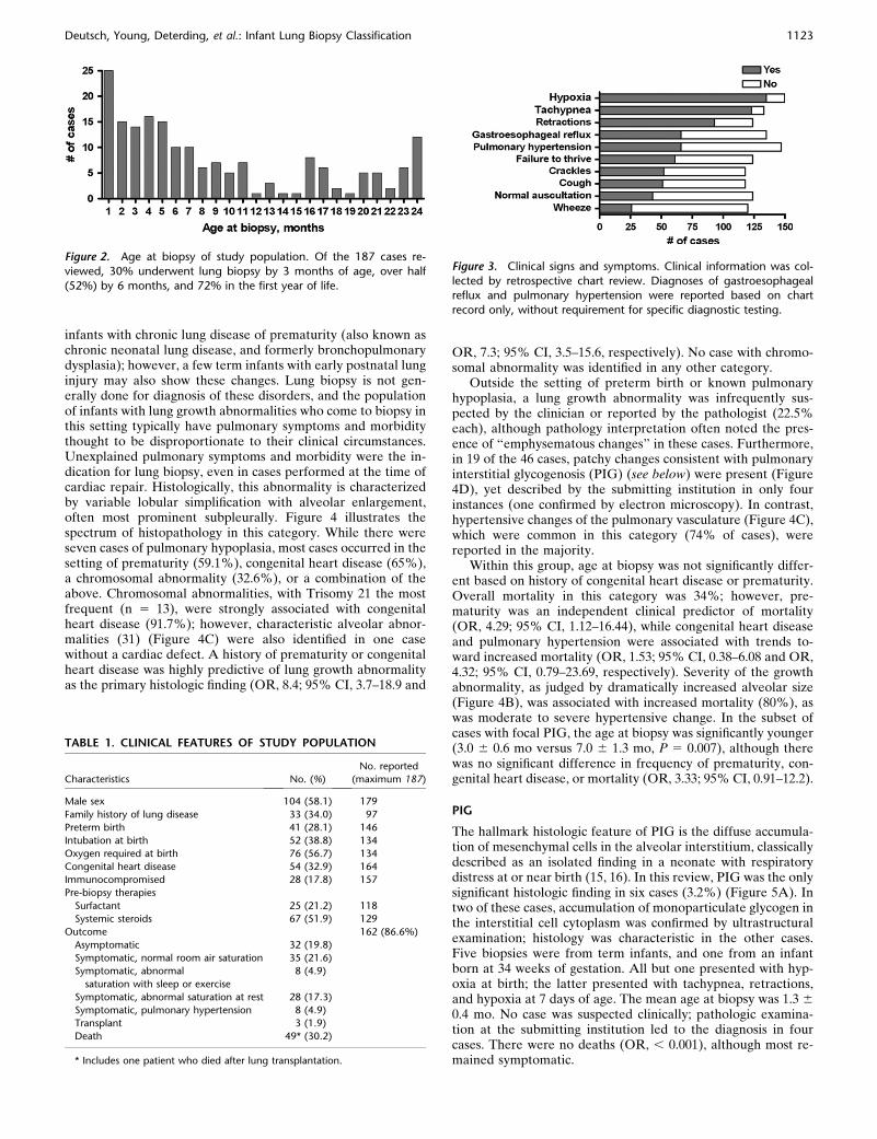
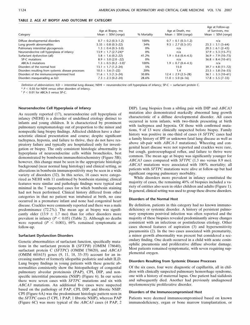
Eleven centers provided pathologic material, clinical data, andimaging studies from children less than 2 years of age who hada lung biopsy for diagnosis of radiographically diffuse lungdisease, yielding 187 cases for review. There was marked varia-tion in the number of biopsies performed among the partici-pating centers (range, 4–37; mean, 17). Age at biopsy favoredyounger infants (mean 8.3 6 0.6 mo) (Figure 2). Clinical fea-tures of the study population are shown in Table 1. A sub-stantial proportion (57%) required oxygen at birth, althoughprematurity was a feature of only 51% of the cases that requiredearly intubation. A variable constellation of signs and symptomswas observed (Figure 3). Over half of patients received systemicsteroids before lung biopsy. Imaging studies of sufficient qualitywere not available in enough cases to provide meaningfulradiologic description and correlation.
Outcome data was available for 86.6% of cases (Table 2). Themean age at follow-up for patients alive at the time of study datacollection was 31.3 months, with a significantly longer mean follow-up duration after biopsy compared with the patients who died(21.8 versus 3.6 mo after lung biopsy, respectively, P , 0.001).Forty-nine (30.2%) patients had died, with a mean age of death of7.7 months. A disease severity scale (7, 27) was used to classify the79 cases that were symptomatic at the time of follow-up. Ina logistic regression model of clinical predictors, mortality wasnot significantly different based on female sex (OR, 1.75; 95%confidence interval [CI], 0.73–4.20) or congenital heart disease(OR, 0.49; 95% CI, 0.17–1.36). Prematurity and a clinical di-agnosis of pulmonary hypertension at the time of lung biopsywere associated with increased mortality (OR, 3.60; 95% CI,1.18–10.96 and OR, 6.84; 95% CI, 2.57–18.20, respectively).
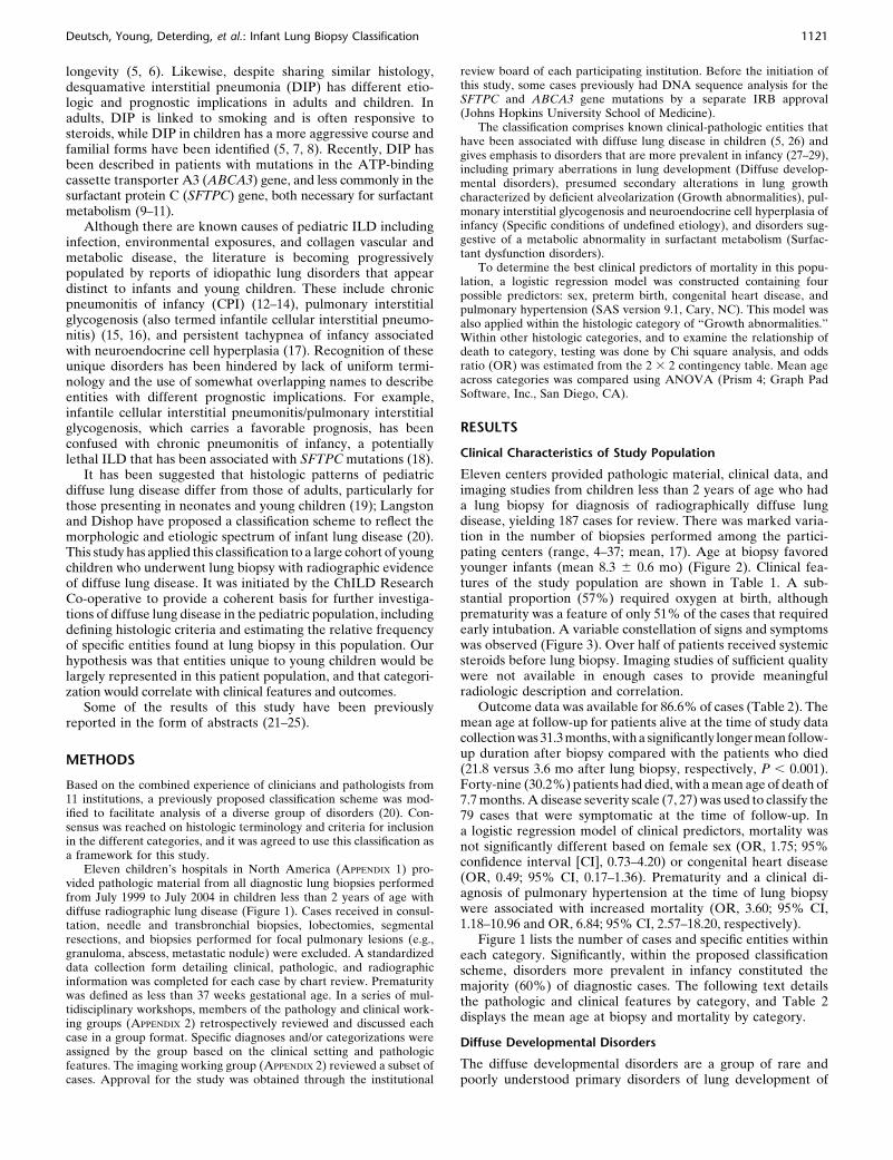
Figure 1 lists the number of cases and specific entities withineach category. Significantly, within the proposed classificationscheme, disorders more prevalent in infancy constituted themajority (60%) of diagnostic cases. The following text detailsthe pathologic and clinical features by category, and Table 2displays the mean age at biopsy and mortality by category.
Diffuse Developmental Disorders
The diffuse developmental disorders are a group of rare andpoorly understood primary disorders of lung development of
Deutsch, Young, Deterding, et al.: Infant Lung Biopsy Classification 1121
unknown etiology, but largely believed to be due to aberrationsin one of the primary molecular mechanisms of lung and/orpulmonary vascular development; they include acinar dysplasia,congenital alveolar dysplasia, and alveolar capillary dysplasiawith misalignment of pulmonary veins (ACDMPV). Acinardysplasia is characterized by lung growth arrest in the pseudo-glandular or early canalicular phase. Congenital alveolar dys-plasia is characterized by growth arrest in the late canalicular/early saccular phase of lung development. The constellation ofmalposition of pulmonary veins adjacent to small pulmonaryarteries, medial hypertrophy of pulmonary arteries and arterio-les, and reduced capillary density with lobular maldevelopmentwas considered diagnostic for ACDMPV. Eleven (6%) caseswere classified as diffuse developmental disorders. No case ofacinar dysplasia was identified in this cohort; identified casesincluded congenital alveolar dysplasia (n 5 2) and ACDMPV(n 5 9). All biopsies were characterized by a striking arrest inlobular development and reduced alveolar capillary density.Cases occurred in term infants, and all presented at birth with
hypoxia and persistent pulmonary hypertension unresponsive toventilatory support. With the exception of one child withACDMPV who underwent lung transplantation at 5 monthsof age, mortality was 100% within the first month of life.
Growth Abnormalities
A lung growth abnormality reflective of impaired pre- or post-natal alveolarization was the principal diagnosis in this review(24.6% of total) and the leading category for 5 of 11 centers.These abnormalities of alveolarization are largely secondaryand may be seen in a wide variety of circumstances. The bestknown of these is pulmonary hypoplasia, in which prenatal con-ditions (restriction of fetal thoracic space, abnormalities in am-niotic fluid volume, skeletal anomalies, neuromuscular problemsresulting in decreased or absent fetal breathing movements,cardiac abnormalities limiting pulmonary blood supply, abdom-inal wall defects, and a variety of chromosomal abnormalities)result in deficient lung growth (30). Postnatal growth abnor-malities are largely limited to the population of premature
Figure 1. Study cohort and
proposed classification of dif-
fuse lung disease in children.The study cohort was com-
posed of patients under the
age of 2 who had a diagnostic
lung biopsy during the desig-nated 5-year interval; exclusion
criteria are listed. The clinical–
pathologic classification scheme
is detailed with numbers ofcases and specific entities iden-
tified within each category.
Some unusual entities not seenare also listed, but there are
clearly other entities associated
with diffuse lung disease not
seen in this cohort that havenot been listed.
1122 AMERICAN JOURNAL OF RESPIRATORY AND CRITICAL CARE MEDICINE VOL 176 2007
infants with chronic lung disease of prematurity (also known aschronic neonatal lung disease, and formerly bronchopulmonarydysplasia); however, a few term infants with early postnatal lunginjury may also show these changes. Lung biopsy is not gen-erally done for diagnosis of these disorders, and the populationof infants with lung growth abnormalities who come to biopsy inthis setting typically have pulmonary symptoms and morbiditythought to be disproportionate to their clinical circumstances.Unexplained pulmonary symptoms and morbidity were the in-dication for lung biopsy, even in cases performed at the time ofcardiac repair. Histologically, this abnormality is characterizedby variable lobular simplification with alveolar enlargement,often most prominent subpleurally. Figure 4 illustrates thespectrum of histopathology in this category. While there wereseven cases of pulmonary hypoplasia, most cases occurred in thesetting of prematurity (59.1%), congenital heart disease (65%),a chromosomal abnormality (32.6%), or a combination of theabove. Chromosomal abnormalities, with Trisomy 21 the mostfrequent (n 5 13), were strongly associated with congenitalheart disease (91.7%); however, characteristic alveolar abnor-malities (31) (Figure 4C) were also identified in one casewithout a cardiac defect. A history of prematurity or congenitalheart disease was highly predictive of lung growth abnormalityas the primary histologic finding (OR, 8.4; 95% CI, 3.7–18.9 and
OR, 7.3; 95% CI, 3.5–15.6, respectively). No case with chromo-somal abnormality was identified in any other category.
Outside the setting of preterm birth or known pulmonaryhypoplasia, a lung growth abnormality was infrequently sus-pected by the clinician or reported by the pathologist (22.5%each), although pathology interpretation often noted the pres-ence of ‘‘emphysematous changes’’ in these cases. Furthermore,in 19 of the 46 cases, patchy changes consistent with pulmonaryinterstitial glycogenosis (PIG) (see below) were present (Figure4D), yet described by the submitting institution in only fourinstances (one confirmed by electron microscopy). In contrast,hypertensive changes of the pulmonary vasculature (Figure 4C),which were common in this category (74% of cases), werereported in the majority.
Within this group, age at biopsy was not significantly differ-ent based on history of congenital heart disease or prematurity.Overall mortality in this category was 34%; however, pre-maturity was an independent clinical predictor of mortality(OR, 4.29; 95% CI, 1.12–16.44), while congenital heart diseaseand pulmonary hypertension were associated with trends to-ward increased mortality (OR, 1.53; 95% CI, 0.38–6.08 and OR,4.32; 95% CI, 0.79–23.69, respectively). Severity of the growthabnormality, as judged by dramatically increased alveolar size(Figure 4B), was associated with increased mortality (80%), aswas moderate to severe hypertensive change. In the subset ofcases with focal PIG, the age at biopsy was significantly younger(3.0 6 0.6 mo versus 7.0 6 1.3 mo, P 5 0.007), although therewas no significant difference in frequency of prematurity, con-genital heart disease, or mortality (OR, 3.33; 95% CI, 0.91–12.2).
PIG
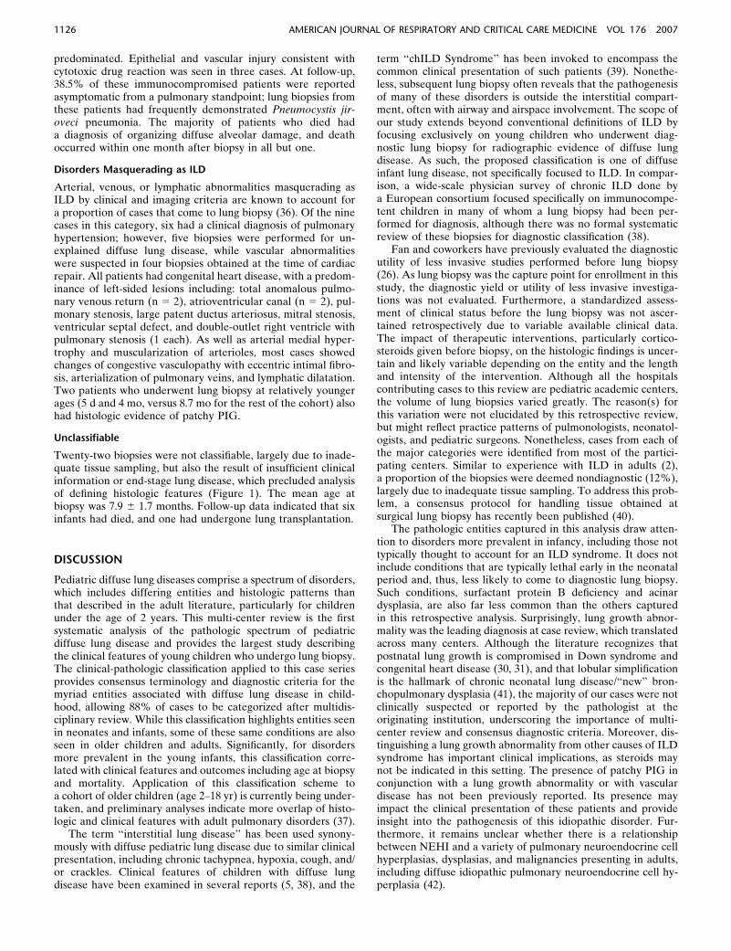
The hallmark histologic feature of PIG is the diffuse accumula-tion of mesenchymal cells in the alveolar interstitium, classicallydescribed as an isolated finding in a neonate with respiratorydistress at or near birth (15, 16). In this review, PIG was the onlysignificant histologic finding in six cases (3.2%) (Figure 5A). Intwo of these cases, accumulation of monoparticulate glycogen inthe interstitial cell cytoplasm was confirmed by ultrastructuralexamination; histology was characteristic in the other cases.Five biopsies were from term infants, and one from an infantborn at 34 weeks of gestation. All but one presented with hyp-oxia at birth; the latter presented with tachypnea, retractions,and hypoxia at 7 days of age. The mean age at biopsy was 1.3 6
0.4 mo. No case was suspected clinically; pathologic examina-tion at the submitting institution led to the diagnosis in fourcases. There were no deaths (OR, , 0.001), although most re-mained symptomatic.
Figure 2. Age at biopsy of study population. Of the 187 cases re-
viewed, 30% underwent lung biopsy by 3 months of age, over half
(52%) by 6 months, and 72% in the first year of life.
Figure 3. Clinical signs and symptoms. Clinical information was col-
lected by retrospective chart review. Diagnoses of gastroesophagealreflux and pulmonary hypertension were reported based on chart
record only, without requirement for specific diagnostic testing.
TABLE 1. CLINICAL FEATURES OF STUDY POPULATION
Characteristics No. (%)
No. reported
(maximum 187)
Male sex 104 (58.1) 179
Family history of lung disease 33 (34.0) 97
Preterm birth 41 (28.1) 146
Intubation at birth 52 (38.8) 134
Oxygen required at birth 76 (56.7) 134
Congenital heart disease 54 (32.9) 164
Immunocompromised 28 (17.8) 157
Pre-biopsy therapies
Surfactant 25 (21.2) 118
Systemic steroids 67 (51.9) 129
Outcome 162 (86.6%)
Asymptomatic 32 (19.8)
Symptomatic, normal room air saturation 35 (21.6)
Symptomatic, abnormal
saturation with sleep or exercise
8 (4.9)
Symptomatic, abnormal saturation at rest 28 (17.3)
Symptomatic, pulmonary hypertension 8 (4.9)
Transplant 3 (1.9)
Death 49* (30.2)
* Includes one patient who died after lung transplantation.
Deutsch, Young, Deterding, et al.: Infant Lung Biopsy Classification 1123
Neuroendocrine Cell Hyperplasia of Infancy
As recently reported (17), neuroendocrine cell hyperplasia ofinfancy (NEHI) is a disorder of undefined etiology distinct toinfants and young children. It is characterized by prominentpulmonary symptomatology out of proportion to the minor andnonspecific lung biopsy findings. Affected children have a char-acteristic clinical presentation and course; despite significanttachypnea, hypoxia, and failure to thrive, they do not have res-piratory failure and typically are hospitalized only for investi-gation or biopsy. The only consistent histologic abnormality ishyperplasia of neuroendocrine cells within bronchioles, bestdemonstrated by bombesin immunohistochemistry (Figure 5B);however, this change must be seen in the appropriate histologicbackground (near normal lung histology) and clinical setting, asalterations in bombesin immunopositivity may be seen in a widevariety of disorders (32). In this series, 18 cases were catego-rized as NEHI with 11 confirmed by bombesin staining; clinicalfeatures were similar and pathologic findings were typical andminimal in the 7 suspected cases for which bombesin staininghad not been performed. Clinical history differed from othercategories in that no patient was intubated at birth, no caseoccurred in a premature infant and none had congenital heartdisease. Crackles were commonly reported and there was a malepredominance (72.2%). The mean age at biopsy was signifi-cantly older (13.9 6 1.7 mo) than for other disorders moreprevalent in infancy (P , 0.05) (Table 2). Although no deathswere reported (P , 0.001), 69% remained symptomatic atfollow-up.
Surfactant Dysfunction Disorders
Genetic abnormalities of surfactant function, specifically muta-tions in the surfactant protein B (SFTPB) (OMIM 178640),surfactant protein C (SFTPC) (OMIM 178620), and ABCA3(OMIM 601615) genes (9, 11, 18, 33–35) account for an in-creasing number of formerly idiopathic pediatric and adult ILD.Lung biopsy findings in young patients with these genetic ab-normalities consistently show the histopathology of congenitalpulmonary alveolar proteinosis (PAP), CPI, DIP, and non-specific interstitial pneumonia (NSIP) (Figure 6). In our seriesthere were seven cases with SFTPC mutations and six withABCA3 mutations. An additional five cases were suspectedbased on the pathology of PAP, CPI, DIP, and fibrotic NSIP.CPI (Figure 6A) was the predominant histologic pattern seen inthe SFTPC cases (5 CPI, 1 PAP, 1 fibrotic NSIP), whereas PAP(Figure 6C) was more typical of the ABCA3 cases (4 PAP, 2
DIP). Lung biopsies from a sibling pair with DIP and ABCA3mutation also demonstrated markedly abnormal lung growthcharacteristic of a diffuse developmental disorder. All casesoccurred in term infants, with two-thirds presenting at birthwith tachypnea and hypoxia. Of those with confirmed muta-tions, 9 of 13 were clinically suspected before biopsy. Familyhistory was positive in one-third of cases (4 SFTPC cases hada family history of IPF or unknown fatal lung disease as well asabove sib-pair with ABCA-3 mutations). Wheezing and con-genital heart disease were not reported and crackles were rare,but cough, gastroesophageal reflux, and failure to thrive werecommon. The mean age at biopsy was significantly younger forABCA3 cases compared with SFTPC (1.3 mo versus 8.9 mo).ABCA3 mutations were associated with 100% mortality; allpatients with SFTPC mutations were alive at follow-up but hadsignificant ongoing pulmonary morbidity.
While disorders more prevalent in infancy constituted themajority of cases, the remaining 40% of biopsies included a va-riety of entities also seen in older children and adults (Figure 1).In general, clinical setting was used to group these diverse disorders.
Disorders of the Normal Host
By definition, patients in this category had no known immuno-deficiency or systemic disorder. A history of persistent pulmo-nary symptoms postviral infection was often reported and themajority of these biopsies revealed predominantly airway changesconsistent with an infectious or postinfectious etiology. Othercases showed features of aspiration (3) and hypersensitivitypneumonitis (2). In the two cases associated with prematurity,a minor growth abnormality was present but considered a sec-ondary finding. One death occurred in a child with acute eosin-ophilic pneumonia and proliferative diffuse alveolar damage.Most patients remained symptomatic, with seven requiring sup-plemental oxygen.
Disorders Resulting from Systemic Disease Processes
Of the six cases, four were diagnostic of capillaritis, all in chil-dren with clinically suspected pulmonary hemorrhage syndrome,one with a history of maternal lupus. One patient had sialidosisand subsequently died. Another had previously undiagnosedmyelomonocytic proliferative disorder.
Disorders of the Immunocompromised Host
Patients were deemed immunocompromised based on knownimmunodeficiency, organ or bone marrow transplantation, or
TABLE 2. AGE AT BIOPSY AND OUTCOME BY CATEGORY
Category
Age at Biopsy, mo
Mean 6 SEM (range) % Mortality
Age at Death, mo
Mean 6 SEM (range)
Age at Follow-up
of Survivors, mo
Mean 6 SEM (range)
Diffuse developmental disorders 0.7 6 0.2 (0.3–1.2) 100% 0.7 6 0.1 (0.3–1.2) n/a
Lung growth abnormalities 5.35 6 0.8 (0.3–22) 34% 9.5 6 2.7 (0.3–31) 25.3 6 3.1 (3–64)
Pulmonary interstitial glycogenosis 1.3 6 0.4 (0.3–3.0) 0% n/a 20.3 6 6.1 (2–43)
Neuroendocrine cell hyperplasia of infancy 13.9 6 1.7 (2.7-24)* 0% n/a 37.9 6 5.3 (15–71)
Surfactant dysfunction (all) 5.8 6 1.6 (0.2–22) 41.2% 1.9 6 0.6 (0.4–4.5) 36.9 6 7.9 (10–73)
SP-C mutations 8.9 6 3.0 (2.0 –22) 0% n/a 36.8 6 8.4 (10–61)
ABCA-3 mutations 1.3 6 0.5 (0.2 –3.0)† 100% 1.9 6 0.7 (0.4–4.5) n/a
Disorders of the normal host 13.1 6 1.7 (1.2–24) 5% 15.0 39.7 6 4.8 (11–72)
Disorders resulting from systemic disease processes 10.5 6 3.6 (1–22) 20% 1.4 23.5 6 3.8 (16–33)
Disorders of the immunocompromised host 11.6 6 1.3 (1.5–24) 30.8% 12.4 6 2.9 (2.5–28) 36.1 6 3.3 (10–61)
Disorders masquerading as ILD 7.3 6 2.3 (0.2–24) 28.6% 11.0 6 5.0 (6–16) 17.8 6 5.5 (7–33)
Definition of abbreviations: ILD 5 interstitial lung disease; NEHI 5 neuroendocrine cell hyperplasia of infancy; SP-C 5 surfactant protein C.
* P , 0.05 for NEHI versus other disorders of infancy.† P , 0.01 for ABCA-3 versus SP-C.
1124 AMERICAN JOURNAL OF RESPIRATORY AND CRITICAL CARE MEDICINE VOL 176 2007
receiving chemotherapy for malignancy. Those receiving immu-nomodulating therapies for their systemic disorders were clas-sified according to their underlying disease process. In the im-munocompromised group, 9 biopsies were from infants who had
undergone bone marrow transplantation, 1 after heart trans-plantation, 11 with congenital immune deficiency, 2 with HIVinfection, and 5 being treated for malignancy. Clinical presen-tation was variable, and infectious and postinfectious diagnoses
Figure 6. Histology consistent with surfactant dysfunction
disorder. The lung biopsies of an infant with an SFTPCmutation (A, H&E, 3200) and an infant with ABCA3 mu-
tations (C, H&E, 3200) are both characterized by diffuse
alveolar septal thickening with uniform prominent type II
cells. Accumulation of intra-alveolar macrophages and in-terstitial infiltrate is more typical in the patient with the
SFTPC mutation (chronic pneumonitis of infancy pattern),
while granular proteinosis is more prominent in the biopsyof the patient with ABCA3 mutations (pulmonary alveolar
proteinosis pattern). Electron micrograph demonstrates
the abnormal lamellar bodies with dense inclusions in
a 3-month-old with ABCA3 mutations (D, 316,000). Patchyinterstitial fibrosis, characteristic of NSIP, is present in
a 22-month-old child who was found to have an SP-C
mutation (B, H&E, 3200); a small degree of proteinosis with
foamy macrophages is still visible (arrow).
Figure 5. Pulmonary interstitial glycogenosis and neuroen-
docrine cell hyperplasia of infancy. There is diffuse interstitial
widening by mesenchymal cells in a 22-day-old term neo-nate with tachypnea at birth (A, H&E, 3200). The presence
of monoparticulate glycogen within the cells, diagnostic of
pulmonary interstitial glycogenosis, is demonstrated by ultra-
structural examination (inset, A, 315,200). Clusters ofbombesin-immunopositive cells within bronchioles (arrows)
and within the lobular parenchyma (arrowheads) are shown
from a patient with neuroendocrine cell hyperplasia of infancy(B, bombesin immunostain [polyclonal; ImmunoStar] 3100).
Figure 4. Lung growth abnormalities. Compared with
lung from a normal term infant (A, hematoxylin and eosin
[H&E], 340), there is reduced alveolarization (deficient
lung growth) in an infant with pulmonic stenosis withmarkedly enlarged alveoli (see size relative to bronchioles)
(B, H&E, 340). Deficient alveolarization in a patient with
Trisomy 21 is characterized by cystic dilatation of sub-
pleural alveoli (C, H&E, 340); pulmonary arterioles dem-onstrate occlusive intimal hyperplasia (insert, H&E, 3200).
Lobular simplification in an infant born at 27 weeks
gestation is accompanied by patchy pulmonary interstitialglycogenosis (D, H&E, 340). Inset (H&E, 3200) displays
the characteristic cells with round to oval nuclei, vacuo-
lated cytoplasm, and indistinct cell borders.
Deutsch, Young, Deterding, et al.: Infant Lung Biopsy Classification 1125
predominated. Epithelial and vascular injury consistent withcytotoxic drug reaction was seen in three cases. At follow-up,38.5% of these immunocompromised patients were reportedasymptomatic from a pulmonary standpoint; lung biopsies fromthese patients had frequently demonstrated Pneumocystis jir-oveci pneumonia. The majority of patients who died hada diagnosis of organizing diffuse alveolar damage, and deathoccurred within one month after biopsy in all but one.
Disorders Masquerading as ILD
Arterial, venous, or lymphatic abnormalities masquerading asILD by clinical and imaging criteria are known to account fora proportion of cases that come to lung biopsy (36). Of the ninecases in this category, six had a clinical diagnosis of pulmonaryhypertension; however, five biopsies were performed for un-explained diffuse lung disease, while vascular abnormalitieswere suspected in four biopsies obtained at the time of cardiacrepair. All patients had congenital heart disease, with a predom-inance of left-sided lesions including: total anomalous pulmo-nary venous return (n 5 2), atrioventricular canal (n 5 2), pul-monary stenosis, large patent ductus arteriosus, mitral stenosis,ventricular septal defect, and double-outlet right ventricle withpulmonary stenosis (1 each). As well as arterial medial hyper-trophy and muscularization of arterioles, most cases showedchanges of congestive vasculopathy with eccentric intimal fibro-sis, arterialization of pulmonary veins, and lymphatic dilatation.Two patients who underwent lung biopsy at relatively youngerages (5 d and 4 mo, versus 8.7 mo for the rest of the cohort) alsohad histologic evidence of patchy PIG.
Unclassifiable
Twenty-two biopsies were not classifiable, largely due to inade-quate tissue sampling, but also the result of insufficient clinicalinformation or end-stage lung disease, which precluded analysisof defining histologic features (Figure 1). The mean age atbiopsy was 7.9 6 1.7 months. Follow-up data indicated that sixinfants had died, and one had undergone lung transplantation.
DISCUSSION
Pediatric diffuse lung diseases comprise a spectrum of disorders,which includes differing entities and histologic patterns thanthat described in the adult literature, particularly for childrenunder the age of 2 years. This multi-center review is the firstsystematic analysis of the pathologic spectrum of pediatricdiffuse lung disease and provides the largest study describingthe clinical features of young children who undergo lung biopsy.The clinical-pathologic classification applied to this case seriesprovides consensus terminology and diagnostic criteria for themyriad entities associated with diffuse lung disease in child-hood, allowing 88% of cases to be categorized after multidis-ciplinary review. While this classification highlights entities seenin neonates and infants, some of these same conditions are alsoseen in older children and adults. Significantly, for disordersmore prevalent in the young infants, this classification corre-lated with clinical features and outcomes including age at biopsyand mortality. Application of this classification scheme toa cohort of older children (age 2–18 yr) is currently being under-taken, and preliminary analyses indicate more overlap of histo-logic and clinical features with adult pulmonary disorders (37).
The term ‘‘interstitial lung disease’’ has been used synony-mously with diffuse pediatric lung disease due to similar clinicalpresentation, including chronic tachypnea, hypoxia, cough, and/or crackles. Clinical features of children with diffuse lungdisease have been examined in several reports (5, 38), and the
term ‘‘chILD Syndrome’’ has been invoked to encompass thecommon clinical presentation of such patients (39). Nonethe-less, subsequent lung biopsy often reveals that the pathogenesisof many of these disorders is outside the interstitial compart-ment, often with airway and airspace involvement. The scope ofour study extends beyond conventional definitions of ILD byfocusing exclusively on young children who underwent diag-nostic lung biopsy for radiographic evidence of diffuse lungdisease. As such, the proposed classification is one of diffuseinfant lung disease, not specifically focused to ILD. In compar-ison, a wide-scale physician survey of chronic ILD done bya European consortium focused specifically on immunocompe-tent children in many of whom a lung biopsy had been per-formed for diagnosis, although there was no formal systematicreview of these biopsies for diagnostic classification (38).
Fan and coworkers have previously evaluated the diagnosticutility of less invasive studies performed before lung biopsy(26). As lung biopsy was the capture point for enrollment in thisstudy, the diagnostic yield or utility of less invasive investiga-tions was not evaluated. Furthermore, a standardized assess-ment of clinical status before the lung biopsy was not ascer-tained retrospectively due to variable available clinical data.The impact of therapeutic interventions, particularly cortico-steroids given before biopsy, on the histologic findings is uncer-tain and likely variable depending on the entity and the lengthand intensity of the intervention. Although all the hospitalscontributing cases to this review are pediatric academic centers,the volume of lung biopsies varied greatly. The reason(s) forthis variation were not elucidated by this retrospective review,but might reflect practice patterns of pulmonologists, neonatol-ogists, and pediatric surgeons. Nonetheless, cases from each ofthe major categories were identified from most of the partici-pating centers. Similar to experience with ILD in adults (2),a proportion of the biopsies were deemed nondiagnostic (12%),largely due to inadequate tissue sampling. To address this prob-lem, a consensus protocol for handling tissue obtained atsurgical lung biopsy has recently been published (40).
The pathologic entities captured in this analysis draw atten-tion to disorders more prevalent in infancy, including those nottypically thought to account for an ILD syndrome. It does notinclude conditions that are typically lethal early in the neonatalperiod and, thus, less likely to come to diagnostic lung biopsy.Such conditions, surfactant protein B deficiency and acinardysplasia, are also far less common than the others capturedin this retrospective analysis. Surprisingly, lung growth abnor-mality was the leading diagnosis at case review, which translatedacross many centers. Although the literature recognizes thatpostnatal lung growth is compromised in Down syndrome andcongenital heart disease (30, 31), and that lobular simplificationis the hallmark of chronic neonatal lung disease/‘‘new’’ bron-chopulmonary dysplasia (41), the majority of our cases were notclinically suspected or reported by the pathologist at theoriginating institution, underscoring the importance of multi-center review and consensus diagnostic criteria. Moreover, dis-tinguishing a lung growth abnormality from other causes of ILDsyndrome has important clinical implications, as steroids maynot be indicated in this setting. The presence of patchy PIG inconjunction with a lung growth abnormality or with vasculardisease has not been previously reported. Its presence mayimpact the clinical presentation of these patients and provideinsight into the pathogenesis of this idiopathic disorder. Fur-thermore, it remains unclear whether there is a relationshipbetween NEHI and a variety of pulmonary neuroendocrine cellhyperplasias, dysplasias, and malignancies presenting in adults,including diffuse idiopathic pulmonary neuroendocrine cell hy-perplasia (42).
1126 AMERICAN JOURNAL OF RESPIRATORY AND CRITICAL CARE MEDICINE VOL 176 2007

Case reports and small series of patients with surfactantmutations (9–11, 18, 34, 35) have shown four histologic patterns,PAP, CPI, DIP, and NSIP, previously thought to representdistinct disorders. Systematic review of lung biopsies in thiscategory demonstrates that these distinctive histologic patternsare functionally linked by related genetic mechanisms involvingdefects in a common pathway. The recognition that these previ-ously separate diagnostic categories have related genetic mech-anisms provides a rationale for classifying the lung pathologyobserved in these infants under the inclusive term of surfactantdysfunction. While the number of cases available for analysiswas small, this review also provides the first indication thata specific histologic pattern may distinguish an SFTPC fromABCA3 mutations. With the recent availability of testing inclinical genetic laboratories, lung disease due to abnormalitiesin surfactant metabolism may be diagnosed noninvasively,thereby obviating the need for biopsy in selected cases. Ifetiologies for specific disorders such as PIG and NEHI can beidentified, these will also allow for a more mechanistic classifi-cation of diffuse lung disease and ILD in children.
Pediatric ILD has been associated with high morbidity andmortality, and previous studies have suggested that outcomesmay be particularly poor for infants and young children (7). Inthis study, 30% of children had died, and 50% had ongoingpulmonary symptoms at follow-up. Clinical presentation andoutcome were consistent with that of previous case series (5, 9,16, 17), and pulmonary hypertension was the single greatestclinical predictor of mortality (7). Immunocompromised statusand the presence of congenital heart disease were not indepen-dent predictors of outcome. The defining histopathology in thisstudy frequently suggested important prognostic information,particularly in disorders more prevalent in young children. Forexample, disorders within the categories of Diffuse develop-mental, Growth abnormality, and PIG often had similar clinicalpresentations, but outcome was related to histologic diagnosis.
This multidisciplinary working group reviewed a large num-ber of lung biopsies from young children to formalize diagnosticcriteria and terminology for classification of pediatric diffuselung disease. As diagnostic criteria for the infant lung disorderswere formulated during the review process, interobserver agree-ment and concordance were not assessed. While an imaginggroup actively participated in this study, the limited availabilityof high-quality scans precluded inclusion of radiographic de-scription in the current study. We anticipate that the evolvingimplementation of standard techniques for high-quality CT imag-ing in young children at a greater number of centers will providea foundation for clinical-radiographic–pathologic correlation inthese disorders. Until specific radiographic–histologic correla-tions are established, it is unknown whether imaging patternsmay decrease the need for lung biopsy in certain infant lungdisorders, as has occurred for adults with IPF.
This cross-sectional study highlights the utility of the pro-posed classification by grouping clinically distinct patients anddisease entities. Prospective studies that include clinical, radio-graphic, and pathologic correlation are required to evaluate theprognostic value of this classification scheme. Protocol-drivenevaluation and therapeutic intervention for these unique enti-ties will only be possible once standardized terminology anddiagnostic techniques are utilized. Multi-center and multidisci-plinary involvement is vital to this effort.
Conflict of Interest Statement: R.R.D. received $6,294 in 2005, $5,132 in 2006,and $606 in 2007 in clinical research grant dollars from Inspire Pharmaceuticalsfor a multi-center clinical trial. R.R.D. has received speaking fees totaling $2,000from Inspire Pharmaceuticals for 2006. None of the other authors has a financialrelationship with a commercial entity that has an interest in the subject of thismanuscript.
Acknowledgment: The authors are indebted to Pam Groen for her assistance indevelopment of the database, and to all the physicians who participated in theclinical care of these patients.
References
1. Fan LL, Deterding RR, Langston C. Pediatric interstitial lung diseaserevisited. Pediatr Pulmonol 2004;38:369–378.
2. Travis WD, King T, Bateman ED, Lynch DA, Capron F, Center M,Colby TV, Cordier J-F, duBois RM, Galvin JR, et al. ATS/ERSInternational multidisciplinary consensus classification of idiopathicinterstitial pneumonia. Am J Respir Crit Care Med 2002;165:277–304.
3. Sharief N, Crawford OF, Dinwiddie R. Fibrosing alveolitis and desqua-mative interstitial pneumonitis. Pediatr Pulmonol 1994;17:359–365.
4. Nicholson AG, Kim H, Corrin B, Bush A, du Bois RM, Rosenthal M,Sheppard MN. The value of classifying interstitial pneumonitis inchildhood according to defined histological patterns. Histopathology1998;33:203–211.
5. Fan LL, Mullen AL, Brugman SM, Inscore SC, Parks DP, White CW.Clinical spectrum of chronic interstitial lung disease in children.J Pediatr 1992;121:867–872.
6. Fan LL, Langston C. Pediatric interstitial lung disease: children are notsmall adults. Am J Respir Crit Care Med 2002;165:1466–1467.
7. Fan LL, Kozinetz CA. Factors influencing survival in children withchronic interstitial lung disease. Am J Respir Crit Care Med 1997;156:939–942.
8. Tal A, Maor E, Bar-Ziv J, Gorodischer R. Fatal desquamative inter-stitial pneumonia in three infants siblings. J Pediatr 1984;104:873–876.
9. Shulenin S, Nogee LM, Annilo T, Wert SE, Whitsett JA, Dean M.ABCA3 gene mutations in newborns with fatal surfactant deficiency.N Engl J Med 2004;350:1296–1303.
10. Nogee LM, Dunbar AE III, Wert SE, Askin F, Hamvas A, Whitsett JA.A mutation in the surfactant protein C gene associated with familialinterstitial lung disease. N Engl J Med 2001;344:573–579.
11. Bullard JE, Wert SE, Whitsett JA, Dean M, Nogee LM. ABCA3mutations associated with pediatric interstitial lung disease. Am JRespir Crit Care Med 2005;172:1026–1031.
12. Fisher M, Roggli V, Merten D, Mulvihill D, Spock A. Coexistingendogenous lipoid pneumonia, cholesterol granulomas, and pulmo-nary alveolar proteinosis in a pediatric population: a clinical, radio-graphic, and pathologic correlation. Pediatr Pathol 1992;12:365–383.
13. Katzenstein AL, Gordon LP, Oliphant M, Swender PT. Chronicpneumonitis of infancy: a unique form of interstitial lung diseaseoccurring in early childhood. Am J Surg Pathol 1995;19:439–447.
14. Kavantzas N, Theocharis S, Agapitos E, Davaris P. Chronic pneumonitisof infancy: an autopsy study of 12 cases. Clin Exp Pathol 1999;47:96–100.
15. Canakis AM, Cutz E, Manson D, O’Brodovich H. Pulmonary interstitialglycogenosis: a new variant of neonatal interstitial lung disease. Am JRespir Crit Care Med 2002;165:1557–1565.
16. Schroeder SA, Shannon DC, Mark EJ. Cellular interstitial pneumonitisin infants: a clinicopathologic study. Chest 1992;101:1065–1069.
17. Deterding RR, Pye C, Fan LL, Langston C. Persistent tachypnea ofinfancy is associated with neuroendocrine cell hyperplasia. PediatrPulmonol 2005;40:157–165.
18. Cameron HS, Somaschini M, Carrera P, Hamvas A, Whitsett JA, Wert SE,Deutsch G, Nogee LM. A common mutation in the surfactant protein Cgene associated with lung disease. J Pediatr 2005;146:370–375.
19. Langston C. Pediatric lung biopsy, diagnostic pulmonary pathology. NewYork: Marcel Dekker, Inc.; 2000.
20. Langston C, Dishop MK. Infant lung biopsy: clarifying the pathologicspectrum. Pathol Int 2004;54:S414–S427.
21. Young LR, Deterding R, Deutsch GH, Hilman B, Dell S, Fan L,Langston C, and the pILD Pathology and Imaging Cooperative.Clinical characteristics of a new histologic classification of interstitiallung disease in infants [abstract]. Proc Am Thorac Soc 2005;2:A474.
22. Dishop MK, Deutsch GH, Deterding R, Fan LL, Young LR, Cutz E,Langston C, and the pILD Pathology and Imaging Cooperative.A working histologic classification of the pediatric interstitial lung dis-ease cooperative group [abstract]. Proc Am Thorac Soc 2005;2:A474.
23. Dell SD, Young LR, Deterding R, Deutsch GH, Hilman B, Fan LL,Langston C. Lung biopsies and clinical characteristics of ILD in in-fants: Experience of 11 centers, 1999–2004 [abstract]. Proc Am ThoracSoc 2005;A475.
24. Deutsch GH, Albright E, Chou PM, Cool CD, Coventry S, Davis MM,Dishop MK, Galambos C, Patterson K, Wert SE, et al., for thepILD Pathology and Imaging Cooperative. Defining the spectrum
Deutsch, Young, Deterding, et al.: Infant Lung Biopsy Classification 1127
of diffuse lung disease in infancy: a working classification of thePediatric Interstitial Lung Disease Cooperative. Mod Pathol 2005;18:304.
25. Young LR, Deutsch GH, Long F, White F, Sweet S, Fan LL, Langston
C, Deterding R, Brody A, and the Children’s Interstitial LungDisease Network. Growth abnormalities: an under-recognized causeof diffuse lung disease in young children [abstract]. Proc Am ThoracSoc 2006;3:A677.
26. Fan LL, Kozinetz CA, Deterding RR, Brugman SM. Evaluation of
a diagnostic approach to pediatric interstitial lung disease. Pediatrics1998;101:82–85.
27. Fan LL, Langston C. Chronic interstitial lung disease in children. Pediatr
Pulmonol 1993;16:184–196.28. Langston C, Fan LL. Diffuse interstitial lung disease in infants. Pediatr
Pulmonol Suppl 2001;23:74–76.29. Langston C, Fan LL. The spectrum of interstitial lung disease in
childhood. Pediatr Pulmonol Suppl 2001;23:70–71.30. Sherer DM, Davis JM, Woods JR Jr. Pulmonary hypoplasia: a review.
Obstet Gynecol Surv 1990;45:792–803.31. Cooney TP, Thurlbeck WM. Pulmonary hypoplasia in Down’s syn-
drome. N Engl J Med 1982;307:1170–1173.32. Johnson DE, Georgieff MK. Pulmonary neuroendocrine cells: their
secretory products and their potential roles in health and chronic lungdisease in infancy. Am Rev Respir Dis 1989;140:1807–1812.
33. Nogee LM, Dunbar AE III, Wert S, Askin F, Hamvas A, Whitsett JA.
Mutations in the surfactant protein C gene associated with interstitiallung disease. Chest 2002;121:20S–21S.
34. Nogee LM, de Mello DE, Dehner LP, Colten HR. Brief report:
deficiency of pulmonary surfactant protein B in congenital alveolarproteinosis. N Engl J Med 1993;328:406–410.
35. Chibbar R, Shih F, Baga M, Torlakovic E, Ramlall K, Skomro R,
Cockcroft DW, Lemire EG. Nonspecific interstitial pneumonia andusual interstitial pneumonia with mutation in surfactant protein C infamilial pulmonary fibrosis. Mod Pathol 2004;17:973–980.
36. Sondheimer HM, Lung MC, Brugman SM, Ikle DN, Fan LL, White CW.
Pulmonary vascular disorders masquerading as interstitial lung dis-ease. Pediatr Pulmonol 1995;20:284–288.
37. Dishop MK, Askin FB, Galambos C, White FV, Deterding RR, Young
LR, Langston C, for the chILD Network. Classification of diffuse lungdisease in older children and adolescents: a multi-institutional studyof the Children’s Interstitial Lung Disease (chILD) pathology work-ing group. Mod Pathol 2007;20:287–288.
38. Clement A. Task force on chronic interstitial lung disease in immuno-
competent children. Eur Respir J 2004;24:686–697.39. Deterding R, Fan LL. Surfactant dysfunction mutations in children’s
interstitial lung disease and beyond. Am J Respir Crit Care Med 2005;172:940–941.
40. Langston C, Patterson K, Dishop MK, Askin F, Baker P, Chou P, Cool
C, Coventry S, Cutz E, Davis M, et al. A protocol for the handling oftissue obtained by operative lung biopsy: recommendations of thechILD pathology co-operative group. Pediatr Dev Pathol 2006;9:173–180.
41. Husain AN, Siddiqui NH, Stocker JT. Pathology of arrested acinardevelopment in postsurfactant bronchopulmonary dysplasia. HumPathol 1998;29:710–717.
42. Davies SJ, Gosney JR, Hansell DM, Wells AU, du Bois RM, BurkeMM, Sheppard MN, Nicholson AG. Diffuse idiopathic pulmonaryneuroendocrine cell hyperplasia: an under-recognised spectrum ofdisease. Thorax 2007;62:248–252.
APPENDIX 1
Institutions contributing case material were: Children’s Hospital,Columbus, Ohio; Children’s Hospital, Denver, Colorado; Child-ren’s Hospital and Regional Medical Center, Seattle, Washington;Children’s Hospital of Pittsburgh, Pittsburgh, Pennsylvania;Children’s Memorial Hospital, Chicago, Illinois; CincinnatiChildren’s Hospital Medical Center, Cincinnati, Ohio; Hospitalfor Sick Children, Toronto, Ontario; James Whitcomb RileyHospital for Children, Indianapolis, Indiana; Kosair Children’sHospital, Louisville, Kentucky; Washington University, St.Louis, Missouri; Texas Children’s Hospital, Houston, Texas.
APPENDIX 2
Members of the ChILD Research Co-operative who partici-pated in the current study.
Pathology Working GroupChair: Claire Langston, M.D.; Members: Eric Albright,
M.D., Fred Askin, M.D., Peter Baker, M.D., Pauline Chou,M.D., Carlyne Cool, M.D., Susan Coventry, M.D., Ernest Cutz,M.D., Mary Davis, M.D., Gail Deutsch, M.D., Megan Dishop,M.D., William Funkhouser, M.D., Csaba Galambos, M.D.,Kathleen Patterson, M.D., William Travis, M.D., Susan Wert,Ph.D., Frances White, M.D.
Clinical Working GroupChair: Robin Deterding, M.D.; Members: Robert Castile,
M.D., Sharon Dell, M.D., Leland Fan, M.D., Aaron Hamvas,M.D., Bettina Hilman, M.D., Geoffrey Kurland, M.D., GeorgeMallory, M.D., Susanna McColley, M.D., Ronald Morton,M.D., Lawrence Nogee, M.D., Gregory Redding, M.D., StuartSweet, M.D., Lisa Young, M.D.
Imaging Working GroupChair: Alan Brody, M.D.; Members: Eric Crotty, M.D., Eric
Effman, M.D., Paul Guillerman, M.D., Fred Long, M.D., DavidLynch, M.D.
1128 AMERICAN JOURNAL OF RESPIRATORY AND CRITICAL CARE MEDICINE VOL 176 2007