Modeling suspended sediment sources and transport in the Ishikari River Basin using SPARROW

Differential Regulation of Adipokines May InfluenceMigratory Behavior in the White-Throated Sparrow(Zonotrichia albicollis)Erica F. Stuber1,2, Jessica Verpeut1, Maria Horvat-Gordon1, Ramesh Ramachandran1, Paul A. Bartell1,2*
1Department of Animal Science, The Pennsylvania State University, University Park, Pennsylvania, United States of America, 2 Ecology Graduate Program, The
Pennsylvania State University, University Park, Pennsylvania, United States of America
Abstract
White-throated sparrows increase fat deposits during pre-migratory periods and rely on these fat stores to fuel migration.Adipose tissue produces hormones and signaling factors in a rhythmic fashion and may be controlled by a clock in adiposetissue or driven by a master clock in the brain. The master clock may convey photoperiodic information from theenvironment to adipose tissue to facilitate pre-migratory fattening, and adipose tissue may, in turn, release adipokines toindicate the extent of fat energy stores. Here, we present evidence that a change in signal from the adipokines adiponectinand visfatin may act to indicate body condition, thereby influencing an individual’s decision to commence migratory flight,or to delay until adequate fat stores are acquired. We quantified plasma adiponectin and visfatin levels across the day incaptive birds held under constant photoperiod. The circadian profiles of plasma adiponectin in non-migrating birds wereapproximately inverse the profiles from migrating birds. Adiponectin levels were positively correlated to body fat, and bodyfat was inversely related to the appearance of nocturnal migratory restlessness. Visfatin levels were constant across the dayand did not correlate with fat deposits; however, a reduction in plasma visfatin concentration occurred during the migratoryperiod. The data suggest that a significant change in the biological control of adipokine expression exists between the twomigratory conditions and we propose a role for adiponectin, visfatin and adipose clocks in the regulation of migratorybehaviors.
Citation: Stuber EF, Verpeut J, Horvat-Gordon M, Ramachandran R, Bartell PA (2013) Differential Regulation of Adipokines May Influence Migratory Behavior inthe White-Throated Sparrow (Zonotrichia albicollis). PLoS ONE 8(6): e59097. doi:10.1371/journal.pone.0059097
Editor: Claudia Mettke-Hofmann, Liverpool John Moores University, United Kingdom
Received August 7, 2012; Accepted February 12, 2013; Published June 13, 2013
Copyright: � 2013 Stuber et al. This is an open-access article distributed under the terms of the Creative Commons Attribution License, which permitsunrestricted use, distribution, and reproduction in any medium, provided the original author and source are credited.
Funding: This project was funded by The Pennsylvania State University Dept. of Poultry Science and College of Agricultural Sciences. The funders had no role instudy design, data collection and analysis, decision to publish, or preparation of the manuscript.
Competing Interests: The authors have declared that no competing interests exist.
* E-mail: [email protected]
Introduction
Small songbirds have evolved many strategies to cope with the
costly rigors of long-distance migration. During the pre-migratory
period, physiological and endocrine systems change in preparation
for migration; birds become hyperphagic and alter their metab-
olism resulting in increased fat deposition [1,2]. Fat provides more
than eight times the energy per gram as protein or carbohydrates
and is stored with minimal water, making it a valuable source of
energy during migration [3]. The migratory programs of many
small songbirds consist of segments of nocturnal flights and
subsequent daytime stops to rest and refuel. Flights during
successive nights under poor conditions may lower fuel reserves
below critical thresholds, requiring birds to delay longer at
stopover sites to replenish fat stores [4,5]. Although many small
passerines commence migratory flight after peak fat deposition has
been reached [1], how each individual determines whether it has
enough fat accumulated to initiate migratory flight is unclear [6,7].
Pre-migratory hyperphagia and fattening may lead to changes in
metabolic signals, such as adipokines, that circulate in the blood of
individual birds. Consequently, changes in metabolic signals could
be utilized to initiate migration once fat stores reach a critical level.
Changes in food intake and dietary composition during pre-
migration and the migratory period during stopovers can elicit
changes in individuals’ migratory behaviors [7]. Birds are able to
compensate for low diet quality by increasing food intake and
decreasing migratory restlessness [7,8]. Additionally, previous
work has demonstrated that physical condition is related to the
initiation of spring migration, where birds in better physical
condition leave the wintering site earlier than individuals in worse
condition [9,10]. Birds are able to adjust their behavior in response
to changing energetic conditions [11]. Recently, Cerasale et al.
[12] have highlighted a potential role of fat hormones in regulating
energy intake in migrating birds. However, although a relationship
between body condition and migratory behavior has been
highlighted in the literature, little is known of the physiological
mechanisms linking endogenous information about fat stores and
behavioral flexibility.
Avian migrants rely heavily on circannual and circadian clocks
to regulate components of migration and the processes of these
clocks are shaped by changes in photoperiod [7]. Biological clocks
and photoperiodic timers dictate the initiation, duration, termi-
nation, and organization of the migratory period (see reviews in
[13,14,15,16,17]). Hyperphagia and pre-migratory fattening are
important aspects of the physiological preparation for long-
distance flight and these preparations are all under the control
of biological clocks [1,18,19]. The circannual rhythm of migration
PLOS ONE | www.plosone.org 1 June 2013 | Volume 8 | Issue 6 | e59097

is shaped by changes in photoperiod, indicating an influence of
environmental cues in regulating this program. The degree to
which photoperiodic cues directly regulate migratory behaviors
changes among taxa and vary even within the same genus [20].
The appearance of ‘‘nocturnal migratory restlessness’’, commonly
called Zugunruhe, in otherwise diurnal animals is under the direct
control of an endogenous circadian clock [21,22]. In Zonotrichia
albicollis, the white-throated sparrow (WTSP), it has been
demonstrated that circadian clocks and photoperiodic timers
regulate the seasonal appearance of Zugunruhe [20,23,24]. Clocks
are generated by molecular rhythms in the expression and activity
of so called ‘‘clock genes’’ which regulate behavioral rhythms
through positive and negative autoregulatory feedback loops [25].
Molecular clocks are influenced by metabolism, in particular
through alterations in redox states and gas responsive elements
[26,27,28]. The molecular oscillations of endogenous clocks
convey rhythmic information to peripheral systems and modulate
system-specific rhythmic behaviors [29]. During the course of the
day, rates of lipogenesis and lipolysis vary to accommodate
changes in energy demands [30]. Recent evidence demonstrates
the existence of molecular circadian clocks located within adipose
tissue [31,32]. Clocks in adipose tissue can be influenced by
feeding regimens or central pacemakers through multiple modal-
ities [33] and may precipitate changes in sensitivity to factors
promoting either storage or mobilization of triglycerides in
anticipation of feeding, fasting, or exercise [34]. Alteration or
uncoupling of the clock in adipose tissue from clocks in the brain
could arise from changes in the hormonal signals released by
adipose cells, such as adiponectin, thereby permitting expression of
seasonal behaviors such as hyperphagia or migration.
Although still controversial, many scientists are of the opinion
that birds do not produce leptin, a fat hormone with the primary
role in mammals of signaling energy sufficiency and adiposity,
even though avian leptin receptors are conserved [34,35,36]. In
non-avian organisms, leptin, which circulates in the blood with a
circadian rhythmicity [37,38,39], acts on the brain to regulate
appetite and feeding behavior [40], metabolism and energy
homeostasis, and satiation responses during feeding [41,42].
Whereas in mammals, adiponectin generally displays a negative
correlation with body fat, a positive correlation exists between the
amount of circulating leptin and the extent of body fat in
mammals [43]. Thus, regulation of energy expenditure via a
feedback loop between adiponectin and leptin has been suggested
where leptin should decrease food intake (a signal of satiety) and
adiponectin should increase food intake (a signal of starvation)
[44]. In rodents, leptin stimulates AMP-kinase activity, promoting
an increase in fatty acid oxidation [45]. Although leptin receptors
have been conserved in avian species, and injections of exogenous
leptin induce behavioral responses [46,47], the DNA which
encodes leptin has been lost from the avian genome [36,46]. A
recent study of leptin in migrating birds revealed that birds
administered exogenous leptin increased their energy intake [12],
but was unable to demonstrate an effect of leptin administration
on other key biochemical responses, including downregulation of
fatty acid transporter gene expression and increases in the activity
of key enzymes involved in fatty acid oxidadtion [48], that are
typically observed in mammals. Without endogenously produced
leptin, birds must utilize other physiological signals to regulate
weight and energy expenditure, as energy homeostasis is critical to
successful migration. Adiponectin may subserve the role of leptin
in its absence in birds, as it has been hypothesized to act on the
brain and with peripheral tissues to mediate ingestive behaviors
and energy homeostasis. Consequently, by controlling physiolog-
ical changes associated with hyperphagia and increased fat
deposition, biological clocks may modulate signals of body
condition that could inform an individual whether it has enough
fuel stored to commence or continue migration.
Historically, adipose tissue was thought to function exclusively
as a storage depot for energy. More recently, the role of adipose
tissue as an endocrine organ secreting adipokines has been detailed
[49,50,51]. Adiponectin (ADP), one such adipokine, is a hormone
secreted by adipocytes and regulates energy homeostasis [52,53].
Consequently, adiponectin may play a role in the switch to lipid
metabolism as the main source of energy fueling migratory flight in
birds. ADP is a self-aggregating protein that circulates in the
plasma as trimer, hexamer and (primarily) as a high molecular
weight isoform (HMW) [53,54]. The function of specific isoforms
remains unclear, as studies have produced conflicting results
[52,55,57]. It is generally accepted, however, that HMW isoforms
of ADP are considered to be the most physiologically protective for
their anti-atherogenic and anti-inflammatory properties
[56,57,58,59]. Both normal-weight human subjects and rodent
models display circadian rhythmicity of plasma ADP levels and
ADP gene expression and plasma ADP concentrations increase
after weight loss [19,60,61]. Calvani et al. [60] also observed that
the daily expression of plasma ADP in obese subjects had smaller
amplitudes compared with normal weight subjects. Although ADP
is secreted primarily by adipose tissue, ADP and ADP receptor
mRNA are also expressed in various other tissues including the
liver, skeletal muscle, kidney, pituitary gland, and diencephalon,
suggesting autocrine or paracrine roles for this hormone [62,63].
Because of its role in stimulating fatty-acid oxidation [51,64], ADP
could potentially act as a signal of body condition during the
migratory period, a time when birds rely on fat as their main
energy source. Adiponectin could signal the brain to modulate
energy expenditures during food intake and fasting. We hypoth-
esize that ADP levels are regulated by a biological clock, similar to
other autocrine factors such as prolactin [65], serotonin [66], and
melatonin [67,68]. Furthermore, we hypothesize that a change in
the circadian rhythm of ADP signaling, whether in amplitude or
phase, between non-migratory and migratory conditions could
influence migratory activity by communicating changes in
adiposity to the clock in the brain which regulates Zugunruhe.
Another potentially important adipokine, visfatin, has been
implicated in directly linking metabolism to the molecular
clockwork. Few studies have examined avian visfatin. Work
performed in chicken has demonstrated that visfatin is expressed
to a greater degree in skeletal muscle compared with visceral fat
[69]. As a rate-limiting enzyme in the NAD+ biosynthetic
pathway, visfatin maintains a reservoir of NAD to function in
redox reactions, metabolism, and to act as a substrate for sirtuins
[70,71]. In order to function, SIRT1 requires NAD, whose
availability fluctuates with metabolic state [72]. In mammals,
SIRT1binds directly to the CLOCK/BMAL dimer complex and
is necessary for transcription of core clock genes in the generation
of circadian rhythms [26,73]. Thus, visfatin plays an indirect role
in providing metabolic information to biological clocks by
regulating the production of NAD [72]. Changes in the biological
clockwork of nocturnal migrants occurs during migration [22];
changes in visfatin signals between the non-migratory and
migratory periods would provide different information about
metabolic state and potentially influence clock mediated migratory
behavior. Although controversial, visfatin, which currently has no
known receptors, has also been labeled an insulin mimic [74].
We used Western Blotting and enzyme-linked immunosorbant
assay techniques to quantify the levels of ADP and visfatin,
respectively, in the blood plasma of a common migratory bird, the
white-throated sparrow, Zonotrichia albicollis, and compared plasma
Adipokine Signaling in Avian Migration
PLOS ONE | www.plosone.org 2 June 2013 | Volume 8 | Issue 6 | e59097
levels of these adipokines at different times of the day and during
the migratory and non-migratory periods in order to determine if
migratory behavior occurs concomitant with changes in adipokine
signaling. Because white-throated sparrows migrate at night,
migratory behavior could be quantified by measuring the amount
of nocturnal activity present in individuals while caged (Figure 1).
Nocturnal migratory restlessness in white-throated sparrows is
under the direct control of a circadian clock [23,24]. Unlike
migratory behaviors in other Zonotrichia species, nocturnal migra-
tory restlessness in WTSP is regulated by biological clocks,
photoperiod, and the changes in daily expression of locomotor
activity [2,20,23,24,75,76]. Our results provide new evidence
regarding the roles of adipose signaling in birds and that adipose
signaling may be used to regulate circannual and circadian
migratory behaviors.
Results
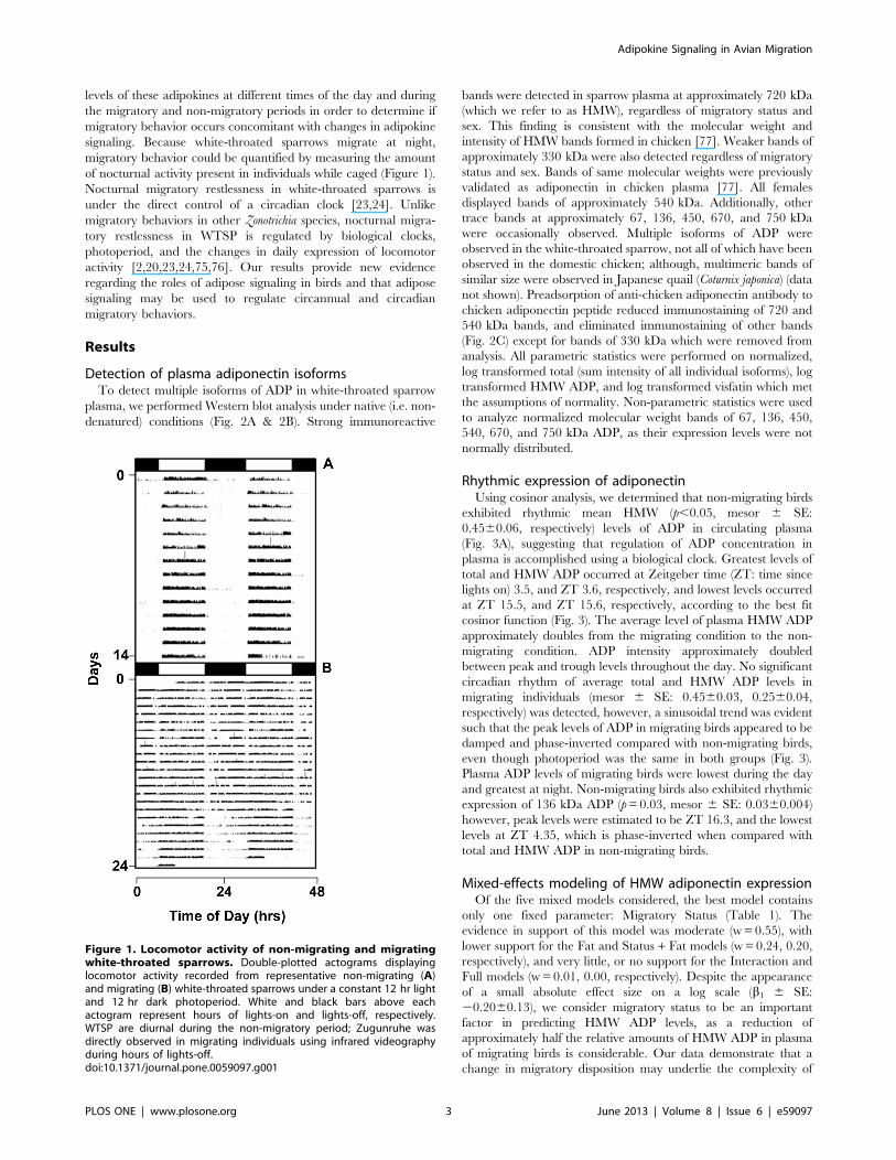
Detection of plasma adiponectin isoformsTo detect multiple isoforms of ADP in white-throated sparrow
plasma, we performed Western blot analysis under native (i.e. non-
denatured) conditions (Fig. 2A & 2B). Strong immunoreactive
bands were detected in sparrow plasma at approximately 720 kDa
(which we refer to as HMW), regardless of migratory status and
sex. This finding is consistent with the molecular weight and
intensity of HMW bands formed in chicken [77]. Weaker bands of
approximately 330 kDa were also detected regardless of migratory
status and sex. Bands of same molecular weights were previously
validated as adiponectin in chicken plasma [77]. All females
displayed bands of approximately 540 kDa. Additionally, other
trace bands at approximately 67, 136, 450, 670, and 750 kDa
were occasionally observed. Multiple isoforms of ADP were
observed in the white-throated sparrow, not all of which have been
observed in the domestic chicken; although, multimeric bands of
similar size were observed in Japanese quail (Coturnix japonica) (data
not shown). Preadsorption of anti-chicken adiponectin antibody to
chicken adiponectin peptide reduced immunostaining of 720 and
540 kDa bands, and eliminated immunostaining of other bands
(Fig. 2C) except for bands of 330 kDa which were removed from
analysis. All parametric statistics were performed on normalized,
log transformed total (sum intensity of all individual isoforms), log
transformed HMW ADP, and log transformed visfatin which met
the assumptions of normality. Non-parametric statistics were used
to analyze normalized molecular weight bands of 67, 136, 450,
540, 670, and 750 kDa ADP, as their expression levels were not
normally distributed.
Rhythmic expression of adiponectinUsing cosinor analysis, we determined that non-migrating birds
exhibited rhythmic mean HMW (p,0.05, mesor 6 SE:
0.4560.06, respectively) levels of ADP in circulating plasma
(Fig. 3A), suggesting that regulation of ADP concentration in
plasma is accomplished using a biological clock. Greatest levels of
total and HMW ADP occurred at Zeitgeber time (ZT: time since
lights on) 3.5, and ZT 3.6, respectively, and lowest levels occurred
at ZT 15.5, and ZT 15.6, respectively, according to the best fit
cosinor function (Fig. 3). The average level of plasma HMW ADP
approximately doubles from the migrating condition to the non-
migrating condition. ADP intensity approximately doubled
between peak and trough levels throughout the day. No significant
circadian rhythm of average total and HMW ADP levels in
migrating individuals (mesor 6 SE: 0.4560.03, 0.2560.04,
respectively) was detected, however, a sinusoidal trend was evident
such that the peak levels of ADP in migrating birds appeared to be
damped and phase-inverted compared with non-migrating birds,
even though photoperiod was the same in both groups (Fig. 3).
Plasma ADP levels of migrating birds were lowest during the day
and greatest at night. Non-migrating birds also exhibited rhythmic
expression of 136 kDa ADP (p=0.03, mesor 6 SE: 0.0360.004)
however, peak levels were estimated to be ZT 16.3, and the lowest
levels at ZT 4.35, which is phase-inverted when compared with
total and HMW ADP in non-migrating birds.
Mixed-effects modeling of HMW adiponectin expressionOf the five mixed models considered, the best model contains
only one fixed parameter: Migratory Status (Table 1). The
evidence in support of this model was moderate (w= 0.55), with
lower support for the Fat and Status + Fat models (w= 0.24, 0.20,
respectively), and very little, or no support for the Interaction and
Full models (w= 0.01, 0.00, respectively). Despite the appearance
of a small absolute effect size on a log scale (b1 6 SE:
20.2060.13), we consider migratory status to be an important
factor in predicting HMW ADP levels, as a reduction of
approximately half the relative amounts of HMW ADP in plasma
of migrating birds is considerable. Our data demonstrate that a
change in migratory disposition may underlie the complexity of
Figure 1. Locomotor activity of non-migrating and migratingwhite-throated sparrows. Double-plotted actograms displayinglocomotor activity recorded from representative non-migrating (A)and migrating (B) white-throated sparrows under a constant 12 hr lightand 12 hr dark photoperiod. White and black bars above eachactogram represent hours of lights-on and lights-off, respectively.WTSP are diurnal during the non-migratory period; Zugunruhe wasdirectly observed in migrating individuals using infrared videographyduring hours of lights-off.doi:10.1371/journal.pone.0059097.g001
Adipokine Signaling in Avian Migration
PLOS ONE | www.plosone.org 3 June 2013 | Volume 8 | Issue 6 | e59097
the relationship between this fat hormone and body condition
which may ultimately affect migratory behavior.
Correlative analysis of plasma adiponectin and fat scoreA strong positive correlation between ZT 6 HMW ADP and fat
score was detected in migrating birds (HMW: r= 0.93; p,0.05).
Strong (t=20.84) negative correlations were detected in 540 kDa
ADP in migrating birds and in 136 kDa ADP of non-migrating
birds (p = 0.05). We detected a negative correlation between fat
score and the ratio of daytime to nighttime activity in migrating
birds (t=20.79, p = 0.11) however, sample sizes were too small
for statistical significance.
On average, non-migrating birds had a fat score 1 fat class lower
than migrating birds (t-test: p,0.05; 95% confidence interval:
21.58, 20.44). We did not detect a sexual dimorphism in the
average levels of plasma ADP, as has been observed in mammals
[55], however, our sample of females was small (n = 3).
Plasma visfatinPlasma visfatin levels ranged from 0.34 ng/mL to 70.55 ng/mL
(mean = 12.01; med. = 9.37 ng/mL) (not log-transformed). We do
not consider plasma visfatin rhythmic although, when values were
averaged by time, migrating individuals as a group trended
towards rhythmicity (p,0.10) with maximum concentrations at
ZT6 and minimum concentrations at ZT18 (Figure 4).
Results from mixed-effects modeling of plasma visfatin are
displayed in Table 2. Of the models considered, the best model
contains only one fixed effect: Migratory Status. Evidence for this
model was strong (w= 0.82), with less support for the Status + Fat
model (w= 0.12) and little or no support for the remaining models.
The Status model estimates a reduction of 0.62 ng/mL (SE= 0.18)
(log scale) between the non-migrating and migrating groups,
approximately a 20% reduction.
Discussion
Our results demonstrate programmatic variations in the
expression of the adipokines adiponectin and visfatin between
the migratory and non-migratory conditions in white-throated
sparrows. Adiponectin secretion is regulated by a circadian clock
during non-migrating periods, and expression either becomes
uncoupled from a circadian clock, or the rhythm of ADP secretion
is damped and phase-inverted when birds are migrating. Although
we do not know what aspect of the biological clockwork causes
such regulatory changes in ADP, one may speculate that ADP
Figure 2. Multiple isoforms of white-throated sparrow plasma adiponectin. Western blot analysis of plasma ADP under native conditions.Representative western blots from two white-throated sparrows A, female sparrow in non-migrating condition in the left panel; same bird undermigrating conditions in the right panel where plasma samples were collected at four times of day (ZT 0, 6, 12, 18). B,Male non-migrating condition inthe left panel; same bird under migrating conditions in the right panel where plasma samples were collected at four times of day (ZT 0, 6, 12, 18). Forall birds, lights came on at ZT0 and went out at ZT 12. C, white-throated sparrow (W) and broiler chicken (Br) plasma separated under nativeconditions. Primary anti-chicken antibody was either not preadsorbed (2 blocking peptide), or preadsorbed with adiponectin peptide (+ blockingpeptide). A high molecular weight protein marker was included to determine molecular weight (kDa) of adiponectin isoforms.doi:10.1371/journal.pone.0059097.g002
Adipokine Signaling in Avian Migration
PLOS ONE | www.plosone.org 4 June 2013 | Volume 8 | Issue 6 | e59097

output is a manifestation of changed clockwork in the adipose
tissue of migrants. Although we did not detect a circadian rhythm
of plasma visfatin concentration, we did observe a reduction in
plasma visfatin during the migratory period. Because visfatin is
able to communicate metabolic information to the molecular
clock, a change in visfatin expression could modify the phase or
amplitude of the biological clock. Our data therefore suggests a
possible mechanism for regulating the changes in locomotor
profiles observed during migration by modifying either ADP or
visfatin expression, as plasma profiles of both of these adipokines
changed between the non-migratory and migratory periods.
Alternatively, ADP and visfatin levels could be altered more
locally as part of the bird’s overall preparation for increased
metabolic demand needed during migration. For example, visfatin
from skeletal muscle may act in a paracrine fashion to regulate
glucose utilization locally. During migration, a birds’ primary
source of energy comes from the oxidation of adipose fatty acids
[78]. Glucose is relatively unimportant as a fuel source in the long-
distance endurance flights of migrating birds. In humans, visfatin
has the ability to increase glucose uptake, playing a role in glucose
homeostasis [79]. However, the seasonal decrease in plasma
visfatin coinciding with the migratory period could reduce glucose
utilization and allow for a greater reliance, on and utilization of,
fat stores and fatty acids to fuel migration.
The shape of the circadian ADP profile approximates the daily
profile of locomotor activity. In migrating birds of appropriate
body condition, a low ADP profile during the day coincides with
little daytime activity, and a higher ADP profile during the night
coincides with the time of migratory flight. Conversely, the profile
of ADP in diurnal, non-migrating birds is highest during the day,
and lowest during the night. Compared to non-migrating birds,
the phase shift in levels of ADP in migrating birds might be
particularly adaptive for regulating lipid metabolism. Because
adiponectin stimulates lipid metabolism without suppressing food
intake [80,81], it would be beneficial for migrating birds to
increase ADP production at night. This hypothesis is supported in
Fig. 3B, where it is demonstrated that birds signal to increase lipid
metabolism during times of migratory flight. When migrating birds
rest and refuel during the daytime, ADP levels are decreased,
thereby allowing fat synthesis and preservation to occur until
migratory flight continues [82]. In non-migrating birds, ADP
levels are greatest during the day potentially allowing lipid
metabolism to coincide with times of increased activity. Addition-
ally, ADP levels are reduced at night when birds are sleeping,
thereby preserving fat stores in non-migrating birds.
Correlations between HMW ADP and fat score, a proxy of
body condition, in migrating birds are particularly intriguing
because studies of mammals and chicken have demonstrated that
levels of ADP decrease during obesity [77,83,84]. Our findings
here demonstrate a positive correlation between fat and HMW
ADP, the predominant ADP isoform in avian species. However,
our scoring results were based solely on subcutaneous fat. The
conflicting correlative findings regarding obese mammals and
Figure 3. Circadian expression of adiponectin. Cosinor analysis of (A) total plasma ADP in non-migrating birds, (B) total plasma ADP inmigrating birds, (C) HMW plasma ADP in non-migrating birds, and (D) HMW plasma ADP in migrating birds. For all birds, lights came on at ZT0 andwent out at ZT 12. The solid wave shows the best-fit curve determined by linear harmonic regression. Individual data points are plotted as mean6 SEof actual measurements. ZT0 has been double-plotted as ZT24 for visualization.doi:10.1371/journal.pone.0059097.g003
Table 1. Model selection results to predict plasma HMWadiponectin levels in the white-throated sparrow.
Modela Ki AICci Di wi
MigratoryStatus 5 9.61 0 0.55
Fat 5 11.21 1.60 0.24
MigratoryStatus + Fat 6 11.59 1.97 0.20
Interaction 7 17.38 7.77 0.01
Full 7 22.82 13.21 0.00
aN=40; K is the number of parameters included in each model; AICc is thesecond order Akaike’s information criteria; Di is the difference between the AICcvalues of each model and the model with the lowest AICc; wi is the weight ofevidence for each model.doi:10.1371/journal.pone.0059097.t001
Adipokine Signaling in Avian Migration
PLOS ONE | www.plosone.org 5 June 2013 | Volume 8 | Issue 6 | e59097

temporarily obese migrating birds may suggest that there are
physiological differences between the genetically programmed,
temporary subcutaneous fat stores maintained by birds to deal
with the enormous energetic costs of migration and typical obesity
in humans. In chicken, food deprivation leads to a significant
decrease in ADP gene expression in adipose tissue [62], however,
fasting has no effect on plasma ADP concentrations. Our results
regarding non-migrating passerines are in opposition to these
findings in chicken and demonstrate that plasma ADP is low at
night when birds are not actively foraging (fasting), which could be
a response to conserve fat stores until plasma ADP is high during
the day, and food is available. Our results regarding migrating
birds are similar, however, to those observed by Gomez-Abellan
et al. [61], where the amplitude of ADP rhythms are attenuated in
obese human subjects. Temporarily obese migrating white-
throated sparrows display an average rhythm with a smaller
amplitude compared with non-migrating individuals. Our results
also agree with the findings of Calvani et al. [60], that plasma
ADP concentrations increase after weight loss in human subjects.
The circadian profile of non-migrating sparrows also matches the
diurnal profile of normal-weight human subjects [60]. ADP is
positively related to fat score which also displays a negative
correlation with the ratio of daytime to nighttime activity, a
relationship recently confirmed by Fusani et al. [85]. Migrating
birds with low fat stores display greater daytime activity, perhaps
foraging to replace lost fat, and migrating birds with greater fat
stores spend more time night-active, displaying migratory restless-
ness.
Associations between sleep-wake cycles and weight regulation
have been well documented, although the mechanisms linking the
two processes are not well-understood [37,86,87]. If clocks in
adipose tissue are able to communicate with clocks in the brain, in
particular with the circadian clock controlling Zugunruhe [22], its
disruption might account for the dramatically altered sleep-wake
cycles associated with nocturnal migration. We suggest a role for
clocks in adipose tissue regulating behavior, but further experi-
mentation is needed to elucidate the mechanisms regulating
peripheral oscillators in fat and the precise mechanisms regulating
how they can influence behavior.
Our results provide evidence that a biological clock modulates
the physiological transitions from non-migratory to nocturnal
migratory behaviors. Adipokine signals may play a significant role
in the initiation, continuation, and termination of nocturnal
migration in small songbirds by indicating the amount of available
fuel from fat and the overall body condition. Future experimen-
tation utilizing restricted feeding schedules could be performed to
force a ‘‘stopover’’ in captive birds during the migratory period.
Analysis of plasma samples collected during forced stopovers could
determine whether the change in the ADP profile observed in
migrating versus non-migrating birds is applicable to stopover
situations as well. Experiments to localize ADP receptors in the
Figure 4. Expression of Visfatin. Levels of visfatin in plasma of non-migrating (A) and migrating (B) white-throated sparrows. For all birds, lightscame on at ZT0 and went out at ZT 12. Migrating individuals as a group trended towards rhythmicity (p,0.10) with maximum concentrations at ZT6and minimum concentrations at ZT18. Plasma levels in non-migrating birds were approximately 20% higher than those of migrating birds.doi:10.1371/journal.pone.0059097.g004
Table 2. Model selection results to predict plasma visfatinconcentrations in the white-throated sparrow.
Modela Ki AICci Di wi
MigratoryStatus 5 131.31 0 0.82
MigratoryStatus+Fat 5 135.05 3.74 0.12
Full 6 137.78 6.47 0.03
Interaction 6 138.09 6.78 0.03
Fat 7 140.65 9.34 0.01
Sex + Fat 7 142.87 11.56 0.00
aN=66; K is the number of parameters included in each model; AICc is thesecond order Akaike’s information criteria; Di is the difference between the AICcvalues of each model and the model with the lowest AICc; wi is the weight ofevidence for each model.doi:10.1371/journal.pone.0059097.t002
Adipokine Signaling in Avian Migration
PLOS ONE | www.plosone.org 6 June 2013 | Volume 8 | Issue 6 | e59097

brain would also provide valuable information as to the specific
location of neural substrates regulating the transition of behaviors
in migratory birds. As this is the first study of the adipokines
adiponectin and visfatin in migrant birds, further investigation of
these and other adipokines in migrant species that store and use fat
differently than the white-throated sparrow, such as shorter or
long-distance migrants, would be relevant to a more complete
understanding of the role of adipokines in metabolism and
migration.
It is unclear from this study whether a change in adipokine
signal drives a change in migratory disposition, influencing
initiation or termination of migration or stopover delays, or a
change in migratory status drives change in adipokine signals.
However, because of adiponectin’s close relationship with body
fat, as well as migratory disposition, it is possible that ADP drives
change in migratory disposition and changes in other adipokines
such as visfatin make up the long list of adaptations necessary for
successful migration.
Materials and Methods
Housing of animalsWhite-throated sparrows were captured in Centre County
Pennsylvania during Spring 2008 using mist nets (USFWS
#MB170276-0; PAGC #COL00194). Wild-caught sparrows
were housed at the Poultry Education and Research Center
(The Pennsylvania State University, University Park, PA) in a
large, environmentally controlled indoor aviary. Birds were
maintained under a 12 hr light, 12 hr dark photoperiod and were
provided with food and water ad libitum. Birds were transferred to
individual wire grated bird cages (906120615.50, Top WingH) inauditory contact with other birds and were allowed to acclimate
for at least 2 weeks before experimentation began. All procedures
were performed in accordance with standards approved by the
Pennsylvania State University Institutional Animal Care and Use
Committee (IACUC #27105, and #29985).
Locomotor and behavioral activityInfrared motion sensors (Quorum International) were mounted
above each cage to record locomotor activity. Locomotor data
were collected in 5 minute bins using the software program,
VitalView (Mini Mitter, Respironics) and evaluated for daily
activity with the software package, ActiView (Mini Mitter,
Respironics). Migratory status was determined using the locomo-
tor activity record by noting exclusively diurnal patterns of activity
(non-migrating), and periods of excessive nighttime activity
(migrating), as has been previously reported for WTSP [23,24].
Nocturnal migratory restlessness is apparent in caged individuals
as increased nocturnal activity, including flightless wing-whirring
which corresponds to migratory flight in free-living birds. We used
infrared video cameras (Q-SeeH) to continuously record the
behaviors of individual birds.
We know that circannual program continued to run in these
species based upon the presence of seasonal changes in molt,
migratory restlessness, and reproductive behaviors and physiology
(testicular recrudescence in males, and egg-laying in females
during the summer months). Birds did experience temperature
fluctuations across the seasons due to poor insulation in the
building and it is possible that these seasonal temperature changes
could provide seasonal cues to the birds. Temperatures ranged
from approximately 17–28 Celsius across the year. Repeated
changes in nocturnal activity observed in captivity approximated
autumn and spring migratory periods of free-living conspecifics
and indicate an endogenous control of migratory rhythms. In
Zonotrichia albicollis, migratory behavior is expressed as the presence
of nocturnal locomotor activity concomitant with little change in
daytime activity [23,24]. This is different than the locomotor
profiles reported for other Zonotrichia species in captivity (i.e. a
complete switch to exclusively nocturnal activity) [2,20]. Changes
in nocturnal activity were quantified using infrared motion sensors
and video cameras were used to validate migratory disposition
(Fig. 1 and Video S1) [22].
Fat scoringWe utilized the subcutaneous fat score classification system
developed by A. Kaiser [88], previously tested and validated by
Soxhlet extraction in similar species of either the same order
(Passiformes) or family (Emberizidae) [89]. This classification
system has been shown to produce highly repeatable results by
ranking fat scores from 0 (no fat) to 8 (extremely fat) by
subcutaneous fat load in the furcular depression, breast muscles,
and abdomen. Birds were evaluated bi-weekly, between the times
of 3 and 4 ZT (ZT= zeitgeber time, time since lights on) to control
for any daily changes in fat deposits. This method is widely
practiced in field studies of small songbirds [90,91,92,93].
Western blot analysis of plasma ADPBlood samples (,50 ml) were collected from non-migrating
(n = 5; fall, 2009) and migrating (n = 7; Winter/Spring 2010) birds
from the brachial vein with heparinized capillary tubes. Two
additional birds changed their migratory status during sampling
and were excluded from the analysis. Samples were collected at
four different times of day, in 6 hr intervals beginning with ZT0
(time of lights on) during one continuous autumn migratory period
or non-migratory period. We allowed three days between each
blood drawing for recovery. Samples were centrifuged at
2500 rpm for 20 minutes at 4uC and the plasma collected and
stored until use at 280uC.Using the Novex mini gel system and NativePAGE gels
(InvitrogenH) we performed gel electrophoresis according to the
manufacturer’s instructions. Serial dilutions of plasma were
performed to optimize the amount of protein loaded per well. A
standard curve was generated from the signal intensity measures of
seven different plasma dilutions to determine the linear range
(r2 = 0.97) of the standard curve. The 1:20 plasma dilution was
chosen for further use from within the linear range of the standard
curve. Plasma samples were prepared by adding 5 ul of 1:20
diluted plasma sample to 4X Sample Buffer, G-250 Sample
Additive, and deionized water. 10 ml of sample mixture was
loaded into NativePAGE 4–16% Bis-Tris gel with NativeMark
unstained (InvitrogenH), and Novex Sharp pre-stained (Invitro-
genH) protein standards to provide a molecular mass reference,
and a 1:100 dilution of broiler chicken plasma was included as a
positive control.
After separation, proteins were electrotransferred to a poly-
vinylidenedifluoride membrane. To check the uniformity of
transfer, the gel was stained using Coomassie Blue. Membranes
were incubated in SuperBlock Blocking Buffer (Pierce) for 1hr at
room temperature, incubated in a custom generated primary
antibody against a keyhole limpet hemocyanin conjugate in the N-
terminal domain of chicken adiponectin (Rabbit anti-cADN;
1:2500) [77,94] overnight at 4uC, and incubated in secondary
antibody (Pierce anti-rabbit IgG-HRP 1:10,000) at room temper-
ature for 1 hr. Enhanced chemiluminescence detection was
performed on transparency film with ECL Plus Detection Reagent
(Amersham) and imaged on the STORM 860 optical scanner
using blue wavelength fluorescence. To determine the specific
Adipokine Signaling in Avian Migration
PLOS ONE | www.plosone.org 7 June 2013 | Volume 8 | Issue 6 | e59097

immunoreactivity of anti-adiponectin antibody, primary antibody
was preadsorbed with chicken adiponectin protein [77].
Enzyme-linked immunosorbant assay (ELISA) analysis ofplasma visfatinBlood samples (,200 ml) were collected from the brachial vein
using heparinized capillary tubes from non-migrating (n = 9) and
spring migrating (n = 8) individuals. Samples were collected at four
different times of day (ZT 0, 6, 12, 18) to assess circadian
rhythmicity. We allowed at least 2 days between sampling for the
birds to recover. Blood samples were centrifuged at 2500 rpm for
20 minutes at 4uC and the plasma was stored until use at 280uC.Two plasma samples were found to have inadequate volume and
were discarded from the analysis.
Undiluted plasma visfatin concentrations were determined by
ELISA (Phoenix Pharmaceuticals, Burlingame, CA, Visfatin C-
Terminal (Human) Kit), following the manufacturer’s instructions.
The use of this kit for detecting avian plasma visfatin was
previously validated in chicken [69]. Samples and controls were
assayed in duplicate using two plates (intra-assay variation: plate
1 = 11.6%; plate 2 = 5.1%). Samples from both migrating and
non-migrating birds were interspersed on both plates as a
safeguard against nondemonic intrusion. Samples were read at
450 nm using the FLUOstar Omega microplate reader; standard
curves were generated using 4-parameter logistics based on blank
corrected averages of duplicates. Sample volumes of one time
point in two individuals were inadequate and not used for analysis.
Data analysisThe ECL signal intensity of ADP bands from Western blots was
quantified (Image Quant 5.1) in arbitrary intensity units. Protein
amount was determined by Bradford assay and signal intensity was
normalized to individual protein concentrations. A calibration
curve was constructed for each Bradford protein assay using five
dilutions of bovine serum albumin in triplicate, including water
blanks, using Bio-Rad Protein Assay Dye Reagent Concentrate.
Sparrow plasma samples were diluted 1:100 to fall within the
linear range of BSA standard curves and assayed in duplicate.
Total plasma adiponectin levels were quantified and additional
analyses were performed to quantify individual multimeric bands.
Assumptions of homoscedacity and normality were assessed by
evaluating normal probability plots. Non-normally distributed
samples were log10 transformed to accommodate this assumption;
bands that did not meet the normality assumption after
transformation were analyzed using nonparametric statistics
(Kendall tau rank correlation). Average total ADP (sum intensity
of all isoforms), individual isoforms of ADP, and visfatin
concentrations were analyzed using linear harmonic regression
in cosinor analysis using the program CircWave [95] to determine
the presence of a circadian rhythm.
To accommodate dependency among levels of hormone
sampled over a short time series, we employed a mixed modeling
approach to examine the importance of migratory status
(migrating vs. non-migrating), time of day (ZT 0, 6, 12, 18), sex
(visfatin data only) and fat score in predicting hormone levels. To
identify the most important variables in predicting levels of plasma
HMW ADP, we compared five linear mixed-effects models using
second order Akaike information criteria (AICc) to correct for
small sample sizes, fitted in the software program ‘‘R’’ 2.10.1 [96]
which is based on log-likelihood and the number of model
parameters to quantify the evidence of support for each proposed
model. The AICc value provides a measure of distance between
competing models and allows for model comparison. Akaike
weights are similar to probabilities and describe the weight of
evidence in support for each model in minimizing the distance
from the ‘‘true model’’ and allows for comparison of evidence for
each model in a group of models. This weight can be interpreted
as the relative probability of the model given the data [97,98]. A
model with an Akaike weight approaching 1 is unambiguously
supported by the observed data [99]. We began by considering a
Full model (‘‘Full’’), including all three explanatory variables. We
dropped the least important variable (time) and considered a
model with migratory status and fat (‘‘Status + Fat’’), and a model
with an interaction between group and fat (‘‘Interaction’’). Lastly,
we considered models with migratory status (‘‘Status’’) and fat
(‘‘Fat’’) individually. To account for the effects of autocorrelation
in repeated sampling over time, we used an AR1 error structure
and included ‘‘individual’’ as a random effect. The model with the
smallest relative AICc value indicates the best model of those
proposed. Residuals were examined to assess homogeneity and
verify normality. Examination of residuals versus fitted values
ensured that the model fit our data well.
Six mixed-effects models were evaluated to predict plasma
visfatin in white-throated sparrows. Migratory status, fat score, and
sex, were included as fixed effects while individual and time of day
were included as random effects. We used time as a random effect
because plasma visfatin did not display circadian rhythmicity. We
considered a Full model (‘‘Full’’), including all three fixed effects,
an interaction model between migratory status and fat (‘‘Interac-
tion’’), an additive model of migratory status and fat (‘‘Status +Fat’’), an additive model of sex and fat (‘‘Sex + Fat’’), as well as
models with migratory status (‘‘Status’’) and fat (‘‘Fat’’) alone.
Residual plots were used to evaluate fit of the models.
We calculated correlations between circulating plasma ADP
isoform levels and fat score, and the ratio of daytime to nighttime
activity and fat score with ‘‘R’’. We calculated this activity ratio
rather than raw nighttime activity values to correct for individuals
with varying baseline activity levels. Data for correlations of ADP
and fat score were grouped by migratory disposition (exhibiting
migratory restlessness or not) and ADP levels were evaluated at ZT
6 (the time closest to when fat scores were collected). Correlations
between fat score and activity ratio were performed based on
values averaged by individual. Because the fat classification system
used in this study has a relatively large category size, and the
distribution of ordinally measured fat scores is normal, ordinal fat
score values were treated as continuous [100,101].
Supporting Information
Video S1 Shown is a segment of a video recording takenduring the night under infrared illumination. Video
images show intense activity, including wing-whirring, features
which were used to determine whether a bird was exhibiting
nocturnal migratory restlessness.
(WMV)
Acknowledgments
We would like to thank Bill Hendricks and Susan Krzysik-Walker for their
assistance in the design of Western blotting protocols.
Author Contributions
Conceived and designed the experiments: ES PB. Performed the
experiments: ES JV MG. Analyzed the data: ES PB. Contributed
reagents/materials/analysis tools: RR. Wrote the paper: ES PB.
Adipokine Signaling in Avian Migration
PLOS ONE | www.plosone.org 8 June 2013 | Volume 8 | Issue 6 | e59097

References
1. Odum EP (1960) Premigratory Hyperphagia in Birds. American Journal of
Clinical Nutrition 8: 621–629.
2. King J, Farner D (1963) The relationship of fat deposition to zugunruhe andmigration. The Condor 65: 200–223.
3. Jenni L, Jenni-Eiermann S (1998) Fuel supply and metabolic constraints in
migrating birds. Journal of Avian Biology 29: 521–528.
4. Goymann W, Spina F, Ferri A, Fusani L (2010) Body fat influences departurefrom stopover sites in migratory birds: evidence from whole-island telemetry.
Biology Letters 6: 478–481.
5. Arizaga J, Belda EJ, Barba E (2011) Effect of fuel load, date, rain and wind ondeparture decisions of a migratory passerine. Journal of Ornithology 152: 991–
999.
6. Stutchbury BJM, Gow EA, Done T, MacPherson M, Fox JW, et al. (2011)Effects of post-breeding moult and energetic condition on timing of songbird
migration into the tropics. Proceedings of the Royal Society B-BiologicalSciences 278: 131–137.
7. Coppack T, Bairlein F (2011) Circadian control of nocturnal songbird
migration. Journal of Ornithology 152: 67–73.
8. Aamidor SE, Bauchinger U, Mizrahy O, McWilliams SR, Pinshow B (2011)During Stopover, Migrating Blackcaps Adjust Behavior and Intake of Food
Depending on the Content of Protein in Their Diets. Integrative andComparative Biology 51: 385–393.
9. Studds CE, Marra PP (2005) Nonbreeding habitat occupancy and population
processes: An upgrade experiment with a migratory bird. Ecology 86: 2380–2385.
10. Marra PP, Hobson KA, Holmes RT (1998) Linking winter and summer events
in a migratory bird by using stable-carbon isotopes. Science 282: 1884–1886.
11. Covino KM, Holberton RL (2011) The Influence of Energetic Condition onFlight Initiation and Orientation of Migratory Songbirds in the Gulf of Maine
Region. Auk 128: 313–320.
12. Cerasale DJ, Zajac DM, Guglielmo CG (2011) Behavioral and physiologicaleffects of photoperiod-induced migratory state and leptin on a migratory bird,
Zonotrichia albicollis: I. Anorectic effects of leptin administration. General and
Comparative Endocrinology 174: 276–286.13. Berthold P (1984) The Endogenous Control of Bird Migration – a Survey of
Experimental-Evidence. Bird Study 31: 19–27.
14. Berthold P (1990) Spatiotemporal Programs and Genetics of Orientation.
Experientia 46: 363–371.15. Gwinner E (1977) Circannual Rhythms in Bird Migration. Annual Review of
Ecology and Systematics 8: 381–405.
16. Gwinner E (1996) Circadian and circannual programmes in avian migration.Journal of Experimental Biology 199: 39–48.
17. Gwinner E (1996) Circannual clocks in avian reproduction and migration. Ibis
138: 47–63.
18. Yokoyama K (1976) Hypothalamic and Hormonal-Control of Photoperiodi-cally Induced Vernal Functions in White-Crowned Sparrow, Zonotrichia-
Leucophrys-Gambelii. 1. Effects of Hypothalamic-Lesions and ExogenousHormones. Cell and Tissue Research 174: 391–416.
19. Barnea M, Madar Z, Froy O (2009) High-Fat Diet Delays and Fasting
Advances the Circadian Expression of Adiponectin Signaling Components in
Mouse Liver. Endocrinology 150: 161–168.20. Eyster MB (1954) Quantitative Measurement of the Influence of Photoperiod,
Temperature, and Season on the Activity of Captive Songbirds. Ecological
Monographs 24: 1–28.21. Kumar V, Wingfield JC, Dawson A, Ramenofsky M, Rani S, et al. (2010)
Biological Clocks and Regulation of Seasonal Reproduction and Migration in
Birds. Physiological and Biochemical Zoology 83: 827–835.22. Bartell PA, Gwinner E (2005) A separate circadian oscillator controls nocturnal
migratory restlessness in the songbird Sylvia borin. Journal of Biological
Rhythms 20: 538–549.23. Mcmillan JP (1972) Pinealectomy Abolishes Circadian-Rhythm of Migratory
Restlessness. Journal of Comparative Physiology 79: 105–112.
24. Mcmillan JP, Gauthreaux SA, Helms CW (1970) Spring Migratory
Restlessness in Caged Birds – a Circadian Rhythm. Bioscience 20: 1259–1260.25. King DP, Takahashi JS (2000) Molecular genetics of circadian rhythms in
mammals. Annual Review of Neuroscience 23: 713–742.
26. Rutter J, Reick M, Wu LC, McKnight SL (2001) Regulation of clock andNPAS2 DNA binding by the redox state of NAD cofactors. Science 293: 510–
514.
27. Rutter J, Reick M, McKnight SL (2002) Metabolism and the control ofcircadian rhythms. Annual Review of Biochemistry 71: 307–331.
28. Dioum EM, Rutter J, Tuckerman JR, Gonzalez G, Gilles-Gonzalez MA, et al.
(2002) NPAS2: A gas-responsive transcription factor. Science 298: 2385–2387.
29. Bell-Pedersen D, Cassone VM, Earnest DJ, Golden SS, Hardin PE, et al.(2005) Circadian rhythms from multiple oscillators: Lessons from diverse
organisms. Nature Reviews Genetics 6: 544–556.
30. Meier AH, Burns JT (1976) Circadian Hormone Rhythms in Lipid Regulation.American Zoologist 16: 649–659.
31. Bray MS, Young ME (2007) Circadian rhythms in the development of obesity:
potential role for the circadian clock within the adipocyte. Obesity Reviews 8:169–181.
32. Zvonic S, Ptitsyn AA, Conrad SA, Scott LK, Floyd ZE, et al. (2006)
Characterization of peripheral circadian clocks in adipose tissues. Diabetes 55:962–970.
33. Bartness TJ, Song CK (2007) Brain-adipose tissue neural crosstalk. Physiology
& Behavior 91: 343–351.
34. Friedman-Einat M, Boswell T, Horev G, Girishvarma G, Dunn IC, et al.(1999) The chicken leptin gene: Has it been cloned? General and Comparative
Endocrinology 115: 354–363.
35. Pitel F, Monbrun C, Gellin J, Vignal A (2000) The chicken LEP (OB) gene hasnot been mapped. Animal Genetics 31: 281–281.
36. Sharp PJ, Dunn IC, Waddington D, Boswell T (2008) Chicken leptin. General
and Comparative Endocrinology 158: 2–4.
37. Arble DM, Vitaterna MH, Turek FW (2011) Rhythmic Leptin Is Required forWeight Gain from Circadian Desynchronized Feeding in the Mouse. Plos One
6.
38. Kalsbeek A, Fliers E, Romijn JA, la Fleur SE, Wortel J, et al. (2001) Thesuprachiasmatic nucleus generates the diurnal changes in plasma leptin levels.
Endocrinology 142: 2677–2685.
39. Kalra SP, Bagnasco M, Otukonyong EE, Dube MG, Kalra PS (2003)Rhythmic, reciprocal ghrelin and leptin signaling: new insight in the
development of obesity. Regulatory Peptides 111: 1–11.
40. Penicaud L, Meillon S, Brondel L (2012) Leptin and the central control offeeding behavior. Biochimie 94: 2069–2074.
41. Farooqi IS, O’Rahilly S (2009) Leptin: a pivotal regulator of human energy
homeostasis. American Journal of Clinical Nutrition 89: 980s–984s.
42. Lago F, Gomez R, Gomez-Reino JJ, Dieguez C, Gualillo O (2009) Adipokinesas novel modulators of lipid metabolism. Trends in Biochemical Sciences 34:
500–510.
43. Staiger H, Tschritter O, Machann J, Thamer C, Fritsche A, et al. (2003)Relationship of serum adiponectin and leptin concentrations with body fat
distribution in humans. Obesity Research 11: 368–372.
44. Attie AD, Scherer PE (2009) Adipocyte metabolism and obesity. Journal ofLipid Research 50: S395–S399.
45. Janovska A, Hatzinikolas G, Staikopoulos V, McInerney J, Mano M, et al.
(2008) AMPK and ACC phosphorylation: Effect of leptin, muscle fibre typeand obesity. Molecular and Cellular Endocrinology 284: 1–10.
46. Ohkubo T, Adachi H (2008) Leptin Signaling and Action in Birds. Journal of
Poultry Science 45: 233–240.
47. te Marvelde L, Visser ME (2012) Manipulation of life-history decisions usingleptin in a wild passerine. Plos One 7: e34090.
48. Zajac DM, Cerasale DJ, Landman S, Guglielmo CG (2011) Behavioral and
physiological effects of photoperiod-induced migratory state and leptin onZonotrichia albicollis: II. Effects on fatty acid metabolism. General and
Comparative Endocrinology 174: 269–275.
49. Mohamed-Ali V, Pinkney JH, Coppack SW (1998) Adipose tissue as anendocrine and paracrine organ. International Journal of Obesity 22: 1145–
1158.
50. Kershaw EE, Flier JS (2004) Adipose tissue as an endocrine organ. Journal ofClinical Endocrinology & Metabolism 89: 2548–2556.
51. Ahima RS (2006) Adipose tissue as an endocrine organ. Obesity 14: 242–249.
52. Yamauchi T, Kamon J, Minokoshi Y, Ito Y, Waki H, et al. (2002) Adiponectin
stimulates glucose utilization and fatty-acid oxidation by activating AMP-activated protein kinase. Nature Medicine 8: 1288–1295.
53. Ahima RS (2006) Metabolic actions of adipocyte hormones: Focus on
adiponectin. Obesity 14: 9S–15S.
54. Lara-Castro C, Luo N, Wallace P, Klein RL, Garvey WT (2006) Adiponectinmultimeric complexes and the metabolic syndrome trait cluster. Diabetes 55:
249–259.
55. Pajvani UB, Du XL, Combs TP, Berg AH, Rajala MW, et al. (2003) Structure-function studies of the adipocyte-secreted hormone Acrp30/adiponectin –
Implications for metabolic regulation and bioactivity. Journal of BiologicalChemistry 278: 9073–9085.
56. Kadowaki T, Yamauchi T, Kubota N, Hara K, Ueki K, et al. (2006)
Adiponectin and adiponectin receptors in insulin resistance, diabetes, and themetabolic syndrome. Journal of Clinical Investigation 116: 1784–1792.
57. Scherer PE (2006) Adipose tissue – From lipid storage compartment to
endocrine organ. Diabetes 55: 1537–1545.
58. Del Turco S, Navarra T, Gastaldelli A, Basta G (2011) Protective role ofadiponectin on endothelial dysfunction induced by AGEs: A clinical and
experimental approach. Microvascular Research 82: 73–76.
59. Hui XY, Lam KSL, Vanhoutte PM, Xu AM (2012) Adiponectin andcardiovascular health: an update. British Journal of Pharmacology 165: 574–
590.
60. Calvani M, Scarfone A, Granato L, Mora EV, Nanni G, et al. (2004)Restoration of adiponectin pulsatility in severely obese subjects after weight
loss. Diabetes 53: 939–947.
61. Gomez-Abellan P, Gomez-Santos C, Madrid JA, Milagro FI, Campion J, et al.(2010) Circadian Expression of Adiponectin and Its Receptors in Human
Adipose Tissue. Endocrinology 151: 115–122.
62. Maddineni S, Metzger S, Ocon O, Hendricks G, Ramachandran R (2005)Adiponectin gene is expressed in multiple tissues in the chicken: Food
Adipokine Signaling in Avian Migration
PLOS ONE | www.plosone.org 9 June 2013 | Volume 8 | Issue 6 | e59097

deprivation influences adiponectin messenger ribonucleic acid expression.
Endocrinology 146: 4250–4256.63. Goto T, Teraminami A, Lee JY, Ohyama K, Funakoshi K, et al. (2012)
Tiliroside, a glycosidic flavonoid, ameliorates obesity-induced metabolic
disorders via activation of adiponectin signaling followed by enhancement offatty acid oxidation in liver and skeletal muscle in obese-diabetic mice. Journal
of Nutritional Biochemistry 23: 768–776.64. Liu Q, Yuan B, Lo KA, Patterson HC, Sun Y, et al. (2012) Adiponectin
regulates expression of hepatic genes critical for glucose and lipid metabolism.
Proc Natl Acad Sci U S A 109: 14568–14573.65. Waldstreicher J, Duffy JF, Brown EN, Rogacz S, Allan JS, et al. (1996) Gender
differences in the temporal organization of prolactin (PRL) secretion: Evidencefor a sleep-independent circadian rhythm of circulating PRL levels – A clinical
research center study. Journal of Clinical Endocrinology & Metabolism 81:1483–1487.
66. Snyder SH, Zweig M, Axelrod J (1964) Control of the Circadian Rhythm in
Serotonin Content of the Rat Pineal Gland. Life Sciences 3: 1175–1179.67. Cagnacci A, Elliott JA, Yen SSC (1992) Melatonin – a Major Regulator of the
Circadian-Rhythm of Core Temperature in Humans. Journal of ClinicalEndocrinology & Metabolism 75: 447–452.
68. Cajochen C, Krauchi K, Wirz-Justice A (2003) Role of melatonin in the
regulation of human circadian rhythms and sleep. Journal of Neuroendocri-nology 15: 432–437.
69. Krzysik-Walker SM, Ocon-Grove OM, Maddineni SR, Hendricks GL,Ramachandran R (2008) Is visfatin an adipokine or myokine? Evidence for
greater visfatin expression in skeletal muscle than visceral fat in chickens.Endocrinology 149: 1543–1550.
70. Mattevi A (2006) A close look at NAD biosynthesis. Nature Structural &
Molecular Biology 13: 563–564.71. Ramsey KM, Yoshino J, Brace CS, Abrassart D, Kobayashi Y, et al. (2009)
Circadian Clock Feedback Cycle Through NAMPT-Mediated NAD(+)Biosynthesis. Science 324: 651–654.
72. Sassone-Corsi P (2012) Minireview: NAD(+), a Circadian Metabolite with an
Epigenetic Twist. Endocrinology 153: 1–5.73. Asher G, Gatfield D, Stratmann M, Reinke H, Dibner C, et al. (2008) SIRT1
regulates circadian clock gene expression through PER2 deacetylation. Cell134: 317–328.
74. Xie H, Tang SY, Luo XH, Huang J, Cui RR, et al. (2007) Insulin-like effects ofvisfatin on human osteoblasts. Calcif Tissue Int. 80(3): 201–10.
75. Weise CM (1956) Nightly Unrest in Caged Migratory Sparrows under Outdoor
Conditions. Ecology 37: 274–287.76. Farner DS, King JR (1957) The Development of Vernal Migratory Behavior in
Caged Individuals of Several Taxa of Zonotrichia. Anatomical Record 128:546–546.
77. Hendricks GL, Hadley JA, Krzysik-Walker SM, Prabhu KS, Vasilatos-
Younken R, et al. (2009) Unique Profile of Chicken Adiponectin, aPredominantly Heavy Molecular Weight Multimer, and Relationship to
Visceral Adiposity. Endocrinology 150: 3092–3100.78. Guglielmo CG (2010) Move That Fatty Acid: Fuel Selection and Transport in
Migratory Birds and Bats. Integrative and Comparative Biology 50: 336–345.79. Bottcher Y, Teupser D, Enigk B, Berndt J, Kloting N, et al. (2006) Genetic
variation in the visfatin gene (PBEF1) and its relation to glucose metabolism
and fat-depot-specific messenger ribonucleic acid expression in humans.Journal of Clinical Endocrinology & Metabolism 91: 2725–2731.
80. Fruebis J, Tsao TS, Javorschi S, Ebbets-Reed D, Erickson MRS, et al. (2001)Proteolytic cleavage product of 30-kDa adipocyte complement-related protein
increases fatty acid oxidation in muscle and causes weight loss in mice.
Proceedings of the National Academy of Sciences of the United States of
America 98: 2005–2010.81. Qi Y, Takahashi N, Hileman SM, Patel HR, Berg AH, et al. (2004)
Adiponectin acts in the brain to decrease body weight (vol 10, pg 524, 2004).
Nature Medicine 10: 649–649.82. Pilo B, George JC (1983) Diurnal and seasonal variation in liver glycogen and
fat in relation to metabolic status of liver and m. pectoralis in the migratorystarling, Sturnus roseus, wintering in India. Comp Biochem Physiol A Comp
Physiol 74: 601–604.
83. Arita Y, Kihara S, Ouchi N, Takahashi M, Maeda K, et al. (1999) Paradoxicaldecrease of an adipose-specific protein, adiponectin, in obesity. Biochemical
and Biophysical Research Communications 257: 79–83.84. Ukkola O, Santaniemi M (2002) Adiponectin: a link between excess adiposity
and associated comorbidities? Journal of Molecular Medicine-Jmm 80: 696–702.
85. Fusani L, Cardinale M, Carere C, Goymann W (2009) Stopover decision
during migration: physiological conditions predict nocturnal restlessness in wildpasserines. Biology Letters 5: 302–305.
86. Knutson KL, Cauter E (2008) Associations between sleep loss and increasedrisk of obesity and diabetes. Molecular and Biophysical Mechanisms of Arousal,
Alertness, and Attention 1129: 287–304.
87. Froy O (2010) Metabolism and circadian rhythms – implications for obesity.Endocr Rev 31: 1–24.
88. Kaiser A (1993) A New Multi-Category Classification of Subcutaneous FatDeposits of Songbirds. Journal of Field Ornithology 64: 246–255.
89. Kaiser A (1992) Fat Deposition and Theoretical Flight Range of Small AutumnMigrants in Southern Germany. Bird Study 39: 96–110.
90. Partecke J, Gwinner E (2007) Increased sedentariness in European Blackbirds
following urbanization: A consequence of local adaptation? Ecology 88: 882–890.
91. Schaub M, Jenni L (2000) Fuel deposition of three passerine bird species alongthe migration route. Oecologia 122: 306–317.
92. Scheuerlein A, Van’t Hof TJ, Gwinner E (2001) Predators as stressors?
Physiological and reproductive consequences of predation risk in tropicalstonechats (Saxicola torquata axillaris). Proceedings of the Royal Society of
London Series B-Biological Sciences 268: 1575–1582.93. Salewski V, Schmaljohann H, Liechti F (2010) Spring passerine migrants
stopping over in the Sahara are not fall-outs. Journal of Ornithology 151: 371–378.
94. Ocon-Grove OM, Krzysik-Walker SM, Maddineni SR, Hendricks GL,
Ramachandran R (2008) Adiponectin and its receptors are expressed in thechicken testis: influence of sexual maturation on testicular ADIPOR1 and
ADIPOR2 mRNA abundance. Reproduction 136: 627–638.95. Hut RA (2007) CircWave. 1.4 ed. Groningen, Netherlands.
96. Team RDC (2008) R: A Language and Environment for Statistical Computing.
Vienna.97. Akaike H (1973) Information Theory as an Extension of the Maximum
Likelihood Principle. Second International Symposium on InformationTheory: 267–281.
98. Alfaro ME, Huelsenbeck JP (2006) Comparative performance of Bayesian andAIC-based measures of phylogenetic model uncertainty. Syst Biol 55: 89–96.
99. Johnson JB, Omland KS (2004) Model selection in ecology and evolution.
Trends Ecol Evol 19: 101–108.100. Labovitz S (1970) Assignment of Numbers to Rank Order Categories.
American Sociological Review 35: 515–524.101. O’Brien R (1979) The use of Pearson’s with ordinal data. American
Sociological Review 44: 851–857.
Adipokine Signaling in Avian Migration
PLOS ONE | www.plosone.org 10 June 2013 | Volume 8 | Issue 6 | e59097
Copyright © 2022 FDOKUMEN