Diagnostic Evaluation of the Adrenal Incidentaloma: Decision and Cost-Effectiveness Analyses
7
1998;39:707-712. J Nucl Med. Shapiro Ben A. Dwamena, Richard T. Kloos, A. Mark Fendrick, Milton D. Gross, Isaac R. Francis, Melvyn T. Korobkin and Brahm Analyses Diagnostic Evaluation of the Adrenal Incidentaloma: Decision and Cost-Effectiveness http://jnm.snmjournals.org/content/39/4/707 This article and updated information are available at: http://jnm.snmjournals.org/site/subscriptions/online.xhtml Information about subscriptions to JNM can be found at: http://jnm.snmjournals.org/site/misc/permission.xhtml Information about reproducing figures, tables, or other portions of this article can be found online at: (Print ISSN: 0161-5505, Online ISSN: 2159-662X) 1850 Samuel Morse Drive, Reston, VA 20190. SNMMI | Society of Nuclear Medicine and Molecular Imaging is published monthly. The Journal of Nuclear Medicine © Copyright 1998 SNMMI; all rights reserved. by on October 6, 2014. For personal use only. jnm.snmjournals.org Downloaded from by on October 6, 2014. For personal use only. jnm.snmjournals.org Downloaded from
Transcript of Diagnostic Evaluation of the Adrenal Incidentaloma: Decision and Cost-Effectiveness Analyses

1998;39:707-712.J Nucl Med. ShapiroBen A. Dwamena, Richard T. Kloos, A. Mark Fendrick, Milton D. Gross, Isaac R. Francis, Melvyn T. Korobkin and Brahm AnalysesDiagnostic Evaluation of the Adrenal Incidentaloma: Decision and Cost-Effectiveness
http://jnm.snmjournals.org/content/39/4/707This article and updated information are available at:
http://jnm.snmjournals.org/site/subscriptions/online.xhtml
Information about subscriptions to JNM can be found at:
http://jnm.snmjournals.org/site/misc/permission.xhtmlInformation about reproducing figures, tables, or other portions of this article can be found online at:
(Print ISSN: 0161-5505, Online ISSN: 2159-662X)1850 Samuel Morse Drive, Reston, VA 20190.SNMMI | Society of Nuclear Medicine and Molecular Imaging
is published monthly.The Journal of Nuclear Medicine
© Copyright 1998 SNMMI; all rights reserved.
by on October 6, 2014. For personal use only. jnm.snmjournals.org Downloaded from by on October 6, 2014. For personal use only. jnm.snmjournals.org Downloaded from

Diagnostic Evaluation of the Adrenal Incidentaloma:Decision and Cost-Effectiveness AnalysesBen A. Dwamena, Richard T. Kloos, A. Mark Fendrick, Milton D. Gross, Isaac R. Francis, Melvyn T. Korobkinand Brahm ShapiroDivisions of Nuclear Medicine and General Medicine, Department of Internal Medicine, and Departments of Radiology andHealth Services Management and Policy. School of Public Health. The University of Michigan, and Department of VeteranAffairs Medical Centers. Ann Arbor. Michigan
The goal of this study was to examine the clinical and economicoutcomes of alternative diagnostic strategies for differentiating benign from malignant adrenal masses. Methods: We used cost-effectiveness assessment derived from decision analysis and theeconomic perspective of the payer of health care services. One-timeevaluation with fine-needle aspiration (FNA) and combinations ofchemical-shift MRI, noncontrast CT, 131l-6ß-iodomethylnorcholes-terol (NP-59) scintigraphy, with or without FNA, in a hypotheticalcohort of 1000 patients with incidentally discovered unilateral,nonhypersecretory adrenal masses. We calculated and comparedthe diagnostic effectiveness, costs and cost-effectiveness of thealternative strategies based on estimates from published literatureand institutional charge data. Results: At an assumed baselinemalignancy rate of 0.25, diagnostic utility varied from 0.31 (CT0) to0.965 (NP-59) and diagnostic accuracy from 0.655 [noncontrast CTusing a cut-off attenuation value of >0 (CT0)]to 0.983 (NP-59). Theaverage cost per patient per strategy ranged from $746 (NP-59) to$1745 (MRI ±FNA).The best and worst potential cost-to-diagnosticutility ratios were 773 (NP-59) and 2839 (CT0)and 759 (NP-59) and1982 (MRI ±FNA) for cost and diagnostic accuracy, respectively.The NP-59 strategy was the optimal choice regardless of theexpected outcome examined: cost, diagnostic utility, diagnosticaccuracy or cost-effectiveness. Varying the prevalence of malignancy did not alter the cost-effectiveness advantage of NP-59 overthe other diagnostic modalities. Conclusion: Based on availableestimates of reimbursement costs and diagnostic test performanceand using reasonable clinical assumptions, our results indicate thatthe NP-59 strategy is the most cost-effective diagnostic tool forevaluating adrenal incidentalomas over a wide range of malignancyrates and that additional clinical studies are warranted to confirmthis cost-effectiveness advantage.
Key Words: adrenal mass; incidentaloma;adrenocortical scintigraphy; cost-effectiveness analysis
J NucÃMed 1998; 39:707-712
Adr ircnal masses that are incidentally discovered when a CTscan is performed for reasons other than known or suspectedadrenal disease have been termed incidentalomas (1,2). Theselesions have become a common clinical problem with the morefrequent and widespread use of high-resolution imaging procedures. In the vast majority of patients, adrenal incidentalomasare benign, nonhypersecretory adenomas (1.2). Nevertheless, itis important to distinguish benign lesions from others, such asprimary adrenal cancer or métastasesto the the adrenals, inwhich intervention, or the lack thereof, may alter morbidity andmortality. Presently, there is little or no consensus regarding thediagnostic approach to biochemically nonhypersecretory incidentalomas. Management strategies based on adrenal mass size
Received Jan. 27. 1997; revision accepted Jul. 4. 1997.For correspondence or reprints contact: Milton D. Gross, MD. Nuclear Medicine
Service. Ann Arbor Veterans Affairs Medical Center. 2215 Fuller Rd., Ann Arbor. Ml48105.
are neither sensitive nor specific (2). Although 76%-100% ofadrenal masses greater than 5 cm in diameter are benign, asmany as 50% of those smaller than 2.5 cm are malignant (3-8).
Other diagnostic strategies, used alone or in combination,have included serial, conventional CT with size determinationsto assess stability (adenomas) or growth (tumor), morphologiccharacterization with MRI or CT, with measurements of x-rayattenuation or functional adrenal imaging with mI-6/3-iodom-
ethyl-norcholesterol (NP-59) and biopsy using percutaneousfine-needle aspiration (FNA). Biopsy is an invasive procedurewith a small, but significant, morbidity of 3%-12% (9-17).Thus, some researchers have suggested using noninvasiveimaging to optimize the selection of patients for adrenal biopsyand/or choice of definitive therapy.
The reality of increasingly limited resources for health carecreates new pressures for providers to achieve the best possibleoutcomes for patients at the lowest cost. To accomplish this,increased attention is being directed to the determinants ofcost-effectiveness of the spectrum of diagnostic options available in the work-up of a patient with a particular disease state.To date, however, available biopsy or imaging techniques forthe work-up of adrenal incidentalomas have not been formallysubjected to a rigorous comparative investigation. The clinicaland economic consequences of various management approaches incorporating one or more of these procedures isunknown.
Using decision analysis (18-20), published literature and
institutional charge data, we examined the costs, diagnosticutility, diagnostic accuracy and the cost-effectiveness (21.22) ofseveral diagnostic strategies for differentiating between benignand malignant adrenal incidentalomas incorporating one ormore combinations of chemical-shift MRI, CT using nonen-hanced attenuation values, NP-59 and/or FNA. Sensitivityanalysis was used to evaluate the impact of changes in clinicalinput and resource use parameters on the results.
MATERIALS AND METHODS
Decision Analytic ModelUsing the software program Decision Analysis by TreeAge
(DATA 3.0, Williamstown, MA), we constructed a computersimulation of the diagnostic evaluation of incidentally discoveredunilateral nonhypersecretory adrenal masses. The analysis involveda hypothetical cohort of patients without known malignancy inwhom adrenal incidentalomas were identified during thoracoab-dominal imaging for unrelated medical problems. The patients hadundergone clinical and biochemical evaluation to exclude adrenalhyperfunction before entering the model. The decision analyticmodel begins with a decision node representing an active choice ofone of nine strategies incorporating chemical-shift MRI, noncontrast CT with measurement of x-ray attenuation in Houndsfield
MANAGEMENTOFADRENALINCIDENTALOMAS•Dwamena et al. 707
by on October 6, 2014. For personal use only. jnm.snmjournals.org Downloaded from

units (HU), NP-59 scintigraphy and/or FNA categorized as fol
lows.Imaging
1. CT0(>0 HU cutoff);2. CT10(> 10 HU cutoff);3. MR!;4. NP-59;
Biopsy
5. FNA;
¡making-Biopsy
6. CT0 and, if positive FNA, CT0 ±FNA;7. CT„,and, if positive FNA, CTIO ±FNA;8. MRI and, if positive FNA, MRI ±FNA;9. NP-59 and, if positive FNA, NP-59 ±FNA.
The points of uncertainty (malignancy or nonadenoma rates,complication rates and diagnostic test performance characteristics)are represented by a chance node and each is associated with aprobability. Each branch of the tree ends with a terminal node orfinal outcome incorporating the four test outcomes [true-positive(TP), true-negative (TN), false-positive (FP) and false-negative(FN)] and representing the average costs and diagnostic effectiveness of each respective decision path. The estimated value of astrategy was determined by weighting the value of the outcome bythe path probability. Each test or test sequence result is interpretedas positive or negative for a nonadenoma. For evaluating two-teststrategies, we assumed conditional independence and calculatedperformance characteristics according to established methodologyusing a conjunctive positivity criterion, i.e., result of testing isconsidered positive only if both tests are positive (23,24) (Appendix 1). Thus, the next test in sequence is performed only if thesepreceding test result was positive suggesting malignancy. Anynegative test excludes patients from additional testing and presumes that the lesion is benign. (Decision tree is available uponrequest from BAD.)
AssumptionsWe developed the following assumptions:
1. FNA is used alone (strategy 5) or only as the final modalityin a test sequence (strategies 6-9) in clinical practice.
2. There are no tangible complications associated with noncon-trast CT, chemical-shift MRI or NP-59 scintigraphy.
3. There are no indeterminate test results.4. The costs of initial clinical and laboratory evaluation before
entering the model are the same regardless of strategy.5. Patients arc indifferent to the choice of strategy.
Data SourcesInput variables used in the model were obtained from published
literature (MEDLINE database for English-language articles andreview of bibliographies of selected articles, current issues ofpeer-reviewed general medicine, diagnostic imaging, surgical andendocrinology journals) on the prevalence of nonhypersecretoryadenomas, adrenocortical carcinoma, adrenal métastasesin adrenalincidentalomas, diagnostic performance of biopsy and imagingprocedures and morbidity of invasive modalities and from costs ofdiagnostic procedures (from hospital charges and professional feedata).
Summary of Available Data for Baseline Estimates andRange of Values
Prevalence of Adrenal Incidenialomci. In nononcologic andgeneral patient populations, 70%-94% of adrenal incidentalomasare nonhypersecretory adenomas (2) (Table 1). In the setting of a
TABLE 1Baseline Probabilities, Estimates and Range of Values
VariableProbability
of nonadenoma(2)Sensitivityof FNA(13,14,16,17)Specificityof FNA(13,14,16,17)Sensitivityof CT0(25-28,44)Sensitivityof CT10(25-28)Specificityof CT0(25-28)SpecificityofCT10(25-28)Sensitivityof NP-59(8,36-40)*Specificity
of NP-59(8,36-40)Sensitivityof MRI(29-32)Specificityof MRI(29-32)Morbidity
ofFNACostof FNA-morbidity($)Cost
of FNA($)Costof CT($)Costof NP-59($)Cost
of MRI ($)Base
casevalues0.250.830.991.000.960.540.730.931.000.990.890.00100011388807461369Range
ofvalues0.05-0.500.50-1.000.50-1.000.50-1.000.50-1.000.50-1.000.50-1.000.50-1.000.50-1.000.50-1.000.50-1.000-0.150-2500250-2500250-2500250-2500250-2500
"For adrenal masses of >2 cm in diameter.
known or suspected extra-adrenal primary malignancy, the incidence of métastasesincreases, ranging from 32% to 73% (2).
Diagnostic TestsTable 2 is a summary of diagnostic test characteristics from the
published literature, and Appendix 2 summarizes diagnostic testcriteria used in the evaluation of hormonally inactive adrenalincidentalomas. Overall test characteristics for each modality wereobtained by pooling available data for which there was sufficientinformation to permit construction of contingency tables andcalculation of weighted averages. Definition of test outcomes areshown in Appendix 3.
Noncontrast CT. Several recent publications have suggested thatnonenhanced CT attenuation values may be used to distinguishbenign from metastatic lesions better than size (25-27). A nonen
hanced CT attenuation coefficient of O HU or less has beenreported as 100% specific for a benign adenoma versus a metastaticlesion (25-27). However, given the high percentage of benignadrenal adenomas with attenuation values greater than O HU, lowsensitivities of only 33% to 58% (25-27) have been reported.Similarly, thresholds of 10-18 HU have yielded variable results(25-27). Korobkin et al. (28) have summarized the results of fourlarge published series totalling 195 patients. The sensitivity-to-specificity ratios for adenomas were 54:100% and 73:96% forthresholds of sO HU and <10 HU, respectively.
MRI. Several MRI techniques may provide tissue characterization of adrenal masses. Chemical-shift imaging, first reported byMitchell et al. (29) has the greatest promise of MRI accuracy. Thistechnique relies on differences in resonance frequencies betweenthe protons in water and lipid molecules to distinguish lesions withrelatively high lipid content (e.g., adenomas) from those with low
TABLE 2Diagnostic Test Performance Characteristics
TestFNA
(73,74,76, 77)MRI(29-^2)NP-59(8,40)CT0(25-28)CT10
(25-28)No.
ofmasses391252229195195Sensitivity0.830.990.931.000.96Specificity0.990.891.000.540.73
708 THEJOURNALOFNUCLEARMEDICINE•Vol. 39 •No. 4 •April 1998
by on October 6, 2014. For personal use only. jnm.snmjournals.org Downloaded from
lipid content (e.g., métastases).Signal intensity changes may beanalyzed visually in reference to another tissue (e.g., liver orspleen) or quantitatively (30-32).
FNA Biopsy of the Adrenal Gland. Histologie distinction between adenomas and well-differentiated primary malignancy of theadrenal gland is often difficult. Percutaneous aspiration biopsy isbest able to distinguish adrenal from nonadrenal tissues and thusmay be most useful in patients with known extra-adrenal malignancies who are at risk for adrenal métastases.This raises thequestion of whether a person without a known cancer shouldundergo FNA of an adrenal mass given the risk of needle tractseeding of malignant cells and that diagnosis may be uncertain.Multiple biopsies of a single mass may be necessary for adequatesampling (9-17).
NP-59 Scintigraphy. Adrenocortical scintigraphy provides bothanatomic localization and in vivo functional characterization of theadrenal glands due to the uptake and accumulation of the radio-tracer [e.g., [13lI]-6/3-iodomethyl-norcholesterol (NP-59)] by
functioning adrenal cortical tissues (normal hyperplastic glandsand adenomas) (8,33-40). Nonfunctioning primary and secondary
malignancies of the adrenal gland demonstrate an imaging patternof decreased, distorted or absent radiocholesterol uptake by theaffected adrenal gland which is described as "discordant" (8,33-
40). Nonhypersecretory adrenal adenomas demonstrate NP-59accumulation and thus scintigraphic visualization on the side of theknown adrenal mass (concordant pattern) (8,33-35,40). The efficacy of NP-59 for adrenal masses that are a 2 cm is 0.93 and 1.0sensitivity and specificity, respectively (40).
Procedure-Related Complications. Strategies that use FNA biopsy of adrenal masses may subject patients with nonhypersecre-tory adrenal cortical adenomas to an invasive procedure that carriesa low incidence of well-documented serious risks (9-17). The mostcommon complication is pneumothorax. Silverman et al. (17)reported a 3% pleural complication rate with two-thirds of thesepatients requiring chest tube placement. Complication rates rangefrom 0% to 12% with reported overall rates of 5.3% (9).
Outcome MeasuresClinical Outcomes. We determined, for each strategy, diagnostic
utility and diagnostic accuracy. The diagnostic utility is theprobability-weighted sum of the utilities of the four test outcomes:TP, TN, FP and FN. The details of this concept have been welldescribed elsewhere (41) (Appendix 4). The diagnostic accuracy isthe fraction of correctly identified masses.
Economic Outcomes. The average cost per per patient bystrategy was computed using charges for diagnostic procedures,which consisted of hospital and professional (physician, pathologyand/or technical) fees.
Cost-Effectiveness. Strategies were compared by determiningcost per diagnostic utility and cost per diagnostic accuracy from theperspective of the third-party payer.
We also determined the incremental cost-effectiveness of aconfirmatory FNA after a positive imaging test. In estimating theincremental cost-effectiveness ratio, the numerator and denominator both represent differences between the alternative strategies(22,42): Incremental cost-effectiveness = difference in cost/difference in effectiveness, where difference in cost = cost of strategy —cost of alternative, and difference in effectiveness = effectivenessof strategy —effectiveness of alternative.
Sensitivity AnalysesTo evaluate the impact of changes in clinical input and resource
use parameters on the results of the baseline analysis, the probability of nonadenomatous adrenal masses and the other variableswere subjected to sensitivity analysis over the range of valuesshown in Table 1.
TABLE 3Average Costs, Number of Correct Diagnoses, Diagnostic Utilityand Diagnostic Accuracy by Strategy Under Baseline Conditions
StrategyCT0CT10MRINP-59FNACTO-FNACT10-FNAMRI-FNANP-59-FNAAveragecost880880136974611381557138417451011Correctdiagnoses655788915983950954947880943Diagnosticutility0.3100.5750.8300.9650.9000.9080.8940.7610.886Diagnosticaccuracy0.6550.7880.9150.9830.9500.9540.9470.8800.943
RESULTS
Baseline AnalysesThe results of the primary analysis of the different testing
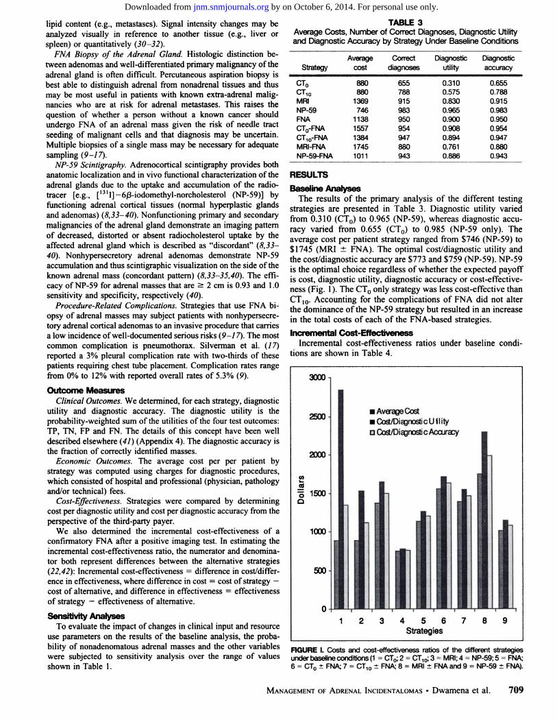
strategies are presented in Table 3. Diagnostic utility variedfrom 0.310 (CT0) to 0.965 (NP-59), whereas diagnostic accuracy varied from 0.655 (CT0) to 0.985 (NP-59 only). Theaverage cost per patient strategy ranged from $746 (NP-59) to$1745 (MRI ±FNA). The optimal cost/diagnostic utility andthe cost/diagnostic accuracy are $773 and $759 (NP-59). NP-59is the optimal choice regardless of whether the expected payoffis cost, diagnostic utility, diagnostic accuracy or cost-effectiveness (Fig. 1). The CT0 only strategy was less cost-effective thanCT10. Accounting for the complications of FNA did not alterthe dominance of the NP-59 strategy but resulted in an increasein the total costs of each of the FNA-based strategies.
Incremental Cost-EffectivenessIncremental cost-effectiveness ratios under baseline condi
tions are shown in Table 4.
3000
2500.
2000-
10t.coO 1500Q
1000
500-
•AverageCost•Cost/DiagnosticU flityD Cosi/Diagnostie Accuracy
456Strategies
FIGURE I. Costs and cost-effectiveness ratios of the different strategiesunder baseline conditions (1 = CT0; 2 = CT10;3 = MRI; 4 = NP-59; 5 = FNA;6 = CTO±FNA; 7 = CT10 ±FNA; 8 = MRI ±FNA and 9 = NP-59 ±FNA).
MANAGEMENTOF ADRENALINCIDENTALOMAS•Dwamena et al. 709
by on October 6, 2014. For personal use only. jnm.snmjournals.org Downloaded from

TABLE 4Incremental Cost-Effectiveness Ratios*
StrategiescomparedCT0
t FNA vs. CT0CT,0±FNAvs.CT10MRI ±FNA vs. MRINP-59 ±FNA vs. NP-59Cost/Diagnostic
utility1132
1580MRI dominatesNP-59 dominatesCost/Diagnostic
accuracy2264
3170MRIdominates
NP-59 dominates*
'Incremental cost-effectiveness indicates the additional cost per addi
tional unit of diagnostic effectiveness.fNP-59 (or MRI) dominates: more expensive strategy (i.e., NP-59 ±FNA
or MRI ±FNA) not more effective (than NP-59 or MRI).
These ratios indicate the additional cost incurred for eachadditional unit of effectiveness obtained using a more costlystrategy (in this case by performing FNA after a positiveimaging test).
Sensitivity AnalysesThe cost-effectiveness advantage of the NP-59 only strategy
was not altered by clinically plausible changes in the malignancy rate and sensitivity of any of the diagnostic tests wheneither of the measures of cost-effectiveness were used but was
sensitive to variations in the cost of the diagnostic tests. Theadvantage of NP-59 was impacted by changes in the specificityof NP-59, CT() and CT,0 when cost/diagnostic utility was theC/E measure of interest. Using cost/diagnostic accuracy as theoutcome measure, the results of the analysis were insensitive tovariations in diagnostic test characteristics except the specificityof NP-59. The thresholds of cost and specificity required forNP-59 to remain cost-effective were <$1180 and >0.68 forcost/diagnostic utility and <$1155 and >0.63 for the cost/
diagnostic accuracy measure. Varying the rate and cost ofcomplications from FNA affected only the biopsy (strategy 5)and the imaging-biopsy (strategies 6-9) options.
DISCUSSIONMost adrenal incidentalomas are nonhypersecretory adeno
mas (2). It is important, however, to distinguish these benignlesions from primary adrenal cancer or métastasesto the adrenalglands to provide timely and appropriate treatment. Despite themultiplicity of testing modalities, there is no consensus regarding the most appropriate diagnostic strategy. With the currenthealth care climate, there is a need for more and betterawareness of the costs and benefits of diagnostic tests. As aresult, physicians are requested to behave in a more cost-effective manner when choosing among numerous alternatives.We have used decision analytic modeling to estimate thecost-effectiveness of several available diagnostic strategies fordifferentiating between benign and malignant adrenal incidentalomas.
Our results showed that an NP-59 strategy was associatedwith the lowest total costs, highest diagnostic utility, highestdiagnostic accuracy and was the most cost-effective. Althoughthe efficacy of NP-59 is derived from our experience, recentresults from other smaller series independently confirm theclinical utility of scintigraphy in the evaluation of the incidentally discovered adrenal mass (36-40). This cost-effectiveadvantage was maintained when the malignancy (nonadenoma)rate was subjected to sensitivity analyses. Accounting for the
complications of FNA did not alter the dominance of the NP-59strategy but resulted in an increase in the total costs of each ofthe FNA-based strategies. The thresholds of cost and specificityrequired for NP-59 to remain cost-effective were ^ $1180 and(3) 0.68 for cost/diagnostic utility and < $1155 and (3) 0.63 for
the cost/diagnostic accuracy measure. The cost thresholds arehigher and the specificity thresholds are lower than are currently available in clinical practice, suggesting that NP-59 iscurrently the most preferred test in evaluating adrenal incidentalomas.
Because the endpoints of interest were diagnostic effectiveness and the costs associated with a one time evaluation ofadrenal incidentalomas, quality-adjusted life years (QALYS)were not used. It is conceivable that the long-term clinical andeconomic outcomes of using the alternative strategies, incorporating therapeutic choices and survival outcomes may bedifferent from that obtained by this model.
Our model reveals that confirmatory testing with FNA isseldom, if ever, preferred to a single test for all diseaseprobabilities, given that the single-test strategies were generallymore cost-effective than the two-test diagnostic approaches.Although imaging appears cost-effective and would sparepatients the risk and discomfort of FNA and other invasivetesting, it is not clear whether physicians or patients are willingto base management decisions regarding adrenal incidentalomas on the results of a noninvasive test alone.
There was no advantage in evaluating patients with FNAinstead of NP-59 or MRI or subjecting patients with positiveimaging tests to confirmatory FNA. Additionally, strategies thatuse FNA may subject patients with benign adrenal incidentalomas to invasive procedures, which carry a low incidence ofwell-documented serious risks (9-17). The awareness thatcytologie distinction between adenomas and well-differentiatedprimary malignancy of the adrenal gland is often difficult raisesthe question of whether a person without a known cancer shouldundergo FNA of an adrenal mass given the risk of needle tractseeding of malignant cells and an often inconclusive result.
MRI ±FNA and CT0 strategies appeared to be the leastcost-effective. For the former, this may be a reflection of thefact that chemical-shift MRI is not only the newest imagingtechnique for characterization of adrenal masses but is currentlythe most expensive, with diagnostic accuracy and utility nobetter than NP-59 or FNA. For the latter, this is because, despiteexcellent sensitivity, it has the lowest specificity. This studyindicates that in situations where CT may be the only modalityavailable for noninvasive evaluation of adrenal incidentalomas,a threshold of > 10 HU may be more useful: it was associatedwith higher diagnostic utility and diagnostic accuracy and morecost-effective than the > 0 HU criterion. Although, CT0 ±FNA was associated with slightly higher diagnostic utility anddiagnostic accuracy than CT1() ±FNA, the latter was morecost-effective using either cost/diagnostic utility or cost/diagnostic accuracy.
Although we assumed that patients are indifferent to the typeof strategy chosen, it is well known that some patients may berisk-averse and prefer noninvasive imaging to FNA or otherinvasive procedures. Although, there may be no tangible complications from the imaging procedures, it is possible thatpatients' attitudes toward the claustrophobia associated with
MRI suites, potential radiation risks from CT or NP-59 and therelative costs of the different tests, among other factors, may berelevant to choice of strategy.
Many physicians, especially in the community hospital setting, may be using serial, conventional CT with size determinations to assess stability (adenomas) or growth (tumor) of
710 THKJOURNALOF NUCLEARMEDICINE•Vol. 39 •No. 4 •April 1998
by on October 6, 2014. For personal use only. jnm.snmjournals.org Downloaded from
adrenal incidentalomas. However, the optimal number of subsequent studies and intervals between them are not wellestablished (2). There is no documented growth rate predictiveof malignancy and operating characteristics for these approaches have not been defined. Typically, additional (two tofive) CT scans may be performed over 18-24 mo before
malignancy is excluded.We did not assess this strategy as part of the baseline analysis
because of insufficient data from the published literature. However, assuming perfect diagnostic performance and an average ofthree CT scans per patient, the average cost per patient (as well ascost per diagnostic accuracy and cost per diagnostic utility) will be$2640, making serial CT testing the least cost-effective approach.Additionally, patients may be subjected to significant cumulativeradiation, may be lost to follow-up and/or those with malignanciesdiagnosed by demonstrable tumor growth may suffer a delay inpotentially curative therapy.
Within the limitations of our assumptions and based onavailable estimates of reimbursement costs and diagnostic testperformance, we conclude that:
1. The NP-59 strategy is the most cost-effective diagnostictool for evaluating adrenal incidentalomas over a widerange of malignancy rates; and
2. Comparative clinical studies to confirm this cost-effectiveness advantage are warranted.
ACKNOWLEDGMENTSThis work was presented in part at the 43rd Annual Meeting of
the Society of Nuclear Medicine, June 3-5, 1996, Denver, CO.
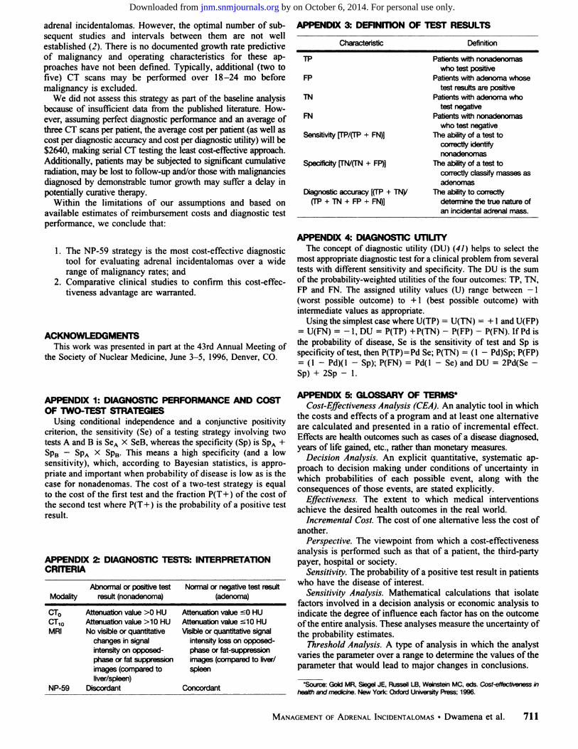
APPENDIX 3: DEFINITION OF TEST RESULTS
Characteristic Definition
TP
FP
TN
FN
Sensitivity [TP/(TP + FN)]
Specificity n~N/(TN + FP)]
Diagnostic accuracy [(TP + TNy(TP + TN + FP + FN)]
Patients with nonadenomaswho test positive
Patients with adenoma whosetest results are positive
Patients with adenoma whotest negative
Patients with nonadenomaswho test negative
The ability of a test tocorrectly identifynonadenomas
The ability of a test tocorrectly classify masses asadenomas
The ability to correctlydetermine the true nature ofan incidental adrenal mass.
APPENDIX 4: DIAGNOSTIC UTILITYThe concept of diagnostic utility (DU) (41) helps to select the
most appropriate diagnostic test for a clinical problem from severaltests with different sensitivity and specificity. The DU is the sumof the probability-weighted utilities of the four outcomes: TP, TN,FP and FN. The assigned utility values (U) range between —1
(worst possible outcome) to +1 (best possible outcome) withintermediate values as appropriate.
Using the simplest case where U(TP) = U(TN) = +1 and U(FP)= U(FN) = -1,DU = P(TP) +P(TN)- P(FP) - P(FN). IfPdis
the probability of disease, Se is the sensitivity of test and Sp isspecificity of test, then P(TP)=Pd Se; P(TN) = (1 - Pd)Sp; P(FP)= (1 - Pd)(l - Sp); P(FN) = Pd(l - Se) and DU = 2Pd(Se -Sp) + 2Sp - 1.
APPENDIX 1: DIAGNOSTIC PERFORMANCE AND COSTOF TWO-TEST STRATEGIES
Using conditional independence and a conjunctive positivitycriterion, the sensitivity (Se) of a testing strategy involving twotests A and B is SeA X SeB, whereas the specificity (Sp) is SpA +SpB —SpA X SpB. This means a high specificity (and a low
sensitivity), which, according to Bayesian statistics, is appropriate and important when probability of disease is low as is thecase for nonadenomas. The cost of a two-test strategy is equalto the cost of the first test and the fraction P(T+) of the cost ofthe second test where P(T+) is the probability of a positive testresult.
APPENDIX 2: DIAGNOSTIC TESTS: INTERPRETATIONCRITERIA
Abnormal or positive testModality result (nonadenoma)
Normal or negative test result(adenoma)
CT0CT10MPINP-59Attenuationvalue >0HUAttenuationvalue >10HUNo
visible orquantitativechangesinsignalintensityonopposed-phase
or fatsuppressionimages(comparedtoliver/spleen)DiscordantAttenuation
value sOHUAttenuationvalue slOHUVisible
or quantitativesignalintensityloss onopposed-phase
orfat-suppressionimages
(compared toliver/spleenConcordant
APPENDIX 5:GLOSSARY OF TERMS*Cost-Effectiveness Analysis (CEA). An analytic tool in which
the costs and effects of a program and at least one alternativeare calculated and presented in a ratio of incremental effect.Effects are health outcomes such as cases of a disease diagnosed,years of life gained, etc., rather than monetary measures.
Decision Analysis. An explicit quantitative, systematic approach to decision making under conditions of uncertainty inwhich probabilities of each possible event, along with theconsequences of those events, are stated explicitly.
Effectiveness. The extent to which medical interventionsachieve the desired health outcomes in the real world.
Incremental Cost. The cost of one alternative less the cost ofanother.
Perspective. The viewpoint from which a cost-effectivenessanalysis is performed such as that of a patient, the third-partypayer, hospital or society.
Sensitivity. The probability of a positive test result in patientswho have the disease of interest.
Sensitivity Analysis. Mathematical calculations that isolatefactors involved in a decision analysis or economic analysis toindicate the degree of influence each factor has on the outcomeof the entire analysis. These analyses measure the uncertainty ofthe probability estimates.
Threshold Analysis. A type of analysis in which the analystvaries the parameter over a range to determine the values of theparameter that would lead to major changes in conclusions.
•Source:Gold MR. Siegel JE, Russell LB. Weinstein MC, eds. Cost-effectiveness in
health and medicine. New York: Oxford University Press: 1996.
MANAGEMENTOF ADRENALINCIDENTALOMAS•Dwamena et al. 711
by on October 6, 2014. For personal use only. jnm.snmjournals.org Downloaded from
Utility. A concept in decision analysis referring to thepreference for or desirability of a particular outcome. Forexample, in our analysis, TP and TN test results are assigned autility of +1 and FP and FN a utility of -1.
REFERENCES1. Copeland PM. The incidentally discovered adrenal mass. Ann Intern Med 1983;98:
940-945.2. Kloos RT, Gross MD. Francis IR, Korobkin M. Shapiro B. Incidentally discovered
adrenal masses. Endocrino! Rev 1995:16:460-484.3. King DR. Lack EE. Adrenal cortical carcinoma: a clinical and pathologic study of 49
cases. Cancer 1979:44:239-244.4. Nosher JL. Amorosa JK. Leiman S. Plafker J. Fine-needle aspiration of the kidney and
adrenal gland. J Ural 1982:128:895-899.
5. Tang C, Gray G. Adrenocortical neoplasms. Prognosis and morphology. Urology1975:5:691-695.
6. Dominguez-Gadea L. Diez L. Bas C. Crespo A. Differential diagnosis of solid adrenalmasses using adrenocortical scintigraphy. Clin Radial 1994:49:796-799.
7. Gaboardi F. Carbone M, Bozzola A. Galli L. Adrenal incidentalomas: what is the roleof fine-needle biopsy? ini Urol Nephrol 1991:23:197-207.
8. Gross MD, Shapiro B, Francis IR. et al. Scintigraphic evaluation of clinically silentadrenal masses. J NucÃMed 1994:35:1145 1152.
9. Mody MK, Kazerooni EA. Korobkin M. Percutaneous CT-guided biopsy of adrenalmasses: immediate and delayed complications. J Comput Assist Tomogr 1995:19:434 -
439.10. Zomoza J. Ordonez N. Bernardino ME. Cohen MA. Percutaneous biopsy of adrenal
rumors. Urology 1981:18:412-416.11. Heaston DK. Handel DB. Ashlon PR. Korobkin M. Narrow gauge needle aspiration of
solid adrenal masses. Am J Roenlgemtl 1982:138:1143-1148.12. Katz RL. Palei S, Mackay B. Zomoza J. Fine-needle aspiration cytology of the adrenal
gland. Acta Cytol 1984:28:269-282.13. Bernardino ME. Walther MM. Phillips VM. et al. CT-guided adrenal biopsy: accuracy,
safety, and indications. Am J Roenigenol 1985:144:67-69.
14. Yankaskas BC. Staab EV. Craven MB. Blatt PM. Sokhandan M, Carney CM. Delayedcomplications from fine-needle biopsies of solid masses of the abdomen. Invest Radio!1986:21:325-328.
15. Kane NM, Korobkin M. Francis IR. Quint LE, Cascade PN. Percutaneous biopsy ofleft adrenal masses: prevalence of pancreatitis after anterior approach. Am J Roenigenol 1991:157:777-780.
16. Welch TJ, Sheedy Pn. Stephens DH. Johnson CM. Swensen SJ. Sheedy PN.Percutaneous adrenal biopsy: review of a 10-year experience. Radiology 1994:193:341-344.
17. Silverman SO, Mueller PR, Pinkney LP, Koenker RM, Seltzer SE. Predictive value ofimage-guided adrenal biopsy: analysis of results of 101 biopsies. Radiologi- 1993:187:715-718.
18. Hagen MD. Decision analysis: a review. Fam Med 1992:24:349-54.19. Pauker SG. Kassirer JP. Decision analysis. N Engt J Med 1987:316:250-258.
20. Phelps CE. Mushlin AI. Focusing technology assessment using medical decisiontheory. Med Decision Making 1988:8:279-289.
21. Powe NR. Economic and cost-effectiveness investigations of radiologie practices.Radiology 1994:192:11 -18.
22. Weinstein MC. Stason WB. Foundations of cost-effectiveness analysis for health andmedical practices. ,V Engl J Med 1977:296:716-21.
23. Ament A, Hasman A. Optimal test strategy in the case of two tests and one disease. IntJ Biomed Compai 1993:33:179-197.
24. Hershey JC, Cebul RD, Williams SV. Clinical guidelines for using two dichotomoustests. Med Decision Making 1986:6:68-78.
25. Lee M, Hahn P, Papanicolaou N, et al. Benign and malignant adrenal masses: CTdistinction with attenuation coefficients, size and observer analysis. Radiology 1991;179:415-418.
26. Singer AA, Obuchowski NA, Einstein DM, Paushter DM. Metastasis or adenoma?Computed tomographic evaluation of the adrenal mass. Cleveland Clin J MedI994;61:200-205.
27. van Erkel AR. van Gils AP, Lequin M, Kruitwagen C, Bloem JL, Falke TH. CT andMR distinction of adenomas and nonadenomas of the adrenal gland. J Comput AssistTomogr 1994:18:432-438.
28. Korobkin M, Brodeur F, Yutzy G, et al. Differentiation of Adrenal Adenomas fromNonadenomas using CT Attenuation values. Am J Roenigenol 1996:166:531-536.
29. Mitchell DO, Crovello M, Matteucci T, Petersen RO, Miettinen MM. Benignadrenocortical masses: diagnosis with chemical shift MR imaging. Radiology 1992;185:345-351.
30. Bilbey JH, McLoughlin RF, Kurkjian PS, et al. MR imaging of adrenal masses: valueof chemical-shift imaging for distinguishing adenomas from other tumors. Am JRoenigenol 1995:164:637-642.
31. Mayo-Smith WW, Lee MJ, McNicholas MM, Hahn PF, Boland GW, Saini S.Characterization of adrenal masses (<5 cm) by use of chemical shift MR imaging:observer performance versus quantitative measures. Am J Roenigenol 1995:165:91-95.
32. Outwater EK, Mitchell DO. Differentiation of adrenal masses with chemical shift MRimaging [Letter]. Radiology 1994:193:877-878.
33. Francis I, Smid A. Gross M, Shapiro B, Naylor B, Glazer G. Adrenal masses inoncologie patients: functional and morphologic evaluation. Radiology 1988:166:353-
356.34. Gross MD, Wilton OP. Shapiro B, et al. Functional and scintigraphic evaluation of the
silent adrenal mass. J NucÃMed 1987:28:1401-1407.
35. Gross MD, Shapiro B. Bouffard JA, et al. Distinguishing benign from malignanteuadrenal masses. Ann Intern Med 1988:109:613-618.
36. Dominguez-Gadea L, Diez L, Crespo A. Differential diagnosis of solid adrenal massesusing adrenocortical scintigraphy. Clin Radial 1994;49:796-799.
37. Nakajo M. Nakacelppin Y, Yonejura R, Icuashita S, Goto T. The role of adrenocorticalscintigraphy in the evaluation of unilateral incidentally discovered adrenal andjuxtaadrenal masses. Ann NucÃMed 1992:7:157-166.
38. Bardet S. Rohmer V, Margat A, et al. and the West France Study Group ofIncidentalomas (WFSGI). lodine-131-6 ß-iodomethylnorcholesterol scintigraphy: an
assessment of its role in the investigation of adrenocortical incidentalomas. ClinEndocrinol 1996;44:587-596.
39. Reschini E, Catania A. Clinical experience with the adrenal scanning agents iodine-19-iodocholesterol and selenium 75Se-6-selenomethyl/cholesterol. Eur J NucÃMed
1991:18:817-823.
40. Kloos R, Gross MD, Shapiro B, Francis IR, Korobkin M, Thompson NW. Thediagnostic dilemma of small incidentally discovered adrenal masses: a role for'"l-6ß-iodomethylnoncholesterol [NP-59] scintigraphy. World J Surg 1997:21:36-
40.41. Patton DD, Woolfenden JM. A utility-based model for comparing the cost-effective
ness of diagnostic tests. Invest Radiology 1989:24:263-271.42. Detsky AS. Naglie IG. A clinician's guide to cost-effectiveness analysis. Ann Intern
Med 1990:13:147-154.
Radioiodine Treatment Outcomes in Thyroid GlandsPreviously Irradiated for Graves' HyperthyroidismWilliam D. Leslie, Anne E. Peterdy and Jacqueline O. DupontDepartment of Nuclear Medicine. University of Manitoba, St. Boniface General Hospital, Winnipeg, Manitoba, Canada
Persistent or recurrent Graves' hyperthyroidism after an initial treat
ment dose of radioactive iodine (RAI) is not uncommon and usuallynecessitates additional administrations. The radiation sensitivity ofthe previously irradiated thyroid gland is unknown but is of importance in selecting the retreatment dose. Methods: A retrospectiveanalysis of patients receiving RAI for Graves' hyperthyroidism was
undertaken. A first treatment dose was given to 1076 patients, and168 of these patients subsequently required a second dose forpersistent or recurrent hyperthyroidism (interval between RAI treatments, 8.5 ±17.1 mo). Results: Paired comparisons for retreatedpatients showed similar RAI doses (291 ±95 MBq and 283 ±129MBq; p = ns) and treatment intensities (3.26 ±1.87 MBq g 1 and
Received Jan. 27, 1997; revision accepted Jun. 12, 1997.For correspondence or reprints contact: William D. Leslie, MD. Department of Internal
Medicine (C5121), St. Boniface General Hospital, 409 Tache Ave., Winnipeg, Manitoba,Canada R2H 2A6.
3.48 ±1.88 MBq g 1; p = ns) for first and second treatments.
Hypothyroidism occurred significantly earlier and more frequentlyafter the first RAI dose (p = 0.002), but there was no difference forpersistent or recurrent hyperthyroid events (p = 0.14). Multivariateregression established that the RAI treatment number (first or second)was a significant independent determinant of hypothyroid (p = 0.008)and combined (p = 0.001) events, whereas RAI dose and doseintensity were not. Conclusion: We conclude that previous RAItreatment failure does not lessen the chance of successfully eradicating Graves' hyperthyroidism with additional RAI treatment. Further
more, the previously irradiated thyroid gland may be less susceptibleto early hypothyroidism than the RAI-naive thyroid gland.
Keywords: hyperthyroidism;Graves' disease; iodine-131
J NucÃMed 1998; 39:712-716
712 THE JOURNALOF NUCLEARMEDICINE•Vol. 39 •No. 4 •April 1998
by on October 6, 2014. For personal use only. jnm.snmjournals.org Downloaded from