Diagnosis and Treatment of Perianal Crohn Disease
12
Copyright 2013 by ESPGHAN and NASPGHAN. Unauthorized reproduction of this article is prohibited. Diagnosis and Treatment of Perianal Crohn Disease: NASPGHAN Clinical Report and Consensus Statement Edwin F. de Zoeten, z Brad A. Pasternak, § Peter Mattei, Robert E. Kramer, and y Howard A. Kader ABSTRACT Inflammatory bowel disease is a chronic inflammatory disorder of the gastrointestinal tract that includes both Crohn disease (CD) and ulcerative colitis. Abdominal pain, rectal bleeding, diarrhea, and weight loss characterize both CD and ulcerative colitis. The incidence of IBD in the United States is 70 to 150 cases per 100,000 individuals and, as with other autoimmune diseases, is on the rise. CD can affect any part of the gastrointestinal tract from the mouth to the anus and frequently will include perianal disease. The first description connecting regional enteritis with perianal disease was by Bissell et al in 1934, and since that time perianal disease has become a recognized entity and an important consideration in the diagnosis and treatment of CD. Perianal Crohn disease (PCD) is defined as inflammation at or near the anus, including tags, fissures, fistulae, abscesses, or stenosis. The symptoms of PCD include pain, itching, bleeding, purulent discharge, and incontinence of stool. In this report, we review and discuss the etiology, diagnosis, evaluation, and treatment of PCD. Key Words: perianal abscess, perianal Crohn disease, perianal fistula (JPGN 2013;57: 401–412) I nflammatory bowel disease (IBD) is a chronic inflammatory disorder of the gastrointestinal tract that includes both Crohn disease (CD) and ulcerative colitis (UC). Abdominal pain, rectal bleeding, diarrhea, and weight loss characterize both CD and UC. The incidence of IBD in the United States is 70 to 150 cases per 100,000 individuals and, as with other autoimmune diseases, is on the rise (1). CD can affect any part of the gastrointestinal tract from the mouth to the anus and frequently will include perianal disease. The first description connecting regional enteritis with perianal disease was by Bissell et al in 1934 (2), and since that time perianal disease has become a recognized entity and an important consideration in the diagnosis and treatment of CD. Perianal Crohn disease (PCD) is defined as inflammation at or near the anus, including tags, fissures, fistulae, abscesses, or stenosis. The symptoms of PCD include pain, itching, bleeding, purulent discharge, and incontinence of stool. INCIDENCE AND NATURAL HISTORY Limited pediatric data describe the incidence and prevalence of PCD. The incidence of PCD in the pediatric age group has been estimated to be between 13.6% and 62% (3). In a review by Palder et al (4), 62% of 325 children with CD developed PCD. Skin tags were present in 114 (35%), fissures in 185 (51%), fistulae in 41 (15%), and perirectal abscesses in 47 (13%). In another study of 276 patients with CD, Keljo et al identified perianal lesions in 41 (15%) who were studied within 30 days of diagnosis (3). A 2012 study in New Zealand reported that 26.5% of children with CD develop some form of PCD (5). In the adult literature, the reported incidence of PCD ranges from 25% to 80% (6) and fistulizing PCD from 17% to 43% (7 – 9). The clinical course of PCD depends on the type, including skin tag, rectal stricture, fistulae or fissures, and location of disease. Skin tags in general do not resolve completely with treatment, but remain present and benign. Fissures will often heal completely with minor medical therapy. Simple fistulizing disease, when superficial and confined to the anal canal, heals spontaneously in 50% of cases (10). In contrast, rectovaginal and complex fistulae rarely heal without therapy (11). Complex fistulae include those with origin of the fistulous tract that is high intersphincteric, high transsphincteric, extrasphincteric, or suprasphincteric. ETIOLOGY CD is defined in part by the transmural nature of inflammation, which in turn predisposes patients to fistulae and abscess formation around the anus and in other locations. Two mechanisms have been postulated with regard to the pathogenesis of fistulae and abscesses: initial inflammation in the rectum forms either ulcers or shallow fistulae, which subsequently extend into deep or penetrating fistulae with the persistent exposure to feces and pressure caused by defeca- tion (12); infected anal glands penetrate the intrasphincteric space and then progress to form fistulae or abscesses (13). Increasing evidence supports a role for genetic predisposition and microbiota influence in the development of perianal fistulae (PF) and abscesses (PAs). A study from New Zealand looking at 715 patients younger than 20 years found that PCD not only was significantly associated with younger age at diagnosis, complicated intestinal disease, and ileal disease location but it also had a genetic link (5). The characterization of the genetic background in CD Received June 18, 2013; accepted June 6, 2013. From the Department of Pediatrics, University of Colorado School of Medicine, Children’s Hospital Colorado, Digestive Health Institute, Pediatric Inflammatory Bowel Disease Center, Denver, CO, the y Department of Pediatrics, Division of Pediatric Gastroenterology and Nutrition, University of Maryland School of Medicine Pediatric Inflam- matory Bowel Disease Center, Baltimore, MD, the z Department of Pediatric Gastroenterology, Phoenix Children’s Hospital, Phoenix, AZ, and the § Department of Surgery, University of Pennsylvania, The Children’s Hospital of Philadelphia, Philadelphia, PA. Address correspondence and reprint requests to Edwin de Zoeten, MD, PhD, Pediatric IBD Center, Children’s Hospital Denver Digestive Health Institute, Anschutz Medical Campus, 13123 East 16th Ave, B290 Aurora, CO 80045. This article has been developed as a Journal CME Activity by NASP- GHAN. Visit http://www.naspghan.org/wmspage.cfm?parm1=742 to view instructions, documentation, and the complete necessary steps to receive CME credit for reading this article. Copyright # 2013 by European Society for Pediatric Gastroenterology, Hepatology, and Nutrition and North American Society for Pediatric Gastroenterology, Hepatology, and Nutrition DOI: 10.1097/MPG.0b013e3182a025ee CLINICAL REPORT JPGN Volume 57, Number 3, September 2013 401
-
Upload
khangminh22 -
Category
Documents
-
view
4 -
download
0
Transcript of Diagnosis and Treatment of Perianal Crohn Disease

Co
CLINICAL REPORT
Diagnosis and Treatment of Perianal Crohn
Disease: NASPGHAN Clinical Report and
Consensus Statement
Mattei, �Robert E. K ward A. Kader
�Edwin F. de Zoeten, zBrad A. Pasternak, §Peterpyright 2013 by ESPGHAN and NASPGHAN. Unauthorized repro
on the rise (1). CD can affect any part of the gastrointestinal tractfrom the mouth to the anus and frequently will include perianal
intestinal disease, and ilink (5). The characte
Received June 18, 2013; accepted June 6, 2013.From the �Department of Pediatrics, University of Colorado School of
Medicine, Children’s Hospital Colorado, Digestive Health Institute,Pediatric Inflammatory Bowel Disease Center, Denver, CO, theyDepartment of Pediatrics, Division of Pediatric Gastroenterology andNutrition, University of Maryland School of Medicine Pediatric Inflam-matory Bowel Disease Center, Baltimore, MD, the zDepartment ofPediatric Gastroenterology, Phoenix Children’s Hospital, Phoenix,AZ, and the §Department of Surgery, University of Pennsylvania,The Children’s Hospital of Philadelphia, Philadelphia, PA.
Address correspondence and reprint requests to Edwin de Zoeten, MD,PhD, Pediatric IBD Center, Children’s Hospital Denver DigestiveHealth Institute, Anschutz Medical Campus, 13123 East 16th Ave,B290 Aurora, CO 80045.
This article has been developed as a Journal CME Activity by NASP-GHAN. Visit http://www.naspghan.org/wmspage.cfm?parm1=742 toview instructions, documentation, and the complete necessary stepsto receive CME credit for reading this article.
Copyright # 2013 by European Society for Pediatric Gastroenterology,Hepatology, and Nutrition and North American Society for PediatricGastroenterology, Hepatology, and Nutrition
DOI: 10.1097/MPG.0b013e3182a025ee
JPGN � Volume 57, Number 3, September 2013
ramer, and yHo
ABSTRACT
Inflammatory bowel disease is a chronic inflammatory disorder of the
gastrointestinal tract that includes both Crohn disease (CD) and ulcerative
colitis. Abdominal pain, rectal bleeding, diarrhea, and weight loss
characterize both CD and ulcerative colitis. The incidence of IBD in the
United States is 70 to 150 cases per 100,000 individuals and, as with other
autoimmune diseases, is on the rise. CD can affect any part of the
gastrointestinal tract from the mouth to the anus and frequently will include
perianal disease. The first description connecting regional enteritis with
perianal disease was by Bissell et al in 1934, and since that time perianal
disease has become a recognized entity and an important consideration in the
diagnosis and treatment of CD. Perianal Crohn disease (PCD) is defined as
inflammation at or near the anus, including tags, fissures, fistulae, abscesses,
or stenosis. The symptoms of PCD include pain, itching, bleeding, purulent
discharge, and incontinence of stool. In this report, we review and discuss
the etiology, diagnosis, evaluation, and treatment of PCD.
Key Words: perianal abscess, perianal Crohn disease, perianal fistula
(JPGN 2013;57: 401–412)
I nflammatory bowel disease (IBD) is a chronic inflammatorydisorder of the gastrointestinal tract that includes both Crohn
disease (CD) and ulcerative colitis (UC). Abdominal pain, rectalbleeding, diarrhea, and weight loss characterize both CD and UC.The incidence of IBD in the United States is 70 to 150 cases per100,000 individuals and, as with other autoimmune diseases, is
disease. The first description connecting regional enteritis withperianal disease was by Bissell et al in 1934 (2), and since that timeperianal disease has become a recognized entity and an importantconsideration in the diagnosis and treatment of CD. PerianalCrohn disease (PCD) is defined as inflammation at or near theanus, including tags, fissures, fistulae, abscesses, or stenosis.The symptoms of PCD include pain, itching, bleeding, purulentdischarge, and incontinence of stool.
INCIDENCE AND NATURAL HISTORYLimited pediatric data describe the incidence and prevalence
of PCD. The incidence of PCD in the pediatric age group has beenestimated to be between 13.6% and 62% (3). In a review by Palderet al (4), 62% of 325 children with CD developed PCD. Skin tagswere present in 114 (35%), fissures in 185 (51%), fistulae in41 (15%), and perirectal abscesses in 47 (13%). In another studyof 276 patients with CD, Keljo et al identified perianal lesions in41 (15%) who were studied within 30 days of diagnosis (3). A 2012study in New Zealand reported that 26.5% of children with CDdevelop some form of PCD (5).
In the adult literature, the reported incidence of PCD rangesfrom 25% to 80% (6) and fistulizing PCD from 17% to 43% (7–9).The clinical course of PCD depends on the type, including skin tag,rectal stricture, fistulae or fissures, and location of disease. Skin tagsin general do not resolve completely with treatment, but remainpresent and benign. Fissures will often heal completely with minormedical therapy. Simple fistulizing disease, when superficial andconfined to the anal canal, heals spontaneously in 50% of cases (10).In contrast, rectovaginal and complex fistulae rarely heal withouttherapy (11). Complex fistulae include those with origin of thefistulous tract that is high intersphincteric, high transsphincteric,extrasphincteric, or suprasphincteric.
ETIOLOGYCD is defined in part by the transmural nature of inflammation,
which in turn predisposes patients to fistulae and abscess formationaround the anus and in other locations. Two mechanisms have beenpostulated with regard to the pathogenesis of fistulae and abscesses:initial inflammation in the rectum forms either ulcers or shallowfistulae, which subsequently extend into deep or penetrating fistulaewith the persistent exposure to feces and pressure caused by defeca-tion (12); infected anal glands penetrate the intrasphincteric space andthen progress to form fistulae or abscesses (13).
Increasing evidence supports a role for genetic predispositionand microbiota influence in the development of perianal fistulae(PF) and abscesses (PAs). A study from New Zealand looking at715 patients younger than 20 years found that PCD not only wassignificantly associated with younger age at diagnosis, complicated
duction of this article is prohibited.
leal disease location but it also had a geneticrization of the genetic background in CD
401

Co
continues to develop rapidly, especially by means of genome-wideassociation studies. An association of CD fistulae and abscesses hasbeen linked to specific gene variants at the susceptibility locuson chromosome 5q31 (IBD5), including OCTN (14) and IRGM(14–16). Impaired OCTN activity or expression may reduce carni-tine transport, which can lead to defects in oxygen burst–mediatedpathogen killing, fatty acid oxidation defects in the intestinalepithelial cells, or defects in transport of bacterial toxins intoand out of the epithelial cells causing inflammation. Whether theseare mechanisms that cause PCD is unknown. IRGM is part ofthe autophagy pathway that eliminates intracellular organisms.Although the study from New Zealand did not show the sameassociation with IRGM, it did note an association with a novel geneneutrophil cytosolic factor 4 (NCF4), a gene that has a role inphagocytosis and bacterial killing (5).
A few investigators have studied the association between theNOD2/CARD15 genotype and PCD. To date, no correlation existsbetween this genotype and PF (17); however, NOD2/CARD15 genestatus appears to influence the response of PF to antibiotic therapy,with a greater response noted in patients with NOD2/CARD15 wildtype (18).
Although the use of antibiotics and fecal diversion appearsuseful in some patients with PF and PA, suggesting a pathogeniccontribution of the microbiome, their incomplete efficacy suggeststhat microbes alone cannot explain PCD. The search for a singleorganism as the causative agent has yielded inconsistent results.In general, many bacterial species have been implicated in inflam-mation, including enteroadherent strains of Escherichia coli (19)Chlamydia (20), and mycobacteria (21). Whether these changes inbacterial flora are primary or secondary to the disease process is notclear. Few studies have assessed microbiological changes in Crohnfistulae. One involved the aspiration of pus from the fistulous tractsof 13 Crohn patients with perianal disease (22). These sampleswere analyzed by standard bacterial culture techniques. Gram-positive organisms, in particular staphylococci, streptococci, andcorynebacter, predominated over Gram-negative enteric organisms(23). This differs from idiopathic fistula (22), in which themicrobiota found on excised granulation tissue were largely ofgastrointestinal origin. Additional investigations are needed toclarify the role of the microbiology of CD fistulizing disease.
CLINICAL PRESENTATIONManifestations of PCD fall into 3 distinct categories: tissue
destruction, which includes anal fissures, tags, and deep ulcers linedwith exuberant granulation tissue; fistulae and abscesses, which areoften multiple and complex and are frequently related; and rectalstricture, which is likely the result of long-standing or even healedcircumferential Crohn-related inflammation of the rectum oranoderm. The spectrum of PCD is variable in that some may haverelatively mild disease, whereas others can have a severe, destruc-tive, and debilitating course leading to constipation, fecal incon-tinence, recurrent infections and sepsis, compromise of sexualfunction, significantly diminished quality of life, and, in somecases resulting in a diverting colostomy or ileostomy (3,24). Evenin its most severe form, PCD can be surprisingly painless. Childrenand adolescents are often reticent to volunteer information aboutbowel habits, drainage, or anal pruritus; it is therefore extremelyimportant to ask direct questions about these symptoms. Fissuresmay cause bleeding but, despite their depth or severity, do notalways cause pain with defecation. Anal skin tags, classic hallmarksof CD, may result in cosmetic concerns, but are not frequentlyassociated with pain unless inflamed.
de Zoeten et al
pyright 2013 by ESPGHAN and NASPGHAN. Un
Strictures are typically the result of long-standing inflam-mation (25) and generally form extremely slowly, which allows
402
patients to accommodate themselves to the relative narrowing of therectum. Thus, often patients state an ability to pass stool normallydespite having a rectal stricture. Nevertheless, although children withPCD may not complain of pain, the functional impairments (consti-pation, incontinence, spontaneous drainage) and the social implica-tions (odor, stained undergarments, anxiety) are often significant.
PERIANAL FISTULA
DiagnosisDiagnosing PF in CD is extremely important because
this ultimately influences not only the medical but also the surgicalmanagement and approach to therapy (Fig. 1). In addition,definition of the anatomic location of the fistula is critical if surgicalintervention is considered.
Grading and Naming Systems
Various attempts to develop a consistent and useful severityscore or staging system have been described over the years; however,their clinical value, especially in children, has been limited. Several ofthe more common approaches are discussed here.
Cardiff System
The earliest and perhaps still most commonly used classifi-cation scheme is the Cardiff system, originally described in 1978 byHughes (12) and later modified in 1992 (26). The Cardiff classi-fication provides a thorough anatomic description of the disease, butits real clinical significance has been questioned (6,27). Each majorcomponent of the disease (ulceration, fistula, and stricture) isgraded on a scale of 0, 1, or 2. The score is also modified bythe presence of associated anal conditions (hemorrhoids, malig-nancy), proximal intestinal disease, and relative disease activity(active inflammation).
Park Classification
PF are still often classified according to the descriptivesurgical Park classification, which was originally used to describefistula-in-ano not associated with CD. Although helpful inthe operative setting, this system may be difficult to use in clinicalpractice (28). Park described 5 types of fistula (Fig. 2, left side):superficial, which stays below the internal and external sphincters,travels inferior to the sphincters, and involves the ischioanal fossa;intersphincteric, which travels between the internal and externalanal sphincters; transsphincteric, which passes through the externalsphincter; suprasphincteric, a complex fistula that passes throughthe internal sphincter and then travels up through the levatorani musculature before tracking to the skin, which can also resultin supralevator abscesses as the tract extends through the levatorani muscle toward the skin after leaving the intersphincteric spaceby going over the top of the puborectalis muscle; and extra-sphincteric, which stays external to the sphincters connectingrectum and skin. These fistulae pass through the ischiorectal fossaand levator ani muscle opening into the rectum (28). Patients withPF often have complex, branching, and interconnected series offistulae that defy easy description, let alone objective classification.
Simple Versus Complex Fistula
Location of the fistula (high or low in relation to the anorectal
JPGN � Volume 57, Number 3, September 2013
authorized reproduction of this article is prohibited.
ring) is traditionally used by clinicians to classify fistulae; however,a recent description as to whether it is a simple or complex fistula,
www.jpgn.org

Co
Obturator internusmuscle External iliac vessels
External anal sphincter
Dentate line
Internal anal sphincter
Levator ani muscle
Ischiorectal fossa
Transverse fibrousseptum
ESF SSF SPF CPFTSF
Park classification Bell classification
ISF SF
FIGURE 1. Schematic illustration of 2 classification systems for perianal fistulas. On the left, the Park classification identifies strictures based on
their anatomic relation to the internal and external anal sphincters. On the right, the Bell classification divides fistulas into either simple or complex
categories, depending on how low the internal opening is or if the fistula contains an abscess or multiple branches. CPF¼ complex perianal
ic fihin
JPGN � Volume 57, Number 3, September 2013 NASPGHAN Clinical Report: Perianal Crohn Disease
such as that suggested by Bell et al (29) in 2003 (Fig. 2, right side),may be more relevant for the clinician. To determine a simpleversus complex fistula, the clinician assesses the perianal region forthe presence of abscesses, strictures, and fistulous connections by
fistula (orange); ESF¼ extrasphincteric fistula (red); ISF¼ intrasphincterfistula (dark blue); SSF¼ suprasphincteric fistula (purple); TSF¼ transp
pyright 2013 by ESPGHAN and NASPGHAN. Un
inspection as well as endoscopically. A simple fistula involves a lowintersphincteric or transphinteric location, a single short tract, an
Assessment of intestinal disease History and physical Colonoscopy to evaluate rectal inf Perianal assessment Examine for pain, fluctuance, eryt Digital rectal exam for stricture
Absent: Pain, fluctuance, or str
• EUA+/–EU or MRI
• Antibiotics• Anti-TNFα• Immunomodulators
• Antibiotics• Anti-TNFα• Immunomodulators
Anti-TNFα
Consider noncuttingseton or fistulotomy C
c
Simple fistula no rectal inflammation Complex or simple fistrectal inflammatio
FIGURE 2. Perianal fistula—assessment and treatment algorithm.
www.jpgn.org
internal opening lower and closer to the anal verge, and the externalopening near the anal verge without an abscess. A complex fistulainvolves the sphincter muscle/anorectal ring, multiple fistulae, andbranches with or without an abscess; an internal opening of the
stula (light blue); SF¼ superficial fistula (yellow); SPF¼ simple perianalcteric fistula (green). Illustration by Robert Kramer, MD.
authorized reproduction of this article is prohibited.
fistulous tract is above the sphincter muscles, with the externalopening further away from the anal opening.
lammation
hema
icture
• Antibiotics• Anti-TNFα• Immunomodulators
Anti-TNFα Anti-TNFα
onsider non-utting seton
• Consider advancement flapin rectovaginal fistulae
ula withn
Complex fistula no rectalinflammation
403

Co
PCD Activity Index
Pikarsky et al (30) in 2002 proposed a much more compre-hensive score, the PCD activity index, which entails separatelygrading the degree of abscesses, fistula, ulcer, fissure, stenosis,incontinence, and concomitant upper-tract bowel disease byseparate scales and adding them together to produce a scorebetween 0 and 55. Although more sophisticated and possibly morepredictive than the Cardiff classification, the clinical usefulness ofthis more cumbersome system has yet to be demonstrated and mayhave more use in clinical trials and outcome assessment than inclinical practice (31).
Several imaging modalities are available to adequatelydiagnose, define, and classify a PF. Complete documentation ofPCD is accomplished with radiologic imaging, visual inspectionunder anesthesia, and endoscopic assessment to formulate anindividual treatment plan.
Imaging of PF
All children with CD, with or without perianal complaints,should at minimum have an external examination of the perianalregion. Without external visible signs of PCD, most childrenwill tolerate a gentle and brief digital rectal examination to identifya significant stricture or fluctuance; however, if clear signs ofPCD are present, radiographic evaluation or examination underanesthesia are more useful and less traumatic and should beconsidered (32).
Radiographic imaging modalities include magneticresonance imaging (MRI), computerized tomography (CT), endo-scopic anorectal ultrasound (EUS), and fistulography. It is oftendifficult to visualize the enteric opening of a fistula or to attainretrograde fill of a fistulous tract with contrast externally, making afistulogram challenging. Consequently, the accuracy of fistulogra-phy has been reported to be poor, 16% to 50% (33). EUS hastechnical limitations and requires considerable expertise, especiallyif a stricture/stenosis or abscess is present. CT scans, althoughbeneficial for identifying strictures, thickened bowel wall, and fluidcollections/abscesses, is not useful for delineating soft tissue andmuscle layers containing the fistulous tracts. Consequently, themost useful radiologic modality to evaluate abscesses and fistulae isMRI. The MRI can show separate soft tissues with high distinction;images can be viewed in both coronal and sagittal planes to betterdelineate landmarks, and nonionizing radiation avoids exposureto radiation (34). MRI is highly sensitive for fistulae andabscesses. Compared with examination under anesthesia (EUA),the specificity of MRI in identifying the fistula and its path rangesfrom 76% to 100%. Additionally, MRI incorporates the benefit thata CT offers without the radiation exposure, and thus is considered tobe the radiologic study of choice under most circumstances (8).Most patients, therefore, benefit from MRI followed by EUA.
Abdominal MRI and pelvic MRI have been applied in thediagnosis of fistulae and PF in children (35–39). Advances such ashigher field strength and greater signal-to-noise ratio have reducedscan time and improved image resolution (35–39). New techniquesin breath hold image capture, fat suppression, oral contrastagents, and intravenous gadolinium generate consistent high-quality MRI images in children (12,14). In pediatrics, MRI hasbeen used to identify fistula, inflammation, and evaluate relatedcomplications in the perianal and perirectal regions, similar toadult studies (15,16). Others have shown the use of MRI in
de Zoeten et al
pyright 2013 by ESPGHAN and NASPGHAN. Un
determining the extent of the disease to assist in operative planningand management in adult patients (40–44).
404
EUA is a safe and well-tolerated procedure that has areported specificity of 91% (45,46). Where available, EUA is thepreferred diagnostic choice if a PA is suspected (47). Consensusrecommendations from the European Crohn’s and ColitisOrganization in 2006 support the combination of MRI and procto-sigmoidoscopy evaluation in making a diagnosis of PF or PAand for planning therapy (8). The European Crohn’s and ColitisOrganization statement also recommended an EUA alone if thereis a contraindication to using the MRI and determined that the EUAis considered the criterion standard in the hands of an experiencedsurgeon (8). Although EUA is considered a diagnostic procedure,it often offers the concomitant opportunity to provide symptomaticrelief or to treat or prevent an infectious complication (33).
To perform EUA, careful external examination is performed,followed by digital examination. The degree and extent of inflam-matory changes and any degree of stricture, if present, should benoted. Areas of induration or fluctuance may indicate an abscess,highlighting the benefit of preoperative imaging. Aspiration with alarge-bore needle and syringe can be used to identify pockets ofpurulent fluid that require drainage. Any dimple or potential fistulashould be probed gently to identify cavities or communicationwith the rectum or anoderm. In boys, the base of the scrotumand in girls the labia majora and the vaginal walls should beincluded in the field to help identify all potential sites of diseaseextension and fistulae. Meticulous probing should allow character-ization of the full extent of most PF; dilute methylene blue orhydrogen peroxide injected gently into an external opening may behelpful to identify an internal fistulous tract.
In summary, the most common approach to evaluatePCD involves an external examination and rectal examination,followed by imaging with MRI and finally EUA performed byan experienced surgeon (Fig. 1).
PAClassification of a PA involves the relation of the abscess to
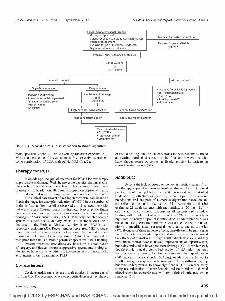
the internal sphincter and levator muscles (Fig. 3). Abscessesmay be perianal, ischiorectal, or submucosal, indicating collectionsof pus in the shallower or more superficial layers of the skin aroundthe anus. A pelvirectal abscess is deeper, occurring between thelevator muscle and the anterior peritoneal reflection. Approxi-mately 60% of such abscesses are perianal, with another 30% beingischiorectal, and the remainder are equally split at 5% for sub-mucosal/intramural and pelvirectal (48). The goal of diagnosing aPA is not only to correctly identify this entity but also to promptlyinstitute therapy to alleviate the clinical signs and symptoms.
The diagnosis of a PA relies primarily on visual inspectionand physical examination. Features include erythema and swellinginvolving the skin around the anus with a visual bulge and apalpable bump that may be either firm, tense, and painful to thetouch or fluctuant. Pain at the anus can also be a symptom as well assigns of pus or purulent drainage from the anus. Fever, chills, andmalaise may be systemic signs of a PA.
Imaging modalities that may be useful to evaluate the extent ofthe PA include CT scan, MRI, or ultrasound. CT with intravenous andrectal contrast may show the presence and the location of a perirectalabscess and can identify deeper abscesses than ultrasonography;however, ultrasound is useful to assess the presence, location, andextent of the submucosal and intersphincteric abscesses and is a cost-effective method for documenting perirectal and perianal fluidcollections. Other advantages to ultrasound include the absence ofnonionizing radiation and bedside performance (49). Ultrasoundtechniques include endoanorectal, transperineal, and transvaginal
JPGN � Volume 57, Number 3, September 2013
authorized reproduction of this article is prohibited.
approaches. MRI, like CT, is good at identifying deep abscesses,detecting granulation tissue, and separating the anatomical plains
www.jpgn.org
Co
Assessment of intestinal disease History and physical Colonoscopy to evaluate rectal inflammation Perianal assessment Examine for pain, fluctuance, erythema Digital rectal exam for stricture
No pain, fluctuation or stricture
Proceed to perianal fistulaalgorithm
Present: Pain, fluctuance or stricture
• EUA+/–EUSor
• MRI pelvis
• Incision and drainageand
• Antibiotics
Abscess present
Deep abscess
High perianal fistula identified
Place a noncutting seton Place a mushroom catheter
Perianal fistula not identified
Superficial abscess
• Incision and drainage• If associated with low perianal fistula, a noncutting seton may be placed• Antibiotics
Antibiotics for cellulitis if presenttreat intestinal disease• Anti-TNFα• Azathioprine/6MP• Methotrexate
Treat intestinal disease• Anti-TNFα• Azathioprine/6MP• Methotrexate
Abscess present
JPGN � Volume 57, Number 3, September 2013 NASPGHAN Clinical Report: Perianal Crohn Disease
more specifically than CT while avoiding radiation exposure (50).Most adult guidelines for evaluation of PA presently recommendsome combination of EUA with pelvic MRI (Fig. 3).
Therapy for PCD
A decade ago, the goal of treatment for PF and PA was simplyimprovement in drainage. With the newer therapeutics, the aim is com-plete healing of abscesses and complete fistula closure with cessation ofdrainage (51). In addition, attention is focused on improved qualityof life, decreased need for surgery, and prevention of recurrence.
The clinical assessment of healing in most studies is based onfistula drainage, for example, reduction of �50% in the number ofdraining fistulae from baseline observed at �2 consecutive visits>4 weeks apart. Closure means no drainage despite gentle fingercompression at examination, and remission is the absence of anydrainage at 2 consecutive visits (31,52). No widely accepted scoringsystem to assess fistula activity exists, but many studies use adecrease in the Perianal Disease Activity Index (PDAI) as asecondary endpoint (53). Recent studies have used MRI to docu-ment fistula closure because track closure may lag behind clinicalremission of luminal disease by months (54). It is important torecognize that this is a more rigorous endpoint for fistula healing.
Present treatment modalities are based on a combinationof surgery, antibiotics, immunosuppressive agents, and biologics.No studies have shown benefit of sulfasalazine or 5-aminosalicylicacid agents in the treatment of PCD.
Corticosteroids
FIGURE 3. Perianal abscess—assessment and treatment algorithm.
pyright 2013 by ESPGHAN and NASPGHAN. Un
Corticosteroids must be used with caution in treatment ofPF from CD. The presence of active proctitis decreases the chance
www.jpgn.org
of fistula healing, and the use of steroids in these patients is aimedat treating luminal disease, not the fistulae; however, studieshave shown worse outcomes in fistula activity in patients insteroid-treated groups (55).
Antibiotics
Despite the lack of strong evidence, antibiotics remain first-line therapy, especially in simple fistula or abscess. An adult clinicalpractice guideline published in 2003 revealed no controlledtrials showing effectiveness, yet they remain a part of the recom-mendations and are part of numerous algorithms based on un-controlled studies and case series (31). Bernstein et al (56)evaluated 21 adult patients with metronidazole (20 mg � kg�1 �day�1) and noted clinical response in all patients and completehealing with rapid onset of improvement in 56%. Unfortunately, ahigh rate of relapse upon discontinuation of metronidazole wasnoted and long-term metronidazole was associated with nausea,glossitis, metallic taste, peripheral neuropathy, and parasthesias(57). Because of these adverse effects, ciprofloxacin began to gainfavor (58). Only anecdotal reports and small case series documentthe efficacy of ciprofloxacin. Eight adult patients who were initiallyresistant to metronidazole showed improvement on ciprofloxacin,but half continued to have persistent drainage (59). A randomized,double-blind, placebo-controlled study of 25 adult patientswith actively draining fistulae randomized to ciprofloxacin(500 mg/day), metronidazole (500 mg), or placebo for 10 weeksresulted in higher response and remission in the ciprofloxacin groupbut was underpowered to show significance (60). Another studyusing a combination of ciprofloxacin and metronidazole showed
authorized reproduction of this article is prohibited.
effectiveness in severe disease, with two-thirds of patients showingresponse (61).
405

Co
Antibiotic effectiveness as a bridge or adjuvant toother immunomodulatory therapy was evaluated in 2 studies. Aprospective, open-label trial of ciprofloxacin (500–1000 mg/day)and/or metronidazole (1000–1500 mg/day) with azathioprine(2–2.5 mg/kg) showed some benefit for antibiotics as a bridge,with a response of 35% and complete healing in 18% at week 20.Patients taking the immunomodulator were more likely torespond, and those not bridged to azathioprine experienced ahigher relapse rate (62). In addition, a double-blind, placebo-controlled study, in which all patients were induced withinfliximab and randomized to receive ciprofloxacin or placebofor 12 weeks, showed a 73% response, with significant improve-ment in the PDAI in the antibiotic group compared with 39%in placebo-treated patients (63). No controlled pediatric studieshave been performed to evaluate the use of antibiotics in PCD, andno studies to date have evaluated the efficacy of newer agents suchas rifaxamin on PCD.
Immunomodulators
Azathioprine/6-MercaptopurineInitial use of immunomodulators for PF was based on
a meta-analysis of 5 controlled adult trials with closure as asecondary endpoint; however, no controlled trials exist with thisoutcome as the primary endpoint (3,64,65). The subanalysis of apivotal adult trial by Present et al (63) revealed 24% with partialresponse and 31% with complete response in patients takingazathioprine, but time to response was delayed. A meta-analysisof 41 adult patients with PCD given either 6-mercaptopurine/azathioprine or placebo reported fistula healing in 54% onthiopurines versus 21% given placebo (66). The largest adult seriesevaluated 34 patients (18 with fistula) on 1.5 mg/kg 6-mercapto-purine, showing 39% with complete closure and 26% with clinicalimprovement (3). Multiple uncontrolled studies in children havebeen published. A retrospective review of a pediatric IBD registryrevealed 75% of patients whose fistulae resolved were treated withimmunomodulators before resolution (67). The pivotal Markowitzstudy evaluating the use of azathioprine or 6-mercaptopurineas maintenance therapy in CD included 40% with PCD beforetherapy with no new development, and only 14% reported tohave disease during therapy (68). Kirschner et al (69) evaluatedthe safety of immunomodulators in pediatric CD and included10 patients with fistulae; 3 had complete closure, 3 had afluctuating course, and 4 had no change while receiving therapy.Another retrospective review of 15 pediatric patients with signifi-cant PF treated for >6 months with azathioprine resulted in 67%with improved drainage, 40% with fistula closure, and an improve-ment in PDAI (69). Although these studies suggest benefit, theirslow onset of action has led the American GastroenterologicalAssociation (AGA) to suggest these agents be used to maintainclosure, rather than for induction (31).
Methotrexate
A few uncontrolled published trials have reported use ofmethotrexate in PF in adults; none reported use in pediatric patients.A retrospective study showed 44% with partial or complete closureafter 6 months of therapy (70). A chart review of 16 patientsreceiving 3 months of intramuscular and then oral therapy reportedcomplete closure in 25% and partial closure in 31% (70,71).More recently, a series evaluating a regimen of inductionwith infliximab followed by maintenance with methotrexate
de Zoeten et al
pyright 2013 by ESPGHAN and NASPGHAN. Un
in 12 consecutive patients resulted in persistent complete closurein 4 (33%) and partial closure in 3 (25%) (71).
406
Enteral Therapy
Enteral therapy has been shown to be effective at bothinducing and maintaining remission in CD, with limited dataspecific to PCD. An adult study by Day et al evaluated enteralnutrition as primary therapy and showed benefit in treating perianaldisease, whether isolated or in combination with luminal disease(72). A report of 3 pediatric patients with PCD described successfulinduction of remission with primary enteral therapy and avoidanceof steroids, infliximab, and repeat surgical procedures (73). Anothercase series of 6 adult patients receiving enteral therapy for routinemanagement demonstrated improved fistula response in 4 patientsand complete healing in 1 patient (74). A retrospective review of112 adult patients with CD on an elemental diet reported achievingremission in 8 of 10 patients with fistula and 9 of 12 patients withgeneral PCD; however, relapse occurred in all but 1 patient oncenormal food resumed (75).
Biologics
The introduction of anti-tumor necrosis factor (TNF)antibodies has clearly enhanced the management of PCD. A numberof studies confirmed the benefit of anti-TNF agents in the inductionand maintenance of PCD, including as an adjuvant to surgery.The AGA clinical practice guidelines state that anti-TNF agents arethe treatment of choice in complex PF (31).
Infliximab
The initial study of infliximab in adult CD by Present et alincluded 94 adult patients with PF and showed that 62% had areduction of�50% from baseline in the number of draining fistulasand 55% had complete closure of all fistulae, with a medianduration until closure of 12 weeks (76). An analysis ofACCENT II reviewed 195 patients with fistulizing CD and showedearly benefit. A partial response at week 14 was shown in 64%, andat 54 weeks, a durable response was noted with partial response in46% and complete response in 36% compared with 19% partialresponse in the placebo group. In addition, the treatment groupshowed decreased rates of hospitalizations, further surgeries,and procedures (77,78). A prospective study of 34 patients withPCD showed that 6 months of treatment led to a remission rate of58% and a response rate of 37%, with a reduction of PDAI based onMRI findings and increased quality of life (54). An Italian studyevaluated predictors of infliximab response in 190 patients withfistulizing disease; 72% of these patients had a 50% reduction oftheir fistula, and 41% had total closure; however, relapse was seenonce infliximab was stopped (79). In addition, a study evaluatingthe evolution of CD after discontinuing infliximab showed that theprobability of being relapse free after discontinuation at 12 monthswas 69%, but perianal disease resulted in earlier relapse, with only34% maintaining remission after 1 year (80).
Various small pediatric studies with different dosingregimens have shown benefit of infliximab on PF (81,82). A posthoc analysis of the REACH trial revealed 22 patients with PCDat baseline; response rates were 41% at week 2 and 72% at week 54(1 partial and 15 complete) (81). De Ridder (83) reviewed 16pediatric patients with refractory CD and draining fistulae oninfliximab and reported that 9 (56%) had long-term clinicalresponse. A retrospective review of a pediatric IBD registry showed70% of patients treated with infliximab within 3 months ofdiagnosis experienced resolution of the fistulae. More recently,
JPGN � Volume 57, Number 3, September 2013
authorized reproduction of this article is prohibited.
a pediatric study evaluating the ‘‘top down’’ versus ‘‘step up’’strategy noted significant improvement in PF in the top down group
www.jpgn.org

Co
(58% complete closure vs 17% in step up group) at 8 weeks and50% (3/6) in the step up group compared with 100% (12/12)completely closed in the top down group at 1 year (84).
The combination of infliximab and surgery has beenassessed, looking at the use of infliximab first, surgery first,and infliximab with interval EUA and seton (85). Two seriesevaluated infliximab before a primary drainage procedure, andboth showed improved healing and decreased recurrence of fistulaecompared with surgery alone (85,86). Another study evaluating aprotocol of primary drainage or seton placement for abscesses orfistulae, followed by infliximab (average of 7.68� 7.85 dosesgiven), then followed by seton removal and maintenance metho-trexate resulted in an 85% response rate (74% complete response)that was maintained in 50% at 1 year and noted significant decreasein PDAI (87). In a retrospective review of 93 patients with fistulawho had setons placed followed by infliximab, 70% were able toavoid further surgical treatment (88). Talbot et al (89) evaluatedcomplex fistulae using a seton to maintain drainage and removalof the seton after the second dose of infliximab, which resulted in apartial response in all patients and complete healing in 47%.A similar study using EUS in 32 patients to guide seton placementand removal combined with infliximab had a complete responsein 86%, and 76% maintained a long-term response (90).
Adalimumab
Several studies have been conducted on the effects ofadalimumab on PCD in adults, yet none are reported in a pediatricpopulation (91,92). The CHARM study included 130 patientswith fistulae, in which 33% achieved long-term fistula closure at56 weeks. An open-label extension of this study looked at the meannumber of draining fistulae per day showing a significant increasein closure, with 41% healed at week 56 and 90% maintained after anadditional year of treatment (92). Additional case reports andsingle-center studies have shown similar benefit.
Several studies have evaluated adalimumab followinginfliximab failure or intolerance. The GAIN study, in which patientshave failed previous biologic therapy, had 45 patients enrolled withfistulae with no difference in improvement between adalimumaband placebo (93). In a single-center study with 54 patients who lostresponse to infliximab, complete closure was induced in 60%,which was sustained up to 3 years (94). A study from Spain inwhich 30% had active fistulizing disease, with 60% having previousinfliximab exposure, resulted in complete response in 50% after4 weeks and 87.5% maintained remission after 48 weeks (95).One case report of a patient who developed a fistula while takinginfliximab documented complete closure with seton placement plusadalimumab (96).
Certolizumab
Fewer studies have evaluated certolizumab in treating PF inCD in adults, with no studies performed in children. In PRECISE2,108 adults with PF were induced with 400 mg at baseline, week 2,and week 4 with significantly more patients with complete closure(36%) at week 26 than placebo; however, protocol-defined closure(>50% closure at 2 consecutive visits) was not significant, thusconcluding it did not improve the likelihood of sustained closure(97). The FACTS survey evaluated complex CD, including56% with fistulae or stricture, and many of these patients hadprevious immunomodulator and/or biologic exposure (98). Of thosewith PF, disease activity significantly decreased; 54% with fistula
JPGN � Volume 57, Number 3, September 2013
pyright 2013 by ESPGHAN and NASPGHAN. Un
had a response to treatment, and 40% of those fistulae wentinto remission.
www.jpgn.org
Tacrolimus
A few studies report on the role of tacrolimus in PF.The AGA guidelines do not recommend this therapy in simplefistulae because of toxicity, but its role appears to be in those failingmultimodality treatment (31). A single, short-term, placebo-controlled trial with 46 patients had a 43% response rate comparedwith 8% of placebo, with no difference in complete closure responseand a high rate of nephrotoxicity being noted (99); however, in15 patients previously treated with infliximab, 47% did improve ontacrolimus (100). In an open-label, nonrandomized study evaluating10 patients with fistulae refractory to infliximab, 40% had completeresponse and 50% had partial response based on the PCAI and anMRI-based scoring system (101).
Topical tacrolimus (0.5 mg/g applied twice daily) wasstudied in 19 patients (12 with fistulizing and 7 with ulceratingperianal disease) showing no benefit compared with placebo (102);however, a small retrospective study showed efficacy in 6 of7 children with PCD using topical tacrolimus (103).
Cyclosporine
No controlled trials have been performed illustrating efficacyin PCD, although several uncontrolled trials have shown benefit.Ten adult trials in a total of 64 patients given 4 mg � kg�1 � day�1 ofintravenous cyclosporine have been conducted, with an initial rapidresponse rate of up to 83% (104). Unfortunately, its use is limited bya high relapse rate once the drug is discontinued. Because of this,it has been typically used as rescue therapy to induce closurefollowed by oral cyclosporine for 4 to 6 months to bridge to anotherimmunomodulator (105). A study of 20 patients evaluatedthis protocol with initial intravenous therapy followed by oralcyclosporine with 80% to 85% showing acute symptomaticimprovement, 25% with improvement of the lesion, and 45% withclosure of the fistula 20 months following initiation of treatment(106).
Thalidomide
Some uncontrolled series indicated that thalidomide maybe effective treatment for PCD, but severe adverse effects werecommon. An open series, in which 6 of 12 patients had fistulae,showed improvement in 5 patients (107). In a retrospective reviewof 11 patients, 9 (82%) reported at least 50% improvement ofdraining fistulae and 3 (27%) had complete closure (106). A singlestudy of lenalidomide (a thalidomide analog with less teratogeni-city) showed a trend toward efficacy in fistulizing disease, but thestudy was limited by a small sample size (108).
Granulocyte Colony-Stimulating Factor
Two studies evaluated the use of granulocyte colony-stimulat-ing factor (GCSF) for severe complicated CD, which included PF.A study of 5 patients with severe ileitis following intestinal resectiongiven 300 mg GCSF subcutaneously 3 times per week for 12 weeksresulted in closure of both an anovaginal and perianal fistula in1 patient (109).Anotheropen-labeled study using GCSF in 20 patientswith CD, 4 of which had PF, identified a response (closure of 50% ofthe fistulae) in 3 of 4 of the treated patients (110).
Mycophenolate Mofetil
NASPGHAN Clinical Report: Perianal Crohn Disease
authorized reproduction of this article is prohibited.
A single published study evaluated 20 adult patientswith complicated Crohn disease (8 with fistula) treated with 1 g
407

Co
of mycophenolate mofetil BID for up to 7 years with improvementin 7 of the 8; however, relapse was common once therapy wasdiscontinued (109).
In summary, current recommendations for medical therapyof PF include antibiotics, immunomodulators and infliximab(Fig. 1), whereas PA are generally treated with surgical drainagefollowed by antibiotics and in the case of deep abscesses medicaltreatment of intestinal disease with infliximab (Fig. 3).
Surgical Aspects of PCD
Many patients with PCD eventually come to the attentionof the pediatric or colorectal surgeon for EUA, relief of symptoms,or definitive treatment. Here we discuss surgical interventionfor PCD.
Therapeutic Surgical Intervention
As a general rule, the success of traditional surgical tech-niques (incising, mobilizing tissue flaps, suturing) in the setting ofactive tissue inflammation is extremely limited. This is becausethe risk of creating large, poorly healing wounds and morecomplications is high (111); however, patients who have respondedwell to medical therapy, although now candidates for surgicalrepair, rarely have persistent manifestations that require complexor elaborate interventions. Therefore, in the presence of activeinflammation, surgery, whether for PA or PF, should be limitedif possible to draining abscesses and relieving acute symptoms.Once the inflammation has resolved, surgery may be plannedelectively to deal with strictures and chronic fistula.
Surgery and PA
A PA cavity should always be evacuated (112) Mostabscesses are near the skin (‘‘pointing’’) and can be incisedand drained through a small skin incision. A silastic drain of asize appropriate to maintain adequate drainage should be placedafter the cavity has been completely evacuated of pus, debris, andgranulation tissue. It is almost never necessary to ‘‘pack’’an abscess cavity with gauze—in most cases this is ineffective,associated with complications, and simply extremely painful.Although sometimes difficult to identify, every abscess shouldbe assumed to have an associated internal fistulous opening.Abscesses drained ‘‘internally’’ by making an incision withinthe anal canal or rectum without making an additional skin incisionwill usually quickly recur and need to be drained again. Although itis certainly true that the internal and external fistulous tracts willevolve into a true fistula even after the abscess heals, a controlledfistula, open and draining both internally and externally is farpreferable to an uncontrolled fistula, which will be a persistentsource of recurring abscesses. In fact, when draining an abscess, it ishelpful to pass a heavy silk or braided nylon thread through boththe internal and external openings of a new or established fistula andto tie it loosely to itself in the manner of a seton. It can be used tosecure the silastic drain—thus avoiding the use of skin sutures—andcan be kept in place indefinitely to control the fistula, decreasingthe likelihood of a recurrent abscess. Most silastic drains can beremoved in 2 to 3 weeks, and in most cases antibiotics are continuedfor 10 to 14 days beyond the removal of the drain.
Surgery and Perianal Fistula
de Zoeten et al
pyright 2013 by ESPGHAN and NASPGHAN. Un
Setons are useful because they keep the fistulae open, pre-sumably allowing the egress of colonized fluid and preventing
408
abscess formation. The seton does delay fistula healing and closure,but infliximab may be given while a seton is in place. One strategyis to place setons in patients with a known fistulae who areabout to start therapy with infliximab, specifically for the preven-tion of an abscess while on therapy (113). Noncutting setonsare the rule because ‘‘cutting’’ setons, which were sequentiallytightened at regular intervals until they cut through the enclosedtissue, are considered a dangerous remnant of a bygone erabecause of the damage they can cause to the anal sphincters andresulting incontinence.
Setons can be left in place indefinitely and usually deteriorateand fall out on their own in about 1 year. They are well toleratedby most patients and they cause no long-term harm. If thereare persistent signs of inflammation or infection, it is best toreplace them every 6 months. Patients who have responded wellto infliximab will generally have the seton removed, which can bedone easily and painlessly in the physician’s office. After removalof the seton, medical therapy should be continued.
Setons are generally reserved for the complex fistulae.Simple fistulae can be opened (fistulotomy) or excised (fistulect-omy) and either left open to heal by secondary intention or closedwith suture with or without a drain. The patient with no signs ofactive inflammation or infection but who has a persistent fistula(most fistulae will heal when the underlying inflammation hasresolved) could be considered for direct surgical repair (114);however, such an approach for a complex fistula increases therisk of sphincter injury and incontinence. Stubborn fistulae thatpersist despite otherwise effective medical therapy have beentreated successfully with fibrin plug (115) or injection of fibringlue (116), although the efficacy of these remains questionableand multiple attempts are often necessary. Numerous surgicalprocedures are described for the creation of advancement flaps,especially for women with a rectovaginal fistula. Surgery shouldbe approached with caution because in the presence of activeinflammation, these complex operations are usually doomed tofail, and in the absence of active inflammation, they are usuallynot necessary.
Ostomy Diversion
Patients with severe PCD or complications may benefit fromcolostomy or ileostomy diversion. Some are able to subsequentlyheal enough to have the ostomy reversed; however, the risk of theostomy becoming permanent is significant. Diversion is especiallyuseful for the treatment of refractory infectious complications(cellulitis, recurrent abscesses, destructive deep infections) butsometimes disappointingly ineffective at reducing the progressionof the inflammatory and fibrotic aspects of the disease (fissures,fistulae, or stricture) (117). Patients with minimal colitis can havea sigmoid (left lower quadrant) colostomy, whereas others willrequire an ileostomy (right lower quadrant). Transverse colostomiesand other ostomies created above the level of the umbilicus aredifficult to manage and cosmetically unappealing. Patients whohave complete resolution of their PCD or manageable sequelae(skin tags, epithelialized chronic fistulae) can be consideredfor ostomy closure, but this is typically only a consideration after6 to 12 months. This type of patient should also be warned about thehigh likelihood of recurrent symptoms and the possible need foranother diversion.
PF and PA can be a frustrating and painful manifestationof CD, with significant deleterious effects on the patient’s self-image and quality of life. Like all CD, treatment is primarily
JPGN � Volume 57, Number 3, September 2013
authorized reproduction of this article is prohibited.
medical. Surgical intervention, although rarely curative, is usefulfor assessment of the extent of disease and helping to manage
www.jpgn.org

Co
complications. The goals of the surgeon should be to control sepsis,relieve discomfort, and help maintain good function so that childrenand adolescents with the disease can have a normal lifestyle andavoid long-term complications.
Other Perianal Complications of CD
Skin TagsAnal skin tags (ASTs) are commonly associated with CD
and can precede intestinal disease by months or years (118,119).ASTs are generally fleshy and sometimes pendulous in appearanceand associated with fissures. Evidence about the association ofASTs with activity of disease is conflicting. Skin tags have beenclassified into 2 categories: type 1 ‘‘elephant ears’’ and type 2arising from healed ulcers, fissures, or hemorrhoids (120).Noninflamed ASTs are rarely painful, rarely require treatment,and should not be excised—the subsequent wound may heal poorlyand the skin tag will typically recur. In patients with ASTsbut whose diagnosis of CD is uncertain, a skin tag may be excisedfor biopsy purposes because they have been found to harborthe granulomas diagnostic of CD in up to 30% of patients (121).In general, however, they should be left alone.
Anal Fissures
Fissures make up approximately 20% to 30% of anal lesionsin PCD. Fissures in patients with CD tend to be broad-based,have undermining edges, and are most often found posteriorly.Associated symptoms include discharge, pruritus, and bleeding.An anal fissure may be an external sign raising suspicion for PA orPF. Fissures suggest active CD and are therefore treated medically.It is occasionally necessary to gently debride a fissure when thegranulating bed is filled with necrotic tissue, debris, or thickexudate. In the presence of acute inflammation (robust granulationtissue, induration), it is futile and potentially dangerous to attempt toexcise a fissure or close it with suture. The repair is extremelyunlikely to heal and the resulting wound will be larger and moreinflamed than that upon presentation. Fortunately, the discomfortassociated with fissures is rarely debilitating and they typicallyrespond to effective medical therapy using nitrate-based ointments(122), and sitz baths. No controlled trials show any efficacy of oralantibiotics in the treatment of perianal fissures.
Rectal Strictures
Rectal strictures occur in a small subset of pediatric patientswith CD; evaluation of the adult data suggests that these stricturesare a predictor of poor outcome (25) and have been noted in 7%to 9% of adult patients with IBD. Stricture may be more commonbut asymptomatic, and diagnosis is often delayed until other PCDdevelops. In 1 study, up to 50% of strictures were noted in therectum and 34% in the anus and a majority of these were associatedwith proctitis (123). Rectal strictures vary in degree (caliber of theopening), extent (location and length), and response to therapy.They result from circumferential rectal or anal inflammation eitherwhen it has been present for a long time or, ironically, after theinflammation has resolved in response to therapy. They are mostcommonly located at the dentate line (distal rectum), but can occurin the mid-rectum or proximal rectum. They can occasionallyoccur only in the anal canal, in which they resemble a fibroticdermal stricture, or they can be extremely long, involvingessentially the entire length of the rectum.
JPGN � Volume 57, Number 3, September 2013
pyright 2013 by ESPGHAN and NASPGHAN. Un
Most patients respond to anal dilatation, but this almostalways requires a general anesthetic and typically needs to be
www.jpgn.org
repeated at frequent intervals, sometimes indefinitely. No surgicalprocedure allows one to definitively resect, remove, or other-wise permanently open a rectal stricture. Anal dilatation is mosteasily performed using cervical dilators, starting with the largestone that can be inserted easily, and progressing stepwise up to aminimum of 18 mm, if possible, or, in adolescents and young adults,up to 24 to 26 mm in diameter. The goal is to gradually and gentlystretch the fibrotic ring. Some tearing (and brisk bleeding) isinevitable but excessive tearing is thought to create more scartissue and therefore worsening of the stricture. Some patientscan be taught to dilate themselves at home with smaller dilator(15–16 mm), but few patients can seem to do so regularly andsuccessfully. Dilation is safe, although dilatation of proximalstrictures carries a significant risk of perforation into theabdomen and subsequent peritonitis. Patients with tight, long, orrefractory strictures should be considered for ostomy diversion andproctectomy.
SUMMARYPCD is a severe and debilitating manifestation of CD,
yet despite our increased understanding of CD, this issue remainspoorly studied. Our present knowledge remains in its infancy andalso leaves opportunities for further research. Present conceptsinclude proper diagnosis and classification using imaging andEUA, followed by treatment with antibiotics, immunomodulators,and/or a biologic. Although evaluation by a surgeon is importantfor understanding the extent of patient issues, extensive surgicalintervention should be undertaken with caution.
REFERENCES1. Bach JF. The effect of infections on susceptibility to autoimmune
and allergic diseases. N Engl J Med 2002;347:911–20.
2. Bissell AD. Localized chronic ulcerative ileitis. Ann Surg 1934;99:957–66.
3. Keljo DJ, Markowitz J, Langton C, et al. Course and treatment ofperianal disease in children newly diagnosed with Crohn’s disease.Inflamm Bowel Dis 2009;15:383–7.
4. Palder SB, Shandling B, Bilik R, et al. Perianal complications ofpediatric Crohn’s disease. J Pediatr Surg 1991;26:513–5.
5. Eglinton TW, Roberts R, Pearson J, et al. Clinical and genetic riskfactors for perianal Crohn’s disease in a population-based cohort. Am JGastroenterol 2012;107:589–96.
6. Vermeire S, Van Assche G, Rutgeerts P. Perianal Crohn’s disease:classification and clinical evaluation. Dig Liver Dis 2007;39:959–62.
7. Farmer RG, Hawk WA, Turnbull RB Jr. Clinical patterns in Crohn’sdisease: a statistical study of 615 cases. Gastroenterology 1975;68:627–35.
8. Williams DR, Coller JA, Corman ML, et al. Anal complications inCrohn’s disease. Dis Colon Rectum 1981;24:22–4.
9. Goebell H. Perianal complications in Crohn’s disease. Neth J Med1990;37(Suppl 1):S47–51.
10. Halme L, Sainio AP. Factors related to frequency, type, and outcome ofanal fistulas in Crohn’s disease. Dis Colon Rectum 1995;38:55–9.
11. Judge TA, Lichtenstein GR. Treatment of fistulizing Crohn’s disease.Gastroenterol Clin North Am 2004;33:421–54xi–xiii.
12. Hughes LE. Surgical pathology and management of anorectal Crohn’sdisease. J R Soc Med 1978;71:644–51.
13. Parks AG. Pathogenesis and treatment of fistuila-in-ano. Br Med J1961;1:463–9.
14. Vermeire S, Pierik M, Hlavaty T, et al. Association of organiccation transporter risk haplotype with perianal penetrating Crohn’sdisease but not with susceptibility to IBD. Gastroenterology 2005;129:1845–53.
15. Latiano A, Palmieri O, Cucchiara S, et al. Polymorphism of the IRGM
NASPGHAN Clinical Report: Perianal Crohn Disease
authorized reproduction of this article is prohibited.
gene might predispose to fistulizing behavior in Crohn’s disease. Am JGastroenterol 2009;104:110–6.
409

Co
16. Armuzzi A, Ahmad T, Ling KL, et al. Genotype-phenotype analysisof the Crohn’s disease susceptibility haplotype on chromosome 5q31.Gut 2003;52:1133–9.
17. Brant SR, Picco MF, Achkar JP, et al. Defining complex contributionsof NOD2/CARD15 gene mutations, age at onset, and tobacco use onCrohn’s disease phenotypes. Inflamm Bowel Dis 2003;9:281–9.
18. Angelberger S, Reinisch W, Dejaco C, et al. NOD2/CARD15 genevariants are linked to failure of antibiotic treatment in perianalfistulating Crohn’s disease. Am J Gastroenterol 2008;103:1197–202.
19. Masseret E, Boudeau J, Colombel JF, et al. Genetically relatedEscherichia coli strains associated with Crohn’s disease. Gut 2001;48:320–5.
20. Chen W, Li D, Wilson I, et al. Detection of Chlamydia pneumoniae bypolymerase chain reaction-enzyme immunoassay in intestinal mucosalbiopsies from patients with inflammatory bowel disease and controls.J Gastroenterol Hepatol 2002;17:987–93.
21. Romero C, Hamdi A, Valentine JF, et al. Evaluation of surgical tissuefrom patients with Crohn’s disease for the presence of Mycobacteriumavium subspecies paratuberculosis DNA by in situ hybridizationand nested polymerase chain reaction. Inflamm Bowel Dis 2005;11:116–25.
22. Seow-Choen F, Hay AJ, Heard S, et al. Bacteriology of anal fistulae.Br J Surg 1992;79:27–8.
23. West RL, Van der Woude CJ, Endtz HP, et al. Perianal fistulas inCrohn’s disease are predominantly colonized by skin flora: implica-tions for antibiotic treatment? Dig Dis Sci 2005;50:1260–3.
24. Tolia V. Perianal Crohn’s disease in children and adolescents. Am JGastroenterol 1996;91:922–6.
25. Fields S, Rosainz L, Korelitz BI, et al. Rectal strictures in Crohn’sdisease and coexisting perirectal complications. Inflamm Bowel Dis2008;14:29–31.
26. Hughes LE. Clinical classification of perianal Crohn’s disease. DisColon Rectum 1992;35:928–32.
27. Francois Y, Vignal J, Descos L. Outcome of perianal fistulae in Crohn’sdisease—value of Hughes’ pathogenic classification. Int J ColorectalDis 1993;8:39–41.
28. Parks AG, Gordon PH, Hardcastle JD. A classification of fistula-in-ano. Br J Surg 1976;63:1–12.
29. Bell SJ, Williams AB, Wiesel P, et al. The clinical course of fistulatingCrohn’s disease. Aliment Pharmacol Ther 2003;17:1145–51.
30. Pikarsky AJ, Gervaz P, Wexner SD. Perianal Crohn disease: anew scoring system to evaluate and predict outcome of surgicalintervention. Arch Surg 2002;137:774–7discussion 778.
31. Sandborn WJ, Fazio VW, Feagan BG, et al. AGA technical review onperianal Crohn’s disease. Gastroenterology 2003;125:1508–30.
32. Schwartz DA, Wiersema MJ, Dudiak KM, et al. A comparison ofendoscopic ultrasound, magnetic resonance imaging, and exam underanesthesia for evaluation of Crohn’s perianal fistulas. Gastroenterology2001;121:1064–72.
33. Lewis RT, Maron DJ. Anorectal Crohn’s disease. Surg Clin North Am2010;90:83–97.
34. Mackalski BA, Bernstein CN. New diagnostic imaging tools forinflammatory bowel disease. Gut 2006;55:733–41.
35. Dagia C, Ditchfield M, Kean M, et al. Feasibility of 3-T MRI forthe evaluation of Crohn disease in children. Pediatr Radiol 2010;40:1615–24.
36. Darbari A, Sena L, Argani P, et al. Gadolinium-enhanced magneticresonance imaging: a useful radiological tool in diagnosing pediatricIBD. Inflamm Bowel Dis 2004;10:67–72.
37. Essary B, Kim J, Anupindi S, et al. Pelvic MRI in children withCrohn disease and suspected perianal involvement. Pediatr Radiol2007;37:201–8.
38. Laghi A, Borrelli O, Paolantonio P, et al. Contrast enhanced magneticresonance imaging of the terminal ileum in children with Crohn’sdisease. Gut 2003;52:393–7.
39. Magnano G, Granata C, Barabino A, et al. Polyethylene glycol andcontrast-enhanced MRI of Crohn’s disease in children: preliminaryexperience. Pediatr Radiol 2003;33:385–91.
40. Bartram C, Buchanan G. Imaging anal fistula. Radiol Clin North Am2003;41:443–57.
de Zoeten et al
pyright 2013 by ESPGHAN and NASPGHAN. Un
41. Halligan S, Stoker J. Imaging of fistula in ano. Radiology 2006;239:18–33.
410
42. Horsthuis K, Stoker J. MRI of perianal Crohn’s disease. AJR Am JRoentgenol 2004;183:1309–15.
43. Morris J, Spencer JA, Ambrose NS. MR imaging classification ofperianal fistulas and its implications for patient management. Radio-graphics 2000;20:623–37.
44. Szurowska E, Wypych J, Izycka-Swieszewska E. Perianal fistulas inCrohn’s disease: MRI diagnosis and surgical planning: MRI infistulazing perianal Crohn’s disease. Abdom Imaging 2007;32:705–18.
45. Haggett PJ, Moore NR, Shearman JD, et al. Pelvic and perinealcomplications of Crohn’s disease: assessment using magneticresonance imaging. Gut 1995;36:407–10.
46. Schwartz DA, Loftus EV Jr, Tremaine WJ, et al. The natural historyof fistulizing Crohn’s disease in Olmsted County, Minnesota. Gastro-enterology 2002;122:875–80.
47. Caprilli R, Gassull MA, Escher JC, et al. European evidence basedconsensus on the diagnosis and management of Crohn’s disease:special situations. Gut 2006;55(suppl 1):i36–58.
48. Liechty R, Soper R. Fundamentals of Surgery. Anorectum. 6th ed. StLouis: CV Mosby; 1983:244–50.
49. Stewart LK, McGee J, Wilson SR. Transperineal and transvaginalsonography of perianal inflammatory disease. AJR Am J Roentgenol2001;177:627–32.
50. Cuenod CA, de Parades V, Siauve N, et al. MR imaging of ano-perinealsuppurations. J Radiol 2003;84:516–28.
51. Vavricka SR, Rogler G. Fistula treatment: the unresolved challenge.Dig Dis 2010;28:556–64.
52. Bernstein LH, Frank MS, Brandt LJ, et al. Healing of perineal Crohn’sdisease with metronidazole. Gastroenterology 1980;79:599.
53. Irvine EJ. Usual therapy improves perianal Crohn’s disease asmeasured by a new disease activity index. McMaster IBD StudyGroup. J Clin Gastroenterol 1995;20:27–32.
54. Ng SC, Plamondon S, Gupta A, et al. Prospective evaluation of anti-tumor necrosis factor therapy guided by magnetic resonance imagingfor Crohn’s perineal fistulas. Am J Gastroenterol 2009;104:2973–86.
55. Malchow H, Ewe K, Brandes JW, et al. European Cooperative Crohn’sDisease Study (ECCDS): results of drug treatment. Gastroenterology1984;86:249–66.
56. Bernstein LH, Frank MS, Brandt LJ, et al. Healing of perineal Crohn’sdisease with metronidazole. Gastroenterology 1980;79:357–65.
57. Brandt LJ, Bernstein LH, Boley SJ, et al. Metronidazole therapyfor perineal Crohn’s disease: a follow-up study. Gastroenterology1982;83:383–7.
58. Peppercorn MA. Is there a role for antibiotics as primary therapy inCrohn’s ileitis? J Clin Gastroenterol 1993;17:235–7.
59. Turunen UFM, Seppala K. Long-term treatment of peri-anal orfistulous Crohn’s disease with ciprofloxacin. Scand J Gastroenterol1989;24:144.
60. Thia KT, Mahadevan U, Feagan BG, et al. Ciprofloxacin ormetronidazole for the treatment of perianal fistulas in patients withCrohn’s disease: a randomized, double-blind, placebo-controlled pilotstudy. Inflamm Bowel Dis 2009;15:17–24.
61. Solomon M, McLeod R, O’Conner B, et al. Combination ciprofloxacinand metronidazole in severe perianal Crohn’s disease. Can J Gastro-enterol 1993;7:571–3.
62. West RL, van der Woude CJ, Hansen BE, et al. Clinical and endosono-graphic effect of ciprofloxacin on the treatment of perianal fistulae inCrohn’s disease with infliximab: a double-blind placebo-controlledstudy. Aliment Pharmacol Ther 2004;20:1329–36.
63. Freire P, Portela F, Donato MM, et al. CARD15 mutations andperianal fistulating Crohn’s disease: correlation and predictive valueof antibiotic response. Dig Dis Sci 2010;56:853–9.
64. Tozer PJ, Burling D, Gupta A, et al. Review article: medical, surgicaland radiological management of perianal Crohn’s fistulas. AlimentPharmacol Ther 2010;33:5–22.
65. O’Brien JJ, Bayless TM, Bayless JA. Use of azathioprine or6-mercaptopurine in the treatment of Crohn’s disease. Gastroentero-logy 1991;101:39–46.
66. Korelitz BI, Present DH. Favorable effect of 6-mercaptopurine onfistulae of Crohn’s disease. Dig Dis Sci 1985;30:58–64.
67. Jeshion WC, Larsen KL, Jawad AF, et al. Azathioprine and
JPGN � Volume 57, Number 3, September 2013
authorized reproduction of this article is prohibited.
6-mercaptopurine for the treatment of perianal Crohn’s disease inchildren. J Clin Gastroenterol 2000;30:294–8.
www.jpgn.org

Co
�
68. Hyams JS, Lerer T, Mack D, et al. Pediatric InflammatoryBowel Disease Collaborative Research Group R. Outcome followingthiopurine use in children with ulcerative colitis: a prospective multi-center registry study. Am J Gastroenterol 2011;106:981–7.
69. Kirschner BS. Safety of azathioprine and 6-mercaptopurine inpediatric patients with inflammatory bowel disease. Gastroenterology1998;115:813–21.
70. Mahadevan U, Marion JF, Present DH. Fistula response to metho-trexate in Crohn’s disease: a case series. Aliment Pharmacol Ther2003;18:1003–8.
71. Schroder O, Blumenstein I, Schulte-Bockholt A, et al. Combininginfliximab and methotrexate in fistulizing Crohn’s disease resistant orintolerant to azathioprine. Aliment Pharmacol Ther 2004;19:295–301.
72. Day AS, Whitten KE, Lemberg DA, et al. Exclusive enteral feedingas primary therapy for Crohn’s disease in Australian children andadolescents: a feasible and effective approach. J Gastroenterol Hepatol2006;21:1609–14.
73. Wong S, Lemberg DA, Day AS. Exclusive enteral nutrition inthe management of perianal Crohn’s disease in children. J Dig Dis2010;11:185–8.
74. Calam J, Crooks PE, Walker RJ. Elemental diets in the management ofCrohn’s perianal fistulae. JPEN J Parenter Enteral Nutr 1980;4:4–8.
75. Teahon K, Bjarnason I, Pearson M, et al. Ten years’ experiencewith an elemental diet in the management of Crohn’s disease. Gut1990;31:1133–7.
76. Present DH, Rutgeerts P, Targan S, et al. Infliximab for the treatmentof fistulas in patients with Crohn’s disease. N Engl J Med 1999;340:1398–405.
77. Sands BE, Blank MA, Diamond RH, et al. Maintenance infliximabdoes not result in increased abscess development in fistulizingCrohn’s disease: results from the ACCENT II study. Aliment Phar-macol Ther 2006;23:1127–36.
78. Lichtenstein GR, Yan S, Bala M, et al. Infliximab maintenancetreatment reduces hospitalizations, surgeries, and procedures infistulizing Crohn’s disease. Gastroenterology 2005;128:862–9.
79. Domenech E, Hinojosa J, Nos P, et al. Clinical evolution of luminaland perianal Crohn’s disease after inducing remission with infliximab:how long should patients be treated? Aliment Pharmacol Ther 2005;22:1107–13.
80. Cezard JP, Nouaili N, Talbotec C, et al. A prospective study of theefficacy and tolerance of a chimeric antibody to tumor necrosis factors(remicade) in severe pediatric crohn disease. J Pediatr GastroenterolNutr 2003;36:632–6.
81. Crandall W, Hyams J, Kugathasan S, et al. Infliximab therapy inchildren with concurrent perianal Crohn disease: observations fromREACH. J Pediatr Gastroenterol Nutr 2009;49:183–90.
82. Ruemmele FM, Lachaux A, Cezard JP, et al. Efficacy of infliximab inpediatric Crohn’s disease: a randomized multicenter open-label trialcomparing scheduled to on demand maintenance therapy. InflammBowel Dis 2009;15:388–94.
83. de Ridder L, Escher JC, Bouquet J, et al. Infliximab therapy in30 patients with refractory pediatric crohn disease with and withoutfistulas in The Netherlands. J Pediatr Gastroenterol Nutr 2004;39:46–52.
84. Kim MJ, Lee JS, Lee JH, et al. Infliximab therapy in childrenwith Crohn’s disease: a one-year evaluation of efficacy comparing‘top-down’ and ‘step-up’ strategies. Acta Paediatr 2010;100:451–5.
85. Roumeguere P, Bouchard D, Pigot F, et al. Combined approach withinfliximab, surgery, and methotrexate in severe fistulizing anoperinealCrohn’s disease: results from a prospective study. Inflamm Bowel Dis2010;17:69–76.
86. van der Hagen SJ, Baeten CG, Soeters PB, et al. Anti-TNF-alpha(infliximab) used as induction treatment in case of active proctitis in amultistep strategy followed by definitive surgery of complex analfistulas in Crohn’s disease: a preliminary report. Dis Colon Rectum2005;48:758–67.
87. Higashi D, Futami K, Egawa Y, et al. Infliximab treatment foranal fistula in patients with Crohn’s disease. Anticancer Res 2009;29:927–33.
88. Talbot C, Sagar PM, Johnston MJ, et al. Infliximab in the surgical
JPGN Volume 57, Number 3, September 2013
pyright 2013 by ESPGHAN and NASPGHAN. Un
management of complex fistulating anal Crohn’s disease. ColorectalDis 2005;7:164–8.
www.jpgn.org
89. Schwartz DA, White CM, Wise PE, et al. Use of endoscopic ultrasoundto guide combination medical and surgical therapy for patients withCrohn’s perianal fistulas. Inflamm Bowel Dis 2005;11:727–32.
90. Colombel JF, Sandborn WJ, Rutgeerts P, et al. Adalimumab formaintenance of clinical response and remission in patients withCrohn’s disease: the CHARM trial. Gastroenterology 2007;132:52–65.
91. Kouklakis G, Efremidou EI, Zezos P, et al. Adalimumab—an effectiveand promising treatment for patients with fistulizing Crohn’s disease:a case series. J Med Case Rep 2011;5:109.
92. Colombel JF, Schwartz DA, Sandborn WJ, et al. Adalimumabfor the treatment of fistulas in patients with Crohn’s disease. Gut2009;58:940–8.
93. Sandborn WJ, Rutgeerts P, Enns R, et al. Adalimumab inductiontherapy for Crohn disease previously treated with infliximab: a rando-mized trial. Ann Intern Med 2007;146:829–38.
94. Peyrin-Biroulet L, Laclotte C, Bigard MA. Adalimumab maintenancetherapy for Crohn’s disease with intolerance or lost response toinfliximab: an open-label study. Aliment Pharmacol Ther 2007;25:675–80.
95. Echarri A, Castro J, Barreiro M, et al. Evaluation of adalimumabtherapy in multidisciplinary strategy for perianal Crohn’s diseasepatients with infliximab failure. J Crohns Colitis 2010;4:654–60.
96. Tursi A, Papa A, Maiorano M. Onset of severe perianal diseasein Crohn’s disease under treatment with infliximab: successful treat-ment with adalimumab and setons drainage. Inflamm Bowel Dis2011;17:676–8.
97. Schreiber S, Lawrance IC, Thomsen OO, et al. Randomised clinicaltrial: certolizumab pegol for fistulas in Crohn’s disease—subgroupresults from a placebo-controlled study. Aliment Pharmacol Ther2010;33:185–93.
98. Schoepfer AM, Vavricka SR, Binek J, et al. Efficacy and safety ofcertolizumab pegol induction therapy in an unselected Crohn’s diseasepopulation: results of the FACTS survey. Inflamm Bowel Dis 2009;16:933–8.
99. Sandborn WJ, Present DH, Isaacs KL, et al. Tacrolimus for thetreatment of fistulas in patients with Crohn’s disease: a randomized,placebo-controlled trial. Gastroenterology 2003;125:380–8.
100. Gonzalez-Lama Y, Abreu L, Vera MI, et al. Long-term oral tacrolimustherapy in refractory to infliximab fistulizing Crohn’s disease: a pilotstudy. Inflamm Bowel Dis 2005;11:8–15.
101. Hart AL, Plamondon S, Kamm MA. Topical tacrolimus in the treat-ment of perianal Crohn’s disease: exploratory randomized controlledtrial. Inflamm Bowel Dis 2007;13:245–53.
102. Casson DH, Eltumi M, Tomlin S, et al. Topical tacrolimus maybe effective in the treatment of oral and perineal Crohn’s disease.Gut 2000;47:436–40.
103. Niyogi A, Agarwal T, Broadhurst J, et al. Management of perianalabscess and fistula-in-ano in children. Eur J Pediatr Surg 2009;20:35–9.
104. Bressler B, Sands BE. Review article: medical therapy for fistulizingCrohn’s disease. Aliment Pharmacol Ther 2006;24:1283–93.
105. Egan LJ, Sandborn WJ, Tremaine WJ. Clinical outcome followingtreatment of refractory inflammatory and fistulizing Crohn’s diseasewith intravenous cyclosporine. Am J Gastroenterol 1998;93:442–8.
106. Present DH, Lichtiger S. Efficacy of cyclosporine in treatment offistula of Crohn’s disease. Dig Dis Sci 1994;39:374–80.
107. Ehrenpreis ED, Kane SV, Cohen LB, et al. Thalidomide therapyfor patients with refractory Crohn’s disease: an open-label trial.Gastroenterology 1999;117:1271–7.
108. Mansfield JC, Parkes M, Hawthorne AB, et al. A randomized, double-blind, placebo-controlled trial of lenalidomide in the treatment ofmoderately severe active Crohn’s disease. Aliment Pharmacol Ther2007;26:421–30.
109. Dejaco C, Lichtenberger C, Miehsler W, et al. An open-label pilotstudy of granulocyte colony-stimulating factor for the treatmentof severe endoscopic postoperative recurrence in Crohn’s disease.Digestion 2003;68:63–70.
110. Korzenik JR, Dieckgraefe BK. An open-labelled study of granulocyte
NASPGHAN Clinical Report: Perianal Crohn Disease
authorized reproduction of this article is prohibited.
colony-stimulating factor in the treatment of active Crohn’s disease.Aliment Pharmacol Ther 2005;21:391–400.
411

Co
111. Strong SA. Perianal Crohn’s disease. Semin Pediatr Surg 2007;16:185–93.
112. Steele SR. Operative management of Crohn’s disease of thecolon including anorectal disease. Surg Clin North Am 2007;87:611–31.
113. Regueiro M, Mardini H. Treatment of perianal fistulizing Crohn’sdisease with infliximab alone or as an adjunct to exam under anesthesiawith seton placement. Inflamm Bowel Dis 2003;9:98–103.
114. Singh B, George BD, Mortensen NJ. Surgical therapy of perianalCrohn’s disease. Dig Liver Dis 2007;39:988–92.
115. Schwandner O, Fuerst A. Preliminary results on efficacy inclosure of transsphincteric and rectovaginal fistulas associatedwith Crohn’s disease using new biomaterials. Surg Innov 2009;16:162–8.
116. Vitton V, Gasmi M, Barthet M, et al. Long-term healing of Crohn’s
de Zoeten et al
pyright 2013 by ESPGHAN and NASPGHAN. Un
21:1453–7.
412
117. Yamamoto T, Allan RN, Keighley MR. Effect of fecal diversion aloneon perianal Crohn’s disease. World J Surg 2000;24:1258–62discussion1262–3.
118. Morales MS, Marini M, Caminero M, et al. Perianal Crohn’s disease.Int J Dermatol 2000;39:616–8.
119. Homan WP, Tang C, Thorgjarnarson B. Anal lesions complicatingCrohn disease. Arch Surg 1976;111:1333–5.
120. Bonheur JL, Braunstein J, Korelitz BI, et al. Anal skin tags ininflammatory bowel disease: new observations and a clinical review.Inflamm Bowel Dis 2008;14:1236–9.
121. Taylor BA, Williams GT, Hughes LE, et al. The histology of anal skintags in Crohn’s disease: an aid to confirmation of the diagnosis. Int JColorectal Dis 1989;4:197–9.
122. Nelson RL, Thomas K, Morgan J, et al. Non surgical therapy for analfissure. Cochrane Database Syst Rev 2012;2:CD003431.
123. Linares L, Moreira LF, Andrews H, et al. Natural history and treatment
JPGN � Volume 57, Number 3, September 2013
of anorectal strictures complicating Crohn’s disease. Br J Surg 1988;
anal fistulas with fibrin glue injection. Aliment Pharmacol Ther 2005;authorized reproduction of this article is prohibited.
75:653–5.
www.jpgn.org