Diagnosis and Surgical Treatment of Epilepsy - MDPI
136
Diagnosis and Surgical Treatment of Epilepsy Warren W. Boling www.mdpi.com/journal/brainsci Edited by Printed Edition of the Special Issue Published in Brain Sciences brain sciences
-
Upload
khangminh22 -
Category
Documents
-
view
1 -
download
0
Transcript of Diagnosis and Surgical Treatment of Epilepsy - MDPI
Diagnosis and Surgical Treatment of Epilepsy
Warren W. Boling
www.mdpi.com/journal/brainsci
Edited by
Printed Edition of the Special Issue Published in Brain Sciences
brainsciences

Diagnosis and Surgical Treatment ofEpilepsy


Diagnosis and Surgical Treatment ofEpilepsy
Special Issue Editor
Warren W. Boling
MDPI • Basel • Beijing • Wuhan • Barcelona • Belgrade
Special Issue Editor
Warren W. Boling
Loma Linda University Medical
Center
USA
Editorial Office
MDPI
St. Alban-Anlage 66
4052 Basel, Switzerland
This is a reprint of articles from the Special Issue published online in the open access journal
Brain Sciences (ISSN 2076-3425) in 2018 (available at: http://www.mdpi.com/journal/brainsci/
special issues/epilepsy)
For citation purposes, cite each article independently as indicated on the article page online and as
indicated below:
LastName, A.A.; LastName, B.B.; LastName, C.C. Article Title. Journal Name Year, Article Number,
Page Range.
ISBN 978-3-03897-449-9 (Pbk)
ISBN 978-3-03897-450-5 (PDF)
c© 2018 by the authors. Articles in this book are Open Access and distributed under the Creative
Commons Attribution (CC BY) license, which allows users to download, copy and build upon
published articles, as long as the author and publisher are properly credited, which ensures maximum
dissemination and a wider impact of our publications.
The book as a whole is distributed by MDPI under the terms and conditions of the Creative Commons
license CC BY-NC-ND.

Contents
About the Special Issue Editor . . . . . . . . . . . . . . . . . . . . . . . . . . . . . . . . . . . . . . vii
Warren Boling
Diagnosis and Surgical Treatment of EpilepsyReprinted from: Brain Sciences 2018, 8, 115, doi:10.3390/brainsci8070115 . . . . . . . . . . . . . . 1
Timothy Marc Eastin and Miguel Angel Lopez-Gonzalez
Stimulation and Neuromodulation in the Treatment of EpilepsyReprinted from: Brain Sciences 2018, 8, 2, doi:10.3390/brainsci8010002 . . . . . . . . . . . . . . . . 4
Venkatraman Sadanand
Epilepsy: A Call for HelpReprinted from: Brain Sciences 2018, 8, 22, doi:10.3390/brainsci8020022 . . . . . . . . . . . . . . . 14
Warren W. Boling
Surgical Considerations of Intractable Mesial Temporal Lobe EpilepsyReprinted from: Brain Sciences 2018, 8, 35, doi:10.3390/brainsci8020035 . . . . . . . . . . . . . . . 22
Firas Bannout, Sheri Harder, Michael Lee, Alexander Zouros, Ravi Raghavan, Travis Fogel,
Kenneth De Los Reyes and Travis Losey
Epilepsy Surgery for Skull-Base Temporal Lobe Encephaloceles: Should We Spare theHippocampus from Resection?Reprinted from: Brain Sciences 2018, 8, 42, doi:10.3390/brainsci8030042 . . . . . . . . . . . . . . . 44
Abdelaziz M. Hussein, Mohamed Adel, Mohamed El-Mesery, Khaled M. Abbas, Amr N. Ali
and Osama A. Abulseoud
L-Carnitine Modulates Epileptic Seizures in Pentylenetetrazole-Kindled Rats via Suppressionof Apoptosis and Autophagy and Upregulation of Hsp70Reprinted from: Brain Sciences 2018, 8, 45, doi:10.3390/brainsci8030045 . . . . . . . . . . . . . . . 54
Chinekwu Anyanwu and Gholam K. Motamedi
Diagnosis and Surgical Treatment of Drug-Resistant EpilepsyReprinted from: Brain Sciences 2018, 8, 49, doi:10.3390/brainsci8040049 . . . . . . . . . . . . . . . 72
Warren Boling, Margaret Means and Anita Fletcher
Quality of Life and Stigma in Epilepsy, Perspectives from Selected Regions of Asia andSub-Saharan AfricaReprinted from: Brain Sciences 2018, 8, 59, doi:10.3390/brainsci8040059 . . . . . . . . . . . . . . . 92
Churl-Su Kwon, Valeria Ripa, Omar Al-Awar, Fedor Panov, Saadi Ghatan and Nathalie Jette
Epilepsy and Neuromodulation—Randomized Controlled TrialsReprinted from: Brain Sciences 2018, 8, 69, doi:10.3390/brainsci8040069 . . . . . . . . . . . . . . . 103
v


About the Special Issue Editor
Warren W. Boling, MD, FAANS, FRCSC, FRACS, Professor and Chairman, Department of
Neurosurgery, Loma Linda University. Dr. Boling completed his neurosurgery training at University
of Kentucky. He then completed fellowships in Epilepsy and Functional Neurosurgery at Montreal
Neurological Institute of McGill University, Canada and University of Melbourne, Australia.
Dr. Boling began his career as Assistant Professor faculty in the Department Neurosurgery of McGill
University and the MNI. He later served as Associate Professor of Neurosurgery at West Virginia
University then Associate Professor Faculty, Department of Surgery at University of Melbourne.
Dr. Boling was previously Professor of Neurosurgery and Interim Chairman of the Department
of Neurosurgery, University of Louisville. Dr. Boling is currently Professor and Chairman of the
Department of Neurosurgery at Loma Linda University. He is the past president of the Society of
Brain Mapping and Therapeutics and a member of the SBMT Board of Directors. He is Board Certified
by the American Board of Neurological Surgeons and a Fellow of the Royal College of Surgeons of
Canada and the Royal Australasian College of Surgeons.
vii

brainsciences
Editorial
Diagnosis and Surgical Treatment of Epilepsy
Warren Boling
MD, FAANS, FRCSC, FRACS, Loma Linda University Medical Center, Loma Linda, CA 92354, USA;[email protected]; Tel.: +1-909-558-4419
Received: 13 June 2018; Accepted: 14 June 2018; Published: 21 June 2018
Epilepsy is a common neurological disease that can affect all ages. Although the majority of peoplewith epilepsy will have excellent seizure control with medication, about 30% will fail anti-epilepticdrugs. For those with medically intractable epilepsy, the recurrent seizures lead to increased mortality,risks of injury, and the seizures themselves are socially disabling [1–3]. Fortunately for many people withintractable epilepsy, epilepsy can be cured, or seizures better controlled with surgical treatment [4,5].
Localization of the seizure focus followed by surgical resection provides the best opportunityto cure the epilepsy, and a better understanding of the neuro-anatomy and physiology of epilepsyimproves our ability to define the epileptic network and effectively treat the epilepsy [6]. An importantadvance that has improved patient care is minimal access surgical approaches, which result in morerapid recovery from surgery, less pain, and more satisfied patients [7].
Additionally, for individuals without an opportunity for cure of their epilepsy, new and emergingtechnologies have promise to reduce seizure frequency and severity, thus improving quality of life andpreventing injuries and mortality that result from intractable epilepsy.
In this special issue, Boling et al. examines the profound negative consequences of medicallyintractable epilepsy that impacts the majority of the world’s population who reside in the developingworld of Asia and sub-Saharan Africa. Stigma is a major driver of the significantly reduced qualityof life of people with epilepsy, which is amplified in severely underserved and low-resource regionsof the world due to poverty, severe treatment gaps, high mortality and morbidity of intractable epilepsy,lack of education and knowledge about epilepsy, and widespread misconceptions that epilepsy isrelated to witchcraft or sorcery as well as beliefs that epilepsy is contagious. Boling et al. then describeproof of principle in the developing world that surgery of medically intractable epilepsy can elevatequality of life and significantly reduce stigma.
Even in the developed world, due to seemingly inextricable reasons, wait times for patients withmedically intractable epilepsy to be evaluated in a comprehensive epilepsy program are unnecessarilylong. The delay to evaluation of surgically remedial epilepsies results in many patients being exposed fartoo long to the elevated mortality risk and reduced quality of life that results from intractable seizures.Sadanand explores this knotty problem using a novel mathematical approach of non-cooperativegame theory. He then contrasts and compares the medical communities approach to glioblastomamultiforme, which has better defined treatment algorithms and expectations of care, with the medicalcommunity’s approach to intractable epilepsy treatment, and explains the discrepancies identified usinggame theory models.
Anyanwu et al. reviews the definition of medical intractability and provides a broad overviewof the treatment options available to patients who have failed medication. Although approximately20 anticonvulsant medications are available in North America and Europe today, the authors point outthat only two anticonvulsant medications need to be adequately trialed and failed prior to a patientbeing deemed intractable. The presurgical evaluation particularly EEG and imaging are discussed.Finally the various surgical procedures for both palliative and curative goals are covered.
New treatment options have recently become available for epilepsy patients that directly stimulatethe brain to suppress the seizure focus. The Neuropace RNS system (Mountain View, CA, USA) is
Brain Sci. 2018, 8, 115; doi:10.3390/brainsci8070115 www.mdpi.com/journal/brainsci1

Brain Sci. 2018, 8, 115
a closed loop device that monitors EEG in order to identify a seizure onset then delivers a stimulusto prevent the seizure from spreading to become symptomatic. Most recently, the FDA in theUnited States approved anterior nucleus of the thalamus stimulation for adults with medicallyintractable partial epilepsy using a Medtronic DBS stimulation device (Minneapolis, MN, USA).Two articles, one from Kwon et al. and another from Eastin et al. explore the recent developmentsand historical underpinnings of neuromodulation treatments of epilepsy. Eastin et al. reviews thevarious stimulation targets that have been explored for neuromodulation of epilepsy, and they discussmany of the individuals who have championed these efforts. Kwon et al. discuss mechanismsof action in neuromodulation treatment of epilepsy then the authors specifically explore randomizedcontrolled trials of stimulation treatment strategies for epilepsy that have been published in theliterature. The pioneering work of Irving Cooper in the 1970s was the first human brain stimulationperformed for epilepsy, but randomized studies that followed soon after did not show benefit witha cerebellar target. Most of the modern interest in brain neuromodulation has focused on thalamicand supratentorial cortical targets, plus there is promising research targeting the hippocampus forstimulation. However, the most common neuromodulation strategy for epilepsy today continues to bevagus nerve stimulation.
In this special issue, Boling describes the various surgical approaches, nuances and pitfalls ofsurgery of medically intractable temporal lobe epilepsy with an emphasis on mesial temporal lobeepilepsy (MTLE). Selective and keyhole approaches to treat intractable MTLE allow for more rapidrecovery of the patient. Recently after two new laser ablation devices became available there hasbeen a resurgent interest in thermal ablation as a treatment option for MTLE. Despite the advancesof minimal access keyhole surgery and thermal ablation techniques, an accurate diagnosis of MTLEremains paramount in the surgical treatment success of medically intractable MTLE.
High quality imaging is a critical component of the evaluation of intractable epilepsy.Identification of a lesion aides in defining the epileptogenic zone and significantly improves theseizure freedom opportunity of surgery. Skull base temporal lobe encephaloceles are fascinating andoften overlooked lesions that can result in temporal lobe epilepsy. Bannout et al. examined theirgroups experience with these lesions and reviewed the literature in order to determine if a limitedlesionectomy of the encephalocele was sufficient or a more extensive anterior temporal lobe resectionwas required to achieve seizure freedom in cases of medical intractability.
Finally, Hussein et al. evaluates in an animal model a common nutritional supplementL-Carnitine that seems to have an anticonvulsant effect. The authors identify that the currentantiepileptic medications are ineffective in about 30% of people with epilepsy who will be intractable,and anticonvulsants used today mostly reduce neuronal excitation in order to lower the seizurethreshold. In a rat model of epilepsy, the authors identified that L-Carnitine was associated with amarked reduction of the seizure frequency and shortened the seizure duration. Also, rats treatedwith L-Carnitine had relative neuroprotection with less neuronal death identified in the hippocampus.The beneficial effects of L-Carnitine were demonstrated by Hussein et al. to work through a novelmechanism of reduction of oxidative stress and up regulation of heat shock proteins. The authorscaution that further research needs to be done to further elucidate mechanisms of action. However,L-Carnitine is promising as a novel class of anticonvulsant medication with a mechanism of actiondifferent from the standard anticonvulsants that lower the threshold of neuronal excitability.
Conflicts of Interest: The author declares no conflict of interest.
References
1. Cockerell, O.C.; Johnson, A.L.; Sander, J.W.A.S.; Hart, Y.M.; Goodridge, D.M.G.; Shorvon, S.D. Mortalityfrom epilepsy: Results from a prospective population-based study. Lancet 1994, 344, 918–921. [CrossRef]
2. Sperling, M.R.; Feldman, H.; Kinman, J.; Liporace, J.D.; O’Connor, M.J. Seizure control and mortality inepilepsy. Ann. Neurol. 1999, 46, 45–50. [CrossRef]
2

Brain Sci. 2018, 8, 115
3. Baker, G.A. The psychosocial burden of epilepsy. Epilepsia 2002, 43, 26–30. [CrossRef] [PubMed]4. Kwan, P.; Arzimanoglou, A.; Berg, A.T.; Brodie, M.J.; Hauser, W.A.; Mathern, G.; Moshé, S.L.; Perucca, E.;
Wiebe, S.; French, J. Definition of drug resistant epilepsy. Consensus proposal by the ad hoc Task Force ofthe ILAE Commission on Therapeutic Strategies. Epilepsia 2010, 51, 1069–1077. [CrossRef] [PubMed]
5. Fletcher, A.; Sims-Williams, H.; Wabulya, A.; Boling, W. Stigma and quality of life at long-term follow-upafter surgery for epilepsy in Uganda. Epilepsy Behav. 2015, 52, 128–131. [CrossRef] [PubMed]
6. Olivier, A.; Boling, W.; Tanriverdi, T. Techniques in Epilepsy Surgery, The MNI Approach, 1st ed.; CambridgeUniversity Press: Cambridge, UK, 2012.
7. Boling, W. Minimal access keyhole surgery for mesial temporal lobe epilepsy. J. Clin. Neurosci. 2010, 17,1180–1184. [CrossRef] [PubMed]
© 2018 by the author. Licensee MDPI, Basel, Switzerland. This article is an open accessarticle distributed under the terms and conditions of the Creative Commons Attribution(CC BY) license (http://creativecommons.org/licenses/by/4.0/).
3

brainsciences
Review
Stimulation and Neuromodulation in the Treatmentof Epilepsy
Timothy Marc Eastin and Miguel Angel Lopez-Gonzalez *
Department of Neurosurgery, Loma Linda University, Loma Linda, CA 92354, USA; [email protected]* Correspondence: [email protected]; Tel.: +1-909-558-0800
Received: 3 November 2017; Accepted: 18 December 2017; Published: 21 December 2017
Abstract: Invasive brain stimulation technologies are allowing the improvement of multipleneurological diseases that were non-manageable in the past. Nowadays, this technology is widelyused for movement disorders and is undergoing multiple clinical and basic science research fordevelopment of new applications. Epilepsy is one of the conditions that can benefit from theseemerging technologies. The objective of this manuscript is to review literature about historicalbackground, current principles and outcomes of available modalities of neuromodulation and deepbrain stimulation in epilepsy patients.
Keywords: brain stimulation; neuromodulation; epilepsy; surgery
1. Introduction
It is estimated that epilepsy affects more than 50 million people worldwide. The majority of thosepatients (60–70%) reach an adequate control based on pharmacological treatments, while a refractorygroup of patients can become candidates for epilepsy surgery. In some situations, surgical resectionis not feasible due to expected increased morbidity for epileptogenic focus located within highlyfunctional cortical areas. In temporal lobe epilepsy, 30–40% of patients do not improve satisfactorilyafter resective surgery [1]. The failure rate may increase among reported series due to multiple factors,such as location of surgical target (frontal lobe, insula), classification of failure according to studies(strict Engel IV classification, or both Engel III and IV classification).
Neuromodulation allows the possibility to treat different pathologies as reversible andnon-lesional alternatives. The term “neuromodulation” is essentially electrical stimulation of thenervous system in order to modulate or modify a specific function (as in movement disorders, pain,epilepsy), and can be delivered in different ways: through stimulation over skin surface, peripheralnerve stimulation, cortical stimulation, or deep brain stimulation. The different neuromodulationsystems are connected to an implantable pulse generator where different stimulation settings can bemodified (Figure 1). Different locations and targets for surgically implanted neuromodulation systemshave been proposed for the management of epilepsy, and this review will analyze the success rate andoutcomes within the available literature.
Brain Sci. 2018, 8, 2; doi:10.3390/brainsci8010002 www.mdpi.com/journal/brainsci4

Brain Sci. 2018, 8, 2
(a) (b)
Figure 1. (a) Lateral skull X-rays view of thalamic deep brain stimulator system; (b) Left infraclavicularimplantable pulse generator.
2. History of Neuromodulation in Epilepsy
The initial evidence of neuromodulation dates from 15 CE (Common Era) by Scribonius;he observed that gout pain improved by accidental contact with a torpedo fish and after this discovery,electrical shock was used to treat multiple types of pain [2]. Sir Victor Horsley, in 1886, performedthe first cortical stimulation for focal lesion resection in patient with epilepsy; by 1908, while he wasassociated with Clark, they developed the stereotaxis frame for experimental stimulation in the lab [3].Fedor Krause, between 1893 and 1912, continued performing cortical stimulation, producing the firstaccurate map of human motor strip, later improved by Foerster, a neurologist trained by Dejerineand Wernicke. Foerster later applied his neurological anatomical knowledge to perform surgicalprocedures [4]. Wilder Penfield learned the motor mapping techniques from Foerster in Germany.He eventually went beyond with mapping of speech, hearing, vision, and memory functions [4].After 1948, the year when Spiegel and Wycis published their experience on human stereotaxy, thesurgical activity in the stereotactic field expanded dramatically, with the development of multipleother frames and techniques [5]. The experimental models used by Hassler in the 1940s allowed himto develop the thalamic atlas with the nomenclature most widely used nowadays [6]. During the 1950sand 1960s the stereotactic lesions were commonly used in epilepsy with the evidence of change inelectroencephalographic recordings during stimulation of thalamus and globus pallidus [2]. In 1955,the cerebellar cortex stimulation was studied by Cooke and Snider [7], and the first trial for chroniccerebellar stimulation that suggested decreased seizure frequency was performed by Cooper in1973 [8]. This was followed by multiple studies for deep cerebellar [9], and thalamic centromediannucleus stimulation [10]. The anterior thalamic nucleus stimulation was analyzed with SANTE trial(Stimulation of anterior nucleus of the thalamus in epilepsy) although not obtaining Food and DrugAdministration (FDA) approval [11]. Other potential central nervous system (CNS) targets such as thehippocampus [12] and subthalamic nucleus [13] were analyzed, evolving more recently to the directcortical stimulation or responsive neurostimulation [14,15] which was FDA-approved in 2013.
Peripheral nervous system stimulation in epilepsy was initially performed by Bailey in 1938.He developed a cat model for afferent vagal stimulation that modified electroencephalogramactivity [16]. The mechanism described involved the effect in locus ceruleus and nucleus solitarius,and its initial clinical application of vagal nerve stimulation was performed in 1988 [17], obtainingFDA approval in 1997. More recently, based on hypothesis of connections between locus ceruleus,nucleus solitarius and trigeminal nucleus, the idea of external trigeminal nerve stimulation through
5

Brain Sci. 2018, 8, 2
transdermal or subcutaneous electrodes to stimulate trigeminal nerve supraorbital branches wastriggered. This is not FDA approved yet [18].
3. Central Nervous System Stimulation
3.1. Cerebellar Stimulation
This was initially performed by Cooper in 1973, based on the idea of inhibitory effect overefferent pathways. He reported a 50% reduction in seizure activity in 18 out of 32 patients [8].Experimental studies favored upper medial cerebellar cortex stimulation as a potential target forgeneralized seizures, while deep cerebellar nuclei could potentially control limbic seizures, althoughother studies did not show any effect at all [19]. Van Buren et al. [9], in a case series did not showseizure improvement, further contradicted by Velasco et al. [20], but later, a systematic review showedinconsistent outcomes [21].
3.2. Thalamic Stimulation
Thalamic nucleus stimulation is well established for treatment of movement disorders, such asessential tremor, with FDA approval since 1997. The initial ideas of therapeutic thalamic involvementon epileptic activity dates back from stereotactic lesions for seizure control since the 1960s [22],while pioneering deep brain stimulation for epilepsy was reported by Cooper and Upton until1985 [23]. Two specific targets have been studied, the anterior thalamic nucleus and centromedianthalamic nucleus.
3.2.1. Anterior Thalamic Nucleus
The rationale for anterior thalamic nucleus (AN) stimulation is based on its role as a primary relaynucleus of the limbic system, receiving projections from mammillary bodies, cingulum, amygdala,hippocampus and orbito-frontal cortex [24,25]. Isolated case series, with seizure suppression andacceptable safety features, prompted a larger multicenter trial, the SANTE study (stimulation ofthe anterior nucleus of thalamus for epilepsy) [11]. A total of 110 patients in 17 US institutions wereincluded, and showed 40% reduction in seizure frequency in the treatment group against 15% reductionin the control group during the blinded on/off evaluation. At a 12-month open label follow-up, 54% ofpatients showed more than 50% seizure reduction. The complications reported included paresthesias(18.2%), infection (12.7%), hemorrhage (4.5%), status epilepticus (4.5%) and death (4.5%). The deathswere attributed to sudden unexpected death in epilepsy (SUDEP), and did not occur within 30 daysafter surgery. It is considered that AN deep brain stimulation can benefit frontal–temporal onsetepilepsies. This is currently approved in Europe and Canada, but not FDA approved.
3.2.2. Centromedian Thalamic Nucleus
The Centromedian (CM) thalamic nucleus, initially described by Luys in 1895, is consideredpart of the intralaminar nucleus of the thalamus, and has been associated with many physiologicaland pathological states [26]. Wilder and Penfield first postulated that this nucleus could modulateor ameliorate seizures. They receive extensive input from mesencephalic, pontine, and medullaryreticular formation, while the output is described as diffuse and non-specific. Stimulation in theseareas causes the so-called cortical recruiting effect. Low-frequency stimulation causes slow-waveelectroencephalogram (EEG) activity associated with somnolence, while high-frequency stimulationresults in desynchronized cortical activity, arousal and even epileptiform activity if stimulation is veryhigh [27]. Extensive research and clinical work by Velasco et al., reported an overall benefit of CMstimulation with seizure frequency reduction of generalized tonic clonic seizures by 80–100%, and60% for complex partial seizures [28]. These results were similar to those reported by Fisher et al. [29]and Valentin et al. [30], with more than 50% reduction of generalized seizure frequency. Overall, theseresults derived from case series with small numbers of patients. With current information, it appears
6
Brain Sci. 2018, 8, 2
that CM nucleus stimulation can be considered as a treatment option in refractory generalized epilepsy,although stimulation is less effective in frontal lobe epilepsies. At present, this target stimulation is notFDA approved.
3.3. Mesiotemporal Stimulation
The role of amygdala and hippocampus in epilepsy has been long studied in experimental models,and also from clinical experience, with surgical resections and success over medically intractableepilepsy. The hypothesis of hippocampal stimulation for epilepsy is based on in vitro studies exploringthe effect of electrical stimulation showing suppression of epileptiform discharges with different formsof stimulation [31]. In experimental models, low-frequency stimulation inhibits the developmentand progression of seizures, while high-frequency stimulation increases threshold and latency ofafter-discharges [32]. Current use of responsive stimulation is giving clinical information about highfrequency stimulation parameters and its efficacy. This option of neuromodulation has been consideredwhen bilateral onset of mesial temporal epilepsy is present, or when high risk of neurocognitive deficitis expected after surgical resection.
Few clinical studies with small numbers of patients were conducted, showing overall, more than50% seizure reduction. The initial study of hippocampal stimulation in epilepsy was conducted byVelasco et al. [12] in 2000, studying ten patients with a short follow-up before temporal lobectomy.The potential mechanism of action proposed was activation of perforant pathway with inhibitoryinfluence on epileptogenic neurons of areas cornu ammonis 1–4 (CA1–CA4). They found that in 70% ofpatients, seizures were abolished. Later, the same group performed a randomized controlled trial withlonger follow-up in nine patients with bitemporal seizure onset, and encountered seizure reduction of>95% in five patients with normal magnetic resonance imaging (MRI) and between 50–70% seizurereduction in four patients with presence of mesial temporal sclerosis [33]. Neuropsychological testingin those patients did not show impairment after stimulation period. Another randomized controlledtrial by Tellez-Zenteno et al. [1] in 2006, studied four patients with unilateral mesial temporal epilepsyand hippocampal sclerosis that had high risk for memory loss with resective surgery. The reduction inseizure frequency was 15% during “On” periods. The comparison with seizure frequency at baselineshowed a median reduction of 26% when the stimulator was “On”, and an increase of 49% when thestimulator was “Off”. Given the small number of patients in all available studies with mesial temporalstimulation, there is no robust evidence for its wide application. This option can be considered inpatients with high risk of memory or neuropsychological decline after resective surgery, and currently,widespread use of responsive neurostimulation will allow for gathering of more data regardingits effectiveness.
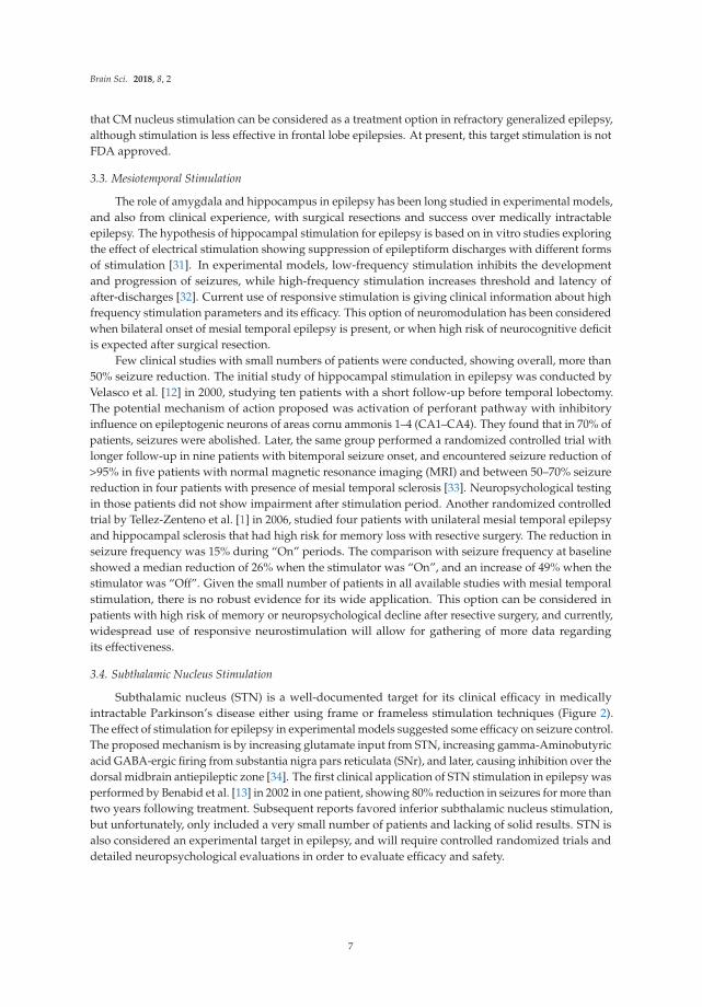
3.4. Subthalamic Nucleus Stimulation
Subthalamic nucleus (STN) is a well-documented target for its clinical efficacy in medicallyintractable Parkinson’s disease either using frame or frameless stimulation techniques (Figure 2).The effect of stimulation for epilepsy in experimental models suggested some efficacy on seizure control.The proposed mechanism is by increasing glutamate input from STN, increasing gamma-Aminobutyricacid GABA-ergic firing from substantia nigra pars reticulata (SNr), and later, causing inhibition over thedorsal midbrain antiepileptic zone [34]. The first clinical application of STN stimulation in epilepsy wasperformed by Benabid et al. [13] in 2002 in one patient, showing 80% reduction in seizures for more thantwo years following treatment. Subsequent reports favored inferior subthalamic nucleus stimulation,but unfortunately, only included a very small number of patients and lacking of solid results. STN isalso considered an experimental target in epilepsy, and will require controlled randomized trials anddetailed neuropsychological evaluations in order to evaluate efficacy and safety.
7

Brain Sci. 2018, 8, 2
(a) (b)
Figure 2. (a) Frame based system for deep brain stimulation; (b) Frameless bases system for deepbrain stimulation.
3.5. Cortical Stimulation
Chronic subdural cortical stimulation: The use of surgical electrocorticography during functionalbrain mapping and lesion resection trigger after discharges that can progress to full seizures. Briefpulses of stimulation initially performed by Lesser et al. [35] were able to control after discharges;the mechanism of this effect is not clearly understood. In theory, stimulation trigger alterations toGABA, calcium channels, and extracellular potassium, that can induce depolarization. Experimentaland small case reports trying to optimize stimulation parameters are still under investigation to clarifyits application [36,37].
Responsive stimulation: The principle of responsive neurostimulation is the first closed-loopsystem available with the ability of both recording and stimulating. This system involves implantationof subdural and depth electrodes connected to a skull implant. The electrodes are able to registerminutes-worth of data from sliding-window of EEG recordings, sense seizure onset and to deliverelectrical activity with the objective to abort and inhibit the spread of epileptogenic activity (Figure 3).The device is intended to be placed in eloquent areas, and the initial clinical experience was byOsorio et al. [15], and after further studies, it was approved by FDA in 2013. A large multicenter trialby Morrell et al. [38] evaluated efficacy with seizure reduction of 44% from baseline at one year, and65.7% by six years. The main complication rate encountered was implant site infection in (9.4%), andhemorrhage (4.7%). Half of the patients had temporal lobe epilepsy, and among them, the majorityhad bitemporal onset. The ability to sense epileptic activity, quantify frequency of seizures, seizureduration activity, preceding electroencephalographic events that vary among patients, and to deliverimmediate electrical stimulation, are significant advantages over other neuromodulation systems, andcan open the gateway for development and improvement of the technique.
8

Brain Sci. 2018, 8, 2
(a) (b)
Figure 3. (a) Antero-posterior skull X-rays of implanted responsive neurostimulation; (b) Lateral skullX-rays of implanted responsive neurostimulation.
4. Peripheral Nervous System Stimulation
4.1. Vagal Nerve Stimulation (VNS)
The inhibition of motor activity by stimulation of vagal nerve afferents was reported by Schweitzerand Wright in 1937 [39]. In 1938, a cat model was developed, where afferent vagal stimulation modifiedelectroencephalogram activity [16]. The exact mechanism of vagal nerve stimulation to control seizuresis not well understood; however, the hypothesis is based on the anatomic projections of the vagusnerve to reticular activating system, central autonomic network, limbic system and noradrenergicprojection system [17,40].
It is estimated that about 80% of the vagus nerve is composed of afferent fibers arising from viscera,while around 20% is composed of efferent fibers that innervate larynx and have parasympatheticcontrol of the heart, lungs, and gastrointestinal system [41]. The main trunk of the vagus nerve iseasily exposed, surgically, within the carotid sheath between and behind the internal jugular vein andcommon carotid artery (Figure 4). The efferent cardiac branches join the sympathetic fibers high withinthe neck, and are very unlikely to reach those branches during standard dissection. The right vagalnerve innervates the sinoatrial node, and for that reason, the vagal nerve stimulator is implanted onthe left side. Overall, given anatomical variability, it is important to communicate with anesthesia teamat the time of initial stimulation, in order to manage remote, but possible, bradycardia and asystole.
The vagal nerve stimulation is indicated for medically refractory partial onset seizures, althoughcommonly used for generalized seizures [42]. The largest long-term study by Morris et al. [43] showeda median seizure reduction of 35% at one year and 44% at three years. Interestingly, another smallerlong-term study showed 65.7% seizure reduction at six years after implantation, and 75.7% seizurereduction at 10 years [44]. It was FDA approved since 1997, and the new model has a closed-loopsystem that can detect a 20% or higher sudden increase in heart rate, considering that this often signalsthe onset of a seizure. This new model was FDA approved in 2015. The adverse events reportedare hoarseness and cough in around 50% of patients, which can later be improved by habituationor modification of stimulation settings. By two years of stimulation, reported hoarseness decreasedto 19.8% [43]. Serious complications are uncommon, such as vocal cord paralysis (1%) and infection(1.5%) [45]. Recent studies show that earlier use of VNS therapy is proven to offer better long-termoutcomes for children [46] which favored FDA approval in 2017 for children as young as four years old.
9
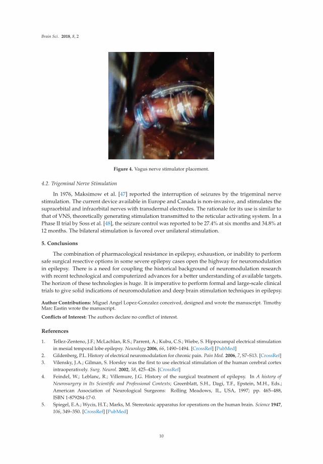
Brain Sci. 2018, 8, 2
Figure 4. Vagus nerve stimulator placement.
4.2. Trigeminal Nerve Stimulation
In 1976, Maksimow et al. [47] reported the interruption of seizures by the trigeminal nervestimulation. The current device available in Europe and Canada is non-invasive, and stimulates thesupraorbital and infraorbital nerves with transdermal electrodes. The rationale for its use is similar tothat of VNS, theoretically generating stimulation transmitted to the reticular activating system. In aPhase II trial by Soss et al. [48], the seizure control was reported to be 27.4% at six months and 34.8% at12 months. The bilateral stimulation is favored over unilateral stimulation.
5. Conclusions
The combination of pharmacological resistance in epilepsy, exhaustion, or inability to performsafe surgical resective options in some severe epilepsy cases open the highway for neuromodulationin epilepsy. There is a need for coupling the historical background of neuromodulation researchwith recent technological and computerized advances for a better understanding of available targets.The horizon of these technologies is huge. It is imperative to perform formal and large-scale clinicaltrials to give solid indications of neuromodulation and deep brain stimulation techniques in epilepsy.
Author Contributions: Miguel Angel Lopez-Gonzalez conceived, designed and wrote the manuscript. TimothyMarc Eastin wrote the manuscript.
Conflicts of Interest: The authors declare no conflict of interest.
References
1. Tellez-Zenteno, J.F.; McLachlan, R.S.; Parrent, A.; Kubu, C.S.; Wiebe, S. Hippocampal electrical stimulationin mesial temporal lobe epilepsy. Neurology 2006, 66, 1490–1494. [CrossRef] [PubMed]
2. Gildenberg, P.L. History of electrical neuromodulation for chronic pain. Pain Med. 2006, 7, S7–S13. [CrossRef]3. Vilensky, J.A.; Gilman, S. Horsley was the first to use electrical stimulation of the human cerebral cortex
intraoperatively. Surg. Neurol. 2002, 58, 425–426. [CrossRef]4. Feindel, W.; Leblanc, R.; Villemure, J.G. History of the surgical treatment of epilepsy. In A history of
Neurosurgery in Its Scientific and Professional Contexts; Greenblatt, S.H., Dagi, T.F., Epstein, M.H., Eds.;American Association of Neurological Surgeons: Rolling Meadows, IL, USA, 1997; pp. 465–488,ISBN 1-879284-17-0.
5. Spiegel, E.A.; Wycis, H.T.; Marks, M. Stereotaxic apparatus for operations on the human brain. Science 1947,106, 349–350. [CrossRef] [PubMed]
10

Brain Sci. 2018, 8, 2
6. Krack, P.; Dostrovsky, J.; Ilinksy, I.; Kultas-Ilinsky, K.; Lenz, F.; Lozano, A.; Vitek, J. Surgery of the motorthalamus: Problems with the present nomenclatures. Mov. Disord. 2002, 17, S2–S8. [CrossRef] [PubMed]
7. Cooke, P.M.; Snider, R.S. Some cerebellar influences on electrically-induced cerebral seizures. Epilepsia 1955,4, 19–28. [CrossRef] [PubMed]
8. Cooper, I.S.; Amin, I.; Gilman, S. The effect of chronic cerebellar stimulation upon epilepsy in man. Trans. Am.Neurol. Assoc. 1973, 98, 192–196. [PubMed]
9. Van Buren, J.M.; Wood, J.H.; Oakley, J.; Hambrecht, F. Preliminary evaluation of cerebellar stimulation bydouble-blind stimulation and biological criteria in the treatment of epilepsy. J. Neurosurg. 1978, 48, 407–416.[CrossRef] [PubMed]
10. Velasco, F.; Velasco, M.; Ogarrio, C.; Fanghanel, G. Electrical stimulation of the centromedian thalamicnucleus in the treatment of convulsive seizures: A preliminary report. Epilepsia 1987, 28, 421–430. [CrossRef][PubMed]
11. Fisher, R.; Salanova, V.; Witt, T.; Worth, R.; Henry, T.; Gross, R.; Oommen, K.; Osorio, I.; Nazzaro, J.; Labar, D.; et al.Electrical stimulation of the anterior nucleus of thalamus for treatment of refractory epilepsy. Epilepsia 2010,51, 899–908. [CrossRef] [PubMed]
12. Velasco, M.; Velasco, F.; Velasco, A.L.; Boleaga, B.; Jimenez, F.; Brito, F.; Marquez, I. Subacute electricalstimulation of the hippocampus blocks intractable temporal lobe seizures and paroxysmal EEG activities.Epilepsia 2000, 41, 158–169. [CrossRef] [PubMed]
13. Benabid, A.L.; Minotti, L.; Koudsie, A.; de Saint Martin, A.; Hirsch, E. Antiepileptic effect of high-frequencystimulation of the subthalamic nucleus (corpus luysi) in a case of medically intractable epilepsy caused byfocal dysplasia: A 30 month follow up: Technical case report. Neurosurgery 2002, 50, 1385–1391. [PubMed]
14. Osorio, I.; Frei, M.G.; Wilkinson, S.B. Real-time automated detection and quantitative analysis of seizuresand short-term prediction of clinical onset. Epilepsia 1998, 39, 615–627. [CrossRef] [PubMed]
15. Osorio, I.; Frei, M.G.; Sunderam, S.; Giftakis, J.; Bhavaraju, N.C.; Schaffner, S.F.; Wilkinson, S.B. Automatedseizure abatement in humans using electrical stimulation. Ann. Neurol. 2005, 57, 258–268. [CrossRef][PubMed]
16. Bailey, P.B.F. A sensory cortical representation of the vagus nerve. J. Neurophysiol. 1938, 1, 405–412.17. Penry, J.K.; Dean, J.C. Prevention of intractable partial partial seizures by intermittent vagal stimulation in
humans: Preliminary results. Epilepsia 1990, 31, s40–s43. [CrossRef] [PubMed]18. DeGiorgio, C.; Murray, D.; Markovic, D.; Whitehurst, T. Trigeminal nerve stimulation for epilepsy: Long-term
feasibility and efficacy. Neurology 2009, 72, 936–938. [CrossRef] [PubMed]19. Lockard, J.S.; Ojemann, G.A.; Congdon, W.C.; DuCharme, L.L. Cerebellar stimulation in alumina-gel monkey
model: Inverse relationship between clinical seziures and EEG interictal burst. Epilepsia 1979, 20, 223–234.[CrossRef] [PubMed]
20. Velasco, F.; Carrillo-Ruiz, J.D.; Brito, F.; Velasco, M.; Velasco, A.L.; Marquez, I.; Davis, R. Double-blind,randomized controlled pilot study of bilateral cerebellar stimulation for treatment of intractable motorseizures. Epilepsia 2005, 46, 1071–1081. [CrossRef] [PubMed]
21. Fountas, K.N.; Kapsalaki, E.; Hadjigeorgiou, G. Cerebellar stimulation in the management of medicallyintractable epilepsy: A systematic and critical review. Neurosurg. Focus 2010, 29, E8. [CrossRef] [PubMed]
22. Mullan, S.; Vailati, G.; Karasick, J.; Mailis, M. Thalamic lesions for the control of epilepsy. A study of ninecases. Arch. Neurol. 1967, 16, 277–285. [CrossRef] [PubMed]
23. Cooper, I.S.; Upton, A.R.M. Therapeutic implications of modulation of metabolism and functional activityof cerebral cortex by chronic stimulation of cerebellum and thalamus. Biol. Psychiatry 1985, 20, 809–811.[CrossRef]
24. Mirski, M.A.; Rossell, L.A.; Terry, J.B.; Fisher, R.S. Anticonvulsant effect of anterior thalamic high frequencyelectrical stimulation in the rat. Epilepsy Res. 1997, 28, 89–100. [CrossRef]
25. Hamani, C.; Hodaie, M.; Chiang, J.; del Campo, M.; Andrade, D.M.; Sherman, D.; Mirski, M.; Mello, L.E.;Lozano, A.M. Deep brain stimulation of the anterior nucleus of the thalamus: Effects of electrical stimulationon pilocarpine-induced seizures and status epilepticus. Epilepsy Res. 2008, 78, 117–123. [CrossRef] [PubMed]
26. Fisher, R.S. Deep brain stimulation for epilepsy. In Handbook of Clinical Neurology; Lozano, A.M.,Hallett, M., Eds.; Elsevier: Amsterdam, Netherlands, 2013; Volume 116, pp. 217–234, ISBN 978-0-444-53497-2.
11

Brain Sci. 2018, 8, 2
27. Van der Werf, Y.D.; Witter, M.P.; Groenewegen, H.J. The intralaminar and midline nuclei of the thalamus.Anatomical and functional evidence for participation in processes of arousal and awareness. Brain Res. Rev.2002, 39, 107–140. [CrossRef]
28. Velasco, F.; Velasco, A.L.; Velasco, M.; Jimenez, F.; Carrillo-Ruiz, J.D.; Castro, G. Centromedian thalamicstimulation for epilepsy. In Textbook of Stereotactic and Functional Neurosurgery, 2nd ed.; Lozano, A.M.,Gildenberg, P.L., Tasker, R.R., Eds.; Springer: Berlin/Heidelberg, Germany, 2009; Volume 2, pp. 2777–2791,ISBN 978-3-540-70779-0.
29. Fisher, R.S.; Uematsu, S.; Krauss, G.L.; Cysyk, B.J.; McPherson, R.; Lesser, R.P.; Gordon, B.; Schwerdt, P.;Rise, M. Placebo-controlled pilot study of centromedian thalamic stimulation in treatment of intractableseizures. Epilepsia 1992, 33, 841–851. [CrossRef] [PubMed]
30. Valentin, A.; Garcia Navarrete, E.; Chelvarajah, R.; Torres, C.; Navas, M.; Vico, L.; Torres, N.; Pastor, J.;Selway, R.; Sola, R.G.; et al. Deep brain stimulation of the centromedian thalamic nucleus for the treatmentof generalized and frontal epilepsies. Epilepsia 2013, 54, 1823–1833. [CrossRef] [PubMed]
31. Tellez-Zenteno, J.F.; Wiebe, S. Hippocampal stimulation in the treatment of epilepsy. Neurosurg. Clin. N. Am.2011, 22, 465–475. [CrossRef] [PubMed]
32. Wyckhuys, T.; De Smedt, T.; Claeys, P.; Raedt, R.; Waterschoot, L.; Vonck, K.; Van den Broecke, C.; Mabilde, C.;Leybaert, L.; Wadman, W.; et al. High frecuency deep brain stimulation in the hippocampus modifies seizurecharactheristics in kindled rats. Epilepsia 2007, 48, 1543–1550. [CrossRef] [PubMed]
33. Velasco, A.L.; Velasco, F.; Velasco, M.; Trejo, D.; Castro, G.; Carrillo-Ruiz, J.D. Electrical stimulation of thehippocampal epileptic foci for seizure control: A double-blind, long-term follow up study. Epilepsia 2007,48, 1895–1903. [CrossRef] [PubMed]
34. Vercueil, L.; Benazzouz, A.; Deransart, C.; Bressand, K.; Marescaux, C.; Depaulis, A.; Benabid, A.L.High-frequency stimulation of the subthalamic nucleus suppresses absence seizures in the rat: Comparisonwith neurotoxic lesions. Epilepsy Res. 1998, 31, 39–46. [CrossRef]
35. Lesser, R.P.; Kim, S.H.; Beyderman, L.; Miglioretti, D.L.; Webber, W.R.; Bare, M.; Cysyk, B.; Krauss, G.;Gordon, B. Brief bursts of pulse stimulation terminate afterdischarges caused by cortical stimulation.Neurology 1999, 53, 2073–2081. [CrossRef] [PubMed]
36. Child, N.D.; Stead, M.; Wirrell, E.C.; Nickels, K.C.; Wetjen, N.M.; Lee, K.H.; Klassen, B.T. Chronicsubthreshold subdural cortical stimulation for the treatment of focal epilepsy originating from eloquentcortex. Epilepsia 2014, 55, e18–e21. [CrossRef] [PubMed]
37. Lundstrom, B.N.; Worrell, G.A.; Stead, M.; VanGompel, J.J. Chronic subthreshold cortical stimulation:A therapeutic and potentially restorative therapy for focal epilepsy. Exp. Rev. Neurother. 2017, 17, 661–666.[CrossRef] [PubMed]
38. Bergey, G.K.; Morrell, M.J.; Mizrahi, E.M.; Goldman, A.; King-Stephens, D.; Nair, D.; Srinivasan, S.; Jobst, B.;Gross, R.E.; Shields, D.C.; et al. Long-term treatment with responsive brain stimulation in adults withrefractory partial seizures. Neurology 2015, 84, 810–817. [CrossRef] [PubMed]
39. Schweitzer, A.; Wright, S. Effects on the knee jerk of stimulation of the central end of the vagus and of variouschanges in the circulation and respiration. J. Physiol. 1937, 88, 459–475. [CrossRef] [PubMed]
40. Henry, T.R. Functional imaging studies of epilepsy therapies. Adv. Neurol. 2000, 83, 305–317. [PubMed]41. Henry, T.R. Therapeutic mechanisms of vagus nerve stimulation. Neurology 2002, 59, S3–S14. [CrossRef]
[PubMed]42. Morris, G.L.; Gloss, D.; Buchhalter, J.; Mack, K.J.; Nickels, K.; Harden, C. Evidence-based guideline update:
Vagus nerve stimulation for the treatment of epilepsy: Report of the guideline development subcommitteeof the American Academy of Neurology. Neurology 2013, 81, 1453–1459. [CrossRef] [PubMed]
43. Morris, G.L.; Mueller, W.M. Long-term treatment with vagus nerve stimulation in patients with refractoryepilepsy. The Vagus Nerve Stimulation Study Group E01–E05. Neurology 1999, 53, 1731–1735. [CrossRef][PubMed]
44. Elliott, R.E.; Morsi, A.; Tanweer, O.; Grobelny, B.; Geller, E.; Carlson, C.; Devinsky, O.; Doyle, W.K. Efficacyof vagus nerve stimulation over time: Review of 65 consecutive patients with treatment-resistant epilepsytreated with VNS >10 years. Epilepsy Behav. 2011, 20, 478–483. [CrossRef] [PubMed]
45. Santos, P.M. Surgical placement of the vagus nerve stimulator. Oper. Tech. Otolaryngol. 2004, 15, 201–209.[CrossRef]
12

Brain Sci. 2018, 8, 2
46. Otsuki, T.; Kim, H.D.; Luan, G.; Inoue, Y.; Baba, H.; Oguni, H.; Hong, S.C.; Kameyama, S.; Kobayashi, K.;Hirose, S.; et al. Surgical versus medical treatment for children with epileptic encephalopathy in infancyand early childhood: Results of an international multicenter cohort study in Far-East Asia (the FACE Study).Brain Dev. 2016, 38, 449–460. [CrossRef] [PubMed]
47. Maksimow, K. Interruption of grand mal epileptic seizures by the trigeminal nerve stimulation. Neurol.Neurochir. Pol. 1976, 10, 205–208. [PubMed]
48. Soss, J.; Heck, C.; Murray, D.; Markovic, D.; Oviedo, S.; Corrale-Leyva, G.; Gordon, S.; Kealey, C.;DeGiorgio, C. A prospective long-term study of external trigeminal nerve stimulation for drug-resistantepilepsy. Epilepsy Behav. 2015, 42, 44–47. [CrossRef] [PubMed]
© 2017 by the authors. Licensee MDPI, Basel, Switzerland. This article is an open accessarticle distributed under the terms and conditions of the Creative Commons Attribution(CC BY) license (http://creativecommons.org/licenses/by/4.0/).
13

brainsciences
Article
Epilepsy: A Call for Help
Venkatraman Sadanand *
Department of Neurosurgery, Loma Linda University; Loma Linda, CA 92354, USA
Received: 15 November 2017; Accepted: 16 January 2018; Published: 28 January 2018
Abstract: Epilepsy is a considerable individual and social economic burden. In properly selectedpatients, epilepsy surgery can provide significant relief from disease, including remission. However,the surgical treatment of epilepsy lags in terms of knowledge and technology. The problem arisesdue to its slow adaptation and dissemination. This article explores this issue of a wide treatment gapand its causes. It develops a framework for a rational decision-making process that is appropriate forextant circumstances and will result in the speedy delivery of surgical care for suitable patients withmedically intractable epilepsy.
Keywords: epilepsy; epilepsy surgery; decision analysis; treatment gap; game theory; economics;efficiency; resource allocation; return on investment
1. Introduction
Epilepsy is known to be a devastating disease not only for the patient but also for the patient’sfamily and for society. If left untreated, the long-term impact can be significant. If treatment can resultin being seizure-free or reduce the severity or frequency of seizures, then it may be worth treating.In fact, surgical treatment is worthwhile, but not everyone who is a good surgical candidate gets timelysurgery for their epilepsy. The wait times to see a neurosurgeon are often large. In most countries,the time from diagnosis to referral to an epilepsy center ranges from 18.9 [1] to 20 years [2].
This paper explores why this might be the case. Physicians tend to rightly believe that surgicaldecision is based solely on medical evidence. This paper argues that this alone is not sufficient. In fact,the eventual decision for surgery of a patient is the end step in a complex decision-making processinvolving multiple stakeholders. This paper therefore has two tasks: first, to identify the stakeholdersand their incentives; second, to model the complex interactions among this group of entities withvested interests.
For this purpose, the present paper will consider two factors that characterize this analysis.First, it postulates that the primary objective of individual stakeholders is different for each stakeholderand may conflict with those of others. From a purely public health perspective, the objective is tomaximize the return on investment (ROI) while maximizing public welfare. Using the ROI approach,we will compare ROI in epilepsy surgery as the use case with ROI in glioblastoma multiforme (GBM) asa control case. The control case could be any disease with postulated lower ROI and higher investmentsthan epilepsy surgery. Second, the complex interactions among stakeholders with diverse objectivesworking in an environment of strategic interactions is impossible to model using standard decisionanalysis wherein individual stakeholders are working with fixed outcomes of their actions. In otherwords, present models portray an individual stakeholder making decisions under the assumption thattheir decisions directly impact only the final outcome. In particular, they do not account for the factthat, in addition, their decisions will also have an impact upon the decisions of other stakeholderswho are involved, which will alter the final outcomes. Thus, all stakeholders take actions that affectall other stakeholders, and this inevitably leads to strategic decision-making by each while beingcognizant of the interdependence on others, until an equilibrium is reached where everyone obtainsan outcome. This is not like previous models where a given fixed net outcome is shared by all the
Brain Sci. 2018, 8, 22; doi:10.3390/brainsci8020022 www.mdpi.com/journal/brainsci14
Brain Sci. 2018, 8, 22
stakeholders (denoted sometimes as a problem of dividing a fixed pie. Those problems are referredto as zero-sum games. The current problem we analyze is not a zero-sum game). In our analysis,when equilibrium is reached, each stakeholder obtains an outcome that is of a specific value to thatstakeholder. What we require is a way to model the strategic interactions among multiple stakeholderswhen each one has an individualized objective that may conflict with those of others in the sameenvironment. Such models of strategic decision-making were first postulated mathematically byJohn Nash [3] and subsequently developed further through applications to other fields [4]. This fieldof mathematics is called non-cooperative game theory.
This is the first time non-cooperative game theory is being used in the literature on medicaldecision analysis. Why is non-cooperative game theory a better tool for medical decision analysisthan standard Markov decision trees? The reason is that it is more realistic and hence a more accuratemodel of stakeholder interaction. The stakeholders are not passive observers in the system as in thecurrent modeling techniques in medical decision analysis. In fact, every stakeholder is a maximizer ofhis own objective function and, in that process, recognizes and adjusts dynamically to the behaviorcalculus of every other stakeholder. Each stakeholder will try to predict the behaviors of all otherstakeholders in an environment of uncertainty and incomplete information about others. This is theessence of non-cooperative game theory. Economic modeling, market analysis, individual behavioranalysis, group dynamics, and military decision-making have all abandoned the previous naïve modelsof dynamic programming with fixed incentive for players. Instead these models have evolved togame theory. This shift from fixed incentives-based interactions to a dynamic interaction of multipleplayers with diverse objectives who think through others’ thought processes and adjust on the gois the very basis of a complex mathematical analysis leading to the Nobel Prize in Economics in1994 [3–5]. The mathematics behind such analysis has been referred to as game theory. Even individualbiological cell behavior is now seen to follow such models, that have been referred to as biologicalgames. Thus, this paper deviates from existing literature and advocates the use of game theory formedical decision-making and considers the techniques of static and dynamic programming with fixedincentives as a source of misleading results and not a model of real behavior.
Consider the example of a low returns (ROI)–high investment disease such as glioblastomamultiforme (GBM), an aggressively malignant brain tumor. This is also a devastating disease withconsiderable negative impact on the individual and his or her family. Despite the best treatmentoptions, median survival is about 18 months [6], and most patients who are diagnosed with thisdisease have low productive horizons ahead of them.
Current data appears to establish that, if we compare epilepsy with GBM, the incidence of GBMis lower, prevalence is lower, direct treatment costs are higher, indirect treatment costs are higher,benefits are lower, contribution to GDP is lower, and ROI is lower—yet we invest more readily in GBMtreatment than in epilepsy surgery. A patient with GBM reaches the surgeon usually within a few daysafter diagnosis. A patient with epilepsy takes 18–20 years [1,2] from diagnosis to reach the surgeon inmost countries. This is a problem.
This chapter addresses this issue, quantifies it, and suggests an approach based on game theoreticanalysis to develop a methodology to take a deeper look at the nature of this treatment gap andsocio-economic inefficiency.
In public health, as in public policy, economic efficiency is measured by a concept borrowed fromapplied mathematics called Pareto optimum [7]. When the welfare of stakeholders can be improvedfrom a given situation without making anyone less well-off than the status quo, it is called a Paretoimprovement. It is well established in the economics and public policy literature that economicefficiency requires Pareto optimal outcomes. This well-known result led to the Nobel Prize in 1972 forKenneth Arrow [8]. More recently, it has been advocated that Pareto optimization cannot be ignored inmedical decision-making and public health [9]. Game theory will model realistic interaction amongstakeholders, and the theorem of Pareto optimality will yield economic efficiency. Thus, amid paucity,the creation of epilepsy centers of excellence can be Pareto optimal.
15

Brain Sci. 2018, 8, 22
2. Problem Definition
The underlying problem explored in this chapter is best described by Table 1, where the twodiseases are compared: glioblastoma multiforme (GBM) and surgical epilepsy (SE). SE is 3000 timesmore prevalent than GBM. There are 400 times more patients who are candidates for SE than forGBM. Total expenditures on research and treatment is roughly the same for both diseases. However,the benefits of surgery per year for a patient with SE is 80,000 times that of a patient with GBM.
In Table 1, the direct costs for epilepsy include neurology specialist consultations, primary carevisits for reasons related to the disease, number and type of diagnostic tests performed (basic bloodtest, analysis of anti-epileptic drug levels, brain Computed Tomography (CT) scan, brain MagneticResonance Imagine (MRI), brain Single Photon Emission Computed Tomography (SPECT) and PositronEmission Tomography (PET), Electrocardiogram (ECG), Electroencephalogram (EEG), EEG withsleep deprivation, Holter EEG, Video Electroencephalogram (VEEG), and Holter ECG), days ofhospitalization for diagnostic work up, and the treatment administered. Non-medical direct costsinclude the use of transport to and from hospital and psycho-educational and social support. Costs ofanticonvulsant medications were the highest factor in direct costs. Furthermore, the indirect costsof epilepsy are higher than direct costs in most studies. This is because of the value of a productiveperson in society even if only accounting for a small probability of diminished abilities after surgery.
In the case of GBM, direct costs include consultations with primary care physicians, neurologyor neurosurgery specialists, and diagnostic tests such as CT scans, brain MRI, as well as ECGs andblood tests. Once the diagnosis of a brain tumor is made, the patient goes immediately (duringthe same admission in most cases) to surgery. Surgery and recovery then become direct costs.The last component of direct costs arise from post-operative care, chemotherapy, radiation therapy,rehabilitation, and a repeat of direct costs for each recurrence that is treated. Indirect costs for GBM arelower than for epilepsy because of the limited life expectancy post-diagnosis. In addition, the averageage of diagnosis is greater in GBM compared to epilepsy. Hence the span of productive life lost is alsoless than for epilepsy.
16

Brain Sci. 2018, 8, 22
Ta
ble
1.
Cos
t-Be
nefit
Com
pari
son
ofG
BMve
rsus
SE.
Dis
ea
seP
rev
ale
nce
(Glo
ba
l)In
cid
en
ce(G
lob
al)
Su
rgic
al
Ca
nd
ida
tes
(Glo
ba
l)
Me
dia
nS
urv
iva
lA
ge
Dis
trib
uti
on
of
Dis
ea
se
Co
sts:
Dir
ect
/Ye
ar
inU
S$
(Re
sea
rch
,P
rev
en
tio
n,
Tre
atm
en
t)
Co
sts:
Ind
ire
ct/Y
ea
rin
US
$(I
nd
ivid
ua
l,F
am
ily,
So
cie
ty)
Be
ne
fits
:P
er
Ye
ar
inU
S$
(Est
ima
ted
Va
lue
of
Pro
du
ctiv
eL
ife
)
Be
ne
fit/
Co
stR
ati
o(R
OI
Eq
uiv
ale
nt)
Glio
blas
tom
aM
ulti
form
e(G
BM)
1.6×
104
[10]
5.0×
104
[11]
5.0×
104
[11]
1.5
year
s[6
]45
–70
[6]
1045
×10
8[1
2,13
]39
0×
108
[14]
20×
108
[15]
0.01
Surg
ical
Epile
psy
(SE)
50×
106
[16]
2.4×
106
[16]
20×
106
[16]
60ye
ars
[17]
Mul
tim
odal
[18]
250×
108
[19]
700×
108
[19,
20]
160×
1012
[15]
1684
17

Brain Sci. 2018, 8, 22
Waiting times for GBM patients, interestingly, are extremely low compared to epilepsy patients.Median time from diagnosis to surgery is less than 1 week and median times from surgery to adjuvanttherapy is 27 days. This is despite the costs reported [13] to be between $50,000 and $92,000 per QualityAdjusted Life Years (QALY), barely making it over the accepted cost-effectiveness thresholds of $50,000per QALY.
In the United States of America, 369 resective epilepsy surgeries are being carried out per year onthe average, whereas there are about 1 million surgical candidates for epilepsy surgery [21]. In India,with a population approximately 4 times that of the United States, only 734 resective epilepsy surgeriesare carried out per year when there are 2.5 million surgical candidates [22]. In China, there are about1.8 million surgical candidates for epilepsy surgery and only about 2500 epilepsy surgeries are carriedout per year [23]. Based on the data from these studies [21,23], China has roughly twice the number ofsurgical candidates but performs eight times more epilepsy surgeries per year. However, one mustbe cautious before this finding is applauded. Both countries are still woefully short of treating thenumber of epilepsy surgery candidates. There is a considerable treatment gap impacting individualand social welfare and that treatment gap is the subject of this paper. It is even more surprising thatthere is such a treatment gap when the seizure-free rate averaging over all types of seizure surgeries is68% and about 90% for certain kind of seizure foci [24].
We have thus far examined the treatment gap, the cost–benefit ratio, and the time taken fromdiagnosis to surgical attention as measures of this seeming social inefficiency. We may also considerother measures such as the cost-effectiveness ratio and the return on investment (ROI).
Any medical intervention that extends lives is measured by the cost-effectiveness ratio. It isthe ratio of costs divided by number of years gained. This does not account for the value ascribedto those years. The smaller the number, the more worthwhile the intervention. To calculate this,it should be noted that investment for GBM research and treatment extends life by about 1.5 years [6],while investment for epilepsy extends good quality of life by about 40 years on average. This isa conservative estimate due to the multimodal distribution of incidence (computed from [13] by takingthe scaled mean of histograms and subtracting that from the WHO 2015 estimate of life expectancy).
Cost-Effectiveness Ratio (CER) = Direct and Indirect Costs in US$/Benefits in Years GainedFor GBM, the CER: 1435 × 108/1.5 = 956 × 108.For Epilepsy, the CER is: 1700 × 108/40 = 42.5 × 10⁸.The Benefit Cost Ratio (BCR) is like the CER but considers the value of life lived. This then
becomes a proxy for the ROI and has been computed and is shown in the last column of Table 1.While there are several other measures one could use, such as the QALY, the point is clear:
SE research and treatment, compared to GBM research and treatment, is more efficient, more efficacious,better for the patient, and better for the society. However, SE treatment is gravely underutilized.Thus, how is this market inefficiency explained and how can this be addressed? A purely medicaldecision-making is an evidence-based decision. On that basis alone, there is no paucity of evidenceto show that, for the properly identified candidate, epilepsy surgery is the right option rather thanmedical management. However, this would ignore the reality of a medical economy. One must reviewthe incentives and disincentives of all stakeholders in this game of budget allocation for surgery toadvance the patient from diagnosis to surgery. Just because medical evidence shows that epilepsysurgery is better for properly selected patients compared to medical management, it does not meanthat those patients will receive surgery in an environment where there are several stakeholders withconflicting and diverse goals.
3. The Stakeholders, Their Incentives and Pitfalls
Surgical decision-making in epilepsy involves several stakeholders along the path from diagnosisto surgery. The first is the physician. The second is the patient and his or her family. The third ishospital administration. The fourth is the insurance industry. The fifth is society.
18

Brain Sci. 2018, 8, 22
The physician’s decisions are typically evidence-based. This is a necessary condition for surgerybut not sufficient. However, their role can push to decrease the large gap of almost 20 years fromdiagnosis to surgery and, once cleared for surgery, to push for appropriate budget allocations toenable surgery. The doctor, in most epilepsy centers, must take on the role of a physician and that ofa patient advocate.
Next, the patient and his family are dealing with a disease that not only results in a loss of incomeand productivity but also the social stigma and discrimination associated with epilepsy. Therefore,they need to be educated in the social and quality of life advantages of epilepsy surgery. Such patienteducation is glaringly lacking until the patient somehow makes his first contact with a doctor. In somecountries, epileptologists are usually based in urban centers while many epilepsy patients are inrural areas.
Hospital administrations must balance budgets and face multiple demands from differentsubspecialties. Resource allocation then becomes an issue of educating administrators about thecost-effectiveness and efficacies inherent in epilepsy surgery, even when compared to other specialtiesthat may have their own demands for fixed budgets, finding a proper fit with corporate missions andmedicolegal experiences.
Insurance industry needs to be made aware of the cost–benefit analysis of surgery versus themedical care of epilepsy patients. In computing these costs and benefits, care must be taken to accountfor the discounted present values along with the non-zero rates of return. Insurance companiestherefore must now weigh the cost of surgical care against the discounted present value of the futurecost of medical care. Social welfare and lost productivity are often not in their optimization problemand may be considered externalities [25]. Externalities impose costs on other stakeholders but have noimpact on the given stakeholder’s mathematical profit maximization function.
The last stakeholder is society. How does one account for the value of epilepsy surgery to society?Epilepsy surgery creates productive people who can live healthy lives. That is the very basis ofmeasures such as BCR and ROI. Thus, the impact of epilepsy surgery on this last stakeholder ismeasured by the value of an individual on society. The current belief is that the value of an individual’slife is measured by the market value of the goods and services produced by that individual. However,this may be incorrect. The value of an individual’s productivity to society may exceed the actualproductive output of the individual due to social multiplier effects and what is known as Okun’slaw [26,27], and this must also be accounted for in computing society’s objectives in this interactivestrategic decision analysis.
4. Decision Analysis
What we have now is a complex problem of resource allocation with the following characteristics:the existence of multiple strategic stakeholders (so current models of a single stakeholder suchas a physician, making medical evidence-based decisions alone is an inadequate model), diversegoals (so a multiple stakeholder decision analysis wherein all stakeholders have the same goalof patient welfare is also inadequate), computational complexities of payoffs to each stakeholder(so a model of multiple stakeholders with fixed objectives is inadequate), the interdependence ofmultiple stakeholders wherein one stakeholder’s decisions affects not only his own but also theoutcomes of others (hence a model where a stakeholder’s choices only affect his outcome is inadequate)and the freedom to choose what is best for each stakeholder (so a model of decision analysis thatignores the strategic freedom to choose for each stakeholder is also inadequate). In this environment,it would not be helpful to model this situation as a simple static evidence-based decision for thesurgeon. Firstly, the surgeon is not the only stakeholder. Secondly, not every stakeholder has the sameobjective and incentives as the surgeon. Lastly, not every stakeholder fully knows what others know.This is the reason why models of what ought to be the outcome (surgery, in this case) often differsfrom what becomes the outcome (treatment gap, in this case).
19

Brain Sci. 2018, 8, 22
The process of medical decision-making is therefore best described and computed by what isknown as strategic game theory [28]. Each stakeholder is working in an environment of incompleteinformation (not everyone knows what others fully know) about the incentives faced and consideredby other stakeholders and uncertainty about their actual decisions. A decision analysis will createa game of imperfect information in which all the stakeholders choose strategies to maximize their gainsunder uncertainty in the environment and under uncertainty about others’ choices. Such complexinteractions may involve coalition forming among the stakeholders and may even result in sharinggains from the outcome.
In such games, every player first defines what his or her goal is and what would be themathematical representation of those goals. It then seeks to maximize that mathematical entitywhile accounting for other stakeholders’ similar thought processes. Outcomes in such situations arecalled equilibria of games [29]. Since every player functions under some degree of uncertainty aboutthe other players’ motives and moves, the final action of each stakeholder is dictated by what is knownas Bayesian expected utility and interactive equilibrium [29]. This is the decision-making processused by most Forbes 100 businesses, and it may be about time that medical decision-making followsthis approach by considering the strategic nature, the informational asymmetries, uncertainties in theenvironment and the diverse goals of stakeholders.
5. Conclusions
There is medical evidence that, for carefully selected candidates, surgery is better than medicalmanagement for patients with medically intractable epilepsy. However, SE is underutilized and thereis a treatment gap. SE patients must wait a long time after diagnosis to be referred to a neurosurgeon,resulting in patients who do not receive the surgeries they need. This is not the case for GBM. Patientswith diagnosed GBM have waiting times for referral for surgery of a few days, whereas epilepsypatients can wait years. In addition, almost every patient with a first-time diagnosis of possible GBMundergoes surgery.
The problem may stem from complex interactions of several stakeholders. This paper advocatesa method of analysis, called mathematical game theory, that has been used in the fields of business,economics, public policy, political science, and engineering and biology.
Decision-making for GBM surgery is much easier as all stakeholders appear to focus on the shortlife expectancy after the diagnosis of GBM and the apprehensive determination to do something tochange that.
It would seem, that fear of death drives decisions differently than hope of improving the qualityof long-term life.
Acknowledgments: The author has received no funding for this research and its publication.
Conflicts of Interest: The author declares no conflict of interest.
References
1. Martínez-Juárez, I.E.; Funes, B.; Moreno-Castellanos, J.C.; Bribiesca-Contreras, E.; Martínez-Bustos, V.;Zertuche-Ortuño, L.; Hernández-Vanegas, L.E.; Ronquillo, L.H.; Rizvi, S.; Adam, W.; et al. A comparisonof waiting times for assessment and epilepsy surgery between a Canadian and a Mexican referral center.Epilepsia Open 2017, 2, 453–458. [CrossRef]
2. Thornton, J.G.; Lilford, R.J.; Johnson, N. Decision analysis in medicine. Br. Med. J. 1992, 304, 1099. [CrossRef]3. Nash, J.F., Jr. Equilibrium points in n-person games. Proc. Natl. Acad. Sci. USA 1950, 36, 48–49. [CrossRef]
[PubMed]4. Basar, T.; Olsder, G.I. Dynamic Noncooperative Game Theory, 2nd ed.; Society for Industrial and Applied
Mathematics (Classics in Applied Mathematics): Philadelphia, PA, USA, 1999.5. Reinhard Selten—Facts. Nobelprize.org; Nobel Media. Available online: http://www.nobelprize.org/nobel_
prizes/economic-sciences/laureates/1994/selten-facts.html (accessed on 12 December 2017).
20

Brain Sci. 2018, 8, 22
6. Johnson, D.R.; O’Neil, B.O. Glioblastoma survival in the United States before and during the temozolomideera. J. Neurooncol. 2012, 107, 359–364. [CrossRef] [PubMed]
7. Saule, C.; Giegerich, R. Pareto optimization in algebraic dynamic programming. Algorithms Mol. Biol. 2015,10, 22. [CrossRef] [PubMed]
8. Kenneth, J. Arrow—Facts. Nobelprize.org; Nobel Media. Available online: http://www.nobelprize.org/nobel_prizes/economic-sciences/laureates/1972/arrow-facts.html (accessed on 12 December 2017).
9. Dewar, D. Essentials of Health Economics, 2nd ed.; Jones & Bartlett Learning: Burlington, MA, USA, 2017.10. Glioblastoma. Available online: http://www.orpha.net/consor/cgi-bin/OC_Exp.php?Expert=360
(accessed on 12 December 2017).11. Louis, D.N. WHO Classification of Tumours of the Central Nervous System; International Agency for Research
on Cancer: Lyon, France, 2007.12. Wasserfallen, J.-B.; Ostermann, S.; Leyvraz, S.; Stupp, R. Cost of temozolomide therapy and global care for
recurrent malignant gliomas followed until death. Neuro Oncol. 2005, 7, 189–195. [CrossRef] [PubMed]13. Raizer, J.J.; Fitzner, K.A.; Jacobs, D.I.; Bennett, C.L.; Liebling, D.B.; Luu, T.H.; Trifilio, S.M.; Grimm, S.A.;
Fisher, M.J.; Haleem, M.S.; et al. Economics of malignant gliomas: A critical review. J. Oncol. Pract. 2014, 11,e59–e65. [CrossRef] [PubMed]
14. Blomqvist, P.; Lycke, J.; Strang, P.; Törnqvist, H.; Ekbom, A. Brain tumours in Sweden 1996: Care and costs.J. Neurol. Neurosurg. Psychiatry 2000, 69, 792–798. [CrossRef] [PubMed]
15. How Much Will We Pay for a Year of Life? Available online: https://www.gsb.stanford.edu/insights/how-much-will-we-pay-year-life (accessed on 12 December 2017).
16. World Health Organization. Atlas: Epilepsy Care in the World; World Health Organization Press: Geneva,Switzerland, 2005.
17. Gaitatzis, A.; Johnson, A.L.; Chadwick, D.W.; Shorvon, S.D.; Sander, J.W. Life expectancy in people withnewly diagnosed epilepsy. Brain 2004, 127, 2427–2432. [CrossRef] [PubMed]
18. Kotsopoulos, I.A.; Van Merode, T.; Kessels, F.G.; de Krom, M.C.; Knottnerus, J.A. Systematic Review andMeta-analysis of Incidence Studies of Epilepsy and Unprovoked Seizures. Epilepsia 2002, 43, 1402–1409.[CrossRef] [PubMed]
19. Pato-Pato, A. Evaluation of the Direct Costs of Epilepsy. J. Neurol. Neurosci. 2013, 4, 3. [CrossRef]20. Kotsopoulos, I.A.W.; Evers, S.M.A.A.; Ament, A.J.H.A.; De Krom, M.C.T.F.M. Estimating the Costs of Epilepsy:
An International Comparison of Epilepsy Cost Studies. Epilepsia 2001, 42, 634–640. [CrossRef] [PubMed]21. Englot, D.J.; Ouyang, D.; Garcia, P.A.; Barbaro, N.M.; Chang, E.F. Epilepsy surgery trends in the United
States, 1990–2008. Neurology 2012, 78, 1200–1206. [CrossRef] [PubMed]22. Rathore, C.; Radhakrishnan, K. Epidemiology of Epilepsy Surgery in India. Neurol. India 2017, 65, 52–59.
[CrossRef]23. Xu, L.; Xu, M. Epilepsy Surgery in China: Past, Present and Future. Eur. J. Neurol. 2009, 17, 189–193.
[CrossRef] [PubMed]24. Spencer, S.S.; Berg, A.T.; Vickrey, B.G.; Sperling, M.R.; Bazil, C.W.; Shinnar, S.; Langfitt, J.T.; Walczak, T.S.;
Pacia, S.V. Predicting Long-Term Seizure Outcome after Resective Epilepsy Surgery: The Multicenter Study.Neurology 2005, 65, 912–918. [CrossRef] [PubMed]
25. Arrow, K. The organization of economic activity: Issues pertinent to the choice of market versus non-marketallocation. In Public Expenditure and Policy Analysis; Haveman, R.H., Margolis, J., Eds.; United States CongressJoint Economic Committee: Washington, DC, USA, 1970; pp. 59–73.
26. Okun, A.M. Potential GNP, Its Measurement and Significance; Cowles Foundation, Yale University: New Haven,CT, USA, 1962.
27. Gordon, R.J. Productivity, Growth, Inflation and Unemployment; Cambridge University Press: Cambridge,England, 2004.
28. Von Neumann, J.; Morgenstern, O. Theory of Games and Economic Behavior; Princeton University Press:Princeton, NJ, USA, 1944.
29. Harsanyi, J.C.; Selten, R. A General Theory of Equilibrium Selection in Games; MIT-Press: Cambridge, MA, USA, 1988.
© 2018 by the author. Licensee MDPI, Basel, Switzerland. This article is an open accessarticle distributed under the terms and conditions of the Creative Commons Attribution(CC BY) license (http://creativecommons.org/licenses/by/4.0/).
21

brainsciences
Review
Surgical Considerations of Intractable MesialTemporal Lobe Epilepsy
Warren W. Boling
11234 Anderson Street, Room 2562-B, Loma Linda, CA 92354, USA; [email protected]; Tel.: +1-909-558-4479
Received: 1 February 2018; Accepted: 15 February 2018; Published: 20 February 2018
Abstract: Surgery of temporal lobe epilepsy is the best opportunity for seizure freedom in medicallyintractable patients. The surgical approach has evolved to recognize the paramount importance of themesial temporal structures in the majority of patients with temporal lobe epilepsy who have a seizureorigin in the mesial temporal structures. For those individuals with medically intractable mesialtemporal lobe epilepsy, a selective amygdalohippocampectomy surgery can be done that providesan excellent opportunity for seizure freedom and limits the resection to temporal lobe structuresprimarily involved in seizure genesis.
Keywords: temporal lobe epilepsy; selective amygdalohippocampectomy; epilepsy surgery; mesialtemporal lobe epilepsy
1. Introduction
Temporal lobe epilepsy (TLE) affects a substantial number of individuals with medicallyintractable epilepsy. TLE is the most common operated epilepsy, and the majority of patients withlocalization related epilepsy seen in tertiary epilepsy centers have TLE [1,2]. Although TLE is notthe most common epilepsy. TLE in the general population of Minnesota has been estimated at about10.4 per 100,000 this is compared with 54.3 per 1000 incidence of epilepsy in the population as awhole [3].
The frequent occurrence of intractable epilepsy in the temporal lobe bears witness to the highlyepileptogenic nature of the limbic structures that comprise the mesial portion of the temporal lobe.The mesial part of the temporal lobe is richly connected with surrounding extra-temporal corticalregions especially the orbitomesial frontal lobe via the uncinate fasciculus and the fornix carriesfibers from the hippocampus that project to the anteromesial frontal lobe and anterior nucleus of thethalamus [4,5]. The mesial temporal structures are highly connected as well with the anterolateralneocortical temporal lobe. Therefore, due to the strong connections of the mesial temporal structureswith the anterior and lateral temporal lobe in addition to other limbic regions, TLE most commonlymanifests the semiology of staring and automatisms regardless of the seizure onset zone in lateral ormesial structures of the temporal lobe.
Despite the frequent occurrence and intractable nature of temporal lobe epilepsy, it responds verywell to surgery with high rates of resulting seizure freedom and the risks of surgery are quite low [6–8].The decision to consider a patient with medically intractable TLE for surgery has become much clearernowadays since the International League Against Epilepsy (ILAE) defined medical intractability asthe failure of adequate trials of two tolerated, appropriately chosen and used antiepileptic drugs [9].A clear definition of intractable epilepsy combined with the excellent results of surgery for symptomaticepilepsy especially arising in the temporal lobe, has resulted in numerous consensus reports affirmingthat individuals with drug resistant epilepsy should be evaluated in a comprehensive epilepsy programto identify opportunities for surgical cure [10].
Temporal lobe epilepsy can be categorized into one of two types based on the anatomical regionof seizure onset. Seizures that originate from the temporal cortex lateral to the collateral sulcus
Brain Sci. 2018, 8, 35; doi:10.3390/brainsci8020035 www.mdpi.com/journal/brainsci22

Brain Sci. 2018, 8, 35
are defined as lateral or neocortical epilepsy, and seizures that have a focus of onset medial to thecollateral sulcus are named mesial temporal lobe epilepsy (MTLE). The International League AgainstEpilepsy recognizes there are sufficient distinguishing characteristics for MTLE to be considered adistinct syndromic entity [11]. Although the two categories of TLE frequently share the same limbicsemiology [11–13], in general, MTLE more commonly displays the epigastric, cephalic or experientialaura, loss or awareness, staring, automatisms and posturing that are typical temporal lobe seizurepatterns and are the result of a seizure prominently involving the limbic structures [14]. The imagingfinding that defines MTLE is atrophy and sclerosis of the hippocampus, so-called mesial temporalsclerosis (MTS) [15]. The childhood history of patients with MTLE commonly includes the presenceof childhood febrile convulsions, especially of a prolonged and complicated nature [16]. Individualswith neocortical epilepsy are more likely to manifest signs related to peri-Sylvian structures such as asimple auditory hallucinations or, in the dominant hemisphere, postictal aphasia [17,18]. Althoughneocortical epilepsy often spreads along fibers richly connected with the mesial temporal structuresmanifesting the signs and symptoms of limbic involvement, which are the semiological features ofMTLE as well.
Due to the considerable overlap in semiology between the two categories of temporal lobeepilepsy, multiple noninvasive data elements must converge to localize the TLE focus to the mesialor lateral structures of the temporal lobe [19,20]. However, when patient history, seizure semiology,EEG localization, and imaging findings point to MTLE, there is a high degree of confidence in thediagnosis [21,22]. In a minority of patients, intracranial electrode monitoring may be required toinvestigate lateralization of the seizure onset to a temporal lobe [23] or to confirm temporal lobarlocalization in one hemisphere [24].
2. Surgery of Temporal Lobe Epilepsy
2.1. Temporal Lobectomy
Temporal lobectomy is a frequently a misused term that is defined as removal of the entiretemporal lobe including the mesial structures. A complete removal of the temporal lobe is rarelyperformed today. The description of lobectomy to describe a resection should be reserved for theunique situations in which an entire anatomical temporal lobe actually is removed. The standardresections done for temporal lobe epilepsy are preferably described as an anterior temporal resection orcortico-amygdalohippocampectomy (CAH), which more accurately describes the anatomic structuresthat are removed.
2.2. Cortico-Amygdalohippocampectomy (CAH)
This is the standard temporal resection commonly performed at most epilepsy centers.It essentially corresponds to an anterior temporal neocortical resection followed by a removal of thetemporal mesial structures. Variations on this approach have been described including anatomicallystandardized resections [24–26] as well as a tailored type of operation [27].
The anatomical resection approach consists, in the dominant hemisphere of the resection alongthe superior temporal gyrus (T1) posteriorly to the level of the pre-central sulcus, which corresponds toabout 3.5–4 cm from the temporal pole. In addition, in the non-dominant hemisphere the T1 removal isdelineated posteriorly by the central sulcus, which is about 4–4.5 cm from the temporal pole. Figure 1The cortex and white matter lateral to the collateral sulcus is removed en bloc as a specimen in the firststep. Next, the temporal horn is opened in order to identify the mesial temporal structures, which aredisconnected and removed as described below for the selective amygdalohippocampectomy (SAH).
23

Brain Sci. 2018, 8, 35
Figure 1. 3-D reconstruction of the non-dominant right hemisphere. The dashed line illustratesthe anatomical resection approach of cortico-amygdalohippocampectomy. In the non-dominanthemisphere, the resection is taken posteriorly to the level of the central sulcus along T1. In the dominanthemisphere, the T1 resection is no further posterior than the precentral sulcus in order to respect andpreserve posterior language areas. Single star = central sulcus, double star = pre-central sulcus,T1 = superior temporal gyrus, T2 = middle temporal gyrus, PoC = post central gyrus, PrC = precentralgyrus, SM = supramarginal gyrus, ANG = angular gyrus, O2 = second occipital gyrus, which is thegyral continuum of T2 in the occipital lobe.
2.3. Cortico-Amygdalectomy (CA)
As the name implies, CA consists of an anterior temporal resection along with the amygdala anduncus. The remaining hippocampal formation, parahippocampus, and entorhinal cortex are spared.The lateral neocortical resection is limited posteriorly as described above for CAH. Figure 1 In the CA,the cortical resection is limited medially by the collateral sulcus thus preserving parahippocampus andentorhinal cortex. The anterior temporal horn is opened for the purpose of identifying intraventricularlandmarks to confirm that the hippocampus and the adjacent parahippocampal gyrus are left intact.Opening the ventricle also facilitates removal of the amygdala and subpial emptying of the uncus.This surgical approach is most useful in patients who are at risk for a functional memory decline fromremoval of the hippocampal complex or have failed the ipsilateral intracarotid amobarbital test formemory. Kim, et al. identified with neuropsychological testing before and after surgery that thissurgical approach avoids memory decline, yet provides a good opportunity for seizure freedom inpatients with TLE when the hippocampus retains significant memory function [28,29].
2.4. Selective Amygdalohippocampectomy (SAH)
The SAH surgical approach is based on the understanding that the seizure focus of MTLE isconfined to the mesial temporal structures. Hughlings Jackson first illustrated this concept whenhe described a patient with a lesion in the uncus causing psychomotor seizures (Jackson called adreamy state), which was the first description of the role the temporal lobe structures in humanepilepsy [30]. More modern experimental studies have subsequently identified the critical role of themesial temporal structures in experimental and human TLE [31–36]. Additionally, seizure freedom
24

Brain Sci. 2018, 8, 35
results realized from many patients operated for MTLE with SAH confirms the importance of themesial temporal and limbic structures in MTLE [35,37–40]. Because MTLE represents a sufficientlydistinct pathophysiological entity, the International League Against Epilepsy has concluded that itdoes represent a specific epilepsy syndrome [11].
The SAH surgical approach was first described by the Brazilian neurosurgeon, Paolo Niemeyerin the 1950’s to treat TLE arising from the mesial temporal structures. Niemeyer described theinnovative surgical technique he called “transventricular amygdala-hippocampectomy” and presentedthe rational for a selective approach to surgery for temporal lobe epilepsy at the InternationalColloquium on Temporal Lobe Epilepsy in 1957 that took place at the National Institutes of Health [41].Figure 2 Subsequently, the importance of MTLE in medically intractable TLE has been establishedby reports confirming the utility of SAH for the treatment of MTLE, [38,42–44] and MR imaging nowgreatly enables the diagnosis of patients with MTLE by defining the anatomical correlate, which ishippocampal sclerosis [11,15].
Figure 2. Reproduced with permission from Temporal Lobe Epilepsy: A Colloquium Sponsored bythe National Institute of Neurological Diseases and Blindness, National Institutes of Health, Bethesda,Maryland, in Cooperation with the International League Against Epilepsy [45] Copyright©1959American Medical Association. All rights reserved. In his presentation at the colloquium, Niemeyerdescribed the convincing evidence from experimental research and clinical experience that themesial temporal structures have a critical role in epileptogeneis of TLE. Niemeyer described asurgical technique he developed to selectively remove the mesial temporal structures via a trans-T2trans-ventricular approach.
Niemeyer’s description of a white matter corridor through the middle temporal gyrus (T2) tothe temporal horn to selectively remove the mesial temporal structures was a very different approachthan the anterior temporal resection popular at the time. In fact, research carried out in Montrealby Scoville and Milner [46] and Penfield and Milner [47], which had stressed the important role ofthe hippocampus in memory, as well as the earlier described Klüver-Bucy syndrome had influencedcenters surgically treating epilepsy in North America to preserve the hippocampus. Concerns aboutmemory were reflected in the first publication of a series of patients operated for TLE in which Penfieldand Flanigin described hippocampus removal in only 2 of 32 patients operated [48]. However, soon
25
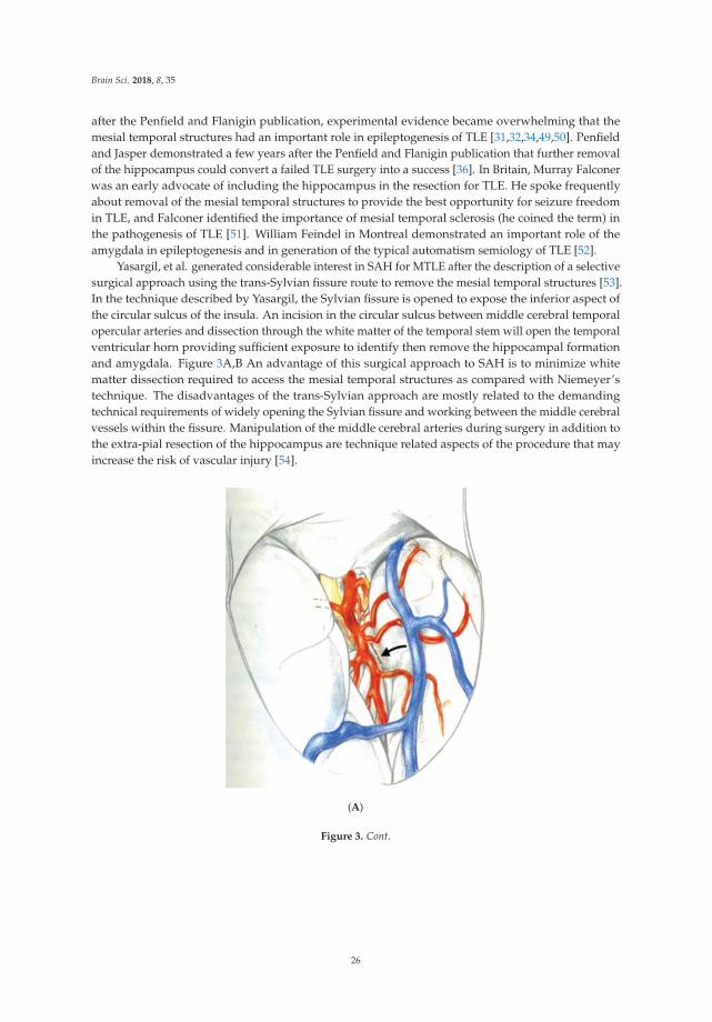
Brain Sci. 2018, 8, 35
after the Penfield and Flanigin publication, experimental evidence became overwhelming that themesial temporal structures had an important role in epileptogenesis of TLE [31,32,34,49,50]. Penfieldand Jasper demonstrated a few years after the Penfield and Flanigin publication that further removalof the hippocampus could convert a failed TLE surgery into a success [36]. In Britain, Murray Falconerwas an early advocate of including the hippocampus in the resection for TLE. He spoke frequentlyabout removal of the mesial temporal structures to provide the best opportunity for seizure freedomin TLE, and Falconer identified the importance of mesial temporal sclerosis (he coined the term) inthe pathogenesis of TLE [51]. William Feindel in Montreal demonstrated an important role of theamygdala in epileptogenesis and in generation of the typical automatism semiology of TLE [52].
Yasargil, et al. generated considerable interest in SAH for MTLE after the description of a selectivesurgical approach using the trans-Sylvian fissure route to remove the mesial temporal structures [53].In the technique described by Yasargil, the Sylvian fissure is opened to expose the inferior aspect ofthe circular sulcus of the insula. An incision in the circular sulcus between middle cerebral temporalopercular arteries and dissection through the white matter of the temporal stem will open the temporalventricular horn providing sufficient exposure to identify then remove the hippocampal formationand amygdala. Figure 3A,B An advantage of this surgical approach to SAH is to minimize whitematter dissection required to access the mesial temporal structures as compared with Niemeyer’stechnique. The disadvantages of the trans-Sylvian approach are mostly related to the demandingtechnical requirements of widely opening the Sylvian fissure and working between the middle cerebralvessels within the fissure. Manipulation of the middle cerebral arteries during surgery in addition tothe extra-pial resection of the hippocampus are technique related aspects of the procedure that mayincrease the risk of vascular injury [54].
(A)
Figure 3. Cont.
26

Brain Sci. 2018, 8, 35
(B)
Figure 3. Reprinted and Modified by permission from Springer Nature: Adv Tech Stand Neurosurg,Yasargil MG, Teddy PJ, Roth P. Selective amygdalohippocampectomy: operative anatomy and surgicaltechnique. Copyright, 1985 [55]. (A) The Sylvian fissure is opened widely to expose the inferiordelineation of the insula called the circular sulcus with the planned white matter incision markedin a dashed line between two middle cerebral opercular temporal arteries; (B) Surgeon’s view intothe opened ventricle illustrating the choroid plexus (C), hippocampus (H), collateral eminence (star),middle cerebral vessels (M1 and M2), and amygdala (A). Numbers represent steps in the extra-pialremoval of mesial temporal structures: coagulation and division of the hippocampal vessels (steps1 and 2), disconnection of the hippocampal body from the hippocampal tail (step 3), opening thecollateral eminence to empty and remove the parahippocampus (steps 4 and 5), and remove uncus andamygdala (step 6).
Hori contributed importantly to the SAH literature by describing a subtemporalamygdalohippocampectomy technique to perform SAH [56]. This approach includes at leastpartial removal of fusiform gyrus in order to gain access to the parahippocampal gyrus and mesialstructures via a subtemporal route. Figure 4 Hori’s original description was extra-pial resection ofthe mesial temporal structures but later he modified the approach to a subpial resection technique.To minimize retraction on the temporal lobe, Hori drilled down the roof of the external auditory meatus.A preoccupation with the subtemporal approach is retraction injury of the anastomotic vein of Labbé,which Hori was able to successfully preserve using techniques described in the skull base surgeryliterature [57]. The advantages of the subtemporal approach that Hori described were preservation ofvisual fields, he identified no superior quadrantanposia after surgery, and the temporal lobe is notdisconnected in contradistinction to Yasargil’s trans-Sylvian approach that requires dissecting throughthe temporal stem to access the mesial temporal structures. The disadvantages of the subtemporalapproach are related mostly to the extended resection of the fusiform gyrus that is not required inother SAH approaches as well as an inherent risk of injury to the anastomotic draining vein of Labbéfrom elevating the temporal lobe with a retractor, which is a particular concern in the dominanttemporal lobe.
27
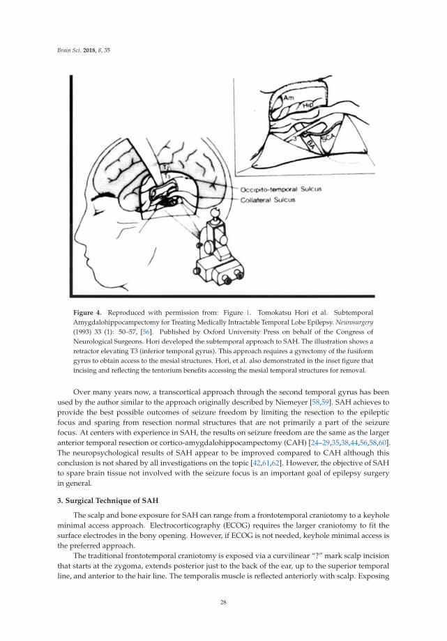
Brain Sci. 2018, 8, 35
Figure 4. Reproduced with permission from: Figure 1. Tomokatsu Hori et al. SubtemporalAmygdalohippocampectomy for Treating Medically Intractable Temporal Lobe Epilepsy. Neurosurgery(1993) 33 (1): 50–57, [56]. Published by Oxford University Press on behalf of the Congress ofNeurological Surgeons. Hori developed the subtemporal approach to SAH. The illustration shows aretractor elevating T3 (inferior temporal gyrus). This approach requires a gyrectomy of the fusiformgyrus to obtain access to the mesial structures. Hori, et al. also demonstrated in the inset figure thatincising and reflecting the tentorium benefits accessing the mesial temporal structures for removal.
Over many years now, a transcortical approach through the second temporal gyrus has beenused by the author similar to the approach originally described by Niemeyer [58,59]. SAH achieves toprovide the best possible outcomes of seizure freedom by limiting the resection to the epilepticfocus and sparing from resection normal structures that are not primarily a part of the seizurefocus. At centers with experience in SAH, the results on seizure freedom are the same as the largeranterior temporal resection or cortico-amygdalohippocampectomy (CAH) [24–29,35,38,44,56,58,60].The neuropsychological results of SAH appear to be improved compared to CAH although thisconclusion is not shared by all investigations on the topic [42,61,62]. However, the objective of SAHto spare brain tissue not involved with the seizure focus is an important goal of epilepsy surgeryin general.
3. Surgical Technique of SAH
The scalp and bone exposure for SAH can range from a frontotemporal craniotomy to a keyholeminimal access approach. Electrocorticography (ECOG) requires the larger craniotomy to fit thesurface electrodes in the bony opening. However, if ECOG is not needed, keyhole minimal access isthe preferred approach.
The traditional frontotemporal craniotomy is exposed via a curvilinear “?” mark scalp incisionthat starts at the zygoma, extends posterior just to the back of the ear, up to the superior temporalline, and anterior to the hair line. The temporalis muscle is reflected anteriorly with scalp. Exposing
28

Brain Sci. 2018, 8, 35
the root of the zygoma is important to gain adequate exposure to the floor of the middle fossa. Afterelevation of the frontotemporal bone flap, a craniectomy of the temporal bone is required to extend thebone opening to the zygoma inferiorly and to the anterior extent of the middle fossa. After the durais reflected away from the temporal lobe, important sulcal and gyral landmarks, such as the Sylvianfissure, superior temporal gyrus (T1), and the central sulcus are confirmed with image guidance.
A corticectomy along the superior aspect of the second temporal gyrus (T2) is made to begin thecorridor to the mesial temporal structures. A 2 to 3 cm corticectomy provides sufficient working space.Figure 5 The posterior extent of the corticectomy is limited by the central sulcus in the non-dominanthemisphere and by the precentral sulcus in the dominant hemisphere. Image guidance is sufficient tolocalize precentral and central sulcus, which corresponds to about 3.5 cm and 4.5 cm from the temporaltip, respectively. The author has found that using an ultrasonic aspirator at the lowest settings ofaspiration and amplitude to be the best subpial dissection and aspiration tool. A corridor is fashioneddown to the temporal horn that is opened, which is typically 3–4 cm deep from the cortical surface.The corridor trajectory parallels the superior temporal sulcus, which points to the temporal horn andstops just short of it. A retractor is inserted once the temporal horn is opened to facilitate fully exposingthe mesial temporal structures by opening the ventricle from its anterior most tip to the posterior limitof the corridor Figure 6.
Figure 5. SAH corticectomy of about 2.5 cm is made along T2 just below the superior temporal sulcus.A white matter corridor is fashioned that follows the superior temporal sulcus down to the temporalhorn, which is opened to visualize directly the mesial temporal structures.
29

Brain Sci. 2018, 8, 35
Figure 6. Reprinted from Journal of Clinical Neuroscience 17 (9), Boling W, Minimal accesskeyhole surgery for mesial temporal lobe epilepsy, 1180–1184, Copyright 2010, with permission fromElsevier, [59]. Coronal view of the temporal lobe and nearby structures. A retractor is placed alongthe white matter corridor after opening the ventricle to fully expose its contents. The first step in theSAH is to enter the lateral ventricular sulcus (star) that lies between the bulges into the ventricle of thehippocampus and collateral eminence in order to empty the parahippocampus in a subpial fashion.Dotted line illustrates mesial temporal structures to be removed in this view, namely the hippocampalcomplex and parahippocampus. SF = Sylvian Fissure, T1= superior temporal gyrus, T2 = middletemporal gyrus, T4 = Fusiform gyrus, T5(PH) = parahippocampal gyrus.
The resection of the mesial temporal structures is subpial preferably using the ultrasonic aspiratorat its lowest settings of aspiration and amplitude, which is an excellent tool for subpial resection. Thereare several key intraventricular landmarks identified in the mesial temporal region that guide theSAH surgery. The lateral ventricular sulcus lying between the two bulges into the ventricle of thehippocampus and the collateral eminence (see Figures 6 and 7) is the entry into the parahippocampalgyrus that is emptied subpially as the first surgical maneuver. The P2 segment of the posteriorcerebral artery is usually identified bulging into the parahippocampus, which fills the space betweenthe collateral sulcus and hippocampal sulcus. The hippocampus can now be retracted laterallyinto the space created by removing the parahippocampus in order to aspirate the fimbria from theunderlying pia. The hippocampus is next removed en bloc by disconnection from the hippocampaltail posteriorly, which enables the hippocampus to be elevated and separated from its vascular pedicle,the hippocampal sulcus, by teasing and separating vessels from the hippocampus or by coagulationand division. Additional hippocampus can be aspirated and removed back to the typical posteriorlimit, which corresponds to the level of the midbrain tectum, a landmark visualized readily withimage guidance.
30

Brain Sci. 2018, 8, 35
Figure 7. Cadaver dissection of the mesial temporal lobe cut longitudinally along the temporal horn toillustrate the mesial temporal structures. Coll = collateral eminence, arrowheads point to the lateralventricular sulcus, Hippo = head of the hippocampus, CP = choroid plexus.
The anterior extent of the SAH resection includes the entire uncus that should be completelyemptied subpial. The uncus is composed mostly of the hippocampus and dentate gyri that have curvedback on themselves to create a hook-like appearance. In very close proximity to the transparent pia ofthe uncus, cranial nerve III and the P1 segment of the posterior cerebral artery are typically visualizedbeneath the pia lying in their cistern. Figure 8 The amygdala, which contributes to the dorsomedialportion of the uncus, called the semilunar gyrus, is either aspirated completely or dissected aroundand removed en bloc. The choroid plexus is an important landmark to identify early and repeatedlyrefer to because it limits the mesiosuperior resection extent.
The completion of the SAH corresponds to removal of the mesial temporal structures foundbetween the choroid plexus and collateral sulcus that includes the uncus, amygdala, hippocampus,parahippocampus, and fimbria. All of which are removed by keeping the underlying pia intact.The anterior limit is to the anterior extent of the uncus at the tentorial incisura and the posterior limitis back to the level of the midbrain tectum (Figure 9).
31
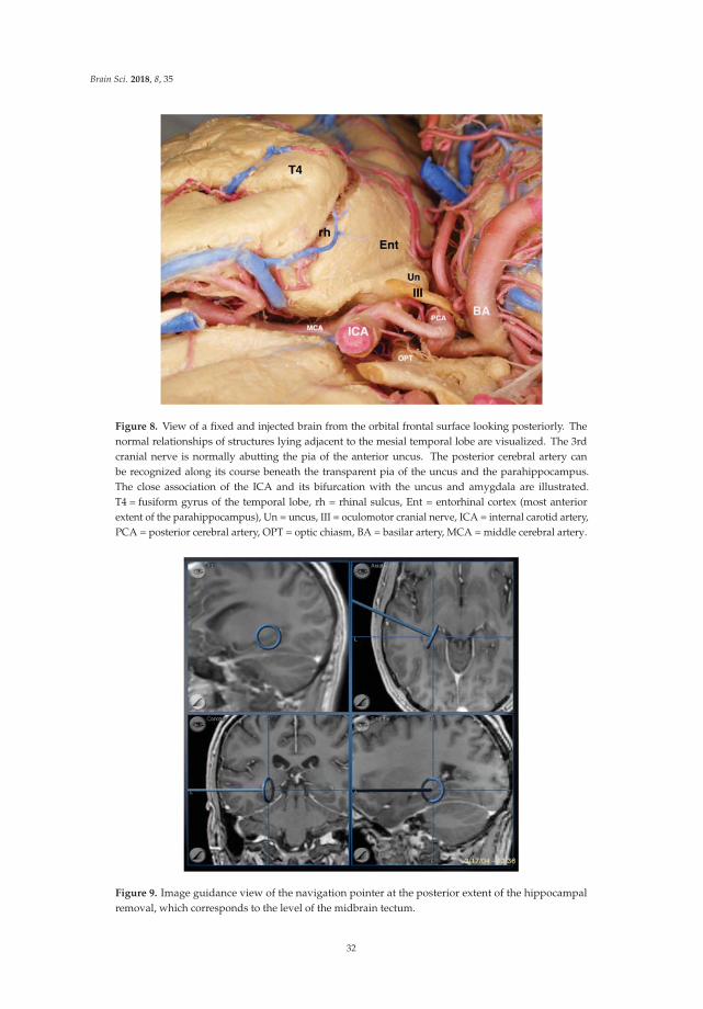
Brain Sci. 2018, 8, 35
Figure 8. View of a fixed and injected brain from the orbital frontal surface looking posteriorly. Thenormal relationships of structures lying adjacent to the mesial temporal lobe are visualized. The 3rdcranial nerve is normally abutting the pia of the anterior uncus. The posterior cerebral artery canbe recognized along its course beneath the transparent pia of the uncus and the parahippocampus.The close association of the ICA and its bifurcation with the uncus and amygdala are illustrated.T4 = fusiform gyrus of the temporal lobe, rh = rhinal sulcus, Ent = entorhinal cortex (most anteriorextent of the parahippocampus), Un = uncus, III = oculomotor cranial nerve, ICA = internal carotid artery,PCA = posterior cerebral artery, OPT = optic chiasm, BA = basilar artery, MCA = middle cerebral artery.
Figure 9. Image guidance view of the navigation pointer at the posterior extent of the hippocampalremoval, which corresponds to the level of the midbrain tectum.
32

Brain Sci. 2018, 8, 35
4. Key Hole Approach in SAH
Minimal access surgery has potential to benefit patients by providing more rapid recoveryand improved patient outcomes compared with traditional larger surgical incisions andexposures [60,63,64]. The minimal access (or keyhole) approach lends itself quite well to SAH usingeither the trans-middle temporal gyrus or subtemporal approaches since the necessary maneuversrequired to remove the mesial temporal structures can be accomplished with relative ease through asmall scalp incision and bony opening [29,59,60,65]. The SAH approach was envisioned and developedin order to spare from resection cerebral tissue that is not a part of the primary seizure focus. Therefore,minimizing the scalp and bony opening to accomplish SAH represents a logical progression from thelarger more traditional scalp incision and craniotomy exposure. In individuals with clear-cut MTLEwho are candidates for SAH, the keyhole, minimal access approach is preferred. Because the exposureis too limited to confidently recognize sulcal and gyral landmarks, keyhole SAH should be performedwith the aid of image guidance.
The scalp incision is curvilinear starting at the zygoma. Figure 10 The temporalis muscle issplit and held open with hooks or a self-retaining retractor. The bone opening is a silver dollar sizedcraniotomy plus additional craniectomy anterior and inferior centered over the second temporal gyrusand the planned cortical incision. Figure 11 The keyhole and non-keyhole SAH approach to the mesialtemporal structures are essentially identical. Image guidance is critical to confirm the corticectomy ispositioned properly in the middle temporal gyrus anterior to the central sulcus in the non-dominanthemisphere and anterior to the pre-central sulcus in the dominant hemisphere.
Figure 10. The keyhole minimal access approach benefits the patient with smaller skin incision andcranial opening. A slight curve in the scalp incision helps exposure and reduces retraction forces on thetemporalis muscle compared with a straight linear incision.
33
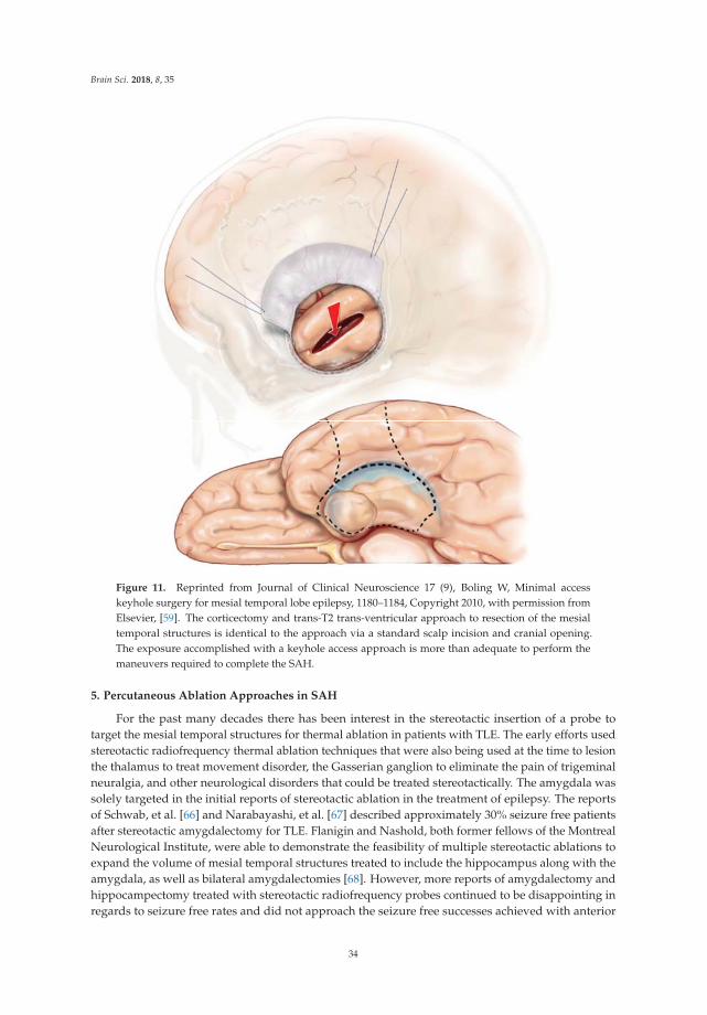
Brain Sci. 2018, 8, 35
Figure 11. Reprinted from Journal of Clinical Neuroscience 17 (9), Boling W, Minimal accesskeyhole surgery for mesial temporal lobe epilepsy, 1180–1184, Copyright 2010, with permission fromElsevier, [59]. The corticectomy and trans-T2 trans-ventricular approach to resection of the mesialtemporal structures is identical to the approach via a standard scalp incision and cranial opening.The exposure accomplished with a keyhole access approach is more than adequate to perform themaneuvers required to complete the SAH.
5. Percutaneous Ablation Approaches in SAH
For the past many decades there has been interest in the stereotactic insertion of a probe totarget the mesial temporal structures for thermal ablation in patients with TLE. The early efforts usedstereotactic radiofrequency thermal ablation techniques that were also being used at the time to lesionthe thalamus to treat movement disorder, the Gasserian ganglion to eliminate the pain of trigeminalneuralgia, and other neurological disorders that could be treated stereotactically. The amygdala wassolely targeted in the initial reports of stereotactic ablation in the treatment of epilepsy. The reportsof Schwab, et al. [66] and Narabayashi, et al. [67] described approximately 30% seizure free patientsafter stereotactic amygdalectomy for TLE. Flanigin and Nashold, both former fellows of the MontrealNeurological Institute, were able to demonstrate the feasibility of multiple stereotactic ablations toexpand the volume of mesial temporal structures treated to include the hippocampus along with theamygdala, as well as bilateral amygdalectomies [68]. However, more reports of amygdalectomy andhippocampectomy treated with stereotactic radiofrequency probes continued to be disappointing inregards to seizure free rates and did not approach the seizure free successes achieved with anterior
34

Brain Sci. 2018, 8, 35
temporal lobe resection [69–72]. The early efforts at stereotactic ablation of the mesial temporalstructures to treat intractable TLE were largely hampered by the challenges of targeting the mesialtemporal structures using the methods of the time; stereotactic targets were defined by imaging withangiogram and pneumoencephaolgraphy or by measurements made from the AC-PC line. Also, theearly stereotactic frames were best suited for orthogonal trajectories, which made treatment along thelongitudinal axis of the mesial temporal structures a more arduous treatment plan.
With the advent of CT and MR imaging, the mesial temporal structures could be directly and moreaccurately targeted for stereotactic treatment. Parrent and Blume reported the experience of stereotacticamygdalohippocampectomy for TLE with the use of modern MR imaging [73]. The authors describedoutcomes from patients with TLE and evidence of exclusively ipsilateral MTS in 11 out of 19 patientstreated. Surgery was performed using a stereotactic headframe with radiofrequency lesioning of theamygdala and hippocampus. Postoperative MRI identified the best results were in patients with morecomplete stereotactic ablations involving the entire amygdala and measuring 15–34 mm along thehippocampus. In this group of patients, 9 out of 15 had favorable results defined as greater than 90%reduction in seizure frequency or seizure freedom. Only 1 out 5 patients with incomplete ablation ofthe amygdala and hippocampus had a favorable result. Parrent and Blume were able to demonstratethe improved results of stereotactic ablation surgery using modern imaging to target the thermalprobe, and that more complete treatment of the amygdala and hippocampus produced better resultsof seizure freedom and seizure frequency reduction.
Three additional advances have enabled further improvement in the technique of stereotacticablation surgery of TLE. One is frameless stereotaxy or image guidance. Now that stereotacticsurgery can be performed without the traditional stereotactic headframe in place, neurosurgeons haveconsiderably more freedom to plan non-orthogonal trajectories to targets that were more difficult oreven impossible to accomplish previously. Another advance is the development of low cost fiber-opticlasers for thermal ablation. The radiofrequency ablation probe has not been able to be manufacturedto allow use in the MRI magnet. Using the laser ablation technology, only the non-ferromagneticfiber optic cable is in the MRI environment. Therefore, the thermal treatment can be delivered withthe patient in the MRI to confirm the anatomy treated and the thermal dose delivered, which seguesto the third advance, the ability to create a thermal map using normal MRI acquisition protocols.Several measurable MRI variables are affected by temperature in a linear relationship and can beused to construct a thermal map, such as diffusion coefficient [74], chemical shift, T1 relaxation [75],magnetization transfer, proton density and proton resonance frequency (PRF). The excellent linearityand temperature dependence of the PRF in relation to most all tissue types have made PRF-basedphase mapping methods the preferred choice for thermal mapping and this acquisition protocol isavailable on most all modern MRI units.
There are 2 FDA approved laser thermal ablation devices that overlay anatomy with a thermalmap, NeuroBlate (Monteris Medical, Plymouth, MN) and Visualase (Medtronic, Minneapolis, MN).Both devices use a pulsed laser thermal source. The probe, which consists of a fiber-optic cable todeliver the laser plus a cooling system, is inserted stereotactically to the desired target in the operatingroom. Then the actual thermal ablation treatment is performed in the MRI magnet. There has beenconsiderable renewed interest in the use of thermal ablation for the treatment of intractable MTLE, andthe fiber-optic laser technologies have improved the ability to target the mesial temporal structuresand assess the treatment delivered essentially real-time.
The fiber-optic laser thermal probe is typically inserted in a posterior to anterior trajectory to allowtreatment of the mesial temporal structures along the longitudinal axis. Multiple thermal treatmentsare delivered as the fiber-optic probe is pulled back until a sufficient volume of mesial temporalstructures are ablated. One of the challenges of treatment planning is to insert the probe in such atrajectory that the uncus and hippocampal head can be completely ablated. Jermakowicz, et al. foundlateral trajectories that did not include the uncus and hippocampal head in the ablation treatment weresignificantly correlated with persistent disabling seizures [76]. Overall the laser ablation of mesial
35

Brain Sci. 2018, 8, 35
temporal structures has been shown to be a safe minimally invasive alternative to craniotomy surgeryof MTLE. Although the data thus far point to reduced odds of seizure freedom in patients treated withlaser thermal ablation versus craniotomy approaches to resect the mesial temporal structures [77].
6. Patient Outcomes from SAH
The success of SAH to achieve seizure freedom is dependent on the accurate diagnosis of MTLElateralized to the side of surgery and the successful removal or disconnection of the seizure focus atsurgery. At centers with expertise in the diagnosis of MTLE, the seizure freedom rates after SAH arethe same as anterior temporal resection [29,35,59,60,65,78,79]. To date a randomized controlled studyto evaluate and compare the surgical approaches of SAH and ATL for the treatment of MTLE hasnot been undertaken. The literature up to now demonstrates no clear difference in ability to becomeseizure free in patients with surgery of SAH or ATL for MTLE (Table 1).
Table 1. Studies comparing ATL with SAH for outcomes of seizure freedom and reducing seizure frequency.
Renowden et al. (1995) [80] Same outcome for two types of SAH
Arruda et al. (1996) [81] Similar
Pauli et al. (1999) [82] Similar
Clusmann et al. (2002) [83] Similar
Clusmann et al. (2004) [40] Better in ATL in children + adolescents
Lutz et al. (2004) [40] Same outcome for two types of SAH
Paglioli et al. (2006) [84] Similar
Tanriverdi, et al. (2007) [85] Similar
Bate et al. (2007) [86] Better in ATL in children + adolescents
Tanriverdi, et al. (2010) [87] Similar
Sagher, et al. (2012) [78] Similar
Wendling, et al. (2013) [88] Similar
Bujarski, et al. (2013) [89] Similar
Josephson, et al. (2013) [90] Better outcome in ATL
Hu, et al. (2013) [91] Better outcome in ATL
Nascimento, et al. (2016) [92] Similar although ATL associated with more complications
Schmeiser, et al. (2017) [93] No difference in three types of SAH nor in SAH versus ATL
Foged, et al. (2018) [94] No difference at 1 year and 7 years after surgery
One potential advantage of SAH is that cognitive function may be improved in patients withmore limited temporal lobe resections. This topic was reviewed by Schramm [94] who found theliterature lacking consistency on reporting of crucial study variables such as MRI confirmation ofextent of resection and studies were mostly retrospective reports. However, Schramm concludedfrom the available literature that “in summary there is considerable evidence for somewhat betterneuropsychological results with SAH, although this was not consistently found.” An analysis of studiespublished from 1992 to 2018 supports Schramm’s conclusion that in general there tends to be moreneuropsychological deficits in ATL than SAH surgery, although the heterogeneity of studies and thelack of a randomized controlled study comparing the two surgical techniques makes firm conclusionsabout neuropsychological impact impossible to make (Table 2).
36

Brain Sci. 2018, 8, 35
Table 2. Studies comparing temporal lobe surgery of ATL with SAH for neuropsychological outcome.
Goldstein, et al. (1992) [95] No difference when using global memory test
Goldstein, et al. (1993) [96] SAH short-term beneficial effect on memory
Wolf et al. (1993) [97] No difference
Renowden et al. (1995) [80] SAH better in verbal IQ and nonverbal memory
Helmstaedter et al. (1996) [98] Immediate recall better in SAH
Jones-Gotman (1997) [42] Similar deficits in learning + retention tasks in seizure free patients
Pauli et al. (1999) [82] SAH better for verbal memory
Helmstaedter et al. (2002) [99] SAH has advantage over ATL in long-term follow-up (2–10 years)
Clusmann (2004) [40] SAH in adults: higher rate of improvement + lower rate ofdeterioration in overall neuropsychological score
Hader et al. (2005) [100] No difference
Morino et al. (2006) [101] SAH better memory function
Paglioli et al. (2006) [84] SAH better for verbal memory score(30% deterioration in both groups)
Tanriverdi, et al. (2007) [85] SAH less decline for verbal memory
Helmstaedter et al. (2008) [102] SAH better for R-sided resection, ATL better for L-sided resection formaterial-specific memory
Tanriverdi, et al. (2010) [87] Worse verbal IQ after SAH
Sagher, et al. (2012) [78] No difference
Bujarski, et al. (2013) [89] No difference, except more post-surgical paranoia after ATL
Wendling, et al. (2013) [88] Worse memory after ATL
Boucher, et al. (2015) [103]Worse after ATL on immediate recall of Logical Memory subtest of
Wechsler Memory Scales. Delayed recognition trial of Rey AuditoryVerbal Learning Test worse after SAH
Nascimento, et al. (2016) [92] No difference
Gül, et al. (2017) [104] No difference
Schmeiser, et al. (2017) [93] No difference
Foged, et al. (2018) [94] SAH better than ATL verbal memory in left hemisphere surgery only
An additional outcome parameter of considerable importance to the patient is the ease and speedof recovery from brain surgery. SAH has an opportunity to provide patients shorter operative time,quicker recovery, and reduced length of stay in the hospital particularly if surgery is performed usingminimal access or key hole approaches [59,105]. Reduced hospital length of stay is a marker of bothquicker patient recovery and a more cost effective operation. Boling identified significantly reducedoperative time and shorter hospital length of stay in patients who underwent key hole craniotomyversus a standard craniotomy approach for SAH [59].
7. Conclusions
Patients with medically intractable MTLE have an excellent opportunity for seizurefreedom from resection surgery that can be a standard anterior temporal removal or selectiveamygdalohippocampectomy. The potential advantage of the SAH is a selective removal of the seizurefocus sparing temporal lobe regions that are not the actual epileptogenic zone. SAH, especially minimalaccess approaches, provide direct advantages of quicker patient recovery from surgery, and evidencepoints to a probable cognitive benefit compared with standard temporal resections.
Conflicts of Interest: The author reports no conflicts of interest.
37

Brain Sci. 2018, 8, 35
References
1. Téllez-Zenteno, J.F.; Hernández-Ronquillo, L. A Review of the Epidemiology of Temporal Lobe Epilepsy.Epilepsy Res. Treat. 2012, 630853. [CrossRef] [PubMed]
2. Semah, F.; Picot, M.C.; Adam, C.; Broglin, D.; Arzimanoglou, A.; Bazin, B.; Cavalcanti, D.; Baulac, M.Is the underlying cause of epilepsy a major prognostic factor for recurrence? Neurology 1998, 51, 1256–1262.[CrossRef] [PubMed]
3. Hauser, W.A.; Annegers, J.F.; Kurland, L.T. Prevalence of epilepsy in Rochester, Minnesota: 1940–1980.Epilepsia 1991, 32, 429–445. [CrossRef] [PubMed]
4. Papez, J.W. A proposed mechanism of emotion. Arch. Neurol. Psychiatry 1937, 38, 725–743. [CrossRef]5. Duvernoy, H.M.; Cattin, F.; Risold, P.Y. The Human Hippocampus Functional Anatomy, Vascularization and Serial
Sections with MRI; Springer: New York, NY, USA, 2013.6. Wiebe, S.; Blume, W.T.; Girvin, J.P.; Eliasziw, M. A randomized, controlled trial of surgery for temporal-lobe
epilepsy. N. Engl. J. Med. 2001, 345, 311–318. [CrossRef] [PubMed]7. Tanriverdi, T.; Ajlan, A.; Poulin, N.; Olivier, A. Morbidity in epilepsy surgery: An experience based on based
on 2449 epilepsy surgery procedures from a single institution. J. Neurosurg. 2009, 110, 1111–1123. [CrossRef][PubMed]
8. Engel, J., Jr.; Wiebe, S.; French, J.; Sperling, M.; Williamson, P.; Spencer, D.; Gumnit, R.; Zahn, C.; Westbrook, E.;Enos, B. Practice parameter: Temporal lobe and localized neocortical resections for epilepsy: Report of theQuality Standards Subcommittee of the American Academy of Neurology, in association with the AmericanEpilepsy Society and the American Association of Neurological Surgeons. Neurology 2003, 60, 538–547.
9. Kwan, P.; Arzimanoglou, A.; Berg, A.T.; Brodie, M.J.; Allen Hauser, W.; Mathern, G.; Moshé, S.L.; Perucca, E.;Wiebe, S.; French, J. Definition of drug resistant epilepsy: Consensus proposal by the ad hoc Task Force ofthe ILAE Commission on Therapeutic Strategies. Epilepsia 2010, 51, 1069–1077. [CrossRef] [PubMed]
10. Kwon, C.S.; Neal, J.; Telléz-Zenteno, J.; Metcalfe, A.; Fitzgerald, K.; Hernandez-Ronquillo, L.; Hader, W.;Wiebe, S.; Jetté, N.; CASES Investigators. Resective focal epilepsy surgery—Has selection of candidateschanged? A systematic review. Epilepsy Res. 2016, 122, 37–43. [CrossRef] [PubMed]
11. Wieser, H.G.; ILAE Commission on Neurosurgery of Epilepsy. Mesial temporal lobe epilepsy withhippocampal sclerosis. Epilepsia 2004, 45, 695–714. [PubMed]
12. O’Brien, T.J.; Kilpatrick, C.; Murrie, V.; Vogrin, S.; Morris, K.; Cook, M.J. Temporal lobe epilepsy caused bymesial temporal sclerosis and temporal neocortical lesions. A clinical and electroencephalographic study of46 pathologically proven cases. Brain 1996, 119, 2133–2141. [CrossRef] [PubMed]
13. Burgerman, R.S.; Sperling, M.R.; French, J.A.; Saykin, A.J.; O’Connor, M.J. Comparison of mesial versusneocortical onset temporal lobe seizures: Neurodiagnostic findings and surgical outcome. Epilepsia 1995, 36,662–670. [CrossRef] [PubMed]
14. Olivier, A.; Gloor, P.; Andermann, F.; Quesney, L.F. The place of stereotactic depth electrode recording inepilepsy. Appl. Neurophysiol. 1985, 48, 395–399. [CrossRef] [PubMed]
15. Berkovic, S.F.; Andermann, F.; Olivier, A.; Ethier, R.; Melanson, D.; Robitaille, Y.; Kuzniecky, R.; Peters, T.;Feindel, W. Hippocampal sclerosis in temporal lobe epilepsy demonstrated by magnetic resonance imaging.Ann. Neurol. 1991, 29, 175–182. [CrossRef] [PubMed]
16. Cendes, F. Febrile seizures and mesial temporal sclerosis. Curr. Opin. Neurol. 2004, 17, 161–164. [CrossRef][PubMed]
17. Schramm, J.; Kral, T.; Grunwald, T.; Blumcke, I. Surgical treatment for neocortical temporal lobe epilepsy:Clinical and surgical aspects and seizure outcome. J. Neurosurg. 2001, 94, 33–42. [CrossRef] [PubMed]
18. Pacia, S.V.; Devinsky, O.; Perrine, K.; Ravdin, L.; Luciano, D.; Vazquez, B.; Doyle, W.K. Clinical features ofneocortical temporal lobe epilepsy. Ann. Neurol. 1996, 40, 724–730. [CrossRef] [PubMed]
19. Maillard, L.; Vignal, J.P.; Gavaret, M.; Guye, M.; Biraben, A.; McGonigal, A.; Chauvel, P.; Bartolomei, F.Semiologic and electrophysiologic correlations in temporal lobe seizure subtypes. Epilepsia 2004, 45,1590–1599. [CrossRef] [PubMed]
20. Adam, C.; Clemenceau, S.; Semah, F.; Hasboun, D.; Samson, S.; Aboujaoude, N.; Samson, Y.; Baulac, M.Variability of presentation in medial temporal lobe epilepsy: A study of 30 operated cases. Acta. Neurol. Scand.1996, 94, 1–11. [CrossRef] [PubMed]
38

Brain Sci. 2018, 8, 35
21. Pfander, M.; Arnold, S.; Henkel, A.; Weil, S.; Werhahn, K.J.; Eisensehr, I.; Winkler, P.A.; Noachtar, S. Clinicalfeatures and EEG findings differentiating mesial from neocortical temporal lobe epilepsy. Epileptic Disord.2002, 4, 189–195. [PubMed]
22. Foldvary, N.; Lee, N.; Thwaites, G.; Mascha, E.; Hammel, J.; Kim, H.; Friedman, A.H.; Radtke, R.A. Clinicaland electrographic manifestations of lesional neocortical temporal lobe epilepsy. Neurology 1997, 49, 757–763.[CrossRef] [PubMed]
23. So, N.; Gloor, P.; Quesney, L.F.; Jones-Gotman, M.; Olivier, A.; Andermann, F. Depth electrode investigationsin patients with bitemporal epileptiform abnormalities. Ann. Neurol. 1989, 25, 423–431. [CrossRef] [PubMed]
24. Olivier, A.; Boling, W.; Tanriverdi, T. (Eds.) Techniques in Epilepsy Surgery: The MNI Approach; CambridgeUniversity Press: New York, NY, USA, 2012.
25. Spencer, D.D.; Spencer, S.S.; Mattson, R.H.; Williamson, P.D.; Novelly, R.A. Access to the posterior medialtemporal lobe structures in the surgical treatment of temporal lobe epilepsy. Neurosurgery 1984, 15, 667–671.[CrossRef] [PubMed]
26. Olivier, A. Temporal resections in the surgical treatment of epilepsy. Epilepsy Res. Suppl. 1992, 5, 175–188.[PubMed]
27. Silbergeld, D.L.; Ojemann, G.A. The tailored temporal lobectomy. Neurosurg. Clin. N. Am. 1993, 4, 273–281.[PubMed]
28. Kim, H.I.; Olivier, A.; Jones-Gotman, M.; Primrose, D.; Andermann, F. Corticoamygdalectomy inmemory-impaired patients. Stereotact. Funct. Neurosurg. 1992, 58, 162–167. [CrossRef] [PubMed]
29. Boling, W.; Longoni, N.; Palade, A.; Moran, M.; Brick, J. Surgery for temporal lobe epilepsy. W V Med. J. 2006,102, 18–21. [PubMed]
30. Jackson, J.H.; Colman, W.S. Case of epilepsy with tasting movements and “dreamy state” with very smallpatch of softening in the left uncinate gyrus. Brain 1898, 21, 580–590. [CrossRef]
31. Kaada, B.R. Somatomotor, autonomic and electrographic responses to electrical stimulation of “rhinencephalic”and other structures in primates cat and dog: A study of responses from limbic, subcallosal, orbito-insular,pyriform and temporal cortex, hippocampus, fornix and amygdala. Acta. Physiol. Scand. Suppl. 1951, 83, 1–285.
32. Vigouroux, R.; Gastaut, H.R.; Badier, M. Provocation des principales manifestations cliniques de l’épilepsiedite temporale par stimulation des structures rhinencéphaliques chez le chat non-anaesthésié. Rev. Neurol.1951, 85, 505–508. [PubMed]
33. Gastaut, H.R.; Vigouroux, R.; Naquet, R. Lésions épileptogènes amygdalo-hippocampiques provoquées chezle chat par l’injection de “crème d’albumine”. Rev. Neurol. 1952, 87, 607–609. [PubMed]
34. Green, J.D.; Shimamoto, T. Hippocampal seizures and their propagation. Arch. Neurol. Psychiatry 1953, 7,687–702. [CrossRef]
35. Wieser, H.G.; Ortega, M.; Friedman, A.; Yonekawa, Y. Long-term seizure outcomes followingamygdalohippocampectomy. J. Neurosurg. 2003, 98, 751–763. [CrossRef] [PubMed]
36. Penfield, W.; Jasper, H. Epilepsy and the Functional Anatomy of the Human Brain, 1st ed.; Little Brown: Boston,MA, USA, 1954.
37. Olivier, A. Relevance of removal of limbic structures in surgery for temporal lobe epilepsy. Can. J. Neurol. Sci.1991, 18 (Suppl. 4), 628–635. [CrossRef] [PubMed]
38. Olivier, A. Transcortical selective amygdalohippocampectomy in temporal lobe epilepsy. Can. J. Neurol. Sci.2000, 27 (Suppl. 1), S68–S76. [CrossRef] [PubMed]
39. Abosch, A.; Bernasconi, N.; Boling, W.; Jones-Gotman, M.; Poulin, N.; Dubeau, F.; Andermann, F.; Olivier, A.Factors predictive of suboptimal seizure control following selective amygdalohippocampectomy. J. Neurosurg.2002, 97, 1142–1151. [CrossRef] [PubMed]
40. Lutz, M.T.; Clusmann, H.; Elger, C.E.; Schramm, J.; Helmstaedter, C. Neuropsychological outcome afterselective amygdalohippocampectomy with transsylvian versus transcortical approach: A randomizedprospective clinical trial of surgery for temporal lobe epilepsy. Epilepsia 2004, 45, 809–816. [CrossRef][PubMed]
41. Niemeyer, P. The Transventricular Amygdala-Hippocampectomy in Temporal Lobe Epilepsy. In TemporalLobe Epilepsy: A Colloquium Sponsored by the National Institute of Neurological Diseases and Blindness, NationalInstitutes of Health, Bethesda, Maryland, in Cooperation with the International League Against Epilepsy; Baldwin, M.,Bailey, P., Eds.; Charles C Thomas: Springfield, IL, USA, 1958; pp. 461–482.
39

Brain Sci. 2018, 8, 35
42. Jones-Gotman, M.; Zatorre, R.J.; Olivier, A.; Andermann, F.; Cendes, F.; Staunton, H.; McMackin, D.;Siegel, A.M.; Wieser, H.G. Learning and retention of words and designs following excision from medial orlateral temporal-lobe structures. Neuropsychologia 1997, 35, 963–973. [CrossRef]
43. Wieser, H.G. Selective amygdalo-hippocampectomy for temporal lobe epilepsy. Epilepsia 1988, 29 (Suppl. 2),S100–S113. [CrossRef] [PubMed]
44. Tanriverdi, T.; Olivier, A.; Poulin, N.; Andermann, F.; Dubeau, F. Long-term seizure outcomeafter mesial temporal lobe epilepsy surgery: Corticalamygdalohippocampectomy versus selectiveamygdalohippocampectomy. J. Neurosurg. 2008, 108, 517–524. [CrossRef] [PubMed]
45. JAMA Network. Temporal Lobe Epilepsy: A Colloquium Sponsored by the National Institute of NeurologicalDiseases and Blindness, National Institutes of Health, Bethesda, Maryland, in Cooperation with theInternational League Against Epilepsy. JAMA 1959, 169, 1143–1144. [CrossRef]
46. Scoville, W.; Milner, B. Loss of recent memory after bilateral hippocampal lesions. J. Neurol. Neurosurg. Psych.1957, 20, 11–21. [CrossRef]
47. Penfield, W.; Milner, B. Memory deficit produced by bilateral lesions in the hippocampal zone. Arch. Neurol.Psychiatry 1958, 79, 475–497. [CrossRef]
48. Penfield, W.; Flanigin, H. Surgical therapy of temporal lobe seizures. Arch. Neurol. Psychiatry 1950, 64,491–500. [CrossRef] [PubMed]
49. Gastaut, H. So-called “psychomotor” and “temporal” epilepsy. Epilepsia 1953, 2, 59–76. [CrossRef]50. Sano, K.; Malamud, N. Clinical significance of sclerosis of the cornu ammonis. Arch. Neurol. Psychiatry 1953,
70, 40–53. [CrossRef]51. Hill, D.; Falconer, M.A.; Pampiglione, G.; Liddell, D.W. Discussion on the surgery of temporal lobe epilepsy.
Proc. R Soc. Med. 1953, 46, 965–976. [PubMed]52. Feindel, W.; Penfield, W. Localization of discharge in temporal lobe automatism. Arch. Neurol. Psychiatry
1954, 72, 605–630. [CrossRef]53. Yasargil, M.G.; Teddy, P.J.; Roth, P. Selective amygdalo-hippocampectomy: Operative anatomy and surgical
technique. In Advances and Technical Standards in Neurosurgery; Symon, L., Ed.; Springler-Wien: New York,NY, USA, 1985; Volume 12.
54. Martens, T.; Merkel, M.; Holst, B.; Brückner, K.; Lindenau, M.; Stodieck, S.; Fiehler, J.; Westphal, M.; Heese, O.Vascular events after transsylvian selective amygdalohippocampectomy and impact on epilepsy outcome.Epilepsia 2014, 55, 763–769. [CrossRef] [PubMed]
55. Yasargil, M.G.; Teddy, P.J.; Roth, P. Selective Amygdalo-Hippocampectomy Operative Anatomy and SurgicalTechnique. Adv. Tech. Stand. Neurosurg. 1985, 12, 93–123. [PubMed]
56. Hori, T.; Tabuchi, S.; Kurosaki, M.; Kondo, S.; Takenobu, A.; Watanabe, T. Subtemporalamygdalohippocampectomy for treating medially intractable temporal lobe epilepsy. Neurosurgery 1993, 33,50–56. [PubMed]
57. Sugita, K.; Kobayashi, S.; Yokoo, A. Preservation of large bridging veins during brain retraction. Technicalnote. J. Neurosurg. 1982, 57, 856–858. [CrossRef] [PubMed]
58. Boling, W. Selective Amygdalohippocampectomy. In Operative Techniques in Epilepsy Surgery, 2nd ed.;Baltuch, G., Cukiert, A., Eds.; Thieme Medical Publishers: New York, NY, USA, 2018.
59. Boling, W. Minimal Access Keyhole Surgery for Mesial Temporal Lobe Epilepsy. J. Clin. Neurosci. 2010, 17,1180–1184. [CrossRef] [PubMed]
60. Little, A.S.; Smith, K.A.; Kirlin, K.; Baxter, L.C.; Chung, S.; Maganti, R.; Treiman, D.M. Modifications tothe subtemporal selective amygdalohippocampectomy using a minimal-access technique: Seizure andneuropsychological outcomes. J. Neurosurg. 2009, 111, 1263–12674. [CrossRef] [PubMed]
61. Kessels, R.P.; Hendriks, M.; Schouten, J.; Van Asselen, M.; Postma, A. Spatial memory deficits in patientsafter unilateral selective amygdalohippocampectomy. J. Int. Neuropsychol. Soc. 2004, 10, 907–912. [CrossRef][PubMed]
62. Gleissner, U.; Helmstaedter, C.; Schramm, J.; Elger, C.E. Memory outcome after selectiveamygdalohippocampectomy in patients with temporal lobe epilepsy: One-year follow-up. Epilepsia 2004, 45,960–962. [CrossRef] [PubMed]
63. Arts, S.; Delye, H.; van Lindert, E.J. Intraoperative and postoperative complications in the surgical treatmentof craniosynostosis: Minimally invasive versus open surgical procedures. J. Neurosurg. Pediatr. 2017, 24, 1–7.[CrossRef] [PubMed]
40

Brain Sci. 2018, 8, 35
64. Morcos, M.W.; Jiang, F.; McIntosh, G.; Johnson, M.; Christie, S.; Wai, E.; Ouellet, J.; Bailey, C.; Ahn, H.;Paquet, J.; et al. Predictors of Blood Transfusion in Posterior Lumbar Spinal Fusion: A Canadian SpineOutcome and Research Network Study. Spine 2018, 43, E35–E39. [CrossRef] [PubMed]
65. Duckworth, E.A.; Vale, F.L. Trephine epilepsy surgery: The inferior temporal gyrus approach. Neurosurgery2008, 63 (Suppl. 1), ONS156–ONS160. [CrossRef]
66. Schwab, R.S.; Sweet, W.H.; Mark, V.H.; Kjellberg, R.N.; Ervin, F.R. Treatment of intractable temporal lobeepilepsy by stereotactic amygdala lesions. Trans. Am. Neurol. Assoc. 1965, 90, 12–19. [PubMed]
67. Narabayashi, H.; Mizutani, T. Epileptic seizures and the stereotaxic amygdalotomy. Confin. Neurol. 1970, 32,289–297. [CrossRef] [PubMed]
68. Flanigin, H.F.; Nashold, B.S. Stereotactic lesions of the amygdala and hippocampus in epilepsy. Acta Neurochir.1976, 23, 235–239.
69. Heimberger, R.F.; Small, I.F.; Milstein, V.; Moore, D. Stereotactic amygdalotomy for convulsive andbehavioural disorders. Appl. Neurophysiol. 1978, 41, 43–45.
70. Vaernet, K. Stereotaxic amygdalotomy in temporal lobe epilepsy. Confin. Neurol. 1972, 34, 176–180. [CrossRef][PubMed]
71. Mempel, E.; Witkiewicz, B.; Stadnicki, R.; Luczywek, E.; Kucinski, L.; Pawłowski, G.; Nowak, J. The effectof medial amygdalotomy and anterior hippocampotomy on behavior and seizures in epileptic patients.Acta Neurochir. 1980, 30, 161–167.
72. Nadvornik, P.; Sramka, M.; Gajdosova, D.; Kokavec, M. Longitudinal hippocampectomy. Confin. Neurol.1975, 37, 244–248. [CrossRef]
73. Parrent AG and Blume, W. Stereotactic Amygdalohippocampotomy for the Treatment of Medial TemporalLobe Epilepsy. Epilepsia 1999, 40, 1408–1416. [CrossRef]
74. Le Bihan, D.; Delannoy, J.; Levin, R.L. Temperature mapping with MR imaging of molecular diffusion:Application to hyperthermia. Radiology 1989, 171, 853–857. [CrossRef] [PubMed]
75. Bertsch, F.; Mattner, J.; Stehling, M.K.; Müller-Lisse, U.; Peller, M.; Loeffler, R.; Weber, J.; Messmer, K.;Wilmanns, W.; Issels, R.; et al. Non-invasive temperature mapping using MRI: comparison of two methodsbased on based on chemical shift and T1-relaxation. Magn. Reson. Imaging 1998, 16, 393–404. [CrossRef]
76. Jermakowicz, W.J.; Kanner, A.M.; Sur, S.; Bermudez, C.; D’Haese, P.F.; Kolcun, J.P.G.; Cajigas, I.; Li, R.;Millan, C.; Ribot, R.; et al. Laser thermal ablation for mesiotemporal epilepsy: Analysis of ablation volumesand trajectories. Epilepsia 2017, 58, 801–810. [CrossRef] [PubMed]
77. Kang, J.Y.; Wu, C.; Tracy, J.; Lorenzo, M.; Evans, J.; Nei, M.; Skidmore, C.; Mintzer, S.; Sharan, A.D.;Sperling, M.R. Laser interstitial thermal therapy for medically intractable mesial temporal lobe epilepsy.Epilepsia 2016, 57, 325–334. [CrossRef] [PubMed]
78. Sagher, O.; Thawani, J.P.; Etame, A.B.; Gomez-Hassan, D.M. Seizure outcomes and mesial resection volumesfollowing selective amygdalohippocampectomy and temporal lobectomy. Neurosurg. Focus 2012, 32, E8.[CrossRef] [PubMed]
79. Schramm, J. Temporal lobe epilepsy surgery and the quest for optimal extent of resection: A review. Epilepsia2008, 49, 1296–1307. [CrossRef] [PubMed]
80. Renowden, S.A.; Matkovic, Z.; Adams, C.B.; Carpenter, K.; Oxbury, S.; Molyneux, A.J.; Anslow, P.; Oxbury, J.Selective amygdalohippocampectomy for hippocampal sclerosis: Postoperative MR appearance. AJNR Am.J. Neuroradiol. 1995, 16, 1855–1861. [PubMed]
81. Arruda, F.; Cendes, F.; Andermann, F.; Dubeau, F.; Villemure, J.G.; Jones-Gotman, M.; Poulin, N.; Arnold, D.L.;Olivier, A. Mesial atrophy and outcome after amygdalohippocampectomy or temporal lobe removal.Ann. Neurol. 1996, 40, 446–450. [CrossRef] [PubMed]
82. Pauli, E.; Pickel, S.; Schulemann, H.; Buchfelder, M.; Stefan, H. Neuropsychologic findings depending on thetype of the resection in temporal lobe epilepsy. Adv. Neurol. 1999, 81, 371–377. [PubMed]
83. Clusmann, H.; Schramm, J.; Kral, T.; Helmstaedter, C.; Ostertun, B.; Fimmers, R.; Haun, D.; Elger, C.E.Prognostic factors and outcome after different types of resection for temporal lobe epilepsy. J. Neurosurg.2002, 97, 1131–1141. [CrossRef] [PubMed]
84. Paglioli, E.; Palmini, A.; Portuguez, M.; Azambuja, N.; da Costa, J.C.; da Silva Filho, H.F.; Martinez, J.V.;Hoeffel, J.R. Seizure and memory outcome following temporal lobe surgery: Selective compared withnonselective approaches for hippocampal sclerosis. J. Neurosurg. 2006, 104, 70–78. [CrossRef] [PubMed]
41

Brain Sci. 2018, 8, 35
85. Tanriverdi, T.; Olivier, A. Cognitive changes after unilateral cortico-amygdalohippocampectomy unilateralselective-amygdalohippocampectomy mesial temporal lobe epilepsy. Turk. Neurosurg. 2007, 17, 91–99.[PubMed]
86. Bate, H.; Eldridge, P.; Varma, T.; Wieshmann, U.C. The seizure outcome after amygdalohippocampectomyand temporal lobectomy. Eur. J. Neurol. 2007, 14, 90–94. [CrossRef] [PubMed]
87. Tanriverdi, T.; Dudley, R.W.; Hasan, A.; Al Jishi, A.; Al Hinai, Q.; Poulin, N.; Colnat-Coulbois, S.; Olivier, A.Memory outcome after temporal lobe epilepsy surgery: Corticoamygdalohippocampectomy versus selectiveamygdalohippocampectomy. J. Neurosurg. 2010, 113, 1164–1175. [CrossRef] [PubMed]
88. Wendling, A.S.; Hirsch, E.; Wisniewski, I.; Davanture, C.; Ofer, I.; Zentner, J.; Bilic, S.; Scholly, J.; Staack, A.M.;Valenti, M.P.; et al. Selective amygdalohippocampectomy versus standard temporal lobectomy in patientswith mesial temporal lobe epilepsy and unilateral hippocampal sclerosis. Epilepsy Res. 2013, 104, 94–104.[CrossRef] [PubMed]
89. Bujarski, K.A.; Hirashima, F.; Roberts, D.W.; Jobst, B.C.; Gilbert, K.L.; Roth, R.M.; Flashman, L.A.;McDonald, B.C.; Saykin, A.J.; Scott, R.C.; et al. Long-term seizure, cognitive, and psychiatric outcomefollowing trans-middle temporal gyrus amygdalohippocampectomy and standard temporal lobectomy.J. Neurosurg. 2013, 119, 16–23. [CrossRef] [PubMed]
90. Josephson, C.B.; Dykeman, J.; Fiest, K.M.; Liu, X.; Sadler, R.M.; Jette, N.; Wiebe, S. Systematic reviewand meta-analysis of standard vs selective temporal lobe epilepsy surgery. Neurology 2013, 80, 1669–1676.[CrossRef] [PubMed]
91. Hu, W.H.; Zhang, C.; Zhang, K.; Meng, F.G.; Chen, N.; Zhang, J.G. Selective amygdalohippocampectomyversus anterior temporal lobectomy in the management of mesial temporal lobe epilepsy: A meta-analysisof comparative studies. J. Neurosurg. 2013, 119, 1089–1097. [CrossRef] [PubMed]
92. Nascimento, F.A.; Gatto, L.A.; Silvado, C.; Mäder-Joaquim, M.J.; Moro, M.S.; Araujo, J.C. Anterior temporallobectomy versus selective amygdalohippocampectomy in patients with mesial temporal lobe epilepsy.Arq. Neuropsiquiatr. 2016, 74, 35–43. [CrossRef] [PubMed]
93. Schmeiser, B.; Wagner, K.; Schulze-Bonhage, A.; Mader, I.; Wendling, A.S.; Steinhoff, B.J.; Prinz, M.;Scheiwe, C.; Weyerbrock, A.; Zentner, J. Surgical Treatment of Mesiotemporal Lobe Epilepsy: WhichApproach is Favorable? Neurosurgery 2017, 81, 992–1004. [CrossRef] [PubMed]
94. Foged, M.T.; Vinter, K.; Stauning, L.; Kjær, T.W.; Ozenne, B.; Beniczky, S.; Paulson, O.B.; Madsen, F.F.;Pinborg, L.H.; Danish Epilepsy Surgery Group. Verbal learning and memory outcome in selectiveamygdalohippocampectomy versus temporal lobe resection in patients with hippocampal sclerosis.Epilepsy Behav. 2018, 79, 180–187. [CrossRef] [PubMed]
95. Goldtein, L.H.; Polkey, C.E. Behavioural memory after temporal lobectomy or amygdalo-hippocampectomy.Br. J. Clin. Psychol. 1992, 31 Pt 1, 75–81. [CrossRef]
96. Golstein, L.H.; Polkey, C.E. Short-term cognitive changes after unilateral temporal lobectomy or unilateralamygdalo-hippocampectomy for the relief of temporal lobe epilepsy. J. Neurol. Neurosurg. Psychiatry 1993,56, 135–140. [CrossRef]
97. Wolf, R.L.; Ivnik, R.J.; Hirschorn, K.A.; Sharbrough, F.W.; Cascino, G.D.; Marsh, W.R. Neurocognitiveefficiency following left temporal lobectomy: Standard versus limited resection. J. Neurosurg. 1993, 79, 76–83.[CrossRef] [PubMed]
98. Helmstaedter, C.; Elger, C.E. Cognitive consequences of two-thirds anterior temporal lobectomy on verbalmemory in 144 patients: A three-month follow-up study. Epilepsia 1996, 37, 171–180. [CrossRef] [PubMed]
99. Hemstaedter, C.; Reuber, M.; Elger, C.C. Interaction of cognitive aging and memory deficits related toepilepsy surgery. Ann. Neurol. 2002, 52, 89–94. [CrossRef] [PubMed]
100. Hader, W.J.; Pillay, N.; Myles, S.T.; Partlo, L.; Wiebe, S. The benefit of selective over standard surgicalresections in the treatment of intractable temporal lobe epilepsy. Epilepsia 2005, 46, 253–260.
101. Morino, M.; Uda, T.; Naito, K.; Yoshimura, M.; Ishibashi, K.; Goto, T.; Ohata, K.; Hara, M. Comparisonof neuropsychological outcomes after selective amygdalohippocampectomy versus anterior temporallobectomy. Epilepsy Behav. 2006, 9, 95–100. [CrossRef] [PubMed]
102. Helmstaedter, C.; Richter, S.; Roske, S.; Oltmanns, F.; Schramm, J.; Lehmann, T.N. Differential effects oftemporal pole resection with amygdalohippocampectomy versus selective amygdalohippocampectomy onmaterial-specific memory in patients with mesial temporal lobe epilepsy. Epilepsia 2008, 49, 88–97. [CrossRef][PubMed]
42

Brain Sci. 2018, 8, 35
103. Boucher, O.; Dagenais, E.; Bouthillier, A.; Nguyen, D.K.; Rouleau, I. Different effects of anterior temporallobectomy and selective amygdalohippocampectomy on verbal memory performance of patients withepilepsy. Epilepsy Behav. 2015, 52, 230–235. [CrossRef] [PubMed]
104. Gül, G.; Yandim Kuscu, D.; Özerden, M.; Kandemir, M.; Eren, F.; Tugcu, B.; Keskinkiliç, C.; Kayrak, N.; Kirbas, D.Cognitive Outcome after Surgery in Patients with Mesial Temporal Lobe Epilepsy. Noro. Psikiyatr. Ars. 2017, 54,43–48. [CrossRef] [PubMed]
105. Yang, P.F.; Zhang, H.J.; Pei, J.S.; Lin, Q.; Mei, Z.; Chen, Z.Q.; Jia, Y.Z.; Zhong, Z.H.; Zheng, Z.Y. Keyholeepilepsy surgery: Corticoamygdalohippocampectomy for mesial temporal sclerosis. Neurosurg. Rev. 2016,39, 99–108. [CrossRef] [PubMed]
© 2018 by the author. Licensee MDPI, Basel, Switzerland. This article is an open accessarticle distributed under the terms and conditions of the Creative Commons Attribution(CC BY) license (http://creativecommons.org/licenses/by/4.0/).
43

brainsciences
Case Report
Epilepsy Surgery for Skull-Base Temporal LobeEncephaloceles: Should We Spare the Hippocampusfrom Resection?
Firas Bannout 1,*, Sheri Harder 2, Michael Lee 3, Alexander Zouros 4, Ravi Raghavan 5,
Travis Fogel 6, Kenneth De Los Reyes 4 and Travis Losey 1
1 Department of Neurology, Loma Linda University Health, Loma Linda, CA 92354, USA; [email protected] Department of Radiology (Division of Neuroradiology); Loma Linda University Health, Loma Linda,
CA 92354, USA; [email protected] Department of Radiology, Los Angeles County and University of Southern California Medical Center,
Los Angeles, CA 90033, USA; [email protected] Department of Neurosurgery, Loma Linda University Health, Loma Linda, CA 92354, USA;
[email protected] (A.Z.); [email protected] (K.D.L.R.)5 Department of Pathology, Human Anatomy & Neurosurgery, Loma Linda University Health, Loma Linda,
CA 92354, USA; [email protected] Loma Linda Physical Medicine and Rehabilitation, Neuropsychology; Loma Linda University Health,
Loma Linda, CA 92354, USA; [email protected]* Correspondence: [email protected]
Received: 31 January 2018; Accepted: 8 March 2018; Published: 12 March 2018
Abstract: The neurosurgical treatment of skull base temporal encephalocele for patients with epilepsyis variable. We describe two adult cases of temporal lobe epilepsy (TLE) with spheno-temporalencephalocele, currently seizure-free for more than two years after anterior temporal lobectomy (ATL)and lesionectomy sparing the hippocampus without long-term intracranial electroencephalogram(EEG) monitoring. Encephaloceles were detected by magnetic resonance imaging (MRI) andconfirmed by maxillofacial head computed tomography (CT) scans. Seizures were captured byscalp video-EEG recording. One case underwent intraoperative electrocorticography (ECoG) withpathology demonstrating neuronal heterotopia. We propose that in some patients with skullbase temporal encephaloceles, minimal surgical resection of herniated and adjacent temporalcortex (lesionectomy) is sufficient to render seizure freedom. In future cases, where an associatedmalformation of cortical development is suspected, newer techniques such as minimally invasive EEGmonitoring with stereotactic-depth EEG electrodes should be considered to tailor the surroundingmargins of the resected epileptogenic zone.
Keywords: temporal lobe epilepsy; encephalocele; meningoencephalocele; tailored surgery
1. Introduction
An encephalocele refers to the protrusion of brain tissue, meninges, and cerebral spinal fluid (CSF)through a calvarial or skull base defect. Most of these lesions are due to congenital defects, althoughthey can be acquired following trauma, surgery, or infection [1,2]. When associated with temporal lobeseizures, encephaloceles are associated with bony defects in the middle cranial fossa [3,4]. Associateddefects have also been localized to the petrosal bone [5,6], cribriform plate [7], and diffusely throughoutthe skull.
Imaging is often reported to be normal, with the skull-base lesion noted only upon reassessmentof initial neuroimaging [3,5,6,8,9]. Magnetic resonance imaging (MRI) and high-resolution CT scanimaging of the skull base allow for identification of these rare lesions and provide critical information
Brain Sci. 2018, 8, 42; doi:10.3390/brainsci8030042 www.mdpi.com/journal/brainsci44

Brain Sci. 2018, 8, 42
needed to decide if a surgical approach is required as part of the treatment plan. High-resolution CTimaging has been shown to detect bony defects associated with temporal lobe encephaloceles that arenot evident on MRI [3,8,10].
Patients with skull-base temporal encephaloceles may rarely present with seizures [3,4]. Theseseizures are usually due to involvement of the neocortex, and are frequently refractory to medicaltherapy [4,10,11]. Suggested mechanisms include tissue traction, gliosis, or associated dysplasia [7].Associated cranial abnormalities are reported in only 15% of cases. These include band heterotopia,nodular heterotopia, diffuse cortical dysplasia, and schizencephaly [7].
Resection of the encephalocele with temporal neocortical excision (lesionectomy) or performingan anterior temporal lobectomy with amygdalohippocampectomy (ATLAH) has been reportedto be effective in controlling seizures [3,4,7,8,10,12–14]. For example, in 18 cases collected byFaulkner et al., 2010, 56% underwent local excision and 44% underwent a wider excision with alobectomy. The majority of extra temporal cases (33%) are treated with local excision, while themajority of temporal lobe cases (67%) were treated with lobectomy. There was no difference in thereported seizure freedom rates postoperatively (100% seizure freedom in all patients).
We reviewed 44 patients with epilepsy and surgically treated temporal encephalocele as describedin the English-language medical literature (Table 1). Only one case report of bitemporal encephalocelewas found, and the remaining cases were evenly divided between the left (54%) and the right(52%) temporal side. Sixty-three percent of cases were treated conservatively with lesionectomyor anterior temporal lobectomy (unclear extension of resected tissue), and 34% of cases were treatedwith ATLAH. The largest case report series were compiled by Panov et al., 2016 (six patients) [14] andSaavalainen et al., 2015 (twelve patients) [15].
Table 1. Reported surgically treated temporal lobe encephaloceles in patients with epilepsy.
Authors, Year Age, Sex LocationCongenital orAcquired
SurgicalApproach
PathologySeizureOutcome/Duration
Ruiz Garcia et al., 1971 [9] 30, F L temporal Congenital ATL Gliosis and fibrosis Free/NA
Hyson et al., 1984 [5]
40, W R temporalAcquired(after rightmastoidectomy)
Lesionectomy NA Free/NA
12, M R temporal Acquired(after trauma) ATL
Mixed astrocytomagrade I andoligodendroglioma.
NA
37, F L temporal Congenital ATL Gliosis Free/3 y
Rosenbaum et al., 1985 [17]38, F R temporal Congenital ATL + AH NA Only Aura
30, F R temporal Congenital ATL + AH NA Free/NA
Whiting et al., 1990 [18] 18, F R temporal Congenital ATL Meningo-angiomatosis. Only Aura
Le Blanc et al., 1991 [13]
37, F L temporal Congenital ATL + AH Gliosis Free/NA
36, M L temporal Congenital ATL + AH Gliosis Free/NA
26, M L temporal Acquired ATL + AH Gliosis Free/NA
Wilkins et al., 1993 [19] 36, F R temporal Congenital ATL Gliosis Free/18 Mo
Mulcahy et al., 1997 [20] 25, F L temporal Congenital Lesionectomy NA NA
Yang et al., 2004 [6] 46, M Bitemporal Congenital Lesionecotmy Inflamed neuroglial Free/NA
Byrne et al., 2010 [21]
26, M L temporal Congenital Lesionectomy Astrocystotis Free/7 y
42, F L temporal Congenital ATL + AH Astrocystotis Free/2 y
57, M R temporal Congenital ATL + AH Astrocystotis Free/1 y
Aquilina et al., 2010 [12] 14, F L temporal Congenital (14) ATL + AH
Diffuse temporal gliosisinvolving the HC +microdysgenesis of theamygdala.
Free
Abou-Hamden et al., 2010 [3]
39, F L temporal Congenital ATL Gliosis Free/22 Mo
26, F L temporal Congenital ATL Gliosis Free/12 Mo
26, F L temporalAcquired(forcepsdelivery)
ATL Gliosis Free/12 Mo
45

Brain Sci. 2018, 8, 42
Table 1. Cont.
Authors, Year Age, Sex LocationCongenital orAcquired
SurgicalApproach
PathologySeizureOutcome/Duration
Giulioni et al., 2014 [10]41, M L temporal Congenital ATL Microdysgenesis Free/5 y
63, M L temporal Congenital ATL Microdysgenesis Free/4 y
Gasparinin et al., 2014 [11] 20, M L temporal Congenital Lesionectomy Mild gliosis Free/20 Mo
Shimada et al., 2015 [4]21, M L temporal Congenital Temporopolar
disconnection NA Free/5 y
36, M L temporal Congenital Temporopolardisconnection NA Free/15 Mo
Saavalainen et al., 2015 [15]
22, M L temporal NA ATL +disconnection Gliosis Free/2.5 y
43, F L temporal NA ATL +disconnection Gliosis Free/2.1 y
45, F R temporal NA ATL +disconnection Gliosis Engl II/0.79 y
45, F L temporal NA ATL +disconnection Gliosis Engle II/1.2 y
32, M L temporal NA ATL +disconnection Gliosis Engle 3 A/1.3 y
40, M R temporal NA ATL +disconnection Gliosis Free/1.1 y
30, M L temporal NA ATL +disconnection Gliosis Free/3 Mo
Saavalainen et al., 2015 [15] *
33, M R temporal NA ATL + AH Gliosis Aura only/4.9 y
31, M R temporal NA ATL + AH Gliosis Free/6.2 y
43, F R temporal NA ATL + AH Gliosis Free/5.9 y
44, F R temporal NA ATL + AH Gliosis Free/3.2 y
43, M L temporal NA ATL + AH Gliosis Free/3.8 y
Panov et al., 2016 [14]
24, F L temporal NADisconnection(concern forverbal memory)
Mild gliosis Free
50, M R temporal NALesionectomy(patientpreference)
Reactive astrogliosis Free
45, F R temporal NAATL + AH(Szs originatedfrom HC)
Severe astrogliosis Free
23, M R temporal NAATL +AH(increased volumeor amygdala)
Mild gliosisEngle IIb(recurred Szs butoverall improved)
45, F R temporal NA
ATL +AH(increase volumeand T2 MRI signalof HC)
Mild Chaslin’s gliosis,with FCD-IC affectingHC
Free
39, M R temporal NA
ATL +AH(No reported MRIor EEG earlyinvolvementof HC)
Moderate gliosis andmild astrogliosis of HC Free
De Souza et al., 2018 [16] 18, F L temporal NAATL +amygdalectomyalone
Architecturaldisorganizationsuggestive of FCD
Free/1 y
AH: amygdalohippocampectomy, ATL: anterior temporal lobectomy, EEG; electroencephalography, F: female,FCD-IC: focal cortical dysplasia, with abnormal radial and tangential cortical amination according to Blumcke’sclassification, HC: hippocampus, L: left, M: male, Mo: month, MRI: magnetic resonance imaging, NA: not available,R: right, Szs: seizures, T2: Time 2, y: year. * Samples from the base of the encephalocele were obtained from all 12surgically treated patients. All of these samples showed gliosis and five patients (42%) had mild cortical laminardisorganization. The temporal lobe samples showed gliosis in 11 patients and heterotopic neurons in four patients.Hippocampus and amygdala were resected in five patients; the samples revealed normal mesial structures (n = 2),gliosis (n = 2), or small focal neuronal loss in CA2 area interpreted as possible mild hippocampal degeneration(n = 1).
Surgical approaches vary depending on seizure type, origin of encephalocele (congenital vs.acquired), location (temporal vs. extra-temporal), the type of brain lesion seen on MRI (withassociated signal abnormality vs. none), and concordance of studies (scalp EEG MRI, positron emission
46

Brain Sci. 2018, 8, 42
tomography (PET) and neuropsychological testing). Most recently, stereotactic-depth EEG electrodeimplantation was used in one case report of temporal lobectomy and amygdalectomy alone, sparingthe hippocampus, with reported seizure freedom [16].
2. Case Series
We report two cases of temporal lobe encephalocele, one congenital and another traumatic, whichwere treated with local excision and reparation of the encephalocele. Both cases underwent temporallobe neocortical resection, sparing the hippocampus. Both patients have remained with Engel class-Isurgical outcome (42 months for case 1 and 23 months for case 2).
2.1. Case 1: 24-Year-Old Ambidextrous, Bilingual (English and German) Man with Seizures Since 22 Years ofAge. Seizure Semiology: Ictal Expressive Aphasia, Right Forced Head and Eyes Deviation, Lip-Smacking,and Sensation of Fear
Preoperative MRI (Figure 1A,B) and maxillofacial CT scan (Figure 1C) showed a largetemporo-sphenoidal encephalocele with a defect in the floor of the middle cranial fossa with herniationof gliotic temporal and frontal opercular tissue into the defect.
Figure 1. Preoperative brain imaging. (A) Axial FLAIR sequence showing increased signal in theleft medial temporal lobe (small white arrows). (B) Axial T2 sequence shows herniated intracranialcontents extending through the skull base defect. There is thickening of the regional cortex (black arrow).(C) Coronal CT maxillofacial showing the large left temporo-sphenoidal encephalocele (white arrows).
Scalp video-EEG monitoring captured five complex partial seizures, localized broadly tothe left fronto-temporal channels (T3/T5/O1 and FP1/F3/F7). The ictal pattern was from EEGelectrodecremental response, followed by 1.5–2 Hz rhythmic activity then 12–15 Hz low amplituderhythmic activity (maximally at T5/O1), which is not typical for medial temporal lobe onset seizurebut rather for neocortical onset seizure [22] (Figure 2). Electrocardiogram (ECG) correlated withtachycardia up to 120 bpm.
Intraoperative ECoG monitoring with three 2 × 4 grids were used to cover the left frontaloperculum, anterior temporal tip containing the dysplastic tissue, and mid-posterior/lateral temporalcortex. One 1 × 8 strip was placed in the inferior medial and anterior temporal lobe. The strip and gridlocated in the inferior and anterior temporal lobes revealed occasional dysmorphic spikes anteriorly.
Epileptiform discharges were anteriorly located with the temporal pole and possibly the frontalopercular region. There was no evidence of epileptiform discharges coming from the medial-posteriortemporal lobe strips.
Surgical approach included the insertion of a lumbar cerebrospinal fluid intrathecal drain, harvestof abdominal fat through a separate incision, left temporal craniotomy and temporal lobectomy withintraoperative electrocorticography, fronto-zygomatic craniotomy, and repair of left sphenonasalencephalocele with microscopic dissection. The ECoG findings allowed for identification of a 3-cmmargin from the temporal tip for partial resection.
47

Brain Sci. 2018, 8, 42
Figure 2. Scalp EEG recording showing broad periodic left fronto-temporal sharp waves (arrow 1 fromthe left) followed by attenuation at T3/T5 and T3/T1 (arrow 2) then low amplitude rhythmic betafrequencies at T5/O1 (arrow 3).
Surgical pathology of the resected tissue showed benign cerebral parenchyma and focal neuronalheterotropia within the deeper portion of the white matter (Figure 3). There was no evidence of gliosisor inflammation. There were no abnormal inclusions, myelin-related or metabolic abnormalities, orneoplastic changes.
Figure 3. Histology of the resected left temporal lobe tissues. (A) Heterotopic white matter neuronsHE stain × 400. (B) Heterotopic white matter neurons (5 in the field) highlighted by NeuNimmunostain × 400.
48

Brain Sci. 2018, 8, 42
Outcome: Seizure frequency prior to surgery was 4–5 times per week. He has remainedseizure-free for 42 months since his surgery, during which levetiracetam was discontinued andlacosamide was reduced from 200 mg bid to 100 mg bid. Patient was able to continue his collegiatestudies and scored 90th percentile on his Medical College Admission Test (MCAT). Post-operative MRIshowed preserved left hippocampus (Figure 4A) and the left temporal surgical bed (Figure 4B).The amygdala was resected as well (Figure 4C).
Figure 4. One year post operative brain MRI. (A) Coronal T2 sequence showing preserved lefthippocampus (arrow). (B) Axial T2 sequence showing resected left temporal pole (arrow). (C) CoronalT2 sequence showing left amydalectomy (arrow).
2.2. Case 2: 66-Year-Old Right-Handed Man with Intractable Epilepsy since the Age of 43 Years, after He WasStruck on Forehead at Work by a Pump While Pumping Oil from a Compressor. Seizure Semiology Consisted ofDizziness, Starring Off, Lip-Smacking, Excessive Drooling, Right Hand Rolling Movements, and OccasionalBilateral Tonic-Clonic Seizures
Preoperative MRIs in 2009 and 2013 were reported as normal (1.5 Tesla). Preoperativehigh-resolution 3.0 Tesla MRI showed herniated right temporal pole encephalocele (Figure 5). Bothhippocampi appeared normal and symmetric on MRI (Figure 5C).
Figure 5. Preoperative brain MRI. (A) Coronal T2 sequence showing symmetrical bilateral hippocampi(vertical arrows). Coronal and Sagittal T2 sequence (B,C) showing small right sphenotemporalmeningocele with small encephalocele and focal encephalomalacia of the inferior right temporalcortex (arrows).
Scalp video-EEG monitoring captured nine stereotyped electroclinical seizures (starring off,lip-smacking with occasional right manual automatisms). Ictal onset originated from the right anteriorand mid-temporal channels. The electrographic ictal pattern consisted of rhythmic 2–3 Hz activity,suggesting neocortical rather than mesial temporal onset seizure pattern [22] (Figure 6).
49

Brain Sci. 2018, 8, 42
Figure 6. Scalp EEG recording showing right anterior temporal evolving 2–3 Hz ictal rhythm (arrows).
Neuropsychological testing showed relatively intact neuropsychological functioning with someareas of relative weaknesses, including evidence of weaker nonverbal (visual) new learning andmemory compared to his auditory (verbal) new learning and memory as well as significantvisuoconstructional weaknesses. The absence of any obvious nonverbal (visual) memory impairmentsprovided support for the absence of any right mesial temporal dysfunction and the greater likelihoodof seizure control with selective resection of the inferior temporal lobe.
Surgical approach included right temporal craniotomy, repair of lateral sphenoid encephalocelewith repair of dural defect and rotational temporalis muscle and fascial flap, partial resection of rightmiddle and inferior temporal gyrus for resection of seizure focus, and lumbar drain placement.
To address the seizure focus, the middle and inferior temporal gyri were identified andcauterized. Then, a corticotomy was performed followed by partial resection of the middle andinferior temporal gyri.
Outcome: Seizure occurred several times per month before surgery. He has remained seizure-freefor 23 months since surgery. Seizure medications were reduced, during which lamotrigine was reducedfrom 900 mg to 600 mg daily and pregabalin was switched to gabapentin (from insurance perspective).Patient also has early onset Parkinson’s disease and developed insomnia and restless leg syndrome.Clonazepam was added at 1 mg at night. Pathology was not done due to the use of cauterized resection.A post-operative MRI (Figure 7) showed the surgical cavity in the region of the previously notedencephalocele with a small residual fluid collection in the right infra-temporal fossa (Figure 7B).
50

Brain Sci. 2018, 8, 42
Figure 7. A post-operative brain MRI showing postsurgical changes related to resection of anencephalocele and the floor of the right middle cranial fossa. (A) Preserved right hippocampus(vertical arrow). (B) A surgical defect at the anterior aspect of the right temporal lobe (arrow).
3. Discussion
Meningoencephaloceles can be associated with microscopic congenital malformations. As such,it is important to realize that the meningoencephalocele may be the “the tip of the iceberg” and thediagnosis should lead one to carefully search for evidence of an underlying malformation of corticaldevelopment. However, seizure rates and associated cortical congenital abnormalities are probablymore uncommon in this rare form of encephaloceles compared to non-skull base encephaloceles(cranial-vault) seen in pediatric patients, which commonly have multifocal cortical abnormalities andgeneralized epilepsy [23].
The optimal surgical strategy for intractable epilepsy patients with a temporal encephalocele is notwell established. Tailored surgery of temporal pole encephaloceles was described by Giulioni et al. [10].As it has been reported before, local excision (in extra-temporal cases) has a similar chance ofseizure freedom as lobectomy (in temporal cases), regardless of the location of the encephalocele(Faulkner). In our review of nearly 44 cases published so far, a conservative surgical approach wasseen more often than the larger resection of the anterior temporal lobe, including the amygdala andthe hippocampus combined.
We describe here two cases of temporal lobe encephaloceles and related epilepsy in which tailoredtemporal lobe resection was performed. The left (dominant) hippocampus was preserved in case 1 dueto the demanding high cognitive/memory function of a college student, and the right (non-dominant)hippocampus was preserved in case 2 due to patient choice and concern of worsening memory in thesetting of his advanced age.
In the presence of electrographic or radiographic evidence of wide mesial structure involvement,the approach of ATLAH is reasonable, as shown by Panov et al., who reported four out of six surgicalcases of temporal encephaloceles treated with ATLAH. However, two of those six cases were treated,similarly to our cases, with lesionectomy and temporal lobe disconnection. Our cases also illustratethat temporal lobe encephaloceles can be treated successfully with a conservative surgical approachsparing the mesial temporal structures.
The result of post-operative seizure freedom after encephalocele repair with only limited temporallobe resection, sparing the hippocampus, demonstrates the possibility and the importance of preservingthe vital memory function of the hippocampus without jeopardizing post-surgical seizure freedom.
51

Brain Sci. 2018, 8, 42
As shown by De Souza et al., most recent advances in stereotactic depth electrode implantationmay provide us with better options for intracranial EEG monitoring in future cases, which wouldallow us to identify the epileptogenic zone more precisely for better outcome
Acknowledgments: This case series was presented in a poster format at the American Epilepsy Society annualmeeting in December 2015 and the Korean Congress of Radiology in May 2015.
Author Contributions: Bannout, F wrote the paper and was the first author and correspondent for this article.Harder, S and Lee, M provided the included radiographic images and participated in describing the nature andthe radiographic appearance of temporal lobe encephaloceles. De Los Reyes, K and Zouros, a provided theneurosurgical techniques and description of the operational reports of two cases and additional comments onpediatric cranial vault encephaloceles. Raghavan, R provided the histopathologic images and comments of theoperated two cases. Fogel, T described the neuropsycholgical analysis of case 2. Losey, T was the senior authorwho supervised writing the initial manuscript and analyzed the data and references thoroughly.
Conflicts of Interest: The authors declare no conflict of interest.
References
1. Jeevan, D.S.; Ormond, D.R.; Kim, A.H.; Meiteles, L.Z.; Stidham, K.R.; Linstrom, C.; Murali, R. Cerebrospinalfluid leaks and encephaloceles of temporal bone origin: Nuances to diagnosis and management. WorldNeurosurg. 2015, 83, 560–566. [CrossRef] [PubMed]
2. Mosnier, I.; Fiky, L.E.; Shahidi, A.; Sterkers, O. Brain herniation and chronic otitis media: Diagnosis andsurgical management. Clin. Otolaryngol. Allied Sci. 2000, 25, 385–391. [CrossRef] [PubMed]
3. Abou-Hamden, A.; Lau, M.; Fabinyi, G.; Berkovic, S.F.; Jackson, G.D.; Mitchell, L.A.; Kalnins, R.; Fitt, G.;Archer, J.S. Small temporal pole encephaloceles: A treatable cause of “lesion negative” temporal lobe epilepsy.Epilepsia 2010, 51, 2199–2202. [CrossRef] [PubMed]
4. Shimada, S.; Kunii, N.; Kawai, K.; Usami, K.; Matsuo, T.; Uno, T.; Koizumi, T.; Saito, N. SpontaneousTemporal Pole Encephalocele Presenting with Epilepsy: Report of Two Cases. World Neurosurg. 2015, 84,e1–e6. [CrossRef] [PubMed]
5. Hyson, M.; Andermann, F.; Olivier, A.; Melanson, D. Occult encephaloceles and temporal lobe epilepsy:Developmental and acquired lesions in the middle fossa. Neurology 1984, 34, 363–366. [CrossRef] [PubMed]
6. Yang, E.; Yeo, S.B.; Tan, T.Y. Temporal lobe encephalocoele presenting with seizures and hearing loss.Singap. Med. J. 2004, 45, 40–42.
7. Faulkner, H.J.; Sandeman, D.R.; Love, S.; Likeman, M.J.; Nunez, D.A.; Lhatoo, S.D. Epilepsy surgery forrefractory epilepsy due to encephalocele: A case report and review of the literature. Epileptic Disord. 2010, 12,160–166. [PubMed]
8. Morone, P.J.; Sweeney, A.D.; Carlson, M.L.; Neimat, J.S.; Weaver, K.D.; Abou-Khalil, B.W.; Arain, A.M.;Singh, P.; Wanna, G.B. Temporal Lobe Encephaloceles: A Potentially Curable Cause of Seizures. Otol. Neurotol.2015, 36, 1439–1442. [CrossRef] [PubMed]
9. Ruiz Garcia, F. A case of temporal lobe epilepsy caused by an encephalocele. Rev. Esp. Otoneurooftalmol.Neurocir. 1971, 29, 216–220. [PubMed]
10. Giulioni, M.; Licchetta, L.; Bisulli, F.; Rubboli, G.; Mostacci, B.; Marucci, G.; Martinoni, M.; Ferri, L.; Volpi, L.;Calbucci, F.; et al. Tailored surgery for drug-resistant epilepsy due to temporal pole encephalocele andmicrodysgenesis. Seizure 2014, 23, 164–166. [CrossRef] [PubMed]
11. Gasparini, S.; Ferlazzo, E.; Villani, F.; Didato, G.; Deleo, F.; Bellavia, M.A.; Cianci, V.; Latella, M.A.;Campello, M.; Giangaspero, F.; et al. Refractory epilepsy and encephalocele: Lesionectomy or tailoredsurgery? Seizure 2014, 23, 583–584. [CrossRef] [PubMed]
12. Aquilina, K.; Clarke, D.F.; Wheless, J.W.; Boop, F.A. Microencephaloceles: Another dual pathology ofintractable temporal lobe epilepsy in childhood. J. Neurosurg. Pediatr. 2010, 5, 360–364. [CrossRef] [PubMed]
13. Leblanc, R.; Tampieri, D.; Robitaille, Y.; Olivier, A.; Andermann, F.; Sherwin, A. Developmental anterobasaltemporal encephalocele and temporal lobe epilepsy. J. Neurosurg. 1991, 74, 933–999. [CrossRef] [PubMed]
14. Panov, F.; Li, Y.; Chang, E.; Knowlton, R.; Cornes, S. Epilepsy with temporal encephalocele: Characteristicsof electrocorticography and surgical outcome. Epilepsia 2016, 57, e33–e38. [CrossRef] [PubMed]
52

Brain Sci. 2018, 8, 42
15. Saavalainen, T.; Jutila, L.; Mervaala, E.; Kälviäinen, R.; Vanninen, R.; Immonen, A. Temporal anteroinferiorencephalocele: An underrecognized etiology of temporal lobe epilepsy? Neurology 2015, 85, 1467–1474.[CrossRef] [PubMed]
16. De Souza, J.P.S.A.S.; Mullin, J.; Wathen, C.; Bulacio, J.; Chauvel, P.; Jehi, L.; Gonzalez-Martinez, J.The usefulness of stereo-electroencephalography (SEEG) in the surgical management of focal epilepsyassociated with “hidden” temporal pole encephalocele: A case report and literature review. Neurosurg. Rev.2018, 41, 347–354. [CrossRef] [PubMed]
17. Rosenbaum, T.J.; Laxer, K.D.; Rafal, R.D.; Smith, W.B. Temporal lobe encephaloceles: Etiology of partialcomplex seizures? Neurology 1985, 35, 287–288. [CrossRef] [PubMed]
18. Whiting, D.M.; Awad, I.A.; Miles, J.; Chou, S.S.; Lüders, H. Intractable complex partial seizures associatedwith occult temporal lobe encephalocele and meningoangiomatosis: A case report. Surg. Neurol. 1990, 34,318–322. [CrossRef]
19. Wilkins, R.H.; Radtke, R.A.; Burger, P.C. Spontaneous temporal encephalocele. Case report. J. Neurosurg.1993, 78, 492–498. [CrossRef] [PubMed]
20. Mulcahy, M.M.; McMenomey, S.O.; Talbot, J.M.; Delashaw, J.B., Jr. Congenital encephalocele of the medialskull base. Laryngoscope 1997, 107, 910–914. [CrossRef] [PubMed]
21. Byrne, R.W.; Smith, A.P.; Roh, D.; Kanner, A. Occult middle fossa encephaloceles in patients with temporallobe epilepsy. World Neurosurg. 2010, 73, 541–546. [CrossRef] [PubMed]
22. Ebersole, J.S.; Pacia, S.V. Localization of Temporal Lobe Foci by Ictal EEG Patterns. Epilepsia 1996, 37, 386–399.[CrossRef] [PubMed]
23. Bui, C.J.; Tubbs, R.S.; Shannon, C.N.; Acakpo-Satchivi, L.; Wellons, J.C., III; Blount, J.P.; Oakes, W.J.Institutional experience with cranial vault encephaloceles. J. Neurosurg. 2007, 107 (Suppl. 1), 22–25. [PubMed]
© 2018 by the authors. Licensee MDPI, Basel, Switzerland. This article is an open accessarticle distributed under the terms and conditions of the Creative Commons Attribution(CC BY) license (http://creativecommons.org/licenses/by/4.0/).
53

brainsciences
Article
L-Carnitine Modulates Epileptic Seizures inPentylenetetrazole-Kindled Rats via Suppression ofApoptosis and Autophagy and Upregulation of Hsp70
Abdelaziz M. Hussein 1,* ID , Mohamed Adel 1, Mohamed El-Mesery 2, Khaled M. Abbas 3,
Amr N. Ali 3 and Osama A. Abulseoud 4
1 Department of Medical Physiology, Faculty of Medicine, Mansoura University, Mansoura 35516, Egypt;[email protected]
2 Department of Biochemistry, Faculty of Pharmacy, Mansoura University, Mansoura 35516, Egypt;[email protected]
3 Faculty of Medicine, Mansoura University, Mansoura 35516, Egypt; [email protected] (K.M.A.);[email protected] (A.N.A.)
4 Neuroimaging Research Branch, IRP, National Institute on Drug Abuse, National Institutes of Health,Biomedical Research Center, 251 Bayview Blvd, Baltimore, MD 21224, USA; [email protected]
* Correspondence: [email protected] or [email protected]; Tel.: +20-1002421140
Received: 30 January 2018; Accepted: 9 March 2018; Published: 14 March 2018
Abstract: L-Carnitine is a unique nutritional supplement for athletes that has been recently studiedas a potential treatment for certain neuropsychiatric disorders. However, its efficacy in seizurecontrol has not been investigated. Sprague Dawley rats were randomly assigned to receive eithersaline (Sal) (negative control) or pentylenetetrazole (PTZ) 40 mg/kg i.p. × 3 times/week × 3 weeks.The PTZ group was further subdivided into two groups, the first received oral L-carnitine (L-Car)(100 mg/kg/day × 4 weeks) (PTZ + L-Car), while the second group received saline (PTZ + Sal).Daily identification and quantification of seizure scores, time to the first seizure and the duration ofseizures were performed in each animal. Molecular oxidative markers were examined in the animalbrains. L-Car treatment was associated with marked reduction in seizure score (p = 0.0002) that wasindicated as early as Day 2 of treatment and continued throughout treatment duration. Furthermore,L-Car significantly prolonged the time to the first seizure (p < 0.0001) and shortened seizure duration(p = 0.028). In addition, L-Car administration for four weeks attenuated PTZ-induced increase in thelevel of oxidative stress marker malondialdehyde (MDA) (p < 0.0001) and reduced the activity ofcatalase enzyme (p = 0.0006) and increased antioxidant GSH activity (p < 0.0001). Moreover, L-Carsignificantly reduced PTZ-induced elevation in protein expression of caspase-3 (p < 0.0001) andβ-catenin (p < 0.0001). Overall, our results suggest a potential therapeutic role of L-Car in seizurecontrol and call for testing these preclinical results in a proof of concept pilot clinical study.
Keywords: L-carnitine; PTZ; epilepsy; apoptosis; β-catenin; oxidative stress
1. Introduction
Epilepsy is the second most common neurological issue after stroke affecting around 65 millionpeople around the world [1,2]. As early as around 400 BC, it was proposed in the Hippocraticcompositions that seizures start from the mind; however, the connection was not settled until seminalscientific investigations were conducted in the mid-Nineteenth Century by John Hughlings Jackson [3].From that point forward, epileptology has grown significantly in parallel with fundamental discoveriesin neuroscience.
Brain Sci. 2018, 8, 45; doi:10.3390/brainsci8030045 www.mdpi.com/journal/brainsci54

Brain Sci. 2018, 8, 45
Epilepsy is a neurological disease that is characterized by recurrent epileptic fits [4]. This diseaseinvolves several subtypes with particular phenotypes and pathophysiologies that are classifiedaccording to their etiology into symptomatic (secondary to other disorders, e.g., cancer or braininsult), idiopathic and cryptogenic (undetermined etiology, probably symptomatic) [5]. Basically,the term ictogenesis means the neurobiological basis for the transition of the brain from the interictalstate to the ictal state, whereas epileptogenesis refers to the fundamental processes that lead to thedevelopment of the chronic phase of epilepsy with spontaneous recurrent seizures [6]. The molecularmechanisms behind ictogenesis and epileptogenesis are under intense investigation [7]. Currentpharmacological treatment strategies largely aim at decreasing neuronal excitability and therebypreventing the occurrence of seizures. An alternative approach is to prevent the emergence of theepileptic state [8]. However, breakthrough seizures, treatment resistance [8], increased epilepsy-relatedmorbidity and mortality [9] are fairly common, highlighting the fact that the efficacy of our currentantiepileptogenic medications remains suboptimal [10]. This major public health concern calls fornovel therapeutic targets for epilepsy [11].
L-carnitine (L-Car) is a dietary supplement that is available in health food stores. It is anendogenous molecule that plays an important role in β-oxidation via transport of acyl-moieties fromfatty acids through the mitochondrial membrane [12]. After oral administration of L-Car, its plasmaand cerebrospinal fluid (CSF) concentrations increase because it is easily transported through theblood-brain barrier via the organic cation/carnitine transporter OCTN2 [13]. Semland et al. [14]demonstrated that oral administration of L-Car in drinking water failed to stop the development ofepilepsy in a kindling animal model, although it was associated with significant improvement ofbrain metabolism and energy production in pentylenetetrazole (PTZ)-induced epilepsy mouse model.Therefore, the present study was designed to explore the possible antiepileptic role of L-car and itseffects on oxidative stress markers, apoptosis, autophagy and the neuroprotective heat shock proteinsexpression in the PTZ-kindled rat model.
2. Materials and Methods
2.1. Experimental Animals
Thirty male Sprague Dawely rats, 12–16-week-old at the beginning of the experiment, were housedin individual cages and had free access to food and water with a 12-h light-dark cycle. One weekbefore the experiments, rats were adapted to these conditions with monitoring of the body weight andgeneral conditions throughout the study. All protocols and experimental procedures were approvedby the Institutional Review Board (IRB) at Mansoura Faculty of Medicine on 6 February 2017 withApproval Code # r/16.12.90.
2.2. Study Groups
Rats were randomly assigned to one of three groups (n = 10/group): 1, control negative: receivedsaline (Sal group); 2, control positive pentylenetetrazole (PTZ); (Sal + PTZ group): rats received Saland PTZ (40 mg/kg i.p. 3-times per week till full kindling) [15]; and 3, L-Car + PTZ group: same as theSal + PTZ group, but rats received L-Car (100 mg/kg/day via gastric gavage) [16].
2.3. Pentylenetetrazole-Kindled Rat Model and Scoring of Epileptic Seizure
For induction of epilepsy, we used the method described by Hansen et al. [15]. After each PTZinjection, each rat was observed for 30 min for latency to epileptic fit, duration of seizure and seizurestage according to a modified Racine scale [15]. Full kindling was defined as exhibiting Stage 4 or 5 ofseizure score on three consecutive trials.
55
Brain Sci. 2018, 8, 45
2.4. Euthanasia and Collection of Brain Samples
Rats achieving full kindling were euthanized the following day after deep anesthesia usingsodium thiopental (120 mg/kg i.p.). Five rats in each group were perfused transcardially with 100 mLheparinized saline followed by 150 mL of 10% formalin. The brain was then collected and placed in10% paraformaldehyde for 4 h for fixation before it was stored in a 25% sucrose plus 0.1% sodium azidesolution until processing. For the biochemical assay of markers of oxidative stress and Western blotting,whole brain tissues (of the remaining 5 rats in each group) were collected after saline perfusion onlyand stored in liquid nitrogen until the time of the required experiments.
2.5. Assay of Lipid Peroxidations Marker (MDA) and Antioxidants (GSH Activity) and Catalase inBrain Tissues
About 50–100 mg of brain tissues were homogenized using a mortar and pestle in 1–2 mL of coldbuffer solution (50 mM potassium phosphate, pH 7.5, 1 mM ethylenediaminetetraacetic acid (EDTA))then centrifuged at 4000 rpm for 15 min at 4 ◦C. The supernatant was kept at −20 ◦C until used foranalysis. Malondialdehyde (MDA), reduced glutathione (GSH) and catalase enzyme activity in thesupernatant of kidney homogenates were measured using a colorimetric method according to themanufacturer’s instructions (Bio-Diagnostics, Dokki, Giza, Egypt).
2.6. Gel Electrophoresis and Western Blotting for Caspase-3 and β-Catenin
Brain tissues were lysed using RIPA buffer solution (50 mM Tris-HCl pH 7.4, 1% triton-X, 0.1% SDS,150 mM NaCl, 2 mM EDTA, 50 mM NaF) containing protease inhibitor (Roche Diagnostics, Mannheim,Germany) and phosphatase inhibitor mixture II (Sigma Aldrich, Deisenhofen, Germany) to obtain totalcell lysates. Afterwards, protein concentrations in each sample were assayed using the Bradford proteinassay, and 30 μg of each sample were mixed with Laemmli buffer (pH 8.0) containing 8% sodiumdodecyl sulfate (SDS), 0.2 M Tris, 10% β-mercaptoethanol and 40% glycerol. Then, protein sampleswere denaturated by heating at 95 ◦C for 5 min. Vertical SDS-PAGE (polyacrylamide) electrophoresiswas performed to fractionate total cell lysate proteins according to their molecular weight. Then,the fractionated proteins were transferred to the nitrocellulose membrane by the wet blotting method.Afterwards, membranes were blocked by incubation with 5% BSA solution for 1 h at room temperature.Samples were incubated at 4 ◦C overnight with primary antibodies specific for caspase-3 (molecularweight (MW) 35 kDa, #9662, Cell Signaling Technology, Danvers, MA, USA), β-catenin (MW 92 kDa,clone 6B3, Cell Signaling Technology, Danvers, MA, USA) and tubulin-α (MW 50 kDa, Neomarkers,Fremont, CA, USA). The secondary antibodies were anti-mouse-HRP (Dako-Cytomation, Glostrup,Denmark) and anti-rabbit-HRP (Cell Signaling Technology, Danvers, MA, USA). Finally, membraneswere visualized using the ECL Western blotting detection system (Thermo Fisher Scientific, Waltham,MA, USA) according to the manufacturer’s instructions.
2.7. Histopathological Examination of Hippocampal Neurons by Hematoxylin and Eosin
Twenty micrometer-thick sections of brain slices were stained with hematoxylin and eosin (H&E),and the slides of hippocampus were stained with hematoxylin for 15 min and in HCl alcohol solutionfor 35 s. Then, the sections were immersed with eosin for 10 min and 90% ethanol for 40 s. After that,the section was examined, and images of the cornu ammonis (CA3) region were captured underlight microscope.
2.8. Measurement of Expression of Heat Shock Protein (Hsp) 70 and Microtubule-Associated Protein1A/1B-Light Chain 3 (LC3) by Immunohistochemistry in Hippocampus
Serial coronal sections (40 μm) were sliced using a freezing sledge microtome, and a 1:6 series wasused for all quantitative immunohistochemistry. Peroxidase-based immunostaining was completedas described previously [17]. In brief, following quenching of endogenous peroxidase activity where
56

Brain Sci. 2018, 8, 45
appropriate (using a solution of 3% hydrogen peroxide/10% methanol in distilled water) and blockingof nonspecific secondary antibody binding (using 3% normal serum in Tris-buffered saline (TBS)with 0.2% Triton X-100 at room temperature for 1 h), sections were incubated overnight at roomtemperature with the appropriate primary antibody diluted in 1% normal serum in TBS with 0.2%Triton X-100 (polyclonal anti-LC3 rabbit antibody, Cat#YPA1340, dilution 1:200; Chongqing BiospesCo., Ltd., Chongqing, China) and with primary rabbit polyclonal anti-Hsp70 (Cat #NBP1-35969, NovusBio, Littleton, CO, USA) (diluted 1:50). Processing of brain sections and the process of immunostainingand image capture and analysis were mentioned in full detail in our previous work [18].
2.9. Statistical Analysis
Behavioral data (i.e., seizure scores, time to first seizure and the duration of seizures) are presentedas the mean ± standard errors of mean (SEM). Molecular data on the effects of L-Car on oxidantsand antioxidants are also presented as the mean ± SEM. Separate repeated measures analyses ofvariance (ANOVA) with treatment (saline and L-Car) and time (days) factors were used to compareeach behavioral variable between control positive (PTZ + saline) and experimental (PTZ + L-Car)groups. When a significant interaction was detected, post-hoc t-tests were used to compare the twogroups at different time points. Two separate survival analyses were used to examine differences inthe time to first seizure and differences in the duration of seizure between the two groups. Separateone-way analyses of variance (ANOVA) were used to compare each molecular variable betweencontrol negative (saline), control positive (PTZ + saline) and experimental (PTZ + L-Car) groups.Pearson correlation analyses were used to study the relationships between seizure stage and individualoxidative stress markers in the PTZ group. All data were analyzed using GraphPad Prism Version 7software, (GraphPad Software, La Jolla, CA, USA). Results were considered significant when p ≤ 0.05.
3. Results
3.1. The Behavioral Effects of L-Car on PTZ-Induced Seizure
L-Car treatment was associated with marked reduction in seizure score (F (1, 16) = 22.3, p = 0.0002)that was evident as early as Day 2 of treatment (L-Car + PTZ vs. Sal + PTZ Day 1 mean ± SEM = 1.2 ±0.4 vs. 2.4 ± 0.17 t = 2.61 df = 16, p = 0.018) and continued throughout treatment (Day 14: 1.4 ± 0.5 vs.4.2 ± 0.2, t = 4.64 df = 16, p = 0.0003; Figure 1A). Furthermore, L-Car significantly prolonged the timeto the first seizure (median survival time L-Car + PTZ vs. Sal + PTZ = 165 vs. 100 s, X2 = 31.07, df = 1,p < 0.0001; Figure 1B) and shortened seizure duration (median survival time L-Car + PTZ vs. Sal + PTZ= 30 vs. 35 s, X2 = 4.81, df = 1, p = 0.028; Figure 1C). Furthermore, we studied the relationship betweenseizure latency and seizure score and found a significant inverse correlation (r = −4.96, p < 0.0001,n = 91; Figure 1D). Moreover, animals with short latency (≤100 s) compared to long latency (>100 s)have a significantly higher seizure score (t = 5.739, df = 88, p < 0.0001; Figure 1E).
57

Brain Sci. 2018, 8, 45
Figure 1. Cont.
58

Brain Sci. 2018, 8, 45
Figure 1. The behavioral effects of L-Car on PTZ-induced seizures. (A) Two-way ANOVA showedsignificant effects of treatment (F1, 16 = 22.35, p = 0.0002), time (F6, 96 = 6.07, p < 0.0001) and theinteraction between the two factors (F6, 96 = 3.15, p = 0.007) with significant differences (*) in seizurescores between L-Car + PTZ and Sal + PTZ groups on Day 2 (1.22 ± 0.43 vs. 2.4 ± 0.17, df = 16,p = 0.018), Day 3 (0.88 ± 0.42 vs. 2.8 ± 0.26, df = 16, p = 0.001), Day 4 (1.1 ± 0.38 vs. 3.1 ± 0.35, df= 16, p = 0.005), Day 5 (2.56 ± 0.6 vs. 4.2 ± 0.2, df = 16, p = 0.035), Day 6 (1.7 ± 0.49 vs. 3.6 ± 0.28,df = 16, p = 0.004) and Day 7 (1.4 ± 0.5 vs. 4.2 ± 0.2, t = 4.64, df = 16, p = 0.0003) by the two-tailed t-test,nine animals per group. (B) Survival analysis shows significant delay in time to first seizure in L-Car +PTZ vs. Sal + PTZ animals. Median survival ratio = 1.65 (165 vs. 100 s), 95% CI of ratio = 1.073–2.537,p < 0.0001. (C) Seizure duration is significantly reduced in L-Car + PTZ vs. Sal + PTZ animals. Mediansurvival ratio is 0.85% (30 vs. 35 s), 95% CI of ratio = 0.523–0.404, p = 0.028. (D) Correlation betweenseizure latency and seizure score (r = −4.96, p < 0.0001, n = 91). (E) Seizure score in animals with shortlatency (≤100 s) and long latency (>100 s) (t = 5.739, df = 88, p < 0.0001). (*) significant in seizure scoresbetween rats with sort latency and long latency. PTZ: pentylenetetrazole; Sal: saline; L-Car: L-carnitine.
59
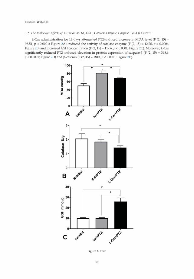
Brain Sci. 2018, 8, 45
3.2. The Molecular Effects of L-Car on MDA, GSH, Catalase Enzyme, Caspase-3 and β-Catenin
L-Car administration for 14 days attenuated PTZ-induced increase in MDA level (F (2, 15) =98.51, p < 0.0001; Figure 2A), reduced the activity of catalase enzyme (F (2, 15) = 12.76, p = 0.0006;Figure 2B) and increased GSH concentration (F (2, 15) = 117.6, p < 0.0001; Figure 2C). Moreover, L-Carsignificantly reduced PTZ-induced elevation in protein expression of caspase-3 (F (2, 15) = 348.6,p < 0.0001; Figure 2D) and β-catenin (F (2, 15) = 1813, p < 0.0001; Figure 2E).
Figure 2. Cont.
60

Brain Sci. 2018, 8, 45
Figure 2. The molecular effects of L-car on oxidants and antioxidants. (A) PTZ-treated animals hadsignificantly (*) higher malondialdehyde (MDA) compared to saline controls (*): mean difference =31.35, 95% CI = 25.52–37.18, p < 0.0001, and L-Car-treated PTZ animals compared to saline controls (*):mean difference = 18.22, 95% CI = 12.39–24.04, p < 0.0001, as well as compared to PTZ-treated animals:mean difference = −13.13, 95% CI = −18.96–−7.307, p < 0.0001, by one-way ANOVA followed byTukey’s multiple comparisons test, six animals per group. (B) Significant difference in catalase enzymeactivity (F2, 15 = 12.76, p = 0.0006) by one-way ANOVA with marked reduction in L-Car-treated animals
61

Brain Sci. 2018, 8, 45
(compared to saline control (*): mean difference = −0.63, 95% CI = −0.97–−0.30, p = 0.0005, andcompared to PTZ-treated animals (*): mean difference = −0.42, 95% CI = −0.75–−0.08, p = 0.013). Nosignificant difference between PTZ-treated animals and saline controls (mean difference = −0.21, 95%CI = −0.55–0.11, p = ns) by Tukey’s multiple comparisons test, six animals per group. (C) Robustincrease in GSH concentration (F2, 15 = 117.6, p < 0.0001) by one-way ANOVA. L-Car-treated animalsshowed a marked increase compared to saline-treated animals (*): mean difference = 15.8, 95% CI= 12.7–18.9, p < 0.0001, and compared to PTZ-treated animals (*): mean difference = 15.8, 95% CI =12.7–18.9, p < 0.0001. No significant difference between PTZ-treated animals and saline controls (meandifference = 0, 95% CI = −0.31–3.1, p = ns) by Tukey’s multiple comparisons test, six animals per group.(D) Significant difference between groups in caspase-3 protein expression (F2, 15 = 348.6, p < 0.0001) byone-way ANOVA. PTZ increased the expression compared to saline controls (*): mean difference =0.72, 95% CI = 0.65–0.80, p < 0.0001. Furthermore, L-Car increased the expression of caspase-3 proteincompared to saline controls (*): mean difference = 0.60, 95% CI = 0.53–0.68, p < 0.0001. However, L-Cartreatment attenuated the PTZ-induced increase in caspase-3 protein expression (*): L-Car + PTZ vs. Sal+ PTZ mean difference = −0.12, 95% CI = −0.197–−0.043, p = 0.002, by Tukey’s multiple comparisonstest, six animals per group. (E) Marked effect of treatment on β-catenin (F2, 15 = 1813, p < 0.0001) byone-way ANOVA. PTZ increased the expression of β-catenin compared to saline control (*): meandifference = 1.28, 95% CI = 1.23–1.34, p < 0.0001. Furthermore, L-Car increased the protein expressionof β-catenin compared to saline controls (*): mean difference = 0.48, 95% CI = 0.43–0.54, p < 0.0001.However, L-Car treatment attenuated the PTZ-induced increase in β-catenin protein expression (*):L-Car + PTZ vs. Sal + PTZ mean difference = −0.80, 95% CI = −0.85–−0.74, p < 0.0001, by Tukey’smultiple comparisons test, six animals per group, (F) Products of Western blot for caspase-3, β-cateninprotein and tubulin (housekeeping gene) protein expression in the Sal group [1], Sal + PTZ group [2]and L-Car + PTZ group [3].
3.3. Effects of L-Car on the Morphology of Neurons in the CA3 Region of Hippocampus
Brain sections from the Sal group (Figure 3A) showed a normal appearance for neurons in theCA3 region of hippocampus, while those obtained from the Sal + PTZ group (Figure 3B) showedirregular arrangement of neurons with a reduction in the number of neurons, and the neurons showedpyknosis (darkly-stained nucleus and cytoplasm). The number of abnormal neurons was significantlyreduced in the brains tissues obtained from the L-Car + PTZ group (Figure 3C).
Figure 3. Cont.
62

Brain Sci. 2018, 8, 45
Figure 3. Histopathological examination of the CA3 region in hippocampus. Normal morphology forcells and adequate number in CA3 in brains obtained from the Sal group (A, H&E, 400×), reducednumber of neurons with pyknotic changes (black arrows) in neurons of CA3 in brains obtained fromthe Sal + PTZ group (B, H&E, 400×) and significant number of neurons with normal morphology inCA3 in brains obtained from the L-Car + PTZ group (C, H&E, 400×).
3.4. Effects of L-Car on Hsp70 and LC3 Expression in the CA3 Region of Hippocampus
Immunohistochemical examination showed a significant decrease in the mean area of interest(AOI) of Hsp70-positive cells in the CA3 region of hippocampus in the Sal + PTZ and L-Car +PTZ groups compared to the Sal group (p < 0.001). On the other hand, the L-Car + PTZ groupshowed a significant increase in the number of Hsp70-positive cells compared to the Sal + PTZ group(p < 0.01) (Figure 4A). Brain sections showed a high expression of Hsp70 in the Sal group (Figure 4B),mild cytoplasmic expression in the Sal + PTZ group (Figure 4C) and high cytoplasmic expression inthe L-Car + PTZ group (Figure 4D).
Figure 4. Cont.
63

Brain Sci. 2018, 8, 45
Figure 4. Mean area of interest of Hsp70 positivity in the CA3 region of hippocampus from differentgroups (A). The HSP70 protein expression mean area of interest was compared between the threegroups (PTZ + Sal, Sal + Sal and PTZ + L-Car using one-way ANOVA. F(2,15)=20.53, p < 0.0001. Tukeymultiple comparisons test was done between the groups. There was a significant difference betweenPTZ + Sal vs. PTZ + L-Car (*) mean difference = −43.67. 95% CI of difference = −67.47 to −19.86, p =0.0007) and also between Sal + Sal vs. PTZ + L-Car (*) mean difference = −55.83. 95% CI of difference =−79.64 to −32.03, p < 0.0001. Section of brain in the CA3 region of hippocampus showing negativeexpression for Hsp70 in the Sal group (B, 200×), moderate cytoplasmic expression in the Sal + PTZgroup (C, 200×) and high cytoplasmic brown staining for Hsp70 in the L-Car + PTZ group (D, 200×).
Furthermore, immunohistochemical examination showed a significant increase in the mean areaof interest (AOI) of LC3-positive cells in the CA3 region of hippocampus in the Sal + PTZ groupcompared to the Sal group (p < 0.001). On the other hand, the L-Car + PTZ group showed a significantdecrease in the LC3 compared to the Sal + PTZ group (p < 0.01) (Figure 5A). Brain sections showednegative expression of LC3 in the Sal group (Figure 5B), high cytoplasmic expression in the Sal + PTZgroup (Figure 5C) and minimal cytoplasmic expression in the L-Car + PTZ group (Figure 5D).
64

Brain Sci. 2018, 8, 45
Figure 5. Mean area of interest of LC3 positivity in the CA3 region of hippocampus from differentgroups (A). LC mean area of interest by one-way ANOVA F(2,15) = 72,703, p < 0.0001. Tukey multiplecomparisons test was done between the groups. There was a significant difference between PTZ + Salvs. PTZ + L-Car (*) mean difference = 750.8. 95% CI of difference = 745.5 to 756.2, p < 0.0001 and alsobetween Sal + Sal vs. PTZ + L-Car (*) mean difference = 160.4. 95% CI of difference = 155 to 165.8,p < 0.0001, and also between PTZ + Sal vs. Sal + Sal (mean difference = 590.4 95% CI of difference = 585to 595.8, p < 0.0001). Section of brain in the CA3 region of hippocampus showing negative expressionfor LC3 in the Sal group (B, 400×), high cytoplasmic expression in the Sal + PTZ group (C, 400×) andlow expression for LC3 in the L-Car + PTZ group (D, 400×).
3.5. Correlations between the Stage of Seizure, Oxidative Stress Markers, Hsp70 Expression and LC3Expression in Hippocampus
Seizure stage showed significant positive correlations between seizure stage and MDA, LC3,caspase-3 and Hsp70 with significant negative correlations with GSH and CAT (p ≤ 0.01). Furthermore,expression of LC3 and Hsp70 showed a significant positive correlation with MDA and a significant
65

Brain Sci. 2018, 8, 45
negative correlation with CAT (p ≤ 0.01). In addition, LC3 expression showed significant positivecorrelation with Hsp70 expression (p < 0.01) (Table 1).
Table 1. Correlation between seizure stage and oxidative stress markers, expression of caspase-3,β-catenin, LC3 and Hsp70 in the PTZ group.
Parameters Stage of Seizure MDA GSH CAT Caspase-3 β-Catenin Hsp70 LC3
Seizure stage r 0.78 −0.74 −0.34 0.65 0.63 −0.61 0.67p 0.001 0.02 0.092 0.005 0.03 0.04 0.011
MDAr −0.811 −0.44 0.88 0.67 −0.74 0.75p 0.01 0.067 0.005 0.01 0.01 0.01
GSHr 0.32 −0.82 0.85 0.67 −0.71p 0.21 0.005 0.001 0.004 0.003
CATr −0.13 −0.32 0.64 −0.67p 0.62 0.21 0.01 0.009
Caspase-3 r 0.68 −0.62 0.68p 0.01 0.04 0.03
β-cateninr 0.63 0.62p 0.01 0.01
Hsp70 r 0.62p 0.011
MDA = malondialdehyde, GSH = reduced glutathione concentration, CAT = catalase activity, Hsp70 = heat shockprotein 70. r, Pearson’s correlation coefficient. p < 0.05 is considered significant.
4. Discussion
The main findings in the present study are: (a) treatment with pentylenetetrazole (PTZ) causedfull kindling of rats, enhanced redox state and upregulated the expression of LC3, Hsp70, caspase-3and β-catenin in the rat hippocampal CA3 region; (b) chronic daily treatment with L-Car (100 mg/kg)caused significant attenuation in seizure stage and duration and reversed the PTZ-induced alterationsin the redox sate and the expression of caspase-3, β-catenin and more upregulation of Hsp 70 expressionin the rat hippocampal CA3 region.
Acetyl L-Car is a well-known potent antioxidant and has several properties that may suggestits antiepileptic effect, such as protection against glutamate toxicity [19], a favorable effect on energyhomeostasis [20–22] and lowering oxidative damage and improving mitochondrial function [23].Moreover, pretreatment with L-Car before administration of a single convulsive dose of PTZ to miceprolonged latency to seizures and reduced the frequency of tonic-clonic seizures in a dose-dependentmanner [24]. On the other hand, Smeland et al. [14] reported in a model of chronic epilepsy(PTZ-kindled epileptic rat) that L-Car failed to slow down the progression and latency to clonicconvulsions. However, they reported that it normalized some metabolic parameters in brain tissuessuch as lactate, (3–13C) alanine, dopamine, myo-inositol and succinate in the cortex.
In the present study, the kindling model (a chronic widely-accepted animal model of epilepsyproduced by chemical or electrical stimuli) is implicated; of the chemical stimuli, pentylenetetrazole(PTZ) (a gamma amino butyric acid (GABA) A receptor antagonist) produces severe convulsions whenadministered to animals by using subconvulsive doses that are applied intermittently and repetitivelyto produce full-blown convulsions [25]. The present study reported a significant increase in seizurescore and duration with significant reduction in latency in the PTZ group. These findings are inline with our previous work from our group [18] and others [26,27]. Furthermore, the present studydemonstrated that treatment with L-Car in the PTZ-kindling model greatly attenuated tonic-clonicconvulsions and the duration of these convulsions, as well as delayed the seizure onset.
It has been demonstrated that reactive oxygen species play a role in the development andprogression of epileptic seizure [18,28]. During convulsive seizure, the low antioxidant defense systemscan predispose the brain to oxidative stress. Furthermore, the hippocampus may be particularlysensitive to oxidative stress because it has low endogenous levels of vitamin E, an important
66

Brain Sci. 2018, 8, 45
biochemical antioxidant, relatively to other brain regions [29]. The present study demonstrated a highoxidative stress state in the CA3 region of the hippocampus of PTZ kindled rats in the form a significantincrease in MDA (marker of lipid peroxidation) and a significant reduction in antioxidants such ascatalase and reduced glutathione (GSH). These findings are in line with previous studies [18,27,30]and suggest that the induction of seizure produces a state of oxidative stress. Furthermore, our resultsprovide good evidence for the role of ROS in the pathophysiology of seizure in this model.
In the present study, co-treatment with L-Car significantly reduced MDA level and increasedGSH and catalase levels relative to the PTZ group. In agreement with these findings, previous studiesreported that L-car attenuated the seizure duration and severity in pilocarpine-induced epilepsythough its antioxidant effects, which involved the suppression of MDA and upregulation of CAT andSOD activities [31]. These findings lend more evidence to the hypothesis that the antiepileptic effect ofL-Car could be partially related to its antioxidant effects.
Autophagy is a regulated process in which the intracellular cellular proteins aggregate anddamaged organelles are degraded by the cell lysosomes [32]. LC3 is considered the most reliablecellular marker for autophagy activation [33]. Therefore, we evaluated in the present study LC3 proteinexpression in the CA3 region of hippocampus by immunohistochemistry. According to our results,little expression of LC3 in the CA3 region in the saline negative control group was demonstrated,while the PTZ group showed marked LC3 expression in brains. Furthermore, there was a positivecorrelation between LC3 expression and seizure stage, suggesting a role for autophagy in PTZ-inducedepilepsy. In agreement with these findings, Shacka et al. [34] showed significant accumulation ofLC3-positive autophagy vacuoles in the hippocampus of kainite-induced epileptic mice. Furthermore,Cao et al. [35] demonstrated the presence of autophagy in pilocarpine-induced status epilepticus (SE)models. In addition, Scherz-Shouval and Elazar [36] and Scherz-Shouval et al. [37] hypothesizedthat reactive oxygen species are the most important activators of autophagy. In the present study,we reported a positive correlation between autophagy and MDA level, which is in line with theprevious hypothesis. Moreover, in the present study, we demonstrated for the first time that L-Carinhibits autophagy in PTZ-induced chronic epilepsy.
The role of apoptosis in epilepsy was demonstrated in many previous studies and in differentanimal models. Sudha et al. [28] and Simonian et al. [38] demonstrated activation of apoptosis inhippocampal regions of KA-induced epilepsy. Moreover, Naseer et al. [39] showed a significantincrease in caspase-3 expression with activation of neuronal apoptosis in PTZ-induced epilepsy inadult rats. In line with these previous studies, we demonstrated a significant increase in caspase-3expression in the CA3 region of hippocampus of the PTZ group when compared to the saline controlgroup. Moreover, treatment with L-Car decreased apoptosis significantly in the hippocampus CA3region compared to the PTZ group. Activation of apoptosis in PTZ-induced epilepsy could be due tooxidative stress and generation of reactive oxygen species, which disrupt mitochondrial membranepotential and activate the mitochondrial pathway for apoptosis [40]. These findings, along with otherssuggest a potential role for apoptosis in epilepsy and possible anti-apoptotic effects for L-Car in theepileptic rat model.
Heat shock proteins (HSPs) are stress-induced proteins that play an important role in cellularresponses to stress [41]. The expression of HSPs has been detected in different types of cells in thenervous system including neuralgia, neurons and endothelial cells [42]. Previous studies examined theexpression of Hsp70 in epilepsy and reported direct relationship between seizure frequency, duration,intensity and Hsp70 expression in both animal models [18,43,44] and human epilepsy [45,46]. It wassuggested that the expression of Hsp70 has a protective role for brain cells during epilepsy. Thepresent study demonstrated significant upregulation in Hsp70 expression in the hippocampal CA3region with positive correlations with the seizure stage. Moreover, we demonstrated that L-Cartreatment caused upregulation in Hsp70 in hippocampus, which was reflected in improvementof behavioral changes. These results suggest that upregulation of Hsp70 might be one of thepotential mechanism of the antiepileptic effects of L-Car. It is plausible to assume that the highly
67

Brain Sci. 2018, 8, 45
expressed Hsp70 could protect neurons against oxidative stress and apoptosis. In accordance withthis assumption, Li et al. [47] and Zhao et al. [48] reported that the highly inducible Hsp70 has aneuroprotective role in hindering apoptosis, possibly through interacting with p53, which elicits theapoptotic process [49,50]. Furthermore, Kanitkara and Bhonde [51] reported that Hsp-70 can reduceoxidative stress in beta cells and increase glucose-induced insulin release. Moreover, Ayala and Tapiademonstrated that induction of Hsp70 protects the hippocampal neurodegeneration via modulatingendogenous glutamate expression [52]. A previous study has shown increased hippocampal CA1glutamate release in PTZ-treated animals that was inhibited by Chai-Long-Ku-Li-Tan, a Chinese herbalmedicine, as a mechanism of its anticonvulsant properties [53]. Examining the effect of L-Car onglutamatergic neurotransmission is highly indicated, however beyond the scope of the current study,and requires further investigation.
In the development of the nervous system, the Wnt-β-catenin signaling pathway plays animportant role in neurogenesis, neural differentiation, synapse development and plasticity [54,55].The Wnt signaling pathway might be involved in a number of CNS disorders such as Alzheimer’sdisease, schizophrenia and mood disorders [56–58]. Regarding epilepsy, the role of the Wnt-β-cateninsignaling pathway showed controversies. Some studies showed upregulation in β-catenin in epilepticanimal models such as pilocarpine-induced status epilepticus [59], electroconvulsive seizures rat [60]and Theihaber et al. [61], who reported upregulation of β-catenin in the hypoxic rat model. Inaddition, Xing et al. [62] demonstrated that β-catenin may not be involved in the developmentof hippocampal sclerosis of mesial temporal lobe epilepsy. On the other hand, other studiesdemonstrated downregulation of β-catenin during epileptic animal models such as the Kainite-inducedrat model [63,64]. Moreover, Campos et al. [65] demonstrated that β-catenin knockout mice have highseizure susceptibility to PTZ. In the present study, we demonstrated upregulation of β-catenin in CA3hippocampal regions in the PTZ group, which could be in response to oxidative stress. Downregulationof β-catenin in the L-Car group compared to the PTZ group supports this hypothesis.
5. Conclusions
The results of the present study confirmed the neuroprotective and antiepileptic action of L-Caragainst PTZ-induced epilepsy. These effects might be explained on the basis of detected L-Car actionssuch as antioxidant activity, anti-apoptotic effects, suppression of autophagy and upregulation ofneuroprotective heat shock protein. However, further knockout experiments will be required to studythe detailed molecular mechanism of L-Car.
Acknowledgments: The authors thank Azza Abdelaziz for helping us with the histopathological examination.
Author Contributions: Abdelaziz M. Hussein: shared in the idea, the induction of epilepsy model, biochemicalanalysis, data analysis and paper writing. Mohamed Adel: shared in the idea, the induction of epilepsy modeland paper writing. Mohamed El-Mesery: Western blotting, biochemical analysis and paper writing Khaled M.Abbas: the induction of epilepsy model and sample collections. Amr N. Ali: the induction of epilepsy model andsample collections. Osama A. Abulseoud: shared in the idea, data analysis and paper writing and editing.
Conflicts of Interest: The authors declare no conflict of interest.
References
1. Forsgren, L.; Beghi, E.; Oun, A.; Sillanpaa, M. The epidemiology of epilepsy in Europe—A systematic review.Eur. J. Neurol. 2005, 12, 245–253. [CrossRef] [PubMed]
2. Hauser, W.A.; Annegers, J.F.; Rocca, W.A. Descriptive epidemiology of epilepsy: Contributions ofpopulation-based studies from Rochester, Minnesota. Mayo Clin. Proc. 1996, 71, 576–586. [CrossRef][PubMed]
3. Eadie, M. Epilepsy-from the Sakikku to hughlings Jackson. J. Clin. Neurosci. 1995, 2, 156–162. [CrossRef]4. Fisher, R.S.; Boas, W.V.; Blume, W.; Elger, C.; Genton, P.; Lee, P.; Engel, J. Epileptic seizures and epilepsy:
Definitions proposed by the International League Against Epilepsy (ILAE) and the International Bureau forEpilepsy (IBE). Epilepsia 2005, 46, 470–472. [CrossRef] [PubMed]
68

Brain Sci. 2018, 8, 45
5. Engel, J., Jr. Introduction to temporal lobe epilepsy. Epilepsy Res. 1996, 26, 141–150. [CrossRef]6. Pitkanen, A.; Lukasiuk, K. Mechanisms of epileptogenesis and potential treatment targets. Lancet Neurol.
2011, 10, 173–186. [CrossRef]7. Cunliffe, V.T.; Baines, R.A.; Giachello, C.N.; Lin, W.H.; Morgan, A.; Reuber, M.; Russell, C.; Walker, M.C.;
Williams, R.S. Epilepsy research methods update: Understanding the causes of epileptic seizures andidentifying new treatments using non-mammalian model organisms. Seizure 2015, 24, 44–51. [CrossRef][PubMed]
8. Tang, F.; Hartz, A.M.S.; Bauer, B. Drug-Resistant Epilepsy: Multiple Hypotheses, Few Answers. Front. Neurol.2017, 8, 301. [CrossRef] [PubMed]
9. Levira, F.; Thurman, D.J.; Sander, J.W.; Hauser, W.A.; Hesdorffer, D.C.; Masanja, H.; Odermatt, P.;Logroscino, G. Premature mortality of epilepsy in low- and middle-income countries: A systematic reviewfrom the Mortality Task Force of the International League Against Epilepsy. Epilepsia 2017, 58, 6–16.[CrossRef] [PubMed]
10. Zaccara, G.; Giannasi, G.; Oggioni, R.; Rosati, E.; Tramacere, L.; Palumbo, P.; Convulsive Status EpilepticusStudy Group of the Uslcentro Toscana, Italy. Challenges in the treatment of convulsive status epilepticus.Seizure 2017, 47, 17–24. [CrossRef] [PubMed]
11. Kwan, P.; Brodie, M.J. Early identification of refractory epilepsy. N. Engl. J. Med. 2000, 342, 314–319.[CrossRef] [PubMed]
12. Bartlett, K.; Eaton, S. Mitochondrial beta-oxidation. Eur. J. Biochem. 2004, 271, 462–469. [CrossRef] [PubMed]13. Kido, Y.; Tamai, I.; Ohnari, A.; Sai, Y.; Kagami, T.; Nezu, J.; Nikaido, H.; Hashimoto, N.; Asano, M.; Tsuji, A.
Functional relevance of carnitine transporter OCTN2 to brain distribution of L carnitine and acetyl-L-carnitineacross the blood-brain barrier. J. Neurochem. 2001, 79, 959–969. [CrossRef] [PubMed]
14. Smeland, O.S.; Meisingset, T.W.; Sonnewald, U. Dietary supplementation with acetyl-L-carnitine improvesbrain energy metabolism in healthy mice and increases noradrenaline and serotonin content. Neurochem. Int.2012, 61, 100–107. [CrossRef] [PubMed]
15. Hansen, S.L.; Nielsen, A.H.; Knudsen, K.E.; Artmann, A.; Petersen, G.; Kristiansen, U.; Hansen, H.S.Ketogenic diet is antiepileptogenic in pentylenetetrazole kindled mice and decrease levels ofN-acylethanolamines in hippocampus. Neurochem. Int. 2009, 54, 199–204. [CrossRef] [PubMed]
16. Couturier, A.; Ringseis, R.; Mooren, F.C.; Kruger, K.; Most, E.; Eder, K. Carnitine supplementation to obeseZucker rats prevents obesity-induced type II to type I muscle fiber transition and favors an oxidativephenotype of skeletal muscle. Nutr. Metab. (Lond.) 2013, 10, 48. [CrossRef] [PubMed]
17. Moloney, T.C.; Dockery, P.; Windebank, A.J.; Barry, F.P.; Howard, L.; Dowd, E. Survival and immunogenicityof mesenchymal stem cells from the green fluorescent protein transgenic rat in the adult rat brain. Neurorehabil.Neural Repair 2010, 24, 645–656. [CrossRef] [PubMed]
18. Hussein, A.M.; Abbas, K.M.; Abuoleseod, O. Effects of Ferulic Acid on Oxidative Stress, Heat Shock Protein70, Connexin 43 and Monoamines in Hippocampus of Pentylenetetrazole-Kindled Rats. J. Physiol. Pharmacol.2017, 97, 579–585. [CrossRef] [PubMed]
19. Forloni, G.; Angeretti, N.; Smiroldo, S. Neuroprotective activity of acetyl-L-carnitine: Studies in vitro.J. Neurosci. Res. 1994, 37, 92–96. [CrossRef] [PubMed]
20. Aureli, T.; Miccheli, A.; Ricciolini, R.; Di Cocco, M.E.; Ramacci, M.T.; Angelucci, L.; Ghirardi, O.; Conti, F.Aging brain: Effect of acetyl-L-carnitine treatment on rat brain energy and phospholipid metabolism. A studyby 31P and 1H NMR spectroscopy. Brain Res. 1990, 526, 108–112. [CrossRef]
21. Aureli, T.; Di Cocco, M.E.; Puccetti, C.; Ricciolini, R.; Scalibastri, M.; Miccheli, A.; Manetti, C.; Conti, F.Acetyl-L-carnitine modulates glucose metabolism and stimulates glycogen synthesis in rat brain. Brain Res.1998, 796, 75–81. [CrossRef]
22. Scafidi, S.; Fiskum, G.; Lindauer, S.L.; Bamford, P.; Shi, D.; Hopkins, I.; McKenna, M.C. Metabolism ofacetyl-L-carnitine for energy and neurotransmitter synthesis in the immature rat brain. J. Neurochem. 2010,114, 820–831. [CrossRef] [PubMed]
23. Liu, J.; Killilea, D.W.; Ames, B.N. Age-associated mitochondrial oxidative decay: Improvement of carnitineacetyltransferase substrate-binding affinity and activity in brain by feeding old rats acetyl-L-carnitine and/orR-alpha-lipoic acid. Proc. Natl. Acad. Sci. USA 2002, 99, 1876–1881. [CrossRef] [PubMed]
24. Yu, Z.; Iryo, Y.; Matsuoka, M.; Igisu, H.; Ikeda, M. Suppression of pentylenetetrazol-induced seizures bycarnitine in mice. Naunyn-Schmiedeberg’s Arch. Pharmacol. 1997, 355, 545–549. [CrossRef]
69
Brain Sci. 2018, 8, 45
25. Dhir, A. Pentylenetetrazol (PTZ) Kindling Model of Epilepsy. Curr. Protoc. Neurosci. 2012, 58, 9–37. [CrossRef]26. Rajabzadeh, A.; Bideskan, A.E.; Fazelm, A.; Sankian, M.; Rafatpanah, H.; Haghir, H. The effect of
PTZ-induced epileptic seizures on hippocampal expression of PSA-NCAM in offspring born to kindled rats.J. Biomed. Sci. 2012, 19, 56. [CrossRef] [PubMed]
27. Saha, L.; Bhandari, S.; Bhatia, A.; Banerjee, D.; Chakrabarti, A. Anti-kindling Effect of Bezafibrate,a Peroxisome Proliferator-activated Receptors Alpha Agonist, in Pentylenetetrazole Induced KindlingSeizure Model. J. Epilepsy Res. 2014, 4, 45–54. [CrossRef] [PubMed]
28. Sudha, K.; Rao, A.V.; Rao, A. Oxidative stress and antioxidants in epilepsy. Clin. Chim. Acta. 2001, 303, 19–24.[CrossRef]
29. Danjo, S.; Ishihara, Y.; Watanabe, M.; Nakamura, Y.; Itoh, K. Pentylenetrazole-induced loss of blood-brainbarrier integrity involves excess nitric oxide generation by neuronal nitric oxide synthase. Brain Res. 2013,1530, 44–53. [CrossRef] [PubMed]
30. Shehata, A.M. Neurophysiological Studies on the Effect of Acetone on Pentylenetetrazole-Induced Seizurein Rats. Bull. Egypt. Soc. Physiol. Sci. 2011, 31, 135–146.
31. Ahmed, M.F.; Mahmoud, M.A. Effect of L-Carnitine on Pilocarpine-Induced Seizures in Rats. J. Am. Sci.2012, 8, 612–618.
32. Zhao, X.; Liu, G.; Shen, H.; Gao, B.; Li, X.; Fu, J.; Zhou, J.; Ji, Q. Liraglutide inhibits autophagy and apoptosisinduced by high glucose through GLP-1R in renal tubular epithelial cells. Int. J. Mol. Med. 2015, 35, 684–692.[CrossRef] [PubMed]
33. Wang, Y.; Han, R.; Liang, Z.Q.; Wu, J.C.; Zhang, X.D.; Gu, Z.L.; Qin, Z.H. An autophagic mechanism isinvolved in apoptotic death of rat striatal neurons induced by the non-N-methyl-Daspartate receptor agonistkainic acid. Autophagy 2008, 4, 214–226. [CrossRef] [PubMed]
34. Shacka, J.J.; Lu, J.; Xie, Z.L.; Uchiyama, Y.; Roth, K.A.; Zhang, J. Kainic acid induces early and transientautophagic stress in mouse hippocampus. Neurosci. Lett. 2007, 414, 57–60. [CrossRef] [PubMed]
35. Cao, L.; Xu, J.; Lin, Y.; Zhao, X.; Liu, X.; Chi, Z. Autophagy is upregulated in rats with status epilepticus andpartly inhibited by vitamin E. Biochem. Biophys. Res. Commun. 2009, 379, 949–953. [CrossRef] [PubMed]
36. Scherz-Shouval, R.; Elazar, Z. ROS, mitochondria and the regulation of autophagy. Trends Cell Biol. 2007, 17,422–427. [CrossRef] [PubMed]
37. Scherz-Shouval, R.; Shvets, E.; Fass, E.; Shorer, H.; Gil, L.; Elazar, Z. Reactive oxygen species are essential forautophagy and specifically regulate the activity of Atg4. EMBO J. 2007, 26, 1749–1760. [CrossRef] [PubMed]
38. Simonian, N.A.; Getz, R.L.; Leveque, J.C.; Konradi, C.; Coyle, J.T. Kainic acid induces apoptosis in neurons.Neuroscience 1996, 75, 1047–1055. [CrossRef]
39. Naseer, M.I.; Ullah, I.; Ullah, N.; Lee, H.Y.; Cheon, E.W.; Chung, J.; Kim, M.O. Neuroprotective effect ofvitamin C against PTZ induced apoptotic neurodegeneration in adult rat brain. Pak. J. Pharm. Sci. 2011, 24,263–268. [PubMed]
40. Green, D.R.; Reed, J.C. Mitochondria and apoptosis. Science 1998, 281, 1309–1312. [CrossRef] [PubMed]41. Soti, C.; Nagy, E.; Giricz, Z.; Vígh, L.; Csermely, P.; Ferdinandy, P. Heat shock proteins as emerging therapeutic
targets. Br. J. Pharmacol. 2005, 146, 769–780. [CrossRef] [PubMed]42. Foster, J.A.; Brown, I.R. Differential induction of heat shock mRNA in oligodendrocytes, microglia, and
astrocytes following hyperthermia. Brain Res. Mol. Brain Res. 1997, 45, 207–218. [CrossRef]43. Gass, P.; Prior, P.; Kiessling, M. Correlation between seizure intensity and stress protein expression after
limbic epilepsy in the rat brain. Neuroscience 1995, 65, 27–36. [CrossRef]44. Zhang, X.; Boulton, A.A.; Yu, P.H. Expression of heat shock protein-70 and limbic seizure-induced neuronal
death in the rat brain. Eur. J. Neurosci. 1996, 8, 1432–1440. [CrossRef] [PubMed]45. Thom, M.; Seetah, S.; Sisodiya, S.; Koepp, M.; Scaravilli, F. Sudden and unexpected death in
epilepsy (SUDEP): Evidence of acute neuronal injury using HSP-70 and c-Jun immunohistochemistry.Neuropathol. Appl. Neurobiol. 2003, 29, 132–143. [CrossRef]
46. Dericioglu, N.; Soylemezoglu, F.; Gursoy-Ozdemir, Y.; Akalan, N.; Saygi, S.; Dalkara, T. Cell death andsurvival mechanisms are concomitantly active in the hippocampus of patients with mesial temporal sclerosis.Neuroscience 2013, 237, 56–65. [CrossRef] [PubMed]
47. Li, W.X.; Chen, C.H.; Ling, C.C.; Li, G.C. Apoptosis in heat induced cell killing: The protective role of hsp-70and the sensitization eVect of the c-myc gene. Radiat. Res. 1996, 145, 324–330. [CrossRef] [PubMed]
70

Brain Sci. 2018, 8, 45
48. Zhao, Z.G.; Ma, Q.Z.; Xu, C.X. Abrogation of heat-shock protein (HSP) 70 expression induced cell growthinhibition and apoptosis in human androgen-independent prostate cancer cell line PC-3m. Asian J. Androl.2004, 6, 319–324. [PubMed]
49. Lee, C.S.; Montebello, J.; Rush, M.; Georgiou, T.; Wawryk, S.; Rode, J. Overexpression of heat shock protein(HSP) 70 associated with abnormal p53 expression in cancer of the pancreas. Zentralbl. Pathol. 1994, 140,259–264. [PubMed]
50. Merrick, B.A.; He, C.; Witcher, L.L.; Patterson, R.M.; Reid, J.J.; Pence-Pawlowski, P.M.; Selkirk, J.K. HSPbinding and mitochondrial localization of p53 protein in human HT1080 and mouse C3H10T1/2 cell lines.Biochim. Biophys. Acta 1996, 1297, 57–68. [CrossRef]
51. Kanitkara, M.; Bhonde, R.R. Curcumin treatment enhances islet recovery by induction of heat shock responseproteins, Hsp70 and heme oxygenase-1, during cryopreservation. Life Sci. 2008, 82, 182–189. [CrossRef][PubMed]
52. Ayala, G.X.; Tapia, R. HSP70 expression protects against hippocampal neurodegeneration induced byendogenous glutamate in vivo. Neuropharmacology 2008, 55, 1383–1390. [CrossRef] [PubMed]
53. Wu, H.M.; Huang, C.C.; Li, L.H.; Tsai, J.J.; Hsu, K.S. The Chinese herbal medicineChai-Hu-Long-Ku-Mu-Li-Tan (TW-001) exerts anticonvulsant effects against different experimentalmodels of seizure in rats. Jpn. J. Pharmacol. 2000, 82, 247–260. [CrossRef] [PubMed]
54. Lie, D.C.; Colamarino, S.A.; Song, H.J.; Desire, L.; Mira, H.; Consiglio, A.; Lein, E.S.; Jessberger, S.;Lansford, H.; Dearie, A.R.; et al. Wnt signalling regulates adult hippocampal neurogenesis. Nature 2005, 437,1370–1375. [CrossRef] [PubMed]
55. Wisniewska, M.B. Physiological role of beta-catenin/TCF signaling in neurons of the adult brain. Neurochem.Res. 2013, 38, 1144–1155. [CrossRef] [PubMed]
56. De Ferrari, G.V.; Papassotiropoulos, A.; Biechele, T.; Wavrant De-Vrieze, F.; Avila, M.E.; Major, M.B.; Myers, A.;Sáez, K.; Henríquez, J.P.; Zhao, A.; et al. Common genetic variation within the low-density lipoproteinreceptor-related protein 6 and late-onset Alzheimer’s disease. Proc. Natl. Acad. Sci. USA 2007, 104, 9434–9439.[CrossRef] [PubMed]
57. Lovestone, S.; Guntert, A.; Hye, A.; Lynham, S.; Thambisetty, M.; Ward, M. Proteomics of Alzheimer’s disease:Understanding mechanisms and seeking biomarkers. Expert Rev. Proteom. 2007, 4, 227–238. [CrossRef][PubMed]
58. Zhang, Y.; Yuan, X.; Wang, Z.; Li, R. The canonical Wnt signaling pathway in autism. CNS Neurol. Disord.Drug Targets 2014, 13, 765–770. [CrossRef] [PubMed]
59. Fasen, K.; Beck, H.; Elger, C.E.; Lie, A.A. Differential regulation of cadherins and catenins during axonalreorganization in the adult rat CNS. J. Neuropathol. Exp. Neurol. 2002, 61, 903–913. [CrossRef] [PubMed]
60. Madsen, T.M.; Newton, S.S.; Eaton, M.E.; Russell, D.S.; Duman, R.S. Chronic electroconvulsive seizureup-regulates betacatenin expression in rat hippocampus: Role in adult neurogenesis. Biol. Psychiatry 2003,54, 1006–1014. [CrossRef]
61. Theilhaber, J.; Rakhade, S.N.; Sudhalter, J.; Kothari, N.; Klein, P.; Pollard, J.; Jensen, F.E. Gene expressionprofiling of a hypoxic seizure model of epilepsy suggests a role for mTOR and Wnt signaling inepileptogenesis. PLoS ONE 2013, 8, e74428. [CrossRef] [PubMed]
62. Xing, X.L.; Sha, L.Z.; Zhang, D.; Shen, Y.; Wu, L.W.; Xu, Q. Role of beta-catenin in the pathogenesis of mesialtemporal lobe epilepsy. Zhongguo Yi Xue Ke Xue Yuan Xue Bao 2011, 33, 659–662. [PubMed]
63. Busceti, C.L.; Biagioni, F.; Aronica, E.; Riozzi, B.; Storto, M.; Battaglia, G.; Giorgi, F.S.; Gradini, R.; Fornai, F.;Caricasole, A.; et al. Induction of the Wnt inhibitor, Dickkopf-1, is associated with neurodegeneration relatedto temporal lobe epilepsy. Epilepsia 2007, 48, 694–705. [CrossRef] [PubMed]
64. Goodenough, S.; Schleusner, D.; Pietrzik, C.; Skutella, T.; Behl, C. Glycogen synthase kinase 3beta linksneuroprotection by 17 beta-estradiol to key Alzheimer processes. Neuroscience 2005, 132, 581–589. [CrossRef][PubMed]
65. Campos, V.E.; Du, M.; Li, Y. Increased seizure susceptibility and cortical malformation in beta-catenin mutantmice. Biochem. Biophys. Res. Commun. 2004, 320, 606–614. [CrossRef] [PubMed]
© 2018 by the authors. Licensee MDPI, Basel, Switzerland. This article is an open accessarticle distributed under the terms and conditions of the Creative Commons Attribution(CC BY) license (http://creativecommons.org/licenses/by/4.0/).
71
brainsciences
Review
Diagnosis and Surgical Treatment ofDrug-Resistant Epilepsy
Chinekwu Anyanwu 1 and Gholam K. Motamedi 2,* ID
1 Department of Neurology, Virginia Tech Carilion School of Medicine, Roanoke, VA 24016, USA;[email protected]
2 Department of Neurology, Georgetown University Medical Center, Washington, DC 20007, USA* Correspondence: [email protected]; Tel.: +1-202-444-4564; Fax: +1-877-245-1499
Received: 24 November 2017; Accepted: 16 March 2018; Published: 21 March 2018
Abstract: Despite appropriate trials of at least two antiepileptic drugs, about a third of patientswith epilepsy remain drug resistant (intractable; refractory). Epilepsy surgery offers a potentialcure or significant improvement to those with focal onset drug-resistant seizures. Unfortunately,epilepsy surgery is still underutilized which might be in part because of the complexity of presurgicalevaluation. This process includes classifying the seizure type, lateralizing and localizing the seizureonset focus (epileptogenic zone), confirming the safety of the prospective brain surgery in terms ofpotential neurocognitive deficits (language and memory functions), before devising a surgical plan.Each one of the above steps requires special tests. In this paper, we have reviewed the process ofpresurgical evaluation in patients with drug-resistant focal onset epilepsy.
Keywords: medically intractable epilepsy; EEG; epilepsy surgery; MRI; epilepsy; seizures
1. Introduction
The majority of patients with epilepsy can become seizure-free with antiepileptic drugs (AEDs) butabout a third will continue to have seizures. However, patients with drug-resistant focal onset epilepsy(the most common type of epilepsy), may become seizure-free, or have significant seizure reduction,through epilepsy surgery. In particular, the efficacy and safety of anterior temporal lobectomy inpatients with temporal lobe epilepsy has been established through randomized controlled trials [1].
Despite the evidence of its superiority to medical therapy in patients with drug-resistant epilepsy(DRE), epilepsy surgery is still underutilized as only a small group of these patients undergo presurgicalevaluation and even a smaller group end up having surgery. It is still a common practice to try almost allavailable AEDs before referring patients for possible epilepsy surgery, while with proper management,diagnosis of DRE can be established within 1–2 years after seizure onset.
Currently, out of about one million patients with DRE in the United States—the majority of whomare suffering from focal onset epilepsy and potential candidates for surgery—less than 1% are referredto epilepsy centers and only 2000 to 3000 undergo surgery. The recommended approach is to referpatients who have failed adequate trials of two AEDs, either as monotherapy or in combination, to anepilepsy center for presurgical evaluation [2–4].
This article will review proper management of patients with DRE, presurgical evaluation,and different surgical treatment options based on the current recommendations by the internationalleague against epilepsy (IALE), and established guidelines.
2. New Classification of Seizure Types
In 2017, the international league against epilepsy (ILAE) released a new classification of seizuretypes. This revision was based on the previous classification that has been in use since 1981. The new
Brain Sci. 2018, 8, 49; doi:10.3390/brainsci8040049 www.mdpi.com/journal/brainsci72
Brain Sci. 2018, 8, 49
classification was developed to reflect the developments in our understanding of the disease sincethen, in particular in brain imaging, electrophysiology, and genetics. The main differences in the newclassification include listing of certain new focal seizure types that used to be under the categoryof generalized seizures, replacing the term consciousness with awareness in description of seizures,and classifying focal seizures based on the first clinical manifestation (except for altered awareness),adding new types of generalized seizures, and adding or changing certain terms to clarity theterminology. In particular, the most common type of epilepsy, which is also potentially treatablethrough surgery, complex partial epilepsy, will be referred to as focal onset epilepsy with loss ofawareness [5,6].
3. Drug-Resistant (Refractory) Epilepsy
It has been shown that 47% of patients with new-onset epilepsy can achieve completeseizure-freedom with the first AED, 13% of the remaining 53% may become seizure-free using asecond agent, and only 4% with a third agent and/or polytherapy [7]. Therefore, failure of twoAEDs, either as monotherapy or combination therapy, would meet the criteria for DRE [8]. However,it should be emphasized that the AEDs must have been tried adequately. An “adequate trial” includes,choosing an appropriate AED for the type of seizure being treated (e.g., failure of carbamazepinein treating idiopathic generalized epilepsy would not be considered a true failure), and titrating upthe dose to the maximum tolerated dose. An AED that triggers allergic reaction or causes significantadverse effects that requires switching to another AED, may not be counted as failure either.
It is also critical to define “seizure-freedom” when assessing the success or failure of an AED.The recommended definition is sustained seizure freedom for a period 3 times the longest inter-seizureinterval, or 1 year, whichever that is longer [8].
4. Presurgical Evaluation
The process referred to as presurgical evaluation is the most critical aspect of epilepsy surgery.All patients who are suspected of having- or properly diagnosed with- drug-resistant epilepsy shouldbe referred to an epilepsy center for presurgical evaluation. During this process, the patient will befurther evaluated to confirm the diagnosis and to classify the seizure type.
This step is critical since close to half of patients with epilepsy have idiopathic generalizedseizures which cannot be treated with resective epilepsy surgery. Only focal onset epilepsy isamenable to surgery. Therefore, only those with DRE who have focal seizures with loss of awareness(formerly, complex partial seizures), with or without secondary generalization, are surgical candidatesand can complete the process of presurgical evaluation.
It is also crucial to confirm that the patient has true epilepsy and not an epilepsy mimic sincepatients may seem refractory for presumed seizures but may have non-epileptic spells such as syncopeor psychogenic non-epileptic seizures (PNES). Therefore, the initial evaluation includes obtainingproper history, seizure semiology, and electroencephalographic (EEG) findings are role in this process.
The ultimate goal of presurgical evaluation is to identify the epileptogenic zone (region), and toestablish the safety of a prospective brain surgery, so that the resection can be performed with minimalfunctional impairment while achieving seizure freedom, if possible.
Identification of seizure focus involves extensive workup including seizure characterization,lateralization, and localization through detailed evaluation of history, video-EEG (VEEG)monitoring using scalp EEG electrodes or—if indicated—via surgically implanted intracranialelectrodes, different neuro-imaging modalities, neuropsychological testing, and if indicated,intracarotid Amobarbital injection procedure (IAP; Wada test).
The fundamental goal in presurgical workup includes defining the epileptogenic zone(epileptogenic region) which is the area sufficient for generation of seizures, and the minimum amountof cortex that must be resected (inactivated or completely disconnected) to produce seizure freedom.A variety of diagnostic tools should be utilized in evaluation of the various cortical zones. They include
73
Brain Sci. 2018, 8, 49
seizure semiology, VEEG recording, neuro functional testing and neuroimaging techniques with agoal to define the cortical zones involved in seizure generation and propagation. Other cortical zonesof interest besides the epileptogenic zone include seizure-onset zone (cortical region that initiatesclinical seizures), symptomatogenic zone (where initial ictal semiology is produced), irritative zone(where interictal spikes are generated), and functional deficit zone (cortical area that functionsabnormally during interictal period) [9,10].
While the ultimate tool in detecting the epileptogenic zone is the VEEG findings (more accuratelyvia direct intracranial recording), it can be significantly facilitated by high resolution brainmagnetic resonance imaging (MRI) (structural integrity), diffusion tensor imaging (cellular integrity),magnetic resonance spectroscopy (metabolite and biochemical data), or through physiological imagingmodalities such as positron emission tomography (PET) and single-photon emission computerizedtomography (SPECT) scans (glucose or other ligands’ metabolism, and cerebral blood flow,respectively). The irritative zone (area that generates interictal epileptiform activities) can be detectedby EEG, magnetoencephalography (MEG) and special functional MRI techniques. The seizure-onsetzone (area of cortex that initiates clinical seizures) can be measured by scalp/intracranial EEG recordingand ictal SPECT.
The symptomatogenic zone (area of cortex which produces the initial ictal symptoms or signs)can be determined by the seizure semiology. The functional deficit zones are measured withneuropsychological examination and functional imaging studies at the baseline (interictal period).
Despite the availability of multiple diagnostic modalities, different cases may require differentdiagnostic tests therefore, the evaluations process should be tailored to the individual. In particular,since a major classification in epilepsy is differentiating temporal lobe epilepsy (TLE) fromextra-temporal epilepsy, it is crucial to make that determination during the presurgical evaluation.This distinction is based on the critical role of temporal lobe in neurocognitive functioning, its uniqueanatomy, and the clinical evidence regarding its responsiveness to surgery.
In particular, TLE with mesial temporal sclerosis (MTS) may not require extensive diagnostic workup as it can be confirmed with concordant ipsilateral anterior-mid temporal epileptiform dischargeswithout the need for intracranial recording. However, some patients may require intracranial (subdural)monitoring for seizure focus lateralization and/or accurate localization as well as functional corticalmapping. In general, about half of patients with TLE and almost all extra-temporal cases requireintracranial EEG recording for both purposes of pinpointing the seizure onset focus and/or corticalmapping to delineate eloquent cortices for safety [11].
It should be noted that while patients who fail two appropriate trials of AEDs should be considereddrug-resistant and should undergo presurgical evaluation, they may benefit from adjustments to theircurrent AED regimens as seen appropriate. Therefore, starting the process of presurgical evaluationdoes not preclude using other AEDs, especially given the available variety of AEDs with differentmechanisms of action and side effect profiles.
4.1. Scalp Video-EEG Monitoring
The standard procedure after confirming that the patient has refractory epilepsy usually startswith VEEG monitoring to further characterize, lateralize and possibly localize the seizure focus.Localization of the seizure onset zone through scalp EEG may be limited as it detects epileptiformactivity that synchronizes at least 6 cm2 of the cortex [12]. Deeper seizure foci may not be detectedvia surface (scalp) electrodes. Further, scalp EEG recording is usually insufficient in extra-temporalepilepsy or even in non-lesional (normal MRI) TLE. Using extra electrodes placed based on the 10-10international electrode system may add significant accuracy to scalp recording and in some cases avoidintracranial recording (Figure 1).
74

Brain Sci. 2018, 8, 49
Figure 1. Right temporal lobe seizure onset in a patient with right hippocampal sclerosis and temporallobe epilepsy (TLE). The EEG shows buildup of rhythmic repetitive sharp theta discharges arising inthe right anterior-mid temporal regions. Scalp recording during long-term monitoring using extraelectrodes placed according to the 10-10 electrode system for clear distinction between frontal andbasal-lateral temporal head regions.
When seizure onset zone is poorly lateralized because of alternating seizure onset lateralization,bitemporal asynchrony, or frequent bilateral epileptiform discharges, it indicates a less favorablepostsurgical seizure outcome [13]. Unilateral hippocampal atrophy on MRI and concordant unilateralinterictal spikes are highly predictive of concordant ictal localization [14].
Patients are often admitted for several days, depending on their seizure frequency. Most oftentheir AEDs are tapered off in order to capture about 3–5 typical and consistent seizures. There isno consensus on tapering process but in general very rapid tapering is avoided to minimize risk ofStatus Epilepticus or triggering aberrant seizure onset zones. Upon the completion of scalp VEEGmonitoring, some patients may not qualify for surgery; this includes patients with primary generalizedseizures and those with clear multifocal seizures, although some in the latter group may qualify forintracranial recording for further elaboration. When scalp EEG cannot adequately localize the seizurefocus, particularly in patients with normal brain MRI, intracranial EEG monitoring would be justified.
4.2. Brain Imaging Techniques
Advances in brain imaging technology have substantially improved seizure localization andsurgical outcome [12]. Neuroimaging studies are not routinely indicated in all types of epilepsy.For example, patients with idiopathic (primary) generalized epilepsy syndromes such as absenceepilepsy and Juvenile myoclonic epilepsy diagnosed clinically and through EEG typically do notrequire imaging [13]. All patients with clinical and EEG evidence of focal onset epilepsy should havebrain imaging which includes brain MRI.
4.2.1. MRI (Magnetic Resonance Imaging)
The principal role of MRI is to define structural abnormalities that may be the cause of seizuredisorder. A high resolution brain MRI, usually referred to as Epilepsy Protocol MRI, is recommendedas the modality of choice for all patients presenting with symptomatic focal epilepsies, with or withoutgeneralization. Detailed MRI sequences may be added to increase diagnostic yield depending on
75

Brain Sci. 2018, 8, 49
the etiology. Common sequences used by most epilepsy centers include thin-section (1 mm) coronaloblique T1 gradient echo, coronal oblique T2 series, high-resolution 3D sequences (sensitive to subtlecortical dysplasias or small tumors), and T2 FLAIR (fluid attenuated inversion recovery) images,performed on 3 Tesla, or higher, MRI systems [14].
Temporal lobe epilepsy is the most common type of focal epilepsy and the most common MRIfinding in these patients is MTS, although this pathology may not be present at the time of seizure onsetand may take many years to develop. The main radiological findings in MTS include hippocampalatrophy, internal structural derangement, and T2 hyperintensity as best seen on coronal T2 flair images(Figure 2). Other cortical epileptogenic lesions include focal cortical dysplasia (FCD), tubers, gliosis,infectious process (e.g., neurocysticercosis, abscesses), benign and malignant tumors, and inflammatoryprocesses (Rasmussen’s encephalitis). Not all lesions are epileptogenic and patients may present withdual pathologies. In MRI-negative neocortical epilepsies, the most common pathology is FCD type 1and 2. Type 2 FCDs are preferentially located at the bottom of the sulcus with high-resolution MRImaking it possible to visually identify such in an increasing number [15,16].
Figure 2. Right mesial temporal sclerosis (MTS). There is atrophy and increased signal intensityin the CA1-3 regions of the right hippocampus. Coronal view; fluid-attenuated inversion recovery(FLAIR) sequence.
4.2.2. SPECT Scan (Single-Photon Emission Computerized Tomography)
Structural lesions do not always correlate with clinical semiology and EEG findings but theregional cerebral blood flow is useful tool to localize these seizures. Ictal SPECT is more sensitive andspecific in temporal lobe epilepsies given that the seizures are longer. The strength of SPECT is theability to obtain images related to regional cerebral blood flow (rCBF) at the time of seizures.
Interictal or ictal SPECT have been used to localize the ictal onset and ictal propagation patternsand add to the evidence of abnormalities in the involved site [15,17]. Meta-analytic sensitivitiesof SPECT in patients with TLE have been reported as 44% (interictally), 75% (postictally) and 97%(ictally) [18] as opposed to 66% (ictally), and 40% (interictally) in extra-temporal seizures [19,20].
Ictal SPECT studies appear to correlate with outcome when there is close concordance betweenthe area of hyperperfusion and the resected area and vice versa with surgical failure when there is poorconcordance. Both ictal and interictal SPECT showing a change from hypoperfusion (interictal period)to hyperperfusion (ictal period) is more reliable than an abnormality in either stage alone. However,it has been shown that the sensitivity of ictal SPECT is higher than that of interictal SPECT because of
76
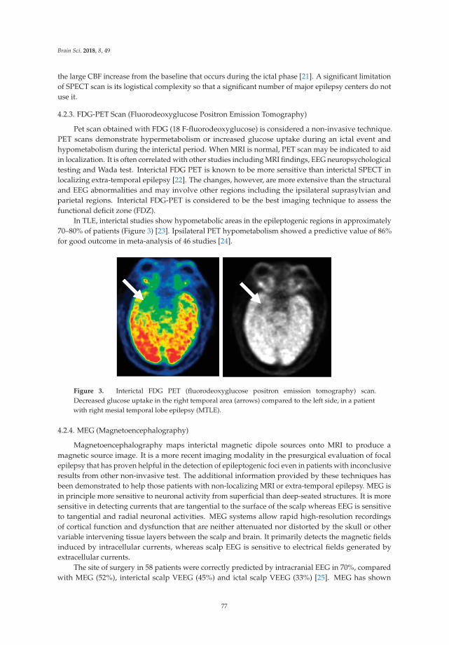
Brain Sci. 2018, 8, 49
the large CBF increase from the baseline that occurs during the ictal phase [21]. A significant limitationof SPECT scan is its logistical complexity so that a significant number of major epilepsy centers do notuse it.
4.2.3. FDG-PET Scan (Fluorodeoxyglucose Positron Emission Tomography)
Pet scan obtained with FDG (18 F-fluorodeoxyglucose) is considered a non-invasive technique.PET scans demonstrate hypermetabolism or increased glucose uptake during an ictal event andhypometabolism during the interictal period. When MRI is normal, PET scan may be indicated to aidin localization. It is often correlated with other studies including MRI findings, EEG neuropsychologicaltesting and Wada test. Interictal FDG PET is known to be more sensitive than interictal SPECT inlocalizing extra-temporal epilepsy [22]. The changes, however, are more extensive than the structuraland EEG abnormalities and may involve other regions including the ipsilateral suprasylvian andparietal regions. Interictal FDG-PET is considered to be the best imaging technique to assess thefunctional deficit zone (FDZ).
In TLE, interictal studies show hypometabolic areas in the epileptogenic regions in approximately70–80% of patients (Figure 3) [23]. Ipsilateral PET hypometabolism showed a predictive value of 86%for good outcome in meta-analysis of 46 studies [24].
Figure 3. Interictal FDG PET (fluorodeoxyglucose positron emission tomography) scan.Decreased glucose uptake in the right temporal area (arrows) compared to the left side, in a patientwith right mesial temporal lobe epilepsy (MTLE).
4.2.4. MEG (Magnetoencephalography)
Magnetoencephalography maps interictal magnetic dipole sources onto MRI to produce amagnetic source image. It is a more recent imaging modality in the presurgical evaluation of focalepilepsy that has proven helpful in the detection of epileptogenic foci even in patients with inconclusiveresults from other non-invasive test. The additional information provided by these techniques hasbeen demonstrated to help those patients with non-localizing MRI or extra-temporal epilepsy. MEG isin principle more sensitive to neuronal activity from superficial than deep-seated structures. It is moresensitive in detecting currents that are tangential to the surface of the scalp whereas EEG is sensitiveto tangential and radial neuronal activities. MEG systems allow rapid high-resolution recordingsof cortical function and dysfunction that are neither attenuated nor distorted by the skull or othervariable intervening tissue layers between the scalp and brain. It primarily detects the magnetic fieldsinduced by intracellular currents, whereas scalp EEG is sensitive to electrical fields generated byextracellular currents.
The site of surgery in 58 patients were correctly predicted by intracranial EEG in 70%, comparedwith MEG (52%), interictal scalp VEEG (45%) and ictal scalp VEEG (33%) [25]. MEG has shown
77
Brain Sci. 2018, 8, 49
equal effectiveness compared to EEG in terms of recording epileptic spikes with a tendency to registerepileptic spikes in more patients [26].
4.2.5. fMRI (Functional MRI)
While fMRI has not been established as a valid test to replace Wada memory test [27], it may be apotential complement to standard neuropsychological test to predict postoperative verbal memoryoutcome. It has been suggested [28,29] that it may be used to determine language dominance prior totemporal lobectomy [30–33]. Overall, besides the evidence indicating its utility as a replacement ofWada language test [34,35], to date, the replacement of the Wada memory test has proved to be moredifficult [33,36,37].
The concordance between fMRI and Wada language lateralization has been shown in a numberof studies reporting concordance. In one such study in 100 patients with different focal epilepsies,there was 91% concordance between the two tests. The overall rate of false categorization by fMRI was9%, ranging from 3% in left-sided TLE to 25% in left-sided extra-temporal epilepsy [38].
4.2.6. Intracarotid Amobarbital Injection Procedure (IAP, Wada Test)
A main focus of the presurgical evaluation is to estimate the risk of language- and verbal memorydecline in patients undergoing anterior temporal lobectomy (ATL) on the language dominant side(usually, the left). Intracarotid Amobarbital injection procedure, commonly referred to as Wada test(after Juhn Atsushi Wada), is considered the gold standard for pre-operative lateralization of languagedominance, and a measure of memory function [39]. The Wada test is also effective in predictingseizure control and degree of verbal memory decline post operatively.
Wada test is indicated in patients who may undergo temporal lobectomy. However, despite thehigh accuracy of the Wada test in lateralizing language and memory function, this test is associatedwith false negatives and false positives and needs to be performed by experienced practitioners [40].In good hands, the test is considered reasonably safe with potential minor and major complicationsthat have been reported to be rare, but in some reports as high as 1.09% [41]. Despite the risk, Wada testclearly remains useful in patients without clear language lateralization or with suspected atypicallanguage lateralization, but more critically in patients at risk of postsurgical memory loss [42].
In some cases the validity of the test has been questioned [43]. Prior to the procedure, cerebralangiography is used to assess the vasculature and extent of cross-over flow to contralateral arteries.There are different anatomical variations that lead to unreliable results. EEG will be continuouslyrecorded throughout the procedure. During the procedure, amobarbital (100–150 mg) is injectedinto each carotid artery to produce, one at a time, to induce a temporary disruption in function inthe ipsilateral temporal lobe while the patient gets involved in a series of different language andmemory related tasks. Other agents including, propofol, methohexital and etomidate have also beenused [44–46].
Upon injection of the language dominant side, there will be global aphasia, while the patientwould have only dysarthria after injecting the non-dominant temporal lobe. The more delicate functionof Wada test is to determine the “adequacy” of the contralateral hippocampus, while shedding lighton the “reserve” capacity of the ipsilateral side (seizure side). To be able to undergo anterior temporallobectomy, ideally the patient is required to correctly identify at least 3 more objects out of a total of8 target objects, after injecting the ipsilateral side (“split” score of 3). This would indicate enoughadequacy on the contralateral hippocampus for a reasonable function ipsilateral temporal lobectomy.A suggested partial alternative to Wada test is fMRI for language lateralization however, there are nostandardized methods to assess memory using fMRI.
Currently, the test is performed in a subset of patients who appear to be at risk for clinicallysignificant memory loss. Therefore, some patients with TLE, including those with left-sided TLE,may not need Wada test, depending on results of VEEG, MRI, fMRI, neuropsychological and PET scanresults. Patients with left-sided TLE and MTS, ipsilateral hypometabolism on PET scan, low baseline
78
Brain Sci. 2018, 8, 49
performance in verbal memory and naming on neuropsych testing, and left language dominanceon fMRI, may not undergo the test. However, those with nonlesional brain MRI, normal PET scan,and normal baseline verbal memory and naming on neuropsych testing may benefit from Wadatest [27,47].
4.2.7. Neuropsychological Testing
While Wada test may not be required in all patients undergoing presurgical evaluationcases, the non-invasive outpatient neuropsychological testing should be performed in all cases.It establishes a baseline neurocognitive profile that can be used for postoperative comparison, andalso helps with seizure focus lateralization, localization, and postoperative outcome prediction [48–50].Neuropsychological testing evaluates a variety of neurocognitive features particularly, but not limitedto, in patients with TLE. These include verbal memory, visual memory, verbal fluency, and otheraspects of cognitive performance. For example, a typical patient with dominant (usually left-sided)epilepsy would have significant language and memory related deficits such as verbal memory andword finding difficulties.
On the other hand, the same type of seizure originating in the non-language dominant side wouldtypically result in visual memory deficits. Therefore, it is crucial to both establish a baseline, and also tocorroborate the results of prior work up since neuropsychological test results may further confirm—orargue against—the assumed localization and lateralization of the epileptogenic zone. The testingincludes evaluation of pre-morbid intellectual function, memory function, higher executive function,language function and visuo-spartial function.
Memory and language testing contribute to the assessment of the functional integrity ofthe contralateral lobe. When testing suggests severe memory and language deficiencies in thecontralateral hemisphere, the risk of developing postoperative memory and language impairmentis high. Memory disturbances have consistently been found in patients with temporal lobe seizuresbecause of the relationship between mesial temporal structures and memory processes [51–55].
5. Intracranial EEG Recording (Intracranial Electroencephalography; iEEG)
In an increasing number of patients EEG data recorded through scalp electrodes fails to clearlylateralize or localize the seizure focus. This mandates, recording the EEG directly from the brainthrough surgically implanted subdural surface electrodes and/or depth electrodes inserted into thebrain tissue. This group of patients include those with non-lesional brain MRI, unclear seizureonset lateralization, uncertain seizure onset localization, possible bilateral or multifocal seizuredisorder, suspected dual pathology, discordant noninvasive data (such as interictal and ictal EEG,structural or functional neuroimaging and neuropsychological testing or EEG findings), and seizureslikely originating in eloquent cortex requiring accurate localization and cortical mapping prior to apossible resection surgery.
Implanting recording electrodes, either as depth- or grids and strips, comes with its ownchallenges. The location and number of hardware should be decided on an individual basis. In general,it seems safer to use minimal required hardware, the same principle that applies to AEDs. However,such general principles cannot dictate the exact surgical plan and therefore the decision-making processshould be individualized in every case [56,57].
The use of intracranial EEG to guide surgical resections in some patients with TLE is controversial,particularly in those with presumed unilateral mesial temporal lobe foci [58]. Most patients withunilateral hippocampal atrophy with concordant scalp EEG or functional imaging studies haveexcellent outcomes after standard antero-medial temporal resections without intracranial monitoringwith continued postoperative seizures seen in about 30% of patients [59] and some of these may bedue to a dual pathology [60]. Patients with bilateral temporal lobe seizure onsets are known to havepoorer surgical outcome than those with unilateral foci [61].
79
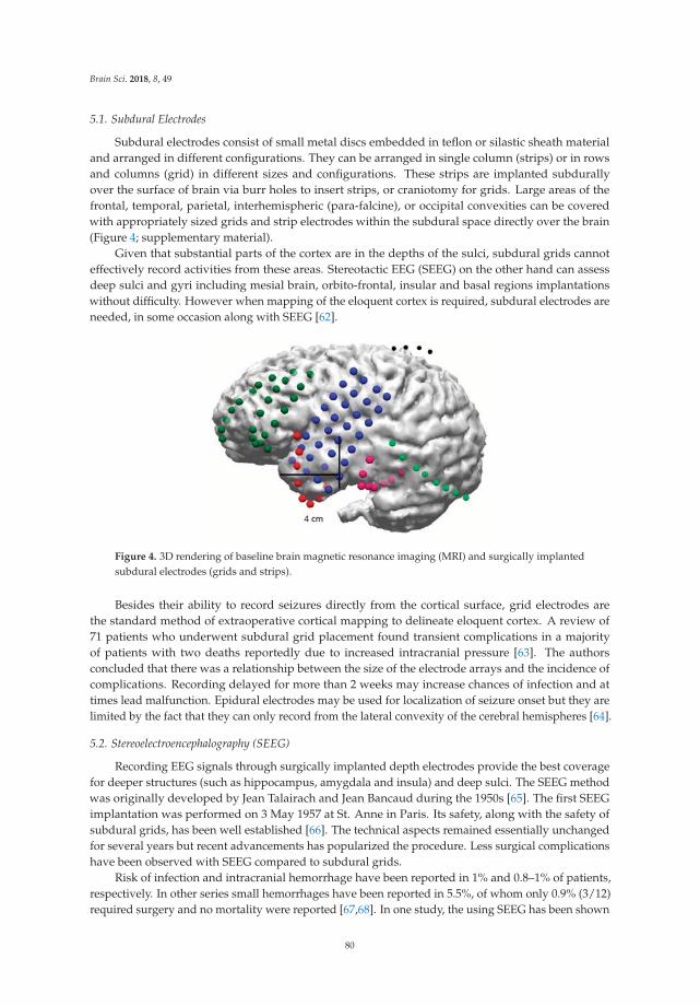
Brain Sci. 2018, 8, 49
5.1. Subdural Electrodes
Subdural electrodes consist of small metal discs embedded in teflon or silastic sheath materialand arranged in different configurations. They can be arranged in single column (strips) or in rowsand columns (grid) in different sizes and configurations. These strips are implanted subdurallyover the surface of brain via burr holes to insert strips, or craniotomy for grids. Large areas of thefrontal, temporal, parietal, interhemispheric (para-falcine), or occipital convexities can be coveredwith appropriately sized grids and strip electrodes within the subdural space directly over the brain(Figure 4; supplementary material).
Given that substantial parts of the cortex are in the depths of the sulci, subdural grids cannoteffectively record activities from these areas. Stereotactic EEG (SEEG) on the other hand can assessdeep sulci and gyri including mesial brain, orbito-frontal, insular and basal regions implantationswithout difficulty. However when mapping of the eloquent cortex is required, subdural electrodes areneeded, in some occasion along with SEEG [62].
Figure 4. 3D rendering of baseline brain magnetic resonance imaging (MRI) and surgically implantedsubdural electrodes (grids and strips).
Besides their ability to record seizures directly from the cortical surface, grid electrodes arethe standard method of extraoperative cortical mapping to delineate eloquent cortex. A review of71 patients who underwent subdural grid placement found transient complications in a majorityof patients with two deaths reportedly due to increased intracranial pressure [63]. The authorsconcluded that there was a relationship between the size of the electrode arrays and the incidence ofcomplications. Recording delayed for more than 2 weeks may increase chances of infection and attimes lead malfunction. Epidural electrodes may be used for localization of seizure onset but they arelimited by the fact that they can only record from the lateral convexity of the cerebral hemispheres [64].
5.2. Stereoelectroencephalography (SEEG)
Recording EEG signals through surgically implanted depth electrodes provide the best coveragefor deeper structures (such as hippocampus, amygdala and insula) and deep sulci. The SEEG methodwas originally developed by Jean Talairach and Jean Bancaud during the 1950s [65]. The first SEEGimplantation was performed on 3 May 1957 at St. Anne in Paris. Its safety, along with the safety ofsubdural grids, has been well established [66]. The technical aspects remained essentially unchangedfor several years but recent advancements has popularized the procedure. Less surgical complicationshave been observed with SEEG compared to subdural grids.
Risk of infection and intracranial hemorrhage have been reported in 1% and 0.8–1% of patients,respectively. In other series small hemorrhages have been reported in 5.5%, of whom only 0.9% (3/12)required surgery and no mortality were reported [67,68]. In one study, the using SEEG has been shown
80

Brain Sci. 2018, 8, 49
to confirm the epileptogenic zone in 154 patients (77.0%) with 134 patients undergoing SEEG guidedresection of whom 61 patients who were followed up remained seizure free at 12 months [69,70].
The planning of SEEG implantation requires formulating precise hypotheses about the possibleepileptogenic zone, seizure onset and propagation zones to be tested. It is also important to understandthe functional networks involved in the primary organization of the epileptic activity while formulatinga hypothesis. The trajectory passes through several target points including deep sulci, different lobesand gyri orthogonally or obliquely considering different cortical cytoarchitectural areas involved inseizure patterns. For example, in a suspected frontal lobe epilepsy case, a trajectory may pass throughprimary motor cortex, supplementary motor area, frontal eye field, and deep sulci.
Depth electrodes in various lengths and number of contacts are implanted using conventionalstereotactic technique or by the assistance of stereotactic robotic devices through 2.5 mm diameterdrill holes.
5.3. Intracranial Seizure Lateralization and Localization
After implanting the intracranial electrodes, the patients are monitored in the epilepsy monitoringunit for 1–2 weeks or longer in some centers. The duration of iEEG recording is determined by seizurefrequency, number of seizures needed to make a decision, and the time needed to perform mapping ofthe eloquent cortex.
Interpretation of the data is based on EEG pattern recognition as well as clinical semiology(symptomatology). An accurate interpretation of intracranial ictal EEG requires appropriate electrodecoverage of potential epileptogenic areas. A key point is to have placed the iEEG electrodes in properlocations i.e., at—or in close vicinity to—the epileptogenic zone and areas around it. Besides the morecommon ictal EEG patterns such as buildup of rhythmic repetitive sharp or spike discharges (Figure 5),there is increasing evidence that high frequency oscillations (HFOs) that may be detected visually atseizure onset, represent the epileptogenic zone. Interpretation may be difficult in some cases sincefrequency range differs from one cerebral area to another because of neurophysiological properties ofthe structure and from one etiology to another [71].
Figure 5. Ictal onset EEG recording through two 1 × 8 subdural strips placed over the basal temporalregion in a patient with left temporal lobe epilepsy (TLE). Buildup of rhythmic sharp discharges initiatedat a few electrodes (red circles) before rapid propagation to other electrodes and generalization.
81

Brain Sci. 2018, 8, 49
Bursts of HFOs may also be recorded during interictal periods and may be a marker ofepileptogenicity if the same bursts will develop into sustained fast activity at seizure onset.Sometimes ictal baseline direct current (DC) shifts may precede the fast ictal discharges [72].
A specific interictal pattern in FCD is the occurrence of fast discharges combined with almostcontinuous interictal spiking in the same region [73]. In localization of seizure onset, the most reliablepart is the first clear synchronizing electrical change seen in a limited number of electrodes [74] thatprecedes the onset of clinical semiology. Mesial temporal lobe seizures may also begin as low-voltage,high-frequency discharge without preictal spiking [75].
5.4. Electrical Stimulation (Cortical) Mapping
The purpose of cortical mapping is to delineate the borders of eloquent cortex. This procedure isperformed in patients whose seizure onset zone is located in the vicinity of language or motor cortices.The purpose is to avoid removing such cortical areas hence functional deficits. An alternating polaritysquare wave pulse pair stimulation (0.3 msec in duration, at 50–100 Hz, lasting 3–5 s) is applied to apair of electrodes located in areas near the border of presumed eloquent cortex (Figure 6). Caution mustbe taken when stimulating cortex as a seizure may be provoked particularly when stimulating highlyepileptogenic cortical regions such as primary motor cortex and mesial temporal structure. A provokedseizure is usually heralded by after discharges (Figure 7). [76].
Figure 6. Stimulation cortical mapping. Electrodes 20 and 21, located in the prerolandic motor cortex,are stimulated based on a particular paradigm including slow titration of current density. The patientwill be observed for any motor activity in the corresponding body part (face and hand) while engagedin a conversation or reading out loud.
Figure 7. During stimulation mapping the pulse stimulus (LS, localization stimulus) may induceafterdischarges (AD). In some cases, a brief pulse stimulus (BPS) may successfully terminate the ADand prevent a clinical seizure.
82

Brain Sci. 2018, 8, 49
6. Epilepsy Surgery
Following the completion of presurgical evaluation and localization of the epileptogenic zoneas well as establishing the safety of a resection surgery in the individual patient, the proper surgicaloption can be decided.
Depending on the seizure type, seizure localization, presence of absence of an identifiablepathology on brain imaging (lesional vs. nonlesional cases, respectively), and patient’s functionalbaseline, different surgical approaches and methods can be used. These include potentially curativesurgeries i.e., ATL (using different variations in technique), and resection of the epileptogeniczone (often extra-temporal), or hemispherectomy and palliative surgical approaches such ascorpus callosotomy.
Given the higher prevalence of temporal lobe epilepsy and its distinct features i.e., its critical rolein basic cognitive functioning and its unique anatomy, epilepsy surgery is commonly categorized anddiscussed as two separate categories of temporal- vs. extra-temporal.
Temporal lobe epilepsy surgery most often is performed as anterior temporal lobectomy andincludes the removal of anterior and mesial structures which naturally raises concerns aboutpostoperative language and memory deficits [1]. Therefore, as discussed earlier, specific diagnostictests have to be performed to address those concerns before a safe surgical method can be devised.
Extratemporal epilepsy surgery on the other hand, involves the removal of an identified seizurefocus as long it does not overlap with eloquent cortex. Generally, well delineated lesions such ascavernous angioma, and FCD are more amenable to resection surgery with better outcome in lesionalcases of extratemporal epilepsy than nonlesional cases [77].
In general, a complete resection of the epileptogenic brain region provides higher chances ofseizure freedom but the risks of postoperative deficits would increase with the extension of resection.Therefore, the extent of resection should be weighed against such risks and individualized in every case.
While traditionally the mainstay of surgical treatment has been open brain resection, recentinnovations have allowed for less invasive ablative techniques such as MRI-guided laser interstitialthermotherapy (LITT). A brief review of the different technical approaches and methods follows.
6.1. Surgery of Temporal Lobe Epilepsy
Anterior temporal lobectomy (ATL) is the most commonly performed surgery for the treatmentof refractory temporal lobe epilepsy. Most patients who undergo ATL have concordant findings withMRI, VEEG, PET scan, and Neuropsychological testing. This procedure is performed using a varietyof methods. The standard (en bloc) resection includes 3–6 cm of anterior temporal neocortex and1–3 cm of mesial structures (amygdala and hippocampus) in the resection, with more limited resectionsperformed on the language dominant side.
Selective amygdalo-hippocampectomy (SAH), is another modified surgical method that wasdeveloped to avoid the resection of lateral (neocortical) temporal tissue to minimize language deficits.The SAH is performed by accessing temporal horn through an incision in the middle temporalgyrus and selectively removing mesial temporal structures leaving the neocortical region intact [78].Approximately 70% of properly selected surgical candidates become seizure-free following ATL,and the majority of remaining group benefit significantly by achieving seizure reduction and improvedquality of life (QOL) [79,80].
6.2. Laser Ablation Surgery
Another method of operation used in temporal lobe as well as extra-temporal epilepsy is a lessinvasive procedure that can ablate epileptogenic focus that avoids craniotomy. Laser ablation surgeryhas been effectively applied in lesional and non-lesional cases including MTS, FCD, failed prior opensurgery, and on deeper lesions that are inaccessible to open surgery.
83

Brain Sci. 2018, 8, 49
This procedure has the advantage of selectively targeting small lesions responsible for seizures,with less pain and shorter hospitalization. The goal of an MRIgLITT (MRI-guided laser interstitialthermal therapy) system is to necrotize soft tissues through interstitial irradiation or thermal therapyunder MRI guidance. Various pathologies have been treated with this technique including MTS,hypothalamic hamartoma, cavernous angioma, malformations of cortical development, and tuberoussclerotic lesions [81,82].
Mesial temporal epilepsy is also suitable for MRIgLITT. A history of prior laser ablative surgerywould not preclude further open resection or repeat ablation procedure. Its safety and accuracy,in patients with difficult-to-localize seizures has been shown [83], and its efficacy has been reportedas comparable to that of open surgery but with less morbidity [84,85] including in older patients [86].Evidence suggests that selective surgical approaches for mesial temporal lobe epilepsy lower cognitiverisks compared to the standard ATL. [87,88].
6.3. Corpus Callosotomy
Corpus collosotomy is a palliative surgical procedure devised to alleviate debilitating seizures inindividuals with no focally resectable lesion [89]. Disconnection of the corpus callosum decreases theseverity and frequency of rapidly propagating secondarily generalized focal seizures in patientswho are not otherwise good surgical candidates particularly those with drop attacks (tonic oratonic seizures).
6.4. Multiple Subpial Transection Procedure
This technique introduced by Morrell in 1989 is primarily used for treatment of refractoryepilepsies where resection is risky such as cases of seizure foci in the vicinity of, or with overlapping,eloquent cortex. The technique leaves the vertical columnar arrangement of the cortex intact therebypreserving function while preventing horizontal propagation of the seizure discharge. The procedurehas been shown to be of value in cases of epileptogenic foci involving an eloquent cortex but lesseffective than excision surgery [90].
6.5. Hemispherectomy
Hemispherectomy (FH) is used mainly in children with refractory focal epilepsies confined to onehemisphere but also rarely in adults. Two main surgical techniques include anatomic hemispherectomy(which aims to resect all cortical tissue on one hemisphere along with various amounts of subcorticaltissue), and functional hemispherectomy (which consist of more disconnection and less resection thanthe anatomic hemispherectomy.
Hemispherectomy is most commonly used in patients with perinatal strokes, Sturge-Webersyndrome, Rasmussen encephalitis, Infantile Hemiplegia Seizure Syndrome (IHSS), andhemimegalencephaly. Surgical candidates are selected based on a number of factors includingseverity of the underlying epilepsy, neurological condition, natural history of the underlying disease,and patient’s age. Serious late post-op complications including superficial cerebral hemosiderosisand hydrocephalus can occur although modifications of the procedure have decreased thesecomplications [91,92].
7. Non-Resective Surgical Treatments
Patients who are determined to be unqualified for seizure focus removal via resective surgery,such as those with multifocal seizures and those with overlapping seizure onset zone and eloquentcortex, may benefit from other surgical options. Attempts to control seizures through electricalstimulation started in 1960s have resulted in a few available treatment modalities starting withvagus nerve stimulation (VNS) in 1980s. Currently, there are two such procedures approved in theUnited States and one other approved elsewhere. These surgically implanted modalities do not requireremoval of brain tissue.
84

Brain Sci. 2018, 8, 49
7.1. Vagus Nerve Stimulation (VNS)
This is an adjunctive therapy approved by the Food and Drug Administration (FDA) for refractoryfocal onset epilepsies with or without secondary generalization in patients 4 years or older. The VNSsystem is comprised of a battery generator programmed to deliver intermittent electrical stimuli via theleft vagus nerve to the brain. The generator is implanted in the left upper chest and is tunneled underthe skin to the vagus nerve. A retrospective review of efficacy of VNS in 48 patients with intractablepartial epilepsy showed progressive decrease in mean seizure frequency by 26% after 1 year, 30% after5 years, and 52% after 12 years [93].
7.2. Responsive Neurostimulation (RNS)
The RNS system was approved by the FDA in 2013 for medically refractory focal epilepsy.It provides cortical stimulation in response to ictal discharges detected by the RNS device. This isa programmable neurostimulator which is cranially implanted and connected to one or two depthand/or subdural cortical strip electrodes placed over the seizure foci. The Neurostimulator continuallysenses electrocorticographic activity and when detects onset of an ictal discharges, provides briefpulses of stimulation in response (closed-loop system).
The pivotal randomized study of 191 subjects showed a progressive reduction in the frequency oftotal disabling seizures in the treatment group (41.5%) compared to the sham group (9.4%) in the finalmonths of the 2 year study [94].
7.3. Stimulation of the Anterior Nucleus of Thalamus (Deep Brain Stimulation, DBS)
In a multicenter trial, that evaluated stimulation of bilateral anterior nuclei of the thalamus,the treatment group had 29% greater reduction in seizures compared to the control group in the firstmonth, and at least 50% seizure reduction in 54% of patients on 2 years follow up [95]. Stimulation ofthe anterior nucleus of thalamus has been approved as adjunctive treatment for drug-resistant focalepilepsy in adults, in the European Union since 2010, but its approval in the by FDA for use in theUnited States is still pending.
8. Postsurgical Outcome
Postsurgical outcome in epilepsy surgery is mainly assessed based on seizure-freedom orseizure reduction, neurocognitive functioning, and change in overall QOL. More detailed elementsin measuring outcome include factors such as work eligibility, driving, etc. Of particular concern,are language and verbal memory function following temporal lobe surgeries.
Interestingly, despite the evidence supporting the efficacy and safety of epilepsy surgery, until2001 there was no such evidence based on randomized, controlled trials (RCT). The first RCT included80 patients with TLE randomly assigned to surgery (ATL), or continuing medical therapy, for one yearto compare both seizure control outcome, and quality of life (QOL), disability, and mortality.
The study showed, for the first time, that after one year follow up, 58% of patients in the ATLgroup were seizure- free compared to only 8% in the medical therapy group (p < 0.001). Further,those in the surgical group had less frequent focal seizures and significantly better quality of lifecompared to the non-surgical group (p < 0.001 for both comparisons). In the surgical group 4 patients(10%) had mild language and memory deficits against one dead in the non-surgical group [1].
Since postsurgical outcome significantly depends on seizure type and seizure onset location,different types of epilepsies and surgical approaches are to be studied separately. A summary of a fewselective outcome studies, including particular surgical methods used, follows.
85
Brain Sci. 2018, 8, 49
8.1. Mesial Temporal Lobe Epilepsy with Hippocampal Sclerosis
8.1.1. Standard (en Block) Anterior Temporal Lobectomy (ATL)
Patients with MTLE and MTS can achieve long-term seizure control following standard ATLhowever the risk of seizure recurrence after ≥2 years exists. A review of 50 consecutive cases of ATLin patients with MTLE and MTS reported 82% seizure-freedom rate at 12 months, 76% at 24 months,and 64% at 63 months (mean follow up 5.8 years). The improvement in seizure control was associatedwith improved long-term QOL. The study also indicated reduction in AED as the major risk factor forseizure recurrence [96].
A more recent study of a similar group of patients undergoing standard ATL and followed formean 6.7 years found 89% seizure-freedom, and significant improvement—defined as Engel ClassI or II in 94%. Complete seizure freedom was seen in 103 patients (89%), and Engel Class I or IIoutcome (free of disabling seizures, or rare disabling seizures, respectively) in 94%. This study alsodetermined concordance rates between the final resection site and the following factors: VEEG (100%),PET (100%), MRI (99.0%), and Wada test (90.4%). The lowest concordance was found with SPECT(84.6%), and neuropsychological testing (82.5%). Interestingly, a strong Wada memory lateralizationwas associated with excellent long-term seizure control, and vice versa, i.e., low disparity in scoresbetween the sites predicted continued seizures [97].
8.1.2. Selective Amygdalohippocampectomy (SAH)
Patients with MTLE and MTS who undergo this technique may also achieve similar outcomescomparable to ATL with more extensive (neocortical temporal) resections [98]. While SAH wasoriginally introduced as a method that could minimize neurocognitive deficits by preserving theneocortical temporal cortex, the controversy about its outcome involves neuropsychological deficitsrather than seizure-freedom rates, as compared to standard ATL. While there is some evidence of SAHresulting in better cognitive function than ATL [99]. SAH performed in patients without adequatememory reserve can result in verbal memory deficits in dominant temporal lobe cases [100]. This topichas been reviewed by Schramm who concluded that there was considerable evidence of improvedneuropsychological outcomes in SAH [101].
9. Neurocognitive Deficits Following ATL
At the presurgical baseline, patients with epilepsy, in particular those with TLE, often have somedegree of cognitive impairment which may be further affected by surgery. It has been shown thatlarger temporal lobe resections provide better seizure control outcome however, they are more likelyto induce cognitive deficits since larger resection risks including more of the functional tissue [102].
To further quantify association between the extent of temporal lobe resection and cognitiveoutcome a group of 47 right-handed patients with left MTLE who underwent ATL was studied.Cognitive changes were compared between limited resection (1–2 cm for mesial, and ≤4 cm forneocortical), and more extensive resection (>2 cm for mesial, and ≥4 cm for neocortical). There wereno differences in cognitive outcome between the groups however, a negative correlation was foundwith age at seizure onset [103].
10. Conclusions
Drug-resistant epilepsy, defined as failure of two anti-seizure medications to completely controlseizures, can be effectively treated with surgery. Therefore, proper use of diagnostic methodsto determine a patient’s eligibility for surgical treatment i.e., presurgical evaluation, is of utmostimportance in managing patients with DRE. This process, as discussed in this article, also determineswhat type of surgical approach would be the safest and the most beneficial approach to the particularpatient. With further developments in diagnostic and therapeutic methods, both presurgical evaluationand the surgical methods used, will improve constantly.
86
Brain Sci. 2018, 8, 49
Supplementary Materials: The following are available online at http://www.mdpi.com/2076-3425/8/4/49/s1,Animated 3D reconstruction of subdural grid, strip, and depth electrodes.
Conflicts of Interest: The authors declare no conflicts of interest.
References
1. Wiebe, S.; Blume, W.T.; Girvin, J.P.; Eliasziw, M.; Effectiveness and Efficiency of Surgery for Temporal LobeEpilepsy Study Group. A randomized, controlled trial of surgery for temporal lobe epilepsy. N. Engl. J. Med.2001, 345, 311–318. [CrossRef] [PubMed]
2. Engel, J., Jr. Why is there still doubt to cut it out? Epilepsy Curr. 2013, 13, 198–204. [CrossRef] [PubMed]3. Engel, J. Another Good Reason to Consider Surgical Treatment for Epilepsy More Often and Sooner.
Arch. Neurol. 2011, 68, 707–708. [CrossRef] [PubMed]4. Tellez-Zenteno, J.F.; Dhar, R.; Wiebe, S. Long-term seizure outcomes following epilepsy surgery: A systematic
review and meta-analysis. Brain 2005, 128, 1188–1198. [CrossRef] [PubMed]5. Scheffer, I.E.; Berkovic, S.; Capovilla, G.; Connolly, M.B.; Frence, J.; Guihoto, L.; Hirsch, E.; Jain, S.;
Mathern, G.W.; Moshe, S.L.; et al. ILAE classification of the epilepsies: Position paper of the ILAECommission for Classification and Terminology. Epilepsia 2017, 58, 512–521. [CrossRef] [PubMed]
6. Fisher, R.S.; Cross, J.H.; D’Souza, C.; French, J.A.; Haut, S.R.; Hiqurashi, N.; Hisch, E.; Jansen, F.E.; Laqae, L.;Moshe, S.L.; et al. Instruction manual for the ILAE 2017 operational classification of seizure types. Epilepsia2017, 58, 531–542. [CrossRef] [PubMed]
7. Kwan, P.; Brodie, M.J. Early Identification of Refractory Epilepsy. N. Engl. J. Med. 2000, 342, 314–319.[CrossRef] [PubMed]
8. Kwan, P.; Arzimanoglou, A.; Berg, A.T.; Brodie, M.J.; Hauser, A.W.; Mathern, G.; Moshé, S.L.; Perucca, E.;Wiebe, S.; French, J. Definition of drug resistant epilepsy: Consensus proposal by the ad hoc Task Force ofthe ILAE Commission on Therapeutic Strategies. Epilepsia 2010, 51, 1069–1077. [CrossRef] [PubMed]
9. Lüders, H.O.; Najm, I.; Nair, D.; Widdess-Walsh, P.; Bingman, W. The epileptogenic zone: General principles.Epileptic Disord. 2006, 8 (Suppl. 2), S1–S9. [PubMed]
10. Carreño, M.; Lüders, H.O. General Principles of Presurgical Evaluation. In Epilepsy Surgery, 2nd ed.;Lüders, H.O., Comair, Y.G., Eds.; Lippincott Williams & Wilkins: Philadelphia, PA, USA, 2001; pp. 185–200.
11. Spencer, S.S.; Sperling, M.R.; Shewmon, D.A. Intracranial electrodes. In Epilepsy: A Comprehensive Textbook;Engel, J., Pedley, T.A., Eds.; Lippincott-Raven: Philadelphia, PA, USA, 1997; pp. 1719–1747.
12. Cooper, R.; Winter, A.L.; Crow, H.J.; Grey Walter, W. Comparison of subcortical, cortical and scalp activityusing chronically indwelling electrodes in man. Electroencephalogr. Clin. Neurophysiol. 1964. [CrossRef]
13. Schulz, R.; Lüders, H.O.; Hoppe, M.; Tuxhorn, I.; May, T.; Ebner, A. Interictal EEG and ictal scalp EEGpropagation are highly predictive of surgical outcome in mesial temporal lobe epilepsy. Epilepsia 2000, 41,564–570. [CrossRef] [PubMed]
14. Pataraia, E.; Lurger, S.; Serles, W.; Lindinger, G.; Aull, S.; Leutmezer, F.; Bacher, J.; Olbrich, A.; Czech, T.;Novak, K.; et al. Ictal scalp EEG in unilateral mesial temporal lobe epilepsy. Epilepsia 1998, 39, 608–614.[CrossRef] [PubMed]
15. Kuzniecky, R.I. Neuroimaging of Epilepsy: Therapeutic Implications. NeuroRx 2005, 2, 384–393. [CrossRef][PubMed]
16. Boxerman, J.L.; Hawash, K.; Bali, B.; Clarke, T.; Rogg, J.; Pal, D.K. Is Rolandic Epilepsy Associated withAbnormal Findings On Cranial MRI? Epilepsy Res. 2007, 75, 180–185. [CrossRef] [PubMed]
17. Spencer, D. MRI (Minimum Recommended Imaging) in Epilepsy. Epilepsy Curr. 2014, 14, 261–263. [CrossRef][PubMed]
18. Bernasconi, A. Magnetic resonance imaging in intractable epilepsy: Focus on structural image analysis.In Advances in Neurology; Blume, W., Carlen, P.L., Wiebe, S., Young, A.B., Padjen, A., Starrveld, E., Eds.;Lippincott-Williams: Philadelphia, PA, USA, 2005; pp. 273–278.
19. Chassoux, F.; Landre, E.; Mellerio, C.; Turak, B.; Mann, M.W.; Daumas-Duport, C.; Chiron, C.; Devaux, B.Type II focal cortical dysplasia: Electroclinical phenotype and surgical outcome related to imaging. Epilepsia2012, 53, 349–358. [CrossRef] [PubMed]
20. Adams, C.; Hwang, P.A.; Gilday, D.L.; Armstrong, D.C.; Becker, L.E.; Hoffman, H.J. Comparison of SPECT,EEG, CT, MRI, and pathology in partial epilepsy. Pediatr. Neurol. 1992, 8, 97–103. [CrossRef]
87

Brain Sci. 2018, 8, 49
21. Devous, M.D., Sr.; Thisted, R.A.; Morgan, G.F.; Leroy, R.F.; Rowe, C.C. SPECT brain imaging in epilepsy:A meta-analysis. J. Nucl. Med. 1998, 39, 285–293. [PubMed]
22. Spencer, S.S. The relative contributions of MRI SPECT and PET imaging in epilepsy. Epilepsia 1994, 35,S72–S89. [CrossRef] [PubMed]
23. Weil, S.; Noachtar, S.; Arnold, S.; Yousry, T.A.; Winkler, P.A.; Tatsch, K. Ictal ECD-SPECT differentiatesbetween temporal and extratemporal epilepsy: Confirmation by excellent postoperative seizure control.Nucl. Med. Commun. 2001, 22, 233–237. [CrossRef] [PubMed]
24. Ingvar, D.H. Epilepsy related to cerebral blood flow and metabolism. Acta Psychiatr. Scand. Suppl. 1984, 313,21–26. [CrossRef] [PubMed]
25. Sunhee, K.; Mountz, J.M. Review Article: SPECT Imaging of Epilepsy: An Overview and Comparison withF-18 FDG PET. Int. J. Mol. Imaging 2011, 2011, 813028. [CrossRef]
26. Engel, J.J.; Brown, W.J.; Kuhl, D.E.; Phelps, M.E.; Mazziotta, J.C.; Crandall, P.H. Pathological findingsunderlying focal temporal lobe hypometabolism in partial epilepsy. Ann. Neurol. 1982, 12, 518–528.[CrossRef] [PubMed]
27. Meadow, K.J.; Loring, D.W. The Wada test for language and memory lateralization. Neurology 2005, 65, 659.28. Willmann, O.; Wennberg, R.; May, T.; Woermann, F.G.; Pohlmann-Eden, B. The contribution of 18F-FDG
PET in preoperative epilepsy surgery evaluation for patients with temporal lobe epilepsy A meta-analysis.Seizure 2007, 16, 509–520. [CrossRef] [PubMed]
29. Wheless, J.W.; Willmore, L.J.; Breier, J.I.; Kataki, M.; Smith, J.R.; King, D.W.; Meador, K.J.; Park, Y.D.;Loring, D.W.; Clifton, G.L.; et al. A comparison of magnetoencephalography, MRI and V-EEG in patientsevaluated for epilepsy surgery. Epilepsia 1999, 40, 931–941. [CrossRef] [PubMed]
30. Heers, M.; Rampp, S.; Kaltenhäuser, M.; Pauli, E.; Rauch, C.; Dölken, M.T.; Stefan, H. Detection of epilepticspikes by magnetoencephalography and electroencephalography after sleep deprivation. Seizure 2010, 19,397–403. [CrossRef] [PubMed]
31. Abou-Khalil, B. An update on determination of language dominance in screening for epilepsy surgery:The Wada test and newer noninvasive alternatives. Epilepsia 2007, 48, 442–455. [CrossRef] [PubMed]
32. Klöppel, S.; Büchel, C. Alternatives to the Wada test: A critical view of functional magnetic resonanceimaging in preoperative use. Curr. Opin. Neurol. 2005, 18, 418–423. [CrossRef] [PubMed]
33. Binder, J.R.; Swanson, S.J.; Hammeke, T.A.; Morris, G.L.; Mueller, W.M.; Fischer, M.; Benbadis, S.; Frost, J.A.;Rao, S.M.; Hauqhton, V.M. Determination of language dominance using functional MRI: A comparison withthe Wada test. Neurology 1996, 46, 978–984. [CrossRef] [PubMed]
34. Powell, H.W.; Duncan, J.S. Functional magnetic resonance imaging for assessment of language and memoryin clinical practice. Curr. Opin. Neurol. 2005, 18, 161–166. [CrossRef] [PubMed]
35. Sabsevitz, D.S.; Swanson, S.J.; Hammeke, T.A.; Spanaki, M.V.; Possing, E.T.; Morris, G.L.; Mueller, W.M.;Binder, J.R. Use of preoperative functional neuroimaging to predict language deficits from epilepsy surgery.Neurology 2003, 60, 1788–1792. [CrossRef] [PubMed]
36. Binder, J.R.; Sabsevitz, D.S.; Swanson, S.J.; Hammeke, T.A.; Raghavan, M.; Mueller, W.M. Use of preoperativefunctional MRI to predict verbal memory decline after temporal lobe epilepsy surgery. Epilepsia 2008, 49,1377–1394. [CrossRef] [PubMed]
37. Lehéricy, S.; Cohen, L.; Bazin, B.; Samson, S.; Giacomini, E.; Rougetet, R.; Hertz-Pannier, L.; Le Bihan, D.;Marsault, C.; Baulac, M. Functional MR evaluation of temporal and frontal language dominance comparedwith the Wada test. Neurology 2000, 54, 1625–1633. [CrossRef] [PubMed]
38. Dupont, S. Can functional MRI replace the Wada test? Neurochirurgie 2008, 54, 208–211. [CrossRef] [PubMed]39. Constable, R.T.; Carpentier, A.; Pugh, K.; Westerveld, M.; Oszunar, Y.; Spencer, D.D. Investigation of
the hippocampal formation using a randomized event-related paradigm and z-shimmed functional MRI.Neuroimage 2000, 12, 55–62. [CrossRef] [PubMed]
40. Binder, J.R. Functional MRI is a valid noninvasive alternative to Wada testing. Epilepsy Behav. 2011, 20,214–222. [CrossRef] [PubMed]
41. Woermann, F.G.; Jokeit, H.; Luerding, R.; Freitag, H.; Schulz, R.; Guertler, S.; Okujava, M.; Wolf, P.; Tuxhorn, I.;Ebner, A. Language lateralization by Wada test and fMRI in 100 patients with epilepsy. Neurology 2003, 61,699–701. [CrossRef] [PubMed]
42. Wada, J.A. A new method for determination of the side of cerebral dominance: A preliminary report on theintra-carotid injection of sodium amytal in man. Igaku Seibutsugaku 1949, 14, 221–222.
88

Brain Sci. 2018, 8, 49
43. Simkins-Bullock, J. Beyond speech lateralization: A review of the variability, reliability, and validityof the intracarotid amobarbital procedure and its nonlanguage uses in epilepsy surgery candidates.Neuropsychol. Rev. 2000, 10, 41–74. [CrossRef] [PubMed]
44. Haag, A.; Knake, S.; Hamer, H.M.; Boesebeck, F.; Freitag, H.; Schulz, R.; Baum, P.; Helmstaedter, C.;Wellmer, J.; Urbach, H.; et al. The Wada test in Austrian, Dutch, German, and Swiss epilepsy centers from2000 to 2005: A review of 1421 procedures. Epilepsy Behav. 2008, 13, 83–89. [CrossRef] [PubMed]
45. Bauer, P.R.; Reitsma, J.B.; Houweling, B.M.; Ferrier, C.H.; Ramsey, N.F. Can fMRI safely replace the Wada testfor preoperative assessment of language lateralisation? A meta-analysis and systematic review. J. Neurol.Neurosurg. Psychiatry 2014, 85, 581–588. [CrossRef] [PubMed]
46. Dade, L.A.; Jones-Gotman, M. Sodium amobarbital memory tests: What do they predict? Brain Cogn. 1997,33, 189–209. [CrossRef] [PubMed]
47. Loring, D.W.; Bowden, S.C.; Lee, G.P.; Meadow, K.J. Diagnostic utility of WADA memory Asymmetries:Sensitivity, specificity and likelihood ratio characterization. Neuropsychology 2009, 23, 687–693. [CrossRef][PubMed]
48. Curot, J.; Denuelle, M.; Busigny, T.; Barragan-Jason, G.; Kany, M.; Tall, P.; Marlat, F.; Fabre, N.; Valton, L.Bilateral Wada test: Amobarbital or propofol? Seizure 2014, 23, 122–128. [CrossRef] [PubMed]
49. Möddel, G.; Lineweaver, T.; Schuele, S.U.; Reinholz, J.; Loddenkemper, T. Atypical language lateralization inepilepsy patients. Epilepsia 2009, 50, 1505–1516. [CrossRef] [PubMed]
50. Gilmore, R.L.; Heilman, K.M.; Schmidt, R.P.; Fennell, E.M.; Quisling, R. Anosognosia during Wada testing.Neurology 1992, 42, 925–927. [CrossRef] [PubMed]
51. Andelman, F.; Fried, I.; Neufeld, M.Y. Quality of life self-assessment as a function of lateralization of lesionin candidates for epilepsy surgery. Epilepsia 2001, 42, 549–555. [CrossRef] [PubMed]
52. Andelman, F.; Neufeld, M.Y.; Fried, L. Contribution of neuropsychology to epilepsy surgery. Isr. J. PsychiatryRelat. Sci. 2004, 41, 125–132. [PubMed]
53. Jones-Gotman, M.; Smith, M.L.; Zatorre, R.J. Neuropsychological testing for localizing and lateralizaingthe epileptogenic region. In Surgical Treatment of the Epilepsies, 2nd ed.; Engel, J., Jr., Ed.; Raven Press, Ltd.:New York, NY, USA, 1993; pp. 245–261.
54. Raspall, T.; Donate, M.; Boget, T.; Carreno, M.; Donaire, A.; Agudo, R.; Bargallo, N.; Rumia, J.; Setoain, X.;Pintor, L.; et al. Neuropsychological tests with lateralizing value in patients with temporal lobe epilepsy:Reconsidering material-specific theory. Seizure 2005, 14, 569–576. [CrossRef] [PubMed]
55. Squire, L.R.; Zola, S.M. Structure and function of declarative and non-declarative memory systems. Proc. Natl.Acad. Sci. USA 1996, 93, 13515–13522. [CrossRef] [PubMed]
56. Jenssen, S.; Liporace, J.; Nei, M.; O′Connor, M.J.; Sperling, M.R. Value of non-invasive testing when there areindependent bitemporal seizures in the scalp EEG. Epilepsy Res. 2006, 68, 115–122. [CrossRef] [PubMed]
57. Spencer, S.S.; Nguyen, D.K.; Duckrow, R.B. Invasive EEG in presurgical evaluation of epilepsy. In TheTreatment of Epilepsy, 3rd ed.; Shoruon, S., Perucca, E., Engel, J., Jr., Eds.; Wiley Blackwell: West Sussex, UK,2009; pp. 767–798.
58. Schiller, Y.; Cascino, G.D.; Sharbrough, F.W. Chronic intracranial EEG monitoring for localizing theepileptogenic zone: An electroclinical correlation. Epilepsia 1998, 39, 1302–1308. [CrossRef] [PubMed]
59. Engel, J.; Van Ness, P.C.; Rasmussen, T.B.; Ojemann, L.M. Outcome with respect to epileptic seizures.In Surgical Treatment of the Epilepsies; Engel, J., Jr., Ed.; Raven Press: New York, NY, USA, 1993; pp. 609–621.
60. Fisher, R.S.; Webber, W.R.; Lesser, R.P.; Arroyo, S.; Uamatsu, S. High-frequency EEG activity at the start ofseizures. Clin. Neurophysiol. 1992, 9, 441–448.
61. Gloor, P. Preoperative electroencephalographic investigation in temporal lobe epilepsy: Extracranial andintracranial recordings. Can. J. Neurol. Sci. 1991, 18, 554–558. [CrossRef] [PubMed]
62. Kaiboriboon, K.; Malkhachroum, A.M.; Zrik, A.; Daif, A.; Schiltz, N.M.; Labiner, D.M.; Lhatoo, S.D. Epilepsysurgery in the United States: Analysis of data from the national association of epilepsy centers. Epilepsy Res.2015, 116, 105–109. [CrossRef] [PubMed]
63. Wong, C.H.; Birkett, J.; Byth, K.; Dexter, M.; Somerville, E.; Gill, D.; Chaseling, R.; Fearnside, M.; Bleasel, A.Risk factors for complications during intracranial electrode recording in presurgical evaluation of drugresistant partial epilepsy. Acta Neurochir. (Wien) 2009, 151, 37–50. [CrossRef] [PubMed]
64. Barnett, G.H.; Burgess, R.C.; Awad, I.A.; Skipper, G.J.; Edwards, C.R.; Luders, H. Epidural peg electrodes forthe presurgical evaluation of intractable epilepsy. Neurosurgery 1990, 27, 113–115. [CrossRef] [PubMed]
89
Brain Sci. 2018, 8, 49
65. Bancaud, J.; Dell, M.B. Technics and method of stereotaxic functional exploration of the brain structures inman (cortex, subcortex, central gray nuclei). Rev. Neurol. 1959, 101, 213–227. (In French) [PubMed]
66. Jeha, L.E.; Najm, I.; Bingaman, W.; Dinner, D.; Widdess-Walsh, P.; Luders, H. Surgical outcome and prognosticfactors of frontal lobe epilepsy surgery. Brain 2007, 130, 574–584. [CrossRef] [PubMed]
67. Cossu, M.; Chabardes, S.; Hoffmann, D.; Lo Russo, G. Presurgical evaluation of intractable epilepsy usingstereo-electro-encephalography methodology: Principles, technique and morbidity. Neurochirurgie 2008, 54,367–373. (In French) [CrossRef] [PubMed]
68. Tanriverdi, T.; Ajlan, A.; Poulin, N.; Olivier, A. Morbidity in epilepsy surgery: An experience based on2449 epilepsy surgery procedures from a single institution. J. Neurosurg. 2009, 110, 1111–1123. [CrossRef][PubMed]
69. Guenot, M.; Isnard, J. Epilepsy and insula. Neurochirurgie 2008, 54, 374–381. [CrossRef] [PubMed]70. Gonzalez-Martinez, J. The Stereo-Electroencephalography: The Epileptogenic Zone. J. Clin. Neurophysiol.
2016, 33, 522–529. [CrossRef] [PubMed]71. Bulacio, J.C.; Chauvel, P.; McGonigal, A. Stereoelectroencephalography: Interpretation. J. Clin. Neurophysiol.
2016, 33, 503–510. [CrossRef] [PubMed]72. Wu, S.; Kunhi Veedu, H.P.; Lhatoo, S.D.; Kouberssi, M.Z.; Miller, J.P.; Luders, H.O. Role of ictal baseline
shifts and ictal high-frequency oscillations in stereo-electroencephalography analysis of mesial temporallobe seizures. Epilepsia 2014, 55, 690–698. [CrossRef] [PubMed]
73. Chassoux, F.; Devaux, B.; Landré, E.; Turak, B.; Nataf, F.; Varlet, P.; Chodkiewicz, J.P.; Daumas-Duport, C.Stereoelectroencephalography in focal cortical dysplasia A 3D approach to delineating the dysplastic cortex.Brain 2000, 123, 1733–1751. [CrossRef] [PubMed]
74. Chabardès, S.; Kahane, P.; Minotti, L.; Tassi, L.; Grand, S.; Hoffmann, D.; Benabid, A.L. The temporopolarcortex plays a pivotal role in temporal lobe seizures. Brain 2005, 128, 1818–1831. [CrossRef] [PubMed]
75. Townsend, J.B.; Engel, J. Clinicopathological correlations of low voltage fast and high amplitude spike andwave medial temporal sterioencephalographic ictal onsets. Epilepsia 1991, 32, 21.
76. Motamedi, G.K.; Lesser, R.P.; Miglioretti, D.L.; Mizuno-Matsumoto, Y.; Gordon, B.; Webber, W.R.;Jackson, D.C.; Sepkuty, J.P.; Crone, N.E. Optimizing parameters for terminating cortical 1446 afterdischargeswith pulse stimulation. Epilepsia 2002, 43, 836–846. [CrossRef] [PubMed]
77. Englot, D.J.; Wang, D.D.; Rolston, J.D.; Shih, T.T.; Chang, E.F. Rates and predictors of long-term seizurefreedom after frontal lobe epilepsy surgery: A systematic review and meta-analysis. J. Neurosurg. 2012, 116,1042–1048. [CrossRef] [PubMed]
78. Olivier, A.; Boling, W.W.; Tanriverdi, T. Techniques in Epilepsy Surgery: The MNI Approach; CambridgeUniversity Press: Cambridge, UK, 2012.
79. Engel, J., Jr. Surgery for seizures. N. Engl. J. Med. 1996, 334, 647–657. [CrossRef] [PubMed]80. Spencer, D.D.; Spencer, S.S.; Mattson, R.H.; Williamson, P.D.; Novelly, R.A. Access to the posterior medial
temporal lobe structure in surgical treatment of temporal lobe epilepsy. Neurosurgery 1984, 15, 667–671.[CrossRef] [PubMed]
81. Gross, R.E.; Willie, J.T.; Drane, D.L. The Role of Stereotactic Laser Amygdalohippocampotomy in MesialTemporal Lobe Epilepsy. Neurosurg. Clin. N. Am. 2016, 27, 37–50. [CrossRef] [PubMed]
82. Prince, E.; Hakimian, S.; Ko, A.L.; Ojemann, J.G.; Kim, M.S.; Miller, J.W. Laser Interstitial Thermal Therapyfor Epilepsy. Curr. Neurol. Neurosci. Rep. 2017, 17, 63. [CrossRef] [PubMed]
83. Gonzalez-Martinez, J.; Vadera, S.; Mullin, J.; Enatsu, R.; Alexopoulos, A.V.; Patwardhan, R.; Bingaman, W.;Najm, I. Robot-assisted stereotactic laser ablation in medically intractable epilepsy: Operative technique.Neurosurgery 2014, 10 (Suppl. 2), 167–172; [CrossRef] [PubMed]
84. Shukla, N.D.; Ho, A.L.; Pendharkar, A.V.; Sussman, E.S.; Halpern, C.H. Laser interstitial thermal therapyfor the treatment of epilepsy: Evidence to date. Neuropsychiatr. Dis. Treat. 2017, 13, 2469–2475. [CrossRef][PubMed]
85. Kang, J.Y.; Wu, C.; Tracy, J.; Lorenzo, M.; Evans, J.; Nei, M.; Skidmore, C.; Mintzer, S.; Sharan, A.D.;Sperling, M.R. Laser interstitial thermal therapy for medically intractable mesial temporal lobe epilepsy.Epilepsia 2016, 57, 325–334. [CrossRef] [PubMed]
86. Waseem, H.; Osborn, K.E.; Schoenberg, M.R.; Kelley, V.; Bozorg, A.; Cabello, D.; Benbadis, S.R.; Vale, F.L.Laser ablation therapy: An alternative treatment for medically resistant mesial temporal lobe epilepsy afterage 50. Epilepsy Behav. 2015, 51, 152–157. [CrossRef] [PubMed]
90

Brain Sci. 2018, 8, 49
87. Dredla, B.K.; Lucas, J.A.; Wharen, R.E.; Tatum, W.O. Neurocognitive outcome following stereotactic laserablation in two patients with MRI-/PET+ mTLE. Epilepsy Behav. 2016, 56, 44–47. [CrossRef] [PubMed]
88. Gleissner, U.; Helmstaedter, C.; Schramm, J.; Elger, C.E. Memory outcome after selectiveamygdalohippocampectomy: A study in 140 patients with temporal lobe epilepsy. Epilepsia 2002, 43,87–95. [CrossRef] [PubMed]
89. Stigsdotter-Broman, L.; Olsson, I.; Flink, R.; Rydenhag, B.; Malmgren, K. Long-term follow-up aftercallosotomy-a prospective, population-based, observational study. Epilepsia 2014, 55, 316–321. [CrossRef][PubMed]
90. Morrell, F.; Whisler, W.W.; Bleck, T.P. Multiple subpial transection: A new approach to the surgical treatmentof focal epilepsy. J. Neurosurg. 1989, 70, 231–239. [CrossRef] [PubMed]
91. Villemure, J.G.; Rasmussen, T. Functional hemispherectomy in children. Neuropediatrics 1993, 24, 53–55.[CrossRef] [PubMed]
92. Bien, C.G.; Schramm, J. Treatment of Rasmussen encephalitis half a century after its initial description:Promising prospects and a dilemma. Epilepsy Res. 2009, 86, 101–112. [CrossRef] [PubMed]
93. Uthman, B.M.; Reichl, A.M.; Dean, J.C.; Eisenschenk, S.; Gilmore, R.; Reid, S.; Roper, S.N.; Wilder, B.J.Effectiveness of vagus nerve stimulation in epilepsy patients: A 12-year observation. Neurology 2004, 63,1124–1126. [CrossRef] [PubMed]
94. Heck, C.N.; King-Stephens, D.; Massey, A.D.; Nair, D.R.; Jobst, B.C.; Barkley, G.L.; Salanova, V.; Cole, A.J.;Smith, M.C.; Gwinn, R.P.; et al. Two-year seizure reduction in adults with medically intractable partial onsetepilepsy treated with responsive neurostimulation: Final results of the RNS System Pivotal trial. Epilepsia2014, 55, 432–441. [CrossRef] [PubMed]
95. Fisher, R.; Salanova, V.; Witt, T.; Worth, R.; Henry, T.; Gross, R.; Oommen, K.; Osori, I.; Nazzzaro, J.;Labar, D.; et al. Electrical stimulation of the anterior nucleus of thalamus for treatment of refractory epilepsy.Epilepsia 2010, 51, 899–908. [CrossRef] [PubMed]
96. Lowe, A.J.; David, E.; Kilpatrick, C.J.; Matkovic, Z.; Cook, M.J.; Kaye, A.; O’Brien, T.J. Epilepsy surgery forpathologically proven Hippocampal sclerosis provides long-term seizure control and improved quality oflife. Epilepsia 2004, 45, 237–242. [CrossRef] [PubMed]
97. Elliott, R.E.; Bollo, R.J.; Berliner, J.L.; Silverberg, A.; Carlson, C.; Geller, E.B.; Barr, W.B.; Barr, W.B.;Devinsky, O.; Doyle, W.K. Anterior temporal lobectomy with amygdalohippocampectomy for mesialtemporal sclerosis: Predictors of long-term seizure control. J. Neurosurg. 2013, 119, 261–272. [CrossRef][PubMed]
98. Tanriverdi, T.; Olivier, A.; Poulin, N.; Andermann, F.; Dubeau, F. Long-term seizure outcomeafter mesial temporal lobe epilepsy surgery: Corticalamygdalohippocampectomy versus selectiveamygdalohippocampectomy. J. Neurosurg. 2008, 108, 517–524. [CrossRef] [PubMed]
99. Wieser, H.G.; Yasargil, M.G. Selective amygdalohippocampectomy as a surgical treatment of mesiobasallimbic epilepsy. Surg. Neurol. 1982, 17, 445–457. [CrossRef]
100. Tanriverdi, T.; Dudley, R.W.; Hasan, A.; Al Jishi, A.; Al Hinai, Q.; Poulin, N.; Coulnat-Coulbois, S.; Olivier, A.Memory outcome after temporal lobe epilepsy surgery: Corticoamygdalohippocampectomy versus selectiveamygdalohippocampectomy. J. Neurosurg. 2010, 113, 1164–1175. [CrossRef] [PubMed]
101. Schramm, J. Temporal lobe epilepsy surgery and the quest for optimal extent of resection: A review. Epilepsia2008, 49, 1296–1307. [CrossRef] [PubMed]
102. Helmstaedter, C. Cognitive outcomes of different surgical approaches in temporal lobe epilepsy.Epileptic Disord. 2013, 15, 221–239. [PubMed]
103. Wolf, R.L.; Ivnik, R.J.; Hirschorn, K.A.; Sharbrough, F.W.; Cascino, G.D.; Marsh, W.R. Neurocognitiveefficiency following left temporal lobectomy: Standard versus limited resection. J. Neurosurg. 1993, 79, 76–83.[CrossRef] [PubMed]
© 2018 by the authors. Licensee MDPI, Basel, Switzerland. This article is an open accessarticle distributed under the terms and conditions of the Creative Commons Attribution(CC BY) license (http://creativecommons.org/licenses/by/4.0/).
91

brainsciences
Review
Quality of Life and Stigma in Epilepsy, Perspectivesfrom Selected Regions of Asia andSub-Saharan Africa
Warren Boling 1,*, Margaret Means 2 and Anita Fletcher 3
1 Department of Neurosurgery, Loma Linda University, Loma Linda, CA 92354, USA2 School of Medicine, University of Louisville, Louisville, KY 40202, USA; [email protected] Department of Neurology, University of Louisville, Louisville, KY 40202, USA; [email protected]* Correspondence: [email protected]; Tel.: +1-909-558-4419
Received: 13 November 2017; Accepted: 29 March 2018; Published: 1 April 2018
Abstract: Epilepsy is an important and common worldwide public health problem that affects peopleof all ages. A significant number of individuals with epilepsy will be intractable to medication.These individuals experience an elevated mortality rate and negative psychosocial consequencesof recurrent seizures. Surgery of epilepsy is highly effective to stop seizures in well-selectedindividuals, and seizure freedom is the most desirable result of epilepsy treatment due to thepositive improvements in psychosocial function and the elimination of excess mortality associatedwith intractable epilepsy. Globally, there is inadequate data to fully assess epilepsy-related quality oflife and stigma, although the preponderance of information we have points to a significant negativeimpact on people with epilepsy (PWE) and families of PWE. This review of the psychosocial impact ofepilepsy focuses on regions of Asia and Sub-Saharan Africa that have been analyzed with populationstudy approaches to determine the prevalence of epilepsy, treatment gaps, as well as factors impactingpsychosocial function of PWE and their families. This review additionally identifies models of carefor medically intractable epilepsy that have potential to significantly improve psychosocial function.
Keywords: medically intractable epilepsy; surgery of epilepsy; stigma; quality of life;developing world
1. Literature Review
1.1. Prevalence of Epilepsy
Epilepsy is an important public health problem representing 0.6% of the global burden ofdisease [1] that particularly impacts the people living in the lowest income countries where epilepsyincidence may be 10 fold more than in the developed world. The prevalence of epilepsy is about0.8% in North America [2]. However, prevalence rates have been estimated to vary from 49 to 215 per100,000 people among regions of Africa [3], and an overall prevalence of 15 in 1000 has been describedfor Sub-Saharan Africa (SSA) [4]. In Asia, median prevalence has been observed to be similar to thatfound in Europe and North America, although with wide variation and with significant prevalencedifferences between rural and urban dwellers [4].
Sub-Saharan Africa includes 11% of the world population, but benefits from only 1.3% of worldincome, and of the least wealthy countries in the world, 80% are in Africa [5]. With this wealth disparityas a backdrop, it is understandable that many Sub-Saharan residents experience inadequate accessto epilepsy care. Additionally, Sub-Saharan Africa is a mostly rural dwelling population, and of theabout 36% of Africans who live in cities, most live in extreme poverty situations. A resource poorenvironment combined with inadequate access to medical care may explain the very high epilepsy
Brain Sci. 2018, 8, 59; doi:10.3390/brainsci8040059 www.mdpi.com/journal/brainsci92
Brain Sci. 2018, 8, 59
prevalence rates observed in Africa, confounding these challenges this difficult environment resultsin significant difficulty determining the true epidemiology of the disease [5]. However, severalauthors have undertaken population studies of epilepsy in isolated regions of Sub-Saharan Africa.In a cross-sectional study using a survey questionnaire of rural and urban dwelling population inKampala, Uganda and surrounding countryside, Kaddumukasa, et al. identified an overall epilepsyprevalence of 13.3% [6]. Ae-Ngibise, et al. [7] conducted a cross-sectional survey of individualsresiding in the middle part of Ghana. The authors determined that 10.1 per 1000 individuals residingin this West African region had active convulsive epilepsy. In rural Zambia a door-to-door surveywas conducted by Birbeck, et al. to determine active epilepsy prevalence identified an adjustedprevalence of 12.5/1000 [8]. Age-specific prevalence rates were found to be highest among children5–15 years old with a second smaller peak in the over 65-years old age group. Ngugi, et al. [9]undertook a population-based cross-sectional and case-control study of prevalence and risk factorsof convulsive epilepsy in five African Health and Demographic Surveillance System centres (Kilifi,Kenya; Agincourt, South Africa; Iganga-Mayuge, Uganda; Ifakara, Tanzania; and Kintampo, Ghana)to evaluate the reasons for significant variation in estimates of prevalence in different African regions.Adjusted prevalence rates for convulsive epilepsy ranged from a high of 14.8 per 1000 in Tanzania to7.0 per 1000 in South Africa. Several epilepsy-related risk factors were found to be associated withepilepsy prevalence. However, the epilepsy risk factor of most importance was exposure to parasites,and this risk factor likely explained most of the regional variation in epilepsy prevalence.
Regional variation of prevalence rates has been identified in Asia as well. In Southern provincesof China, a door-to-door survey identified the prevalence of epilepsy active within the last year andthe last five years of 2.8% and 3.7%, respectively, and the prevalence of epilepsy was significantlyhigher in rural areas [10]. Wang, et al. also identified a contrast in epilepsy prevalence between ruraland urban provinces with higher epilepsy rates in more rural provinces of Heilongjiang and Ningxiaand relatively lower epilepsy rates in the more urban province of Henan [11]. Epidemiology studiesof epilepsy in Pakistan and India have identified twice the epilepsy prevalence in rural comparedwith urban areas (6.23 versus 3.04 per 1000), and this difference was not related to neurocysticercosisinfection rates [12,13].
The full epidemiological burden of epilepsy is likely underestimated in the door-to-door surveymethodology most commonly employed in population studies of epilepsy in the developing world.In this method of case ascertainment in population-based studies of epilepsy, a questionnaireis administered to screen for members of a household with epilepsy. However, the commonlyused screening questionnaire [14] is most sensitive to detect generalized tonic-clonic seizures sothat individuals with partial complex, dyscognitive, or myoclonic seizures who do not experiencegeneralized convulsions will be under-recognized. Furthermore, epilepsy associated stigma may leadto a reluctance to admit to an epileptic disorder in a household resulting in further underestimation ofthe number of prevalent cases.
A common finding worldwide is higher epilepsy rates in rural dwelling populations. The causesare most likely multifactorial and related to the recognized factors that result in a treatment gap inthe developing world such as lower socioeconomic status of rural dwelling people and less accessto medical clinics, health care providers, and medications. Additional possible factors impactinghigher rates of epilepsy in rural areas include exposure to environmental etiologies of epilepsy suchas parasites and less knowledge in rural areas of medical treatment options and greater reliance ontraditional healers.
1.2. Epilepsy Related Mortality and Morbidity
In addition to an elevated prevalence, mortality of intractable epilepsy is high worldwide andparticularly so in Sub-Saharan Africa. Epilepsy in East Africa has a substantially elevated standardizedmortality ratio of 7.2 times age-matched controls, among the highest of the poorest areas of theworld [15]. Low socioeconomic status is associated with an elevated risk of death from epilepsy [16,17],
93

Brain Sci. 2018, 8, 59
and epilepsy more frequently causes death in Sub-Saharan Africa than in developed countries [6].The causes of epilepsy-related death include the underlying epilepsy etiologies, medical complicationsof epilepsy, trauma, suicide, and sudden unexplained death in epilepsy [18–20]. In Africa, burns arecommonly encountered resulting from ubiquitous open-hearth fires in the home, and burn injuriesare disproportionately encountered in people with epilepsy [21,22]. Birbeck reported burns or fallsrequiring hospitalization in 31% of epilepsy patients in rural Zambia [23]. In South Africa, 11% ofaccidental scalds presenting to one burn unit were epilepsy related [24]. In Tanzania, a long termfollow-up study found that over 50% of deaths in epilepsy patients were related to status epilepticus,burns or drowning [25].
1.3. Epilepsy Treatment Gap
The treatment gap in epilepsy can be defined as the proportion of people with epilepsy whorequire treatment but do not receive it, and this parameter has been proposed as a useful conceptto compare quality of care of epilepsy patients across regions and countries [26]. Treatment gapis strongly influenced by socioeconomics of a region or country. Meyer, et al. identified for everyone-level decrease in World Bank income category, the treatment gap increased by a factor of 1.55 [26].In the developing countries of Sub-Saharan Africa and Latin America, up to 90% of people withepilepsy have been reported to receive inadequate treatment or no treatment at all [12].
In Asian countries, the treatment gap is broadly estimated to be 29–98%, with a gap for mostcountries between 50% and 80% [12]. In Yueyang County, Hunan province, China, a more rural regionof China, a door-to-door epidemiological survey of epilepsy was conducted by Pi, et al. [27] whofound the lifetime prevalence rate of epilepsy was 4.5%. In this survey area, 35.0% of the people withepilepsy (PWE) had never been diagnosed or treated, 57.3% of PWE received non-standard treatments,and only 7.7% of patients received standard treatment. The non-standard treatments included theuse of Chinese medicine in 42.7%. The authors identified in this rural Chinese region that 93.4% ofpatients with active epilepsy in the last year had a treatment gap as high as 96.6%. Hu, et al. [28]conducted questionnaire-based interviews in rural Western China to identify individuals with activeconvulsive epilepsy. The estimated prevalence of convulsive epilepsy was found to be 1.8 per 1000 inthis population with a treatment gap estimated to be 66.3%. The authors were able to identify thata majority of individuals with epilepsy had consulted a doctor but failed to receive or adhere to anappropriate treatment program.
In Sub-Saharan Africa, the epilepsy treatment gap has not been adequately studied althoughthe information we have points to the presence of a significant treatment gap. Koba Bora, et al. [29]identified in a charity run neuropsychiatric clinic in Lubumbashi, the second largest city in theDemocratic Republic of Congo, the epilepsy treatment gap was above 67%. When asked to describe thecause of their epilepsy, 55.3% of patients or their families considered epilepsy to be of spiritualor religious origin. In a random cluster sampling survey, questionnaires were administered toindividuals living in rural and urban parts of Rwanda to evaluate aspects of epilepsy in the country [30].The investigators found a prevalence of 41/1000 of people having active epilepsy, and a treatmentgap of 67.8% that consisted of 43% receiving no treatment and the remainder of the gap due totraditional healer treatment or a mixture of traditional and medical treatment. Mbuba, et al. conducteda cross-sectional survey and risk-factor analysis of the epilepsy treatment gap in Kilifi, Kenya [31].PWE were identified in a cross-sectional survey to establish the prevalence of active convulsive epilepsyand to determine the presence of any treatment gap. In those with epilepsy, the investigators evaluatedblood levels of antiepileptic drugs (AEDs) to corroborate the questionnaire results concerning medicaltreatment. The epilepsy treatment gap based on AEDs detected in blood samples was 62.4% and ofthe PWE who sought medical treatment, 77% were prescribed antiepileptic medications. The authorsfound that non-adherence to medical treatment was associated with negative beliefs and attitudesabout epilepsy. South Africa is a unique country in Sub-Saharan Africa due to its economic size andhigh sophistication level of its institutions. The gross domestic product of South Africa is the third
94
Brain Sci. 2018, 8, 59
highest in Africa after Nigeria and Egypt. South Africa is generally considered to be the most advancedmedical system in Africa regarding patient care delivery and training of healthcare professionals.Ngugi, et al. identified prevalence of active convulsant epilepsy in Sub-Saharan Africa to be lowestin Agincourt, South Africa and highest in Ifakara, Tanzania with a significant difference between thetwo [9].
Worldwide, particularly in low resource areas, a treatment gap is found commonly to be muchhigher in rural than in urban areas. Common themes are identified that contribute to a gap intreatment that includes poverty, a lack of access to AEDs, inadequate access to physicians trained tomanage epilepsy, poor knowledge about epilepsy among the community and healthcare providers,failed models of healthcare delivery, as well as stigma of epilepsy arising largely from commonmisconceptions about epilepsy that it is contagious or has supernatural origins [23,32].
1.4. Stigma and Psychosocial Impact of Epilepsy
The burden of epilepsy in the developing world affects the individual with epilepsy, their family,and society in general. PWE are impacted by the physical dangers of a seizure that brings risk ofinjury and death. There is a psychosocial impact as well on PWE who may be stigmatized andmarginalized in society due to epilepsy. A child with epilepsy in a low-resource family frequentlycannot participate in the activities of family life such as collecting water for fear of drowning or cookingfor fear of falling in the fire. Financial security is at risk due to the cost of epilepsy medication andtreatment [33]. Additionally, some parents must leave their jobs and experience lost income in order toprovide care for the epileptic child. Psychosocial development is affected by the stigma of epilepsy,which is present worldwide to some degree but is most visible in the developing world. In EastAfrica, epilepsy is frequently thought to result from demonic possession and a seizure is believedto be contagious. In Asia, misconceptions that epilepsy is hereditary is a common belief about thecause of epilepsy, which results in an inability to marry for the PWE and negatively impacts theentire family of PWE [34]. In both China and Vietnam, the lack of marriageability of PWE centerson two main issues: (1) the possibility that epilepsy is inheritable and so could be passed on tooffspring, and (2) the perceived inability of PWE to carry out everyday living tasks and contributeadequately to the family economy [35]. The psychological burden of epilepsy in China was assessedby Wang, et al. [36] by asking PWE the question “What do you worry about most”. Eighty percentof responders replied: “when the next seizure might occur” and the second most frequent responsewas: “facing discrimination”. In rural areas, traditional beliefs shape the definitions and treatment ofepilepsy, which results in patients and families seeking less Western medical treatment. In rural China,Wang et al. [11] found the attitudes towards PWE are mostly negative. About half of the populationbelieved that PWE should not be employed, and epilepsy was often identified as a mental disorderequivalent to insanity. Even among highly educated individuals and community leaders in China,about half of people perceived epilepsy as a ‘terrible’ condition, and that the disease was not curableand may be hereditary [37]. Studies in China have identified high rates of stigma felt by PWE andtheir families [38]. In a survey of PWE in China, about 1/3 of Chinese with epilepsy thought they weretreated differently by others because of their epilepsy and 50% chose to keep their epilepsy secret [39].In both China and Vietnam, surveys of attitudes about PWE found most people thought PWE havelow intelligence, negative changes in character, and are often unfit for school [35].
In East and South-central Africa, children with epilepsy often are prevented from attendingschool due to stigma. The epileptic child may be shunned from family meals. Stigma creates anenvironment that leads to few or no childhood friends or play [40]. Witchcraft and sorcery may beinvoked to combat the suspected demonic origin of epilepsy, which has additional risks of injury forthe PWE and additional costs from traditional healers often sought out and exhausted prior to medicalsolutions [41,42]. As a result, PWE frequently experience rejection and isolation due to commonly heldmisconceptions of epilepsy etiologies and transmissibility [43,44].
95

Brain Sci. 2018, 8, 59
Many facets of life are impacted by epilepsy. Personal health security is threatened by epilepsy.Fear of injury, concern about the social consequences of having a seizure, and the stigmatizationrelated to a diagnosis of epilepsy leads to social isolation. Baskind and Birbeck have described threeforms of stigmatization associated with epilepsy: enacted, felt, and courtesy [45]. Enacted stigmaoccurs when the source of discrimination is another person. Felt stigma results from a fear of beingdiscriminated against. Courtesy stigma occurs when someone close to a PWE, in relation or proximity,feels stigmatized. Studies have indicated that nearly half, and sometimes as many as 70%, of PWEreport feeling stigma [46–49]. PWE who report greater felt stigma are more likely to suffer from lowself-esteem, poorer psychological function, and more uncertainty about the future [50]. Psychiatriccomorbidities, including depression and anxiety, are more prevalent in PWE who report greater feltstigma [51–54]. Importantly, quality of life is appreciably decreased in these individuals [53,54]. In fact,psychosocial factors related to the stigma of epilepsy have a greater impact on the quality of life ofPWE than clinical variables, such as side effects of medications [54,55]. The elements contributing tofelt stigma vary by region and culture, but those commonly cited include seizure worry, lack of socialsupport, and seizure severity [56–59]. Family members and close supporters of PWE also report highlevels of stigma [57,60]. Increased levels of felt and enacted stigma are associated with lower levelsof education, lower socioeconomic status, minorities, and those with less exposure to PWE [53,61].Although low socioeconomic status is highly correlated with felt stigma, low socioeconomic statusalone does not account for felt stigma. Leaffer, et al. [53] found that quality of life, depressive symptoms,and social support have the greatest impact on reported felt stigma in PWE. The researchers alsoidentified that felt stigma is significantly associated with quality of life in low socioeconomic statusindividuals and with depression severity and social support in individuals with high socioeconomicstatus. Stigma, in all forms, limits the personal, educational, and social opportunities of the personwith epilepsy, leading to a significant impact on the quality of life of both the person with epilepsy andhis or her family members.
The causes and consequences of epilepsy are heterogeneous across countries and regions, althoughthere are significant knowledge gaps in many areas of the world including much of the Asian continent.It is clear that striking geographic differences exist in the etiologies of epilepsy [40–42]. There is aswell cultural variability as to how a society relates to epilepsy [43–46]. The treatment gap found inlow resource regions constrains the type and level of care that can be delivered to most of the peopleof the world with epilepsy since a majority of the worlds’ population lives in low resource countries.There are formidable political and economic forces responsible for the worldwide treatment gap inepilepsy, yet regardless of one’s ability to access appropriate treatment stigma continues to be animportant factor in the life of PWE, and it is likely that stigma of epilepsy would need to be overcomein order to improve quality of life of individuals with epilepsy [9,19,47,48].
Projects in the developing world such as the Global Campaign against Epilepsy are currentlyworking to change attitudes about epilepsy through education, to dispel myths, and reduce socialisolation [62]. However, changes in attitude are very slow to develop. Evidence in the developed worldpoints to seizure freedom as a critical factor to improving quality of life (QOL) in PWE, especially ifachieved at an early age when social and cognitive skills are still developing [63,64]. The earlier in lifeseizures can be controlled the more likely it is that an individual will develop normal interpersonalskills and integrate into society (i.e., complete schooling, find work, and marry) [65]. Therefore, themost effective intervention to alleviate stigma is most likely to be elimination of the recurrent seizures.
1.5. Treatment of Medically Intractable Epilepsy
About 30% of PWE will fail medical treatment with ongoing and recurrent seizures despitemedication, so-called intractable epilepsy. Intractable epilepsy has now been defined by theInternational League Against Epilepsy (ILAE) as recurring seizures that continue unabated despitetrying and failing at least 2 anticonvulsant medications over at least one year [66]. Despite over20 anticonvulsant medications available in North America and the European Union today for the
96
Brain Sci. 2018, 8, 59
treatment of epilepsy, the new medications are not more efficacious in controlling seizures compared tothe old medications although newer medicines may have better side effect profiles. Intractable epilepsy,which is dangerous and life-threatening, significantly elevates mortality rates of PWE to 4.69 times thatof age-matched controls [67]. The most effective means of reducing the morbidity and mortality ofepilepsy is seizure control [68]. And intervention to treat medically intractable epilepsy with surgeryto stop the seizures reduces mortality to that of the general population [67].
Surgery of epilepsy is a highly effective treatment to stop seizures for the majority of PWE [69,70].The “low hanging fruit” of surgical treatment is surgery of temporal lobe epilepsy (TLE) due to thefact that TLE is common, frequently does not respond to medication, and has excellent opportunity toachieve seizure freedom from surgery. Boling et al. demonstrated in an epilepsy program established inUganda, East Africa that surgery for medically intractable TLE can be accomplished in the developingworld with good results on seizure freedom without serious complications [71]. In brief, the programrecruited children with a history and seizure semiology typical for TLE from regional clinics ofcentral and northern Uganda. Epilepsy characterization and seizure focus localization with videoelectroencephalography EEG, computed tomography CT brain imaging, and neuropsychology testingwas done at CURE Childrens’ Hospital of Uganda (CCHU). The hurdles that must be overcome inany developing world approach to epilepsy treatment are related primarily to inadequate expertisefor reliable EEG video interpretation, a shortage of available technology, and a harsh environment.The lack of expertise was overcome with remote analysis of video EEG and CT imaging in NorthAmerica by epilepsy experts and teleconference linkage with the developing world site. The array ofsophisticated technology available to epilepsy programs in the developed world will not be availablein the foreseeable future to the majority of people in the world, who live in countries that are severelyresource constrained. Therefore, only technology reasonably available in the developing world site canfeasibly be used in the establishment of a sustainable epilepsy program. At the time the program wasestablished in Uganda, there was no MRI available in the country but CT was available, which providedadequate imaging identification of mesial temporal sclerosis in 50% of the surgical patients. Likewise,the candidates selected for surgery were straightforward TLE and most likely to benefit from surgery,so-called low hanging fruit. The harsh environment relates to frequent power outages, temperatureextremes, and lack of technicians to repair and upkeep sophisticated and sensitive equipment. The CTscan functions well in this environment and it was learned that video EEG does as well. The details ofthe methods and results of the CCHU epilepsy program are available at Boling, et al. [71]. This programestablished a paradigm for sustainable epilepsy care in a setting of severe resource constraints thatoptimized available technology rather than maximizing technological requirements.
Fletcher, et al. then went on to analyze patients 8 years after an epilepsy evaluation at CCHU andfound 70% were seizure-free after surgery, all the seizure-free patients had stopped their anti-epilepticmedication, and none of the non-operated patients were seizure-free [72]. The authors retrospectivelyanalyzed quality of life (QOL), stigma, and self-esteem of individuals both cured of epilepsy withsurgery and non-operated with continued epilepsy. QOL was analyzed at long-term follow-upusing an outcomes inventory developed and validated for people with epilepsy, the quality of lifein epilepsy-31 inventory QOLIE-31 [73]. The test contains seven multi-item scales analyzing factorscommonly impacting PWE and a single item that assesses overall health. The authors identified asignificantly elevated QOL for patients in the surgical treatment group compared to non-surgicalpatients. Stigma was evaluated by Fletcher, et al. with a questionnaire developed by Joan Austin, et al.for children with epilepsy and their parents [74]. The Austin, et al. stigma measure has two scales,one for testing the child with epilepsy and another for the parent. The parent responds to five itemson a 5-point Likert scale from 1 (strongly disagree) to 5 (strongly agree). To score, the five items aresummed and divided by the number of items. A higher score reflects greater perceptions of stigmaassociated with their child having epilepsy. The child responds to 8 stigma related questions that askhow often they felt or acted in the ways described on a 5-point Likert scale from 1 (never) to 5 (veryoften). The questions broadly relate to subjective experiences with peer relationships, seizure and
97

Brain Sci. 2018, 8, 59
medication side effects, and day to day life challenges. To score, the items are summed and dividedby the number of items. A higher score reflects greater perceptions of stigma. In this East Africangroup of PWE, patients who were seizure-free after surgery showed significantly lower perceivedstigma compared to non-surgical patients with continued epilepsy. The Austin parent survey likewiserevealed lower perceived stigma in the parents with seizure-free children versus those parents withchildren who continued with seizures.
Fletcher, et al. [72] found significant psychosocial improvement in individuals who realizedseizure freedom after surgery for intractable epilepsy compared with those who continued withepilepsy, and similar psychosocial benefits were identified in the parents/caretakers of seizure-freeindividuals. The authors also identified that the developing world model of TLE surgery was similarlyeffective as the developed world experience to achieve seizure freedom, surgery had very low risks,and seizure-free results were robust at long-term follow-up.
2. Conclusions
Sub-Saharan Africa appears to have a very high overall prevalence of epilepsy. However, there issignificant regional variation Asia and Africa in regards to epilepsy prevalence that may be relatedto the presence of endemic disease and parasites that are epilepsy etiologies and/or related to theavailability of appropriate medical treatment of epilepsy. In the developed and developing regions ofthe world, stigma appears to be an important factor that affects QOL in PWE. In most of the developingworld, there are differences between rural and urban populations in regards to degree of stigmaexperienced by PWE, and the degree of epilepsy-related stigma is largely reduced in a population by ahigher level of education and acceptance of medical models of epilepsy etiology and treatment.
Medically intractable epilepsy is a chronic, disabling and dangerous disease. However, surgerywill stop seizures in the majority of well-selected individuals, and seizure freedom will reducethe elevated mortality risk of epilepsy to that of age-matched controls [75]. Additionally, surgicaltreatment of drug-resistant epilepsy in the developed world improves QOL and reduces stigma [76,77].These outcomes, which underpin an emphasis in the developed world to surgically treat intractableepilepsy when a clear opportunity for seizure freedom exists, are translatable to the developingand low resource regions of the world. Surgery is the only opportunity to cure intractable epilepsy,and, therefore, should be considered as an important treatment approach in high and low resourceregions of the world to eliminate excess mortality of intractable epilepsy and mitigate the psychosocialconsequences that impact individuals with medically intractable epilepsy.
Acknowledgments: The authors received no grants or support for this research work, neither did the authorsreceive funds to cover the costs to publish in open access.
Author Contributions: Warren Boling contributed to the review of the literature, writing, and editing of thismanuscript. Margaret Means contributed to the review of the literature, writing, and editing of this manuscript.Anita Fletcher contributed to the review of the literature, writing, and editing of this manuscript.
Conflicts of Interest: The authors report no conflicts of interest.
References
1. Murray, C.J.; Vos, T.; Lozano, R.; Naghavi, M.; Flaxman, A.D.; Michaud, C.; Ezzati, M.; Shibuya, K.;Salomon, J.A.; Abdalla, S.; et al. Disability-adjusted life years (DALYs) for 291 diseases and injuries in 21regions, 1990–2010: A systematic analysis for the Global Burden of Disease Study. Lancet 2012, 380, 2197–2223.[CrossRef]
2. Hesdorffer, D.C.; Beck, V.; Begley, C.E.; Bishop, M.L.; Cushner-Weinstein, S.; Holmes, G.L.; Shafer, P.O.;Sirven, J.I.; Austin, J.K. Research implications of the Institute of Medicine Report, Epilepsy Across theSpectrum: Promoting Health and Understanding. Epilepsia 2013, 54, 207–216. [CrossRef] [PubMed]
3. Ngugi, A.K.; Kariuki, S.M.; Bottomley, C.; Kleinschmidt, I.; Sander, J.W.; Newton, C.R. Incidence of epilepsy:A systematic review and meta-analysis. Neurology 2011, 77, 1005–1012. [CrossRef] [PubMed]
98
Brain Sci. 2018, 8, 59
4. Yemadje, L.P.; Houinato, D.; Quet, F.; Druet-Cabanac, M.; Preux, P.M. Understanding the differences inprevalence of epilepsy in tropical regions. Epilepsia 2011, 52, 1376–1381. [CrossRef] [PubMed]
5. Epilepsy in the WHO African Region—World Health Organization. Available online: http://www.who.int/mental_health/management/epilepsy_in_African-region.pdf (accessed on 26 December 2017).
6. Kaddumukasa, M.; Mugeny, L.; Kaddumukasa, M.N.; Ddumba, E.; Devereaux, M.; Furlan, A.; Sajatovic, M.;Katabira, E. Prevalence and incidence of neurological disorders among adult Ugandans in rural and urbanMukono district; a cross-sectional study. BMC Neurol. 2016, 16, 227. [CrossRef] [PubMed]
7. Ae-Ngibise, K.A.; Akpalu, B.; Ngugi, A.; Akpalu, A.; Agbokey, F.; Adjei, P.; Punguyire, D.; Bottomley, C.;Newton, C.; Owusu-Agyei, S. Prevalence and risk factors for Active Convulsive Epilepsy in Kintampo,Ghana. Pan Afr. Med. J. 2015, 21, 29. [CrossRef] [PubMed]
8. Birbeck, G.L.; Kalichi, E.M. Epilepsy prevalence in rural Zambia: A door-to-door survey. Trop. Med.Int. Health 2004, 9, 92–95. [CrossRef] [PubMed]
9. Ngugi, A.K.; Bottomley, C.; Kleinschmidt, I.; Wagner, R.G.; Kakooza-Mwesige, A.; Ae-Ngibise, K.;Owusu-Agyei, S.; Masanja, H.; Kamuyu, G.; SEEDS Group; et al. Prevalence of active convulsive epilepsy inSub-Saharan Africa and associated risk factors: Cross-sectional and case-control studies. Lancet Neurol. 2013,12, 253–263. [CrossRef]
10. Pi, X.; Zhou, L.; Cui, L.; Liu, A.; Zhang, J.; Ma, Y.; Liu, B.; Cai, C.; Zhu, C.; Zhou, T.; et al. Prevalence andclinical characteristics of active epilepsy in southern Han Chinese. Seizure-Eur. J. Epilepsy 2014, 23, 636–640.[CrossRef] [PubMed]
11. Wang, W.; Wu, J.; Dai, X.; Ma, G.; Yang, B.; Wang, T.; Yuan, C.; Ding, D.; Hong, Z.; Kwan, P.; et al. Globalcampaign against epilepsy: Assessment of a demonstration project in rural China. Bull. WHO 2008, 86,964–969. [PubMed]
12. Mac, T.L.; Tran, D.; Quet, F.; Odermatt, P.; Preux, P.; Tan, C.T. Epidemiology, aetiology, and clinicalmanagement of epilepsy in Asia: A systematic review. Lancet Neurol. 2007, 6, 533–543. [CrossRef]
13. Rajshekhar, V.; Raghava, M.V.; Prabhakaran, V.; Oommen, A.; Muliyil, J. Active epilepsy as an index ofburden of neurocysticercosis in Vellore district, India. Neurology 2006, 67, 2135–2139. [CrossRef] [PubMed]
14. Placencia, M.; Sander, J.W.; Shorvon, S.D.; Ellison, R.H.; Cascante, S.M. Validation of a screening questionnairefor the detection of epileptic seizures in epidemiological studies. Brain 1992, 115, 783–794. [CrossRef][PubMed]
15. Newton, C.R.; Garcia, H.H. Epilepsy in poor regions of the world. Lancet 2012, 380, 1193–1201. [CrossRef]16. Nayel, M.H. Mutual benefits from epilepsy surgery in developed and developing countries. Epilepsia 2000,
41 (Suppl. 4), S28–S30. [CrossRef] [PubMed]17. Cockerell, O.C.; Johnson, A.L.; Sander, J.W.A.S.; Hart, Y.M.; Goodridge, D.M.G.; Shorvon, S.D. Mortality
from epilepsy: Results from a prospective population-based study. Lancet 1994, 344, 918–921. [CrossRef]18. Gaitatzis, A.; Sander, J.W. The mortality of epilepsy revisited. Epileptic Disord. 2004, 6, 3–13. [PubMed]19. Pompili, M.; Girardi, P.; Ruberto, A.; Tatarelli, R. Suicide in the epilepsies: A meta-analytic investigation of
29 cohorts. Epilepsy Behav. 2005, 7, 305–310. [CrossRef] [PubMed]20. Tellez-Zenteno, J.F.; Ronquillo, L.H.; Wiebe, S. Sudden unexpected death in epilepsy: Evidence-based
analysis of incidence and risk factors. Epilepsy Res. 2005, 65, 101–115. [CrossRef] [PubMed]21. Birbeck, G.L.; Munsat, T. Neurologic services in Sub-Saharan Africa: A case study among Zambian primary
healthcare workers. J. Neurol. Sci. 2002, 200, 75–78. [CrossRef]22. Amayo, E.O. Kenya. Pract. Neurol. 2006, 6, 261. [CrossRef]23. Birbeck, G.L. Seizures in rural Zambia. Epilepsia 2000, 41, 277–281. [CrossRef] [PubMed]24. Hudson, D.A.; Duminy, F. Hot water burns in Cape Town. Burns 1995, 21, 54–56. [CrossRef]25. Jilek-Aall, L.; Rwiza, H.T. Prognosis of epilepsy in a rural African community: A 30-year follow-up of 164
patients in an outpatient clinic in rural Tanzania. Epilepsia 1992, 33, 645–650. [CrossRef] [PubMed]26. Bulletin of the World Health Organization. Available online: http://www.who.int/bulletin/volumes/88/4/
09-064147/en/ (accessed on 26 December 2017).27. Pi, X.; Cui, L.; Liu, A.; Zhang, J.; Ma, Y.; Liu, B.; Cai, C.; Zhu, C.; Zhou, T.; Chen, J.; et al. Investigation of
prevalence, clinical characteristics and management of epilepsy in Yueyang city of China by door-to-doorsurvey. Epilepsy Res. 2012, 101, 129–134. [CrossRef] [PubMed]
99
Brain Sci. 2018, 8, 59
28. Hu, J.; Si, Y.; Zhou, D.; Mu, J.; Li, J.; Liu, L.; Zhu, C.R.; Deng, Y.; He, J.; Zhang, N.M.; et al. Prevalenceand treatment gap of active convulsive epilepsy: A large community-based survey in rural West China.Seizure-Eur. J. Epilepsy 2014, 23, 333–337. [CrossRef] [PubMed]
29. Koba Bora, B.; Lez, D.M.; Luwa, D.O.; Baguma, M.B.; Katumbay, D.T.; Kalula, T.K.; Mesu’a Kabwa, P.L.Living with epilepsy in Lubumbashi (Democratic Republic of Congo): Epidemiology, risk factors andtreatment gap. Pan Afr. Med. J. 2015, 26, 303. [CrossRef] [PubMed]
30. Sebera, F.; Munyandamutsa, N.; Teuwen, D.E.; Ndiaye, I.P.; Diop, A.G.; Tofighy, A.; Boon, P.; Dedeken, P.Addressing the treatment gap and societal impact of epilepsy in Rwanda–Results of a survey conducted in2005 and subsequent actions. Epilepsy Behav. 2015, 46, 126–132. [CrossRef] [PubMed]
31. Mbuba, C.K.; Ngugi, A.K.; Fegan, G.; Ibinda, F.; Muchohi, S.N.; Nyundo, C.; Odhiambo, R.; Edwards, T.;Odermatt, P.; Carter, J.A.; et al. Risk factors associated with the epilepsy treatment gap in Kilifi, Kenya:A cross-sectional study. Lancet Neurol. 2012, 11, 688–696. [CrossRef]
32. Reis, R. Evil in the body, disorder of the brain: Interpretation of epilepsy and the treatment gap in Swaziland.Trop. Geogr. Med. 1994, 46, S40–S43. [PubMed]
33. Nsengiyumva, G.; Druet-Cabanac, M.; Nzisabira, L.; Preux, P.M.; Vergnenègre, A. Economic evaluation ofepilepsy in Kiremba (Burundi): A case-control study. Epilepsia 2004, 45, 673–677. [CrossRef] [PubMed]
34. Snape, D.; Wang, W.; Wu, J.; Jacoby, A.; Baker, G.A. Knowledge gaps and uncertainties about epilepsy:Findings from an ethnographic study in China. Epilepsy Behav. 2009, 14, 172–178. [CrossRef] [PubMed]
35. Jacoby, A.; Wang, W.; Dang Vu, T.D.; Wu, J.; Snape, D.; Aydemir, N.; Parr, J.; Reis, R.; Begley, C.;de Boer, H.; et al. Meanings of epilepsy in its sociocultural context and implications for stigma: Findingsfrom ethnographic studies in local communities in China and Vietnam. Epilepsy Behav. 2008, 12, 286–297.[CrossRef] [PubMed]
36. Wang, W.; Zhao, D.; Wu, J.; Wang, T.; Dai, X.; Ma, G.; Yang, B.; Yuan, C.; Bell, G.S.; de Boer, H.M.; et al.Changes in knowledge, attitude, and practice of people with epilepsy and their families after an interventionin rural China. Epilepsy Behav. 2009, 16, 76–79. [CrossRef] [PubMed]
37. Yang, R.; Wang, W.; Snape, D.; Chen, G.; Zhang, L.; Wu, J.Z.; Baker, G.A.; Zheng, X.Y.; Jacoby, A. Stigma ofPeople with Epilepsy in China: Views of health professionals, teachers, employers and community leaders.Epilepsy Behav. 2011, 21, 261–266. [CrossRef] [PubMed]
38. Kleinman, A.; Wang, W.Z.; Li, S.C.; Cheng, X.M.; Dai, X.Y.; Li, K.T.; Kleinman, J. The social course of epilepsy:Chronic illness as social experience in interior China. Soc. Sci. Med. 1995, 40, 1319–1330. [CrossRef]
39. Li, S.; Wu, J.; Wang, W.; Jacoby, A.; de Boer, H.; Sander, J.W. Stigma and epilepsy: The Chinese perspective.Epilepsy Behav. 2010, 17, 242–245. [CrossRef] [PubMed]
40. Matuja, W.B.; Rwiza, H.T. Knowledge, attitude and practice (KAP) towards epilepsy in secondary schoolstudents in Tanzania. Cent. Afr. J. Med. 1994, 40, 13–18. [PubMed]
41. Osuntokun, B.O. Epilepsy in the developing countries. The Nigerian profile. Epilepsia 1972, 13, 107–111.[CrossRef] [PubMed]
42. Osuntokun, B.O. Epilepsy in Africa. Epidemiology of epilepsy in developing countries in Africa.Trop. Geogr. Med. 1978, 30, 23–32. [PubMed]
43. Baker, G.A. The psychosocial burden of epilepsy. Epilepsia 2002, 43 (Suppl. 6), 26–30. [CrossRef] [PubMed]44. Jilek-Aall, L.; Jilek, M.; Kaaya, J.; Mkombachepa, L.; Hillary, K. Psychosocial study of epilepsy in Africa.
Soc. Sci. Med. 1997, 45, 783–795. [CrossRef]45. Baskind, R.; Birbeck, G.L. Epilepsy-associated stigma in Sub-Saharan Africa: The social landscape of a
disease. Epilepsy Behav. 2005, 7, 68–73. [CrossRef] [PubMed]46. Sleeth, C.; Drake, K.; Labiner, D.M.; Chong, J. Felt and enacted stigma in elderly persons with epilepsy:
A qualitative approach. Epilepsy Behav. 2016, 55, 108–112. [CrossRef] [PubMed]47. Lee, G.H.; Lee, S.A.; No, S.K.; Lee, S.M.; Ryu, J.Y.; Jo, K.D.; Kwon, J.H.; Kim, O.J.; Park, H.; Kwon, O.Y.;
et al. Factors contributing to the development of perceived stigma in people with newly diagnosed epilepsy:A one-year longitudinal study. Epilepsy Behav. 2016, 54, 1–6. [CrossRef] [PubMed]
48. Baker, G.; Brooks, J.; Buck, D.; Jacoby, A. The Stigma of Epilepsy: A European Perspective. Epilepsia 1999, 41,98–104. [CrossRef]
100

Brain Sci. 2018, 8, 59
49. Luna, J.; Nizard, M.; Becker, D.; Gerard, D.; Cruz, A.; Ratsimbazafy, V.; Dumas, M.; Cruz, M.; Preux, P.M.Epilepsy-associated levels of perceived stigma, their associations with treatment, and related factors:A cross-sectional study in urban and rural areas in Ecuador. Epilepsy Behav. 2017, 68, 71–77. [CrossRef][PubMed]
50. Jacoby, A.A. Felt versus enacted stigma: A concept revisited. Evidence from a study of people with epilepsyin remission. Soc. Sci. Med. 1994, 38, 269–274. [CrossRef]
51. Fernandez, P.; Snape, D.; Beran, R.; Jacoby, A. Epilepsy stigma: What do we know and where next?Epilepsy Behav. 2011, 22, 55–62. [CrossRef] [PubMed]
52. Wang, Y.H.; Haslam, M.; Yu, M.; Ding, J.; Lu, Q.; Pan, F. Family functioning, marital quality and socialsupport in Chinese patients with epilepsy. Health Qual. Life Outcomes 2015, 13, 10. [CrossRef] [PubMed]
53. Leaffer, E.; Hesdorffer, D.; Begley, C. Psychosocial and sociodemographic associates of felt stigma in epilepsy.Epilepsy Behav. 2014, 37, 104–109. [CrossRef] [PubMed]
54. Suurmeijer, T.P.B.M.; Reuvekamp, M.F.; Aldenkamp, B.P. Social Functioning, Psychological Functioning, andQuality of Life in Epilepsy. Epilepsia 2001, 42, 1160–1168. [CrossRef] [PubMed]
55. Hermann, B.; Whitman, S.; Wyler, A.; Anton, M.; Vanderzwagg, R. Psychosocial Predictors ofPsychopathology in Epilepsy. Br. J. Psychiatry 1990, 156, 98–105. [CrossRef]
56. Austin, J.K.; Perkins, S.M.; Dunn, D.W. A model for internalized stigma in children and adolescents withepilepsy. Epilepsy Behav. 2014, 36, 74–79. [CrossRef] [PubMed]
57. Benson, A.; O’Toole, S.; Lambert, V.; Gallagher, P.; Shahwan, A.; Austin, J.K. The stigma experiences andperceptions of families living with epilepsy: Implications for epilepsy-related communication within andexternal to the family unit. Patient Educ. Couns. 2016, 99, 1473–1481. [CrossRef] [PubMed]
58. Smith, G.; Ferguson, P.L.; Saunders, L.L.; Wagner, J.L.; Wannamaker, B.B.; Selassie, A.W. Psychosocial factorsassociated with stigma in adults with epilepsy. Epilepsy Behav. 2009, 16, 484–490. [CrossRef] [PubMed]
59. Kanemura, H.; Sano, F.; Ohyama, T.; Sugita, K.; Aihara, M. Seizure severity in children with epilepsy isassociated with their parents’ perception of stigma. Epilepsy Behav. 2016, 63, 42–45. [CrossRef] [PubMed]
60. Rood, J.E.; Schultz, J.R.; Rausch, J.R.; Modi, A.C. Examining perceived stigma of children withnewly-diagnosed epilepsy and their caregivers over a two-year period. Epilepsy Behav. 2014, 39, 38–41.[CrossRef] [PubMed]
61. Herrmann, L.K.; Welter, E.; Berg, A.T.; Perzynski, A.T.; Van Doren, J.R.; Sajatovic, M. Epilepsy misconceptionsand stigma reduction: Current status in Western countries. Epilepsy Behav. 2016, 60, 165–173. [CrossRef][PubMed]
62. World Health Organization. Global Campaign against Epilepsy: Out of the Shadows. Available online:http://www.who.int/mental_health/management/globalepilepsycampaign/en/index.html (accessed on11 January 2008).
63. McLachlan, R.S.; Rose, K.J.; Derry, P.A.; Bonnar, C.; Blume, W.T.; Girvin, J.P. Health-related quality of life andseizure control in temporal lobe epilepsy. Ann. Neurol. 1997, 41, 482–489. [CrossRef] [PubMed]
64. Gilliam, F.; Kuzniecky, R.; Meador, K.; Martin, R.; Sawrie, S.; Viikinsalo, M.; Morawetz, R.; Faught, E.Patient-oriented outcome assessment after temporal lobectomy for refractory epilepsy. Neurology 1999, 53,687–694. [CrossRef] [PubMed]
65. Rausch, R.; Crandall, P.H. Psychological status related to surgical control of temporal lobe seizures. Epilepsia1982, 23, 191–202. [CrossRef] [PubMed]
66. Kwan, P.; Arzimanoglou, A.; Berg, A.T.; Brodie, M.J.; Allen Hauser, W.; Mathern, G.; Moshé, S.L.; Perucca, E.;Wiebe, S.; French, J. Definition of drug resistant epilepsy: Consensus proposal by the ad hoc Task Force ofthe ILAE Commission on Therapeutic Strategies. Epilepsia 2010, 51, 1069–1077. [CrossRef] [PubMed]
67. Sperling, M.R.; Feldman, H.; Kinman, J.; Liporace, J.D.; O’Connor, M.J. Seizure control and mortality inepilepsy. Ann. Neurol. 1999, 46, 45–50. [CrossRef]
68. Salanova, V.; Markand, O.; Worth, R. Temporal lobe epilepsy surgery: Outcome, complications, and latemortality rate in 215 patients. Epilepsia 2002, 43, 170–174. [CrossRef] [PubMed]
69. Wiebe, S.; Blume, W.T.; Girvin, J.P.; Eliasziw, M. A randomized, controlled trial of surgery for temporal-lobeepilepsy. N. Engl. J. Med. 2001, 345, 311–318. [CrossRef] [PubMed]
70. Alonso-Vanegas, M.A.; Freire Carlier, I.D.; San-Juan, D.; Martínez NPsych, A.R.; Trenado, C.Parahippocampectomy as a new surgical approach to mesial temporal lobe epilepsy due to hippocampalsclerosis: A pilot randomized comparative clinical trial. World Neurosurg. 2017. [CrossRef]
101
Brain Sci. 2018, 8, 59
71. Boling, W.; Palade, A.; Wabulya, A.; Longoni, N.; Warf, B.; Nestor, S.; Alpitsis, R.; Bittar, R.; Howard, C.;Andermann, F. Surgery for Pharmacoresistant Epilepsy in the Developing World: A Pilot Study. Epilepsia2009, 50, 1256–1261. [CrossRef] [PubMed]
72. Fletcher, A.; Sims-Williams, H.; Wabulya, A.; Boling, W. Stigma and quality of life at long-term follow-upafter surgery for epilepsy in Uganda. Epilepsy Behav. 2015, 52, 128–131. [CrossRef] [PubMed]
73. Cramer, J.A.; Perrine, K.; Devinsky, O.; Bryant-Comstock, L.; Meador, K.; Hermann, B. Development andcross-cultural translation of a 31-item quality of life questionnaire (QOLIE-31). Epilepsia 1998, 39, 81–88.[CrossRef] [PubMed]
74. Austin, J.K.; MacLeod, J.; Dunn, D.W.; Shen, J.; Perkins, S.M. Measuring stigma in children with epilepsy andtheir parents: Instrument development and testing. Epilepsy Behav. 2004, 5, 472–482. [CrossRef] [PubMed]
75. Sperling, M.R.; O’Connor, M.J.; Saykin, A.J.; Plummer, C. Temporal lobectomy for refractory epilepsy. JAMA1996, 276, 470–475. [CrossRef] [PubMed]
76. Markand, O.; Salanova, V.; Whelihan, E.; Emsley, C.L. Health-related quality of life outcome in medicallyrefractory epilepsy treated with anterior temporal lobectomy. Epilepsia 2000, 41, 749–759. [CrossRef][PubMed]
77. Jobst, B.C.; Cascino, G.D. Resective epilepsy surgery for drug-resistant focal epilepsy: A review. JAMA 2015,313, 285–923. [CrossRef] [PubMed]
© 2018 by the authors. Licensee MDPI, Basel, Switzerland. This article is an open accessarticle distributed under the terms and conditions of the Creative Commons Attribution(CC BY) license (http://creativecommons.org/licenses/by/4.0/).
102
brainsciences
Article
Epilepsy and Neuromodulation—RandomizedControlled Trials
Churl-Su Kwon 1,2,3,*, Valeria Ripa 4, Omar Al-Awar 5, Fedor Panov 3, Saadi Ghatan 3
and Nathalie Jetté 1,2
1 Department of Neurology, Icahn School of Medicine at Mount Sinai, New York, NY 10029, USA;[email protected]
2 Department of Population Health Science and Policy, Icahn School of Medicine at Mount Sinai,New York, NY 10029, USA
3 Department of Neurosurgery, Icahn School of Medicine at Mount Sinai, New York, NY 10029, USA;[email protected] (F.P.); [email protected] (S.G.)
4 St. George’s Medical School, St. George, Grenada; [email protected] Department of Neurosurgery, Oxford University, John Radcliffe Hospital, Oxford OX3 9DU, UK;
[email protected]* Correspondence: [email protected]; Tel.: +1-212-241-9951; Fax: +1-646-537-9515
Received: 19 March 2018; Accepted: 16 April 2018; Published: 18 April 2018
Abstract: Neuromodulation is a treatment strategy that is increasingly being utilized in thosesuffering from drug-resistant epilepsy who are not appropriate for resective surgery. The numberof double-blinded RCTs demonstrating the efficacy of neurostimulation in persons with epilepsy isincreasing. Although reductions in seizure frequency is common in these trials, obtaining seizurefreedom is rare. Invasive neuromodulation procedures (DBS, VNS, and RNS) have been approved astherapeutic measures. However, further investigations are necessary to delineate effective targeting,minimize side effects that are related to chronic implantation and to improve the cost effectivenessof these devices. The RCTs of non-invasive modes of neuromodulation whilst showing muchpromise (tDCS, eTNS, rTMS), require larger powered studies as well as studies that focus at bettertargeting techniques. We provide a review of double-blinded randomized clinical trials that havebeen conducted for neuromodulation in epilepsy.
Keywords: epilepsy; neuromodulation; randomized clinical trials (RCT); deep brain stimulation(DBS); transcranial direct current stimulation (tDCS); vagal nerve stimulation (VNS); externaltrigeminal nerve stimulation (eTNS); repetitive transcranial magnetic stimulation (rTMS); responsiveneurostimulation (RNS)
1. History of Neuromodulation
There has been a long experimental history of cortical and deep brain neuromodulation inepilepsy. Only in the last 20 years however, through improved knowledge of brain networks, accuratestereotactic neurosurgery and robust trial design has this been translated into accepted clinical use.The dawn of human stereotactic deep brain stimulation (DBS) can be seen in medial thalamotomypsychosurgery by Spiegel and Wycis in 1947 in an attempt to decrease the brutal but commonlyperformed frontal lobotomies [1]. Gildenberg, a fellow at the time to Spiegel and Wycis, stated thatintraoperative brain stimulation was used as a means of investigating the target prior to lesioning.Hence, from the origins of DBS, electrical stimulation was adopted as a physiological tool to evaluatedeep brain structures [2]. Epilepsy too became an interest amongst early DBS pioneers, and initialstudies looked to target epileptic foci with implantation of chronic stereotactic deep brain electrodesfor interrogation and intermittent stimulation [3]. In 1947, the first human stereotactic apparatus was
Brain Sci. 2018, 8, 69; doi:10.3390/brainsci8040069 www.mdpi.com/journal/brainsci103

Brain Sci. 2018, 8, 69
designed and used by Talairach in order to record and stimulate temporal structures in patients withepilepsy [4]. Cooper pioneered therapeutic chronic stimulation for epilepsy in the early 1970s witha focus on the cerebellum due to the existing evidence of the inhibitory effects of this structure.
An additional early target for epilepsy was the anterior nucleus of the thalamus (ANT). In fact,as early as 1979, Cooper implanted chronic ANT DBS electrodes in patients with drug-resistant focalimpaired awareness seizures [5]. This very same structure has resurfaced recently in a double-blindedRCT showing clinical benefit in some adults with drug-resistant focal seizures and focal to bilateraltonic clonic seizures [6]. Velasco and colleagues in 1987 targeted the centromedian thalamic nuclei fortreatment of generalized or multifocal uncontrollable seizures in five patients, where clinical seizureswere significantly reduced as were EEG interictal spikes and slow waves [7]. The author performedfurther studies addressing the role of the centromedian thalamic nuclei in epilepsy pathogenesis andexamining the long term effect of chronic electrical stimulation [8–10].
The technique of chronic stimulation of subcortical structures was proposed soon after theintroduction of human stereotactic surgery in 1947 [11]. What was first used as a tool to study andtreat neuropsychiatric disorders led to applications in pain management, then epilepsy, and finallymovement disorders. The historical evidence presented challenges the notion of more recent publishedstudies declaring DBS “a new approach” in epilepsy treatment [11].
2. Mechanism
Mechanisms of action of neuromodulation for epilepsy control are poorly understood andacknowledged as multifaceted and multifarious. Stimulation parameters used in clinical trials havebeen commonly experimental in nature, often derived from subjective configurations investigatedwith each anatomic structure in question. A chasm is still evident between insufficient animal data andlimited clinical models. A need exists for more studies looking into the optimal stimulation parametersfor the clinical management of seizures.
Early animal models by Ranck investigated the amounts of current necessary to stimulate variousmyelinated and unmyelinated neural structures within the central nervous system, showing thatelectrical fields have a variance in effect on different neuronal structures [12]. A proposed mechanismof action based on studies performed by Velasco and colleagues suggested that high frequencystimulation of kindled neuronal structures increases after-discharge thresholds with subsequentseizure reduction [13]. Additionally, it was noted that high frequency stimulation reduced regionalcerebral blood supply in the stimulated area and in the centromedian thalamic nucleus, causingsuppression of thalamic and cortical spike-wave and synchronous firing. Parahippocampal cortex highfrequency stimulation was seen to enhance the GABAergic benzodiazepine receptor numbers in theoperated field [13]. Interestingly, low-frequency stimulation had the potential to trigger or exacerbateepilepsy in some susceptible areas, but had an inhibitory effect in others [13,14].
Some authors speculate that direct electrical current may have inhibitory effects on neuronsthat participate in initiation, propagation, and protraction of epileptic activity in certain anatomicalregions [15]. Such inhibitory effects may be due to high extracellular potassium accumulation post highfrequency stimulation, causing depolarization of neurons and tonic inactivation of sodium channels,further prohibiting initiation of action potential and consequently seizure activation and propagation.Small elevations of potassium are capable of lowering the seizure threshold, but large increases causean opposite reaction and thus inhibit pathological bursting [15].
Other important studies have shown that the mechanism of seizure inhibition may bemore complex and possibly involves alteration of gene expression and protein synthesis [14].The antiepileptogenic effects of low-frequency stimulation have been associated with the attenuationof adenosine receptor gene expression via inhibition of the dentate gyrus and chronic vagal nervestimulation is seen to alter various amino acids and neurotransmitters in the brain, with decreasesin aspartate (excitatory amino acid), increases in GABA (inhibitory), and increases in ethanolamine,a membrane lipid precursor [16].
104

Brain Sci. 2018, 8, 69
Further mechanisms of action regarding each modulation intervention are described in theirrespective sections.
3. Modes of Treatment and Anatomical Targets for Stimulation in Epilepsy
Epilepsy affects 1% of the population worldwide, and approximately 30% of patients aredrug-resistant [17,18]. Other than the few candidates for resective surgery, most will have persistentoften disabling seizures for the rest of their lives [19]. Neuromodulation is an alternative treatmentstrategy for patient with drug-resistant epilepsy. It is most often used in those for whom resectivesurgery is not feasible (i.e., very extensive network, multifocal epilepsy, or epileptogenic zone ineloquent cortex).
There is much heterogeneity in the modes of electrical stimulation for the treatment ofepilepsy, with a variety of anatomical structures, stimulation parameters, and outcome measures.Further discussion will cover structures and neuromodulation modes of treatment for epilepsy witha focus on published double-blinded RCTs (Table 1).
3.1. Deep Brain Stimulation (DBS)
Cerebellum: The earliest target of deep brain stimulation was the cerebellum. Therapeuticchronic stimulation became a treatment modality for epilepsy in the early 1970s pioneered by Cooper,and targeted due to the existing evidence of the inhibitory effects of this structure [20]. In his study, 10 of15 patients with drug-resistant epilepsy had significant reduction or complete seizure inhibition duringthree years of chronic anterior lobe cerebellar stimulation [20]. According to Rosenow, in anothergroup, Cooper targeted the anteromedial cerebellar surface for electrode placement and was able toachieve >50% reduction in seizure frequency in 18 of 32 patients [5]. Following these results, Van Burenpresented a double-blinded cerebellar stimulation study of five patients with drug-resistant seizuresthat showed no significant difference in outcome [21]. Due to the resultant uncertainty surrounding thelong-term outcomes of cerebellar stimulation, Wright et al. performed a double-blinded trial of chroniccerebellar stimulation in 12 patients with severe epilepsy [22]. Two 8-button pads were positionedon the upper surface of the cerebellum providing a mean peak current of 5–7 mA and frequency of10 cathodal pulses per second of alternating polarity. Two of the patients had more bespoke parametersbased on their responses. The patients received three modes of stimulation with randomly allocatedtwo month phases of (1) continuous stimulation, (2) intermittent contingent stimulation, and (3) nostimulation. There was no significant reduction in seizure frequency in any of the groups within thistrial [22]. After these results, cerebellar stimulation fell out of favor, until Velasco et al. re-evaluated thecontroversial topic with the aid of improved technology from radiofrequency-linked pulse generators toa fully implantable programmable battery-operated pulse generator [23]. Five drug-resistant epilepsypatients were involved in the study with insertion of two four-contact plates onto the supero-medialcerebellar surface. Fixed pulse width of 0.45 ms with current at 3.8 mA producing a charge density of2.0 μC/cm2/phase was utilized. Pulse frequency was 10 pulses per second as used in the prior trials.The patients served as their own controls and in the initial three months double-blinded stage, a 33%reduction in seizures was reported in those with the stimulation initially on. All five patients in theunblinded stimulation period at six months had a mean seizure reduction rate of 41%, with significantreductions in tonic-clonic seizures and tonic seizures. Adverse events were all infectious in nature [23].
The differing target areas of stimulation and seizure patterns may be a reason for the variancesseen in these studies. However, positive results seen in certain trials show that the cerebellum remainsa potential target for neuromodulation in epilepsy.
Centromedian nucleus of the thalamus (CMT): As part of the cortico–striato–thalamic pathway,the CMT has extensive projections to the cortex and has been observed to be involved in corticalexcitation and seizure propagation [24,25]. Several pioneering studies have shown seizure reductionand decreased frequency in EEG spiking for generalized epilepsy [7,26–29]; other case reports havealso proven its clinical effectiveness in refractory status epilepticus [30,31].
105

Brain Sci. 2018, 8, 69
Two double-blinded RCTs have been performed on this structure. Fisher et al. performed insix patients a cross-over on/off stimulation protocol in three month blocks with a three month washoutperiod. A 30% reduction in tonic-clonic seizure frequency with stimulation vs. 8% reduction in thesham period was noted using stimulator parameters of 90 μs pulses at 65 pulses/s, 1 min of each5 min, for 2 h/day. To maintain effective blinding, the stimulation amplitude was set to 50% ofsensory threshold. No statistically significant improvement was seen in the double-blinded phase ofstudy. However, thresholds were increased to 90% in the open phase follow-up of the investigation,where three of the six patients saw a >50% reduction in generalized seizure frequency. Velasco et al.performed in 13 patients a double-blinded 6 month cross-over protocol with 3 month period of nostimulation (between 6–12 months after implantation; when stimulating-alternating right and leftparadigm using parameters of 60 Hz, 4–6 V. 1 min of each 5 min, for 24 h/day) [27]. No significancewas seen in mean seizure frequency reduction. Long-term open-label follow-up however showeda mean seizure reduction of 81.6% in patients with Lennox-Gastaut syndrome compared to 57.3% inpatients with focal epilepsy [27].
Anterior nuclei of the thalamus (ANT): As part of the limbic system, the ANT is connected to thehippocampus and receives projections from the mammillary bodies via the mamillothalamic tract andfornix, while itself projecting to the cingulate gyrus, orbito-frontal, and mesial prefrontal cortices [32].Neuronal activity of the thalamic nuclei includes two major types of discharge: tonic and burst-firing.Additionally, theta activity is noted in some neurons and thought to play an important role in synapticplasticity of the hippocampal circuit. Based on animal studies, and its central location with abundantconnectivity, the ANT became a common and attractive target for DBS for the treatment of drug-resistantepilepsy. ANT DBS is an approved target for therapy in Europe in treating focal epilepsy for adults,whereas it is still awaiting approval from the United States Food and Drug Administration (FDA).
Cooper and Upton first published subjects who underwent ANT DBS for drug-resistant focalimpaired awareness seizures in 1985. Five of six patients had a reduction of more than 60% in seizurefrequency with stimulation at 3.5 V and 60–70 Hz [5]. Following this study, various case series reporteda mean seizure reduction of >50% after ANT DBS [33–39]. These results subsequently led to a largedouble-blinded RCT for the Stimulation of the Anterior Nucleus of Thalamus for Epilepsy (SANTE) inpatients with drug-resistant focal epilepsy, of which more than half of the study subjects had priorepilepsy or VNS surgery. Data from 110 patients with focal seizures focal to bilateral tonic-clonicseizures were collected [6]. Bilateral leads were implanted and after a one month post-operativebaseline period, patients were randomized to receive three months of stimulation with duration 90 μs,frequency 145 Hz, at 5 V (on for one minute then off for five minutes) vs. no stimulation. After thedouble-blinded period, all participants received nine months of stimulation. Long-term follow-upshowed a median seizure reduction from baseline at one year of 41% to 69% at five years. In thefive years of follow-up, 16% of patients were seizure-free. Quality of life significantly improvedfrom baseline at year one and five. Adverse effects included stimulation-related paresthesia (22.7%),implant site pain (20.9%), and infection (12.7%). Other open-label studies reported a reduction inseizure frequency >50%, however insertion effects could not be ruled out due to the nature of studydesign [36,38,40].
Hippocampus (HCP): Focal epilepsy involving the temporal lobe is well known to be the mostresistant to pharmacological treatment. Even though surgical resection of the epileptic focus achievesmore than 70% success rate in appropriately selected patients, this treatment option is not feasiblefor patients with bilateral disease or in those where resection would necessitate removal of thecritical amygdalo-hippocampal complex responsible for verbal memory [18,41,42]. In such patients,the hippocampus serves as an appealing target for neuromodulation, and HCP sclerosis is known tobe the most responsive to surgical treatment with favorable outcome [43,44].
Experiments on low-frequency amygdalo-hippocampal stimulation were initiated in the 1990sand showed profound effect on seizure development, expression, and thresholds, where it has beenproposed that chronic low-frequency stimulation inhibits kindling [45,46]. Certainly, in long-term
106

Brain Sci. 2018, 8, 69
animal models, chronic low-frequency stimulation is associated with suppressed inhibitory effectsover time and thus increased seizure thresholds [47,48]. Alternatively, a proposed mechanism for theeffectiveness of high frequency hippocampal stimulation for epilepsy includes increased after-dischargethresholds and latencies that shorten the duration of the after discharges, which then reduce excitabilityas well as inducing hypoperfusion of the amygdalo-hippocampal complex [49–51].
Five RCTs evaluated hippocampal DBS [52–56]. The first by Tellez-Zenteno et al. performeda double-blind cross over RCT in four patients with refractory mesial temporal lobe epilepsy(MTLE) [52]. A median seizure reduction of 15% (not significant) was observed comparing stimulationvs. no stimulation. Seizure severity and symptomatology did not change [52]. Velasco et al. proceededto a one month blinded trial in nine MTLE patients and reported a median seizure frequency reductionof 40% in the stimulation group vs. 0% in the no stimulation group (graphical presentation) [53].However, neither numerical values were given nor were the statistical significance of the resultsstated [53]. McLachlan et al. also performed a cross-over RCT in two MTLE patients with meanreduction in seizures of 33% in those with stimulation [54]. Although, these three studies showeddecreases in seizure frequency, sample sizes were too small and there was variability between thestudies in placebo effect [52–54]. Thus, a larger trial was designed by Wiebe et al. who lookedto examined whether hippocampal DBS was more safe and effective than simply implanting anelectrode without stimulation [55]. Despite being a multicenter trial, only six out of a target sampleof 57 were recruited leading to a halt in the RCT. None of the outcomes were statistically significant,likely due largely to the small sample size [55]. Cukiert et al. recently published a double-blindedRCT of hippocampal DBS in patients with drug-resistant temporal lobe epilepsy. Sixteen patientswere randomized 1:1 to either stimulation or no stimulation. All patients received bipolar continuousstimulation with duration of 300 μs, frequency at 130 Hz and weekly 0.4 V stimulus intensity incrementsto a maximum of 2 V. A significant reduction in seizure frequency was observed in the stimulationgroup at full generator activation of 2 V. Half of the active group became seizure free. Seven ofthe eight participants had at least 50% reduction in seizure frequency. Local skin erosions were themain side-effects noted in this study [56]. Other open-label studies quoted seizure freedom rates of15–45% [53,57–59].
The mechanisms involved in the reduction of seizures, using optimal stimulating parameters andtargets are still unclear for hippocampal DBS. Further investigations into these variables are critical indelineating its potential benefit.
Nucleus Accumbens (NAc): This structure has an important role in both the anatomical andfunctional connectivity between frontal and temporal lobes [60]. In animal models, the NAc is seen to beinvolved in the propagation of epileptiform activity [61,62]. In the only RCT of this structure performedby Kowski et al. 4 patients underwent a cross-over protocol with bilateral DBS implantation of theNAc and ANT. One month post-surgery the patients were randomized to receive NAc stimulationor no stimulation (125 Hz, 5 V, 90 μs, 1 min stimulation/5 min off). The treatment protocol lastedthree months and after a one month washout period the patients switched to the other protocol.The ANT was continuously switched on in all patients. Three out of the four patients experienced>50% reduction in frequency of disabling seizures with no further improvement with additional ANTstimulation [63]. These results will need to be further interrogated by higher powered studies inthe future.
New DBS targets are continuously being identified and characterized for patients with difficult totreat epilepsy. The subthalamic nucleus (STN) has an important role in motor control and motor-relatedseizures and is thought to desynchronize motor pathways [64]. Several small case series exist of STNDBS inserted for cases of motor-related seizures with favorable outcome [65–67]. Following these smallsuccesses, a double-blinded RCT (STIMEP trial) was put in place. However, this was terminated dueto insufficient enrollment. The caudate nucleus has also been seen as a target due to its involvementin the cortico–striato–thalamic pathway and rationale of inducing cortical hyperpolarization vianeuromodulation [68,69]. Chkhenkeli et al. exhibited in a subset of their large cohort of patients that
107
Brain Sci. 2018, 8, 69
with low frequency 4–8 Hz stimulation, cortical and hippocampal interictal spiking and epileptiformactivity decreased. However, due to the heterogeneity of the population, varying stimulation protocols,uncontrolled observations, and short follow-up it is difficult to properly interpret these studies.
3.2. Transcranial Direct Current Stimulation (tDCS)
tDCS is an emerging noninvasive stimulation technique that modulates cortical activity [70].It utilizes weak direct current to modulate neuronal membrane potentials and hence corticalactivity. The continuous stimulation in turn displaces polar-sensitive molecules, neurotransmitters,and receptors in brain tissue, triggering a polarity shift in membrane potentials [70,71].Negative cathodal stimulation is proposed to cause cortical inhibition and diminish epileptiformdischarges. The only double-blinded RCT compared cathodal tDCS at 2 mA for 30 min overthe epileptic foci in three settings: three consecutive days, five consecutive days, and placebostimulation [72]. In their 28 patients (3 day n = 12; 5 day n = 8; placebo n = 8), there was a significantreduction in seizure frequency at one and two months post-cathodal tDCS vs. baseline in all threearms of the study. There was significant mean seizure frequency reduction in both three and five daycathodal tDCS as compared with placebo at two months follow-up (48% reduction in the treatmentgroup vs. 6.3% reduction in the placebo group). There was significantly increased reduction in seizuresin both the three days cathodal tDCS group (43% reduction in the treatment group vs. 6.3% reductionin the placebo group and five days group (55% reduction in the treatment group vs. 6.3% reduction inthe placebo group). Short-term interictal epileptiform discharges were also significantly reduced afterstimulation in all groups. There were limited side effects with this mode of treatment, mostly localsensory discomfort and mild headaches [72].
tDCS can be used to manage both focal and generalized epilepsy in both children and adultsand provides a slightly cheaper (vs. repetitive transcranial magnetic stimulation (rTMS)), portableand alternative mode of treatment especially in the younger population who cannot tolerate rTMS.The studies for tDCS are limited and the majority of investigations are preliminary, however muchpromise is seen.
3.3. Vagal Nerve Stimulation (VNS)
The first report of VNS was by Schwetzer and Wright in 1937, who looked at the effects ofthe knee jerk and various physiological changes in circulation and respiration by stimulating vagalafferents [73]. In 1997, the US FDA approved VNS as the first neuromodulation mode of treatmentfor drug-resistant epilepsy. In VNS surgery, the left vagus nerve, due to decreased cardiac side effectscompared to the right, is stimulated. The stimulation of mostly (80%) afferent fibers is seen to convergeonto the nucleus tractus solitarius, after which it proceeds onto the locus coeruleus [74]. Functionalneuroimaging and electrophysiological studies of VNS have examined several areas of the brain(thalamus, cerebellum, orbitofrontal cortex, limbic system, hypothalamus, and medulla) and theirlevels of immediate response following stimulation. Albeit extensive, the clinical and animal VNSresearch remains inconclusive. It has been hypothesized that synaptic connections may be alteredvia VNS, modifying the electrical network in the brain [75]. Long-term VNS has been suggested totransform specific subcortical locations which in turn influence larger areas of the cortex [75–77].
Five RCTs investigating the efficacy of VNS were published between 1994 and 2014 [78–82].The blinded phases of two key RCTs ultimately led to the approval of this mode of therapy by theFDA in epilepsy patients with drug-resistant epilepsy. These two RCTs showed that seizure frequencydecreased >50% in 23–31% in the treatment groups vs. 13–15% in the control groups [78,79]. The studyby Klinkenberg et al. is the only pediatric RCT to date [80]. This study demonstrated no significancein responder rate (>50% reduction in seizure frequency) with rates of 16% in high and 21% in lowstimulation. One RCT compared VNS and best medical therapy vs. medical therapy alone, with nosignificant reduction in seizure frequency nor any difference in responder rates between treatmentand control groups [81]. Aihua et al. explored the efficacy and safety of transcutaneous vagus nerve
108

Brain Sci. 2018, 8, 69
stimulation (tVNS) in patients with drug-resistant epilepsy. After 12 months, the monthly seizurefrequency was lower in the tVNS group than in the control group (8.0 to 4.0; p = 0.003). All patients hadimproved Self-Rating Anxiety Scale, Self-Rating Depression Scale, Liverpool Seizure Severity Scale,and Quality of Life in Epilepsy Inventory-31 scores with minimal adverse effects including dizzinessand drowsiness [82]. Other studies have stated complications of VNS placement included hoarseness(30%), dyspnea (13%), infection (12%), cough (7%), and throat pain (7%) [78–82].
3.4. Trigeminal Nerve Stimulation (TNS)
Following the treatment effects of VNS in epilepsy and evidence showing its mechanismsinvolving the locus ceruleus and nucleus solitarius, the trigeminal nerve was seen as a potentialtarget since both the locus ceruleus and nucleus solitarius project onto the trigeminal nucleus [83–85].External TNS (eTNS) is a noninvasive mode of treatment delivered by stimulating the trigeminalsensory roots within the facial tissue. Similarly to VNS, TNS is thought to produce an arousal-like effectvia triggering the reticular activating system causing cortical and thalamic desynchronization [86].
DeGorgio et al. evaluated the safety and efficacy of eTNS in patients with drug-resistant epilepsyusing a double-blind RCT design, to test the suitability of treatment and control parameters inpreparation for a phase III multicenter clinical trial [87]. The responder rate, defined as >50%reduction in seizure frequency, was 30.2% for the eTNS group (120 Hz) vs. 21.1% for the activecontrol group (2 Hz) for the 18-week treatment period (not significant, p = 0.31) [87]. The seizurefrequency as measured by response ratio improved within each group compared to baseline. However,no differences were seen between the treatment and control groups. There was improvement in thepatients’ depression within and between groups (Beck Depression Inventory score change of −8.13in treatment group vs. −3.95 in the control group). Although eTNS may not seem so efficacious,there are still advantages to this treatment that one should consider. It is non-invasive and moreeconomical. This RCT provides preliminary evidence that eTNS is safe and may be effective in subjectswith drug-resistant epilepsy. Side effects are primarily limited to anxiety, headache, and skin irritation.Patients may also benefit from dual stimulation due to the central connections of the crossed anduncrossed pathways of the trigeminal nerve and the links to key subcortical structures such as thelocus ceruleus, ascending reticular activating system, and projections to other subcortical nuclei [88].
3.5. Repetitive Transcranial Magnetic Stimulation (rTMS)
Developed in 1985 in the United Kingdom, TMS was used to examine cortical excitability invarious epilepsy syndromes, the antiepileptic medication effects on the brain and to interrogate areasof the brain for potential surgery [89]. Once rTMS could be used to excite or suppress neural activityfor prolonged periods of time, studies began to look at its potential use of TMS as a treatment modalityfor epilepsy. TMS is an electrode free-electrical stimulator, which uses alternating magnetic fieldsin order to create electrical currents that in turn stimulate regional neurons to improve epilepsysymptomatology. The magnetic pulse stimulates a small area of cortical tissue, depolarizing nearbyaxons [90]. Repetitive stimulation is seen to lengthen the effects of depolarization, maintaining itseffect for more than an hour post-treatment [91]. Other than targeting the specific area of interest viaanatomical positioning over the patient’s head, there is no way to target specific cell types, nor theinteractions between inhibitory and excitatory cells. However, repetitive low-frequency stimulationhas been hypothesized to cause prolonged inhibition as each pulse arrives during the late inhibitoryphase of the last pulse, thus abating cortical hyper-excitability [90].
There has been a single double-blinded RCT of rTMS in patients with malformations of corticaldevelopment (MCD). Patients underwent five consecutive low-frequency (1 Hz, 1200 pulses) rTMSsessions targeting the MCD foci [92]. There was a significant reduction in seizure frequency in therTMS group (−58% from baseline) vs. sham group (no change) as well as a significant reductionin epileptiform discharges straight after treatment (−31% from baseline) and at four weeks (−16%from baseline) vs. sham group (no change). There was improvement in subjective measures of social
109

Brain Sci. 2018, 8, 69
interaction and energy level and cognition in the treatment group vs. control. No serious adverseeffects were reported. Headaches were experienced both in the treatment (25%) and control (22%)groups. There was no worsening of seizures. One patient in the control group reported insomnia [92].Other open-label studies have shown reductions in seizure frequency with pathologies near thecortex in frontal and centro-parietal epilepsy, whereas targeting deeper structures in mesial temporalepilepsy has been associated with poor efficacy [93–95]. The majority of studies however have onlyshown evidence of decreasing epileptiform discharges, with no significant change in seizure reduction.A reason for such inconsistencies can be due to the selection bias, blinding bias, and differences instimulation parameters [93–95].
rTMS is a non-invasive, inexpensive, pain-free procedure that has can modulate cortical brainactivity. Evidence shows that rTMS is effective at abating epileptiform discharges, however the evidencefor seizure reduction is still inconclusive. rTMS can certainly provide an alternative mode of treatmentwhen considering foci lying over eloquent cortex that is not appropriate for surgery. However, furtherwell-designed RCTs looking into the efficacy of this treatment are required to determine optimalstimulation frequencies, duration of treatment, intensity, and even the shapes of the magnet we use.
3.6. Responsive Neurostimulation (RNS)
The FDA approved RNS System (NeuroPace, Mountain View, CA, USA) is the first intracranialclosed-loop system providing responsive stimulation directly to one or two seizure foci. Real timeabnormal electrographic activity is detected and an automatic responsive stimulation is triggeredthus halting any evolving seizure activity from propagating. Detection and stimulation parametersmay be adjusted according to clinical benefit and to minimize side-effects related to stimulation.With the aforementioned possible mechanisms of stimulation in treating epilepsy, the RNS system ismore amenable to the physiological transformations occurring during stimulation, thus potentiallyproviding a better treatment modality over nonresponsive or continuous stimulation.
In a large multi-centered double-blinded RCT, 191 patients who had drug-resistant focal seizures(defined in this RCT as failed ≥2 antiepileptic medication trials, ≥3 seizures/month, and 1 or 2 seizurefoci) were implanted with an RNS system [96]. Morrell et al. reported 37.9% reduction in seizurefrequency in the treatment arm vs. 17.3% reduction in the sham group at the end of the blindedphase [76]. However, no difference in responder rates was seen between the treatment and shamgroups during the blinded phase of the study; additional reductions in seizure frequency in thetreatment group to 44% at one year and 53% at two years were reported during the open-labelextension [96,97]. Complications of RNS include a 4.7% rate of intracerebral hemorrhage and a 9%rate of infection after a mean of 5.4 years of follow-up, requiring neurostimulator explantation in 4.7%of the cases [96,97]. The six-year long-term analysis from this trial has shown that RNS mitigatedsubstantial and sustained seizure reduction in their cohort of 111 patients with drug-resistant mesialtemporal lobe epilepsy [98]. Using last observation carried forward (LOCF) analyses a median of70% (interquartile range 31.8–92.9%; n = 106) seizure reduction was seen, and 50% responder ratesof 66% (95% CI 56.6–74.4%) at six years. Forty-five percent (50/111) of patients reporting a seizurefree period of ≥3 months, 29% (32/111) of patients ≥6 months, and 15% (17/111) of patients ≥ 1 year.It was also noted that the seizure reduction did not correlate with clinical characteristics suchas mesial temporal sclerosis, bilateral seizure onset, and prior respective surgery/VNS operation.This treatment continues to improve with each year of implantation, and thus there is true potentialfor true neuromodulation and slow improvement [98]. In this same cohort, seizure reduction responsein those with partial-onset seizures arising from eloquent cortex was investigated [99]. Over two-to-sixyear period post-implantation, a median seizure reduction of 70% was seen in frontal onset seizures,58% in temporal neocortex and 51% in those of multilobar onset (LOCF analysis). It was also noted thattherapeutic stimulation of eloquent cortex could be given sub-threshold and not exhibit side effectssuch as involuntary motor movement of altered motor performance when stimulating the primarymotor cortex [99].
110

Brain Sci. 2018, 8, 69
Ta
ble
1.
Sum
mar
yof
doub
le-b
linde
dra
ndom
ized
cont
rolle
dtr
ials
ofne
urom
odul
atio
nin
the
man
agem
ento
fdru
gre
sist
ante
pile
psy.
Ran
do
miz
ed
Co
ntr
oll
ed
Tri
als
of
VN
S
Stu
dy,
Year
Co
un
try
Inte
rven
tio
nS
tud
yS
ett
ing
Po
pu
lati
on
Resu
lts
Fo
llo
wU
pC
om
pli
cati
on
s
Ben-
Men
ache
met
al.
1994
[78]
Swed
enH
igh
vs.l
owst
imul
atio
ntr
eatm
ent
Mul
tice
nter
,C
hild
ren
and
adul
ts
-D
rug
resi
stan
tsei
zure
s-
Num
bers
—H
igh:
n=
54vs
.Lo
w:n
=60
-M
ean
age—
Hig
h:33
.1ye
ars
vs.
Low
:33.
5ye
ars
-Fe
mal
es—
Hig
h:39
%vs
.Lo
w:3
7%-
Mea
ndu
rati
on—
Hig
h:23
.1ye
ars
vs.L
ow:2
0ye
ars
-R
educ
tion
inse
izur
efr
eque
ncy—
Hig
h:24
.5%
vs.
Low
:6.1
%(p
=0.
01)
-A
tlea
st50
%re
duct
ion
inse
izur
efr
eque
ncy—
Hig
h:31
%vs
.Low
:13%
(p=
0.02
)-
No
seiz
ure
free
pati
ents
-Se
izur
ety
pes
wer
eno
tsig
nific
ant
-Im
plan
twas
wel
ltol
erat
ed
-12
wee
kspo
st2-
wee
kre
cove
rype
riod
from
surg
ical
impl
anta
tion-
Hoa
rsen
ess
(33%
)-
1pa
tien
tdie
dfr
omm
yoca
rdia
linf
arct
ion
-1
pati
entd
evel
oped
tota
lvoc
alco
rdpa
raly
sis
Han
dfor
thet
al.
1998
[79]
USA
Hig
hvs
.low
stim
ulat
ion
trea
tmen
t
Mul
tice
nter
,C
hild
ren
and
adul
ts
-D
rug
resi
stan
tfoc
alim
pair
edaw
aren
ess
seiz
ures
(pat
ient
had
atle
astf
ocal
seiz
ures
over
30da
ysor
foca
lsei
zure
sto
bila
tera
lton
ic-c
loni
cse
izur
es)
-N
umbe
rs—
Hig
h:n
=95
vs.
Low
:n=
103
-M
ean
age—
Hig
h:32
.1ye
ars
vs.
Low
:34.
2ye
ars
-Fe
mal
es—
Hig
h:48
%vs
.Lo
w:5
7%-
Mea
ndu
rati
on—
Hig
h:22
.1ye
ars
vs.L
ow:2
3.7
year
s
-R
educ
tion
inse
izur
efr
eque
ncy—
Hig
h:27
.9%
vs.
Low
:15.
2%(p
=0.
04)
-N
odi
ffer
ence
inbe
twee
n-gr
oup
com
pari
son
for
50%
resp
onde
rs—
Hig
h:23
.4%
vs.L
ow:1
5.7%
-O
nepa
tien
tsei
zure
free
(Hig
h)-
No
chan
gein
phys
iolo
gic
indi
cato
rsof
card
iac
orpu
lmon
ary
func
tion
-12
–16
wee
ksaf
ter
2-w
eek
ram
p-up
peri
od
-H
oars
enes
s(3
0%)
-D
yspn
ea(1
3%)
-In
fect
ion
(12%
)
Klin
kenb
erg
etal
.20
12[8
0]N
ethe
rlan
ds
Hig
hvs
.low
stim
ulat
ion
trea
tmen
tfor
20w
eeks
,the
nal
lre
ceiv
edhi
ghfo
r19
wee
ks
Sing
lece
nter
,C
hild
ren
-41
child
ren
tota
l(35
wit
hfo
cal
epile
psy:
25st
ruct
ural
,10
unkn
own
etio
logy
;6w
ith
gene
raliz
edep
ileps
y)-
Num
bers
—H
igh:
n=
21vs
.Lo
w:n
=20
-M
ean
age—
Hig
h:10
year
s11
mon
ths
vs.L
ow:1
1ye
ars
6m
onth
s-
Mea
ndu
rati
on—
Hig
h:7
year
s8
mon
ths
vs.L
ow:9
year
s5
mon
ths
-Se
izur
efr
eque
ncy
and
seve
rity
wer
ere
cord
edus
ing
diar
ies
and
the
adap
ted
Cha
lfon
tSe
izur
eSe
veri
tySc
ale
-R
educ
tion
inse
izur
efr
eque
ncy
atle
ast5
0%—
Hig
h:16
%vs
.Lo
w:2
1%-
20w
eeks
-Vo
ice
alte
rati
on(2
0%)
-C
ough
ing
(7%
)-
Thro
atpa
in(7
%)
-In
fect
ion
(5%
)
111

Brain Sci. 2018, 8, 69
Ta
ble
1.
Con
t.
Ran
do
miz
ed
Co
ntr
oll
ed
Tri
als
of
VN
S
Stu
dy,
Year
Co
un
try
Inte
rven
tio
nS
tud
yS
ett
ing
Po
pu
lati
on
Resu
lts
Fo
llo
wU
pC
om
pli
cati
on
s
Ryv
linet
al.
2014
[81]
Fran
ce
PuLs
E(O
pen
Pros
pect
ive
Ran
dom
ized
Long
-Ter
mEf
fect
iven
ess)
:V
NS
+Be
stm
edic
alpr
acti
ce(B
MP)
vs.B
MP
Mul
tice
nter
,A
dult
s–ea
rly
term
inat
ion
oftr
iald
ueto
low
enro
llmen
t
-D
rug
resi
stan
tfoc
alse
izur
es(a
vaila
ble
base
line
data
and≥1
post
-op
QO
LIE-
89:4
8w
ith
VN
S+
BMP,
and
48w
ith
BMP
alon
e)-
Mea
nag
e—Tr
eatm
entg
roup
:38
year
svs
.Con
trol
grou
p:41
year
s-
Fem
ales
—Tr
eatm
entg
roup
:50
%vs
.Con
trol
grou
p:44
%-
Mea
ndu
rati
on—
Trea
tmen
tgr
oup:
25ye
ars
vs.C
ontr
olgr
oup:
25ye
ars
-Si
gnifi
cant
impr
ovem
ents
inH
RQ
oL(Q
OLI
E-89
)—Tr
eatm
entg
roup
(VN
S+BM
P):5
.5po
ints
vs.
Con
trol
grou
p(B
MP)
:1.2
(p<
0.05
)-
No
diff
eren
cein
seco
ndar
yen
dpoi
nts:
1.Se
izur
efr
eque
ncy
2.R
espo
nder
rate
3.C
ES-D
4.N
DD
I-e
5.A
EP
-A
EDLo
ad
-24
mon
ths
inse
ven
pati
ents
,12
mon
ths
in60
.
-Tr
ansi
entv
ocal
cord
para
lysi
s(4
%)
-Br
iefp
erio
dof
resp
irat
ory
arre
st(3
%)
Aih
uaet
al.
2014
[82]
Chi
na
Tran
scut
aneo
us:
Ram
say
Hun
tzo
nest
imul
atio
n(t
reat
men
tgro
up)
vs.e
arlo
be(c
ontr
olst
imul
atio
n)
Sing
lece
nter
,C
hild
ren
and
adul
ts
-N
umbe
rs—
60pa
tien
tsra
ndom
lydi
vide
din
totw
ogr
oups
base
don
stim
ulat
ion
zone
-M
ean
age—
Trea
tmen
tgro
up:
34.5
year
svs
.Con
trol
grou
p:29
.0ye
ars
-M
ean
dura
tion
—Tr
eatm
ent
grou
p:10
.7ye
ars
vs.C
ontr
olgr
oup:
17.6
year
s-
Seiz
ures
type
s—Tr
eatm
ent
grou
p:fo
cala
war
e(6
5%),
foca
lim
pair
edaw
aren
ess
(11%
),ge
nera
lized
(23%
)vs.
Con
trol
grou
p:fo
calo
nset
awar
e(7
1%),
foca
lim
pair
edaw
aren
ess
(14%
),ge
nera
lised
(14%
)
-R
educ
tion
inse
izur
efr
eque
ncy
at12
mon
ths—
Trea
tmen
tgr
oup:
8/m
onth
vs.C
ontr
olgr
oup:
4/m
onth
(p=
0.00
3)-
Ant
iepi
lept
icdr
ugs
wer
em
aint
aine
dat
aco
nsta
ntle
vel
inal
lsub
ject
s.-
All
pati
ents
show
edim
prov
edSA
S,SD
S,LS
SS,
QO
LIE-
31sc
ores
-12
mon
ths
-D
izzi
ness
(3%
)-
Dro
wsi
ness
(9%
)
Ran
do
miz
ed
con
tro
lled
tria
lso
fD
BS
Van
Bure
net
al.
1978
[21]
USA
Bila
tera
lst
imul
atio
nof
the
supe
rior
surf
ace
ofth
ece
rebe
llum
.Tr
eatm
entg
roup
:10
–14
V,10
Hz
vs.
offs
tim
ulat
ion
Sing
lece
nter
,A
dult
s
-D
rug-
resi
stan
tepi
leps
y-
Num
bers
—5
-M
ean
age—
27.2
year
s(1
8–34
)-
Mea
ndu
rati
on—
8to
23ye
ars
-N
osi
gnifi
cant
diff
eren
ces
inse
izur
efr
eque
ncy
wer
eid
enti
fied
-U
pto
1or
mor
ew
eeks
(tot
al52
days
)ofb
linde
dph
ase
over
15–2
1m
onth
s
-N
oco
mpl
icat
ions
men
tione
d
112

Brain Sci. 2018, 8, 69
Ta
ble
1.
Con
t.
Ran
do
miz
ed
Co
ntr
oll
ed
Tri
als
of
VN
S
Stu
dy,
Year
Co
un
try
Inte
rven
tio
nS
tud
yS
ett
ing
Po
pu
lati
on
Resu
lts
Fo
llo
wU
pC
om
pli
cati
on
s
Wri
ghte
tal.
1984
[22]
Uni
ted
Kin
gdom
Stim
ulat
ion
ofth
eup
per
surf
ace
ofth
ece
rebe
llum
2cm
from
mid
line
onea
chsi
deTr
eatm
entg
roup
:1–
7m
A,1
0H
zin
eith
erco
ntin
uous
orco
ntin
gent
sess
ion
vs.s
ham
stim
ulat
ion
Sing
lece
nter
,A
dult
s
-D
rug-
resi
stan
tepi
leps
y-
Num
bers
—12
-M
ean
age—
30ye
ars
(20–
38)
-Fe
mal
e—17
%-
Mea
ndu
rati
on—
10to
32ye
ars
-N
ore
duct
ion
inse
izur
efr
eque
ncy
occu
rred
that
coul
dbe
attr
ibut
edto
stim
ulat
ion
-C
ereb
ella
rst
imul
atio
nis
notr
ecom
men
ded
-6-
mon
thbl
inde
dph
ase
cons
iste
dof
3×
2m
onth
peri
ods-
Infe
ctio
nw
ith
elec
trod
esre
mov
al(1
6.7)
-El
ectr
ode
disp
lace
men
tre
quir
edre
oper
atio
n(2
5%)
-Le
adpa
inth
atre
quir
edre
posi
tion
ing
(8.3
%)
-A
ppar
atus
failu
re(8
.3%
)-
Rec
eive
rpo
cket
burs
t(8
.3%
)
Vela
sco
etal
.20
05[2
3]M
exic
o
Cer
ebel
lar
stim
ulat
ion
-bila
tera
lmod
ified
four
-con
tact
plat
eel
ectr
odes
adju
sted
to2.
0μ
C/c
m2 /p
hase
Sing
lece
nter
,A
dult
s
-D
rug-
resi
stan
tfoc
alm
otor
seiz
ures
-N
umbe
rs—
n=
5(n
=3
wit
hst
imul
atio
nO
Nan
dn
=2
wit
hst
imul
atio
nO
FFin
blin
ded
phas
e)-
Ran
dom
ized
blin
ded
phas
esfo
r3
mon
ths
follo
wed
byal
lO
Nst
imul
atio
n-
Patie
nts
serv
edas
own
cont
rols
(Com
pare
dse
izur
efr
eque
ncy
pre-
impl
ant(
3m
onth
s)vs
.po
st-i
mpl
antp
hase
s(a
vera
ge,
eigh
tepo
chs
of3
mon
ths
each
)
-R
educ
tion
inse
izur
efr
eque
ncy—
Stim
ulat
ion
ON
:G
TCs
to33
%vs
.OFF
:no
chan
ge(a
t3m
onth
s)(p
atie
nt2,
21%
;pat
ient
3,46
%;p
atie
nt4,
32%
)(p
=0.
023)
Ope
nla
belf
or6
mon
ths:
-M
ean
seiz
ure
rate
of41
%of
the
base
line
-3
mon
ths
-1
infe
ctio
nth
atre
quir
edim
plan
trem
oval
Fish
eret
al.
1992
[26]
USA
Bila
tera
lst
imul
atio
nof
the
cent
rom
edia
nth
alam
icnu
cleu
s(0
.5to
10V,
65H
z,90
μs
puls
ew
idth
)vs
.sha
mst
imul
atio
n
Mul
tice
nter
,A
dult
s
-D
rug-
resi
stan
tfoc
alep
ileps
y(1
),fo
cale
pile
psy
wit
hge
nera
lizat
ion
(1),
gene
raliz
edep
ileps
y(5
)-
Num
bers
—7
-M
ean
age—
28-
Fem
ale—
57.1
%-
Mea
ndu
rati
on—
14to
29ye
ars
-R
educ
tion
inse
izur
efr
eque
ncy—
trea
tmen
tgro
up:
30%
redu
ctio
nvs
.8%
sham
stim
ulat
ion
-D
urin
gop
enla
belt
ime,
3pa
tien
tsre
port
ed50
%de
crea
sein
seiz
ure
freq
uenc
y
-3
mon
ths
peri
odof
eith
ertr
eatm
ento
rsh
amst
imul
atio
nw
ith
a3-
mon
thw
asho
utph
ase
betw
een
-Fo
llow
edby
3–13
mon
ths
open
labe
lwit
hal
lon
-N
oco
mpl
icat
ions
men
tione
d
113

Brain Sci. 2018, 8, 69
Ta
ble
1.
Con
t.
Ran
do
miz
ed
Co
ntr
oll
ed
Tri
als
of
VN
S
Stu
dy,
Year
Co
un
try
Inte
rven
tio
nS
tud
yS
ett
ing
Po
pu
lati
on
Resu
lts
Fo
llo
wU
pC
om
pli
cati
on
s
Vela
sco
etal
.20
00[2
7]M
exic
o
Alt
erna
test
imul
atio
nbe
twee
nth
ele
ftan
dth
eri
ght
cent
rom
edia
nth
alam
icnu
cleu
s(4
–6V,
60H
z,45
0μ
spu
lse
wid
th)v
s.sh
amst
imul
atio
n
Sing
lece
nter
,A
dult
san
dch
ildre
n
-Le
nnox
-Gas
taut
synd
rom
ew
ith
atyp
ical
abse
nces
and
GTC
S(8
),co
mpl
expa
rtia
land
seco
ndar
yge
nera
lized
(5)
-N
umbe
rs—
13-
Mea
nag
e—19
.2ye
ars
-Fe
mal
e—38
%-
Mea
ndu
rati
on—
4to
33ye
ars
-To
taln
umbe
rof
seiz
ure,
GTC
S,ab
senc
ean
dC
PSw
ere
redu
ced
sign
ifica
ntly
(abs
olut
ean
dre
lati
vepe
rcen
tage
decr
ease
)tu
rnin
gof
fsti
mul
atio
ndi
dno
tca
use
are
turn
toba
selin
ele
vels
-N
ost
atis
tica
lsig
nific
ant
diff
eren
ces
betw
een
the
onan
dof
fper
iods
(p=
0.23
)-
5ou
tof1
1pa
tien
tsw
ith
GTC
San
d2
outo
f8pa
tien
tsw
ith
abse
nce
seiz
ures
wer
ese
izur
efr
ee
-6–
9m
onth
spe
riod
ofst
imul
atio
nfo
llow
edby
a6-
mon
thcr
oss-
over
pair
s:2×
3m
onth
phas
eO
n/O
ffor
Off
/On
-Fo
llow
edby
42m
onth
sfo
llow
up
-1
deat
hdu
eto
herp
esen
ceph
alit
is
Fish
eret
al.2
010
[6]
USA
Ant
erio
rnu
clei
ofth
alam
usst
imul
atio
n
Mul
tice
nter
,A
dult
s
-D
rug
resi
stan
tfoc
alse
izur
es-
Num
bers
—Tr
eatm
ent
grou
p=
54vs
.Con
trol
grou
p=
5-
Mea
nag
e—Tr
eatm
entg
roup
:35
.2ye
ars
vs.C
ontr
olgr
oup:
36.8
year
s-
Fem
ale—
Trea
tmen
tgro
up:
54%
vs.C
ontr
olgr
oup:
46%
-M
ean
dura
tion
—Tr
eatm
ent
grou
p:21
.6ye
ars
vs.C
ontr
olgr
oup:
22.9
year
s
-R
educ
tion
inse
izur
efr
eque
ncy—
Trea
tmen
tgro
up:
29%
grea
ter
redu
ctio
nvs
.co
ntro
lgro
up(l
astm
onth
ofbl
inde
dtr
ial)
-R
espo
nder
rate
54%
by2
year
s-
14pa
tien
tsse
izur
efr
eefo
rat
leas
t6m
onth
s
-3
mon
ths
blin
ded
follo
wed
by9
mon
ths
open
labe
lwit
hal
lon
-5-
year
follo
w-u
pst
udy
(Sal
anov
a)
-Pa
rest
hesi
as(2
2.7%
)-
Impl
ants
ite
pain
(20.
9%)
-Im
plan
tsit
ein
fect
ion
(12.
7%)
Telle
z–Z
ente
noet
al.
2006
[52]
Can
ada
Left
hipp
ocam
pal
stim
ulat
ion
(1.8
Vto
4.5
V,19
0H
z,90
μs
puls
ew
idth
)vs
.sha
mst
imul
atio
n
Sing
lece
nter
,A
dult
s
-D
rug-
resi
stan
tlef
tun
ilate
ralM
TLE
-N
umbe
rs—
4-
Mea
nag
e—31
.8ye
ars
-Fe
mal
e—75
%-
Mea
ndu
rati
on—
16to
24ye
ars
-A
med
ian
redu
ctio
nin
seiz
ure
freq
uenc
ydu
ring
trea
tmen
t—15
%-
Seiz
ure
impr
oved
inth
ree
pati
ents
,but
the
resu
ltw
asno
tst
atis
tica
llysi
gnifi
cant
-3×
2-m
onth
trea
tmen
tpai
rsw
ith
mon
thly
phas
eO
nor
Off
-N
oco
mpl
icat
ions
men
tione
d
114

Brain Sci. 2018, 8, 69
Ta
ble
1.
Con
t.
Ran
do
miz
ed
Co
ntr
oll
ed
Tri
als
of
VN
S
Stu
dy,
Year
Co
un
try
Inte
rven
tio
nS
tud
yS
ett
ing
Po
pu
lati
on
Resu
lts
Fo
llo
wU
pC
om
pli
cati
on
s
Vela
sco
2007
etal
.[53
]M
exic
o
Bila
tera
lor
unila
tera
lhi
ppoc
ampa
lst
imul
atio
nTr
eatm
entg
roup
:13
0H
z,45
0μ
spu
lse
wid
thvs
.co
ntro
lgro
up:N
ost
imul
atio
n
Sing
lece
nter
,C
hild
ren
and
adul
ts
-D
rug-
resi
stan
tCPS
and
GTC
S-
Num
bers
—Tr
eatm
entg
roup
:n
=4
vs.C
ontr
olgr
oup:
n=
5-
Age
—Tr
eatm
entg
roup
:20
–40
year
svs
.Con
trol
grou
p:14
–43
year
s-
Fem
ale—
Trea
tmen
tgro
up:
25%
vs.C
ontr
olgr
oup:
40%
-M
ean
dura
tion
—Tr
eatm
ent
grou
p:12
.0ye
ars
vs.C
ontr
olgr
oup:
10.4
year
s
-Se
izur
ere
duct
ion
intr
eatm
ent
grou
pvs
.bas
elin
ese
izur
efr
eque
ncy
inco
ntro
lgro
up,
prov
ing
that
the
init
ials
eizu
rede
crea
seis
notd
ueto
elec
trod
eim
plan
tati
onef
fect
.-
18m
onth
sfo
llow
up:>
95%
seiz
ure
redu
ctio
nin
5pa
tien
tsw
ith
norm
alM
RI,
and
50–7
0%se
izur
ere
duct
ion
in4
pati
ents
wit
hhi
ppoc
ampa
lscl
eros
is
-1-
mon
thbl
inde
dph
ase
follo
wed
by18
–84
mon
ths
open
labe
lwit
hal
lon
-Sk
iner
osio
nw
ith
loca
linf
ecti
onin
3pa
tien
ts,o
neof
whi
chre
quir
edpl
asti
csu
rger
yan
dev
entu
alel
ectr
ode
rem
oval
McL
achl
anet
al.
2010
[54]
Can
ada
Bila
tera
lhi
ppoc
ampa
lst
imul
atio
nTr
eatm
entg
roup
:18
5H
z,90
μs
puls
ew
idth
Sing
lece
nter
,A
dult
s
-D
rug-
resi
stan
tfoc
alep
ileps
yw
ith
bite
mpo
ralo
rigi
nati
on-
Num
bers
—2
-A
ge—
45an
d54
year
s-
Fem
ale—
50%
-M
ean
dura
tion
—15
and
29ye
ars
-R
educ
tion
inse
izur
efr
eque
ncy
by33
%in
the
two
pati
ents
duri
ngst
imul
atio
n-
Red
uctio
nin
seiz
ure
freq
uenc
yby
25%
for
the
3m
onth
spo
stst
imul
atio
nbe
fore
retu
rnto
base
line
(p<
0.01
)
-3
mon
ths
peri
odof
stim
ulat
ion
On/
Off
follo
wed
by3
mon
ths
was
hout
and
repe
atcy
cle
-N
oco
mpl
icat
ions
note
d
Wie
beet
al.
2013
[55]
Can
ada
Hip
poca
mpa
lst
imul
atio
n,un
ilate
ralo
rbi
late
ral
(Tre
atm
entg
roup
:13
5H
zco
ntin
uous
cath
odal
stim
ulat
ion
ofal
lel
ectr
odes
invo
lved
inse
izur
ege
nera
tion
vs.c
ontr
olgr
oup:
nost
imul
atio
n)
Mul
tice
nter
,A
dult
-D
rug-
resi
stan
tMTL
E-
Num
bers
—Tr
eatm
entg
roup
:2
pati
ents
vs.C
ontr
olgr
oup:
4pa
tien
ts-
Mea
nag
e—Tr
eatm
entg
roup
:30
year
svs
.Con
trol
grou
p:35
–46
-Ba
selin
ese
izur
efr
eque
ncy—
Trea
tmen
tgro
up:
12se
izur
espe
rm
onth
vs.
Con
trol
grou
p:10
seiz
ures
per
mon
th
-St
atis
tica
llyno
thin
gsi
gnifi
cant
-M
ean
seiz
ure
redu
ctio
n:Tr
eatm
entg
roup
:45%
vs.
Con
trol
grou
p:60
%in
crea
se-
Hal
foft
hepa
tien
tsin
trea
tmen
tgro
upha
d>5
0%re
duct
ion.
-Im
prov
emen
twit
hhi
ppoc
ampu
ssc
lero
sis
inth
efr
eque
ncy
ofal
ltyp
esof
seiz
ures
,and
insu
bjec
tive
mem
ory
func
tion
-Bo
rder
line
sign
ifica
ntim
prov
emen
tin
atte
ntio
n/co
ncen
trat
ion
-R
ecal
lfun
ctio
nw
orse
-7
mon
ths
-N
oco
mpl
icat
ions
men
tione
d
115

Brain Sci. 2018, 8, 69
Ta
ble
1.
Con
t.
Ran
do
miz
ed
Co
ntr
oll
ed
Tri
als
of
VN
S
Stu
dy,
Year
Co
un
try
Inte
rven
tio
nS
tud
yS
ett
ing
Po
pu
lati
on
Resu
lts
Fo
llo
wU
pC
om
pli
cati
on
s
Cuk
iert
etal
.20
17[5
6]Br
azil
Hip
poca
mpa
lst
imul
atio
n,un
ilate
ralo
rbi
late
ral
(Tre
atm
entg
roup
:ac
tive
stim
ulat
ion
atco
ntin
uous
130
Hz,
dura
tion
300μ
s,fin
alin
tens
ity
of2
V(0
.4V
incr
emen
ts)
vs.c
ontr
olgr
oup:
nost
imul
atio
n)
Sing
lece
nter
,C
hild
ren
and
adul
ts
-D
rug
resi
stan
ttem
pora
llo
beep
ileps
y-
Num
bers
—Tr
eatm
entg
roup
:8pa
tien
tsvs
.Con
trol
grou
p:8
pati
ents
-M
ean
age—
38.4
year
s-
Mea
npr
e-op
erat
ive
seiz
ure
freq
uenc
y12
.5/m
onth
-Tr
eatm
entg
roup
:50%
ofpa
tient
sse
izur
efr
ee,8
7.5%
had
>50%
seiz
ure
redu
ctio
n-
Sign
ifica
ntre
duct
ion
inse
izur
es(f
ocal
impa
ired
awar
enes
s)fr
omfir
stm
onth
toen
dof
blin
ded
phas
e-
Sign
ifica
ntre
duct
ion
inse
izur
es(f
ocal
awer
e)ex
cept
3rd
mon
thof
blin
ded
phas
e
-6
mon
ths
blin
ded
phas
e-
Loca
lski
ner
osio
n(1
2.5%
)
Kow
skie
tal.
2015
[63]
Ger
man
y
Bila
tera
lst
imul
atio
nof
nucl
eus
accu
mbe
nsan
dth
ean
teri
orth
alam
icnu
clei
Trea
tmen
tgro
up:
5V,
125
Hz,
90μ
spu
lse
wid
th
Sing
lece
nter
,A
dult
s
-In
clus
ion
crit
eria
:3m
ajor
seiz
ures
ever
y4
wee
ksdu
ring
3m
onth
peri
od-
Num
bers
—4
-M
ean
age—
36.7
-Fe
mal
e—75
%-
Mea
ndu
rati
on—
12.5
year
s
-R
educ
tion
inse
izur
efr
eque
ncy
>50%
in3
pati
ents
-3
mon
ths
peri
odof
stim
ulat
ion
On/
Off
,fol
low
edby
1m
onth
was
hout
and
repe
atcy
cle
-3-
mon
thop
enla
bel
-A
nter
ior
thal
amic
nucl
eial
way
sst
imul
ated
-1
infe
ctio
nth
atre
quir
edim
plan
tre
mov
al,b
uta
patie
ntre
-par
tici
pate
din
the
stud
yaf
ter
clea
ranc
eof
infe
ctio
n
San-
Juan
etal
.20
17[7
2]M
exic
o
Tran
scra
nial
dire
ctcu
rren
tst
imul
atio
n(t
DC
S)–(
rand
omiz
edin
toth
ree
trea
tmen
tarm
s:2
mA
cath
odal
dire
ctcu
rren
tst
imul
atio
nfo
r30
min
:(1)
thre
eda
ys(2
)five
days
vs.(
3)pl
aceb
o)
Mul
tice
nter
,A
dult
s
-D
rug-
resi
stan
tMTL
Ew
ith
hipp
ocam
pals
cler
osis
-N
umbe
rs—
n=
28-
Mea
nag
e—37
.8ye
ars
-R
educ
tion
inse
izur
efr
eque
ncy—
Trea
tmen
tgro
ups:
48%
vs.P
lace
bogr
oup:
6.3%
(at2
mon
ths)
(p=
0.00
8)
-2
mon
ths
-2
pati
ents
had
afo
cal
impa
ired
awar
enes
sse
izur
eto
war
dsth
een
dof
first
day
sess
ion,
how
ever
not
belie
ved
tobe
inte
rven
tion
-rel
ated
give
nhi
ghba
selin
ese
izur
efr
eque
ncy
116

Brain Sci. 2018, 8, 69
Ta
ble
1.
Con
t.
Ran
do
miz
ed
Co
ntr
oll
ed
Tri
als
of
VN
S
Stu
dy,
Year
Co
un
try
Inte
rven
tio
nS
tud
yS
ett
ing
Po
pu
lati
on
Resu
lts
Fo
llo
wU
pC
om
pli
cati
on
s
Vela
sco
etal
.20
05[2
3]M
exic
o
Cer
ebel
lar
stim
ulat
ion-
bila
tera
lm
odifi
edfo
ur-c
onta
ctpl
ate
elec
trod
esad
just
edto
2.0μ
C/c
m2 /p
hase
Sing
lece
nter
,A
dult
s
-D
rug-
resi
stan
tfoc
alm
otor
seiz
ures
-N
umbe
rs—
n=
5(n
=3
wit
hst
imul
atio
nO
Nan
dn
=2
wit
hst
imul
atio
nO
FFin
blin
ded
phas
e)-
Ran
dom
ized
blin
ded
phas
esfo
r3
mon
ths
follo
wed
byal
lO
Nst
imul
atio
n-
Patie
nts
serv
edas
own
cont
rols
(Com
pare
dse
izur
efr
eque
ncy
pre-
impl
ant(
3m
onth
s)vs
.po
st-i
mpl
antp
hase
s(a
vera
ge,
8ep
ochs
of3
mon
ths
each
)
-R
educ
tion
inse
izur
efr
eque
ncy—
Stim
ulat
ion
ON
:G
TCs
to33
%vs
.OFF
:no
chan
ge(a
t3m
onth
s)(p
atie
nt2,
21%
;pat
ient
3,46
%;p
atie
nt4,
32%
)(p
=0.
023)
Ope
nla
belf
or6
mon
ths:
-M
ean
seiz
ure
rate
of41
%of
the
base
line
-3
mon
ths
-1
infe
ctio
nth
atre
quir
edim
plan
trem
oval
Ran
do
miz
ed
con
tro
lled
tria
lo
fT
NS
DeG
iorg
ioet
al.
2013
[87]
USA
Exte
rnal
trig
emin
alne
rve
stim
ulat
ion
(eTN
S)–t
reat
men
tgr
oup:
eTN
S12
0H
zvs
.con
trol
grou
p:eT
NS
2H
z
Mul
tice
nter
,A
dult
s
-A
tlea
st2
foca
lsei
zure
s/m
onth
-N
umbe
rs—
Trea
tmen
tgro
up:
n=
25vs
.Con
trol
grou
p:n
=25
-M
ean
age—
Trea
tmen
tgro
up:
33.1
year
svs
.Con
trol
grou
p:34
year
s-
Fem
ale—
Trea
tmen
tgro
up:
64%
vs.C
ontr
olgr
oup:
44%
-M
ean
dura
tion
—Tr
eatm
ent
grou
p:16
.7ye
ars
vs.C
ontr
olgr
oup:
12.0
year
s
-N
odi
ffer
ence
inre
spon
der
rate
—Tr
eatm
entg
roup
:31%
vs.
Con
trol
grou
p:21
.1%
(p>
0.05
)-
Impr
oved
seiz
ure
freq
uenc
yw
ithin
each
grou
pas
mea
sure
dby
resp
onse
rati
o,bu
tno
diff
eren
cebe
twee
ntr
eatm
ent
vs.c
ontr
olgr
oup
-Im
prov
emen
tin
depr
essi
on—
Trea
tmen
tgro
up:
BDIs
core
chan
geof
−8.1
3vs
.C
ontr
olgr
oup:
BDIs
core
chan
geof
−3.9
5(p
=0.
002)
-18
wee
ks-
Skin
irri
tati
on(1
4%)
-A
nxie
ty(4
%)
-H
eada
che
(4%
)
Ran
do
miz
ed
con
tro
lled
tria
lo
frT
MS
Freg
niet
al.
2006
[92]
USA
Rep
etit
ive
tran
scra
nial
mag
neti
cst
imul
atio
n(r
TMS)
–tre
atm
ent
grou
p:1
Hz,
1200
puls
esvs
.sh
amgr
oup
Sing
lece
nter
,A
dult
s
-M
CD
-N
umbe
rs—
Trea
tmen
tgro
up:
n=
12vs
.Sha
mgr
oup:
n=
9-
Mea
nag
e—Tr
eatm
entg
roup
:21
.3vs
.Sha
mgr
oup:
22.7
-R
educ
tion
inse
izur
efr
eque
ncy—
Trea
tmen
tgro
up:
58%
redu
ctio
nvs
.Sha
mgr
oup:
No
diff
eren
cefr
omba
selin
e-
Intr
eatm
entg
roup
only
:si
gnifi
cant
decr
ease
inth
enu
mbe
rof
epile
ptif
orm
disc
harg
esim
med
iate
lypo
sttr
eatm
ent(
p=
0.01
)and
atw
eek
4(p
=0.
03)
-60
days
-H
eada
che
(Tre
atm
ent
grou
p:5%
vs.S
ham
grou
p:22
%)
-In
som
nia
(11%
)
117

Brain Sci. 2018, 8, 69
Ta
ble
1.
Con
t.
Ran
do
miz
ed
Co
ntr
oll
ed
Tri
als
of
VN
S
Stu
dy,
Year
Co
un
try
Inte
rven
tio
nS
tud
yS
ett
ing
Po
pu
lati
on
Resu
lts
Fo
llo
wU
pC
om
pli
cati
on
s
Ran
do
miz
ed
con
tro
lled
tria
lo
fR
NS
Mor
rell
etal
.20
11[9
6]U
SAR
espo
nsiv
ene
urom
odul
atio
nM
ulti
cent
er,
Adu
lts
-Fa
iled≥2
anti
epile
ptic
med
icat
ion
tria
ls,≥
3se
izur
es/m
onth
,and
1or
2se
izur
efo
ci-
Num
bers
—A
ctiv
est
imul
atio
n:n
=97
vs.S
ham
stim
ulat
ion
n=
94-
Mea
nag
e—A
ctiv
est
imul
atio
n:34
.0vs
.Sha
mst
imul
atio
n:35
.9-
Fem
ale—
Act
ive
stim
ulat
ion
48%
vs.S
ham
stim
ulat
ion
47%
-M
ean
dura
tion
—A
ctiv
est
imul
atio
n:20
.0ye
ars
vs.
Sham
stim
ulat
ion
21.0
year
s
-R
educ
tion
inse
izur
efr
eque
ncy—
Act
ive
stim
ulat
ion:
−37.
9%vs
.Sha
mst
imul
atio
n:−1
7.3%
(p=
0.01
2)du
ring
blin
ded
peri
od-
Res
pond
erra
te—
Act
ive
stim
ulat
ion:
29%
vs.S
ham
stim
ulat
ion:
27%
-2
case
sin
acti
vest
imul
atio
ngr
oup
wer
ese
izur
efr
eefo
rth
ebl
inde
dph
ase
-Q
OLI
E-89
scor
esim
prov
edin
acti
vean
dsh
amst
imul
atio
n,co
ntin
ued
thro
ugh
1an
d2
year
s
Ope
nla
bel:
-M
edia
n%
redu
ctio
nin
seiz
ure
freq
uenc
yof
44%
(1st
year
),53
%(2
ndye
ar)
-St
atis
tica
llysi
gnifi
cant
impr
ovem
enti
nQ
OLI
Esc
ales
at1
and
2ye
ars
(p<
0.05
)
-12
-wee
kbl
inde
dpe
riod
follo
wed
by84
-wee
kop
en-l
abel
peri
od
-In
trac
ereb
ral
hem
orrh
age
(4.7
%)
-In
fect
ion
(5.2
%at
end
ofop
en-l
abel
phas
e,9.
0%af
ter
mea
nfo
llow
-up
5.4
year
sof
whi
ch4.
7%un
derw
ente
xpla
ntat
ion)
VN
S(V
agu
sN
erve
Stim
ula
tion
);D
BS
(Dee
pB
rain
Stim
ula
tion
);T
NS
(Tri
gem
inal
Ner
veSt
imu
lati
on);
rTM
S(R
epet
itiv
eTr
ansc
rani
alM
agne
tic
Stim
ula
tion
);R
NS
(Res
pon
sive
Neu
rost
imul
atio
n);C
ES-D
(Cen
ter
for
Epid
emio
logi
cSt
udie
sD
epre
ssio
nSc
ale)
;ND
DI-
e(N
euro
logi
calD
isor
ders
Dep
ress
ion
Inve
ntor
y–Ep
ileps
y);A
EP(A
dver
seEv
entP
rofil
e);A
ED(A
ntie
pile
pti
cD
rug)
;SA
S(S
elf-
Rat
ing
Anx
iety
Scal
e);S
DS
(Sel
f-R
atin
gD
epre
ssio
nSc
ale)
;LSS
S(L
iver
poo
lSei
zure
Seve
rity
Scal
e);M
TL
E(M
esia
lTem
por
alL
obe
Ep
ilep
sy);
MC
D(M
alfo
rmat
ion
ofC
orti
calD
evel
opm
ent)
;QO
LIE-
31(Q
ualit
yof
Life
inEp
ileps
yIn
vent
ory)
;BM
P(B
estM
edic
alPr
acti
ce);
HR
QoL
(Hea
lth-
Rel
ated
Qua
lity
OfL
ife)
.
118

Brain Sci. 2018, 8, 69
In addition, some recent open label studies with chronic continuous stimulation have alsoexhibited good outcomes [100–102]. Child et al. produced a proof of concept paper to show thatcontinuous neocortical neurostimulation could provide significant reduction in seizure frequency,especially in those ineligible for resective surgery due to focal epilepsy arising from eloquentcortex [100]. Valentin et al. exhibited >90% seizure frequency reduction with one patient experiencingresolution of epilepsia partialis continua [101]. Lundstrom et al., in their cohort of 13 patients thatunderwent subthreshold cortical stimulation, showed suppression of interictal epileptiform dischargesand improvement in clinical seizures [102].
RNS is a promising treatment for focal epilepsies even in bilateral disease and seizure foci affectingeloquent cortex. RNS, in comparison to the open-loop DBS and VNS, has a longer battery life due toa lower dose of stimulation. Stimulation is also not felt by patients, even when placed in an eloquentarea, due to its low intensity. RNS is generally offered to those patients who are not suitable forresective surgery and who have foci in one or two areas of the brain. Further studies are necessaryto optimize seizure detection, improve stimulation parameters, and build more contacts for cases ofmultiple foci.
4. Conclusions
Neuromodulation is a treatment strategy that is being used increasingly in those suffering fromdrug-resistant epilepsy that is not suitable for resective surgery. We are seeing more double-blindedRCTs demonstrating the efficacy of neurostimulation seizure patients. Although reductions in epilepsyfrequency and focus firing are common in these trials, obtaining seizure freedom is rare. Invasiveneuromodulation procedures (DBS, VNS, and RNS) have been approved as treatment measures.However, further investigations are necessary to delineate effective targeting, minimize side effectsthat are related to chronic implantation and to improve the cost effectiveness of these devices. The RCTsinvolved in the non-invasive modes of treatment whilst showing much promise (tDCS, eTNS, rTMS),have had only small recruitment numbers within their trials. Thus, they have not been sufficientlylarge enough to provide strong evidence on efficacy. Certainly, larger studies are needed, as well asstudies that focus on better targeting techniques.
Acknowledgments: The authors have not received funding for this research and its publication.
Author Contributions: C.S.K. and N.J. conceived and designed the project. All authors contributed to the writingof this manuscript.
Conflicts of Interest: The authors declare no conflict of interest.
References
1. Spiegel, E.A.; Wycis, H.T.; Marks, M.; Lee, A.J. Stereotaxic Apparatus for Operations on the Human Brain.Science 1947, 106, 349–350. [CrossRef] [PubMed]
2. Gildenberg, P.L. Evolution of neuromodulation. Stereotact. Funct. Neurosurg. 2005, 83, 71–79. [CrossRef][PubMed]
3. Sheer, D.E. Electrical Stimulation of the Brain: An Interdisciplinary Survey of Neurobehavioral Integrative Systems;University of Texas Press: Austin, TX, USA, 1961.
4. Talairach, J.; Hecaen, H.; David, M.; Monnier, M.; Deajuriaguerra, J. Recherches Sur La CoagulationTherapeutique Des Structures Sous-Corticales Chez Lhomme. Rev. Neurol. 1949, 81, 4–24.
5. Rosenow, J.; Das, K.; Rovit, R.L.; Couldwell, W.T.; Irving, S. Cooper and his role in intracranial stimulationfor movement disorders and epilepsy. Stereotact. Funct. Neurosurg. 2002, 78, 95–112. [CrossRef] [PubMed]
6. Fisher, R.; Salanova, V.; Witt, T.; Worth, R.; Henry, T.; Gross, R.; Oommen, K.; Osorio, I.; Nazzaro, J.;Labar, D.; et al. Electrical stimulation of the anterior nucleus of thalamus for treatment of refractory epilepsy.Epilepsia 2010, 51, 899–908. [CrossRef] [PubMed]
7. Velasco, F.; Velasco, M.; Ogarrio, C.; Fanghanel, G. Electrical stimulation of the centromedian thalamicnucleus in the treatment of convulsive seizures: A preliminary report. Epilepsia 1987, 28, 421–430. [CrossRef][PubMed]
119

Brain Sci. 2018, 8, 69
8. Velasco, F.; Velasco, M.; Marquez, I.; Velasco, G. Role of the centromedian thalamic nucleus in the genesis,propagation and arrest of epileptic activity. An electrophysiological study in man. Acta Neurochir. Suppl.1993, 58, 201–204. [PubMed]
9. Velasco, F.; Velasco, M.; Velasco, A.L.; Jimenez, F. Effect of chronic electrical stimulation of the centromedianthalamic nuclei on various intractable seizure patterns: I. Clinical seizures and paroxysmal EEG activity.Epilepsia 1993, 34, 1052–1064. [CrossRef] [PubMed]
10. Velasco, F.; Velasco, M.; Velasco, A.L.; Jimenez, F.; Marquez, I.; Rise, M. Electrical stimulation of thecentromedian thalamic nucleus in control of seizures: Long-term studies. Epilepsia 1995, 36, 63–71. [CrossRef][PubMed]
11. Schulze-Bonhage, A. Deep Brain Stimulation: A New Approach to the Treatment of Epilepsy. Dtsch. Arztebl. Int.2009, 106, 407–412. [PubMed]
12. Ranck, J.B., Jr. Which elements are excited in electrical stimulation of mammalian central nervous system:A review. Brain Res. 1975, 98, 417–440. [CrossRef]
13. Velasco, F.; Velasco, A.L.; Velasco, M.; Carrillo-Ruiz, J.D.; Castro, G.; Trejo, D.; Nunez, J.M. Central nervoussystem neuromodulation for the treatment of epilepsy I-Efficiency and safety of the method. Neurochirurgie2008, 54, 418–427. [CrossRef] [PubMed]
14. Jahanshahi, A.; Mirnajafi-Zadeh, J.; Javan, M.; Mohammad-Zadeh, M.; Rohani, R. The antiepileptogeniceffect of electrical stimulation at different low frequencies is accompanied with change in adenosine receptorsgene expression in rats. Epilepsia 2009, 50, 1768–1779. [CrossRef] [PubMed]
15. Boon, P.; Raedt, R.; de Herdt, V.; Wyckhuys, T.; Vonck, K. Electrical stimulation for the treatment of epilepsy.Neurotherapeutics 2009, 6, 218–227. [CrossRef] [PubMed]
16. Ben-Menachem, E.; Hamberger, A.; Hedner, T.; Hammond, E.J.; Uthman, B.M.; Slater, J.; Treig, T.; Stefan, H.;Ramsay, R.E.; Wernicke, J.F.; et al. Effects of vagus nerve stimulation on amino acids and other metabolitesin the CSF of patients with partial seizures. Epilepsy Res. 1995, 20, 221–227. [CrossRef]
17. Fiest, K.M.; Sauro, K.M.; Wiebe, S.; Patten, S.B.; Kwon, C.S.; Dykeman, J.; Pringsheim, T.; Lorenzetti, D.L.;Jette, N. Prevalence and incidence of epilepsy: A systematic review and meta-analysis of internationalstudies. Neurology 2017, 88, 296–303. [CrossRef] [PubMed]
18. Kwan, P.; Brodie, M.J. Early identification of refractory epilepsy. N. Engl. J. Med. 2000, 342, 314–319.[CrossRef] [PubMed]
19. Engel, J., Jr.; Wiebe, S.; French, J.; Sperling, M.; Williamson, P.; Spencer, D.; Gumnit, R.; Zahn, C.; Westbrook, E.;Enos, B.; et al. Practice parameter: Temporal lobe and localized neocortical resections for epilepsy: Reportof the Quality Standards Subcommittee of the American Academy of Neurology, in association with theAmerican Epilepsy Society and the American Association of Neurological Surgeons. Neurology 2003, 60,538–547. [PubMed]
20. Cooper, I.S.; Amin, I.; Riklan, M.; Waltz, J.M.; Poon, T.P. Chronic cerebellar stimulation in epilepsy. Clinicaland anatomical studies. Arch. Neurol. 1976, 33, 559–570. [CrossRef] [PubMed]
21. Van Buren, J.M.; Wood, J.H.; Oakley, J.; Hambrecht, F. Preliminary evaluation of cerebellar stimulation bydouble-blind stimulation and biological criteria in the treatment of epilepsy. J. Neurosurg. 1978, 48, 407–416.[CrossRef] [PubMed]
22. Wright, G.D.; McLellan, D.L.; Brice, J.G. A double-blind trial of chronic cerebellar stimulation in twelvepatients with severe epilepsy. J. Neurol. Neurosurg. Psychiatry 1984, 47, 769–774. [CrossRef] [PubMed]
23. Velasco, F.; Carrillo-Ruiz, J.D.; Brito, F.; Velasco, M.; Velasco, A.L.; Marquez, I.; Davis, R. Double-blind,randomized controlled pilot study of bilateral cerebellar stimulation for treatment of intractable motorseizures. Epilepsia 2005, 46, 1071–1081. [CrossRef] [PubMed]
24. Mondragon, S.; Lamarche, M. Suppression of Motor Seizures after Specific Thalamotomy in Chronic EpilepticMonkeys. Epilepsy Res. 1990, 5, 137–145. [CrossRef]
25. Miller, R. Cortico-thalamic interplay and the security of operation of neural assemblies and temporal chainsin the cerebral cortex. Biol. Cybern. 1996, 75, 263–275. [CrossRef] [PubMed]
26. Velasco, A.L.; Velasco, F.; Jimenez, F.; Velasco, M.; Castro, G.; Carrillo-Ruiz, J.D.; Fanghanel, G.; Boleaga, B.Neuromodulation of the centromedian thalamic nuclei in the treatment of generalized seizures and theimprovement of the quality of life in patients with Lennox-Gastaut syndrome. Epilepsia 2006, 47, 1203–1212.[CrossRef] [PubMed]
120

Brain Sci. 2018, 8, 69
27. Velasco, F.; Velasco, M.; Jimenez, F.; Velasco, A.L.; Brito, F.; Rise, M.; Carrillo-Ruiz, J.D. Predictors in thetreatment of difficult-to-control seizures by electrical stimulation of the centromedian thalamic nucleus.Neurosurgery 2000, 47, 295–304; discussion 304–305. [CrossRef] [PubMed]
28. Cukiert, A.; Burattini, J.A.; Cukiert, C.M.; Argentoni-Baldochi, M.; Baise-Zung, C.; Forster, C.R.; Mello, V.A.Centro-median stimulation yields additional seizure frequency and attention improvement in patientspreviously submitted to callosotomy. Seizure 2009, 18, 588–592. [CrossRef] [PubMed]
29. Valentin, A.; Garcia Navarrete, E.; Chelvarajah, R.; Torres, C.; Navas, M.; Vico, L.; Torres, N.; Pastor, J.;Selway, R.; Sola, R.G.; et al. Deep brain stimulation of the centromedian thalamic nucleus for the treatmentof generalized and frontal epilepsies. Epilepsia 2013, 54, 1823–1833. [CrossRef] [PubMed]
30. Valentin, A.; Nguyen, H.Q.; Skupenova, A.M.; Agirre-Arrizubieta, Z.; Jewell, S.; Mullatti, N.; Moran, N.F.;Richardson, M.P.; Selway, R.P.; Alarcon, G. Centromedian thalamic nuclei deep brain stimulation in refractorystatus epilepticus. Brain Stimul. 2012, 5, 594–598. [CrossRef] [PubMed]
31. Lehtimaki, K.; Langsjo, J.W.; Ollikainen, J.; Heinonen, H.; Mottonen, T.; Tahtinen, T.; Haapasalo, J.;Tenhunen, J.; Katisko, J.; Ohman, J.; et al. Successful management of super-refractory status epilepticus withthalamic deep brain stimulation. Ann. Neurol. 2017, 81, 142–146. [CrossRef] [PubMed]
32. Child, N.D.; Benarroch, E.E. Anterior nucleus of the thalamus: Functional organization and clinicalimplications. Neurology 2013, 81, 1869–1876. [CrossRef] [PubMed]
33. Hodaie, M.; Wennberg, R.A.; Dostrovsky, J.O.; Lozano, A.M. Chronic anterior thalamus stimulation forintractable epilepsy. Epilepsia 2002, 43, 603–608. [CrossRef] [PubMed]
34. Kerrigan, J.F.; Litt, B.; Fisher, R.S.; Cranstoun, S.; French, J.A.; Blum, D.E.; Dichter, M.; Shetter, A.; Baltuch, G.;Jaggi, J.; et al. Electrical stimulation of the anterior nucleus of the thalamus for the treatment of intractableepilepsy. Epilepsia 2004, 45, 346–354. [CrossRef] [PubMed]
35. Lim, S.N.; Lee, S.T.; Tsai, Y.T.; Chen, I.A.; Tu, P.H.; Chen, J.L.; Chang, H.W.; Su, Y.C.; Wu, T. Electricalstimulation of the anterior nucleus of the thalamus for intractable epilepsy: A long-term follow-up study.Epilepsia 2007, 48, 342–347. [CrossRef] [PubMed]
36. Osorio, I.; Overman, J.; Giftakis, J.; Wilkinson, S.B. High frequency thalamic stimulation for inoperablemesial temporal epilepsy. Epilepsia 2007, 48, 1561–1571. [CrossRef] [PubMed]
37. Oh, Y.S.; Kim, H.J.; Lee, K.J.; Kim, Y.I.; Lim, S.C.; Shon, Y.M. Cognitive improvement after long-term electricalstimulation of bilateral anterior thalamic nucleus in refractory epilepsy patients. Seizure 2012, 21, 183–187.[CrossRef] [PubMed]
38. Lehtimaki, K.; Mottonen, T.; Jarventausta, K.; Katisko, J.; Tahtinen, T.; Haapasalo, J.; Niskakangas, T.;Kiekara, T.; Ohman, J.; Peltola, J. Outcome based definition of the anterior thalamic deep brain stimulationtarget in refractory epilepsy. Brain Stimul. 2016, 9, 268–275. [CrossRef] [PubMed]
39. Piacentino, M.; Durisotti, C.; Garofalo, P.G.; Bonanni, P.; Volzone, A.; Ranzato, F.; Beggio, G. Anteriorthalamic nucleus deep brain Stimulation (DBS) for drug-resistant complex partial seizures (CPS) with orwithout generalization: Long-term evaluation and predictive outcome. Acta Neurochir. 2015, 157, 1525–1532;discussion 1532. [CrossRef] [PubMed]
40. Lee, K.J.; Jang, K.S.; Shon, Y.M. Chronic deep brain stimulation of subthalamic and anterior thalamic nucleifor controlling refractory partial epilepsy. Acta Neurochir. Suppl. 2006, 99, 87–91. [PubMed]
41. Wiebe, S. Outcome measures in intractable epilepsy. Adv. Neurol. 2006, 97, 11–15. [PubMed]42. Cohen-Gadol, A.A.; Wilhelmi, B.G.; Collignon, F.; White, J.B.; Britton, J.W.; Cambier, D.M.; Christianson, T.J.;
Marsh, W.R.; Meyer, F.B.; Cascino, G.D. Long-term outcome of epilepsy surgery among 399 patients withnonlesional seizure foci including mesial temporal lobe sclerosis. J. Neurosurg. 2006, 104, 513–524. [CrossRef][PubMed]
43. Wiebe, S.; Blume, W.T.; Girvin, J.P.; Eliasziw, M. Effectiveness and Efficiency of Surgery for Temporal LobeEpilepsy Study Group. A randomized, controlled trial of surgery for temporal-lobe epilepsy. N. Engl. J. Med.2001, 345, 311–318. [CrossRef] [PubMed]
44. Engel, J., Jr. Why is there still doubt to cut it out? Epilepsy Curr. 2013, 13, 198–204. [CrossRef] [PubMed]45. Weiss, S.R.; Li, X.L.; Rosen, J.B.; Li, H.; Heynen, T.; Post, R.M. Quenching: Inhibition of development and
expression of amygdala kindled seizures with low frequency stimulation. Neuroreport 1995, 6, 2171–2176.[CrossRef] [PubMed]
46. Weiss, S.R.; Eidsath, A.; Li, X.L.; Heynen, T.; Post, R.M. Quenching revisited: Low level direct current inhibitsamygdala-kindled seizures. Exp. Neurol. 1998, 154, 185–192. [CrossRef] [PubMed]
121

Brain Sci. 2018, 8, 69
47. Kile, K.B.; Tian, N.; Durand, D.M. Low frequency stimulation decreases seizure activity in a mutation modelof epilepsy. Epilepsia 2010, 51, 1745–1753. [CrossRef] [PubMed]
48. Zhang, S.H.; Sun, H.L.; Fang, Q.; Zhong, K.; Wu, D.C.; Wang, S.; Chen, Z. Low-frequency stimulation of thehippocampal CA3 subfield is anti-epileptogenic and anti-ictogenic in rat amygdaloid kindling model ofepilepsy. Neurosci. Lett. 2009, 455, 51–55. [CrossRef] [PubMed]
49. Wyckhuys, T.; De Smedt, T.; Claeys, P.; Raedt, R.; Waterschoot, L.; Vonck, K.; Van den Broecke, C.; Mabilde, C.;Leybaert, L.; Wadman, W.; et al. High frequency deep brain stimulation in the hippocampus modifies seizurecharacteristics in kindled rats. Epilepsia 2007, 48, 1543–1550. [CrossRef] [PubMed]
50. Wyckhuys, T.; Staelens, S.; Van Nieuwenhuyse, B.; Deleye, S.; Hallez, H.; Vonck, K.; Raedt, R.; Wadman, W.;Boon, P. Hippocampal deep brain stimulation induces decreased rCBF in the hippocampal formation of therat. Neuroimage 2010, 52, 55–61. [CrossRef] [PubMed]
51. Velasco, A.L.; Velasco, M.; Velasco, F.; Menes, D.; Gordon, F.; Rocha, L.; Briones, M.; Marquez, I. Subacuteand chronic electrical stimulation of the hippocampus on intractable temporal lobe seizures: Preliminaryreport. Arch. Med. Res. 2000, 31, 316–328. [CrossRef]
52. Tellez-Zenteno, J.F.; McLachlan, R.S.; Parrent, A.; Kubu, C.S.; Wiebe, S. Hippocampal electrical stimulationin mesial temporal lobe epilepsy. Neurology 2006, 66, 1490–1494. [CrossRef] [PubMed]
53. Velasco, A.L.; Velasco, F.; Velasco, M.; Trejo, D.; Castro, G.; Carrillo-Ruiz, J.D. Electrical stimulation of thehippocampal epileptic foci for seizure control: A double-blind, long-term follow-up study. Epilepsia 2007, 48,1895–1903. [CrossRef] [PubMed]
54. McLachlan, R.S.; Pigott, S.; Tellez-Zenteno, J.F.; Wiebe, S.; Parrent, A. Bilateral hippocampal stimulation forintractable temporal lobe epilepsy: Impact on seizures and memory. Epilepsia 2010, 51, 304–307. [CrossRef][PubMed]
55. Wiebe, S.; Kiss, Z.; Ahmed, N. Medical vs. electrical therapy for mesial temporal lobe epilepsy: A multicenterrandomized trial. Epilepsy Curr. 2013, 13 (Suppl. 1), 288.
56. Cukiert, A.; Cukiert, C.M.; Burattini, J.A.; Mariani, P.P.; Bezerra, D.F. Seizure outcome after hippocampal deepbrain stimulation in patients with refractory temporal lobe epilepsy: A prospective, controlled, randomized,double-blind study. Epilepsia 2017, 58, 1728–1733. [CrossRef] [PubMed]
57. Velasco, A.L.; Velasco, F.; Velasco, M.; Jimenez, F.; Carrillo-Ruiz, J.D.; Castro, G. The role of neuromodulationof the hippocampus in the treatment of intractable complex partial seizures of the temporal lobe.Acta Neurochir. Suppl. 2007, 97, 329–332. [PubMed]
58. Cukiert, A.; Cukiert, C.M.; Argentoni-Baldochi, M.; Baise, C.; Forster, C.R.; Mello, V.A.; Burattini, J.A.;Lima, A.M. Intraoperative neurophysiological responses in epileptic patients submitted to hippocampal andthalamic deep brain stimulation. Seizure 2011, 20, 748–753. [CrossRef] [PubMed]
59. Cukiert, A.; Cukiert, C.M.; Burattini, J.A.; Lima, A.M. Seizure outcome after hippocampal deep brainstimulation in a prospective cohort of patients with refractory temporal lobe epilepsy. Seizure 2014, 23, 6–9.[CrossRef] [PubMed]
60. Sturm, V.; Lenartz, D.; Koulousakis, A.; Treuer, H.; Herholz, K.; Klein, J.C.; Klosterkotter, J. The nucleusaccumbens: A target for deep brain stimulation in obsessive-compulsive- and anxiety-disorders.J. Chem. Neuroanat. 2003, 26, 293–299. [CrossRef] [PubMed]
61. Lothman, E.W.; Hatlelid, J.M.; Zorumski, C.F. Functional mapping of limbic seizures originating in thehippocampus: A combined 2-deoxyglucose and electrophysiologic study. Brain Res. 1985, 360, 92–100.[CrossRef]
62. Ma, J.; Leung, L.S. Kindled seizure in the prefrontal cortex activated behavioral hyperactivity and increase inaccumbens gamma oscillations through the hippocampus. Behav. Brain Res. 2010, 206, 68–77. [CrossRef][PubMed]
63. Kowski, A.B.; Voges, J.; Heinze, H.J.; Oltmanns, F.; Holtkamp, M.; Schmitt, F.C. Nucleus accumbensstimulation in partial epilepsy—A randomized controlled case series. Epilepsia 2015, 56, e78–e82. [CrossRef][PubMed]
64. Klinger, N.V.; Mittal, S. Clinical efficacy of deep brain stimulation for the treatment of medically refractoryepilepsy. Clin. Neurol. Neurosurg. 2016, 140, 11–25. [CrossRef] [PubMed]
65. Chabardes, S.; Kahane, P.; Minotti, L.; Koudsie, A.; Hirsch, E.; Benabid, A.L. Deep brain stimulation inepilepsy with particular reference to the subthalamic nucleus. Epileptic Disord. 2002, 4 (Suppl. 3), S83–S93.[PubMed]
122

Brain Sci. 2018, 8, 69
66. Wille, C.; Steinhoff, B.J.; Altenmuller, D.M.; Staack, A.M.; Bilic, S.; Nikkhah, G.; Vesper, J. Chronichigh-frequency deep-brain stimulation in progressive myoclonic epilepsy in adulthood—Report of five cases.Epilepsia 2011, 52, 489–496. [CrossRef] [PubMed]
67. Capecci, M.; Ricciuti, R.A.; Ortenzi, A.; Paggi, A.; Durazzi, V.; Rychlicki, F.; Provinciali, L.; Scerrati, M.;Ceravolo, M.G. Chronic bilateral subthalamic stimulation after anterior callosotomy in drug-resistant epilepsy:Long-term clinical and functional outcome of two cases. Epilepsy Res. 2012, 98, 135–139. [CrossRef] [PubMed]
68. Chkhenkeli, S.A.; Chkhenkeli, I.S. Effects of therapeutic stimulation of nucleus caudatus on epilepticelectrical activity of brain in patients with intractable epilepsy. Stereotact. Funct. Neurosurg. 1997, 69, 221–224.[CrossRef] [PubMed]
69. Chkhenkeli, S.A.; Sramka, M.; Lortkipanidze, G.S.; Rakviashvili, T.N.; Bregvadze, E.; Magalashvili, G.E.;Gagoshidze, T.; Chkhenkeli, I.S. Electrophysiological effects and clinical results of direct brain stimulationfor intractable epilepsy. Clin. Neurol. Neurosurg. 2004, 106, 318–329. [CrossRef] [PubMed]
70. Stagg, C.J.; Best, J.G.; Stephenson, M.C.; O’Shea, J.; Wylezinska, M.; Kincses, Z.T.; Morris, P.G.; Matthews, P.M.;Johansen-Berg, H. Polarity-sensitive modulation of cortical neurotransmitters by transcranial stimulation.J. Neurosci. 2009, 29, 5202–5206. [CrossRef] [PubMed]
71. Cogiamanian, F.; Vergari, M.; Pulecchi, F.; Marceglia, S.; Priori, A. Effect of spinal transcutaneous directcurrent stimulation on somatosensory evoked potentials in humans. Clin. Neurophysiol. 2008, 119, 2636–2640.[CrossRef] [PubMed]
72. San-Juan, D.; Lopez, D.A.E.; Gregorio, R.V.; Trenado, C.; Aragon, M.F.G.; Morales-Quezada, L.; Ruiz, A.H.;Hernandez-Gonzalez, F.; Alcaraz-Guzman, A.; Anschel, D.J.; et al. Transcranial Direct Current Stimulation inMesial Temporal Lobe Epilepsy and Hippocampal Sclerosis. Brain Stimul. 2017, 10, 28–35. [CrossRef] [PubMed]
73. Schweitzer, A.; Wright, S. Effects on the knee jerk of stimulation of the central end of the vagus and of variouschanges in the circulation and respiration. J. Physiol. 1937, 88, 459–475. [CrossRef] [PubMed]
74. Rutecki, P. Anatomical, Physiological, and Theoretical Basis for the Antiepileptic Effect of VagusNerve-Stimulation. Epilepsia 1990, 31, S1–S6. [CrossRef] [PubMed]
75. Groves, D.A.; Brown, V.J. Vagal nerve stimulation: A review of its applications and potential mechanismsthat mediate its clinical effects. Neurosci. Biobehav. Rev. 2005, 29, 493–500. [CrossRef] [PubMed]
76. Vonck, K.; De Herdt, V.; Boon, P. Vagal nerve stimulation—A 15-year survey of an established treatmentmodality in epilepsy surgery. Adv. Tech. Stand. Neurosurg. 2009, 34, 111–146. [PubMed]
77. Chae, J.H.; Nahas, Z.; Lomarev, M.; Denslow, S.; Lorberbaum, J.P.; Bohning, D.E.; George, M.S. A reviewof functional neuroimaging studies of vagus nerve stimulation (VNS). J. Psychiatr. Res. 2003, 37, 443–455.[CrossRef]
78. Benmenachem, E.; Manonespaillat, R.; Ristanovic, R.; Wilder, B.J.; Stefan, H.; Mirza, W.; Tarver, W.B.;Wernicke, J.F.; Augustinsson, L.; Barolat, G.; et al. Vagus Nerve-Stimulation for Treatment of Partial Seizures:1. A Controlled-Study of Effect on Seizures. Epilepsia 1994, 35, 616–626. [CrossRef]
79. Handforth, A.; DeGiorgio, C.M.; Schachter, S.C.; Uthman, B.M.; Naritoku, D.K.; Tecoma, E.S.; Henry, T.R.;Collins, S.D.; Vaughn, B.V.; Gilmartin, R.C.; et al. Vagus nerve stimulation therapy for partial-onsetseizures—A randomized active-control trial. Neurology 1998, 51, 48–55. [CrossRef] [PubMed]
80. Klinkenberg, S.; Aalbers, M.W.; Vles, J.S.H.; Cornips, E.M.J.; Rijkers, K.; Leenen, L.; Kessels, F.G.H.;Aldenkamp, A.P.; Majoie, M. Vagus nerve stimulation in children with intractable epilepsy: A randomizedcontrolled trial. Dev. Med. Child Neurol. 2012, 54, 855–861. [CrossRef] [PubMed]
81. Ryvlin, P.; Gilliam, F.G.; Nguyen, D.K.; Colicchio, G.; Iudice, A.; Tinuper, P.; Zamponi, N.; Aguglia, U.;Wagner, L.; Minotti, L. The long-term effect of vagus nerve stimulation on quality of life in patients withpharmacoresistant focal epilepsy: The PuLsE (Open Prospective Randomized Long-term Effectiveness) trial.Epilepsia 2014, 55, 893–900. [CrossRef] [PubMed]
82. Aihua, L.; Lu, S.; Liping, L.; Xiuru, W.; Hua, L.; Yuping, W. A controlled trial of transcutaneous vagus nervestimulation for the treatment of pharmacoresistant epilepsy. Epilepsy Behav. 2014, 39, 105–110. [CrossRef][PubMed]
83. Magdaleno-Madrigal, V.M.; Valdes-Cruz, A.; Martinez-Vargas, D.; Martinez, A.; Almazan, S.; Fernandez-Mas, R.;Fernandez-Guardiola, A. Effect of electrical stimulation of the nucleus of the solitary tract on the developmentof electrical amygdaloid kindling in the cat. Epilepsia 2002, 43, 964–969. [CrossRef] [PubMed]
84. Neuman, R.S. Suppression of Penicillin-Induced Focal Epileptiform Activity by Locus-CeruleusStimulation—Mediation by an Alpha-1-Adrenoceptor. Epilepsia 1986, 27, 359–366. [CrossRef] [PubMed]
123
Brain Sci. 2018, 8, 69
85. DeGiorgio, C.M.; Shewmon, D.A.; Whitehurst, T. Trigeminal nerve stimulation for epilepsy. Neurology 2003,61, 421–422. [CrossRef] [PubMed]
86. Fanselow, E.E.; Reid, A.P.; Nicolelis, M.A.L. Reduction of pentylenetetrazole-induced seizure activity inawake rats by seizure-triggered trigeminal nerve stimulation. J. Neurosci. 2000, 20, 8160–8168. [CrossRef][PubMed]
87. DeGiorgio, C.M.; Soss, J.; Cook, I.A.; Markovic, D.; Gornbein, J.; Oviedo, D.M.S.; Oviedo, S.; Gordon, S.;Corralle-Leyva, G.; Kealey, C.P.; et al. Randomized controlled trial of trigeminal nerve stimulation fordrug-resistant epilepsy. Neurology 2013, 80, 786–791. [CrossRef] [PubMed]
88. Faught, E.; Tatum, W. Trigeminal stimulation A superhighway to the brain? Neurology 2013, 80, 780–781.[CrossRef] [PubMed]
89. Kimiskidis, V.K. Transcranial Magnetic Stimulation for Drug-Resistant Epilepsies: Rationale and ClinicalExperience. Eur. Neurol. 2010, 63, 205–210. [CrossRef] [PubMed]
90. Reithler, J.; Peters, J.C.; Sack, A.T. Multimodal transcranial magnetic stimulation: Using concurrentneuroimaging to reveal the neural network dynamics of noninvasive brain stimulation. Prog. Neurobiol. 2011,94, 149–165. [CrossRef] [PubMed]
91. Huang, Y.Z.; Edwards, M.J.; Rounis, E.; Bhatia, K.P.; Rothwell, J.C. Theta burst stimulation of the humanmotor cortex. Neuron 2005, 45, 201–206. [CrossRef] [PubMed]
92. Fregni, F.; Otachi, P.T.M.; do Valle, A.; Boggio, P.S.; Thut, G.; Rigonatti, S.P.; Pascual-Leone, A.; Valente, K.D.A randomized clinical trial of repetitive transcranial magnetic stimulation in patients with refractory epilepsy.Ann. Neurol. 2006, 60, 447–455. [CrossRef] [PubMed]
93. Sun, W.; Mao, W.; Meng, X.H.; Wang, D.Q.; Qiao, L.; Tao, W.; Li, L.P.; Jia, X.Y.; Han, C.Y.; Fu, M.M.; et al.Low-frequency repetitive transcranial magnetic stimulation for the treatment of refractory partial epilepsy:A controlled clinical study. Epilepsia 2012, 53, 1782–1789. [CrossRef] [PubMed]
94. Wang, X.X.; Yang, D.B.; Wang, S.X.; Zhao, X.Q.; Zhang, L.L.; Chen, Z.Q.; Sun, X.R. Effects of low-frequencyrepetitive transcranial magnetic stimulation on electroencephalogram and seizure frequency in 15 patientswith temporal lobe epilepsy following dipole source localization. Neural Regen. Res. 2008, 3, 1257–1260.
95. Cantello, R.; Rossi, S.; Varrasi, C.; Ulivelli, M.; Civardi, C.; Bartalini, S.; Vatti, G.; Cincotta, M.; Borgheresi, A.;Zaccara, G.; et al. Slow repetitive TMS for drug-resistant epilepsy: Clinical and EEG findings ofa placebo-controlled trial. Epilepsia 2007, 48, 366–374. [CrossRef] [PubMed]
96. Morrell, M.J. Responsive cortical stimulation for the treatment of medically intractable partial epilepsy.Neurology 2011, 77, 1295–1304. [CrossRef] [PubMed]
97. Heck, C.N.; King-Stephens, D.; Massey, A.D.; Nair, D.R.; Jobst, B.C.; Barkley, G.L.; Salanova, V.; Cole, A.J.;Smith, M.C.; Gwinn, R.P.; et al. Two-year seizure reduction in adults with medically intractable partial onsetepilepsy treated with responsive neurostimulation: Final results of the RNS System Pivotal trial. Epilepsia2014, 55, 432–441. [CrossRef] [PubMed]
98. Geller, E.B.; Skarpaas, T.L.; Gross, R.E.; Goodman, R.R.; Barkley, G.L.; Bazil, C.W.; Berg, M.J.; Bergey, G.K.;Cash, S.S.; Cole, A.J.; et al. Brain-responsive neurostimulation in patients with medically intractable mesialtemporal lobe epilepsy. Epilepsia 2017, 58, 994–1004. [CrossRef] [PubMed]
99. Jobst, B.C.; Kapur, R.; Barkley, G.L.; Bazil, C.W.; Berg, M.J.; Bergey, G.K.; Boggs, J.G.; Cash, S.S.; Cole, A.J.;Duchowny, M.S.; et al. Brain-responsive neurostimulation in patients with medically intractable seizuresarising from eloquent and other neocortical areas. Epilepsia 2017, 58, 1005–1014. [CrossRef] [PubMed]
100. Child, N.D.; Stead, M.; Wirrell, E.C.; Nickels, K.C.; Wetjen, N.M.; Lee, K.H.; Klassen, B.T. Chronicsubthreshold subdural cortical stimulation for the treatment of focal epilepsy originating from eloquentcortex. Epilepsia 2014, 55, e18–e21. [CrossRef] [PubMed]
101. Valentin, A.; Ughratdar, I.; Cheserem, B.; Morris, R.; Selway, R.; Alarcon, G. Epilepsia partialis continuaresponsive to neocortical electrical stimulation. Epilepsia 2015, 56, e104–e109. [CrossRef] [PubMed]
102. Lundstrom, B.N.; Van Gompel, J.; Britton, J.; Nickels, K.; Wetjen, N.; Worrell, G.; Stead, M. ChronicSubthreshold Cortical Stimulation to Treat Focal Epilepsy. JAMA Neurol. 2016, 73, 1370–1372. [CrossRef][PubMed]
© 2018 by the authors. Licensee MDPI, Basel, Switzerland. This article is an open accessarticle distributed under the terms and conditions of the Creative Commons Attribution(CC BY) license (http://creativecommons.org/licenses/by/4.0/).
124

MDPISt. Alban-Anlage 66
4052 BaselSwitzerland
Tel. +41 61 683 77 34Fax +41 61 302 89 18
www.mdpi.com
Brain Sciences Editorial OfficeE-mail: [email protected]
www.mdpi.com/journal/brainsci


MDPI St. Alban-Anlage 66 4052 Basel Switzerland
Tel: +41 61 683 77 34 Fax: +41 61 302 89 18
www.mdpi.com ISBN 978-3-03897-450-5